EP2045599A2 - Hochempfindliches Magnetreduktionsmesssystem und Methode zur Bestimmung der Konzentration von Epitop-Biomolekülen - Google Patents
Hochempfindliches Magnetreduktionsmesssystem und Methode zur Bestimmung der Konzentration von Epitop-Biomolekülen Download PDFInfo
- Publication number
- EP2045599A2 EP2045599A2 EP08075808A EP08075808A EP2045599A2 EP 2045599 A2 EP2045599 A2 EP 2045599A2 EP 08075808 A EP08075808 A EP 08075808A EP 08075808 A EP08075808 A EP 08075808A EP 2045599 A2 EP2045599 A2 EP 2045599A2
- Authority
- EP
- European Patent Office
- Prior art keywords
- biomolecules
- magnetic
- sample
- reagent
- bio
- Prior art date
- Legal status (The legal status is an assumption and is not a legal conclusion. Google has not performed a legal analysis and makes no representation as to the accuracy of the status listed.)
- Withdrawn
Links
Images





Classifications
-
- G—PHYSICS
- G01—MEASURING; TESTING
- G01N—INVESTIGATING OR ANALYSING MATERIALS BY DETERMINING THEIR CHEMICAL OR PHYSICAL PROPERTIES
- G01N27/00—Investigating or analysing materials by the use of electric, electrochemical, or magnetic means
- G01N27/72—Investigating or analysing materials by the use of electric, electrochemical, or magnetic means by investigating magnetic variables
- G01N27/74—Investigating or analysing materials by the use of electric, electrochemical, or magnetic means by investigating magnetic variables of fluids
- G01N27/745—Investigating or analysing materials by the use of electric, electrochemical, or magnetic means by investigating magnetic variables of fluids for detecting magnetic beads used in biochemical assays
-
- G—PHYSICS
- G01—MEASURING; TESTING
- G01N—INVESTIGATING OR ANALYSING MATERIALS BY DETERMINING THEIR CHEMICAL OR PHYSICAL PROPERTIES
- G01N27/00—Investigating or analysing materials by the use of electric, electrochemical, or magnetic means
- G01N27/72—Investigating or analysing materials by the use of electric, electrochemical, or magnetic means by investigating magnetic variables
- G01N27/74—Investigating or analysing materials by the use of electric, electrochemical, or magnetic means by investigating magnetic variables of fluids
-
- G—PHYSICS
- G01—MEASURING; TESTING
- G01N—INVESTIGATING OR ANALYSING MATERIALS BY DETERMINING THEIR CHEMICAL OR PHYSICAL PROPERTIES
- G01N27/00—Investigating or analysing materials by the use of electric, electrochemical, or magnetic means
- G01N27/72—Investigating or analysing materials by the use of electric, electrochemical, or magnetic means by investigating magnetic variables
- G01N27/74—Investigating or analysing materials by the use of electric, electrochemical, or magnetic means by investigating magnetic variables of fluids
- G01N27/76—Investigating or analysing materials by the use of electric, electrochemical, or magnetic means by investigating magnetic variables of fluids by investigating susceptibility
-
- G—PHYSICS
- G01—MEASURING; TESTING
- G01N—INVESTIGATING OR ANALYSING MATERIALS BY DETERMINING THEIR CHEMICAL OR PHYSICAL PROPERTIES
- G01N33/00—Investigating or analysing materials by specific methods not covered by groups G01N1/00 - G01N31/00
- G01N33/48—Biological material, e.g. blood, urine; Haemocytometers
- G01N33/50—Chemical analysis of biological material, e.g. blood, urine; Testing involving biospecific ligand binding methods; Immunological testing
- G01N33/53—Immunoassay; Biospecific binding assay; Materials therefor
- G01N33/543—Immunoassay; Biospecific binding assay; Materials therefor with an insoluble carrier for immobilising immunochemicals
- G01N33/54313—Immunoassay; Biospecific binding assay; Materials therefor with an insoluble carrier for immobilising immunochemicals the carrier being characterised by its particulate form
- G01N33/54326—Magnetic particles
Definitions
- the present invention relates to a system for measuring a concentration of biomolecules and a method of measuring a concentration of biomolecules using the same. More particularly, the present invention relates to an ultra-sensitive magnetoreduction measurement system and an ultra-sensitive, wash-free assay using the same.
- immunoassay which is based on the reaction of an antibody or antibodies to its antigen, is often applied.
- One such immunoassay is the enzyme-linked immunosorbent assay (ELISA).
- ELISA enzyme-linked immunosorbent assay
- the conventional ELISA or the so-called sandwich ELISA
- the sample with the unknown amount of antigen is then added and allowed to complex with the bound antibody.
- another antibody with the same antigen specificity as the immobilized captured antibody is added to form a complex with the antigen.
- the detection antibody can then be covalently linked to an enzyme.
- the plate is typically washed to remove any unbound proteins or antibodies.
- the plate is developed by adding a chromogenic substrate or fluorogenic substrate to produce a signal that indicates the quantity of biomolecules in the sample.
- the sandwich ELISA requires two kinds of antibodies (captured antibodies and detection antibodies). Further, the two kinds of antibodies bind to epitopes that do not overlap on the antigen. Basically sandwich ELISA is inapplicable for detecting molecules having a single active epitope, for example, small molecules.
- bio-functionalized magnetic nanoparticles in which the surfaces of magnetic nanoparticles are coated with bio-receptors, have been used to mark specific biomolecules. Then, the differences in the magnetic properties of the biomolecules marked by or conjugated with the bio-functionalized magnetic nanoparticles are measured for determining the amounts of the biomolecules.
- MLD magnetically labeled diagnosis
- MLD metal-organic chemical vapor deposition
- the present invention provides an ultra-sensitive, magnetoreduction measurement system for assaying biomolecules, wherein the sensitivity of the ultra-sensitive magnetoreduction measurement system is 1 ppt (parts per trillion) or below.
- the present invention also provides an ultra-sensitive magnetoreduction measurement system which is capable of measuring the amounts of multiple-active-eptiope biomolecules or single-active-eptiope biomolecules, wherein the single-active-eptiope biomolecules includes small biomolecules.
- the ultra-sensitive magnetoreduction measurement system of the invention includes a sample unit and a senor unit, wherein a sample containing magnetic nanoparticles is housed in the sample unit and the magnetization of the sample is transferred to a magnetometer in the sensor unit via a couple coil.
- the magnetometer is spatially far from the sample and the excitation magnetic fields provided to the sample. Hence, the magnetometer remains undisturbed and the sensitivity of the measurement is enhanced.
- the magnetometer includes a superconducting quantum interference device (SQUID).
- SQUID superconducting quantum interference device
- the magnetometer is a high-transition-temperature (high-T c ) rf (radio frequency) SQUID.
- the sample unit further includes two excitation coils to supply a magnetic flux to the sample.
- the sample unit further includes a pick-up coil, wherein the sample that includes at least bio-receptor-coated magnetic nanoparticles is configured inside one section of the pick-up coil.
- the pick-up coil is connected with the couple coil.
- the system measures the ac magnetic susceptibility of the sample, wherein the difference in the ac magnetic susceptibility of the sample before and after the biomolecules are associated with the magnetic nanoparticles increases with the increasing amount of the biomolecules.
- the present invention provides an ultra-sensitive method for quantitatively determining the amount of biotargets, wherein the sensitivity of the method is about 1 ppt or below
- the present invention also provides an ultra-sensitive method for quantitatively determining the amount of biotargets, wherein a high level of specificity is achieved.
- the present invention provides an ultra-sensitive method for quantitatively determining the amount of biotargets, wherein the method is capable of quantitatively measuring the amount of multiple-active-eptiope biomolecules or single-active-eptiope biomolecules, wherein, the single-active-eptiope biomolecules further includes small molecules.
- the present invention provides an ultra-sensitive method for quantitatively determining the amount of biotargets, wherein the method is substantially free of wash processes between steps.
- the ultra-sensitive method of the invention includes providing a magnetic reagent, measuring the ac magnetic susceptibility of the magnetic reagent ( ⁇ ac,o ), mixing the magnetic reagent with the sample solution containing the biotargets, measuring the ac magnetic susceptibility of the magnetic reagent after mixing with the sample solution ( ⁇ ac, ⁇ ), and calculating a difference in the ac magnetic susceptibility of the reagent before and after the mixing with the biomolecules ( ⁇ ac, ⁇ ), wherein ⁇ ac, ⁇ ⁇ ( ⁇ ac,o - ⁇ ac, ⁇ ).
- a characteristic curve between the ⁇ ac, ⁇ and various known concentrations of the biotargets is established and the concentration of the biotargets in the sample solution is determined according to the characteristic curve.
- a normalized characteristic curve between the ⁇ ac, ⁇ / ⁇ ac,o and various known concentrations of the biotargets is established and the concentrations of the biotargets in the sample solution is determined according to the characteristic curve.
- a parameter ⁇ ac, ⁇ / ⁇ ac,o is defined as an indicator for a concentration of the biotargets, where ⁇ ac, ⁇ / ⁇ ac,o ⁇ ( ⁇ ac,o - ⁇ ac, ⁇ )/ ⁇ ac,o .
- the magnetic reagent is formed by suspending magnetic nanoparticles in a buffer solution with bio-receptors bound to the magnetic nanoparticles.
- the bio-receptors are capable of conjugating with the biotargets after the magnetic reagent is mixed with the sample solution.
- the magnetic nanoparticles are coated with a hydrophilic surfactant and the bio-receptors are bound to hydrophilic surfactant.
- the method is capable of measuring small biomolecules, in which when the small molecules are conjugated with the bio-receptors, each of the conjugated small molecule is substantially enveloped by one of the bio-receptors.
- the bio-receptors are antibodies
- the antibodies can be monoclonal or polyclonal.
- the ac magnetic susceptibility is measured by a SQUID-based magnetoreduction measurement system.
- the magnetic reagent before and after mixing with the sample solution is placed inside one section of a pick-up coil in the sample unit of the SQUID-based magnetoreduction measurement system, and an induced magnetic flux from the magnetic reagent is detected by the pick-up coil and is transferred to the SQUID magnetometer in the senor unit of the SQUID-based magnetoreduction measurement system via the couple coil connected with the pick-up coil in the sample unit.
- the step of measuring the ac magnetic susceptibility of the magnetic reagent after mixing with the sample solution ( ⁇ ac, ⁇ ) is performed without having to remove the free, un-conjugated magnetic nanoparticles or biotargets.
- the hydrophilic surfactant includes but not limited to dextran, protein G, protein A, liposome, and organic acids.
- a diameter of each magnetic nanoparticle ranges from 5 nm to 700 nm.
- the core material of the magnetic nanoparticles includes but not limited to Fe 3 O 4 , MnFe 2 O 4 , Fe 2 O 3 , NiFe 2 O 4 or CoFe 2 O 4 .
- Figure 1 is a schematic diagram showing the association between the to-be-detected bio-molecules and magnetic nanoparticles coated with antibody, and the magnetic nanoparticles become clustered due to the binding with to-be-detected bio-molecules, as circled with the dashed line.
- Figure 2 is a schematic diagram showing the association between the to-be-detected bio-molecules and magnetic nanoparticles coated antibody, and the magnetic nanoparticles become larger due to the binding with to-be-detected bio-molecules, as circled with the dashed line.
- Figure 3 is a schematic diagram illustrating the SQUID-based MRA system assisted with flux-transfer technology.
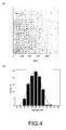
- Figure 4(a) is an image of dextran coated magnetic nanoparticles taken with a magnetic force microscope.
- Figure 4(b) is a curve showing the diameter distribution of dextran coated magnetic nanoparticles.
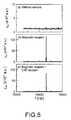
- Figure 5(a) is an ⁇ ac spectrum for the noise level of the SQUID-based MRA system
- Figure 5(b) is an ⁇ ac spectrum for 100- ⁇ L and 0.32-emu/g magnetic reagent
- Figure 5(c) is an ⁇ ac spectrum for a mixture of 100- ⁇ L and 0.32-emu/g magnetic reagent and 20- ⁇ L and 10 -5 -mg/L CRP solution.
- Figure 6 is a diagram showing ⁇ ac, ⁇ / ⁇ ac,o as a function of ⁇ CRP from 5x10 -7 to 10 -3 mg/L, and the ⁇ ac, ⁇ / ⁇ ac,o - ⁇ CRP curve at lower ⁇ CRP 's is enlarged in the inset.
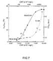
- Figure 7 is a diagram showing the CRP concentration ⁇ CRP dependent ⁇ ac, ⁇ / ⁇ ac (solid line) and OD 450 (dashed line).
- Figure 8 is a diagram showing the F9 concentration ⁇ F9 dependent ⁇ ac, ⁇ / ⁇ ac (solid line) and OD 450 (dashed line).
- Figure 9 is a diagram showing the LMG concentration ⁇ LMG dependent ⁇ ac / ⁇ ac (solid line) and OD 450 (dashed line).
- the present invention provides an alternative methodology for the magnetically labeled diagnosis (MLD), in which the amount of the to-be-measured biotargets can be correlated with the degree of reduction in multiple-frequency ac magnetic susceptibility of the magnetic nanoparticles after the magnetic nanoparticles are conjugated with the biotargets.
- MRA magnetoreduction assay
- the present invention also provides an ultra-sensitive system to perform the magnetoreduction assay.
- the ultra-sensitive MRA system of the invention adopts magnetometers or gradiometers, such as a superconducting quantum interference device (SQUID) as sensors for the system and applies the flux-transfer technology in order to perform assays with high sensitivity, which is particularly requisite for certain kinds of bio-targets, such as cytokines or vascular endothelial growth factor (VEGF) or a protein expressed by tumors.
- SQUID superconducting quantum interference device
- bio-targets such as cytokines or vascular endothelial growth factor (VEGF) or a protein expressed by tumors.
- MRA Magnetoreduction Assay
- a magnetic reagent which is a solution having homogeneously dispersed magnetic nanoparticles coated with a hydrophilic surfactants and bio-receptors.
- the preparation of the reagent principally includes mixing magnetic nanoparticles in a surfactant solution for coating the surfaces of the magnetic nanoparticles with the surfactant.
- the bio-receptors are then added to the solution and to bind with the surfactants on the surface of the magnetic nanoparticles.
- the bio-receptors include, for example, antibodies or antigens, which can be monoclonal or polyclonal.
- Under external multiple ac magnetic fields the magnetic nanoparticles oscillate via magnetic interaction.
- the magnetic reagent under the external multiple ac magnetic fields manifests a magnetic property, known as multiple-frequency ac magnetic susceptibility ⁇ ac .
- the to-be-detected biomolecules When a solution of the to-be-detected biomolecues is mixed with the reagent, the to-be-detected biomolecules may become associated with the surfactant-and-bio-receptor-coated magnetic nanoparticles through the bio-receptors bound to the outmost shell of the magnetic nanoparticles. Due to the association with the to-be-detected biomolecules, the surfactant-and-bio-receptor-coated magnetic nanoparticles either become larger or form clusters as shown in the schematic illustration in Figure 1 . In the situation that the biomolecules have only one effective single active eptiope, each biomolecule only conjugates with or binds to one magnetic nanoparticle. Hence, the magnetic nanoparticles would become larger instead of forming clusters as shown in Figure 2 .
- this method is referred as a magnetoreduction assay.
- a magnetoreduction assay when more amounts of the to-be-detected biomolecules are mixed into the reagent, more magnetic nanoparticles become larger or clustered. Ultimately, a larger reduction in ⁇ ac is observed.
- the MRA of the present invention presents at least the following merits. First of all, it is unnecessary to remove the unbounded to-be-detected biomolecules and magnetic nanoparticles. These unbounded biomolecules and magnetic nanoparticles may remain in the regent. Hence, the assay process of the invention is simpler by obviating the tedious washing processes. Secondly, only one kind of antibody is used. Thirdly, MRA is direct and homogeneous assay, which usually shows high reliability and sensitivity. Fourthly, because the degree of reduction in ⁇ ac can be accurately measured to correspond to the concentration of the biomolecules, the concentration of the biomolecules can be quantitatively determined. Additionally, MRA is capable of measuring small biomolecules.
- the small molecules Due to the small size, the small molecules are almost fully enveloped by the bio-receptor after the association. Accordingly, once a small molecule is conjugated with a bio-receptor, other epitope on the small molecule is unavailable to be bound to other bio-receptors. Hence, small biomolecules are effectively single-active-epitope due to their structure.
- the MRA of the present invention has been demonstrated for not only its convenience since the series of wash processes can be eliminated, it is highly sensitive and has a high level of specificity.
- SQUID-based MRA system Since the ac magnetic signals of samples are detected in MRA, the sensitivity in detecting biomolecules can be promoted by either using an ultra-sensitive sensor to probe the ac magnetic signals or by enhancing the ac magnetic signals to be detected. It has been demonstrated that the superconducting quantum interference devices (SQUIDs) are promising candidates as sensors in an MRA system.
- SQUIDs superconducting quantum interference devices
- the difference in the ac magnetic susceptibility ⁇ ac of a magnetic reagent before and after the bio-targets have been added therein and conjugated with the bio-receptors on the magnetic nanoparticles is measured under the actions of two excitation magnetic fields.
- the SQUID is disturbed by the two excitation magnetic fields when a magnetic reagent is positioned closed to the SQUID.
- FIG. 3 is a schematic diagram illustrating the SQUID-based MRA system according to the present invention.
- the system comprises two units: the sample unit 100a and the sensor unit 100b.
- the sample unit 100a includes at least two excitation coils 101, 102 driven by two independent function generators 103, 104 to provide a to-be-detected sample 105 with two ac excitation magnetic fields having frequencies f 1 and f 2 , respectively.
- the sample 105 is placed inside one section of a pick-up coil 106, which is constructed of two sections of coils wired in opposite directions.
- the ac magnetization of the sample 105 under the actions of the two excitation magnetic fields is detected by the pick-up coil 106.
- the sensor unit 100b includes at least a high-transition-temperature (high-T c ) rf SQUID magnetometer 109, which is immersed in liquid nitrogen contained a dewar 110. It should be appreciated that other types of SQUID magnetometers or gradiometers may also be used as sensors in the system of the present invention, even including low-transition-temperature SQUIDs immersed in liquid helium contained in a dewar.
- the dewar 110 capable of holding 5 L of liquid nitrogen 111 for example, is configured inside the magnetically shielded box and a rf shielded room 113 to prevent the SQUID from the environmental low- or high-frequency noises.
- the shielded factor approaches 90dB at high frequencies.
- a sponge 114 is used to insulate the SQUID from mechanical vibrations.
- a couple coil 107 manufactured from copper (Cu) and connected with the pick-up coil is used. Due to the ac magnetic flux through the pick-up coil 106, voltage is induced, and then a current is generated along the pick-up coil 106 and the couple coil 107. When the ac current flowed through the couple coil, an ac magnetic field is generated.
- the SQUID is positioned inside the couple coil, so that the ac magnetic field generated by the couple coil is probed by the SQUID.
- the ac magnetic flux originally generated at the sample part is efficiently transferred to the sensor part of the SQUID-based MRA system via a transfer set consisted of a pick-up coil 106 and a couple coil 107.
- a transfer set consisted of a pick-up coil 106 and a couple coil 107.
- the SQUID electronics 115 and read-out electronics 116 are used for the SQUID to detect the ac magnetic flux originally generated by the sample at the sample part.
- the output voltages from the electronics are fed into a spectrum analyzer.
- polyclonal anti-goat-CRP is coated onto the magnetic nanoparticles
- monoclonal anti-mouse-F9 is coated onto magnetic nanoparticles to magnetically label F9.
- the to-be-detected CRP functions as a multiple-active-epitope
- F9 functions as a single-active-epitope molecule.
- the small molecules are effectively single-active-epitope molecules due to their structure.
- the MRA results are compared with data obtained from ELISA.
- the magnetic nanoparticles dispersed in a phosphate buffer saline (PBS) solution are coated with a hydrophilic surfactant, such as dextran (GABC Co.).
- a hydrophilic surfactant such as dextran (GABC Co.).
- the surfactant helps the dispersion of the magnetic nanoparticles in the PBS solution or alternatively improves the binding of the bio-receptors to the surface of the nanoparticles.
- the material of the core of the magnetic nanoparticles includes, for example, Fe 3 O 4 .
- MnFe 2 O 4 Fe 2 O 3 , NiFe 2 O 4 or CoFe 2 O 4 are also applicable as the material of the magnetic nanoparticles and be comprised within the scope of the invention.
- MFM magnetic force microscope
- Figure 4(a) A typical image of magnetic nanoparticles taken by MFM is shown in Figure 4(a) .
- the dark spots correspond to the individual magnetic nanoparticles.
- Figure 4(b) The guiding line in Figure 4(b) follows Gaussian distribution.
- the mean value and the standard deviation in diameter of the magnetic nanoparticles are 29.3 nm and 1.4 nm, respectively.
- the mean diameter of magnetic nanoparticles used in following assays range from about 5 nm to about 700 nm.
- a hydrophilic surfactant for example, dextran
- a hydrophilic surfactant for example, dextran
- bio-receptors Through magnetic separation, the unbound bio-receptors are removed and the magnetic nanoparticles conjugated with biorecetpors are obtained.
- CRP polyclonal anti-goat-CRP
- F9 monoclonal anti-mouse-F9
- LMG anti-rabbit-LMG (GlycoNex Inc.) is coated onto the nanoparticles through the surfactant on the surfaces of the nanoparticles and envelops substantially the entirety of the individual nanoparticle.
- the ac magnetic susceptibility ⁇ ac spectra of 100- ⁇ l of the various magnetic reagents are measured using the SQUID-based MRA system. Then, each magnetic reagent is mixed with 20- ⁇ l solutions of various amounts of human CRP (Sigma, C4063) or F9 (Abnova) or LMG (GlycoNex Inc.), respectively, to form mixture solutions.
- Varying concentrations of immune complexes of CRP-anti-goat-CRP (or F9-anti-mouse-F9 or LMG-anti-rabbit-LMG) are developed in the mixture solutions after incubation. Thereafter, the ⁇ ac spectra of each mixture solution having immune complexes are analyzed by using the SQUID-based MRA system. A reduction in the ⁇ ac at a given frequency is observed for each reagent solution mixed with the various concentrations of CRP (or F9, or LMG). Hence, a relationship between a reduction in ⁇ ac and the concentration of CRP (or F9 or LMG) is established.
- ELISA Measurement A commercial assay kits (Anogen, EL 10022) of sandwich ELISA is used for quantitative detection of large molecules, such as human CRP. The assay procedure is described briefly herein. A 100- ⁇ l of human CRP is added to an ELISA plate, on the bottom of which an antibody pre-coated plate is seated. The well is covered with a plastic cover, and the CRP-antibody immune complex is incubated for 30 minutes. The solution is removed by filling the well with a wash buffer, and pouring the solution from the well. The wash process is repeated for four more times. After the final wash, the plate is inverted and is furthered dried by tapping the plate on an absorbent paper until no obvious moisture remained.
- HRP horseradish peroxide
- Sandwich ELISA is also used for assaying F9.
- a combination of anti-Human coagulation factor IX, F9 (Abnova, H00002158-M01) and polyclonal rabbit anti-GST antibody is used for quantitative detection of recombinant GST-tagged human F9 (H000002158-Q01).
- Human F9 antibody is initially coated on the microtiter plate at 1 ⁇ g/100 ⁇ l/well at 4 °C overnight. The wells are then blocked with 5% skim milk in PBS at room temperature for one hour. Different concentrations of recombinant GST-tagged human F9 in 100 ⁇ l of diluents were then added to appropriate wells. The plate is then incubated at room temperature for one hour.
- Goat anti rabbit InG H+L
- HRP secondary antibody is applied to the wells and the plate is incubated at room temperature for one hour.
- a 100- ⁇ l OPD substrate solution is then added to the well to develop for 30 minutes, followed by adding a stop solution into the well.
- the optical density of the solution in the wells is measured with an ELISA reader (BIO-TEK, uQuant).
- sandwich ELISA is inapplicable. Instead, competitive ELISA (GlycoNex Inc., 101G002A) is used to quantitatively detect LMG.
- the general protocols for competitive ELISA are usually available in the manual of the assay kit and thus will not be further reiterated.
- the noise spectrum of the SQUID-based MRA around the target frequency mf 1 +nf 2 is measured and is illustrated in Figure 5(a) , where f 1 and f 2 are excitation frequencies of several to tens of kHz, and m and n are non-zero integers.
- the results reveal a noise level at 1.7 x 10 -5 (a.u.) for the ⁇ ac spectrum.
- the frequency dependent ⁇ ac of the magnetic reagent is shown in Figure 5(b) when the 100- ⁇ l magnetic reagent of 0.32 emu/g in concentration is located inside one section of the pick-up coil.
- a clear peak having a maximum value of 117x10 -5 (a.u.) is observed at mf 1 +nf 2 . This maximum value is referred to as ⁇ ac,o .
- the results shown in Figure 5(b) suggest a value of about 76 for the signal-to-noise ratio.
- the stable ⁇ ac spectrum is shown in Figure 5(c) after adding 20- ⁇ l CRP solution of a given concentration ⁇ CRP , for example, 10 -5 mg/L, into the magnetic reagent.
- the maximum value of the peak at mf 1 +mf 2 is 84.4x10 -5 (a.u.), which is referred to as ⁇ ac, ⁇ .
- ⁇ ac, ⁇ is smaller than ⁇ ac,o .
- a parameter ⁇ ac, ⁇ / ⁇ ac,o is defined as an indicator for the concentration of the amount of CRP, where ⁇ ac, ⁇ / ⁇ ac,o ⁇ ( ⁇ ac, ⁇ - ⁇ ac, ⁇ )/ ⁇ ac,o .
- ⁇ ac, ⁇ / ⁇ ac,o is determined to be 27.9%.
- the ⁇ ac, ⁇ / ⁇ ac,o as a function of ⁇ CRP from 5x10 -7 to 10 -3 mg/L is plotted in Figure 6 .
- the ⁇ ac, ⁇ / ⁇ ac,o vs. ⁇ CRP curve at the lower ⁇ CPR 's is enlarged in the insert of Figure 6 .
- the results clearly demonstrate that the sensitivity in concentration of assaying CRP using the SQUID-based MRA system of the invention is around 10 -6 mg/L, i.e. 1 ppt.
- the sensitivity for assaying CRP in mole units is around 1.4x10 -20 mole.
- the ⁇ ac, ⁇ / ⁇ ac,o vs. ⁇ CRP curve is compared with the assaying characteristic curve (OD 450 - ⁇ CRP curve) via ELISA as shown in Figure 7 respectively with the solid line and the dash line. It is clear that the sensitivity on assaying CRP via ELISA is around 0.1 mg/L, which is much less sensitive than that of the SQUID-based MRA system by a factor of 10 5 .
- F9 human coagulation factor IX (F9) measured by the SQUID-based MRA system with ultra high sensitivity vs. the conventional ELISA are summarized as follows.
- F9 circulates in the blood as an inactive zymogen.
- F9 is converted to an active form by factor XIa, which excises the activation peptide and thus generates a heavy chain and a light chain held together by one or more disulfide bonds.
- the role of activated F9 in the blood coagulation cascade is to activate factor X to its active form through interactions with Ca +2 ions, membrane phospholipids, and factor VIII.
- Alterations of F9, including point mutations, insertions and deletions induce F9 deficiency, which is a recessive X-linked disorder, and is known as hemophilia B or Christmas disease. It is worthy to note that the anti-mouse-F9 coated on the magnetic nanoparticles in this aspect of the invention is monoclonal. This implies that the F9 in this case is effectively a single-active-epitope molecule.
- the ⁇ ac, ⁇ / ⁇ ac,o curve for F9 via the SQUID-based MRA measurement is plotted with the solid line in Figure 8 .
- the sensitivity of the SQUID-based MRA system is demonstrated to be 10 -6 mg/L, for example, 1 ppt.
- ELISA results demonstrate a sensitivity of 10 -3 mg/L, for example 1 ppb. Accordingly, the sensitivity of ELISA for F9 is just one part of thousand of that of the SQUID-based MRI system. In brief, ultra-high sensitivity is also achievable with the SQUID-based MRA for assaying single-active-epitope molecules.
- LMG having a molecular weight of 23.6 Da
- LMG is the principle metabolite of malachite green in aquatic beings. Malachite green is used medicinally in dilute solution as a local antiseptic or to treat parasites, fungal infections for aquatic eggs and young fry.
- an over dose of LMG may lead to liver cancer for human.
- LMG is one kind of carcinogens to human.
- the solid line denotes the characteristic curve for the relationship between the ⁇ ac, ⁇ / ⁇ ac,o and the LMG concentration ⁇ LMG via the SQUID-based MRA measurements.
- the sensitivity is shown to be around 10 -9 mg/L, for example about 10 -3 ppt.
- the characteristic curve for the relationship between OD 450 and the LMG concentration ⁇ LMG is presented with the dashed line in Figure 9 .
- the sensitivity for competitive ELISA is shown to have a sensitivity of 5x10 -5 mg/L, for example 0.05 ppb.
- the results shown in Figure 9 reveal the fact that the SQUID-based MRA is much more sensitive than ELISA by about four orders of magnitude for assaying small molecules, such as LMG
- a high-T c SQUID-based MRA system has been developed for assaying bio-targets including small biomolecules with ultra-high sensitivity.
- the SQUID sensor is prevented from being disturbed by the ac excited magnetic fields; hence, the SQUID sensor can be stable in a long-time operation.
- the sensor-end loop of the transfer coil can be placed very close to the SQUID, implying that an ultra-high sensitivity achieved for probing detected magnetic signals.
- the present invention has also demonstrated that the ultra-high sensitive SQUID-based MRA system is applicable to performing assays on multiple-active-epitope molecules, single-active-epitope molecules or small molecules.
- the assay results show that sensitivity for the SQUID-based MRA system of the invention is 1 ppt or lower. It is believed that the sensitivity could be further improved when antibodies having higher titer to the detected molecules are coated onto magnetic nanoparticles. Accordingly, the SQUID-based MRA is a promising diagnostic tool for detecting proteins, toxic molecules, bacteria, viruses or even DNA at ultra-low levels of concentration.
Landscapes
- Health & Medical Sciences (AREA)
- Chemical & Material Sciences (AREA)
- Immunology (AREA)
- Life Sciences & Earth Sciences (AREA)
- Engineering & Computer Science (AREA)
- Pathology (AREA)
- General Physics & Mathematics (AREA)
- General Health & Medical Sciences (AREA)
- Biochemistry (AREA)
- Analytical Chemistry (AREA)
- Physics & Mathematics (AREA)
- Urology & Nephrology (AREA)
- Molecular Biology (AREA)
- Hematology (AREA)
- Biomedical Technology (AREA)
- Chemical Kinetics & Catalysis (AREA)
- Electrochemistry (AREA)
- Food Science & Technology (AREA)
- Medicinal Chemistry (AREA)
- Microbiology (AREA)
- Cell Biology (AREA)
- Biotechnology (AREA)
- Investigating Or Analyzing Materials By The Use Of Magnetic Means (AREA)
Applications Claiming Priority (1)
| Application Number | Priority Date | Filing Date | Title |
|---|---|---|---|
| US11/867,207 US8034634B2 (en) | 2005-11-16 | 2007-10-04 | Ultra-sensitive magnetoreduction measurement system and ultra-sensitive, wash-free assay using the same |
Publications (2)
| Publication Number | Publication Date |
|---|---|
| EP2045599A2 true EP2045599A2 (de) | 2009-04-08 |
| EP2045599A3 EP2045599A3 (de) | 2010-06-02 |
Family
ID=40336393
Family Applications (1)
| Application Number | Title | Priority Date | Filing Date |
|---|---|---|---|
| EP08075808A Withdrawn EP2045599A3 (de) | 2007-10-04 | 2008-10-04 | Hochempfindliches Magnetreduktionsmesssystem und Methode zur Bestimmung der Konzentration von Epitop-Biomolekülen |
Country Status (5)
| Country | Link |
|---|---|
| US (1) | US8034634B2 (de) |
| EP (1) | EP2045599A3 (de) |
| JP (1) | JP5059730B2 (de) |
| CN (2) | CN101403725A (de) |
| TW (1) | TWI429903B (de) |
Families Citing this family (19)
| Publication number | Priority date | Publication date | Assignee | Title |
|---|---|---|---|---|
| JP5205807B2 (ja) * | 2007-05-17 | 2013-06-05 | 株式会社日立製作所 | 磁気信号計測装置 |
| CN101868286A (zh) | 2007-09-20 | 2010-10-20 | 马格雷股份有限公司 | 采用磁性传感器进行的分析物检测 |
| US9176206B2 (en) * | 2008-03-07 | 2015-11-03 | California Institute Of Technology | Effective-inductance-change based magnetic particle sensing |
| US8053250B2 (en) * | 2008-06-27 | 2011-11-08 | Rex Chin-Yih Hong | Method and system for suppressing bindings on magnetic particles |
| US9599591B2 (en) | 2009-03-06 | 2017-03-21 | California Institute Of Technology | Low cost, portable sensor for molecular assays |
| US20110074403A1 (en) * | 2009-09-30 | 2011-03-31 | Herng-Er Horng | High-sensitivity, in-vivo, and dynamic detection of magnetic particles within living organism using a probe-type squid system |
| TWI421490B (zh) * | 2010-03-17 | 2014-01-01 | Univ Nat Sun Yat Sen | 偵測與顯像磁性金屬蛋白之方法 |
| WO2012011477A1 (ja) * | 2010-07-21 | 2012-01-26 | 株式会社日立製作所 | 磁場計測装置 |
| CN103926399A (zh) * | 2011-02-24 | 2014-07-16 | 磁量生技股份有限公司 | 食品中动物用药残留的检测方法 |
| CN102650634A (zh) * | 2011-02-24 | 2012-08-29 | 磁量生技股份有限公司 | 食品中动物用药残留的检测试剂 |
| JP6101215B2 (ja) * | 2011-03-09 | 2017-03-22 | マグアレイ,インコーポレイテッド | サンプル中の分析対象物に対するハイスループット検出を行うシステム及び方法 |
| EP2735879A1 (de) * | 2012-11-23 | 2014-05-28 | Universiteit Twente | Verfahren und Vorrichtung zum Messen des Anteils an superparamagnetischem Material in einem Objekt |
| JP6422935B2 (ja) | 2013-03-15 | 2018-11-14 | マグアレイ,インコーポレイテッド | 磁気トンネル接合センサ及びその使用方法 |
| CN104237526B (zh) * | 2013-06-18 | 2016-08-17 | 磁量生技股份有限公司 | 一种检测阿兹海默症罹患风险的系统 |
| US10145928B2 (en) * | 2013-11-28 | 2018-12-04 | Medimagemetric LLC | Differential approach to quantitative susceptibility mapping without background field removal |
| DE102014202604B4 (de) * | 2014-02-13 | 2018-09-20 | Siemens Healthcare Gmbh | Automatisierte Ermittlung der Resonanzfrequenzen von Protonen für Magnetresonanzexperimente |
| TWI637722B (zh) * | 2017-01-12 | 2018-10-11 | 廖書賢 | 一種磁性檢測裝置及使用磁性檢測裝置檢測的方法 |
| CN107807142A (zh) * | 2017-10-26 | 2018-03-16 | 北京航空航天大学 | 一种固体所含杂质浓度的测量系统及测量方法 |
| CN112652402B (zh) * | 2020-12-11 | 2021-10-15 | 华中科技大学 | 一种基于磁纳米粒子谐波响应的病毒检测方法及系统 |
Citations (1)
| Publication number | Priority date | Publication date | Assignee | Title |
|---|---|---|---|---|
| US20070111330A1 (en) * | 2005-11-16 | 2007-05-17 | Chin-Yih Rex Hong | Methods of quantitatively measuring biomolecules |
Family Cites Families (15)
| Publication number | Priority date | Publication date | Assignee | Title |
|---|---|---|---|---|
| US3826972A (en) * | 1973-08-17 | 1974-07-30 | Univ Leland Stanford Junior | Method and apparatus for detecting nuclear magnetic resonance |
| JPS6390765A (ja) | 1986-10-03 | 1988-04-21 | Nippon Telegr & Teleph Corp <Ntt> | Squid免疫測定法 |
| IL82873A0 (en) * | 1987-06-15 | 1987-12-20 | Orgenics Ltd | Reversed competitive solid phase immunoassay for detecting single epitope analytes and kit therefor |
| US5268305A (en) * | 1989-06-15 | 1993-12-07 | Biocircuits Corporation | Multi-optical detection system |
| US5256541A (en) * | 1991-11-06 | 1993-10-26 | Sangstat Medical Corporation | Detection of soluble alloantigen immune complexes |
| JPH0666905A (ja) * | 1992-08-21 | 1994-03-11 | Furukawa Electric Co Ltd:The | 超電導磁気干渉計 |
| JPH0743441A (ja) * | 1993-08-02 | 1995-02-14 | Furukawa Electric Co Ltd:The | 超電導磁気センサ |
| DE19615254C2 (de) * | 1996-04-18 | 1999-03-11 | Diagnostikforschung Inst | Gerät zur höchstempfindlichen magnetischen Detektion von Analyten |
| FR2758884B1 (fr) | 1997-01-30 | 1999-04-02 | Bio Merieux | Procede pour isoler, notamment detecter ou quantifier un analyte dans un milieu |
| JP4171139B2 (ja) * | 1999-07-21 | 2008-10-22 | 住友電気工業株式会社 | 磁性体標識による免疫検査方法とその装置 |
| AU2002320058A1 (en) * | 2001-06-06 | 2002-12-16 | The General Hspital Corporation | Magnetic-nanoparticle conjugates and methods of use |
| WO2003016328A1 (en) * | 2001-08-13 | 2003-02-27 | Massachusetts Institute Of Technology | Methods for the treatment and prevention of cancer |
| WO2003019188A1 (en) | 2001-08-31 | 2003-03-06 | Imego Ab | Methdo and arrangement for analyzing substances |
| US6825655B2 (en) * | 2001-08-31 | 2004-11-30 | Imego Ab | Method and arrangement for detecting changes of a magnetic response in magnetic particles |
| US20030076087A1 (en) * | 2001-08-31 | 2003-04-24 | Imego Ab | Method and arrangement relating to substance analysis |
-
2007
- 2007-10-04 US US11/867,207 patent/US8034634B2/en active Active
-
2008
- 2008-02-05 TW TW097104627A patent/TWI429903B/zh active
- 2008-03-18 CN CNA2008100830223A patent/CN101403725A/zh active Pending
- 2008-03-18 CN CN2010105823793A patent/CN102128878A/zh active Pending
- 2008-10-03 JP JP2008259020A patent/JP5059730B2/ja active Active
- 2008-10-04 EP EP08075808A patent/EP2045599A3/de not_active Withdrawn
Patent Citations (1)
| Publication number | Priority date | Publication date | Assignee | Title |
|---|---|---|---|---|
| US20070111330A1 (en) * | 2005-11-16 | 2007-05-17 | Chin-Yih Rex Hong | Methods of quantitatively measuring biomolecules |
Non-Patent Citations (1)
| Title |
|---|
| HONG C-Y ET AL: "Nanomagnetic Particles for SQUID-Based Magnetically Labeled Immunoassay", IEEE TRANSACTIONS ON APPLIED SUPERCONDUCTIVITY, IEEE SERVICE CENTER, LOS ALAMITOS, CA, US, vol. 15, no. 2, 1 June 2005 (2005-06-01), pages 668 - 671, XP011133687, ISSN: 1051-8223, DOI: 10.1109/TASC.2005.849995 * |
Also Published As
| Publication number | Publication date |
|---|---|
| JP5059730B2 (ja) | 2012-10-31 |
| EP2045599A3 (de) | 2010-06-02 |
| CN102128878A (zh) | 2011-07-20 |
| JP2009098145A (ja) | 2009-05-07 |
| US20080024117A1 (en) | 2008-01-31 |
| CN101403725A (zh) | 2009-04-08 |
| TWI429903B (zh) | 2014-03-11 |
| US8034634B2 (en) | 2011-10-11 |
| TW200916768A (en) | 2009-04-16 |
Similar Documents
| Publication | Publication Date | Title |
|---|---|---|
| US8034634B2 (en) | Ultra-sensitive magnetoreduction measurement system and ultra-sensitive, wash-free assay using the same | |
| US6825655B2 (en) | Method and arrangement for detecting changes of a magnetic response in magnetic particles | |
| Chieh et al. | Hyper-high-sensitivity wash-free magnetoreduction assay on biomolecules using high-Tc superconducting quantum interference devices | |
| US8217647B2 (en) | Measuring agglutination parameters | |
| KR100412006B1 (ko) | 분석물의자기이완측정검출을위한방법및화합물,및그의용도 | |
| US8624584B2 (en) | Method and arrangement relating to analyses of a substance | |
| JP4184268B2 (ja) | 物質を分析するための方法と構成 | |
| US10488408B2 (en) | Detection of target molecules in a sample by using a magnetic field | |
| US20090164161A1 (en) | Device for measuring ac magnetization of materials and method for detecting bio-molecules | |
| US10197564B2 (en) | Nuclear magnetic resonance apparatus and methods | |
| KR20060127918A (ko) | 온-칩 자기공명 측정 장치, 방법 및 이 장치의 사용 방법 | |
| Wang et al. | Highly sensitive and quantitative magnetic nanoparticle-based lateral flow immunoassay with an atomic magnetometer | |
| US20030076087A1 (en) | Method and arrangement relating to substance analysis | |
| US11885800B2 (en) | Method and system for detecting analyte of interest using magnetic field sensor and magnetic particles | |
| Sun et al. | Sandwich immunoassay for the prostate specific antigen using a micro-fluxgate and magnetic bead labels | |
| CN101627297B (zh) | 测量凝集参数 | |
| Yang et al. | Ultra-highly sensitive and wash-free bio-detection of H5N1 virus by immunomagnetic reduction assays | |
| Tsukada et al. | Using magnetic field gradients to shorten the antigen-antibody reaction time for a magnetic immunoassay | |
| JP2004530896A (ja) | アナライトを定性的および定量的に検出する方法および装置 | |
| US20160025825A1 (en) | Nuclear magnetic resonance apparatus and methods | |
| JP2007292748A (ja) | センサ素子、ならびにこの素子を用いた磁性粒子の検出方法及び標的物質の検出方法 | |
| US20080268481A1 (en) | Sensitive Magnetic Catch Assay By Building a Strong Binding Couple | |
| US10094897B2 (en) | Nuclear magnetic resonance apparatus and methods | |
| Oisjoen et al. | Fast and sensitive measurement of specific antigen-antibody binding reactions with magnetic nanoparticles and HTS SQUID | |
| US6979574B1 (en) | Process for detecting binding reactions with use of the measurement of the relaxation of the double refraction of magnetic particles |
Legal Events
| Date | Code | Title | Description |
|---|---|---|---|
| PUAI | Public reference made under article 153(3) epc to a published international application that has entered the european phase |
Free format text: ORIGINAL CODE: 0009012 |
|
| 17P | Request for examination filed |
Effective date: 20081004 |
|
| AK | Designated contracting states |
Kind code of ref document: A2 Designated state(s): AT BE BG CH CY CZ DE DK EE ES FI FR GB GR HR HU IE IS IT LI LT LU LV MC MT NL NO PL PT RO SE SI SK TR |
|
| AX | Request for extension of the european patent |
Extension state: AL BA MK RS |
|
| PUAL | Search report despatched |
Free format text: ORIGINAL CODE: 0009013 |
|
| AK | Designated contracting states |
Kind code of ref document: A3 Designated state(s): AT BE BG CH CY CZ DE DK EE ES FI FR GB GR HR HU IE IS IT LI LT LU LV MC MT NL NO PL PT RO SE SI SK TR |
|
| AX | Request for extension of the european patent |
Extension state: AL BA MK RS |
|
| RIC1 | Information provided on ipc code assigned before grant |
Ipc: G01N 33/543 20060101ALI20100426BHEP Ipc: G01N 27/74 20060101AFI20090213BHEP |
|
| AKX | Designation fees paid |
Designated state(s): AT BE BG CH CY LI |
|
| RBV | Designated contracting states (corrected) |
Designated state(s): AT BE BG CH CY CZ DE DK EE ES FI FR GB GR HR HU IE IS IT LI LT LU LV MC MT NL NO PL PT RO SE SI SK TR |
|
| REG | Reference to a national code |
Ref country code: DE Ref legal event code: 8566 |
|
| RAP1 | Party data changed (applicant data changed or rights of an application transferred) |
Owner name: MAGQU CO., LTD. |
|
| RIN1 | Information on inventor provided before grant (corrected) |
Inventor name: HORNG, HERNG-ER Inventor name: YANG, HONG-CHANG Inventor name: YANG, SHIEH-YUEH Inventor name: HONG REX, CHIN-YIH |
|
| 17Q | First examination report despatched |
Effective date: 20160229 |
|
| APBK | Appeal reference recorded |
Free format text: ORIGINAL CODE: EPIDOSNREFNE |
|
| APBN | Date of receipt of notice of appeal recorded |
Free format text: ORIGINAL CODE: EPIDOSNNOA2E |
|
| APBR | Date of receipt of statement of grounds of appeal recorded |
Free format text: ORIGINAL CODE: EPIDOSNNOA3E |
|
| APAF | Appeal reference modified |
Free format text: ORIGINAL CODE: EPIDOSCREFNE |
|
| APBT | Appeal procedure closed |
Free format text: ORIGINAL CODE: EPIDOSNNOA9E |
|
| APAM | Information on closure of appeal procedure modified |
Free format text: ORIGINAL CODE: EPIDOSCNOA9E |
|
| APBB | Information on closure of appeal procedure deleted |
Free format text: ORIGINAL CODE: EPIDOSDNOA9E |
|
| APBT | Appeal procedure closed |
Free format text: ORIGINAL CODE: EPIDOSNNOA9E |
|
| STAA | Information on the status of an ep patent application or granted ep patent |
Free format text: STATUS: THE APPLICATION HAS BEEN WITHDRAWN |
|
| 18W | Application withdrawn |
Effective date: 20211008 |