EP2053393A1 - Pladienolid-zielmolekül, zur bindung an das zielmolekül fähige verbindung und screening-verfahren für die verbindung - Google Patents
Pladienolid-zielmolekül, zur bindung an das zielmolekül fähige verbindung und screening-verfahren für die verbindung Download PDFInfo
- Publication number
- EP2053393A1 EP2053393A1 EP07792236A EP07792236A EP2053393A1 EP 2053393 A1 EP2053393 A1 EP 2053393A1 EP 07792236 A EP07792236 A EP 07792236A EP 07792236 A EP07792236 A EP 07792236A EP 2053393 A1 EP2053393 A1 EP 2053393A1
- Authority
- EP
- European Patent Office
- Prior art keywords
- group
- compound
- hydrogen
- methyl
- sf3b
- Prior art date
- Legal status (The legal status is an assumption and is not a legal conclusion. Google has not performed a legal analysis and makes no representation as to the accuracy of the status listed.)
- Withdrawn
Links
- 150000001875 compounds Chemical class 0.000 title claims abstract description 161
- 238000000034 method Methods 0.000 title claims abstract description 62
- 230000027455 binding Effects 0.000 title claims abstract description 39
- 238000012216 screening Methods 0.000 title description 5
- 238000012360 testing method Methods 0.000 claims abstract description 59
- 102000015097 RNA Splicing Factors Human genes 0.000 claims abstract description 7
- 108010039259 RNA Splicing Factors Proteins 0.000 claims abstract description 7
- -1 benzenesulfonyloxy Chemical group 0.000 claims description 151
- 102100021816 Splicing factor 3B subunit 3 Human genes 0.000 claims description 49
- 229910052739 hydrogen Inorganic materials 0.000 claims description 49
- 239000001257 hydrogen Substances 0.000 claims description 49
- 101000616172 Homo sapiens Splicing factor 3B subunit 3 Proteins 0.000 claims description 46
- 101000836101 Homo sapiens Histone deacetylase complex subunit SAP130 Proteins 0.000 claims description 45
- 108090000623 proteins and genes Proteins 0.000 claims description 39
- YBJHBAHKTGYVGT-ZKWXMUAHSA-N (+)-Biotin Chemical compound N1C(=O)N[C@@H]2[C@H](CCCCC(=O)O)SC[C@@H]21 YBJHBAHKTGYVGT-ZKWXMUAHSA-N 0.000 claims description 36
- 102000004169 proteins and genes Human genes 0.000 claims description 36
- 125000002496 methyl group Chemical group [H]C([H])([H])* 0.000 claims description 35
- UFHFLCQGNIYNRP-UHFFFAOYSA-N Hydrogen Chemical compound [H][H] UFHFLCQGNIYNRP-UHFFFAOYSA-N 0.000 claims description 33
- 125000001424 substituent group Chemical group 0.000 claims description 29
- 125000000217 alkyl group Chemical group 0.000 claims description 25
- 239000007850 fluorescent dye Substances 0.000 claims description 24
- 125000002252 acyl group Chemical group 0.000 claims description 22
- 229960002685 biotin Drugs 0.000 claims description 18
- 235000020958 biotin Nutrition 0.000 claims description 18
- 239000011616 biotin Substances 0.000 claims description 18
- 230000000694 effects Effects 0.000 claims description 18
- 125000001495 ethyl group Chemical group [H]C([H])([H])C([H])([H])* 0.000 claims description 18
- 150000002431 hydrogen Chemical class 0.000 claims description 16
- 125000002887 hydroxy group Chemical group [H]O* 0.000 claims description 16
- 125000001072 heteroaryl group Chemical group 0.000 claims description 15
- 125000005915 C6-C14 aryl group Chemical group 0.000 claims description 14
- 230000036961 partial effect Effects 0.000 claims description 14
- 125000003545 alkoxy group Chemical group 0.000 claims description 12
- 239000002246 antineoplastic agent Substances 0.000 claims description 12
- 125000004423 acyloxy group Chemical group 0.000 claims description 9
- 125000004390 alkyl sulfonyl group Chemical group 0.000 claims description 9
- 229910052736 halogen Inorganic materials 0.000 claims description 9
- 150000002367 halogens Chemical class 0.000 claims description 9
- 125000001797 benzyl group Chemical group [H]C1=C([H])C([H])=C(C([H])=C1[H])C([H])([H])* 0.000 claims description 8
- 125000000999 tert-butyl group Chemical group [H]C([H])([H])C(*)(C([H])([H])[H])C([H])([H])[H] 0.000 claims description 8
- 125000004169 (C1-C6) alkyl group Chemical group 0.000 claims description 6
- 125000003668 acetyloxy group Chemical group [H]C([H])([H])C(=O)O[*] 0.000 claims description 6
- 125000005842 heteroatom Chemical group 0.000 claims description 6
- 125000001449 isopropyl group Chemical group [H]C([H])([H])C([H])(*)C([H])([H])[H] 0.000 claims description 6
- 125000001997 phenyl group Chemical group [H]C1=C([H])C([H])=C(*)C([H])=C1[H] 0.000 claims description 6
- 125000003170 phenylsulfonyl group Chemical group C1(=CC=CC=C1)S(=O)(=O)* 0.000 claims description 6
- 125000002777 acetyl group Chemical group [H]C([H])([H])C(*)=O 0.000 claims description 5
- 125000005278 alkyl sulfonyloxy group Chemical group 0.000 claims description 5
- 125000005553 heteroaryloxy group Chemical group 0.000 claims description 5
- 125000005914 C6-C14 aryloxy group Chemical group 0.000 claims description 4
- QVGXLLKOCUKJST-UHFFFAOYSA-N atomic oxygen Chemical compound [O] QVGXLLKOCUKJST-UHFFFAOYSA-N 0.000 claims description 4
- QJGQUHMNIGDVPM-UHFFFAOYSA-N nitrogen group Chemical group [N] QJGQUHMNIGDVPM-UHFFFAOYSA-N 0.000 claims description 4
- 229910052760 oxygen Inorganic materials 0.000 claims description 4
- 239000001301 oxygen Substances 0.000 claims description 4
- 125000006091 1,3-dioxolane group Chemical group 0.000 claims description 3
- 125000004184 methoxymethyl group Chemical group [H]C([H])([H])OC([H])([H])* 0.000 claims description 3
- 125000000468 ketone group Chemical group 0.000 claims 3
- 150000002923 oximes Chemical group 0.000 claims 3
- 239000000523 sample Substances 0.000 description 81
- 210000004027 cell Anatomy 0.000 description 51
- 239000000243 solution Substances 0.000 description 40
- OKKJLVBELUTLKV-UHFFFAOYSA-N Methanol Chemical compound OC OKKJLVBELUTLKV-UHFFFAOYSA-N 0.000 description 36
- XEKOWRVHYACXOJ-UHFFFAOYSA-N Ethyl acetate Chemical compound CCOC(C)=O XEKOWRVHYACXOJ-UHFFFAOYSA-N 0.000 description 25
- 238000006243 chemical reaction Methods 0.000 description 21
- 101000707770 Homo sapiens Splicing factor 3B subunit 2 Proteins 0.000 description 19
- YZCKVEUIGOORGS-NJFSPNSNSA-N Tritium Chemical compound [3H] YZCKVEUIGOORGS-NJFSPNSNSA-N 0.000 description 19
- 229910052722 tritium Inorganic materials 0.000 description 19
- YMWUJEATGCHHMB-UHFFFAOYSA-N Dichloromethane Chemical compound ClCCl YMWUJEATGCHHMB-UHFFFAOYSA-N 0.000 description 18
- 102100031436 Splicing factor 3B subunit 2 Human genes 0.000 description 18
- 239000000203 mixture Substances 0.000 description 18
- FAPWRFPIFSIZLT-UHFFFAOYSA-M Sodium chloride Chemical compound [Na+].[Cl-] FAPWRFPIFSIZLT-UHFFFAOYSA-M 0.000 description 15
- 239000002904 solvent Substances 0.000 description 15
- 238000001114 immunoprecipitation Methods 0.000 description 14
- 239000000047 product Substances 0.000 description 14
- 238000011282 treatment Methods 0.000 description 14
- 108010072724 U2 Small Nuclear Ribonucleoprotein Proteins 0.000 description 13
- 102000006986 U2 Small Nuclear Ribonucleoprotein Human genes 0.000 description 13
- 238000002474 experimental method Methods 0.000 description 12
- XLYOFNOQVPJJNP-UHFFFAOYSA-N water Chemical compound O XLYOFNOQVPJJNP-UHFFFAOYSA-N 0.000 description 12
- CSNNHWWHGAXBCP-UHFFFAOYSA-L Magnesium sulfate Chemical compound [Mg+2].[O-][S+2]([O-])([O-])[O-] CSNNHWWHGAXBCP-UHFFFAOYSA-L 0.000 description 10
- 102100031711 Splicing factor 3B subunit 1 Human genes 0.000 description 10
- 230000000259 anti-tumor effect Effects 0.000 description 10
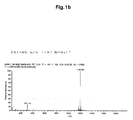
- 238000002330 electrospray ionisation mass spectrometry Methods 0.000 description 10
- 125000004432 carbon atom Chemical group C* 0.000 description 9
- 125000001301 ethoxy group Chemical group [H]C([H])([H])C([H])([H])O* 0.000 description 9
- 239000011347 resin Substances 0.000 description 9
- 229920005989 resin Polymers 0.000 description 9
- 239000011734 sodium Substances 0.000 description 9
- OKKJLVBELUTLKV-MZCSYVLQSA-N Deuterated methanol Chemical compound [2H]OC([2H])([2H])[2H] OKKJLVBELUTLKV-MZCSYVLQSA-N 0.000 description 8
- 101000707567 Homo sapiens Splicing factor 3B subunit 1 Proteins 0.000 description 8
- 238000003556 assay Methods 0.000 description 8
- 230000001085 cytostatic effect Effects 0.000 description 8
- 238000001514 detection method Methods 0.000 description 8
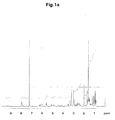
- 238000000425 proton nuclear magnetic resonance spectrum Methods 0.000 description 8
- 229920000936 Agarose Polymers 0.000 description 7
- 125000006615 aromatic heterocyclic group Chemical group 0.000 description 7
- 125000004430 oxygen atom Chemical group O* 0.000 description 7
- 239000008188 pellet Substances 0.000 description 7
- LFQSCWFLJHTTHZ-UHFFFAOYSA-N Ethanol Chemical compound CCO LFQSCWFLJHTTHZ-UHFFFAOYSA-N 0.000 description 6
- MNOMBFWMICHMKG-MGYWSNOQSA-N [(2s,3s,4e,6s,7r,10r)-7,10-dihydroxy-2-[(2e,4e,6r)-6-hydroxy-7-[(2r,3r)-3-[(2r,3s)-3-hydroxypentan-2-yl]oxiran-2-yl]-6-methylhepta-2,4-dien-2-yl]-3,7-dimethyl-12-oxo-1-oxacyclododec-4-en-6-yl] 4-cycloheptylpiperazine-1-carboxylate Chemical compound CC[C@H](O)[C@@H](C)[C@H]1O[C@@H]1C[C@@](C)(O)\C=C\C=C(/C)[C@@H]1[C@@H](C)/C=C/[C@H](OC(=O)N2CCN(CC2)C2CCCCCC2)[C@](C)(O)CC[C@@H](O)CC(=O)O1 MNOMBFWMICHMKG-MGYWSNOQSA-N 0.000 description 6
- 150000002576 ketones Chemical group 0.000 description 6
- 125000003544 oxime group Chemical group 0.000 description 6
- 239000006228 supernatant Substances 0.000 description 6
- 239000000725 suspension Substances 0.000 description 6
- VYPSYNLAJGMNEJ-UHFFFAOYSA-N Silicium dioxide Chemical compound O=[Si]=O VYPSYNLAJGMNEJ-UHFFFAOYSA-N 0.000 description 5
- 210000000172 cytosol Anatomy 0.000 description 5
- 239000012153 distilled water Substances 0.000 description 5
- 238000002372 labelling Methods 0.000 description 5
- 229910052943 magnesium sulfate Inorganic materials 0.000 description 5
- 235000019341 magnesium sulphate Nutrition 0.000 description 5
- 239000002609 medium Substances 0.000 description 5
- 239000012528 membrane Substances 0.000 description 5
- 239000003921 oil Substances 0.000 description 5
- 235000019198 oils Nutrition 0.000 description 5
- 239000012044 organic layer Substances 0.000 description 5
- 239000013612 plasmid Substances 0.000 description 5
- 239000000741 silica gel Substances 0.000 description 5
- 229910002027 silica gel Inorganic materials 0.000 description 5
- 238000010898 silica gel chromatography Methods 0.000 description 5
- 239000011780 sodium chloride Substances 0.000 description 5
- 238000002415 sodium dodecyl sulfate polyacrylamide gel electrophoresis Methods 0.000 description 5
- 125000003774 valeryl group Chemical group O=C([*])C([H])([H])C([H])([H])C([H])([H])C([H])([H])[H] 0.000 description 5
- KXDHJXZQYSOELW-UHFFFAOYSA-M Carbamate Chemical compound NC([O-])=O KXDHJXZQYSOELW-UHFFFAOYSA-M 0.000 description 4
- OKTJSMMVPCPJKN-UHFFFAOYSA-N Carbon Chemical compound [C] OKTJSMMVPCPJKN-UHFFFAOYSA-N 0.000 description 4
- 108091026890 Coding region Proteins 0.000 description 4
- 238000004458 analytical method Methods 0.000 description 4
- 230000000903 blocking effect Effects 0.000 description 4
- 239000012634 fragment Substances 0.000 description 4
- 238000010191 image analysis Methods 0.000 description 4
- VLKZOEOYAKHREP-UHFFFAOYSA-N n-Hexane Chemical compound CCCCCC VLKZOEOYAKHREP-UHFFFAOYSA-N 0.000 description 4
- 239000002244 precipitate Substances 0.000 description 4
- QKNYBSVHEMOAJP-UHFFFAOYSA-N 2-amino-2-(hydroxymethyl)propane-1,3-diol;hydron;chloride Chemical compound Cl.OCC(N)(CO)CO QKNYBSVHEMOAJP-UHFFFAOYSA-N 0.000 description 3
- 125000003903 2-propenyl group Chemical group [H]C([*])([H])C([H])=C([H])[H] 0.000 description 3
- 102000003909 Cyclin E Human genes 0.000 description 3
- 108090000257 Cyclin E Proteins 0.000 description 3
- WSFSSNUMVMOOMR-UHFFFAOYSA-N Formaldehyde Chemical compound O=C WSFSSNUMVMOOMR-UHFFFAOYSA-N 0.000 description 3
- PEDCQBHIVMGVHV-UHFFFAOYSA-N Glycerine Chemical compound OCC(O)CO PEDCQBHIVMGVHV-UHFFFAOYSA-N 0.000 description 3
- KFZMGEQAYNKOFK-UHFFFAOYSA-N Isopropanol Chemical compound CC(C)O KFZMGEQAYNKOFK-UHFFFAOYSA-N 0.000 description 3
- 206010028980 Neoplasm Diseases 0.000 description 3
- WDVSHHCDHLJJJR-UHFFFAOYSA-N Proflavine Chemical compound C1=CC(N)=CC2=NC3=CC(N)=CC=C3C=C21 WDVSHHCDHLJJJR-UHFFFAOYSA-N 0.000 description 3
- 101710190370 Splicing factor 3B subunit 3 Proteins 0.000 description 3
- SDOUORKJIJYJNW-QHOUZYGJSA-N [(2s,3s,4e,6s,7r,10r)-7,10-dihydroxy-2-[(2e,4e,6s)-7-[(2r,3r)-3-[(2r,3s)-3-hydroxypentan-2-yl]oxiran-2-yl]-6-methylhepta-2,4-dien-2-yl]-3,7-dimethyl-12-oxo-1-oxacyclododec-4-en-6-yl] acetate Chemical compound CC[C@H](O)[C@@H](C)[C@H]1O[C@@H]1C[C@H](C)\C=C\C=C(/C)[C@@H]1[C@@H](C)/C=C/[C@H](OC(C)=O)[C@](C)(O)CC[C@@H](O)CC(=O)O1 SDOUORKJIJYJNW-QHOUZYGJSA-N 0.000 description 3
- 125000002619 bicyclic group Chemical group 0.000 description 3
- 201000011510 cancer Diseases 0.000 description 3
- 230000030570 cellular localization Effects 0.000 description 3
- 239000007795 chemical reaction product Substances 0.000 description 3
- 239000003795 chemical substances by application Substances 0.000 description 3
- 239000012230 colorless oil Substances 0.000 description 3
- 230000001747 exhibiting effect Effects 0.000 description 3
- 125000002541 furyl group Chemical group 0.000 description 3
- 238000004128 high performance liquid chromatography Methods 0.000 description 3
- 238000000338 in vitro Methods 0.000 description 3
- 238000001727 in vivo Methods 0.000 description 3
- 230000003834 intracellular effect Effects 0.000 description 3
- 125000000959 isobutyl group Chemical group [H]C([H])([H])C([H])(C([H])([H])[H])C([H])([H])* 0.000 description 3
- 230000004807 localization Effects 0.000 description 3
- 125000002950 monocyclic group Chemical group 0.000 description 3
- 125000004108 n-butyl group Chemical group [H]C([H])([H])C([H])([H])C([H])([H])C([H])([H])* 0.000 description 3
- 229910052757 nitrogen Inorganic materials 0.000 description 3
- 125000004433 nitrogen atom Chemical group N* 0.000 description 3
- 230000003287 optical effect Effects 0.000 description 3
- 125000002924 primary amino group Chemical group [H]N([H])* 0.000 description 3
- 125000004076 pyridyl group Chemical group 0.000 description 3
- 125000000714 pyrimidinyl group Chemical group 0.000 description 3
- 229910052717 sulfur Inorganic materials 0.000 description 3
- 125000001544 thienyl group Chemical group 0.000 description 3
- 239000011534 wash buffer Substances 0.000 description 3
- 238000001262 western blot Methods 0.000 description 3
- 125000004972 1-butynyl group Chemical group [H]C([H])([H])C([H])([H])C#C* 0.000 description 2
- 125000001637 1-naphthyl group Chemical group [H]C1=C([H])C([H])=C2C(*)=C([H])C([H])=C([H])C2=C1[H] 0.000 description 2
- 125000005978 1-naphthyloxy group Chemical group 0.000 description 2
- 125000006017 1-propenyl group Chemical group 0.000 description 2
- 125000000530 1-propynyl group Chemical group [H]C([H])([H])C#C* 0.000 description 2
- IUVCFHHAEHNCFT-INIZCTEOSA-N 2-[(1s)-1-[4-amino-3-(3-fluoro-4-propan-2-yloxyphenyl)pyrazolo[3,4-d]pyrimidin-1-yl]ethyl]-6-fluoro-3-(3-fluorophenyl)chromen-4-one Chemical compound C1=C(F)C(OC(C)C)=CC=C1C(C1=C(N)N=CN=C11)=NN1[C@@H](C)C1=C(C=2C=C(F)C=CC=2)C(=O)C2=CC(F)=CC=C2O1 IUVCFHHAEHNCFT-INIZCTEOSA-N 0.000 description 2
- 125000000069 2-butynyl group Chemical group [H]C([H])([H])C#CC([H])([H])* 0.000 description 2
- 125000001622 2-naphthyl group Chemical group [H]C1=C([H])C([H])=C2C([H])=C(*)C([H])=C([H])C2=C1[H] 0.000 description 2
- 125000005979 2-naphthyloxy group Chemical group 0.000 description 2
- 125000001494 2-propynyl group Chemical group [H]C#CC([H])([H])* 0.000 description 2
- 125000000474 3-butynyl group Chemical group [H]C#CC([H])([H])C([H])([H])* 0.000 description 2
- QGZKDVFQNNGYKY-UHFFFAOYSA-N Ammonia Chemical compound N QGZKDVFQNNGYKY-UHFFFAOYSA-N 0.000 description 2
- 108090001008 Avidin Proteins 0.000 description 2
- 108020004414 DNA Proteins 0.000 description 2
- QUSNBJAOOMFDIB-UHFFFAOYSA-N Ethylamine Chemical compound CCN QUSNBJAOOMFDIB-UHFFFAOYSA-N 0.000 description 2
- TWRXJAOTZQYOKJ-UHFFFAOYSA-L Magnesium chloride Chemical compound [Mg+2].[Cl-].[Cl-] TWRXJAOTZQYOKJ-UHFFFAOYSA-L 0.000 description 2
- 229940124158 Protease/peptidase inhibitor Drugs 0.000 description 2
- 239000012722 SDS sample buffer Substances 0.000 description 2
- UIIMBOGNXHQVGW-UHFFFAOYSA-M Sodium bicarbonate Chemical compound [Na+].OC([O-])=O UIIMBOGNXHQVGW-UHFFFAOYSA-M 0.000 description 2
- 101710190353 Splicing factor 3B subunit 1 Proteins 0.000 description 2
- 239000013504 Triton X-100 Substances 0.000 description 2
- 229920004890 Triton X-100 Polymers 0.000 description 2
- 235000010724 Wisteria floribunda Nutrition 0.000 description 2
- MBNWAUIZHCYCKK-LLACQQGFSA-N [(4e)-7,10-bis(1-ethoxyethoxy)-2-[(2e,4e)-7-[3-[3-(1-ethoxyethoxy)pentan-2-yl]oxiran-2-yl]-6-methylhepta-2,4-dien-2-yl]-3,7-dimethyl-12-oxo-1-oxacyclododec-4-en-6-yl] n-[2-[2-(2-aminoethoxy)ethoxy]ethyl]carbamate Chemical compound CCOC(C)OC(CC)C(C)C1OC1CC(C)\C=C\C=C(/C)C1C(C)/C=C/C(OC(=O)NCCOCCOCCN)C(C)(OC(C)OCC)CCC(OC(C)OCC)CC(=O)O1 MBNWAUIZHCYCKK-LLACQQGFSA-N 0.000 description 2
- 238000009825 accumulation Methods 0.000 description 2
- 230000009471 action Effects 0.000 description 2
- 125000006323 alkenyl amino group Chemical group 0.000 description 2
- 125000003342 alkenyl group Chemical group 0.000 description 2
- 125000003282 alkyl amino group Chemical group 0.000 description 2
- 125000005115 alkyl carbamoyl group Chemical group 0.000 description 2
- 125000006319 alkynyl amino group Chemical group 0.000 description 2
- 125000000304 alkynyl group Chemical group 0.000 description 2
- 125000005336 allyloxy group Chemical group 0.000 description 2
- 125000003275 alpha amino acid group Chemical group 0.000 description 2
- 230000003466 anti-cipated effect Effects 0.000 description 2
- KXDAEFPNCMNJSK-UHFFFAOYSA-N benzene carboxamide Natural products NC(=O)C1=CC=CC=C1 KXDAEFPNCMNJSK-UHFFFAOYSA-N 0.000 description 2
- 239000000872 buffer Substances 0.000 description 2
- 230000022131 cell cycle Effects 0.000 description 2
- 239000003610 charcoal Substances 0.000 description 2
- 230000006957 competitive inhibition Effects 0.000 description 2
- 239000000470 constituent Substances 0.000 description 2
- 238000001816 cooling Methods 0.000 description 2
- 125000000753 cycloalkyl group Chemical group 0.000 description 2
- 125000001995 cyclobutyl group Chemical group [H]C1([H])C([H])([H])C([H])(*)C1([H])[H] 0.000 description 2
- 125000000113 cyclohexyl group Chemical group [H]C1([H])C([H])([H])C([H])([H])C([H])(*)C([H])([H])C1([H])[H] 0.000 description 2
- 125000001511 cyclopentyl group Chemical group [H]C1([H])C([H])([H])C([H])([H])C([H])(*)C1([H])[H] 0.000 description 2
- 125000001559 cyclopropyl group Chemical group [H]C1([H])C([H])([H])C1([H])* 0.000 description 2
- 239000003814 drug Substances 0.000 description 2
- 238000007876 drug discovery Methods 0.000 description 2
- 238000010828 elution Methods 0.000 description 2
- 239000012156 elution solvent Substances 0.000 description 2
- 108010048367 enhanced green fluorescent protein Proteins 0.000 description 2
- IWBOPFCKHIJFMS-UHFFFAOYSA-N ethylene glycol bis(2-aminoethyl) ether Chemical compound NCCOCCOCCN IWBOPFCKHIJFMS-UHFFFAOYSA-N 0.000 description 2
- 238000011156 evaluation Methods 0.000 description 2
- 239000000706 filtrate Substances 0.000 description 2
- 108020001507 fusion proteins Proteins 0.000 description 2
- 102000037865 fusion proteins Human genes 0.000 description 2
- BRZYSWJRSDMWLG-CAXSIQPQSA-N geneticin Chemical compound O1C[C@@](O)(C)[C@H](NC)[C@@H](O)[C@H]1O[C@@H]1[C@@H](O)[C@H](O[C@@H]2[C@@H]([C@@H](O)[C@H](O)[C@@H](C(C)O)O2)N)[C@@H](N)C[C@H]1N BRZYSWJRSDMWLG-CAXSIQPQSA-N 0.000 description 2
- ACGUYXCXAPNIKK-UHFFFAOYSA-N hexachlorophene Chemical compound OC1=C(Cl)C=C(Cl)C(Cl)=C1CC1=C(O)C(Cl)=CC(Cl)=C1Cl ACGUYXCXAPNIKK-UHFFFAOYSA-N 0.000 description 2
- 238000003384 imaging method Methods 0.000 description 2
- 125000002140 imidazol-4-yl group Chemical group [H]N1C([H])=NC([*])=C1[H] 0.000 description 2
- 125000002883 imidazolyl group Chemical group 0.000 description 2
- 239000012133 immunoprecipitate Substances 0.000 description 2
- 238000011534 incubation Methods 0.000 description 2
- 125000002510 isobutoxy group Chemical group [H]C([H])([H])C([H])(C([H])([H])[H])C([H])([H])O* 0.000 description 2
- 125000000555 isopropenyl group Chemical group [H]\C([H])=C(\*)C([H])([H])[H] 0.000 description 2
- 125000003253 isopropoxy group Chemical group [H]C([H])([H])C([H])(O*)C([H])([H])[H] 0.000 description 2
- 229940043355 kinase inhibitor Drugs 0.000 description 2
- 239000007788 liquid Substances 0.000 description 2
- 238000005259 measurement Methods 0.000 description 2
- 230000007246 mechanism Effects 0.000 description 2
- 125000000956 methoxy group Chemical group [H]C([H])([H])O* 0.000 description 2
- 125000004170 methylsulfonyl group Chemical group [H]C([H])([H])S(*)(=O)=O 0.000 description 2
- 239000003068 molecular probe Substances 0.000 description 2
- CZFNISFYDPIDNM-UHFFFAOYSA-N n,n-dimethylformamide;oxolane Chemical compound CN(C)C=O.C1CCOC1 CZFNISFYDPIDNM-UHFFFAOYSA-N 0.000 description 2
- 125000000740 n-pentyl group Chemical group [H]C([H])([H])C([H])([H])C([H])([H])C([H])([H])C([H])([H])* 0.000 description 2
- 125000003506 n-propoxy group Chemical group [H]C([H])([H])C([H])([H])C([H])([H])O* 0.000 description 2
- 125000004123 n-propyl group Chemical group [H]C([H])([H])C([H])([H])C([H])([H])* 0.000 description 2
- 208000015122 neurodegenerative disease Diseases 0.000 description 2
- 239000000137 peptide hydrolase inhibitor Substances 0.000 description 2
- 239000003757 phosphotransferase inhibitor Substances 0.000 description 2
- 230000001766 physiological effect Effects 0.000 description 2
- 239000012264 purified product Substances 0.000 description 2
- 125000005412 pyrazyl group Chemical group 0.000 description 2
- 125000002098 pyridazinyl group Chemical group 0.000 description 2
- ZDYVRSLAEXCVBX-UHFFFAOYSA-N pyridinium p-toluenesulfonate Chemical compound C1=CC=[NH+]C=C1.CC1=CC=C(S([O-])(=O)=O)C=C1 ZDYVRSLAEXCVBX-UHFFFAOYSA-N 0.000 description 2
- 125000005554 pyridyloxy group Chemical group 0.000 description 2
- 125000000168 pyrrolyl group Chemical group 0.000 description 2
- 238000011002 quantification Methods 0.000 description 2
- 125000002914 sec-butyl group Chemical group [H]C([H])([H])C([H])([H])C([H])(*)C([H])([H])[H] 0.000 description 2
- 230000003381 solubilizing effect Effects 0.000 description 2
- 238000010186 staining Methods 0.000 description 2
- 238000003756 stirring Methods 0.000 description 2
- 125000004434 sulfur atom Chemical group 0.000 description 2
- 229940124597 therapeutic agent Drugs 0.000 description 2
- 125000005297 thienyloxy group Chemical group S1C(=CC=C1)O* 0.000 description 2
- 238000004809 thin layer chromatography Methods 0.000 description 2
- 238000013518 transcription Methods 0.000 description 2
- 230000035897 transcription Effects 0.000 description 2
- 238000009281 ultraviolet germicidal irradiation Methods 0.000 description 2
- 125000000391 vinyl group Chemical group [H]C([*])=C([H])[H] 0.000 description 2
- 238000005406 washing Methods 0.000 description 2
- YMXHPSHLTSZXKH-RVBZMBCESA-N (2,5-dioxopyrrolidin-1-yl) 5-[(3as,4s,6ar)-2-oxo-1,3,3a,4,6,6a-hexahydrothieno[3,4-d]imidazol-4-yl]pentanoate Chemical compound C([C@H]1[C@H]2NC(=O)N[C@H]2CS1)CCCC(=O)ON1C(=O)CCC1=O YMXHPSHLTSZXKH-RVBZMBCESA-N 0.000 description 1
- XBMRVOHLFIZDDV-NRFANRHFSA-N (4-nitrophenyl) (2s)-6-[(2-methylpropan-2-yl)oxycarbonylamino]-2-(phenylmethoxycarbonylamino)hexanoate Chemical compound N([C@@H](CCCCNC(=O)OC(C)(C)C)C(=O)OC=1C=CC(=CC=1)[N+]([O-])=O)C(=O)OCC1=CC=CC=C1 XBMRVOHLFIZDDV-NRFANRHFSA-N 0.000 description 1
- ILKMMQSKXNXUID-PLBISDLYSA-N (9e)-4,7-bis(1-ethoxyethoxy)-12-[(2e,4e)-7-[3-[3-(1-ethoxyethoxy)pentan-2-yl]oxiran-2-yl]-6-methylhepta-2,4-dien-2-yl]-7,11-dimethyl-8-(4-nitrophenoxy)-2-oxo-1-oxacyclododec-9-ene-3-carboxylic acid Chemical compound CCOC(C)OC(CC)C(C)C1OC1CC(C)\C=C\C=C(/C)C1C(C)/C=C/C(OC=2C=CC(=CC=2)[N+]([O-])=O)C(C)(OC(C)OCC)CCC(OC(C)OCC)C(C(O)=O)C(=O)O1 ILKMMQSKXNXUID-PLBISDLYSA-N 0.000 description 1
- 125000004209 (C1-C8) alkyl group Chemical group 0.000 description 1
- 125000005927 1,2,2-trimethylpropyloxy group Chemical group 0.000 description 1
- 125000005918 1,2-dimethylbutyl group Chemical group 0.000 description 1
- 125000005926 1,2-dimethylbutyloxy group Chemical group 0.000 description 1
- 125000005923 1,2-dimethylpropyloxy group Chemical group 0.000 description 1
- 125000004973 1-butenyl group Chemical group C(=CCC)* 0.000 description 1
- 125000006039 1-hexenyl group Chemical group 0.000 description 1
- 125000006023 1-pentenyl group Chemical group 0.000 description 1
- IHPYMWDTONKSCO-UHFFFAOYSA-N 2,2'-piperazine-1,4-diylbisethanesulfonic acid Chemical compound OS(=O)(=O)CCN1CCN(CCS(O)(=O)=O)CC1 IHPYMWDTONKSCO-UHFFFAOYSA-N 0.000 description 1
- 125000004974 2-butenyl group Chemical group C(C=CC)* 0.000 description 1
- 125000003858 2-ethylbutoxy group Chemical group [H]C([H])([H])C([H])([H])C([H])(C([H])([H])O*)C([H])([H])C([H])([H])[H] 0.000 description 1
- 125000006176 2-ethylbutyl group Chemical group [H]C([H])([H])C([H])([H])C([H])(C([H])([H])*)C([H])([H])C([H])([H])[H] 0.000 description 1
- 125000006020 2-methyl-1-propenyl group Chemical group 0.000 description 1
- 125000006022 2-methyl-2-propenyl group Chemical group 0.000 description 1
- 125000004493 2-methylbut-1-yl group Chemical group CC(C*)CC 0.000 description 1
- 125000005916 2-methylpentyl group Chemical group 0.000 description 1
- 125000005924 2-methylpentyloxy group Chemical group 0.000 description 1
- 125000004975 3-butenyl group Chemical group C(CC=C)* 0.000 description 1
- 125000003542 3-methylbutan-2-yl group Chemical group [H]C([H])([H])C([H])(*)C([H])(C([H])([H])[H])C([H])([H])[H] 0.000 description 1
- 125000005917 3-methylpentyl group Chemical group 0.000 description 1
- 125000005925 3-methylpentyloxy group Chemical group 0.000 description 1
- FWBHETKCLVMNFS-UHFFFAOYSA-N 4',6-Diamino-2-phenylindol Chemical compound C1=CC(C(=N)N)=CC=C1C1=CC2=CC=C(C(N)=N)C=C2N1 FWBHETKCLVMNFS-UHFFFAOYSA-N 0.000 description 1
- CZPAJVBVULSLGG-UHFFFAOYSA-N 4-[(3r)-3-(trifluoromethyl)diazirin-1-ium-3-yl]benzoate Chemical compound C1=CC(C(=O)O)=CC=C1C1(C(F)(F)F)N=N1 CZPAJVBVULSLGG-UHFFFAOYSA-N 0.000 description 1
- FWMNVWWHGCHHJJ-SKKKGAJSSA-N 4-amino-1-[(2r)-6-amino-2-[[(2r)-2-[[(2r)-2-[[(2r)-2-amino-3-phenylpropanoyl]amino]-3-phenylpropanoyl]amino]-4-methylpentanoyl]amino]hexanoyl]piperidine-4-carboxylic acid Chemical compound C([C@H](C(=O)N[C@H](CC(C)C)C(=O)N[C@H](CCCCN)C(=O)N1CCC(N)(CC1)C(O)=O)NC(=O)[C@H](N)CC=1C=CC=CC=1)C1=CC=CC=C1 FWMNVWWHGCHHJJ-SKKKGAJSSA-N 0.000 description 1
- VHUUQVKOLVNVRT-UHFFFAOYSA-N Ammonium hydroxide Chemical compound [NH4+].[OH-] VHUUQVKOLVNVRT-UHFFFAOYSA-N 0.000 description 1
- IJGRMHOSHXDMSA-UHFFFAOYSA-N Atomic nitrogen Chemical compound N#N IJGRMHOSHXDMSA-UHFFFAOYSA-N 0.000 description 1
- 208000023275 Autoimmune disease Diseases 0.000 description 1
- 208000003174 Brain Neoplasms Diseases 0.000 description 1
- 206010006187 Breast cancer Diseases 0.000 description 1
- 208000026310 Breast neoplasm Diseases 0.000 description 1
- 125000004648 C2-C8 alkenyl group Chemical group 0.000 description 1
- 125000004649 C2-C8 alkynyl group Chemical group 0.000 description 1
- YJLRJYXTJMOKRX-UHFFFAOYSA-N CC(CC1)CC1P Chemical compound CC(CC1)CC1P YJLRJYXTJMOKRX-UHFFFAOYSA-N 0.000 description 1
- 208000035473 Communicable disease Diseases 0.000 description 1
- 206010012289 Dementia Diseases 0.000 description 1
- KCXVZYZYPLLWCC-UHFFFAOYSA-N EDTA Chemical compound OC(=O)CN(CC(O)=O)CCN(CC(O)=O)CC(O)=O KCXVZYZYPLLWCC-UHFFFAOYSA-N 0.000 description 1
- 208000001730 Familial dysautonomia Diseases 0.000 description 1
- 201000011240 Frontotemporal dementia Diseases 0.000 description 1
- 108700042658 GAP-43 Proteins 0.000 description 1
- 241000295146 Gallionellaceae Species 0.000 description 1
- 239000012981 Hank's balanced salt solution Substances 0.000 description 1
- 102100023357 Histone deacetylase complex subunit SAP30 Human genes 0.000 description 1
- 101000686001 Homo sapiens Histone deacetylase complex subunit SAP30 Proteins 0.000 description 1
- 101100533900 Homo sapiens SAP130 gene Proteins 0.000 description 1
- 208000025500 Hutchinson-Gilford progeria syndrome Diseases 0.000 description 1
- 208000000563 Hyperlipoproteinemia Type II Diseases 0.000 description 1
- 238000005684 Liebig rearrangement reaction Methods 0.000 description 1
- 239000012097 Lipofectamine 2000 Substances 0.000 description 1
- 102100024640 Low-density lipoprotein receptor Human genes 0.000 description 1
- 206010068871 Myotonic dystrophy Diseases 0.000 description 1
- 102000007999 Nuclear Proteins Human genes 0.000 description 1
- 108010089610 Nuclear Proteins Proteins 0.000 description 1
- 108700020796 Oncogene Proteins 0.000 description 1
- 235000019502 Orange oil Nutrition 0.000 description 1
- 239000007990 PIPES buffer Substances 0.000 description 1
- 239000002033 PVDF binder Substances 0.000 description 1
- 229930040373 Paraformaldehyde Natural products 0.000 description 1
- RZKYEQDPDZUERB-UHFFFAOYSA-N Pindone Chemical group C1=CC=C2C(=O)C(C(=O)C(C)(C)C)C(=O)C2=C1 RZKYEQDPDZUERB-UHFFFAOYSA-N 0.000 description 1
- 229920001213 Polysorbate 20 Polymers 0.000 description 1
- 208000007932 Progeria Diseases 0.000 description 1
- 201000001638 Riley-Day syndrome Diseases 0.000 description 1
- 229930006000 Sucrose Natural products 0.000 description 1
- CZMRCDWAGMRECN-UGDNZRGBSA-N Sucrose Chemical compound O[C@H]1[C@H](O)[C@@H](CO)O[C@@]1(CO)O[C@@H]1[C@H](O)[C@@H](O)[C@H](O)[C@@H](CO)O1 CZMRCDWAGMRECN-UGDNZRGBSA-N 0.000 description 1
- NINIDFKCEFEMDL-UHFFFAOYSA-N Sulfur Chemical compound [S] NINIDFKCEFEMDL-UHFFFAOYSA-N 0.000 description 1
- 206010045261 Type IIa hyperlipidaemia Diseases 0.000 description 1
- 241000700605 Viruses Species 0.000 description 1
- SDUSVHUQNWGNCQ-MLQHUJDKSA-N [(2s,3s,4e,6s,7r,10r)-7,10-dihydroxy-2-[(2e,4e,6r)-6-hydroxy-7-[(2r,3r)-3-[(2r,3s)-3-hydroxypentan-2-yl]oxiran-2-yl]-6-methylhepta-2,4-dien-2-yl]-3,7-dimethyl-12-oxo-1-oxacyclododec-4-en-6-yl] acetate Chemical compound CC[C@H](O)[C@@H](C)[C@H]1O[C@@H]1C[C@@](C)(O)\C=C\C=C(/C)[C@@H]1[C@@H](C)/C=C/[C@H](OC(C)=O)[C@](C)(O)CC[C@@H](O)CC(=O)O1 SDUSVHUQNWGNCQ-MLQHUJDKSA-N 0.000 description 1
- VKZRXAOUJBHCJL-TXZJFZMISA-N [(2s,3s,6s,7r,10r)-7,10-dihydroxy-3,7-dimethyl-2-[(2e,4e,6s)-6-methyl-7-[(2r,3r)-3-[(2s)-3-oxopentan-2-yl]oxiran-2-yl]hepta-2,4-dien-2-yl]-12-oxo-oxacyclododec-6-yl] acetate Chemical compound CCC(=O)[C@@H](C)[C@H]1O[C@@H]1C[C@H](C)\C=C\C=C(/C)[C@@H]1[C@@H](C)CC[C@H](OC(C)=O)[C@](C)(O)CC[C@@H](O)CC(=O)O1 VKZRXAOUJBHCJL-TXZJFZMISA-N 0.000 description 1
- 230000002159 abnormal effect Effects 0.000 description 1
- 125000000641 acridinyl group Chemical group C1(=CC=CC2=NC3=CC=CC=C3C=C12)* 0.000 description 1
- 239000013543 active substance Substances 0.000 description 1
- 125000004442 acylamino group Chemical group 0.000 description 1
- 125000005092 alkenyloxycarbonyl group Chemical group 0.000 description 1
- 125000004453 alkoxycarbonyl group Chemical group 0.000 description 1
- 125000004656 alkyl sulfonylamino group Chemical group 0.000 description 1
- 125000005360 alkyl sulfoxide group Chemical group 0.000 description 1
- 125000004414 alkyl thio group Chemical group 0.000 description 1
- 125000005225 alkynyloxycarbonyl group Chemical group 0.000 description 1
- 125000002714 alpha-linolenoyl group Chemical group O=C([*])C([H])([H])C([H])([H])C([H])([H])C([H])([H])C([H])([H])C([H])([H])C([H])([H])/C([H])=C([H])\C([H])([H])/C([H])=C([H])\C([H])([H])/C([H])=C([H])\C([H])([H])C([H])([H])[H] 0.000 description 1
- 125000003277 amino group Chemical group 0.000 description 1
- 229910021529 ammonia Inorganic materials 0.000 description 1
- 235000011114 ammonium hydroxide Nutrition 0.000 description 1
- 125000002490 anilino group Chemical group [H]N(*)C1=C([H])C([H])=C([H])C([H])=C1[H] 0.000 description 1
- 125000002178 anthracenyl group Chemical group C1(=CC=CC2=CC3=CC=CC=C3C=C12)* 0.000 description 1
- 230000001093 anti-cancer Effects 0.000 description 1
- 239000007864 aqueous solution Substances 0.000 description 1
- 125000001124 arachidoyl group Chemical group O=C([*])C([H])([H])C([H])([H])C([H])([H])C([H])([H])C([H])([H])C([H])([H])C([H])([H])C([H])([H])C([H])([H])C([H])([H])C([H])([H])C([H])([H])C([H])([H])C([H])([H])C([H])([H])C([H])([H])C([H])([H])C([H])([H])C([H])([H])[H] 0.000 description 1
- 125000002102 aryl alkyloxo group Chemical group 0.000 description 1
- 125000003118 aryl group Chemical group 0.000 description 1
- 125000004069 aziridinyl group Chemical group 0.000 description 1
- 125000003828 azulenyl group Chemical group 0.000 description 1
- 239000011324 bead Substances 0.000 description 1
- 125000003785 benzimidazolyl group Chemical group N1=C(NC2=C1C=CC=C2)* 0.000 description 1
- 125000004618 benzofuryl group Chemical group O1C(=CC2=C1C=CC=C2)* 0.000 description 1
- 125000005874 benzothiadiazolyl group Chemical group 0.000 description 1
- 125000001164 benzothiazolyl group Chemical group S1C(=NC2=C1C=CC=C2)* 0.000 description 1
- 125000004196 benzothienyl group Chemical group S1C(=CC2=C1C=CC=C2)* 0.000 description 1
- 125000003354 benzotriazolyl group Chemical group N1N=NC2=C1C=CC=C2* 0.000 description 1
- 125000004541 benzoxazolyl group Chemical group O1C(=NC2=C1C=CC=C2)* 0.000 description 1
- 230000004071 biological effect Effects 0.000 description 1
- LWISPDYGRSGXME-YDHLFZDLSA-N biotin peg2 amine Chemical compound N1C(=O)N[C@@H]2[C@H](CCCCC(=O)NCCOCCOCCN)SC[C@@H]21 LWISPDYGRSGXME-YDHLFZDLSA-N 0.000 description 1
- 238000009835 boiling Methods 0.000 description 1
- 125000004063 butyryl group Chemical group O=C([*])C([H])([H])C([H])([H])C([H])([H])[H] 0.000 description 1
- 125000003917 carbamoyl group Chemical group [H]N([H])C(*)=O 0.000 description 1
- 125000000609 carbazolyl group Chemical group C1(=CC=CC=2C3=CC=CC=C3NC12)* 0.000 description 1
- 239000003054 catalyst Substances 0.000 description 1
- 238000010531 catalytic reduction reaction Methods 0.000 description 1
- 238000004113 cell culture Methods 0.000 description 1
- 239000013592 cell lysate Substances 0.000 description 1
- 238000005119 centrifugation Methods 0.000 description 1
- 239000003153 chemical reaction reagent Substances 0.000 description 1
- 125000000259 cinnolinyl group Chemical group N1=NC(=CC2=CC=CC=C12)* 0.000 description 1
- 230000000052 comparative effect Effects 0.000 description 1
- 230000002860 competitive effect Effects 0.000 description 1
- 238000010276 construction Methods 0.000 description 1
- 238000004132 cross linking Methods 0.000 description 1
- 125000003074 decanoyl group Chemical group [H]C([H])([H])C([H])([H])C([H])([H])C([H])([H])C([H])([H])C([H])([H])C([H])([H])C([H])([H])C([H])([H])C(*)=O 0.000 description 1
- 230000003247 decreasing effect Effects 0.000 description 1
- 239000003398 denaturant Substances 0.000 description 1
- 238000011161 development Methods 0.000 description 1
- 238000010790 dilution Methods 0.000 description 1
- 239000012895 dilution Substances 0.000 description 1
- 239000013024 dilution buffer Substances 0.000 description 1
- 229910001873 dinitrogen Inorganic materials 0.000 description 1
- 201000010099 disease Diseases 0.000 description 1
- 208000037265 diseases, disorders, signs and symptoms Diseases 0.000 description 1
- BFMYDTVEBKDAKJ-UHFFFAOYSA-L disodium;(2',7'-dibromo-3',6'-dioxido-3-oxospiro[2-benzofuran-1,9'-xanthene]-4'-yl)mercury;hydrate Chemical compound O.[Na+].[Na+].O1C(=O)C2=CC=CC=C2C21C1=CC(Br)=C([O-])C([Hg])=C1OC1=C2C=C(Br)C([O-])=C1 BFMYDTVEBKDAKJ-UHFFFAOYSA-L 0.000 description 1
- 238000010494 dissociation reaction Methods 0.000 description 1
- 230000005593 dissociations Effects 0.000 description 1
- 208000025688 early-onset autosomal dominant Alzheimer disease Diseases 0.000 description 1
- 230000002681 effect on RNA Effects 0.000 description 1
- MVEAAGBEUOMFRX-UHFFFAOYSA-N ethyl acetate;hydrochloride Chemical compound Cl.CCOC(C)=O MVEAAGBEUOMFRX-UHFFFAOYSA-N 0.000 description 1
- 125000006125 ethylsulfonyl group Chemical group 0.000 description 1
- 125000002534 ethynyl group Chemical group [H]C#C* 0.000 description 1
- 208000015756 familial Alzheimer disease Diseases 0.000 description 1
- 201000001386 familial hypercholesterolemia Diseases 0.000 description 1
- 238000001914 filtration Methods 0.000 description 1
- 125000003983 fluorenyl group Chemical group C1(=CC=CC=2C3=CC=CC=C3CC12)* 0.000 description 1
- 238000005194 fractionation Methods 0.000 description 1
- 239000012737 fresh medium Substances 0.000 description 1
- 125000000524 functional group Chemical group 0.000 description 1
- 125000003838 furazanyl group Chemical group 0.000 description 1
- 125000005843 halogen group Chemical group 0.000 description 1
- 125000002192 heptalenyl group Chemical group 0.000 description 1
- 125000004051 hexyl group Chemical group [H]C([H])([H])C([H])([H])C([H])([H])C([H])([H])C([H])([H])C([H])([H])* 0.000 description 1
- 125000003707 hexyloxy group Chemical group [H]C([H])([H])C([H])([H])C([H])([H])C([H])([H])C([H])([H])C([H])([H])O* 0.000 description 1
- 102000056467 human SF3B2 Human genes 0.000 description 1
- 102000056484 human SF3B3 Human genes 0.000 description 1
- DKAGJZJALZXOOV-UHFFFAOYSA-N hydrate;hydrochloride Chemical compound O.Cl DKAGJZJALZXOOV-UHFFFAOYSA-N 0.000 description 1
- 238000005984 hydrogenation reaction Methods 0.000 description 1
- 238000002952 image-based readout Methods 0.000 description 1
- 125000002636 imidazolinyl group Chemical group 0.000 description 1
- 125000004857 imidazopyridinyl group Chemical group N1C(=NC2=C1C=CC=N2)* 0.000 description 1
- 210000001822 immobilized cell Anatomy 0.000 description 1
- 230000006872 improvement Effects 0.000 description 1
- 125000003427 indacenyl group Chemical group 0.000 description 1
- 125000003453 indazolyl group Chemical group N1N=C(C2=C1C=CC=C2)* 0.000 description 1
- 125000003454 indenyl group Chemical group C1(C=CC2=CC=CC=C12)* 0.000 description 1
- 125000003406 indolizinyl group Chemical group C=1(C=CN2C=CC=CC12)* 0.000 description 1
- 125000001041 indolyl group Chemical group 0.000 description 1
- 230000005764 inhibitory process Effects 0.000 description 1
- 125000000904 isoindolyl group Chemical group C=1(NC=C2C=CC=CC12)* 0.000 description 1
- 208000014384 isolated congenital growth hormone deficiency Diseases 0.000 description 1
- 201000002022 isolated growth hormone deficiency Diseases 0.000 description 1
- 238000002955 isolation Methods 0.000 description 1
- 125000005921 isopentoxy group Chemical group 0.000 description 1
- 125000005956 isoquinolyl group Chemical group 0.000 description 1
- 125000001786 isothiazolyl group Chemical group 0.000 description 1
- 125000000842 isoxazolyl group Chemical group 0.000 description 1
- 201000011061 large intestine cancer Diseases 0.000 description 1
- 125000000400 lauroyl group Chemical group O=C([*])C([H])([H])C([H])([H])C([H])([H])C([H])([H])C([H])([H])C([H])([H])C([H])([H])C([H])([H])C([H])([H])C([H])([H])C([H])([H])[H] 0.000 description 1
- 239000010410 layer Substances 0.000 description 1
- 239000006166 lysate Substances 0.000 description 1
- 229910001629 magnesium chloride Inorganic materials 0.000 description 1
- 239000003550 marker Substances 0.000 description 1
- 238000004949 mass spectrometry Methods 0.000 description 1
- UKVIEHSSVKSQBA-UHFFFAOYSA-N methane;palladium Chemical compound C.[Pd] UKVIEHSSVKSQBA-UHFFFAOYSA-N 0.000 description 1
- 230000003274 myotonic effect Effects 0.000 description 1
- 125000001419 myristoyl group Chemical group O=C([*])C([H])([H])C([H])([H])C([H])([H])C([H])([H])C([H])([H])C([H])([H])C([H])([H])C([H])([H])C([H])([H])C([H])([H])C([H])([H])C([H])([H])C([H])([H])[H] 0.000 description 1
- 125000006606 n-butoxy group Chemical group 0.000 description 1
- 125000003136 n-heptyl group Chemical group [H]C([H])([H])C([H])([H])C([H])([H])C([H])([H])C([H])([H])C([H])([H])C([H])([H])* 0.000 description 1
- 125000001298 n-hexoxy group Chemical group [H]C([H])([H])C([H])([H])C([H])([H])C([H])([H])C([H])([H])C([H])([H])O* 0.000 description 1
- 125000001280 n-hexyl group Chemical group C(CCCCC)* 0.000 description 1
- 125000003935 n-pentoxy group Chemical group [H]C([H])([H])C([H])([H])C([H])([H])C([H])([H])C([H])([H])O* 0.000 description 1
- 125000006124 n-propyl sulfonyl group Chemical group 0.000 description 1
- 125000004593 naphthyridinyl group Chemical group N1=C(C=CC2=CC=CN=C12)* 0.000 description 1
- 125000001971 neopentyl group Chemical group [H]C([*])([H])C(C([H])([H])[H])(C([H])([H])[H])C([H])([H])[H] 0.000 description 1
- 230000004770 neurodegeneration Effects 0.000 description 1
- 230000007935 neutral effect Effects 0.000 description 1
- 125000002560 nitrile group Chemical group 0.000 description 1
- 125000000449 nitro group Chemical group [O-][N+](*)=O 0.000 description 1
- 125000000018 nitroso group Chemical group N(=O)* 0.000 description 1
- 238000012758 nuclear staining Methods 0.000 description 1
- 239000010502 orange oil Substances 0.000 description 1
- 125000001715 oxadiazolyl group Chemical group 0.000 description 1
- 125000005968 oxazolinyl group Chemical group 0.000 description 1
- 125000002971 oxazolyl group Chemical group 0.000 description 1
- 125000001312 palmitoyl group Chemical group O=C([*])C([H])([H])C([H])([H])C([H])([H])C([H])([H])C([H])([H])C([H])([H])C([H])([H])C([H])([H])C([H])([H])C([H])([H])C([H])([H])C([H])([H])C([H])([H])C([H])([H])C([H])([H])[H] 0.000 description 1
- 229920002866 paraformaldehyde Polymers 0.000 description 1
- 125000003538 pentan-3-yl group Chemical group [H]C([H])([H])C([H])([H])C([H])(*)C([H])([H])C([H])([H])[H] 0.000 description 1
- 125000001147 pentyl group Chemical group C(CCCC)* 0.000 description 1
- 125000005327 perimidinyl group Chemical group N1C(=NC2=CC=CC3=CC=CC1=C23)* 0.000 description 1
- 125000001828 phenalenyl group Chemical group C1(C=CC2=CC=CC3=CC=CC1=C23)* 0.000 description 1
- 125000001792 phenanthrenyl group Chemical group C1(=CC=CC=2C3=CC=CC=C3C=CC12)* 0.000 description 1
- 125000004934 phenanthridinyl group Chemical group C1(=CC=CC2=NC=C3C=CC=CC3=C12)* 0.000 description 1
- 125000004625 phenanthrolinyl group Chemical group N1=C(C=CC2=CC=C3C=CC=NC3=C12)* 0.000 description 1
- 125000001484 phenothiazinyl group Chemical group C1(=CC=CC=2SC3=CC=CC=C3NC12)* 0.000 description 1
- 125000001644 phenoxazinyl group Chemical group C1(=CC=CC=2OC3=CC=CC=C3NC12)* 0.000 description 1
- 125000005542 phthalazyl group Chemical group 0.000 description 1
- 125000005543 phthalimide group Chemical group 0.000 description 1
- 125000004193 piperazinyl group Chemical group 0.000 description 1
- 125000003386 piperidinyl group Chemical group 0.000 description 1
- 239000000256 polyoxyethylene sorbitan monolaurate Substances 0.000 description 1
- 235000010486 polyoxyethylene sorbitan monolaurate Nutrition 0.000 description 1
- 229920002981 polyvinylidene fluoride Polymers 0.000 description 1
- 125000001844 prenyl group Chemical group [H]C([*])([H])C([H])=C(C([H])([H])[H])C([H])([H])[H] 0.000 description 1
- 238000012746 preparative thin layer chromatography Methods 0.000 description 1
- 238000002203 pretreatment Methods 0.000 description 1
- 230000008569 process Effects 0.000 description 1
- 125000001501 propionyl group Chemical group O=C([*])C([H])([H])C([H])([H])[H] 0.000 description 1
- 125000001436 propyl group Chemical group [H]C([*])([H])C([H])([H])C([H])([H])[H] 0.000 description 1
- 208000020016 psychiatric disease Diseases 0.000 description 1
- 125000001042 pteridinyl group Chemical group N1=C(N=CC2=NC=CN=C12)* 0.000 description 1
- 125000000561 purinyl group Chemical group N1=C(N=C2N=CNC2=C1)* 0.000 description 1
- 125000004309 pyranyl group Chemical group O1C(C=CC=C1)* 0.000 description 1
- 125000003373 pyrazinyl group Chemical group 0.000 description 1
- 125000003226 pyrazolyl group Chemical group 0.000 description 1
- 125000005495 pyridazyl group Chemical group 0.000 description 1
- UBQKCCHYAOITMY-UHFFFAOYSA-N pyridin-2-ol Chemical group OC1=CC=CC=N1 UBQKCCHYAOITMY-UHFFFAOYSA-N 0.000 description 1
- 125000000719 pyrrolidinyl group Chemical group 0.000 description 1
- 125000002294 quinazolinyl group Chemical group N1=C(N=CC2=CC=CC=C12)* 0.000 description 1
- 125000005493 quinolyl group Chemical group 0.000 description 1
- 125000001567 quinoxalinyl group Chemical group N1=C(C=NC2=CC=CC=C12)* 0.000 description 1
- 125000005920 sec-butoxy group Chemical group 0.000 description 1
- 125000003548 sec-pentyl group Chemical group [H]C([H])([H])C([H])([H])C([H])([H])C([H])(*)C([H])([H])[H] 0.000 description 1
- 235000017557 sodium bicarbonate Nutrition 0.000 description 1
- 229910000030 sodium bicarbonate Inorganic materials 0.000 description 1
- 239000007787 solid Substances 0.000 description 1
- 238000001228 spectrum Methods 0.000 description 1
- 208000002320 spinal muscular atrophy Diseases 0.000 description 1
- 125000003696 stearoyl group Chemical group O=C([*])C([H])([H])C([H])([H])C([H])([H])C([H])([H])C([H])([H])C([H])([H])C([H])([H])C([H])([H])C([H])([H])C([H])([H])C([H])([H])C([H])([H])C([H])([H])C([H])([H])C([H])([H])C([H])([H])C([H])([H])[H] 0.000 description 1
- 239000000126 substance Substances 0.000 description 1
- KZNICNPSHKQLFF-UHFFFAOYSA-N succinimide Chemical group O=C1CCC(=O)N1 KZNICNPSHKQLFF-UHFFFAOYSA-N 0.000 description 1
- 239000005720 sucrose Substances 0.000 description 1
- 125000000020 sulfo group Chemical group O=S(=O)([*])O[H] 0.000 description 1
- 125000000472 sulfonyl group Chemical group *S(*)(=O)=O 0.000 description 1
- 239000011593 sulfur Substances 0.000 description 1
- 230000002194 synthesizing effect Effects 0.000 description 1
- 125000004213 tert-butoxy group Chemical group [H]C([H])([H])C(O*)(C([H])([H])[H])C([H])([H])[H] 0.000 description 1
- 125000001973 tert-pentyl group Chemical group [H]C([H])([H])C([H])([H])C(*)(C([H])([H])[H])C([H])([H])[H] 0.000 description 1
- 125000003831 tetrazolyl group Chemical group 0.000 description 1
- MPLHNVLQVRSVEE-UHFFFAOYSA-N texas red Chemical compound [O-]S(=O)(=O)C1=CC(S(Cl)(=O)=O)=CC=C1C(C1=CC=2CCCN3CCCC(C=23)=C1O1)=C2C1=C(CCC1)C3=[N+]1CCCC3=C2 MPLHNVLQVRSVEE-UHFFFAOYSA-N 0.000 description 1
- 230000001225 therapeutic effect Effects 0.000 description 1
- 125000000335 thiazolyl group Chemical group 0.000 description 1
- 125000003396 thiol group Chemical group [H]S* 0.000 description 1
- 230000002103 transcriptional effect Effects 0.000 description 1
- 238000011269 treatment regimen Methods 0.000 description 1
- 125000001425 triazolyl group Chemical group 0.000 description 1
- 125000006168 tricyclic group Chemical group 0.000 description 1
- 241001430294 unidentified retrovirus Species 0.000 description 1
- 229920002554 vinyl polymer Polymers 0.000 description 1
Images


Classifications
-
- A—HUMAN NECESSITIES
- A61—MEDICAL OR VETERINARY SCIENCE; HYGIENE
- A61K—PREPARATIONS FOR MEDICAL, DENTAL OR TOILETRY PURPOSES
- A61K31/00—Medicinal preparations containing organic active ingredients
- A61K31/33—Heterocyclic compounds
- A61K31/335—Heterocyclic compounds having oxygen as the only ring hetero atom, e.g. fungichromin
- A61K31/365—Lactones
-
- A—HUMAN NECESSITIES
- A61—MEDICAL OR VETERINARY SCIENCE; HYGIENE
- A61K—PREPARATIONS FOR MEDICAL, DENTAL OR TOILETRY PURPOSES
- A61K31/00—Medicinal preparations containing organic active ingredients
- A61K31/33—Heterocyclic compounds
- A61K31/395—Heterocyclic compounds having nitrogen as a ring hetero atom, e.g. guanethidine or rifamycins
- A61K31/495—Heterocyclic compounds having nitrogen as a ring hetero atom, e.g. guanethidine or rifamycins having six-membered rings with two or more nitrogen atoms as the only ring heteroatoms, e.g. piperazine or tetrazines
- A61K31/496—Non-condensed piperazines containing further heterocyclic rings, e.g. rifampin, thiothixene or sparfloxacin
-
- A—HUMAN NECESSITIES
- A61—MEDICAL OR VETERINARY SCIENCE; HYGIENE
- A61P—SPECIFIC THERAPEUTIC ACTIVITY OF CHEMICAL COMPOUNDS OR MEDICINAL PREPARATIONS
- A61P35/00—Antineoplastic agents
-
- C—CHEMISTRY; METALLURGY
- C07—ORGANIC CHEMISTRY
- C07D—HETEROCYCLIC COMPOUNDS
- C07D407/00—Heterocyclic compounds containing two or more hetero rings, at least one ring having oxygen atoms as the only ring hetero atoms, not provided for by group C07D405/00
- C07D407/02—Heterocyclic compounds containing two or more hetero rings, at least one ring having oxygen atoms as the only ring hetero atoms, not provided for by group C07D405/00 containing two hetero rings
- C07D407/04—Heterocyclic compounds containing two or more hetero rings, at least one ring having oxygen atoms as the only ring hetero atoms, not provided for by group C07D405/00 containing two hetero rings directly linked by a ring-member-to-ring-member bond
-
- C—CHEMISTRY; METALLURGY
- C07—ORGANIC CHEMISTRY
- C07D—HETEROCYCLIC COMPOUNDS
- C07D495/00—Heterocyclic compounds containing in the condensed system at least one hetero ring having sulfur atoms as the only ring hetero atoms
- C07D495/02—Heterocyclic compounds containing in the condensed system at least one hetero ring having sulfur atoms as the only ring hetero atoms in which the condensed system contains two hetero rings
- C07D495/04—Ortho-condensed systems
-
- G—PHYSICS
- G01—MEASURING; TESTING
- G01N—INVESTIGATING OR ANALYSING MATERIALS BY DETERMINING THEIR CHEMICAL OR PHYSICAL PROPERTIES
- G01N33/00—Investigating or analysing materials by specific methods not covered by groups G01N1/00 - G01N31/00
- G01N33/48—Biological material, e.g. blood, urine; Haemocytometers
- G01N33/50—Chemical analysis of biological material, e.g. blood, urine; Testing involving biospecific ligand binding methods; Immunological testing
- G01N33/68—Chemical analysis of biological material, e.g. blood, urine; Testing involving biospecific ligand binding methods; Immunological testing involving proteins, peptides or amino acids
- G01N33/6875—Nucleoproteins
-
- G—PHYSICS
- G01—MEASURING; TESTING
- G01N—INVESTIGATING OR ANALYSING MATERIALS BY DETERMINING THEIR CHEMICAL OR PHYSICAL PROPERTIES
- G01N2500/00—Screening for compounds of potential therapeutic value
- G01N2500/04—Screening involving studying the effect of compounds C directly on molecule A (e.g. C are potential ligands for a receptor A, or potential substrates for an enzyme A)
Definitions
- the present invention relates to target molecules of pladienolides and the derivatives thereof, compounds binding to the target molecules, and a screening method thereof.
- novel kinase inhibitors have been successively found. Such kinase inhibitors contribute to the treatment of cancer patients or the improvement of QOL.
- cancer treatment results obtained with the use of anticancer agents have not yet been sufficient, and thus it is strongly desired that a novel anticancer agent be developed.
- the development of a novel anticancer agent based on a new drug-discovery target is expected, not only to treat cancer patients who cannot obtain sufficient therapeutic effects from the existing agents, but also to construct a new treatment strategy when it is used together with such existing agents.
- pladienolides exhibit excellent antitumor activity in vitro and in vivo (International Publications WO02/060890 , WO03/099813 , WO04/011661 , and WO04/011459 ). It has been shown that pladienolides have anticancer spectra that completely differ from those of the existing antitumor agents, and suggested that they have novel action mechanisms. However, detailed action mechanisms such as in vivo target molecules have not yet been clarified.
- Objects of the present invention are to identify target molecules that exhibit the physiological activity of pladienolides, to provide probe compounds that are used in the above identification, and to provide a method of screening novel active compounds that act on (bind to) the target molecules of pladienolides, using newly identified target molecule compounds and/or probe-related compounds.
- SAP130 which is also referred to as splicing factor 3b3 or SF3b3
- SAP130 is a component of SF3b (splicing factor 3b, Will Cl et. al, EMBO, 2001, 20(16), 4536-46 )
- SF3b is a constitutional factor of U2 snRNP as a splicing machinery.
- a compound that has an influence upon the functions of SF3b is:
- novel physiologically active substances that bind to SAP130, SF3b, and U2snRNP can be searched.
- the inventors have found that antitumor agents that bind to such molecules can be screened, thereby completing the present invention.
- the present invention is as follows.
- a novel compound that acts on (binds to) the target molecules of pladienolides can be screened.
- the compound represented by the aforementioned formula (I) is related to a compound group, which is described in International Publications WO02/060890 , WO03/099813 , WO04/011661 , and WO04/011459 , and whose antitumor activity has been recognized.
- the above compound can be used as a compound to be labeled in the present invention.
- the above compound is referred to as "pladienolides” in the present specification, and it is also referred to as a "pladienolide analogue or derivative” at times.
- the compound represented by the following formula (IV) or (V) is a preferred example of such pladienolides.
- R 16c , R 17c and R 21c are the same as or different from one another and each represents hydrogen, hydroxy or methoxymethyl; R 7c represents hydroxy, acetoxy, O-CO-NR N1' R N2' (wherein R N1' and R N2' are the same as or different from each other and each represents hydrogen or C 1-6 alkyl)] [wherein R 16 d represents hydrogen or hydroxy, preferably hydrogen; R 7a represents hydroxy, acetoxy, O-CO-NHR N1' (wherein R N1' represents C 1-6 alkyl)]
- the position to be labeled is not limited, as long as antitumor activity is not lost due to such labeling. It is preferable that the acetyl group at position 7 be modified (labeled).
- a labeling method include labeling with a radioisotope and labeling with a fluorochrome (fluorophore), but examples are not limited thereto.
- a method of allowing biotin to bind to a molecule to be labeled, followed by detection using avidin that specifically binds to biotin can also be adopted. However, such a specific bind is not limited to in the case of detection with the combined use of biotin with avidin.
- a structure activated by light irradiation to form a covalent bond together with surrounding functional groups (a photoaffinity moiety) may be introduced into a molecule to be labeled, enabling to prevent the labeled molecule from dissociation in a treatment with a protein denaturant, for example, by SDS, after a covalent bond was formed by light irradiation.
- labeled compound is used to mean a compound obtained by labeling the compound represented by the formula (I), and a preferred example of such a labeled compound is any compound represented by the formula (II).
- An example of the screening method of examining whether or not a test compound acts on (binds to) U2 snRNP, preferably SF3b, and more preferably SAP130 is a method comprising (a) contacting the labeled compound represented by the following formula (I) and a test compound with cells or a cell fraction, and (b) measuring distribution of the bound labeled compound. More specific examples are the following methods. It is to be noted that the term "contact" is used to mean that the labeled compound represented by the formula (I) and the test compound are allowed to exist with the cells or the cell fraction in a single reaction system or culture system.
- a case where the labeled compound represented by the formula (I) and the test compound are added to a cell culture vessel, a case where the cells are cultured in the presence of the labeled compound represented by the formula (I) and the test compound, and a case where the labeled compound represented by the formula (I) and the test compound are mixed with a solution of the cell fraction, and the like are included.
- Cells are cultured in the presence of a test compound and labeled pladienolides.
- the cells can be optically analyzed (for example, quantification of the labeled pladienolides distributed into nuclear speckles, on the basis of the image thereof, using a microscope), so as to examine distribution of the labeled pladienolides into the nuclear speckles. Thereafter, the result obtained by such optical analysis is used as an indicator, and a compound that suppresses distribution of the labeled pladienolides into the nuclear speckles can be examined.
- Such nuclear speckles can be stained with an antibody reacting with a protein existing in the nuclear speckles, such as an anti-SC-35 antibody.
- the labeled pladienolides distributed therein can be quantified, so as to measure distribution of the labeled pladienolides into the nuclear speckles.
- the test compound has the activity of acting on (binding to) U2 snRNP, preferably SF3b, and more preferably SAP130.
- such cells are subjected to optical observation or image analysis, and based on the pattern of distribution of the labeled compound in the nuclear speckles, the binding activity of the test compound to SF3b can be measured.
- the test compound has the activity of acting on (binding to) U2 snRNP, preferably SF3b, and more preferably SAP130.
- the method of the present invention makes it possible to screen a compound, which acts on (binds to) U2 snRNP, preferably SF3b, and more preferably SAP130, so as to exhibit antitumor activity.
- Cells are cultured in the presence of a test compound and labeled pladienolides, preferably photoaffinity labeled pladienolides.
- labeled pladienolides preferably photoaffinity labeled pladienolides.
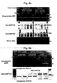
- light is applied to the culture. After applying light, the cells are solubilized. Thereafter, proteins contained in the solubilized components are fractionated, preferably fractionated by SDS-PAGE, and the labeled pladienolides in a fraction that contains SAP130 are then quantified.
- light can be applied to an immunoprecipitation sample obtained by treating solubilized components obtained by solubilizing cells cultured in the presence of a test compound and photoaffinity labeled pladienolides with an anti-SAP155 antibody, an anti-SAP145 antibody, an anti-SAP120 antibody, an anti-U2B" antibody or the like, and preferably with an anti-SAP155 antibody.
- an immunoprecipitation sample obtained by treating solubilized components with an anti-SAP155 antibody, an anti-SAP145 antibody, an anti-SAP120 antibody, an anti-U2B" antibody or the like, and preferably with an anti-SAP155 antibody be treated with a test compound and photoaffinity labeled pladienolides, and that light be then applied to the resultant immunoprecipitation sample.
- the wavelength used in light irradiation is not particularly limited.
- a wavelength that activates a used photoaffinity probe is preferable.
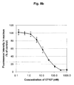
- the quantities of the labeled pladienolides in a U2 snRNP, SF3b, or SAP130 fraction in the presence of a test compound become smaller than those in the absence of the test compound, for example, when the aforementioned quantities are 50% or less, preferably 70% or less, and more preferably 90% or less, it can be determined that the test compound has the activity of acting on (binding to) U2 snRNP, preferably SF3b, and more preferably SAP130.
- test compound and the labeled pladienolides be added to the solubilized components after solubilizing the cells, followed by incubation for a suitable period of time, and that the quantities of the labeled pladienolides contained in the SAP130 fraction be then measured.
- the test compound and the labeled pladienolides may be simultaneously added to the solubilized components, or either the test compound or the labeled pladienolides may be previously added thereto. It is preferable that the test compound be added in advance.
- SAP130 can be fused to a protein acting as a tag, such as GFP, so as to form a fused protein, and such a fused protein is then allowed to be expressed in certain cells.
- a protein acting as a tag such as GFP
- the use of such cells enables easy fractionation of SAP130, and thus it is preferable.
- an immunoprecipitation experiment using an anti-GFP antibody is carried out on cells wherein SAP 145 has been allowed to be expressed in the form of a fusion protein with a protein acting as a tag, such as GFP, so that SF3b can be fractionated.
- Example B7 there is a correlation between the antitumor activity of the test compound and the activity of the test compound to suppress distribution of the labeled pladienolides into the SAP130 fraction. Accordingly, the method of the present invention makes it possible to screen a compound, which acts on (binds to) SAP130 so as to exhibit antitumor activity.
- a compound discovered by the present screening system has an effect on RNA splicing, and thus it is useful as an anticancer agent. Further, it is considered that this compound is also useful as a therapeutic agent for other diseases that are considered to be developed due to abnormal splicing, such as neurodegenerative diseases (e.g. familial Alzheimer's disease), dementia (e.g. frontotemporal dementia ( Hutton, M. et al., Nature, 393:702-705, 1998 )), mental disorders (e.g. familial dysautonomia ( Hims MM et al., J Mol Med. 2007, 85(2):149-61. Epub 2007 )), amyotrophic and myotonic degenerative diseases (e.g.
- neurodegenerative diseases e.g. familial Alzheimer's disease
- dementia e.g. frontotemporal dementia ( Hutton, M. et al., Nature, 393:702-705, 1998
- mental disorders e.g. familial dysautonomia ( Hims MM et al
- the compound is further useful as a therapeutic agent for infectious diseases involving viruses that utilize splicing during their growth process, such as retrovirus (in particular HIV).
- R 2 , R 10 , R 12 , and R 14 are the same as or different from one another and each represents hydrogen or methyl.
- R 3a , R 3b , R 5a , R 5b , R 6a , and R 6b are the same as or different from one another and each represents any one of (1) to (5) below:
- R N1 and R N2 represent 1) or 2) as follows:
- R 7a and R 7b represent (1) or (2) below:
- G represents any one of [1] to [3] below.
- R 16a and R 16b are the same as or different from each other and each represents hydrogen, methyl, or hydroxy.
- R 17a , R 17b , R 18a , R 18b , R 19a , R 19b , R 20a , R 20b , R 21a , and R 21b are the same as or different from one another and each represents any one of (1) to (6) below:
- R 21c represents any one of (1) and (2) below:
- R 16a , R 16b , R17 a , R 17b , R 18a , and R 18b have the same definitions as those described in formula (G-I).
- R 18c represents (1) or (2) below:
- R 16a , R 16b , R 17a , and R 17b have the same definitions as those described in formula (G-I).
- R 17c represents (1) or (2) below:
- C 1-22 alkyl used in the present specification means a linear or branched alkyl group or a cycloalkyl group having 1 to 22 carbon atoms, such as methyl group, ethyl group, n-propyl group, iso-propyl group, n-butyl group, iso-butyl group, sec-butyl group, tert-butyl group, n-pentyl group, 1,1-dimethylpropyl group, 1,2-dimethylpropyl group, 2,2-dimethylpropyl group, 1-ethylpropyl group, n-hexyl group, 1-ethyl-2-methypropyl group, 1,1,2-trimethylpropyl group, 1-propylpropyl group, 1-methylbutyl group, 2-methylbutyl group, 1,1-dimethylbutyl group, 1,2-dimethylbutyl group, 2,2-dimethylbutyl group, 1,3-dimethylbuty group, 2,
- saturated C 3-22 alkyl used in the present specification means a linear or branched alkenyl group having 3 to 22 carbon atoms, or a linear or branched alkynyl group having 3 to 22 carbon atoms, such as allyl group, 1-propenyl group, isopropenyl group, 2-methyl-1-propenyl group, 2-methyl-2-propenyl group, 1-butenyl group, 2-butenyl group, 3-butenyl group, 1-pentenyl group, 1-hexenyl group, 1,3-hexanedienyl group, 1,5-hexanedienyl group, 1-propynyl group, 2-propynyl group, 1-butynyl group, 2-butynyl group, 3-butynyl group, 1-ethynyl-2-propynyl group, 2-methyl-3-propynyl group, 1-pentynyl group, 1-hexynyl group, 1-3-
- C 1-22 alkoxy used in the present specification means a group formed by binding an oxygen atom to the terminus of the above-defined "C 1-22 alkyl.”
- a suitable group include methoxy group, ethoxy group, n-propoxy group, iso-propoxy group, n-butoxy group, iso-butoxy group, sec-butoxy group, tert-butoxy group, n-pentyloxy group, iso-pentyloxy group, sec-pentyloxy group, n-hexoxy group, iso-hexoxy group, 1,1-dimethylpropyloxy group, 1,2-dimethylpropoxy group, 2,2-dimethylpropyloxy group, 1-methyl-2-ethylpropoxy group, 1-ethyl-2-methylpropoxy group, 1,1,2-trimethylpropoxy group, 1,2,2-trimethylpropoxy group, 1,1-dimethylbutoxy group, 1,2-dimethylbutoxy group, 2,2-dimethylbutoxy group, 2,3-di
- unsaturated C 2-22 alkoxy used in the present specification means a group formed by binding an oxygen atom to the terminus of the above-defined "unsaturated C 3-22 alkyl," vinyl, and ethyl.
- a suitable group include vinyloxy group, allyloxy group, 1-propenyloxy group, 2-propenyloxy group, isopropenyloxy group, 2-methyl-1-propenyloxy group, 2-methyl-2-propenyloxy group, 1-butenyloxy group, 2-butenyloxy group, 3-butenyloxy group, 1-pentenyloxy group, 1-hexenyloxy group, 1,3-hexanedienyloxy group, 1,5-hexanedienyloxy group, propargyloxy group, and 2-butynyloxy group; preferably include, allyloxy group, propargyloxy group, and 2-butynyloxy group.
- C 6-14 aryl used in the present specification means an aromatic hydrocarbon cyclic group having 6 to 14 carbon atoms, and it includes a monocyclic group and a condensed ring such as a bicyclic group or a tricyclic group.
- Examples thereof are phenyl group, indenyl group, 1-naphthyl group, 2-naphthyl group, azulenyl group, heptalenyl group, indacenyl group, acenaphthyl group, fluorenyl group, phenalenyl group, phenanthrenyl group, and anthracenyl group; of which a preferred example is phenyl group, 1-naphthyl group, or 2-naphthyl group.
- 5- to 14-membered heteroaryl used in the present specification means a monocyclic, bicyclic or tricyclic, 5- to 14-membered aromatic heterocyclic group, which comprises one or more heteroatoms selected from the group consisting of a nitrogen atom, a sulfur atom and an oxygen atom.
- suitable group examples include a nitrogen-containing aromatic heterocylic group such as pyrrolyl group, pyridyl group, pyridazinyl group, pyrimidinyl group, pyrazinyl group, triazolyl group, tetrazolyl group, benzotriazolyl group, pyrazolyl group, imidazolyl group, benzimidazolyl group, indolyl group, isoindolyl group, indolizinyl group, purinyl group, indazolyl group, quinolyl group, isoquinolyl group, quinolizyl group, phthalazyl group, naphthyridinyl group, quinoxalyl group, quinazolinyl group, cinnolinyl group, pteridinyl group, imidazotriazinyl group, pyrazinopyridazinyl group, acridinyl group, phenanthridinyl group,
- C 6-14 aryloxy used in the present specification means a group formed by binding an oxygen atom to the terminus of the above-defined “C 6-14 aryl.”
- Specific examples include phenyloxy group, indenyloxy group, 1-naphthyloxy group, 2-naphthyloxy group, azulenyloxy group, heptalenyloxy group, indacenyloxy group, acenaphthyloxy group, fluorenyloxy group, phenalenyloxy group, phenanthrenyloxy group, and anthracenyloxy group, of which a preferred example is phenyloxy group, 1-naphthyloxy group, or 2-naphthyloxy group.
- 5- to 14-membered heteroaryloxy used in the present specification means a group formed by binding an oxygen atom to the terminus of the above-defined "5- to 14-membered heteroaryl.”
- Specific examples include pyrrolyloxy group, pyridyloxy group, pyridazinyloxy group, pyrimidinyloxy group, pyrazinyloxy group, triazolyloxy group, tetrazolyloxy group, benzotriazolyloxy group, pyrazolyloxy group, imidazolyloxy group, benzimidazolyloxy group, indolyloxy group, isoindolyloxy group, indolizinyloxy group, purinyloxy group, indazolyloxy group, quiolyloxy group isoquinolyloxy group, quinolizyloxy group, phthalazyloxy group, naphthyridinyloxy group, quinoxalyloxy group, quinazolinyloxy group
- C 2-22 acyl used in the present specification means an acyl group with 2 to 22 carbon atoms.
- suitable groups include linear or branched acyl groups such as acetyl group, propionyl group, butyryl group, iso-butyryl group, valeryl group, iso-valeryl group, pivalyl group, caproyl group, decanoyl group, lauroyl group, myristoyl group, palmitoyl group, stearoyl group, and arachidoyl group.
- C 2-22 acyloxy used in the present specification means a group having a partial structure corresponding to the aforementioned “C 2-22 acyl.”
- unsaturated C 3-22 acyl used in the present specification means an acyl group with 3 to 22 carbon atoms having double bond(s) or triple bond(s).
- Preferred unsaturated C 3-22 acyl groups include linear or branched acyl groups such as acryl group, propiol group, crotonyl group, iso-crotonyl group, oleinol group, and linolenoyl group.
- unsaturated C 3-22 acyloxy used in the present specification means a group having a partial structure corresponding to the aforementioned “unsaturated C 3-22 acyl.”
- C 1-22 alkylsulfonyl used in the present specification means a group formed by binding the above-defined “C 1-22 alkyl” to sulfonyl. Specific examples include methylsulfonyl group, ethylsulfonyl group, n-propylsulfonyl group, and iso-propylsulfonyl group, of which a preferred example is methylsulfonyl group.
- C 1-22 alkylsulfonyloxy used in the present specification means a group formed by binding an oxygen atom to the terminus of the above-defined “C 1-22 alkylsulfonyl,” such as methylsulfonyloxy group, ethylsulfonyloxy group, n-propylsulfonyloxy group, or iso-propylsulfonyloxy group, of which a preferred example is methylsulfonyloxy group.
- 3- to 14-membered nitrogen-containing non-aromatic heterocyclic group used in the present specification means a monocyclic, bicyclic or tricyclic 3- to 14-membered non-aromatic heterocyclic group that may comprise one or more heteroatoms selected from the group consisting of a nitrogen atom, a sulfur atom and an oxygen atom, as well as one nitrogen atom.
- Preferred examples include aziridinyl group, acetidyl group, pyrrolidinyl group, pyrrolyl group, piperidinyl group, piperazinyl group, imidazolyl group, pyrazolidyl group, imidazolidyl group, morpholyl group, thiomorpholyl group, imidazolinyl group, and oxazolinyl group.
- the present non-aromatic heterocyclic groups further include groups induced from a pyridone ring and non-aromatic condensed rings (e.g. groups induced from a phthalimide ring, a succinimide ring, etc.).
- the substituent of the expression “may have substituent(s)" used in the present specification means one or more groups selected from the group consisting of C 1-8 alkyl group, C 2-8 alkenyl group (e.g. vinyl group), C 2-8 alkynyl group (e.g. ethynyl group), C 6-14 aryl group (e.g. phenyl group, etc.), 5- to 14-membered heteroaryl group (e.g.
- thienyl group furyl group, pyridyl group, pyridazyl group, pyrimidyl group, pyrazyl group, etc.), hydroxyl group, C 1-8 alkoxy group, C 1-8 acyl group, C 2-8 acyloxy group, C 2-8 alkenyloxycarbonyl group, C 2-8 alkynyloxycarbonyl group, C 1-8 alkoxycarbonyl group, halogen atom, hydroxycarbonyl group, thiol group, C 1-8 alkylthio group, C 1-8 alkylsulfoxide group, C 1-8 alkylsulfonyl group, C 1-8 alkylsulfonyloxy group, hydroxysulfonyl group, nitrile group, nitro group, nitroso group, amino group, N-C 1-8 alkylamino group, N,N-di-C 1-8 alkylamino group, N-C 2-8 alkenylamino group,
- N-heteroarylamino group e.g. 2-pyridylamino group, 3-pyridylamino group, 1-pyrroylamino group, etc.
- N-C 1-8 alkyl-N-arylamino group e.g. 2-pyridylamino group, 3-pyridylamino group, 1-pyrroylamino group, etc.
- N-C 1-8 alkyl-N-arylamino group e.g. 2-pyridylamino group, 3-pyridylamino group, 1-pyrroylamino group, etc.
- N-C 1-8 alkyl-N-arylamino group e.g. 2-pyridylamino group, 3-pyridylamino group, 1-pyrroylamino group, etc.
- N-C 1-8 alkyl-N-arylamino group e.g. 2-pyridylamino group, 3-pyridylamino group, 1-pyrroylamino group
- pladienolides A, B, D and E7170 are as follows:
- the labeled compound of the present invention was synthesized by the following method.
- 1,8-diamino-3,6-dioxaoctane (127 mg, 0.85 mmol) was dissolved in THF (5 ml). Thereafter, a THF solution (15 ml) of (8E,12E,14E)-3,6,21-tri(1-ethoxyethoxy)-6,10,12,16,20-pentamethyl-7-(4-nitrophenoxy)carboxy-18,19-epoxytricosa-8,12,14-tri en-11-olide (150 mg, 0.17 mmol) described in Example B44 of the Patent Document ( WO02/060890 ) was added dropwise to the obtained solution.
- the reaction solution was stirred at room temperature for 24 hours. Thereafter, the solvent was distilled away under reduced pressure, and the residue was then dissolved in ethyl acetate.
- the obtained solution was sequentially washed with distilled water, a sodium bicarbonate aqueous solution, and a saline solution. Thereafter, the organic layer was dried over magnesium sulfate, and the solvent was then distilled away.
- the titled compound (8 mg) was obtained in the form of a colorless oil product by the same method as that described in Example A2 with the exception that N-(+)biotinyl-1,8-diamino-3,6-dioxaoctane (Pierce; Biotin PEO-Amine) was used instead of 1,8-diamino-3,6-dioxaoctane.
- 1,4-dimethylethylenediamine (0.88 g, 10 mmol) was dissolved in THF (10 ml). While the obtained solution was stirred at room temperature, commercially available Z-Lys (BOC)-ONP (1 g, 2 mmol) was added thereto, dividedly several times. The reaction solution was stirred at room temperature for 12 hours. Thereafter, the solvent was distilled away, and using ethyl acetate, the residue was partitioned between oil and water. The organic layer was sequentially washed with distilled water and a saline solution, and it was then dried over magnesium sulfate. Thereafter, the solvent was distilled away.
- the product (0.9 g, 2 mmol) obtained in (1) above was dissolved in THF-DMF (20 ml-10 ml). While the obtained solution was stirred at room temperature, biotin N-hydroxysuccinimide (0.7 g, 2 mmol) was added thereto, dividedly several times. The reaction solution was stirred at room temperature for 12 hours. Thereafter, the solvent was distilled away, and using ethyl acetate, the residue was partitioned between oil and water. The organic layer was sequentially washed with distilled water and a saline solution, and it was then dried over magnesium sulfate. Thereafter, the solvent was distilled away. The obtained residue was purified by silica gel column chromatography (Merck Art.
- reaction solution was stirred at room temperature for 24 hours.
- the solvent was distilled away, and using ethyl acetate, the residue was partitioned between oil and water.
- the organic layer was sequentially washed with distilled water and a saline solution, and it was then dried over magnesium sulfate. Thereafter, the solvent was distilled away.
- the target protein of pladienolides was identified as described in Examples B 1 to B7 below.
- the labeled pladienolides can be used to examine that a test compound acts on (binds to) U2 snRNP, preferably SF3b, and more preferably SAP 130.
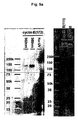
- the tritiated probe compound produced in Example A1 was added to human breast cancer cells MDA-MB-468 (ATCC HTB-132) cultured on a 15-cm dish (>80% confluent), resulting in a concentration of 3 to 30 nM.
- the lysate was centrifuged at 2,000 g at 4°C for 10 minutes, so as to obtain a supernatant (2000 g-sup.) and a precipitate (2000 g-pellet).
- 2000 g-sup was further centrifuged at 100,000 g at 4°C for 60 minutes, so as to obtain a cytosol fraction as a supernatant, and a membrane fraction as a precipitate.
- 2000 g-pellet was suspended in 0.5 M NaCl, and the suspension was then left at rest for 30 minutes under cooling on ice, so as to elute a nuclear protein. Thereafter, centrifugation was carried out at 3,000 g at 4°C for 10 minutes, so as to obtain a nuclear fraction as a supernatant, and a nuclear pellet as a precipitate.
- each of the cytosol fraction and the nuclear fraction was uniformly mixed with Hionic-Fluor (Perkin Elmer).
- each of the membrane fraction and the nuclear pellet was dissolved in Soluene-350 (Packard) (1 ml), and the obtained solution was then mixed with Hionic-Fluor.
- the radioactivity of each mixture was measured with a liquid scintillation counter.
- a radioisotope-non-labeled-pladienolide was used to carry out a cold inhibition experiment. That is, the cells were treated with a pladienolide having a concentration (0.15 to 1.5 ⁇ M) that was 50 times higher than the tritium probe treatment concentration, and they were then cultured at 37°C in an incubator for 1 hour. Thereafter, the culture was treated with a tritium probe, and each fraction was prepared. Then, the radioactivity thereof was measured.
- the highest radioactivity of the tritium probe was detected in the nuclear fraction in all the treating concentrations ranging from 3 to 30 nM.
- the second highest radioactivity was detected in the nuclear pellet. Such radioactivity almost completely disappeared after a pre-treatment with pladienolide B.
- a charcoal assay was carried out on the prepared cytosol fraction and nuclear fraction, so as to examine that the label count in the test sample was derived from a bound probe binding to a protein and the like, or was derived from a non-bound free form.
- Figure 2 shows a graph in which each compound was plotted.
- the longitudinal axis indicates the binding rate (%) of the tritium probe compound that has bound in the presence of various pladienolide analogues or derivatives relative to control samples (no competition), and the horizontal axis indicates IC50 (nM) of the in vitro cytostatic activity of each compound.
- the radioactivity of each sample was proportional to IC50 of the in vitro cytostatic activity of the competing pladienolides and the derivatives thereof to the WiDr cells. That is, stronger competitive inhibition was observed in a compound exhibiting strong cytostatic activity.
- pladienolide analogues or derivatives used in the present example are as follows:
- Example A2 1 ⁇ M of the fluorescent probe produced in Example A2 was added to HeLa cells, and the obtained mixture was incubated at 37°C in an incubator for 1 hour. Thereafter, the medium was removed, and the remaining culture was further cultured for 1 hour in a fresh medium without probes and non-specifically adsorbed compounds were washed. The medium was removed again. The remaining culture was then washed with PBS. Thereafter, the cells were immobilized in 3.7% formaldehyde in PBS.
- the immobilized cells were well washed with PBS, and were then solubilized with 0.5% or 1% Triton X-100 in PBS. Thereafter, blocking was carried out with FBS or a blocking solution, and a primary antibody (anti-SC-35 monoclonal antibody; Sigma; S4045) was added to the resultant, so as to carry out a reaction.
- the reaction product was washed with PBS, and blocking was carried out again with FBS or a blocking solution.
- a fluorescent-labeled secondary antibody (TexasRed-labeled; Jackson) was added to the resultant, so as to carry out a reaction.
- the labeled sample was rapidly rinsed with distilled water, and it was then encapsulated into a Prolong antifade reagent (Molecular Probes). Localization of the fluorescent probe and the anti-SC-35 antibody was observed under a fluorescence microscope (Delta Vision, Applied Precision, Inc.) or a confocal microscope (LSM510; Carl Zeiss).
- the fluorescent probe is mainly localized in granular structures in the nuclei ( Figure 3a , upper). These structures corresponded to localization of the anti-SC-35 antibody.
- SC-35 is a representative molecule of splicing factors and is a marker of nuclear speckles. Thus, it was suggested that the target molecule of pladienolides exists in such nuclear speckles.
- the fluorescence of the fluorescent probe was faded by excessive light irradiation, and the above probe was then photographed under the same above conditions. As a result, no signals were detected in the channel of the fluorescent probe. Thus, it was confirmed that no signals were leaked from anti-SC-35 antibody staining, and that a fluorescent probe-specific signal was detected ( Figure 3a , lower)
- pladienolides or the derivatives thereof were used in the aforementioned experimental operations to carry out a competition assay. That is, before addition of the fluorescent probe, 1 ⁇ M pladienolides or the derivatives thereof were added to the cells, and the obtained mixture was then cultured for 1 hour. Thereafter, the same above operations for immobilization and staining were performed on the culture, followed by observation with a microscope, so as to confirm localization of the fluorescent probe.
- the fluorescence intensity derived from the fluorescent probe contained in each sample was proportional to IC50 of the cytostatic activity of the competing pladienolides or the derivatives thereof (pladienolide A >non-labeled tritium probe compound > pladienolides B and D). That is, stronger competitive inhibition was observed in a compound exhibiting strong cytostatic activity, and the fluorescence derived from the probe almost completely disappeared ( Fig 3b , left column). Moreover, it was observed that the number of nuclear speckles stained with the anti-SC-35 antibody was decreased and that the size thereof was enlarged ( Figure 3b , right column).
- each of antibodies reacting with splicing factors, transcriptional factors, or molecules associated with such factors were added to a concentration of 1 ⁇ g/ml.
- the obtained mixture was slowly stirred for approximately 12 hours in a low-temperature chamber of 4°C, using a sample rotator. Thereafter, 20 ⁇ l of 50% Protein A/G-agarose conjugate suspension was added to the reaction solution, and the obtained mixture was further stirred for 2 hours in the low-temperature chamber.
- An SDS sample buffer was added to the resultant solution, and the obtained mixture was then stirred with a vortex.
- the reaction solution was boiled at 99°C for 10 minutes, so as to elute the immunoprecipitated protein.
- the eluant was mixed homogeneously with Hionic-Fluor, and the radioactivity of a tritium probe existing in the eluant was then measured with a liquid scintillation counter.
- the target molecule of pladienolide is a protein existing in the U2 snRNP complex.
- UV (365 nm and 302 nm) was applied to the obtained immunoprecipitate (agarose resin), and a crosslinking reaction was carried out.
- An SDS sample buffer was added to the reaction product, and the obtained mixture was then stirred with a vortex, followed by boiling at 99°C for 10 minutes, so as to elute the immunoprecipitated proteins.
- the eluant was subjected to SDS-PAGE and blotting onto a PVDF membrane in accordance with common methods. Thereafter, the resultant was treated with streptavidin-HRP, so as to detect probe-bound proteins.
- the same sample (blotting membrane) was subjected to Western blotting using an SAP155 antibody, an SAP145 antibody, and SAP 130 antibody.
- the band position of each of such proteins was compared with the band position of the target molecule, to which a photoaffinity probe had bound.
- the position of the target molecule clearly differed from the position of SAP 155.
- the target molecule SAP145, and SAP130 were observed in almost the same position.
- the target molecule was SAP145 or SAP130.
- a pEGFP-SAP130 plasmid was produced.
- the pEGFP-SAP130 was produced by inserting, into the NheI-PmeI site of a pcDNA3.1(-) vector (Invitrogen), an approximately 4.4 kbp DNA fragment as shown in SEQ ID NO: 1 (an NheI-PmeI fragment of GFP-SAP130; 4,432 bp), which was constituted with an EGFP protein coding sequence (derived from pEGFP-N2; CLONTECH), a human SAP130 (SF3B3) protein coding sequence (GenBank Accession#: NM_012426), and a linker sequence that connects such sequences to one another. It was anticipated that a GFP-SAP130 protein having the amino acid sequence as shown in SEQ ID NO: 2 was allowed to express in cells, into which the above plasmid had been introduced.
- a pEF1-GFP-SAP145 plasmid was produced.
- the pEF1-GFP-SAP145 was produced by inserting, into the HindIII-PmeI site of a pEF1-Myc/His A vector (Invitrogen), an approximately 3.5 kbp DNA fragment as shown in SEQ ID NO: 3 (a HindIII-PmeI fragment of GFP-SAP145; 3,454 bp), which was constituted with an EGFP protein coding sequence (derived from pEGFP-N2; CLONTECH), a human SAP145 (SF3B2) protein coding sequence (GenBank Accession#: NM_006842), and a linker sequence that connects such sequences to one another. It was anticipated that a GFP-SAP145 protein having the amino acid sequence as shown in SEQ ID NO: 4 was allowed to express in cells, into which the above plasmid had been
- Each of the pEF1-GFP-SAP145 and pEGFP-SAP130 was introduced into HeLa cells, using Lipofectamine 2000 reagent (Inyitrogen). Forty hours after introduction of the gene, selection of gene-introduced cells was initiated by addition of Geneticin (Invitrogen) with a final concentration of 500 ⁇ g/ml. Fourteen days after the gene introduction, clones emitting GFP fluorescence were marked by fluorescence microscope observation, and were then picked up. Thereafter, a single colony was isolated by diluted passage. Clone #2 (GFP-145 strain) derived from pEF1-GFP-SAP145, and Clone #3 (GFP-130 strain) derived from pEGFP-SAP130, were used in the subsequent experiments.
- Geneticin Invitrogen
- SAP145 and SAP130 which were considered to be target molecules, were detected in almost the same position on SDS-PAGE.
- strains stably expressing a GFP-fused SAP145 or GFP-fused SAP 130 protein were prepared, and an experiment was then carried out using a photoaffinity biotin probe.
- a 1.5 ⁇ M photoaffinity biotin probe was added to HeLa cells stably expressing GFP-SAP145, and the obtained mixture was then cultured for 1 hour. After washing the cells with PBS, M-PER (PIERCE) was then added thereto, followed by stirring at 4°C for 1 hour. Thereafter, 1/10 vol. of 1.5 M NaCl-0.5% Tween 20 solution was added to the reaction solution, and the obtained mixture was then treated with a vortex (10 sec. x 3). The resultant was centrifuged at 15,000 rpm at 4°C for 5 minutes (TOMY MX-15). The thus obtained supernatant was filtrated with a 0.45- ⁇ m membrane, and the filtrate was used in the following immunoprecipitation experiment.
- a GFP antibody (Q-BIOgene AFP5002) or an SAP155 antibody (MBL D221-3) (10 ⁇ g/ml) was added to the immunoprecipitation solution, and the obtained mixture was then stirred slowly with a rotator at 4°C for 30 minutes. Subsequently, 1/10 vol. of protein A/G-agarose (50% suspension) was added to the immunoprecipitation solution, and the obtained mixture was further stirred at 4°C for 1 hour in the same manner as above.
- the agarose resin was washed with a washing buffer 3 times, and UV was then applied to the resultant resin by the same method as that described in Example B5. Thereafter, the protein bound to the resin was eluted, and it was then subjected to SDS-PAGE and blotting. The resultant was treated with streptavidin-HRP, so as to detect a protein to which the probe had bound.
- SAP145 and SAP130 were detected with the SAP145 antibody and the SAP130 antibody, respectively.
- an agarose resin prepared from probe-untreated cells was treated with a probe in the aforementioned experimental operations. Thereafter, UV irradiation and elution of a protein were carried out by the same operations as above, and it was examined whether or not the same band was detected in the obtained sample.
- Probe-untreated HeLa cells stably expressing GFP-SAP145 were subjected to an immunoprecipitation experiment by the same method as that described in Example B7 using a GFP antibody and an SAP 155 antibody, so as to prepare an immunoprecipitate sample (agarose resin).
- This resin was suspended in a washing buffer, and was pre-treated with pladienolides and the derivative thereof (E7107) at 75 ⁇ M, followed by stirring with a rotator at 4°C for 30 minutes. Subsequently, it was treated with a photoaffinity biotin probe at 1.5 ⁇ M, and it was further stirred for 30 minutes in the same manner as above. The resin was washed with a washing buffer.
- Example B7 Thereafter, UV irradiation and elution of a protein were carried out by the same method as described in Example B7.
- the obtained sample was subjected to SDS-PAGE and blotting to examine whether or not the band derived from the probe disappeared, by the competition between the photoaffinity biotin probe and the pre-treaing pladienolide compound.
- the band derived from the probe disappeared by competition with the pladienolide compounds (Pladienolide B, D and E7107). Accordingly, it can be said that the protein bounded by the photoaffinity biotin probe is the pladienolide-bound protein.
- the obtained image was subjected to image analysis software, Developer (GE Healthcare), so as to analyze the fluorescence intensity of the fluorescent probes in the nuclei and examine whether or not accumulation of the fluorescent probes into the nuclei is inhibited by E7107 binding to SF3b.
- image analysis software Developer (GE Healthcare)
Landscapes
- Health & Medical Sciences (AREA)
- Chemical & Material Sciences (AREA)
- Life Sciences & Earth Sciences (AREA)
- Organic Chemistry (AREA)
- General Health & Medical Sciences (AREA)
- Medicinal Chemistry (AREA)
- Engineering & Computer Science (AREA)
- Veterinary Medicine (AREA)
- Public Health (AREA)
- Animal Behavior & Ethology (AREA)
- Pharmacology & Pharmacy (AREA)
- Immunology (AREA)
- Urology & Nephrology (AREA)
- Epidemiology (AREA)
- Molecular Biology (AREA)
- Biomedical Technology (AREA)
- Hematology (AREA)
- Biochemistry (AREA)
- Physics & Mathematics (AREA)
- General Physics & Mathematics (AREA)
- Pathology (AREA)
- Food Science & Technology (AREA)
- Microbiology (AREA)
- Proteomics, Peptides & Aminoacids (AREA)
- Analytical Chemistry (AREA)
- Cell Biology (AREA)
- Biotechnology (AREA)
- General Chemical & Material Sciences (AREA)
- Nuclear Medicine, Radiotherapy & Molecular Imaging (AREA)
- Chemical Kinetics & Catalysis (AREA)
- Heterocyclic Carbon Compounds Containing A Hetero Ring Having Oxygen Or Sulfur (AREA)
- Investigating Or Analysing Biological Materials (AREA)
- Plural Heterocyclic Compounds (AREA)
- Medicines That Contain Protein Lipid Enzymes And Other Medicines (AREA)
Applications Claiming Priority (2)
| Application Number | Priority Date | Filing Date | Title |
|---|---|---|---|
| US82119906P | 2006-08-02 | 2006-08-02 | |
| PCT/JP2007/065574 WO2008016187A1 (en) | 2006-08-02 | 2007-08-02 | Pladienolide target molecule, compound capable of binding to the target molecule, and screening method for the compound |
Publications (1)
| Publication Number | Publication Date |
|---|---|
| EP2053393A1 true EP2053393A1 (de) | 2009-04-29 |
Family
ID=38997353
Family Applications (1)
| Application Number | Title | Priority Date | Filing Date |
|---|---|---|---|
| EP07792236A Withdrawn EP2053393A1 (de) | 2006-08-02 | 2007-08-02 | Pladienolid-zielmolekül, zur bindung an das zielmolekül fähige verbindung und screening-verfahren für die verbindung |
Country Status (4)
| Country | Link |
|---|---|
| US (1) | US20090227795A1 (de) |
| EP (1) | EP2053393A1 (de) |
| JP (1) | JPWO2008016187A1 (de) |
| WO (1) | WO2008016187A1 (de) |
Families Citing this family (5)
| Publication number | Priority date | Publication date | Assignee | Title |
|---|---|---|---|---|
| JP5482057B2 (ja) * | 2009-09-29 | 2014-04-23 | オリンパス株式会社 | 細胞核を構成する構造体の解析方法 |
| KR101933554B1 (ko) * | 2016-07-21 | 2018-12-31 | 한국과학기술연구원 | Hbt 유도체의 광가교반응을 이용한 단백질 표지 및 검출 방법 |
| EP3801523B1 (de) * | 2018-06-01 | 2024-09-11 | Eisai R&D Management Co., Ltd. | Spleissmodulatoren |
| CN119405832A (zh) | 2018-06-01 | 2025-02-11 | 卫材R&D管理有限公司 | 剪接调节抗体-药物缀合物及其使用方法 |
| CN111973589A (zh) * | 2019-05-21 | 2020-11-24 | 中国科学院近代物理研究所 | pladienolide B在增强宫颈癌细胞辐射敏感性中的应用 |
Family Cites Families (7)
| Publication number | Priority date | Publication date | Assignee | Title |
|---|---|---|---|---|
| TWI312681B (en) | 2001-02-01 | 2009-08-01 | Novel physiologically active substance | |
| JP2002243739A (ja) * | 2001-02-21 | 2002-08-28 | Toyobo Co Ltd | リガンドのレセプター結合活性の測定方法ならびに測定試薬 |
| TWI334866B (en) * | 2002-05-29 | 2010-12-21 | Mercian Corp | Novel physiologically active substances |
| JP4459051B2 (ja) * | 2002-07-31 | 2010-04-28 | メルシャン株式会社 | 新規生理活性物質 |
| CA2494536C (en) * | 2002-07-31 | 2011-10-04 | Mercian Corporation | A 12-membered ring macrolide active substance |
| WO2005075681A1 (ja) * | 2004-02-06 | 2005-08-18 | Eisai R & D Management Co., Ltd. | 抗癌剤に対する癌細胞の感受性を検定する方法 |
| JP2005263748A (ja) * | 2004-03-22 | 2005-09-29 | Japan Science & Technology Agency | 反応性ジアジリン基を有するホモフェニルアラニン誘導体 |
-
2007
- 2007-08-02 EP EP07792236A patent/EP2053393A1/de not_active Withdrawn
- 2007-08-02 US US12/375,516 patent/US20090227795A1/en not_active Abandoned
- 2007-08-02 WO PCT/JP2007/065574 patent/WO2008016187A1/ja not_active Ceased
- 2007-08-02 JP JP2008527826A patent/JPWO2008016187A1/ja not_active Withdrawn
Non-Patent Citations (1)
| Title |
|---|
| See references of WO2008016187A1 * |
Also Published As
| Publication number | Publication date |
|---|---|
| US20090227795A1 (en) | 2009-09-10 |
| WO2008016187A1 (en) | 2008-02-07 |
| JPWO2008016187A1 (ja) | 2009-12-24 |
Similar Documents
| Publication | Publication Date | Title |
|---|---|---|
| Xu et al. | LZTS2 inhibits PI3K/AKT activation and radioresistance in nasopharyngeal carcinoma by interacting with p85 | |
| Dai et al. | A simple and effective coumarin-based fluorescent probe for cysteine | |
| CN114728957A (zh) | 一种atr抑制剂的晶型及其应用 | |
| Wang et al. | Discovery of pyrazolo [1, 5-a] pyrimidine derivative as a novel and selective ALKBH5 inhibitor for the treatment of AML | |
| JPWO2008111464A1 (ja) | スプライシング異常を指標とする抗ガン剤の作用検定方法 | |
| Wang et al. | Small-molecule targeting PKM2 provides a molecular basis of lactylation-dependent fibroblast-like synoviocytes proliferation inhibition against rheumatoid arthritis | |
| JP2016533384A (ja) | 選択的grp94阻害剤およびその使用 | |
| EP2053393A1 (de) | Pladienolid-zielmolekül, zur bindung an das zielmolekül fähige verbindung und screening-verfahren für die verbindung | |
| Wang et al. | Discovery of a far-red carbazole-benzoindolium fluorescent ligand that selectively targets mitochondrial DNA and suppresses breast cancer growth | |
| Niu et al. | Bromodomain and extra-terminal (BET) protein inhibitors suppress chondrocyte differentiation and restrain bone growth | |
| Zhu et al. | In Situ Proteome Profiling and Bioimaging Applications of Small‐Molecule Affinity‐Based Probes Derived From DOT1L Inhibitors | |
| Bai et al. | The Na+, K+-ATPase β1 subunit regulates epithelial tight junctions via MRCKα | |
| Liu et al. | AI26 inhibits the ADP-ribosylhydrolase ARH3 and suppresses DNA damage repair | |
| You et al. | A far-red aza-crown ether fluorescent probe for selective G-quadruplex DNA targeting | |
| Takimoto et al. | Esterification of an unnatural amino acid structurally deviating from canonical amino acids promotes its uptake and incorporation into proteins in mammalian cells | |
| Zheng et al. | Epoxymicheliolide directly targets histone H2B to inhibit neuroinflammation via recruiting E3 ligase RNF20 | |
| EP4163379A1 (de) | Verfahren zur identifizierung und/oder regulierung der seneszenz | |
| Stalinska et al. | Anti-glioblastoma effects of phenolic variants of benzoylphenoxyacetamide (BPA) with high potential for blood brain barrier penetration | |
| Jiang et al. | A lysosomal-targeted switchable fluorescent probe for the detection of peroxynitrite in living tumor cells and in vivo | |
| Yuan et al. | Evidence of nuclear localization of neuronal nitric oxide synthase in cultured astrocytes of rats | |
| Chen et al. | NEDD8 enhances Hippo signaling by mediating YAP1 neddylation | |
| Williams et al. | Generation and characterization of a stable MK2‐EGFP cell line and subsequent development of a high‐content imaging assay on the Cellomics ArrayScan platform to screen for p38 mitogen‐activated protein kinase inhibitors | |
| Kim et al. | The small molecule R-(-)-β-O-methylsynephrine binds to nucleoporin 153 kDa and inhibits angiogenesis | |
| Hernández-Reyes et al. | DDX37 and DDX50 Maintain Genome Stability by Preventing Transcription-dependent R-loop Formation | |
| Zhang et al. | G-quadruplex in mitochondria as a possible biomarker for mitophagy detection |
Legal Events
| Date | Code | Title | Description |
|---|---|---|---|
| PUAI | Public reference made under article 153(3) epc to a published international application that has entered the european phase |
Free format text: ORIGINAL CODE: 0009012 |
|
| 17P | Request for examination filed |
Effective date: 20090223 |
|
| AK | Designated contracting states |
Kind code of ref document: A1 Designated state(s): AT BE BG CH CY CZ DE DK EE ES FI FR GB GR HU IE IS IT LI LT LU LV MC MT NL PL PT RO SE SI SK TR |
|
| AX | Request for extension of the european patent |
Extension state: AL BA HR MK RS |
|
| STAA | Information on the status of an ep patent application or granted ep patent |
Free format text: STATUS: THE APPLICATION HAS BEEN WITHDRAWN |
|
| 18W | Application withdrawn |
Effective date: 20110706 |