EP2054712B1 - Vorrichtungen und verfahren zur verstärkung einer optischen kohärenztomographie-abbildung mithilfe volumetrischer filterungsverfahren - Google Patents
Vorrichtungen und verfahren zur verstärkung einer optischen kohärenztomographie-abbildung mithilfe volumetrischer filterungsverfahren Download PDFInfo
- Publication number
- EP2054712B1 EP2054712B1 EP07841307.7A EP07841307A EP2054712B1 EP 2054712 B1 EP2054712 B1 EP 2054712B1 EP 07841307 A EP07841307 A EP 07841307A EP 2054712 B1 EP2054712 B1 EP 2054712B1
- Authority
- EP
- European Patent Office
- Prior art keywords
- electro
- magnetic radiation
- arrangement
- plane
- filter
- Prior art date
- Legal status (The legal status is an assumption and is not a legal conclusion. Google has not performed a legal analysis and makes no representation as to the accuracy of the status listed.)
- Not-in-force
Links
Images



Classifications
-
- A—HUMAN NECESSITIES
- A61—MEDICAL OR VETERINARY SCIENCE; HYGIENE
- A61B—DIAGNOSIS; SURGERY; IDENTIFICATION
- A61B3/00—Apparatus for testing the eyes; Instruments for examining the eyes
- A61B3/10—Objective types, i.e. instruments for examining the eyes independent of the patients' perceptions or reactions
- A61B3/12—Objective types, i.e. instruments for examining the eyes independent of the patients' perceptions or reactions for looking at the eye fundus, e.g. ophthalmoscopes
- A61B3/1225—Objective types, i.e. instruments for examining the eyes independent of the patients' perceptions or reactions for looking at the eye fundus, e.g. ophthalmoscopes using coherent radiation
-
- A—HUMAN NECESSITIES
- A61—MEDICAL OR VETERINARY SCIENCE; HYGIENE
- A61B—DIAGNOSIS; SURGERY; IDENTIFICATION
- A61B5/00—Measuring for diagnostic purposes; Identification of persons
- A61B5/0059—Measuring for diagnostic purposes; Identification of persons using light, e.g. diagnosis by transillumination, diascopy, fluorescence
- A61B5/0062—Arrangements for scanning
- A61B5/0066—Optical coherence imaging
-
- A—HUMAN NECESSITIES
- A61—MEDICAL OR VETERINARY SCIENCE; HYGIENE
- A61B—DIAGNOSIS; SURGERY; IDENTIFICATION
- A61B5/00—Measuring for diagnostic purposes; Identification of persons
- A61B5/68—Arrangements of detecting, measuring or recording means, e.g. sensors, in relation to patient
- A61B5/6846—Arrangements of detecting, measuring or recording means, e.g. sensors, in relation to patient specially adapted to be brought in contact with an internal body part, i.e. invasive
- A61B5/6847—Arrangements of detecting, measuring or recording means, e.g. sensors, in relation to patient specially adapted to be brought in contact with an internal body part, i.e. invasive mounted on an invasive device
- A61B5/6852—Catheters
-
- A—HUMAN NECESSITIES
- A61—MEDICAL OR VETERINARY SCIENCE; HYGIENE
- A61B—DIAGNOSIS; SURGERY; IDENTIFICATION
- A61B5/00—Measuring for diagnostic purposes; Identification of persons
- A61B5/0059—Measuring for diagnostic purposes; Identification of persons using light, e.g. diagnosis by transillumination, diascopy, fluorescence
- A61B5/0073—Measuring for diagnostic purposes; Identification of persons using light, e.g. diagnosis by transillumination, diascopy, fluorescence by tomography, i.e. reconstruction of 3D images from 2D projections
Definitions
- the present invention relates to apparatus and methods for enhancing optical coherence tomography imaging, and more particularly to such apparatus and methods which can enhance the contrast in the optical coherence tomography images using techniques employing three-dimensional (e.g., volumetric) filtering of measured data which may be used to generate these images.
- three-dimensional e.g., volumetric
- OCT optical coherence tomography
- the proposed approaches for mitigating the impact of speckle noise can be categorized as either physical compounding methods or digital processing methods.
- the physical compounding methods generally function by combining multiple, speckle uncorrelated measurements of the same location in the analyzed tissue.
- the implementation of such methods may require modifications to the imaging system that can complicate a design of the catheter and the design of a minimally-invasive probe.
- these physical compounding methods can include angular compounding, frequency compounding, and polarization compounding (e.g., a polarization diversity detection).
- the digital processing methods have conventionally been applied entirely to two-dimensional images using procedures or filters that aim to preferentially remove the speckle noise, while preserving certain features associated with the tissue structures.
- Such techniques include adaptive filtering, regularization, and wavelet denoising.
- the digital processing methods unlike the compounding methods, are likely limited to the information content contained within the original speckled image. This, it is important for the digital processing methods to outperform the considerable ability of an experience implementer of the OCT to visually filter the noise and recognize the underlying tissue structures.
- a diagnosis may typically be rendered from one or more images sectioned from the dataset.
- these sectioned images can incorporate measured information both from within the section plane (e.g., in-plane measurements) and from adjacent locations out of the sectioning plane (e.g., out-of-plane measurements).
- the OCT image of the fundus is generated from the 3D OCT data by summing (in the Z direction) the 3D OCT data along the axial (Z) direction at each transverse (X direction) point on the retina giving a value (at each X point) for each axial (Z) scan, at each transverse (X) position on the retina, which corresponds to the total backscattering (back reflected) light from all of the retinal layers at that (X) position.
- exemplary embodiments of apparatus and methods can be provided which can enhance the contrast in the optical coherence tomography images using techniques employing three-dimensional (e.g., volumetric) filtering of measured data which may be used to generate these images.
- datasets can be filtered in three dimensions such that enhanced images may be generated.
- an asymmetric volumetric median filter is applied to a three-dimensional OCT dataset prior to imaging the particular section of the tissue.
- the filtering kernel is larger in the dimension out-of-plane with respect to the image section that the dimensions in-plane.
- the present disclosure also concerns an OCT imaging system which may be configured to produce a dithered beam scanning pattern to enable volumetric imaging to be performed with a high fidelity in the presence of a substantial sample motion. Appropriate filtering of this dataset acquired with the dithered beam allows the generation of an enhanced two-dimensional image.
- At least one first fiber arrangement and at least one second fiber arrangement can be provided (each of which having optical transmitting characteristics).
- the first fiber arrangement can be configured to transmit there through at least one electromagnetic radiation and forward the at least one electromagnetic radiation to at least one sample.
- the second fiber can be configured to transmit there through at least one electromagnetic radiation received from the sample, and may house therein at least one portion of the first fiber arrangement.
- the first and second fiber arrangement may each be a fiber.
- the first and second fibers may be filtered using at least one of the first and second filtering arrangements to prevent at least one portion of each of the respective transmitted and received electromagnetic radiations having particular wavelengths from being forwarded therein.
- the received electromagnetic radiation can be a Raman radiation associated with the sample.
- a particular radiation which includes at least one first electro-magnetic radiation is directed to at least one sample and at least one second electro-magnetic radiation is directed to a reference.
- the first electro-magnetic radiation having a particular cross-sectional width is applied to at least one portion of the sample to generate at least one third electro-magnetic radiation.
- the first electro-magnetic radiation is provided in the portion along a particular axis X for a distance between a multiplier of 0.5 and 100 of the particular cross-sectional width.
- An interference is detected between the third electro-magnetic radiation associated with the first electro-magnetic radiation and at least one fourth electro-magnetic radiation associated with the second electro-magnetic radiation. Further, an asymmetrical cross-sectional area of the first electro-magnetic radiation can be provided.
- an upper bound of the multiplier can be 50, 60, 70, 80 and/or 90 of the particular cross-sectional width.
- the first electro-magnetic radiation in the portion is translated along a further axis Y which is different from the particular axis. At least one image associated with the at least one portion is generated as a function of the interference.
- the first electro-magnetic radiation can be translated in the portion in a sinusoidal pattern, a triangular pattern, a saw-tooth pattern and/or a spiral pattern.
- the first electro-magnetic radiation may have a particular cross sectional width along a particular axis that can be greater than a further cross-sectional width of the first electro-magnetic radiation along any other axis.
- the first electro-magnetic radiation may also have a particular cross sectional width along a particular axis that is greater by a factor of at least 2 than a further cross-sectional width of the first electro-magnetic radiation along another axis.
- the first electro-magnetic radiation can be translated in the portion along a further axis, and the further axis can be approximately perpendicular to the particular axis.
- An amplitude profile and/or a phase profile of the first electro-magnetic radiation can be modulated.
- At least one of a spatial light modulating arrangement, a galvanometer arrangement, acousto-optical modulating arrangement, a wave-guide mode scrambling arrangement, and/or an asymmetric wave-guide arrangement can be provided.
- the asymmetric wave-guide arrangement can be configured to propagate at least three orthogonal modes of the first electro-magnetic radiation.
- Figs. 1A shows an illustration of a second-generation optical coherence tomography (“OCT") imaging system based on optical-frequency domain imaging (“OFDI”) technology used in an exemplary embodiment of the present invention.
- OCT optical coherence tomography
- OFDI optical-frequency domain imaging
- the system of Fig. 1A can utilize a wavelength-swept narrowband laser source 100 to record interference fringes as a function of a wavelength using single-element photo-receivers.
- the exemplary system shown in Fig. 1A is described herein as being capable of employing the OFDI techniques, other exemplary embodiments of the methods and arrangements according to the present invention can be equally compatible with other OCT imaging systems, including but not limited to time-domain OCT and spectral-domain OCT techniques.
- a light or other electro-magnetic radiation provided from the source 100 can be divided at a splitter 105 into a reference path 106a and a sample path 106b.
- the sample path 106a can be directed to a sample 140 via an optical circulator 120, a two-dimensional galvanometer mirror 130, and a focusing lens 135.
- the reference light is directed through the reference path 106b which may be intended to match the optical path length of the sample path 106a.
- Certain exemplary configurations are known to achieve this functionality, including non-reflective paths and the configuration depicted in Fig. 1A in which a circulator can be used to direct the reference light to a variable delay line 115.
- the returned reference and sample lights interfere with one another at a combiner 145.
- the output beams from the combiner 145 may be directed to a first polarization beam splitter (PBS) 150a and a second PBS 150b, the respective outputs of which can be directed to a first balanced receiver 155a and a second balanced receiver 155b.
- PBS polarization beam splitter
- exemplary images may be acquired by scanning an imaging beam 136 in two dimensions using a two-dimensional galvanometer mirror 130. For example, arbitrary beam scan patterns in an X-Y plane on a surface of the sample 140 can be generated. As shown in Fig. 1 B , volumetric imaging techniques can, for example, be performed by scanning rapidly along the x-dimension, and repeating for various displacements in the y-dimension (160a,160b, 160c, 160d).
- the beam can be dithered such that the beam oscillates rapidly in the y-dimension while the beam scans more slowly in the x-dimension 165, as shown in Fig. 1C .
- Such exemplary scanning techniques can facilitate the recording of the images in three-dimensions, and enable the application of the volumetric filtering techniques.
- Fig. 2A illustrates an exemplary single cross-sectional image 190 (in x-z plane) obtained from a dataset using a scan pattern depicted in Fig. 1 B without applying the volumetric filtering to illustrate the substantial speckle noise in the baseline image.
- Figs. 2B and 2C show exemplary OFDI images acquired from a human skin using the scan pattern of Fig. 1 B for various sizes of volumetric median filters operating in the in-plane (x-z) and out-of-plane (y) dimensions.
- a subset of the image including the boundary of the epidermis and dermis as indicated in Fig. 2A is shown in Fig. 2B without filtering (a leftmost image 191) and in the images 192-194 of the upper row for increasing in-plane filter kernel sizes with no out-of-plane filtering.
- These exemplary results demonstrate the capabilities and limitations of conventional 2D median filtering algorithms. A substantial blurring is provided in these images that accompanies the reduction in the speckle.
- the effect of increasing out-of-plane filter kernel size is provided.
- FIG. 2C shows an exemplary cross-sectional image 198 generated based on a combination of in-plane and out-of-plane filtering estimated to produce optimal image enhancement.
- Fig. 3A shows exemplary OFDI images 50, 51 generated without a correction thereof by dithered beam scanning (e.g., peak-to-peak dither amplitudes of 0 ⁇ m).
- Figs. 3B and 3C show exemplary images 52, 53 and 54, 55, respectively, that have been enhanced by the application of the dithered beam with different respective amplitudes according to an exemplary embodiment of the present invention.
- a scan using the dithered beam scan be implemented using a 2-D galvanometer with the y-axis mirror driven with a sinusoidal waveform at 500 Hz and varying amplitudes.
- A-line rate e.g. 10 kHz
- a single dither period may contain 20 unique A-lines.
- An exemplary filtering technique can be performed by assembling the acquired dataset as a single image and applying a 2D median filter.
- measurements of human skin in vivo can be obtained at peak-to-peak dither amplitudes of, e.g., 0 ⁇ m to 70 ⁇ m in steps of, e.g., 17.5 ⁇ m.
- Exemplary median filtering techniques can be performed over a single dither period yielding an in-plane filter size of, e.g., 5 ⁇ m (x-dimension) by, e.g., 7.5 ⁇ m (z) and an out-of -plane filter size varying from 0 to 70 ⁇ m.
- Figs. 3B and 3C show the resulting images 52, 53 and 54, 55 for peak-to-peak dither amplitudes of 35 ⁇ m, and of 70, respectively.
- Figs .4A-4C depict three exemplary dithered scan patterns according to the exemplary embodiments of the present invention.
- Fig. 4a illustrates an exemplary sinusoidal scan pattern 200 which includes a fast zero-mean modulation in the y-dimension and slow, constant velocity scanning in the x-dimension.
- Fig. 4B depicts an exemplary spiral scan pattern 205 that can be generated by scanning the x and y dimensions at the same or similar frequency with a 90 degree phase difference while also including a fixed speed slow scanning in the x-dimension.
- Fig. 4C shows an exemplary diagonal scan pattern 210, in which the y-dimension is driven by a fast saw-tooth pattern and the x-dimension by a slow fixed speed scan.
- the extent of displacement in the y-dimension may be, for example, approximately 0.5 to 100 times the focused beam cross-sectional width.
- Fig. 5A depicts an exemplary symmetric imaging beam profile that can enable imaging of the sample with unequal in-plane and out-of-plane resolution scales.
- a circular Gaussian beam focus is illustrated, in which the beam profile at a focus 300 is symmetric in the x (scan direction) and y (out-of-plane dimension) dimensions.
- Fig. 5B shows an exemplary asymmetric imaging beam profile that can enable the imaging of the sample.
- an asymmetric beam profile 305 with a larger extend in the y-dimension can be used.
- the beam scan shown in Fig. 5B cab be generated using a combination of spherical and cylindrical focusing optics or the use of a non-circular waveguide as depicted in Figs. 6A-6C .
- Fig. 6A shows an exemplary asymmetric waveguide arrangement which can be used for conveying the imaging beam from the imaging system to the sample.
- a core 400 of this waveguide arrangement (which can be optionally glass optical fiber or photonic band-gap fiber) may have a larger extent in one dimension relative to another dimension.
- a cladding 405 of the arrangement can be circular as shown in Fig. 6A or asymmetric in shape.
- Fig. 6B shows an exemplary endoscopic optical imaging probe according to the exemplary embodiment of the present invention which can use this fiber that may result in an asymmetric imaging beam profile on the sample.
- the optical fiber having the asymmetric core 410 can be rotated or placed within an outer drive shaft 425 to increase torque conveyance.
- the light can be expand through a section 411, such as air or amorphous glass and focused by a lens 415, and directed sideways by a prism 420 or a mirror.
- the lens 415 can produce a focused spot at an approximate distance ⁇ r away from the fiber probe.
- the angular orientation of the fiber is such that the focused beam can be larger in the Z dimension than the x dimension as shown in Fig. 6B .
- asymmetric imaging beam By rotating the fiber or a drive shaft 425, the asymmetric imaging beam can be translate and may facilitate imaging of a hollow organ.
- Fig. 6C shows the imaging probe from a front view illustrating a tighter focus and a smaller spot size in the x-dimension as compared to the y-dimension.
- Fig. 7 shows an operational diagram of an exemplary method for reducing speckle using the rectangular core fiber in combination with a rotating mirror according to one exemplary embodiment of the present invention.
- Fig. 7 also illustrates an arrangement according to an exemplary embodiment of the present invention which is configured to couple the imaging beam from the system to a proximal end of the rectangular core fiber (e.g., that is used for imaging).
- the arrangement of Fig. 7 enables that phase and amplitude profile of the imaging beam at the distal end of the fiber to be modulated.
- a fiber asymmetric core 502 can be configured to support multiple optical modes in the dimension of the core with the larger extent.
- a Gaussian symmetric input beam 510 can be directed from the imaging system to a galvanometer mirror 515.
- This mirror 515 may direct the light through a lens 505 to focus onto the core at various transverse positions within the rectangular extent of the core.
- the excited phase/amplitude profile of the core can be modulated at the proximal end, and therefore may also be modulated at the distal end.
- a similar probe design as that shown in Fig. 6B can be used for the endoscopic imaging. For example, a returned light can be recollected through the same or similar optics.
- Fig. 8 depicts an operational diagram of an exemplary method for modulating the optical phase/amplitude profile of a rectangular mode fiber according to an exemplary embodiment of the present invention, which may be similar to that described in Fig. 7.
- Fig. 8 also illustrates another exemplary embodiment of the arrangement which can implement this exemplary method.
- a linear spatial light modulator 615 can be instead of the galvanometer mirror 515.
- an input beam 610 from the system can be passed through the linear spatial light modulator 615 that is capable of rapidly modifying the phase and/or amplitude profile of the beam.
- This light can be focused through a lens 605 onto a core 602 of a rectangular core fiber 600.
- an acousto-optic modulator or electro-optic modulator could be used to modify the beam profile.
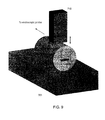
- Fig. 9 illustrates an operational diagram of an exemplary method for modulating the optical phase/amplitude profile of a rectangular mode fiber according to an exemplary embodiment of the present invention.
- the light in a core 702 passes through a portion of the fiber located between a stiff stationary backing 705 and an actuator 710.
- An activation of an actuator 710 can produce a downward or upward motion that changes the compressive stresses within the fiber, and can perturb the modal profile.
- the actuator 710 can optionally be a piezo-electric stack actuator, and the orientation of the core 702 relative to the actuator can be as shown in Fig. 9 or rotated.
- Fig. 10 depicts a diagram of an exemplary endoscopic imaging arrangement for generating a dithered beam scan (similar to the pattern shown in Fig. 4A ) using a piezo-actuator small diameter endoscopic imaging probe to vibrate the fiber end prior to focusing by a lens according to a particular exemplary embodiment of the present invention.
- an optical fiber 800 can direct an imaging light to the focusing lens 820.
- a piezo-actuator 805 can be driven with a sinusoidal signal such that the fiber tip vibrates.
- the light from the fiber can be expanded in an air gap 815, focused by a lens 820, and directed sideways by a prism 825.
- the focused spot of an imaging beam 830 may oscillate in an indicated dithered pattern 835.
- a rotation of the entire catheter that is contained within a housing 810 can scan the beam internally, for example, in a hollow cylindrical organ 840.
Landscapes
- Health & Medical Sciences (AREA)
- Life Sciences & Earth Sciences (AREA)
- Medical Informatics (AREA)
- Molecular Biology (AREA)
- Veterinary Medicine (AREA)
- Biophysics (AREA)
- Public Health (AREA)
- Engineering & Computer Science (AREA)
- Biomedical Technology (AREA)
- Heart & Thoracic Surgery (AREA)
- General Health & Medical Sciences (AREA)
- Physics & Mathematics (AREA)
- Surgery (AREA)
- Animal Behavior & Ethology (AREA)
- Pathology (AREA)
- Nuclear Medicine, Radiotherapy & Molecular Imaging (AREA)
- Radiology & Medical Imaging (AREA)
- Ophthalmology & Optometry (AREA)
- Investigating Or Analysing Materials By Optical Means (AREA)
- Length Measuring Devices By Optical Means (AREA)
Claims (5)
- Bildgebungsvorrichtung für optische Kohärenztomographie zum Verbessern eines Kontrasts in einem oder mehreren volumetrischen optischen Kohärenztomographiebildern durch das Filtern von Daten, wobei die Vorrichtung Folgendes umfasst:wenigstens eine erste Anordnung, die eine Lichtquelle umfasst und dafür konfiguriert ist, eine bestimmte Strahlung bereitzustellen, die in wenigstens eine erste elektromagnetische Strahlung, die auf wenigstens eine Probe gerichtet ist, und wenigstens eine zweite elektromagnetische Strahlung, die auf eine Referenz gerichtet ist, geteilt ist,wenigstens eine zweite Anordnung, die dafür konfiguriert ist, die wenigstens eine erste elektromagnetische Strahlung, die eine bestimmte Querschnittsbreite hat, an wenigstens einen Abschnitt der wenigstens einen Probe anzulegen, um wenigstens eine dritte elektromagnetische Strahlung zu erzeugen,wobei die wenigstens eine zweite Anordnung ferner dafür konfiguriert ist, die wenigstens eine erste elektromagnetische Strahlung in dem wenigstens einen Abschnitt entlang einer bestimmten Achse X für eine Entfernung zwischen einem Multiplikator von 0,5 und 100 der bestimmten Querschnittsbreite zu verschieben,wobei die wenigstens eine zweite Anordnung dafür konfiguriert ist, die wenigstens eine erste elektromagnetische Strahlung in dem wenigstens einen Abschnitt entlang einer weiteren Achse Y, die von der bestimmten Achse X verschieden ist, zu verschieben,wenigstens eine dritte Anordnung, die dafür konfiguriert ist, eine Interferenz zwischen der wenigstens einen dritten elektromagnetischen Strahlung, die mit der wenigstens einen ersten elektromagnetischen Strahlung verknüpft ist, und wenigstens einer vierten elektromagnetischen Strahlung, die mit der wenigstens einen zweiten elektromagnetischen Strahlung verknüpft ist, zu erfassen,wenigstens eine vierte Anordnung, die dafür konfiguriert ist, die erfasste Interferenz in A-Linien in der Z-Dimension umzuwandeln, um 3-dimensionale OCT-Daten zu erlangen, und durch die Verwendung der 3-dimensionalen OCT-Daten wenigstens ein Bild, das mit dem wenigstens einen Abschnitt verknüpft ist, als eine Funktion der erfassten Interferenz zu erzeugen,wobei die vierte Anordnung ein volumetrisches Medianfilter umfasst, wobei das Filter dafür konfiguriert ist, die 3-dimensionalen OCT-Daten innerhalb der Ebene entsprechend der Schnittebene in XZ-Dimensionen zu filtern und die 3-dimensionalen OCT-Daten außerhalb der Ebene entsprechend der Y-Dimension zu filtern, undwobei das volumetrische Filter in der Größe asymmetrisch ist, mit minimalem Filtern in der Ebene und beträchtlichem Filtern außerhalb der Ebene.
- Vorrichtung nach Anspruch 1, wobei eine obere Grenze des Multiplikators 50, 60, 70, 80 und/oder 90 der bestimmten Querschnittsbreite ist.
- Vorrichtung nach Anspruch 1, wobei die wenigstens eine zweite Anordnung ferner dafür konfiguriert ist, die wenigstens eine erste elektromagnetische Strahlung in dem wenigstens einen Abschnitt in einem sinusförmigen Muster, einem dreieckigen Muster, einem Sägezahnmuster und/oder einem Spiralmuster zu verschieben.
- Vorrichtung nach Anspruch 1, wobei die wenigstens eine zweite Anordnung ferner dafür konfiguriert ist, eine asymmetrische Querschnittsfläche der wenigstens einen ersten elektromagnetischen Strahlung bereitzustellen.
- Verfahren zum Abbilden einer Probe zum Verbessern eines Kontrasts in einem oder mehreren volumetrischen optischen Kohärenztomographiebildern durch das Filtern von Daten, wobei das Verfahren Folgendes umfasst:das Bereitstellen einer bestimmten Strahlung von einer Lichtquelle und das Teilen derselben in wenigstens eine erste elektromagnetische Strahlung, die auf die Probe gerichtet ist, und wenigstens eine zweite elektromagnetische Strahlung, die auf eine Referenz gerichtet ist,das Anlegen der wenigstens einen ersten elektromagnetischen Strahlung, die eine bestimmte Querschnittsbreite hat, an wenigstens einen Abschnitt der wenigstens einen Probe, um wenigstens eine dritte elektromagnetische Strahlung zu erzeugen,das Verschieben der wenigstens einen ersten elektromagnetischen Strahlung in dem wenigstens einen Abschnitt entlang einer bestimmten Achse X für eine Entfernung zwischen einem Multiplikator von 0,5 und 100 der bestimmten Querschnittsbreite und das Verschieben der wenigstens einen ersten elektromagnetischen Strahlung in dem wenigstens einen Abschnitt entlang einer weiteren Achse Y, die von der bestimmten Achse X verschieden ist, unddas Erfassen einer Interferenz zwischen der wenigstens einen dritten elektromagnetischen Strahlung, die mit der wenigstens einen ersten elektromagnetischen Strahlung verknüpft ist, und wenigstens einer vierten elektromagnetischen Strahlung, die mit der wenigstens einen zweiten elektromagnetischen Strahlung verknüpft ist,das Umwandeln der erfassten Interferenz in A-Linien in der Z-Dimension, um 3-dimensionale OCT-Daten zu erlangen, und durch die Verwendung der 3-dimensionalen OCT-Daten das Erzeugen wenigstens eines Bildes, das mit dem wenigstens einen Abschnitt verknüpft ist, als eine Funktion der erfassten Interferenz, wobei das Bild unter Verwendung eines volumetrischen Medianfilters erzeugt wird, das dafür konfiguriert ist, die 3-dimensionalen OCT-Daten innerhalb der Ebene entsprechend der Schnittebene in XZ-Dimensionen zu filtern und die 3-dimensionalen OCT-Daten außerhalb der Ebene entsprechend der Y-Dimension zu filtern, undwobei das volumetrische Filter in der Größe asymmetrisch ist, mit minimalem Filtern in der Ebene und beträchtlichem Filtern außerhalb der Ebene.
Priority Applications (1)
| Application Number | Priority Date | Filing Date | Title |
|---|---|---|---|
| EP15188408.7A EP3006920A3 (de) | 2006-08-25 | 2007-08-24 | Vorrichtungen und verfahren zur verstärkung von optischer kohärenztomografiebildgebung mithilfe volumetrischer filterungsverfahren |
Applications Claiming Priority (2)
| Application Number | Priority Date | Filing Date | Title |
|---|---|---|---|
| US84021306P | 2006-08-25 | 2006-08-25 | |
| PCT/US2007/076710 WO2008024948A2 (en) | 2006-08-25 | 2007-08-24 | Apparatus and methods for enhancing optical coherence tomography imaging using volumetric filtering techniques |
Related Child Applications (1)
| Application Number | Title | Priority Date | Filing Date |
|---|---|---|---|
| EP15188408.7A Division EP3006920A3 (de) | 2006-08-25 | 2007-08-24 | Vorrichtungen und verfahren zur verstärkung von optischer kohärenztomografiebildgebung mithilfe volumetrischer filterungsverfahren |
Publications (2)
| Publication Number | Publication Date |
|---|---|
| EP2054712A2 EP2054712A2 (de) | 2009-05-06 |
| EP2054712B1 true EP2054712B1 (de) | 2015-10-07 |
Family
ID=39033720
Family Applications (2)
| Application Number | Title | Priority Date | Filing Date |
|---|---|---|---|
| EP07841307.7A Not-in-force EP2054712B1 (de) | 2006-08-25 | 2007-08-24 | Vorrichtungen und verfahren zur verstärkung einer optischen kohärenztomographie-abbildung mithilfe volumetrischer filterungsverfahren |
| EP15188408.7A Withdrawn EP3006920A3 (de) | 2006-08-25 | 2007-08-24 | Vorrichtungen und verfahren zur verstärkung von optischer kohärenztomografiebildgebung mithilfe volumetrischer filterungsverfahren |
Family Applications After (1)
| Application Number | Title | Priority Date | Filing Date |
|---|---|---|---|
| EP15188408.7A Withdrawn EP3006920A3 (de) | 2006-08-25 | 2007-08-24 | Vorrichtungen und verfahren zur verstärkung von optischer kohärenztomografiebildgebung mithilfe volumetrischer filterungsverfahren |
Country Status (5)
| Country | Link |
|---|---|
| US (1) | US7920271B2 (de) |
| EP (2) | EP2054712B1 (de) |
| JP (2) | JP2010501877A (de) |
| CN (1) | CN101589301B (de) |
| WO (1) | WO2008024948A2 (de) |
Families Citing this family (33)
| Publication number | Priority date | Publication date | Assignee | Title |
|---|---|---|---|---|
| JP5538368B2 (ja) * | 2008-05-15 | 2014-07-02 | アクサン・テクノロジーズ・インコーポレーテッド | Octの結合プローブおよび一体化システム |
| GB0812712D0 (en) * | 2008-07-10 | 2008-08-20 | Imp Innovations Ltd | Improved endoscope |
| US20100253769A1 (en) * | 2008-09-04 | 2010-10-07 | Laser Light Engines | Optical System and Assembly Method |
| JP5624040B2 (ja) * | 2009-08-04 | 2014-11-12 | 国立大学法人宇都宮大学 | 網膜用3次元画像生成装置 |
| US10678061B2 (en) | 2009-09-03 | 2020-06-09 | Laser Light Engines, Inc. | Low etendue illumination |
| US11105686B2 (en) | 2010-05-10 | 2021-08-31 | University of Pittshurgh-Of the Commonwealth System of Higher Education | Spatial-domain low-coherence quantitative phase microscopy |
| WO2012100816A1 (en) * | 2011-01-25 | 2012-08-02 | Optopol Technology S.A. | Optical coherence tomography apparatus and method with speckle suppression |
| KR101226442B1 (ko) * | 2011-04-08 | 2013-01-28 | 이큐메드 주식회사 | 고해상도 분광기를 구비한 광 단층촬영 시스템 및 그 방법 |
| KR101223074B1 (ko) * | 2011-06-09 | 2013-01-17 | 광주과학기술원 | 광간섭 단층촬영 장치 및 그를 이용한 광간섭 단층촬영 방법 |
| CN102322880B (zh) * | 2011-08-18 | 2013-06-05 | 天津大学 | 偏振敏感的分布式光频域反射扰动传感装置和解调方法 |
| KR20130099603A (ko) * | 2012-02-29 | 2013-09-06 | 삼성테크윈 주식회사 | 분광 검사 장치 |
| US8896840B2 (en) * | 2012-04-25 | 2014-11-25 | Canon Kabushiki Kaisha | Interferometric method and digital holographic microscope |
| US9541375B2 (en) | 2012-07-20 | 2017-01-10 | Samsung Electronics Co., Ltd. | Method and apparatus for generating tomography images |
| US9572529B2 (en) | 2012-10-31 | 2017-02-21 | Covidien Lp | Surgical devices and methods utilizing optical coherence tomography (OCT) to monitor and control tissue sealing |
| US9677869B2 (en) | 2012-12-05 | 2017-06-13 | Perimeter Medical Imaging, Inc. | System and method for generating a wide-field OCT image of a portion of a sample |
| JP6053138B2 (ja) * | 2013-01-24 | 2016-12-27 | 株式会社日立エルジーデータストレージ | 光断層観察装置及び光断層観察方法 |
| US8879813B1 (en) | 2013-10-22 | 2014-11-04 | Eyenuk, Inc. | Systems and methods for automated interest region detection in retinal images |
| US10130259B2 (en) * | 2014-02-05 | 2018-11-20 | British Columbia Cancer Agency Branch | Systems for optical imaging of biological tissues |
| JP6869951B2 (ja) | 2015-04-16 | 2021-05-12 | ジェンテュイティ・リミテッド・ライアビリティ・カンパニーGentuity, LLC | 撮像システム |
| WO2017004555A1 (en) * | 2015-07-01 | 2017-01-05 | The Trustees Of Columbia University In The City Of New York | System, method and computer-accessbile medium for multi-plane imaging of neural circuits |
| WO2017040484A1 (en) | 2015-08-31 | 2017-03-09 | Gentuity, Llc | Imaging system includes imaging probe and delivery devices |
| US10426326B2 (en) * | 2017-04-19 | 2019-10-01 | Canon U.S.A, Inc. | Fiber optic correction of astigmatism |
| EP3655748B1 (de) | 2017-07-18 | 2023-08-09 | Perimeter Medical Imaging, Inc. | Probenbehälter zur stabilisierung und ausrichtung exzidierter biologischer gewebeproben zur ex-vivo-analyse |
| EP3700406A4 (de) | 2017-11-28 | 2021-12-29 | Gentuity LLC | Bildgebungssystem |
| CN111819417B (zh) * | 2018-03-01 | 2022-03-22 | 爱尔康公司 | 用于稳定光学相干断层扫描成像的共光路波导 |
| US12262872B2 (en) | 2018-09-17 | 2025-04-01 | Gentuity, Llc | Imaging system with optical pathway |
| US10791923B2 (en) | 2018-09-24 | 2020-10-06 | Canon U.S.A., Inc. | Ball lens for optical probe and methods therefor |
| EP3628210A1 (de) * | 2018-09-28 | 2020-04-01 | Paris Sciences et Lettres - Quartier Latin | Verfahren und systeme zur bildgebung mittels in-vivo-vollfeld-interferenzmikroskopie |
| US12364385B2 (en) | 2019-04-30 | 2025-07-22 | Gentuity, Llc | Imaging probe with fluid pressurization element |
| JP2022533212A (ja) | 2019-05-21 | 2022-07-21 | ジェンテュイティ・リミテッド・ライアビリティ・カンパニー | 患者のoctガイド処置システム及び方法 |
| KR102284115B1 (ko) * | 2019-07-05 | 2021-07-30 | 한국광기술원 | 안구의 혼탁도 감쇄를 위한 접촉형 광 변조 장치 및 방법 |
| CA3202994A1 (en) * | 2020-11-27 | 2022-06-02 | Aliph Medical Llc | Imaging system comprising a scanner and a modulator and corresponding method with improved accuracy |
| CN114886389A (zh) * | 2022-07-14 | 2022-08-12 | 之江实验室 | 一种三维光声/超声双模内窥镜及成像方法 |
Family Cites Families (400)
| Publication number | Priority date | Publication date | Assignee | Title |
|---|---|---|---|---|
| US2339754A (en) * | 1941-03-04 | 1944-01-25 | Westinghouse Electric & Mfg Co | Supervisory apparatus |
| US3090753A (en) | 1960-08-02 | 1963-05-21 | Exxon Research Engineering Co | Ester oil compositions containing acid anhydride |
| GB1257778A (de) | 1967-12-07 | 1971-12-22 | ||
| US3601480A (en) | 1968-07-10 | 1971-08-24 | Physics Int Co | Optical tunnel high-speed camera system |
| JPS4932484U (de) | 1972-06-19 | 1974-03-20 | ||
| US3872407A (en) * | 1972-09-01 | 1975-03-18 | Us Navy | Rapidly tunable laser |
| JPS584481Y2 (ja) * | 1973-06-23 | 1983-01-26 | オリンパス光学工業株式会社 | ナイシキヨウシヤヘンカンコウガクケイ |
| FR2253410A5 (de) | 1973-12-03 | 1975-06-27 | Inst Nat Sante Rech Med | |
| US3941121A (en) * | 1974-12-20 | 1976-03-02 | The University Of Cincinnati | Focusing fiber-optic needle endoscope |
| US3983507A (en) | 1975-01-06 | 1976-09-28 | Research Corporation | Tunable laser systems and method |
| US3973219A (en) | 1975-04-24 | 1976-08-03 | Cornell Research Foundation, Inc. | Very rapidly tuned cw dye laser |
| US4030831A (en) | 1976-03-22 | 1977-06-21 | The United States Of America As Represented By The Secretary Of The Navy | Phase detector for optical figure sensing |
| US4141362A (en) * | 1977-05-23 | 1979-02-27 | Richard Wolf Gmbh | Laser endoscope |
| US4224929A (en) | 1977-11-08 | 1980-09-30 | Olympus Optical Co., Ltd. | Endoscope with expansible cuff member and operation section |
| GB2030313A (en) | 1978-06-29 | 1980-04-02 | Wolf Gmbh Richard | Endoscopes |
| FR2448728A1 (fr) | 1979-02-07 | 1980-09-05 | Thomson Csf | Dispositif joint tournant pour liaison par conducteurs optiques et systeme comportant un tel dispositif |
| US4300816A (en) | 1979-08-30 | 1981-11-17 | United Technologies Corporation | Wide band multicore optical fiber |
| US4295738A (en) | 1979-08-30 | 1981-10-20 | United Technologies Corporation | Fiber optic strain sensor |
| US4428643A (en) * | 1981-04-08 | 1984-01-31 | Xerox Corporation | Optical scanning system with wavelength shift correction |
| US5065331A (en) | 1981-05-18 | 1991-11-12 | Vachon Reginald I | Apparatus and method for determining the stress and strain in pipes, pressure vessels, structural members and other deformable bodies |
| GB2106736B (en) | 1981-09-03 | 1985-06-12 | Standard Telephones Cables Ltd | Optical transmission system |
| US4479499A (en) | 1982-01-29 | 1984-10-30 | Alfano Robert R | Method and apparatus for detecting the presence of caries in teeth using visible light |
| US4601036A (en) | 1982-09-30 | 1986-07-15 | Honeywell Inc. | Rapidly tunable laser |
| HU187188B (en) | 1982-11-25 | 1985-11-28 | Koezponti Elelmiszeripari | Device for generating radiation of controllable spectral structure |
| CH663466A5 (fr) * | 1983-09-12 | 1987-12-15 | Battelle Memorial Institute | Procede et dispositif pour determiner la position d'un objet par rapport a une reference. |
| US4763977A (en) | 1985-01-09 | 1988-08-16 | Canadian Patents And Development Limited-Societe | Optical fiber coupler with tunable coupling ratio and method of making |
| US5318024A (en) | 1985-03-22 | 1994-06-07 | Massachusetts Institute Of Technology | Laser endoscope for spectroscopic imaging |
| EP0590268B1 (de) | 1985-03-22 | 1998-07-01 | Massachusetts Institute Of Technology | Faseroptisches Sondensystem zur spektralen Diagnose von Gewebe |
| US4607622A (en) | 1985-04-11 | 1986-08-26 | Charles D. Fritch | Fiber optic ocular endoscope |
| US4631498A (en) | 1985-04-26 | 1986-12-23 | Hewlett-Packard Company | CW Laser wavemeter/frequency locking technique |
| US4650327A (en) * | 1985-10-28 | 1987-03-17 | Oximetrix, Inc. | Optical catheter calibrating assembly |
| US5040889A (en) | 1986-05-30 | 1991-08-20 | Pacific Scientific Company | Spectrometer with combined visible and ultraviolet sample illumination |
| CA1290019C (en) | 1986-06-20 | 1991-10-01 | Hideo Kuwahara | Dual balanced optical signal receiver |
| US4770492A (en) | 1986-10-28 | 1988-09-13 | Spectran Corporation | Pressure or strain sensitive optical fiber |
| JPH0824665B2 (ja) | 1986-11-28 | 1996-03-13 | オリンパス光学工業株式会社 | 内視鏡装置 |
| US4744656A (en) | 1986-12-08 | 1988-05-17 | Spectramed, Inc. | Disposable calibration boot for optical-type cardiovascular catheter |
| US4751706A (en) | 1986-12-31 | 1988-06-14 | The United States Of America As Represented By The Secretary Of The Army | Laser for providing rapid sequence of different wavelengths |
| US4834111A (en) | 1987-01-12 | 1989-05-30 | The Trustees Of Columbia University In The City Of New York | Heterodyne interferometer |
| GB2209221B (en) | 1987-09-01 | 1991-10-23 | Litton Systems Inc | Hydrophone demodulator circuit and method |
| US5202931A (en) | 1987-10-06 | 1993-04-13 | Cell Analysis Systems, Inc. | Methods and apparatus for the quantitation of nuclear protein |
| US4909631A (en) * | 1987-12-18 | 1990-03-20 | Tan Raul Y | Method for film thickness and refractive index determination |
| US4890901A (en) * | 1987-12-22 | 1990-01-02 | Hughes Aircraft Company | Color corrector for embedded prisms |
| US4892406A (en) * | 1988-01-11 | 1990-01-09 | United Technologies Corporation | Method of and arrangement for measuring vibrations |
| FR2626367B1 (fr) | 1988-01-25 | 1990-05-11 | Thomson Csf | Capteur de temperature multipoints a fibre optique |
| FR2626383B1 (fr) | 1988-01-27 | 1991-10-25 | Commissariat Energie Atomique | Procede de microscopie optique confocale a balayage et en profondeur de champ etendue et dispositifs pour la mise en oeuvre du procede |
| US4925302A (en) | 1988-04-13 | 1990-05-15 | Hewlett-Packard Company | Frequency locking device |
| US4998972A (en) * | 1988-04-28 | 1991-03-12 | Thomas J. Fogarty | Real time angioscopy imaging system |
| US5730731A (en) * | 1988-04-28 | 1998-03-24 | Thomas J. Fogarty | Pressure-based irrigation accumulator |
| US4905169A (en) * | 1988-06-02 | 1990-02-27 | The United States Of America As Represented By The United States Department Of Energy | Method and apparatus for simultaneously measuring a plurality of spectral wavelengths present in electromagnetic radiation |
| US5242437A (en) | 1988-06-10 | 1993-09-07 | Trimedyne Laser Systems, Inc. | Medical device applying localized high intensity light and heat, particularly for destruction of the endometrium |
| WO1990000754A1 (en) | 1988-07-13 | 1990-01-25 | Martin Russell Harris | Scanning confocal microscope |
| GB8817672D0 (en) | 1988-07-25 | 1988-09-01 | Sira Ltd | Optical apparatus |
| US5214538A (en) | 1988-07-25 | 1993-05-25 | Keymed (Medical And Industrial Equipment) Limited | Optical apparatus |
| US4868834A (en) | 1988-09-14 | 1989-09-19 | The United States Of America As Represented By The Secretary Of The Army | System for rapidly tuning a low pressure pulsed laser |
| DE3833602A1 (de) * | 1988-10-03 | 1990-02-15 | Krupp Gmbh | Spektrometer zur gleichzeitigen intensitaetsmessung in verschiedenen spektralbereichen |
| US5419323A (en) | 1988-12-21 | 1995-05-30 | Massachusetts Institute Of Technology | Method for laser induced fluorescence of tissue |
| US5046501A (en) | 1989-01-18 | 1991-09-10 | Wayne State University | Atherosclerotic identification |
| US5085496A (en) * | 1989-03-31 | 1992-02-04 | Sharp Kabushiki Kaisha | Optical element and optical pickup device comprising it |
| US5317389A (en) | 1989-06-12 | 1994-05-31 | California Institute Of Technology | Method and apparatus for white-light dispersed-fringe interferometric measurement of corneal topography |
| US4965599A (en) | 1989-11-13 | 1990-10-23 | Eastman Kodak Company | Scanning apparatus for halftone image screen writing |
| US4984888A (en) * | 1989-12-13 | 1991-01-15 | Imo Industries, Inc. | Two-dimensional spectrometer |
| KR930003307B1 (ko) | 1989-12-14 | 1993-04-24 | 주식회사 금성사 | 입체용 프로젝터 |
| DD293205B5 (de) | 1990-03-05 | 1995-06-29 | Zeiss Carl Jena Gmbh | Lichtleiterfuehrung fuer ein medizinisches Beobachtungsgeraet |
| US5039193A (en) | 1990-04-03 | 1991-08-13 | Focal Technologies Incorporated | Fibre optic single mode rotary joint |
| US5262644A (en) | 1990-06-29 | 1993-11-16 | Southwest Research Institute | Remote spectroscopy for raman and brillouin scattering |
| US5197470A (en) * | 1990-07-16 | 1993-03-30 | Eastman Kodak Company | Near infrared diagnostic method and instrument |
| GB9015793D0 (en) | 1990-07-18 | 1990-09-05 | Medical Res Council | Confocal scanning optical microscope |
| US5127730A (en) | 1990-08-10 | 1992-07-07 | Regents Of The University Of Minnesota | Multi-color laser scanning confocal imaging system |
| US5845639A (en) | 1990-08-10 | 1998-12-08 | Board Of Regents Of The University Of Washington | Optical imaging methods |
| US5305759A (en) | 1990-09-26 | 1994-04-26 | Olympus Optical Co., Ltd. | Examined body interior information observing apparatus by using photo-pulses controlling gains for depths |
| US5241364A (en) | 1990-10-19 | 1993-08-31 | Fuji Photo Film Co., Ltd. | Confocal scanning type of phase contrast microscope and scanning microscope |
| US5250186A (en) | 1990-10-23 | 1993-10-05 | Cetus Corporation | HPLC light scattering detector for biopolymers |
| US5202745A (en) | 1990-11-07 | 1993-04-13 | Hewlett-Packard Company | Polarization independent optical coherence-domain reflectometry |
| US5275594A (en) * | 1990-11-09 | 1994-01-04 | C. R. Bard, Inc. | Angioplasty system having means for identification of atherosclerotic plaque |
| JP3035336B2 (ja) * | 1990-11-27 | 2000-04-24 | 興和株式会社 | 血流測定装置 |
| US5228001A (en) | 1991-01-23 | 1993-07-13 | Syracuse University | Optical random access memory |
| US5784162A (en) | 1993-08-18 | 1998-07-21 | Applied Spectral Imaging Ltd. | Spectral bio-imaging methods for biological research, medical diagnostics and therapy |
| US6198532B1 (en) | 1991-02-22 | 2001-03-06 | Applied Spectral Imaging Ltd. | Spectral bio-imaging of the eye |
| US5293872A (en) * | 1991-04-03 | 1994-03-15 | Alfano Robert R | Method for distinguishing between calcified atherosclerotic tissue and fibrous atherosclerotic tissue or normal cardiovascular tissue using Raman spectroscopy |
| WO1992019930A1 (en) | 1991-04-29 | 1992-11-12 | Massachusetts Institute Of Technology | Method and apparatus for optical imaging and measurement |
| US6485413B1 (en) | 1991-04-29 | 2002-11-26 | The General Hospital Corporation | Methods and apparatus for forward-directed optical scanning instruments |
| US6134003A (en) | 1991-04-29 | 2000-10-17 | Massachusetts Institute Of Technology | Method and apparatus for performing optical measurements using a fiber optic imaging guidewire, catheter or endoscope |
| US6111645A (en) | 1991-04-29 | 2000-08-29 | Massachusetts Institute Of Technology | Grating based phase control optical delay line |
| US5956355A (en) | 1991-04-29 | 1999-09-21 | Massachusetts Institute Of Technology | Method and apparatus for performing optical measurements using a rapidly frequency-tuned laser |
| US6564087B1 (en) | 1991-04-29 | 2003-05-13 | Massachusetts Institute Of Technology | Fiber optic needle probes for optical coherence tomography imaging |
| US6501551B1 (en) | 1991-04-29 | 2002-12-31 | Massachusetts Institute Of Technology | Fiber optic imaging endoscope interferometer with at least one faraday rotator |
| US5748598A (en) | 1995-12-22 | 1998-05-05 | Massachusetts Institute Of Technology | Apparatus and methods for reading multilayer storage media using short coherence length sources |
| US5465147A (en) | 1991-04-29 | 1995-11-07 | Massachusetts Institute Of Technology | Method and apparatus for acquiring images using a ccd detector array and no transverse scanner |
| US5441053A (en) | 1991-05-03 | 1995-08-15 | University Of Kentucky Research Foundation | Apparatus and method for multiple wavelength of tissue |
| US5208651A (en) | 1991-07-16 | 1993-05-04 | The Regents Of The University Of California | Apparatus and method for measuring fluorescence intensities at a plurality of wavelengths and lifetimes |
| DE4128744C1 (de) * | 1991-08-29 | 1993-04-22 | Siemens Ag, 8000 Muenchen, De | |
| US5353790A (en) | 1992-01-17 | 1994-10-11 | Board Of Regents, The University Of Texas System | Method and apparatus for optical measurement of bilirubin in tissue |
| US5212667A (en) | 1992-02-03 | 1993-05-18 | General Electric Company | Light imaging in a scattering medium, using ultrasonic probing and speckle image differencing |
| US5248876A (en) | 1992-04-21 | 1993-09-28 | International Business Machines Corporation | Tandem linear scanning confocal imaging system with focal volumes at different heights |
| US5486701A (en) * | 1992-06-16 | 1996-01-23 | Prometrix Corporation | Method and apparatus for measuring reflectance in two wavelength bands to enable determination of thin film thickness |
| US5716324A (en) * | 1992-08-25 | 1998-02-10 | Fuji Photo Film Co., Ltd. | Endoscope with surface and deep portion imaging systems |
| US5348003A (en) | 1992-09-03 | 1994-09-20 | Sirraya, Inc. | Method and apparatus for chemical analysis |
| US5772597A (en) | 1992-09-14 | 1998-06-30 | Sextant Medical Corporation | Surgical tool end effector |
| US5698397A (en) | 1995-06-07 | 1997-12-16 | Sri International | Up-converting reporters for biological and other assays using laser excitation techniques |
| US5383467A (en) * | 1992-11-18 | 1995-01-24 | Spectrascience, Inc. | Guidewire catheter and apparatus for diagnostic imaging |
| AU5672194A (en) * | 1992-11-18 | 1994-06-22 | Spectrascience, Inc. | Apparatus for diagnostic imaging |
| JPH06222242A (ja) | 1993-01-27 | 1994-08-12 | Shin Etsu Chem Co Ltd | 光ファイバカプラおよびその製造方法 |
| US5987346A (en) | 1993-02-26 | 1999-11-16 | Benaron; David A. | Device and method for classification of tissue |
| JP3369623B2 (ja) * | 1993-03-16 | 2003-01-20 | 興和株式会社 | レーザー走査型眼科撮像装置 |
| JP3112595B2 (ja) * | 1993-03-17 | 2000-11-27 | 安藤電気株式会社 | 光周波数シフタを用いる光ファイバ歪位置測定装置 |
| FI93781C (fi) | 1993-03-18 | 1995-05-26 | Wallac Oy | Biospesifinen multiparametrinen määritysmenetelmä |
| DE4309056B4 (de) | 1993-03-20 | 2006-05-24 | Häusler, Gerd, Prof. Dr. | Verfahren und Vorrichtung zur Ermittlung der Entfernung und Streuintensität von streuenden Punkten |
| DE4310209C2 (de) * | 1993-03-29 | 1996-05-30 | Bruker Medizintech | Optische stationäre Bildgebung in stark streuenden Medien |
| US5485079A (en) | 1993-03-29 | 1996-01-16 | Matsushita Electric Industrial Co., Ltd. | Magneto-optical element and optical magnetic field sensor |
| US5424827A (en) | 1993-04-30 | 1995-06-13 | Litton Systems, Inc. | Optical system and method for eliminating overlap of diffraction spectra |
| DE4314189C1 (de) | 1993-04-30 | 1994-11-03 | Bodenseewerk Geraetetech | Vorrichtung zur Untersuchung von Lichtleitfasern aus Glas mittels Heterodyn-Brillouin-Spektroskopie |
| US5454807A (en) | 1993-05-14 | 1995-10-03 | Boston Scientific Corporation | Medical treatment of deeply seated tissue using optical radiation |
| DE69418248T2 (de) | 1993-06-03 | 1999-10-14 | Hamamatsu Photonics Kk | Optisches Laser-Abtastsystem mit Axikon |
| JP3234353B2 (ja) | 1993-06-15 | 2001-12-04 | 富士写真フイルム株式会社 | 断層情報読取装置 |
| US5803082A (en) | 1993-11-09 | 1998-09-08 | Staplevision Inc. | Omnispectramammography |
| US5983125A (en) | 1993-12-13 | 1999-11-09 | The Research Foundation Of City College Of New York | Method and apparatus for in vivo examination of subcutaneous tissues inside an organ of a body using optical spectroscopy |
| US5450203A (en) | 1993-12-22 | 1995-09-12 | Electroglas, Inc. | Method and apparatus for determining an objects position, topography and for imaging |
| US5411016A (en) | 1994-02-22 | 1995-05-02 | Scimed Life Systems, Inc. | Intravascular balloon catheter for use in combination with an angioscope |
| US5590660A (en) * | 1994-03-28 | 1997-01-07 | Xillix Technologies Corp. | Apparatus and method for imaging diseased tissue using integrated autofluorescence |
| DE4411017C2 (de) | 1994-03-30 | 1995-06-08 | Alexander Dr Knuettel | Optische stationäre spektroskopische Bildgebung in stark streuenden Objekten durch spezielle Lichtfokussierung und Signal-Detektion von Licht unterschiedlicher Wellenlängen |
| TW275570B (de) * | 1994-05-05 | 1996-05-11 | Boehringer Mannheim Gmbh | |
| US5459325A (en) | 1994-07-19 | 1995-10-17 | Molecular Dynamics, Inc. | High-speed fluorescence scanner |
| US6159445A (en) | 1994-07-20 | 2000-12-12 | Nycomed Imaging As | Light imaging contrast agents |
| ES2180597T4 (es) | 1994-08-18 | 2003-07-01 | Zeiss Carl | Aparato quirurgico asistido por tomografia de coherencia optica. |
| US5491524A (en) * | 1994-10-05 | 1996-02-13 | Carl Zeiss, Inc. | Optical coherence tomography corneal mapping apparatus |
| US5740808A (en) | 1996-10-28 | 1998-04-21 | Ep Technologies, Inc | Systems and methods for guilding diagnostic or therapeutic devices in interior tissue regions |
| US5817144A (en) | 1994-10-25 | 1998-10-06 | Latis, Inc. | Method for contemporaneous application OF laser energy and localized pharmacologic therapy |
| US6033721A (en) * | 1994-10-26 | 2000-03-07 | Revise, Inc. | Image-based three-axis positioner for laser direct write microchemical reaction |
| US5566267A (en) | 1994-12-15 | 1996-10-15 | Ceram Optec Industries Inc. | Flat surfaced optical fibers and diode laser medical delivery devices |
| US5600486A (en) * | 1995-01-30 | 1997-02-04 | Lockheed Missiles And Space Company, Inc. | Color separation microlens |
| US5648848A (en) | 1995-02-01 | 1997-07-15 | Nikon Precision, Inc. | Beam delivery apparatus and method for interferometry using rotatable polarization chucks |
| DE19506484C2 (de) | 1995-02-24 | 1999-09-16 | Stiftung Fuer Lasertechnologie | Verfahren und Vorrichtung zur selektiven nichtinvasiven Lasermyographie (LMG) |
| RU2100787C1 (ru) * | 1995-03-01 | 1997-12-27 | Геликонов Валентин Михайлович | Оптоволоконный интерферометр и оптоволоконный пьезоэлектрический преобразователь |
| US5526338A (en) | 1995-03-10 | 1996-06-11 | Yeda Research & Development Co. Ltd. | Method and apparatus for storage and retrieval with multilayer optical disks |
| US5697373A (en) | 1995-03-14 | 1997-12-16 | Board Of Regents, The University Of Texas System | Optical method and apparatus for the diagnosis of cervical precancers using raman and fluorescence spectroscopies |
| US5735276A (en) | 1995-03-21 | 1998-04-07 | Lemelson; Jerome | Method and apparatus for scanning and evaluating matter |
| US5926592A (en) | 1995-03-24 | 1999-07-20 | Optiscan Pty Ltd | Optical fibre confocal imager with variable near-confocal control |
| US5565983A (en) | 1995-05-26 | 1996-10-15 | The Perkin-Elmer Corporation | Optical spectrometer for detecting spectra in separate ranges |
| US5785651A (en) | 1995-06-07 | 1998-07-28 | Keravision, Inc. | Distance measuring confocal microscope |
| US5621830A (en) | 1995-06-07 | 1997-04-15 | Smith & Nephew Dyonics Inc. | Rotatable fiber optic joint |
| WO1997001167A1 (en) | 1995-06-21 | 1997-01-09 | Massachusetts Institute Of Technology | Apparatus and method for accessing data on multilayered optical media |
| ATA107495A (de) | 1995-06-23 | 1996-06-15 | Fercher Adolf Friedrich Dr | Kohärenz-biometrie und -tomographie mit dynamischem kohärentem fokus |
| JP3819032B2 (ja) * | 1995-08-24 | 2006-09-06 | ザ・テキサス・エイ・アンド・エム・ユニバーシティ・システム | 組織およびその他のランダム媒体における蛍光寿命に基づく撮像および分光分析 |
| US6016197A (en) * | 1995-08-25 | 2000-01-18 | Ceramoptec Industries Inc. | Compact, all-optical spectrum analyzer for chemical and biological fiber optic sensors |
| FR2738343B1 (fr) | 1995-08-30 | 1997-10-24 | Cohen Sabban Joseph | Dispositif de microstratigraphie optique |
| US6615071B1 (en) | 1995-09-20 | 2003-09-02 | Board Of Regents, The University Of Texas System | Method and apparatus for detecting vulnerable atherosclerotic plaque |
| CA2231425A1 (en) | 1995-09-20 | 1997-03-27 | Texas Heart Institute | Detecting thermal discrepancies in vessel walls |
| US6763261B2 (en) * | 1995-09-20 | 2004-07-13 | Board Of Regents, The University Of Texas System | Method and apparatus for detecting vulnerable atherosclerotic plaque |
| DE19542955C2 (de) | 1995-11-17 | 1999-02-18 | Schwind Gmbh & Co Kg Herbert | Endoskop |
| US5719399A (en) * | 1995-12-18 | 1998-02-17 | The Research Foundation Of City College Of New York | Imaging and characterization of tissue based upon the preservation of polarized light transmitted therethrough |
| US5748318A (en) | 1996-01-23 | 1998-05-05 | Brown University Research Foundation | Optical stress generator and detector |
| US5840023A (en) | 1996-01-31 | 1998-11-24 | Oraevsky; Alexander A. | Optoacoustic imaging for medical diagnosis |
| US5642194A (en) | 1996-02-05 | 1997-06-24 | The Regents Of The University Of California | White light velocity interferometer |
| US5862273A (en) * | 1996-02-23 | 1999-01-19 | Kaiser Optical Systems, Inc. | Fiber optic probe with integral optical filtering |
| US5843000A (en) | 1996-05-07 | 1998-12-01 | The General Hospital Corporation | Optical biopsy forceps and method of diagnosing tissue |
| ATA84696A (de) * | 1996-05-14 | 1998-03-15 | Adolf Friedrich Dr Fercher | Verfahren und anordnungen zur kontrastanhebung in der optischen kohärenztomographie |
| US6020963A (en) * | 1996-06-04 | 2000-02-01 | Northeastern University | Optical quadrature Interferometer |
| US5795295A (en) | 1996-06-25 | 1998-08-18 | Carl Zeiss, Inc. | OCT-assisted surgical microscope with multi-coordinate manipulator |
| US5842995A (en) | 1996-06-28 | 1998-12-01 | Board Of Regents, The Univerisity Of Texas System | Spectroscopic probe for in vivo measurement of raman signals |
| US6245026B1 (en) | 1996-07-29 | 2001-06-12 | Farallon Medsystems, Inc. | Thermography catheter |
| US6396941B1 (en) | 1996-08-23 | 2002-05-28 | Bacus Research Laboratories, Inc. | Method and apparatus for internet, intranet, and local viewing of virtual microscope slides |
| US5840075A (en) | 1996-08-23 | 1998-11-24 | Eclipse Surgical Technologies, Inc. | Dual laser device for transmyocardial revascularization procedures |
| JPH1090603A (ja) | 1996-09-18 | 1998-04-10 | Olympus Optical Co Ltd | 内視鏡光学系 |
| US5801831A (en) | 1996-09-20 | 1998-09-01 | Institute For Space And Terrestrial Science | Fabry-Perot spectrometer for detecting a spatially varying spectral signature of an extended source |
| US6249349B1 (en) | 1996-09-27 | 2001-06-19 | Vincent Lauer | Microscope generating a three-dimensional representation of an object |
| DE19640495C2 (de) | 1996-10-01 | 1999-12-16 | Leica Microsystems | Vorrichtung zur konfokalen Oberflächenvermessung |
| US5843052A (en) | 1996-10-04 | 1998-12-01 | Benja-Athon; Anuthep | Irrigation kit for application of fluids and chemicals for cleansing and sterilizing wounds |
| US6044288A (en) * | 1996-11-08 | 2000-03-28 | Imaging Diagnostics Systems, Inc. | Apparatus and method for determining the perimeter of the surface of an object being scanned |
| US5872879A (en) * | 1996-11-25 | 1999-02-16 | Boston Scientific Corporation | Rotatable connecting optical fibers |
| US6517532B1 (en) * | 1997-05-15 | 2003-02-11 | Palomar Medical Technologies, Inc. | Light energy delivery head |
| US6437867B2 (en) | 1996-12-04 | 2002-08-20 | The Research Foundation Of The City University Of New York | Performing selected optical measurements with optical coherence domain reflectometry |
| US6249630B1 (en) * | 1996-12-13 | 2001-06-19 | Imra America, Inc. | Apparatus and method for delivery of dispersion-compensated ultrashort optical pulses with high peak power |
| US5871449A (en) * | 1996-12-27 | 1999-02-16 | Brown; David Lloyd | Device and method for locating inflamed plaque in an artery |
| US5991697A (en) | 1996-12-31 | 1999-11-23 | The Regents Of The University Of California | Method and apparatus for optical Doppler tomographic imaging of fluid flow velocity in highly scattering media |
| US5760901A (en) | 1997-01-28 | 1998-06-02 | Zetetic Institute | Method and apparatus for confocal interference microscopy with background amplitude reduction and compensation |
| US5801826A (en) | 1997-02-18 | 1998-09-01 | Williams Family Trust B | Spectrometric device and method for recognizing atomic and molecular signatures |
| US5836877A (en) | 1997-02-24 | 1998-11-17 | Lucid Inc | System for facilitating pathological examination of a lesion in tissue |
| US5968064A (en) | 1997-02-28 | 1999-10-19 | Lumend, Inc. | Catheter system for treating a vascular occlusion |
| US6010449A (en) * | 1997-02-28 | 2000-01-04 | Lumend, Inc. | Intravascular catheter system for treating a vascular occlusion |
| US6120516A (en) | 1997-02-28 | 2000-09-19 | Lumend, Inc. | Method for treating vascular occlusion |
| CA2283949A1 (en) * | 1997-03-13 | 1998-09-17 | Haishan Zeng | Methods and apparatus for detecting the rejection of transplanted tissue |
| US5994690A (en) | 1997-03-17 | 1999-11-30 | Kulkarni; Manish D. | Image enhancement in optical coherence tomography using deconvolution |
| US6117128A (en) | 1997-04-30 | 2000-09-12 | Kenton W. Gregory | Energy delivery catheter and method for the use thereof |
| US5887009A (en) * | 1997-05-22 | 1999-03-23 | Optical Biopsy Technologies, Inc. | Confocal optical scanning system employing a fiber laser |
| US6002480A (en) | 1997-06-02 | 1999-12-14 | Izatt; Joseph A. | Depth-resolved spectroscopic optical coherence tomography |
| DE69840791D1 (de) | 1997-06-02 | 2009-06-10 | Joseph A Izatt | Doppler-abbildung einer strömung mittels optischer kohaerenztomografie |
| US6208415B1 (en) * | 1997-06-12 | 2001-03-27 | The Regents Of The University Of California | Birefringence imaging in biological tissue using polarization sensitive optical coherent tomography |
| US5920390A (en) | 1997-06-26 | 1999-07-06 | University Of North Carolina | Fiberoptic interferometer and associated method for analyzing tissue |
| US6048349A (en) | 1997-07-09 | 2000-04-11 | Intraluminal Therapeutics, Inc. | Systems and methods for guiding a medical instrument through a body |
| US5921926A (en) | 1997-07-28 | 1999-07-13 | University Of Central Florida | Three dimensional optical imaging colposcopy |
| US6014214A (en) * | 1997-08-21 | 2000-01-11 | Li; Ming-Chiang | High speed inspection of a sample using coherence processing of scattered superbroad radiation |
| US5892583A (en) | 1997-08-21 | 1999-04-06 | Li; Ming-Chiang | High speed inspection of a sample using superbroad radiation coherent interferometer |
| US6069698A (en) | 1997-08-28 | 2000-05-30 | Olympus Optical Co., Ltd. | Optical imaging apparatus which radiates a low coherence light beam onto a test object, receives optical information from light scattered by the object, and constructs therefrom a cross-sectional image of the object |
| US6297018B1 (en) | 1998-04-17 | 2001-10-02 | Ljl Biosystems, Inc. | Methods and apparatus for detecting nucleic acid polymorphisms |
| US5920373A (en) | 1997-09-24 | 1999-07-06 | Heidelberg Engineering Optische Messysteme Gmbh | Method and apparatus for determining optical characteristics of a cornea |
| US6193676B1 (en) * | 1997-10-03 | 2001-02-27 | Intraluminal Therapeutics, Inc. | Guide wire assembly |
| US5951482A (en) | 1997-10-03 | 1999-09-14 | Intraluminal Therapeutics, Inc. | Assemblies and methods for advancing a guide wire through body tissue |
| US6091984A (en) | 1997-10-10 | 2000-07-18 | Massachusetts Institute Of Technology | Measuring tissue morphology |
| US5955737A (en) | 1997-10-27 | 1999-09-21 | Systems & Processes Engineering Corporation | Chemometric analysis for extraction of individual fluorescence spectrum and lifetimes from a target mixture |
| US6134010A (en) | 1997-11-07 | 2000-10-17 | Lucid, Inc. | Imaging system using polarization effects to enhance image quality |
| US6107048A (en) | 1997-11-20 | 2000-08-22 | Medical College Of Georgia Research Institute, Inc. | Method of detecting and grading dysplasia in epithelial tissue |
| US6165170A (en) | 1998-01-29 | 2000-12-26 | International Business Machines Corporation | Laser dermablator and dermablation |
| US6831781B2 (en) | 1998-02-26 | 2004-12-14 | The General Hospital Corporation | Confocal microscopy with multi-spectral encoding and system and apparatus for spectroscopically encoded confocal microscopy |
| US6134033A (en) | 1998-02-26 | 2000-10-17 | Tyco Submarine Systems Ltd. | Method and apparatus for improving spectral efficiency in wavelength division multiplexed transmission systems |
| US6048742A (en) | 1998-02-26 | 2000-04-11 | The United States Of America As Represented By The Secretary Of The Air Force | Process for measuring the thickness and composition of thin semiconductor films deposited on semiconductor wafers |
| EP2267507A3 (de) * | 1998-02-26 | 2011-08-17 | The General Hospital Corporation | Konfokale Mikroskopie mit multispektraler Kodierung |
| US6174291B1 (en) * | 1998-03-09 | 2001-01-16 | Spectrascience, Inc. | Optical biopsy system and methods for tissue diagnosis |
| US6066102A (en) | 1998-03-09 | 2000-05-23 | Spectrascience, Inc. | Optical biopsy forceps system and method of diagnosing tissue |
| US6151522A (en) | 1998-03-16 | 2000-11-21 | The Research Foundation Of Cuny | Method and system for examining biological materials using low power CW excitation raman spectroscopy |
| DE19814057B4 (de) | 1998-03-30 | 2009-01-02 | Carl Zeiss Meditec Ag | Anordnung zur optischen Kohärenztomographie und Kohärenztopographie |
| US6384915B1 (en) | 1998-03-30 | 2002-05-07 | The Regents Of The University Of California | Catheter guided by optical coherence domain reflectometry |
| US6175669B1 (en) * | 1998-03-30 | 2001-01-16 | The Regents Of The Universtiy Of California | Optical coherence domain reflectometry guidewire |
| US6996549B2 (en) * | 1998-05-01 | 2006-02-07 | Health Discovery Corporation | Computer-aided image analysis |
| JPH11326826A (ja) | 1998-05-13 | 1999-11-26 | Sony Corp | 照明方法及び照明装置 |
| US6053613A (en) | 1998-05-15 | 2000-04-25 | Carl Zeiss, Inc. | Optical coherence tomography with new interferometer |
| US6549801B1 (en) | 1998-06-11 | 2003-04-15 | The Regents Of The University Of California | Phase-resolved optical coherence tomography and optical doppler tomography for imaging fluid flow in tissue with fast scanning speed and high velocity sensitivity |
| AU5101699A (en) | 1998-07-15 | 2000-02-07 | Corazon Technologies, Inc. | Methods and devices for reducing the mineral content of vascular calcified lesions |
| US6166373A (en) | 1998-07-21 | 2000-12-26 | The Institute For Technology Development | Focal plane scanner with reciprocating spatial window |
| AU6417599A (en) | 1998-10-08 | 2000-04-26 | University Of Kentucky Research Foundation, The | Methods and apparatus for (in vivo) identification and characterization of vulnerable atherosclerotic plaques |
| US6274871B1 (en) | 1998-10-22 | 2001-08-14 | Vysis, Inc. | Method and system for performing infrared study on a biological sample |
| US6324419B1 (en) | 1998-10-27 | 2001-11-27 | Nejat Guzelsu | Apparatus and method for non-invasive measurement of stretch |
| DE69932485T2 (de) | 1998-11-20 | 2007-01-11 | Fuji Photo Film Co. Ltd., Minamiashigara | Blutgefäss Bilddarstellungssystem |
| US5975697A (en) | 1998-11-25 | 1999-11-02 | Oti Ophthalmic Technologies, Inc. | Optical mapping apparatus with adjustable depth resolution |
| US6191862B1 (en) * | 1999-01-20 | 2001-02-20 | Lightlab Imaging, Llc | Methods and apparatus for high speed longitudinal scanning in imaging systems |
| US6272376B1 (en) | 1999-01-22 | 2001-08-07 | Cedars-Sinai Medical Center | Time-resolved, laser-induced fluorescence for the characterization of organic material |
| US6445944B1 (en) | 1999-02-01 | 2002-09-03 | Scimed Life Systems | Medical scanning system and related method of scanning |
| US6615072B1 (en) | 1999-02-04 | 2003-09-02 | Olympus Optical Co., Ltd. | Optical imaging device |
| US6185271B1 (en) * | 1999-02-16 | 2001-02-06 | Richard Estyn Kinsinger | Helical computed tomography with feedback scan control |
| DE19908883A1 (de) | 1999-03-02 | 2000-09-07 | Rainer Heintzmann | Verfahren zur Erhöhung der Auflösung optischer Abbildung |
| US6264610B1 (en) | 1999-05-05 | 2001-07-24 | The University Of Connecticut | Combined ultrasound and near infrared diffused light imaging system |
| US6353693B1 (en) * | 1999-05-31 | 2002-03-05 | Sanyo Electric Co., Ltd. | Optical communication device and slip ring unit for an electronic component-mounting apparatus |
| US6611833B1 (en) | 1999-06-23 | 2003-08-26 | Tissueinformatics, Inc. | Methods for profiling and classifying tissue using a database that includes indices representative of a tissue population |
| US6993170B2 (en) | 1999-06-23 | 2006-01-31 | Icoria, Inc. | Method for quantitative analysis of blood vessel structure |
| US6208887B1 (en) * | 1999-06-24 | 2001-03-27 | Richard H. Clarke | Catheter-delivered low resolution Raman scattering analyzing system for detecting lesions |
| US7426409B2 (en) | 1999-06-25 | 2008-09-16 | Board Of Regents, The University Of Texas System | Method and apparatus for detecting vulnerable atherosclerotic plaque |
| GB9915082D0 (en) * | 1999-06-28 | 1999-08-25 | Univ London | Optical fibre probe |
| US6359692B1 (en) * | 1999-07-09 | 2002-03-19 | Zygo Corporation | Method and system for profiling objects having multiple reflective surfaces using wavelength-tuning phase-shifting interferometry |
| US6485482B1 (en) | 1999-07-30 | 2002-11-26 | Scimed Life Systems, Inc. | Rotational and translational drive coupling for catheter assembly |
| JP2001046321A (ja) * | 1999-08-09 | 2001-02-20 | Asahi Optical Co Ltd | 内視鏡装置 |
| JP3869589B2 (ja) | 1999-09-02 | 2007-01-17 | ペンタックス株式会社 | ファイババンドル及び内視鏡装置 |
| US6687010B1 (en) * | 1999-09-09 | 2004-02-03 | Olympus Corporation | Rapid depth scanning optical imaging device |
| US6198956B1 (en) * | 1999-09-30 | 2001-03-06 | Oti Ophthalmic Technologies Inc. | High speed sector scanning apparatus having digital electronic control |
| US6308092B1 (en) | 1999-10-13 | 2001-10-23 | C. R. Bard Inc. | Optical fiber tissue localization device |
| US6393312B1 (en) | 1999-10-13 | 2002-05-21 | C. R. Bard, Inc. | Connector for coupling an optical fiber tissue localization device to a light source |
| US6538817B1 (en) * | 1999-10-25 | 2003-03-25 | Aculight Corporation | Method and apparatus for optical coherence tomography with a multispectral laser source |
| JP2001125009A (ja) | 1999-10-28 | 2001-05-11 | Asahi Optical Co Ltd | 内視鏡装置 |
| JP2001137192A (ja) * | 1999-11-15 | 2001-05-22 | Canon Inc | 眼科撮影装置及び方法及び記憶媒体 |
| EP1232387A4 (de) | 1999-11-19 | 2008-10-22 | Jobin Yvon Inc | Kompaktes spektrofluorometer |
| EP1232377B1 (de) | 1999-11-24 | 2004-03-31 | Haag-Streit Ag | Verfahren und vorrichtung zur messung optischer eigenschaften wenigstens zweier voneinander distanzierter bereiche in einem transparenten und/oder diffusiven gegenstand |
| US7236637B2 (en) | 1999-11-24 | 2007-06-26 | Ge Medical Systems Information Technologies, Inc. | Method and apparatus for transmission and display of a compressed digitized image |
| US6738144B1 (en) | 1999-12-17 | 2004-05-18 | University Of Central Florida | Non-invasive method and low-coherence apparatus system analysis and process control |
| US6680780B1 (en) * | 1999-12-23 | 2004-01-20 | Agere Systems, Inc. | Interferometric probe stabilization relative to subject movement |
| US6445485B1 (en) | 2000-01-21 | 2002-09-03 | At&T Corp. | Micro-machine polarization-state controller |
| CA2398278C (en) | 2000-01-27 | 2012-05-15 | National Research Council Of Canada | Visible-near infrared spectroscopy in burn injury assessment |
| US6475210B1 (en) | 2000-02-11 | 2002-11-05 | Medventure Technology Corp | Light treatment of vulnerable atherosclerosis plaque |
| US6556305B1 (en) | 2000-02-17 | 2003-04-29 | Veeco Instruments, Inc. | Pulsed source scanning interferometer |
| US6751490B2 (en) * | 2000-03-01 | 2004-06-15 | The Board Of Regents Of The University Of Texas System | Continuous optoacoustic monitoring of hemoglobin concentration and hematocrit |
| US6687013B2 (en) | 2000-03-28 | 2004-02-03 | Hitachi, Ltd. | Laser interferometer displacement measuring system, exposure apparatus, and electron beam lithography apparatus |
| WO2001072215A1 (en) | 2000-03-28 | 2001-10-04 | Board Of Regents, The University Of Texas System | Enhancing contrast in biological imaging |
| US6567585B2 (en) | 2000-04-04 | 2003-05-20 | Optiscan Pty Ltd | Z sharpening for fibre confocal microscopes |
| AU2001259435A1 (en) * | 2000-05-03 | 2001-11-12 | Stephen T Flock | Optical imaging of subsurface anatomical structures and biomolecules |
| US6301048B1 (en) | 2000-05-19 | 2001-10-09 | Avanex Corporation | Tunable chromatic dispersion and dispersion slope compensator utilizing a virtually imaged phased array |
| US6441959B1 (en) | 2000-05-19 | 2002-08-27 | Avanex Corporation | Method and system for testing a tunable chromatic dispersion, dispersion slope, and polarization mode dispersion compensator utilizing a virtually imaged phased array |
| US6975898B2 (en) * | 2000-06-19 | 2005-12-13 | University Of Washington | Medical imaging, diagnosis, and therapy using a scanning single optical fiber system |
| JP4460117B2 (ja) | 2000-06-29 | 2010-05-12 | 独立行政法人理化学研究所 | グリズム |
| US6757467B1 (en) | 2000-07-25 | 2004-06-29 | Optical Air Data Systems, Lp | Optical fiber system |
| US6882432B2 (en) | 2000-08-08 | 2005-04-19 | Zygo Corporation | Frequency transform phase shifting interferometry |
| US6972894B2 (en) | 2000-08-11 | 2005-12-06 | Crystal Fibre A/S | Optical wavelength converter |
| US7625335B2 (en) | 2000-08-25 | 2009-12-01 | 3Shape Aps | Method and apparatus for three-dimensional optical scanning of interior surfaces |
| DE10042840A1 (de) * | 2000-08-30 | 2002-03-14 | Leica Microsystems | Vorrichtung und Verfahren zur Anregung von Fluoreszenzmikroskopmarkern bei der Mehrphotonen-Rastermikroskopie |
| AU2001288320A1 (en) | 2000-09-05 | 2002-03-22 | Arroyo Optics, Inc. | System and method for fabricating components of precise optical path length |
| DE60141090D1 (de) | 2000-10-30 | 2010-03-04 | Gen Hospital Corp | Optische systeme zur gewebeanalyse |
| US6687036B2 (en) * | 2000-11-03 | 2004-02-03 | Nuonics, Inc. | Multiplexed optical scanner technology |
| US6665075B2 (en) | 2000-11-14 | 2003-12-16 | Wm. Marshurice University | Interferometric imaging system and method |
| DE10057539B4 (de) | 2000-11-20 | 2008-06-12 | Robert Bosch Gmbh | Interferometrische Messvorrichtung |
| US6558324B1 (en) | 2000-11-22 | 2003-05-06 | Siemens Medical Solutions, Inc., Usa | System and method for strain image display |
| US6856712B2 (en) | 2000-11-27 | 2005-02-15 | University Of Washington | Micro-fabricated optical waveguide for use in scanning fiber displays and scanned fiber image acquisition |
| US7027633B2 (en) | 2000-11-30 | 2006-04-11 | Foran David J | Collaborative diagnostic systems |
| JP4786027B2 (ja) | 2000-12-08 | 2011-10-05 | オリンパス株式会社 | 光学系及び光学装置 |
| US6501878B2 (en) | 2000-12-14 | 2002-12-31 | Nortel Networks Limited | Optical fiber termination |
| US6687007B1 (en) * | 2000-12-14 | 2004-02-03 | Kestrel Corporation | Common path interferometer for spectral image generation |
| EP1347711B1 (de) | 2000-12-28 | 2006-11-15 | Palomar Medical Technologies, Inc. | Apparat zur therapeutischen elektromagnetischen strahlen therapie von der haut |
| US7177491B2 (en) | 2001-01-12 | 2007-02-13 | Board Of Regents The University Of Texas System | Fiber-based optical low coherence tomography |
| US6697652B2 (en) * | 2001-01-19 | 2004-02-24 | Massachusetts Institute Of Technology | Fluorescence, reflectance and light scattering spectroscopy for measuring tissue |
| US7826059B2 (en) | 2001-01-22 | 2010-11-02 | Roth Jonathan E | Method and apparatus for polarization-sensitive optical coherence tomography |
| US20020140942A1 (en) | 2001-02-17 | 2002-10-03 | Fee Michale Sean | Acousto-optic monitoring and imaging in a depth sensitive manner |
| US6721094B1 (en) | 2001-03-05 | 2004-04-13 | Sandia Corporation | Long working distance interference microscope |
| US6563995B2 (en) | 2001-04-02 | 2003-05-13 | Lightwave Electronics | Optical wavelength filtering apparatus with depressed-index claddings |
| US6552796B2 (en) | 2001-04-06 | 2003-04-22 | Lightlab Imaging, Llc | Apparatus and method for selective data collection and signal to noise ratio enhancement using optical coherence tomography |
| US7139598B2 (en) | 2002-04-04 | 2006-11-21 | Veralight, Inc. | Determination of a measure of a glycation end-product or disease state using tissue fluorescence |
| US20020158211A1 (en) | 2001-04-16 | 2002-10-31 | Dakota Technologies, Inc. | Multi-dimensional fluorescence apparatus and method for rapid and highly sensitive quantitative analysis of mixtures |
| DE10118760A1 (de) | 2001-04-17 | 2002-10-31 | Med Laserzentrum Luebeck Gmbh | Verfahren zur Ermittlung der Laufzeitverteilung und Anordnung |
| EP2333521B1 (de) | 2001-04-30 | 2019-12-04 | The General Hospital Corporation | Verfahren und vorrichtung zur verbesserung der bildklarheit und empfindlichkeit bei der optischen kohärenz-tomographie unter verwendung von dynamischer rückkopplung zur kontrolle der fokussierungseigenschaften und der kohärenzsteuerung |
| US7616986B2 (en) | 2001-05-07 | 2009-11-10 | University Of Washington | Optical fiber scanner for performing multimodal optical imaging |
| JP2003028791A (ja) * | 2001-05-09 | 2003-01-29 | Olympus Optical Co Ltd | 光イメージング装置 |
| US6701181B2 (en) * | 2001-05-31 | 2004-03-02 | Infraredx, Inc. | Multi-path optical catheter |
| US6879851B2 (en) | 2001-06-07 | 2005-04-12 | Lightlab Imaging, Llc | Fiber optic endoscopic gastrointestinal probe |
| US6702744B2 (en) * | 2001-06-20 | 2004-03-09 | Advanced Cardiovascular Systems, Inc. | Agents that stimulate therapeutic angiogenesis and techniques and devices that enable their delivery |
| US20040166593A1 (en) | 2001-06-22 | 2004-08-26 | Nolte David D. | Adaptive interferometric multi-analyte high-speed biosensor |
| US6685885B2 (en) * | 2001-06-22 | 2004-02-03 | Purdue Research Foundation | Bio-optical compact dist system |
| DE10137530A1 (de) | 2001-08-01 | 2003-02-13 | Presens Prec Sensing Gmbh | Anordnung und Verfahren zur Mehrfach-Fluoreszenzmessung |
| AU2002337666A1 (en) | 2001-08-03 | 2003-02-17 | Joseph A. Izatt | Aspects of basic oct engine technologies for high speed optical coherence tomography and light source and other improvements in oct |
| US20030030816A1 (en) * | 2001-08-11 | 2003-02-13 | Eom Tae Bong | Nonlinearity error correcting method and phase angle measuring method for displacement measurement in two-freqency laser interferometer and displacement measurement system using the same |
| JP2003052642A (ja) * | 2001-08-13 | 2003-02-25 | Shiseido Co Ltd | 皮膚の表皮−真皮界面の検出装置 |
| EP1293925A1 (de) * | 2001-09-18 | 2003-03-19 | Agfa-Gevaert | Verfahren zur Bestimmung von RÖntgenbildern |
| US6961123B1 (en) | 2001-09-28 | 2005-11-01 | The Texas A&M University System | Method and apparatus for obtaining information from polarization-sensitive optical coherence tomography |
| US6980299B1 (en) | 2001-10-16 | 2005-12-27 | General Hospital Corporation | Systems and methods for imaging a sample |
| US7006231B2 (en) * | 2001-10-18 | 2006-02-28 | Scimed Life Systems, Inc. | Diffraction grating based interferometric systems and methods |
| US20030216719A1 (en) | 2001-12-12 | 2003-11-20 | Len Debenedictis | Method and apparatus for treating skin using patterns of optical energy |
| DE60220541T2 (de) | 2001-12-14 | 2007-10-04 | Agilent Technologies, Inc. (n.d.Ges.d. Staates Delaware), Santa Clara | Externer resonator mit retro-reflektierender vorrichtung insbesondere für abstimmbare laser |
| US7365858B2 (en) * | 2001-12-18 | 2008-04-29 | Massachusetts Institute Of Technology | Systems and methods for phase measurements |
| US6947787B2 (en) | 2001-12-21 | 2005-09-20 | Advanced Cardiovascular Systems, Inc. | System and methods for imaging within a body lumen |
| EP1324051A1 (de) | 2001-12-26 | 2003-07-02 | Kevin R. Forrester | Bewegungsmessgerät |
| WO2003059162A1 (en) * | 2002-01-15 | 2003-07-24 | Broad Of Regents, The University Of Texas System | Methods and compositions to reduce scattering of light during therapeutic and diagnostic imaging procedures |
| US7355716B2 (en) * | 2002-01-24 | 2008-04-08 | The General Hospital Corporation | Apparatus and method for ranging and noise reduction of low coherence interferometry LCI and optical coherence tomography OCT signals by parallel detection of spectral bands |
| WO2003069272A1 (en) | 2002-02-14 | 2003-08-21 | Imalux Corporation | Method for studying an object and an optical interferometer for carrying out said method |
| US20030165263A1 (en) | 2002-02-19 | 2003-09-04 | Hamer Michael J. | Histological assessment |
| US7116887B2 (en) | 2002-03-19 | 2006-10-03 | Nufern | Optical fiber |
| US7113818B2 (en) | 2002-04-08 | 2006-09-26 | Oti Ophthalmic Technologies Inc. | Apparatus for high resolution imaging of moving organs |
| US7016048B2 (en) | 2002-04-09 | 2006-03-21 | The Regents Of The University Of California | Phase-resolved functional optical coherence tomography: simultaneous imaging of the stokes vectors, structure, blood flow velocity, standard deviation and birefringence in biological samples |
| US20030236443A1 (en) | 2002-04-19 | 2003-12-25 | Cespedes Eduardo Ignacio | Methods and apparatus for the identification and stabilization of vulnerable plaque |
| JP4135551B2 (ja) | 2002-05-07 | 2008-08-20 | 松下電工株式会社 | ポジションセンサ |
| US7272252B2 (en) | 2002-06-12 | 2007-09-18 | Clarient, Inc. | Automated system for combining bright field and fluorescent microscopy |
| AU2003261158A1 (en) | 2002-07-12 | 2004-02-02 | Joe Izatt | Method and device for quantitative image correction for optical coherence tomography |
| US7283247B2 (en) | 2002-09-25 | 2007-10-16 | Olympus Corporation | Optical probe system |
| AU2003269460A1 (en) | 2002-10-18 | 2004-05-04 | Arieh Sher | Atherectomy system with imaging guidewire |
| US6847449B2 (en) | 2002-11-27 | 2005-01-25 | The United States Of America As Represented By The Secretary Of The Navy | Method and apparatus for reducing speckle in optical coherence tomography images |
| EP1426799A3 (de) | 2002-11-29 | 2005-05-18 | Matsushita Electric Industrial Co., Ltd. | Optischer Demultiplexer, optischer Multiplexer/Demultiplexer, und optische Vorrichtung |
| DE10260256B9 (de) | 2002-12-20 | 2007-03-01 | Carl Zeiss | Interferometersystem und Meß-/Bearbeitungswerkzeug |
| GB0229734D0 (en) * | 2002-12-23 | 2003-01-29 | Qinetiq Ltd | Grading oestrogen and progesterone receptors expression |
| JP4148771B2 (ja) | 2002-12-27 | 2008-09-10 | 株式会社トプコン | 医療機械のレーザ装置 |
| US7123363B2 (en) | 2003-01-03 | 2006-10-17 | Rose-Hulman Institute Of Technology | Speckle pattern analysis method and system |
| US7075658B2 (en) | 2003-01-24 | 2006-07-11 | Duke University | Method for optical coherence tomography imaging with molecular contrast |
| AU2004206998B2 (en) | 2003-01-24 | 2009-12-17 | The General Hospital Corporation | System and method for identifying tissue using low-coherence interferometry |
| US6943892B2 (en) | 2003-01-29 | 2005-09-13 | Sarnoff Corporation | Instrument having a multi-mode optical element and method |
| JP4338412B2 (ja) | 2003-02-24 | 2009-10-07 | Hoya株式会社 | 共焦点プローブおよび共焦点顕微鏡 |
| CA2519937C (en) * | 2003-03-31 | 2012-11-20 | Guillermo J. Tearney | Speckle reduction in optical coherence tomography by path length encoded angular compounding |
| JP4135550B2 (ja) | 2003-04-18 | 2008-08-20 | 日立電線株式会社 | 半導体発光デバイス |
| US7347548B2 (en) * | 2003-05-01 | 2008-03-25 | The Cleveland Clinic Foundation | Method and apparatus for measuring a retinal sublayer characteristic |
| US7355721B2 (en) | 2003-05-05 | 2008-04-08 | D4D Technologies, Llc | Optical coherence tomography imaging |
| US7376455B2 (en) | 2003-05-22 | 2008-05-20 | Scimed Life Systems, Inc. | Systems and methods for dynamic optical imaging |
| US7697145B2 (en) | 2003-05-28 | 2010-04-13 | Duke University | System for fourier domain optical coherence tomography |
| WO2004111661A2 (en) * | 2003-05-30 | 2004-12-23 | Duke University | System and method for low coherence broadband quadrature interferometry |
| US7263394B2 (en) | 2003-06-04 | 2007-08-28 | Tomophase Corporation | Coherence-gated optical glucose monitor |
| US6943881B2 (en) | 2003-06-04 | 2005-09-13 | Tomophase Corporation | Measurements of optical inhomogeneity and other properties in substances using propagation modes of light |
| EP2280260B1 (de) * | 2003-06-06 | 2017-03-08 | The General Hospital Corporation | Verfahren und Vorrichtung für eine Lichtquelle mit Abstimmung der Wellenlänge |
| US7458683B2 (en) | 2003-06-16 | 2008-12-02 | Amo Manufacturing Usa, Llc | Methods and devices for registering optical measurement datasets of an optical system |
| US20040260182A1 (en) | 2003-06-23 | 2004-12-23 | Zuluaga Andres F. | Intraluminal spectroscope with wall contacting probe |
| US20050083534A1 (en) | 2003-08-28 | 2005-04-21 | Riza Nabeel A. | Agile high sensitivity optical sensor |
| JP2005077964A (ja) * | 2003-09-03 | 2005-03-24 | Fujitsu Ltd | 分光装置 |
| US20050057680A1 (en) * | 2003-09-16 | 2005-03-17 | Agan Martin J. | Method and apparatus for controlling integration time in imagers |
| US20050059894A1 (en) * | 2003-09-16 | 2005-03-17 | Haishan Zeng | Automated endoscopy device, diagnostic method, and uses |
| US7935055B2 (en) * | 2003-09-19 | 2011-05-03 | Siemens Medical Solutions Usa, Inc. | System and method of measuring disease severity of a patient before, during and after treatment |
| US6949072B2 (en) | 2003-09-22 | 2005-09-27 | Infraredx, Inc. | Devices for vulnerable plaque detection |
| US7142835B2 (en) | 2003-09-29 | 2006-11-28 | Silicon Laboratories, Inc. | Apparatus and method for digital image correction in a receiver |
| JP5567246B2 (ja) | 2003-10-27 | 2014-08-06 | ザ ジェネラル ホスピタル コーポレイション | 周波数ドメイン干渉測定を利用して光学撮像を実行する方法および装置 |
| DE10351319B4 (de) | 2003-10-31 | 2005-10-20 | Med Laserzentrum Luebeck Gmbh | Interferometer für die optische Kohärenztomographie |
| CN1268289C (zh) * | 2003-11-03 | 2006-08-09 | 四川大学 | 光学相干断层成像系统中纵向扫描方法及其装置 |
| US7130320B2 (en) | 2003-11-13 | 2006-10-31 | Mitutoyo Corporation | External cavity laser with rotary tuning element |
| JP5214883B2 (ja) | 2003-11-28 | 2013-06-19 | ザ ジェネラル ホスピタル コーポレイション | 三次元分光的符号化撮像のための方法と装置 |
| US7359062B2 (en) | 2003-12-09 | 2008-04-15 | The Regents Of The University Of California | High speed spectral domain functional optical coherence tomography and optical doppler tomography for in vivo blood flow dynamics and tissue structure |
| DE10358735B4 (de) | 2003-12-15 | 2011-04-21 | Siemens Ag | Kathetereinrichtung umfassend einen Katheter, insbesondere einen intravaskulären Katheter |
| US20110178409A1 (en) | 2004-02-27 | 2011-07-21 | Optiscan Pty Ltd | Optical Element |
| US7242480B2 (en) | 2004-05-14 | 2007-07-10 | Medeikon Corporation | Low coherence interferometry for detecting and characterizing plaques |
| US7190464B2 (en) * | 2004-05-14 | 2007-03-13 | Medeikon Corporation | Low coherence interferometry for detecting and characterizing plaques |
| AU2005270037B2 (en) | 2004-07-02 | 2012-02-09 | The General Hospital Corporation | Endoscopic imaging probe comprising dual clad fibre |
| JP4501007B2 (ja) * | 2004-08-26 | 2010-07-14 | 国立大学法人名古屋大学 | 光干渉断層装置 |
| JP5215664B2 (ja) | 2004-09-10 | 2013-06-19 | ザ ジェネラル ホスピタル コーポレイション | 光学コヒーレンス撮像のシステムおよび方法 |
| JP4997112B2 (ja) | 2004-09-29 | 2012-08-08 | ザ ジェネラル ホスピタル コーポレイション | 少なくとも1つの電磁放射を伝送させるための装置およびその製造方法 |
| US7113625B2 (en) | 2004-10-01 | 2006-09-26 | U.S. Pathology Labs, Inc. | System and method for image analysis of slides |
| US20060100527A1 (en) * | 2004-10-26 | 2006-05-11 | Giovanni Gregori | Speckle noise removal in optical coherence tomography |
| US20080007734A1 (en) * | 2004-10-29 | 2008-01-10 | The General Hospital Corporation | System and method for providing Jones matrix-based analysis to determine non-depolarizing polarization parameters using polarization-sensitive optical coherence tomography |
| US7417740B2 (en) | 2004-11-12 | 2008-08-26 | Medeikon Corporation | Single trace multi-channel low coherence interferometric sensor |
| WO2006058187A2 (en) | 2004-11-23 | 2006-06-01 | Robert Eric Betzig | Optical lattice microscopy |
| GB0426609D0 (en) | 2004-12-03 | 2005-01-05 | Ic Innovations Ltd | Analysis |
| US7450242B2 (en) * | 2004-12-10 | 2008-11-11 | Fujifilm Corporation | Optical tomography apparatus |
| US7336366B2 (en) * | 2005-01-20 | 2008-02-26 | Duke University | Methods and systems for reducing complex conjugate ambiguity in interferometric data |
| US7342659B2 (en) * | 2005-01-21 | 2008-03-11 | Carl Zeiss Meditec, Inc. | Cross-dispersed spectrometer in a spectral domain optical coherence tomography system |
| WO2006078802A1 (en) * | 2005-01-21 | 2006-07-27 | Massachusetts Institute Of Technology | Methods and apparatus for optical coherence tomography scanning |
| US7267494B2 (en) | 2005-02-01 | 2007-09-11 | Finisar Corporation | Fiber stub for cladding mode coupling reduction |
| US7664300B2 (en) * | 2005-02-03 | 2010-02-16 | Sti Medical Systems, Llc | Uterine cervical cancer computer-aided-diagnosis (CAD) |
| US7530948B2 (en) | 2005-02-28 | 2009-05-12 | University Of Washington | Tethered capsule endoscope for Barrett's Esophagus screening |
| KR20080013919A (ko) * | 2005-04-22 | 2008-02-13 | 더 제너럴 하스피탈 코포레이션 | 스펙트럼 도메인 편광 민감형 광간섭 단층촬영을 제공할 수있는 장치, 시스템 및 방법 |
| WO2006124860A1 (en) | 2005-05-13 | 2006-11-23 | The General Hospital Corporation | Arrangements, systems and methods capable of providing spectral-domain optical coherence reflectometry for a sensitive detection of chemical and biological sample |
| WO2006130797A2 (en) | 2005-05-31 | 2006-12-07 | The General Hospital Corporation | Spectral encoding heterodyne interferometry techniques for imaging |
| US7391520B2 (en) | 2005-07-01 | 2008-06-24 | Carl Zeiss Meditec, Inc. | Fourier domain optical coherence tomography employing a swept multi-wavelength laser and a multi-channel receiver |
| US7668342B2 (en) | 2005-09-09 | 2010-02-23 | Carl Zeiss Meditec, Inc. | Method of bioimage data processing for revealing more meaningful anatomic features of diseased tissues |
| KR100743591B1 (ko) * | 2005-09-23 | 2007-07-27 | 한국과학기술원 | 사이드 로브가 제거된 공초점 자가 간섭 현미경 |
| AU2006299659A1 (en) | 2005-09-29 | 2007-04-12 | General Hospital Corporation | Method and apparatus for method for viewing and analyzing of one or more biological samples with progressively increasing resolutions |
| CN101326428B (zh) | 2005-10-11 | 2011-05-18 | 杜克大学 | 用于内窥镜的角度分辨低相干干涉测量的系统和方法 |
| US20070223006A1 (en) | 2006-01-19 | 2007-09-27 | The General Hospital Corporation | Systems and methods for performing rapid fluorescence lifetime, excitation and emission spectral measurements |
| US8145018B2 (en) | 2006-01-19 | 2012-03-27 | The General Hospital Corporation | Apparatus for obtaining information for a structure using spectrally-encoded endoscopy techniques and methods for producing one or more optical arrangements |
| GB0601183D0 (en) | 2006-01-20 | 2006-03-01 | Perkinelmer Ltd | Improvements in and relating to imaging |
| JP2007271761A (ja) | 2006-03-30 | 2007-10-18 | Fujitsu Ltd | 分光装置および波長分散制御装置 |
| US7742173B2 (en) | 2006-04-05 | 2010-06-22 | The General Hospital Corporation | Methods, arrangements and systems for polarization-sensitive optical frequency domain imaging of a sample |
| US7599074B2 (en) * | 2006-06-19 | 2009-10-06 | The Board Of Trustees Of The Leland Stanford Junior University | Grating angle magnification enhanced angular sensor and scanner |
| US20070291277A1 (en) | 2006-06-20 | 2007-12-20 | Everett Matthew J | Spectral domain optical coherence tomography system |
| WO2008027927A2 (en) * | 2006-08-28 | 2008-03-06 | Thermo Electron Scientific Instruments Llc | Spectroscopic microscopy with image -driven analysis |
| US8838213B2 (en) | 2006-10-19 | 2014-09-16 | The General Hospital Corporation | Apparatus and method for obtaining and providing imaging information associated with at least one portion of a sample, and effecting such portion(s) |
| US7898656B2 (en) | 2008-04-30 | 2011-03-01 | The General Hospital Corporation | Apparatus and method for cross axis parallel spectroscopy |
-
2007
- 2007-08-24 EP EP07841307.7A patent/EP2054712B1/de not_active Not-in-force
- 2007-08-24 CN CN200780031332.4A patent/CN101589301B/zh not_active Expired - Fee Related
- 2007-08-24 EP EP15188408.7A patent/EP3006920A3/de not_active Withdrawn
- 2007-08-24 JP JP2009526822A patent/JP2010501877A/ja not_active Ceased
- 2007-08-24 US US11/844,454 patent/US7920271B2/en active Active
- 2007-08-24 WO PCT/US2007/076710 patent/WO2008024948A2/en not_active Ceased
-
2014
- 2014-07-11 JP JP2014143287A patent/JP2014197027A/ja active Pending
Also Published As
| Publication number | Publication date |
|---|---|
| EP3006920A2 (de) | 2016-04-13 |
| US20080049232A1 (en) | 2008-02-28 |
| WO2008024948A3 (en) | 2008-07-03 |
| EP3006920A3 (de) | 2016-08-03 |
| CN101589301A (zh) | 2009-11-25 |
| CN101589301B (zh) | 2012-11-07 |
| EP2054712A2 (de) | 2009-05-06 |
| JP2010501877A (ja) | 2010-01-21 |
| WO2008024948A2 (en) | 2008-02-28 |
| JP2014197027A (ja) | 2014-10-16 |
| US7920271B2 (en) | 2011-04-05 |
Similar Documents
| Publication | Publication Date | Title |
|---|---|---|
| EP2054712B1 (de) | Vorrichtungen und verfahren zur verstärkung einer optischen kohärenztomographie-abbildung mithilfe volumetrischer filterungsverfahren | |
| US9964747B2 (en) | Imaging system and method for imaging an object | |
| JP5917803B2 (ja) | 高速ドップラー光周波数領域撮像法のためのビーム走査パターンを放射するシステムおよび方法 | |
| JP5536854B2 (ja) | サンプルの偏光感応性光周波数領域画像形成のための方法、構成およびシステム | |
| US7954948B2 (en) | Optical tomographic imaging method and apparatus | |
| Shintate et al. | High-speed optical resolution photoacoustic microscopy with MEMS scanner using a novel and simple distortion correction method | |
| JP2014089195A (ja) | 波面変調を使用してスペックル低減を提供する光学的干渉断層撮影法のシステム、構成、及びプロセス | |
| WO2016178298A1 (en) | Imaging apparatus | |
| EP3288440B1 (de) | Bildgebungsvorrichtung | |
| EP3937758B1 (de) | Optisches dentales kohärenztomografiesystem mit hoher geschwindigkeit | |
| CN110836869B (zh) | 一种全光纤高速光学相干层析扫描装置 | |
| Watt et al. | Towards clinical application of freehand optical ultrasound imaging | |
| US8851675B2 (en) | Hybrid OCT scanning device | |
| Schmidt et al. | Asynchronous, semi-reverberant elastography | |
| JP2009119153A (ja) | 光コヒーレントトモグラフィーシステム | |
| JP4603100B2 (ja) | 生体観測装置及び生体断層画像生成方法 | |
| Hsieh et al. | Moving-source elastic wave reconstruction for high-resolution optical coherence elastography | |
| Spellauge et al. | Optical Coherence Tomography: A Valuable Tool for the Study of Sound-Sensitive Organs in Aquatic Animals | |
| Eom et al. | All optical fiber combined-imaging system of photoacoustic and optical coherence tomography | |
| JP2020146203A (ja) | 光干渉断層撮像装置 | |
| JP2007248132A (ja) | Octによる光診断方法及び装置 |
Legal Events
| Date | Code | Title | Description |
|---|---|---|---|
| PUAI | Public reference made under article 153(3) epc to a published international application that has entered the european phase |
Free format text: ORIGINAL CODE: 0009012 |
|
| 17P | Request for examination filed |
Effective date: 20090214 |
|
| AK | Designated contracting states |
Kind code of ref document: A2 Designated state(s): AT BE BG CH CY CZ DE DK EE ES FI FR GB GR HU IE IS IT LI LT LU LV MC MT NL PL PT RO SE SI SK TR |
|
| AX | Request for extension of the european patent |
Extension state: AL BA HR MK RS |
|
| DAX | Request for extension of the european patent (deleted) | ||
| 17Q | First examination report despatched |
Effective date: 20100330 |
|
| GRAP | Despatch of communication of intention to grant a patent |
Free format text: ORIGINAL CODE: EPIDOSNIGR1 |
|
| RIC1 | Information provided on ipc code assigned before grant |
Ipc: A61B 5/00 20060101ALI20150323BHEP Ipc: G01N 21/47 20060101AFI20150323BHEP Ipc: A61B 3/12 20060101ALI20150323BHEP |
|
| INTG | Intention to grant announced |
Effective date: 20150417 |
|
| GRAS | Grant fee paid |
Free format text: ORIGINAL CODE: EPIDOSNIGR3 |
|
| GRAA | (expected) grant |
Free format text: ORIGINAL CODE: 0009210 |
|
| RIN1 | Information on inventor provided before grant (corrected) |
Inventor name: DESJARDINS, ADRIEN, E. Inventor name: VAKOC, BENJAMIN, J. Inventor name: TEARNEY, GUILLERMO, J. Inventor name: BOUMA, EUGENE, BRETT |
|
| AK | Designated contracting states |
Kind code of ref document: B1 Designated state(s): AT BE BG CH CY CZ DE DK EE ES FI FR GB GR HU IE IS IT LI LT LU LV MC MT NL PL PT RO SE SI SK TR |
|
| REG | Reference to a national code |
Ref country code: GB Ref legal event code: FG4D |
|
| REG | Reference to a national code |
Ref country code: AT Ref legal event code: REF Ref document number: 754036 Country of ref document: AT Kind code of ref document: T Effective date: 20151015 Ref country code: CH Ref legal event code: EP |
|
| REG | Reference to a national code |
Ref country code: IE Ref legal event code: FG4D |
|
| REG | Reference to a national code |
Ref country code: DE Ref legal event code: R096 Ref document number: 602007043437 Country of ref document: DE |
|
| REG | Reference to a national code |
Ref country code: NL Ref legal event code: MP Effective date: 20151007 |
|
| REG | Reference to a national code |
Ref country code: AT Ref legal event code: MK05 Ref document number: 754036 Country of ref document: AT Kind code of ref document: T Effective date: 20151007 |
|
| REG | Reference to a national code |
Ref country code: LT Ref legal event code: MG4D |
|
| PG25 | Lapsed in a contracting state [announced via postgrant information from national office to epo] |
Ref country code: IS Free format text: LAPSE BECAUSE OF FAILURE TO SUBMIT A TRANSLATION OF THE DESCRIPTION OR TO PAY THE FEE WITHIN THE PRESCRIBED TIME-LIMIT Effective date: 20160207 Ref country code: ES Free format text: LAPSE BECAUSE OF FAILURE TO SUBMIT A TRANSLATION OF THE DESCRIPTION OR TO PAY THE FEE WITHIN THE PRESCRIBED TIME-LIMIT Effective date: 20151007 Ref country code: NL Free format text: LAPSE BECAUSE OF FAILURE TO SUBMIT A TRANSLATION OF THE DESCRIPTION OR TO PAY THE FEE WITHIN THE PRESCRIBED TIME-LIMIT Effective date: 20151007 Ref country code: IT Free format text: LAPSE BECAUSE OF FAILURE TO SUBMIT A TRANSLATION OF THE DESCRIPTION OR TO PAY THE FEE WITHIN THE PRESCRIBED TIME-LIMIT Effective date: 20151007 Ref country code: LT Free format text: LAPSE BECAUSE OF FAILURE TO SUBMIT A TRANSLATION OF THE DESCRIPTION OR TO PAY THE FEE WITHIN THE PRESCRIBED TIME-LIMIT Effective date: 20151007 |
|
| PG25 | Lapsed in a contracting state [announced via postgrant information from national office to epo] |
Ref country code: GR Free format text: LAPSE BECAUSE OF FAILURE TO SUBMIT A TRANSLATION OF THE DESCRIPTION OR TO PAY THE FEE WITHIN THE PRESCRIBED TIME-LIMIT Effective date: 20160108 Ref country code: FI Free format text: LAPSE BECAUSE OF FAILURE TO SUBMIT A TRANSLATION OF THE DESCRIPTION OR TO PAY THE FEE WITHIN THE PRESCRIBED TIME-LIMIT Effective date: 20151007 Ref country code: PT Free format text: LAPSE BECAUSE OF FAILURE TO SUBMIT A TRANSLATION OF THE DESCRIPTION OR TO PAY THE FEE WITHIN THE PRESCRIBED TIME-LIMIT Effective date: 20160208 Ref country code: PL Free format text: LAPSE BECAUSE OF FAILURE TO SUBMIT A TRANSLATION OF THE DESCRIPTION OR TO PAY THE FEE WITHIN THE PRESCRIBED TIME-LIMIT Effective date: 20151007 Ref country code: AT Free format text: LAPSE BECAUSE OF FAILURE TO SUBMIT A TRANSLATION OF THE DESCRIPTION OR TO PAY THE FEE WITHIN THE PRESCRIBED TIME-LIMIT Effective date: 20151007 Ref country code: SE Free format text: LAPSE BECAUSE OF FAILURE TO SUBMIT A TRANSLATION OF THE DESCRIPTION OR TO PAY THE FEE WITHIN THE PRESCRIBED TIME-LIMIT Effective date: 20151007 Ref country code: LV Free format text: LAPSE BECAUSE OF FAILURE TO SUBMIT A TRANSLATION OF THE DESCRIPTION OR TO PAY THE FEE WITHIN THE PRESCRIBED TIME-LIMIT Effective date: 20151007 |
|
| REG | Reference to a national code |
Ref country code: DE Ref legal event code: R097 Ref document number: 602007043437 Country of ref document: DE |
|
| PG25 | Lapsed in a contracting state [announced via postgrant information from national office to epo] |
Ref country code: CZ Free format text: LAPSE BECAUSE OF FAILURE TO SUBMIT A TRANSLATION OF THE DESCRIPTION OR TO PAY THE FEE WITHIN THE PRESCRIBED TIME-LIMIT Effective date: 20151007 |
|
| PLBE | No opposition filed within time limit |
Free format text: ORIGINAL CODE: 0009261 |
|
| STAA | Information on the status of an ep patent application or granted ep patent |
Free format text: STATUS: NO OPPOSITION FILED WITHIN TIME LIMIT |
|
| PG25 | Lapsed in a contracting state [announced via postgrant information from national office to epo] |
Ref country code: DK Free format text: LAPSE BECAUSE OF FAILURE TO SUBMIT A TRANSLATION OF THE DESCRIPTION OR TO PAY THE FEE WITHIN THE PRESCRIBED TIME-LIMIT Effective date: 20151007 Ref country code: EE Free format text: LAPSE BECAUSE OF FAILURE TO SUBMIT A TRANSLATION OF THE DESCRIPTION OR TO PAY THE FEE WITHIN THE PRESCRIBED TIME-LIMIT Effective date: 20151007 Ref country code: RO Free format text: LAPSE BECAUSE OF FAILURE TO SUBMIT A TRANSLATION OF THE DESCRIPTION OR TO PAY THE FEE WITHIN THE PRESCRIBED TIME-LIMIT Effective date: 20151007 Ref country code: SK Free format text: LAPSE BECAUSE OF FAILURE TO SUBMIT A TRANSLATION OF THE DESCRIPTION OR TO PAY THE FEE WITHIN THE PRESCRIBED TIME-LIMIT Effective date: 20151007 |
|
| 26N | No opposition filed |
Effective date: 20160708 |
|
| PG25 | Lapsed in a contracting state [announced via postgrant information from national office to epo] |
Ref country code: SI Free format text: LAPSE BECAUSE OF FAILURE TO SUBMIT A TRANSLATION OF THE DESCRIPTION OR TO PAY THE FEE WITHIN THE PRESCRIBED TIME-LIMIT Effective date: 20151007 |
|
| PG25 | Lapsed in a contracting state [announced via postgrant information from national office to epo] |
Ref country code: BE Free format text: LAPSE BECAUSE OF FAILURE TO SUBMIT A TRANSLATION OF THE DESCRIPTION OR TO PAY THE FEE WITHIN THE PRESCRIBED TIME-LIMIT Effective date: 20151007 |
|
| REG | Reference to a national code |
Ref country code: DE Ref legal event code: R119 Ref document number: 602007043437 Country of ref document: DE |
|
| PG25 | Lapsed in a contracting state [announced via postgrant information from national office to epo] |
Ref country code: MC Free format text: LAPSE BECAUSE OF FAILURE TO SUBMIT A TRANSLATION OF THE DESCRIPTION OR TO PAY THE FEE WITHIN THE PRESCRIBED TIME-LIMIT Effective date: 20151007 |
|
| REG | Reference to a national code |
Ref country code: CH Ref legal event code: PL |
|
| GBPC | Gb: european patent ceased through non-payment of renewal fee |
Effective date: 20160824 |
|
| PG25 | Lapsed in a contracting state [announced via postgrant information from national office to epo] |
Ref country code: LI Free format text: LAPSE BECAUSE OF NON-PAYMENT OF DUE FEES Effective date: 20160831 Ref country code: CH Free format text: LAPSE BECAUSE OF NON-PAYMENT OF DUE FEES Effective date: 20160831 |
|
| REG | Reference to a national code |
Ref country code: FR Ref legal event code: ST Effective date: 20170428 |
|
| REG | Reference to a national code |
Ref country code: IE Ref legal event code: MM4A |
|
| PG25 | Lapsed in a contracting state [announced via postgrant information from national office to epo] |
Ref country code: FR Free format text: LAPSE BECAUSE OF NON-PAYMENT OF DUE FEES Effective date: 20160831 Ref country code: IE Free format text: LAPSE BECAUSE OF NON-PAYMENT OF DUE FEES Effective date: 20160824 Ref country code: DE Free format text: LAPSE BECAUSE OF NON-PAYMENT OF DUE FEES Effective date: 20170301 Ref country code: GB Free format text: LAPSE BECAUSE OF NON-PAYMENT OF DUE FEES Effective date: 20160824 |
|
| PG25 | Lapsed in a contracting state [announced via postgrant information from national office to epo] |
Ref country code: LU Free format text: LAPSE BECAUSE OF NON-PAYMENT OF DUE FEES Effective date: 20160824 |
|
| PG25 | Lapsed in a contracting state [announced via postgrant information from national office to epo] |
Ref country code: CY Free format text: LAPSE BECAUSE OF FAILURE TO SUBMIT A TRANSLATION OF THE DESCRIPTION OR TO PAY THE FEE WITHIN THE PRESCRIBED TIME-LIMIT Effective date: 20151007 Ref country code: HU Free format text: LAPSE BECAUSE OF FAILURE TO SUBMIT A TRANSLATION OF THE DESCRIPTION OR TO PAY THE FEE WITHIN THE PRESCRIBED TIME-LIMIT; INVALID AB INITIO Effective date: 20070824 |
|
| PG25 | Lapsed in a contracting state [announced via postgrant information from national office to epo] |
Ref country code: TR Free format text: LAPSE BECAUSE OF FAILURE TO SUBMIT A TRANSLATION OF THE DESCRIPTION OR TO PAY THE FEE WITHIN THE PRESCRIBED TIME-LIMIT Effective date: 20151007 Ref country code: MT Free format text: LAPSE BECAUSE OF NON-PAYMENT OF DUE FEES Effective date: 20160831 |
|
| PG25 | Lapsed in a contracting state [announced via postgrant information from national office to epo] |
Ref country code: BG Free format text: LAPSE BECAUSE OF FAILURE TO SUBMIT A TRANSLATION OF THE DESCRIPTION OR TO PAY THE FEE WITHIN THE PRESCRIBED TIME-LIMIT Effective date: 20151007 |