EP2054852B1 - Analyse assistée par ordinateur utilisant une vidéo issue d'endoscopes - Google Patents
Analyse assistée par ordinateur utilisant une vidéo issue d'endoscopes Download PDFInfo
- Publication number
- EP2054852B1 EP2054852B1 EP07837227A EP07837227A EP2054852B1 EP 2054852 B1 EP2054852 B1 EP 2054852B1 EP 07837227 A EP07837227 A EP 07837227A EP 07837227 A EP07837227 A EP 07837227A EP 2054852 B1 EP2054852 B1 EP 2054852B1
- Authority
- EP
- European Patent Office
- Prior art keywords
- image
- analysis module
- image frames
- video
- analysis
- Prior art date
- Legal status (The legal status is an assumption and is not a legal conclusion. Google has not performed a legal analysis and makes no representation as to the accuracy of the status listed.)
- Not-in-force
Links
Images







Classifications
-
- G—PHYSICS
- G06—COMPUTING OR CALCULATING; COUNTING
- G06T—IMAGE DATA PROCESSING OR GENERATION, IN GENERAL
- G06T5/00—Image enhancement or restoration
- G06T5/77—Retouching; Inpainting; Scratch removal
-
- A—HUMAN NECESSITIES
- A61—MEDICAL OR VETERINARY SCIENCE; HYGIENE
- A61B—DIAGNOSIS; SURGERY; IDENTIFICATION
- A61B1/00—Instruments for performing medical examinations of the interior of cavities or tubes of the body by visual or photographical inspection, e.g. endoscopes; Illuminating arrangements therefor
- A61B1/00002—Operational features of endoscopes
- A61B1/00004—Operational features of endoscopes characterised by electronic signal processing
- A61B1/00009—Operational features of endoscopes characterised by electronic signal processing of image signals during a use of endoscope
- A61B1/000094—Operational features of endoscopes characterised by electronic signal processing of image signals during a use of endoscope extracting biological structures
-
- G—PHYSICS
- G06—COMPUTING OR CALCULATING; COUNTING
- G06T—IMAGE DATA PROCESSING OR GENERATION, IN GENERAL
- G06T5/00—Image enhancement or restoration
- G06T5/40—Image enhancement or restoration using histogram techniques
-
- G—PHYSICS
- G06—COMPUTING OR CALCULATING; COUNTING
- G06T—IMAGE DATA PROCESSING OR GENERATION, IN GENERAL
- G06T5/00—Image enhancement or restoration
- G06T5/80—Geometric correction
-
- G—PHYSICS
- G06—COMPUTING OR CALCULATING; COUNTING
- G06T—IMAGE DATA PROCESSING OR GENERATION, IN GENERAL
- G06T5/00—Image enhancement or restoration
- G06T5/90—Dynamic range modification of images or parts thereof
- G06T5/94—Dynamic range modification of images or parts thereof based on local image properties, e.g. for local contrast enhancement
-
- G—PHYSICS
- G06—COMPUTING OR CALCULATING; COUNTING
- G06T—IMAGE DATA PROCESSING OR GENERATION, IN GENERAL
- G06T7/00—Image analysis
- G06T7/0002—Inspection of images, e.g. flaw detection
- G06T7/0012—Biomedical image inspection
-
- G—PHYSICS
- G06—COMPUTING OR CALCULATING; COUNTING
- G06T—IMAGE DATA PROCESSING OR GENERATION, IN GENERAL
- G06T7/00—Image analysis
- G06T7/50—Depth or shape recovery
- G06T7/55—Depth or shape recovery from multiple images
- G06T7/593—Depth or shape recovery from multiple images from stereo images
-
- G—PHYSICS
- G06—COMPUTING OR CALCULATING; COUNTING
- G06T—IMAGE DATA PROCESSING OR GENERATION, IN GENERAL
- G06T2200/00—Indexing scheme for image data processing or generation, in general
- G06T2200/08—Indexing scheme for image data processing or generation, in general involving all processing steps from image acquisition to 3D model generation
-
- G—PHYSICS
- G06—COMPUTING OR CALCULATING; COUNTING
- G06T—IMAGE DATA PROCESSING OR GENERATION, IN GENERAL
- G06T2207/00—Indexing scheme for image analysis or image enhancement
- G06T2207/10—Image acquisition modality
- G06T2207/10016—Video; Image sequence
-
- G—PHYSICS
- G06—COMPUTING OR CALCULATING; COUNTING
- G06T—IMAGE DATA PROCESSING OR GENERATION, IN GENERAL
- G06T2207/00—Indexing scheme for image analysis or image enhancement
- G06T2207/10—Image acquisition modality
- G06T2207/10024—Color image
-
- G—PHYSICS
- G06—COMPUTING OR CALCULATING; COUNTING
- G06T—IMAGE DATA PROCESSING OR GENERATION, IN GENERAL
- G06T2207/00—Indexing scheme for image analysis or image enhancement
- G06T2207/20—Special algorithmic details
- G06T2207/20036—Morphological image processing
-
- G—PHYSICS
- G06—COMPUTING OR CALCULATING; COUNTING
- G06T—IMAGE DATA PROCESSING OR GENERATION, IN GENERAL
- G06T2207/00—Indexing scheme for image analysis or image enhancement
- G06T2207/20—Special algorithmic details
- G06T2207/20112—Image segmentation details
- G06T2207/20152—Watershed segmentation
-
- G—PHYSICS
- G06—COMPUTING OR CALCULATING; COUNTING
- G06T—IMAGE DATA PROCESSING OR GENERATION, IN GENERAL
- G06T2207/00—Indexing scheme for image analysis or image enhancement
- G06T2207/30—Subject of image; Context of image processing
- G06T2207/30004—Biomedical image processing
- G06T2207/30028—Colon; Small intestine
Definitions
- This invention generally relates to medical imaging and more specifically to image processing and computer aided diagnosis for diseases, such as colorectal cancer, using an automated image processing system providing a rapid, inexpensive analysis of video from a standard endoscope, optionally including a 3 dimensional ("3D") reconstructed view of the organ of interest, such as a patient's colon.
- This invention is to be used in real time employing video data from a conventional endoscope that is already being used during an examination, such as a colonoscopy, to provide an instantaneous second opinion without substantially prolonging the examination.
- Colorectal cancer is the second leading cause of cancer-related deaths in the United States. More than 130,000 people are diagnosed with colon cancer each year and about 55,000 people die from the disease annually. Colon cancer can be prevented and cured through early detection, so early diagnosis is of critical importance for patient survival (American Cancer Society, Cancer Facts and Figures, 2004).
- Changes in the cellular pattern (pit pattern) of the colon lining might be the very earliest sign of adenoma or tumors ( Muehldorfer, S.M., Muenzenmayer, C., Mayinger, B., Faller, G. et al., Optical tissue recognition based on color texture by magnifying endoscopy in patients with Barrett's esophagus, Gastrointestinal Endoscopy 57: AB179, 2003 ).
- Pit patterns can be used for a qualitative detection of lesions to measure these textural alterations of the colonic mucosal surface. Though the specificity using pit patterns is low, its relatively high sensitivity can highlight suspicious regions, permitting further examination by other sensors.
- Texture-based pit-pattern analysis can identify tissue types and disease severity.
- Image-based polyp reconstruction can provide 3 dimensional shape and size of a protruding polyp, using video from an endoscope and computer vision algorithms to synthesize multiple-views that can be converted into 3 dimensional images.
- image processing algorithms and enhancement techniques can be used to improve image quality.
- Colonoscopies must be done efficiently in order to be economical, so the colonoscopist must rapidly scan the comparatively large area of the interior surface of the large intestine. Accordingly, there is a risk that a lesion or polyp may be overlooked.
- a lesion or polyp is found during a colonoscopy, it is unnecessary to relocate it in a later procedure because the endosclope is already at the lesion or polyp and most endoscopes are equipped with a means by which to introduce cutting instruments.
- a polyp or lesion can be cut out during the same colonoscopy in which it was detected, either at the time it was first detected, or at a later time during the same colonoscopy.
- US2003/208107A1 discloses an encapsulated medical imaging device which is swallowed by a patient and passes through their gastrointestinal tract by natural action.
- the device includes a plurality of microlenses which each have a different focal length. A focused image from a microlens whose focal distance matches the distance of the device from the intestinal wall is selected and displayed, whereas unfocused data is discarded.
- the present invention is a complex multi-sensor, multi-data and multi-algorithm image processing system.
- the design provides a modular and open architecture built on phenomenology (feature) based processing.
- the feature set includes the same features used by the colonoscopists to assess the disease severity (polyp size, pit pattern, etc.).
- the image-based polyp reconstruction algorithm features several steps: distortion correction, image based modeling, 3D data stitching and reconstruction.
- the texture-based pit-pattern analysis employs morphological operators to extract the texture pattern, and then utilizes a statistical model and machine learning algorithms to classify the disease severity according to the color and texture information of pits. By analyzing the 3D poly shape and pit-pattern the colonoscopist is provided with diagnostic information for macroscopic inspection.
- the open architecture also allows for a seamless integration of additional features ( Maroulis, D.E., Iakovidis, D.K., Karkanis, S.A., and Karras, D.A., CoLD: A versatile detection system for colorectal lesions in endoscopy video-frames, Compute Methods Programs Biomed. 70(2): 151-166. 2003 ; Buchsbaum, P.E.
- the system described in this invention starts from RGB (Red-Green-Blue color space) videos acquired from a digital endoscope.
- a series of algorithms is employed to perform the image preprocessing ( Pascale, D., A Review of RGB Color Spaces, 5700 Hector Desloges, Montreal (Quebec), Canada, the Babel Color Company. 2003 ; Wolf, S., Color Correction Matrix for Digital Still and Video Imaging Systems, NTIA Technical Memorandum TM-04-406. 2003 ), and this is done by two modules, the first being the video quality analysis module, which aims to discard poor quality image frames and delete them from the video; the second being the video quality enhancement module, which aims to improve the image quality and reduce image artifacts.
- the whole framework is shown in FIG. 1 .
- the video quality analysis and enhancement comprises a glint removal algorithm, a blur detection algorithm, a contrast enhancement algorithm, a super-resolution reconstruction algorithm and a video stabilization algorithm.
- the framework is shown in FIG. 2 and FIG. 3 .
- the method is to extract a glint feature signal from the RGB image that provides a good glint to background ratio, finds the glint regions in the image, and then eliminates the glint regions by restoring the estimated image features for those regions.
- G Green
- Glint regions are either detected as saturated regions or small high contrasted regions. Saturated regions are detected using an adaptive thresholding method.
- Small high contrasted bright regions are detected using morphological top hat filters with different sizes and thresholds.
- the full extent of the glint regions are approximated using a morphological constraint watershed segmentation algorithm plus a constant dilation.
- the image features (R,G,B) are first interpolated from the surrounding regions based on Laplace's equation. Then the intensity image feature is restored by adding to the interpolated region intensity function a scaled intensity function that is based on the error function between the interpolated region intensity and the raw intensity data from the region and a signal based on the detected binary glint region.
- the glint detection and elimination algorithm consists of three consecutive processing steps: (1) Glint feature extraction, (2) Glint region detection, and (3) Glint region elimination and image feature reconstruction.
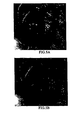
- FIG. 4 shows the algorithm flowchart of the glint detection and elimination algorithm, and the results of the glint detection and elimination algorithm can be viewed in FIGS. 5A and 5B .
- the blur detection algorithm utilizes a normalized image power spectrum method ( Gu J. and Li W., Automatic Image Quality Assessment for Cervical Imagery, SPIE Medical Imaging 2006 ; SPIE Proc. 6146, 2006 ), which can be described as the following steps:
- the method preferably used for contrast enhancement is adaptive histogram equalization.
- Adaptive histogram equalization enhances the contrast of images by transforming the values in the intensity image ( Zuiderveld, K., Contrast limited adaptive histogram equalization, Princeton, NJ: Academic Press, Graphics gems IV, ed. Heckbert, P., 1994 ).
- adaptive histogram equalization operates on small data regions (windows), rather than the entire image.
- Each window's contrast is enhanced, so that the histogram of the output region approximately matches the specified histogram.
- the neighboring windows are then combined using bilinear interpolation in order to eliminate artificially induced boundaries.
- the contrast especially in homogeneous areas, can be limited in order to avoid amplifying the noise which might be present in the image.
- the results of contrast enhancement can be viewed in FIGS. 8A and 8B .
- Super resolution is a technique to use multiple frames of the same object to achieve a higher resolution image
- Kim, S.P., Bose, N.K, and Valensuela, H.M. Recursive reconstruction of high resolution image from noisy undersampled multiframes, IEEE Transaction on Acoustics, Speech, and Signal Processing 38(6): 1031-1027, 1990 ; Irani, M. and Peleg, S., Improving resolution by image registration, CVGIP:GM 53: 231-239, 1991 ).
- Super resolution works when the frames are shifted by fractions of a pixel from each other.
- the super-resolution algorithm is able to produce a larger image that contains the information in the smaller original frames, first an image sub-pixel registration is employed to establish the correspondence between several low resolution images, and then a sub-pixel interpolation algorithm is used to reconstruct the higher resolution image.
- FIGS. 9A, 9B, 9C, 9D and 9E show a super-resolution reconstruction result.
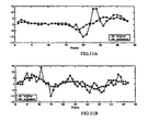
- Video stabilization is the process of generating a compensated video sequence by removing image motion from the camera's undesirable shake or jiggle.
- the preferred video stabilization algorithm consists of a motion estimation (ME) block, a motion smooth (MS) block, and a motion correction (MC) block, as shown in FIG. 10 .
- the ME block estimates the motion between frames and can be divided into a local motion estimator and a global motion decision unit.
- the local motion estimator will return the estimated dense optical flow information between successive frames using typical block-based or gradient-based methods.
- the global motion decision unit will then determine an appropriate global transformation that best characterizes the motion described by the given optical flow information.
- the global motion parameters will be sent to the MS, where the motion parameters are often filtered to remove the unwanted camera motion but retaining intentional camera motion.
- Finally MC warps the current frame using the filtered global transformation information and generates the stabilized video sequence.
- FIGS. 11A and 11B show the result of video stabilization.
- a 3D colon model is a preferred component of a computer-aided diagnosis (CAD) system in colonoscopy, to assist surgeons in visualization, and surgical planning and training.
- CAD computer-aided diagnosis
- the ability to construct a 3D colon model from endoscopic videos (or images) is thus preferred in a CAD system for colonoscopy.
- the mathematical formulations and algorithms have been developed for modeling static, localized 3D anatomic structures within a colon that can be rendered from multiple novel view points for close scrutiny and precise dimensioning ( Mori, K., Deguchi, D., Sugiyama, J., Suenaga, Y. et al., Tracking of a bronchoscope using epipolar geometry analysis and intensity-based image registration of real and virtual endoscopic images, Med. Image Anal. 6(3): 321-336.
- the modeling system of the present invention uses only video images and follows a well-established computer-vision paradigm for image-based modeling. Prominent features are extracted from images and their correspondences are established across multiple images by continuous tracking and discrete matching. These feature correspondences are used to infer the camera's movement.
- the camera motion parameters allow rectifying of images into a standard stereo configuration and inferring of pixel movements (disparity) in these images.
- the inferred disparity is then used to recover 3D surface depth.
- the inferred 3D depth together with texture information recorded in images, allow constructing of a 3D model with both structure and appearance information that can be rendered from multiple novel view points. More precisely, the modeling system comprises the following components:
- FIG. 12 shows the algorithm flowchart of 3D colon modeling and reconstruction.
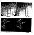
- FIGS. 13A, 13B, 13C and 13D show the distortion correction result and
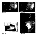
- FIGS 14A, 14B, 14C and 14D show a result of 3D polyp reconstruction.
- the model will allow the clinician to mark areas (on the model) during the entry phase of the colonoscopic exam, and to treat these areas during withdrawal.
- the system of this invention can quantitatively calculate the physical size of the polyp, and send to the colonoscopist a statistical criterion for making diagnostic decisions.
- pit patterns Treatment decisions for flat and depressed lesions are based on a detailed examination of the macroscopic morphological appearance, including the luminal surface structure of the crypts of Lieberkühn, otherwise known as "pit patterns".
- Pit patterns analysis can offer a degree of positive predictive value for both the underlying histology and depth of vertical submucosal invasion.
- the preferred system utilizes the Kudo tissue classification method which describes seven types of pit patterns ( Kudo, S., Hirota, S., Nakajima, T., Hosobe, S., Kusaka, H., Kobayashi, T. et al., Colorectal tumours and pit pattern, Journal of Clinical Pathology. 47(10): 880-885. 1994 .
- the pit pattern analysis module starts from high magnification endoscopic images, and morphological operators are first preformed to extract the texture pattern ( Chen, C.H., Pau, L.F., and Wang, P.S.P., Segmentation Tools In Mathematical Morphology, Handbook of Pattern Recognition and Computer Vision, in Handbook of Pattern Recognition and Computer Vision, pp. 443-456. World Scientific Publishing Co., 1989 ).
- the training feature includes: pit size, pit shape and pit density.
- FIG. 16 shows the algorithm flowchart of color analysis.
- FIG. 17 shows the algorithm flowchart of texture analysis.
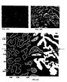
- FIG. 18 shows the result of pit pattern extraction and identification.
- This invention can be used whenever video data from an endoscope is available during an examination of an organ, and especially when it is necessary to diagnose potentially cancerous regions, and motion pictures of the regions are available.
Landscapes
- Engineering & Computer Science (AREA)
- Physics & Mathematics (AREA)
- General Physics & Mathematics (AREA)
- Theoretical Computer Science (AREA)
- Health & Medical Sciences (AREA)
- Computer Vision & Pattern Recognition (AREA)
- Life Sciences & Earth Sciences (AREA)
- Medical Informatics (AREA)
- Nuclear Medicine, Radiotherapy & Molecular Imaging (AREA)
- Radiology & Medical Imaging (AREA)
- General Health & Medical Sciences (AREA)
- Quality & Reliability (AREA)
- Surgery (AREA)
- Pathology (AREA)
- Optics & Photonics (AREA)
- Biophysics (AREA)
- Signal Processing (AREA)
- Biomedical Technology (AREA)
- Heart & Thoracic Surgery (AREA)
- Molecular Biology (AREA)
- Animal Behavior & Ethology (AREA)
- Public Health (AREA)
- Veterinary Medicine (AREA)
- Image Processing (AREA)
- Endoscopes (AREA)
Claims (5)
- Système de traitement d'images automatisé comprenant :un ordinateur couplé à un endoscope pour recevoir des données vidéo au cours d'un examen par colonoscopie d'un gros intestin ;un module d'analyse de la qualité de la vidéo, arrangé pour analyser lesdites données vidéo pour déterminer les trames d'images dans lesquelles la couverture des blocs flous est inférieure à un seuil afin de délivrer des trames d'images satisfaisantes à partir desdites données vidéo, et où ledit module d'analyse de la qualité de la vidéo est arrangé pour exécuter un algorithme de suppression de la scintillation qui procède à l'extraction de la caractéristique de scintillation, à la détection de la région de scintillation et à l'élimination de la région de scintillation et à la reconstruction de la caractéristique de l'image ;un module d'amélioration de la qualité de la vidéo arrangé pour :améliorer le contraste dans lesdites trames d'images satisfaisantes ;appliquer une super résolution auxdites trames d'images satisfaisantes en utilisant plusieurs trames du même objet pour obtenir une image de résolution plus élevée ; etassurer la stabilisation vidéo desdites trames d'images satisfaisantes ; etun module d'analyse arrangé pour détecter et classer toutes lésions ou tous polypes dans lesdites trames d'images satisfaisantes pour délivrer des informations de diagnostic concernant lesdites lésions ou lesdits polypes ;dans lequel, ledit module d'analyse de la qualité de la vidéo, ledit module d'amélioration de la qualité de la vidéo et ledit module d'analyse fonctionnent tous pour délivrer les informations de diagnostic en temps réel pendant l'examen.
- Système selon la revendication 1, dans lequel ledit module d'analyse est arrangé pour procéder à un calibrage de la couleur, à une analyse de la couleur, à une analyse de la texture et à une détection de caractéristique.
- Système selon la revendication 1, dans lequel ledit module d'analyse est arrangé pour procéder à une analyse des vaisseaux sanguins dans les trames d'images satisfaisantes.
- Système selon la revendication 1, dans lequel ledit module d'analyse comprend un module d'analyse de motif de trou arrangé pour procéder à une analyse des motifs de trous dans les trames d'images satisfaisantes.
- Système selon la revendication 1, comprenant en outre un module de modélisation tridimensionnelle et de reconstruction arrangé pour :reconstruire une image fixe en trois dimensions dudit gros intestin à partir desdites trames d'images satisfaisantes pour former un modèle reconstruit, dans lequel ladite reconstruction est effectuée en temps réelle pendant ladite colonoscopie, procéder à une correction de la distorsion en barillet, à un calibrage de la géométrie, à une modélisation basée sur l'image et à un assemblage des données tridimensionnelles ; etdans lequel ledit module d'analyse est en outre arrangé pour retrouver une forme tridimensionnelle et des informations de taille de toutes lésions ou tous polypes du gros intestin à partir dudit modèle reconstruit.
Applications Claiming Priority (2)
| Application Number | Priority Date | Filing Date | Title |
|---|---|---|---|
| US83927506P | 2006-08-21 | 2006-08-21 | |
| PCT/US2007/018600 WO2008024419A1 (fr) | 2006-08-21 | 2007-08-21 | Analyse assistée par ordinateur utilisant une vidéo issue d'endoscopes |
Publications (2)
| Publication Number | Publication Date |
|---|---|
| EP2054852A1 EP2054852A1 (fr) | 2009-05-06 |
| EP2054852B1 true EP2054852B1 (fr) | 2010-06-23 |
Family
ID=38828733
Family Applications (1)
| Application Number | Title | Priority Date | Filing Date |
|---|---|---|---|
| EP07837227A Not-in-force EP2054852B1 (fr) | 2006-08-21 | 2007-08-21 | Analyse assistée par ordinateur utilisant une vidéo issue d'endoscopes |
Country Status (6)
| Country | Link |
|---|---|
| US (1) | US20080058593A1 (fr) |
| EP (1) | EP2054852B1 (fr) |
| JP (1) | JP5113841B2 (fr) |
| AT (1) | ATE472141T1 (fr) |
| DE (1) | DE602007007340D1 (fr) |
| WO (1) | WO2008024419A1 (fr) |
Cited By (4)
| Publication number | Priority date | Publication date | Assignee | Title |
|---|---|---|---|---|
| WO2023039493A1 (fr) * | 2021-09-13 | 2023-03-16 | Satisfai Health Inc. | Système et procédés d'agrégation de caractéristiques dans des trames vidéo pour améliorer la précision d'algorithmes de détection basés sur l'ia |
| US11684241B2 (en) | 2020-11-02 | 2023-06-27 | Satisfai Health Inc. | Autonomous and continuously self-improving learning system |
| US11694114B2 (en) | 2019-07-16 | 2023-07-04 | Satisfai Health Inc. | Real-time deployment of machine learning systems |
| US12167829B2 (en) | 2019-12-19 | 2024-12-17 | Ambu A/S | Image capture selection |
Families Citing this family (162)
| Publication number | Priority date | Publication date | Assignee | Title |
|---|---|---|---|---|
| JP4533327B2 (ja) * | 2006-02-23 | 2010-09-01 | キヤノン株式会社 | 画像処理装置及び方法、コンピュータプログラム及び記憶媒体 |
| WO2008053649A1 (fr) * | 2006-11-02 | 2008-05-08 | Konica Minolta Holdings, Inc. | Procédé d'acquisition d'image grand angle et dispositif d'appareil photographique stéréo grand angle |
| JP2008301968A (ja) * | 2007-06-06 | 2008-12-18 | Olympus Medical Systems Corp | 内視鏡画像処理装置 |
| US20090208143A1 (en) * | 2008-02-19 | 2009-08-20 | University Of Washington | Efficient automated urothelial imaging using an endoscope with tip bending |
| EP2276391A1 (fr) * | 2008-04-15 | 2011-01-26 | Provost, Fellows and Scholars of the College of the Holy and Undivided Trinity of Queen Elizabeth near Dublin | Système d'endoscopie avec détecteurs de mouvement |
| WO2009149131A1 (fr) | 2008-06-02 | 2009-12-10 | Lightlab Imaging, Inc. | Procédés quantitatifs pour obtenir des caractéristiques de tissu à partir d’images de tomographie de cohérence optique |
| US8508550B1 (en) * | 2008-06-10 | 2013-08-13 | Pixar | Selective rendering of objects |
| WO2010019114A1 (fr) * | 2008-08-11 | 2010-02-18 | Sti Medical Systems, Llc | Procédé de manipulation d'image permettant de faire un fondu entre deux cadres d'une image à cadre double |
| US20100033501A1 (en) * | 2008-08-11 | 2010-02-11 | Sti Medical Systems, Llc | Method of image manipulation to fade between two images |
| JP2012500040A (ja) * | 2008-08-15 | 2012-01-05 | エスティーアイ・メディカル・システムズ・エルエルシー | 子宮頸部画像における血管パターンを強調するための方法 |
| JP2010068865A (ja) * | 2008-09-16 | 2010-04-02 | Fujifilm Corp | 画像診断装置 |
| JP5130171B2 (ja) * | 2008-09-22 | 2013-01-30 | 株式会社日立製作所 | 画像信号処理装置および画像信号処理方法 |
| CN102282569A (zh) * | 2008-10-10 | 2011-12-14 | 国际科学技术医疗系统有限责任公司 | 宫颈成像中的组织分类方法 |
| US8553075B2 (en) | 2008-10-22 | 2013-10-08 | Fujifilm Corporation | Endoscope apparatus and control method therefor |
| US20110251454A1 (en) * | 2008-11-21 | 2011-10-13 | Mayo Foundation For Medical Education And Research | Colonoscopy Tracking and Evaluation System |
| US20110273535A1 (en) * | 2009-01-28 | 2011-11-10 | Avi Mendelson | Mole monitoring system |
| CN102413756B (zh) * | 2009-04-29 | 2015-04-01 | 皇家飞利浦电子股份有限公司 | 从单目内窥镜图像估计实时深度 |
| WO2012056453A2 (fr) | 2010-10-28 | 2012-05-03 | Peermedical Ltd. | Systèmes optiques pour endoscopes à capteurs multiples |
| US20120316421A1 (en) * | 2009-07-07 | 2012-12-13 | The Johns Hopkins University | System and method for automated disease assessment in capsule endoscopy |
| TWI425445B (zh) * | 2009-09-29 | 2014-02-01 | Nat Univ Tsing Hua | 用於判斷一移動平台之自我運動量的方法以及偵測系統 |
| US8417062B2 (en) * | 2009-10-19 | 2013-04-09 | Ut-Battelle, Llc | System and method for stabilization of fisheye video imagery |
| TWI432168B (zh) * | 2009-12-31 | 2014-04-01 | Univ Nat Yunlin Sci & Tech | 內視鏡導航方法以及內視鏡導航系統 |
| US9269024B2 (en) * | 2010-02-01 | 2016-02-23 | Qualcomm Incorporated | Image recognition system based on cascaded over-complete dictionaries |
| US20110301447A1 (en) * | 2010-06-07 | 2011-12-08 | Sti Medical Systems, Llc | Versatile video interpretation, visualization, and management system |
| JP2012045130A (ja) * | 2010-08-26 | 2012-03-08 | Hoya Corp | 画像信号処理システムおよび画像信号処理方法 |
| JP5435746B2 (ja) * | 2011-01-24 | 2014-03-05 | 富士フイルム株式会社 | 内視鏡装置 |
| WO2012105141A1 (fr) * | 2011-02-01 | 2012-08-09 | オリンパスメディカルシステムズ株式会社 | Appareil d'aide au diagnostic |
| KR101805624B1 (ko) * | 2011-08-29 | 2017-12-08 | 삼성전자주식회사 | 장기 모델 영상 생성 방법 및 장치 |
| US8896678B2 (en) | 2011-09-16 | 2014-11-25 | The Invention Science Fund I, Llc | Coregistering images of a region of interest during several conditions using a landmark subsurface feature |
| US11871901B2 (en) | 2012-05-20 | 2024-01-16 | Cilag Gmbh International | Method for situational awareness for surgical network or surgical network connected device capable of adjusting function based on a sensed situation or usage |
| FR2996331B1 (fr) * | 2012-09-28 | 2015-12-18 | Morpho | Procede de detection de la realite de reseaux veineux a des fins d'identification d'individus |
| KR101874482B1 (ko) * | 2012-10-16 | 2018-07-05 | 삼성전자주식회사 | 깊이 영상으로부터 고해상도 3차원 영상을 복원하는 장치 및 방법 |
| CN104838421B (zh) * | 2012-11-30 | 2019-01-29 | 皇家飞利浦有限公司 | 基于图像数据的组织表面粗糙度量化和基于所述组织表面粗糙度量化对疾病存在的确定 |
| US9107578B2 (en) | 2013-03-31 | 2015-08-18 | Gyrus Acmi, Inc. | Panoramic organ imaging |
| TWI503788B (zh) * | 2013-10-02 | 2015-10-11 | Jar Ferr Yang | 還原尺寸重調景深圖框爲原始景深圖框的方法、裝置及系統 |
| KR20150049585A (ko) | 2013-10-30 | 2015-05-08 | 삼성전자주식회사 | 용종 검출 장치 및 그 동작방법 |
| WO2015149042A1 (fr) * | 2014-03-28 | 2015-10-01 | Dorin Panescu | Alignement de modèles q3d avec des images 3d |
| JP6854237B2 (ja) | 2014-03-28 | 2021-04-07 | インテュイティブ サージカル オペレーションズ, インコーポレイテッド | 視野内の器具の定量的三次元視覚化 |
| KR102397670B1 (ko) | 2014-03-28 | 2022-05-16 | 인튜어티브 서지컬 오퍼레이션즈 인코포레이티드 | 정량적 3차원 영상화에 기초한 햅틱 피드백을 갖는 수술 시스템 |
| EP3125806B1 (fr) | 2014-03-28 | 2023-06-14 | Intuitive Surgical Operations, Inc. | Imagerie 3d quantitative de scènes chirurgicales |
| CN110251047B (zh) | 2014-03-28 | 2022-01-18 | 直观外科手术操作公司 | 手术植入物的定量三维成像和打印 |
| US10334227B2 (en) | 2014-03-28 | 2019-06-25 | Intuitive Surgical Operations, Inc. | Quantitative three-dimensional imaging of surgical scenes from multiport perspectives |
| KR20150120774A (ko) | 2014-04-18 | 2015-10-28 | 삼성전자주식회사 | 관심영역 검출 시스템 및 방법 |
| US9978142B2 (en) | 2014-04-24 | 2018-05-22 | Arizona Board Of Regents On Behalf Of Arizona State University | System and method for quality assessment of optical colonoscopy images |
| JP6482934B2 (ja) | 2014-06-03 | 2019-03-13 | キヤノンメディカルシステムズ株式会社 | 画像処理装置、放射線検出装置および画像処理方法 |
| WO2016175773A1 (fr) * | 2015-04-29 | 2016-11-03 | Siemens Aktiengesellschaft | Procédé et système de segmentation sémantique de données d'images laparoscopiques et endoscopiques 2d/2,5d |
| EP3297515B1 (fr) | 2015-05-17 | 2020-07-01 | Endochoice, Inc. | Amélioration d'image endoscopique par égalisation d'histogramme adaptative à contraste limité (clahe) mise en oeuvre dans un processeur |
| EP3304423A1 (fr) * | 2015-06-05 | 2018-04-11 | Siemens Aktiengesellschaft | Procédé et système d'analyse de scène et de fusion de modèles simultanées pour une navigation endoscopique et laparoscopique |
| JP6707083B2 (ja) | 2015-06-29 | 2020-06-10 | オリンパス株式会社 | 画像処理装置、内視鏡システム、画像処理方法、及び画像処理プログラム |
| GB201513449D0 (en) * | 2015-07-30 | 2015-09-16 | Optos Plc | Image processing |
| US10510144B2 (en) | 2015-09-10 | 2019-12-17 | Magentiq Eye Ltd. | System and method for detection of suspicious tissue regions in an endoscopic procedure |
| JP6580446B2 (ja) * | 2015-10-09 | 2019-09-25 | サイバネットシステム株式会社 | 画像処理装置及び画像処理方法 |
| CN105654495A (zh) * | 2016-01-07 | 2016-06-08 | 太原工业学院 | 汽车刹车盘内部缺陷的检测方法 |
| US10140554B2 (en) | 2016-01-21 | 2018-11-27 | Wizr Llc | Video processing |
| JP7127538B2 (ja) * | 2016-03-29 | 2022-08-30 | ソニーグループ株式会社 | 画像処理装置、医療機器の作動方法及び医療システム |
| WO2017201494A1 (fr) * | 2016-05-19 | 2017-11-23 | Avantis Medical Systems, Inc. | Procédés de détection de polypes |
| US11055581B2 (en) * | 2016-10-07 | 2021-07-06 | Baylor Research Institute | Classification of polyps using learned image analysis |
| KR101730570B1 (ko) * | 2016-11-11 | 2017-04-26 | 주식회사 마이디바이스 | 글린트 검출 방법 |
| US11633256B2 (en) | 2017-02-14 | 2023-04-25 | Dignity Health | Systems, methods, and media for selectively presenting images captured by confocal laser endomicroscopy |
| US11553829B2 (en) | 2017-05-25 | 2023-01-17 | Nec Corporation | Information processing apparatus, control method and program |
| CN107688092B (zh) * | 2017-08-18 | 2020-05-19 | 国家纳米科学中心 | 一种用于癌症患者预后评估的方法 |
| US11911045B2 (en) | 2017-10-30 | 2024-02-27 | Cllag GmbH International | Method for operating a powered articulating multi-clip applier |
| US11051836B2 (en) | 2017-10-30 | 2021-07-06 | Cilag Gmbh International | Surgical clip applier comprising an empty clip cartridge lockout |
| US11317919B2 (en) | 2017-10-30 | 2022-05-03 | Cilag Gmbh International | Clip applier comprising a clip crimping system |
| US11291510B2 (en) | 2017-10-30 | 2022-04-05 | Cilag Gmbh International | Method of hub communication with surgical instrument systems |
| US11564756B2 (en) | 2017-10-30 | 2023-01-31 | Cilag Gmbh International | Method of hub communication with surgical instrument systems |
| US11510741B2 (en) | 2017-10-30 | 2022-11-29 | Cilag Gmbh International | Method for producing a surgical instrument comprising a smart electrical system |
| US11801098B2 (en) | 2017-10-30 | 2023-10-31 | Cilag Gmbh International | Method of hub communication with surgical instrument systems |
| SG11202003973VA (en) * | 2017-10-30 | 2020-05-28 | Japanese Found For Cancer Res | Image diagnosis assistance apparatus, data collection method, image diagnosis assistance method, and image diagnosis assistance program |
| DE102017221297A1 (de) * | 2017-11-28 | 2019-05-29 | Siemens Healthcare Gmbh | Verfahren und Vorrichtung zur automatisierten Auswertung wenigstens eines mit einer medizinischen Bildaufnahmeeinrichtung aufgenommenen Bilddatensatzes, Computerprogramm und elektronisch lesbarer Datenträger |
| US20190201112A1 (en) | 2017-12-28 | 2019-07-04 | Ethicon Llc | Computer implemented interactive surgical systems |
| US11202570B2 (en) | 2017-12-28 | 2021-12-21 | Cilag Gmbh International | Communication hub and storage device for storing parameters and status of a surgical device to be shared with cloud based analytics systems |
| US11311306B2 (en) | 2017-12-28 | 2022-04-26 | Cilag Gmbh International | Surgical systems for detecting end effector tissue distribution irregularities |
| US11213359B2 (en) | 2017-12-28 | 2022-01-04 | Cilag Gmbh International | Controllers for robot-assisted surgical platforms |
| WO2019133144A1 (fr) | 2017-12-28 | 2019-07-04 | Ethicon Llc | Détection et intensification des réponses de sécurité d'instruments chirurgicaux à des menaces à la gravité croissante |
| US11257589B2 (en) | 2017-12-28 | 2022-02-22 | Cilag Gmbh International | Real-time analysis of comprehensive cost of all instrumentation used in surgery utilizing data fluidity to track instruments through stocking and in-house processes |
| US11324557B2 (en) | 2017-12-28 | 2022-05-10 | Cilag Gmbh International | Surgical instrument with a sensing array |
| US11998193B2 (en) | 2017-12-28 | 2024-06-04 | Cilag Gmbh International | Method for usage of the shroud as an aspect of sensing or controlling a powered surgical device, and a control algorithm to adjust its default operation |
| US11786245B2 (en) | 2017-12-28 | 2023-10-17 | Cilag Gmbh International | Surgical systems with prioritized data transmission capabilities |
| US11857152B2 (en) | 2017-12-28 | 2024-01-02 | Cilag Gmbh International | Surgical hub spatial awareness to determine devices in operating theater |
| US11109866B2 (en) | 2017-12-28 | 2021-09-07 | Cilag Gmbh International | Method for circular stapler control algorithm adjustment based on situational awareness |
| US20190206569A1 (en) | 2017-12-28 | 2019-07-04 | Ethicon Llc | Method of cloud based data analytics for use with the hub |
| US10595887B2 (en) | 2017-12-28 | 2020-03-24 | Ethicon Llc | Systems for adjusting end effector parameters based on perioperative information |
| US12096916B2 (en) | 2017-12-28 | 2024-09-24 | Cilag Gmbh International | Method of sensing particulate from smoke evacuated from a patient, adjusting the pump speed based on the sensed information, and communicating the functional parameters of the system to the hub |
| US11589888B2 (en) | 2017-12-28 | 2023-02-28 | Cilag Gmbh International | Method for controlling smart energy devices |
| JP2021509061A (ja) | 2017-12-28 | 2021-03-18 | エシコン エルエルシーEthicon LLC | 状況認識に基づく外科用デバイスの機能の調節 |
| US10918310B2 (en) | 2018-01-03 | 2021-02-16 | Biosense Webster (Israel) Ltd. | Fast anatomical mapping (FAM) using volume filling |
| US12127729B2 (en) | 2017-12-28 | 2024-10-29 | Cilag Gmbh International | Method for smoke evacuation for surgical hub |
| US11818052B2 (en) | 2017-12-28 | 2023-11-14 | Cilag Gmbh International | Surgical network determination of prioritization of communication, interaction, or processing based on system or device needs |
| US20190201090A1 (en) | 2017-12-28 | 2019-07-04 | Ethicon Llc | Capacitive coupled return path pad with separable array elements |
| US11744604B2 (en) | 2017-12-28 | 2023-09-05 | Cilag Gmbh International | Surgical instrument with a hardware-only control circuit |
| US10892995B2 (en) | 2017-12-28 | 2021-01-12 | Ethicon Llc | Surgical network determination of prioritization of communication, interaction, or processing based on system or device needs |
| US11304763B2 (en) | 2017-12-28 | 2022-04-19 | Cilag Gmbh International | Image capturing of the areas outside the abdomen to improve placement and control of a surgical device in use |
| US11304699B2 (en) | 2017-12-28 | 2022-04-19 | Cilag Gmbh International | Method for adaptive control schemes for surgical network control and interaction |
| US11559308B2 (en) | 2017-12-28 | 2023-01-24 | Cilag Gmbh International | Method for smart energy device infrastructure |
| US11672605B2 (en) | 2017-12-28 | 2023-06-13 | Cilag Gmbh International | Sterile field interactive control displays |
| US12376855B2 (en) | 2017-12-28 | 2025-08-05 | Cilag Gmbh International | Safety systems for smart powered surgical stapling |
| US12290231B2 (en) | 2017-12-28 | 2025-05-06 | Cilag Gmbh International | Method of hub communication, processing, storage and display |
| US11179175B2 (en) | 2017-12-28 | 2021-11-23 | Cilag Gmbh International | Controlling an ultrasonic surgical instrument according to tissue location |
| US12396806B2 (en) | 2017-12-28 | 2025-08-26 | Cilag Gmbh International | Adjustment of a surgical device function based on situational awareness |
| US20190201113A1 (en) | 2017-12-28 | 2019-07-04 | Ethicon Llc | Controls for robot-assisted surgical platforms |
| US11969216B2 (en) | 2017-12-28 | 2024-04-30 | Cilag Gmbh International | Surgical network recommendations from real time analysis of procedure variables against a baseline highlighting differences from the optimal solution |
| US11678881B2 (en) | 2017-12-28 | 2023-06-20 | Cilag Gmbh International | Spatial awareness of surgical hubs in operating rooms |
| US11389164B2 (en) | 2017-12-28 | 2022-07-19 | Cilag Gmbh International | Method of using reinforced flexible circuits with multiple sensors to optimize performance of radio frequency devices |
| US11147607B2 (en) | 2017-12-28 | 2021-10-19 | Cilag Gmbh International | Bipolar combination device that automatically adjusts pressure based on energy modality |
| US11832840B2 (en) | 2017-12-28 | 2023-12-05 | Cilag Gmbh International | Surgical instrument having a flexible circuit |
| US11903601B2 (en) | 2017-12-28 | 2024-02-20 | Cilag Gmbh International | Surgical instrument comprising a plurality of drive systems |
| US11896322B2 (en) | 2017-12-28 | 2024-02-13 | Cilag Gmbh International | Sensing the patient position and contact utilizing the mono-polar return pad electrode to provide situational awareness to the hub |
| US11666331B2 (en) | 2017-12-28 | 2023-06-06 | Cilag Gmbh International | Systems for detecting proximity of surgical end effector to cancerous tissue |
| US11864728B2 (en) | 2017-12-28 | 2024-01-09 | Cilag Gmbh International | Characterization of tissue irregularities through the use of mono-chromatic light refractivity |
| US11464559B2 (en) | 2017-12-28 | 2022-10-11 | Cilag Gmbh International | Estimating state of ultrasonic end effector and control system therefor |
| US11633237B2 (en) | 2017-12-28 | 2023-04-25 | Cilag Gmbh International | Usage and technique analysis of surgeon / staff performance against a baseline to optimize device utilization and performance for both current and future procedures |
| US11659023B2 (en) | 2017-12-28 | 2023-05-23 | Cilag Gmbh International | Method of hub communication |
| US11132462B2 (en) | 2017-12-28 | 2021-09-28 | Cilag Gmbh International | Data stripping method to interrogate patient records and create anonymized record |
| US11969142B2 (en) | 2017-12-28 | 2024-04-30 | Cilag Gmbh International | Method of compressing tissue within a stapling device and simultaneously displaying the location of the tissue within the jaws |
| US11076921B2 (en) | 2017-12-28 | 2021-08-03 | Cilag Gmbh International | Adaptive control program updates for surgical hubs |
| US11786251B2 (en) | 2017-12-28 | 2023-10-17 | Cilag Gmbh International | Method for adaptive control schemes for surgical network control and interaction |
| US11166772B2 (en) | 2017-12-28 | 2021-11-09 | Cilag Gmbh International | Surgical hub coordination of control and communication of operating room devices |
| US11896443B2 (en) | 2017-12-28 | 2024-02-13 | Cilag Gmbh International | Control of a surgical system through a surgical barrier |
| US11937769B2 (en) * | 2017-12-28 | 2024-03-26 | Cilag Gmbh International | Method of hub communication, processing, storage and display |
| US11832899B2 (en) | 2017-12-28 | 2023-12-05 | Cilag Gmbh International | Surgical systems with autonomously adjustable control programs |
| US11376002B2 (en) | 2017-12-28 | 2022-07-05 | Cilag Gmbh International | Surgical instrument cartridge sensor assemblies |
| US10758310B2 (en) | 2017-12-28 | 2020-09-01 | Ethicon Llc | Wireless pairing of a surgical device with another device within a sterile surgical field based on the usage and situational awareness of devices |
| US11419630B2 (en) | 2017-12-28 | 2022-08-23 | Cilag Gmbh International | Surgical system distributed processing |
| US12062442B2 (en) | 2017-12-28 | 2024-08-13 | Cilag Gmbh International | Method for operating surgical instrument systems |
| US11612444B2 (en) | 2017-12-28 | 2023-03-28 | Cilag Gmbh International | Adjustment of a surgical device function based on situational awareness |
| US11771487B2 (en) | 2017-12-28 | 2023-10-03 | Cilag Gmbh International | Mechanisms for controlling different electromechanical systems of an electrosurgical instrument |
| US11576677B2 (en) | 2017-12-28 | 2023-02-14 | Cilag Gmbh International | Method of hub communication, processing, display, and cloud analytics |
| US11399858B2 (en) | 2018-03-08 | 2022-08-02 | Cilag Gmbh International | Application of smart blade technology |
| US12303159B2 (en) | 2018-03-08 | 2025-05-20 | Cilag Gmbh International | Methods for estimating and controlling state of ultrasonic end effector |
| US11259830B2 (en) | 2018-03-08 | 2022-03-01 | Cilag Gmbh International | Methods for controlling temperature in ultrasonic device |
| US11986233B2 (en) | 2018-03-08 | 2024-05-21 | Cilag Gmbh International | Adjustment of complex impedance to compensate for lost power in an articulating ultrasonic device |
| US11259806B2 (en) | 2018-03-28 | 2022-03-01 | Cilag Gmbh International | Surgical stapling devices with features for blocking advancement of a camming assembly of an incompatible cartridge installed therein |
| US11090047B2 (en) | 2018-03-28 | 2021-08-17 | Cilag Gmbh International | Surgical instrument comprising an adaptive control system |
| US11100633B2 (en) | 2018-06-13 | 2021-08-24 | Cosmo Artificial Intelligence—Al Limited | Systems and methods for processing real-time video from a medical image device and detecting objects in the video |
| US10810460B2 (en) | 2018-06-13 | 2020-10-20 | Cosmo Artificial Intelligence—AI Limited | Systems and methods for training generative adversarial networks and use of trained generative adversarial networks |
| US10292769B1 (en) * | 2018-08-07 | 2019-05-21 | Sony Corporation | Surgical assistive device and method for providing assistance in surgery of anatomical portions of internal organ affected by intraoperative shift |
| WO2020054543A1 (fr) * | 2018-09-11 | 2020-03-19 | 富士フイルム株式会社 | Dispositif et procédé de traitement d'image médicale, système d'endoscope, dispositif de processeur, dispositif d'aide au diagnostic et programme |
| US10896494B1 (en) * | 2018-09-27 | 2021-01-19 | Snap Inc. | Dirty lens image correction |
| WO2020079667A1 (fr) | 2018-10-19 | 2020-04-23 | Takeda Pharmaceutical Company Limited | Notation d'image pour pathologie intestinale |
| CN109598716B (zh) * | 2018-12-05 | 2020-08-07 | 武汉楚精灵医疗科技有限公司 | 基于计算机视觉的肠镜退镜速度实时监测方法和系统 |
| US11298130B2 (en) | 2019-02-19 | 2022-04-12 | Cilag Gmbh International | Staple cartridge retainer with frangible authentication key |
| US11464511B2 (en) | 2019-02-19 | 2022-10-11 | Cilag Gmbh International | Surgical staple cartridges with movable authentication key arrangements |
| EP3716207B1 (fr) * | 2019-03-26 | 2023-11-29 | Active Medical B.V. | Procédé et appareil pour l'analyse de diagnostic de la fonction et de la morphologie d'altérations de la microcirculation |
| JP7555181B2 (ja) * | 2019-03-27 | 2024-09-24 | Hoya株式会社 | 内視鏡用プロセッサ、情報処理装置、プログラム、情報処理方法および学習モデルの生成方法 |
| WO2020232374A1 (fr) * | 2019-05-16 | 2020-11-19 | The Regents Of The University Of Michigan | Localisation anatomique et régionale automatisée de caractéristiques de maladie dans des vidéos de coloscopie |
| US10929669B2 (en) * | 2019-06-04 | 2021-02-23 | Magentiq Eye Ltd | Systems and methods for processing colon images and videos |
| US11423318B2 (en) * | 2019-07-16 | 2022-08-23 | DOCBOT, Inc. | System and methods for aggregating features in video frames to improve accuracy of AI detection algorithms |
| US10682108B1 (en) * | 2019-07-16 | 2020-06-16 | The University Of North Carolina At Chapel Hill | Methods, systems, and computer readable media for three-dimensional (3D) reconstruction of colonoscopic surfaces for determining missing regions |
| US11191423B1 (en) * | 2020-07-16 | 2021-12-07 | DOCBOT, Inc. | Endoscopic system and methods having real-time medical imaging |
| KR20220058918A (ko) * | 2019-08-30 | 2022-05-10 | 아우리스 헬스, 인코포레이티드 | 기구 이미지 신뢰성 시스템 및 방법 |
| KR102313749B1 (ko) * | 2019-12-23 | 2021-10-18 | 주식회사 메가젠임플란트 | 인공지능 기반 자동 구강 ct 색상변환장치 및 그 장치의 구동방법 |
| WO2021157487A1 (fr) | 2020-02-06 | 2021-08-12 | 富士フイルム株式会社 | Dispositif de traitement d'image médicale, système d'endoscope, procédé de traitement d'image médicale et programme |
| US11122248B1 (en) * | 2020-07-20 | 2021-09-14 | Black Sesame International Holding Limited | Stereo vision with weakly aligned heterogeneous cameras |
| US11992372B2 (en) * | 2020-10-02 | 2024-05-28 | Cilag Gmbh International | Cooperative surgical displays |
| US11963683B2 (en) | 2020-10-02 | 2024-04-23 | Cilag Gmbh International | Method for operating tiered operation modes in a surgical system |
| US12484897B2 (en) | 2020-10-02 | 2025-12-02 | Cilag Gmbh International | Surgical instrument with adaptive configuration control |
| US11849220B2 (en) | 2021-04-14 | 2023-12-19 | Microsoft Technology Licensing, Llc | Systems and methods for generating depth information from low-resolution images |
| US11831931B2 (en) | 2021-04-14 | 2023-11-28 | Microsoft Technology Licensing, Llc | Systems and methods for generating high-resolution video or animated surface meshes from low-resolution images |
| KR102388535B1 (ko) * | 2021-06-15 | 2022-04-22 | (주)제이엘케이 | 인공지능에 기반하여 내시경 영상을 분석하기 위한 방법 및 장치 |
| WO2024195100A1 (fr) * | 2023-03-23 | 2024-09-26 | 日本電気株式会社 | Dispositif d'assistance à l'endoscopie, procédé d'assistance à l'endoscopie, et support d'enregistrement |
| JP7733754B2 (ja) * | 2024-01-18 | 2025-09-03 | 株式会社フジクラ | 画像処理方法、画像処理装置、画像処理プログラム、及び内視鏡システム |
Family Cites Families (18)
| Publication number | Priority date | Publication date | Assignee | Title |
|---|---|---|---|---|
| US6075905A (en) * | 1996-07-17 | 2000-06-13 | Sarnoff Corporation | Method and apparatus for mosaic image construction |
| US6346940B1 (en) * | 1997-02-27 | 2002-02-12 | Kabushiki Kaisha Toshiba | Virtualized endoscope system |
| US5960108A (en) * | 1997-06-12 | 1999-09-28 | Apple Computer, Inc. | Method and system for creating an image-based virtual reality environment utilizing a fisheye lens |
| US6427082B1 (en) * | 1998-12-23 | 2002-07-30 | Medispectra, Incorporated | Optical methods and systems for rapid screening of the cervix |
| US7187810B2 (en) * | 1999-12-15 | 2007-03-06 | Medispectra, Inc. | Methods and systems for correcting image misalignment |
| IL134017A (en) * | 2000-01-13 | 2008-04-13 | Capsule View Inc | Camera for photography inside the intestines |
| JP2002291690A (ja) * | 2001-04-02 | 2002-10-08 | Asahi Optical Co Ltd | 電子内視鏡装置および電子内視鏡システム |
| US7257273B2 (en) * | 2001-04-09 | 2007-08-14 | Mingjing Li | Hierarchical scheme for blur detection in digital image using wavelet transform |
| EP1419484A2 (fr) * | 2001-08-10 | 2004-05-19 | Koninklijke Philips Electronics N.V. | Appareil d'examen aux rayons x de reconstruction d'un ensemble tridimensionnel de donnees a partir d'images de projection |
| EP1549992A2 (fr) * | 2002-09-16 | 2005-07-06 | Rensselaer Polytechnic Institute | Microscope a champ de vision etendu |
| US7224827B2 (en) * | 2002-09-27 | 2007-05-29 | The Board Of Trustees Of The Leland Stanford Junior University | Method for matching and registering medical image data |
| JP4409166B2 (ja) * | 2002-12-05 | 2010-02-03 | オリンパス株式会社 | 画像処理装置 |
| US20050152588A1 (en) * | 2003-10-28 | 2005-07-14 | University Of Chicago | Method for virtual endoscopic visualization of the colon by shape-scale signatures, centerlining, and computerized detection of masses |
| US20050123179A1 (en) * | 2003-12-05 | 2005-06-09 | Eastman Kodak Company | Method and system for automatic axial rotation correction in vivo images |
| CA2564240C (fr) * | 2004-04-26 | 2014-10-07 | David F. Yankelevitz | Systeme d'imagerie medicale permettant d'apprecier avec precision l'evolution d'une lesion cible |
| US7916173B2 (en) * | 2004-06-22 | 2011-03-29 | Canon Kabushiki Kaisha | Method for detecting and selecting good quality image frames from video |
| US8600125B2 (en) * | 2005-06-22 | 2013-12-03 | The Research Foundation Of State University Of New York | System and method for computer aided polyp detection |
| US8423123B2 (en) * | 2005-09-30 | 2013-04-16 | Given Imaging Ltd. | System and method for in-vivo feature detection |
-
2007
- 2007-08-21 EP EP07837227A patent/EP2054852B1/fr not_active Not-in-force
- 2007-08-21 JP JP2009525622A patent/JP5113841B2/ja not_active Expired - Fee Related
- 2007-08-21 US US11/895,150 patent/US20080058593A1/en not_active Abandoned
- 2007-08-21 DE DE602007007340T patent/DE602007007340D1/de active Active
- 2007-08-21 AT AT07837227T patent/ATE472141T1/de not_active IP Right Cessation
- 2007-08-21 WO PCT/US2007/018600 patent/WO2008024419A1/fr not_active Ceased
Cited By (4)
| Publication number | Priority date | Publication date | Assignee | Title |
|---|---|---|---|---|
| US11694114B2 (en) | 2019-07-16 | 2023-07-04 | Satisfai Health Inc. | Real-time deployment of machine learning systems |
| US12167829B2 (en) | 2019-12-19 | 2024-12-17 | Ambu A/S | Image capture selection |
| US11684241B2 (en) | 2020-11-02 | 2023-06-27 | Satisfai Health Inc. | Autonomous and continuously self-improving learning system |
| WO2023039493A1 (fr) * | 2021-09-13 | 2023-03-16 | Satisfai Health Inc. | Système et procédés d'agrégation de caractéristiques dans des trames vidéo pour améliorer la précision d'algorithmes de détection basés sur l'ia |
Also Published As
| Publication number | Publication date |
|---|---|
| JP5113841B2 (ja) | 2013-01-09 |
| EP2054852A1 (fr) | 2009-05-06 |
| WO2008024419A1 (fr) | 2008-02-28 |
| US20080058593A1 (en) | 2008-03-06 |
| ATE472141T1 (de) | 2010-07-15 |
| JP2010512173A (ja) | 2010-04-22 |
| DE602007007340D1 (de) | 2010-08-05 |
Similar Documents
| Publication | Publication Date | Title |
|---|---|---|
| EP2054852B1 (fr) | Analyse assistée par ordinateur utilisant une vidéo issue d'endoscopes | |
| US11132794B2 (en) | System and method for detection of suspicious tissue regions in an endoscopic procedure | |
| JP4409166B2 (ja) | 画像処理装置 | |
| CN113573654A (zh) | 用于检测并测定病灶尺寸的ai系统 | |
| US8351667B2 (en) | Methods of contrast enhancement for images having blood vessel structures | |
| Sreeja et al. | An improved feature based image fusion technique for enhancement of liver lesions | |
| Maghsoudi et al. | A computer aided method to detect bleeding, tumor, and disease regions in Wireless Capsule Endoscopy | |
| Ševo et al. | Edge density based automatic detection of inflammation in colonoscopy videos | |
| CN118212418A (zh) | 一种基于多任务学习的肝脏肿瘤分割和检测方法 | |
| Nur-A-Alam et al. | An automatic system to detect colorectal polyp using hybrid fused method from colonoscopy images | |
| CN116542952A (zh) | 一种基于三维重建的内镜检查覆盖率评估方法及系统 | |
| Ohura et al. | Computer-aided diagnosis method for detecting early esophageal cancer from endoscopic image by using dyadic wavelet transform and fractal dimension | |
| Häfner et al. | Pit pattern classification using extended local binary patterns | |
| WO2010019121A1 (fr) | Procédés permettant d'améliorer des motifs vasculaires dans le domaine de l'imagerie cervicale | |
| Pooja et al. | Denoising technique and analysis of statistical parameters for endoscopic images of gastric cancer | |
| Aksenov et al. | An ensemble of convolutional neural networks for the use in video endoscopy | |
| Kadeem et al. | Skin Melanoma diagnosis using machine learning and ABCD rule | |
| Zhao et al. | Automatic temporal subtraction of chest radiographs and its enhancement for lung cancers | |
| Supe et al. | Image processing for medical image analysis: a review | |
| Gu et al. | Computer-aided diagnosis (cad) for colonoscopy | |
| Moharana et al. | Diffusion-Based Data Harmonization Across Multiple Oral Cancer Imaging Devices | |
| Junzhou et al. | Contourlet based feature extraction and classification for Wireless Capsule Endoscopic images | |
| Bhargavi et al. | Exploring Filters in Color Segmentation for Detection of Duodenal Cysts in Wireless Capsule Endoscopy Images | |
| Arasi et al. | A Study on Preprocessing Techniques Used for Gastrointestinal Cancer Detection in Endoscopy Images | |
| Siyue et al. | Real-Time Feature Extraction of Fine Mucosal Structures in Endoscopic Images Using Morphological Watershed Segmentation for Early Gastric Cancer Detection |
Legal Events
| Date | Code | Title | Description |
|---|---|---|---|
| PUAI | Public reference made under article 153(3) epc to a published international application that has entered the european phase |
Free format text: ORIGINAL CODE: 0009012 |
|
| 17P | Request for examination filed |
Effective date: 20090211 |
|
| AK | Designated contracting states |
Kind code of ref document: A1 Designated state(s): AT BE BG CH CY CZ DE DK EE ES FI FR GB GR HU IE IS IT LI LT LU LV MC MT NL PL PT RO SE SI SK TR |
|
| AX | Request for extension of the european patent |
Extension state: AL BA HR MK RS |
|
| 17Q | First examination report despatched |
Effective date: 20090713 |
|
| GRAP | Despatch of communication of intention to grant a patent |
Free format text: ORIGINAL CODE: EPIDOSNIGR1 |
|
| GRAS | Grant fee paid |
Free format text: ORIGINAL CODE: EPIDOSNIGR3 |
|
| GRAA | (expected) grant |
Free format text: ORIGINAL CODE: 0009210 |
|
| AK | Designated contracting states |
Kind code of ref document: B1 Designated state(s): AT BE BG CH CY CZ DE DK EE ES FI FR GB GR HU IE IS IT LI LT LU LV MC MT NL PL PT RO SE SI SK TR |
|
| REG | Reference to a national code |
Ref country code: CH Ref legal event code: EP |
|
| REG | Reference to a national code |
Ref country code: IE Ref legal event code: FG4D |
|
| REF | Corresponds to: |
Ref document number: 602007007340 Country of ref document: DE Date of ref document: 20100805 Kind code of ref document: P |
|
| REG | Reference to a national code |
Ref country code: NL Ref legal event code: VDEP Effective date: 20100623 |
|
| PG25 | Lapsed in a contracting state [announced via postgrant information from national office to epo] |
Ref country code: LT Free format text: LAPSE BECAUSE OF FAILURE TO SUBMIT A TRANSLATION OF THE DESCRIPTION OR TO PAY THE FEE WITHIN THE PRESCRIBED TIME-LIMIT Effective date: 20100623 Ref country code: SE Free format text: LAPSE BECAUSE OF FAILURE TO SUBMIT A TRANSLATION OF THE DESCRIPTION OR TO PAY THE FEE WITHIN THE PRESCRIBED TIME-LIMIT Effective date: 20100623 |
|
| LTIE | Lt: invalidation of european patent or patent extension |
Effective date: 20100623 |
|
| PG25 | Lapsed in a contracting state [announced via postgrant information from national office to epo] |
Ref country code: FI Free format text: LAPSE BECAUSE OF FAILURE TO SUBMIT A TRANSLATION OF THE DESCRIPTION OR TO PAY THE FEE WITHIN THE PRESCRIBED TIME-LIMIT Effective date: 20100623 Ref country code: LV Free format text: LAPSE BECAUSE OF FAILURE TO SUBMIT A TRANSLATION OF THE DESCRIPTION OR TO PAY THE FEE WITHIN THE PRESCRIBED TIME-LIMIT Effective date: 20100623 Ref country code: SI Free format text: LAPSE BECAUSE OF FAILURE TO SUBMIT A TRANSLATION OF THE DESCRIPTION OR TO PAY THE FEE WITHIN THE PRESCRIBED TIME-LIMIT Effective date: 20100623 Ref country code: AT Free format text: LAPSE BECAUSE OF FAILURE TO SUBMIT A TRANSLATION OF THE DESCRIPTION OR TO PAY THE FEE WITHIN THE PRESCRIBED TIME-LIMIT Effective date: 20100623 |
|
| PG25 | Lapsed in a contracting state [announced via postgrant information from national office to epo] |
Ref country code: PL Free format text: LAPSE BECAUSE OF FAILURE TO SUBMIT A TRANSLATION OF THE DESCRIPTION OR TO PAY THE FEE WITHIN THE PRESCRIBED TIME-LIMIT Effective date: 20100623 |
|
| PG25 | Lapsed in a contracting state [announced via postgrant information from national office to epo] |
Ref country code: EE Free format text: LAPSE BECAUSE OF FAILURE TO SUBMIT A TRANSLATION OF THE DESCRIPTION OR TO PAY THE FEE WITHIN THE PRESCRIBED TIME-LIMIT Effective date: 20100623 Ref country code: NL Free format text: LAPSE BECAUSE OF FAILURE TO SUBMIT A TRANSLATION OF THE DESCRIPTION OR TO PAY THE FEE WITHIN THE PRESCRIBED TIME-LIMIT Effective date: 20100623 |
|
| PG25 | Lapsed in a contracting state [announced via postgrant information from national office to epo] |
Ref country code: IS Free format text: LAPSE BECAUSE OF FAILURE TO SUBMIT A TRANSLATION OF THE DESCRIPTION OR TO PAY THE FEE WITHIN THE PRESCRIBED TIME-LIMIT Effective date: 20101023 Ref country code: RO Free format text: LAPSE BECAUSE OF FAILURE TO SUBMIT A TRANSLATION OF THE DESCRIPTION OR TO PAY THE FEE WITHIN THE PRESCRIBED TIME-LIMIT Effective date: 20100623 Ref country code: SK Free format text: LAPSE BECAUSE OF FAILURE TO SUBMIT A TRANSLATION OF THE DESCRIPTION OR TO PAY THE FEE WITHIN THE PRESCRIBED TIME-LIMIT Effective date: 20100623 Ref country code: CY Free format text: LAPSE BECAUSE OF FAILURE TO SUBMIT A TRANSLATION OF THE DESCRIPTION OR TO PAY THE FEE WITHIN THE PRESCRIBED TIME-LIMIT Effective date: 20100623 Ref country code: CZ Free format text: LAPSE BECAUSE OF FAILURE TO SUBMIT A TRANSLATION OF THE DESCRIPTION OR TO PAY THE FEE WITHIN THE PRESCRIBED TIME-LIMIT Effective date: 20100623 Ref country code: PT Free format text: LAPSE BECAUSE OF FAILURE TO SUBMIT A TRANSLATION OF THE DESCRIPTION OR TO PAY THE FEE WITHIN THE PRESCRIBED TIME-LIMIT Effective date: 20101025 Ref country code: BE Free format text: LAPSE BECAUSE OF FAILURE TO SUBMIT A TRANSLATION OF THE DESCRIPTION OR TO PAY THE FEE WITHIN THE PRESCRIBED TIME-LIMIT Effective date: 20100623 |
|
| PG25 | Lapsed in a contracting state [announced via postgrant information from national office to epo] |
Ref country code: IT Free format text: LAPSE BECAUSE OF FAILURE TO SUBMIT A TRANSLATION OF THE DESCRIPTION OR TO PAY THE FEE WITHIN THE PRESCRIBED TIME-LIMIT Effective date: 20100623 Ref country code: MC Free format text: LAPSE BECAUSE OF NON-PAYMENT OF DUE FEES Effective date: 20100831 |
|
| PG25 | Lapsed in a contracting state [announced via postgrant information from national office to epo] |
Ref country code: DK Free format text: LAPSE BECAUSE OF FAILURE TO SUBMIT A TRANSLATION OF THE DESCRIPTION OR TO PAY THE FEE WITHIN THE PRESCRIBED TIME-LIMIT Effective date: 20100623 |
|
| PLBE | No opposition filed within time limit |
Free format text: ORIGINAL CODE: 0009261 |
|
| STAA | Information on the status of an ep patent application or granted ep patent |
Free format text: STATUS: NO OPPOSITION FILED WITHIN TIME LIMIT |
|
| PG25 | Lapsed in a contracting state [announced via postgrant information from national office to epo] |
Ref country code: GR Free format text: LAPSE BECAUSE OF FAILURE TO SUBMIT A TRANSLATION OF THE DESCRIPTION OR TO PAY THE FEE WITHIN THE PRESCRIBED TIME-LIMIT Effective date: 20100924 |
|
| 26N | No opposition filed |
Effective date: 20110324 |
|
| REG | Reference to a national code |
Ref country code: DE Ref legal event code: R097 Ref document number: 602007007340 Country of ref document: DE Effective date: 20110323 |
|
| PG25 | Lapsed in a contracting state [announced via postgrant information from national office to epo] |
Ref country code: IE Free format text: LAPSE BECAUSE OF NON-PAYMENT OF DUE FEES Effective date: 20100821 |
|
| PGFP | Annual fee paid to national office [announced via postgrant information from national office to epo] |
Ref country code: CH Payment date: 20110824 Year of fee payment: 5 |
|
| PG25 | Lapsed in a contracting state [announced via postgrant information from national office to epo] |
Ref country code: MT Free format text: LAPSE BECAUSE OF FAILURE TO SUBMIT A TRANSLATION OF THE DESCRIPTION OR TO PAY THE FEE WITHIN THE PRESCRIBED TIME-LIMIT Effective date: 20100623 |
|
| PG25 | Lapsed in a contracting state [announced via postgrant information from national office to epo] |
Ref country code: LU Free format text: LAPSE BECAUSE OF NON-PAYMENT OF DUE FEES Effective date: 20100821 Ref country code: HU Free format text: LAPSE BECAUSE OF FAILURE TO SUBMIT A TRANSLATION OF THE DESCRIPTION OR TO PAY THE FEE WITHIN THE PRESCRIBED TIME-LIMIT Effective date: 20101224 Ref country code: BG Free format text: LAPSE BECAUSE OF FAILURE TO SUBMIT A TRANSLATION OF THE DESCRIPTION OR TO PAY THE FEE WITHIN THE PRESCRIBED TIME-LIMIT Effective date: 20100623 |
|
| PG25 | Lapsed in a contracting state [announced via postgrant information from national office to epo] |
Ref country code: TR Free format text: LAPSE BECAUSE OF FAILURE TO SUBMIT A TRANSLATION OF THE DESCRIPTION OR TO PAY THE FEE WITHIN THE PRESCRIBED TIME-LIMIT Effective date: 20100623 |
|
| PGFP | Annual fee paid to national office [announced via postgrant information from national office to epo] |
Ref country code: GB Payment date: 20120718 Year of fee payment: 6 |
|
| PGFP | Annual fee paid to national office [announced via postgrant information from national office to epo] |
Ref country code: DE Payment date: 20120822 Year of fee payment: 6 Ref country code: FR Payment date: 20120906 Year of fee payment: 6 |
|
| PG25 | Lapsed in a contracting state [announced via postgrant information from national office to epo] |
Ref country code: BG Free format text: LAPSE BECAUSE OF FAILURE TO SUBMIT A TRANSLATION OF THE DESCRIPTION OR TO PAY THE FEE WITHIN THE PRESCRIBED TIME-LIMIT Effective date: 20100923 |
|
| PG25 | Lapsed in a contracting state [announced via postgrant information from national office to epo] |
Ref country code: ES Free format text: LAPSE BECAUSE OF FAILURE TO SUBMIT A TRANSLATION OF THE DESCRIPTION OR TO PAY THE FEE WITHIN THE PRESCRIBED TIME-LIMIT Effective date: 20101004 |
|
| REG | Reference to a national code |
Ref country code: CH Ref legal event code: PL |
|
| GBPC | Gb: european patent ceased through non-payment of renewal fee |
Effective date: 20130821 |
|
| PG25 | Lapsed in a contracting state [announced via postgrant information from national office to epo] |
Ref country code: CH Free format text: LAPSE BECAUSE OF NON-PAYMENT OF DUE FEES Effective date: 20130831 Ref country code: DE Free format text: LAPSE BECAUSE OF NON-PAYMENT OF DUE FEES Effective date: 20140301 Ref country code: LI Free format text: LAPSE BECAUSE OF NON-PAYMENT OF DUE FEES Effective date: 20130831 |
|
| REG | Reference to a national code |
Ref country code: DE Ref legal event code: R119 Ref document number: 602007007340 Country of ref document: DE Effective date: 20140301 |
|
| REG | Reference to a national code |
Ref country code: FR Ref legal event code: ST Effective date: 20140430 |
|
| PG25 | Lapsed in a contracting state [announced via postgrant information from national office to epo] |
Ref country code: GB Free format text: LAPSE BECAUSE OF NON-PAYMENT OF DUE FEES Effective date: 20130821 |
|
| PG25 | Lapsed in a contracting state [announced via postgrant information from national office to epo] |
Ref country code: FR Free format text: LAPSE BECAUSE OF NON-PAYMENT OF DUE FEES Effective date: 20130902 |