EP2058007A2 - Bioconjugués de colorant à base cyanine et d'indocyanine pour applications biomédicales - Google Patents
Bioconjugués de colorant à base cyanine et d'indocyanine pour applications biomédicales Download PDFInfo
- Publication number
- EP2058007A2 EP2058007A2 EP08075854A EP08075854A EP2058007A2 EP 2058007 A2 EP2058007 A2 EP 2058007A2 EP 08075854 A EP08075854 A EP 08075854A EP 08075854 A EP08075854 A EP 08075854A EP 2058007 A2 EP2058007 A2 EP 2058007A2
- Authority
- EP
- European Patent Office
- Prior art keywords
- och
- conh
- nhco
- group
- vary
- Prior art date
- Legal status (The legal status is an assumption and is not a legal conclusion. Google has not performed a legal analysis and makes no representation as to the accuracy of the status listed.)
- Withdrawn
Links
- JBWWSTMQCYWHGP-UHFFFAOYSA-N CC[O]1CCCC1 Chemical compound CC[O]1CCCC1 JBWWSTMQCYWHGP-UHFFFAOYSA-N 0.000 description 1
Images




Classifications
-
- A—HUMAN NECESSITIES
- A61—MEDICAL OR VETERINARY SCIENCE; HYGIENE
- A61K—PREPARATIONS FOR MEDICAL, DENTAL OR TOILETRY PURPOSES
- A61K41/00—Medicinal preparations obtained by treating materials with wave energy or particle radiation ; Therapies using these preparations
- A61K41/0057—Photodynamic therapy with a photosensitizer, i.e. agent able to produce reactive oxygen species upon exposure to light or radiation, e.g. UV or visible light; photocleavage of nucleic acids with an agent
-
- A—HUMAN NECESSITIES
- A61—MEDICAL OR VETERINARY SCIENCE; HYGIENE
- A61K—PREPARATIONS FOR MEDICAL, DENTAL OR TOILETRY PURPOSES
- A61K41/00—Medicinal preparations obtained by treating materials with wave energy or particle radiation ; Therapies using these preparations
-
- A—HUMAN NECESSITIES
- A61—MEDICAL OR VETERINARY SCIENCE; HYGIENE
- A61K—PREPARATIONS FOR MEDICAL, DENTAL OR TOILETRY PURPOSES
- A61K41/00—Medicinal preparations obtained by treating materials with wave energy or particle radiation ; Therapies using these preparations
- A61K41/0028—Disruption, e.g. by heat or ultrasounds, sonophysical or sonochemical activation, e.g. thermosensitive or heat-sensitive liposomes, disruption of calculi with a medicinal preparation and ultrasounds
-
- A—HUMAN NECESSITIES
- A61—MEDICAL OR VETERINARY SCIENCE; HYGIENE
- A61K—PREPARATIONS FOR MEDICAL, DENTAL OR TOILETRY PURPOSES
- A61K49/00—Preparations for testing in vivo
- A61K49/001—Preparation for luminescence or biological staining
- A61K49/0013—Luminescence
Definitions
- This invention relates generally to novel dye-bioconjugates for use in diagnosis and therapy.
- this invention relates to novel compositions of cyanine dye bioconjugates of bioactive molecules for site-specific delivery of these agents for optical tomographic, endoscopic, photoacoustic, sonofluorescent, laser assisted guided surgery, and therapeutic purposes. More particularly, this invention relates to a method of preparation and use of cyanine dye bioconjugates for visualization and detection of tumors. This invention is also related to the method of preventing fluorescence quenching by the use of biocompatible organic solvents.
- the present invention also relates to the novel composition comprising cyanine dye bioconjugates of general formula 2 wherein a' and b' are defined in the same manner as a and b; W 2 and X 2 are defined in the same manner W 1 and X 1 ; Q 2 is defined in the same manner as Q 1 ; R 16 is defined in the same manner as R 1 ; Y 2 is defined in the same manner as Y 1 ; Z 2 is defined in the same manner as Z 1 ; and R 17 to R 28 are defined in the same manner as R 2 .
- This invention is also related to the method of preventing fluorescence quenching. It is known that cyanine dyes generally form aggregates in aqueous media leading to fluorescence quenching. This problem is further accentuated by the conjugation of large hydrophobic dyes to small molecular peptides. We observed that the addition of a biocompatible organic solvent such as 1-50% dimethylsulfoxide (DMSO) restored the fluorescence by preventing aggregation and allowed the visualization of tumors.
- DMSO dimethylsulfoxide
- the dye-peptide conjugates are useful for optical tomographic, endoscopic, photoacoustic and sonofluorescent applications for the detection and treatment of tumors and other abnormalities.
- the dye-peptide conjugates of the invention are useful for localized therapy.
- the dye peptide conjugates of the invention are useful for the detection of the presence of tumors and other abnormalities by monitoring the blood clearance profile of the conjugates.
- the dye-peptide conjugates are useful for laser assisted guided surgery for the detection of small micrometastases of, e.g., somatostatin subtype 2 (SST-2) positive, tumors upon laparoscopy.
- SST-2 somatostatin subtype 2
- the dye-peptide conjugates of this invention are useful for diagnosis of atherosclerotic plaques and blood clots.
- novel bioconjugates of the present invention exploit the symmetric nature of the cyanine and indocyanine dye structures by incorporating one to ten receptor targeting groups, in close proximity to each other such that the receptor binding can be greatly enhanced due to a cooperative effect. Accordingly, several cyanine dyes containing one or more targeting domains have been prepared and tested in vivo for biological activity.
- novel compositions of the present invention comprise dye-bioconjugates of formulas 1 and 2 and offer significant advantages over those currently described in the art. They are applicable to various biomedical applications including, but not limited to, tomographic imaging of organs; monitoring of organ functions; coronary angiography; fluorescence endoscopy; detection, imaging, and therapy of tumors; laser guided surgery, photoacoustic and sonofluorescent methods; and the like. Specific embodiments to accomplish some of the aforementioned biomedical applications are given below.
- the bioconjugates are prepared by the methods well known in the art and are shown in Schemes (1-3).
- the bioconjugates according to the present invention have the general formula 1 wherein a and b vary from 0 to 3; Q 1 is a single bond; R 1 to R 9 are hydrogens; W 1 and X 1 are -CR 10 R 11 ; Y 1 is -(CH 2 ) ⁇ -CONH-Bm, -(CH 2 ) k -NHCO-Bm, or -(CH 2 ) m -N(R 14 )-(CH 2 ) n -CONH-Bm; Z 1 is -(CH 2 ) r -OH, -(CH 2 ) r -CO 2 H, -(CH 2 ) r -NH 2 , -(CH 2 ) t -CONH-Dm, -(CH 2 ) v -NHCO-Dm, -(CH 2 ) w -N(R 14 )-(CH 2 ) x -CONH-Dm, -
- the bioconjugates according to the present invention have the general formula 1 wherein a and b are 3; Q 1 is a single bond; R 1 to R 9 are hydrogens; W 1 and X 1 are -C(CH 3 ) 2 ; Y 1 is -(CH 2 ) ⁇ -CONH-Bm or -CH 2 -(CH 2 OCH 2 ) j -CH 2 -CONH-Bm wherein i varies from 1 to 4; and Z 1 is -(CH 2 ) r -CO 2 H, -(CH 2 ) t -CONH-Dm, -CH 2 -(CH 2 OCH 2 ) s -CH 2 -CO 2 H or -CH 2 -(CH 2 OCH 2 ) u -CH 2 -CONH-Dm, wherein r and t vary from 1-4; and Bm is a tumor specific biomolecule selected from the group consisting of Octreotate and its mimics, Octre
- the bioconjugates according to the present invention have the general formula 2 wherein a' and b' vary from 0 to 3; Q 2 is a single bond; R 16 to R 28 are hydrogens; W 2 and X 2 are -CR 10 R 11 ; Y 2 is -(CH 2 ) i -CONH-Bm, -(CH 2 ) k -NHCO-Bm, or -(CH 2 ) m -N(R 14 )-(CH 2 ) n -CONH-Bm; Z 2 is -(CH 2 ) r -CO 2 H, -(CH 2 ) r -NH 2 , -(CH 2 ) r -OH, -(CH 2 ) r -CONH-Dm, -(CH 2 ) v -NHCO-Dm, -(CH 2 ) w -N(R 14 )-(CH 2 ) x -CONH-Dm,
- the bioconjugates according to the present invention have the general formula 2 wherein a' and b' are 3; Q 2 is a single bond; R 16 to R 28 are hydrogens; W 2 and X 2 are -C(CH 3 ) 2 ; Y 2 is -(CH 2 ) i -CONH-Bm or -CH 2 -(CH 2 OCH 2 ) j -CH 2 -CONH-Bm wherein i varies from 1 to 4; and Z 2 is -(CH 2 ) r -CO 2 H, -(CH 2 ) t -CONH-Dm, -CH 2 -(CH 2 OCH 2 ) s -CH 2 -CO 2 H or -CH 2 -(CH 2 OCH 2 ) u -CH 2 -CONH-Dm, wherein r and t vary from 1-4; and Bm is a tumor specific biomolecule selected from the group consisting of Octreotate derivatives and their mimic
- the methods utilize light of a wavelength in the region of 350-1300 nm.
- a therapeutic procedure comprises attaching a porphyrin to a bioconjugate and using it for photodynamic therapy or shining light of a specific wavelength on the dipeptide conjugate of this invention to achieve a photodynamic therapy effect.
- compositions of the invention can be formulated into diagnostic compositions for enteral or parenteral administration.
- These compositions contain an effective amount of the dye along with conventional pharmaceutical carriers and excipients appropriate for the type of administration contemplated.
- parenteral formulations advantageously contain a sterile aqueous solution or suspension of dye according to this invention.
- Parenteral compositions may be injected directly or mixed with a large volume parenteral composition for systemic administration.
- Such solutions also may contain pharmaceutically acceptable buffers and, optionally, electrolytes such as sodium chloride.
- Formulations for enteral administration may vary widely, as is well known in the art. In general, such formulations are liquids which include an effective amount of the dye in aqueous solution or suspension. Such enteral compositions may optionally include buffers, surfactants, thixotropic agents, and the like. Compositions for oral administration may also contain flavoring agents and other ingredients for enhancing their organoleptic qualities.
- the diagnostic compositions are administered in doses effective to achieve the desired enhancement.
- doses may vary widely, depending upon the particular dye employed, the organs or tissues which are the subject of the imaging procedure, the imaging equipment being used, and the like.
- compositions of the invention are used in the conventional manner.
- the compositions may be administered to a patient, typically a warm-blooded animal, either systemically or locally to the organ or tissue to be imaged, and the patient then subjected to the imaging procedure.
- a combination of the above represents an important approach to the use of small molecular targeting groups to image tumors by the optical methods.
- the present invention is further detailed in the following Examples, which are offered by way of illustration and are not intended to limit the invention in any manner. Standard techniques well known in the art or the techniques specifically described below are utilized.
- This compound was prepared as described in Example 1 except that 1,1,2-trimethylindole was used as the starting material.
- This compound was prepared as described in Example 1 except that ⁇ -bromohexaoxyethyleneglycolpropanoic acid was used in place of bromopropanoic acid and the reaction was carried out in 1,2-dimethoxypropane.
- This compound is readily prepared as described in Example 1 except that 3-bromo-1-(N,N-bis-carboxymethyl)aminopropane is used in place of bromopropanoic acid.
- the octapeptide was prepared by an automated fluorenylmethoxycarbonyl (Fmoc) solid phase peptide synthesis using a commercial peptide synthesizer from Applied Biosystems (Model 432A SYNERGY Peptide Synthesizer).
- the first peptide cartridge contained Wang resin pre-loaded with Fmoc-Thr on 25 ⁇ mole scale.
- Subsequent cartridges contained Fmoc-protected amino acids with side chain protecting groups for the following amino acids: Cys(Acm), Thr(t-Bu), Lys(Boc), Trp(Boc) and Tyr(t-Bu).
- the amino acid cartridges were placed on the peptide synthesizer and the product was synthesized from the C- to the N-terminal position.
- the coupling reaction was carried out with 75 ⁇ moles of the protected amino acids in the presence of 2-(1H-benzotriazol-1-yl)-1,1,3,3-tetramethyluronium hexafluorophosphate (HBTU)/N-hydroxybenzotriazole (HOBt).
- HBTU 2-(1H-benzotriazol-1-yl)-1,1,3,3-tetramethyluronium hexafluorophosphate
- HOBt N-hydroxybenzotriazole
- the Fmoc protecting group was removed with 20% piperidine in dimethylformamide.
- the thiol group was cyclized with thallium trifluoroacetate and the product was cleaved from the solid support with a cleavage mixture containing trifluoroacetic acid (85%):water (5%):phenol (5%):thioanisole (5%) for 6 hours.
- the peptide was precipitated with t-butyl methyl ether and lyophilized with water:acetonitrile (2:3) mixture.
- the peptide was purified by HPLC and analyzed with LC/MS.
- the amino acid sequence of Octreotate is: D-Phe-Cys'-Tyr-D-Trp-Lys-Thr-Cys'-Thr (SEQ ID NO:1), wherein Cys' indicates the presence of an intramolecular disulfide bond between two cysteine amino acids.
- Octreotide was prepared by the same procedure : D-Phe-Cys'-Tyr-D-Trp-Lys-Thr-Cys'-Thr-OH (SEQ ID NO:2), wherein Cys' indicates the presence of an intramolecular disulfide bond between two cysteine amino acids.
- Bombesin analogs were prepared by the same procedure except that cyclization with thallium trifluoroacetate was not needed. Side-chain deprotection and cleavage from the resin was carried out with 50 ⁇ L each of ethanedithiol, thioanisole and water, and 850 ⁇ L of trifluoroacetic acid. Two analogues were prepared: Gly-Ser-Gly-Gln-Trp-Ala-Val-Gly-His-Leu-Met-NH 2 (SEQ ID NO:3) and Gly-Asp-Gly-Gln-Trp-Ala-Val-Gly-His-Leu-Met-NH 2 (SEQ ID NO:4).
- Cholecystokinin octapeptide analogs were prepared as described for Octreotate without the cyclization step. Three analogs were prepared: Asp-Tyr-Met-Gly-Trp-Met-Asp-Phe-NH 2 (SEQ ID NO:5); Asp-Tyr-Nle-Gly-Trp-Nle-Asp-Phe-NH 2 (SEQ ID NO:6); and D-Asp-Tyr-Nle-Gly-Trp-Nle-Asp-Phe-NH 2 (SEQ ID NO:7) wherein Nle is norleucine.
- Neurotensin analog was prepared as described for Octreotate without the cyclization step: D-Lys-Pro-Arg-Arg-Pro-Tyr-Ile-Leu (SEQ ID NO:8).
- the Octreotate was prepared as described in Example 6 but the peptide was not cleaved from the solid support and the N-terminal Fmoc group of Phe was retained. The thiol group was cyclized with thallium trifluoroacetate and the Phe was deprotected to liberate the free amine. Bisethylcarboxymethylindocyanine dye (53 mg, 75 ⁇ moles) was added to an activation reagent consisting of a 0.2 M solution of HBTU/HOBt in DMSO (375 ⁇ L), and 0.2 M solution of diisopropylethylamine in DMSO (375 ⁇ L).
- the activation was complete in about 30 minutes and the resin-bound peptide (25 ⁇ moles) was added to the dye.
- the coupling reaction was carried out at room temperature for 3 hours.
- the mixture was filtered and the solid residue was washed with DMF, acetonitrile and THF.
- the peptide was cleaved from the resin and the side chain protecting groups were removed with a mixture of 85% trifluoroacetic acid, 5% water, 5% thioanisole and 5% phenol.
- the resin was filtered and cold t-butyl methyl ether (MTBE) was used to precipitate the dye-peptide conjugate which was dissolved in acetonitrile:water (2:3) mixture and lyophilized.
- MTBE cold t-butyl methyl ether
- the product was purified by HPLC to give the monoOctreotate-Bisethylcarboxymethylindocyanine dye ( Cytate 1 , 80%) and the bisOctreotate-Bisethylcarboxymethylindocyanine dye ( Cytate 2 , 20%).
- the monoOctreotate conjugate can be obtained almost exclusively (>95%) over the bis conjugate by reducing the reaction time to 2 hours. However, this also leads to incomplete reaction and the free Octreotate must be carefully separated from the dye conjugate in order to avoid saturation of the receptors by the non-dye conjugated peptide.
- Octreotate-bispentylcarboxymethylindocyanine dye was prepared as described above with some modifications.
- Bispentylcarboxymethylindocyanine dye (60 mg, 75 ⁇ moles) was added to an activation reagent consisting of a 0.2 M solution of HBTU/HOBt in DMSO (400 ⁇ L), and 0.2 M solution of diisopropylethylamine in DMSO (400 ⁇ L).
- the activation was complete in about 30 minutes and the resin-bound peptide (25 ⁇ moles) was added to the dye.
- the reaction was carried out at room temperature for 3 hours.
- the mixture was filtered and the solid residue was washed with DMF, acetonitrile and THF.
- the peptide was cleaved from the resin and the side chain protecting groups were removed with a mixture of 85% trifluoroacetic acid, 5% water, 5% thioanisole and 5% phenol.
- the resin was filtered and cold t-butyl methyl ether (MTBE) was used to precipitate the dye-peptide conjugate which was dissolved in acetonitrile:water (2:3) mixture and lyophilized.
- MTBE cold t-butyl methyl ether
- the product was purified by HPLC to give Octreotate-1,1,2-trimethyl-[1H]-benz[e]indole propanoic acid conjugate (10%), monoOctreotate-bispentylcarboxymethylindocyanine dye (Cytate 3, 60%) and bisOctreotate-bispentylcarboxymethylindocyanine dye (Cytate 4, 30%).
- the dye-peptide conjugates are sparingly soluble in water and require the addition of solubilizing agents or co-solvents.
- Addition of 1-20% aqueous ethanol to the conjugates partially quenched the fluorescence intensity in vitro and the fluorescence was completely quenched in vivo (the conjugate was not detected by the CCD camera).
- Addition of 1-50% of DMSO either reestablished or increased the fluorescence intensity of the conjugates in vitro and in vivo .
- the dye fluorescence remained intense for over one week.
- the DMSO formulations were well tolerated by experimental animals used for this invention.
- a non-invasive in vivo fluorescence imaging apparatus was employed to assess the efficacy of contrast agents developed for tumor detection in animal models.
- a LaserMax Inc. laser diode of nominal wavelength 780 nm and nominal power of 40 mW was used.
- the detector was a Princeton Instruments model RTE/CCD-1317-K/2 CCD camera with a Rodenstock 10 mm F2 lens (stock #542.032.002.20) attached.
- An 830 nm interference lens (CVI Laser Corp., part # F10-830-4-2) was mounted in front of the CCD input lens such that only emitted fluorescent light from the contrast agent was imaged.
- an image of the animal was taken pre-injection of contrast agent. This image was subsequently subtracted (pixel by pixel) from the post injection images.
- the background subtraction was never done once the animal had been removed from the sample area and returned at a later time for images taken several hours post injection.
- DSL 6A tumors were induced in male Lewis rats in the left flank area by the introduction of material from a solid (donor) implant and the tumors were palpable in approximately 14 days.
- the animals were anesthetized with rat cocktail (xylazine; ketamine; acepromazine 1.5: 1.5: 0.5) at 0.8 mL/kg via intramuscular injection.
- the area of the tumor (left flank) was shaved to expose tumor and surrounding surface area.
- a 21 gauge butterfly equipped with a stopcock and two syringes containing heparinized saline was placed into the later tail vein of the rat. Patency of the vein was checked prior to administration of the ICG via the butterfly apparatus.
- Each animal received 500 ⁇ L of a 0.42 mg/mL solution of ICG in water.
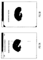
- the images obtained at 2 and 30 minutes post injection are shown in Figures 1A-B .
- Example 9 The imaging apparatus and the procedure used are described in Example 9.
- R3327-H tumors were induced in young male Copenhagen rats in the left flank area from a solid implant. These tumors grow very slowly and palpable masses were present 4-5 months post implant.
- the images obtained at 2 and 30 minutes post injection are shown in Figure 1C-D .
- Rat pancreatic acinar carcinoma expressing the SST-2 receptor (CA20948) were induced by solid implant technique in the left flank area and palpable masses were detected 9 days post implant. The images obtained at 2 and 30 minutes post injection are shown in Figure 1E-F .
- Example 9 The imaging apparatus and the procedure used are described in Example 9 except that each animal received 500 ⁇ L of a 1.0 mg/mL solution of Cytate 1 solution of 25% dimethylsulfoxide in water.
- Rat pancreatic acinar carcinoma expressing the SST-2 receptor (CA20948) were induced by solid implant technique in the left flank area and palpable masses were detected 24 days post implant. Images were obtained at various times post injection. Uptake into the tumor was seen at 2 minutes but was not maximal until about 5 minutes.
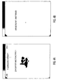
- Figures 2A-B show a comparison of the uptake of ICG and Cytate 1 at 45 minutes in rats with the CA20948 tumor cell line. By 45 minutes the ICG has mostly cleared ( Figure 2A ) whereas the Cytate 1 is still quite intense ( Figure 2B ). This dye fluorescence remained intense in the tumor for several hours post-injection.
- ICG indocyanine green
- DSL 6/A pancreatic
- Dunning R3327-H prostate
- CA20948 pancreatic
- the first two tumor lines are not as highly vascularized as CA20948 which is also rich in somatostatin (SST-2) receptors. Consequently, the detection and retention of a dye in this tumor model is an important index of receptor-mediated specificity.
- Octreotate is known to target somatostatin (SST-2) receptors, hence, we prepared cyano-Octreotates (Cytate 1 and Cytate 2). Cytate 1 was evaluated in the CA20948 Lewis rat model. Using the CCD camera apparatus strong localization of this dye was observed in the tumor at 90 minutes post injection ( Figure 3A ). At 19 hours post injection the animal was again imaged ( Figure 3B ) and tumor visualization was easily observed showing specificity of this agent for the SST-2 receptors present in this tumor line. As a control, the organs were imaged again ( Figure 4A ) and the image was compared with that of the same tissues in the uninjected rat ( Figure 4B ).
- a laser of appropriate wavelength for excitation of the dye chromophore was directed into one end of a fiber optic bundle and the other end was positioned a few millimeters from the ear of a rat.
- a second fiber optic bundle was also positioned near the same ear to detect the emitted fluorescent light and the other end was directed into the optics and electronics for data collection.
- An interference filter (IF) in the collection optics train was used to select emitted fluorescent light of the appropriate wavelength for the dye chromophore.
- Sprague-Dawley or Fischer 344 rats were used in these studies.
- the animals were anesthetized with urethane administered via intraperitoneal injection at a dose of 1.35 g/kg body weight.
- a 21 gauge butterfly with 12" tubing was placed in the lateral tail vein of each animal and flushed with heparinized saline.
- the animals were placed onto a heating pad and kept warm throughout the entire study.
- the lobe of the left ear was affixed to a glass microscope slide to reduce movement and vibration.
- Incident laser light delivered from the fiber optic was centered on the affixed ear. Data acquisition was then initiated, and a background reading of fluorescence was obtained prior to administration of the test agent.
- the peptide-dye conjugate was administered to the animal through a bolus injection in the lateral tail vein, typically of 0.5 to 2.0 mL. The fluorescence signal rapidly increased to a peak value. The signal then decayed as a function of time as the conjugate cleared from the bloodstream.
Landscapes
- Health & Medical Sciences (AREA)
- Life Sciences & Earth Sciences (AREA)
- Animal Behavior & Ethology (AREA)
- Chemical & Material Sciences (AREA)
- Veterinary Medicine (AREA)
- Public Health (AREA)
- General Health & Medical Sciences (AREA)
- Epidemiology (AREA)
- Medicinal Chemistry (AREA)
- Pharmacology & Pharmacy (AREA)
- Molecular Biology (AREA)
- Biochemistry (AREA)
- Engineering & Computer Science (AREA)
- Biomedical Technology (AREA)
- Medicines Containing Antibodies Or Antigens For Use As Internal Diagnostic Agents (AREA)
- Medicines That Contain Protein Lipid Enzymes And Other Medicines (AREA)
- Indole Compounds (AREA)
- Plural Heterocyclic Compounds (AREA)
- Medicinal Preparation (AREA)
- Pharmaceuticals Containing Other Organic And Inorganic Compounds (AREA)
- Investigating Or Analysing Biological Materials (AREA)
- Investigating Or Analysing Materials By The Use Of Chemical Reactions (AREA)
- Peptides Or Proteins (AREA)
Applications Claiming Priority (3)
| Application Number | Priority Date | Filing Date | Title |
|---|---|---|---|
| US13506099P | 1999-05-20 | 1999-05-20 | |
| US09/325,769 US6217848B1 (en) | 1999-05-20 | 1999-06-04 | Cyanine and indocyanine dye bioconjugates for biomedical applications |
| EP00926343A EP1178830B1 (fr) | 1999-05-20 | 2000-04-26 | Nouveaux bioconjugues cyanine et indocyanine pour application medicale |
Related Parent Applications (1)
| Application Number | Title | Priority Date | Filing Date |
|---|---|---|---|
| EP00926343A Division EP1178830B1 (fr) | 1999-05-20 | 2000-04-26 | Nouveaux bioconjugues cyanine et indocyanine pour application medicale |
Publications (2)
| Publication Number | Publication Date |
|---|---|
| EP2058007A2 true EP2058007A2 (fr) | 2009-05-13 |
| EP2058007A3 EP2058007A3 (fr) | 2009-05-27 |
Family
ID=26832945
Family Applications (2)
| Application Number | Title | Priority Date | Filing Date |
|---|---|---|---|
| EP00926343A Expired - Lifetime EP1178830B1 (fr) | 1999-05-20 | 2000-04-26 | Nouveaux bioconjugues cyanine et indocyanine pour application medicale |
| EP08075854A Withdrawn EP2058007A3 (fr) | 1999-05-20 | 2000-04-26 | Bioconjugués de colorant à base cyanine et d'indocyanine pour applications biomédicales |
Family Applications Before (1)
| Application Number | Title | Priority Date | Filing Date |
|---|---|---|---|
| EP00926343A Expired - Lifetime EP1178830B1 (fr) | 1999-05-20 | 2000-04-26 | Nouveaux bioconjugues cyanine et indocyanine pour application medicale |
Country Status (11)
| Country | Link |
|---|---|
| US (1) | US6217848B1 (fr) |
| EP (2) | EP1178830B1 (fr) |
| JP (1) | JP2003500367A (fr) |
| AT (1) | ATE435662T1 (fr) |
| AU (1) | AU4488800A (fr) |
| CA (1) | CA2373475C (fr) |
| DE (1) | DE60042522D1 (fr) |
| DK (1) | DK1178830T3 (fr) |
| ES (1) | ES2329542T3 (fr) |
| PT (1) | PT1178830E (fr) |
| WO (1) | WO2000071162A2 (fr) |
Families Citing this family (54)
| Publication number | Priority date | Publication date | Assignee | Title |
|---|---|---|---|---|
| US6630570B1 (en) * | 1999-04-09 | 2003-10-07 | Insitut für Diagnostikforschung GmbH | Short-chain peptide-dye conjugates as contrast media for optical diagnosis |
| US7175953B2 (en) * | 1999-04-09 | 2007-02-13 | Institute Fuer Diagnostik Forschung | Short-warp peptide-dye conjugate as contrast agent for optical diagnostic |
| US20050182434A1 (en) * | 2000-08-11 | 2005-08-18 | National Research Council Of Canada | Method and apparatus for performing intra-operative angiography |
| US20030017164A1 (en) * | 2001-07-03 | 2003-01-23 | Mallinckrodt Inc. | Dye-azide compounds for dual phototherapy |
| US6190641B1 (en) * | 2000-01-18 | 2001-02-20 | Mallinckrodt Inc. | Indocyanine dyes |
| US7351807B2 (en) * | 2000-01-18 | 2008-04-01 | Mallinckrodt Inc. | Cyanine-sulfenates for dual phototherapy |
| US7790144B2 (en) * | 2000-01-18 | 2010-09-07 | Mallinckrodt Inc. | Receptor-avid exogenous optical contrast and therapeutic agents |
| US7230088B2 (en) * | 2001-07-03 | 2007-06-12 | Mallinckrodt, Inc. | Compounds for dual photodiagnosis and therapy |
| US6180085B1 (en) * | 2000-01-18 | 2001-01-30 | Mallinckrodt Inc. | Dyes |
| US6673334B1 (en) * | 2000-10-16 | 2004-01-06 | Mallinkcrodt, Inc. | Light sensitive compounds for instant determination of organ function |
| US7556797B2 (en) * | 2000-10-16 | 2009-07-07 | Mallinckrodt Inc. | Minimally invasive physiological function monitoring agents |
| US6838074B2 (en) | 2001-08-08 | 2005-01-04 | Bristol-Myers Squibb Company | Simultaneous imaging of cardiac perfusion and a vitronectin receptor targeted imaging agent |
| US20030105300A1 (en) * | 2001-10-17 | 2003-06-05 | Mallinckrodt Inc. | Tumor targeted photodiagnostic-phototherapeutic agents |
| IL148921A0 (en) * | 2002-03-26 | 2002-09-12 | Peptor Ltd | Photo active backbone cyclized somatostatin analogs for optical imaging and photodynamic therapy |
| US20040022731A1 (en) * | 2002-04-26 | 2004-02-05 | Alexei Bogdanov | In vivo imaging of apoptosis |
| US6658143B2 (en) * | 2002-04-29 | 2003-12-02 | Amersham Biosciences Corp. | Ray-based image analysis for biological specimens |
| DE10362402B3 (de) * | 2002-08-28 | 2022-03-03 | Carl Zeiss Meditec Ag | Mikroskopiesystem und Mikroskopieverfahren |
| US7303741B2 (en) * | 2002-09-23 | 2007-12-04 | General Electric Company | Systems and methods for high-resolution in vivo imaging of biochemical activity in a living organism |
| US7226577B2 (en) | 2003-01-13 | 2007-06-05 | Bracco Imaging, S. P. A. | Gastrin releasing peptide compounds |
| DE10302787A1 (de) * | 2003-01-24 | 2004-08-12 | Schering Ag | Hydrophile, Thiol-reaktive Cyaninfarbstoffe und deren Konjugate mit Biomolekülen für die Fluoreszenzdiagnostik |
| CA2523605A1 (fr) * | 2003-03-10 | 2004-09-23 | Charles W. Spangler | Agents cibles pour photodiagnostic et therapie photodynamique |
| WO2005000218A2 (fr) * | 2003-05-31 | 2005-01-06 | Washington University | Bioconjugues macrocycliques a base de cyanine et d'indocyanine pour applications biomedicales ameliorees |
| NO20035683D0 (no) * | 2003-12-18 | 2003-12-18 | Amersham Health As | Optisk avbildning av prostatakreft |
| NO20035681D0 (no) * | 2003-12-18 | 2003-12-18 | Amersham Health As | Optisk avbildning av lungekreft |
| NO20035682D0 (no) * | 2003-12-18 | 2003-12-18 | Amersham Health As | Optisk avbildning av ösofagkreft og Barretts ösofag |
| DE102004022628A1 (de) * | 2004-05-07 | 2005-12-15 | Sensient Imaging Technologies Gmbh | FRET-Bioassay |
| US20050259779A1 (en) * | 2004-05-18 | 2005-11-24 | Siemens Aktiengesellschaft | Biomolecular contrast agents for therapy optimization in radiation therapy with proton or ion beams |
| US7585492B2 (en) * | 2004-05-18 | 2009-09-08 | Siemens Aktiengesellschaft | Biomolecular contrast agents for therapy success and dose monitoring in radiation therapy with proton or ion beams |
| US20050272967A1 (en) * | 2004-05-18 | 2005-12-08 | Siemens Aktiengesellschaft | Biomolecular contrast agents with multiple signal variance for therapy planning and control in radiation therapy with proton or ion beams |
| US20060239916A1 (en) * | 2005-01-07 | 2006-10-26 | Kai Licha | Use of cyanine dyes for the diagnosis of proliferative diseases |
| US20070122344A1 (en) | 2005-09-02 | 2007-05-31 | University Of Rochester Medical Center Office Of Technology Transfer | Intraoperative determination of nerve location |
| US20080161744A1 (en) | 2006-09-07 | 2008-07-03 | University Of Rochester Medical Center | Pre-And Intra-Operative Localization of Penile Sentinel Nodes |
| GB0714202D0 (en) * | 2007-07-20 | 2007-08-29 | Ge Healthcare Bio Sciences Ab | Isoelectric point markers |
| US8406860B2 (en) | 2008-01-25 | 2013-03-26 | Novadaq Technologies Inc. | Method for evaluating blush in myocardial tissue |
| US20090214436A1 (en) | 2008-02-18 | 2009-08-27 | Washington University | Dichromic fluorescent compounds |
| EP2100621A1 (fr) | 2008-03-10 | 2009-09-16 | mivenion GmbH | Conjugués de dendrimère de polyol de polyéther dotés de molécules effectrices pour le ciblage biologique |
| US10219742B2 (en) | 2008-04-14 | 2019-03-05 | Novadaq Technologies ULC | Locating and analyzing perforator flaps for plastic and reconstructive surgery |
| EP3372250B1 (fr) | 2008-05-02 | 2019-12-25 | Novadaq Technologies ULC | Procédés de production et d'utilisation d'érythrocytes chargés de substance pour l'observation et le traitement de l'hémodynamique vasculaire |
| WO2010129258A2 (fr) | 2009-04-27 | 2010-11-11 | Mallinckrodt Inc. | Compositions de scellement tissulaire, dispositifs de fermeture vasculaire et utilisations de ceux-ci |
| US10492671B2 (en) | 2009-05-08 | 2019-12-03 | Novadaq Technologies ULC | Near infra red fluorescence imaging for visualization of blood vessels during endoscopic harvest |
| JPWO2011043061A1 (ja) | 2009-10-05 | 2013-03-04 | キヤノン株式会社 | 光音響イメージング用造影剤、及び、それを用いた光音響イメージング方法 |
| WO2012054784A1 (fr) | 2010-10-20 | 2012-04-26 | Li-Cor, Inc. | Imagerie de fluorescence à colorants à base de cyanine substituée |
| EP2686385B1 (fr) | 2011-03-15 | 2019-01-16 | Ramot at Tel Aviv University, Ltd. | Composés fluorogéniques activables et leurs utilisations en tant que sondes infrarouge proche |
| AU2012305327B2 (en) * | 2011-09-05 | 2016-07-14 | Hiroshi Maeda | Polymer-type fluorescent molecule probe |
| EP2806781B1 (fr) | 2012-01-23 | 2018-03-21 | Washington University | Systèmes et procédés d'imagerie |
| JP6028096B2 (ja) | 2012-06-21 | 2016-11-16 | ノバダック テクノロジーズ インコーポレイテッド | 血管造影及びかん流の定量化並びに解析手法 |
| BR112015003160A2 (pt) * | 2012-10-24 | 2017-07-04 | Becton Dickinson Co | corantes de azaindolina - cianina substituídos por hidroxamato e bioconjugados dos mesmos |
| KR101440109B1 (ko) | 2013-01-29 | 2014-09-12 | 부경대학교 산학협력단 | 위장관암의 림프절 전이 검출용 광음향 단층촬영 시스템 |
| CN107209118B (zh) | 2014-09-29 | 2021-05-28 | 史赛克欧洲运营有限公司 | 在自体荧光存在下生物材料中目标荧光团的成像 |
| EP3203902B1 (fr) | 2014-10-09 | 2021-07-07 | Novadaq Technologies ULC | Quantification de circulation sanguine absolue dans un tissu à l'aide de photopléthysmographie induite par fluorescence |
| US10806804B2 (en) * | 2015-05-06 | 2020-10-20 | Washington University | Compounds having RD targeting motifs and methods of use thereof |
| CA3049922A1 (fr) | 2017-02-10 | 2018-08-16 | Novadaq Technologies ULC | Systemes et procedes d'imagerie a fluorescence portative a champ ouvert |
| US11712482B2 (en) | 2019-12-13 | 2023-08-01 | Washington University | Near infrared fluorescent dyes, formulations and related methods |
| US20260117070A1 (en) | 2022-05-23 | 2026-04-30 | Helmholtz Zentrum München Deutsches Forschungszentrum Für Gesundheit Und Umwelt (Gmbh) | Near-infrared fluorescent heptamethine dye conjugates with favorable bleaching properties |
Citations (5)
| Publication number | Priority date | Publication date | Assignee | Title |
|---|---|---|---|---|
| US5453505A (en) | 1994-06-30 | 1995-09-26 | Biometric Imaging, Inc. | N-heteroaromatic ion and iminium ion substituted cyanine dyes for use as fluorescence labels |
| WO1996017628A1 (fr) | 1994-12-07 | 1996-06-13 | Institut für Diagnostikforschung GmbH an der Freien Universität Berlin | Procede de diagnostic in vivo par rayons infrarouges proches |
| WO1998022146A2 (fr) | 1996-11-19 | 1998-05-28 | Institut für Diagnostikforschung GmbH an der Freien Universität Berlin | Agents pour etablir un diagnostic par voie optique de maladies neurodegeneratives au moyen d'un rayonnement infrarouge proche |
| WO1998048838A1 (fr) | 1997-04-29 | 1998-11-05 | Nycomed Imaging As | Composes |
| WO1998048846A1 (fr) | 1997-04-29 | 1998-11-05 | Nycomed Imaging As | Agents de contraste utilises dans des techniques d'imagerie basees sur la lumiere |
Family Cites Families (10)
| Publication number | Priority date | Publication date | Assignee | Title |
|---|---|---|---|---|
| JPS5745457A (en) * | 1980-09-02 | 1982-03-15 | Fuji Photo Film Co Ltd | Microimmunological measuring method |
| JP3147992B2 (ja) * | 1992-05-29 | 2001-03-19 | イビデン株式会社 | 免疫測定用標識試薬 |
| JP3354975B2 (ja) * | 1992-10-06 | 2002-12-09 | イビデン株式会社 | 蛍光標識試薬および蛍光免疫測定法 |
| US5962424A (en) * | 1995-02-21 | 1999-10-05 | Arch Development Corporation | Methods and compositions for targeting selectins |
| DE19602295C2 (de) * | 1996-01-23 | 2003-08-14 | Deutsches Krebsforsch | Verwendung eines Konjugats aus einer zur Fluoreszenz-fähigen Verbindung, Cyanurchlorid oder einem Derivat davon als Linker und einem Protein |
| WO1997033620A2 (fr) * | 1996-03-15 | 1997-09-18 | Pulsion Verw. Gmbh & Co. Medical Systems Kg | Compose pour traiter des tumeurs |
| CA2326650A1 (fr) * | 1998-04-03 | 1999-10-14 | Mallinckrodt Inc. | Derives azoiques et diazaiques et leurs utilisations en phototherapie |
| WO2000041726A2 (fr) * | 1999-01-15 | 2000-07-20 | Light Sciences Corporation | Therapie vasculaire non invasive |
| US6602274B1 (en) * | 1999-01-15 | 2003-08-05 | Light Sciences Corporation | Targeted transcutaneous cancer therapy |
| JP2002534483A (ja) * | 1999-01-15 | 2002-10-15 | ライト サイエンシーズ コーポレイション | 代謝性骨障害または骨転移のための治療的組成物 |
-
1999
- 1999-06-04 US US09/325,769 patent/US6217848B1/en not_active Expired - Fee Related
-
2000
- 2000-04-26 ES ES00926343T patent/ES2329542T3/es not_active Expired - Lifetime
- 2000-04-26 PT PT00926343T patent/PT1178830E/pt unknown
- 2000-04-26 DE DE60042522T patent/DE60042522D1/de not_active Expired - Lifetime
- 2000-04-26 WO PCT/US2000/011060 patent/WO2000071162A2/fr not_active Ceased
- 2000-04-26 AU AU44888/00A patent/AU4488800A/en not_active Abandoned
- 2000-04-26 AT AT00926343T patent/ATE435662T1/de not_active IP Right Cessation
- 2000-04-26 CA CA2373475A patent/CA2373475C/fr not_active Expired - Lifetime
- 2000-04-26 EP EP00926343A patent/EP1178830B1/fr not_active Expired - Lifetime
- 2000-04-26 EP EP08075854A patent/EP2058007A3/fr not_active Withdrawn
- 2000-04-26 DK DK00926343T patent/DK1178830T3/da active
- 2000-04-26 JP JP2000619463A patent/JP2003500367A/ja active Pending
Patent Citations (5)
| Publication number | Priority date | Publication date | Assignee | Title |
|---|---|---|---|---|
| US5453505A (en) | 1994-06-30 | 1995-09-26 | Biometric Imaging, Inc. | N-heteroaromatic ion and iminium ion substituted cyanine dyes for use as fluorescence labels |
| WO1996017628A1 (fr) | 1994-12-07 | 1996-06-13 | Institut für Diagnostikforschung GmbH an der Freien Universität Berlin | Procede de diagnostic in vivo par rayons infrarouges proches |
| WO1998022146A2 (fr) | 1996-11-19 | 1998-05-28 | Institut für Diagnostikforschung GmbH an der Freien Universität Berlin | Agents pour etablir un diagnostic par voie optique de maladies neurodegeneratives au moyen d'un rayonnement infrarouge proche |
| WO1998048838A1 (fr) | 1997-04-29 | 1998-11-05 | Nycomed Imaging As | Composes |
| WO1998048846A1 (fr) | 1997-04-29 | 1998-11-05 | Nycomed Imaging As | Agents de contraste utilises dans des techniques d'imagerie basees sur la lumiere |
Non-Patent Citations (6)
| Title |
|---|
| BECKER A ET AL.: "Transferrin Mediated Tumor Delivery of Contrast Media for Optical Imaging and Magnetic Resonance Imaging", BIOMEDICAL OPTICS MEETING, 23 January 1999 (1999-01-23) |
| BRINKLEY M: "Perspectives in Bioconjugate Chemistry", 1993, ACS PUBLICATION, article "A Brief Survey of Methods for Preparing Protein Conjugates with Dyes, Haptens, and Cross-Linking Reagents", pages: 59 - 70 |
| JAIN RK: "Barriers to Drug Delivery in Solid Tumors", SCIENTIFIC AMERICAN, vol. 271, 1994, pages 58 - 65 |
| JONG M ET AL., CANCER RES., vol. 58, 1998, pages 437 - 441 |
| PATONAY G; ANTOINE MD: "Near-Infrared Fluorogenic Labels: New Approach to an Old Problem", ANALYTICAL CHEMISTRY, vol. 63, 1991, pages 321 A - 327 A |
| SLAVIK J: "Fluorescent Probes in Cellular and Molecular Biology", 1994, CRC PRESS, INC. |
Also Published As
| Publication number | Publication date |
|---|---|
| JP2003500367A (ja) | 2003-01-07 |
| WO2000071162A3 (fr) | 2001-07-05 |
| EP1178830B1 (fr) | 2009-07-08 |
| ES2329542T3 (es) | 2009-11-27 |
| AU4488800A (en) | 2000-12-12 |
| EP1178830A2 (fr) | 2002-02-13 |
| EP2058007A3 (fr) | 2009-05-27 |
| DE60042522D1 (de) | 2009-08-20 |
| ATE435662T1 (de) | 2009-07-15 |
| US6217848B1 (en) | 2001-04-17 |
| PT1178830E (pt) | 2009-09-02 |
| DK1178830T3 (da) | 2009-10-12 |
| CA2373475C (fr) | 2012-10-23 |
| WO2000071162A2 (fr) | 2000-11-30 |
| CA2373475A1 (fr) | 2000-11-30 |
Similar Documents
| Publication | Publication Date | Title |
|---|---|---|
| EP1178830B1 (fr) | Nouveaux bioconjugues cyanine et indocyanine pour application medicale | |
| US7198778B2 (en) | Tumor-targeted optical contrast agents | |
| US7011817B2 (en) | Hydrophilic cyanine dyes | |
| US7201892B2 (en) | Pathological tissue detection and treatment employing targeted optical agents | |
| US6641798B2 (en) | Tumor-targeted optical contrast agents | |
| US6706254B2 (en) | Receptor-avid exogenous optical contrast and therapeutic agents | |
| US7767194B2 (en) | Optical diagnostic and therapeutic agents and compositions | |
| EP1250095B1 (fr) | Colorants hydrophiles versatiles | |
| WO2003032900A2 (fr) | Colorants carbocyanine pour applications photodiagnostiques et therapeutiques combinees | |
| CA2474920A1 (fr) | Bioconjugues de colorants destines a des applications diagnostiques et therapeutiques optiques simultanees | |
| EP1250091B1 (fr) | Colorants hydrophiles a base de cyanine |
Legal Events
| Date | Code | Title | Description |
|---|---|---|---|
| PUAI | Public reference made under article 153(3) epc to a published international application that has entered the european phase |
Free format text: ORIGINAL CODE: 0009012 |
|
| PUAL | Search report despatched |
Free format text: ORIGINAL CODE: 0009013 |
|
| AC | Divisional application: reference to earlier application |
Ref document number: 1178830 Country of ref document: EP Kind code of ref document: P |
|
| AK | Designated contracting states |
Kind code of ref document: A2 Designated state(s): AT BE CH CY DE DK ES FI FR GB GR IE IT LI LU MC NL PT SE |
|
| AK | Designated contracting states |
Kind code of ref document: A3 Designated state(s): AT BE CH CY DE DK ES FI FR GB GR IE IT LI LU MC NL PT SE |
|
| 17P | Request for examination filed |
Effective date: 20091126 |
|
| AKX | Designation fees paid |
Designated state(s): AT BE CH CY DE DK ES FI FR GB GR IE IT LI LU MC NL PT SE |
|
| 17Q | First examination report despatched |
Effective date: 20100302 |
|
| STAA | Information on the status of an ep patent application or granted ep patent |
Free format text: STATUS: THE APPLICATION IS DEEMED TO BE WITHDRAWN |
|
| 18D | Application deemed to be withdrawn |
Effective date: 20101109 |