EP2059201B1 - Système et procédé de résection du tissu cornéen - Google Patents
Système et procédé de résection du tissu cornéen Download PDFInfo
- Publication number
- EP2059201B1 EP2059201B1 EP07814262.7A EP07814262A EP2059201B1 EP 2059201 B1 EP2059201 B1 EP 2059201B1 EP 07814262 A EP07814262 A EP 07814262A EP 2059201 B1 EP2059201 B1 EP 2059201B1
- Authority
- EP
- European Patent Office
- Prior art keywords
- cornea
- depth
- resection
- recipient
- biomechanical model
- Prior art date
- Legal status (The legal status is an assumption and is not a legal conclusion. Google has not performed a legal analysis and makes no representation as to the accuracy of the status listed.)
- Not-in-force
Links
Images

Classifications
-
- A—HUMAN NECESSITIES
- A61—MEDICAL OR VETERINARY SCIENCE; HYGIENE
- A61F—FILTERS IMPLANTABLE INTO BLOOD VESSELS; PROSTHESES; DEVICES PROVIDING PATENCY TO, OR PREVENTING COLLAPSING OF, TUBULAR STRUCTURES OF THE BODY, e.g. STENTS; ORTHOPAEDIC, NURSING OR CONTRACEPTIVE DEVICES; FOMENTATION; TREATMENT OR PROTECTION OF EYES OR EARS; BANDAGES, DRESSINGS OR ABSORBENT PADS; FIRST-AID KITS
- A61F2/00—Filters implantable into blood vessels; Prostheses, i.e. artificial substitutes or replacements for parts of the body; Appliances for connecting them with the body; Devices providing patency to, or preventing collapsing of, tubular structures of the body, e.g. stents
- A61F2/02—Prostheses implantable into the body
- A61F2/14—Eye parts, e.g. lenses or corneal implants; Artificial eyes
- A61F2/142—Cornea, e.g. artificial corneae, keratoprostheses or corneal implants for repair of defective corneal tissue
-
- A—HUMAN NECESSITIES
- A61—MEDICAL OR VETERINARY SCIENCE; HYGIENE
- A61F—FILTERS IMPLANTABLE INTO BLOOD VESSELS; PROSTHESES; DEVICES PROVIDING PATENCY TO, OR PREVENTING COLLAPSING OF, TUBULAR STRUCTURES OF THE BODY, e.g. STENTS; ORTHOPAEDIC, NURSING OR CONTRACEPTIVE DEVICES; FOMENTATION; TREATMENT OR PROTECTION OF EYES OR EARS; BANDAGES, DRESSINGS OR ABSORBENT PADS; FIRST-AID KITS
- A61F9/00—Methods or devices for treatment of the eyes; Devices for putting in contact-lenses; Devices to correct squinting; Apparatus to guide the blind; Protective devices for the eyes, carried on the body or in the hand
- A61F9/007—Methods or devices for eye surgery
- A61F9/008—Methods or devices for eye surgery using laser
- A61F9/00825—Methods or devices for eye surgery using laser for photodisruption
- A61F9/00831—Transplantation
-
- A—HUMAN NECESSITIES
- A61—MEDICAL OR VETERINARY SCIENCE; HYGIENE
- A61F—FILTERS IMPLANTABLE INTO BLOOD VESSELS; PROSTHESES; DEVICES PROVIDING PATENCY TO, OR PREVENTING COLLAPSING OF, TUBULAR STRUCTURES OF THE BODY, e.g. STENTS; ORTHOPAEDIC, NURSING OR CONTRACEPTIVE DEVICES; FOMENTATION; TREATMENT OR PROTECTION OF EYES OR EARS; BANDAGES, DRESSINGS OR ABSORBENT PADS; FIRST-AID KITS
- A61F9/00—Methods or devices for treatment of the eyes; Devices for putting in contact-lenses; Devices to correct squinting; Apparatus to guide the blind; Protective devices for the eyes, carried on the body or in the hand
- A61F9/007—Methods or devices for eye surgery
- A61F9/008—Methods or devices for eye surgery using laser
- A61F2009/00855—Calibration of the laser system
- A61F2009/00857—Calibration of the laser system considering biodynamics
-
- A—HUMAN NECESSITIES
- A61—MEDICAL OR VETERINARY SCIENCE; HYGIENE
- A61F—FILTERS IMPLANTABLE INTO BLOOD VESSELS; PROSTHESES; DEVICES PROVIDING PATENCY TO, OR PREVENTING COLLAPSING OF, TUBULAR STRUCTURES OF THE BODY, e.g. STENTS; ORTHOPAEDIC, NURSING OR CONTRACEPTIVE DEVICES; FOMENTATION; TREATMENT OR PROTECTION OF EYES OR EARS; BANDAGES, DRESSINGS OR ABSORBENT PADS; FIRST-AID KITS
- A61F9/00—Methods or devices for treatment of the eyes; Devices for putting in contact-lenses; Devices to correct squinting; Apparatus to guide the blind; Protective devices for the eyes, carried on the body or in the hand
- A61F9/007—Methods or devices for eye surgery
- A61F9/008—Methods or devices for eye surgery using laser
- A61F2009/00861—Methods or devices for eye surgery using laser adapted for treatment at a particular location
- A61F2009/00872—Cornea
-
- A—HUMAN NECESSITIES
- A61—MEDICAL OR VETERINARY SCIENCE; HYGIENE
- A61F—FILTERS IMPLANTABLE INTO BLOOD VESSELS; PROSTHESES; DEVICES PROVIDING PATENCY TO, OR PREVENTING COLLAPSING OF, TUBULAR STRUCTURES OF THE BODY, e.g. STENTS; ORTHOPAEDIC, NURSING OR CONTRACEPTIVE DEVICES; FOMENTATION; TREATMENT OR PROTECTION OF EYES OR EARS; BANDAGES, DRESSINGS OR ABSORBENT PADS; FIRST-AID KITS
- A61F9/00—Methods or devices for treatment of the eyes; Devices for putting in contact-lenses; Devices to correct squinting; Apparatus to guide the blind; Protective devices for the eyes, carried on the body or in the hand
- A61F9/007—Methods or devices for eye surgery
- A61F9/008—Methods or devices for eye surgery using laser
- A61F9/009—Auxiliary devices making contact with the eyeball and coupling in laser light, e.g. goniolenses
-
- A—HUMAN NECESSITIES
- A61—MEDICAL OR VETERINARY SCIENCE; HYGIENE
- A61F—FILTERS IMPLANTABLE INTO BLOOD VESSELS; PROSTHESES; DEVICES PROVIDING PATENCY TO, OR PREVENTING COLLAPSING OF, TUBULAR STRUCTURES OF THE BODY, e.g. STENTS; ORTHOPAEDIC, NURSING OR CONTRACEPTIVE DEVICES; FOMENTATION; TREATMENT OR PROTECTION OF EYES OR EARS; BANDAGES, DRESSINGS OR ABSORBENT PADS; FIRST-AID KITS
- A61F9/00—Methods or devices for treatment of the eyes; Devices for putting in contact-lenses; Devices to correct squinting; Apparatus to guide the blind; Protective devices for the eyes, carried on the body or in the hand
- A61F9/007—Methods or devices for eye surgery
- A61F9/013—Instruments for compensation of ocular refraction ; Instruments for use in cornea removal, for reshaping or performing incisions in the cornea
Definitions
- the field of the present invention is systems for transplanting corneas.
- PLK posterior lamellar keratoplasty
- deep lamellar keratoplasty A procedure called posterior lamellar keratoplasty (PLK), or deep lamellar keratoplasty, was previously developed to address these shortcomings for patients with endothelial cell dysfunction.
- a thin posterior or lenticule of stromal tissue (along with descemets membrane and endothelial cells attached) is removed from the cornea of a diseased eye.
- a similar procedure is performed on a donor eye to obtain donor tissue. When the donor tissue is placed in the recipient's eye, no sutures are required because the anterior surface of the recipients' cornea was left intact.
- One advantage of this procedure is that it results in little post-operative astigmatism. While PLK has its advantages, in practice it is difficult to perform, regardless of whether manual or automated instruments are used.
- a procedure called descemets strip endokeratoplasty was introduced in attempts have been made to improve on PLK.
- the stromal tissue is not removed from the recipient's cornea, rather only the descemets membrane and endothelial cells are removed. While this simplifies the procedure, it results in an abnormally thick post-operative cornea and may degrade optical quality.
- the folds or wrinkles can cause the incision to have an irregular surface when the cornea returns to its natural curvature if the incision is made too near the posterior surface of the cornea.
- Such an irregular surface can have adverse effects on wound healing and optical quality of the post-operative cornea.
- EP 1,666,009 describes a system for refractive ophthalmic surgery in which a flap of cornea is cut and then lifted to expose the underlying corneal tissue and the underlying corneal tissue is then reshaped by photoablation.
- US 2006/0015090 describes a corneal refraction procedure providing a customised transition zone to provide continuous curvature between an ablated optical zone and a non-ablated optical zone to address curvature discontinuity.
- US 2006/0100612 describes a laser-based device for non-mechanical three-dimensional trepanation during corneal transplants.
- US 6,325,792 describes the application of low energy ultra-short (femtosecond) pulsed laser radiation to a patient's eye in one of a number of patterns such that the exposed ocular tissue is ablated or excised through the process of optical breakdown or photodisruption.
- EPC describes a system and method for compensating a corneal dissection in which pre-dissection diagnostic information about the transparent material to be dissected is used to define a topology and is analysed to calculate a predicated result of a dissection along a prototypic dissection path.
- a surgical laser emits a pulsed laser beam which is directed into the cornea by a focusing assembly.
- An interface is programmed with a biomechanical model of corneal tissue, which is used to provide a depth range for selection of a resection depth from an anterior corneal surface.
- the selected resection depth is received by a controller which employs the focusing assembly to move the focal point of the pulsed laser beam and make a resection incision at the resection depth for resecting a portion of the cornea.
- the resection depth is first determined using a biomechanical model of the cornea. Thereafter, a resection incision is made at the resection depth for purposes of resecting a posterior portion of the donor cornea.
- the resection depth is determined using the anterior surface of the cornea as a reference.
- a contact lens is placed against the anterior surface of the cornea.
- the contact lens is adapted to conform the anterior surface of the cornea to the shape of the contact lens, which preferably has a radial or planar form. With the contact lens in place, the resection incision is made using a surgical laser.
- the resection depth is selected to minimize surface irregularities resulting from the resection incision.
- the resection depth is selected to minimize post-operative weakening of the cornea.
- the resection incision is placed at a unifonn distance fi-om one of the posterior surface of the cornea or the anterior surface of the cornea.
- a posterior portion of corneal tissue is resected from a donor cornea, and this donor tissue is grafted into the recipient cornea.
- the ratio of the depth of the resection incision in the recipient cornea, as compared to the overall thickness of the recipient cornea is first determined. From this ratio, the depth of the resection incision in the donor cornea may be determined. This is done by making the depth of the resection incision in the donor cornea have the same ratio as compared to the overall thickness of the donor cornea.
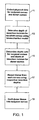
- Fig. 1 is a flow chart illustrating a process for performing a PLK procedure.
- the basic steps of the PLK procedure are (1) collect physical data 11 for both the recipient cornea and the donor cornea; (2) determine the depth of the resection incision 13 for the recipient cornea; (3) determine the depth ratio of the resection incision 15 in the recipient cornea and the depth of the resection incision in the donor cornea; (4) resect tissue 17 from both the recipient and donor corneas; and (5) graft 19 the donor tissue into the recipient cornea.
- Each step is explained in further detail below.
- the physical data collected includes thickness measurements of both the recipient and donor corneas. These thickness measurements are used to develop a thickness profile of each cornea. Additional physical data may also be collected for each cornea, with the type of data collected being dependent upon the requirements of biomechanical model used in the subsequent step of the PLK procedure.
- This thickness profile, along with any other data needed for the biomechanical model may be obtained by any one of the many known methods for measuring the physical structure of the eye, with the preferred method being through optical coherence tomography (OCT).
- OCT optical coherence tomography
- Many commercially available OCT scanners are capable of performing such measurements.
- One example is the VisanteTM OCT scanning system, manufactured by Carl Zeiss Meditec, which has an office in Dublin, California.
- One advantage of the VisanteTM OCT system is that it does not make contact with the cornea when performing the OCT scan.
- the depth of the resection incision for the recipient cornea is determined using the biomechanical model.
- the biomechanical model takes into account stresses introduced into the corneal tissue when the cornea is conformed to the shape of a contact lens set against the anterior surface when the resection incision is made with a surgical laser system. Such stresses can cause folds in the corneal tissue which are more pronounced in the posterior regions of the cornea. A resection incision made in areas with more pronounced folds can lead directly to surface irregularities at the resection incision when contact lens is removed and the cornea returns to its normal shape. Therefore, the biomechanical model is used to place the resection incision at a depth which minimizes or eliminates surface irregularities at the resection incision.
- the biomechanical model also preferably takes into account the post-operative stability of the recipient cornea when determining the depth of the resection incision. Without sufficient post-operative stability, the cornea may undergo weakening through pathological deterioration, e.g., ectasia, thereby exposing the recipient to a risk of vision impairment. Maintaining sufficient tissue between the anterior surface of the recipient cornea and the resection incision is an important factor to maintaining post-operative stability.
- the biomechanical model is therefore employed to determine the minimum depth from the anterior surface at which the resection incision is likely to cause post-operative stability problems.
- the biomechanical model may be developed to employ a database of corneal measurements collected from many patients. Using information from the database, together with at least the known curvature of the contact lens, such a biomechanical model could be used to determine an appropriate depth for the resection incision.
- the resection depth selected for the recipient cornea is selected to be between the minimum depth needed to create post-operative stability in the recipient cornea and an appropriate depth to avoid or eliminate surface irregularities at the resection incision. In the event that there is no region between the two extremes, maintaining post-operative stability in the cornea is preferred. In such instances, the radius of the contact lens upon which the shape of the cornea is conformed for purposes of the PLK procedure and the biomechanical model could be enlarged to reduce stresses on the corneal tissue and help create a region in which post-operative stability can be maintained along with minimizing irregularities at the resection incision.
- the biomechanical model may be one of several previously developed models which are known to skilled artisans, or it may be one which is specifically developed for the PLK procedure.
- Modeling software such as the software published by Structural Research & Analysis Corp. of Santa Monica, CA under the title “Cosmos/M”, may be used to develop biomechanical models.
- One such model is described in the article by G. Djotyan et al., "Finite Element Modeling of Posterior Lamellar Keratoplasty: Construction of Theoretical Nomograms for Induced Refractive Errors", Ophthalmic Research, Vol.38, n.5, 2006 .
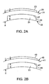
- Fig. 2A illustrates the recipient cornea 21 with a contact lens 23 placed against the anterior surface 25 of the recipient cornea 21.
- the resection incision 27 and the sidecut 29 are made to remove a posterior portion of corneal tissue 31 in preparation for the transplant.
- the resection incision 27 may be made using several different techniques. However, it should be noted that not all techniques provide the same quality of clinical results following the transplant.
- One technique is to make the resection incision 27 at a uniform distance from the posterior surface 33 of the cornea.
- U.S. Pat. App. No. 11/375,542 ( US 2007/0219541 ) discloses a method of making such an incision.
- the thickness of the recipient cornea 21 is expressed as 'a'.
- the depth, b, of the resection incision 27 shown in Fig. 1 is measured from the anterior surface 25 of the cornea.
- the ratio of the depth, b, to the thickness, a, is employed to determine the depth of the resection incision in the donor cornea as described below.
- the sidecut 29 may also be formed through the different techniques that are known to skilled artisans.
- the contact lens 23 is a rigid lens placed against the anterior surface 25, thereby forcing the anterior surface 25 to conform to the shape of the contact lens 23.
- the contact lens 23 confirms the anterior surface 25 to a radial or planar shape.
- U.S. Patent No. 5,549,632 describes making a laser incision by deforming the shape of the cornea.
- U.S. Patent No. 6,863,667 and U.S. Pat. App. No. 11/258,399 ( US 2007-0093796 ) describe patient interface devices which may be used to align the surgical laser with the recipient cornea for purposes of making accurate incisions.
- the contact lens is not needed if the surgical laser system is capable of alignment with sufficient precision without use of the contact lens.
- Fig. 2B illustrates the donor cornea 41 with a contact lens 43 placed against the anterior surface 45 of the donor cornea 41.
- the resection incision 47 and the sidecut 49 are made to remove the donor tissue 51 for grafting into the recipient cornea.
- the resection incision 47 and the sidecut 49 are preferably made using the same techniques employed to incise the recipient cornea.
- the ratio of the depth, d, of the resection incision 47 to the thickness, c, of the donor cornea 41 is preferably the same as the ratio that was set for the recipient cornea.
- a surgical system which may be used to incise both a donor cornea or a recipient cornea.
- a femtosecond surgical laser 51 generates a pulsed laser beam 53 and directs that beam into the focusing assembly 55, which in turn focuses the pulsed beam 53 into the cornea 57.
- a contact lens 59 is placed over the cornea to deform the anterior corneal surface as described above.
- the controller 61 is a programmable computer which precisely controls the location of the beam focal point within the cornea 57 according to parameters received from the programmable surgeon interface 63.
- the interface 63 is programmed with a biomechanical model of the cornea and presents the surgeon with an incision depth range within which the surgeon may select the resection depth.
- the resection depth is received by the controller 61, which uses the focusing assembly 55 to make the resection incision at the resection depth.
- a pulsed laser beam having ultra-short pulses, preferably in the femtosecond range, is employed to make the incisions.
- the laser may be of the type described in U.S. Patent No. 4,764,930 , producing an ultra-short pulsed beam as described in one or both of U.S. Patent No. 5,984,916 and U.S. Patent No. RE37,585 .
Landscapes
- Health & Medical Sciences (AREA)
- Ophthalmology & Optometry (AREA)
- Transplantation (AREA)
- Biomedical Technology (AREA)
- Engineering & Computer Science (AREA)
- Vascular Medicine (AREA)
- Animal Behavior & Ethology (AREA)
- Veterinary Medicine (AREA)
- Public Health (AREA)
- Heart & Thoracic Surgery (AREA)
- General Health & Medical Sciences (AREA)
- Life Sciences & Earth Sciences (AREA)
- Surgery (AREA)
- Physics & Mathematics (AREA)
- Optics & Photonics (AREA)
- Nuclear Medicine, Radiotherapy & Molecular Imaging (AREA)
- Cardiology (AREA)
- Oral & Maxillofacial Surgery (AREA)
- Laser Surgery Devices (AREA)
- Prostheses (AREA)
Claims (11)
- Système pour réséquer du tissu cornéen, le système comprenant :une interface (63) programmée avec un modèle biomécanique de tissu cornéen, l'interface étant conçue pour fournir une plage de profondeurs au moyen du modèle biomécanique pour une sélection de profondeurs de résection à partir d'une surface cornéenne antérieure ;un laser chirurgical (51) conçu pour émettre un faisceau laser à impulsion ;un assemblage de focalisation (55) conçu pour focaliser le faisceau laser à impulsion dans une cornée ;un contrôleur (61) conçu pour recevoir la profondeur de résection depuis l'interface, pour déplacer un point de focalisation du faisceau laser à impulsion dans une cornée (57) au moyen de l'assemblage de focalisation, et pour diriger le point de focalisation du faisceau laser à impulsion pour réaliser une incision de résection (27 ; 47) pour réséquer une partie de la cornée, où l'incision de résection est réalisée à la profondeur de résection (b ; d).
- Système selon la revendication 1, comprenant en outre :une lentille de contact (23 ; 43) conçue pour être placée contre la surface antérieure et pour conformer la surface antérieure à une forme de la lentille de contact, où le modèle biomécanique rend compte de la déformation de la cornée (57).
- Système selon la revendication 2, dans lequel la lentille de contact (23 ; 43) aplanit la surface antérieure.
- Système selon la revendication 1, dans lequel le modèle biomécanique est basé sur un profil d'épaisseur de la cornée et sur des contraintes introduites dans la cornée associées à une mise en conformité de la cornée à la forme de la lentille de contact (23 ; 43) et où la plage de profondeurs est fournie par le modèle biomécanique pour minimiser des irrégularités de surface au niveau de l'incision de résection (27 ; 47).
- Système selon la revendication 1, dans lequel le modèle biomécanique est basé sur un profil d'épaisseur de la cornée et sur des contraintes introduites dans la cornée associées à une mise en conformité de la cornée à la forme de la lentille de contact (23 ; 43) et où la plage de profondeurs est fournie par le modèle biomécanique pour minimiser des faiblesses postopératoires de la cornée.
- Système selon la revendication 1, dans lequel l'incision de résection se situe à une distance uniforme d'une surface postérieure de la cornée (57) ou de la surface antérieure.
- Système selon la revendication 1, dans lequel le contrôleur est conçu pour réaliser l'incision de résection pour réséquer une partie postérieure de la cornée (57).
- Système selon la revendication 1, dans lequel la profondeur de résection se situe entre une première profondeur basée sur le modèle biomécanique et une seconde profondeur basée sur le modèle biomécanique, la première profondeur consistant à maintenir une stabilité postopératoire de la cornée du receveur, la seconde profondeur consistant à minimiser des irrégularités de surface au niveau de la profondeur de résection associées à une incision de résection (27) dans le cornée du receveur tandis que la cornée du receveur est conforme à la forme d'une lentille de contact (23).
- Procédé pour réséquer un tissu de cornée de donneur à partir d'une cornée de donneur, le procédé comprenant les étapes suivantes :déterminer une profondeur de résection à partir d'une surface antérieure de la cornée du donneur au moyen d'un modèle biomécanique cornéen ; etréaliser une incision de résection du donneur (47) pour réséquer une partie postérieure du donneur à partir de la cornée du donneur, où l'incision de résection (47) est réalisée à la profondeur de résection (d).
- Procédé selon la revendication 9, de résection de tissu cornéen à partir d'une cornée de donneur pour une transplantation vers une cornée de receveur, où la détermination de la profondeur de résection comprend l'étape suivants :déterminer la profondeur de résection du receveur au moyen d'un modèle biomécanique de la cornée du receveur.
- Procédé selon la revendication 9 ou la revendication 10, dans lequel la profondeur de résection se situe entre une première profondeur basée sur le modèle biomécanique et une seconde profondeur basée sur le modèle biomécanique, la première profondeur correspondant au maintien d'une stabilité postopératoire de la cornée du receveur, la seconde profondeur correspondant à la minimisation des irrégularités de surface au niveau de la profondeur de résection associées à une incision de résection (27) dans la cornée du receveur tandis que la cornée du receveur est conforme à la forme d'une lentille de contact (23).
Applications Claiming Priority (2)
| Application Number | Priority Date | Filing Date | Title |
|---|---|---|---|
| US11/469,941 US20080082088A1 (en) | 2006-09-05 | 2006-09-05 | System and method for resecting corneal tissue |
| PCT/US2007/076334 WO2008030698A2 (fr) | 2006-09-05 | 2007-08-20 | Système et procédé de résection du tissu cornéen |
Publications (3)
| Publication Number | Publication Date |
|---|---|
| EP2059201A2 EP2059201A2 (fr) | 2009-05-20 |
| EP2059201A4 EP2059201A4 (fr) | 2010-09-08 |
| EP2059201B1 true EP2059201B1 (fr) | 2015-11-11 |
Family
ID=39157934
Family Applications (1)
| Application Number | Title | Priority Date | Filing Date |
|---|---|---|---|
| EP07814262.7A Not-in-force EP2059201B1 (fr) | 2006-09-05 | 2007-08-20 | Système et procédé de résection du tissu cornéen |
Country Status (5)
| Country | Link |
|---|---|
| US (1) | US20080082088A1 (fr) |
| EP (1) | EP2059201B1 (fr) |
| AU (1) | AU2007292547A1 (fr) |
| CA (1) | CA2659852C (fr) |
| WO (1) | WO2008030698A2 (fr) |
Families Citing this family (26)
| Publication number | Priority date | Publication date | Assignee | Title |
|---|---|---|---|---|
| US9603741B2 (en) | 2000-05-19 | 2017-03-28 | Michael S. Berlin | Delivery system and method of use for the eye |
| US8679089B2 (en) | 2001-05-21 | 2014-03-25 | Michael S. Berlin | Glaucoma surgery methods and systems |
| ATE377404T1 (de) | 2000-05-19 | 2007-11-15 | Michael S Berlin | Laserapplikationssystem und methode zur verwendung im auge |
| US20070219541A1 (en) * | 2006-03-14 | 2007-09-20 | Intralase Corp. | System and method for ophthalmic laser surgery on a cornea |
| US20170360609A9 (en) | 2007-09-24 | 2017-12-21 | Ivantis, Inc. | Methods and devices for increasing aqueous humor outflow |
| EP2259833A1 (fr) | 2008-03-05 | 2010-12-15 | Ivantis, INC. | Procédés et appareils pour le traitement du glaucome |
| US8529557B2 (en) * | 2008-05-30 | 2013-09-10 | Technolas Perfect Vision Gmbh | System and method for stray light compensation of corneal cuts |
| DE102009005482A1 (de) | 2009-01-21 | 2010-07-22 | Carl Zeiss Meditec Ag | Vorrichtung und Verfahren zum Erzeugen von Steuerdaten zur operativen Fehlsichtigkeitskorrektur eines Auges |
| US9411938B2 (en) * | 2009-04-02 | 2016-08-09 | Sie Ag, Surgical Instrument Engineering | System for defining cuts in eye tissue |
| AU2010271218B2 (en) | 2009-07-09 | 2017-02-02 | Alcon Inc. | Ocular implants and methods for delivering ocular implants into the eye |
| WO2011006078A1 (fr) | 2009-07-09 | 2011-01-13 | Ivantis, Inc. | Dispositif d'opérateur unique pour pose d'un implant oculaire |
| US20120203215A1 (en) * | 2009-10-05 | 2012-08-09 | Peter Riedel | Device for ophthalmological laser surgery |
| CN103974678B (zh) * | 2011-04-01 | 2017-02-08 | 雷萨公司 | 激光产生的角膜和晶状体 |
| US20120283557A1 (en) | 2011-05-05 | 2012-11-08 | Berlin Michael S | Methods and Apparatuses for the Treatment of Glaucoma using visible and infrared ultrashort laser pulses |
| US8657776B2 (en) | 2011-06-14 | 2014-02-25 | Ivantis, Inc. | Ocular implants for delivery into the eye |
| US8663150B2 (en) | 2011-12-19 | 2014-03-04 | Ivantis, Inc. | Delivering ocular implants into the eye |
| US9358156B2 (en) | 2012-04-18 | 2016-06-07 | Invantis, Inc. | Ocular implants for delivery into an anterior chamber of the eye |
| US10617558B2 (en) | 2012-11-28 | 2020-04-14 | Ivantis, Inc. | Apparatus for delivering ocular implants into an anterior chamber of the eye |
| US10709547B2 (en) | 2014-07-14 | 2020-07-14 | Ivantis, Inc. | Ocular implant delivery system and method |
| JP6837475B2 (ja) | 2015-08-14 | 2021-03-03 | イバンティス インコーポレイテッド | 圧力センサを備えた眼用インプラントおよび送達システム |
| US11938058B2 (en) | 2015-12-15 | 2024-03-26 | Alcon Inc. | Ocular implant and delivery system |
| US10842573B2 (en) * | 2016-08-24 | 2020-11-24 | Alcon Inc. | Predictive apparatus for assisting a physician during ophthalmic surgery |
| EP4371535A3 (fr) | 2018-02-22 | 2024-08-14 | Alcon Inc. | Implant oculaire et système de pose |
| AU2022205382B2 (en) | 2021-01-11 | 2026-02-19 | Alcon Inc. | Systems and methods for viscoelastic delivery |
| EP4190286B1 (fr) | 2021-12-06 | 2026-03-18 | Ziemer Ophthalmic Systems AG | Appareil de traitement et de fabrication ophtalmologique |
| AU2024309881A1 (en) | 2023-06-30 | 2025-11-20 | Alcon Inc. | System and methods for compensating for intraocular lens tilt |
Citations (3)
| Publication number | Priority date | Publication date | Assignee | Title |
|---|---|---|---|---|
| US6325792B1 (en) * | 1991-11-06 | 2001-12-04 | Casimir A. Swinger | Ophthalmic surgical laser and method |
| WO2007012924A1 (fr) * | 2005-07-26 | 2007-02-01 | 20/10 Perfect Vision Ag | Système et procédé pour compenser une dissection cornéenne |
| WO2007143111A2 (fr) * | 2006-06-01 | 2007-12-13 | University Of Southern California | Procédé et appareil destinés à guider une chirurgie cornéenne au laser avec mesures optiques |
Family Cites Families (19)
| Publication number | Priority date | Publication date | Assignee | Title |
|---|---|---|---|---|
| US4669466A (en) * | 1985-01-16 | 1987-06-02 | Lri L.P. | Method and apparatus for analysis and correction of abnormal refractive errors of the eye |
| US4764930A (en) | 1988-01-27 | 1988-08-16 | Intelligent Surgical Lasers | Multiwavelength laser source |
| US5647865A (en) * | 1991-11-01 | 1997-07-15 | Swinger; Casimir A. | Corneal surgery using laser, donor corneal tissue and synthetic material |
| US5984916A (en) | 1993-04-20 | 1999-11-16 | Lai; Shui T. | Ophthalmic surgical laser and method |
| US5549632A (en) * | 1992-10-26 | 1996-08-27 | Novatec Laser Systems, Inc. | Method and apparatus for ophthalmic surgery |
| US5501217A (en) * | 1992-12-28 | 1996-03-26 | Tomey Corporation | Contact lens for intraocular observation |
| US5993438A (en) | 1993-11-12 | 1999-11-30 | Escalon Medical Corporation | Intrastromal photorefractive keratectomy |
| US5656186A (en) | 1994-04-08 | 1997-08-12 | The Regents Of The University Of Michigan | Method for controlling configuration of laser induced breakdown and ablation |
| EP1666009A3 (fr) | 2000-07-21 | 2007-08-22 | The Ohio State University | Système pour la chirurgie ophtalmologique réfractive |
| US6863667B2 (en) | 2001-01-29 | 2005-03-08 | Intralase Corp. | Ocular fixation and stabilization device for ophthalmic surgical applications |
| US20030208190A1 (en) * | 2001-07-20 | 2003-11-06 | Cynthia Roberts | Methods and instruments for refractive ophthalmic surgery |
| US7130835B2 (en) * | 2002-03-28 | 2006-10-31 | Bausch & Lomb Incorporated | System and method for predictive ophthalmic correction |
| US7579442B2 (en) * | 2002-04-16 | 2009-08-25 | Ocugenics, LLC | Method for stromal corneal repair and refractive alteration |
| US6730074B2 (en) | 2002-05-24 | 2004-05-04 | 20/10 Perfect Vision Optische Geraete Gmbh | Cornea contact system for laser surgery |
| DE10237945A1 (de) * | 2002-08-20 | 2004-03-11 | Quintis Gmbh | Laserbasierte Vorrichtung zur nichtmechanischen, dreidimensionalen Trepanation bei Hornhauttransplantationen |
| AU2003284208A1 (en) * | 2002-10-15 | 2004-05-04 | The Ohio State University | Customized transition zone system and method for an ablation pattern |
| US7238176B2 (en) | 2004-04-29 | 2007-07-03 | 20/10 Perfect Vision Optische Geraete Gmbh | Method for intrastromal photodisruption of dome-shaped surfaces |
| US7611507B2 (en) | 2005-10-24 | 2009-11-03 | Amo Development Llc | Disposable patient interface |
| US20070219541A1 (en) | 2006-03-14 | 2007-09-20 | Intralase Corp. | System and method for ophthalmic laser surgery on a cornea |
-
2006
- 2006-09-05 US US11/469,941 patent/US20080082088A1/en not_active Abandoned
-
2007
- 2007-08-20 CA CA2659852A patent/CA2659852C/fr not_active Expired - Fee Related
- 2007-08-20 EP EP07814262.7A patent/EP2059201B1/fr not_active Not-in-force
- 2007-08-20 WO PCT/US2007/076334 patent/WO2008030698A2/fr not_active Ceased
- 2007-08-20 AU AU2007292547A patent/AU2007292547A1/en not_active Abandoned
Patent Citations (3)
| Publication number | Priority date | Publication date | Assignee | Title |
|---|---|---|---|---|
| US6325792B1 (en) * | 1991-11-06 | 2001-12-04 | Casimir A. Swinger | Ophthalmic surgical laser and method |
| WO2007012924A1 (fr) * | 2005-07-26 | 2007-02-01 | 20/10 Perfect Vision Ag | Système et procédé pour compenser une dissection cornéenne |
| WO2007143111A2 (fr) * | 2006-06-01 | 2007-12-13 | University Of Southern California | Procédé et appareil destinés à guider une chirurgie cornéenne au laser avec mesures optiques |
Also Published As
| Publication number | Publication date |
|---|---|
| WO2008030698A3 (fr) | 2008-11-20 |
| CA2659852A1 (fr) | 2008-03-13 |
| WO2008030698A2 (fr) | 2008-03-13 |
| US20080082088A1 (en) | 2008-04-03 |
| CA2659852C (fr) | 2016-02-02 |
| AU2007292547A1 (en) | 2008-03-13 |
| EP2059201A4 (fr) | 2010-09-08 |
| EP2059201A2 (fr) | 2009-05-20 |
Similar Documents
| Publication | Publication Date | Title |
|---|---|---|
| CA2659852C (fr) | Systeme et procede de resection du tissu corneen | |
| US12220351B2 (en) | Device and method for producing control data for the surgical correction of defective eye vision | |
| EP2059203B1 (fr) | Système de résection du tissu cornéen | |
| US7131968B2 (en) | Apparatus and method for opthalmologic surgical procedures using a femtosecond fiber laser | |
| AU2011372762B2 (en) | Device and method for a laser-assisted eye surgery treatment system | |
| US20100179519A1 (en) | System and Method For Ophthalmic Laser Surgery on a Cornea | |
| EP1316298B1 (fr) | Dispositif destiné à la création d'un clapet cornéen | |
| JP4918547B2 (ja) | 角膜切開を補償するシステム及び方法 | |
| CN113692265B (zh) | 用于眼睛激光手术、特别用于角膜移植术的装置和方法 | |
| EP2059202B1 (fr) | Systeme de marquage du tissu corneen dans une procedure de transplantation | |
| US20240091064A1 (en) | Post-treatment in refraction correction during eye surgery | |
| AU2016337413B2 (en) | Centering technique for a cutting laser for refractive ophthalmic surgery | |
| TW201206407A (en) | Method to guide a cataract procedure by corneal imaging | |
| CN107072816B (zh) | 眼外科手术方法 | |
| JP2001276112A (ja) | フラップの下で光変化可能な材料の形状を変えるための装置 | |
| US20140371771A1 (en) | System and method for resecting corneal tissue | |
| EP4649926A1 (fr) | Appareil de traitement et procédé pour fournir une greffe de couche de membrane bowman | |
| WO2020212802A1 (fr) | Kératoplastie lamellaire antérieure |
Legal Events
| Date | Code | Title | Description |
|---|---|---|---|
| PUAI | Public reference made under article 153(3) epc to a published international application that has entered the european phase |
Free format text: ORIGINAL CODE: 0009012 |
|
| 17P | Request for examination filed |
Effective date: 20090216 |
|
| AK | Designated contracting states |
Kind code of ref document: A2 Designated state(s): AT BE BG CH CY CZ DE DK EE ES FI FR GB GR HU IE IS IT LI LT LU LV MC MT NL PL PT RO SE SI SK TR |
|
| AX | Request for extension of the european patent |
Extension state: AL BA HR MK RS |
|
| DAX | Request for extension of the european patent (deleted) | ||
| A4 | Supplementary search report drawn up and despatched |
Effective date: 20100809 |
|
| 17Q | First examination report despatched |
Effective date: 20131120 |
|
| REG | Reference to a national code |
Ref country code: DE Ref legal event code: R079 Ref document number: 602007043892 Country of ref document: DE Free format text: PREVIOUS MAIN CLASS: A61F0009008000 Ipc: A61F0009009000 |
|
| GRAP | Despatch of communication of intention to grant a patent |
Free format text: ORIGINAL CODE: EPIDOSNIGR1 |
|
| RIC1 | Information provided on ipc code assigned before grant |
Ipc: A61F 2/14 20060101ALI20150522BHEP Ipc: A61F 9/013 20060101ALI20150522BHEP Ipc: A61F 9/009 20060101AFI20150522BHEP Ipc: A61F 9/008 20060101ALI20150522BHEP |
|
| INTG | Intention to grant announced |
Effective date: 20150624 |
|
| GRAS | Grant fee paid |
Free format text: ORIGINAL CODE: EPIDOSNIGR3 |
|
| GRAA | (expected) grant |
Free format text: ORIGINAL CODE: 0009210 |
|
| AK | Designated contracting states |
Kind code of ref document: B1 Designated state(s): AT BE BG CH CY CZ DE DK EE ES FI FR GB GR HU IE IS IT LI LT LU LV MC MT NL PL PT RO SE SI SK TR |
|
| REG | Reference to a national code |
Ref country code: GB Ref legal event code: FG4D |
|
| REG | Reference to a national code |
Ref country code: CH Ref legal event code: EP |
|
| REG | Reference to a national code |
Ref country code: IE Ref legal event code: FG4D |
|
| REG | Reference to a national code |
Ref country code: AT Ref legal event code: REF Ref document number: 760067 Country of ref document: AT Kind code of ref document: T Effective date: 20151215 |
|
| REG | Reference to a national code |
Ref country code: DE Ref legal event code: R096 Ref document number: 602007043892 Country of ref document: DE |
|
| REG | Reference to a national code |
Ref country code: LT Ref legal event code: MG4D |
|
| REG | Reference to a national code |
Ref country code: NL Ref legal event code: FP |
|
| REG | Reference to a national code |
Ref country code: AT Ref legal event code: MK05 Ref document number: 760067 Country of ref document: AT Kind code of ref document: T Effective date: 20151111 |
|
| PG25 | Lapsed in a contracting state [announced via postgrant information from national office to epo] |
Ref country code: IS Free format text: LAPSE BECAUSE OF FAILURE TO SUBMIT A TRANSLATION OF THE DESCRIPTION OR TO PAY THE FEE WITHIN THE PRESCRIBED TIME-LIMIT Effective date: 20160311 Ref country code: IT Free format text: LAPSE BECAUSE OF FAILURE TO SUBMIT A TRANSLATION OF THE DESCRIPTION OR TO PAY THE FEE WITHIN THE PRESCRIBED TIME-LIMIT Effective date: 20151111 Ref country code: LT Free format text: LAPSE BECAUSE OF FAILURE TO SUBMIT A TRANSLATION OF THE DESCRIPTION OR TO PAY THE FEE WITHIN THE PRESCRIBED TIME-LIMIT Effective date: 20151111 Ref country code: ES Free format text: LAPSE BECAUSE OF FAILURE TO SUBMIT A TRANSLATION OF THE DESCRIPTION OR TO PAY THE FEE WITHIN THE PRESCRIBED TIME-LIMIT Effective date: 20151111 |
|
| PG25 | Lapsed in a contracting state [announced via postgrant information from national office to epo] |
Ref country code: LV Free format text: LAPSE BECAUSE OF FAILURE TO SUBMIT A TRANSLATION OF THE DESCRIPTION OR TO PAY THE FEE WITHIN THE PRESCRIBED TIME-LIMIT Effective date: 20151111 Ref country code: PT Free format text: LAPSE BECAUSE OF FAILURE TO SUBMIT A TRANSLATION OF THE DESCRIPTION OR TO PAY THE FEE WITHIN THE PRESCRIBED TIME-LIMIT Effective date: 20160311 Ref country code: FI Free format text: LAPSE BECAUSE OF FAILURE TO SUBMIT A TRANSLATION OF THE DESCRIPTION OR TO PAY THE FEE WITHIN THE PRESCRIBED TIME-LIMIT Effective date: 20151111 Ref country code: SE Free format text: LAPSE BECAUSE OF FAILURE TO SUBMIT A TRANSLATION OF THE DESCRIPTION OR TO PAY THE FEE WITHIN THE PRESCRIBED TIME-LIMIT Effective date: 20151111 Ref country code: AT Free format text: LAPSE BECAUSE OF FAILURE TO SUBMIT A TRANSLATION OF THE DESCRIPTION OR TO PAY THE FEE WITHIN THE PRESCRIBED TIME-LIMIT Effective date: 20151111 Ref country code: PL Free format text: LAPSE BECAUSE OF FAILURE TO SUBMIT A TRANSLATION OF THE DESCRIPTION OR TO PAY THE FEE WITHIN THE PRESCRIBED TIME-LIMIT Effective date: 20151111 Ref country code: GR Free format text: LAPSE BECAUSE OF FAILURE TO SUBMIT A TRANSLATION OF THE DESCRIPTION OR TO PAY THE FEE WITHIN THE PRESCRIBED TIME-LIMIT Effective date: 20160212 |
|
| REG | Reference to a national code |
Ref country code: FR Ref legal event code: PLFP Year of fee payment: 10 |
|
| PG25 | Lapsed in a contracting state [announced via postgrant information from national office to epo] |
Ref country code: CZ Free format text: LAPSE BECAUSE OF FAILURE TO SUBMIT A TRANSLATION OF THE DESCRIPTION OR TO PAY THE FEE WITHIN THE PRESCRIBED TIME-LIMIT Effective date: 20151111 |
|
| REG | Reference to a national code |
Ref country code: DE Ref legal event code: R097 Ref document number: 602007043892 Country of ref document: DE |
|
| PG25 | Lapsed in a contracting state [announced via postgrant information from national office to epo] |
Ref country code: DK Free format text: LAPSE BECAUSE OF FAILURE TO SUBMIT A TRANSLATION OF THE DESCRIPTION OR TO PAY THE FEE WITHIN THE PRESCRIBED TIME-LIMIT Effective date: 20151111 Ref country code: RO Free format text: LAPSE BECAUSE OF FAILURE TO SUBMIT A TRANSLATION OF THE DESCRIPTION OR TO PAY THE FEE WITHIN THE PRESCRIBED TIME-LIMIT Effective date: 20151111 Ref country code: EE Free format text: LAPSE BECAUSE OF FAILURE TO SUBMIT A TRANSLATION OF THE DESCRIPTION OR TO PAY THE FEE WITHIN THE PRESCRIBED TIME-LIMIT Effective date: 20151111 Ref country code: SK Free format text: LAPSE BECAUSE OF FAILURE TO SUBMIT A TRANSLATION OF THE DESCRIPTION OR TO PAY THE FEE WITHIN THE PRESCRIBED TIME-LIMIT Effective date: 20151111 |
|
| PLBE | No opposition filed within time limit |
Free format text: ORIGINAL CODE: 0009261 |
|
| STAA | Information on the status of an ep patent application or granted ep patent |
Free format text: STATUS: NO OPPOSITION FILED WITHIN TIME LIMIT |
|
| 26N | No opposition filed |
Effective date: 20160812 |
|
| PG25 | Lapsed in a contracting state [announced via postgrant information from national office to epo] |
Ref country code: SI Free format text: LAPSE BECAUSE OF FAILURE TO SUBMIT A TRANSLATION OF THE DESCRIPTION OR TO PAY THE FEE WITHIN THE PRESCRIBED TIME-LIMIT Effective date: 20151111 |
|
| PG25 | Lapsed in a contracting state [announced via postgrant information from national office to epo] |
Ref country code: BE Free format text: LAPSE BECAUSE OF FAILURE TO SUBMIT A TRANSLATION OF THE DESCRIPTION OR TO PAY THE FEE WITHIN THE PRESCRIBED TIME-LIMIT Effective date: 20151111 |
|
| PG25 | Lapsed in a contracting state [announced via postgrant information from national office to epo] |
Ref country code: MC Free format text: LAPSE BECAUSE OF FAILURE TO SUBMIT A TRANSLATION OF THE DESCRIPTION OR TO PAY THE FEE WITHIN THE PRESCRIBED TIME-LIMIT Effective date: 20151111 |
|
| REG | Reference to a national code |
Ref country code: IE Ref legal event code: MM4A |
|
| REG | Reference to a national code |
Ref country code: FR Ref legal event code: PLFP Year of fee payment: 11 |
|
| PG25 | Lapsed in a contracting state [announced via postgrant information from national office to epo] |
Ref country code: IE Free format text: LAPSE BECAUSE OF NON-PAYMENT OF DUE FEES Effective date: 20160820 |
|
| PG25 | Lapsed in a contracting state [announced via postgrant information from national office to epo] |
Ref country code: LU Free format text: LAPSE BECAUSE OF NON-PAYMENT OF DUE FEES Effective date: 20160820 |
|
| REG | Reference to a national code |
Ref country code: CH Ref legal event code: NV Representative=s name: E. BLUM AND CO. AG PATENT- UND MARKENANWAELTE , CH |
|
| PG25 | Lapsed in a contracting state [announced via postgrant information from national office to epo] |
Ref country code: CY Free format text: LAPSE BECAUSE OF FAILURE TO SUBMIT A TRANSLATION OF THE DESCRIPTION OR TO PAY THE FEE WITHIN THE PRESCRIBED TIME-LIMIT Effective date: 20151111 Ref country code: HU Free format text: LAPSE BECAUSE OF FAILURE TO SUBMIT A TRANSLATION OF THE DESCRIPTION OR TO PAY THE FEE WITHIN THE PRESCRIBED TIME-LIMIT; INVALID AB INITIO Effective date: 20070820 |
|
| PG25 | Lapsed in a contracting state [announced via postgrant information from national office to epo] |
Ref country code: MT Free format text: LAPSE BECAUSE OF NON-PAYMENT OF DUE FEES Effective date: 20160831 Ref country code: TR Free format text: LAPSE BECAUSE OF FAILURE TO SUBMIT A TRANSLATION OF THE DESCRIPTION OR TO PAY THE FEE WITHIN THE PRESCRIBED TIME-LIMIT Effective date: 20151111 |
|
| REG | Reference to a national code |
Ref country code: FR Ref legal event code: PLFP Year of fee payment: 12 |
|
| PG25 | Lapsed in a contracting state [announced via postgrant information from national office to epo] |
Ref country code: BG Free format text: LAPSE BECAUSE OF FAILURE TO SUBMIT A TRANSLATION OF THE DESCRIPTION OR TO PAY THE FEE WITHIN THE PRESCRIBED TIME-LIMIT Effective date: 20151111 |
|
| PGFP | Annual fee paid to national office [announced via postgrant information from national office to epo] |
Ref country code: FR Payment date: 20180827 Year of fee payment: 12 Ref country code: FR Payment date: 20180712 Year of fee payment: 12 |
|
| REG | Reference to a national code |
Ref country code: NL Ref legal event code: MM Effective date: 20190901 |
|
| PG25 | Lapsed in a contracting state [announced via postgrant information from national office to epo] |
Ref country code: CH Free format text: LAPSE BECAUSE OF NON-PAYMENT OF DUE FEES Effective date: 20190831 Ref country code: LI Free format text: LAPSE BECAUSE OF NON-PAYMENT OF DUE FEES Effective date: 20190831 |
|
| PG25 | Lapsed in a contracting state [announced via postgrant information from national office to epo] |
Ref country code: FR Free format text: LAPSE BECAUSE OF NON-PAYMENT OF DUE FEES Effective date: 20190831 Ref country code: NL Free format text: LAPSE BECAUSE OF NON-PAYMENT OF DUE FEES Effective date: 20190901 |
|
| PGFP | Annual fee paid to national office [announced via postgrant information from national office to epo] |
Ref country code: GB Payment date: 20220630 Year of fee payment: 16 |
|
| GBPC | Gb: european patent ceased through non-payment of renewal fee |
Effective date: 20230820 |
|
| PG25 | Lapsed in a contracting state [announced via postgrant information from national office to epo] |
Ref country code: GB Free format text: LAPSE BECAUSE OF NON-PAYMENT OF DUE FEES Effective date: 20230820 |
|
| PG25 | Lapsed in a contracting state [announced via postgrant information from national office to epo] |
Ref country code: GB Free format text: LAPSE BECAUSE OF NON-PAYMENT OF DUE FEES Effective date: 20230820 |
|
| PGFP | Annual fee paid to national office [announced via postgrant information from national office to epo] |
Ref country code: DE Payment date: 20240702 Year of fee payment: 18 |
|
| REG | Reference to a national code |
Ref country code: DE Ref legal event code: R119 Ref document number: 602007043892 Country of ref document: DE |