EP2062047B1 - Verfahren zur untersuchung der reaktion auf eine behandlung mit einem monoklonalen antikörper - Google Patents
Verfahren zur untersuchung der reaktion auf eine behandlung mit einem monoklonalen antikörper Download PDFInfo
- Publication number
- EP2062047B1 EP2062047B1 EP07825673A EP07825673A EP2062047B1 EP 2062047 B1 EP2062047 B1 EP 2062047B1 EP 07825673 A EP07825673 A EP 07825673A EP 07825673 A EP07825673 A EP 07825673A EP 2062047 B1 EP2062047 B1 EP 2062047B1
- Authority
- EP
- European Patent Office
- Prior art keywords
- cells
- monoclonal antibody
- ifnγ
- fcγriiia
- marker
- Prior art date
- Legal status (The legal status is an assumption and is not a legal conclusion. Google has not performed a legal analysis and makes no representation as to the accuracy of the status listed.)
- Not-in-force
Links
Images





Classifications
-
- G—PHYSICS
- G01—MEASURING; TESTING
- G01N—INVESTIGATING OR ANALYSING MATERIALS BY DETERMINING THEIR CHEMICAL OR PHYSICAL PROPERTIES
- G01N33/00—Investigating or analysing materials by specific methods not covered by groups G01N1/00 - G01N31/00
- G01N33/48—Biological material, e.g. blood, urine; Haemocytometers
- G01N33/50—Chemical analysis of biological material, e.g. blood, urine; Testing involving biospecific ligand binding methods; Immunological testing
- G01N33/5005—Chemical analysis of biological material, e.g. blood, urine; Testing involving biospecific ligand binding methods; Immunological testing involving human or animal cells
- G01N33/5008—Chemical analysis of biological material, e.g. blood, urine; Testing involving biospecific ligand binding methods; Immunological testing involving human or animal cells for testing or evaluating the effect of chemical or biological compounds, e.g. drugs, cosmetics
- G01N33/5044—Chemical analysis of biological material, e.g. blood, urine; Testing involving biospecific ligand binding methods; Immunological testing involving human or animal cells for testing or evaluating the effect of chemical or biological compounds, e.g. drugs, cosmetics involving specific cell types
- G01N33/5047—Cells of the immune system
-
- G—PHYSICS
- G01—MEASURING; TESTING
- G01N—INVESTIGATING OR ANALYSING MATERIALS BY DETERMINING THEIR CHEMICAL OR PHYSICAL PROPERTIES
- G01N33/00—Investigating or analysing materials by specific methods not covered by groups G01N1/00 - G01N31/00
- G01N33/48—Biological material, e.g. blood, urine; Haemocytometers
- G01N33/50—Chemical analysis of biological material, e.g. blood, urine; Testing involving biospecific ligand binding methods; Immunological testing
- G01N33/53—Immunoassay; Biospecific binding assay; Materials therefor
- G01N33/577—Immunoassay; Biospecific binding assay; Materials therefor involving monoclonal antibodies binding reaction mechanisms characterised by the use of monoclonal antibodies
Definitions
- the present invention relates to the field of monoclonal antibodies (AcMo), in particular that of recombinant monoclonal antibodies (AcMor) used as medicaments. More specifically, the invention relates to the steps of development or quality control of these antibodies, as well as the selection of patients likely to respond effectively to treatment with a given monoclonal antibody.
- AcMo monoclonal antibodies
- AcMor recombinant monoclonal antibodies
- An antibody (Ac) is a bi-functional molecule consisting of a Fab portion allowing the binding to the antigen (Ag) of which it is specific, and of a portion Fc capable of recruiting after Ag binding of the active molecules (complement system) or cells expressing receptors (FcRs) for this portion. These molecules and cells then exert effector functions such as complement-dependent cytotoxicity (CDC) or antibody-dependent cytotoxicity ( ADCC), which allow the elimination of Ag or Ag expressing cell. (effect sought in anti-cancer treatments for example).
- CDC complement-dependent cytotoxicity
- ADCC antibody-dependent cytotoxicity
- the binding of Ac on Ag which is independent of its Fc portion, may be sufficient to neutralize some of them or even to trigger the apoptosis of the cell that expresses it.
- Therapeutic mAbs can act through these different mechanisms, but the relative share of each of them is most often unrecognized. Work in the mouse has shown that the mechanisms depending on the interaction of the Fc portion of. AcMor with FcRs were involved in the activity of cytolytic (destroying cells) AcMor in vivo. It has also been shown that the clinical response of patients with non-malignant lymphoma Hodgkin's at rituximab depended on the polymorphism of the FCGR3A gene (Cartron et al., 2002).
- Fc ⁇ RIIIa also called CD16a
- Fc ⁇ RIIIa a low affinity FcR for immunoglobulins (Ig) G
- NK natural killer
- F phenylalanine
- NK cells are part of the Large Granular Lymphocyte (LGL) class. They participate in the non-specific immunological defense system against tumor cells, viruses, intracellular bacteria, and allogeneic cells (Trinchieri, 1989). These cells, generally identified by the membrane expression of CD56 and the absence of expression of the CD3 molecule, represent 5 to 20% of the lymphocytes. As the name implies, they exert a cytotoxic activity without prior immunization. This so-called natural cytotoxicity is triggered by the stimulation of activating receptors whose ligands are expressed on stressed target cells, such as tumor transformation or infection with a virus.
- LGL Large Granular Lymphocyte
- NK cells also exert ADCC, which is triggered by the Fc ⁇ RIIIa / CD16a (expressed by about 95% of them) commitment by the Fc portion of the target cell-specific and fixed antibodies. on this last.
- ADCC The involvement of ADCC in the mechanisms of action of cytolytic therapeutic mAbs is now well established (Dall'Ozzo et al., 2004, Greenwood et al., 1993, Reff et al., 1994).
- cytolytic content mainly composed of granzymes and perforin, which have protease and pro-apoptotic activity on the target cells (Smyth et al., 2005).
- granules also contain at the level of their membrane strongly N-glycosylated glycoproteins belonging to the LAMP family (Lysosome Associated Membrane Protein), which include LAMP-1 (CD107a), LAMP-2 (CD107b) and LAMP-3 (CD63) (Fukuda, 1991), which are not expressed on the plasma membrane.
- LAMP-1 CD107a
- CD107b LAMP-2
- CD63 LAMP-3
- CD107 may therefore represent an alternative to conventional cytotoxicity tests based on the release of lysed target cells of markers, such as 51 Cr, from which they have been previously loaded (Brunner et al., 1968).
- NK cells also produce cytokines with the two trigger mechanisms described above: natural or antibody dependent cytokine production ( ADCP).
- This secretion consists mainly of interferon gamma (IFN ⁇ ), tumor necrosis factor alpha (TNF ⁇ ) and GM-CSF (Trinchieri, 1989).
- IFN ⁇ interferon gamma
- TNF ⁇ tumor necrosis factor alpha
- GM-CSF GM-CSF
- ADCP has been relatively little studied.
- Anegon et al. showed that NK cell stimulation by anti-CD16 resulted in the accumulation of IFN ⁇ and TNF ⁇ mRNA (Anegon et al., 1988).
- the secretion of the corresponding cytokines usually requires co-stimulation of NK cells by cytokines such as IL-2, IL-12 and IL-15 (Colucci et al., 2001; Huntington et al. al., 2005; Parihar et al., 2002).
- NK cells placed in the presence of SKBR-3 target cells sensitized with trastuzumab only exert ADCP in the presence of IL-2 or IL12 (Parihar et al., 2002), while they perform ADCC in the absence of these cytokines (Cooley et al., 1999). The two functions are therefore not always regulated in vitro in a coordinated manner.
- the first way is to try to optimize or modify the drug itself. It is thus possible to generate AcMor with a better affinity for the target Ag, to generate AcMor directed against a more relevant epitope or to modify the Fc portion of the AcMor so as to increase the efficiency of the functions dependent on it. .
- Variations of the glycosylation of the Fc portion (by making these AcMor produced by particular cells) or modifications of the protein sequence have thus made it possible to increase the affinity of the AcMor for soluble forms of Fc ⁇ R and at the functional level of to increase ADCC exerted in vitro in the presence of these modified AcMor on specific target cells expressing Ag (Shields et al., 2001, Shinkawa et al., 2003, Umana et al., 1999). Other modifications may also lead to improved complement activation. These modified Fc region AcMor (second generation AcMor) are currently in the development phase.
- Another way to improve the antibody treatments would be to administer adjuvant therapies to increase the Fc ⁇ RIIIa-dependent responses, as has already been done with cytokines such as IL-2 or IL-12.
- cytokines such as IL-2 or IL-12.
- Tools for easily testing the effect of changes to the Fc region and / or adjuvant delivery on effector cell activation would be very useful in this context. Similarly, such tools would make it possible to test the variability (inter-batch) and / or stability (intra-lot) of the functional properties of the Fc portion in the production phase of the AcMor.
- the patent EP1298219 discloses a method for measuring the activity of Fc fragments of monoclonal antibodies by contacting immobilized Fc fragments on a plate with THP-1 cells expressing FC ⁇ RIIIa activated by IFN ⁇ treatment; after incubation, the cellular response is observed by measuring the amount of IL-8 produced.
- Another way to improve the effectiveness of treatment with the AcMor is to optimize the selection of patients likely to receive treatment.
- the identification of genetic factors of therapeutic response such as the polymorphism of FCGR3A, a field of pharmacogenetics, may make it possible to better select responder patients, but may also make it possible to optimize treatment in "non-responders", in particular by modifying the administration schedules, adding synergistic treatments.
- genetics only classifies patients in statistically good or bad responder groups. Thus, among patients belonging to the wrong responder genetic group some show a favorable therapeutic response. Therefore the use of tests to evaluate the individual response quality of Fc ⁇ RIIIa + effector cells of patients could be more predictive of the therapeutic response
- the present invention proposes tools that can be used in the three applications mentioned above (improvement and quality control of therapeutic mAbs and selection of patients), since it concerns a technology that is simple to implement, reproducible and reliable, making it possible to quantify the functions effector cells expressing Fc ⁇ RIIIa (balance ADCC / ADCP), in response to AcMor.
- This technology is based on the combination of several means, and on the demonstration that it is possible to simultaneously analyze the ADCC and ADCP responses of effector cells expressing Fc ⁇ RIIIa (in particular NK cells), by incubating these cells presence of AcMor immobilized on a plastic support which therefore engage Fc ⁇ RIIIa by freeing itself from any cellular target. It thus becomes possible to compare the ability of different AcMor (recognizing different Ag on different targets) to induce ADCC and ADCP by NK cells or other cellular effectors.
- the presence of the CD107 marker on the cell surface, and intracellular IFN ⁇ and / or TNF ⁇ are preferably measured by flow cytometry.
- the incubation is carried out in the presence of a monoclonal antibody anti-CD107 coupled to a marker. This will make it possible to measure, at the end of the incubation, the presence of the CD 107 marker on the cell surface, which reveals the cells having degranulated.
- the measurement of IFN ⁇ and / or TNF ⁇ is preferably carried out by permeabilizing the cells at the end of the incubation, then using a monoclonal antibody directed against the cytokine to be measured, coupled to a marker.
- the above method therefore also comprises a step of measuring the expression of CD16 on the surface of cells expressing Fc ⁇ RIIIa, by flow cytometry after labeling said cells at the end. incubation with an anti-CD16 monoclonal antibody coupled to a marker.
- the flow cytometry technique is well known to those skilled in the art, who will be able to choose appropriate markers to distinguish the different molecules to be measured.
- fluorescein isothiocyanate FITC
- Alexa Fluor 488 Alexa Fluor 405
- PE phycoerythrin
- PerCP peridinin chlorophyll protein
- PC5 phycoerythrin cyanine 5
- PC5.5 phycoerythrin cyanine 5.5
- ECD phycoerythrin Texas red®
- PC7 allophycocyanin
- Alexa Fluor 647 Alexa fluorine 700
- PAF7 Alexa fluor 700
- Alexa fluor 700 and APC cyanine 7 (APC7) Alexa Fluor 647, Alexa fluorine 700, phycoerythrin Alexa Fluor 700 (PAF7), Alexa fluor 700 and APC cyanine 7 (APC7).
- one skilled in the art can choose any agent suitable for inhibiting the secretion of cytokines by cells expressing the Fc ⁇ RIIIa, among the compounds known to inhibit exocytosis, such as, for example, Brefeldin A.
- the monoclonal antibody to be tested and the monoclonal antibody directed against the Fc ⁇ RIIIa receptor used as a control are preferably fixed at a concentration which makes it possible to saturate the support on which they are adsorbed.
- Plates that can be used to carry out the method of the invention can in particular be obtained by sensitizing them with a concentration of approximately 1 to 3 ⁇ g / ml of antibody, for at least 6 hours, preferably 8 to 12 hours or more.
- the plaques will rather be obtained by sensitizing them with a concentration of about 0.01 to 1 ⁇ g / ml of antibody, during the same time as indicated above.
- the incubation of these cells with the monoclonal antibodies should preferably last at least 1 hour, preferably 2 to 4 hours, before the analysis of the different markers of the response.
- a step (ii-bis) of stimulation of cells expressing Fc ⁇ RIIIa by a mixture of a phorbol ester (for example, Phorbol 12-myristate 13-acetate (PMA)) and a calcium ionophore (Cal) (for example, ionophore calcium A23187) is added as a positive control of the (global) response capacity of said cells.
- a phorbol ester for example, Phorbol 12-myristate 13-acetate (PMA)
- a calcium ionophore (Cal) for example, ionophore calcium A23187
- the method described above can be modified by co-stimulating the NK cells, in steps (i) and (ii), with antibodies directed against one or more of the membrane receptors selected from the following non-exhaustive list: NKp30, NKp46, NKG2D, DNAM (CD226), CD2, CD27, 2B4 (CD244), CD11a, CD11b, CD45 and CD160 (in addition to stimulation by the monoclonal antibody to be tested, on the one hand, and by the anti-Fc ⁇ RIIIa antibody used as a control, on the other hand).
- This co-stimulation can be carried out by adsorbent, on the same support as the antibodies to be tested (or controls), monoclonal antibodies directed against the receptor (s) in question.
- the method of the invention may use different sources and levels of purification of cells expressing Fc ⁇ RIIIa.
- it may be a cell line (NK, or T cells), or NK cells or T cells isolated from one or more blood samples (for example, NK cells purified using a protocol as described in the experimental section below).
- NK NK cells
- the above method can also be applied to monocytes, but with less interest. It may also be cells expressing Fc ⁇ RIIIa after transfection of the FCGR3A gene (modified or not) and in association or not with the FCERIG gene encoding the FcR- ⁇ transducing molecule.
- these cells may be either purified or present in fractions comprising several cell types, such as peripheral blood mononuclear cells (PBMC) or peripheral blood lymphocytes ( PBL); advantageously, the method of the invention can also be carried out on a whole blood sample, from healthy donors or patients.
- PBMC peripheral blood mononuclear cells
- PBL peripheral blood lymphocytes
- the methods of the invention can also be carried out on a whole blood sample, from healthy donors or patients.
- the cells used have not been completely purified, for example when the method is carried out on whole blood samples, the cells are preferably also labeled with an anti-CD56 monoclonal antibody coupled to a marker and, where appropriate. optionally, with an anti-CD3 monoclonal antibody and / or an anti-CD14 monoclonal antibody coupled to another marker, prior to the flow cytometric analysis step.
- This labeling preferably carried out before cell permeabilization, makes it possible to distinguish the different cell types of the sample (CD56 + CD3 + NK cells, CD3 +
- a first application of the method described above in its different variants is to perform a predictive diagnostic test of the response of an individual to treatment with a therapeutic monoclonal antibody.
- the method is carried out with, of course, the antibody (or antibodies) that is contemplated to be administered to the patient, and cells expressing Fc ⁇ RIIIa from the patient. These cells can be purified (for example, NK cells isolated from a blood sample), or not (the method is then performed on a patient's whole blood sample).
- the method according to the invention can also be used to quantify the effector functions of cells expressing Fc ⁇ RIIIa in response to monoclonal antibodies, or to compare the ability of different recombinant monoclonal antibodies to induce the decrease of Fc ⁇ RIIIa expression.
- the methods of the invention constitute a powerful research tool for improving the efficacy of recombinant monoclonal antibodies for therapeutic purposes, for example to test the effects of modifications of their Fc portion and / or the effect of different adjuvants such as IL-2 or IL-12.
- the present invention also provides an alternative method to that currently used by the European Pharmacopoeia to perform quality control of the functions of the Fc portion of immunoglobulin (Ig) administered intravenously (IVIg).
- the technology currently used to evaluate these functions is indeed based on the activation of the complement and not on the recruitment of FcR, which limits the interest of its possible use in the AcMor sector.
- the method of the invention can therefore be advantageously used for this purpose.
- Basiliximab (Simulect ®, Novartis Pharma), cetuximab (Erbitux ®, Merck Lipha health), infliximab (Remicade ®, Schering-Plow), rituximab (Mabthera ®, Rituxan ®, Roche) and abciximab (Reopro ® , Lilly France) are chimeric therapeutic mAb, directed respectively against CD25, EGFR (epidermal growth factor receptor), TNF ⁇ , CD20 and GPIIb / IIIa integrin. It should be noted, however, that abciximab does not contain an Fc portion.
- Alemtuzumab (MabCampath ®, Schering AG), bevacizumab (Avastin ®, Roche), palivizumab (Synagis ®, Abbott France), trastuzumab (Herceptin ®, Roche) are humanized rMoAbs respectively directed against CD52, VEGF (vascular endothelial growth factor), the respiratory syncytial virus fusion protein, and the ErbB2 antigen.
- Adalimumab (Humira ® , Abbott France), is a totally human AcMor directed against TNF ⁇ .
- Etanercept (Enbrel ®, Wyeth-Lederlé) is a TNF receptor fused to an Fc portion of human IgG.
- IVIg (Tegeline ®, LFB) are polyvalent human immunoglobulin prepared from human plasma and anti-lymphocyte serum (Thymoglobuline ®, Genzyme) is a rabbit serum directed against the antigens of human thymocytes.
- Phorbol 12-myristate 13-acetate (PMA), and Calcium Ionophore (CaI) A23187 were provided by Sigma (L'Isle d'Abeau Chesne, France).
- the target cell line used is the Daudi lymphoblastoid line. It is derived from Burkitt's lymphoma. It is resistant to natural cytotoxicity by NK (Dall'Ozzo et al., 2004) and expresses on its surface the human CD20 antigen.
- the Daudi cells were cultured in an oven at 37 ° C. under 5% CO 2 in a humid atmosphere.
- the culture medium consisted of RPMI (Eurobio, Les Ulis, France) containing L-Glutamine (Bio Whittaker Europe), 1 mM sodium pyruvate (Invitrogen), 50 IU / ml penicillin and 50 ⁇ g / ml. ml of streptomycin (Bio Whittaker Europe).
- this mixture was supplemented with 10% fetal calf serum (FCS) (Invitrogen) decomplemented at 56 ° C for 30 min.
- FCS fetal calf serum
- the cells were cultured in 75 cm 2 flasks of surface (BD Biosciences), in an average volume of 30 ml. The viability of the cells was regularly checked by the Trypan Blue exclusion test.
- Peripheral venous blood samples (40 to 60 ml) were obtained from healthy donors after written consent, in 6 ml tubes with anticoagulant (Sodium heparinate). The blood was diluted 2/3 in RPMI 1640 medium (Eurobio), and 7 ml was deposited on 4 ml of lymphocyte separating solution Lymphoprep ® (Eurobio). After centrifugation for 20 min at 700 g, the mononuclear cells (PBMC: Peripheral Blood Mononuclear Cells) formed at the interface Lymphoprep ® / plasma whitish ring, recovered and then washed with 10 ml RPMI 1640.
- PBMC Peripheral Blood Mononuclear Cells
- the cell pellet obtained was taken up in 1 ml of PBS buffer containing 2 mM EDTA (Sigma) and the final 10% FCS: PES buffer (PBS / SVF / EDTA).
- PBS / SVF / EDTA PES buffer
- NK cell Isolation Kit II MACS® milky Biotec (Paris, France) negative and indirect magnetic immuno-selection kit (NK cell Isolation Kit II MACS®), according to the manufacturer's instructions: PBMC suspension was centrifuged for 10 min at 560 g, then taken up in PES buffer, at a rate of 40 ⁇ l per 10 7 PBMC. After addition of 10 ⁇ l of solution A, containing a mixture of anti-CD3, anti-CD4, anti-CD14, anti-CD15, anti-CD19, anti-CD36, anti-CD123, and anti-CD235a (Glycophorin) A) coupled to biotin for 10 7 PBMC, a 10 min incubation at 4 ° C was performed.
- NUNC ® Maxisorp plates with 96 wells were coated for 12 hours at 4 ° C with concentrations ranging from 0.01 .mu.g / ml and 10 mcg / ml of each of the therapeutic rMoAbs or etanercept or human IgG1 or human IgG3 or polyvalent human IgG or anti-lymphocyte serum or Anti-CD16 3G8 mAb, in a volume of 200 .mu.l.
- the plate was then washed 3 times with PBS TWEEN (45 ⁇ l of TWEEN 20 (Sigma) in 100 ml of PBS), then saturated for 30 min with 1% bovine serum albumin (BSA) (Sigma), and again washed 3 times with PBS TWEEN before use.
- the plates were sensitized for 12 hours at 4 ° C. with 100 ⁇ l of a solution of anti-CD16 anti-CD16 clone 3G8 (Beckman Coulter) or of infiximab at 3 ⁇ g / ml previously mixed or not mixed.
- the NK cells were deposited in the sensitized plates at a rate of 10 5 cells / well in a final volume of 200 ⁇ l and incubated in the presence of 5 ⁇ l of anti-CD107-PECy5 or -PE mAb and a protein secretion blocker.
- the Daudi cells (2x10 4 ) were plated in 96-well flat-bottomed plates (Falcon 3047, BD Biosciences) with 10 4 to 2 ⁇ 10 5 NK cells (E: C effect-to-effect ratio between 0.5: 1 and 10: 1) in the presence of 2 ⁇ g / ml of rituximab or 3G8, 5 ⁇ l of the anti-CD107-PC5 mAb and in the absence or in the presence of BD GolgiPlug ® , at the rate of 1 ⁇ l / ml of cellular solution , under a total volume of 200 ⁇ l for 4 hours at 37 ° C., under 5% of CO 2 in a humid atmosphere.
- Daudi cells (2x10 4 ) were deposited in flat-bottomed 96-well plates as described in the previous paragraph, in the presence of 2 ⁇ g / ml of rituximab under a total volume of 200 ⁇ l for 4 hours at 37 ° C. 5% CO 2 in a humid atmosphere. 50 .mu.l of supernatant were then taken and frozen at -80.degree. C. for the assay.
- the NK cells were deposited in the plates at a rate of 10 5 cells / well in a final volume of 200 ⁇ l and incubated in the presence of 3.3 ⁇ g / ml at 100 ⁇ g / ml PMA and / or 33 ⁇ g / ml at 3300 ⁇ g / ml of CaI, in the presence of 5 ⁇ l of Anti-CD107-PECy5 mAb and BD GolgiPlug ® at a rate of 1 ⁇ l / ml of cell solution, for 4 hours at 37 ° C., under 5% of CO 2 in humid atmosphere.
- the incubation times were between 1h and 4h.
- the detection of IFN ⁇ was performed according to the published techniques (Mendes et al., 2000, Vitale et al., 2000): after incubation, the NK cells were transferred into cytometry tubes (Beckman Coulter). The cells were washed with 2 ml of PBS buffer, centrifuged for 5 min at 560 g, and incubated or not for 30 min at 4 ° C. with 5 ⁇ l of anti-CD16-FITC mAb. The cells were then washed with 2 ml of PBS and again centrifuged for 5 min. at 4 ° C and incubated for 20 min.
- 10 7 Daudi cells were taken up in 300 ⁇ l of RPMI 1640 medium and incubated with 3 MBq (100 ⁇ Ci) of 51 Cr 2 O 4 Na 2 (Dupont-NEN, Les Ulis, France) for 90 min. at 37 ° C with regular stirring. The cells were then washed twice with 10 ml of RPMI 1640 medium to remove the remaining unbound radioactivity. After an incubation of 60 min. at 37 ° C, and two further washes in RPMI 1640 medium, the cell suspension was adjusted to a concentration of 4.10 5 cells / ml. The cell suspension was then distributed in a 96-well flat bottom Microtest ® plate (BD Biosciences), at the rate of 50 ⁇ l (2x10 4 cells) per well.
- Spontaneous release was obtained by incubating target cells with 150 ⁇ l of culture medium alone, and maximal release (cpm max) by incubating target cells with 150 ⁇ l of culture medium containing 1% Triton X100. (Sigma).
- the assay of IFN ⁇ was performed on the supernatants with an IFN ⁇ immunoenzymatic assay kit (Enzyme Immunoassay Kit®, Immunotech), according to the procedure provided. It is a sandwich-type immunoenzymatic assay in two steps: capture of IFN ⁇ by an anti-IFN ⁇ monoclonal antibody fixed in the wells of a microtiter plate, then binding of a second anti-IFN ⁇ -biotinylated antibody, revealed by a streptavidin-peroxidase conjugate. The enzymatic activity was then determined by the addition of a chromogenic substrate. The intensity of developed staining, read at 450 nm, is proportional to the concentration of IFN ⁇ present in the sample or standard. The results are expressed in IU / ml.
- NUNC plates sensitized with the rMoAbs were washed, then a dilution of 1/4000 th of F (ab ') 2-specific goat anti-human IgG conjugate of the ⁇ chain to peroxidase (Sigma) was deposited at a rate of 100 ⁇ l per well.
- NUNC plates sensitized with the anti-CD16 3G8 were washed, then a dilution of 1/5000 th of goat antibody specific for the Fc portion of mouse IgG conjugated to peroxidase (Jackson Immuno Research Laboratories, France Villepinte ) was deposited at a rate of 100 ⁇ l per well.
- the purified NK cells were adjusted to the concentration of 15 ⁇ 10 6 cells / ml in PBS. 10 ⁇ l of this solution were incubated with 90 ⁇ l of the indicated concentrations of therapeutic mAb or etanercept or polyvalent human IgG in PBS in 5 ml cytometer tubes (Beckman Coulter) for 30 min at 4 ° C. The cells were then incubated with 5 ⁇ l of anti-CD56 PC5 Ac Mo and 45 ⁇ l of anti-CD16-FITC 3G8 mAb at 0.334 ⁇ g / ml (ie a final concentration of 0.1 ⁇ g / ml) for 30 minutes. at 4 ° C.
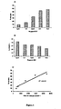
- NK cells efficiently lysed CD20 + Daudi target cells in the presence of rituximab (chimeric anti-CD20 antibody) at a concentration of 2 ⁇ g / ml in a 4h 51 Cr cytotoxicity test (Dall'Ozzo et al., 2004). It has recently been shown that the expression of CD107 on cytotoxic cells makes it possible to evaluate the degranulation thereof and represents an alternative measure to cytotoxicity (Alter et al., 2004, Betts et al., 2003, Betts et al. al., 2004).
- the Figure 1B shows that only a fraction (in all cases ⁇ 50%) of CD56 + NK cells expressed CD107 after 4 hours of incubation. Moreover, unlike the percentages of lysis presented in the Figure 1A , the percentages of CD56 + CD107 + cells decreased with increasing E: C ratio. However, the absolute number of CD107 + NK cells calculated from the number of NK cells initially added for each E: C ratio correlated well with percentage of lysis of Daudi cells for a preparation of NK cells from a given donor.
- EXAMPLE 2 Measurement of the ADCP exerted by the NK cells with respect to the Daudi cells in the presence of rituximab by the assay of the secreted IFN ⁇ and by the detection of the producing cells.
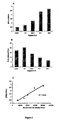
- NK cells In order to study whether ADCP and ADCC can be exerted simultaneously by NK cells, the production of IFN ⁇ by NK cells was evaluated under the previous experimental conditions: 10 4 to 2 ⁇ 10 5 purified NK cells of one same donor were incubated for 4h at 37 ° C in the presence of 2 ⁇ g / ml of rituximab with 2x10 4 unlabeled Daudi cells. The assay of IFN ⁇ was performed in the supernatant by an ELISA technique. As shown in Figure 2A and as was the case for lysis percentages, the concentration of IFN ⁇ in the supernatant increased with the E: C ratio.
- NK cells from the same donor were incubated under identical experimental conditions, but in the presence of brefeldin A which blocks the mechanisms of cellular transport, and therefore secretion.
- the cells were then labeled with a PC5 conjugated anti-CD56 mAb, permeabilized and incubated with a PE conjugated anti-IFN ⁇ mAb, and the percentage of NK cells expressing intracellular IFN ⁇ was evaluated by flow cytometry. after the published techniques (Mendes et al., 2000, Vitale et al., 2000). In the absence of brefeldin A, no CD56 + IFN ⁇ + cell was detected (results not shown).
- Figure 2B shows that a fraction of CD56 + cells was IFN ⁇ + after 4 hours of incubation in the presence of brefeldin A. However, this fraction was relatively low (in all cases ⁇ 25%).
- the percentages of CD56 + IFN ⁇ + cells decreased when the E: C ratio increased between 1.25: 1 and 10: 1.
- the absolute number of NK IFN ⁇ + cells calculated from the number of NK cells initially added for each E: C ratio in the presence of brefeldin A is well correlated with the concentration of IFN ⁇ produced by NK cells from the same donor in the absence of brefeldin A.
- NK cells For this purpose, purified NK cells were incubated for 4 h at 37 ° C. with Daudi cells in the presence of brefeldin A, anti-CD107 mAb and in the absence or in the presence of 2 ⁇ g / ml of rituximab or of Mouse mAb anti-CD16 3G8. This is used to perform a redirected pacing test. Indeed, the Daudi cells express the CD32 / Fc ⁇ RII molecule which has an affinity for the Fc portion of murine mAb IgG1 isotype such as 3G8.
- Targeted mAb 3G8 can recruit by its recognition site the NK cells that express CD16 / Fc ⁇ RIIIa. This binding of AcMo on Fc ⁇ RIIIa / CD16 activates the effector cell and induces its response vis-à-vis the target.
- the high affinity of 3G8 for Fc ⁇ RIIIa / CD16 allows optimal stimulation (Dall'Ozzo et al., 2004).
- the cells were then labeled with anti-CD56 mAb, permeabilized and incubated with anti-IFN ⁇ mAb and analyzed by flow cytometry.
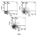
- cytograms of the figure 4 show that no NK CD107 + or IFN ⁇ + cell was detected in the absence of stimulant (cytogram A). At In contrast, significant percentages of CD107 + NK cells and NK IFN ⁇ + cells are observed after stimulation in the presence of rituximab or 3G8 (cytogram B and C). If a fraction of NK cells is both CD107 + and IFN ⁇ + (right upper quadrant), showing that ADCC and ADCP are not mutually exclusive, the majority of responder cells are exclusively CD107 + (upper left quadrant) or exclusively IFN ⁇ + (lower right quadrant). The results obtained with rituximab and 3G8 were comparable. It should be noted that in these experimental conditions, the responder cells represented only a minor fraction of the NK cells.
- NK cells that degranulate and those that synthesize IFN ⁇ belong to populations at least partly different.
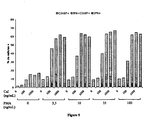
- purified NK cells were stimulated by different concentrations of calcium ionophore (CaI) A23187 and phorbol myristate acetate (PMA). used alone or in combination. Indeed, it has been reported that stimulation by this combination on the one hand allows the degranulation of NK cells (Atkinson et al., 1990) and on the other hand the synthesis of cytokines (Mendes et al., 2000).
- CaI calcium ionophore
- PMA phorbol myristate acetate
- the activation was carried out in a culture plate, where the NK cells were incubated for 4 h at 37 ° C. under CO 2 , in the presence of the anti-CD107 mAb, and then the cells were permeabilized, labeled with the Anti-IFN ⁇ mAb and analyzed by flow cytometry.
- NK IFN ⁇ + cell was detected after stimulation with CaI in the absence of PMA or with PMA in the absence of CaI ( Figure 5 ).
- stimulation of cells with CaI concentrations greater than 100 ng / ml induced expression of CD107 on NK.
- the expression was maximal (around 15% of CD107 + cells) as soon as the concentration of 330 ng / ml of CaI was reached.
- stimulation at concentrations of 3.3-100 ng / ml PMA induced expression of CD107 on approximately 10% of NK cells.
- the expression pattern of both CD107 and IFN ⁇ was identical for all concentrations of PMA used (from 3.3 to 100 ng / ml).
- the kinetics of expression of CD107 and intracellular IFN ⁇ was also studied after stimulation of the cells by a combination of 330 ng / ml of CaI and 33 ng / ml of PMA.
- Purified NK cells were stimulated in the presence of brefeldin A and anti-CD107 mAb. After incubation times of between 0 and 4 h, the cells were permeabilized and then labeled with anti-IFN ⁇ mAb and analyzed by flow cytometry.
- the figure 6 shows that the expression of CD107 on NK cells was already significant after the first hour of stimulation and that the percentages then increased steadily with time.
- NK IFN ⁇ + cells were detected after 2h of stimulation and the percentages obtained did not increase after higher stimulation times.
- NK cells were incubated for 4h at 37 ° C in the presence of brefeldin A and anti-CD107 AeMo in wells.
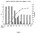
- the amounts of 3G8 actually immobilized at the bottom of the culture plate wells were evaluated by ELISA using an enzyme-labeled mouse anti-IgG antibody.
- the results are presented in absorbance values ( Figure 7 : left ordinate) with respect to the concentration of 3G8 used during the adsorption ( Figure 7 : abscissa).
- the cells were then labeled with FITC-conjugated anti-CD16 AcMo.
- the figure 7 shows that the saturation of the wells of the plate with AcMo 3G8 was obtained for a concentration of the order of 1 ⁇ g / ml.
- a decrease in the membrane expression of CD16 was observed from 0.1 ⁇ g / ml of 3G8, which represents the concentration involved approximately 50% saturation.
- the membrane expression of CD16 then decreased very strongly with the increase in concentration (approximately 15% of cells positive to 0.3 ⁇ g / ml corresponding to approximately 85% of the saturation) to become undetectable at saturating concentrations.
- NK cells lost the expression of CD16 and were therefore activated, after incubation in wells sensitized by a saturating concentration of 3G8.
- CD107 + and / or IFN ⁇ + NK cells were detected after incubation in wells sensitized with a concentration of 0.1 ⁇ g / ml.
- the percentages of NK CD107 + and / or IFN ⁇ + cells then increased with increasing concentration of 3G8 to reach a plateau at a concentration of 0.3 ⁇ g / ml.
- the three populations of responder cells were again observed under these stimulation conditions.
- Example 6 Comparative study of plaque sensitization by several therapeutic mAbs and by human IgGs
- the plates were sensitized with 5 ⁇ g / ml of each of the AcMor or human IgG and then incubated with concentrations of between 0.01 and 10 ⁇ g / mL of human C1q that binds to the CH2 domain of the Fc portion.
- C1q uptake was revealed by ELISA using a peroxidase-coupled human anti-C1q sheep's Ac.
- the figure 9 shows for each of the products tested the profile of the absorbance and therefore the fixing of C1q as a function of the concentration thereof.
- the functional response of cells expressing Fc ⁇ RIIIa to an AcMor-sensitized target depends on the affinity of the receptor for AcMor (Dall'Ozzo et al., 2004).
- the ability of concentrations between 0.001 and 10 mg / mL of each of the mAbs or polyvalent IgG, to inhibit the binding of FITC-conjugated anti-CD16 3G8 mAb to purified NK cells of a donor was studied by flow cytometry as previously described (Dall'Ozzo et al., 2004).
- the figure 10 shows, for each of the products tested, the inhibition of 3G8 binding as a function of the concentration of the product tested.
- inhibition of 3G8 binding increased with concentration to a plateau in a sigmoidal pattern.
- the percentages of inhibition observed depended strongly on the product tested.
- the concentration C1 of infliximab for inhibiting 50% of the binding of 3G8 is about 20-fold lower than the C2 concentration of basiliximab producing the same effect, reflecting the best affinity of the first for the Fc ⁇ RIIIa expressed on NK cells.
- the therapeutic mAbs sold are all IgG1 and therefore have a conserved sequence Fc portion, they have variable affinities for Fc ⁇ RIIIa.
- NK cells To compare the responses of NK cells to the different mAbM, human IgG, anti-lymphocyte serum and anti-CD16 mAb 3G8, the purified NK cells were incubated for 4 hours at 37 ° C in the presence of brefeldin A and the anti-CD107 mAb in culture plate wells, previously sensitized or not with a saturating concentration ( ⁇ 3 ⁇ g / ml) of each of the mAb or polyvalent IgG or IgG1 or IgG3 or anti-serum -lymphocytic or ACMo anti-CD16 3G8. The cells were then labeled with an anti-CD16 mAb.
- the cells were finally permeabilized, incubated with anti-IFN ⁇ mAb and analyzed by flow cytometry.
- the figure 11 shows for each of the products tested the average percentage of NK cells which had a loss of CD16 membrane expression on the one hand and the average percentages of CD107 + and / or IFN ⁇ + cells on the other hand (results ranked in increasing order of loss). of CD16 and obtained in 8 different experiments).
- NK cells incubated in non-sensitized wells did not detectably lose CD16 and did not express intracellular CD107 or IFN ⁇ . It was the same for the NK cells incubated in wells sensitized by abciximab. For all other products the two responses were detected.
- the percentage of cells that had a functional response was always lower than the percentage of cells activated (percentage of cells having lost CD16).
- the percentages of the cells that had a functional response were very well correlated with the percentages of the activated cells.
- As has been shown previously in the figure 7 nearly 100% of the NK cells incubated in wells sensitized with anti-CD16 3G8 mAb have lost the expression of CD16 and have therefore been activated.
- About 1/3 of the NK cells also had a functional response, of which a majority (nearly 85%) expressed CD107 alone or in combination with intracellular IFN ⁇ .
- NK cells incubated in wells sensitized by anti-lymphocyte serum Similar results were observed with NK cells incubated in wells sensitized by anti-lymphocyte serum. NK cells incubated in wells sensitized with human IgG3 had a very low CD16 loss and functional response.
- Basiliximab induced the weakest responses (on average slightly more than 20% of cells that lost CD16), then cetuximab and rituximab (on average just under 30% of cells that lost CD16), then trastuzumab, palivizumab, adalimumab, infliximab and bevacizumab resulted in stronger responses similar to those obtained with polyvalent IgG and human IgG1 (on average between 36.5% and 43.4% of cells CD16). Finally, alemtuzumab induced a massive response similar to that observed with 3G8 (on average 78.5% of cells that lost CD16). Among the NK cells that had a functional response, a majority expressed CD107 alone or associated with IFN ⁇ .
- the individual results of activation and functional response of 3 among the 8 experiments of the figure 11 are presented in the figure 12 .
- the NK cells of the 3 donors had no or very low responses after incubation in non-sensitized or sensitized wells with abciximab and presented very large responses after incubation in the wells sensitized with anti-CD16 mAb 3.G8, the antiserum serum. lymphocyte and alemtuzumab. Responses to other tested products varied greatly with the donor.
- the NK cells of the first donor had a much lower CD16 loss (from 1% for IgG3 to 27% for IgG1) and also very weak functional responses.
- the NK cells of the second donor had a much higher than average loss of C16 (from 17% for IgG3 to 82% for IgG1) and intermediate functional responses.
- a minority expressed CD107 alone or associated with IFN ⁇ including after stimulation with alemtuzumab.
- the NK cells of the third donor had a loss of CD16 (from 22% for IgG3 to 57% for IgG1) and functional responses slightly above average.
- NK cells of this donor that had a functional response, a large majority expressed CD107 alone or associated with IFN ⁇ regardless of the product tested.
- the activation and functional responses of NK cells after incubation in plates sensitized by a saturating concentration of certain mAb or human IgG, vary qualitatively and quantitatively depending on the donor of the cells.
- the PBMCs and the NK cells of the same donor were prepared and incubated for 4 hours at 37 ° C. in the presence of brefeldin A and anti-AcMo.
- CD107 in wells of a culture plate previously sensitized or not with a saturating concentration of rituximab, infliximab, alemtuzumab or anti-CD16 3G8 mAb.
- the cells were then labeled with an anti-CD56 mAb.
- the cells were finally permeabilized, incubated with anti-IFN ⁇ mAb and analyzed by flow cytometry.
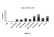
- the figure 13 watch for each of the products tested, the percentages of CD107 + and / or IFN ⁇ + cells among NK CD56 + cells and among CD56 + PBMCs.
- the percentages of responder cells obtained with purified NK cells and with CD56 + PBMCs were comparable.
- EXAMPLE 12 Study of the Loss of CD16, Degranulation and Synthesis of IFN ⁇ by NK Cells Incubated in Co-Sensitized Plates by 3G8 or Infliximab and by AcMo against Activating Molecules co-activation of NK cells.
- NKp30, NKp46, and NKG2D receptors have been shown to be involved in natural cytotoxicity (Pende et al., 2001), and receptors such as CD2, 2B4 (CD244) or LFA-1 (CD11a / CD18) have been implicated in co-activation of the NK cell (Bryceson et al., 2006, Natarajan et al., 2002).
- NKG2D appears to have a role in the natural cytokine synthesis by NK cells (Andre et al., 2004).
- the CD45 membrane molecule appears to be essential for the production of cytokines by mouse NK cells but not for the cytotoxicity exerted by these same cells (Hesslein et al., 2006, Huntington et al., 2005).
- plaques sensitized by antibodies directed against these molecules or against the C56 adhesion molecule (which does not has no co-activating activity) or against a murine IgG1 and co-sensitized or not with anti-CD16 3G8 antibody or with infliximab (1.5 ⁇ g / mL) were used. Stimulation and markings were performed as described in Example 9.
- the figure 14A shows the percentages of NK CD16 +, CD107 + and IFN ⁇ + cells from a donor after incubation in non-sensitized plates or sensitized only by the control IgG1 or by the mAbs directed against CD56, NKG2D, NKP30, CD11a CD244, CD45 or CD2.
- NK cells incubated under these conditions did not detectably lose CD16 and expressed neither CD107 (except for very low expression after incubation in the presence of anti-NKP30 mAb) nor Intracellular IFN ⁇ .
- NK cells 13.6% of the NK cells were CD107 + after incubation in murine IgG1 co-sensitized wells against approximately 20% of the cells incubated in Co-sensitized wells against CD56, CU11a, CD244, CD45 and CD45.
- NK cells were IFN- ⁇ + after incubation in murine IgG1-co-sensitized wells, or against CD56, NKG2D, CD45 and CD2 mAbs against only 2% of the cells.
- Natural killer cell cytotoxicity of breast cancer targets is enhanced by two distinct mechanisms of antibody-dependent cellular cytotoxicity against LFA-3 and HER2 / neu. Exp Hematol 27, 1533-1541 .
- IFN-gamma inhibits the proliferation of murine bone marrow cells stimulated with IL-3, IL-4, or granulocyte-macrophage colony-stimulating factor. J Immunol 141, 2635-2642 .
- NK cell natural cytotoxicity and IFN-gamma production are not always coordinately regulated: commitment of DX9 KIR + NK cells by HLA-B7 variants and target cells. J Immunol 160, 1573-1580 .
Landscapes
- Health & Medical Sciences (AREA)
- Life Sciences & Earth Sciences (AREA)
- Immunology (AREA)
- Engineering & Computer Science (AREA)
- Chemical & Material Sciences (AREA)
- Biomedical Technology (AREA)
- Molecular Biology (AREA)
- Urology & Nephrology (AREA)
- Hematology (AREA)
- Cell Biology (AREA)
- Medicinal Chemistry (AREA)
- General Physics & Mathematics (AREA)
- Biotechnology (AREA)
- Food Science & Technology (AREA)
- Pathology (AREA)
- Physics & Mathematics (AREA)
- Analytical Chemistry (AREA)
- Biochemistry (AREA)
- General Health & Medical Sciences (AREA)
- Microbiology (AREA)
- Chemical Kinetics & Catalysis (AREA)
- Bioinformatics & Cheminformatics (AREA)
- Toxicology (AREA)
- Tropical Medicine & Parasitology (AREA)
- Peptides Or Proteins (AREA)
- Preparation Of Compounds By Using Micro-Organisms (AREA)
- Medicines Containing Antibodies Or Antigens For Use As Internal Diagnostic Agents (AREA)
- Measuring Or Testing Involving Enzymes Or Micro-Organisms (AREA)
Claims (17)
- Verfahren zur in vitro-Bewertung der Effektorfunktionen von FcγRIIIa exprimierenden Zellen im Ansprechen auf einen zu testenden monoklonalen Antikörper, wobei das Verfahren zumindest die folgenden Schritte umfasst:(i) die FcγRIIIa exprimierenden Zellen werden in Gegenwart eines die Sekretion von Zytokinen durch diese Zellen hemmenden Agens mit dem monoklonalen Antikörper in Berührung gebracht, der auf einem Träger immobilisiert ist;(ii) als Positivkontrolle für die Aktivierung der FcγRIIIa exprimierenden Zellen wird der gleiche Versuch durchgeführt, indem anstelle des zu untersuchenden monoklonalen Antikörpers ein gegen den FcγRIIIa-Rezeptor gerichteter monoklonaler Antikörper verwendet wird;(iii) nach einer Inkubationsdauer von mindestens 1 h wird das Ansprechen der FcγRIIIa exprimierenden Zellen untersucht, indem das Vorhandensein des CD107-Markers an der Zelloberfläche sowie das Vorhandensein von intrazellulärem Gamma-Interferon (IFNγ) und/oder von intrazellulärem Tumornekrosefaktor-Alpha (TNFα) gemessen wird.
- Verfahren nach Anspruch 1, wobei die FcγRIIIa exprimierenden Zellen NK-Zellen oder T-Lymphozyten sind.
- Verfahren nach einem der Ansprüche 1 bis 2, wobei die Inkubation in Gegenwart eines an einen Marker gekoppelten monoklonalen anti-CD107 Antikörpers durchgeführt wird.
- Verfahren nach einem der Ansprüche 1 bis 3, wobei die Zellen am Ende der Inkubation permeabilisiert werden, um Gamma-Interferon und/oder TNFα unter Verwendung eines an einen Marker gekoppelten monoklonalen anti-IFNγ Antikörpers und/oder eines an einen Marker gekoppelten monoklonalen anti-TNFα Antikörpers zu messen.
- Verfahren nach einem der Ansprüche 1 bis 4, welches ferner einen Schritt des Messens der Expression von FcγRIIIa / CD16a an der Oberfläche der Zellen mittels Durchflusszytometrie umfasst, nachdem die Zellen am Ende der Inkubation mit einem an einen Marker gekoppelten monoklonalen anti-CD16 Antikörper markiert wurden.
- Verfahren nach einem der Ansprüche 1 bis 5, wobei die an die monoklonalen anti-CD107, anti-IFNγ, anti-TNFα und anti-CD16 Antikörper gekoppelten Marker voneinander verschiedene Fluorochrome sind.
- Verfahren nach einem der Ansprüche 1 bis 6, welches ferner als Positivkontrolle für die Ansprechfähigkeit der FcγRIIIa exprimierenden Zellen einen Schritt (ii-bis) des Stimulierens dieser Zellen mittels einer Mischung aus einem Phorbolester und einem Calcium-Ionophor umfasst.
- Verfahren nach einem der Ansprüche 1 bis 7, wobei nach abgeschlossener Inkubation die folgende Sequenz ausgeführt wird:(iv) die FcγRIIIa exprimierenden Zellen werden mit einem an einen Marker gekoppelten anti-CD16 Antikörper markiert,(v) die FcγRIIIa exprimierenden Zellen werden permeabilisiert und daraufhin mit einem an einen Marker gekoppelten monoklonalen anti-IFNγ Antikörper und/oder mit einem an einen Marker gekoppelten monoklonalen anti-TNFα Antikörper markiert,(vi) die solcherart markierten Zellen werden mittels Durchflusszytometrie analysiert.
- Verfahren nach einem der Ansprüche 2 bis 8, wobei in den Schritten (i) und (ii) die NK-Zellen mit Antikörpern co-stimuliert werden, die gegen einen oder mehrere der Rezeptoren gerichtet sind, welche unter NKp30, NKp46, NKG2D, 2B4, CD2, CD45, DNAM und CD11a ausgewählt sind.
- Verwendung eines Verfahrens nach einem der Ansprüche 1 bis 9 zum Vorhersagen des Ansprechens eines Patienten auf eine Behandlung mit einem gegebenen monoklonalen Antikörper.
- Verwendung nach Anspruch 10, wobei das Verfahren mit FcγRIIIa exprimierenden Zellen durchgeführt wird, die vom Patienten stammen.
- Verwendung eines Verfahrens nach einem der Ansprüche 1 bis 9, zum Vergleichen der Fähigkeit verschiedener rekombinanter monoklonaler Antikörper zum Induzieren der von den Antikörpern abhängigen Zytotoxizität (Antibody-Dependent Cellular Cytotoxicity; ADCC) und der von den Antikörpern abhängigen Produktion von Zytokinen (Antibody-Dependent Cytokine Production; ADCP) durch FcγRIIIa exprimierende Zellen.
- Verwendung eines Verfahrens nach einem der Ansprüche 1 bis 9 als Forschungshilfsmittel zum Verbessern der Effektivität der rekombinanten monoklonalen Antikörper zu therapeutischen Zwecken.
- Verwendung eines Verfahrens nach einem der Ansprüche 1 bis 9 zum Durchführen einer Qualitätskontrolle an einer Probe von rekombinanten monoklonalen Antikörpern.
- Verwendung nach einem der Ansprüche 12 bis 14, wobei das Verfahren mit FcγRIIIa exprimierenden Zellen durchgeführt wird, die von gesunden Testpersonen und/oder von Kranken stammen.
- Verwendung nach einem der Ansprüche 12 bis 14, wobei das Verfahren mit einer oder mehreren Linien von NK-Zellen und/oder T-Lymphozyten durchgeführt wird.
- Kit, welcher für die Durchführung eines Verfahrens nach einem der Ansprüche 1 bis 9 verwendbar ist, wobei dieser Kit mindestens die folgenden Bestandteile aufweist:- einen an einen Marker gekoppelten monoklonalen anti-CD16 Antikörper,- einen an einen Marker gekoppelten monoklonalen anti-IFNγ Antikörper und/oder einen an einen Marker gekoppelten monoklonalen anti-TNFα Antikörper, und- eine Platte, welche mindestens eine Vertiefung aufweist, die mit einem gegen den FcγRIIIa-Rezeptor gerichteten monoklonalen Antikörper beschichtet ist.
Priority Applications (1)
| Application Number | Priority Date | Filing Date | Title |
|---|---|---|---|
| EP07825673A EP2062047B1 (de) | 2006-09-13 | 2007-09-12 | Verfahren zur untersuchung der reaktion auf eine behandlung mit einem monoklonalen antikörper |
Applications Claiming Priority (5)
| Application Number | Priority Date | Filing Date | Title |
|---|---|---|---|
| FR0607990 | 2006-09-13 | ||
| EP06291942A EP1933137A1 (de) | 2006-12-15 | 2006-12-15 | Verfahren zur Untersuchung der Reaktion auf eine Behandlung mit einem monoklonalen Antikörper |
| FR0700456 | 2007-01-23 | ||
| EP07825673A EP2062047B1 (de) | 2006-09-13 | 2007-09-12 | Verfahren zur untersuchung der reaktion auf eine behandlung mit einem monoklonalen antikörper |
| PCT/IB2007/003498 WO2008032217A2 (fr) | 2006-09-13 | 2007-09-12 | Methode d'investigation de la reponse a un traitement par un anticorps monoclonal |
Publications (2)
| Publication Number | Publication Date |
|---|---|
| EP2062047A2 EP2062047A2 (de) | 2009-05-27 |
| EP2062047B1 true EP2062047B1 (de) | 2011-03-09 |
Family
ID=39144376
Family Applications (1)
| Application Number | Title | Priority Date | Filing Date |
|---|---|---|---|
| EP07825673A Not-in-force EP2062047B1 (de) | 2006-09-13 | 2007-09-12 | Verfahren zur untersuchung der reaktion auf eine behandlung mit einem monoklonalen antikörper |
Country Status (5)
| Country | Link |
|---|---|
| US (1) | US20100167315A1 (de) |
| EP (1) | EP2062047B1 (de) |
| AT (1) | ATE501436T1 (de) |
| DE (1) | DE602007013068D1 (de) |
| WO (1) | WO2008032217A2 (de) |
Families Citing this family (10)
| Publication number | Priority date | Publication date | Assignee | Title |
|---|---|---|---|---|
| US20070087005A1 (en) | 2005-10-14 | 2007-04-19 | Lazar Gregory A | Anti-glypican-3 antibody |
| MX2014010379A (es) * | 2012-03-02 | 2015-05-15 | Roche Glycart Ag | Biomarcador predictivo para tratamiento de cancer con anticuerpos mejorados en adcc. |
| TWI693073B (zh) | 2012-12-21 | 2020-05-11 | 日商中外製藥股份有限公司 | 對gpc3標的治療劑療法為有效之患者投與的gpc3標的治療劑 |
| WO2014114595A1 (en) * | 2013-01-23 | 2014-07-31 | Roche Glycart Ag | Predictive biomarker for cancer treatment with adcc-enhanced antibodies |
| TWI735410B (zh) * | 2014-05-08 | 2021-08-11 | 日商中外製藥股份有限公司 | 對gpc3標的治療劑療法為有效之患者投予的gpc3標的治療劑 |
| WO2017002934A1 (ja) | 2015-07-01 | 2017-01-05 | 中外製薬株式会社 | Gpc3標的治療剤が有効である患者に投与されるgpc3標的治療剤 |
| CN115932265B (zh) * | 2016-05-30 | 2026-04-14 | 因塞特公司 | 预测cd19抗体治疗在患者中的治疗益处的方法 |
| AU2018333062B2 (en) | 2017-09-15 | 2022-03-17 | Beckman Coulter, Inc. | Flow based assays for therapeutics |
| CN108663510B (zh) * | 2018-04-28 | 2021-06-04 | 璞晞(广州)生物免疫技术有限公司 | 肝病nk细胞功能的检测评估方法及试剂盒 |
| CN113421627B (zh) * | 2021-05-11 | 2023-04-07 | 深圳市罗湖区人民医院 | 体外评估药物对nk细胞抗衰作用的系统 |
Family Cites Families (6)
| Publication number | Priority date | Publication date | Assignee | Title |
|---|---|---|---|---|
| FR2807767B1 (fr) * | 2000-04-12 | 2005-01-14 | Lab Francais Du Fractionnement | Anticorps monoclonaux anti-d |
| US20030124621A1 (en) * | 2000-07-04 | 2003-07-03 | Izumi Sugo | Method of assaying the function of fc fragment of antibody |
| US7595165B2 (en) * | 2002-09-13 | 2009-09-29 | Laboratoire Francais Du Fractionnement Et Des Biotechnologies | Method of measuring activation of effector cells |
| FR2844521B1 (fr) * | 2002-09-13 | 2004-12-24 | Lab Francais Du Fractionnement | Mesure de la production de cytokines comme marqueur d'activation de cellules effectrices |
| FR2844513B1 (fr) * | 2002-09-13 | 2007-08-03 | Lab Francais Du Fractionnement | Anticorps pour adcc et induisant la production de cytokines. |
| US20080003225A1 (en) * | 2006-06-29 | 2008-01-03 | Henri Vie | Method for enhancing the antibody-dependent cellular cytotoxicity (ADCC) and uses of T cells expressing CD16 receptors |
-
2007
- 2007-09-12 AT AT07825673T patent/ATE501436T1/de not_active IP Right Cessation
- 2007-09-12 EP EP07825673A patent/EP2062047B1/de not_active Not-in-force
- 2007-09-12 WO PCT/IB2007/003498 patent/WO2008032217A2/fr not_active Ceased
- 2007-09-12 US US12/441,237 patent/US20100167315A1/en not_active Abandoned
- 2007-09-12 DE DE602007013068T patent/DE602007013068D1/de active Active
Also Published As
| Publication number | Publication date |
|---|---|
| WO2008032217A2 (fr) | 2008-03-20 |
| US20100167315A1 (en) | 2010-07-01 |
| DE602007013068D1 (de) | 2011-04-21 |
| WO2008032217A3 (fr) | 2008-06-05 |
| EP2062047A2 (de) | 2009-05-27 |
| ATE501436T1 (de) | 2011-03-15 |
Similar Documents
| Publication | Publication Date | Title |
|---|---|---|
| US20250066501A1 (en) | Immune Modulation and Treatment of Solid Tumors with Antibodies that Specifically Bind CD38 | |
| US20240417483A1 (en) | Monoclonal antibody and a method thereof | |
| EP2062047A2 (de) | Verfahren zur untersuchung der reaktion auf eine behandlung mit einem monoklonalen antikörper | |
| KR102713847B1 (ko) | Cd38과 특이적으로 결합하는 항체에 의한 고형 종양의 면역 조절 및 치료 | |
| CN116744962A (zh) | Cd25抗体 | |
| TW202116807A (zh) | 用於治療特定患者之癌症的抗體組合 | |
| Besla et al. | T cell–dependent bispecific therapy enhances innate immune activation and antibody-mediated killing | |
| US11391726B2 (en) | MoT cells as a therapeutic screening tool for regulatory t-cell activity | |
| EP1933137A1 (de) | Verfahren zur Untersuchung der Reaktion auf eine Behandlung mit einem monoklonalen Antikörper | |
| TWI742008B (zh) | 使用特異性結合cd38之抗體免疫調節及治療固態腫瘤 | |
| Madsen et al. | Development of a flow cytometry-based potency assay for prediction of cytokine storms induced by biosimilar monoclonal antibodies | |
| JP2023553634A (ja) | 治療用のcar t細胞を特徴付けるための方法および試薬 | |
| CN118284621A (zh) | 抗flt3抗体、car、car t细胞和使用方法 | |
| EP3729085B1 (de) | Quantitatives zelluläres verfahren zur bestimmung der biologischen aktivität eines anti-cd26-liganden | |
| TWI917318B (zh) | 靶向dll3的嵌合抗原受體和結合劑 | |
| Rhys Thomas | Studies of Glofitamab, a CD3xCD20 bispecific antibody, in B cell malignancies | |
| RU2778454C2 (ru) | Количественный клеточный способ определения биологической активности лиганда антитела против CD26 | |
| Wierz | Characterization of the Tumor Microenvironment in Chronic Lymphocytic Leukemia by Mass Cytometry: Implications for Immunotherapy | |
| Arora | T Cells enhance Natural Killer Cell antibody dependent cellular cytotoxicity | |
| CN118974554A (zh) | 用于供体细胞分析的方法 | |
| HK40119794A (zh) | 用於供体细胞分析的方法 | |
| Karampatzakis | CD16 shedding regulates serial killing and responses to immunotherapies | |
| CA3254552A1 (en) | METHODS FOR ANALYZING DONOR CELLS | |
| HK1254148A1 (en) | Immune modulation and treatment of solid tumors with antibodies that specifically bind cd38 |
Legal Events
| Date | Code | Title | Description |
|---|---|---|---|
| PUAI | Public reference made under article 153(3) epc to a published international application that has entered the european phase |
Free format text: ORIGINAL CODE: 0009012 |
|
| 17P | Request for examination filed |
Effective date: 20090330 |
|
| AK | Designated contracting states |
Kind code of ref document: A2 Designated state(s): AT BE BG CH CY CZ DE DK EE ES FI FR GB GR HU IE IS IT LI LT LU LV MC MT NL PL PT RO SE SI SK TR |
|
| AX | Request for extension of the european patent |
Extension state: AL BA HR MK RS |
|
| DAX | Request for extension of the european patent (deleted) | ||
| GRAP | Despatch of communication of intention to grant a patent |
Free format text: ORIGINAL CODE: EPIDOSNIGR1 |
|
| GRAS | Grant fee paid |
Free format text: ORIGINAL CODE: EPIDOSNIGR3 |
|
| GRAA | (expected) grant |
Free format text: ORIGINAL CODE: 0009210 |
|
| AK | Designated contracting states |
Kind code of ref document: B1 Designated state(s): AT BE BG CH CY CZ DE DK EE ES FI FR GB GR HU IE IS IT LI LT LU LV MC MT NL PL PT RO SE SI SK TR |
|
| REG | Reference to a national code |
Ref country code: GB Ref legal event code: FG4D Free format text: NOT ENGLISH |
|
| REG | Reference to a national code |
Ref country code: CH Ref legal event code: EP |
|
| REG | Reference to a national code |
Ref country code: IE Ref legal event code: FG4D Free format text: LANGUAGE OF EP DOCUMENT: FRENCH |
|
| REF | Corresponds to: |
Ref document number: 602007013068 Country of ref document: DE Date of ref document: 20110421 Kind code of ref document: P |
|
| REG | Reference to a national code |
Ref country code: DE Ref legal event code: R096 Ref document number: 602007013068 Country of ref document: DE Effective date: 20110421 |
|
| REG | Reference to a national code |
Ref country code: CH Ref legal event code: NV Representative=s name: ISLER & PEDRAZZINI AG |
|
| REG | Reference to a national code |
Ref country code: NL Ref legal event code: VDEP Effective date: 20110309 |
|
| PG25 | Lapsed in a contracting state [announced via postgrant information from national office to epo] |
Ref country code: LV Free format text: LAPSE BECAUSE OF FAILURE TO SUBMIT A TRANSLATION OF THE DESCRIPTION OR TO PAY THE FEE WITHIN THE PRESCRIBED TIME-LIMIT Effective date: 20110309 Ref country code: LT Free format text: LAPSE BECAUSE OF FAILURE TO SUBMIT A TRANSLATION OF THE DESCRIPTION OR TO PAY THE FEE WITHIN THE PRESCRIBED TIME-LIMIT Effective date: 20110309 Ref country code: ES Free format text: LAPSE BECAUSE OF FAILURE TO SUBMIT A TRANSLATION OF THE DESCRIPTION OR TO PAY THE FEE WITHIN THE PRESCRIBED TIME-LIMIT Effective date: 20110620 Ref country code: GR Free format text: LAPSE BECAUSE OF FAILURE TO SUBMIT A TRANSLATION OF THE DESCRIPTION OR TO PAY THE FEE WITHIN THE PRESCRIBED TIME-LIMIT Effective date: 20110610 Ref country code: SE Free format text: LAPSE BECAUSE OF FAILURE TO SUBMIT A TRANSLATION OF THE DESCRIPTION OR TO PAY THE FEE WITHIN THE PRESCRIBED TIME-LIMIT Effective date: 20110309 |
|
| LTIE | Lt: invalidation of european patent or patent extension |
Effective date: 20110309 |
|
| PG25 | Lapsed in a contracting state [announced via postgrant information from national office to epo] |
Ref country code: NL Free format text: LAPSE BECAUSE OF FAILURE TO SUBMIT A TRANSLATION OF THE DESCRIPTION OR TO PAY THE FEE WITHIN THE PRESCRIBED TIME-LIMIT Effective date: 20110309 Ref country code: SI Free format text: LAPSE BECAUSE OF FAILURE TO SUBMIT A TRANSLATION OF THE DESCRIPTION OR TO PAY THE FEE WITHIN THE PRESCRIBED TIME-LIMIT Effective date: 20110309 Ref country code: BG Free format text: LAPSE BECAUSE OF FAILURE TO SUBMIT A TRANSLATION OF THE DESCRIPTION OR TO PAY THE FEE WITHIN THE PRESCRIBED TIME-LIMIT Effective date: 20110609 Ref country code: CY Free format text: LAPSE BECAUSE OF FAILURE TO SUBMIT A TRANSLATION OF THE DESCRIPTION OR TO PAY THE FEE WITHIN THE PRESCRIBED TIME-LIMIT Effective date: 20110309 Ref country code: FI Free format text: LAPSE BECAUSE OF FAILURE TO SUBMIT A TRANSLATION OF THE DESCRIPTION OR TO PAY THE FEE WITHIN THE PRESCRIBED TIME-LIMIT Effective date: 20110309 Ref country code: AT Free format text: LAPSE BECAUSE OF FAILURE TO SUBMIT A TRANSLATION OF THE DESCRIPTION OR TO PAY THE FEE WITHIN THE PRESCRIBED TIME-LIMIT Effective date: 20110309 |
|
| REG | Reference to a national code |
Ref country code: IE Ref legal event code: FD4D |
|
| PG25 | Lapsed in a contracting state [announced via postgrant information from national office to epo] |
Ref country code: IE Free format text: LAPSE BECAUSE OF FAILURE TO SUBMIT A TRANSLATION OF THE DESCRIPTION OR TO PAY THE FEE WITHIN THE PRESCRIBED TIME-LIMIT Effective date: 20110309 Ref country code: PT Free format text: LAPSE BECAUSE OF FAILURE TO SUBMIT A TRANSLATION OF THE DESCRIPTION OR TO PAY THE FEE WITHIN THE PRESCRIBED TIME-LIMIT Effective date: 20110711 Ref country code: EE Free format text: LAPSE BECAUSE OF FAILURE TO SUBMIT A TRANSLATION OF THE DESCRIPTION OR TO PAY THE FEE WITHIN THE PRESCRIBED TIME-LIMIT Effective date: 20110309 |
|
| PG25 | Lapsed in a contracting state [announced via postgrant information from national office to epo] |
Ref country code: IS Free format text: LAPSE BECAUSE OF FAILURE TO SUBMIT A TRANSLATION OF THE DESCRIPTION OR TO PAY THE FEE WITHIN THE PRESCRIBED TIME-LIMIT Effective date: 20110709 Ref country code: CZ Free format text: LAPSE BECAUSE OF FAILURE TO SUBMIT A TRANSLATION OF THE DESCRIPTION OR TO PAY THE FEE WITHIN THE PRESCRIBED TIME-LIMIT Effective date: 20110309 Ref country code: SK Free format text: LAPSE BECAUSE OF FAILURE TO SUBMIT A TRANSLATION OF THE DESCRIPTION OR TO PAY THE FEE WITHIN THE PRESCRIBED TIME-LIMIT Effective date: 20110309 Ref country code: RO Free format text: LAPSE BECAUSE OF FAILURE TO SUBMIT A TRANSLATION OF THE DESCRIPTION OR TO PAY THE FEE WITHIN THE PRESCRIBED TIME-LIMIT Effective date: 20110309 |
|
| PLBE | No opposition filed within time limit |
Free format text: ORIGINAL CODE: 0009261 |
|
| STAA | Information on the status of an ep patent application or granted ep patent |
Free format text: STATUS: NO OPPOSITION FILED WITHIN TIME LIMIT |
|
| 26N | No opposition filed |
Effective date: 20111212 |
|
| PG25 | Lapsed in a contracting state [announced via postgrant information from national office to epo] |
Ref country code: PL Free format text: LAPSE BECAUSE OF FAILURE TO SUBMIT A TRANSLATION OF THE DESCRIPTION OR TO PAY THE FEE WITHIN THE PRESCRIBED TIME-LIMIT Effective date: 20110309 Ref country code: DK Free format text: LAPSE BECAUSE OF FAILURE TO SUBMIT A TRANSLATION OF THE DESCRIPTION OR TO PAY THE FEE WITHIN THE PRESCRIBED TIME-LIMIT Effective date: 20110309 |
|
| BERE | Be: lapsed |
Owner name: GLYCODE Effective date: 20110930 Owner name: UNIVERSITE FRANCOIS RABELAIS DE TOURS Effective date: 20110930 |
|
| REG | Reference to a national code |
Ref country code: DE Ref legal event code: R097 Ref document number: 602007013068 Country of ref document: DE Effective date: 20111212 |
|
| PG25 | Lapsed in a contracting state [announced via postgrant information from national office to epo] |
Ref country code: MC Free format text: LAPSE BECAUSE OF NON-PAYMENT OF DUE FEES Effective date: 20110930 |
|
| PG25 | Lapsed in a contracting state [announced via postgrant information from national office to epo] |
Ref country code: IT Free format text: LAPSE BECAUSE OF FAILURE TO SUBMIT A TRANSLATION OF THE DESCRIPTION OR TO PAY THE FEE WITHIN THE PRESCRIBED TIME-LIMIT Effective date: 20110309 |
|
| PG25 | Lapsed in a contracting state [announced via postgrant information from national office to epo] |
Ref country code: BE Free format text: LAPSE BECAUSE OF NON-PAYMENT OF DUE FEES Effective date: 20110930 |
|
| PG25 | Lapsed in a contracting state [announced via postgrant information from national office to epo] |
Ref country code: MT Free format text: LAPSE BECAUSE OF FAILURE TO SUBMIT A TRANSLATION OF THE DESCRIPTION OR TO PAY THE FEE WITHIN THE PRESCRIBED TIME-LIMIT Effective date: 20110309 |
|
| PG25 | Lapsed in a contracting state [announced via postgrant information from national office to epo] |
Ref country code: LU Free format text: LAPSE BECAUSE OF NON-PAYMENT OF DUE FEES Effective date: 20110912 |
|
| PG25 | Lapsed in a contracting state [announced via postgrant information from national office to epo] |
Ref country code: TR Free format text: LAPSE BECAUSE OF FAILURE TO SUBMIT A TRANSLATION OF THE DESCRIPTION OR TO PAY THE FEE WITHIN THE PRESCRIBED TIME-LIMIT Effective date: 20110309 |
|
| PG25 | Lapsed in a contracting state [announced via postgrant information from national office to epo] |
Ref country code: HU Free format text: LAPSE BECAUSE OF FAILURE TO SUBMIT A TRANSLATION OF THE DESCRIPTION OR TO PAY THE FEE WITHIN THE PRESCRIBED TIME-LIMIT Effective date: 20110309 |
|
| REG | Reference to a national code |
Ref country code: DE Ref legal event code: R082 Ref document number: 602007013068 Country of ref document: DE Representative=s name: WINTER, BRANDL, FUERNISS, HUEBNER, ROESS, KAIS, DE |
|
| REG | Reference to a national code |
Ref country code: CH Ref legal event code: PUEA Owner name: UNIVERSITE FRANCOIS RABELAIS, FR Free format text: FORMER OWNER: GLYCODE, FR |
|
| REG | Reference to a national code |
Ref country code: DE Ref legal event code: R081 Ref document number: 602007013068 Country of ref document: DE Owner name: UNIVERSITE FRANCOIS-RABELAIS, FR Free format text: FORMER OWNERS: GLYCODE, UZERCHE, FR; UNIVERSITE FRANCOIS RABELAIS DE TOURS, TOURS, FR Effective date: 20131108 Ref country code: DE Ref legal event code: R082 Ref document number: 602007013068 Country of ref document: DE Representative=s name: WINTER, BRANDL, FUERNISS, HUEBNER, ROESS, KAIS, DE Effective date: 20131128 Ref country code: DE Ref legal event code: R081 Ref document number: 602007013068 Country of ref document: DE Owner name: UNIVERSITE FRANCOIS-RABELAIS, FR Free format text: FORMER OWNER: GLYCODE, UNIVERSITE FRANCOIS RABELAIS DE, , FR Effective date: 20131108 Ref country code: DE Ref legal event code: R082 Ref document number: 602007013068 Country of ref document: DE Representative=s name: WINTER, BRANDL, FUERNISS, HUEBNER, ROESS, KAIS, DE Effective date: 20131108 Ref country code: DE Ref legal event code: R081 Ref document number: 602007013068 Country of ref document: DE Owner name: UNIVERSITE FRANCOIS-RABELAIS, FR Free format text: FORMER OWNER: L'UNIVERSITE FRANGOIS-RABELAIS, TOURS, FR Effective date: 20131128 |
|
| REG | Reference to a national code |
Ref country code: FR Ref legal event code: TP Owner name: UNIVERSITE FRANCOIS-RABELAIS, FR Effective date: 20131213 |
|
| REG | Reference to a national code |
Ref country code: GB Ref legal event code: 732E Free format text: REGISTERED BETWEEN 20150423 AND 20150430 |
|
| REG | Reference to a national code |
Ref country code: FR Ref legal event code: PLFP Year of fee payment: 9 |
|
| PGFP | Annual fee paid to national office [announced via postgrant information from national office to epo] |
Ref country code: DE Payment date: 20150922 Year of fee payment: 9 Ref country code: GB Payment date: 20150917 Year of fee payment: 9 Ref country code: CH Payment date: 20150918 Year of fee payment: 9 |
|
| PGFP | Annual fee paid to national office [announced via postgrant information from national office to epo] |
Ref country code: FR Payment date: 20150730 Year of fee payment: 9 |
|
| REG | Reference to a national code |
Ref country code: DE Ref legal event code: R119 Ref document number: 602007013068 Country of ref document: DE |
|
| REG | Reference to a national code |
Ref country code: CH Ref legal event code: PL |
|
| GBPC | Gb: european patent ceased through non-payment of renewal fee |
Effective date: 20160912 |
|
| REG | Reference to a national code |
Ref country code: FR Ref legal event code: ST Effective date: 20170531 |
|
| PG25 | Lapsed in a contracting state [announced via postgrant information from national office to epo] |
Ref country code: GB Free format text: LAPSE BECAUSE OF NON-PAYMENT OF DUE FEES Effective date: 20160912 Ref country code: LI Free format text: LAPSE BECAUSE OF NON-PAYMENT OF DUE FEES Effective date: 20160930 Ref country code: DE Free format text: LAPSE BECAUSE OF NON-PAYMENT OF DUE FEES Effective date: 20170401 Ref country code: FR Free format text: LAPSE BECAUSE OF NON-PAYMENT OF DUE FEES Effective date: 20160930 Ref country code: CH Free format text: LAPSE BECAUSE OF NON-PAYMENT OF DUE FEES Effective date: 20160930 |