EP2065402A1 - Trimerische Collagengerüst-Antikörper - Google Patents
Trimerische Collagengerüst-Antikörper Download PDFInfo
- Publication number
- EP2065402A1 EP2065402A1 EP08250284A EP08250284A EP2065402A1 EP 2065402 A1 EP2065402 A1 EP 2065402A1 EP 08250284 A EP08250284 A EP 08250284A EP 08250284 A EP08250284 A EP 08250284A EP 2065402 A1 EP2065402 A1 EP 2065402A1
- Authority
- EP
- European Patent Office
- Prior art keywords
- antibody
- domain
- soluble antibody
- trimeric soluble
- scfv
- Prior art date
- Legal status (The legal status is an assumption and is not a legal conclusion. Google has not performed a legal analysis and makes no representation as to the accuracy of the status listed.)
- Granted
Links
Images










Classifications
-
- C—CHEMISTRY; METALLURGY
- C07—ORGANIC CHEMISTRY
- C07K—PEPTIDES
- C07K14/00—Peptides having more than 20 amino acids; Gastrins; Somatostatins; Melanotropins; Derivatives thereof
- C07K14/435—Peptides having more than 20 amino acids; Gastrins; Somatostatins; Melanotropins; Derivatives thereof from animals; from humans
- C07K14/78—Connective tissue peptides, e.g. collagen, elastin, laminin, fibronectin, vitronectin or cold insoluble globulin [CIG]
-
- A—HUMAN NECESSITIES
- A61—MEDICAL OR VETERINARY SCIENCE; HYGIENE
- A61P—SPECIFIC THERAPEUTIC ACTIVITY OF CHEMICAL COMPOUNDS OR MEDICINAL PREPARATIONS
- A61P35/00—Antineoplastic agents
-
- C—CHEMISTRY; METALLURGY
- C07—ORGANIC CHEMISTRY
- C07K—PEPTIDES
- C07K16/00—Immunoglobulins [IG], e.g. monoclonal or polyclonal antibodies
- C07K16/18—Immunoglobulins [IG], e.g. monoclonal or polyclonal antibodies against material from animals or humans
- C07K16/24—Immunoglobulins [IG], e.g. monoclonal or polyclonal antibodies against material from animals or humans against cytokines, lymphokines or interferons
- C07K16/241—Tumor Necrosis Factors
-
- C—CHEMISTRY; METALLURGY
- C07—ORGANIC CHEMISTRY
- C07K—PEPTIDES
- C07K16/00—Immunoglobulins [IG], e.g. monoclonal or polyclonal antibodies
- C07K16/18—Immunoglobulins [IG], e.g. monoclonal or polyclonal antibodies against material from animals or humans
- C07K16/28—Immunoglobulins [IG], e.g. monoclonal or polyclonal antibodies against material from animals or humans against receptors, cell surface antigens or cell surface determinants
- C07K16/2803—Immunoglobulins [IG], e.g. monoclonal or polyclonal antibodies against material from animals or humans against receptors, cell surface antigens or cell surface determinants against the immunoglobulin superfamily
- C07K16/2809—Immunoglobulins [IG], e.g. monoclonal or polyclonal antibodies against material from animals or humans against receptors, cell surface antigens or cell surface determinants against the immunoglobulin superfamily against the T-cell receptor (TcR)-CD3 complex
-
- C—CHEMISTRY; METALLURGY
- C07—ORGANIC CHEMISTRY
- C07K—PEPTIDES
- C07K16/00—Immunoglobulins [IG], e.g. monoclonal or polyclonal antibodies
- C07K16/18—Immunoglobulins [IG], e.g. monoclonal or polyclonal antibodies against material from animals or humans
- C07K16/28—Immunoglobulins [IG], e.g. monoclonal or polyclonal antibodies against material from animals or humans against receptors, cell surface antigens or cell surface determinants
- C07K16/2863—Immunoglobulins [IG], e.g. monoclonal or polyclonal antibodies against material from animals or humans against receptors, cell surface antigens or cell surface determinants against receptors for growth factors, growth regulators
-
- C—CHEMISTRY; METALLURGY
- C07—ORGANIC CHEMISTRY
- C07K—PEPTIDES
- C07K16/00—Immunoglobulins [IG], e.g. monoclonal or polyclonal antibodies
- C07K16/18—Immunoglobulins [IG], e.g. monoclonal or polyclonal antibodies against material from animals or humans
- C07K16/32—Immunoglobulins [IG], e.g. monoclonal or polyclonal antibodies against material from animals or humans against translation products of oncogenes
-
- C—CHEMISTRY; METALLURGY
- C07—ORGANIC CHEMISTRY
- C07K—PEPTIDES
- C07K2317/00—Immunoglobulins specific features
- C07K2317/30—Immunoglobulins specific features characterized by aspects of specificity or valency
- C07K2317/31—Immunoglobulins specific features characterized by aspects of specificity or valency multispecific
-
- C—CHEMISTRY; METALLURGY
- C07—ORGANIC CHEMISTRY
- C07K—PEPTIDES
- C07K2317/00—Immunoglobulins specific features
- C07K2317/60—Immunoglobulins specific features characterized by non-natural combinations of immunoglobulin fragments
- C07K2317/62—Immunoglobulins specific features characterized by non-natural combinations of immunoglobulin fragments comprising only variable region components
- C07K2317/622—Single chain antibody (scFv)
-
- C—CHEMISTRY; METALLURGY
- C07—ORGANIC CHEMISTRY
- C07K—PEPTIDES
- C07K2317/00—Immunoglobulins specific features
- C07K2317/60—Immunoglobulins specific features characterized by non-natural combinations of immunoglobulin fragments
- C07K2317/62—Immunoglobulins specific features characterized by non-natural combinations of immunoglobulin fragments comprising only variable region components
- C07K2317/626—Diabody or triabody
-
- C—CHEMISTRY; METALLURGY
- C07—ORGANIC CHEMISTRY
- C07K—PEPTIDES
- C07K2317/00—Immunoglobulins specific features
- C07K2317/70—Immunoglobulins specific features characterized by effect upon binding to a cell or to an antigen
- C07K2317/73—Inducing cell death, e.g. apoptosis, necrosis or inhibition of cell proliferation
-
- C—CHEMISTRY; METALLURGY
- C07—ORGANIC CHEMISTRY
- C07K—PEPTIDES
- C07K2317/00—Immunoglobulins specific features
- C07K2317/70—Immunoglobulins specific features characterized by effect upon binding to a cell or to an antigen
- C07K2317/74—Inducing cell proliferation
-
- C—CHEMISTRY; METALLURGY
- C07—ORGANIC CHEMISTRY
- C07K—PEPTIDES
- C07K2317/00—Immunoglobulins specific features
- C07K2317/70—Immunoglobulins specific features characterized by effect upon binding to a cell or to an antigen
- C07K2317/76—Antagonist effect on antigen, e.g. neutralization or inhibition of binding
-
- C—CHEMISTRY; METALLURGY
- C07—ORGANIC CHEMISTRY
- C07K—PEPTIDES
- C07K2317/00—Immunoglobulins specific features
- C07K2317/90—Immunoglobulins specific features characterized by (pharmaco)kinetic aspects or by stability of the immunoglobulin
- C07K2317/92—Affinity (KD), association rate (Ka), dissociation rate (Kd) or EC50 value
-
- C—CHEMISTRY; METALLURGY
- C07—ORGANIC CHEMISTRY
- C07K—PEPTIDES
- C07K2318/00—Antibody mimetics or scaffolds
- C07K2318/10—Immunoglobulin or domain(s) thereof as scaffolds for inserted non-Ig peptide sequences, e.g. for vaccination purposes
-
- C—CHEMISTRY; METALLURGY
- C07—ORGANIC CHEMISTRY
- C07K—PEPTIDES
- C07K2318/00—Antibody mimetics or scaffolds
- C07K2318/20—Antigen-binding scaffold molecules wherein the scaffold is not an immunoglobulin variable region or antibody mimetics
-
- C—CHEMISTRY; METALLURGY
- C07—ORGANIC CHEMISTRY
- C07K—PEPTIDES
- C07K2319/00—Fusion polypeptide
-
- C—CHEMISTRY; METALLURGY
- C07—ORGANIC CHEMISTRY
- C07K—PEPTIDES
- C07K2319/00—Fusion polypeptide
- C07K2319/33—Fusion polypeptide fusions for targeting to specific cell types, e.g. tissue specific targeting, targeting of a bacterial subspecies
-
- C—CHEMISTRY; METALLURGY
- C07—ORGANIC CHEMISTRY
- C07K—PEPTIDES
- C07K2319/00—Fusion polypeptide
- C07K2319/70—Fusion polypeptide containing domain for protein-protein interaction
Definitions
- Protein-based binding reagents have various uses in therapeutic or diagnostic application. Antibodies have proven to be an excellent paradigm for such reagents. Indeed, a number of monoclonal antibodies (mAbs) have been successfully used for treating cancers, infectious diseases, and inflammatory diseases ( Adams et al., Nat. Biotechnol. 2005 Sep; 23:1147-57 .).
- mAbs monoclonal antibodies
- Antibody affinity is a key factor in the success of an antibody as a therapeutic agent.
- An antibody with high affinity allows the antibody to compete effectively with the natural ligand for the targeted receptor to reduce dosage, toxicity, and cost.
- Multimerization of antigen binding sites has been shown to be an effective means of increasing the overall strength of the binding of an antibody to an antigen which is defined as the antibody avidity (functional affinity) ( Miller et al., J Immunol 170:4854-4861, 2003 ; Rheinnecker et al., J Immunol 157:2989-2997, 1996 ; Shopes, J Immunol 148:2918-2922, 1992 ; Shuford et al., Science 252:724-727, 1991 ; Wolff et al., J Immunol 148:2469-2474, 1992 ).
- Multivalent antibodies have increased antitumor activity in vivo ( Liu et al., Int Immunopharmacol 6:79 1-799, 2006 ; Wolff et al., Cancer Res 53:2560-2565, 1993 ). Due to the bivalent nature of immunoglobulin G (IgG), conventional and engineered IgG cannot be used for simultaneous binding to more than two different antigens. Thus, there is a need for multi-valent or multi-specific protein-based binding reagents.
- IgG immunoglobulin G
- the murine anti-human CD3 mAb is a potent immunosuppressive agent targeting the T-cell receptor (TCR/CD3 complex on human T cells.
- Collagen is the most abundant protein in mammals. It is an extracellular matrix protein that contains one or more triple-helical regions (collagenous domains) with a repeating triplet sequence Gly-X-Y, where X and Y are frequently proline (amino acid code, P or Pro) and hydroxyproline (amino acid code, O or Hyp). The presence of such triplets allows three collagen polypeptide chains ( ⁇ -chains) to fold into a triple-helical conformation. Many collagen-like proteins with collagenous domains are present in human serum and serve as an innate immune system in protection from infectious organisms.
- complement protein C1q complement protein C1q
- macrophage receptors collectin family proteins - mannose binding lectin (MBL), ficolins and surfactant proteins A and D (SP-A and SP-D).
- MBL mannose binding lectin
- SP-A and SP-D surfactant proteins A and D
- a common structural feature among these "defense collagen” molecules is that all of them are in multi-trimeric protein units with a target-binding domain at the C-terminus. Consequently, multimerization significantly increases the functional affinity of the binding domain of these defense collagen molecules.
- Trimerization of heterologous fusion proteins containing collagenous domain(s) has been accomplished by employing either a homogeneous or heterologous trimerization domain fused to the collagenous domain to drive the collagen triplex formation.
- trimer-oligomerizing domain include a C-propeptide of procollagens, a coiled-coil neck domain of collectin family proteins, a C-terminal portion of FasL and a bacteriophage T4 fibritin foldon domain ( Frank et al., (2001) J Mol Biol 308: .1081-1089 ; Holler et al., (2003) Mol Cell Biol 23: 1428-1440 ; Hoppe et al., (1994) FEBS Lett 344: 191-195 ).
- the sequence Gly-Pro-Hyp is the most stabilizing and most common triplet in collagen and the peptide (Gly-Pro-Hyp) 10 can self-associate into a highly stable triple helical structure ( Chopra and Ananthanarayanan, (1982) Proc Natl Acad Sci USA 79: 7180-7184 ; Engel et al., (1977) Biopolymers 16: 601-622 ; Sakakibara et al., (1973) Biochim Biophys Acta 303: 198-202 ; Yang et al., (1997) J Biol Chem 272: 28837-28840 ).
- the non-fibrillar FACIT fibrill-associated collagen with interrupted triple-helices
- collagens types IX, XII, XIV, XVI, XIX, XX, XXI and XXII
- fibrillar collagens and other matrix components or cells Shaw and Olsen, (1991) Trends Biochem Sci 16: 191-194 ).
- FACITs the two conserved cysteines, separated by four amino acids, are located at the junction of the COL1 and NC1 domains and are responsible for interchain disulfide bonding among the three assembled collagen chains ( Mazzorana et al., (2001) J Biol Chem 276: 27989-27998 ), which is hereby specifically incorporated by reference in its entirety.
- Type XII and XXI minicollagens comprising the extreme C-terminal collagenous (COL1) and noncollagenous (NC1) domains, along with the two subunits of human P4H genes have been co-expressed in a baculovirus-infected Trichoplusia ni and Drosophila S2 insect cells, respectively ( Mazzorana et al., (2001) J Biol Chem 276: 27989-27998 ; Li et al., (2005) Biochem Biophys Res Commun 336: 375-385 ).
- compositions, methods, and kits comprising trimeric antibodies.
- the antibodies can have high avidity, low mitogenic effects, and high in vivo stability.
- the antibodies can be multivalent or multi-specific.
- the invention encompasses a trimeric soluble antibody comprising three polypeptides, wherein each polypeptide comprises a collagen scaffold domain comprising at least 10, G-X-Y repeats; wherein G is glycine, X is any amino acid, and Y is any amino acid,wherein at least 10 of the G-X-Y repeats are G-P-P or G-P-O, wherein, at least 6 of the G-X-Y repeats are G-P-O, and wherein P is proline and O is hydroxyproline; and an antibody domain, wherein the collagen-like domains of the three polypeptides interact with each other to form a trimeric soluble antibody that binds to a ligand with an avidity of at least 10 7 M -1 (shown by equilibrium association constant, K A ).
- the invention also encompasses a trimeric soluble antibody comprising three polypeptides, wherein each polypeptide comprises a collagen scaffold domain comprising at least 10 G-X-Y repeats; wherein G is glycine, X is any amino acid, and Y is any amino acid; wherein at least 6 of the Y residues are hydroxyproline; and an antibody domain, wherein the collagen scaffold domains of the three polypeptides interact with each other to form a trimeric soluble antibody that specifically binds to a ligand with an avidity of at least 10 7 M -1 .
- the invention also encompasses a trimeric soluble antibody comprising three polypeptides, wherein each polypeptide comprises a collagen scaffold domain comprising at least 6, at least 7, or at least 8 G-P-O repeats and an antibody domain; wherein the collagen scaffold domains of the three polypeptides interact with each other to form a trimeric soluble antibody that specifically binds to a ligand with an avidity of at least 10 7 M- 1 .
- the trimeric soluble antibody binds to its ligand with an avidity of at least 10 8 M -1 or with an avidity of at least 10 9 M -1 .
- the ligand for the trimeric soluble antibody is human epidermal growth factor receptor, human CD3, human HER2/neu, or human TNF- ⁇ .
- the trimeric soluble antibody can further comprise a coding sequence for a marker polypeptide.
- the marker polypeptide is a luciferase polypeptide.
- the marker polypeptide is a green fluorescent polypeptide.
- the collagen-like domain comprises the sequence (G-P-P/O) 10 .
- each polypeptide comprises less than 13 G-X-Y repeats. In one embodiment, each polypeptide comprises less than 20 G-X-Y repeats. In one embodiment, each polypeptide comprises less than 30 G-X-Y repeats. In one embodiment, each polypeptide comprises less than 50 G-X-Y repeats.
- each polypeptide does not contain a collagen NCl domain. In one embodiment, each polypeptide does not contain a disulfide knot. In one embodiment, each polypeptide does not contain a bacteriophage T4 fibritin foldon domain.
- each polypeptide has a molecular weight of less than 42 kD. In one embodiment, the trimeric soluble antibody has a molecular weight of less than 130 kD.
- the collagen-like domain comprises the sequence (G-P-P/O) 5 GKPGKP(G-P-P/O) 6 .
- the invention encompasses a nucleic acid encoding a trimeric soluble antibody.
- the invention further encompasses an expression vector that expresses the trimeric soluble antibody, when introduced into a host cell.
- the invention also encompasses a host cell comprising an expression vector that expresses the trimeric soluble antibody.
- the invention encompasses a method and kit for generating a trimeric soluble antibody comprising joining a nucleic acid encoding a collagen scaffold domain comprising 10-30 G-X-Y repeats; wherein G is glycine, X is any amino acid, and Y is any amino acid, and wherein at least 10 of the G-X-Y repeats are G-P-P; in-frame with a nucleic acid encoding an antibody domain for a ligand; and expressing the encoded polypeptide in a cell that hydroxyprolinates at least 6 of the G-P-P repeats at the Y position; wherein the hydroxyprolinated collagen-like domains of three polypeptides interact with each other to form a trimeric soluble antibody that binds to a ligand with an avidity of at least 10 7 M -1 .
- the invention encompasses a method and kit for modulating (i.e., either inhibiting or augmenting) the biological activity of a ligand comprising incubating a trimeric soluble antibody comprising three polypeptides with the ligand; wherein each polypeptide comprises a collagen scaffold domain comprising at least 10 G-X-Y repeats; wherein G is glycine, X is any amino acid, and Y is any amino acid, wherein at least 10 of the G-X-Y repeats are G-P-P or G-P-O, wherein at least 6 of the G-X-Y repeats are G-P-O, and wherein P is proline and O is hydroxyproline; and an antibody domain; wherein the hydroxyprolinated collagen-like domains of three polypeptides interact with each other to form a trimeric soluble antibody that binds to a ligand with an avidity of at least 10 7 M -1 ; and wherein the binding of the trimeric soluble antibody to the ligand inhibits the biological
- the invention encompasses a method and kit for detecting a ligand comprising incubating a trimeric soluble antibody comprising three polypeptides with the ligand; wherein each polypeptide comprises a collagen scaffold domain comprising at least 10 G-X-Y repeats; wherein G is glycine, X is any amino acid, and Y is any amino acid, wherein at least 10 of the G-X-Y repeats are G-P-P or G-P-O, wherein at least 6 of the G-X-Y repeats are G-P-O, and wherein P is proline and O is hydroxyproline; and an antibody domain; wherein the hydroxyprolinated collagen-like domains of three polypeptides interact with each other to form a trimeric soluble antibody that binds to a ligand with an avidity of at least 10 7 M -1 ; and detecting the binding of the trimeric soluble antibody to the ligand.
- the trimeric soluble antibody comprises a luciferase polypeptide or a green fluorescent polypeptide.
- Figure 1 is a diagram showing a protein complex that has a self-assembled triple helix coil collagen scaffold domain, such as that derived from a human type XXI minicollagen or a collagen-like domain of (GPP) 10 , and a heterologous domain.
- a self-assembled triple helix coil collagen scaffold domain such as that derived from a human type XXI minicollagen or a collagen-like domain of (GPP) 10 , and a heterologous domain.
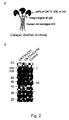
- Figures 2A and 2B are respectively (A) a schematic representation of a trimeric collagen scaffold antibody (CSA) that contains an amino-terminal scFv derived from OKT3 (anti-CD3), 528 (anti-EGFR), or erb (anti-EGFR), which are respectively fused in-frame to a hinge region of human IgG, a collagen scaffold domain of human type XXI minicollagen, followed by a histidine tag. Dashed lines: interchain disulfide-bonds; and (B) a depiction of Western blotting results of the protein complex.
- CSA trimeric collagen scaffold antibody
- OKT3mC21 contains an amino-terminal anti-CD3 scFv derived from OKT3 IgG, a hinge region of human IgG, a human minicollagen XXI polypeptide, followed by a histidine tag;
- OKT3mC21fd contains an amino-terminal anti-CD3 scFv derived from OKT3 IgG, a hinge region of human IgG, a human minicollagen XXI polypeptide, followed by a T4 fibritin foldon domain and a histidine tag.
- the culture media from the stably transfected Drosophila S2 cells was electrophoresed on a SDS-PAGE under non-reducing conditions and then immunoblotted with a monoclonal antibody to the C-terminal of type XXI collagen, 3E2.
- T interchain disulfide-bonded trimers
- Mt monomers containing intrachain disulfide-bonded trimers.
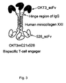
- Figure 3 is a diagram showing a bispecific trimeric CSA, containing an amino-terminal OKT3 single-chain antibody (OKT3_scFv), a hinge region of human IgG, a collagen scaffold domain of human type XXI minicollagen, followed by a C-terminal 528 single-chain antibody (528_scFv).
- OKT3_scFv amino-terminal OKT3 single-chain antibody
- IgG human IgG
- a collagen scaffold domain of human type XXI minicollagen followed by a C-terminal 528 single-chain antibody (528_scFv).
- Figures 4A (a-e) and 4B (a-c) are schematic representations of different formats of antibodies:
- scFv-GPP 10 (b), containing an amino-terminal scFv and a collagen-like domain GSP(GPP) 10 GPS;
- Col-scFv (c) containing an amino-terminal disulfide knot (TCPPCPRSIP), a collagen-like domain (GPP) 10 , followed by a carboxyl-terminal disulfide knot (GICDPSLC) derived from the NCl domain of type XXI collagen and an scFv;
- BiscFv-Col (d) containing an amino-terminal scFv
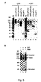
- Figures 5 A-B depict purification and structural characterization of the various antibody molecules in mammalian cells.
- the indicated antibodies were stably expressed in mammalian cells and purified from culture media by column chromatographies. The samples were electrophoresed on a 10% SDS/Bis-Tris polyacrylamide gel with MOPS buffer under non-reducing conditions (lanes 1 to 5) and reducing conditions, where sample was treated with 50 mM of DTT for 10 min at 70 °C (lanes 6 to 10).
- An erb_scFv-Col hexamer is formed by the interchain disulfide-bonding of two trimeric molecules.
- erb_scFv-Col (1 mg/ml) was incubated at 37 °C in the absence (lane 1) or presence (lanes 3) of 10 mM DTT for 1 h. An aliquot from the DTT-treated sample was further reacted with 50 mM N-ethyl-maleimide (NEM) for 30 min. at ambient temperature (lane 2). All samples with equal amounts of protein were electrophoresed on a 7% SDS/Tris-acetate polyacrylamide gel with sodium acetate as a running buffer. The gel was stained with Coomassie blue. "M" indicates molecular mass standards.
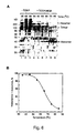
- Figures 6 A-B depict the thermal stability of trimeric structure of erb-scFv-Col.
- A Purified erb_scFv-Col in 50 mM Tris-HCl (pH 8.0), containing 2 M urea, was treated in the absence (lanes 1 to 3) or presence (lanes 4 to 9) of 10 mM TCEP at ambient temperature. The reduced samples were alkylated with 50 mM NEM at ambient temperature. All samples, which had an equal amount of protein, were heated for 10 min at the indicated temperatures and then SDS-loading buffer was added immediately. The samples were electrophoresed on a 10% SDS/Bis-Tris polyacrylamide gel with MOPS buffer under non-reducing conditions.
- FIG. 7 depicts an HPLC elution profile of phenylthiocarbamyl (PTC) amino acid derivatives of erb_scFv-Col following acid hydrolysis.
- PTC phenylthiocarbamyl
- PITC phenylisothiocyanate
- the phenylisothiocyanate (PITC) - derivatized amino acids of erb_scFv-Col after acid hydrolysis were separated on a reverse phase C18 silica column and the PTC chromophores were detected at 254 nm.
- the peak position of hydroxyproline (Hyp) derivative is indicated by an arrow.
- Figures 8 A-C depict surface plasmon resonance analysis of interaction between extracellular domain of EGFR and erb_scFv-Col (A); erb_scFv-Fc (B) or erb_scFv (C). Each antibody was injected at indicated concentrations and flowed over a surface chip with immobilized EGFR extracellular domain at a flow rate of 10 ⁇ l/min.
- Figure 9 depicts an OKT3 displacement assay.
- Human CD3(+) T-cells were incubated with serial dilutions of either OKT3_scFv-Col or OKT3 IgG for 1 h. A saturating amount of OKT3-FITC was added and incubated for additional hour. Cells were washed and bound; OKT3-FITC was quantified by flow cytometry. Values are expressed as percent inhibition of maximal fluorescence, as determined by adding OKT3-FITC without prior blocking antibodies.
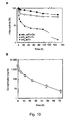
- Figures 10 A-B depict the stability of CSA molecule.
- A Stability of the various forms of erb antibody in human serum. The stability of erb_scFv-Col, erb_scFv-Fc or erb_scFv was determined by incubating at 37 °C in human serum. The amount of active anti-EGFR that remained after various periods of incubation was determined by ELISA using anti-c-myc mAb.
- B Pharmacokinetics of erb_scFv-Col in mice. Male C57BL/6 mice were injected intravenously with 2 mg/Kg of erb_scFv-Col. Blood samples were drawn at different times. The erb_scFv-Col levels in plasma were determined by ELISA using rabbit anti-c-myc antibodies conjugated with HRP. Results were averaged from 3 animals for each time point and error bars represent the standard deviation.
- FIGS 11 A-B depict that the OKT3-derived CSA is non-mitogenic with effective immunosuppressive activity.
- Human PBMCs were collected from three healthy normal donors and incubated individually with serial log dilutions of OKT3 IgG or OKT3_scFv-Col for 72 h, pulsed with 10 ⁇ M BrdU for an additional 8 h.
- the cell proliferation was measured by BrdU-ELISA using chemiluminescent immunoassay to quantify the incorporation of BrdU during DNA synthesis. Each point represents mean ⁇ S.D. of three donors.
- Figures 12 A-F depict release of cytokines induced by OKT3 IgG and OKT3_scFv-Col.
- Human PBMCs were collected from three healthy normal donors and incubated individually with serial log dilutions of OKT3 IgG (filled circles) or OKT3-scFv-Col (open circles).
- the levels of IL-2 and the rest of the indicated cytokines in the culture supernatants were determined by ELISA at 24- and 72-h time points, respectively. Each point represents the mean ⁇ S.D. of three donors.
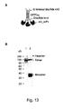
- FIGS 13 A-B depict purification of Col-erb_scFv.
- A Schematic representation of the collagen scaffold antibody, Col-erb_scFv, containing an amino-terminal disulfide knot (TCPPCPRSIP), a collagen-like domain (GPP) 10 , followed by a carboxyl-terminal disulfide knot (GICDPSLC) derived from the NCl domain of type XXI collagen and an erb_scFv (anti-EGFR).
- TCPPCPRSIP amino-terminal disulfide knot
- GGPP collagen-like domain
- GICDPSLC carboxyl-terminal disulfide knot
- Recombinant Col-erb_scFv stably expressed in mouse myeloma NS0 cells was purified from culture media by column chromatographies.
- the samples were electrophoresed on a 10% SDSBis-Tris polyacrylamide gel with MOPS buffer under non-reducing conditions (lane 1) and reducing conditions, where sample was treated with 50 mM of DTT for 10 min at 70 °C (lane 2).
- the gel was stained with ImperialTM Protein Stain solution (Pierce Biotechnology, Inc.).
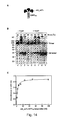
- Figures 14 A-C depict the collagen-scaffold peptide of the CSA, comprising (GPP) 10 , by itself can drive the formation of a thermal-stable non-covalently bound trimeric fusion protein.
- B Thermal stability of trimeric structure of erb_scFv-GPP 10 .
- erb_scFv-GPP 10 in 50 mM Tris-HCl (pH 8.0), containing 2 M urea, was treated in the absence (lanes 1 to 3) or presence (lanes 4 to 9) of 10 mM TCEP at ambient temperature. All samples, which had an equal amount of protein, were heated for 10 min at the indicated temperatures and then SDS-loading buffer was added immediately. The samples were electrophoresed on a 10% SDS/Bis-Tris polyacrylamide gel with MOPS buffer under non-reducing conditions. The gel was stained with ImperialTM Protein Stain solution (Pierce Biotechnology, Inc.). (C) Binding of erb_scFv-GPP 10 to EGFR-ECD by ELISA.
- a 96-well microtiter plate was coated with 1 ⁇ g/ml of EGFR-ECD and subsequently incubated with various concentrations of purified erb_scFv-GPP 10 and HRP conjugated anti-c-myc antibodies. Absorbance at 450 nm was measured.
- Figures 15 A-C depict the purification and characterization of 763_scFv-Col.
- A Schematic representation of 763_scFv-Col, containing an amino-terminal 763_scFv (anti-EGFR), a mutant hinge region of human IgG, a collagen-like domain (GPP) 10 , followed by a disulfide knot (GICDPSLC) of type XXI collagen.
- B The antibody was stably expressed in mouse myeloma NS0 cells and purified from culture media by column chromatographies. The sample was electrophoresed on a 10% SDS/Bis-Tris polyacrylamide gel with MOPS buffer under non-reducing conditions (lane 1) and reducing conditions (lane 2).
- FIGS 16 A-C depict the purification and characterization of 763_CSA2.
- A Schematic representation of 763CSA2, containing an amino-terminal 763_scFv (anti-EGFR), a mutant hinge region of human IgG, a collagen-like domain (GPP) 5 GKPGKP(GPP) 6 , followed by a disulfide knot (GICDPSLC) of type XXI collagen.
- B Identification of trimers. The sample was electrophoresed on a 10% SDS/Bis-Tris polyacrylamide gel with MOPS buffer under non-reducing conditions (lane 2). Lane 1, molecular weight marker.
- C Binding of 763CSA2 to EGFR by ELISA.
- a 96-well microtiter plate was coated with 1 ⁇ g/ml of EGFR and subsequently incubated with various concentrations of purified 763CSA2 and HRP conjugated anti-c-myc antibodies. Absorbance at 450 nm was measured.
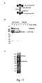
- FIGS 17 A-C depict the purification and characterization of the bispecific CSA, 763CSAOKT3.
- A Schematic representation of 763CSAOKT3, containing an amino-terminal 763_scFv (anti-EGFR), a mutant hinge region of human IgG, a collagen-like domain (GPP) 10 , followed by a disulfide knot (GICDPSLC) of type XXI collagen and a carboxyl-terminal OKT3_scFv (anti-CD3).
- GFP collagen-like domain
- GICDPSLC disulfide knot
- B Western blot analysis of the culture media containing recombinant bispecific 763CSAOKT3 antibody.
- the sample was electrophoresed on a 10% SDS/Bis-Tris polyacrylamide gel with MOPS buffer under non-reducing conditions (lane 1) and reducing conditions, where sample was treated with 50 mM of DTT for 10 min at 70 °C (lane 2).
- the gel was stained with ImperialTM Protein Stain solution (Pierce Biotechnology, Inc.).
- Figures 18 A-C depict flow cytometry analysis of the bispecific CSA, 763CSAOKT3, crosslinking A431 (EGFR-positive) and human CD3(+) T cells. Equal amounts (1 x 10 6 cells) of PKH-67 labeled A431 cells and PKH-26 labeled CD3(+) T cells were mixed in the absence (A) or presence of 1:4 (B); 1:2 dilution (C) of culture media containing recombinant bispecific 763CSAOKT3 antibody.
- Figure 19 A-B depict the purification and characterization of the bifunctional CSA, h4D5CSA-Luc.
- A Schematic representation of h4D5CSA-Luc, containing an amino-terminal h4D5_scFv (anti-HER2/neu), a mutant hinge region of human IgG, a collagen-like domain (GPP) 10 , followed by a disulfide knot (GICDPSLC) of type XXI collagen and a carboxyl-terminal Gaussia luciferase.
- GGPP collagen-like domain
- GICDPSLC disulfide knot
- Recombinant h4D5CSA-Luc stably expressed in mouse myeloma NS0 cells was purified from culture media by column chromatographies.
- Figures 20 A-B depict the purification and characterization of 357_scFv-Col.
- the antibody was stably expressed in mouse myeloma NS0 cells and purified from culture media by column chromatographies. The sample was electrophoresed on a 10% SDS/Bis-Tris polyacrylamide gel with MOPS buffer under non-reducing conditions (lane 1) and reducing conditions (lane 2).
- L929 mouse fibroblast cells (5 x 10 3 cells) were incubated with different concentration of either 357_scFv-Col or 357 IgG, containing 2 ⁇ g/ml of actinomycin D and 10 ng/ml of human recombinant TNF- ⁇ for 24 h.
- TNF- ⁇ induced cell cytotoxicity was determined by MTT assay. The number of viable cells was determined by measuring the optical densities at 460 nm.
- the present invention is based, in part, on the findings that a antibody domain fused in-frame to a collagen scaffold domain allows the trimerization of the scaffold domain to generate a trimeric antibody, and the binding avidity of the resulting trimeric antibody is enhanced as compared to the divalent IgG, and monovalent scFv formats.
- Trimeric antibodies made according to the invention can have functional affinities (avidities) for their ligands of greater than 10 9 M -1 .
- a collagen scaffold domain can be fused in-frame to a binding protein in a fusion polypeptide, such that the collagen scaffold domain drives trimerization of the fusion polypeptide, which retains its ability to bind its ligand.
- the binding domain can be, for example, a cytokine domain, a cytokine receptor domain, or an antibody domain.
- the cytokine is TNF- ⁇ .
- a collagen scaffold domain can be fused in-frame to an antibody domain to generate a trimeric antibody.
- the trimeric antibody is a soluble antibody.
- a soluble antibody is one that is soluble under physiological conditions.
- the soluble trimeric antibody is a secreted antibody.
- a secreted antibody is one that is secreted by a cell. Secretion of an antibody can be targetted by having a signal sequence on the polypeptide comprising the antibody domian.
- the soluble trimeric antibody has an avidity for its ligand of greater than 10 7 M -1 . In one embodiment, the soluble trimeric antibody has an avidity for its ligand of greater than 10 8 M -1 . In one embodiment, the soluble trimeric antibody has an avidity for its ligand of greater than 10 9 M -1 . In certain embodiments, the soluble trimeric antibody has an avidity for its ligand between 10 7 M -1 and 10 10 M -1 , between 10 7 M -1 and 10 9 M -1 , between 10 7 M -1 and 10 8 M -1 , between 10 8 M -1 and 10 10 M -1 , between 10 8 M -1 and 10 9 M -1 , and between 10 9 M -1 and 10 10 M -1 .
- a thermally stable short collagen-like peptide such as (Gly-Pro-Pro) 10
- (Gly-Pro-Pro) 10 is used as a scaffold domain to drive the trimerization of an antibody domain by expressing the fusion construct in a system with sufficient P4H activity.
- This approach facilitates the adoption of the stable triple-helical structure, which affects protein valency, stability, and function in vivo.
- the present invention encompasses using a collagenous sequence, e.g., minicollagen type XII or XXI, or a collagen-like sequence, e.g., (Gly-Pro-Pro) 10 or (GPP) 5 GKPGKP(GPP) 6 , as a collagen scaffold domain capable of self-nucleation and propagation of the heterologous fusion proteins from either C- or N-terminal direction.
- a collagenous sequence e.g., minicollagen type XII or XXI
- a collagen-like sequence e.g., (Gly-Pro-Pro) 10 or (GPP) 5 GKPGKP(GPP) 6
- the present invention avoids the need for any other trimerization structure domains.
- Collagen scaffold domain fusion proteins can form a thermally stable triple helical structure by expression of the fusion construct in a prolyl 4-hydroxylase containing system.
- the self-trimerization collagen scaffold of the invention allows attachment of fusion partners to either terminus, as well as to both termini, simultaneously. This has important consequences as the self-trimerization collagen scaffold may be employed to construct molecules that are able to interact (each end with a binding valency up to 3 or 6) simultaneously with two bulky binding partners.
- the present invention also demonstrates that the trimeric antibodies can fold correctly and exhibit high solubility, avidity, and stability.
- collagen scaffold domain is a collagenous or collagen-like domain which allows for formation of a triplex structure by itself, wherein a "triplex structure” is a covalently or non-covalently bound complex of three subunits.
- collagen scaffold domain refers to the collagenous or collagen-like domains that direct self-trimerization of the scaffold domain.
- the term "collagen scaffold domain” does not refer to C-propeptides of procollagens, coiled-coil neck domains of collectin or ficolin family proteins, the C-type lectin-like domain of tetranectin, beta-galactosidase trimerization domain, the three coiled-coil helix structure of GCN4 leucine zipper mutant ( Harbury et al., (1993) Science 262: 1401-1407 ), Clq and TNF domains of Clq and TNF superfamily proteins, and bacteriophage T4 fibritin foldon domains.
- a "collagen scaffold antibody” or "CSA” is an antibody that includes a collagen scaffold domain fused to an antibody domain.
- a CSA and its coding sequence can comprise any combination of the SEQ IDs that follow. Each of these combinations is specifically contemplated.
- the CSA can contain one or more of SEQ ID NOs 1, 3, 5 and 9.
- one aspect of this invention features an isolated recombinant protein complex that includes a first fusion polypeptide chain containing a first collagen scaffold domain and a first antibody domain fused in-frame to one end of the first collagen scaffold domain; a second fusion polypeptide chain containing a second collagen scaffold domain; and a third fusion polypeptide chain containing a third collagen scaffold domain.
- the first, second, and third collagen scaffold domains are aligned to form a triple helix coil.
- the first collagen scaffold domain and first antibody domain are fused in-frame and on the same peptide chain.
- the fusion polypeptide chain can include the sequence of an enzymatic domain or a fluorescent protein.
- a fluorescent protein include GFP and dsRed, as well as their variants.
- an enzymatic domain include that of glutathione S-transferase, luciferase, ⁇ -galactosidase, and ⁇ -lactamase.
- the fusion polypeptide chain can include, or exclude, the sequence of affinity tags for the purpose of detection and purification of the fusion proteins of the invention.
- affinity tags include polyhistidine-tag, myc-tag, Strep-tag, FLAG, E-tag, hemagglutin tag, T7, Stag, HSV, VSV-G, anti-Xpress, and VS-tag.
- the “antibody domain” includes one or more complementary-determining regions (CDR) of an immunoglobulin. Accordingly, the antibody domain can include antigen binding parts of an antibody, such as a V H domain and an Fab.
- the first antibody domain contains the sequence of an antigen-binding fragment or a single-chain antibody, e.g., that specific for Cluster Designation 3 (CD3), Epidermal Growth Factor Receptor (EGFR), HER2/neu or Tumor necrosis factor-alpha (TNF- ⁇ ).
- CD3 Cluster Designation 3
- EGFR Epidermal Growth Factor Receptor
- HER2/neu Tumor necrosis factor-alpha
- TNF- ⁇ Tumor necrosis factor-alpha
- a second fusion polypeptide chain contains a second antibody domain.
- the first and second antibody domains are identical to each other.
- the first and second antibody binding domains can bind to an identical binding partner or to two different binding partners.
- the first antibody domain and second antibody domain can contain the sequences of a first single-chain antibody and a second single-chain antibody that specifically bind to CD3 and EGFR, respectively.
- the first and second fusion polypeptide both contain the first and second antibody binding domains.
- the second fusion polypeptide chain can contain a third antibody domain fused in-frame to one end of the second scaffold domain, a fourth antibody domain fused in-frame to the other end of the second scaffold domain, or both domains fused in-frame to the two ends.
- the third fusion polypeptide chain can contain a fifth antibody domain fused in-frame to one end of the third scaffold domain, or a sixth antibody domain fused in-frame to the other end of the third scaffold domain, or both. All six antibody domains can be identical to or different from each other. They therefore can bind to 1, 2, 3, 4, 5, or 6 binding partners.
- the protein complex can be mono-, di-, tri-, tetra-, penta-, or hexa-valent.
- each of the three scaffold domains contains one or more triple helix repeats, known as collagenous or collagen-like domains, each repeat containing a sequence of the following formula: (G-X-Y) n , in which G is a Gly residue; X and Y are any amino acid residues, and preferably, the amino acid proline or hydroxyproline; and n is 5 or greater.
- G is a Gly residue
- X and Y are any amino acid residues, and preferably, the amino acid proline or hydroxyproline
- n is 5 or greater.
- a "repeat” refers to two or more sequential G-X-Y sequences.
- the scaffold domain can include a perfect repeating G-X-Y triplet, interrupted by a short imperfection, in which the first position of Gly or the third position of Y residue is missing, found in many naturally occurring collagens and proteins containing collagen-like domains.
- the scaffold domain of this invention human type XXI minicollagen, contains two imperfections, GF and KE, within the collagenous domain.
- the collagen scaffold domain is a collagen scaffold domain comprising at least 8, at least 9, at least 10, at least 11, at least 12, at least 13, at least 14, at least 15, at least 16, at least 17, at least 18, at least 19, or at least 20 G-X-Y repeats; wherein G is glycine, X is any amino acid, and Y is any amino acid.
- at least 5, at least 6, at least 7, at least 8, at least 9, or at least 10 of the G-X-Y repeats are G-P-P or G-P-O.
- at least 5, at least 6, at least 7, at least 8, at least 9, or at least 10 of the G-X-Y repeats are G-P-O, wherein P is proline and O is hydroxyproline.
- the collagen scaffold domain directs self-trimerization.
- the collagen scaffold domain comprises the sequence (G-P-P/O) 10 . In one embodiment, the collagen scaffold domain comprises 10 G-X-Y repeats. In certain embodiments, the collagen scaffold domain comprises less than 10, 11, 12, 13, 14, 15, 16, 17, 18, 19, 20, 22, 25, 27, 30, 35, 40, 45, or 50 G-X-Y repeats. In certain embodiments, the collagen scaffold domain is less than 150, 125, 100, 90, 80, 70, 60, 50, or 40 amino acids in length. In one embodiment, the collagen scaffold domain consists essentially of 10-30 G-X-Y repeats, which cause self-trimerization.
- the scaffold domain comprises the sequence (G-P-P/O) 5 GKPGKP(G-P-P/O) 6 . In one embodiment, the scaffold domain comprises the sequence (G-P-P/O) 10 , where P/O indicates that the Y position is either P or O.
- the collagen scaffold domain comprises at least 10 G-X-Y repeats, wherein at least 5, at least 6, at least 7, at least 8, at least 9, or at least 10 of the Y residues are hydroxyproline.
- the collagen scaffold domain directs self-trimerization.
- the collagen scaffold domain comprises at least 5, at least 6, at least 7, at least 8, at least 9, or at least 10 G-P-O repeats and an antibody domain; wherein the collagen scaffold domains of the three polypeptides interact with each other to form a trimeric soluble antibody that specifically binds to a ligand with an avidity of at least 10 7 M -1 .
- the collagen scaffold domain comprises 6 or 7 G-P-O repeats.
- the G-P-O repeats can be sequential or can be spaced apart.
- the G-P-P repeats can be spaced as two in-frame amino acid sequences comprising 3, 4, or 5 G-P-O repeats separated by 1, 2, 3, 4, or 5, GXY repeats.
- the collagen scaffold domain comprises (G-P-O) 3 GXY(GPO) 4 .
- the collagen scaffold domain is the collagenous (COL1) domain of a non-fibrillar FACIT (fibril-associated collagen with interrupted triple-helices) collagen.
- a trimeric antibody comprising the COL1 domain of a non-fibrillar FACIT does not contain the noncollagenous (NC1) domain of the FACIT.
- the COL1 domain is from types IX, XII, XIV, XVI, XIX, XX, XXI or XXII collagen.
- the trimeric antibody comprises SEQ ID NO:7.
- the collagen scaffold domain has at least 75%, 80%, 85%, 90%, or 95% identity with a complete COL1 domain from types IX, XII, XIV, XVI, XIX, XX, XXI or XXII collagen. In one embodiment, the collagen scaffold domain has at least 75%, 80%, 85%, 90%, or 95% identity with the G-X-Y repeats of a COL1 domain from types IX, XII, XIV, XVI, XIX, XX, XXI or XXII collagen.
- the collagen scaffold domain has at least 75%, 80%, 85%, 90%, or 95% identity with 10, 11, 12, 13, 14, 15, 16, 17, 18, 19, 20, 21, 22, 23, 24, 25, or 30 G-X-Y repeats of a COL1 domain from types IX, XII, XIV, XVI, XIX, XX, XXI or XXII collagen.
- the collagen scaffold domain has at least 75%, 80%, 85%, 90%, or 95% identity with 10 G-X-Y repeats of a COL1 domain from type XXII collagen.
- the G-X-Y sequence of the collagen scaffold domain comprises at least 5, at least 6, at least 7, at least 8, at least 9, or at least 10 hydroxyprolines at the Y position.
- the percent identity can be determined, for example, by comparing sequence information using the GAP computer program, version 6.0 described by Devereux et al. ( Nucl. Acids Res. 12:387, 1984) and available from the University of Wisconsin Genetics Computer Group (UWGCG).
- the GAP program utilizes the alignment method of Needleman and Wunsch (J. Mol. Biol. 48:443, 1970 ), as revised by Smith and Waterman (Adv. Appl. Math 2:482, 1981 ).
- the preferred default parameters for the GAP program include: (1) a unary comparison matrix (containing a value of 1 for identities and 0 for non-identities) for nucleotides, and the weighted comparison matrix of Gribskov and Burgess, Nucl. Acids Res.
- the above-described first, second, and third fusion polypeptides are substantially identical, having at least 75% (e.g., any number between 75% and 100%, inclusive) sequence identity to one another.
- a complex formed by three identical fusion polypeptides is a homotrimer.
- the three fusion polypeptides can be functional equivalents.
- a "functional equivalent” refers to a polypeptide derivative of a common polypeptide, e.g., a protein having one or more point mutations, insertions, deletions, truncations, a fusion protein, or a combination thereof, and retaining substantially the ability to form a triple helix coil and the activity of the heterologous domain, such as binding to a ligand.
- a heterologous polypeptide, nucleic acid, or gene is a polypeptide, nucleic acid, or gene that is associated with another polypeptide, nucleic acid, or gene with which it is not naturally associated. Two fused domains or sequences are heterologous to each other if they are not adjacent to each other in a naturally occurring protein or nucleic acid.
- the invention also inclides an isolated recombinant fusion polypeptide (e.g., each of the above-mentioned three fusion polypeptides) that contains (i) a collagen scaffold domain for forming a triple helix coil and (ii) a first heterologous domain fused in-frame to one end of the scaffold domain or a second heterologous domain fused in-frame to the other end of the scaffold domain.
- the heterologous domain can include one of the antibody domains mentioned above, and can be obtained by various art-recognized methods, such as phage display screening.
- an "isolated" polypeptide or protein complex refers to a polypeptide or a protein complex substantially free from naturally associated molecules, i.e., it is at least 75% (i.e., any number between 75% and 100%, inclusive) pure by dry weight. Purity can be measured by any appropriate standard method, for example, by column chromatography, polyacrylamide gel electrophoresis, or HPLC analysis. An isolated polypeptide or protein complex of the invention can be purified from a natural source, produced by recombinant DNA techniques.
- the three polpeptides that trimerize to form a trimeric antibody are non-contiguous.
- the three polpeptides that trimerize to form a trimeric antibody are contigous, i.e, translated as a single translation product.
- the three polypeptides can be joined by two or more flexible hinge regions.
- the invention also encompasses an isolated nucleic acid that contains a sequence encoding the just-mentioned fusion polypeptide or a complement of the sequence.
- a nucleic acid refers to a DNA molecule (e.g., a cDNA or genomic DNA), an RNA molecule (e.g., an mRNA), or a DNA or RNA analog.
- a DNA or RNA analog can be synthesized from nucleotide analogs.
- the nucleic acid molecule can be single-stranded or double-stranded, but preferably is double-stranded DNA.
- isolated nucleic acid is a nucleic acid the structure of which is not identical to that of any naturally occurring nucleic acid or to that of any fragment of a naturally occurring genomic nucleic acid.
- the term therefore covers, for example, (a) a DNA which has the sequence of part of a naturally occurring genomic DNA molecule but is not flanked by both of the coding sequences that flank that part of the molecule in the genome of the organism in which it naturally occurs; (b) a nucleic acid incorporated into a vector or into the genomic DNA of a prokaryote or eukaryote in a manner such that the resulting molecule is not identical to any naturally occurring vector or genomic DNA; (c) a separate molecule such as a cDNA, a genomic fragment, a fragment produced by polymerase chain reaction (PCR), or a restriction fragment; and (d) a recombinant nucleotide sequence that is part of a hybrid gene, i.e., a gene encoding a
- a vector refers to a nucleic acid molecule capable of transporting another nucleic acid to which it has been linked.
- the vector can be capable of autonomous replication or integrate into a host DNA.
- Examples of a vector include a plasmid, cosmid, or viral vector.
- the vector of this invention includes a nucleic acid in a form suitable for expression of the nucleic acid in a host cell.
- the vector includes one or more regulatory sequences operatively linked to the nucleic acid sequence to be expressed.
- a "regulatory sequence” includes promoters, enhancers, and other expression control elements (e.g., polyadenylation signals).
- Regulatory sequences include those that direct constitutive expression of a nucleotide sequence, as well as tissue-specific regulatory and/or inducible sequences.
- the design of the expression vector can depend on such factors as the choice of the host cell to be transformed, the level of expression of protein desired, and the like.
- the expression vector can be introduced into host cells to produce the polypeptide of this invention.
- a host cell that contains the above-described nucleic acid. Examples include E. coli cells, insect cells (e.g., using Drosophila S2 cells or baculovirus-infected insect cells), yeast cells, or mammalian cells (e.g., mouse myeloma NS0 cell). See e.g., Goeddel, (1990) Gene Expression Technology: Methods in Enzymology 185, Academic Press, San Diego, CA .
- a fusion polypeptide of this invention one can culture a host cell in a medium under conditions permitting expression of the polypeptide encoded by a nucleic acid of this invention, and purify the polypeptide from the cultured cell or the medium of the cell.
- the nucleic acid of this invention can be transcribed and translated in vitro, for example, using T7 promoter regulatory sequences and T7 polymerase.
- a host cell containing a first, second, and third nucleic acids respectively encoding the above-mentioned first, second, and third fusion polypeptides in a medium under a condition permitting expression of polypeptides encoded by the three nucleic acids and formation of a triple helix coil between the expressed polypeptides, and purifying the protein complex from the cultured cell or the medium of the cell.
- the host cell is a eukaryotic cell containing an enzymatic activity that hydroxylates a proline residue.
- a soluble trimeric antibody of this invention enjoys advantages over conventional antibodies.
- the protein complex can have 2-6 antibody domains that are specific for one binding partner (e.g., antigen) in comparison with a conventional antibody, which has only two such domains.
- the protein complex can be di-, tri-, tetra-, penta-, or hexa-valent.
- it can be made to have affinities that are higher than a conventional antibody. Because of the higher affinities, smaller amounts of the protein complex and shorter incubation duration are needed than a conventional antibody to achieve the desired goals, for example, therapeutic effects, thereby lowering treatment costs and minimizing side effects (e.g., unwanted immune responses).
- a protein complex of this invention can have 2-6 antibody domains that are specific for 2-6 different binding partners. Unifying multiple binding partner sites of different specificities into one unit, it has the ability to bring together multiple binding partners and therefore have desirable uses in therapy, tissue reconstruction, and assembly of active protein machinery (e.g., a multi-subunit enzyme) at the nanometer level.
- active protein machinery e.g., a multi-subunit enzyme
- a trimeric antibody of this invention is preferably of human origin.
- it can include a humanized single-chain antibody sequence fused in-frame to a collagen scaffold domain of human origin. Since many collagen-like proteins with collagenous domains are fairly stable in the blood, the scaffold domain fusion proteins should retain structural integrity in blood as well.
- the sequence Gly-Pro-Hyp contributes most to the formation and stabilization of a triple helical structure and the Gly-Pro-Hyp tripeptide repeats self-assemble into a highly stable triple helix.
- the collagenous domain of minicollagen XXI was substituted with a thermally stable short collagen-like peptide (Gly-Pro-Pro) 10 as a scaffold template for the CSAs described herein, which were expressed in a mammalian system with sufficient P4H activity to facilitate the adoption of the stable triple-helical structure.
- both erb_scFv-Col and OKT3_scFv-Col were assembled into a trimeric structure and erb_scFv-Col can be further oligomerized into a hexamer, presumably through the interchain disulfide crosslinking between the two C-terminal cysteine residues within the two trimers.
- the oligomerization of erb_scFv-Col from trimer to hexamer is an intracellullar process because the reduced trimeric structure does not assemble into higher order structures at concentrations in excess of those normally found in the hexameric form oferb_scFv-Col.
- Mouse myeloma NS0 cells are a good expression system for recombinant collagen or collagen-like protein production. Approximately 61% of the total number of proline residues in the Y position of a collagenous GXY triplet sequence of (Gly-Pro-Pro) 10 is hydroxylated in recombinant erb_scFv-Col. Thus, at least 6 of the Gly-Pro-Pro repeats were hydroxylated in this system. The contribution of the prolyl hydroxylated (Gly-Pro-Pro) 10 motif to the trimeric assembly of CSA molecules was prominent, since almost no monomeric form of the CSA was present in culture media, as examined by Western blot analysis.
- the thermally stable trimeric structure of CSA meets the requirement of a multipurpose multimerizing system for future in vivo use.
- Many collagen-like proteins with collagenous domains are present in human serum and serve as an innate immune system in protection from infectious organisms. These include complement protein C1q, collectin family proteins - mannose binding lectin (MBL), ficolins and surfactant proteins A and D (SP-A and SP-D).
- a common structural feature among these "defense collagen” molecules is that all of them are in multi-trimeric protein units with a target-binding domain at the C-terminus. The driving force for the formation of these multi-trimeric structures is similar to those for fibrillar collagens.
- the three polypeptide chains were firstly trimerized using their coiled-coil neck domain, which is N-terminal next to the target-binding domain, then triple helical folding of the collagen-like domain proceeded in a zipper-like fashion from the C to the N terminus, before finally stacking or interchain disulfide crosslinking of the trimeric molecules using their N-terminal cysteine residues ( Hakansson et al., (1999) Structure 7: 255-264 ; Hakansson and Reid, (2000) Protein Sci 9: 1607-1617 ; Sheriff et al., (1994) Nat Struct Biol 1: 789-794 ; Weis and Drickamer, (1994) Structure 2: 1227-1240 ). Consequently, multimerization significantly increases the functional affinity of the binding domain of these defense collagen molecules.
- OKT3_scFv-Col does not induce T-cell proliferation or IL-2 production in human PBMCs, greatly reducing the effects of the OKT3 first dose syndrome that is caused by the release of cytokines as a result of transient T cell activation.
- This new anti-CD3 format may provide a potent immunosuppressive drug with reduced dosing, toxicity, and cost of therapy.
- the trimeric soluble antibody does not contain an Fc domain.
- Affinity maturation by chain-shuffling using phage-display scFv library screening for high-affinity binders is a tedious process; yet the outcome of improving binding affinity is uncertain. Therefore, many therapeutic antibodies may be hampered by low affinity for the target antigen after humanization. In some cases, the properties of the antibody must be further refined. Polymerization of antigen-binding partners greatly increases their availability for binding to a group of specific identical ligands in very close proximity to a target cell. Different approaches have been proposed to obtain multivalent molecules for improving functional affinity.
- Non-IgG protein scaffold fragments such as “anticalins” ( Skerra, (2000) Biochim Biophys Acta 1482: 337-350 ), "ankyrin repeats” ( Binz et al., (2004) Nat Biotechnol 22: 575-582 ), "Affibody molecules” ( Nord et al., (1997) Nat Biotechnol 15: 772-777 ), and the C-type lectin-like domain of tetranectin ( Christian et al. International Publication No. WO 98/56906 ; Graversen et al., (2000) J Biol Chem 275: 37390-37396 ) have recently been adopted to increase target binding affinity, thermal stability, and sensitivity. However, some of these molecules are either heteroantigenetic fragments or not natural components of plasma, and are associated with the risk of an immune response that could severely limit potential therapeutic applications.
- a triplex-forming collagen-like peptide fusion scaffold to form a thermally stable multivalent protein binder was used to demonstrate herein that CSA is a new platform that enables improvements to the functional affinity and mitogenicity of therapeutic antibodies. More importantly, fusion of the scFv and the collagen-like scaffold domains in CSA has been proven to bring each domain to fold correctly, without compromising the target-binding activities or the trimeric assembly of the triple helical structure.
- the triplex-forming collagen-like domain can be used as a scaffold for existing or new protein drugs by trimerizing (even oligomerizing through collagen fibrillar stacking or interchain disulfide crosslinking of the trimeric molecules that are similar to those of the defense collagen family) the active protein in a fusion protein approach, which involves protein hormones, cytokines, lymphokines, growth factors, lectins, enzymes and soluble receptor fragments; or adhension molecules, such as selectins and integrins.
- a protein complex or polypeptide of the invention can be obtained by recombinant technology.
- To facilitate formation of the triple helix coil scaffold one can co-express in the host cells prolyl 4-hydroxylase (P4H), a key enzyme in the biosynthesis of collagen.
- P4H prolyl 4-hydroxylase
- a heterologous protein domain can contain a "binding domain,” which includes, but is not limited to, an antibody or a fragment thereof (e.g., an antigen-binding fragment thereof).
- the term "antibody” as used herein refers to an immunoglobulin molecule or immunologically active portion thereof, i.e., an antigen-binding portion. It refers to a protein comprising at least one, and preferably two, heavy (H) chain variable regions (V H ), and at least one and preferably two light (L) chain variable regions (V L ).
- an "antibody domain” refers to an antibody or an antigen binding portion of an antibody, and includes V H , V L , or Fab domains, Fv fragments of single-chain antibodies (scFv), and VHH domains ( see WO 94/4678 ).
- V H and V L regions can be further subdivided into regions of hypervariability, termed “complementarity determining regions” ("CDR"), interspersed with regions that are more conserved, termed “framework regions” (FR).
- CDR complementarity determining regions
- FR framework regions
- the extent of the framework region and CDR's has been precisely defined (see, Kabat et al. (1991) Sequences of Proteins of Immunological Interest, Fifth Edition, U.S. Department of Health and Human Services, NIH Publication No. 91-3242 , and Chothia et al. (1987) J. Mol. Biol. 196:901-917 , which are incorporated herein by reference.
- Each V H and V L is normally composed of three CDR's and four FRs, arranged from amino-terminus to carboxy-terminus in the following order: FR1, CDR1, FR2, CDR2, FR3, CDR3, FR4.
- the antibody can further include a heavy and light chain constant region, to thereby form a heavy and light immunoglobulin chain, respectively.
- the heavy chain constant region is comprised of three domains, C H1 , C H2 , and C H3 .
- the light chain constant region is comprised of one domain, C L .
- the variable region of the heavy and light chains contains a binding domain that interacts with an antigen.
- the constant regions of the antibodies typically mediate the binding of the antibody to host tissues or factors, including various cells of the immune system (e.g., effector cells) and the first component (C1q) of the classical complement system.
- immunoglobulin refers to a protein consisting of one or more polypeptides substantially encoded by immunoglobulin genes.
- the recognized human immunoglobulin genes include the kappa, lambda, alpha (IgA1 and IgA2), gamma (IgG1, IgG2, IgG3, and IgG4), delta, epsilon, and mu constant region genes, as well as the myriad immunoglobulin variable region genes.
- Full-length immunoglobulin "light chains” (about 25 kD or 214 amino acids) are encoded by a variable region gene at the NH 2 -terminus (about 110 amino acids) and a kappa or lambda constant region gene at the COOH-terminus.
- Full-length immunoglobulin "heavy chains” (about 50 kD or 446 amino acids ), are similarly encoded by a variable region gene (about 116 amino acids ) and one of the other aforementioned constant region genes, e.g., gamma (encoding about 330 amino acids ).
- antibody portion refers to one or more fragments of a full-length antibody that retain the ability to specifically bind to its ligand, or antigen, e.g., EGFR or CD3 polypeptide or a fragment thereof.
- antigen-binding fragments of the antibody include, but are not limited to: (i) a Fab fragment, a monovalent fragment consisting of the V L , V H , C L , and C H1 domains; (ii) a F(ab') 2 fragment, a bivalent fragment comprising two Fab fragments linked by a disulfide bridge at the hinge region; (iii) a Fd fragment consisting of the V H and C H1 domains; (iv) a Fv fragment consisting of the V L and V H domains of a single arm of an antibody, (v) a dAb fragment ( Ward et al., ( 1989) Nature 341:544-546 ), which consists of a V H domain; (vi) an isolated complementarity determining region (CDR), and (vii) V L or V H domains.
- a Fab fragment a monovalent fragment consisting of the V L , V H , C L , and C H1 domains
- the two domains of the Fv fragment, V L and V H are coded for by separate genes, they can be joined, using recombinant methods, by a synthetic linker that enables them to be made as a single protein chain in which the V L and V H regions pair to form monovalent molecules (known as single-chain Fv (scFv); see e.g., Bird et al. (1988) Science 242:423-426 ; and Huston et al. (1988) Proc. Natl. Acad. Sci. USA 85:5879-5883 ).
- single-chain Fv single-chain Fv
- Such single-chain antibodies are also encompassed within the term "antigen-binding fragment" of an antibody.
- These antibody fragments can be obtained using conventional techniques known to those with skill in the art, and the fragments are screened for utility in the same manner as are intact antibodies.
- An antibody can be a monoclonal antibody.
- the antibody can be recombinantly produced, e.g., produced by phage display or by combinatorial methods.
- Phage display and combinatorial methods for generating antibodies are known in the art (see e.g., Ladner et al. U.S. Patent No. 5,223,409 ; Kang et al. International Publication No. WO 92/18619 ; Dower et al. International Publication No. 25 WO 91/17271; Winter et al . International Publication WO 92/20791; Markland et al . International Publication No. WO 92/15679 ; Breitling et al.
- the antibody is a fully human antibody (e.g., an antibody made in a mouse which has been genetically engineered to produce an antibody from a human immunoglobulin sequence), or a non-human antibody, e.g., a rodent (mouse or rat), goat, primate (e.g., monkey), or camel antibody.
- a rodent mouse or rat
- the non-human antibody is a rodent (mouse or rat antibody).
- Methods of producing rodent antibodies are known in the art.
- Human monoclonal antibodies can be generated using transgenic mice carrying the human immunoglobulin genes rather than the mouse system. Splenocytes from these transgenic mice immunized with the antigen of interest are used to produce hybridomas that secrete human mAbs with specific affinities for epitopes from a human protein (see, e.g., Wood et al. International Application WO 91/00906 , Kucherlapati et al. PCT 15 publication WO 91/10741 ; Lonberg et al. International Application WO 92/03918 ; Kay et al. International Application 92/03917 ; Lonberg, et al. (1994) Nature 368: 856-859 ; Green, L.L.
- An antibody can be one in which the variable region, or a portion thereof, e.g., the CDR's, are generated in a non-human organism, e.g., a rat or mouse. Chimeric, CDR-grafted, and humanized antibodies can be used. Antibodies generated in a non-human organism, e.g., a rat or mouse, and then modified, e.g., in the variable framework or constant region, to decrease antigenicity in a human are within the invention.
- Chimeric antibodies can be produced by recombinant DNA techniques known in the art. For example, a gene encoding the Fe constant region of a murine (or other species) monoclonal antibody molecule is digested with restriction enzymes to remove the region encoding the murine Fc, and the equivalent portion of a gene encoding a human Fc constant region is substituted (see Robinson et al., International Patent Publication PCT/US86/02269 ; Akira, et al., European Patent Application 184, 187 ; Taniguchi, M., European Patent Application 171,496 ; Morrison et al., European Patent Application 173,494 ; Neuberger et al., International Application WO 86/01533 ; Cabilly et al.
- a humanized or CDR-grafted antibody will have at least one or two but generally all three recipient CDR's (of heavy and/or light immunoglobulin chains) replaced with a donor CDR.
- the antibody may be replaced with at least a portion of a non-human CDR or only some of the CDR's may be replaced with non-human CDR's. It is only necessary to replace the number of CDR's required for binding of the humanized antibody or a fragment thereof.
- the donor will be a rodent antibody, e.g., a rat or mouse antibody
- the recipient will be a human framework or a human consensus framework.
- the immunoglobulin providing the CDR's is called the "donor” and the immunoglobulin providing the framework is called the “acceptor.”
- the donor immunoglobulin is a non-human (e.g., rodent).
- the acceptor framework is a naturally-occurring (e.g., a human) framework or a consensus framework, or a sequence about 85% or higher, preferably 90%, 95%, 99% or higher identical thereto.
- the term "consensus sequence” refers to the sequence formed from the most frequently occurring amino acids (or nucleotides) in a family of related sequences (See e.g., Winnaker, From Genes to Clones (Verlagsgesellschaft, Weinheim, Germany 1987 ).
- each position in the consensus sequence is occupied by the amino acid occurring most frequently at that position in the family. If two amino acids occur equally frequently, either can be included in the consensus sequence.
- a "consensus framework” refers to the framework region in the consensus immunoglobulin sequence.
- An antibody can be humanized by methods known in the art. Humanized antibodies can be generated by replacing sequences of the Fv variable region which are not directly involved in antigen binding with equivalent sequences from human Fv variable regions. General methods for generating humanized antibodies are provided by Morrison, S. L., (1985) Science 229: 1202-1207 , by Oi et al., (1986) Bio Techniques 4: 214 , and by Queen et al. US 5,585,089 , US 5,693,761 and US 5,693,762 , the contents of all of which are hereby incorporated by reference.
- Those methods include isolating, manipulating, and expressing the nucleic acid sequences that encode all or part of immunoglobulin Fv variable regions from at least one of a heavy or light chain.
- Sources of such nucleic acid are well known to those skilled in the art and, for example, may be obtained from a hybridoma producing an antibody against a polypeptide of interest or fragment thereof.
- the recombinant DNA encoding the humanized antibody, or fragment thereof can then be cloned into an appropriate expression vector.
- Humanized antibodies in which specific amino acids have been substituted, deleted, or added can also be fused to the scaffold.
- Preferred humanized antibodies have amino acid substitutions in the framework region, such as to improve binding to an antigen.
- a humanized antibody will have framework residues identical to the donor framework residue or to another amino acid other than the recipient framework residue.
- a selected, small number of acceptor framework residues of the humanized immunoglobulin chain can be replaced by the corresponding donor amino acids.
- Preferred locations of the substitutions include amino acid residues adjacent to the CDR, or which are capable of interacting with a CDR. Criteria for selecting amino acids from the donor are described in US 5,585,089 , the contents of which are hereby incorporated by reference.
- Other techniques for humanizing antibodies are described in Padlan et al. EP 519596 A1 .
- each polypeptide of a trimeric antibody does not contain a collagen NCl domain. In one embodiment, each polypeptide of a trimeric antibody does not contain a disulfide knot. In one embodiment, each polypeptide of a trimeric antibody does not contain a bacteriophage T4 fibritin foldon domain.
- the scaffold of a trimeric antibody is of a minimal size.
- the collagen-like domain comprises the sequence (G-P-P/O) 10 .
- each polypeptide comprises less than 13 G-X-Y repeats.
- the trimeric antibody comprises less than 11, 12, 13, 14, 15, 16, 17, 18, 19, 20, 22, 25, 27, 30, 35, 40, 45, or 50 G-X-Y repeats.
- each polypeptide of a trimeric antibody has a molecular weight of less than 35, 37, 39, 40, 41, 42, 43, 44, 45, 46, 47, 48, 49, or 50 kD.
- the trimeric soluble antibody has a molecular weight of less than 105, 110, 115, 120, 125, 130, 135, 140, 145, or 150 kD.
- the trimeric soluble antibody comprises three polypeptides, wherein each polypeptide comprises a collagen scaffold domain comprising at least 10 G-X-Y repeats, and wherein at at least 5, at least 6, at least 7, at least 8, at least 9, or at least 10 of the Y residues are hydroxyproline; and an antibody domain,wherein the collagen-like domains of the three polypeptides interact with each other to form a trimeric soluble antibody that specifically binds to a ligand with an avidity of at least 10 7 M -1 .
- the invention also includes a nucleic acid which encodes a fusion polypeptide that forms a protein complex of this invention.
- the nucleic acid can be screened from phage display library or isolated (e.g., by RT-PCR) from cell lines expressing the above-described suitable antibodies or antibody derivatives.
- the nucleic acid can be functionally ligated into an expression vector.
- Cells transformed with the nucleic acid or vector can be used to produce the fusion polypeptide or protein complex of this invention.
- Cells useful for producing an antibody include insect cells and mammalian cells. These cells include, but are not limited to myeloma NS0 cells, CHO cells, and lymphatic cells.
- the invention encompasses methods for generating a trimeric soluble antibody by joining a nucleic acid comprising a collagen scaffold domain with nucleic acid comprising an antibody domain, for example, using conventional molecular techniques.
- the collagen scaffold domain can be joined directly to the antibody domain or can be separated by additional sequences, such as nucleotide sequences encoding a hinge region.
- the nucleic acid can be expressed in a cell system that allows for hydroxyprolination, such as NS0 cells.
- the invention encompasses a method for generating a trimeric soluble antibody by joining a nucleic acid encoding a collagen-like domain in-frame with a nucleic acid encoding an antibody domain.
- the collagen-like domain comprises more than 10 G-X-Y repeats.
- the collagen-like domain comprises 10-30 G-X-Y repeats; wherein G is glycine, X is any amino acid, and Y is any amino acid, and wherein at least 10 of the G-X-Y repeats are G-P-P.
- the collagen-like domain is joined in-frame with a nucleic acid encoding an antibody domain for a ligand; and expressing the encoded polypeptide in a cell that hydroxyprolinates at least 5, at least 6, at least 7, at least 8, at least 9, or at least 10 of the G-P-P repeats at the Y position; wherein the hydroxyprolinated collagen-like domains of three polypeptides interact with each other to form a trimeric soluble antibody that specifically binds to the ligand with an avidity of at least 10 7 M -1 .
- the soluble trimeric antibody has an avidity for its ligand of greater than 10 7 M -1 . In one embodiment, the soluble trimeric antibody has an avidity for its ligand of greater than 10 8 M -1 . In one embodiment, the soluble trimeric antibody has an avidity for its ligand of greater than 10 9 M -1 . In certain embodiments, the soluble trimeric antibody has an avidity for its ligand between 10 7 M -1 and 10 10 M -1 , between 10 7 M -1 and 10 9 M -1 , between 10 7 M -1 and 10 8 M -1 , between 10 8 M -1 and 10 10 M -1 , between 10 8 M -1 and 10 9 M -1 , and between 10 9 M -1 and 10 10 M -1 .
- the ligand for the trimeric soluble antibody is human epidermal growth factor receptor. In one embodiment, the ligand for the trimeric soluble antibody is human HER2/neu. In one embodiment, the ligand for the trimeric soluble antibody is human CD3. In one embodiment, the ligand for the trimeric soluble antibody is human HER2/neu. In one embodiment, the ligand for the trimeric soluble antibody is human TNF- ⁇ .
- Scaffold domain proteins and scaffold domain fusion proteins can be expressed from vectors, preferably expression vectors, containing a nucleic acid encoding a polypeptide of the invention.
- vector refers to a nucleic acid molecule capable of transporting another nucleic acid to which it has been linked and can include a plasmid, cosmid, or viral vector.
- the vector can be capable of autonomous replication or it can integrate into a host DNA.
- Viral vectors include, e.g., replication defective retroviruses, adenoviruses, and adeno-associated viruses. Expression of proteins in prokaryotes is most often carried out in E. coli with vectors containing constitutive or inducible promoters directing the expression of either fusion or non-fusion proteins.
- Fusion vectors can add a number of amino acids to a protein encoded therein, usually to the amino terminus of the recombinant protein. Such fusion vectors typically serve three purposes: 1) to increase expression of recombinant protein; 2) to increase the solubility of the recombinant protein; and 3) to aid in the purification of the recombinant protein by acting as a ligand in affinity purification. Often, a proteolytic cleavage site is introduced at the junction of the fusion moiety and the recombinant protein to enable separation of the recombinant protein from the fusion moiety subsequent to purification of the fusion protein. Such enzymes, and their cognate recognition sequences, include Factor Xa, thrombin, and enterokinase.
- Typical fusion expression vectors include pGEX (Pharmacia Biotech Inc; Smith, D. B. and Johnson, K. S. (1988) Gene 67: 31-40 ), pMAL (New England Biolabs, Beverly, Mass.) and pRIT5 (Pharmacia, Piscataway, N.J.) which fuse glutathione S-transferase (GST), maltose E binding protein, or protein A, respectively, to the target recombinant protein.
- GST glutathione S-transferase
- a host cell can be any prokaryotic or eukaryotic cell.
- the proteins of the invention can be expressed in bacterial cells (such as E. coli ), insect cells, yeast, or mammalian cells (such as Chinese hamster ovary cells (CHO) or COS cells (African green monkey kidney cells CV-1 origin SV40 cells; Gluzman (1981) Cell 23: 175 182 )).
- bacterial cells such as E. coli
- insect cells such as E. coli
- yeast or mammalian cells
- mammalian cells such as Chinese hamster ovary cells (CHO) or COS cells (African green monkey kidney cells CV-1 origin SV40 cells; Gluzman (1981) Cell 23: 175 182 )
- Other suitable host cells are known to those skilled in the art.
- Vector DNA can be introduced into host cells via conventional transformation or transfection techniques.
- the invention encompasses a method for inhibiting the biological activity of a ligand comprising incubating a trimeric soluble antibody comprising three polypeptides with a ligand; wherein the binding of the trimeric soluble antibody to the ligand inhibits the biological activity of the ligand.
- each polypeptide comprises a collagen scaffold domain comprising at least 10 G-X-Y repeats; wherein G is glycine, X is any amino acid, and Y is any amino acid, wherein at least 10 of the G-X-Y repeats are G-P-P or G-P-O, wherein at least 5, at least 6, at least 7, at least 8, at least 9, or at least 10 of the G- X - Y repeats are G-P-O, and wherein P is proline and O is hydroxyproline; and an antibody domain; wherein the hydroxyprolinated collagen-like domains of three polypeptides interact with each other to form a trimeric soluble antibody that binds to a ligand with an avidity of at least 10 7 M -1 .
- the soluble trimeric antibody has an avidity for its ligand of greater than 10 7 M -1 . In one embodiment, the soluble trimeric antibody has an avidity for its ligand of greater than 10 8 M -1 . In one embodiment, the soluble trimeric antibody has an avidity for its ligand of greater than 10 9 M -1 . In certain embodiments, the soluble trimeric antibody has an avidity for its ligand between 10 7 M -1 and 10 10 M -1 , between 10 7 M -1 and 10 9 M -1 , between 10 7 M -1 and 10 8 M -1 , between 10 8 M -1 and 10 10 M -1 , between 10 8 M -1 and 10 9 M -1 , and between 10 9 M -1 and 10 10 M -1 .
- the ligand for the trimeric soluble antibody is human epidermal growth factor receptor, human HER2/neu, human CD3, human HER2/neu,or human TNF- ⁇ .
- a protein complex of this invention may be conjugated to a therapeutic moiety such as a cytotoxin, a therapeutic agent, or a radioactive ion.
- a cytotoxin or cytotoxic agent includes any agent that is detrimental to cells. Examples include taxol, cytochalasin B, gramicidin D, ethidium bromide, emetine, mitomycin, etoposide, tenoposide, vincristine, vinblastine, colchicine, doxorubicin, daunorubicin, dihydroxy anthracin dione, mitoxantrone, mithramycin, actinomycin D, 1-dehydrotestosterone, glucocorticoids, procaine, tetracaine, lidocaine, propranolol, puromycin, maytansinoids, e.g.
- Therapeutic agents include, but are not limited to, antimetabolites (e.g., methotrexate , 6-mercaptopurine, 6-thioguanine, cytarabine, 5-fluorouracil decarbazine), alkylating agents (e.g., mechlorethamine, thioepa chlorambucil, CC-1065, melphalan, carmustine (BSNU) and lomustine (CCNU), cyclothosphamide, busulfan, dibromomannitol, streptozotocin, mitomycin C, and cis-dichlorodiamine platinum (II) (DDP) cisplatin), anthracyclines (e.g.
- antimetabolites e.g., methotrexate , 6-mercaptopurine, 6-thioguanine, cytarabine, 5-fluorouracil decarbazine
- alkylating agents e.g., mechlorethamine,
- daunorubicin (formerly daunomycin) and doxorubicin
- antibiotics e.g. , dactinomycin (formerly actinomycin ), bleomycin, mithramycin, and anthramycin (AMC)
- anti-mitotic agents e.g. , vincristine, vinblastine, taxol and maytansinoids
- Radioactive ions contemplated in embodiments of the invention include, but are not limited to, 111 Indium, 113 Indium, 99 Rhenium, 105 Rhenium, 101 Rhenium, 99 Mtechnetium, 121 Mtellurium, 122 Mtellurium, 115 Mtelluriunm, 165 Thulium, 167 Thulium, 168 Thulium, 123 Iodine, 115 Iodine, 116 Iodine, 131 Iodine, 133 Iodine, 81 Krypton, 33 Xenon, 90 Yttrium, 213 Bismuth, 77 Bromine, 18 Fluorine, 95 Ruthenium, 97 Ruthenium, 103 Ruthenium, 105 Ruthenium, 107 MMercury, 203 Mercury, 67 Gallium, 68 Gallium, 35 Sulphur, and 14 Carbon.
- the conjugates can be used for modifying a given biological response by administering the conjugate to a host.
- the drug moiety is not to be construed as limited to classical chemical therapeutic agents.
- the drug moiety may be a protein or polypeptide possessing a desired biological activity.
- Such proteins may include, for example, a toxin such as abrin, ricin A, pseudomonas exotoxin, or diphtheria toxin; a protein such as tumor necrosis factor, ⁇ -interferon, ⁇ -interferon, nerve growth factor, platelet derived growth factor, tissue plasminogen activator; or, biological response modifiers such as, for example, lymphokines, interleukin-1 ("IL-1"), interleukin-2 (“IL-2”), interleukin-6 (“IL-6”), granulocyte macrophase colony stimulating factor (“GM-CSF”), granulocyte colony stimulating factor (“G-CSF”), or other growth factors.
- a toxin such as abrin, ricin A, pseudomonas exotoxin, or diphtheria toxin
- a protein such as tumor necrosis factor, ⁇ -interferon, ⁇ -interferon, nerve growth factor, platelet derived growth factor, tissue plasminogen activator
- a scaffold domain fusion protein can be conjugated to a polymer.
- polymers include, but are not limited to polyethylene glycol, polypropylene glycol, and polyoxyethylated polyol.
- the above-described protein complexes and conjugates can be used for treating various disorders, including cancers, inflammation diseases, metabolism diseases, fibrosis diseases, and cardiovascular diseases.
- the invention therefore features a method of treating such a disorder, e.g., by administering to a subject in need thereof an effective amount of a protein complex of the invention to treat the disorder.
- Subjects to be treated can be identified as having, or being at risk for acquiring, a condition characterized by the disorder. This method can be performed alone or in conjunction with other drugs or therapy.
- one heterologous domain in the protein complex is capable of activating cytotoxic cells (e.g., cytotoxic T cells) by specifically binding to an effector antigen on the cytotoxic cells, while another heterologous domain specifically binds to a target antigen on a pathogen cell or a malignant cell to be destroyed.
- cytotoxic cells e.g., cytotoxic T cells
- another heterologous domain specifically binds to a target antigen on a pathogen cell or a malignant cell to be destroyed.
- the protein complex can treat a disorder caused by the pathogen or malignant cells.
- treating is defined as administration of a composition to a subject with the purpose to cure, alleviate, relieve, remedy, prevent, or ameliorate a disorder, the symptom of the disorder, the disease state secondary to the disorder, or the predisposition toward the disorder.
- An "effective amount” is an amount of the composition that is capable of producing a medically desirable result, e.g., as described above, in a treated subject.
- Activation of the cytotoxic T cell may occur via binding of the CD3 antigen as an effector antigen on the surface of the cytotoxic T cell by a protein complex of the invention.
- Other lymphoid cell-associated effector antigens include the human CD 16 antigen, NKG2D antigen, NKp46 antigen, CD2 antigen, CD28 antigen, CD25 antigen, CD64 antigen, and CD89 antigen. Binding to these effector antigens leads to activation of effector cells such as monocytes, neutrophilic granulocytes, and dendritic cells. These activated cells then exert a cytotoxic or an apoptotic effect on target cells.
- the target antigen is an antigen which is uniquely expressed on a target cell associate with a disease condition, but which is not expressed, expressed at a low level, or non-accessible in a healthy condition.
- target antigens associates with malignant cells include EpCAM, CCR5, CD19, HER 2/neu, HER3, HER4, EGFR, PSMA, CEA, MUC-1 (mucin), MUC2, MUC3, MUC4, MUC5.sub.AC, MUC5.sub.B, MUC7, beta-hCG, Lewis-Y, CD20, CD33, CD30, ganglioside GD3, 9-O-Acetyl-GD3, GM2, Globo H, fucosyl GM1, Poly SA, GD2, Carboanhydrase IX (MN/CA IX), CD44v6, Sonic Hedgehog (Shh), Wue-1, Plasma Cell Antigen, (membrane-bound) IgE, Melanoma Chondroitin Sulfate Proteoglycan
- a therapeutic composition e.g., a composition containing a protein complex the invention
- a pharmaceutically-acceptable carrier e.g., physiological saline
- intravenous infusion or injected or implanted subcutaneously, intramuscularly, intrathecally, intraperitoneally, intrarectally, intravaginally, intranasally, intragastrically, intratracheally, or intrapulmonarily.
- Suitable dosages are in the range of 0.01-100.0 mg/kg. Suitable dosages are in the range of 0.01-100.0 mg/kg or more specifically 0.1-100, 0.1-75, 0.1-50, 0.1-25, 0.1-10, 0.5-100, 0.5-75, 0.5-50, 0.5-25, 0.5-10, 1-100, 1-75, 1-50, or 1-25mg/kg.
- Preferable dosages include 1-10, 10-100, 10-75, 10-50, 10-25, 25-50, 50-75, 25-100, 25-50, 50-100, or 75-100 mg/kg. Most preferably, dosages can range from 1-2, 3-4, 5-6, 7-8, or 9-10 mg/kg.
- Therapeutic compositions of the invention can be administered daily, one time, two times, or three times or more per week for between about 1 to 10 weeks, preferably between 2 to 8 weeks, more preferably between about 3 to 7 weeks, and even more preferably for about 4, 5, or 6 weeks. Variations in the needed dosage are to be expected in view of the variety of compositions available and the different efficiencies of various routes of administration. For example, oral administration would be expected to require higher dosages than administration by intravenous injection.
- compositions in these dosage levels can be adjusted using standard empirical routines for optimization as is well understood in the art.
- Encapsulation of the composition in a suitable delivery vehicle e.g., polymeric microparticles or implantable devices
- a suitable delivery vehicle e.g., polymeric microparticles or implantable devices
- the pharmaceutically acceptable carrier includes a solvent, a dispersion medium, a coating, an antibacterial and antifungal agent, and an isotonic and absorption delaying agent.
- these agents can include saline solution, fixed oils, polyethylene glycols, glycerine, propylene glycol or other synthetic solvents; antibacterial agents such as benzyl alcohol or methyl parabens; antioxidants such as ascorbic acid or sodium bisulfite; chelating agents such as ethylenediaminetetraacetic acid; buffers such as acetates, citrates or phosphates; and agents for the adjustment of tonicity such as sodium chloride or dextrose.
- the pH of the pharmaceutical composition can be adjusted with acids or bases, such as hydrochloric acid or sodium hydroxide.
- a pharmaceutical composition that contains a pharmaceutically acceptable carrier and an effective amount of a protein complex of the invention.
- the pharmaceutical composition can be used to treat the disorders listed above.
- the pharmaceutically acceptable carrier includes a solvent, a dispersion medium, a coating, an antibacterial and antifungal agent, and an isotonic and absorption delaying agent.
- the pharmaceutical composition can be formulated into dosage forms for different administration routes utilizing conventional methods.
- composition of this invention can be evaluated both in vitro and in vivo.
- the composition can be injected into an animal (e.g., a mouse model) and its therapeutic effects are then accessed. Based on the results, an appropriate dosage range and administration route can be determined.
- the terms "directed against” and “specifically binds to” mean that the antibody or fragment of an antibody has an avidity of at least 10 -6 M for its ligand.
- the first scaffold domain fusion protein is a fusion of the (GPP) 10 to an antibody directed against the EGFR.
- the collagen scaffold domain fusion protein is a fusion of the (GPP) 10 to a fragment of an antibody directed against the EGFR, wherein the fragment can be, but is not limited to, an scFv, V L , V H , or Fab fragment.
- the first collagen scaffold domain fusion protein is a fusion of the (GPP) 10 to an antibody directed against CD3.
- the collagen scaffold domain fusion protein is a fusion of the (GPP) 10 to a fragment of an antibody directed against CD3, wherein the fragment can be, but is not limited to, an scFv, V L , V H , or Fab fragment.
- the first scaffold domain fusion protein is a dual-specific fusion protein of the (GPP) 10 with either an antibody or fragment of an antibody against EGFR and an antibody or fusion protein directed against CD3.
- the antibody or fragment of antibody directed against EGFR can be fused to the N-terminus of the (GPP) 10 and the antibody or fragment of an antibody directed against CD3 can be fused to the C-terminus of the (GPP) 10 .
- This embodiment includes, but is not limited to the dual-specific collagen scaffold domain fusion protein 763CSAOKT3.
- the antibody or fragment of antibody directed against CD3 can be fused to the N-terminus of the (GPP) 10 and the antibody or fragment of an antibody directed against EGFR can be fused to the C-terminus of the (GPP) 10 .
- a first collagen scaffold domain fusion protein is a fusion between (GPP) 10 and an antibody or a fragment of an antibody directed against EGFR, wherein the antibody or fragment of antibody is fused to either the N-terminus or C-terminus of the (GPP) 10 .
- a second scollagen caffold domain fusion protein can be a fusion between (GPP) 10 and an antibody or a fragment of an antibody directed against CD3, wherein the antibody or fragment of antibody is fused to either the N-terminus or C-terminus of the (GPP) 10 .
- the anti-EGFR scFv collagen scaffold domain protein is 763_scFv-Col.
- the anti-CD3 scFv collagen scaffold domain fusion protein is OKT3_scFv-Col.
- the first collagen scaffold domain fusion protein is a fusion of the (GPP) 10 to an antibody directed against TNF- ⁇ .
- the collagen scaffold domain fusion protein is a fusion of the (GPP) 10 to a fragment of an antibody directed against the TNF- ⁇ , wherein the fragment can be, but is not limited to, an scFv, V L , V H , or Fab fragment.
- the anti-TNF- ⁇ scFv collagen scaffold domain fusion protein is 357_scFv-Col.
- the first collagen scaffold domain fusion protein is a dual-specific fusion protein of the (GPP) 10 with both an antibody or fragment of an antibody directed against TNF- ⁇ , EGFR and an antibody or fusion protein directed against CD3.
- the antibody or fragment of antibody directed against TNF- ⁇ , EGFR can be fused to the N-terminus of the (GPP) 10 and the antibody or fragment of an antibody directed against CD3 can be fused to the C-terminus of the (GPP) 10 .
- the antibody or fragment of antibody directed against CD3 can be fused to the N-terminus of the (GPP) 10 and the antibody or fragment of an antibody directed against TNF- ⁇ , EGFR can be fused to the C-terminus of the (GPP) 10 .
- a first collagen scaffold domain fusion protein is a fusion between (GPP) 10 and an antibody or a fragment of an antibody directed against TNF- ⁇ , wherein the antibody or fragment of antibody is fused to either the N-terminus or C-terminus of the (GPP) 10 .
- a second scaffold domain fusion protein can be a fusion between (GPP) 10 and an antibody or a fragment of an antibody directed against CD3, wherein the antibody or fragment of antibody is fused to either the N-terminus or C-terminus of the (GPP) 10 .
- the first collagen scaffold domain fusion protein is a dual-specific fusion protein of the (GPP) 10 with both an antibody and fragment of an antibody against EGFR and an antibody or fusion protein directed against CD3.
- the antibody or fragment of antibody directed against EGFR can be fused to the N-terminus of the (GPP) 10 and the antibody or fragment of an antibody directed against CD3 can be fused to the C-terminus of the (GPP) 10 .
- the antibody or fragment of antibody directed against CD3 can be fused to the N-terminus of the (GPP) 10 and the antibody or fragment of an antibody directed against EGFR can be fused to the C-terminus of the (GPP) 10 .
- a first collagen scaffold domain fusion protein is a fusion between (GPP) 10 and an antibody or a fragment of an antibody directed against EGFR, wherein the antibody or fragment of antibody is fused to either the N-terminus or C-terminus of the (GPP) 10 .
- a second collagen scaffold domain fusion protein can be a fusion between (GPP) 10 and an antibody or a fragment of an antibody directed against CD3, wherein the antibody or fragment of antibody is fused to either the N-terminus or C-terminus of the (GPP) 10.
- a collagen scaffold domain fusion protein can include fusion to a marker protein.
- Marker proteins include, but are not limited to, luciferase, green fluorescent protein, and enhanced green fluorescent protein. These embodiments include, but are not limited to, the collagen scaffold domain fusion proteins h4D5CSA-Luc, which is directed to HER2/neu.
- Collagen scaffold domain fusion proteins that include marker proteins can be used in diagnostic and molecular imaging.
- collagen scaffold domain fusion proteins that include marker proteins or radioactive ions, or other fusion moieties can be packaged in a kit including the scaffold domain fusion protein and other reagents necessary for imaging of specific molecules.
- reagents can include, but are not limited to, reagents for the preparation of biological samples and reagents for the visualization of the marker protein.
- each polypeptide comprises a collagen scaffold domain comprising at least 10 G-X-Y repeats; wherein G is glycine, X is any amino acid, and Y is any amino acid, wherein at least 10 of the G-X-Y repeats are G-P-P or G-P-O, wherein at least 5, at least 6, at least 7, at least 8, at least 9, or at least 10 of the G-X-Y repeats are G-P-O, and wherein P is proline and O is hydroxyproline; and an antibody domain; wherein the hydroxyprolinated collagen-like domains of three polypeptides interact with each other to form a trimeric soluble antibody that binds to a ligand with an avidity of at least 10 7 M -1 .
- each polypeptide comprises a collagen scafold domain comprising at least 10 G-X-Y repeats; wherein G is glycine, X is any amino acid, and Y is any amino acid, wherein at least 5, at least 6, at least 7, at least 8, at least 9, or at least 10 of the G-X-Y repeats are G-X-O, wherein O is hydroxyproline; and an antibody domain; wherein the hydroxyprolinated collagen scaffold domains of three polypeptides interact with each other to form a trimeric soluble antibody that binds to a ligand with an avidity of at least 10 7 M -1 .
- the soluble trimeric antibody has an avidity for its ligand of greater than 10 7 M -1 . In one embodiment, the soluble trimeric antibody has an avidity for its ligand of greater than 10 8 M -1 . In one embodiment, the soluble trimeric antibody has an avidity for its ligand of greater than 10 9 M -1 . In certain embodiments, the soluble trimeric antibody has an avidity for its ligand between 10 7 M -1 and 10 10 M -1 , between 10 7 M -1 and 10 9 M -1 , between 10 7 M -1 and 10 8 M -1 , between 10 8 M -1 and 10 10 M -1 , between 10 8 M -1 and 10 9 M -1 , and between 10 9 M -1 and 10 10 M -1 .
- the trimeric soluble antibody comprises a luciferase polypeptide.
- Embodiments of the invention include a recombinant protein complex comprising a first fusion polypeptide chain containing a first collagen scaffold domain and a first heterologous domain fused to one end of the first scaffold domain; a second fusion polypeptide chain containing a second collagen scaffold domain; and a third fusion polypeptide chain containing a third scaffold domain; wherein the first, second, and third scaffold domains are aligned to form a triple helix coil.
- the invention provides for a protein complex wherein the first fusion polypeptide chain further contains a second heterologous domain fused to the other end of the first collagen scaffold domain.
- Other embodiments of the invention included protein complex wherein the first heterologous domain contains the sequence of a first single-chain antibody that specifically binds to CD3 or a second heterologous domain that contains the sequence of a second single-chain antibody that specifically binds EGFR.
- the protein complex includes a second fusion polypeptide chain comprising a third heterologous domain fused to one end of the second scaffold domain, and a second fusion polypeptide chain comprising a fourth heterologous domain fused to the other end of the second scaffold domain, a third fusion polypeptide chain contains a fifth heterologous domain fused to one end of the third scaffold domain and a sixth heterologous domain fused to the other end of the third scaffold domain, wherein each repeat contains a sequence of (GPP) 10 .
- An erb phagemid containing an epidermal growth factor receptor extracellular domain (EGFR-ECD)-binding variable fragment (scFv) was isolated by screening a human single fold scFv phage display libraries (Tomlinson I + J; kindly provided by I. M. Tomlinson and G. Winter, MRC Laboratory of Molecular Biology, Cambridge, UK ). Selections were performed using immunotubes (Maxisorp; Nunc, Roskilde, Denmark) coated with 10 ⁇ g of purified recombinant extracellular domain of EGF receptor (EGFR-ECD; Research Diagnostics, Inc.). Blocking, panning, washing, elution, and reamplification of eluted phage were carried out according to the manufacturer's protocol.
- EGFR-ECD epidermal growth factor receptor extracellular domain
- scFv binding variable fragment
- erb_scFv A numbers of clones were identified. After confirmation by Western Blotting and ELISA, one clone, erb_scFv, was selected for further experiment.
- the cDNA encoding erb_scFv was obtained and ligated into an expression vector by a standard method. Listed below are the polypeptide sequence of erb_scFv (SEQ ID NO: 1) and the nucleotide sequence encoding it (SEQ ID NO:2).
- the expression vector was then expressed in insect cell line Drosophila S2 .
- the anti-EGFR erb_scFv was purified and subjected to Western Blot analysis and ELISA to confirm its specificity against EGFR.
- RT-PCR was conducted to obtain from hybridoma cell lines cDNAs encoding the heavy chain variable region (V H ) and light chain variable region (V L ) of anti-CD3 monoclonal antibody OKT3. Then, the two cDNAs were ligated to generate a fusion sequence that encodes a fusion protein of V H -V L of OKT3. Listed below are the polypeptide sequence of this fusion protein (SEQ ID NO: 3) and the cDNA sequence encoding it (SEQ ID NO: 4).
- the same procedures were performed to obtain cDNAs encoding V H and V L of anti -EGFR monoclonal antibody 528 and a fusion sequence that encodes a fusion protein of V H -V L of the anti-EGFR 528.
- the 528 monoclonal antibody binds to EGFR on cellular membranes, e.g., on human epidermoid carcinoma A431 cells.
- the polypeptide sequence of this 528 single-chain antibody (SEQ ID NO: 5) and the cDNA sequence encoding it (SEQ ID NO: 6) are listed below.
- the cDNAs encoding the above-described anti-EGFR erb, OKT3 V H -V L , and anti-EGFR 528V H -V L were respectively fused in-frame to the human minicollagen XXI cDNA containing a hinge region of human IgG, EPKSCDKTHTCPPCPRSIP, at the 5' end and a histidine tag sequence at the 3' end. Shown below are the scaffold domain of human minicollagen XXI polypeptide and cDNA sequences (SEQ ID NO: 7 and 8, respectively).
- OKT3mC21fd containing an amino-terminal anti-CD3 scFv derived from OKT3 IgG, a hinge region of human IgG, a human minicollagen XXI polypeptide, followed by a T4 fibritin foldon domain and a histidine tag was constructed.
- the bacteriophage T4 fibritin foldon domain which consists of 27 amino acids, NH2- GYIPEAPRDGQAYVRKDGEWVLLSTFL-COOH, and forms a ⁇ -propellar-like structure with a hydrophobic interior, was sufficient to drive the trimerization and correct folding of the collagen doamin ( Frank et al., (2001) J Mol Biol 308: 1081-1089 ).
- the resulting expression vectors for both OKT3mC21 and OKT3mC21fd were transfected into Drosophila S2 cells carrying a stably expressed human prolyl 4-hydroxylase genes, respectively.
- the cells were cultured in the presence blasticidin to select blasticidin-resistant cells.
- Cell culture supernatants were collected and analyzed by Western blotting with monoclonal antibody 3E2, which recognizes the C-terminal NC1 domain of ⁇ 1(XXI) collagen.
- Fig. 2 it was found that both OKT3mC21 and OKT3mC21fd formed a trimeric structure (indicated as T) under non-reducing conditions.
- Intrachain disulfide-bonded trimer (indicated as Mt) was also detected in both OKT3mC21 and OKT3mC21fd.
- the results demonstrate that the heterologous fusion protein of OKT3_scFv does not affect the trimerization property of the scaffold domain of minicollagen XXI, even without any trimerizing structure such as the T4 foldon domain.
- the collagenous domain of minicollagen XXI was substituted with a thermally stable short collagen-like peptide (Gly-Pro-Pro) 10 as a scaffold template for the CSAs.
- Eight fusion polypeptides: erb_scFv-Col, OKT3_scFv-Col, 763_scFv-Col, 357_scFv-Col, erb_scFv-GPP 10 , Col-erb_scFv, 763CSA-OKT3, and h4D5CSA-Luc were generated.
- Another collagen-like peptide, (GPP) 5 GKPGKP(GPP) 6 ) was used as trimerizing scaffold for the construction of 763_CSA2.
- These scaffold domain fusion proteins were stably expressed as soluble secretory proteins in mouse myeloma NS0 cells.
- the cDNA coding for the scFv of erb was PCR amplified from the erb phagemid.
- a sequence encoding murine IgG2a anti-CD3 mAb OKT3 (Ortho Pharmaceutical Corporation) was obtained by reverse transcription product from OKT3 hybridoma (ATCC, CRL-8001).
- the cDNAs for the V H and V L of the OKT3 mAb were obtained by RT-PCR based on the published nucleotide sequence.
- the scFv PCR fusion of erb and OKT3 were generated by joining the V H and V L chains with a glycine-linker (GGGGS) 3 .
- the cDNAs coding for the V L and V H of mAb 763 were PCR amplified using primer sets derived from the cDNAs of panitumumab (Vectibix, Amgen, Inc) based on the published nucleotide sequence ( US patent number 6,235,883 ).
- the scFv PCR fusion of 763 was generated by joining the V L and V H chains with a glycine-linker (GGGGS) 3 .
- RT-PCR was conducted to obtain cDNAs encoding the light chain variable region (V L ) and heavy chain variable region (V H ) of an anti-TNF- ⁇ monoclonal antibody 357, derived from the hybridoma 357-101-4 cell line (ECACC No. 92030603), a mouse anti-human TNF- ⁇ mAb with strong neutralizing activities. Then, the two cDNAs were joined with a glycine-linker (GGGGS) 3 (italics) to generate a fusion sequence that encodes a fusion protein of 357 scFv. Listed below are the polypeptide sequence of this fusion protein (SEQ ID NO: 9) and the cDNA sequence encoding it (SEQ ID NO: 10).
- the coding region of scFv-Col included an N-terminal scFv nucleotide sequence and a C-terminal gene coding for a peptide sequence of EPKSCDKTHTCPPCP RSIP( GPP ) 10 GICDPSLCFSVIARRDPFRKGPNY (SEQ ID NO: 11), which includes a hinge region of human IgG (italics), a collagen-like scaffold domain (in boldface), and the NC1 domain of type XXI collagen. Shown below are the synthetic collagen scaffold-containing polypeptide and cDNA sequences (SEQ ID NOs: 11 and 12, respectively).
- This synthetic sequence (SEQ ID NO: 12) was prepared by overlapping PCR and the PCR product flanking with NotI and XhoI sites was cloned into the expression vector pSecTag2/Hygro (Invitrogen) at the same sites.
- the scFvs of erb, OKT3, 763 and 357 were then cloned in-frame to the above C-terminal collagen scaffold-containing construct at AscI and NotI sites to make the expression constructs of erb_scFv-Col, OKT3_scFv-Col, 763_scFv-Col and 357_scFv-Col, respectively.
- erb_scFv-GPP 10 was generated to demonstrate that the collagen-scaffold peptide of the CSA, (GPP) 10 , by itself can drive the formation of a non-covalently bound trimeric fusion protein, without the aid of interchain crosslinking amino acid residues (such as Cys and Lys) present within or flanking the collagen-like domain.
- the coding region of erb_scFv-GPP 10 included an N-terminal nucleotide sequence of erb_scFv and a C-terminal synthetic gene coding for a peptide sequence of GSP(GPP) 10 GPS SGG, which includes a collagen-like scaffold domain (in boldface). Shown below are the synthetic collagen scaffold-containing polypeptide and cDNA sequence (SEQ ID NOs: 13 and 14, respectively).
- the cDNA of erb_scFv was cloned in-frame to the above C-terminal collagen scaffold sequence by overlapping PCR and the PCR product flanking with AscI and AgeI sites was cloned into the expression vector pSecTag2/Hygro (Invitrogen) to make the expression construct of erb_scFv-GPP 10 .
- Col-erb_scFv The collagen scaffold region of Col-erb_scFv contains a peptide sequence of TCPPCPRSIP (GPP) 10 GICDPSLC, which includes a collagen-like domain (GPP) 10 flanking with two disulfide knots, TCPPCPRSIP and GICDPSLC. Shown below are the synthetic collagen scaffold-containing polypeptide and cDNA sequence (SEQ ID NOs: 15 and 16, respectively).
- the cDNA of erb_scFv was cloned in-frame to the above N-terminal collagen scaffold sequence (SEQ ID NO: 16) by overlapping PCR and the PCR product flanking with BamHI and AgeI sites was cloned into the expression vector pSecTag2/Hygro (Invitrogen) to make the expression construct of Col-erb_scFv.
- the coding region of 763CSA2 included an amino-terminal 763_scFv (anti-EGFR) and a C-terminal synthetic collagen scaffold gene coding for a peptide sequence of EPKSGDKTHTCPPCP RSIP( GPP ) 5 GKPGKP(GPP) 6 GICDPSLC (SEQ ID NO: 17), which includes a mutant hinge region of human IgG (italics), a collagen-like domain (in boldface), and a disulfide knot (GICDPSLC) of type XXI collagen. Shown below are the synthetic collagen scaffold-containing polypeptide and cDNA sequences (SEQ ID NOs: 17 and 18, respectively).
- This synthetic sequence (SEQ ID NO: 17) was prepared by overlapping PCR and the PCR product flanking with NotI and XhoI sites was cloned into the expression vector pSecTag2/Hygro (Invitrogen) at the same sites. The scFvs of 763 was then cloned in-frame to the above C-terminal collagen scaffold-containing construct at AscI and NotI sites to make the expression constructs of 763CSA2.
- a bispecific CSA, 763CSAOKT3, was generated as follow.
- the scFv of OKT3 was cloned in-frame to the C-terminus of 763_scFv-Col at AgeI and BamHI sites to make the expression construct of 763CSAOKT3, where an anti-EGFR scFv of 763 is placed at the N-terminus, followed by a collagen scaffold polypeptide (SEQ ID NOs: 11) and a C-terminal anti-CD3 scFv of OKT3.
- h4D5CSA-Luc Another bifunctional binding partner, h4D5CSA-Luc, was constructed as follow.
- h4D5CSA was made by fusing an amino-terminal h4D5_scFv, derived from a humanized anti-HER2/neu IgG ( Carter et al. (1992) Proc Natl Acad Sci U S A 89, 4285-4289 ) to the C-terminal collagen scaffold-containing expression vector as mentioned in the section of construction of 763CSA2.
- a Gaussia luciferase cDNA US patent number 6,232,107
- Each open reading frame of the various scFv, scFv-Fc, and CSA molecules contain sequences encoding an N-terminal leader sequence and a C-terminal myc epitope/polyhistidine tags for secretion, detecting, and purification purposes.
- scFv, scFv-Fc, and CSA constructs were transfected in mouse myeloma NS0 cells using Effectene (Qiagen) according to the manufacturer's instructions. After selection with Hygromycin (400 ⁇ g/ml) for 4 weeks, each stable clone was cultured in a shaker flask at an initial seeding density of 5 x 10 5 cells/ml in a chemically-defined medium HyQCDM4NS0 (Hyclone) containing 2% of fetal bovine serum. The culture was maintained at 150 rpm for five days at 37 °C. Sodium ascorbate (80 ⁇ g/ml) was added to the culture media daily for those cells carrying expression constructs encoding proteins containing the above-mentioned antibody domains and the collagen scaffold domain, i.e., collagen scaffold antibodies (CSA).
- CSA collagen scaffold antibodies
- each of the filtered culture media were applied to a T-gel column (1.5 x 8 cm, Pierce) equilibrated with 50 mM Tris-HCl buffer containing 0.5 M of KCl, pH 8.0 at a flow rate of 60 ml/hour. After washing with the same buffer, the recombinant protein or protein complexes were eluted with 50 mM of sodium acetate buffer, pH4.0.
- SDS-PAGE was carried out using either a 10% NuPAGE bis-Tris polyacrylamide gel with MOPS or a 7% SDS/Tris-acetate polyacrylamide gel with sodium acetate as running buffer (Invitrogen). Proteins were then stained with Coomassie brilliant blue R-250. Densities of protein bands were quantified by densitometry using Chemilmager 5500 (Alpha Inn8).
- erb_scFv-Col (1 mg/ml) was incubated at 37 °C in the absence or presence of 10 mM DTT for 1 h. An aliquot from the DTT treated sample was further reacted with 50 mM N-ethyl-maleimide (NEM) for 30 minutes at an ambient temperature to permanently block free sulfhydryls and reformation of trimers. An equal amount of protein from each sample was electrophoresed on a 7% SDS/Tris-acetate polyacrylamide gel with sodium acetate as the running buffer. The gel was stained with Coomassie blue. It was found that the purified CSAs were homotrimers or interchain disulfide-bonded hexamer, which can be dissociated into two trimers under mild reducing conditions.
- the primary structure of erb_scFv-Col or OKT3_scFv-Col comprises a human or mouse single-chain Fv targeting domain, a human IgG 1 hinge region, a (Gly-Pro-Pro) 10 collagen-like peptide, and the NC 1 domain of type XXI collagen.
- Recombinant antibodies of erb_scFv-Col, OKT3_scFv-Col, erb_scFv-Fc and erb_scFv were expressed as soluble secretory proteins in mouse myeloma NS0 cells and were purified individually from culture media by column chromatography as previously described.
- Fig. 5A depicts the SDS-PAGE analysis of these purified antibodies.
- cysteine residues in erb_scFv-Col were first completely reduced using a strong reducing agent TCEP at room temperature, and then alkylated with NEM to prevent the reformation of the disulfides bonds. Equal amounts of nonreduced or reduced/alkylated samples were incubated in Tris-HCl (50 mM, pH 8) that contained 2M urea at the indicated temperatures and the dissociation of the triplex was assayed by SDS-PAGE under non-reducing conditions to estimate the thermal stability of the collagen triple helix.
- the interchain disulfide-bonded hexamer specie was readily dissociated into trimers at 35 °C ( FIG. 6A , compare lanes 1 and 4).
- the trimers dissociated into monomers significantly as the incubation temperature was increased ( Fig. 6A , lanes 4-9).
- the midpoint transition temperature (Tm) of erb_scFv-Col under 2 M urea after reduction/alkylation was determined to be 66 °C, at which half of the trimer was unfolded into monomers ( Fig. 6A ).
- the same experiment under non-reducing conditions did not show any alteration of the hexameric or trimeric structure, although erb_scFv-Col was partially degraded at high incubation temperatures.
- Hydroxyproline is important to the thermal stability of a collagen triplex structure.
- Amino acid composition analyses were conducted on a purified sample to investigate the presence of hydroxyproline in erb_scFv-Col.
- Purified erb_scFv-Col was dialyzed against 50 mM acetic acid, hydrolyzed in 6 N of HCl at 110 °C for 24 h and subjected to amino acid analysis in a Waters Pico•Tag ® system. A close match was observed between the determined amino acid composition and the predicted data based on the deduced cDNA sequence of erb_scFv-Col (Table 2).
- prolyl hydroxylation in erb_scFv-Col is 61%, as determined from the theoretical value of ten fully hydroxylated proline residues in its (Gly-Pro-Pro) 10 motif (see Hyp residues in Table 2).
- the results indicate that the (Gly-Pro-Pro) 10 motif in CSA molecules is a good substrate for prolyl 4-hydroxylase and the mouse myeloma NS0 cells exhibit sufficient prolyl hydroxylase activity for the biosynthesis of collagen molecules.
- Table 2 The extent of prolyl hydroxylation in erb_scFv-Col is 61%, as determined from the theoretical value of ten fully hydroxylated proline residues in its (Gly-Pro-Pro) 10 motif (see Hyp residues in Table 2).
- the results indicate that the (Gly-Pro-Pro) 10 motif in CSA molecules is a good substrate for prolyl 4-hydroxylase and the mouse myeloma NS0 cells exhibit sufficient prolyl hydroxylase activity for the biosynthesis of collagen molecules
- the invention allows the trimerization of antibodies, the formation of a soluble antibody, and the secretion of soluble trimeric antibodies.
- the binding kinetics of erb antibody variants to the EGFR-ECD were measured using a BIAcore X biosensor (BIACORE, Inc., Uppsala, Sweden) in the running buffer HBS-EP (10 mM HEPES, pH 7.4, 150 mM NaCl, 3 mM EDTA, 0.005% surfactant P20).
- HBS-EP running buffer
- EGFR-ECD was immobilized onto a Cl sensor chip via amine coupling to a level of 1700 response units (RU) and purified antibodies with different concentrations were injected at a flow rate of 10 ⁇ l/min. The surface was regenerated by injection of 5 ⁇ l of 10 mM glycine-HCl, pH 3.5.
- the binding avidity oferb_scFv-Col for EGFR-ECD is approximately 20- and 1000-fold stronger than the bivalent (erb_scFv-Fc) and monovalent (erb_scFv) mAb counterparts, respectively.
- Antibody binding analysis demonstrates that the structure design of CSA molecules does not alter the binding activity of the scFv domains.
- Surface plasmon resonance and cell flow cytometry were used to examine whether scFv binding activity is retained in erb_scFv-Col and OKT3_scFv-Col, respectively. Additionally, the increase in antigen-binding avidity by trivalent scFv in CSA molecules was compared with that by their bivalent and/or monovalent counterparts.
- the K D of the binding of monovalent erb_scFv with EGFR-ECD ligands is of the order of 10 -6 M
- the K D of the binding of bivalent erb_scFv-Fc and trivalent erb_scFv-Col to EGFR is of the order of 10 -7 and 10 -9 M, respectively ( Fig. 8 and Table 3).
- the increase in the apparent avidity of the trivalent erb CSA over that of the bivalent and monovalent counterparts is approximately 20- and 1000-fold, respectively.
- the dissociation rate constant ( k off ) of erb_ scFv-Col is 8.22 ⁇ 10 -4 s -1
- erb_ scFv-Fc is 94.4 ⁇ 10 -4 s -1 , representing an 11-fold improvement in the off-rate for the trivalent species.
- the functional affinities of OKT3_scFv-Col and OKT3 IgG for binding to CD3 molecules on the cell surface of human CD3(+) T-cells were determined by flow cytometry analysis using antibody displacement assay with a saturated concentration (0.25 ⁇ g/ml) of OKT3-FITC as a competitor. All of the following procedures were conducted at 4 °C. Human T cells were suspended in an FCM buffer (phosphate-buffered saline with 2 % FBS and 0.1% sodium azide) at a density of 1 x 10 6 cells/ml.
- FCM buffer phosphate-buffered saline with 2 % FBS and 0.1% sodium azide
- the cells were treated with mouse total IgGs (2 ⁇ g/ml, Jackson ImmunoResearch Laboratories) for 30 minutes and were then incubated with a serial dilution of OKT3_scFv-Col or OKT3 antibody for 1 hour.
- the data were presented as percent inhibition of maximal fluorescence intensity, which is defined as the mean fluorescence intensity obtained by staining T cells with OKT3-FITC in the absence of blocking antibodies.
- concentration of each mAb required to inhibit half the maximal fluorescence intensity (IC 50 ) was calculated.
- binding analysis results obtained using surface plasmon resonance and cell-binding assays reveal that both the trivalent erb and OKT3 CSAs significantly improve the binding avidity compared to their bivalent counterparts.
- the binding analyses also show that trimeric soluble antibodies of the invention can show a binding affininity in the nanomolar range. Consequently, soluble trimeric antibodies with high affinities for their ligands can be achieved with the invention.
- the stability of various forms of erb_scFv-Col, erb_scFv-Fc, or erb_scFv antibody was determined by incubating with human serum at 37°C. The amount of active anti-EGFR remaining after different periods of incubation times was measured by quantitative ELISA.
- the ELISA was conducted by employing the recombinant EGFR-ECD (as capture reagent) and anti-c-myc mAb (9E10, Sigma Chemical Co.), followed by an HRP-conjugated affinity-purified polyclonal goat anti-mouse IgG and chemiluminescent substrates (Pierce Biotechnology, Inc.).
- mice were used to analyze erb_scFv-Col clearance. Briefly, following a pre-bleed, each mouse was injected subcutaneously (s.c.) with 25 ⁇ g (2 mg/kilogram of body weight) of erb_scFv-Col. During the next 70 h, periodic blood samples were collected and evaluated for their content of erb_scFv-Col by ELISA.
- the triple-helical structure of the collagenous domain normally makes collagens resistant to non-specific proteolytic enzymes.
- the serum stability of erb_scFv-Col was studied and compared with those of erb_scFv-Fc and erb_scFv by incubating each of the purified antibody variants in human serum at 37 °C for various periods.
- the immunoreactivity of the various erb antibodies was determined by ELISA. As shown in Fig. 10A , erb_scFv-Col was more stable than erb_svFv-Fc in human serum at physiological temperature, retaining 60% of its initial binding activity within 72 h of incubation.
- the erb_scFv degraded rapidly in human serum, retaining less than 40% of its initial binding activity within 1 h of incubation.
- the soluble trimeric antibodies of the invention can have higher serum stability than monomeric or dimeric antibodies.
- Fig. 10B presents the pharmacokinetic profile of erb_scFv-Col in mice.
- Kinetics of the two-compartment model was determined after a single intravenous administration of erb_scFv-Col at 2 mg/kg.
- the plasma level of immunoreactivity decreased biphasically with a distribution phase half-life (t1/2 ⁇ ) of 0.21 h and a terminal elimination phase half-life (t1/2 ⁇ ) of 4.78 h.
- T cell proliferation assay and mixed lymphocyte reaction (MLR) T cell proliferation assay and mixed lymphocyte reaction (MLR)
- PBMCs peripheral blood mononuclear cells
- RhdU 5-bromo-2'-deoxyuridine cell proliferation assay was performed. Briefly, human peripheral blood mononuclear cells (PBMCs) were plated in a black 96-well flat bottom tissue culture plate at 2 x10 5 cells/well in 100 ⁇ l RPMI-1640 medium with 10% FBS at 37 °C in the presence of 10-fold serial dilution of OKT3 (eBioscience, Inc.) or OKT3_scFv-Col for 66 h. The cells were then pulsed with 10 ⁇ M of BrdU for 6 h. After removing the culture medium, the cells were fixed and DNA was denatured in one step with FixDenat.
- OKT3 eBioscience, Inc.
- OKT3_scFv-Col 5-bromo-2'-deoxyuridine
- T cell proliferation and immunosuppression in the one-way mixed lymphocyte reaction was assessed as follows.
- Human PBMCs were obtained from two healthy donors (stimulator and responder). Stimulator or responder cells were treated with 25 ⁇ g/ml of mitomycin C (Sigma-Aldrich) in a complete medium (RPMI 1640 supplemented with 10% human AB serum, 2 mM glutamine, 50 nM 2-mercaptoethanol, and 100 units/ml each of penicillin and streptomycin) for 30 minutes in humidified air containing 5% CO 2 at 37 °C, followed by three washes in RPMI 1640 medium.
- mitomycin C Sigma-Aldrich
- Responder cells were cultured alone or mixed with mitomycin C treated stimulator or mitomycin C responder cells at 1:1 ratio at 2 x 10 5 cells/well in 200 ⁇ l of complete medium.
- Purified OKT3_scFv-Col or OKT3 was added at different concentrations to cultures immediately after responder cell plating. After 5 days, cultured cells were pulsed with 10 ⁇ M of BrdU and harvested 24 h later. Cell proliferation assay was then performed in the manner described above.
- OKT3_scFv-Col upon increasing the binding avidity to CD3(+) T-cells, can exhibit immunosuppressive activity that is superior to that of the parental OKT3 IgG, both were tested for T-cell mitogenic activation in a one-way mixed lymphocyte reaction (MLR).
- MLR mixed lymphocyte reaction
- Fig. 11B filled square
- Treating mixed PBMC cultures with OKT3 IgG further stimulates the proliferation of T cells ( Fig. 11B , filled circles).
- OKT3_scFv-Col suppressed MLR in a dose-dependent manner, reaching the background level at a concentration of 100 ng/ml ( Fig. 11B , open circles).
- the mitogenic activity of murine OKT3 is caused by extensive T cell receptor (TCR)- CD3 crosslinking via binding to FcR-positive cells. Therefore, efforts have recently been made to develop non-mitogenic forms of anti-CD3 by altering the binding to the Fc receptor.
- OKT3_scFv-Col was generated to test whether it is nonmitogenic by replacing the Fc region of OKT3 IgG with a collagen-like peptide.
- Human PBMCs were plated at 2 x 10 5 cells/well in 0.1 ml RPMI-1640 medium with 10% FBS at 37 °C in the presence of 10-fold serial dilution of OKT3 or OKT3_scFvCol. The supernatants were harvested at different time points and multiple cytokines were measured using a human cytokine immunoassay kit (eBioscience, Inc.).
- OKT3 IgG and OKT3_scFv-Col The capacities of OKT3 IgG and OKT3_scFv-Col to induce T cell proliferation and release inflammatory and other cytokines (IL-2, IFN- ⁇ and TNF- ⁇ ) were measured.
- OKT3 IgG induced T cell proliferation and cytokine production at a very low dose, while no detectable T cell proliferation or cytokine production was induced by OKT3_scFv-Col, even at high concentrations ( Fig. 11A and Fig. 12 ). Therefore, these results demonstrate that, unlike OKT3 IgG, OKT3_scFv-Col does not exhibit T-cell activating properties.
- administration of OKT3_scFv-Col causes negligible cytokine release as compared with murine OKT3 IgG.
- erb_scFv-GPP 10 A CSA molecule, erb_scFv-GPP 10 , was generated to demonstrate that the collagen-like peptide, comprising (GPP) 10 , by itself can drive the formation of a non-covalently bound trimeric fusion protein, without the aid of any other trimerization domains or interchain crosslinking amino acid residues (such as Cys and Lys) present within or flanking the collagen-like domain.
- the coding region of erb_scFv-GPP 10 included an N-terminal nucleotide sequence of erb_scFv and a C-terminal synthetic collagen scaffold gene coding for a peptide sequence of GSP(GPP) 10 GPSSGG ( Fig. 14A ). As shown in Fig.
- erb_scFv-GPP 10 forms a thermally stable trimer only, with a melting temperature similar to that of the reduced/alkylated interchain disulfide-bonded structure of erb_scFv-Col ( Fig. 6A ). Meanwhile, erb_scFv-GPP 10 retains a strong binding avidity toward EGFR-ECD, indicating that the erb_scFv is folded correctly ( Fig. 14C ).
- CSA The scFv derived from Vectibix (panitumumab; Amgen, Thousand Oaks, CA, USA), a therapeutic fully-human anti-EGFR mAb was used to construct two different types of CSA: 763_scFv-Col and 763CSA2 with different collagen-like peptides, (GPP) 10 ( Fig. 15A ) and (GPP) 5 GKPGKP(GPP) 6 ( Fig. 16A )), respectively as scaffold domains. Both CSAs assembled into trimers as analyzed by SDS-PAGE ( Fig. 15B and 16B ). The 763_scFv-Col effectively blocked EGFR signaling at a concentration equivalent to the parental panitumumab ( Fig. 15C ). Meanwhile, 763CSA2 retains a strong binding avidity toward EGFR (purified from human epithelial carcinoma A431 cell line), indicating that the erb_scFv is folded correctly ( Fig. 16C ).
- a bispecific CSA molecule, 763CSAOKT3 was generated to demonstrate that the self-trimerization collagen scaffold is more versatile in that it allows attachment of fusion partners to both termini simultaneously ( Fig. 17A ).
- the culture media containing secretory 763CSAOKT3 from four different stable clones were examined by Western blot analysis.
- the 763CSAOKT3 molecules were assembled into a trimeric structure and they can be further oligomerized into a hexamer, presumably through the interchain disulfide crosslinking between the two C-terminal cysteine residues within the two trimers ( Fig. 17B ). After purification, the major form of 763CSAOKT3 is present as a trimer under non-reducing conditions ( Fig. 17C , lane 1).
- the self-trimerization collagen scaffold may be deployed to construct molecules that are able to interact (each end with a binding valency up to 3 or 6) simultaneously with two bulky binding partners.
- the bispecific 763CSAOKT3 can crosslink A431 (EGFR-positive) and human CD3(+) T cells. Consequently, 763CSAOKT3 can serve as a T-cell engager capable of redirecting T-cell cytotoxicity against various EGFR-expressing cancer cells.
- h4D5CSA-Luc A bifunction CSA molecule, h4D5CSA-Luc, was generated as depicted in Fig. 19A .
- the culture media containing secretory h4D5CSA-Luc was purified by chromatographies.
- the h4D5CSA-Luc molecules were assembled into a trimeric structure and they can be further oligomerized into a hexamer, presumably through the interchain disulfide crosslinking between the two C-terminal cysteine residues within the two trimers ( Fig. 19B ).
- a bioluminescence ELISA was performed to demonstrate that the h4D5CSA-Luc retained both HER2/neu binding and bioluminescence activities.
- purified h4D5CSA-Luc captured in wells coated with HER2/neu overexpressing human ovarian SKOV-3 carcinoma cells retained the ability to catalyze coelenterazine and emit light in a concentration dependent manner.
- the self-trimerization collagen scaffold may be deployed to construct molecules that are able to interact (each end with a binding valency up to 3 or 6) with a binding partner and the interaction can be detected directly by incubating the C-terminal luciferase domain of the bifunctional CSA molecule with bioluminescent substrate.
- h4D5CSA-Luc can serve as a reagent for molecular diagnostics or optical imaging.
- the scFv derived from the hybridoma 357-101-4 cell line (ECACC No. 92030603), a mouse anti-human TNF- ⁇ mAb with strong neutralizing activities was used to construct a CSA molecule, 357scFv-Col.
- the purified 357_scFv-Col exhibits a structure feature similar to that observed in the format of scFv-Col ( Fig. 20A ).
- the degree of neutralization of TNF- ⁇ -induced apoptosis of L929 cells by 357_scFv-Col and 357 IgG was compared. As shown in Fig. 20B , the 357_scFv-Col exhibits a neutralizing activity about 4-fold stronger than the bivalent 357 IgG.
- Binz H. K., Amstutz, P., Kohl, A., Stumpp, M. T., Briand, C., Forrer, P., Grutter, M. G, and Pluckthun, A. (2004). High-affinity binders selected from designed ankyrin repeat protein libraries. Nat Biotechnol 22, 575-582 .
- Kivirikko K. I., Myllyla, R., and Pihlajaniemi, T. (1992). Hydroxylation of proline and lysine residues in collagens and other animal and plant proteins. In Post-Translational Modifications of Proteins (Harding, J. J., and Crabbe, M. J. C., Eds.). CRC Press, Boca Raton, FL., 1-51 .
- FACIT collagens diverse molecular bridges in extracellular matrices. Trends Biochem Sci 16, 191-194 .
- Trimeric structure of a C-type mannose-binding protein Structure 2, 1227-1240 .
- Gly-Pro-Arg confers stability similar to Gly-Pro-Hyp in the collagen triple-helix of host-guest peptides. J Biol Chem 272, 28837-28840 .
Landscapes
- Health & Medical Sciences (AREA)
- Chemical & Material Sciences (AREA)
- Organic Chemistry (AREA)
- Immunology (AREA)
- Life Sciences & Earth Sciences (AREA)
- General Health & Medical Sciences (AREA)
- Medicinal Chemistry (AREA)
- Biochemistry (AREA)
- Biophysics (AREA)
- Genetics & Genomics (AREA)
- Molecular Biology (AREA)
- Proteomics, Peptides & Aminoacids (AREA)
- Zoology (AREA)
- Toxicology (AREA)
- Gastroenterology & Hepatology (AREA)
- Oncology (AREA)
- Pharmacology & Pharmacy (AREA)
- Animal Behavior & Ethology (AREA)
- Public Health (AREA)
- Veterinary Medicine (AREA)
- Chemical Kinetics & Catalysis (AREA)
- Nuclear Medicine, Radiotherapy & Molecular Imaging (AREA)
- General Chemical & Material Sciences (AREA)
- Peptides Or Proteins (AREA)
- Investigating Or Analysing Biological Materials (AREA)
- Measuring Or Testing Involving Enzymes Or Micro-Organisms (AREA)
- Preparation Of Compounds By Using Micro-Organisms (AREA)
- Micro-Organisms Or Cultivation Processes Thereof (AREA)
Applications Claiming Priority (1)
| Application Number | Priority Date | Filing Date | Title |
|---|---|---|---|
| US11/987,498 US10183986B2 (en) | 2005-12-15 | 2007-11-30 | Trimeric collagen scaffold antibodies |
Publications (2)
| Publication Number | Publication Date |
|---|---|
| EP2065402A1 true EP2065402A1 (de) | 2009-06-03 |
| EP2065402B1 EP2065402B1 (de) | 2018-03-28 |
Family
ID=39727217
Family Applications (1)
| Application Number | Title | Priority Date | Filing Date |
|---|---|---|---|
| EP08250284.0A Active EP2065402B1 (de) | 2007-11-30 | 2008-01-23 | Trimerische Collagengerüst-Antikörper |
Country Status (9)
| Country | Link |
|---|---|
| US (1) | US10183986B2 (de) |
| EP (1) | EP2065402B1 (de) |
| JP (1) | JP5543070B2 (de) |
| CN (1) | CN101445559B (de) |
| AU (1) | AU2008200282B2 (de) |
| CA (1) | CA2618374C (de) |
| DK (1) | DK2065402T3 (de) |
| ES (1) | ES2670398T3 (de) |
| TW (1) | TWI374891B (de) |
Cited By (9)
| Publication number | Priority date | Publication date | Assignee | Title |
|---|---|---|---|---|
| EP2441776A1 (de) * | 2010-10-15 | 2012-04-18 | Leadartis, S.L. | Erzeugung multifunktionaler und multivalenter Polypeptidkomplexe mit Kollagen-XVIII-Trimerisationsdomäne |
| WO2012049328A1 (en) | 2010-10-15 | 2012-04-19 | Leadartis, S.L. | Generation of multifunctional and multivalent polypeptide complexes with collagen xviii trimerization domain |
| WO2013055404A1 (en) | 2011-10-10 | 2013-04-18 | City Of Hope | Meditopes and meditope-binding antibodies and uses thereof |
| WO2014100913A1 (en) * | 2012-12-24 | 2014-07-03 | Beijing Anxinhuaide Biotech. Co., Ltd | Improving the half life of a therapeutic polypeptide by fusing with a trimeric scaffold protein via a spacer |
| US9212231B2 (en) | 2010-04-13 | 2015-12-15 | Medimmune, Llc | TRAIL R2-specific multimeric scaffolds |
| WO2016054603A2 (en) | 2014-10-02 | 2016-04-07 | City Of Hope | Multivalent meditopes, meditope-binding antibodies and uses thereof |
| WO2017098005A1 (en) * | 2015-12-11 | 2017-06-15 | Leadartis, S.L. | Single chain fusionconstructs comprising multimeric antibody fragments fused to collagen trimerization domains |
| EP3482773A1 (de) | 2010-10-08 | 2019-05-15 | City of Hope | Framework-region eines monoklonalen antikörpers als bindungs-schnittstelle für meditope, meditopenabgabesysteme und verfahren zu ihrer verwendung |
| WO2021245240A1 (en) | 2020-06-05 | 2021-12-09 | Pieris Pharmaceuticals Gmbh | Multimeric immunomodulator targeting 4-1bb |
Families Citing this family (44)
| Publication number | Priority date | Publication date | Assignee | Title |
|---|---|---|---|---|
| DE60336746D1 (de) * | 2002-10-29 | 2011-05-26 | Anaphore Inc | Trimer-proteine zur trimer-zytokine |
| WO2008017517A1 (en) | 2006-08-11 | 2008-02-14 | Life Sciences Research Partners Vzw | Immunogenic peptides and their use in immune disorders |
| WO2009100505A1 (en) * | 2008-02-14 | 2009-08-20 | Life Sciences Research Partners Vzw | Immunogenic peptides and their use in transplantati |
| WO2009101205A2 (en) | 2008-02-14 | 2009-08-20 | Life Sciences Research Partners Vzw | Immunogenic control of tumours and tumour cells |
| US20120009678A1 (en) | 2008-02-14 | 2012-01-12 | Jean-Marie Saint-Remy | Immunotherapy targeting intracellular pathogens |
| CA2787099A1 (en) | 2009-03-30 | 2010-10-14 | Anice C. Lowen | Influenza virus hemagglutinin polypeptides containing stem domains, vaccines and uses thereof |
| JP5941841B2 (ja) | 2009-05-26 | 2016-06-29 | アイカーン スクール オブ メディシン アット マウント サイナイ | 周期的投与によって作製されたインフルエンザウイルスに対するモノクローナル抗体及びその使用 |
| US20110117171A1 (en) * | 2009-11-18 | 2011-05-19 | Affinergy, Inc. | Implantable bone graft materials |
| US9701723B2 (en) | 2010-02-18 | 2017-07-11 | Icahn School Of Medicine At Mount Sinai | Vaccines for use in the prophylaxis and treatment of influenza virus disease |
| WO2011123495A1 (en) | 2010-03-30 | 2011-10-06 | Mount Sinai School Of Medicine | Influenza virus vaccines and uses thereof |
| EP2420253A1 (de) * | 2010-08-20 | 2012-02-22 | Leadartis, S.L. | Manipulation multifunktioneller und multivalenter Moleküle mit Kollagen-XV-Trimerisationsdomäne |
| TWI538918B (zh) * | 2010-10-20 | 2016-06-21 | 財團法人工業技術研究院 | 人源化之單株抗體、其核苷酸序列與其用途 |
| US10023847B2 (en) | 2010-11-25 | 2018-07-17 | Imnate Sarl | Immunogenic peptides for use in the prevention and/or treatment of infectious diseases, autoimmune diseases, immune responses to allofactors, allergic diseases, tumors, graft rejection and immune responses against viral vectors used for gene therapy or gene vaccination |
| CN102690353B (zh) * | 2011-03-25 | 2014-05-14 | 温州医学院 | 肿瘤坏死因子受体ⅱ-脂联素球部融合蛋白改构体快速筛选方法及其应用 |
| BR112013027224A8 (pt) | 2011-04-22 | 2018-08-14 | Emergent Product Dev Seattle | Proteínas de ligação de antígeno de membrana específico de próstata e composições e métodos relacionados |
| EP2758075B1 (de) | 2011-09-20 | 2023-05-03 | Icahn School of Medicine at Mount Sinai | Influenzavirus-impfstoffe und verwendungen davon |
| TWI476001B (zh) * | 2011-12-26 | 2015-03-11 | Ind Tech Res Inst | 三倍體Fc融合蛋白及其用途 |
| GB201201511D0 (en) | 2012-01-30 | 2012-03-14 | Univ Leuven Kath | Modified epitopes for boosting CD4+ T-cell responses |
| CN103382223B (zh) | 2012-04-01 | 2015-06-10 | 上海益杰生物技术有限公司 | 针对表皮生长因子受体隐蔽表位和t细胞抗原的多功能抗体多肽 |
| EP2934581B1 (de) | 2012-12-18 | 2022-09-14 | Icahn School of Medicine at Mount Sinai | Influenzavirus-impfstoffe und verwendungen davon |
| US10329350B2 (en) | 2012-12-26 | 2019-06-25 | Industrial Technology Research Institute | Method for producing a multivalent fab fragment with collagen-like peptide |
| JP2014124186A (ja) * | 2012-12-26 | 2014-07-07 | Industrial Technology Research Institute | 多価抗体フラグメントおよびその三量体化複合体 |
| US9908930B2 (en) | 2013-03-14 | 2018-03-06 | Icahn School Of Medicine At Mount Sinai | Antibodies against influenza virus hemagglutinin and uses thereof |
| JP6618463B2 (ja) * | 2013-05-07 | 2019-12-11 | エフ.ホフマン−ラ ロシュ アーゲーF. Hoffmann−La Roche Aktiengesellschaft | 三量体型抗原結合分子 |
| GB201309469D0 (en) | 2013-05-28 | 2013-07-10 | Imcyse Sa | Detection of CD4+ T lymphocytes |
| GB201418433D0 (en) | 2014-10-17 | 2014-12-03 | Imcyse Sa | Novel immunogenic peptides |
| JP2018504412A (ja) | 2015-01-23 | 2018-02-15 | アイカーン スクール オブ メディシン アット マウント サイナイ | インフルエンザウイルスワクチン接種レジメン |
| US10729791B2 (en) | 2015-05-18 | 2020-08-04 | Imcyse Sa | Animal models for evaluating pharmaceutical compounds |
| TWI680986B (zh) * | 2015-09-17 | 2020-01-01 | 財團法人工業技術研究院 | 細胞毒性藥物之共軛物、包含其之醫藥組成物及其用途 |
| HK1251480A1 (zh) | 2015-09-21 | 2019-02-01 | Aptevo Research And Development Llc | Cd3结合多肽 |
| EP3352782B1 (de) | 2015-09-25 | 2021-03-31 | ImCyse SA | Verbesserte verfahren und verbindungen zur beseitigung von immunreaktionen auf therapeutische mittel |
| WO2017182528A1 (en) | 2016-04-19 | 2017-10-26 | Imcyse Sa | Novel immunogenic cd1d binding peptides |
| EP3471767A4 (de) | 2016-06-15 | 2020-01-15 | Icahn School of Medicine at Mount Sinai | Influenzavirus-hämagglutinin-proteine und verwendungen davon |
| WO2018187706A2 (en) | 2017-04-07 | 2018-10-11 | Icahn School Of Medicine At Mount Sinai | Anti-influenza b virus neuraminidase antibodies and uses thereof |
| WO2019246363A1 (en) | 2018-06-21 | 2019-12-26 | Icahn School Of Medicine At Mount Sinai | Mosaic influenza virus hemagglutinin polypeptides and uses thereof |
| CN110333354A (zh) * | 2019-03-11 | 2019-10-15 | 兰州大学 | 一种靶向识别变性胶原蛋白的多肽探针及其检测方法 |
| WO2020219719A1 (en) | 2019-04-24 | 2020-10-29 | Icahn School Of Medicine At Mount Sinai | Anti-influenza b virus neuraminidase antibodies and uses thereof |
| WO2021016316A1 (en) * | 2019-07-22 | 2021-01-28 | University Of Florida Research Foundation, Incorporated | Multimeric protein domains for multifunctionality and enhanced secretion of therapeutic proteins |
| CN115003328A (zh) | 2019-10-22 | 2022-09-02 | 西奈山伊坎医学院 | 重组神经氨酸酶及其用途 |
| US11197910B1 (en) * | 2020-08-19 | 2021-12-14 | Vitruviae LLC | Fusion proteins for the diagnosis, prophylaxis and treatment of infectious diseases |
| WO2022079308A1 (en) * | 2020-10-16 | 2022-04-21 | Institut Pasteur | Chimeric constructs useful in vaccination and cancer therapy |
| CN119233999A (zh) * | 2022-05-25 | 2024-12-31 | 羿尊生物医药(浙江)有限公司 | 靶向细胞膜受体蛋白的融合蛋白及其应用 |
| CN115960209B (zh) | 2022-09-29 | 2023-08-18 | 广东省禾基生物科技有限公司 | 一种重组人源化胶原蛋白及其应用 |
| WO2025096668A1 (en) * | 2023-10-30 | 2025-05-08 | The Board Of Trustees Of The Leland Stanford Junior University | System and compositions comprising mhc scaffold and methods of identifying antigens using the same |
Citations (29)
| Publication number | Priority date | Publication date | Assignee | Title |
|---|---|---|---|---|
| EP0125023A1 (de) | 1983-04-08 | 1984-11-14 | Genentech, Inc. | Rekombinante Immunoglobulin-Präparate, Verfahren zu ihrer Herstellung, DNA-Sequenzen, Expressionsvektoren und rekombinante Wirkzellen hierfür |
| EP0171496A2 (de) | 1984-08-15 | 1986-02-19 | Research Development Corporation of Japan | Verfahren zur Herstellung von chimärischen monoklonalen Antikörpern |
| EP0173494A2 (de) | 1984-08-27 | 1986-03-05 | The Board Of Trustees Of The Leland Stanford Junior University | Chimäre Rezeptoren durch Verbindung und Expression von DNS |
| WO1986001533A1 (en) | 1984-09-03 | 1986-03-13 | Celltech Limited | Production of chimeric antibodies |
| EP0184187A2 (de) | 1984-12-04 | 1986-06-11 | Teijin Limited | Chimärische Maus-Mensch-Immunoglobulin-Schwerkette und dieselbe kodierende chimärische DNS |
| WO1987002671A1 (en) | 1985-11-01 | 1987-05-07 | International Genetic Engineering, Inc. | Modular assembly of antibody genes, antibodies prepared thereby and use |
| WO1990002809A1 (en) | 1988-09-02 | 1990-03-22 | Protein Engineering Corporation | Generation and selection of recombinant varied binding proteins |
| WO1991000906A1 (en) | 1989-07-12 | 1991-01-24 | Genetics Institute, Inc. | Chimeric and transgenic animals capable of producing human antibodies |
| WO1991010741A1 (en) | 1990-01-12 | 1991-07-25 | Cell Genesys, Inc. | Generation of xenogeneic antibodies |
| WO1991017271A1 (en) | 1990-05-01 | 1991-11-14 | Affymax Technologies N.V. | Recombinant library screening methods |
| WO1992001047A1 (en) | 1990-07-10 | 1992-01-23 | Cambridge Antibody Technology Limited | Methods for producing members of specific binding pairs |
| WO1992003918A1 (en) | 1990-08-29 | 1992-03-19 | Genpharm International, Inc. | Transgenic non-human animals capable of producing heterologous antibodies |
| WO1992003917A1 (en) | 1990-08-29 | 1992-03-19 | Genpharm International | Homologous recombination in mammalian cells |
| WO1992009690A2 (en) | 1990-12-03 | 1992-06-11 | Genentech, Inc. | Enrichment method for variant proteins with altered binding properties |
| WO1992015679A1 (en) | 1991-03-01 | 1992-09-17 | Protein Engineering Corporation | Improved epitode displaying phage |
| WO1992018619A1 (en) | 1991-04-10 | 1992-10-29 | The Scripps Research Institute | Heterodimeric receptor libraries using phagemids |
| WO1992020791A1 (en) | 1990-07-10 | 1992-11-26 | Cambridge Antibody Technology Limited | Methods for producing members of specific binding pairs |
| EP0519596A1 (de) | 1991-05-17 | 1992-12-23 | Merck & Co. Inc. | Verfahren zur Verringerung der Immunogenität der variabelen Teile von Antikörpern |
| WO1993001288A1 (de) | 1991-07-08 | 1993-01-21 | Deutsches Krebsforschungszentrum Stiftung des öffentlichen Rechts | Phagemid zum screenen von antikörpern |
| US5208020A (en) | 1989-10-25 | 1993-05-04 | Immunogen Inc. | Cytotoxic agents comprising maytansinoids and their therapeutic use |
| US5223409A (en) | 1988-09-02 | 1993-06-29 | Protein Engineering Corp. | Directed evolution of novel binding proteins |
| WO1994004678A1 (en) | 1992-08-21 | 1994-03-03 | Casterman Cecile | Immunoglobulins devoid of light chains |
| WO1995031540A1 (en) * | 1994-05-16 | 1995-11-23 | Medical Research Council | Trimerising polypeptides, their manufacture and use |
| US5475092A (en) | 1992-03-25 | 1995-12-12 | Immunogen Inc. | Cell binding agent conjugates of analogues and derivatives of CC-1065 |
| US5585089A (en) | 1988-12-28 | 1996-12-17 | Protein Design Labs, Inc. | Humanized immunoglobulins |
| WO1998056906A1 (en) | 1997-06-11 | 1998-12-17 | Thoegersen Hans Christian | Trimerising module |
| US6232107B1 (en) | 1998-03-27 | 2001-05-15 | Bruce J. Bryan | Luciferases, fluorescent proteins, nucleic acids encoding the luciferases and fluorescent proteins and the use thereof in diagnostics, high throughput screening and novelty items |
| US6235883B1 (en) | 1997-05-05 | 2001-05-22 | Abgenix, Inc. | Human monoclonal antibodies to epidermal growth factor receptor |
| EP1798240A1 (de) * | 2005-12-15 | 2007-06-20 | Industrial Technology Research Institute | Rekombinante Polypeptide auf Triplex-Gerüst-Basis |
Family Cites Families (15)
| Publication number | Priority date | Publication date | Assignee | Title |
|---|---|---|---|---|
| US6713662B1 (en) | 1994-07-27 | 2004-03-30 | Pharming Intellectual Property B.V. | Production of collagen in the milk of transgenic mammals |
| US6492508B1 (en) | 1996-06-03 | 2002-12-10 | United States Surgical Corp. A Division Of Tyco Healthcare Group | Nucleic acids encoding extracellular matrix proteins |
| US6187907B1 (en) | 1998-08-31 | 2001-02-13 | James Chen | Triple helix coil template having a biologically active ligand |
| JP2000125872A (ja) | 1998-09-07 | 2000-05-09 | Terumo Corp | 三量体キメラタンパク質およびキメラタンパク質を含有するコラーゲンマトリックス |
| DE10122140A1 (de) | 2001-05-08 | 2002-11-28 | Apotech Res & Dev Ltd | Rekombinante Fusionsproteine und deren Trimere |
| ATE346866T1 (de) | 2001-09-14 | 2006-12-15 | Affimed Therapeutics Ag | Multimerische, einzelkettige, tandem-fv- antikörper |
| JP2004212638A (ja) * | 2002-12-27 | 2004-07-29 | Fuji Photo Film Co Ltd | 光変調素子及び平面表示素子 |
| JP3803790B2 (ja) | 2003-02-17 | 2006-08-02 | 株式会社東北テクノアーチ | 新規なダイアボディ型二重特異性抗体 |
| WO2005037852A2 (en) | 2003-08-22 | 2005-04-28 | Barnes-Jewish Hospital | Trimerizing polypeptides and their uses |
| US7268116B2 (en) | 2003-10-02 | 2007-09-11 | Genhunter Corp. | Methods and compositions for producing secreted trimeric receptor analogs and biologically active fusion proteins |
| JP4906094B2 (ja) | 2004-01-16 | 2012-03-28 | リジェネロン・ファーマシューティカルズ・インコーポレイテッド | レセプターを活性化し得る融合ポリペプチド |
| CN101128217B (zh) | 2005-02-23 | 2012-04-25 | 巴法里安诺迪克有限公司 | 改良的痘病毒快速诱导抗痘病毒或其它传染物的免疫力的用途 |
| GB0509318D0 (en) * | 2005-05-06 | 2005-06-15 | Chiron Srl | TCR-independent activation of T cells |
| US7279329B2 (en) | 2005-10-17 | 2007-10-09 | Industrial Technology Research Institute | Expression system for producing collagen |
| US20090299034A1 (en) * | 2007-08-01 | 2009-12-03 | Mabel Alamino Cejas | Collagen-related peptides |
-
2007
- 2007-11-30 US US11/987,498 patent/US10183986B2/en active Active
-
2008
- 2008-01-18 CA CA2618374A patent/CA2618374C/en active Active
- 2008-01-18 JP JP2008008995A patent/JP5543070B2/ja active Active
- 2008-01-21 AU AU2008200282A patent/AU2008200282B2/en active Active
- 2008-01-23 ES ES08250284.0T patent/ES2670398T3/es active Active
- 2008-01-23 EP EP08250284.0A patent/EP2065402B1/de active Active
- 2008-01-23 DK DK08250284.0T patent/DK2065402T3/en active
- 2008-09-18 TW TW097135780A patent/TWI374891B/zh active
- 2008-11-18 CN CN2008101812716A patent/CN101445559B/zh active Active
Patent Citations (35)
| Publication number | Priority date | Publication date | Assignee | Title |
|---|---|---|---|---|
| US4816567A (en) | 1983-04-08 | 1989-03-28 | Genentech, Inc. | Recombinant immunoglobin preparations |
| EP0125023A1 (de) | 1983-04-08 | 1984-11-14 | Genentech, Inc. | Rekombinante Immunoglobulin-Präparate, Verfahren zu ihrer Herstellung, DNA-Sequenzen, Expressionsvektoren und rekombinante Wirkzellen hierfür |
| EP0171496A2 (de) | 1984-08-15 | 1986-02-19 | Research Development Corporation of Japan | Verfahren zur Herstellung von chimärischen monoklonalen Antikörpern |
| EP0173494A2 (de) | 1984-08-27 | 1986-03-05 | The Board Of Trustees Of The Leland Stanford Junior University | Chimäre Rezeptoren durch Verbindung und Expression von DNS |
| WO1986001533A1 (en) | 1984-09-03 | 1986-03-13 | Celltech Limited | Production of chimeric antibodies |
| EP0184187A2 (de) | 1984-12-04 | 1986-06-11 | Teijin Limited | Chimärische Maus-Mensch-Immunoglobulin-Schwerkette und dieselbe kodierende chimärische DNS |
| WO1987002671A1 (en) | 1985-11-01 | 1987-05-07 | International Genetic Engineering, Inc. | Modular assembly of antibody genes, antibodies prepared thereby and use |
| WO1990002809A1 (en) | 1988-09-02 | 1990-03-22 | Protein Engineering Corporation | Generation and selection of recombinant varied binding proteins |
| US5223409A (en) | 1988-09-02 | 1993-06-29 | Protein Engineering Corp. | Directed evolution of novel binding proteins |
| US5585089A (en) | 1988-12-28 | 1996-12-17 | Protein Design Labs, Inc. | Humanized immunoglobulins |
| US5693762A (en) | 1988-12-28 | 1997-12-02 | Protein Design Labs, Inc. | Humanized immunoglobulins |
| US5693761A (en) | 1988-12-28 | 1997-12-02 | Protein Design Labs, Inc. | Polynucleotides encoding improved humanized immunoglobulins |
| WO1991000906A1 (en) | 1989-07-12 | 1991-01-24 | Genetics Institute, Inc. | Chimeric and transgenic animals capable of producing human antibodies |
| US5208020A (en) | 1989-10-25 | 1993-05-04 | Immunogen Inc. | Cytotoxic agents comprising maytansinoids and their therapeutic use |
| WO1991010741A1 (en) | 1990-01-12 | 1991-07-25 | Cell Genesys, Inc. | Generation of xenogeneic antibodies |
| WO1991017271A1 (en) | 1990-05-01 | 1991-11-14 | Affymax Technologies N.V. | Recombinant library screening methods |
| WO1992020791A1 (en) | 1990-07-10 | 1992-11-26 | Cambridge Antibody Technology Limited | Methods for producing members of specific binding pairs |
| WO1992001047A1 (en) | 1990-07-10 | 1992-01-23 | Cambridge Antibody Technology Limited | Methods for producing members of specific binding pairs |
| WO1992003917A1 (en) | 1990-08-29 | 1992-03-19 | Genpharm International | Homologous recombination in mammalian cells |
| WO1992003918A1 (en) | 1990-08-29 | 1992-03-19 | Genpharm International, Inc. | Transgenic non-human animals capable of producing heterologous antibodies |
| WO1992009690A2 (en) | 1990-12-03 | 1992-06-11 | Genentech, Inc. | Enrichment method for variant proteins with altered binding properties |
| WO1992015679A1 (en) | 1991-03-01 | 1992-09-17 | Protein Engineering Corporation | Improved epitode displaying phage |
| WO1992018619A1 (en) | 1991-04-10 | 1992-10-29 | The Scripps Research Institute | Heterodimeric receptor libraries using phagemids |
| EP0519596A1 (de) | 1991-05-17 | 1992-12-23 | Merck & Co. Inc. | Verfahren zur Verringerung der Immunogenität der variabelen Teile von Antikörpern |
| WO1993001288A1 (de) | 1991-07-08 | 1993-01-21 | Deutsches Krebsforschungszentrum Stiftung des öffentlichen Rechts | Phagemid zum screenen von antikörpern |
| US5475092A (en) | 1992-03-25 | 1995-12-12 | Immunogen Inc. | Cell binding agent conjugates of analogues and derivatives of CC-1065 |
| US5585499A (en) | 1992-03-25 | 1996-12-17 | Immunogen Inc. | Cyclopropylbenzindole-containing cytotoxic drugs |
| US5846545A (en) | 1992-03-25 | 1998-12-08 | Immunogen, Inc. | Targeted delivery of cyclopropylbenzindole-containing cytotoxic drugs |
| WO1994004678A1 (en) | 1992-08-21 | 1994-03-03 | Casterman Cecile | Immunoglobulins devoid of light chains |
| WO1995031540A1 (en) * | 1994-05-16 | 1995-11-23 | Medical Research Council | Trimerising polypeptides, their manufacture and use |
| US6235883B1 (en) | 1997-05-05 | 2001-05-22 | Abgenix, Inc. | Human monoclonal antibodies to epidermal growth factor receptor |
| WO1998056906A1 (en) | 1997-06-11 | 1998-12-17 | Thoegersen Hans Christian | Trimerising module |
| US6232107B1 (en) | 1998-03-27 | 2001-05-15 | Bruce J. Bryan | Luciferases, fluorescent proteins, nucleic acids encoding the luciferases and fluorescent proteins and the use thereof in diagnostics, high throughput screening and novelty items |
| EP1798240A1 (de) * | 2005-12-15 | 2007-06-20 | Industrial Technology Research Institute | Rekombinante Polypeptide auf Triplex-Gerüst-Basis |
| US20070264687A1 (en) | 2005-12-15 | 2007-11-15 | Min-Yuan Chou | Recombinant triplex scaffold-based polypeptides |
Non-Patent Citations (134)
| Title |
|---|
| "Atlas of Protein Sequence and Structure, National Biomedical Research Foundation", 1979, pages: 353 - 358 |
| ABRAMOWICZ ET AL., TRANSPLANTATION, vol. 47, 1989, pages 606 - 608 |
| ADAMS ET AL., NAT. BIOTECHNOL., vol. 23, September 2005 (2005-09-01), pages 1147 - 57 |
| BACHINGER ET AL., EUR J BIOCHEM, vol. 106, 1980, pages 619 - 632 |
| BACHINGER, H. P. ET AL.: "Folding mechanism of the triple helix in type-III collagen and type-III pN-collagen. Role of disulfide bridges and peptide bond isomerization", EUR J BIOCHEM, vol. 106, 1980, pages 619 - 632, XP009040281, DOI: doi:10.1111/j.1432-1033.1980.tb04610.x |
| BARBAS ET AL., PROC NATL ACAD SCI, vol. 88, 1991, pages 7978 - 7982 |
| BERG, R. A.; PROCKOP, D. J.: "The thermal transition of a non- hydroxylated form of collagen. Evidence for a role for hydroxyproline in stabilizing the triple-helix of collagen", BIOCHEM BIOPHYS RES COMMUN, vol. 52, 1973, pages 115 - 120, XP024845230, DOI: doi:10.1016/0006-291X(73)90961-3 |
| BERG; PROCKOP, BIOCHEM BIOPHYS RES COMMUN, vol. 52, 1973, pages 115 - 120 |
| BETTER ET AL., SCIENCE, vol. 240, 1988, pages 1041 - 1043 |
| BINZ ET AL., NAT BIOTECHNOL, vol. 22, 2004, pages 575 - 582 |
| BINZ ET AL., NAT BIOTECHNOL, vol. 23, 2005, pages 1257 - 1268 |
| BINZ, H. K. ET AL.: "High-affinity binders selected from designed ankyrin repeat protein libraries", NAT BIOTECHNOL, vol. 22, 2004, pages 575 - 582, XP002343919, DOI: doi:10.1038/nbt962 |
| BINZ, H. K.; AMSTUTZ, P.; PLUCKTHUN, A.: "Engineering novel binding proteins from nonimmunoglobulin domains", NAT BIOTECHNOL, vol. 23, 2005, pages 1257 - 1268, XP002381839, DOI: doi:10.1038/nbt1127 |
| BIRD ET AL., SCIENCE, vol. 242, 1988, pages 423 - 426 |
| BOUDKO ET AL., J MOL BIOL, vol. 317, 2002, pages 459 - 470 |
| BOUDKO, S. ET AL.: "Nucleation and propagation of the collagen triple helix in single-chain and trimerized peptides: transition from third to first order kinetics", J MOL BIOL, vol. 317, 2002, pages 459 - 470, XP004468980, DOI: doi:10.1006/jmbi.2002.5439 |
| BRUGGEMAN ET AL., EUR J IMMUNOL, vol. 21, 1991, pages 1323 - 1326 |
| BRUGGEMAN ET AL., YEAR IMMUNOL, vol. 7, 1993, pages 33 - 40 |
| CARTER ET AL., PROC NATL ACAD SCI, vol. 89, 1992, pages 4285 - 4289 |
| CARTER, P. ET AL.: "Humanization of an anti- p185HER2 antibody for human cancer therapy", PROC NATL ACAD SCI, vol. 89, 1992, pages 4285 - 4289, XP000275844, DOI: doi:10.1073/pnas.89.10.4285 |
| CHATENOUD ET AL., N ENGL J MED, vol. 320, 1989, pages 1420 - 1421 |
| CHIA-YU FAN ET AL: "Productoin of multivalent protein binders using a self-trimerizing collagen-like peptide scaffold", FASEB, vol. 22, 17 July 2008 (2008-07-17), pages 1 - 10, XP008097940, Retrieved from the Internet <URL:http://www.ncbi.nlm.nih.gov/pubmed/18635738?ordinalpos=2&itool=EntrezSystem2.PEntrez.Pubmed.Pubmed_ResultsPanel.Pubmed_DefaultReportPanel.Pubmed_RVDocSum> [retrieved on 20081027] * |
| CHOPRA, R. K.; ANANTHANARAYANAN, V. S.: "Conformational implications of enzymatic proline hydroxylation in collagen", PROC NATL ACAD SCI, vol. 79, 1982, pages 7180 - 7184 |
| CHOPRA; ANANTHANARAYANAN, PROC NATL ACAD SCI, vol. 79, 1982, pages 7180 - 7184 |
| CHOTHIA ET AL., J. MOL. BIOL., vol. 196, 1987, pages 901 - 917 |
| CHOU, M. Y.; LI, H. C.: "Genomic organization and characterization of the human type XXI collagen (COL21A1) gene", GENOMICS, vol. 79, 2002, pages 395 - 401, XP004468757, DOI: doi:10.1006/geno.2002.6712 |
| CLACKSON ET AL., NATURE, vol. 352, 1991, pages 624 - 628 |
| COSIMI ET AL., N ENGL J MED, vol. 305, 1981, pages 308 - 314 |
| DEVEREUX ET AL., NUCL. ACIDS RES., vol. 12, 1984, pages 387 |
| DUBEL ET AL., J IMMUNOL METHODS, vol. 178, 1995, pages 201 - 209 |
| DUBEL, S. ET AL.: "Bifunctional and multimeric complexes of streptavidin fused to single-chain antibodies (scFv", J IMMUNOL METHODS, vol. 178, 1995, pages 201 - 209, XP008135095, DOI: doi:10.1016/0022-1759(94)00257-W |
| ENGEL ET AL., BIOPOLYMERS, vol. 16, 1977, pages 601 - 622 |
| ENGEL, J. ET AL.: "The triple helix in equilibrium with coil conversion of collagen-like polytripeptides in aqueous and nonaqueous solvents. Comparison of the thermodynamic parameters and the binding of water to (L-Pro-L-Pro-Gly)n and (L-Pro-L-Hyp-Gly)n", BIOPOLYMERS, vol. 16, 1977, pages 601 - 622, XP009075906, DOI: doi:10.1002/bip.1977.360160310 |
| FIETZEK, P. P.; KUHN, K.: "The primary structure of collagen", INT REV CONNECT TISSUE RES, vol. 7, 1976, pages 1 - 60 |
| FRANK ET AL., J BIOL CHEM, vol. 278, 2003, pages 7747 - 7750 |
| FRANK ET AL., J MOL BIOL, vol. 308, 2001, pages 1081 - 1089 |
| FRANK S ET AL: "Stabilization of short collagen-like triple helices by protein engineering", 18 May 2001, JOURNAL OF MOLECULAR BIOLOGY, LONDON, GB, PAGE(S) 1081 - 1089, ISSN: 0022-2836, XP004466182 * |
| FRANK, S. ET AL.: "Collagen triple helix formation can be nucleated at either end", J BIOL CHEM, vol. 278, 2003, pages 7747 - 7750 |
| FRANK, S. ET AL.: "Stabilization of short collagen-like triple helices by protein engineering", J MOL BIOL, vol. 308, 2001, pages 1081 - 1089, XP004466182, DOI: doi:10.1006/jmbi.2001.4644 |
| FUCHS ET AL., BIOLTECHNOLOGY, vol. 9, 1991, pages 1370 - 1372 |
| GARRAD ET AL., BIOL/TECHNOLOGY, vol. 9, 1991, pages 1373 - 1377 |
| GLUZMAN, CELL, vol. 23, 1981, pages 175 - 182 |
| GOEDDEL: "Gene Expression Technology: Methods in Enzymology", vol. 185, 1990, ACADEMIC PRESS |
| GOLDMAN ET AL., TRANSPLANTATION, vol. 50, 1990, pages 158 - 159 |
| GOTTESMAN, S.: "Gene Expression Technology: Methods in Enzymology", vol. 185, 1990, ACADEMIC PRESS, pages: 119 - 128 |
| GRAM ET AL., PROC NATL ACAD SCI, vol. 89, 1992, pages 3576 - 3580 |
| GRAVERSEN ET AL., J BIOL CHEM, vol. 275, 2000, pages 37390 - 37396 |
| GRAVERSEN, J. H. ET AL.: "Mutational analysis of affinity and selectivity of kringle-tetranectin interaction. Grafting novel kringle affinity ontp the trtranectin lectin scaffold", J BIOL CHEM, vol. 275, 2000, pages 37390 - 37396 |
| GREEN, L.L. ET AL., NATURE GENET., vol. 7, 1994, pages 13 - 21 |
| GRIBSKOV; BURGESS, NUCL. ACIDS RES., vol. 14, 1986, pages 6745 |
| GRIFFTHS ET AL., EMBO J, vol. 12, 1993, pages 725 - 734 |
| GROUP, N ENGL J MED, vol. 313, 1985, pages 337 - 342 |
| HAKANSSON ET AL., STRUCTURE, vol. 7, 1999, pages 255 - 264 |
| HAKANSSON, K. ET AL.: "Crystal structure of the trimeric alpha-helical coiled-coil and the three lectin domains of human lung surfactant protein D", STRUCTURE, vol. 7, 1999, pages 255 - 264, XP001157313, DOI: doi:10.1016/S0969-2126(99)80036-7 |
| HAKANSSON, K.; REID, K. B., COLLECTIN STRUCTURE: A REVIEW. PROTEIN SCI, vol. 9, 2000, pages 1607 - 1617 |
| HAKANSSON; REID, PROTEIN SCI, vol. 9, 2000, pages 1607 - 1617 |
| HARBURY ET AL., SCIENCE, vol. 262, 1993, pages 1401 - 1407 |
| HARBURY, P. B. ET AL.: "A switch between two-, three-, and four-stranded coiled coils in GCN4 leucine zipper mutants", SCIENCE, vol. 262, 1993, pages 1401 - 1407, XP000941971, DOI: doi:10.1126/science.8248779 |
| HAWKINS ET AL., J MOL BIOL, vol. 226, 1992, pages 889 - 896 |
| HAY ET AL., HUM ANTIBOD HYBRIDOMAS, vol. 3, 1992, pages 81 - 85 |
| HOLLER ET AL., MOL CELL BIOL, vol. 23, 2003, pages 1428 - 1440 |
| HOLLER, N. ET AL.: "Two adjacent trimeric Fas ligands are required for Fas signaling and formation of a death-inducing signaling complex", MOL CELL BIOL, vol. 23, 2003, pages 1428 - 1440, XP002258597, DOI: doi:10.1128/MCB.23.4.1428-1440.2003 |
| HOLLIGER, P.; HUDSON, P. J.: "Engineered antibody fragments and the rise of single domains", NAT BIOTECHNOL, vol. 23, 2005, pages 1126 - 1136, XP008076746, DOI: doi:10.1038/nbt1142 |
| HOLLIGER; HUDSON, NAT BIOTECHNOL, vol. 23, 2005, pages 1126 - 1136 |
| HOOGENBOOM ET AL., NUC ACID RES, vol. 19, 1991, pages 4133 - 4137 |
| HOPPE ET AL., FEBS LETT, vol. 344, 1994, pages 191 - 195 |
| HOPPE, H. J.; BARLOW, P. N.; REID, K. B.: "A parallel three stranded alpha-helical bundle at the nucleation site of collagen triple-helix formation", FEBS LETT, vol. 344, 1994, pages 191 - 195, XP025615564, DOI: doi:10.1016/0014-5793(94)00383-1 |
| HUSE ET AL., SCIENCE, vol. 246, 1989, pages 1275 - 1281 |
| HUSTON ET AL., PROC. NATL. ACAD. SCI., vol. 85, 1988, pages 5879 - 5883 |
| ITO, W.; KUROSAWA, Y.: "Development of an artificial antibody system with multiple valency using an Fv fragment fused to a fragment of protein A", J BIOL CHEM, vol. 268, 1993, pages 20668 - 20675 |
| ITO; KUROSAWA, J BIOL CHEM, vol. 268, 1993, pages 20668 - 20675 |
| KABAT ET AL., SEQUENCES OF PROTEINS OF IMMUNOLOGICAL INTEREST, 1991 |
| KIVIRIKKO, K. I.; MYLLYLA, R.; PIHLAJANIEMI, T.: "In Post-Translational Modifications of Proteins", 1992, CRC PRESS, article "Hydroxylation of proline and lysine residues in collagens and other animal and plant proteins", pages: 1 - 51 |
| KUNG ET AL., SCIENCE, vol. 206, 1979, pages 347 - 349 |
| LI ET AL., BIOCHEM BIOPHYS RES COMMUN, vol. 336, 2005, pages 375 - 385 |
| LI, H. C. ET AL.: "Assembly of homotrimeric type XXI minicollagen by coexpression of prolyl 4-hydroxylase in stably transfected Drosophila melanogaster S2 cells", BIOCHEM BIOPHYS RES COMMUN, vol. 336, 2005, pages 375 - 385, XP027203704, DOI: doi:10.1016/j.bbrc.2005.08.018 |
| LIU ET AL., INT IMMUNOPHARMACOL, vol. 6, 2006, pages 79 1 - 799 |
| LIU ET AL., J IMMUNOL., vol. 139, 1987, pages 3521 - 3526 |
| LIU ET AL., PROC. NATL. ACAD. SCI., vol. 84, 1987, pages 3439 - 3443 |
| LONBERG ET AL., NATURE, vol. 368, 1994, pages 856 - 859 |
| LYNN, A. K.; YANNAS, I. V.; BONFIELD, W.: "Antigenicity and immunogenicity of collagen", J BIOMED MATER RES B APPL BIOMATER, vol. 71, 2004, pages 343 - 354 |
| MARLENE MAZZORANA ET AL: "Collagenous sequence govens the trimeric assembly of collagen XII", JOURNAL OF BIOLOGICAL CHEMISTRY, AMERICAN SOCIETY OF BIOLOCHEMICAL BIOLOGISTS, BIRMINGHAM,; US, vol. 276, no. 30, 25 May 2001 (2001-05-25), pages 27989 - 27998, XP009107273, ISSN: 0021-9258 * |
| MAZZORANA ET AL., J BIOL CHEM, vol. 276, 2001, pages 27989 - 27998 |
| MAZZORANA ET AL., J BIOLCHEM, vol. 276, 2001, pages 27989 - 27998 |
| MAZZORANA, M. ET AL.: "Collagenous sequence governs the trimeric assembly of collagen XII", J BIOL CHEM, vol. 276, 2001, pages 27989 - 27998, XP009107273, DOI: doi:10.1074/jbc.M101633200 |
| MILLER ET AL., J IMMUNOL, vol. 170, 2003, pages 4854 - 4861 |
| MOHS ET AL., J. BIOL. CHEM., vol. 282, 2007, pages 29757 - 29765 |
| MOHS, A. ET AL.: "Mechanism of stabilization of a bacterial collagen triple helix in the absence of hydroxyproline", J. BIOL. CHEM., vol. 282, 2007, pages 29757 - 29765 |
| MORRISON ET AL., PROC. NATL. ACAD. SCI., vol. 81, 1994, pages 6851 - 6855 |
| MORRISON, S. L., SCIENCE, vol. 229, 1985, pages 1202 - 1207 |
| NISHIMURA ET AL., CANC. RES., vol. 47, 1987, pages 999 - 1005 |
| NORD ET AL., NAT BIOTECHNOL, vol. 15, 1997, pages 772 - 777 |
| NORD, K. ET AL.: "Binding proteins selected from combinatorial libraries of an alpha-helical bacterial receptor domain", NAT BIOTECHNOL, vol. 15, 1997, pages 772 - 777, XP002923927, DOI: doi:10.1038/nbt0897-772 |
| OI ET AL., BIO TECHNIQUES, vol. 4, 1986, pages 214 |
| PERSIKOV ET AL., BIOCHEMISTRY, vol. 39, 2000, pages 14960 - 14967 |
| PERSIKOV ET AL., PROTEIN SCI., vol. 13, 2004, pages 893 - 902 |
| PERSIKOV, A. V. ET AL.: "Amino acid propensities for the collagen triple-helix", BIOCHEMISTRY, vol. 39, 2000, pages 14960 - 14967, XP003009530, DOI: doi:10.1021/bi001560d |
| PERSIKOV, A. V.; XU, Y.; BRODSKY, B.: "Equilibrium thermal transitions of collagen model peptides", PROTEIN SCI., vol. 13, 2004, pages 893 - 902 |
| PROCKOP, D. J.; KIVIRIKKO, K. I.: "Collagens: molecular biology, diseases, and potentials for therapy", ANNU REV BIOCHEM, vol. 64, 1995, pages 403 - 434, XP000652750, DOI: doi:10.1146/annurev.bi.64.070195.002155 |
| PROCKOP; KIVIRIKKO, ANNU REV BIOCHEM, vol. 64, 1995, pages 403 - 434 |
| QUEEN ET AL., PROC NATL ACAD SCI, vol. 86, 1989, pages 10029 - 10033 |
| QUEEN, C. ET AL.: "A humanized antibody that binds to the interleukin 2 receptor", PROC NATL ACAD SCI, vol. 86, 1989, pages 10029 - 10033, XP000310534, DOI: doi:10.1073/pnas.86.24.10029 |
| RHEINNECKER ET AL., J IMMUNOL, vol. 157, 1996, pages 2989 - 2997 |
| RHEINNECKER, M. ET AL.: "Multivalent antibody fragments with high functional affinity for a tumor-associated carbohydrate antigen", J IMMUNOL, vol. 157, 1996, pages 2989 - 2997 |
| RIECHMANN ET AL., NATURE, vol. 332, 1988, pages 323 - 327 |
| RIECHMANN, L. ET AL.: "Reshaping human antibodies for therapy", NATURE, vol. 332, 1988, pages 323 - 327, XP002007067, DOI: doi:10.1038/332323a0 |
| ROSENBLOOM ET AL., ARCH BIOCHEM BIOPHYS, vol. 158, 1973, pages 478 - 484 |
| ROSENBLOOM, J.; HARSCH, M.; JIMENEZ, S.: "Hydroxyproline content determines the denaturation temperature of chick tendon collagen", ARCH BIOCHEM BIOPHYS, vol. 158, 1973, pages 478 - 484, XP024809693, DOI: doi:10.1016/0003-9861(73)90539-0 |
| SAKAKIBARA ET AL., BIOCHIM BIOPHYS ACTA, vol. 303, 1973, pages 198 - 202 |
| SAKAKIBARA, S. ET AL.: "Synthesis of (Pro-Hyp-Gly) n of defined molecular weights. Evidence for the stabilization of collagen triple helix by hydroxypyroline", BIOCHIM BIOPHYS ACTA, vol. 303, 1973, pages 198 - 202, XP025482285, DOI: doi:10.1016/0005-2795(73)90164-5 |
| SANCHEZ-AREVALO LOBO VICTOR J ET AL: "Enhanced antiangiogenic therapy with antibody-collagen XVIII NCl domain fusion proteins engineered to exploit matrix remodeling events", INTERNATIONAL JOURNAL OF CANCER, JOHN WILEY & SONS, INC, UNITED STATES, SWITZERLAND, GERMANY, vol. 119, no. 2, 1 July 2006 (2006-07-01), pages 455 - 462, XP009107338, ISSN: 0020-7136 * |
| SHAW ET AL., J. NATL CANCER INST., vol. 80, 1988, pages 1553 - 1559 |
| SHAW, L. M.; OLSEN, B. R.: "FACIT collagens: diverse molecular bridges in extracellular matrices", TRENDS BIOCHEM SCI, vol. 16, 1991, pages 191 - 194, XP025864729, DOI: doi:10.1016/0968-0004(91)90074-6 |
| SHAW; OLSEN, TRENDS BIOCHEM SCI, vol. 16, 1991, pages 191 - 194 |
| SHERIFF ET AL., NAT STRUCT BIOL, vol. 1, 1994, pages 789 - 794 |
| SHERIFF, S.; CHANG, C. Y.; EZEKOWITZ, R. A.: "Human mannose-binding protein carbohydrate recognition domain trimerizes through a triple alpha-helical coiled-coil", NAT STRUCT BIOL, vol. 1, 1994, pages 789 - 794 |
| SHOPES, J IMMUNOL, vol. 148, 1992, pages 2918 - 2922 |
| SHUFORD ET AL., SCIENCE, vol. 252, 1991, pages 724 - 727 |
| SKERRA, A.: "Lipocalins as a scaffold", BIOCHIM BIOPHYS ACTA, vol. 1482, 2000, pages 337 - 350, XP004279086, DOI: doi:10.1016/S0167-4838(00)00145-X |
| SKERRA, BIOCHIM BIOPHYS ACTA, vol. 1482, 2000, pages 337 - 350 |
| SMITH, D. B.; JOHNSON, K. S., GENE, vol. 67, 1988, pages 31 - 40 |
| SUN ET AL., PROC. NATL. ACAD. SCI., vol. 84, 1987, pages 214 - 218 |
| TOUSSAINT ET AL., TRANSPLANTATION, vol. 48, 1989, pages 524 - 526 |
| TUAILLON ET AL., PROC. NATL. ACAD. SCI., vol. 90, 1993, pages 3720 - 3724 |
| WADA ET AL., NUCLEIC ACIDS RES., vol. 20, 1992, pages 2111 - 2118 |
| WARD ET AL., NATURE, vol. 341, 1989, pages 544 - 546 |
| WEIS, W. I.; DRICKAMER, K.: "Trimeric structure of a C-type mannose-binding protein", STRUCTURE, vol. 2, 1994, pages 1227 - 1240, XP024248016, DOI: doi:10.1016/S0969-2126(94)00124-3 |
| WEIS; DRICKAMER, STRUCTURE, vol. 2, 1994, pages 1227 - 1240 |
| WINNAKER: "From Genes to Clones", 1987, VERLAGSGESELLSCHAFT |
| WOLFF ET AL., CANCER RES, vol. 53, 1993, pages 2560 - 2565 |
| WOLFF ET AL., J IMMUNOL, vol. 148, 1992, pages 2469 - 2474 |
| WOOD ET AL., NATURE, vol. 314, 1985, pages 446 - 449 |
| YANG ET AL., J BIOL CHEM, vol. 272, 1997, pages 28837 - 28840 |
| YANG, W. ET AL.: "Gly-Pro-Arg confers stability similar to Gly-Pro-Hyp in the collagen triple-helix of host-guest peptides", J BIOL CHEM, vol. 272, 1997, pages 28837 - 28840, XP055034794, DOI: doi:10.1074/jbc.272.46.28837 |
Cited By (13)
| Publication number | Priority date | Publication date | Assignee | Title |
|---|---|---|---|---|
| US9212231B2 (en) | 2010-04-13 | 2015-12-15 | Medimmune, Llc | TRAIL R2-specific multimeric scaffolds |
| EP3482773A1 (de) | 2010-10-08 | 2019-05-15 | City of Hope | Framework-region eines monoklonalen antikörpers als bindungs-schnittstelle für meditope, meditopenabgabesysteme und verfahren zu ihrer verwendung |
| WO2012049328A1 (en) | 2010-10-15 | 2012-04-19 | Leadartis, S.L. | Generation of multifunctional and multivalent polypeptide complexes with collagen xviii trimerization domain |
| EP2441776A1 (de) * | 2010-10-15 | 2012-04-18 | Leadartis, S.L. | Erzeugung multifunktionaler und multivalenter Polypeptidkomplexe mit Kollagen-XVIII-Trimerisationsdomäne |
| WO2013055404A1 (en) | 2011-10-10 | 2013-04-18 | City Of Hope | Meditopes and meditope-binding antibodies and uses thereof |
| EP3741773A1 (de) | 2011-10-10 | 2020-11-25 | City of Hope | Meditope und meditopbindende antikörper und verwendungen davon |
| WO2014100913A1 (en) * | 2012-12-24 | 2014-07-03 | Beijing Anxinhuaide Biotech. Co., Ltd | Improving the half life of a therapeutic polypeptide by fusing with a trimeric scaffold protein via a spacer |
| EP4218810A2 (de) | 2014-10-02 | 2023-08-02 | City of Hope | Multivalente meditope, meditopbindende antikörper und verwendungen davon |
| WO2016054603A2 (en) | 2014-10-02 | 2016-04-07 | City Of Hope | Multivalent meditopes, meditope-binding antibodies and uses thereof |
| WO2017098005A1 (en) * | 2015-12-11 | 2017-06-15 | Leadartis, S.L. | Single chain fusionconstructs comprising multimeric antibody fragments fused to collagen trimerization domains |
| US11306147B2 (en) | 2015-12-11 | 2022-04-19 | Leadartis, S.L. | Single chain fusionconstructs comprising multimeric antibody fragments fused to collagen trimerization domains |
| US12552867B2 (en) | 2015-12-11 | 2026-02-17 | Leadartis, S.L. | Single chain fusionconstructs comprising multimeric antibody fragments fused to collagen trimerization domains |
| WO2021245240A1 (en) | 2020-06-05 | 2021-12-09 | Pieris Pharmaceuticals Gmbh | Multimeric immunomodulator targeting 4-1bb |
Also Published As
| Publication number | Publication date |
|---|---|
| US20080176247A1 (en) | 2008-07-24 |
| EP2065402B1 (de) | 2018-03-28 |
| US10183986B2 (en) | 2019-01-22 |
| AU2008200282A1 (en) | 2009-06-18 |
| ES2670398T3 (es) | 2018-05-30 |
| CA2618374A1 (en) | 2009-05-30 |
| AU2008200282B2 (en) | 2014-01-16 |
| JP5543070B2 (ja) | 2014-07-09 |
| CN101445559B (zh) | 2012-09-12 |
| TWI374891B (en) | 2012-10-21 |
| JP2009131237A (ja) | 2009-06-18 |
| CN101445559A (zh) | 2009-06-03 |
| TW200922944A (en) | 2009-06-01 |
| CA2618374C (en) | 2013-11-26 |
| DK2065402T3 (en) | 2018-07-02 |
Similar Documents
| Publication | Publication Date | Title |
|---|---|---|
| US10183986B2 (en) | Trimeric collagen scaffold antibodies | |
| US20240294647A1 (en) | Uses of nk cell engaging antibody fusion constructs for treatments | |
| AU2006252078B2 (en) | Recombinant Triplex Scaffold-based Polypeptides | |
| RU2573914C2 (ru) | Биспецифические антигенсвязывающие белки | |
| RU2604189C2 (ru) | Биспецифические четырехвалентные антигенсвязывающие белки | |
| RU2570633C2 (ru) | Три- или тетраспецифические антитела | |
| US11634502B2 (en) | Heterodimeric bispecific antibodies | |
| JP2020534811A (ja) | Fc領域を含有する条件的に活性化された結合部分 | |
| JP2022524337A (ja) | 腫瘍抗原を標的とするfc領域及び部分を含有する条件的に活性化された結合タンパク質 | |
| CN106397598B (zh) | 多价多特异性抗体及免疫杂合蛋白的表达和制备方法 | |
| AU2020273903B2 (en) | Novel modified immunoglobulin Fc-fusion protein and use thereof | |
| CN114269380A (zh) | IgM糖变体 | |
| IL318835A (en) | IL-18 fusion proteins and methods for producing IL-18 | |
| IL307308A (en) | Multispecific antibodies targeting BCMA | |
| CN103159857B (zh) | 以重组的三股螺旋支架为基础的复合物 | |
| CN101311193A (zh) | 以重组的三股螺旋支架为基础的复合物 | |
| EA051272B1 (ru) | Мультиспецифическое антитело, нацеленное на bcma | |
| HK1184471A1 (zh) | 包含二硫键稳定性fv片段的双特异性抗体 |
Legal Events
| Date | Code | Title | Description |
|---|---|---|---|
| PUAI | Public reference made under article 153(3) epc to a published international application that has entered the european phase |
Free format text: ORIGINAL CODE: 0009012 |
|
| AK | Designated contracting states |
Kind code of ref document: A1 Designated state(s): AT BE BG CH CY CZ DE DK EE ES FI FR GB GR HR HU IE IS IT LI LT LU LV MC MT NL NO PL PT RO SE SI SK TR |
|
| AX | Request for extension of the european patent |
Extension state: AL BA MK RS |
|
| 17P | Request for examination filed |
Effective date: 20091203 |
|
| 17Q | First examination report despatched |
Effective date: 20100111 |
|
| AKX | Designation fees paid |
Designated state(s): AT BE BG CH CY CZ DE DK EE ES FI FR GB GR HR HU IE IS IT LI LT LU LV MC MT NL NO PL PT RO SE SI SK TR |
|
| AXX | Extension fees paid |
Extension state: AL Payment date: 20091203 Extension state: RS Payment date: 20091203 Extension state: MK Payment date: 20091203 Extension state: BA Payment date: 20091203 |
|
| REG | Reference to a national code |
Ref country code: DE Ref legal event code: R079 Ref document number: 602008054574 Country of ref document: DE Free format text: PREVIOUS MAIN CLASS: C07K0016280000 Ipc: C07K0016320000 |
|
| GRAP | Despatch of communication of intention to grant a patent |
Free format text: ORIGINAL CODE: EPIDOSNIGR1 |
|
| STAA | Information on the status of an ep patent application or granted ep patent |
Free format text: STATUS: GRANT OF PATENT IS INTENDED |
|
| RIC1 | Information provided on ipc code assigned before grant |
Ipc: C07K 16/24 20060101ALI20170710BHEP Ipc: C07K 16/32 20060101AFI20170710BHEP Ipc: C07K 14/78 20060101ALI20170710BHEP Ipc: C07K 16/28 20060101ALI20170710BHEP |
|
| INTG | Intention to grant announced |
Effective date: 20170728 |
|
| GRAS | Grant fee paid |
Free format text: ORIGINAL CODE: EPIDOSNIGR3 |
|
| GRAA | (expected) grant |
Free format text: ORIGINAL CODE: 0009210 |
|
| STAA | Information on the status of an ep patent application or granted ep patent |
Free format text: STATUS: THE PATENT HAS BEEN GRANTED |
|
| AK | Designated contracting states |
Kind code of ref document: B1 Designated state(s): AT BE BG CH CY CZ DE DK EE ES FI FR GB GR HR HU IE IS IT LI LT LU LV MC MT NL NO PL PT RO SE SI SK TR |
|
| AX | Request for extension of the european patent |
Extension state: AL BA MK RS |
|
| REG | Reference to a national code |
Ref country code: GB Ref legal event code: FG4D |
|
| REG | Reference to a national code |
Ref country code: CH Ref legal event code: EP |
|
| REG | Reference to a national code |
Ref country code: AT Ref legal event code: REF Ref document number: 983346 Country of ref document: AT Kind code of ref document: T Effective date: 20180415 |
|
| REG | Reference to a national code |
Ref country code: IE Ref legal event code: FG4D |
|
| REG | Reference to a national code |
Ref country code: DE Ref legal event code: R096 Ref document number: 602008054574 Country of ref document: DE |
|
| REG | Reference to a national code |
Ref country code: ES Ref legal event code: FG2A Ref document number: 2670398 Country of ref document: ES Kind code of ref document: T3 Effective date: 20180530 |
|
| REG | Reference to a national code |
Ref country code: SE Ref legal event code: TRGR |
|
| REG | Reference to a national code |
Ref country code: NL Ref legal event code: FP |
|
| REG | Reference to a national code |
Ref country code: DK Ref legal event code: T3 Effective date: 20180629 |
|
| PG25 | Lapsed in a contracting state [announced via postgrant information from national office to epo] |
Ref country code: LT Free format text: LAPSE BECAUSE OF FAILURE TO SUBMIT A TRANSLATION OF THE DESCRIPTION OR TO PAY THE FEE WITHIN THE PRESCRIBED TIME-LIMIT Effective date: 20180328 Ref country code: HR Free format text: LAPSE BECAUSE OF FAILURE TO SUBMIT A TRANSLATION OF THE DESCRIPTION OR TO PAY THE FEE WITHIN THE PRESCRIBED TIME-LIMIT Effective date: 20180328 Ref country code: NO Free format text: LAPSE BECAUSE OF FAILURE TO SUBMIT A TRANSLATION OF THE DESCRIPTION OR TO PAY THE FEE WITHIN THE PRESCRIBED TIME-LIMIT Effective date: 20180628 |
|
| REG | Reference to a national code |
Ref country code: LT Ref legal event code: MG4D |
|
| PG25 | Lapsed in a contracting state [announced via postgrant information from national office to epo] |
Ref country code: BG Free format text: LAPSE BECAUSE OF FAILURE TO SUBMIT A TRANSLATION OF THE DESCRIPTION OR TO PAY THE FEE WITHIN THE PRESCRIBED TIME-LIMIT Effective date: 20180628 Ref country code: GR Free format text: LAPSE BECAUSE OF FAILURE TO SUBMIT A TRANSLATION OF THE DESCRIPTION OR TO PAY THE FEE WITHIN THE PRESCRIBED TIME-LIMIT Effective date: 20180629 Ref country code: LV Free format text: LAPSE BECAUSE OF FAILURE TO SUBMIT A TRANSLATION OF THE DESCRIPTION OR TO PAY THE FEE WITHIN THE PRESCRIBED TIME-LIMIT Effective date: 20180328 |
|
| PG25 | Lapsed in a contracting state [announced via postgrant information from national office to epo] |
Ref country code: RO Free format text: LAPSE BECAUSE OF FAILURE TO SUBMIT A TRANSLATION OF THE DESCRIPTION OR TO PAY THE FEE WITHIN THE PRESCRIBED TIME-LIMIT Effective date: 20180328 Ref country code: PL Free format text: LAPSE BECAUSE OF FAILURE TO SUBMIT A TRANSLATION OF THE DESCRIPTION OR TO PAY THE FEE WITHIN THE PRESCRIBED TIME-LIMIT Effective date: 20180328 Ref country code: EE Free format text: LAPSE BECAUSE OF FAILURE TO SUBMIT A TRANSLATION OF THE DESCRIPTION OR TO PAY THE FEE WITHIN THE PRESCRIBED TIME-LIMIT Effective date: 20180328 |
|
| PG25 | Lapsed in a contracting state [announced via postgrant information from national office to epo] |
Ref country code: CZ Free format text: LAPSE BECAUSE OF FAILURE TO SUBMIT A TRANSLATION OF THE DESCRIPTION OR TO PAY THE FEE WITHIN THE PRESCRIBED TIME-LIMIT Effective date: 20180328 Ref country code: SK Free format text: LAPSE BECAUSE OF FAILURE TO SUBMIT A TRANSLATION OF THE DESCRIPTION OR TO PAY THE FEE WITHIN THE PRESCRIBED TIME-LIMIT Effective date: 20180328 |
|
| PG25 | Lapsed in a contracting state [announced via postgrant information from national office to epo] |
Ref country code: PT Free format text: LAPSE BECAUSE OF FAILURE TO SUBMIT A TRANSLATION OF THE DESCRIPTION OR TO PAY THE FEE WITHIN THE PRESCRIBED TIME-LIMIT Effective date: 20180730 |
|
| REG | Reference to a national code |
Ref country code: DE Ref legal event code: R097 Ref document number: 602008054574 Country of ref document: DE |
|
| PLBE | No opposition filed within time limit |
Free format text: ORIGINAL CODE: 0009261 |
|
| STAA | Information on the status of an ep patent application or granted ep patent |
Free format text: STATUS: NO OPPOSITION FILED WITHIN TIME LIMIT |
|
| 26N | No opposition filed |
Effective date: 20190103 |
|
| PG25 | Lapsed in a contracting state [announced via postgrant information from national office to epo] |
Ref country code: SI Free format text: LAPSE BECAUSE OF FAILURE TO SUBMIT A TRANSLATION OF THE DESCRIPTION OR TO PAY THE FEE WITHIN THE PRESCRIBED TIME-LIMIT Effective date: 20180328 |
|
| PG25 | Lapsed in a contracting state [announced via postgrant information from national office to epo] |
Ref country code: MC Free format text: LAPSE BECAUSE OF FAILURE TO SUBMIT A TRANSLATION OF THE DESCRIPTION OR TO PAY THE FEE WITHIN THE PRESCRIBED TIME-LIMIT Effective date: 20180328 |
|
| PG25 | Lapsed in a contracting state [announced via postgrant information from national office to epo] |
Ref country code: LU Free format text: LAPSE BECAUSE OF NON-PAYMENT OF DUE FEES Effective date: 20190123 |
|
| REG | Reference to a national code |
Ref country code: AT Ref legal event code: UEP Ref document number: 983346 Country of ref document: AT Kind code of ref document: T Effective date: 20180328 |
|
| REG | Reference to a national code |
Ref country code: IE Ref legal event code: MM4A |
|
| PG25 | Lapsed in a contracting state [announced via postgrant information from national office to epo] |
Ref country code: IE Free format text: LAPSE BECAUSE OF NON-PAYMENT OF DUE FEES Effective date: 20190123 |
|
| PG25 | Lapsed in a contracting state [announced via postgrant information from national office to epo] |
Ref country code: TR Free format text: LAPSE BECAUSE OF FAILURE TO SUBMIT A TRANSLATION OF THE DESCRIPTION OR TO PAY THE FEE WITHIN THE PRESCRIBED TIME-LIMIT Effective date: 20180328 |
|
| PG25 | Lapsed in a contracting state [announced via postgrant information from national office to epo] |
Ref country code: MT Free format text: LAPSE BECAUSE OF NON-PAYMENT OF DUE FEES Effective date: 20190123 |
|
| PG25 | Lapsed in a contracting state [announced via postgrant information from national office to epo] |
Ref country code: CY Free format text: LAPSE BECAUSE OF FAILURE TO SUBMIT A TRANSLATION OF THE DESCRIPTION OR TO PAY THE FEE WITHIN THE PRESCRIBED TIME-LIMIT Effective date: 20180328 |
|
| PG25 | Lapsed in a contracting state [announced via postgrant information from national office to epo] |
Ref country code: IS Free format text: LAPSE BECAUSE OF FAILURE TO SUBMIT A TRANSLATION OF THE DESCRIPTION OR TO PAY THE FEE WITHIN THE PRESCRIBED TIME-LIMIT Effective date: 20180728 |
|
| PG25 | Lapsed in a contracting state [announced via postgrant information from national office to epo] |
Ref country code: HU Free format text: LAPSE BECAUSE OF FAILURE TO SUBMIT A TRANSLATION OF THE DESCRIPTION OR TO PAY THE FEE WITHIN THE PRESCRIBED TIME-LIMIT; INVALID AB INITIO Effective date: 20080123 |
|
| REG | Reference to a national code |
Ref country code: CH Ref legal event code: U11 Free format text: ST27 STATUS EVENT CODE: U-0-0-U10-U11 (AS PROVIDED BY THE NATIONAL OFFICE) Effective date: 20260201 |
|
| PGFP | Annual fee paid to national office [announced via postgrant information from national office to epo] |
Ref country code: NL Payment date: 20260126 Year of fee payment: 19 |
|
| PGFP | Annual fee paid to national office [announced via postgrant information from national office to epo] |
Ref country code: SE Payment date: 20260127 Year of fee payment: 19 |
|
| PGFP | Annual fee paid to national office [announced via postgrant information from national office to epo] |
Ref country code: GB Payment date: 20260127 Year of fee payment: 19 |
|
| PGFP | Annual fee paid to national office [announced via postgrant information from national office to epo] |
Ref country code: ES Payment date: 20260202 Year of fee payment: 19 |
|
| PGFP | Annual fee paid to national office [announced via postgrant information from national office to epo] |
Ref country code: DE Payment date: 20260128 Year of fee payment: 19 Ref country code: DK Payment date: 20260126 Year of fee payment: 19 |
|
| PGFP | Annual fee paid to national office [announced via postgrant information from national office to epo] |
Ref country code: AT Payment date: 20260128 Year of fee payment: 19 |
|
| PGFP | Annual fee paid to national office [announced via postgrant information from national office to epo] |
Ref country code: FI Payment date: 20260126 Year of fee payment: 19 Ref country code: IT Payment date: 20260121 Year of fee payment: 19 Ref country code: BE Payment date: 20260127 Year of fee payment: 19 |
|
| PGFP | Annual fee paid to national office [announced via postgrant information from national office to epo] |
Ref country code: FR Payment date: 20260126 Year of fee payment: 19 |
|
| PGFP | Annual fee paid to national office [announced via postgrant information from national office to epo] |
Ref country code: CH Payment date: 20260201 Year of fee payment: 19 |