EP2068906B1 - Antivirales mittel gegen tierische viren - Google Patents
Antivirales mittel gegen tierische viren Download PDFInfo
- Publication number
- EP2068906B1 EP2068906B1 EP07808288A EP07808288A EP2068906B1 EP 2068906 B1 EP2068906 B1 EP 2068906B1 EP 07808288 A EP07808288 A EP 07808288A EP 07808288 A EP07808288 A EP 07808288A EP 2068906 B1 EP2068906 B1 EP 2068906B1
- Authority
- EP
- European Patent Office
- Prior art keywords
- scfv
- protein
- nucleic acid
- proteins
- cells
- Prior art date
- Legal status (The legal status is an assumption and is not a legal conclusion. Google has not performed a legal analysis and makes no representation as to the accuracy of the status listed.)
- Not-in-force
Links
Images






Classifications
-
- A—HUMAN NECESSITIES
- A61—MEDICAL OR VETERINARY SCIENCE; HYGIENE
- A61P—SPECIFIC THERAPEUTIC ACTIVITY OF CHEMICAL COMPOUNDS OR MEDICINAL PREPARATIONS
- A61P31/00—Antiinfectives, i.e. antibiotics, antiseptics, chemotherapeutics
- A61P31/12—Antivirals
-
- A—HUMAN NECESSITIES
- A61—MEDICAL OR VETERINARY SCIENCE; HYGIENE
- A61K—PREPARATIONS FOR MEDICAL, DENTAL OR TOILETRY PURPOSES
- A61K38/00—Medicinal preparations containing peptides
- A61K38/16—Peptides having more than 20 amino acids; Gastrins; Somatostatins; Melanotropins; Derivatives thereof
-
- C—CHEMISTRY; METALLURGY
- C07—ORGANIC CHEMISTRY
- C07K—PEPTIDES
- C07K16/00—Immunoglobulins [IG], e.g. monoclonal or polyclonal antibodies
- C07K16/08—Immunoglobulins [IG], e.g. monoclonal or polyclonal antibodies against material from viruses
- C07K16/10—RNA viruses
-
- C—CHEMISTRY; METALLURGY
- C07—ORGANIC CHEMISTRY
- C07K—PEPTIDES
- C07K16/00—Immunoglobulins [IG], e.g. monoclonal or polyclonal antibodies
- C07K16/08—Immunoglobulins [IG], e.g. monoclonal or polyclonal antibodies against material from viruses
- C07K16/10—RNA viruses
- C07K16/102—Coronaviridae (F)
-
- C—CHEMISTRY; METALLURGY
- C07—ORGANIC CHEMISTRY
- C07K—PEPTIDES
- C07K16/00—Immunoglobulins [IG], e.g. monoclonal or polyclonal antibodies
- C07K16/44—Immunoglobulins [IG], e.g. monoclonal or polyclonal antibodies against material not provided for elsewhere, e.g. haptens, metals, DNA, RNA, amino acids
-
- A—HUMAN NECESSITIES
- A61—MEDICAL OR VETERINARY SCIENCE; HYGIENE
- A61K—PREPARATIONS FOR MEDICAL, DENTAL OR TOILETRY PURPOSES
- A61K39/00—Medicinal preparations containing antigens or antibodies
- A61K2039/505—Medicinal preparations containing antigens or antibodies comprising antibodies
-
- A—HUMAN NECESSITIES
- A61—MEDICAL OR VETERINARY SCIENCE; HYGIENE
- A61K—PREPARATIONS FOR MEDICAL, DENTAL OR TOILETRY PURPOSES
- A61K2300/00—Mixtures or combinations of active ingredients, wherein at least one active ingredient is fully defined in groups A61K31/00 - A61K41/00
-
- A—HUMAN NECESSITIES
- A61—MEDICAL OR VETERINARY SCIENCE; HYGIENE
- A61K—PREPARATIONS FOR MEDICAL, DENTAL OR TOILETRY PURPOSES
- A61K39/00—Medicinal preparations containing antigens or antibodies
-
- A—HUMAN NECESSITIES
- A61—MEDICAL OR VETERINARY SCIENCE; HYGIENE
- A61K—PREPARATIONS FOR MEDICAL, DENTAL OR TOILETRY PURPOSES
- A61K39/00—Medicinal preparations containing antigens or antibodies
- A61K39/395—Antibodies; Immunoglobulins; Immune serum, e.g. antilymphocytic serum
-
- C—CHEMISTRY; METALLURGY
- C07—ORGANIC CHEMISTRY
- C07K—PEPTIDES
- C07K14/00—Peptides having more than 20 amino acids; Gastrins; Somatostatins; Melanotropins; Derivatives thereof
- C07K14/435—Peptides having more than 20 amino acids; Gastrins; Somatostatins; Melanotropins; Derivatives thereof from animals; from humans
- C07K14/46—Peptides having more than 20 amino acids; Gastrins; Somatostatins; Melanotropins; Derivatives thereof from animals; from humans from vertebrates
- C07K14/47—Peptides having more than 20 amino acids; Gastrins; Somatostatins; Melanotropins; Derivatives thereof from animals; from humans from vertebrates from mammals
-
- C—CHEMISTRY; METALLURGY
- C07—ORGANIC CHEMISTRY
- C07K—PEPTIDES
- C07K2317/00—Immunoglobulins specific features
- C07K2317/60—Immunoglobulins specific features characterized by non-natural combinations of immunoglobulin fragments
- C07K2317/62—Immunoglobulins specific features characterized by non-natural combinations of immunoglobulin fragments comprising only variable region components
- C07K2317/622—Single chain antibody (scFv)
-
- C—CHEMISTRY; METALLURGY
- C07—ORGANIC CHEMISTRY
- C07K—PEPTIDES
- C07K2317/00—Immunoglobulins specific features
- C07K2317/70—Immunoglobulins specific features characterized by effect upon binding to a cell or to an antigen
- C07K2317/76—Antagonist effect on antigen, e.g. neutralization or inhibition of binding
-
- C—CHEMISTRY; METALLURGY
- C07—ORGANIC CHEMISTRY
- C07K—PEPTIDES
- C07K2317/00—Immunoglobulins specific features
- C07K2317/90—Immunoglobulins specific features characterized by (pharmaco)kinetic aspects or by stability of the immunoglobulin
- C07K2317/92—Affinity (KD), association rate (Ka), dissociation rate (Kd) or EC50 value
Definitions
- the present invention relates to an antiviral agent for use in the prevention or treatment of RNA-typed animal virus mediated diseases. More specifically, the present invention relates to an antiviral agent for use in the prevention or treatment of RNA-typed animal virus mediated diseases the antiviral agent containing, as an active ingredient, a protein that has binding ability and degrading ability to foreign nucleic acid chains invaded in an animal cell and has no cytotoxicity to the animal cell itself, or a nucleic acid sequence encoding the protein, and an antiviral animal cell containing the protein or the nucleic acid sequence encoding the protein.
- Antiviral agents developed to date are mostly non-protein drugs that exhibit superior antiviral activities such as viral adsorption inhibition, viral penetration inhibition, viral uncoating inhibition, transcription (or translation) inhibition of viral nucleic acid (e.g., viral thymidine kinase inhibition, viral reverse transcriptase inhibition, viral polymerase inhibition, and viral mRNA-capping inhibition) and viral protein synthesis inhibition according to the type of viruses infected and that have relatively low toxicity.
- viral nucleic acid e.g., viral thymidine kinase inhibition, viral reverse transcriptase inhibition, viral polymerase inhibition, and viral mRNA-capping inhibition
- Korean Patent Application No. 10-2001-0021449 targeting viral transcription inhibition, discloses fusion proteins that inhibit expression of RNA on proviral long terminal repeats (LTRs) in a cell nucleus.
- LTRs proviral long terminal repeats
- U.S. Patent No. 5,849,800 discloses an amantadine associated with viral uncoating.
- U.S. Patent No. 4,957,924 discloses an acyclovir as an antiviral drug that inhibits biosynthesis of viral nucleic acid.
- the acyclovir is a medication prepared by modifying a sugar in nucleic acids and is used to treat herpesvirus infections.
- the acyclovir competes with deoxyguanosine triphosphate (dGTP) and is incorporated into viral DNA, thus terminating DNA replication.
- dGTP deoxyguanosine triphosphate
- U.S. Patent Nos. 5,108,993 and 5,026,687 disclose zidovudine and dideoxyinosine, respectively, reverse transcriptase inhibitors used to treat human immunodeficiency virus (HIV) infections.
- U.S. Patent No. 5,962,725 discloses nelfinavir that suppresses activities of HIV proteases to prevent HIV from producing required proteins.
- arildone and pleconaril are known as antiviral drugs that mediate binding of viruses with host cells, followed by penetration and uncoating. These drugs are reported to inhibit uncoating of capsid proteins from RNA genomes.
- Trifluorothymidine is known to be a pyrimidine analogue which is introduced into DNA and thus causes growth of mutants during DNA replication.
- Foscamet is known to be bound to herpes DNA polymerase to inhibit DNA replication.
- Ribavirin and neoplanocin A are known to be guanosine and adenosine analogues that inhibit formation of mRNA cap structures to suppress RNA synthesis.
- antiviral drugs such as saquinavir, ritonavir and indinavir suppress activities of HIV proteases to prevent HIV from producing required proteins.
- Antibodies known as immune proteins, are widely used to treat viral diseases. But, clinical use of the antibodies is restricted to passive immunization which involves neutralization mechanism of antibodies against specific viruses.
- palivizumab was known ( Cardenas, et al., Expert Rev Anti Infect Ther 3:719-726, 2005 ), which is an antibody directed against an epitope in the A antigenic site of F-glycoprotein present in a respiratory syncytial virus (RSV) envelope.
- RSV respiratory syncytial virus
- U.S. Patent No. 6,818,216 discloses novel anti-RSV antibodies which can permit administration of lower dosages owing to their superior affinities, compared to conventional anti-RSV antibodies.
- BV04-01 that has a structure based on recombinant single-chain variable fragment (scFv) and exhibits catalytic activity as an anti-DNA catalytic antibody degrading single- and double-strand DNAs ( Gololobov, et al., Mol Immunol 34:1083-1093, 1997 ).
- an antiviral agent for use in the prevention or treatment of RNA-typed animal virus mediated diseases, the antiviral agent containing, as an active ingredient, a protein that has binding and degrading abilities selective for foreign nucleic acid chains invaded in an animal cell and that has no cytotoxicity to the animal cell itself.
- an antiviral agent for use in the prevention or treatment of RNA-typed animal virus mediated diseases containing, as an active ingredient, a nucleic acid sequence encoding a protein that has binding and degrading abilities selective for foreign nucleic acid chains invaded in an animal cell and that has no cytotoxicity to the animal cell itself.
- an antiviral animal cell containing, as an active ingredient, a protein that has binding and degrading abilities selective for foreign nucleic acid chains introduced into an animal cell and that has no cytotoxicity to the animal cell itself.
- an antiviral animal cell containing, as an active ingredient, a nucleic acid sequence encoding a protein that has binding and degrading abilities selective for foreign nucleic acid chains invaded in an animal cell and that has no cytotoxicity to the animal cell itself.
- the antiviral agent of the present invention is capable of selectively degrading foreign nucleic acid chains invaded in animal cells and has no cytotoxicity on the animal cells, thus exhibiting advantageous effects in that the host cells can be protected and the foreign nucleic acid chains only can be removed.
- Rodkey et al., App. Biochem. & Biotech., 83: 95-103, 2000 discloses DNA hydrolysis by monoclonal autoantibody BV 04-01. Therein it is reported that the catalytic activity 04-01 was significantly enhanced in the presence of Mg 2+ . None is mentioned regarding RNA hydrolysis activity of VH, VL and scFv. Furthermore, the antiviral activity of an anti-DNA antibody and an animal cell expressing the anti-DNA antibody is not disclosed.
- U.S. Patent No. 6, 726, 909 discloses methods and compositions for treatment of autoimmune diseases. More specifically, compositions comprising anti-DNA anti-idiotypic antibody compositions are employed, wherein the antiviral activity of an anti-DNA antibody and an animal cell expressing the anti-DNA antibody is not disclosed.
- Calcutt et al., Gene, 137: 77-83, 1993 discloses the isolation and characterization of nucleic acid-binding antibody fragments from autoimmune mice-derived bacteriophage display libraries. Therein, one of the nucleic acid-binding FAB has been highly purified and shown to bind a ssDNA ligand with high affinity and a stoichiometry of approximately 1:1.
- Kim, et al., J. Biol. Chem., 281: 15287-15295, 2006 discloses protein preparations and its DNA hydrolysis activity of VH, VL and scFv. However, it does not disclose RNA hydrolysis activity of VH, VL and scFv. In this regard, it is well known that lots of proteins (DNAses or RNAses) have only DNA hydrolysis activity or only RNA hydrolysis activity.
- FIG. 1 shows base and nucleic acid sequences of VH, VL and a linker of 3D8 scFv proteins according to the present invention (The part, represented by “box”, corresponds to a sequence of the linker);
- FIG. 2 shows the maps of expression vectors to express 3D8 scFv, 3D8 VH and 3D8 VL proteins of the present invention in bacteria
- FIG. 3 shows SDS-PAGE analysis results ascertaining the purities of 3D8 scFv, 3D8 VH and 3D8 VL proteins of the present invention expressed in bacteria and purified;
- FIG. 4 shows HPLC results ascertaining that VH and VL proteins of the present invention exist in the form of monomers
- FIG. 5 shows surface plasmon resonance (SPR) analysis results ascertaining the association between VH and VL according to the present invention
- FIG. 6 shows surface plasmon resonance (SPR) analysis results ascertaining affinities of the 3D8 scFv, VH and VL antibody proteins of the present invention with ssDNA and dsDNA having various base sequences;
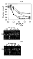
- FIG. 7 shows enzyme-linked immunosorbent assay (ELISA) results in the presence of various concentrations of NaCl, ascertaining that binding of 3D8 scFv, VH and VL antibody proteins of the present invention to DNAs is mainly caused by electrostatic force.
- ELISA enzyme-linked immunosorbent assay
- FIGs. 8 to 10 show agarose gel electrophoresis results, ascertaining that double-stranded (ds(N) 40 ) DNA-degrading activities of the 3D8 scFv, VH and VL proteins of the present invention depend on Mg 2+ and reaction time (In FIGs.
- B is BSA protein used as a control group
- Mg is a Mg 2+ treatment group
- E is an EDTA treatment group
- M is a marker
- rc is an abbreviation for "relaxed circular form” of DNA
- lain is an abbreviation for a “linear form” and refers to a linear DNA
- sc refers to an abbreviation for "supercoiled form” and refers to a completely coiled DNA
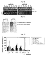
- FIG. 11 shows in situ acrylamide gel assay results, ascertaining that dsDNA-degrading activities of 3D8 scFv, VH and VL proteins of the present invention are based on the proteins'inherent natures ("Etbr treatment group” is a group in which black-white colors of data results are contrasted and a dark site is where DNA was not reacted with Etbr);
- FIG. 12 shows affinity-linked oligonucleotide nuclease assay (ALONA) results, ascertaining that DNase activities of 3D8 scFv, VH and VL proteins of the present invention are non-specific for base sequences;
- FIG. 13 shows the structure of 3D8 scFv protein crystals of the present invention analyzed by X-Ray diffraction
- FIG. 14 shows the overlapping structure of 3D8 scFv protein crystals according to the present invention and BV04-01 scFv as a conventional anti-DNA catalytic antibody;
- FIG. 15 shows the structure of 3D8 VH protein crystals of the present invention.
- FIG. 16 shows the structure of 3D8 VL protein crystals of the present invention
- FIG. 17 shows enzyme-linked immunosorbent assay (ELISA) results, ascertaining DNA-binding ability and DNase activity of a scFv mutant in which two histidine (His) residues are substituted by alanine (black bar: ds-(dN:dN') 40 , white bar: ss-(dN) 40 ).
- ELISA enzyme-linked immunosorbent assay
- FIG. 18 shows agarose gel electrophoresis results ascertaining DNase activity of a scFv mutant in which two histidine (His) residues are substituted by alanine
- B is BSA protein used as a control group
- Mg is a Mg 2+ treatment group
- E is an EDTA treatment group
- M is a marker
- rc is an abbreviation for "relaxed circular form” of DNA
- lain is an abbreviation for a “linear form” and refers to a linear DNA
- sc refers to an abbreviation for "supercoiled form” and refers to a completely coiled DNA
- FIG. 19 shows enzyme-linked immunosorbent assay (ELISA) results ascertaining variations in DNA-binding ability of a 3D8 VH mutant and a 3D8 VL mutant in which two histidine (His) residues are substituted by alanine (black bar: ds-(dN:dN') 40 , white bar: ss-(dN) 40 ).
- ELISA enzyme-linked immunosorbent assay
- FIG. 20 shows agarose gel electrophoresis results, ascertaining variations in DNase activity of a 3D8 VH mutant and a 3D8 VL mutant in which one histidine (His) residue are substituted by alanine
- B is BSA protein used as a control group
- Mg is a Mg 2+ treatment group
- E is an EDTA treatment group
- M is a marker
- rc is an abbreviation for "relaxed circular form” of DNA
- lain is an abbreviation for a “linear form” and refers to a linear DNA
- sc refers to an abbreviation for "supercoiled form” and refers to a completely coiled DNA
- FIG. 21 shows agarose gel electrophoresis results, ascertaining the RNase activity of the scFv proteins of the present invention according to reaction time
- FIG. 22 shows in situ hydrolysis assay, ascertaining that the RNase activity of 3D8 scFv proteins of the present invention was attributed to the proteins'inherent nature ("Etbr treatment group” is a group in which black-white colors of the data results are contrasted and a dark site is where RNA was not reacted with Etbr).
- FIG. 23 shows affinity-linked oligonucleotide nuclease assay (ALONA) results, ascertaining that RNase activity of 3D8 scFv proteins of the present invention is not dependant on Mg 2+ .
- FIGs. 24 and 25 are fluorescence microscope images, ascertaining that 3D8 scFv proteins of the present invention transferred in vero cells exhibit inhibitory activity of vesicular stomatitis virus (VSV) proliferation
- VSV vesicular stomatitis virus
- FIGs. 26 and 27 are fluorescence microscope images, ascertaining that 3D8 scFv proteins of the present invention transferred in vero cells exhibit inhibitory activity of vesicular stomatitis virus (VSV) proliferation
- VSV vesicular stomatitis virus
- FIG. 28 shows MTT assay results, ascertaining whether or not cytopathic effects (CPE) by VSV infection are inhibited on respective vero cells into which 3D8 scFv, 3D8 VH, 3D8 VL or other protein is introduced;
- CPE cytopathic effects
- FIG. 29 shows immunofluorescence staining results by confocal microscope, ascertaining inhibitory activities of VSV proliferation on respective HeLa cells, into which 3D8 scFv, VH, and VL proteins are transferred ("3D8-” refers to “3D8 scFv protein-free”, “3D8+” refers to “3D8 scFv protein-injected”, “VSV-” refers to a “VSV non-treated”, and “VSV+” refers to "VSV-treated”).
- FIG. 30 is a schematic diagram of animal cell-expression vectors into which 3D8 scFv genes are cloned
- FIG. 31 shows RT-PCR results, ascertaining that 3D8 scFv mRNAs are favorably expressed in NIH/3T3 cells into which 3D8 scFv genes are stably transfected;
- FIG. 32 shows results of immunofluorescence staining assays using TRITC, ascertaining expression of 3D8 scFv mRNAs in NIH/3T3 cells into which 3D8 scFv genes are stably transfected ("a” is an image showing 3D8 scFv-free NIH/3T3 cells and "b” is an image showing 3D8 scFv-transfected NIH/3T3 cells;
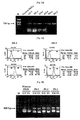
- FIG. 33 is an agarose gel electrophoresis image showing RT-PCR results ascertaining expression of the 3D8 scFv mRNA in transformed HeLa cell lines;
- FIG. 34 shows flow cytometry results ascertaining the expression of 3D8 scFv proteins in representative transformed HeLa cell lines
- FIG. 35 is an agarose gel electrophoresis image showing RT-PCR results ascertaining expression of 3D8 scFv mRNA in transformed PK15 cells;
- FIG. 36 shows MTT assay results, ascertaining whether or not cytopathic effect (cell death) by VSV is inhibited on 3D8 scFv protein-expressing NIH/3T3 cells;
- FIG. 37 shows fluorescence microscopy assay results, ascertaining whether or not cytopathic effect (cell death) by VSV is inhibited on 3D8 scFv protein-expressing HeLa cells;
- FIG. 38 shows intracellular staining results, ascertaining that multiplication of CSFV (classical swine fever virus) is inhibited on transformed PK15 cells;
- FIG. 39 shows MTT assay results ascertaining quantitative anti-VSV activity of the 3D8 scFv protein-expressing HeLa cells
- FIG. 40 shows fluorescence microscopy assay results ascertaining anti-VSV activity (cell death inhibition) of 3D8 scFv protein-expressing HeLa cells;
- FIG. 41 shows microscopy assay results ascertaining anti-ADV activity (cell death inhibition) of the 3D8 scFv protein-expressing HeLa cells.
- FIG. 42 shows an agarose gel electrophoresis image showing RT-PCR results ascertaining ADV virus-multiplication inhibition activity of the transformed HeLa cell lines.
- the present invention utilizes an antiviral agent containing the protein as defined according to the present invention or a nucleic acid sequence encoding the protein, as an active ingredient, and an antiviral animal cell containing the protein according to the present invention or a nucleic acid sequence encoding the protein as an active ingredient.
- the present invention is directed to an antiviral agent for use in the prevention or treatment of RNA-typed animal virus mediated diseases the antiviral agent-containing, as an active ingredient, a protein that has binding and degrading abilities selective for foreign nucleic acid chains invaded in an animal cell and that has no cytotoxicity to the animal cell itself, and to an antiviral agent against animal viruses containing, as an active ingredient, a nucleic acid sequence encoding a protein that has binding ability and degrading ability to foreign nucleic acid chains invaded in an animal cell and that has no cytotoxicity to the animal cell itself.
- the present invention is directed to an antiviral animal cell containing, as an active ingredient, a protein that has binding ability and degrading ability to foreign nucleic acid chains invaded in an animal cell and that has no cytotoxicity to the animal cell itself, and to an antiviral animal cell containing, as an active ingredient, a nucleic acid sequence encoding a protein that has binding ability and degrading ability to foreign nucleic acid chains invaded in the animal cell and that has no cytotoxicity to the animal cell itself.
- the protein used for the antiviral agent and the antiviral animal cell must be capable of binding to foreign nucleic acid chains and degrading the chains while having no cytotoxicity to the animal cell.
- 3D8 proteins As one example of proteins that meet these requirements, separated and purified 3D8 proteins were evaluated for antiviral activity against animal viruses. As can be seen from the following Example 1, the evaluation results demonstrated that the 3D8 proteins exhibit antiviral activity against animal viruses and have no cytotoxicity.
- binding ability to foreign nucleic acid chains refers to an ability of proteins that can recognize nucleic acid (such as DNA and RNA) chains and bind to the nucleic acid chains.
- the binding of proteins to nucleic acid chains is based on the couplings between positive charges in amino acids constituting proteins and negative charges in phosphate backbones of DNA or RNA.
- Anti-nucleic acid antibodies and nucleases are generally bound to DNA or RNA by recognizing specific sequences of the DNA or RNA. As apparent from Experimental Examples 2 and 5, the protein of the present invention is bound to nucleic acid chains without any sequence specificity, thus being widely utilized in the research fields associated with nucleic acid degradation activity.
- degradation ability to foreign nucleic acid chains refers to an ability of proteins that can cleave nucleic acid such as DNA and RNA chains by hydrolysis. This term is also called a “catalytic ability”.
- cytotoxicity refers to a phenomenon in which cells are killed by invading foreign materials.
- DNases DNA-degrading enzymes
- RNases RNA-degrading enzymes
- the protein as utilized according to the present invention is characterized in that it has no cytotoxicity to self-derived cells.
- the nucleic acid sequence encoding a protein that has binding and degrading abilities to foreign nucleic acid chains introduced into animal cells and has no cytotoxicity to the animal cells may be either a RNA or DNA sequence. Preferred is the DNA sequence in view of stability.
- the DNA sequence may be a nucleic acid sequence introduced into vectors capable of expressing genes in animal cells. As can be confirmed from Example 4, the introduction of genes having such a DNA sequence into animal cells leads to efficient expression of proteins and exhibition of efficacious antiviral activity.
- the protein capable of binding to invading foreign nucleic acid chains and degrading the chains and having no cytotoxicity to the animal cell itself, which is used for the antiviral agent and the antiviral animal cell according to the present invention is obtained from autoimmune disease-induced animals. This is the reason that proteins bound to nucleic acids or reacted therewith are known to be intimately associated with autoimmune diseases.
- the animal there is no particular restriction as to the animal. Any animal may be used without particular limitation so long as it naturally induces an autoimmune disease. Preferred is the use of a mouse.
- an Ipr gene is preferably malfunctioned.
- Ipr genes mediate production of Fas proteins which are one of the major factors causing apoptosis.
- the Fas proteins mediate cell apoptosis on the surface of cells. When the Fas proteins are activated, cells begin to undergo the apoptosis mechanism. On the other hand, when Ipr genes are injured, formation of Fas proteins is inhibited and normal cell apoptosis thus does not occur. Such a mechanism suppresses removal of lymphocytes capable of attacking self-cells, thus leading to an autoimmune disease.
- immunoglobulin or a fragment thereof may be used that has binding and degrading abilities specific for foreign nucleic acid chains.
- the immunoglobulin may be preferably immunoglobulin G (IgG). More preferably, the IgG may be a 3D8 protein.
- Immunoglobulin herein used is a generic term for proteins that are serum ingredients playing an important role in immunity and acting as antibodies.
- Such immunoglobulin consists of a pair of light chains (CL) having a molecular weight of about 23,000 and a pair of heavy chains (CH) having a molecular weight of about 50,000 to 70,000 which are connected by disulfide bonds.
- the type of heavy chains denoted by ⁇ , ⁇ , ⁇ , ⁇ and ⁇ defines the class of antibody, i.e ., IgG, IgA, IgM, IgD and IgE, respectively.
- IgG consists of a pair of light chains having a molecular weight of about 25,000 and a pair of heavy chains having a molecular weight of about 50,000, and has a total molecular weight of 150,000.
- IgG is an immunoglobulin essential in the higher animals (amphibian or higher), takes about 70% of overall human immunoglobulins, passes through placenta and exhibits a variety of antibody activities.
- an immunoglobulin fragment is be used selected from scFv, VH, VL, and Fv with an association of VH and VL.
- a scFv single chain fragment variable is a fusion of a variable heavy (VH) chain and a variable light chain (VL), linked through a linker.
- VH variable heavy
- VL variable light chain
- Such a scFv was known to have a strong antigen-binding force, compared to VH or VL used singly.
- the linker that mediates the binding between VH and VL may be of any type commonly used in the art.
- VH or VL the antigen-binding force of VH or VL is generally known to be inferior to that of scFv.
- preferred is VH or VL in some cases where improvement in permeability into the tissue is intended.
- the scFv has a sequence set forth in SEQ ID NO 6, the VH has a sequence set forth in SEQ ID NO 2 and the VL has a sequence set forth in SEQ ID NO 4.
- proteins having binding and degrading abilities specific for RNA chains only can be applied to RNA viruses only and proteins having binding and degrading capabilities specific for DNA only chains can be applied to DNA viruses only.
- proteins that have binding and degrading abilities to both DNA and RNA chains in that they are applicable to both RNA viruses and DNA viruses.
- the virus invaded in the animal cells.
- the virus may be RNA-based virus, and more specifically may be HIV virus or SARS virus.
- VH, VL and scFv domains of 3D8 antibodies obtained in accordance with the method of Preparative Example 1 were used as one example.
- the VH, VL and scFv domain antibodies were obtained from 3D8 hybridoma cells in accordance with the method of Preparative Example 2. These antibodies were used following over-expression in E. coli and purification.
- Affinity-linked oligonucleotide nuclease assay was used to probe s ubstrate-specificity of DNase activity.
- the scFv, VH and VL proteins substantially exhibited no DNA-substrate specificity on DNase activity.
- 3D8 scFv proteins were used to evaluate RNase activity.
- the scFv proteins exhibited RNase activities on dsRNAs as well as on ssRNAs, depending on the reaction time.
- the substrate-specificity assay results indicated that scFv antibody proteins are capable of degrading RNA/DNA hybrids as well as ssRNAs and dsRNAs on RNase activities.
- Example 1 after 3D8 scFv protein-injected animal cells are transfected with vesicular stomatitis virus, antiviral activity of 3D8 scFv protein against the virus was confirmed. As a result, it can be confirmed that animal cells, into which no 3D8 scFv protein is introduced, underwent cell death which is one of cytopathic effects (CPEs) by VSV ( FIG. 24 ), whereas animal cells, into which 3D8 scFv proteins are introduced, were still alive regardless of VSV infection. In addition, that antiviral activity, i.e., cell death inhibitory activity, of the 3D8 scFv proteins on HeLa cells (human cervical carcinoma cell lines) was observed.
- CPEs cytopathic effects
- Example 2 The results obtained from Example 2 ascertained that 3D8 VH and VL proteins as well as 3D8 scFv proteins exhibited antiviral activities against animal cells.
- nucleic acid sequences encoding the protein that has binding ability and degrading ability to foreign nucleic acid chains invaded in animal cells and has no cytotoxicity to the animal cells were invaded in animal cells, the nucleic acid sequences are proven to exhibit anti-virus activity against virus-infected animal cells. Specifically, it can be confirmed from Example 4 that 3D8 scFv gene-transfected animal cells also produce scFv proteins and also exhibit resistance against various viruses.
- Antiviral agent for use in the prevention or treatment of RNA-typed animal virus mediated diseases containing, as an active ingredient, a protein having binding and degrading abilities to foreign nucleic acid chains invaded in an animal cell and having no cytotoxicity to the animal cell itself>
- the antiviral agent for use in the prevention or treatment of RNA-typed animal virus mediated diseases containing the protein as defined according to the present invention as an active ingredient is capable of selectively degrading invaded foreign nucleic acid chains and has no cytotoxicity to the animal cell itself.
- the antiviral agent as used according to the present invention is capable of removing invading-foreign viruses only, while not killing self-cells, thus being useful for virus-mediated diseases such as acquired immunodeficiency syndrome (AIDS). More specifically, the treatment of the diseases affects virus-infected cells and thus prevents death of the cells.
- AIDS acquired immunodeficiency syndrome
- the antiviral agent as used according to the present invention contains, as an active ingredient, a protein that has binding ability and degrading ability to foreign nucleic acid chains invaded in an animal cell and has no cytotoxicity to the animal cell itself.
- antiviral agent contains protein as an active ingredient
- the protein is added to the antiviral agent in an amount such that the protein can exert desirable antiviral activity, and that, in a broad sense, the protein is formulated in a variety of dosage forms together with various adjuvants for drug delivery and stabilization.
- the protein may include immunoglobulin or a fragment thereof that has binding/degrading abilities to foreign nucleic acid chains. More preferably, the immunoglobulin may be immunoglobulin G (IgG). Most preferably, the IgG may be a 3D8 protein. Preferably, the immunoglobulin fragment may be one selected from scFv, VH, VL, and Fv with an association of VH and VL.
- immunoglobulin G immunoglobulin G
- the immunoglobulin fragment may be one selected from scFv, VH, VL, and Fv with an association of VH and VL.
- the antiviral agent as used according to the present invention may further comprise a pharmaceutically acceptable carrier or additive (generally, a buffer solution, an isotonic solution, an aqueous suspension or the like, and optionally, a stabilizer, an antiseptic, or the like).
- a pharmaceutically acceptable carrier or additive generally, a buffer solution, an isotonic solution, an aqueous suspension or the like, and optionally, a stabilizer, an antiseptic, or the like.
- Formulations generally comprise saline and optionally protected or stabilized molecules such as a high molecular weight of proteins (e.g ., human serum albumin).
- the protein that has binding and degrading abilities to foreign nucleic acid chains invaded in an animal cell and has no cytotoxicity to the animal cell itself, preferably IgG or a fragment thereof, is administered to a receptor in a dosage rendering the protein to be "effective” or "efficacious for treatment".
- the effective dosage of the protein administered to a receptor may be in a range of about 0.1 mg/kg to about 20 mg/kg.
- the effective dosage of proteins is determined by various factors such as protein type (e.g ., overall IgG or fragments thereof), affinities and individual characteristics of specific therapeutic proteins and depends on pharmacokinetic parameters. Accordingly, the effective dosage of the protein according to the present invention may depend on these various factors.
- the use of the protein-containing antiviral agent enables the realization of the desirable treatment efficacy despite of administration of the therapeutic protein in a reduced dosage.
- the use (e.g ., dosage and prescription designs) of the therapeutic protein of the present invention may be restricted by undesired side effects (e.g ., pyrexia, headache, gasp, blood pressure drop, and the like).
- a standard dosage of therapeutic proteins to be administered to the patients may be varied according to individual characteristics of patients.
- a skilled clinical physician may design the most preferable therapeutic strategies (e.g ., optimal dosage and dose regimen) for patients taking into consideration factors e.g ., specific necessities and overall conditions of the patients.
- Suitable dosages of therapeutic proteins may be determined in accordance with various references.
- a physician may gradually reduce a given amount of therapeutic proteins while administrating the protein-containing antiviral agent as defined according to the present invention to patients.
- the relative concentration and dose form of therapeutic proteins may be controlled to optimize therapeutic efficacy and minimize side effects.
- the therapeutic protein as defined according to the present invention may be generally determined in vitro or by an animal model.
- an optimal dosage may be determined by in vitro introducing various concentrations of therapeutic proteins as defined according to the present invention into target cells (virus-infected cells), followed by evaluating a death ratio of the target cells.
- the therapeutic protein-containing antiviral agent as used according to the present invention may be directly administered to subjects by intravenous, intraperitoneal, intraarterial, intramuscular or intradermal injection.
- Monoclonal antibodies such as rituxan (marketed under the name of RituximabTM) and omalizumab (marketed under the name of XolairTM) were efficacious in clinical treatments and equivalent prescription regimes ( i.e ., formulation and/or dosage and/or dose protocol) may be used for the therapeutic protein-containing antiviral agent as described according to the present invention.
- the antiviral agent as described according to the present invention may be simultaneously or sequentially used in conjunction with chemotherapies or therapeutic programs for treating viral diseases or a combination thereof.
- Antiviral agent for use in the prevention or treatment of RNA-typed animal virus mediated diseases the antiviral agent containing, as an active ingredient, a nucleic acid sequence encoding a protein that has binding and degrading abilities to foreign nucleic acid chains invaded in an animal cell and has no cytotoxicity to the animal cell itself>
- DNA sequences as nucleic acid sequences encoding proteins, may be carried out using individual expression vectors or a single expression vector.
- the nucleic acid sequence encoding the protein as defined according to the present invention includes immunoglobulin or a fragment thereof that has binding/degrading abilities to foreign nucleic acid chains. More preferably, the immunoglobulin may be immunoglobulin G (IgG). Most preferably, the IgG may be a 3D8 protein.
- the immunoglobulin fragment may be nucleic acid encoding one fragment selected from scFv, VH, VL, and Fv with an association of VH and VL.
- the vectors may be mixed before injection and electroporation, in order to allow individual muscle cells to take up and express each of the vectors.
- expression vector refers to a plasmid or vehicle that is manipulated by insertion or incorporation of a polypeptide-encoding nucleic acid and that is capable of directing the expression of the polypeptide when the vector is in an appropriate environment.
- a suitable expression vector typically includes a promoter, an origin of replication, a poly-A recognition sequence, and a ribosome recognition site or internal ribosome entry site, and may include other regulatory elements such as an enhancer (tissue-specificity).
- the promoter which facilitates the efficient transcription of the inserted encoding nucleic acid sequence in muscle, can be constitutive or, if desired, inducible, tissue specific or developmental stage specific.
- promoters useful for the present invention include promoters normally functioning in muscles such as the skeletal actin gene promoter ( Muscat, et al., Mol. Cell. Biol. 7, 4089 (1989 )), the muscle creatine kinase promoter ( Sternberg, et al., Mol. ell. Biol., 8, 2896 (1988 )) and the myosin light chain enhancer/promoter ( Donoghue, et al, Proc. Natl. Acad. Sci., USA 88, 5847 (1991 )).
- a preferred expression vector comprises an expression cassette that has multiple endonuclease restriction sites allowing ready cloning of DNA encoding different polypeptides into the cassette in a manner that places the encoding DNA in operative linkage with the promoter or other transcriptional regulatory elements of the vector.
- a preferred expression vector also may have an origin of replication for a procaryotic cell and at least one selective marker to aid in cloning in such a cell. Those skilled in the art would know how to optimize expression by selecting a vector properly configured with the appropriate combination of promoter, enhancer and other transcriptional or translational regulatory element for the polypeptide (s) to be expressed in muscle.
- the methodology of the present invention allows for multi-chain protein expression from skeletal muscles, smooth muscles, and cardiac muscles, respectively. Expression following injection of skeletal muscle is preferred because of the abundance and ready access of this muscle source.
- the expression vector may be injected through the skin and into the skeletal muscle via traditional means such as a syringe and needle, or by a needle-less injection device. Such latter devices are well-known in the art and, generally, involve pressure-assisted delivery through a tiny orifice held against the skin.
- the injection apparatus may be suitably selected with reference to U.S. Patent No. 4,596,556 issued to Morrow, et al ; U.S. Patent No. 4,913,699 issued to Parsons ; and U.S. Patent No. 5,730,723 issued to Castellano, et al .
- Another available commercially injection apparatus is BIOJECT TM (available from Bioject Medical Technologies, Inc., Portland Oregon U.S.A.).
- Another needless injection apparatus is a gene gun delivery system that uses pressurized gas to deliver small particles (e.g ., gold particles) into the targeted skin site, as a function of the gas pressure.
- An example of a biolistic delivery device is PDS-1000 "gene gun" (marketed by Dupont, Inc., Wilmington, Delaware, U.S.A.).
- Expression vectors can be administered in 0.9% sodium chloride, however, there are a variety of solvents and excipients that may be added without taking any effect on the expression level. For example, it is well known in the art that sucrose is capable of increasing DNA uptake in skeletal muscles.
- the animal cell exhibiting antiviral efficiencies against animal viruses as described according to the present invention may be utilized as an experimental use and as a cell medication according to well-known methods.
- Spleen cells of MRL- lpr / lpr mice (available from Jackson Laboratory, U.S.A.) and V653 cells (available from the Korean cell line bank) were fused with polyethylene glycol (PEG) 4000 and cultured in a hypoxanthine-aminopterin-thymidine (HAT) medium (available from Sigma-Aldrich, Inc.) in a 5% CO 2 incubator at 37°C for 15 days.
- PEG polyethylene glycol
- HAT hypoxanthine-aminopterin-thymidine
- ELISA enzyme-linked immunosorbent assay
- the cell lines were cultured twice by limiting dilution to obtain about ten kinds of hybridoma cell lines that secrete anti-DNA antibodies.
- the anti-DNA antibody-secreting cell line that shows the greatest DNA-binding capacity was selected from the hybridoma cell lines by ELISA.
- the selected cell line was deposited at the Korean Cell Line Research Foundation (KCLRF) on Sep. 13,2006 (Accession No: KCLRF-BP-00146).
- the isotype of the antibody the selected hybridoma cell line secretes was detected using an isotype detection kit (Pierce, Inc.). As a result, the antibody derived from the cell line was determined to be IgG2a/kappa and was called "3D8".
- RNAzol B TM RNA extraction reagent
- TEL-TEST, Inc. RNAzol B TM ; TEL-TEST, Inc.
- 0.02 ml of chloroform was added to the solution, thoroughly stirred for 15 seconds and allowed to stand in an ice bath for 5 minutes.
- the resulting solution was centrifuged and the resulting supernatant was then separated.
- 0.25 mL of ethanol was added to the residue, allowed to stand at 4°C for 15 minutes and was then centrifuged (at 12000 g, 4°C) for 15 min.
- the resulting RNA precipitate was washed with 70% ethanol, dried and dissolved in distilled water.
- the absorbance of the RNA at 260 nm and 280 nm was measured and a purity and yield of the RNA were determined.
- cDNAs were synthesized from the RNA thus obtained using a RT-PreMix kit (Bioneer Inc.). At this time, oligo-(dT) oligonucleotide was used as a primer. VH and VL genes were amplified with a polymerase chain reaction (PCR) method using the cDNAs as templates.
- PCR polymerase chain reaction
- the primer set used for VH gene amplification is as follows:
- the VH gene amplification was carried out by a touchdown PCR for 30 cycles using a Taq DNA polymerase.
- the touchdown PCR conditions (in each cycle) were given as follows: at 94°C for one min -> decreasing 0.5°C per cycle (from an initial annealing temperature (65°C) for one min -> at 72°C for one minute.
- the primer set used for VL gene amplification is as follows:
- the VL gene amplification was carried out by a touchdown PCR for 30 cycles using a Taq DNA polymerase.
- the touchdown PCR conditions (in each cycle) were given as follows: at 94°C for one min -> decreasing 0.3°C per cycle (from an initial annealing temperature (63°C) for one min -> at 72°C for one minute.
- Respective PCR reaction solutions were subjected to electrophoresis with a 1% agarose gel, and VH and VL genes were extracted from the gel and cloned in pGEM-T Easy vectors (available from Promega Corp.).
- the base sequence of the cloned genes was analyzed by a Perkin Elmer 373A automated DNA sequencer (Applied Biosystems, Inc.).
- the primer set used for the base sequence analysis is as follows:
- VH and VL genes obtained in Preparative Example 2 were cloned in pIg20H and pIg20L vectors (available from the professor, Stollar BD, Tufts University, U.S.A).
- a primer containing an Xma I/ Xba I fragment as a restriction enzyme recognition site was prepared and used for the VH cloning.
- a primer containing a Bgl II/ Nco I fragment as a restriction enzyme recognition site was prepared and used for the VL cloning.
- VH and VL genes separately cloned on the pGEM-T Easy vector (Promega Corp.) were sequentially sub-cloned in pIg20 expression vectors (available from the Professor, Stollar BD, Tufts University, U.S.A.) containing a linker DNA (decoded as "GGGGS 3 ").
- the base sequence of the primer used herein is as follows:
- pIg20H-3D8 VH, pIg20L-3D8 VL and pIg20-3D8 scFv vectors were obtained (The base sequences of scFv genes cloned in the pIg20 vector are shown in FIG. 1 , and the maps of pIg20H-3D8 VH, pIg20L-3D8 VL and pIg20-3D8 scFv vectors are shown in FIG. 2 ).
- the scFv genes have a base sequence and an amino acid sequence as set forth in SEQ ID NO 5 and in SEQ ID NO 6, respectively.
- pIg20H-3D8 VH, pIg20L-3D8 VL, and pIg20-3D8 scFv vectors were transformed into E. coli BL21 (DE3) pLysE cells (available from Novagen Inc.) and cultured in a LB culture medium containing 100 ⁇ g ml of ampicillin and 20 ⁇ g/ml of chloramphenicol until the absorbance (A600) reached 0.8.
- 0.5 mM of isopropyl- ⁇ -D-thiogalactopyranoside (IPTG) was added to the culture medium and further cultured at ambient temperature for 4 hours.
- the cultured cells were centrifuged (at 10,000 ⁇ g, 4°C) for 10 min and the resulting supernatant was filtered through a 0.45 ⁇ m filter membrane.
- the filtrate was passed though an IgG-sepharose column (Amersham pharmacia Biotech Ltd.) at a flow rate of 1 ml/min, washed with a phosphate buffer solution (PBS, pH 7.4) and ammonium acetate (pH 5.0), and eluted with 0.1 M acetic acid (pH 3.4).
- the eluted proteins were dialyzed against a PBS and were subjected to sodium dodecyl sulfate polyacrylamide gel electrophoresis (SDS-PAGE) under reduction conditions to determine the purity of the proteins.
- FIG. 3 shows SDS-PAGE analysis results of VH, VL and scFv proteins. After 20 ⁇ g of respective proteins was subjected to SDS-PAGE and stained with coomassie blue, three recombinant antibodies were confirmed to be purified as purities (95% or more).
- VH, VL, scFv and Fv (defined as a variable fragment having a non-covalent bond between VH and VL) proteins were subjected to size exclusion chromatography (SEC) using a HPLC system.
- SEC size exclusion chromatography
- 0.02 ml of a protein (conc. 5-20 ⁇ M) was injected into a TSK G3000SWXL column (TosoHaas, Japan) and a phosphate buffer solution (50 mM sodium phosphate/150 mM NaCl, pH 7.4) was flowed therein at a flow rate of 0.7 ml/min and the chromatogram of the proteins was obtained from the absorbance at 280 nm.
- VH-VL associated Fv was seen from FIG. 4 .
- Quantitative association between VH and VL proteins was analyzed with a surface plasmon resonance (SPR) biosensor (Biacore ® 2000, Pharmacia, Sweden).
- SPR surface plasmon resonance
- VH proteins (12.5, 25, 50, 100, 200 nM) were flowed thereon under phosphate buffer solution conditions.
- the sensorgram analysis showed that VH was coupled to VL with a binding affinity of K D 14 nM ( FIG. 5 ).
- the quantitative binding capacity of the purified VH, VL and scFv proteins with DNAs with various base sequences was evaluated by surface plasmon resonance (SPR) analysis.
- a 40-mer synthetic DNA fragment containing a biotin-labeled 3'-terminal was immobilized onto a streptavidin-coated sensor chip, scFv (5 - 200 nM), VH (0.2 - 50 ⁇ M) and VL (1.6 - 200 ⁇ M) proteins previously diluted with a HES buffer (10 mM HEPES, pH 7.4 150 mM NaCl, 3 mM EDTA, 0.005% surfactant P20) were flowed to the chip at a flow rate of 50 ⁇ l/min for 3 min and a HES buffer was further flowed thereto at a flow rate of 50 ⁇ l/min for 3 min to induce dissociation of the proteins linked to the DNAs.
- a HES buffer 10 mM HEPES, pH 7.4 150 mM NaCl, 3 mM EDTA, 0.005% surfactant P20
- the sensorgram analysis ascertained that all of the three antibody proteins (scFv, VH and VL antibody proteins) have a considerably high binding capacity with ssDNA and dsDNA.
- the high binding capacity indicates that scFv, VH and VL antibody proteins hardly have base sequence specificity for DNAs ( FIG. 6 ).
- the DNA-affinity of scFv was 100-fold to 1,000-fold higher than those of VH and VL. This result shows that DNA-affinities of proteins where VH and VL domains coexist are superior to those of proteins where VH and VL domains separately exist.
- ELISA was performed using single-stranded (ss(N) 40 ) or double-stranded (ds(N) 40 ) nucleic acid having a 40-mer predetermined base sequence as a substrate in the presence of various concentrations (from 0 M to 0.8 M) of NaCl.
- the symbol “ ⁇ ” represents a ss(N) 40 substrate-bound scFv
- the symbol “ ⁇ ” represents a ds(N) 40 substrate-bound scFv
- the symbol “ ⁇ ” represents ss(N) 40 substrate-bound VH
- the symbol “ ⁇ ” represents a ds(N) 40 substrate-bound VH
- the symbol “ ⁇ ” represents ss(N) 40 substrate-bound VL
- the symbol “ ⁇ ” represents ds(N) 40 substrate-bound VL).
- the purified scFv (0.8 ⁇ M), VH (5 ⁇ M) and VL (5 ⁇ M) proteins were reacted with 300 ng of plasmid M13mp18 (available from New England Biolabs, Ltd) in the presence of a TBS solution (20 mM Tris-HCl, pH 8.0, 50 mM NaCl, 5 mM MgCl 2 ) for 1 to 12 hours.
- the reaction mixture was treated with trypsin and subjected to agarose gel electrophoresis.
- FIG. 8 shows agarose gel electrophoresis results after the reaction for one hour.
- FIG. 9 shows agarose gel electrophoresis results after the reaction for 12 hours.
- rc is an abbreviation for "relaxed circular form” of DNA
- the term “lin” is an abbreviation for a "linear form” and refers to a linear DNA
- sc refers to an abbreviation for "supercoiled form” and refers to a completely coiled DNA.
- the sc-type DNAs are sequentially degraded into rc- and lin-type DNAs. Subsequently, the lin-type DNAs are completely degraded into smaller DNA fragments.
- FIG. 11 shows results obtained from in situ acrylamide gel assay.
- the acrylamide gel assay was carried out in accordance with the following procedure. First, 2 to 10 ⁇ g of scFv proteins were subjected to SDS-PAGE on an 12% acrylamide gel containing ds M13mp18 DNA (20 ⁇ g/ml) or ss M13mp18 DNA (20 ⁇ g/ml).
- Etbr treatment group is a group in which black-white colors of data results are contrasted and a dark site is where the DNA was not reacted with Etbr).
- Affinity-linked oligonucleotide nuclease assay was used to probe substrate-specificity of DNase activity.
- Oligonucleotides having a digoxigenin-labeled 5'-terminal and a biotin-labeled 3'-terminal i.e ., ss-(dT) 40 , ds-(dT:dA) 40 , ss-(dN) 40 , ds-(dN:dN') 40 , ss-(dG-dC) 20 , and ds-(dG-dC:dC-dG) 20 were immobilized on a streptavidin-coated 96-well culture plate through biotin-streptavidin bindings, and allowed to react with 3D8 scFv (0.8 ⁇ M), Fv (5 ⁇ M), VH (5 ⁇ M) and VL (5 ⁇ M) in a MgCl 2 -containing T
- the ss-(dT) 40 herein used refers to a single-stranded nucleic acid consisting of thymine.
- the ds-(dT:dA) 40 herein used refers to a double-stranded nucleic acid consisting of 40-mer thymines and 40-mer adenines.
- the ss-(dN) 40 herein used refers to a single-stranded nucleic acid consisting of 40-mer specific bases.
- the ds-(dN:dN') 40 herein used refers to a double-stranded nucleic acid in which one 40-mer bases are complementarily bound to another 40-mer bases.
- the ss-(dG-dC) 20 herein used refers to a 20-mer single-stranded nucleic acid in which guanine is alternatively linked to cytosine.
- the ds-(dG-dC:dC-dG) 20 herein used refers to a double-stranded nucleic acid in which a 20-mer guanine-cytosine strand is linked to a 20-mer cytosine-guanine strand.
- the remaining DNAs were reacted with alkaline phosphatase-linked anti-digoxigenine antibodies and a p-NPP solution (1 mg/ml p-NPP in 0.1 M glycine, 1 mM ZnCl 2 , 1mM MgCl 2 , pH 10.3) was added thereto as a substrate of alkaline phosphatase.
- the absorbance at 405 nm was measured ( FIG. 12 ).
- the DNase activities of scFv, VH and VL are substantially non-specific for DNA substrates.
- His35 of VH and His94 of VL i.e., residues represented by the numbers "35” and "94" in FIG. 1
- the resulting proteins were expressed in bacteria and purified.
- the variations in DNA-binding ability and DNA-degrading ability of the proteins were evaluated.
- the DNA-binding ability of the proteins was evaluated by ELISA.
- a polystyrene 96-well culture plate was coated with a 40-mer oligonucleotide (10 ⁇ g/ml, 100 ⁇ l) dissolved in a TBS solution (20 mM Tris-HCl, pH 8.0, 50 mM NaCl, 5 mM MgCl 2 ) at 37°C for one hour.
- the plate was washed three times with TBST (0.1% Tween 20-containing TBS), 3% BSA-TBS was added thereto, allowed to stand at 37°C for one hour and washed three times with TBST.
- the purified scFv, VH, and VL wild-type proteins (20 ⁇ g/ml, 100 ⁇ l) and histidine mutant proteins were introduced into each well and allowed to react at 37 °C for one hour, and washed three times with TBST.
- the resulting proteins were reacted with rabbit IgG (10 ⁇ g/ml, 100 ⁇ l) at 37 °C for one hour, washed three times with TBST, reacted with alkaline phosphatase-linked anti-rabbit IgG (1:10,000 dilution, 100 ⁇ l) at 37°C for one hour, and washed three times with TBST.
- a p -NPP solution (1 mg/ml p -NPP in 0.1 M glycine, 1 mM ZnCl 2 , 1 mM MgCl 2 , pH 10.3) as a substrate of alkaline phosphatase was added to the resulting proteins.
- the absorbance of the proteins at 405 nm was measured.
- rc is an abbreviation for "relaxed circular form” of DNA
- the term “lin” is an abbreviation for a “linear form” and refers to a linear DNA
- sc refers to an abbreviation for "supercoiled form” and refers to a completely coiled DNA.
- the symbol "*" refers to a mutant in which histidine is substituted by alanine.
- ssRNA and dsRNA substrates were primarily prepared.
- the ssRNAs were prepared by synthesizing ssRNAs through in vitro transcription using a T7 promoter of tobacco mosaic virus-coat protein (TMV-CP) gene-cloned pLITMUS vectors (available from New England BioLabs, Inc.), followed by purification.
- T7 promoter of tobacco mosaic virus-coat protein (TMV-CP) gene-cloned pLITMUS vectors available from New England BioLabs, Inc.
- the dsRNAs were obtained by synthesizing ssRNAs through in vitro transcription using T7 and SP6 promoters of pLITMUS vectors, followed by purification and hybridization.
- ssRNAs 300 ng of the ssRNAs (or dsRNAs) thus prepared were reacted with 0.8 ⁇ M of scFv proteins in a TBS solution for 1 to 5 hours and subjected to agarose gel electrophoresis. From the agarose gel electrophoresis results, it can be confirmed that the scFv proteins exhibited RNase activities on dsRNAs as well as ssRNAs, depending on the reaction time ( FIG. 21 ).
- 3D8 scFv proteins were subjected to SDS-PAGE on an acrylamide gel containing 20 ⁇ g/ml of dsRNA or 20 ⁇ g/ml of ssRNA. The gel was washed with 7 M for one hour and allowed to stand in a Tris solution (10 mM Tris, pH 8.0, 10 mM MgCl 2 , 50 mM NaCl) at 37°C for overnight.
- oligonucleotide nuclease assay AONA
- oligonucleotides having a digoxigenin-labeled 5'-terminal and a biotin-labeled 3'-terminal i.e., ss-(rU) 40 , ds-(rU:rA) 40 , and ds-(rU:dA) 40 (RNA/DNA hybrid) were used as substrates.
- the ss-(rU) 40 herein used refers to a single-stranded nucleic acid consisting of 40-mer ribouracils.
- the ds-(rU:rA) 40 herein used refers to a double-stranded nucleic acid consisting of 40-mer ribouracils and 40-mer riboadenines.
- the ds-(rU:dA) 40 herein used refers to a double-stranded nucleic acid in the form of a RNA/DNA hybrid consisting of 40-mer ribouracils and 40-mer deoxyribose adenines, arranged such that ribouracil is alternatively linked to deoxyribose adenine.
- RNAs and RNA/DNA hybrid were reacted with alkaline phosphatase-linked anti-digoxigenine antibodies, and a p -NPP solution (1 mg/ml p- NPP in 0.1 M glycine, 1 mM ZnCl 2 , 1mM MgCl 2 , pH 10.3) was added thereto as a substrate of alkaline phosphatase.
- a p -NPP solution (1 mg/ml p- NPP in 0.1 M glycine, 1 mM ZnCl 2 , 1mM MgCl 2 , pH 10.3 was added thereto as a substrate of alkaline phosphatase.
- the absorbance of the resulting mixture at 405 nm was measured. The results are shown in FIG. 23 .
- scFv antibody proteins are capable of degrading RNA/DNA hybrids as well as ssRNAs and dsRNAs, and the RNase activity of scFv antibody proteins is independent of Mg 2+ , which is different from DNase activity of the scFv antibody proteins.
- the Mg 2+ -independent RNase activity is known to be observed on conventional RNases.
- Example 1 Ascertainment of antiviral activity on 3D8 scFv protein- transferred animal cells by fluorescence microscopy
- the 3D8 scFv antibodies thus expressed in bacteria and purified were fluorescence-labeled with an Alexa 488 labeling kit (Molecular probes, Inc., green fluorescence). Then, the 3D8 scFv antibodies were injected into vero cells (African green monkey kidney cells) with a microporator (available from Digital Bio Technology Co., Ltd., Korea) under the electroporation conditions of voltage 100, pulse width 35 and pulse number 2.
- scFv proteins 20 ⁇ g were introduced into 2 x 10 5 of the vero cells, the protein-introduced cells were inoculated on a 48-well culture plate containing 0.5 ml of DMEM-10% fetal bovine serum, and the inoculated plates were incubated in a 5% CO 2 incubator for 10 hours.
- the cells were transfected with vesicular stomatitis virus (VSV) having single RNA strand as genome in a 5% CO 2 incubator for one hour at MOI 5 and were incubated in a new DMEM-10% fetal bovine serum medium.
- VSV vesicular stomatitis virus
- 3D8- refers to "a 3D8 scFv protein-free”
- 3D8+ refers to a “3D8 scFv protein-injected”
- VSV+ refers to a "VSV-treated”.
- the 3D8 scFv proteins were transfected into HeLa cells (human cervical carcinoma cell lines) by electroporation (voltage 1000, pulse width 35, pulse number 2) and transfected (infected) with VSV. It was observed that Hela cells showed antiviral activity, i.e. , cell death inhibition, similar to the vero cells (See FIG. 27 , and it can be seen from FIG. 26 that 3D8 scFv protein-transferred Hela cells, into which VSV is transfected, exhibited no inherent cytotoxicity of the 3D8 scFv proteins (See the sections of "3D8+NSV-" in FIG. 27 ).
- 3D8- refers to “3D8 scFv protein-free”
- 3D8+ refers to “3D8 scFv protein-transferred”
- VSV- refers to "VSV non-treated”
- VSV+ refers to "VSV-treated”
- Example 2 Quantitative assay of antiviral activity on 3D8 scFv, VH or VL protein-transferred animal cells
- bovine serum albumin BSA
- lysozyme as control groups were electroporated into HeLa cells at 20 ⁇ g/2 x 10 5 cells, divided onto 96-well culture plates at 1 ⁇ 10 5 cells/well, and incubated in a 5% CO 2 incubator for 24 hours.
- a MTT solution (1 mg/ml PBS, 100 ⁇ l) was added thereto and cultured in a 5% CO 2 incubator for 4 to 5 hours.
- the resulting fomazan was dissolved in 200 ⁇ l of dimethyl sulfoxide (DMSO).
- DMSO dimethyl sulfoxide
- Example 3 Ascertainment of antiviral activities on 3D8 scFv, VH and VL protein-transferred animal cells by confocal microscopy
- the scFv, VH and VL proteins were electroporated into HeLa cells on 24-well culture plates at 20 ⁇ g/2 ⁇ 10 5 cells/well and cultured in wells with a sterilized cover glass for 24 hours.
- the cells were transfected with VSV for one hour and cultured in a new medium for 12 hours.
- the immunofluorescence staining was conducted. Specifically, the medium was separated from each well, the cells were washed once with a PBS buffer solution containing 2% FBS, 400 ⁇ l of a fixation/permeabilization solution (Becton Dickinson, Inc.) was added thereto and allowed to stand at 4 °C for 20 min.
- a fixation/permeabilization solution Becton Dickinson, Inc.
- the resulting cells were washed twice with a 2% FBS-containing PBS buffer solution, reacted with 500 ⁇ l of anti-VSV-FITC antibodies (1:1000 dilution) (Abcam, Inc.) at 4°C for 40 min and washed three times with 2% FBS-containing PBS buffer solution.
- a fixation solution (Invitrogen, Corp.) containing 3 ⁇ l of DAPI (4'-6-Diamidino-2-phenylindole), as a regent that stains a cell nucleus blue in color, was added to a slide glass previously prepared, the slide glass was covered with the cell-adhered cover glass, the remaining adjacent region was sealed with the fixation solution, and VSV particles were observed with a confocal microscope.
- VSV- refers to "3D8 scFv protein-free”.
- 3D8+ refers to "3D8 scFv protein-transferred”
- VSV- refers to "VSV non-treated”
- VSV+ refers to "VSV-treated”.
- PCR was conducted with pIg20-3D8 plasmids to obtain 3D8 scFv genes and the 3D8 scFv genes were cloned into animal cell-expression vectors ( FIG. 30 ).
- the 3D8 scFv gene cloning was carried out using three vectors including pcDNA3.1 vector (invitrogen, Inc.), retrovirus vector pLXIN (invitrogen, Inc.), and retrovirus vector pQCXIN (invitrogen, Inc.) (These vectors were represented by "a", "b” and "c", respectively, in FIG. 30 ).
- the respective PCR primer set herein used is as follows:
- the resulting mixture was added to the HeLa cell lines or NIH/3T3 cell lines cultured on the 60 mm culture plates. After 24 hours, the used medium was replaced with a selective medium containing a G418 antibiotic (1 mg/ml), and substantially, replacement of the selective medium (at intervals of 2-3 days for 2 weeks) was continuously repeated to obtain colonies.
- RT-PCR reverse transcriptase-polymerase chain reaction
- FIG. 31 is an agarose gel electrophoresis image showing RT-PCR analysis results of the NIH/3T3 cells.
- Lane 1 is a size marker (100 bp ladder available from Bioneer, Inc.)
- Lane 2 is a PCR pattern of a negative control group in which D.W is used as a PCR template
- Lane 3 is a PCR pattern of a negative control group in which cDNA synthesized from non-transfected NIH/3T3 cells is used as a PCR template
- Lane 4 shows a PCR pattern of a group in which cDNA synthesized from pcDNA3.1-3D8 scFv-transfected NIH3T3 cells is used as a PCR template.
- the transfected NIH3T3 cells expressed 3D8 scFv proteins was ascertained by immunofluorescence staining. That is, the cells were fixed by 4% paraformaldehyde, and were sequentially subjected to membrane-permeabilization using 0.1% Triton X-100 and membrane-blocking using a 5% skim milk (DIFCO, Inc.).
- the resulting NIH3T3 cells were reacted with rabbit-derived anti-3D8 scFv primary antibody (at a dilution of 1:50 in 3% skim milk-TBST) at ambient temperature for 30 min, washed twice with 3% skim milk-TBST, and reacted with TRITC-linked anti-rabbit IgG (1:2,000 dilution) at ambient temperature for 30 min and washed twice with a 3% skim milk-TBST.
- a is an image showing 3D8 scFv-free NIH/3T3 cells and "b” is an image showing 3D8 scFv-transfected NIH/3T3 cells.
- the G418 were selected from 3D8 scFv-transfected HeLa cell lines and limiting dilution was performed on 96-well plates such that 0.5 cells are introduced into one well to establish monoclonal cell lines.
- FIG. 33 is an agarose gel electrophoresis image showing RT-PCR results, ascertaining expression of scFv mRNA in transfected HeLa cell lines.
- Lane M is a 100 bp ladder (available from Bioneer, Inc.)
- Lane Normal Hela is a PCR pattern of a negative control group, in which cDNA synthesized from non-transfected HeLa cells is used as a template
- the third to seventh Lanes in this order from the left are RT-PCR patterns of groups in which cDNA synthesized from transformed pcDNA3.1-3D8 scFv-transfected HeLa cell lines is used as a template, respectively.
- the resulting (NIH3T3) cells were reacted with rabbit-derived anti-3D8 scFv primary antibody (at a dilution of 1:50 in 2% fetal calf serum-TBST) at ambient temperature for 30 min, washed twice with 2% fetal calf serum-TBS, reacted with FITC-coupled anti-rabbit IgG (1:2,000 dilution) at ambient temperature for 30 min and washed twice with 2% fetal calf serum-TBST.
- the two retrovirus vectors i.e., pLXIN-3D8 scFv and pQCXIN-3D8 scFv, into which 3D8 scFv genes are cloned, were transfected into PT67 cells as packaging cell lines. 2 ⁇ 10 5 of the PT67 cells were grown on one well of 6-well plates for about 24 hours and washed with serum-free TOM TM (Welgene, Inc.).
- HeLa cells and PK15 cells were cultured on 60 mm culture plates for about 24 hours, and incubated in a 5% CO 2 incubator using a 10% serum-containing DMEM medium supplemented with polyblene (10 ⁇ g/ml) at 37°C for two weeks.
- the cells were incubated in the medium supplemented with 5 mL of the viral supernatant obtained by culturing the PT67 packaging cells at 37 °C for 24 hours, and G418 selection was conducted using 1 mg/ml of a G418-containing medium.
- H4-2, H5-14, H5-18 and H5-26 were established as representative HeLa cell lines transfected with the retrovirus and P1-1, P1-2 and P1-3 were established as representative PK cell lines transfected with the retrovirus.
- FIG. 35 is an agarose gel electrophoresis image showing RT-PCR results ascertaining expression of 3D8 scFv mRNA in the transformed PK15 cell lines.
- Lane M is a 100 bp ladder (available from Bioneer, Inc.)
- Lane Normal PK15 is a PCR pattern of a negative control group, in which cDNA synthesized from non-transfected PK15 cells is used as a template
- the third to seventh Lanes in this order from the left are RT-PCR patterns of groups in which cDNA synthesized from transformed pcDNA3.1-3D8 scFv-transfected HeLa cell lines is used as a template, respectively
- Lanes P1-1, P1-2 and P1-3 are RT-PCR patterns of groups in which cDNA synthesized from transformed PK15 cell lines is used as a template, respectively.
- Example 4 Antiviral activity assay of 3D8 scFv protein-expressing animal cells
- Antiviral activities of the NIH/3T3, and HeLa cell lines, in which 3D8 scFv genes were proven to be expressed were ascertained. Specifically, the NIH/3T3 cell lines were evaluated for antiviral activity against vesicular stomatitis virus (VSV), the HeLa cell lines were evaluated for antiviral activity against VSV, herpes simplex virus (HSV) and Aujesky's disease virus (ADV), and the PK15 cell lines were evaluated for antiviral activity against classical swine fever virus (CSFV).
- VSV vesicular stomatitis virus
- HSV herpes simplex virus
- ADV Aujesky's disease virus
- CSFV classical swine fever virus
- the amount of virus infected into the transformed cell lines was in a multiplication of infection (MOI) range of 0.1 to 1.0.
- MOI multiplication of infection
- the cell lines were infected with viruses, and after about 20-72 hours, cell death monitoring, RT-PCR or intracellular virus staining was performed.
- the antiviral activities of respective cell lines were ascertained in accordance with the following manner.
- HSV-GFP green fluorescent protein
- FIG. 36 shows quantitative MTT assay results ascertaining anti-VSV activity of the 3D8 scFv protein-expressing NIH/3T3 cells in the same manner as in Example 2.
- cell survival rates of the cell lines were compared with one another. Specifically, the survival rate of the non-transformed NIH/3T3 cell lines was about 47%, whereas survival rate of the 3D8 scFv gene-transformed NIH/3T3 cell lines was about 76%. Similarly, the survival rate of the non-transformed HeLa cells was about 32%, whereas the survival rate of 3D8 scFv-transformed HeLa cells was about 85% ( FIG. 36 ). These results indicated that 3D8 scFv proteins are capable of inhibiting VSV (RNA virus) activity in NIH/3T3 cells.
- VSV RNA virus
- FIG. 37 shows fluorescence microscopy assay results ascertaining anti-VSV activity of the 3D8 scFv protein-expressing HeLa cells.
- the cell behaviors were observed. From the cell observation, it was seen that the death rate of transformed HeLa cells was less than that of non-transformed HeLa cells (In FIG. 37 , "Normal Hela” is a normal cell that expresses no 3D8 scFv protein, and H4-2, H5-14, H5-18, and H5-26 are cells that express 3D8 scFv proteins). These results indicated 3D8 scFv proteins are capable of inhibiting VSV (RNA virus) activity in HeLa cells.
- VSV RNA virus
- the cells were reacted with 3B6 monoclonal antibodies (anti-E2 antibodies available from the National Veterinary Research & Quarantine Service, Korea) that recognize CSFV E2 antigens at 37 °C for 40 min.
- the resulting cells were washed three times with PBS, and reacted with biotin-coupled anti-mouse antibodies at 37 °C for 40 min.
- the resulting cells were washed three times with PBS, cultured in an AB solution for 40 min, washed three times with PBS, and reacted with a diaminobenzidine tetrahydrochloride (DAB) substrate solution at ambient temperature for about 10 min.
- DAB diaminobenzidine tetrahydrochloride
- FIG. 39 shows quantitative MTT assay results ascertaining anti-VSV activity of the 3D8 scFv protein-expressing HeLa cells in the same manner as in Example 2.
- FIG. 40 shows fluorescence microscopy assay results ascertaining anti-HSV activity of the 3D8 scFv protein-expressing HeLa cells in the same manner as in Example 4.
- FIG. 41 shows microscopy assay results ascertaining anti-ADV activity of the 3D8 scFv protein-expressing HeLa cells.
- ADV DNA virus
- FIG. 42 is an agarose gel electrophoresis image showing RT-PCR results ascertaining ADV virus-multiplication inhibition activity of the transformed HeLa cell lines.
- Lane M is a 100 bp ladder (available from Bioneer, Inc.)
- Lane Normal Hela is a RT-PCR pattern of a negative control group in which cDNA synthesized from non-transformed HeLa cells was used as a template
- the third to seventh Lanes in this order from the left are RT-PCR patterns of groups in which cDNA synthesized from transformed HeLa cell lines (H1-2, H2-2, H3-1, H3-2, and H3-3) was used as a template, respectively.
- the base sequence of primer used for the PCR is as follows.
- the primer targets gp50 genes of ADV virus.
- ADV forward primer 5'- CGTACCGCGCCCACGTGGCC -3' (SEQ ID NO 23)
- ADV reverse primer 5'- GTCGGTGAGGATGTTCACGC -3' (SEQ ID NO 24)
- a composition (20 mL) was prepared by dissolving the ingredients in an injection-purpose physiological saline, subjected to sterile-filtration, divided into 2-mL vials, lyophilized and then sealed.
- a composition (20 mL) was prepared by dissolving the ingredients in an injection-purpose physiological saline, subjected to sterile-filtration, divided into 2-mL vials, lyophilized and then sealed.
- a composition (20 mL) was prepared by dissolving the ingredients in a 0.01M phosphate buffer solution (PBS, pH 7.0), subjected to sterile-filtration, divided into 2-mL vials, lyophilized and then sealed.
- PBS phosphate buffer solution
- vector DNAs expressing ScFv proteins (SEQ ID NO: 6) were intramuscularly injected into quadricep muscles of mice. 50 ⁇ g of the vector DNAs diluted in 0.9% NaCl were each injected into both quadricep muscles of mice.
- electroporation was performed by placing an electrode on muscles adjacent to the site injected and applying an electrical potential having 10 trains of 1,000 pulses to the site. At this time, a length of the pulse applied was 200 ⁇ s (twice), an interval between two successive pulses was 600 ⁇ s and a current limit was 50 mA.
- a conductive gel was used for the skin. In the case of larger animals, the electrode may be inserted into muscles of the animals.
- the present invention can be applied to animal cells, in particular, to human cells, thus being widely utilized in the medication industry associated with the treatments for diseases derived from a variety of virus including HIV virus and SARS virus.
Landscapes
- Health & Medical Sciences (AREA)
- Chemical & Material Sciences (AREA)
- Life Sciences & Earth Sciences (AREA)
- Organic Chemistry (AREA)
- Virology (AREA)
- Medicinal Chemistry (AREA)
- General Health & Medical Sciences (AREA)
- Proteomics, Peptides & Aminoacids (AREA)
- Immunology (AREA)
- Genetics & Genomics (AREA)
- Molecular Biology (AREA)
- Biophysics (AREA)
- Biochemistry (AREA)
- Animal Behavior & Ethology (AREA)
- Veterinary Medicine (AREA)
- Public Health (AREA)
- Pharmacology & Pharmacy (AREA)
- Communicable Diseases (AREA)
- Oncology (AREA)
- Chemical Kinetics & Catalysis (AREA)
- General Chemical & Material Sciences (AREA)
- Nuclear Medicine, Radiotherapy & Molecular Imaging (AREA)
- Epidemiology (AREA)
- Bioinformatics & Cheminformatics (AREA)
- Engineering & Computer Science (AREA)
- Gastroenterology & Hepatology (AREA)
- Medicines That Contain Protein Lipid Enzymes And Other Medicines (AREA)
- Pharmaceuticals Containing Other Organic And Inorganic Compounds (AREA)
- Micro-Organisms Or Cultivation Processes Thereof (AREA)
- Peptides Or Proteins (AREA)
Claims (12)
- Antivirales Mittel zur Verwendung zur Prävention oder Behandlung von durch tierische Viren vom RNA-Typ vermittelten Erkrankungen, wobei das antivirale Mittel als Wirkstoff ein Protein enthält, das gegenüber fremden Nukleinsäureketten, die tierische Zellen befallen haben, bindende und degradierende Eigenschaften hat und keine Cytotoxizität gegenüber der tierischen Zelle selbst hat, oder eine Nukleinsäuresequenz, die ein Protein codiert, das bindende und degradierende Eigenschaften hat, die selektiv für fremde Nukleinsäureketten sind, die eine tierische Zelle befallen haben und keine Cytotoxizität gegenüber der tierischen Zelle selbst hat,
wobei das Protein ausgewählt ist aus scFv, VH, VL, und Fv mit einer Assoziierung von VH und VL, wobei das scFv eine wie in SEQ ID NO 6 dargestellte Sequenz hat, wobei das VH eine wie in SEQ ID NO2 dargestellte Sequenz hat, wobei das VL eine wie in SEQ ID NO4 dargestellte Sequenz hat. - Antivirales Mittel zur Verwendung nach Anspruch 1, wobei die Virusvermittelte Krankheit das erworbene Immunschwächesyndrom (AIDS) oder das schwerwiegende akute respiratorische Syndrom (SARS) Virus ist.
- Antivirales Mittel zur Verwendung nach Anspruch 1, wobei die Nukleinsäuresequenz eine DNA-Sequenz ist.
- Antivirales Mittel zur Verwendung nach Anspruch 3, wobei die DNA-Sequenz eine Nukleinsäuresequenz ist, die in einen Vektor integriert ist, der in der Lage ist, Gene in tierischen Zellen zu exprimieren.
- Antivirales Mittel zur Verwendung nach Anspruch 1, wobei das Protein von einer Autoimmunerkrankung-induzierten Maus erhalten wird.
- Antivirales Mittel zur Verwendung nach Anspruch 5, wobei die Autoimmunerkrankung durch eine Fehlfunktion eines Ipr-Gens induziert wird.
- Antivirale tierische Zelle, die als Wirkstoff ein Protein enthält, das gegenüber fremden Nukleinsäureketten, die tierische Zellen befallen haben, bindende und degradierende Eigenschaften hat und keine Cytotoxizität gegenüber der tierischen Zelle selbst hat, oder eine Nukleinsäuresequenz, die ein Protein codiert, das bindende und degradierende Eigenschaften hat, die selektiv für fremde Nukleinsäureketten sind, die eine tierische Zelle befallen haben und keine Cytotoxizität gegenüber der tierischen Zelle selbst hat,
wobei das Protein ausgewählt ist aus scFv, VH, VL, und Fv mit einer Assoziierung von VH und VL, wobei das scFv eine wie in SEQ ID NO 6 dargestellte Sequenz hat, wobei das VH eine wie in SEQ ID NO2 dargestellte Sequenz hat, wobei das VL eine wie in SEQ ID NO4 dargestellte Sequenz hat. - Antivirale tierische Zelle gemäß Anspruch 7, wobei die Nukleinsäuresequenz eine DNA-Sequenz ist.
- Antivirale tierische Zelle nach Anspruch 8, wobei die DNA-Sequenz eine Nukleinsäuresequenz ist, die in einen Vektor integriert ist, der in der Lage ist, Gene in tierischen Zellen zu exprimieren.
- Antivirale tierische Zelle nach Anspruch 7, wobei das Protein von einer Autoimmunerkrankung-induzierten Maus erhalten wird.
- Antivirale tierische Zelle nach Anspruch 10, wobei die Autoimmunerkrankung durch Fehlfunktion eines IPR-Gens induziert ist.
- Antivirale tierische Zelle nach Anspruch 7, wobei die Bindungs- und Degradationseigenschaften gegenüber fremden Nukleinsäureketten bindende und degradierende Eigenschaften gegenüber RNA-Ketten und/oder bindende und degradierende Eigenschaften gegenüber DNA-Ketten ist.
Applications Claiming Priority (2)
| Application Number | Priority Date | Filing Date | Title |
|---|---|---|---|
| KR20060090361 | 2006-09-19 | ||
| PCT/KR2007/004498 WO2008035894A1 (en) | 2006-09-19 | 2007-09-18 | Antiviral agent against animal viruses |
Publications (3)
| Publication Number | Publication Date |
|---|---|
| EP2068906A1 EP2068906A1 (de) | 2009-06-17 |
| EP2068906A4 EP2068906A4 (de) | 2010-02-03 |
| EP2068906B1 true EP2068906B1 (de) | 2012-08-01 |
Family
ID=39200690
Family Applications (1)
| Application Number | Title | Priority Date | Filing Date |
|---|---|---|---|
| EP07808288A Not-in-force EP2068906B1 (de) | 2006-09-19 | 2007-09-18 | Antivirales mittel gegen tierische viren |
Country Status (5)
| Country | Link |
|---|---|
| US (2) | US20110201104A1 (de) |
| EP (1) | EP2068906B1 (de) |
| KR (1) | KR100890463B1 (de) |
| CN (1) | CN101534848B (de) |
| WO (1) | WO2008035894A1 (de) |
Families Citing this family (12)
| Publication number | Priority date | Publication date | Assignee | Title |
|---|---|---|---|---|
| WO2009134027A2 (en) * | 2008-04-30 | 2009-11-05 | Ajou University Industry-Academic Cooperation Foundation | Anti-nucleic acid antibody inducing cell death of cancer cells and composition for preventing or treating cancers comprising the same |
| GB0812277D0 (en) * | 2008-07-04 | 2008-08-13 | Fusion Antibodies Ltd | Antibody and uses thereof |
| EP4349868A3 (de) * | 2010-05-14 | 2024-08-28 | The Board of Trustees of the Leland Stanford Junior University | Humanisierte und chimäre monoklonale antikörper gegen cd47 |
| BR112013009008A2 (pt) * | 2010-10-14 | 2017-10-17 | Veridex Llc | métodos e kits para a detecção de células circulantes de tumor em pacientes pancreáticos com o uso de reagentes de detecção de captura e de coquetel poliespecíficos |
| US9163090B2 (en) | 2012-05-07 | 2015-10-20 | Cellerant Therapeutics, Inc. | Antibodies specific for CLL-1 |
| KR101514415B1 (ko) | 2013-08-16 | 2015-04-27 | 대한민국 | 재조합 3D8 scFv 항체 |
| KR102261190B1 (ko) | 2019-10-04 | 2021-06-07 | 주식회사 노블젠 | 발현 효율이 향상된 항원 결합 단백질 |
| KR102333496B1 (ko) | 2019-10-04 | 2021-12-01 | 주식회사 노블젠 | 가수분해활성이 향상된 항원 결합 단백질 |
| KR102667849B1 (ko) * | 2020-08-07 | 2024-05-21 | 주식회사 노블젠 | 코로나바이러스에 대한 항바이러스용 조성물 |
| WO2022031137A1 (ko) * | 2020-08-07 | 2022-02-10 | 주식회사 노블젠 | 코로나바이러스에 대한 항바이러스용 조성물 |
| EP4212548A4 (de) * | 2020-09-08 | 2024-04-17 | Novelgen Co., Ltd. | Antivirale zusammensetzung gegen influenza-a-virus |
| CN112526128A (zh) * | 2020-11-20 | 2021-03-19 | 航科中投生物技术(北京)有限公司 | 一种快速检测多种病毒或病原体污染的检测试纸 |
Family Cites Families (13)
| Publication number | Priority date | Publication date | Assignee | Title |
|---|---|---|---|---|
| GB8719367D0 (en) * | 1987-08-15 | 1987-09-23 | Wellcome Found | Therapeutic compounds |
| EP0362162B1 (de) * | 1988-09-28 | 1992-03-04 | Sigma-Tau Industrie Farmaceutiche Riunite S.p.A. | Pharmazeutische Zubereitung, die Zidovudin und Inosiplex oder Bestandteile davon enthält, für die Behandlung von AIDS-verwandten Syndromen |
| US5026687A (en) * | 1990-01-03 | 1991-06-25 | The United States Of America As Represented By The Department Of Health And Human Services | Treatment of human retroviral infections with 2',3'-dideoxyinosine alone and in combination with other antiviral compounds |
| GB9221654D0 (en) * | 1992-10-15 | 1992-11-25 | Scotgen Ltd | Recombinant human anti-cytomegalovirus antibodies |
| JPH11510047A (ja) * | 1995-07-21 | 1999-09-07 | ユニバーシティ・オブ・ネブラスカ・ボード・オブ・リージェンツ | HIV gp120の加水分解を触媒する組成物および方法 |
| US5962725A (en) * | 1996-09-05 | 1999-10-05 | Agouron Pharmaceuticals, Inc. | Intermediate compounds useful for making HIV protease inhibitors such as nelfinavir |
| WO1998026086A1 (en) * | 1996-12-11 | 1998-06-18 | University Of Florida | Novel methods and compositions for treatment of autoimmune diseases |
| US5849800A (en) * | 1997-03-28 | 1998-12-15 | The Penn State Research Foundation | Use of amantadine for treatment of Hepatitis C |
| AUPO930697A0 (en) * | 1997-09-19 | 1997-10-09 | Walter And Eliza Hall Institute Of Medical Research, The | Catalytic antibodies and a method of producing same |
| JP3406250B2 (ja) | 1999-08-30 | 2003-05-12 | 日本エー・エス・エム株式会社 | 窒化珪素系膜の成膜方法 |
| US6818216B2 (en) * | 2000-11-28 | 2004-11-16 | Medimmune, Inc. | Anti-RSV antibodies |
| EP1544291A4 (de) * | 2002-07-19 | 2006-08-02 | Towa Kagaku Co Ltd | Neues verfahren zur herstellung eines antikörperenzyms, neues antikörperenzym und verwendung davon |
| WO2009134027A2 (en) * | 2008-04-30 | 2009-11-05 | Ajou University Industry-Academic Cooperation Foundation | Anti-nucleic acid antibody inducing cell death of cancer cells and composition for preventing or treating cancers comprising the same |
-
2007
- 2007-09-18 EP EP07808288A patent/EP2068906B1/de not_active Not-in-force
- 2007-09-18 KR KR1020070094861A patent/KR100890463B1/ko active Active
- 2007-09-18 WO PCT/KR2007/004498 patent/WO2008035894A1/en not_active Ceased
- 2007-09-18 US US12/441,965 patent/US20110201104A1/en not_active Abandoned
- 2007-09-18 CN CN2007800428718A patent/CN101534848B/zh not_active Expired - Fee Related
-
2014
- 2014-08-06 US US14/453,149 patent/US9534040B2/en not_active Expired - Fee Related
Non-Patent Citations (2)
| Title |
|---|
| Immunoglobulin heavy chain variable region [Mus musculus]. NCBI accession number AAF79128. * |
| Immunoglobulin kappa chain variable region [Mus musculus]. NCBI accession number AAF79129. * |
Also Published As
| Publication number | Publication date |
|---|---|
| US20110201104A1 (en) | 2011-08-18 |
| WO2008035894A1 (en) | 2008-03-27 |
| KR100890463B1 (ko) | 2009-03-26 |
| EP2068906A1 (de) | 2009-06-17 |
| US9534040B2 (en) | 2017-01-03 |
| US20160083455A1 (en) | 2016-03-24 |
| CN101534848A (zh) | 2009-09-16 |
| EP2068906A4 (de) | 2010-02-03 |
| KR20080026064A (ko) | 2008-03-24 |
| CN101534848B (zh) | 2013-07-17 |
Similar Documents
| Publication | Publication Date | Title |
|---|---|---|
| US9534040B2 (en) | Antiviral agent against animal viruses | |
| CN113354729B (zh) | 一种抗新型冠状病毒的单克隆抗体及其应用 | |
| US8282929B2 (en) | Methods of treating a disease with anti-C5 alpha antibodies | |
| BR112021003410A2 (pt) | anticorpos de domínio único anti-bcma, grupo de genes dos ditos anticorpos, polipeptídeo, vetor de expressão, célula hospedeira, receptor de antígeno quimérico, célula t modificada pelo mesmo, composição farmacêutica e usos dos ditos anticorpos | |
| JP6423550B2 (ja) | 広域スペクトルモノクローナル抗FluB抗体およびその使用 | |
| CN113683687B (zh) | 抗新型冠状病毒Spike蛋白抗体及其应用 | |
| Heng et al. | Making cell-permeable antibodies (Transbody) through fusion of protein transduction domains (PTD) with single chain variable fragment (scFv) antibodies: potential advantages over antibodies expressed within the intracellular environment (Intrabody) | |
| CN102906111A (zh) | 特异性结合乙型肝炎病毒表面抗原的人抗体 | |
| CN111378048A (zh) | 针对中东呼吸综合征冠状病毒的抗体-多肽双特异性免疫治疗剂 | |
| WO2010040281A1 (zh) | H5亚型禽流感病毒血凝蛋白的人源化抗体及其用途 | |
| JP2016539188A (ja) | 新規カベオリンモジュレーターを使用する自己免疫および/または炎症疾患の治療 | |
| WO2025246679A1 (zh) | 一种广谱中和冠状病毒的抗体及其应用 | |
| CN117510621A (zh) | 靶向新冠病毒ntd的广谱性纳米抗体n103和n235、其构建体及其应用 | |
| CN115028738B (zh) | 一种Fc融合蛋白双重靶向降解剂及应用 | |
| JP2020072716A (ja) | 細胞透過組成物およびそれを用いる方法 | |
| TW202302629A (zh) | 抗SARS-CoV-2抗體 | |
| Zhang et al. | Generation and characterization of a fusion protein of single-chain fragment variable antibody against hemagglutinin antigen of avian influenza virus and truncated protamine | |
| CN115073593B (zh) | 新型冠状病毒抗体及其用途 | |
| US20230399385A1 (en) | Neutralizing antibodies against sars-cov-2 | |
| KR101946779B1 (ko) | 마이코플라스마 중화 에피토프 및 이에 대한 항체 | |
| US12091446B1 (en) | SARS-CoV-2 variant nanobodies and constructs comprising such nanobodies | |
| CN117069831A (zh) | 广谱中和新冠病毒的纳米抗体、含其的融合蛋白及其应用 | |
| KR20250173487A (ko) | 항체-펩타이드 융합 단백질 | |
| CN113880947A (zh) | 小分子抗体及其编码基因和制备方法及应用和药物组合物 | |
| CN116813789A (zh) | 一种广谱中和新冠病毒Omicron亚变种的全人源双特异性抗体 |
Legal Events
| Date | Code | Title | Description |
|---|---|---|---|
| PUAI | Public reference made under article 153(3) epc to a published international application that has entered the european phase |
Free format text: ORIGINAL CODE: 0009012 |
|
| 17P | Request for examination filed |
Effective date: 20090417 |
|
| AK | Designated contracting states |
Kind code of ref document: A1 Designated state(s): AT BE BG CH CY CZ DE DK EE ES FI FR GB GR HU IE IS IT LI LT LU LV MC MT NL PL PT RO SE SI SK TR |
|
| AX | Request for extension of the european patent |
Extension state: AL BA HR MK RS |
|
| RIN1 | Information on inventor provided before grant (corrected) |
Inventor name: KIM, YOUNG-RIM Inventor name: KIM, YONG-SUNG Inventor name: LEE, SUK-CHAN Inventor name: LEE, SEUNG-HYUN Inventor name: KWON, MYUNG-HEE Inventor name: SOHN, JONG-NAM Inventor name: KIM, TAI-HYUN Inventor name: KIM, JEONG-SUN Inventor name: LEE, WOO-RAM Inventor name: JUNG, YI-JUNG Inventor name: KIM, KI-YOON Inventor name: JUNG, YU-CHUL Inventor name: PARK, EUL-YONG Inventor name: SHIM, HYE-KYUNG Inventor name: LEE, GUN-SUP |
|
| RIN1 | Information on inventor provided before grant (corrected) |
Inventor name: SOHN, JONG-NAM Inventor name: LEE, GUN-SUP Inventor name: LEE, SUK-CHAN Inventor name: KIM, JEONG-SUN Inventor name: PARK, EUL-YONG Inventor name: SHIM, HYE-KYUNG Inventor name: KIM, KI-YOON Inventor name: LEE, SEUNG-HYUN Inventor name: LEE, WOO-RAM Inventor name: JUNG, YI-JUNG Inventor name: JUNG, YU-CHUL Inventor name: KWON, MYUNG-HEE Inventor name: KIM, TAI-HYUN Inventor name: KIM, YOUNG-RIM Inventor name: KIM, YONG-SUNG |
|
| RAP1 | Party data changed (applicant data changed or rights of an application transferred) |
Owner name: MINISTRY FOR FOOD, AGRICULTURE, FORESTRY AND F Owner name: INVITROPLANT CO., LTD., Owner name: AJOU UNIVERSITY INDUSTRY-ACADEMIC COOPERATION FOUN Owner name: SUNGKYUNKWAN UNIVERSITY FOUNDATION FOR CORPORATE C |
|
| A4 | Supplementary search report drawn up and despatched |
Effective date: 20100107 |
|
| 17Q | First examination report despatched |
Effective date: 20101021 |
|
| GRAP | Despatch of communication of intention to grant a patent |
Free format text: ORIGINAL CODE: EPIDOSNIGR1 |
|
| DAX | Request for extension of the european patent (deleted) | ||
| GRAS | Grant fee paid |
Free format text: ORIGINAL CODE: EPIDOSNIGR3 |
|
| GRAA | (expected) grant |
Free format text: ORIGINAL CODE: 0009210 |
|
| AK | Designated contracting states |
Kind code of ref document: B1 Designated state(s): AT BE BG CH CY CZ DE DK EE ES FI FR GB GR HU IE IS IT LI LT LU LV MC MT NL PL PT RO SE SI SK TR |
|
| REG | Reference to a national code |
Ref country code: GB Ref legal event code: FG4D |
|
| REG | Reference to a national code |
Ref country code: CH Ref legal event code: EP Ref country code: AT Ref legal event code: REF Ref document number: 568285 Country of ref document: AT Kind code of ref document: T Effective date: 20120815 |
|
| REG | Reference to a national code |
Ref country code: IE Ref legal event code: FG4D |
|
| REG | Reference to a national code |
Ref country code: DE Ref legal event code: R096 Ref document number: 602007024392 Country of ref document: DE Effective date: 20120927 |
|
| REG | Reference to a national code |
Ref country code: NL Ref legal event code: VDEP Effective date: 20120801 |
|
| REG | Reference to a national code |
Ref country code: AT Ref legal event code: MK05 Ref document number: 568285 Country of ref document: AT Kind code of ref document: T Effective date: 20120801 |
|
| REG | Reference to a national code |
Ref country code: LT Ref legal event code: MG4D Effective date: 20120801 |
|
| PG25 | Lapsed in a contracting state [announced via postgrant information from national office to epo] |
Ref country code: CY Free format text: LAPSE BECAUSE OF FAILURE TO SUBMIT A TRANSLATION OF THE DESCRIPTION OR TO PAY THE FEE WITHIN THE PRESCRIBED TIME-LIMIT Effective date: 20120801 Ref country code: FI Free format text: LAPSE BECAUSE OF FAILURE TO SUBMIT A TRANSLATION OF THE DESCRIPTION OR TO PAY THE FEE WITHIN THE PRESCRIBED TIME-LIMIT Effective date: 20120801 Ref country code: AT Free format text: LAPSE BECAUSE OF FAILURE TO SUBMIT A TRANSLATION OF THE DESCRIPTION OR TO PAY THE FEE WITHIN THE PRESCRIBED TIME-LIMIT Effective date: 20120801 Ref country code: LT Free format text: LAPSE BECAUSE OF FAILURE TO SUBMIT A TRANSLATION OF THE DESCRIPTION OR TO PAY THE FEE WITHIN THE PRESCRIBED TIME-LIMIT Effective date: 20120801 Ref country code: IS Free format text: LAPSE BECAUSE OF FAILURE TO SUBMIT A TRANSLATION OF THE DESCRIPTION OR TO PAY THE FEE WITHIN THE PRESCRIBED TIME-LIMIT Effective date: 20121201 |
|
| PG25 | Lapsed in a contracting state [announced via postgrant information from national office to epo] |
Ref country code: GR Free format text: LAPSE BECAUSE OF FAILURE TO SUBMIT A TRANSLATION OF THE DESCRIPTION OR TO PAY THE FEE WITHIN THE PRESCRIBED TIME-LIMIT Effective date: 20121102 Ref country code: PL Free format text: LAPSE BECAUSE OF FAILURE TO SUBMIT A TRANSLATION OF THE DESCRIPTION OR TO PAY THE FEE WITHIN THE PRESCRIBED TIME-LIMIT Effective date: 20120801 Ref country code: PT Free format text: LAPSE BECAUSE OF FAILURE TO SUBMIT A TRANSLATION OF THE DESCRIPTION OR TO PAY THE FEE WITHIN THE PRESCRIBED TIME-LIMIT Effective date: 20121203 Ref country code: LV Free format text: LAPSE BECAUSE OF FAILURE TO SUBMIT A TRANSLATION OF THE DESCRIPTION OR TO PAY THE FEE WITHIN THE PRESCRIBED TIME-LIMIT Effective date: 20120801 Ref country code: BE Free format text: LAPSE BECAUSE OF FAILURE TO SUBMIT A TRANSLATION OF THE DESCRIPTION OR TO PAY THE FEE WITHIN THE PRESCRIBED TIME-LIMIT Effective date: 20120801 Ref country code: SE Free format text: LAPSE BECAUSE OF FAILURE TO SUBMIT A TRANSLATION OF THE DESCRIPTION OR TO PAY THE FEE WITHIN THE PRESCRIBED TIME-LIMIT Effective date: 20120801 Ref country code: SI Free format text: LAPSE BECAUSE OF FAILURE TO SUBMIT A TRANSLATION OF THE DESCRIPTION OR TO PAY THE FEE WITHIN THE PRESCRIBED TIME-LIMIT Effective date: 20120801 |
|
| PG25 | Lapsed in a contracting state [announced via postgrant information from national office to epo] |
Ref country code: NL Free format text: LAPSE BECAUSE OF FAILURE TO SUBMIT A TRANSLATION OF THE DESCRIPTION OR TO PAY THE FEE WITHIN THE PRESCRIBED TIME-LIMIT Effective date: 20120801 |
|
| PG25 | Lapsed in a contracting state [announced via postgrant information from national office to epo] |
Ref country code: MC Free format text: LAPSE BECAUSE OF NON-PAYMENT OF DUE FEES Effective date: 20120930 Ref country code: EE Free format text: LAPSE BECAUSE OF FAILURE TO SUBMIT A TRANSLATION OF THE DESCRIPTION OR TO PAY THE FEE WITHIN THE PRESCRIBED TIME-LIMIT Effective date: 20120801 Ref country code: RO Free format text: LAPSE BECAUSE OF FAILURE TO SUBMIT A TRANSLATION OF THE DESCRIPTION OR TO PAY THE FEE WITHIN THE PRESCRIBED TIME-LIMIT Effective date: 20120801 Ref country code: ES Free format text: LAPSE BECAUSE OF FAILURE TO SUBMIT A TRANSLATION OF THE DESCRIPTION OR TO PAY THE FEE WITHIN THE PRESCRIBED TIME-LIMIT Effective date: 20121112 Ref country code: CZ Free format text: LAPSE BECAUSE OF FAILURE TO SUBMIT A TRANSLATION OF THE DESCRIPTION OR TO PAY THE FEE WITHIN THE PRESCRIBED TIME-LIMIT Effective date: 20120801 Ref country code: DK Free format text: LAPSE BECAUSE OF FAILURE TO SUBMIT A TRANSLATION OF THE DESCRIPTION OR TO PAY THE FEE WITHIN THE PRESCRIBED TIME-LIMIT Effective date: 20120801 |
|
| REG | Reference to a national code |
Ref country code: CH Ref legal event code: PL |
|
| PG25 | Lapsed in a contracting state [announced via postgrant information from national office to epo] |
Ref country code: SK Free format text: LAPSE BECAUSE OF FAILURE TO SUBMIT A TRANSLATION OF THE DESCRIPTION OR TO PAY THE FEE WITHIN THE PRESCRIBED TIME-LIMIT Effective date: 20120801 Ref country code: IT Free format text: LAPSE BECAUSE OF FAILURE TO SUBMIT A TRANSLATION OF THE DESCRIPTION OR TO PAY THE FEE WITHIN THE PRESCRIBED TIME-LIMIT Effective date: 20120801 |
|
| PLBE | No opposition filed within time limit |
Free format text: ORIGINAL CODE: 0009261 |
|
| REG | Reference to a national code |
Ref country code: IE Ref legal event code: MM4A |
|
| REG | Reference to a national code |
Ref country code: FR Ref legal event code: ST Effective date: 20130531 |
|
| 26N | No opposition filed |
Effective date: 20130503 |
|
| GBPC | Gb: european patent ceased through non-payment of renewal fee |
Effective date: 20121101 |
|
| PG25 | Lapsed in a contracting state [announced via postgrant information from national office to epo] |
Ref country code: BG Free format text: LAPSE BECAUSE OF FAILURE TO SUBMIT A TRANSLATION OF THE DESCRIPTION OR TO PAY THE FEE WITHIN THE PRESCRIBED TIME-LIMIT Effective date: 20121101 Ref country code: LI Free format text: LAPSE BECAUSE OF NON-PAYMENT OF DUE FEES Effective date: 20120930 Ref country code: IE Free format text: LAPSE BECAUSE OF NON-PAYMENT OF DUE FEES Effective date: 20120918 Ref country code: CH Free format text: LAPSE BECAUSE OF NON-PAYMENT OF DUE FEES Effective date: 20120930 |
|
| REG | Reference to a national code |
Ref country code: DE Ref legal event code: R097 Ref document number: 602007024392 Country of ref document: DE Effective date: 20130503 |
|
| PG25 | Lapsed in a contracting state [announced via postgrant information from national office to epo] |
Ref country code: FR Free format text: LAPSE BECAUSE OF NON-PAYMENT OF DUE FEES Effective date: 20121001 |
|
| PG25 | Lapsed in a contracting state [announced via postgrant information from national office to epo] |
Ref country code: MT Free format text: LAPSE BECAUSE OF FAILURE TO SUBMIT A TRANSLATION OF THE DESCRIPTION OR TO PAY THE FEE WITHIN THE PRESCRIBED TIME-LIMIT Effective date: 20120801 Ref country code: GB Free format text: LAPSE BECAUSE OF NON-PAYMENT OF DUE FEES Effective date: 20121101 |
|
| PG25 | Lapsed in a contracting state [announced via postgrant information from national office to epo] |
Ref country code: TR Free format text: LAPSE BECAUSE OF FAILURE TO SUBMIT A TRANSLATION OF THE DESCRIPTION OR TO PAY THE FEE WITHIN THE PRESCRIBED TIME-LIMIT Effective date: 20120801 |
|
| PG25 | Lapsed in a contracting state [announced via postgrant information from national office to epo] |
Ref country code: LU Free format text: LAPSE BECAUSE OF NON-PAYMENT OF DUE FEES Effective date: 20120918 |
|
| PG25 | Lapsed in a contracting state [announced via postgrant information from national office to epo] |
Ref country code: HU Free format text: LAPSE BECAUSE OF FAILURE TO SUBMIT A TRANSLATION OF THE DESCRIPTION OR TO PAY THE FEE WITHIN THE PRESCRIBED TIME-LIMIT Effective date: 20070918 |
|
| PLAA | Information modified related to event that no opposition was filed |
Free format text: ORIGINAL CODE: 0009299DELT |
|
| PLBE | No opposition filed within time limit |
Free format text: ORIGINAL CODE: 0009261 |
|
| STAA | Information on the status of an ep patent application or granted ep patent |
Free format text: STATUS: NO OPPOSITION FILED WITHIN TIME LIMIT |
|
| R26N | No opposition filed (corrected) |
Effective date: 20130503 |
|
| RIN2 | Information on inventor provided after grant (corrected) |
Inventor name: CHOI, EUN-JIN Inventor name: LEE, SUK-CHAN Inventor name: LEE, SEUNG-HYUN Inventor name: KIM, KI-YOON Inventor name: LEE, WOO-RAM Inventor name: SOHN, JONG-NAM Inventor name: KIM, YOUNG-RIM Inventor name: JUNG, YU-CHUL Inventor name: SONG, JAE-YOUG Inventor name: KIM, JEONG-SUN Inventor name: JUNG, YI-JUNG Inventor name: SHIM, HYE-KYUNG Inventor name: KIM, TAI-HYUN Inventor name: KWON, MYUNG-HEE Inventor name: KIM, YONG-SUNG Inventor name: PARK, EUL-YONG Inventor name: LEE, GUN-SUP |
|
| 26N | No opposition filed |
Effective date: 20130503 |
|
| REG | Reference to a national code |
Ref country code: DE Ref legal event code: R082 Ref document number: 602007024392 Country of ref document: DE Representative=s name: MITSCHERLICH, PATENT- UND RECHTSANWAELTE PARTM, DE Ref country code: DE Ref legal event code: R081 Ref document number: 602007024392 Country of ref document: DE Owner name: NOVELGEN CO., LTD., SUWON-SI,, KR Free format text: FORMER OWNERS: AJOU UNIVERSITY INDUSTRY-ACADEMIC COOPERATION FOUNDATION, SUWON, KYONGGI, KR; INVITROPLANT CO., LTD., SUWON, KR; MINISTRY FOR FOOD, AGRICULTURE, FORESTRY AND FISHERIES,, ANYANG, KR; SUNGKYUNKWAN UNIVERSITY FOUNDATION FOR CORPORATE COLLABORATION, SUWON, KYONGGI, KR Ref country code: DE Ref legal event code: R081 Ref document number: 602007024392 Country of ref document: DE Owner name: MINISTRY FOR FOOD, AGRICULTURE, FORESTRY AND F, KR Free format text: FORMER OWNERS: AJOU UNIVERSITY INDUSTRY-ACADEMIC COOPERATION FOUNDATION, SUWON, KYONGGI, KR; INVITROPLANT CO., LTD., SUWON, KR; MINISTRY FOR FOOD, AGRICULTURE, FORESTRY AND FISHERIES,, ANYANG, KR; SUNGKYUNKWAN UNIVERSITY FOUNDATION FOR CORPORATE COLLABORATION, SUWON, KYONGGI, KR |
|
| PGFP | Annual fee paid to national office [announced via postgrant information from national office to epo] |
Ref country code: DE Payment date: 20230920 Year of fee payment: 17 |
|
| REG | Reference to a national code |
Ref country code: DE Ref legal event code: R119 Ref document number: 602007024392 Country of ref document: DE |
|
| PG25 | Lapsed in a contracting state [announced via postgrant information from national office to epo] |
Ref country code: DE Free format text: LAPSE BECAUSE OF NON-PAYMENT OF DUE FEES Effective date: 20250401 |