EP2073899B1 - Utilisation d'un agent endommageant l'adn et d'un ligand pour le traitement de cancer - Google Patents
Utilisation d'un agent endommageant l'adn et d'un ligand pour le traitement de cancer Download PDFInfo
- Publication number
- EP2073899B1 EP2073899B1 EP07815348.3A EP07815348A EP2073899B1 EP 2073899 B1 EP2073899 B1 EP 2073899B1 EP 07815348 A EP07815348 A EP 07815348A EP 2073899 B1 EP2073899 B1 EP 2073899B1
- Authority
- EP
- European Patent Office
- Prior art keywords
- cells
- cell
- tumour
- agent
- neoplastic
- Prior art date
- Legal status (The legal status is an assumption and is not a legal conclusion. Google has not performed a legal analysis and makes no representation as to the accuracy of the status listed.)
- Active
Links
Images


Classifications
-
- A—HUMAN NECESSITIES
- A61—MEDICAL OR VETERINARY SCIENCE; HYGIENE
- A61K—PREPARATIONS FOR MEDICAL, DENTAL OR TOILETRY PURPOSES
- A61K31/00—Medicinal preparations containing organic active ingredients
- A61K31/28—Compounds containing heavy metals
- A61K31/282—Platinum compounds
-
- A—HUMAN NECESSITIES
- A61—MEDICAL OR VETERINARY SCIENCE; HYGIENE
- A61K—PREPARATIONS FOR MEDICAL, DENTAL OR TOILETRY PURPOSES
- A61K31/00—Medicinal preparations containing organic active ingredients
- A61K31/33—Heterocyclic compounds
- A61K31/335—Heterocyclic compounds having oxygen as the only ring hetero atom, e.g. fungichromin
- A61K31/337—Heterocyclic compounds having oxygen as the only ring hetero atom, e.g. fungichromin having four-membered rings, e.g. taxol
-
- A—HUMAN NECESSITIES
- A61—MEDICAL OR VETERINARY SCIENCE; HYGIENE
- A61K—PREPARATIONS FOR MEDICAL, DENTAL OR TOILETRY PURPOSES
- A61K31/00—Medicinal preparations containing organic active ingredients
- A61K31/33—Heterocyclic compounds
- A61K31/395—Heterocyclic compounds having nitrogen as a ring hetero atom, e.g. guanethidine or rifamycins
- A61K31/40—Heterocyclic compounds having nitrogen as a ring hetero atom, e.g. guanethidine or rifamycins having five-membered rings with one nitrogen as the only ring hetero atom, e.g. sulpiride, succinimide, tolmetin, buflomedil
- A61K31/403—Heterocyclic compounds having nitrogen as a ring hetero atom, e.g. guanethidine or rifamycins having five-membered rings with one nitrogen as the only ring hetero atom, e.g. sulpiride, succinimide, tolmetin, buflomedil condensed with carbocyclic rings, e.g. carbazole
- A61K31/404—Indoles, e.g. pindolol
-
- A—HUMAN NECESSITIES
- A61—MEDICAL OR VETERINARY SCIENCE; HYGIENE
- A61K—PREPARATIONS FOR MEDICAL, DENTAL OR TOILETRY PURPOSES
- A61K31/00—Medicinal preparations containing organic active ingredients
- A61K31/33—Heterocyclic compounds
- A61K31/395—Heterocyclic compounds having nitrogen as a ring hetero atom, e.g. guanethidine or rifamycins
- A61K31/55—Heterocyclic compounds having nitrogen as a ring hetero atom, e.g. guanethidine or rifamycins having seven-membered rings, e.g. azelastine, pentylenetetrazole
- A61K31/553—Heterocyclic compounds having nitrogen as a ring hetero atom, e.g. guanethidine or rifamycins having seven-membered rings, e.g. azelastine, pentylenetetrazole having at least one nitrogen and one oxygen as ring hetero atoms, e.g. loxapine, staurosporine
-
- A—HUMAN NECESSITIES
- A61—MEDICAL OR VETERINARY SCIENCE; HYGIENE
- A61K—PREPARATIONS FOR MEDICAL, DENTAL OR TOILETRY PURPOSES
- A61K31/00—Medicinal preparations containing organic active ingredients
- A61K31/70—Carbohydrates; Sugars; Derivatives thereof
- A61K31/7028—Compounds having saccharide radicals attached to non-saccharide compounds by glycosidic linkages
- A61K31/7034—Compounds having saccharide radicals attached to non-saccharide compounds by glycosidic linkages attached to a carbocyclic compound, e.g. phloridzin
- A61K31/704—Compounds having saccharide radicals attached to non-saccharide compounds by glycosidic linkages attached to a carbocyclic compound, e.g. phloridzin attached to a condensed carbocyclic ring system, e.g. sennosides, thiocolchicosides, escin, daunorubicin
-
- A—HUMAN NECESSITIES
- A61—MEDICAL OR VETERINARY SCIENCE; HYGIENE
- A61K—PREPARATIONS FOR MEDICAL, DENTAL OR TOILETRY PURPOSES
- A61K31/00—Medicinal preparations containing organic active ingredients
- A61K31/70—Carbohydrates; Sugars; Derivatives thereof
- A61K31/7042—Compounds having saccharide radicals and heterocyclic rings
- A61K31/7052—Compounds having saccharide radicals and heterocyclic rings having nitrogen as a ring hetero atom, e.g. nucleosides, nucleotides
- A61K31/706—Compounds having saccharide radicals and heterocyclic rings having nitrogen as a ring hetero atom, e.g. nucleosides, nucleotides containing six-membered rings with nitrogen as a ring hetero atom
- A61K31/7064—Compounds having saccharide radicals and heterocyclic rings having nitrogen as a ring hetero atom, e.g. nucleosides, nucleotides containing six-membered rings with nitrogen as a ring hetero atom containing condensed or non-condensed pyrimidines
- A61K31/7068—Compounds having saccharide radicals and heterocyclic rings having nitrogen as a ring hetero atom, e.g. nucleosides, nucleotides containing six-membered rings with nitrogen as a ring hetero atom containing condensed or non-condensed pyrimidines having oxo groups directly attached to the pyrimidine ring, e.g. cytidine, cytidylic acid
-
- A—HUMAN NECESSITIES
- A61—MEDICAL OR VETERINARY SCIENCE; HYGIENE
- A61P—SPECIFIC THERAPEUTIC ACTIVITY OF CHEMICAL COMPOUNDS OR MEDICINAL PREPARATIONS
- A61P35/00—Antineoplastic agents
-
- A—HUMAN NECESSITIES
- A61—MEDICAL OR VETERINARY SCIENCE; HYGIENE
- A61P—SPECIFIC THERAPEUTIC ACTIVITY OF CHEMICAL COMPOUNDS OR MEDICINAL PREPARATIONS
- A61P35/00—Antineoplastic agents
- A61P35/02—Antineoplastic agents specific for leukemia
Definitions
- the present disclosure relates generally to a method of treating a neoplastic condition and to agents useful for same. More particularly, it is directed to a method of facilitating the treatment of a metastatic neoplastic tumour in a localised manner by effecting the exposure of neoplastic cell intra-cellular molecules, preferably intra-nuclear molecules, suitable for use as a therapeutic target.
- neoplastic cell intra-cellular molecules preferably intra-nuclear molecules
- Malignant tumours, or cancers grow in an uncontrolled manner, invade normal tissues, and often metastasize and grow at sites distant from the tissue of origin.
- cancers are derived from one or only a few normal cells that have undergone a poorly understood process called malignant transformation. Cancers can arise from almost any tissue in the body. Those derived from epithelial cells, called carcinomas, are the most common kinds of cancers.
- Sarcomas are malignant tumours of mesenchymal tissues, arising from cells such as fibroblasts, muscle cells, and fat cells. Solid malignant tumours of lymphoid tissues are called lymphomas, and marrow and blood-borne malignant tumours of lymphocytes and other hematopoietic cells are called leukemias.
- Cancer is one of the three leading causes of death in industrialised countries. As treatments for infectious diseases and the prevention of cardiovascular disease continues to improve, and the average life expectancy increases, cancer is likely to become the most common fatal disease in these countries. Therefore, successfully treating cancer requires that all the malignant cells be removed or destroyed without killing the patient. An ideal way to achieve this would be to induce an immune response against the tumour that would discriminate between the cells of the tumour and their normal cellular counterparts. However, immunological approaches to the treatment of cancer have been attempted for over a century with unsustainable results.
- Solid tumours cause the greatest number of deaths from cancer and mainly comprise tumours of the linings of the bronchial tree and the alimentary tract that are known as carcinomas. In the year 2000 in Australia, cancer accounted for 30% of male deaths and 25% of female deaths (Cancer in Australia 2000, 2003) and it accounted for 24% of male and 22% of female deaths in the US in year 2001 ( Arias et al. 2003, National Vital Statistics Reports 52: 111-115 ). Solid tumours are not usually curable once they have spread or 'metastasised' throughout the body. The prognosis of metastatic solid tumours has improved only marginally in the last 50 years.
- a minor proportion may be cured or at least achieve a durable remission from cancer by the addition of adjuvant systemic treatments such as cytotoxic chemotherapy or hormones.
- solid cancer has been treated locally with surgery and/or radiotherapy, and during its metastatic stage with systemically administered cytotoxic drugs, which often interfere with the cell cycle of both normal and malignant cells.
- cytotoxic drugs which often interfere with the cell cycle of both normal and malignant cells.
- the relative selectivity of this approach for the treatment of malignant tissues is based to some extent on the more rapid recovery of normal tissues from cytotoxic drug damage.
- the targeted therapy of cancer has aimed to improve the therapeutic ratio of cancer treatment by enhancing its specificity and/or precision of delivery to malignant tissues while minimising adverse consequences to normal non-malignant tissues.
- Two of the major classes of targeted therapy are (i) the small molecule inhibitors such as the tyrosine kinase inhibitors imatinib mesylate (Glivec ® ), gefitinib (Iressa ® ) and erlotinib (Tarceva ® ), and (ii) the monoclonal antibodies (mAb) such as rituximab (Mabthera ® ) and trastuzumab (Herceptin ® ).
- the small molecule inhibitors such as the tyrosine kinase inhibitors imatinib mesylate (Glivec ® ), gefitinib (Iressa ® ) and erlotinib (Tarceva ® )
- mAb monoclonal antibodies
- Combined modality treatment using external beam radiation and radiosensitising chemotherapeutic drugs such as 5-fluorouracil and cisplatin has improved survival in a number of solid tumours such as those of head and neck, lung, oesophagus, stomach, pancreas and rectum because of both improved local tumour control and reduced rates of distant failure ( TS Lawrence. Oncology (Huntington) 17, 23-28, 2003 ).
- radiosensitising drugs increase tumour response, they also increase toxicity to adjacent normal tissues, which is especially true of the potent new generation radiosensitisers, gemcitabine and docetaxel.
- Chemoradiotherapy may overcome mutually reinforcing resistance mechanisms, which may only manifest in vivo.
- Radioimmunotherapy is a systemic treatment that takes advantage of the specificity and avidity of the antigen-antibody interaction to deliver lethal doses of radiation to cells that bear the target antigen.
- Radio-isotopes that emit ⁇ -particies e . g . 131 Iodine, 90 Yttrium, 188 Rhenium, and 67 Copper
- mAb monoclonal antibodies
- the energy from ⁇ -radiation is released at relatively low intensity over distances measured in millimeters ( Waldmann, Science 252: 1657-1662,1991 ; Bender et al., Cancer Research 52: 121-126,1992 ; O'Donoghue et al.
- RIT has greater cytocidal effects than a larger dose of radiation conveyed as external beam radiotherapy ( Dadachova et al. Proceedings of the National Academy of Sciences of the United States of America 101: 14865-14870,2004 ). Nonetheless, the efficiency of RIT as a treatment for solid tumours may be hampered by the low penetration of antibody through the tissue barriers that surround the target antigen in the tumour, which will consequently extend circulatory half life of the antibody ( Britz-Cunningham et al. Journal of Nuclear Medicine 44: 1945-1961, 2003 ). Furthermore, RIT is often impeded by the heterogeneity of the target antigen's expression within the tumour.
- RIT affords molecular targeting of tumour cells
- the major limitation of RIT remains the toxicity that may result from large doses of radiation that are delivered systemically in order to achieve sufficient targeting (Britz-Cunningham et al. 2003, supra ; Christiansen et al. Molecular Cancer Therapy 3: 1493-1501, 2004 ).
- a useful therapeutic index using RIT has proven difficult to achieve clinically ( Sellers et al. Journal of Clinical Investigation 104: 1655-1661, 1999 ).
- the method of the present invention provides a means of delivering a highly effective second step treatment regime in a more localised manner, thereby reducing unwanted side effects.
- targetting nuclear molecules which have been actively increased subsequently to the exposure of a neoplastic cell to a DNA damaging agent provides a means of still further minimising the incidence of the induction of normal tissue cell death due to the localisation of significant concentrations of the second step effector mechanism to normal cells which have undergone cell death.
- the term "derived from” shall be taken to indicate that a particular integer or group of integers has originated from the species specified, but has not necessarily been obtained directly from the specified source. Further, as used herein the singular forms of "a”, “and” and “the” include plural referents unless the context clearly dictates otherwise.
- One aspect of the present disclosure provides a method for the localisation treatment of a neoplastic condition in a subject, said method comprising:
- the present disclosure provides a method for the localised treatment of a malignant neoplastic condition in a subject, said method comprising:
- a metastatic neoplastic condition in a subject comprising:
- a metastatic neoplastic condition in a subject comprising:
- Another aspect is directed to the use of:
- the present disclosure contemplates a pharmaceutical composition
- a pharmaceutical composition comprising the agents as hereinbefore defined together with one or more pharmaceutically acceptable carriers and/or diluents.
- the present invention is predicated, in part, on the determination that intra-cellular molecules, in particular intra-nuclear molecules, which are exposed to the extracellular environment as a result of DNA damaging treatment regimes provide a suitable target to enable a more localised second step neoplastic treatment regime. Still further, the use of molecules which are overexpressed in malignant cells and/or, as determined in the context of the development of the present invention, the level of which are increased due to the first step exposure to a DNA damaging agent provides a highly valuable and effective means of effecting the preferential delivery of a second step treatment to populations of neoplastic cells, thereby minimising the inadvertent exposure of normal cells.
- the method of the invention conveniently enables the delivery of lower concentrations of first step systemic treatments, thereby minimising unwanted side effects, prior to the delivery of a localised higher dose second step treatment.
- the reduction in the concentration of the non-targeted first step treatment also contributes to reducing the side effects which would be apparent in the context of conventional systemic cytotoxic treatment regimes where this treatment step would be delivered at a much higher dose and, further, often in the context of multiple repeated rounds over a period of months.
- one aspect provides a method for the treatment of a neoplastic condition in a subject, said method comprising:
- references to "localised” treatment hereafter should be understood as a reference to treatment which is focussed on the sites of neoplastic cells, as opposed to acting on non-neoplastic tissue.
- the method of the present invention is designed to effect an increased incidence of localised treatment but may not necessarily entirely eliminate the incidence of neoplastic tissue effects although this is usefully reduced.
- references to a "neoplastic condition" should be understood as a reference to a condition characterised by the presence of development of encapsulated or unencapsulated growths or aggregates of neoplastic cells.
- Reference to a "neoplastic cell” should be understood as a reference to a cell exhibiting abnormal growth.
- growth should be understood in its broadest sense and includes reference to enlargement of neoplastic cell size as well as proliferation.
- abnormal growth in this context is intended as a reference to cell growth which, relative to normal cell growth, exhibits one or more of an increase in individual cell size and nuclear/cytoplasmic ratio, an increase in the rate of cell division, an increase in the number of cell divisions, a decrease in the length of the period of cell division, an increase in the frequency of periods of cell division or uncontrolled proliferation and evasion of apoptosis.
- the common medical meaning of the term “neoplasia” refers to "new cell growth” that results as a loss of responsiveness to normal growth controls, eg. to neoplastic cell growth. Neoplasias include “tumours" which may be benign, pre-malignant or malignant.
- the term “neoplasm” should be understood as a reference to a lesion, tumour or other encapsulated or unencapsulated mass or other form of growth or cellular aggregate which comprises neoplastic cells.
- nuclear in the context of the present invention should be understood to include reference to all types of cancerous growths or oncogenic processes, metastatic tissues or malignantly transformed cells, tissues or organs irrespective of histopathologic type or state of invasiveness.
- carcinoma is recognised by those skilled in the art and refers to malignancies of epithelial or endocrine tissues including respiratory system carcinomas, gastrointestinal system carcinomas, genitourinary system carcinomas, testicular carcinomas, breast carcinomas, prostate carcinomas, endocrine system carcinomas and melanomas. Exemplary carcinomas include those forming from tissue of the breast.
- the term also includes carcinosarcomas, e.g. which include malignant tumours composed of carcinomatous and sarcomatous tissues.
- An "adenocarcinoma” refers to a carcinoma derived from glandular tissue or in which the tumour cells form recognisable glandular structures.
- squamous cell cancers squamous cell cancers
- breast and prostate cancers lung cancer (both small and non-small cell lung cancer), kidney cancers (e.g. renal cell adenocarcinoma), oesophagogastric cancers, hepatocellular carcinoma, pancreaticobiliary neoplasias (e.g. adenocarcinomas and islet cell tumours), colorectal cancer, cervical and anal cancers, uterine and other reproductive tract cancers, urinary tract cancers (e.g. of ureter and bladder), germ cell tumours (e.g. testicular germ cell tumours or ovarian germ cell tumours), ovarian cancer (e.g.
- ovarian epithelial cancers carcinomas of unknown primary, human immunodeficiency associated malignancies (e.g. Kaposi's sarcoma), lymphomas, malignant melanomas, sarcomas, endocrine tumours (e.g. of thyroid gland), mesothelioma and other pleural or peritoneal tumours, neuroendocrine tumours and carcinoid tumours.
- human immunodeficiency associated malignancies e.g. Kaposi's sarcoma
- lymphomas e.g. malignant melanomas, sarcomas
- endocrine tumours e.g. of thyroid gland
- mesothelioma and other pleural or peritoneal tumours e.g. of thyroid gland
- neuroendocrine tumours e.g. of carcinoid tumours.
- the present disclosure is directed to the treatment of a malignant neoplastic condition and even more preferably a metastatic neoplastic condition.
- a malignant neoplastic condition and even more preferably a metastatic neoplastic condition.
- the method of the invention can be applied to the treatment of any neoplasm (whether a solid tumour or not, it is particularly useful in terms of the treatment of metastasised neoplasms.
- non-metastasised primary tumours are treatable either by the method of the disclosure or by conventional treatment regimes such as surgical excision of the tumour or radiotherapy.
- tumours which have metastasised are not curable by either of these conventional treatment regimes due to the often extensive spread and growth of metastatic nodules.
- the systemic administration of the DNA damaging agent is only required to kill a proportion of the total neoplastic cell population, so as to provide a localised target for a more aggressive second step treatment regime (preferably a radioimmunotherapy regime) the occurrence of side effects can be minimised via the administration of lower doses of the DNA damaging agent, such as a cytotoxic agent, and the existence or occurrence of chemoresistant cells becomes less relevant, or at least be susceptible to elimination by means of a second-step effector activity such as a ⁇ particle, an ⁇ particle or a cytotoxin such as calicheamicin, which may exert beneficial therapeutic effects via non-crossreactive mechanisms of action.
- a second-step effector activity such as a ⁇ particle, an ⁇ particle or a cytotoxin such as calicheamicin
- the present disclosure therefore more particularly provides a method for the treatment of a malignant neoplastic condition in a subject, said method comprising:
- said malignant condition is a metastatic malignant condition.
- Reference to "metastatic” should be understood as a reference to a condition with either has undergone metastatisation or may have undergone metastatisation.
- the method is based on a two step procedure wherein in the first step sufficient DNA damaging agent is administered to induce some neoplastic cell damage.
- damage is meant that the integrity of the cellular and nuclear membranes is compromised such that the molecules comprising these compartment are exposed to the extracellular environment.
- this type of damage is usually inextricably linked to the cell death process since the permeabilisation of the cell membrane is often regarded as defining cell death.
- said damage is death.
- a metastatic neoplastic condition in a subject comprising:
- the cell death process ultimately results in the breakdown of the cellular structures and the exposure of both intracytoplasmic and intra-nuclear molecules (herein collectively referred to as intra-cellular molecules).
- intra-cellular molecules intracytoplasmic and intra-nuclear molecules
- the nucleus is compartmentalized structurally and functionally although membranes do not divide these compartments.
- the two largest interacting parts of the nucleus are DNA-containing chromatin and ribonucleoprotein (RNP)-containing structures, which include RNA. Large morphological and molecular changes in the nucleus occur during irreversible commitment to apoptosis.
- HERDS Heterogenous Ectopic RNP-Derived Structures
- the nucleolus is the dominant nuclear substructure and among other functions, it acts as a ribosome factory. During apoptosis, the nucleolus is disassembled and eventually disappears and its components may aggregate with nucleoplasmic RNPs in the nucleus, cytoplasm and apoptotic blebs. RNA molecules are also extruded from the nucleus and are readily detectable in the cytoplasm of apoptotic cells including in HERDS. However, the identification of nucleolar proteins in HERDS marks irreversible progression of HERDS formation and commitment to apoptosis.
- said intracellular molecules are nuclear molecules.
- said intra-cellular molecule is a nuclear molecule.
- Reference to "nuclear molecule” should be understood as a reference to any proteinaceous or non-proteinaceous molecule which is permanently or transiently present in the nucleus.
- the molecule may be one which is transiently expressed, for example in response to a specific signal, or it may be one which is constitutively expressed.
- said nuclear molecule is RNP-1, hnRNP or La. Most preferably, said nuclear molecule is La.
- a method for the treatment of a malignant neoplastic condition in a subject comprising:
- said nuclear molecule is La.
- La includes reference to all forms of La or their homologues, or orthologs or derivatives. Reference to “La” should be understood to include reference to any isoforms which arise from alternative splicing of La mRNA or mutants or polymorphic variants of La. It should also be understood that “La” is a molecule which is alternatively termed SS-B.
- Reference to the "exposure" of a nuclear molecule should be understood as a reference to rendering the subject molecule accessible to the extracellular environment such that a ligand present in the extracellular environment can interact with said molecule. To this end, it should be understood that the exposure of these molecules may occur in the context of a dead cell or a dying cell.
- the targeted second step treatment if timed appropriately, can be designed to predominantly target the nuclear molecules of dead neoplastic cells by virtue of the fact that normal cells which may have been killed in the first step treatment have been, or are in the process of, clearance.
- the overall effect of the second step ligand binding is reduced in the context of normal cells due to the fact that the concentration of ligand binding to any given normal intracellular molecules is reduced due to the lower concentration of available molecule.
- This therefore correlates to a lower concentration of the cell death inducing effector mechanism to which the normal tissue proximally located to the normal intracellular target molecule is exposed and in fact enable the design of ligands which, per unit, exhibit a lower dose of the effector mechanism than would previously have been envisioned since increased concentration of these ligands are now enabled to be bound at any neoplastic growth of interest.
- the DNA damaging agent which is administered in the first treatment round is one which preferably induces cell death, thereby leading to membrane permeability and exposure of the nuclear molecules of the targeted cell. Due to the fact that the objective of this treatment step is only to induce the death of a subpopulation of the subject neoplastic cells, the DNA damaging agent which is selected for use may be administered at lower doses or in some other manner which achieves this objective but which is at a lower dose than if the treatment was required to be administered in the context of being the sole systemic treatment regime. Accordingly, this minimises unwanted side effects.
- DNA damaging agent should be understood as a reference to any proteinaceous or non-proteinaceous agent which acts to damage cellular DNA.
- the agent may be a cytotoxic agent or a non-cytotoxic agent. Without limiting the present invention to any one theory or mode of action, many such agents function via the induction of apoptotic processes. However, this is not the only mechanism by which such agents function and it is conceivable that the subject DNA damage may be induced by some other mechanism.
- DNA damaging agents include, but are not limited to, the traditionally understood chemotherapy agents such as Actinomycin D, Arsenic Trioxide, Asparaginase, Bleomycin, Busulfan, Carboplatin, Carmustine, Chlorambucil, Cisplatin, Corticosteroids, Cyclophosphamide, Daunorubicin, Docetaxel, Doxorubicin, Epirubicin, Etoposide, Fludarabine, Fluorouracil, Gemcitabine, Hydroxyurea, Idarubicin, Ifosfamide, Irinotecan, Lomustine, Melphalan, Mercaptopurine, Methotrexate, Mitomycin, Mitoxantrone, Oxaliplatin, Paclitaxel, Procarbizine, Raltitrexed, Streptozocin, Thioguanine, Thiotepa, Topotecan, Treosulfan, Vinblastine, Vincristine, Vindesine, Vinorelbine.
- DNA damage examples include ionising radiation as well as the use of molecules such as inhibitors of poly-(ADP ribosyl) transferase (PARP) or agents which induce DNA damage as part of a synergistic process with another agent, for example e.g. Gemcitabine or Irinotecan and CHK1/2 inhibitors such as CBP-501 or AZD7762.
- PARP poly-(ADP ribosyl) transferase
- antineoplastic agents such as histone deacetylase inhibitors (HDACi) e.g. vorinostat, BH3 mimetics e.g.
- ABT737 and Tumor Necrosis Factor-Related Apoptotis-Inducing Ligand (TRAIL) are pro-apoptotic particularly when administered in conjunction with conventional cytotoxic agents. Hence, singly or in combination, these pro-apoptotic compounds will likely increase the amount of La-specific signal detectable in the malignant neoplasm.
- TRAIL Tumor Necrosis Factor-Related Apoptotis-Inducing Ligand
- the subject agent is a radiosensitising agent.
- radiosensitising agent should be understood as a reference to an agent which potentiates the effectiveness of radiation therapy in destroying unwanted cells.
- some radiosensitising agents function by arresting cell cycling at a stage which renders the cell particular sensitive to radiation.
- radiosensitising agents include, but are not limited to, platinum drugs, carmustine, topotecan, tumour necrosis factor, antimetabolites such as gemcitabine, 5-fluorouracil, capectibine, fludarabine, taxanes such as paclitaxel and docetaxel, Epidermal Growth Factor Receptor (EGFR) inhibitors such as cetuximab, erlotinib and gefitinib, histone deacetylase inhibitors such as vorinostat, DNA-dependent protein kinase inhibitors, CHK1/2 inhibitors, and hypoxic radiosensitizers such as nitroimidazole compounds.
- EGFR Epidermal Growth Factor Receptor
- a metastatic neoplastic condition in a subject comprising:
- said radiosensitising agent is gemcitabine and/or cisplatin and said nuclear molecule is La.
- the method is not limited to the use of only one DNA damaging agent but may extend to the sequential or simultaneous use of two or more agents. It would also be appreciated that the body of knowledge in relation to the characteristics and use of DNA damaging agents is extensive and the person of skill in the art could design an administration protocol to meet the parameters of the present invention as a matter of routine procedure.
- a metastatic neoplastic condition in a subject comprising:
- the exposure of the nuclear molecules of a subpopulation of the neoplastic cells of interest provides a more selective target to which an effector mechanism can be targeted in order to achieve the bystander killing of proximally located viable neoplastic cells.
- this method also provides a means of reducing some of the problems associated with neoplastic cell non-responsiveness to conventional treatment regimes.
- systemic anti-cancer treatments usually kill by chemotherapy induced apoptosis.
- tumour cells are the source of clinical relapse of disease that ultimately kills most patients with advanced cancers and a significant proportion of patients with earlier stage cancers.
- additional gains in survival and quality of life may be made if another non-cross resistant treatment modality were also to be employed either further to or, preferably, alternatively to the conventional treatment regime.
- the method provides a means of either avoiding or else overcoming this problem by targetting a suitable therapy, such as a radiation based therapy regimen or immunotoxin, to a neoplastic condition in a manner which can target both the primary tumour and metastases.
- a suitable therapy such as a radiation based therapy regimen or immunotoxin
- ligand should be understood as a reference to any molecule having specificity (not necessarily exclusive specificity, although this is preferable) and binding affinity for a nuclear molecule.
- ligands include immunointeractive molecules, affibodies, phylomers, aptamers, peptidomimetic agents, lanthamide metals (which interact with RNA species), enzymatic substrates (which interact with cell death-related enzymes) and putrescine (which interacts with tissue transglutaminase).
- said ligand is preferably an immunointeractive molecule.
- a preferred immunointeractive molecule is an immunoglobulin molecule
- the present invention extends to other immunointeractive molecules such as antibody fragments, single chain antibodies, deimmunized antibodies including humanized antibodies and T-cell associated antigen-binding molecules (TABMs).
- the immunointeractive molecule is an antibody such as a polyclonal or monoclonal antibody. It should be understood that the subject ligand may be linked, bound or otherwise associated to any other proteinaceous or non-proteinaceous molecule or cell.
- the ligand is "directed to" the nuclear molecule, for example La, or, to the extent that the ligand is an immunointeractive molecule, to an antigenic determinant or epitope. It should be understood that the molecule may not necessarily exhibit complete exclusivity, although this is preferable. For example, antibodies are known to sometimes crossreact with other antigens.
- An antigenic determinant or epitope includes that part of the molecule to which an immune response can be directed.
- the antigenic determinant or epitope may be a Bell epitope or where appropriate a T-cell receptor binding molecule.
- the subject nuclear molecule is La and the subject ligand is an antibody.
- a method for the treatment of a neoplastic condition in a subject comprising:
- said DNA damaging agent is a radiosensitising agent or a cytotoxic agent such as calicheamicin or a maytansinoid.
- antibodies in particular monoclonal antibodies, to detect nuclear molecules such a La is a preferred method.
- Discussion hereinafter in relation to La should be understood as non-limiting and is intended to be exemplary of any cellular antigen.
- Antibodies may be prepared by any of a number of means.
- human-human monoclonal antibody hybridomas may be derived from B cells, which have been obtained from patients who make anti-La autoantibodies because they have systemic autoimmune diseases such as systemic lupus erythematosis (SLE) or Sjorgren's syndrome ( Ravirajan et al. Lupus 1(3):157-165,1992 ).
- Antibodies are generally but not necessarily derived from non-human animals such as primates, livestock animals (e.g. sheep, cows, pigs, goats, horses), laboratory test animals (e.g. mice, rats, guinea pigs, rabbits) and companion animals (e.g. dogs, cats). Generally, antibody based assays are conducted in vitro on cell or tissue biopsies. However, if an antibody is suitably deimmunized or, in the case of human use, humanized, then the antibody can be labelled with, for example, a nuclear tag, administered to a patient and the site of nuclear label accumulation determined by radiological techniques. The La antibody is regarded, therefore, as a cellular apoptosis targeting agent. Accordingly, the present invention extends to deimmunized forms of the antibodies for use in cellular apoptosis imaging in human and non-human patients. This is described further below.
- this molecule is required to be extracted from a biological sample whether this be from animal including human tissue or from cell culture if produced by recombinant means.
- the La can be separated from the biological sample by any suitable means.
- the separation may take advantage of any one or more of La's surface charge properties, size, density, biological activity and its affinity for another entity (e.g. another protein or chemical compound to which it binds or otherwise associates).
- separation of La from the biological fluid may be achieved by any one or more of ultra-centrifugation, ion-exchange chromatography (e.g. anion exchange chromatography, cation exchange chromatography), electrophoresis (e.g.
- polyacrylamide gel electrophoresis isoelectric focussing
- size separation e.g., gel filtration, ultra-filtration
- affinity-mediated separation e.g. immunoaffinity separation including, but not limited to, magnetic bead separation such as DynabeadTM separation, immunochromatography, immuno-precipitation.
- Choice of the separation technique(s) employed may depend on the biological activity or physical properties of the La sought or from which tissues it is obtained.
- the separation of La from the biological fluid preserves conformational epitopes present on the protein and, thus, suitably avoids techniques that cause denaturation of the enzyme.
- Persons of skill in the art will recognize the importance of maintaining or mimicking as close as possible physiological conditions peculiar to La (e.g. the biological fluid from which it is obtained) to ensure that the antigenic determinants or active sites on La, which are exposed to the animal, are structurally identical to that of the native protein. This ensures the raising of appropriate antibodies in the immunised animal that would recognize the native protein.
- La is separated from the biological fluid using any one or more of affinity separation, gel filtration and ultra-filtration.
- Immunization and subsequent production of monoclonal antibodies can be carried out using standard protocols as for example described by Kohler and Milstein, Nature 256: 495-499, 1975 ; Kohler and Milstein, Eur. J. Immunol. 6(7): 511-519, 1976 ; Coligan et al., Current Protocols in Immunology, John Wiley & Sons, Inc., 1991-1997 , or Toyama et al., "Monoclonal Antibody, Experiment Manual", published by Kodansha Scientific, 1987 .
- an animal is immunized with a La-containing biological fluid or fraction thereof by standard methods to produce antibody-producing cells, particularly antibody-producing somatic cells (e.g. B lymphocytes). These cells can then be removed from the immunized animal for immortalization.
- somatic cells e.g. B lymphocytes
- carrier any substance of typically high molecular weight to which a non- or poorly immunogenic substance (e.g. a hapten) is naturally or artificially linked to enhance its immunogenicity.
- Immortalization of antibody-producing cells may be carried out using methods which are well-known in the art.
- the immortalization may be achieved by the transformation method using Epstein-Barr virus (EBV) ( Kozbor et al., Methods in Enzymology 121: 140, 1986 ).
- EBV Epstein-Barr virus
- antibody-producing cells are immortalized using the cell fusion method (described in Coligan et al., 1991-1997, supra ), which is widely employed for the production of monoclonal antibodies.
- somatic antibody-producing cells with the potential to produce antibodies, particularly B cells are fused with a myeloma cell line.
- somatic cells may be derived from the lymph nodes, spleens and peripheral blood of humans with circulating La-reactive antibodies, and primed animals, preferably rodent animals such as mice and rats. Mice spleen cells are particularly useful. It would be possible, however, to use rat, rabbit, sheep or goat cells, or cells from other animal species instead.
- myeloma cell lines have been developed from lymphocytic tumours for use in hybridoma-producing fusion procedures (Kohler and Milstein, 1976, supra ; Shulman et al., Nature 276: 269-270, 1978 ; Volk et al., J. Virol. 42(1): 220-227, 1982 ). These cell lines have been developed for at least three reasons. The first is to facilitate the selection of fused hybridomas from unfused and similarly indefinitely self-propagating myeloma cells. Usually, this is accomplished by using myelomas with enzyme deficiencies that render them incapable of growing in certain selective media that support the growth of hybridomas.
- lymphocytic tumour cells To eliminate the production of tumour cell antibodies by the hybridomas, myeloma cell lines incapable of producing endogenous light or heavy immunoglobulin chains are used. A third reason for selection of these cell lines is for their suitability and efficiency for fusion.
- myeloma cell lines may be used for the production of fused cell hybrids, including, e.g. P3X63-Ag8, P3X63-AG8.653, P3/NS1-Ag4-1 (NS-1), Sp2/0-Ag14 and S194/5.XXO.Bu.1.
- the P3X63-Ag8 and NS-1 cell lines have been described by Köhler and Milstein (1976, supra ).
- Shulman et al. (1978, supra ) developed the Sp2/0-Ag14 myeloma line.
- the S194/5.XXO.Bu.1 line was reported by Trowbridge, J. Exp. Med. 148(1): 313-323,1978 .
- Methods for generating hybrids of antibody-producing spleen or lymph node cells and myeloma cells usually involve mixing somatic cells with myeloma cells in a 10:1 proportion (although the proportion may vary from about 20:1 to about 1:1), respectively, in the presence of an agent or agents (chemical, viral or electrical) that promotes the fusion of cell membranes. Fusion methods have been described (Kohler and Milstein, 1975, supra ; 1976, supra ; Gefter et al., Somatic Cell Genet. 3: 231-236, 1977 ; Volk et al., 1982, supra ). The fusion-promoting agents used by those investigators were Sendai virus and polyethylene glycol (PEG).
- PEG polyethylene glycol
- fusion procedures produce viable hybrids at very low frequency (e.g. when spleens are used as a source of somatic cells, only one hybrid is obtained for roughly every 1x10 5 spleen cells), it is preferable to have a means of selecting the fused cell hybrids from the remaining unfused cells, particularly the unfused myeloma cells.
- a means of detecting the desired antibody-producing hybridomas among other resulting fused cell hybrids is also necessary.
- the selection of fused cell hybrids is accomplished by culturing the cells in media that support the growth of hybridomas but prevent the growth of the unfused myeloma cells, which normally would go on dividing indefinitely.
- the somatic cells used in the fusion do not maintain long-term viability in in vitro culture and hence do not pose a problem.
- myeloma cells lacking hypoxanthine phosphoribosyl transferase HPRT-negative
- HPRT-negative hypoxanthine phosphoribosyl transferase
- HAT hypoxanthine/aminopterin/thymidine
- myeloma cells with different genetic deficiencies (drug sensitivities, etc.) that can be selected against in media supporting the growth of genotypically competent hybrids is also possible.
- each cell line may be propagated in either of two standard ways.
- a suspension of the hybridoma cells can be injected into a histocompatible animal. The injected animal will then develop tumours that secrete the specific monoclonal antibody produced by the fused cell hybrid.
- the body fluids of the animal such as serum or ascites fluid, can be tapped to provide monoclonal antibodies in high concentration.
- the individual cell lines may be propagated in vitro in laboratory culture vessels.
- the culture medium containing high concentrations of a single specific monoclonal antibody can be harvested by decantation, filtration or centrifugation, and subsequently purified.
- the cell lines are tested for their specificity to detect the La by any suitable immunodetection means.
- cell lines can be aliquoted into a number of wells and incubated and the supernatant from each well is analyzed by enzyme-linked immunosorbent assay (ELISA), indirect fluorescent antibody technique, or the like.
- ELISA enzyme-linked immunosorbent assay
- the cell line(s) producing a monoclonal antibody capable of recognizing the target La but which does not recognize non-target epitopes are identified and then directly cultured in vitro or injected into a histocompatible animal to form tumours and to produce, collect and purify the required antibodies.
- These antibodies are La specific. This means that the antibodies are capable of distinguishing La from other molecules. More broad spectrum antibodies may be used provided that they do not cross react with molecules in a normal cell.
- the subject antibody is anti-human La monoclonal antibodies, 8G3 and 9A5 ( Bachmann et al. Proc Natl Acad Sci USA 83 (20):7770-7774, 1986 ), anti-human La monoclonal antibody (mAb), La1B5 ( Mamula et al. J Immunol 143(9):2923-2928, 1989 ), anti-human La monoclonal antibodies ( Carmo-Fonseca et al. ExpCell Res 185(1):73-85, 1989 ), anti-human and anti-bovine La monoclonal antibodies, SW1, SW3 and SW5 ( Pruijn et al.
- the deimmunization process may take any of a number of forms including the preparation of chimeric antibodies which have the same or similar specificity as the monoclonal antibodies prepared.
- Chimeric antibodies are antibodies whose light and heavy chain genes have been constructed, typically by genetic engineering, from immunoglobulin variable and constant region genes belonging to different species.
- CDRs complementary determining regions from a non-human (e.g. murine) monoclonal antibody can be grafted onto a human antibody, thereby "humanizing" the murine antibody (European Patent Publication No. 0 239 400 ; Jones et al., Nature 321: 522-525, 1986 ; Verhoeyen et al., Science 239: 1534-1536, 1988 ; Richmann et al., Nature 332: 323-327, 1988 ).
- the deimmunizing process is specific for humans.
- the CDRs can be grafted onto a human antibody variable region with or without human constant regions.
- the non-human antibody providing the CDRs is typically referred to as the "donor” and the human antibody providing the framework is typically referred to as the "acceptor”.
- Constant regions need not be present, but if they are, they must be substantially identical to human immunoglobulin constant regions, i.e. at least about 85-90%, preferably about 95% or more identical.
- all parts of a humanized antibody, except possibly the CDRs are substantially identical to corresponding parts of natural human immunoglobulin sequences.
- a “humanized antibody” is an antibody comprising a humanized light chain and a humanized heavy chain immunoglobulin.
- a donor antibody is said to be “humanized”, by the process of "humanization”, because the resultant humanized antibody is expected to bind to the same antigen as the donor antibody that provides the CDRs.
- Reference herein to "humanized” includes reference to an antibody deimmunized to a particular host, in this case, a human host.
- the deimmunized antibodies may have additional conservative amino acid substitutions which have substantially no effect on antigen binding or other immunoglobulin functions.
- Exemplary conservative substitutions may be made according to Table 1. TABLE 1 ORIGINAL RESIDUE EXEMPLARY SUBSTITUTIONS Ala Ser Arg Lys Asn Gln, His Asp Glu Cys Ser Gln Asn Glu Asp Gly Pro His Asn, Gln Ile Leu, Val Leu Ile, Val Lys Arg, Gln, Glu Met Leu, Ile Phe Met, Leu, Tyr Ser Thr Thr Ser Trp Tyr Tyr Trp, Phe Val He, Leu
- the present disclosure contemplates the use of a deimmunized antibody molecule having specificity for an epitope recognized by a monoclonal antibody to La wherein at least one of the CDRs of the variable domain of said deimmunized antibody is derived from the said monoclonal antibody to La and the remaining immunoglobulin-derived parts of the deimmunized antibody molecule are derived from an immunoglobulin or an analogue thereof from the host for which the antibody is to be deimmunized.
- This aspect involves manipulation of the framework region of a non-human antibody.
- the present disclosure extends to the use of mutants, analogues and derivatives of the subject antibodies but which still retain specificity for La.
- mutant or “derivatives” includes one or more amino acid substitutions, additions and/or deletions.
- CDR includes CDR structural loops which covers the three light chain and the three heavy chain regions in the variable portion of an antibody framework region which bridge ⁇ strands on the binding portion of the molecule. These loops have characteristic canonical structures ( Chothia et al., J. Mol. Biol. 196: 901, 1987 ; Chothia et al., J. Mol. Biol. 227: 799, 1992 ).
- framework region region of an immunoglobulin light or heavy chain variable region, which is interrupted by three hypervariable regions, also called CDRs.
- the extent of the framework region and CDRs have been precisely defined (see, for example, Kabat et al., "Sequences of Proteins of Immunological Interest", U.S. Department of Health and Human Services, 1983 ).
- the sequences of the framework regions of different light or heavy chains are relatively conserved within a species.
- a “human framework region” is a framework region that is substantially identical (about 85% or more, usually 90-95% or more) to the framework region of a naturally occurring human immunoglobulin.
- the framework region of an antibody that is the combined framework regions of the constituent light and heavy chains, serves to position and align the CDRs.
- the CDRs are primarily responsible for binding to an epitope of La.
- the term “heavy chain variable region” means a polypeptide which is from about 110 to 125 amino acid residues in length, the amino acid sequence of which corresponds to that of a heavy chain of a monoclonal antibody, starting from the amino-terminal (N-terminal) amino acid residue of the heavy chain.
- the term “light chain variable region” means a polypeptide which is from about 95 to 130 amino acid residues in length, the amino acid sequence of which corresponds to that of a light chain of a monoclonal antibody, starting from the N-terminal amino acid residue of the light chain.
- Full-length immunoglobulin "light chains” (about 25 Kd or 214 amino acids) are encoded by a variable region gene at the NH 2 -terminus (about 110 amino acids) and a ⁇ or ⁇ constant region gene at the COOH-terminus.
- Full-length immunoglobulin "heavy chains” (about 50 Kd or 446 amino acids), are similarly encoded by a variable region gene (about 116 amino acids) and one of the other aforementioned constant region genes, e.g. ⁇ (encoding about 330 amino acids).
- immunoglobulin or "antibody” is used herein to refer to a protein consisting of one or more polypeptides substantially encoded by immunoglobulin genes.
- the recognized immunoglobulin genes include the ⁇ , ⁇ , ⁇ , ⁇ (IgG 1 , IgG 2 , IgG 3 , IgG 4 ), 8, ⁇ and ⁇ constant region genes, as well as the myriad immunoglobulin variable region genes.
- One form of immunoglobulin constitutes the basic structural unit of an antibody. This form is a tetramer and consists of two identical pairs of immunoglobulin chains, each pair having one light and one heavy chain.
- immunoglobulins may exist in a variety of other forms including, for example, Fv, Fab, Fab' and (Fab') 2 .
- the disclosure also contemplates the use and generation of fragments of monoclonal antibodies produced by the method including, for example, Fv, Fab, Fab' and F(ab') 2 fragments.
- fragments may be prepared by standard methods as for example described by Coligan et al. (1991-1997, supra ).
- Antigen-binding molecules of this type may comprise a synthetic stabilised Fv fragment.
- Exemplary fragments of this type include single chain Fv fragments (sFv, frequently termed scFv) in which a peptide linker is used to bridge the N terminus or C terminus of a V H domain with the C terminus or N-terminus, respectively, of a V L domain.
- sFv single chain Fv fragments
- scFv single chain Fv fragments
- ScFv lack all constant parts of whole antibodies and are not able to activate complement.
- Suitable peptide linkers for joining the V H and V L domains are those which allow the V H and V L domains to fold into a single polypeptide chain having an antigen binding site with a three dimensional structure similar to that of the antigen binding site of a whole antibody from which the Fv fragment is derived.
- Linkers having the desired properties may be obtained by the method disclosed in U.S. Patent No 4,946,778 . However, in some cases a linker is absent.
- ScFvs may be prepared, for example, in accordance with methods outlined in Krebber et al. ( Krebber et al., J. Immunol. Methods 201(1): 35-55, 1997 ). Alternatively, they may be prepared by methods described in U.S.
- Patent No 5,091,513 European Patent No 239,400 or the articles by Winter and Milstein ( Winter and Milstein, Nature 349: 293, 1991 ) and Plückthun et al. ( Plückthun et al., In Antibody engineering: A practical approach 203-252, 1996 ).
- the synthetic stabilized Fv fragment comprises a disulphide stabilized Fv (dsFv) in which cysteine residues are introduced into the V H and V L domains such that in the fully folded Fv molecule the two residues will form a disulphide bond therebetween.
- dsFv disulphide stabilized Fv
- Suitable methods of producing dsFv are described, for example, in ( Glockshuber et al., Biochem. 29: 1363-1367, 1990 ; Reiter et al., Biochem. 33: 5451-5459, 1994 ; Reiter et al., Cancer Res. 54: 2714-2718, 1994 ; Reiter et al., J. Biol. Chem. 269: 18327-18331, 1994 ; Webber et al., Mol. Immunol. 32: 249-258,1995 ).
- dAbs single variable region domains
- the synthetic or recombinant antigen-binding molecule may comprise a "minibody".
- minibodies are small versions of whole antibodies, which encode in a single chain the essential elements of a whole antibody.
- the minibody is comprised of the V H and V L domains of a native antibody fused to the hinge region and CH3 domain of the immunoglobulin molecule as, for example, disclosed in U.S. Patent No 5,837,821 .
- the synthetic or recombinant antigen binding molecule may comprise non-immunoglobulin derived, protein frameworks.
- non-immunoglobulin derived, protein frameworks For example, reference may be made to ( Ku & Schutz, Proc. Natl. Acad. Sci. USA 92: 6552-6556,1995 ) which discloses a four-helix bundle protein cytochrome b562 having two loops randomized to create CDRs, which have been selected for antigen binding.
- the synthetic or recombinant antigen-binding molecule may be multivalent (i.e. having more than one antigen binding site). Such multivalent molecules may be specific for one or more antigens. Multivalent molecules of this type may be prepared by dimerization of two antibody fragments through a cysteinyl-containing peptide as, for example disclosed by ( Adams et al., Cancer Res. 53: 4026-4034, 1993 ; Cumber et al., J. Immunol. 149: 120-126, 1992 ). Alternatively, dimerization may be facilitated by fusion of the antibody fragments to amphiphilic helices that naturally dimerize ( Plünckthun, Biochem.
- the present disclosure further encompasses chemical analogues of amino acids in the subject antibodies.
- chemical analogues of amino acids is useful inter alia to stabilize the molecules such as if required to be administered to a subject.
- the analogues of the amino acids contemplated herein include, but are not limited to, modifications of side chains, incorporation of unnatural amino acids and/or their derivatives during peptide, polypeptide or protein synthesis and the use of crosslinkers and other methods which impose conformational constraints on the proteinaceous molecule or their analogues.
- side chain modifications include modifications of amino groups such as by reductive alkylation by reaction with an aldehyde followed by reduction with NaBH 4 ; amidination with methylacetimidate; acylation with acetic anhydride; carbamoylation of amino groups with cyanate; trinitrobenzylation of amino groups with 2, 4, 6-trinitrobenzene sulphonic acid (TNBS); acylation of amino groups with succinic anhydride and tetrahydrophthalic anhydride; and pyridoxylation of lysine with pyridoxal-5-phosphate followed by reduction with NaBH 4 .
- modifications of amino groups such as by reductive alkylation by reaction with an aldehyde followed by reduction with NaBH 4 ; amidination with methylacetimidate; acylation with acetic anhydride; carbamoylation of amino groups with cyanate; trinitrobenzylation of amino groups with 2, 4, 6-trinitrobenzene sulphonic acid (TNBS); acylation of amino groups with
- the guanidine group of arginine residues may be modified by the formation of heterocyclic condensation products with reagents such as 2,3-butanedione, phenylglyoxal and glyoxal.
- the carboxyl group may be modified by carbodiimide activation via O-acylisourea formation followed by subsequent derivatisation, for example, to a corresponding amide.
- Sulphydryl groups may be modified by methods such as carboxymethylation with iodoacetic acid or iodoacetamide; performic acid oxidation to cysteic acid; formation of a mixed disulphides with other thiol compounds; reaction with maleimide, maleic anhydride or other substituted maleimide; formation of mercurial derivatives using 4-chloromercuribenzoate, 4-chloromercuriphenylsulphonic acid, phenylmercury chloride, 2-chloromercuri-4-nitrophenol and other mercurials; carbamoylation with cyanate at alkaline pH.
- Tryptophan residues may be modified by, for example, oxidation with N-bromosuccinimide or alkylation of the indole ring with 2-hydroxy-5-nitrobenzyl bromide or sulphenyl halides.
- Tyrosine residues on the other hand, may be altered by nitration with tetranitromethane to form a 3-nitrotyrosine derivative.
- Modification of the imidazole ring of a histidine residue may be accomplished by alkylation with iodoacetic acid derivatives or N-carbethoxylation with diethylpyrocarbonate.
- Examples of incorporating unnatural amino acids and derivatives during peptide synthesis include, but are not limited to, use of norleucine, 4-amino butyric acid, 4-amino-3-hydroxy-5-phenylpentanoic acid, 6-aminohexanoic acid, t-butylglycine, norvaline, phenylglycine, ornithine, sarcosine, 4-amino-3-hydroxy-6-methylheptanoic acid, 2-thienyl alanine and/or D-isomers of amino acids.
- a list of unnatural amino acid, contemplated herein is shown in Table 2.
- Non-conventional amino acid Code Non-conventional amino acid Code ⁇ -aminobutyric acid Abu L-N-methylalanine Nmala ⁇ -amino- ⁇ -methylbutyrate Mgabu L-N-methylarginine Nmarg aminocyclopropane-carboxylate Cpro L-N-methylasparagine Nmasn L-N-methylaspartic acid Nmasp aminoisobutyric acid Aib L-N-methylcysteine Nmcys aminonorbornyl- Norb L-N-methylglutamine Nmgln carboxylate L-N-methylglutamic acid Nmglu cyclohexylalanine Chexa L-Nmethylhistidine Nmhis cyclopentylalanine Cpen L-N-methylisolleucine Nmile D-alanine Dal L-N-methylleucine Nmleu D-arginine Darg L-N-methyllysine Nmlys D-aspartic
- immunointractive molecules directed to La may become fixed, thereby providing a robust bystander effector mechanism. Still further, La has also been observed to become localised to the cellular cytoplasm, subsequently to the induction of cell damage, or may become associated with apoptotic bodies.
- the ligand hereinbefore described is designed to deliver to the tumour site an anti-neoplastic treatment regime designed to downregulate the growth of the tumour cells or, most preferably, induce the death of these cells.
- an "effector mechanism" should be understood as a reference to any suitable mechanism which, when localised to the site of neoplastic cells (by virtue of the ligand based targetting), either directly or indirectly downregulates the growth of, and preferably kills, proximally located viable neoplastic cells.
- the effector mechanism may be any proteinaceous or non-proteinaceous molecule or group of molecules which achieve this outcome. Examples of effector mechanisms suitable for use in the method include, but are not limited to:
- references to an effector mechanism being "associated" with a ligand should be understood as a reference to any covalent or noncovalent interactive mechanism which achieves linking of the two molecules. This includes, but is not limited to the use of peptide bonds, ionic bonds, hydrogen bonds, van Der Waals forces or any other interactive bonding mechanism.
- the two steps of the present invention are preferably performed sequentially. However, it should also be understood that this method may be modified to incorporate other steps. For example, one may seek to perform a diagnostic/screening step after administration of the DNA damaging agent in order to assess the effectiveness of the first step. Such screening step may also be applied later in the treatment regime to monitor the effectiveness of the treatment. It would also be appreciated that it is well within the skill of the person in the art, and in light of the teaching provided herein, to select and design an administration protocol for the elements herein described including, for example, the DNA damaging agent, target nuclear molecule, ligand and the effector mechanism.
- references to "growth" of a cell or neoplasm should be understood as a reference to the proliferation, differentiation and/or maintenance of viability of the subject cell, while “down-regulating the growth" of a cell or neoplasm is a reference to the process of cellular senescence or to reducing, preventing or inhibiting the proliferation, differentiation and/or maintenance of viability of the subject cell.
- the subject growth is proliferation and the subject down-regulation is killing. In this regard, killing may be achieved either by delivering a fatal hit to the cell or by delivering to the cell a signal which induces the cell to apoptose.
- a method for the treatment of a neoplastic tumour in a subject comprising:
- said radiosensitising agent is one or more of Camptosar, Cisplatin, Docetaxel, Gemcitabina, Gemcitabine, Gemzar, Irinotecan, Platinex, Platinol, Trihydrate, Vinorelbina, Vinorelbine and said radioisotope is 90 Y or 213 Bi.
- said immunointeractive molecule is a monoclonal antibody.
- said neoplastic tumour is metastatic.
- references herein to a "subject” should be understood to encompass humans, primates, livestock animals (eg. sheep, pigs, cattle, horses, donkeys), laboratory test animals (eg. mice, rabbits, rats, guinea pigs), companion animals (eg. dogs, cats) and captive wild animals (eg. foxes, kangaroos, deer).
- livestock animals eg. sheep, pigs, cattle, horses, donkeys
- laboratory test animals eg. mice, rabbits, rats, guinea pigs
- companion animals eg. dogs, cats
- captive wild animals eg. foxes, kangaroos, deer.
- the mammal is a human.
- treatment does not necessarily imply that a subject is treated until total recovery. Accordingly, treatment includes reducing the severity of an existing condition, amelioration of the symptoms of a particular condition or preventing or otherwise reducing the risk of developing a particular condition.
- Administration of the DNA damaging agent and the ligand, in the form of pharmaceutical compositions may be performed by any convenient means.
- the pharmaceutical composition is contemplated to exhibit therapeutic activity when administered in an amount which depends on the particular case. The variation depends, for example, on the human or animal and the particular agent, ligand and effector mechanism selected for use. A broad range of doses may be applicable. Dosage regimes may be adjusted to provide the optimum therapeutic response.
- Routes of administration include, but are not limited to, respiratorally, intratracheally, nasopharyngeally, intravenously, intraperitoneally, subcutaneously, intracranially, intradermally, intramuscularly, intraoccularly, intrathecally, intracereberally, intranasally, infusion, orally, rectally, via IV drip patch and implant.
- the agent defined in accordance with the present invention may be coadministered with one or more other compounds or molecules.
- coadministered is meant simultaneous administration in the same formulation or in two different formulations via the same or different routes or sequential administration by the same or different routes.
- the subject agent may be administered together with an agonistic agent in order to enhance its effects.
- sequential administration is meant a time difference of from seconds, minutes, hours or days between the administration of the two types of molecules. These molecules may be administered in any order.
- Another aspect is directed to the use of:
- neoplasms and neoplastic cells include, but are not limited central nervous system tumours, retinoblastoma, neuroblastoma, paediatric tumours, head and neck cancers (eg. squamous cell cancers), breast and prostate cancers, lung cancer (both small and non-small cell lung cancer), kidney cancers (eg. renal cell adenocarcinoma), oesophagogastric cancers, hepatocellular carcinoma, pancreaticobiliary neoplasias (eg. adenocarcinomas and islet cell tumours), colorectal cancer, cervical and anal cancers, uterine and other reproductive tract cancers, urinary tract cancers (eg.
- germ cell tumours eg. testicular germ cell tumours or ovarian germ cell tumours
- ovarian cancer eg. ovarian epithelial cancers
- carcinomas of unknown primary human immunodeficiency associated malignancies (eg. Kaposi's sarcoma), lymphomas, malignant melanomas, sarcomas, endocrine tumours (eg. of thyroid gland), mesothelioma and other pleural tumours, neuroendocrine tumours and carcinoid tumours.
- the present disclosure contemplates a pharmaceutical composition
- a pharmaceutical composition comprising the agents as hereinbefore defined together with one or more pharmaceutically acceptable carriers and/or diluents. Said agents are referred to as the active ingredients.
- the pharmaceutical forms are preferably suitable for injectable use and include sterile aqueous solutions (where water soluble) or dispersions and sterile powders for the extemporaneous preparation of sterile injectable solutions or dispersion. It must be stable under the conditions of manufacture and storage and must be preserved against the contaminating action of microorganisms such as bacteria and fungi.
- the carrier can be a solvent or dispersion medium containing, for example, water, ethanol, polyol (for example, glycerol, propylene glycol and liquid polyethylene glycol, and the like), suitable mixtures thereof, and vegetable oils.
- the proper fluidity can be maintained, for example, by the use of a coating such as lecithin, by the maintenance of the required particle size in the case of dispersion and by the use of superfactants.
- the preventions of the action of microorganisms can be brought about by various antibacterial and antifungal agents, for example, parabens, chlorobutanol, phenol, sorbic acid, thimerosal and the like. In many cases, it will be preferable to include isotonic agents, for example, sugars or sodium chloride.
- Prolonged absorption of the injectable compositions can be brought about by the use in the compositions of agents delaying absorption, for example, aluminum monostearate and gelatin.
- Sterile injectable solutions are prepared by incorporating the active compounds in the required amount in the appropriate solvent with various of the other ingredients enumerated above, as required, followed by filtered sterilisation.
- dispersions are prepared by incorporating the various sterilised active ingredient into a sterile vehicle which contains the basic dispersion medium and the required other ingredients from those enumerated above.
- the preferred methods of preparation are vacuum drying and the freeze-drying technique which yield a powder of the active ingredient plus any additional desired ingredient from previously sterile-filtered solution thereof.
- the active ingredients When the active ingredients are suitably protected they may be orally administered, for example, with an inert diluent or with an assimilable edible carrier, or it may be enclosed in hard or soft shell gelatin capsule, or it may be compressed into tablets, or it may be incorporated directly with the food of the diet. It would be appreciated, for example, that some chemotherapy agents can be delivered orally.
- the active compound may be incorporated with excipients and used in the form of ingestible tablets, buccal tablets, troches, capsules, elixirs, suspensions, syrups, wafers, and the like. Such compositions and preparations should contain at least 1% by weight of active compound.
- compositions and preparations may, of course, be varied and may conveniently be between about 5 to about 80% of the weight of the unit.
- Preferred compositions or preparations are prepared so that an oral dosage unit form contains between about 0.1 ⁇ g and 2000 mg of active compound.
- the tablets, troches, pills, capsules and the like may also contain the components as listed hereafter: a binder such as gum, acacia, corn starch or gelatin; excipients such as dicalcium phosphate; a disintegrating agent such as corn starch, potato starch, alginic acid and the like; a lubricant such as magnesium stearate; and a sweetening agent such as sucrose, lactose or saccharin may be added or a flavouring agent such as peppermint, oil of wintergreen, or cherry flavouring.
- a binder such as gum, acacia, corn starch or gelatin
- excipients such as dicalcium phosphate
- a disintegrating agent such as corn starch, potato starch, alginic acid and the like
- a lubricant such as magnesium stearate
- a sweetening agent such as sucrose, lactose or saccharin
- a flavouring agent such as peppermint, oil of wintergreen, or
- tablets, pills, or capsules may be coated with shellac, sugar or both.
- a syrup or elixir may contain the active compound, sucrose as a sweetening agent, methyl and propylparabens as preservatives, a dye and flavouring such as cherry or orange flavour.
- any material used in preparing any dosage unit form should be pharmaceutically pure and substantially non-toxic in the amounts employed.
- the active compound(s) may be incorporated into sustained-release preparations and formulations.
- Yet another aspect is directed to a kit comprising a DNA damaging agent and an intracellular molecule ligand associated with an effector mechanism.
- Cell culture media RPMI-1640, DMEM and Ham's F12, and fetal calf serum (FCS) were all purchased from JRH Biosciences Inc. (KS, USA). Trypsin-EDTA solution, trypan blue, propidium iodide (PI), bovine serum albumin (BSA), hydrocortisone and staurosporine (STS) were obtained from Sigma-Aldrich Co. (MO, USA).
- Hybond-P membrane (PVDF), ECF TM substrate, L-[U- 14 C]Leucine, D-[U- 14 C]Glucose and Protein G purification columns were purchased from Amersham Biosciences, (NJ, USA).
- the miniPERM bioreactor was obtained from Vivascience (Hannover, Germany) and the BCA Protein Reagent Assay from Pierce Biotechnology Inc. (IL, USA). Solvable TM and UltimaGold TM were purchased from PerkinElmer Inc. (MA, USA). H 2 O 2 .
- the anti-poly(ADP-ribose) polymerase (PARP) monoclonal antibody (IgG1 mAb) clone C-2-10 was obtained from Oncogene TM Research Products (EMD Biosciences Inc., CA, USA).
- the anti-actin (N-20) affinity purified goat polyclonal antibody was purchased from Santa Cruz Biotechnology Inc. (CA, USA).
- Goat anti-mouse IgG alkaline phosphatase (AP)-conjugated antibody and rabbit anti-goat IgG AP-conjugated antibody were purchased from Johnson Laboratories (USA).
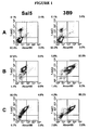
- the anti-La/SS-B IgG mAb 3B9 cell line (Tran et al., 2002), prepared by Dr M. Bachmann (Oklahoma Medical Research Foundation, OK, USA), was a generous gift from Dr T.P. Gordon (Department of Immunology, Allergy, and Arthritis, Flinders Medical Centre, SA, Australia).
- the irrelevant 1D4.5 mAb Sal5 cell line prepared by Dr L.K. Ashman (Medical Science Building, University of Newcastle, NSW, Australia), was a kind gift from Dr S.
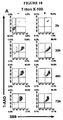
- tumour cell lines Jurkat (ATCC# TIB-152, acute T cell leukemia), EL4 (ATCC# TIB-39, mouse T-lymphocyte lymphoma), U-937 (ATCC# CRL-1593.2, monocytic leukemia) and Raji (ATCC# CCL-86, Burkitt's lymphoma) were routinely grown as suspension cultures in RPMI-1640 containing 5 % FCS and passaged every 48-72 h at 1:4 dilution.
- the U2OS osteosarcoma cell line (ATCC# HTB-96), SAOS-2 osteosarcoma cell line (ATCC# HTB-85) and HeLa cervical adenocarcinoma (ATCC# CCL-2) were routinely cultured in DMEM containing 5 % FCS and passaged every 48 - 72 h after detachment using trypsin-EDTA solution.
- the squamous cell carcinoma cell line, SCC-25 (ATCC# CRL-1628), was cultured in a 1:1 mixture of DMEM and Ham's F12 medium containing supplemented with 400 ng/mL hydrocortisone and 10 % FCS and passaged after detachment using trypsin-EDTA solution.
- PBMC peripheral blood monocytic cells
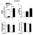
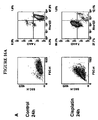
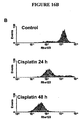
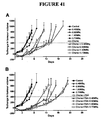
- Apoptosis in all cultures was induced by incubation of these cells in culture media described above and in the presence of specified concentrations of cytotoxic chemotherapy drugs (see Figure legends).
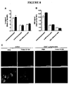
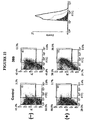
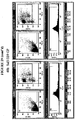
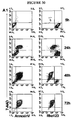
- Direct immunofluorescence staining was performed using 1-2 x 10 5 cells at 10 6 cells/mL for 30 min at room temperature in PBS containing 0.1 % BSA and 5 ⁇ g/mL of FITC-conjugated mAb. Cells were thoroughly washed using PBS and centrifugation at 450xg. Cells were resuspended in PBS containing 0.5 ⁇ g/mL PI and acquired immediately by a Becton-Dickinson FACScanTM flow cytometry system (BD Biosciences, CA, USA). Positive staining using the mAb-FITC conjugates was determined in comparison to FITC-conjugated isotype control mAb detected using the FL-1 channel (530-nm filter).
- Indirect immunofluorescent staining was performed using purified mouse antibodies followed by anti-mouse IgG conjugated to Alexa 488 detected using the FL-1 channel (530-nm filter). Cell viability was assessed by the exclusion of PI detected using the FL-2 (585-nm filter) or the exclusion of 7-AAD detected using the FL-3 (> 650 nm filter). Flow cytometry data was analysed using WinMDI v 2.8 (Scripps Research Institute, CA, USA). Unless otherwise specified, no gating was performed in any of the analysis shown in this paper.
- Blotting was performed as per standard procedure using 3B9 or anti-PARP mAbs followed by AP-conjugated anti-mouse IgG mAb or anti-actin polyclonal antibody (pAb) followed by AP-conjugated anti-goat IgG mAb. All blots were developed using the ECF TM substrate and scanned using the Fluorlmager TM 595 (Molecular Dynamics, Amersham Biosciences, NJ, USA) with 488 nm excitation laser and emission collected using 570 nm filter.
- Sal5 isotype and 3B9 mAb were labelled with 14 C by incubating the hybridoma cells (35 million cells) in 35 ml RPMI-1640 containing 10 % FCS in the production module of miniPERM bioreactor and 400 ml of RPMI-1640 containing 10 % FCS and 250 ⁇ Ci of D- [U- 14 C]glucose and 250 ⁇ Ci of L-[U- 14 C]Leucine in the nutrient module.
- the bioreactor was incubated in 5 % CO 2 humidified air at 37°C on a bottle-rotating device for 5 days.
- the medium in the production chamber was collected for antibody purification using protein G purification columns as per manufacturer instructions.
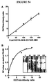
- Radioactivity of purified antibodies (10 ⁇ l sample) was counted in UltimaGold TM scintillation liquid (1 ml) for 20 min. using Tri-Carb 3100 ⁇ -counter (Packard, regularly calibrated using supplied 14 C standards). Protein concentration was determined using BCA Protein Reagent Assay as per manufacturer instructions. The specific radioactivity of 14 C-Sal5 and 14 C-3B9 was 120.3 and 130.8 dpm/ ⁇ g, respectively.
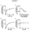
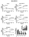
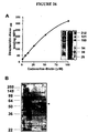
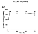
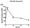
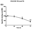
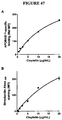
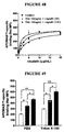
- Saturation binding study was performed by incubating apoptotic EL4 cells (5 x 10 5 cells) at 24 h after treatment with etoposide and cyclophosphamide with increasing concentration of 14 C-3B9 in the absence (total) or presence (non specific) of 50-fold molar excess of unlabelled 3B9. After 30 min, cells were washed thoroughly using PBS and radioactivity was measured using the ⁇ -counter as described above. Specific binding was calculated as the difference between total and non-specific binding and plotted as a function of concentration of 14 C-3B9. Competition binding curve was constructed by incubating apoptotic EL4 cells with 14 C-3B9 in the presence of increasing concentrations of unlabelled 3B9.
- Radioactivity was measured as described earlier and-plotted as a function of unlabelled 3B9 concentration. Association time course was performed by incubation of apoptotic EL4 cells with 14 C-3B9 in the absence or presence of 50-fold molar excess of unlabelled 3B9 for the specified times. Samples were washed and radioactivity was measure and specific binding was plotted as function of time.
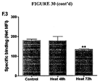
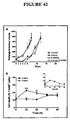
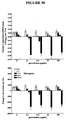
- EL4 cells established from a lymphoma induced in a C57BL/6 mouse ( Gorer, British Journal of Cancer 4: 372-379, 1950 ), were used to establish subcutaneous tumour implants in 6 - 8 weeks old C57BL/6 mice. Mice were housed and treated as per protocols approved by the Animal Ethics Committee at The University of Sydney. Briefly, 10 5 EL4 cells were injected subcutaneously in the right flank of each mouse. Once the tumour reached 1 cm diameter, mice were randomly divided into two groups one of which received intraperitoneal injection of etoposide and cyclophosphamide to achieve a dose of 76 mg/kg and 100 mg/kg, respectively (time 0). These two groups (untreated or treated) were used for the studied described below.
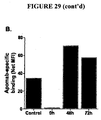
- mice received a second injection of etoposide and cyclophosphamide at 24 h after the first injection while untreated mice were left untreated. After 24 h (i.e. time point 48 h), all mice were euthanised, whole blood was obtained by cardiac puncture and EL4 tumours were excised from these sacrificed mice. Excised tumours tissue was disrupted to produce a single cell suspension, washed with PBS and used for immunofluorescent staining with 3B9 and PI and flow cytometry analysis as described earlier.
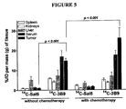
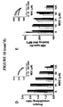
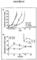
- mice that received the first chemotherapy injection at time 0 received a second injection of etoposide and cyclophosphamide at 24 h and an intravenous injection of specified amount of 14 C-3B9 or 14 C-Sal5.
- Untreated mice only received the intravenous injection of 14 C-3B9 or 14 C-Sal5.
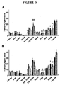
- All mice were euthanased, whole blood was obtained by cardiac puncture and EL4 tumours as well as other organs were collected for radioactivity measurement. Serum and organs were solubilised using 1 ml of Solvable TM for 2 h at 50°C, decolourised using 100 ⁇ l of H 2 O 2 (30 %).
- UltimaGold TM scintillation liquid (1 ml) was added and samples were counted for 10 min. using the ⁇ -counter.
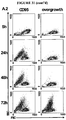
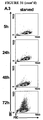
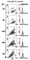
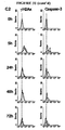
- Anti-La mAb binds to apoptotic malignant EL4 thymic lymphoma cells in vitro
- Sulforhodamine B stains proteins indiscriminately and sulforhodamine staining of malignant EL4 cells, which were rendered apoptotic by cytotoxic drug treatment, was also resistant to detergent treatment ( Figure 2C ).
- Figure 2D shows that protein-protein cross linking in apoptotic cells was not confined to the La antigen but also included the 3B9 mAb itself.
- Anti-La mAb binds specifically, efficiently and irreversibly to apoptotic EL4 cells in vitro
- La-specific mAb which was biosynthetically labelled with the radioisotope carbon-14 ( 14 C-3B9), bound to apoptotic EL4 cells after they were treated with cyclophosphamide and etoposide in a specific and saturable manner.
- the concentration of agent required to reach half-maximal saturation of one million apoptotic cells was 18 nM and maximal binding was ⁇ 7500 femtomole/million apoptotic cells ( Figure 3A ).
- Half-maximal saturation of 14 C-3B9 binding was found to occur within 5 minutes at room temperature ( Figure 3B ).
- La-specific mAb targets EL4 tumours in vivo especially after cytotoxic chemotherapy
- EL4 tumour-bearing mice were used to demonstrate that 3B9 La-specific mAb targeted a tumour mass in vivo and that the 3B9 targeting was enhanced 48 hours after the mice were treated with cytotoxic chemotherapy.
- 100 ⁇ g 14 C-labelled isotype control mAb (Sal5) did not accumulate significantly in any organ or tissue including the tumour.
- 100 ⁇ g 14 C-3B9 accumulated significantly in serum and the tumour both before and after chemotherapy (P ⁇ 0.001).
- after the mice were treated with cytotoxic chemotherapy only tumours accumulated significantly more 14 C-3B9 (P ⁇ 0.001) ( Figure 5 ).
- Anti-La as a potential tumour targeting agent for human malignancies
- the nuclear structural and molecular changes that accompany malignancy together with additional apoptosis-related changes in the expression levels, processing and translocation of the constituent nuclear proteins suggest that other of these proteins may be suitable targets for this approach.
- the criteria that have been developed to characterize targeting of the La antigen for the purpose of radio-imaging and radio-immunotherapy can be applied in order to define other appropriate nuclear target antigens for the application of the present technology (Apomab criteria).
- radiosensitising chemotherapy creates new abundant targets in the tumour mass, which were not previously available, and these revealed antigens may be targeted using monoclonal antibodies, which are conjugated to therapeutic radionuclides in order to deliver cytotoxic radiation to the radiosensitised tumour and supporting tissue.
- the Apomab criteria used to determine nuclear antigens (Ag) that are particularly suitable for targeting dead tumour cells for diagnostic and therapeutic purposes are:
- antigens can be targeted that produce an enhanced signal to noise ratio for diagnostic imaging and an improved therapeutic ratio for targeted radiation.
- La targeting data suggest that some of these antigens may be accessible at low levels in untreated tumours, it is found that cytotoxic chemotherapy exposes an abundance of antigen-specific binding sites. Therefore, the combination of cytotoxic radiosensitising chemotherapy or non-cytotoxic radiosensitising drug and the method of RIT will not only produce more effective and safer anti-cancer treatment than either modality (chemotherapy or RIT) alone but will also be considerably more advantageous than other methods of RIT that target pre-existing dead cells.
- the scheduling and dose of the component modalities of combination therapy may need to be varied to achieve the optimal combination.
- use of full doses of potent radiosensitizing drugs such as gemcitabine to induce cytotoxicity may cause apoptotic cell death in susceptible normal tissues such as bone marrow and the intestinal mucosa.
- Milas and colleagues studied the effects of gemcitabine and showed that the time course of apoptosis was short and that recovery was complete within a 24-36 hour period ( Milas L et al. Cancer Res 59, 107-114, 1999 ).
- Combination chemotherapy with the radiosensitising drugs, cisplatin and gemcitabine produces higher response rates in pancreatic cancer patients than the use of single agent gemcitabine but it does not improve overall survival and is associated with greater toxicity compared with the use of single agent gemcitabine.
- combination cisplatin/gemcitabine chemotherapy presumably fails to eradicate systemic metastases, which re-grow to kill the patient.
- Armed Apomab enables delivery of higher and potentially curative doses of radiation to tumour metastases by taking advantage of the more potent cytotoxic effects and hence anti-tumour properties of the cisplatin/gemcitabine combination.
- tumour targets Auto-targeting
- cytotoxic chemotherapy results from two major effects, which are shown schematically in Table 4:
- the potential significance of the scaling concept is related to the dose-dependent effects that radiation has on both supporting tumour vasculature and the tumour cells themselves. If Apomab RIT were given in conjunction with combination cytotoxic chemotherapy such as cisplatin and gemcitabine then higher in vivo binding to apoptotic/necrotic tumours would occur than if it were given in conjunction with single agent chemotherapy. In which case, it would be expected that higher doses of radiation would be delivered to neighbouring viable tumour cells. For example, if a radiation dose of ⁇ 8-10Gy were delivered to a tumour by combination cisplatin/gemcitabine chemotherapy then apoptosis of tumour endothelial cells may result from release of ceramide ( Z Fuks and R Kolesnick.
- HIF1 ⁇ protects against tumour endothelial cell apoptosis thus inducing radioresistance but simultaneously sensitises tumour cells to radiation in a p53-dependent manner (Fuks and Kolesnick 2005, supra ).
- HIF1 ⁇ inhibitors or inhibitors of VEGF or bFGF may provide additional radiosensitisation when delivering lower doses of radiation (1.8-3Gy) to tumours using Apomab RIT.
- Oncomine TM is a cancer-specific database containing microarray data from 962 studies of which 209 were analysed.
- the database contains 14,177 microarrays from 35 cancer types (information publicly available at the website www.oncomine.org).
- Several cancer signatures have been deduced from large scale analysis of data held in the database ( Hampton, Jama 292(17): 2073, 2004 ; Rhodes et al., Proc Natl Acad Sci USA 101(25): 9309-14, 2004a ; Rhodes et al., Neoplasia 6(1): 1-6, 2004b ; Rhodes and Chinnaiyan, Nat Genet 37 Suppl: S31-7, 2005 ; Rhodes et al., Nat Genet 37(6): 579-83, 2005 ).
- 209 studies in the database as described below were analysed in order to investigate certain malignancy signatures, which may provide useful targets for the present invention.
- the ribonucleoprotein (RNP-1) motif was identified , which comprises the RNA recognition motif (RRM), to be at higher odds of being associated with a cancer signature compared to the nucleus as a cellular component.
- RRM RNA recognition motif
- the RNA binding gene set which was enriched as a cellular function, was also at high odds of being associated with a cancer signature.
- nucleolar and heteronuclear ribonucleoprotein (hnRNP) components appeared to have very high odds as gene sets in association with a cancer signature.
- components with the RNP-1 motif generally and hnRNP complex specifically are suitable targets for the method of the present invention.
- This recommendation is justified by (1) association of hnRNP with cancer at the gene expression level (mRNA data reported herein), (2) the relative abundance of this protein and its correlation with cancer and cancer progression (see review Carpenter et al., Biochimica et Biophysica Acta 1765(2): 85-100, 2006 ), and (3) the reorganisation of the hnRNP network during apoptosis into the HERDS ( Biggiogera et al., Biologie Cellulaire 96(8): 603-15. 2004 ) and the accessibility of the hnRNP network for detection with antibodies.
- nucleolar proteins generally and specifically nucleophosmin represent suitable targets for this strategy. This is justified by the data shown in the analysis of the Oncomine TM data base as well as the literature regarding nucleolar function in cancer ( Maggi and Weber, Cancer Investigation 23(7): 599-608, 2005 ) and, in particular, nucleophosmin as putative-proto-oncogene ( Grisendi et al., Nature Reviews. Cancer 6(7): 493-505,2006 ). Table 5: mRNA expression in clinical tumors. La mRNA is upregulated in malignancy Tissue type No.
- hnRNP A/B 2.3E-4 Cellular function RNA binding 2.8 1.7E-14 Cellular component Nucleus 1.88 2.2E-21 hnRNP complex 17.47 2.4E-7 Nucleolus 4.03 2.2E-4 Brain (Shai et al., 2003) 12625 Glioblastoma Multiforme vs . Normal white matter 17% InterPro motif RNP-1 motif 2.29 3.3E-5 e.g. hnRNP A0 6.6E-10 Cellular function RNA binding 2.43 7E-11 Cellular component Nucleus 1.47 7.5E-9 e.g. PCNA 8.9E-7 hnRNP complex 6.75 3.6E-4 Nucleolus 4.38 6E-4 e.g.
- nucleolin 5.7E-5 Brain (Liang et al., 2005) 23079 Dead vs. alive glioblastoma multiforme 1 year survival 0.2% InterPro motif RNP-1 motif 2.70 1.2E-8 e.g. hnRNP A3 2.5E-4 Cellular function RNA binding 2.38 5.7E-12 Cellular component Nucleus 1.75 4.8E-12 Nucleolus 3.3 9.6E-4 Brain (Rickman et al., 2001) 6668 Glioma vs . normal neocortex of temporal lobe 7% InterPro motif RNP-1 motif 2.64 9.9E-5 e.g.
- nucleophosmin 6.5E-7 Mammary epithelial cells - oncogene transfected (Bild et al., 2006, supra ) 54675 c-Myc vs .
- GFP transfected primary cells 3.7% InterPro motif RNP-1 motif 2.5 7.6E-7 e.g. Nucleolin 2.8E-11 hnRNP A/B 9.8E-7 La/SS-B 8.5E-6 Cellular function RNA binding 3.13 1.2E-20 Cellular Nucleolus 12.57 9.3E-13 component e.g. nucleophosmin 7.2E-6 Colon (Notterman et al., 2001) 6745 Colon adenocarcinoma vs.
- hnRNP F 2.9E-4 Cellular function RNA binding 5.79 2.6E-13 Cellular component Nucleus 1.73 2.4E-4 Liver (Chen et al., 2002) 22033 Metastasis to liver vs. hepatocellular carcinoma 6.2% InterPro motif RNP-1 motif 2.33 1.2E-6 e.g. hnRNP A3 1.2E-5 Cellular function RNA binding 1.91 2.5E-7 Cellular component Nucleus 1.38 1.7E-7 Lung (Bhattacharjee et al., 2001) 11158 Lung adenocarcinoma vs. normal lung 31.1% InterPro motif RNP-1 motif 2.72 6.3E-7 e.g.
- PCNA 3.6E-6 PARP-1 4.4E-6 Lung (Bhattacharjee et al., 2001, supra ) 10030 Squamous cell lung carcinoma vs. normal lung 7.1% InterPro motif RNP-1 motif 2.39 2.7E-5 e.g. hnRNP A3 8.5E-7 Cellular function RNA binding 2.72 9.2E-13 Cellular component Nucleus 1.48 3.4E-8 e.g. PARP-1 5.7E-8 PCNA 7.7E-8 hnRNP complex 6.27 8.1E-4 Lymph (Hummel et al., 2006) 22215 Ig-Myc fusion vs. translocation negative lymphoma 27.9% InterPro motif RNP-1 motif 3 . 94 2.2E-17 e.g.
- hnRNP A3 4.4E-20 La/SS-B 5.6E-13 hnRNP A2/B1 6.3E-11 Cellular function RNA binding 4.11 5.9E-37 Cellular component Nucleus 1.71 2.1E-21 hnRNP complex 15.22 3.1E-6 Nucleolus 8.68 4.9E-11 e.g. nucleophosmin 4.7E-28 Lymph (Hummel et al., 2006, supra ) 22215 Definite Burkitt's lymphoma vs . not definite 27.9% InterPro motif RNP-1 motif 3.47 2.7E-14 e.g.
- hnRNP C 5.6E-4 Cellular function RNA binding 3.4 5.8E-15 Cellular component Nucleus 1.39 2.6E-9 hnRNP complex 11.51 2.3E-5 Nucleolus 7.84 7.2E-8 e.g. nucleophosmin 1.2E-5 Prostate (Singh et al., 2002) 9892 Prostate tumour vs. non-tumour prostate 4.9% InterPro motif RNP-1 motif 3.08 1.9E-8 e.g.
- Cell culture media RPMI-1640, DMEM and Ham's F12, and fetal calf serum (FCS) (JRH Biosciences Inc., Lenexa, KS); Trypsin-EDTA solution, trypan blue, propidium iodide (PI), bovine serum albumin (BSA), BCIP/NBT premixed substrate solution for alkaline phophatase (AP), hydrocortisone, monodansylcadaverine (MDC) and staurosporine (STS) and mouse anti-human ⁇ -tubulin mAb (TUB 2.1) (Sigma-Aldrich Co., St. Louis, MO).
- FCS fetal calf serum
- Hybond-P membrane (PVDF) and protein G purification columns (Amersham Biosciences, Piscataway, NJ). BCA Protein Reagent Assay (Pierce Biotechnology Inc., Rockford, IL).
- PARP Anti-poly(ADP-ribose) polymerase
- mAb monoclonal antibody
- PCNA anti-proliferating cell nuclear antigen
- Trichostatin A TSA
- Anti-phospho-histone H2AX Ser139
- Anti-human H2A polyclonal antibody Millipore Inc., Billerica, MA
- Anti- ⁇ fodrin mAb Chemicon International, Temecula, CA
- Anti- ⁇ actin affinity-purified rabbit polyclonal antibody Fluorescein isothiocyanate (FITC)-conjugated goat anti-rabbit IgG and AP-conjugated goat anti-rabbit IgG antibodies (Rockland, Gilbertsville, PA).
- FITC Fluorescein isothiocyanate
- the anti-La/SS-B 3B9 mAb hybridoma is a murine IgG 2a autoantibody, which is crossreactive with human La and which was prepared by Dr M. Bachmann (Oklahoma Medical Research Foundation, OK, USA), was a kind gift from Dr T.P. Gordon (Department of Immunology, Allergy and Arthritis, Flinders Medical Centre, SA, Australia).
- the isotype control Sal5 (1D4.5) mAb hybridoma prepared by Dr L.K. Ashman (Medical Science Building, University of Newcastle, NSW, Australia), was kindly supplied by Dr S. McColl (School of Molecular Biosciences, University of Sydney, SA, Australia).
- the mAb were affinity-purified using protein G columns and FITC-conjugates were prepared according to the manufacturer's instructions (Sigma-Aldrich Co., St. Louis, MO).
- Etoposide Pfizer Inc., NY, USA
- cyclophosphamide and cisplatin Bristol-Myers Squibb Company, Princeton, NJ
- gemcitabine Eli Lilly, Indianapolis, IN
- the squamous cell carcinoma cell line, SCC-25 was cultured in a 1:1 mixture of DMEM and Ham's F12 medium supplemented with 400ng/mL hydrocortisone and 5% FCS and passaged after detachment using trypsin-EDTA solution.
- Peripheral blood mononuclear cells (PBMC) were isolated from fresh heparinized blood obtained from normal healthy volunteer donors using Lymphoprep separation and cultured overnight in RPMI-1640 containing 5% FCS to separate adherent cells from those remaining in suspension. Cytofluographic analysis indicated that >70% of both suspension and adherent cells were CD3 + and CD14 + , respectively.
- CD3- and CD14-enriched PBMC preparations will be described as peripheral blood lymphocytes and monocytes, respectively.
- Clonetics conditioned cultures of primary human cells were maintained according to the manufacturer's instructions (Cambrex Corporation, East Rutherford, NJ) and included cultures of Human Mammary Epithelial Cells (HMEC), Prostate Epithelial Cells (PrEC) and Normal Human Bronchial Epithelium (NHBE).
- HMEC Human Mammary Epithelial Cells
- PrEC Prostate Epithelial Cells
- NHBE Normal Human Bronchial Epithelium
- Apoptosis or cell death was induced by adding cytotoxic drugs to culture media at the specified concentrations.
- cells were starved by serum deprivation.
- TSA used at the specified concentrations was prepared from 1 mg/mL stock solution in absolute ethanol.
- Control (untreated) cells had ⁇ 90% viability determined by PI or trypan blue staining (data not shown).
- Viable cells were fixed and permeabilized by incubating cells at 5x10 6 cells/mL in 2% w/v paraformaldehyde solution in PBS (150mM sodium phosphate and 150 sodium chloride, pH 7.2) for 10min followed by 1:10 dilution in absolute methanol (at -20°C) for 1-3 min. before a final wash with PBS.
- Indirect immunofluorescence staining was performed using purified mouse antibodies at 5 ⁇ g/mL for 30 min. at room temperature (RT) in PBS followed by Alexa 488 -conjugated anti-mouse IgG at 2 ⁇ g/mL for 30 min. at RT in PBS. Fluorescence was detected using the FL-1 channel (530nm filter). Cell viability was assessed by the exclusion of PI (0.5 ⁇ g/mL) and detected using the FL-2 channel (585nm filter) or by the exclusion of 7-AAD (2 ⁇ g/mL for 15 min. at RT) and detected using the FL-3 channel (>650nm filter).
- Staining for ⁇ H2AX was performed using 0.2 ⁇ g/mL of anti- ⁇ H2AX-biotin for 30 min. at RT followed by 2 ⁇ g/mL of streptavidin-PE or streptavidin-Alexa 488 .
- Staining of polyclonal antibodies was performed using 5 ⁇ g/mL of antibody solution for 30 min. at RT.
- Control incubations were performed using protein G purified rabbit IgG from normal rabbit serum (IMVS, SA, Australia). Cells were washed then incubated (30min. at RT) with 2 ⁇ g/mL of FITC-conjugated anti-rabbit IgG antibody, which was detected using the FL-1 channel.
- Cell lysates were prepared in SDS lysis solution (2% w/v SDS, 10% v/v glycerol and 62.5 mM Tris-HCl) and protein concentration was determined using BCA protein reagent assay kit according to the manufacturer's instructions. Bromophenol blue (0.05% w/v) and ⁇ -mercaptoethanol (5% v/v) were added to lysates after BCA assay measurement. SDS-PAGE was performed according to the manufacturer's instructions with the Hoefer Mighty Small II SE 250 electrophoresis system (Amersham Biosciences, Piscataway, NJ) under reducing conditions using 12% resolving polyacrylamide gel as per Laemmli's method.
- the transfer of the polyacrylamide gel to Hybond-P membrane was carried out according to the manufacturer's instructions using the TE 22 Mini Tank Transfer Unit (Amersham Biosciences, Piscataway, NJ). Immunoblotting was done using standard methods in which staining with 3B9 was followed by staining with AP-conjugated anti-mouse IgG mAb (Jackson ImmunoResearch Laboratories Inc., West Grove, PA) or anti-actin (N-20) affinity purified goat polyclonal antibody (Santa Cruz Biotechnology Inc., Santa Cruz, CA) followed by AP-conjugated anti-goat IgG mAb (Jackson ImmunoResearch). Some blots were developed using the BCIP/NBT premixed solution as specified by the manufacturer and analyzed using GelPro Analyzer v3.1 (Media Cybernetics Inc., Silver Spring, MD).
- Transglutaminase 2 (TG2)-mediated protein-protein crosslinking in apoptotic cells was investigated using a modification of a previously described method. Briefly, cells were resuspended at 1x10 6 cells/mL in PBS containing 1% Triton X-100, 0.2 ⁇ g/mL sulforhodamine 101 and 0.1 ⁇ M SytoxGreen. Samples were vortexed and incubated for 5min. before analysis by flow cytometry where fluorescence from Sytox Green and sulforhodamine were detected using FL-1 and FL-3, respectively.
- the degree of crosslinking in apoptotic cells was calculated as the ratio of the MFI of sulforhodamine staining in apoptotic cells to the MFI in control cells (protein crosslinking ratio).
- cells were incubated with PBS or 1% Triton X-100 in PBS for 10min. at RT with intermittent vortexing.
- MDC monodansylcadaverine
- TG2 substrates were collected after 48h, washed extensively with PBS and incubated with streptavidin-Alexa 488 for cytofluographic analysis.
- streptavidin-Alexa 488 for cytofluographic analysis.
- cadaverine-biotin labelled cells were lysed for SDS-PAGE with or without prior immunoprecipitation using 3B9 mAb.
- PAGE gels were transferred to PVDF membranes and probed using streptavidin-AP to visualise both the total pool of TG2 substrates and 3B9-reactive TG2 substrates.
- Cells were stained using immunofluorescence methods described above and then spotted onto glass slides using the cytospin method.
- the prepared slides were mounted with coverslips using non-fluorescence mounting medium (Dako, Carpinteria, CA). Slides were analyzed using a BioRad Olympus Confocal microscope with appropriate filters and under constant conditions of laser voltage, iris aperture and photomultiplier tube amplification.
- Jurkat cells were incubated in the presence or absence of 20 ⁇ g/mL cisplatin and pelleted 3h after treatment to prepare soluble nuclear and chromatin fractions as decribed. Samples of these fractions were fractionated using 12% SDS-PAGE for immunoblotting as described above. Membranes were probed with 1 ⁇ g/mL 3B9, 2 ⁇ g/mL anti-H2A antibodies or 0.2 ⁇ g/mL anti- ⁇ H2AX-biotin followed by the appropriate AP-conjugated secondary antibodies or strepatividin-AP. The presence of H2A in chromatin and not soluble fractions was used to assess the quality of preparation of the chromatin fractions.
- Chamber slides were seeded with PANC-1 cells and incubated overnight in RPMI-1640 containing 10% FCS before replacement with medium containing 20 ⁇ g/mL cisplatin alone or in combination with TSA. After 3h, cells were fixed and permeabilized using paraformaldehyde and methanol as described above. Cells were washed and blocked with 5% BSA solution in PBS then stained with 10 ⁇ g/mL 3B9 followed by 2 ⁇ g/mL Alexa 488 -conjugated anti-mouse IgG antibody. Cells were washed and incubated with 0.2 ⁇ g/mL anti- ⁇ H2AX-biotin followed by 2 ⁇ g/mL streptavidin-PE.
- Oncomine is a database of microarray data, which holds data from 962 studies of which 209 were analyzed.
- the database contains 14,177 microarrays from 35 cancer types (information publicly available at the website www.oncomine.org).
- Several cancer signatures have been deduced from large scale analysis of data in the database (24-28).
- We analyzed the 209 studies using the Advanced Analysis module limiting results to overexpressed genes only and gene enrichment was selected using the following options: (1) InterPro for analysis of motifs in the overexpresed genes, (2) Gene Ontogeny (GO) molecular function for analysis of functions of overexpressed genes, and (3) GO cellular component for cellular compartmentation of the overexpressed genes.
- Two paramaters were used to describe the gene sets deduced from the above analysis (a) Odds Ratio and (b) P-value, which were provided by the database.
- La is overexpressed in human malignant cells and is induced by DNA-damaging drugs
- the 3B9 target antigen, La is overexpressed in malignant cells with respect to the corresponding primary cell type.
- Oncomine which is a cancer gene expression database
- RRM RNA Recognition Motif
- La/SS-B mRNA expression indicated that La mRNA was overexpressed in malignancy (Tables 2 and 3). Together, these results support the notion that La protein and mRNA overexpression is a feature of malignancy in common with other components of the transcriptional and translational apparatus.
- 3B9 binding is indicative of DNA damage during drug-induced apoptosis
- Fig. 14A illustrates, the extent of 3B9-specific binding per dead Jurkat cell after induction of apoptosis using the DNA-damaging agent, cisplatin, was proportional to cisplatin dose. Increasing the dose of cisplatin increased the amount of DNA damage, which was measured by cytofluographic detection of the DNA damage marker, ⁇ H2AX (inset, Fig. 14A ). The correlation of 3B9-specific binding with DNA damage may result from increased expression and/or redistribution and/or accessibility of the La target antigen.
- Cisplastin-treatment of Jurkat cells causes apoptosis and intranuclear accumulation of La
- Fig. 16A staining with the mitochondrial-permeant dye, rhodamine-123, and the DNA binding dye, 7-AAD, confirms that cisplatin-treated Jurkat cells undergo an apoptotic mode of cell death.
- cisplatin-treated Jurkat cells undergo an apoptotic mode of cell death.
- Fig. 16A left-hand panels.
- cisplatin-treated cells bind 7-AAD and show loss of mitochondrial retention of rhodamine-123 ( Fig. 16A , right-hand panels).
- the mitochondrial loss of rhodamine-123 is characteristic of apoptosis ( Fig. 16B ).
- the same dose of cisplatin is used to treat Jurkat cells that are subsequently permeabilised and fixed and stained with mAb specific for a number of nuclear antigens ( Fig. 17 ).
- the brightness of staining per cell, which is expressed as the antigen-specific mean fluorescence intensity (MFI) of the cisplatin-treated Jurkat cells is compared with the MFI of control untreated Jurkat cells, which are also permeabilised, fixed and stained.
- MFI mean fluorescence intensity
- NPM nucleophosmin
- detection of highly expressed PCNA is markedly reduced after cisplatin treatment.
- cytoplasmic cytoskeletal protein actin are all less detectable in permeabilised and fixed apoptotic Jurkat cells compared with permeabilised and fixed control cells ( Fig. 17A , B and C).
- detection of the La antigen is significantly increased in apoptotic compared with control Jurkat cells, particularly 48h after apoptosis induction.
- histone H2A is increased in apoptotic Jurkat cells 72h post-apoptosis induction.
- Triton X-100 Resistance of apoptotic cells to the non-ionic detergent, Triton X-100, has been reported and correlates with the activity of tissue transglutaminase or transglutaminase 2 (TG2), which mediates protein-protein crosslinking. It was found that during apoptosis, the proportion of 7-AAD + cells and the level of 3B9 binding to these cells were comparable irrespective of Triton X-100 treatment ( Fig 18A ), which suggested that crosslinking was an integral component of apoptosis in this in vitro system.
- TG2 activity reduces 3B9 binding to apoptotic cells because of reduced protein-protein crosslinking.
- cisplatin the effect of increasing doses of cisplatin on the activity of TG2 in Jurkat cells was investigated using the TG2 substrate cadaverine-biotin. Incorporation of cadaverine-biotin into Jurkat cells showed a dose-dependent relationship to cisplatin concentration ( Fig. 18C ). Based on these results, cisplatin at 20 ⁇ g/mL was chosen to induce maximal activity of TG2. Then the level of protein crosslinking in apoptotic cells was investigated.
- the concentration of MDC ( ⁇ SEM) required to inhibit half of the binding of 3B9 was 260 ⁇ 1, 110 ⁇ 1 and 150 ⁇ 1 ⁇ M at 24, 48 and 72h, respectively. Together, these results indicated that the La antigen was retained in apoptotic cells by a generalized protein crosslinking process, which was most likely mediated by TG2.
- Protein crosslinking covalently attaches 3B9 to the interior of leaky dead malignant cells during apoptosis
- the EL4 murine T-lymphoblastic lymphoma cell line was obtained from American Type Cell Culture (TIB-39) and murine thymocytes were freshly prepared from 6-8 week old C57BL/6 mice. Cells were routinely cultured in RPMI-1640 containing 5% FCS (JRH Biosciences Inc., Lenexa, KS) and passaged every 48-72h at 1:4 dilution. 3B9 and the relevant control (Sal5) were prepared as described previously (first paper). Fluorescein isothiocyanate (FITC) conjugated of these agents was prepared as described by manufacturer's instructions (Sigma-Aldrich Co., St. Louis, MO).
- Apoptosis in cultures was induced by adding 20 ⁇ g/mL etoposide (Pfizer Inc., NY, USA) and 20 ⁇ g/mL cyclophosphamide (Bristol-Myers Squibb Company, Princeton, NJ) to the culture media.
- Data presented here from control (untreated cells) originated from samples with higher than 90% viability as determined by PI or trypan blue (Sigma-Aldrich Co., St. Louis, MO) staining (data not shown). Permeabilization of viable cells was performed by 10 min.
- Direct immunofluorescence staining was performed using 1-2 x 10 5 cells at 10 6 cells/mL for 30 min. at room temperature (RT) in PBS containing 0.1% bovine serum albumin (BSA, Sigma-Aldrich Co., St. Louis, MO) and 5 ⁇ g/mL of FITC-conjugated 3B9 or FITC-conjugated Sal5 isotype control mAb.
- Indirect immunofluorescence staining was performed using purified mouse antibodies (5 ⁇ g/mL for 30 min. at RT in PBS) followed by anti-mouse IgG conjugated to Alexa 488 (Invitrogen, Carlsbad, CA) (2 ⁇ g/mL for 30 min.
- Cell lysates were prepared in SDS lysis solution (2% w/v SDS, 10% v/v glycerol and 62.5 mM Tris-HCl) and protein concentration was determined using BCA protein reagent assay kit according to the manufacturer's instructions (Pierce Biotechnology Inc., Rockford, IL). Bromophenol blue (0.05% w/v) and ⁇ -mercaptoethanol (5% v/v) (Sigma-Aldrich Co., St. Louis, MO) were added to lysates after BCA assay measurement. An amount of 12 ⁇ g of lysate was added to each lane for SDS-PAGE.
- SDS-PAGE was performed according to the manufacturer's instructions with the Hoefer ® Mighty Small II SE 250 electrophoresis system (Amersham Biosciences, Piscataway, NJ) under reducing condition using 12% resolving polyacrylamide gel as per Laemmli's method.
- the transfer of the polyacrylamide gel to Hybond-P membrane was carried out according to the manufacturer's instructions using the TE 22 Mini Tank Transfer Unit (Amersham Biosciences, Piscataway, NJ).
- Immunoblotting was done using standard methods in which staining with 3B9 was followed by staining with alkaline phosphatase (AP)-conjugated anti-mouse IgG mAb (Jackson ImmunoResearch Laboratories, Inc., West Grove, PA). Blots were developed using the BCIP/NBT premixed solution as specified by manufacturer (Sigma-Aldrich Co., St. Louis, MO) and analyzed using GelPro Analyzer v3.1 (Media Cybernetics Inc., Silver Spring, MD).
- AP alkaline phosphatase
- 3B9 and matched control were labelled with 14 C by incubating the hybridoma cells (35 million cells) in 35mL RPMI-1640 containing 5% FCS in the production module of miniPERM bioreactor (Vivascience GmbH, Hannover, Germany) and 400mL of RPMI-1640 containing 5% FCS and 250 ⁇ Ci of D-[U- 14 C]glucose and 250 ⁇ Ci of L-[U- 14 C]leucine (Amersham Biosciences, Piscataway, NJ) in the nutrient module.
- the bioreactor was incubated in 5% CO 2 humidified air at 37°C on a bottle-rotating device for 5 days.
- the medium in the production chamber was collected for purification using Protein G purification columns.
- Radioactivity of purified antibodies (10 ⁇ L sample) was counted in UltimaGold TM scintillation liquid (1mL) for 20 min using Packard Tri-Carb 3100 ⁇ -counter (PerkinElmer Inc., Wellesley, MA) regularly calibrated using supplied 14 C standards. Protein concentration was determined using BCA protein reagent assay. The specific radioactivity of 14 C-control and 14 C-3B9 was 120.3 and 130.8dpm/ ⁇ g, respectively.
- Saturation binding assays were performed by incubating apoptotic EL4 cells (5 x 10 5 cells) after 48h treatment with etoposide and cyclophosphamide with increasing concentrations of 14 C-3B9 in the presence (to measure non-specific binding) or absence (to measure total binding) of a 50-fold molar excess of unlabelled 3B9. After 30 min., cells were washed thoroughly using PBS and radioactivity was measured using the ⁇ -counter as described above. Specific binding was calculated as the difference between total and non-specific binding and plotted as a function of the concentration of 14 C-3B9.
- Competition binding assays were carried out by incubating apoptotic EL4 cells with 100 nM 14 C-3B9 in the presence of increasing concentrations of unlabelled 3B9. Radioactivity was measured as described above and plotted as a function of unlabelled 3B9 concentration. Association assays were performed by incubation of apoptotic EL4 cells with 100 nM 14 C-3B9 in the presence or absence of a 50-fold molar excess of unlabelled 3B9 for the specified times. Samples were washed, radioactivity was measured and specific binding was plotted as function of time.
- Dissociation assays were performed after incubation of apoptotic EL4 cells with 100 nM 14 C-3B9 in the absence or presence of 50-fold molar excess of unlabelled 3B9 for 30 min. at RT. Cells were washed, incubated at 37°C in PBS and samples were removed at the specified times. These samples were washed with PBS, radioactivity was measured and specific binding was plotted as function of time.
- the EL4 thymic lymphoblastic lymphoma is a robust model of apoptosis, which is induced by the cytotoxic drugs, cyclophosphamide and etoposide (14).
- Subcutaneous EL4 tumour implants were established in 6-8 week old syngeneic C57BL/6 mice where 10 6 EL4 cells were injected subcutaneously in the right flank of each mouse. Mice were housed and treated as per protocols approved by the Animal Ethics Committee at The University of Sydney and the Animal Ethics Committee at the Institute of Medical and Veterinary Sciences (IMVS). Once the tumour reached 1cm diameter, mice were randomly divided into control or treatment groups.
- Treatment comprised intraperitoneal injections of cytotoxic chemotherapy given in one of the following five regimens of reducing dose intensity.
- the respective cyclophosphamide and etoposide doses are indicated and are mg/kg.
- quarter dose 1d 25, 19 at 0h.
- mice bearing EL4 tumours were euthanized 48h after treatment and tumours were excised.
- Excised tumour tissue was disrupted by incubation in RPMI-1640 containing 2mg/mL collagenase (Sigma-Aldrich Co., St. Louis, MO) for 30 min. at 37°C with constant shaking. Single cell suspensions were washed with PBS and used for immunofluorescent staining with 3B9-FITC or Sal-FITC and PI and flow cytometry analysis as described earlier.
- mice received an intravenous injection of 14 C-3B9 or 14 C-Sal5 (control) at time 0.
- mice were euthanized whole blood was collected by cardiac puncture, and EL4 tumours and other tissues were collected for radioactivity measurement.
- Serum and body tissues were solubilized using 1mL Solvable TM (PerkinElmer Inc., Wellesley, MA) for 2h at 50°C, and then decolorized using 100 ⁇ L H 2 O 2 (30%) (Sigma-Aldrich Co., St. Louis, MO).
- UltimaGold TM scintillation liquid (1mL) was added and samples were counted for 10min. using the ⁇ -counter.
- Protein G purified 3B9 or Sal5 mAb were mixed at 2.5mg/mL in 0.1M sodium bicarbonate and 0.1M sodium phosphate buffer (pH8.5) with 50-fold molar excess of DOTA-NHS-Ester (Macrocyclics, Dallas, TX) dissolved in DMSO (Sigma-Aldrich Co., St. Louis, MO). DMSO represented ⁇ 10% of the final reaction, which was incubated for 2h at 23°C with constant vigorous shaking.
- DOTA-NHS-Ester was added at a 50-fold molar excess to the antibody as this ratio was found to minimize inactivation of 3B9 and showed favorable in vivo biodistribution compared to Sal5 conjugated at the same ratio and to 3B9 conjugated at higher ratios (data not shown).
- the concentration of IgG was measured using a BCA protein assay kit and the concentration of conjugated DOTA was measured using a modification of a published Cu:Arsenazo(III) assay.
- the ligand/protein (L/P) ratio of 7.5 was calculated as the ratio of the concentrations ( ⁇ M) of DOTA-immunoconjugates to IgG.
- the reaction was stored at 4°C before radiolabeling.
- DOTA-immunoconjugate solutions were concentrated and buffer-exchanged into metal free 0.2M ammonium acetate buffer (pH5.5) (Sigma-Aldrich Co., St. Louis, MO) containing 6mg/mL ascorbic acid (Sigma-Aldrich Co., St. Louis, MO).
- Indium-111 chloride (PerkinElmer Inc., Wellesley, MA) was diluted in the same buffer (5mCi/mL; 185MBq/mL) and mixed with DOTA immunoconjugates (10mg/mL).
- Radiolabeling reactions were incubated at 37°C for 2h then mixed with an equal volume of 0.2M ammonium acetate buffer (pH8) containing 5mM EDTA to quench free and loosely bond radionuclide. Quenched reactions were loaded onto 100kDa-cutoff microconcentrators and purified by three washes of 500 ⁇ L endotoxin free PBS. The incorporation of 111 In in the purified reactions was determined using instant thin layer chromatography (ITLC) which was performed using ITLC-SG strips (Pall Corporation, East Hills, NY) in 0.2M ammonium acetate solution (pH 8) containing 5mM EDTA as mobile phase.
- ITLC instant thin layer chromatography
- 111 In-DOTA-3B9 72 ⁇ g in 100 ⁇ L of PBS was given by intravenous injection to EL4 tumour-bearing mice, which were left untreated or treated with cytotoxic chemotherapy as described above. Mice were euthanized at selected time points, blood was collected by cardiac puncture, and tumours and organs were collected. Blood and organs were weighed and placed in gamma-counter tubes and radioactivity was measured using normalized Cobra5010 gamma counter. Radioactivity in organs was normalized to the weight of the organs and accumulation was calculated as the percentage of radioactivity per gram in the organs to the radioactivity of the injected dose of 111 In-DOTA-3B9 at time 0 (%ID/g).
- La is overexpressed in malignant EL4 cells and La-specific mAb binding to dead EL4 cells is induced by cytotoxic drug treatment
- Fig. 20D also illustrates that, after fixation and permeabilization, the per cell binding of 3B9 mAb was significantly higher for the malignant EL4 cell than for its normal murine counterpart cell type of the thymocytes. This cytofluographic evidence of La overexpression in EL4 cells was corroborated by immunoblot analysis of cell lysates prepared from thymocytes and EL4 cells (inset, Fig. 20D ).
- biosynthetically labelled 3B9 mAb was used to further characterize the binding of La-specific mAb to dead EL4 cells, which were killed with cytotoxic drugs. It was found that 14 C-3B9 bound dead EL4 cells in a specific and saturable manner. The concentration of 14 C-3B9 required to reach half-maximal saturation was 18nM and maximal binding was ⁇ 7500 femtomole/million dead cells ( Fig. 21A ). Half-maximal saturation of 14 C-3B9 binding occurred within 5 minutes at room temperature ( Fig. 21B ). The concentration of unlabelled 3B9 required to inhibit half-maximal binding of 14 C-3B9 (IC 50 ) was estimated to be 118nM ( Fig.
- Fig. 20 The in vitro findings depicted in Fig. 20 were confirmed in vivo using the EL4 lymphoma model.
- the fraction of PI + cells in the tumour explants increased significantly from a mean ( ⁇ SEM) of 50 ⁇ 2% to 70 ⁇ 1% ( P ⁇ 0.001).
- the 3B9 + subset of PI + cells increased significantly from 15 ⁇ 1% to 38 ⁇ 2% (P ⁇ 0.01) with use of cytotoxic chemotherapy whereas isotype control staining did not alter with use of chemotherapy ( Fig. 22 ).
- 3B9 targets EL4 tumours in vivo especially after cytotoxic chemotherapy
- tumour/organ ratio The specificity of 111 In-DOTA-3B9 towards the dying tumours was expressed as the ratio of tumour accumulation to that in normal organs (tumour/organ ratio, Fig. 24 ).
- tumour/muscle ratio At 24h after chemotherapy and 111 In-DOTA-3B9 administration, only the tumour/muscle ratio was significantly higher compared to control mice ( P ⁇ 0.001, data not shown).
- the tumour/organ ratio at 48h ( Fig. 24A ) and 72h ( Fig. 24B ) after treatment was significantly higher for all organs except for blood. Further evidence for tumour specificity was obtained from biodistribution studies of 14 C-3B9 in EL4 tumour-bearing mice which were extended to include lower doses of the radiolabelled agent. As illustrated in Fig.
- the La antigen La is a TG2 substrate
- a biotinylated form of cadaverine which is a polyamine TG2 substrate and which consequently becomes covalently bound to other TG2 substrates, is used to identify protein targets of cross-linking during apoptosis.
- cadaverine-biotin As shown in Fig. 26A , the incorporation of cadaverine-biotin in apoptotic Jurkat cells was confirmed by fluorocytometric analysis using streptavidin-Alexa 488 . Immunoblot analysis of Jurkat cells induced to undergo apoptosis in the presence of cadaverine-biotin results in the detection of additional protein bands labelled with cadaverine-biotin (lane 2 in inset, Fig. 26 ).
- cadaverine-biotin is incorporated in an SDS-stable manner into cellular proteins during apoptosis.
- Immunoprecipitation with Apomab of Jurkat cell lysates after treatment of the cells with cadaverine-biotin during apoptosis induction demonstrates incorporation of cadaverine-biotin into a putative La antigen band as detected by streptavidin-AP on the immunoblot (arrow, lane 2, Fig. 26B ).
- Ficoll gradient separation was performed using a 10 mL peripheral blood sample from a newly diagnosed and untreated ALL patient. The buffy coat was collected and washed with culture medium before incubation for 10 min in red cell lysis buffer. Cells were washed, aliquots were removed for immediate analysis while other aliquots were incubated or not in different concentrations of cytotoxic drugs singly or in combination.
- ALL blasts were stained with 10 ⁇ g/mL Sal5 control or Apomab then with 2 ⁇ g/mL Alexa 488 -conjugated anti-mouse IgG. Cells were washed and incubated with 2 ⁇ g/mL 7-AAD then analysed by flow cytometry immediately.
- Apomab binding to permeabilised primary ALL blasts was similar to the level found in blasts dying spontaneously in culture (control, Fig. 28 ).
- Apomab binding was highest after ALL blasts were treated with double strand DNA break (DSB)-inducing agents, etoposide and/or cisplatin, and peaked 48h after treatment ( Fig. 28 ).
- DSB double strand DNA break
- DNA-damaging antineoplastic induces the intrinsic apoptosis pathway and induces sustained Apomab to apoptotic cells unlike extrinsic pathway activation, culture stress, serum withdrawal or primary necrosis
- apoptosis of Jurkat cells was induced more rapidly than by the intrinsic pathway inducers of cisplatin and ⁇ -radiation because caspase-3 activation was evident only 5h after treatment. At this time point also were detected marked elevations of ⁇ H2AX and La antigen, which subsided 24h later as Jurkat cells were losing cell membrane integrity manifest as 7-AAD staining ( Fig. 30C ). After Fas ligation, DSB may occur as the cell undergoes apoptosis and is dismantled into apobodies.
- caspase-3 After Fas ligation, similar considerations applied to activation of caspase-3. As cells turned into apobodies, caspase-3 was activated simultaneously. Caspase activation indicated that cells were being dismantled into apobodies and the caspase-mediated processes likely included DNA degradation. Hence, the creation of simultaneous DSB seemed to be responsible for the striking induction of ⁇ H2AX foci and the concomitant induction of La. Activated caspase-3 activation persisted in cells as they converted to apobodies. On the other hand, the ⁇ H2AX and La induction occurred initially and transiently in all cells, and so appeared to be a process that was executed in response to the death stimulus applied to all cells and then was complete ( Fig. 31C ).
- Apomab or its Sal5 isotype control mAb was used to stain malignant cells after treatment with the pan-tyrosine kinase inhibitor (STS), the topoisomerase II inhibitor, etoposide, or the tubule depolymerising agent, vincristine ( Fig. 33 and Table 6).
- STS pan-tyrosine kinase inhibitor
- etoposide the topoisomerase II inhibitor
- tubule depolymerising agent vincristine
- tumour anti-La antibody uptake was compared with tumour anti-La antibody uptake after CE chemotherapy to determine if it exceeded the level expected if it were only proportionate to the tumour content of dead cells.
- Mice were killed at specified time points, tumours were excised, minced into pieces ⁇ 10mm 3 using scissors, and 0.1 g of minced tumour weighed and placed in 10 mL 2 mg/mL solution of collagenase Type 1 in HBSS containing 2.5 mM Ca 2+ . Solutions were incubated at 37°C with constant rotation for 1h. Digested tumour cell suspensions were passed sequentially through syringes with 19 G, 23 G then 25 G needles.
- Suspensions were centrifuged at 2000 rpm for 5 min and pellets were washed with 10 mL HBSS. Washed pellets were resuspended in 1 mL HBSS and 100 ⁇ L aliquots were stained for 10 min at room temperature (RT) in duplicate with 2 ⁇ g/mL 7-AAD. Samples were analysed using flow cytometry for the percentage of cells that bound 7-AAD to measure the percentage of dead cells (7-AAD + ).
- Apomab binding to dead tumour cells was related directly to the tumour content of dead cells at each time point ( Fig. 35 ).
- the ratio of Apomab accumulation (%ID/g) to tumour content of dead cells (%7-AAD + cells) never exceeded 100% and indeed declined as tumours grew whereas, after chemotherapy, the ratio increased beyond 100% with time.
- DNA-damaging antineoplastic treatment induces expression of the La antigen and so augments per cell binding of Apomab, which has been demonstrated previously in vitro and as ex vivo staining of tumour cell suspensions obtained from mice treated or not with DNA-damaging chemotherapy.
- cytotoxic radiosensitising drugs such as cisplatin, the antimetabolites gemcitabine and 5-fluorouracil, and the taxanes, will augment the potency of La-directed radioimmunotherapy (RIT) by lowering the threshold for tumour cell death. Therefore, it would be expected that appropriately scheduled administration of a cytotoxic radiosensitising drug and La-directed RIT would lower the effective dose of RIT required for tumour cell kill.
- PARP inhibitors induce DSB and sensitise to apoptosis those tumour cells that lack DNA repair mechanisms based homologous recombination (HR) such as BRCA nullizygous tumour cells, or tumour cells with features of 'BRCAness' ( N Turner et al.
- HR homologous recombination
- LnCap cells (5x10 6 cells) prepared in 50:50 PBS:matrigel were injected in the right flanks of 6-8 weeks old Balb/c female nude mice. When tumours reached 120cm 3 in volume mice were randomly divided into 6 groups of 5-10 mice each (tumour volumes were not significantly different before treatment) ( Figure 36 ) (Table 7)
- PANC-1 cells (5x10 6 cells) prepared in 50:50 PBS:matrigel were injected in the right flanks of 6-8 weeks old Balb/c female nude mice. When tumours reached 50cm 3 in volume mice were randomly divided into 4 groups of 5 mice each (tumour volumes were not significantly different before treatment) ( Figure 37 ) (Table 8)
- LL2 tumours were grown in C57 mice (9 days) then treated with gemcitabine and cisplatin ( A ) or gemcitabine, cisplatin and TSA ( B ). Tumour volume was measured and the percentage change in tumour volume was calculated. Combination of RIT and chemo ⁇ TSA showed synergy in comparison to single agent treatments ( Figure 41 ).
- Tumour doubling time (tDt) and time for growth to 50% (TG50) were measured from growth curves of LL2 tumour model treated with 1 cycle therapy (Table 9) or 2 cycle therapy (Table 10).
- 90 Y-DOTA-Apomab was administered at doses of 0.46 Gy, 0.90 Gy, 1.80 Gy, or 3.70 Gy per mouse alone or in combination with chemotherapy.
- LL2 Lewis Lung Carcinoma-2
- Tumours were excised from control and treated mice after completion of chemotherapy treatment on day 1 (0h), day 2 (24h), day 3 (48h), day 4 (72h), and day 5 (96h).
- EL4 tumours were cured after La-directed RIT was administered at doses of 1.80 Gy or 3.70 Gy per mouse, and thus indicated that the bystander killing exerted by radiation crossfire was effective in a lymphoma model because lymphoma is highly radiosensitive.
- the dead cell content of EL4 lymphomas approached 70% 7-AAD + cells after mice were given chemotherapy, and some of the synergistic therapeutic effect achieved by the combining low-dose chemotherapy and low-dose RIT in the treatment of EL4 lymphoma-bearing mice could be attributed to the augmented dead cell content of tumours in mice given chemotherapy.
- Table 11 LL2 tumour doubling times Treatment tDt (days ⁇ SEM) 1 Control 2.14 ⁇ 0.03 2 0.46MBq 2.14 ⁇ 0.02 3 0.90MBq 2.17 ⁇ 0.05 4 1.80MBq 2.20 ⁇ 0.04 5 3.70MBq 2.70 ⁇ 0.03 6 Chemo 2.83 ⁇ 0.02 7 Chemo + 0.46MBq 2.85 ⁇ 0.02 8 Chemo+0.90MBq 3.81 ⁇ 0.02 9 Chemo+1.80MBq 3.99 ⁇ 0.02 10 Chemo+3.70MBq 4.44 ⁇ 0.02
- Adding TSA to cisplatin treatment of Jurkat cells in vitro produces a TSA dose-dependent increase in Apomab-specific binding to dead Jurkat cells ( Fig. 36 ).
- FIG. 36 it is hypothesised that the observed effect results from TSA augmenting the DNA damage response initiated by cisplatin, which together 'induce' La in dead malignant cells.
- Figure 38 exposure of Jurkat cells to cisplatin is required at the time that TSA is added to the Jurkat cell cultures for an increase in Apomab-specific binding to be manifest. If increased Apomab binding does occur as the result of enhanced DNA damage, then these data would be consistent with findings in which HDACi increase DNA damage initiated by conventional DNA-damaging agents.
- DNA damage which is measured by the phosphorylation of H2A to create ⁇ H2AX, was induced by epirubicin in cell lines and potentiated by the HDAC inhibitors, suberoylanilide hydroxamic acid (SAHA) ( Marchion DC et al. J Cell Biochem 92,223-237, 2004 ) and valproic acid ( Marchion DC et al. Mol Cancer Ther 4, 1993-2000, 2005 ). Nonetheless, La is 'induced' in malignant cells after cisplatin treatment with or without the addition of TSA, As Figure 39 shows, the induced La becomes 'fixed' in the dead malignant cells and its detection is clearly shown to be detergent resistant.
- SAHA suberoylanilide hydroxamic acid
- TSA pancreatic cancer cell line
- the TSA doses of 200, 100, 50, 25 and 12.5ng/mL are equivalent to TSA concentrations of 667, 333, 167, 83 and 42nM, respectively.
- TSA significantly inhibited proliferation of PANC-1 cells in vitro ( Piacentini P et al.Virchows Arch 448, 797-804, 2006 ).
- a moderate dose-dependent effect of TSA was observed on the induction of markers of apoptosis among cultures of PANC-1 cells in vitro ( Donadelli M et al. Mol Carcinogenesis 38, 59-69, 2003 ).
- this assay shows that enhancement of Apomab-specific binding to the dead 7-AAD + PANC-1 cells only occurs 48 hours after the induction of cell death and this effect is not observed at the 72 hour time point ( Fig. 41 ).
- a further such assay was performed at the 48 hour time point and again showed the synergistic effect of combination treatment with TSA and gemcitabine on both cell death and Apomab-specific binding to the dead PANC-1 cells at all concentrations of gemcitabine when the TSA concentrations were either 100ng/mL or 200ng/mL ( Fig. 42 ).
- gemcitabine may not be viewed as a conventional DNA-damaging agent, recent evidence indicates that it produces ⁇ H2AX DNA damage foci in a PC-3 tumour xenograft model ( McArthur GA et al. J Clin Oncol 24(18S) 3045a, 2006 ).
- TSA The synergistic effect of TSA on the induction of cell death among gemcitabine-treated PANC-1 cells together with Apomab-specific binding to the dead cells was evident at the lowest concentration of gemcitabine.
- La as the target antigen for Apomab binding in dead malignant cells
- La is a prime example of a target that is created by the use of anti-neoplastic treatment.
- La is 'induced' by the action of anti-neoplastic treatment, particularly DNA-damaging drugs, via an unknown mechanism, and its retention in dead malignant cells as a target antigen is enhanced by 'fixation', which probably results from the activity of apoptosis-induced transglutaminase-2.
- DOTA-NHS-ESTER (Macrocyclics, Dallas, TX) was dissolved in DMSO (Sigma-Aldrich Co., St. Louis, MO) at 25mg/mL. Purified 3B9 and Sal5 in 0.1 M sodium phosphate/0.1 M sodium bicarbonate buffer (pH 8.6) were mixed at 2mg/mL with 50-, 100-, 150- and 200-fold molar excess of DOTA-NHS-ESTER. Control reactions were prepared using equivalent volume of DMSO to replace the NHS-ESTER compound. Reactions were incubated at 23°C for 2 h with constant rotation and were stopped using 1.5M Tris-HCl (pH 8.3) at 10% v/v final concentration.
- Protein concentration was determined using the BCA protein assay kit as described by manufacturer instructions (Pierce Biotechnology Inc., Rockford, IL). Protein concentration was converted from units of mg/mL to ⁇ M based on the Mr of IgG (150'000 g/mol).
- the concentration of DOTA was determined using a modified form of the previously described Arsenazo(III) assay ( Pippin et al. 1992, Bioconjug Chem 3(4):342-5 ; Dadachova et al. 1999, Nucl Med Biol, 26(8):977-82 ; Brady et al. 2004, Nucl Med Biol, 31(6):795-802 ). Briefly, assays were performed in 96-wells titre plates to reduce the volume used.
- Stock solutions of Cu:Arsenazo(III) were prepared using 100 ⁇ L of 1mg/mL standard Cu atomic absorption solution (Sigma-Aldrich Co., St. Louis, MO), 0.875mg Arsenazo(III) (Sigma-Aldrich Co., St. Louis, MO) and 3mL of metal free 5M ammonium acetate (Sigma-Aldrich Co., St. Louis, MO) in a final 10 mL volume of milliQ water and stored at room temperature (RT) in the dark. Standard concentrations of DOTA were prepared using DOTA-NHS-ESTER dissolved in milliQ water.
- Native polyacrylamide gel electrophoresis was used to determine the relative fractionation (Rf) value for unmodified and conjugated antibody. Briefly, 7% native gel was prepared using 100 ⁇ L APS, 20 ⁇ L TEMED (Sigma-Aldrich Co., St. Louis, MO), 1.75mL of 40% acrylamide (Biorad®) and 1.25mL of 1.5MTris-HCl (pH 8.6) in a final 10mL volume of milliQ water. Native PAGE running buffer was prepared by dissolving 3.4g of glycine and 1.2g of Tris in 500mL of milliQ water (pH adjusted to 8.5).
- Unmodified and conjugated antibody were mixed with equal volume of native PAGE loading dye (20% v/v glycerol, 1.5M Tris-HCl and 0.05% w/v bromophenol blue in milliQ water) and samples were loaded onto gel. Electrophoresis was performed at 200V for 2.5h and gel was stained using brilliant blue R250 (BBR250) solution (1g of BBR250 [Sigma-Aldrich Co., St. Louis, MO] in 200mL methanol and 100mL H 2 O). The Rf values were determined using the gel analysis program, GelPro Analyzer TM v3.1 (Media Cybernetics Inc., Silver Spring, MD).
- Jurkat cell were fixed and permeabilized by incubation at 1x10⁷ cells/mL in 2% paraformaldehyde for 10min, diluted 1:10 in -20°C methanol (5 min) then washed extensively using PBS.
- Direct immunofluorescent staining was performed using 5 ⁇ g/mL of 3B9-FITC or 5 ⁇ g/mL Sal5-FITC.
- Indirect immunofluorescent assays were performed using permeabilized cells incubated with 5 ⁇ g/mL of unmodified 3B9,3B9-DOTA or matching irrelevant isotype (Sal5 or Sal5-DOTA) for 30 min at RT.
- 3B9-DOTA conjugates Activity of 3B9-DOTA conjugates was tested by their ability to inhibit the binding of 3B9-FITC. Briefly, fixed/permeabilized Jurkat cells were incubated for 30 min at RT with 5 ⁇ g/mL of 3B9-FITC in the absence or presence of 5, 25, 50 and 100 ⁇ g/mL of 3B9-DOTA conjugates. Cells were washed and analyzed by flow cytometry as described above.
- Arsenazo(III) reagent was utilized to study the kinetics of labelling of 3B9-DOTA with Terbium (Tb).
- TbCl 3 Sigma-Aldrich Co., St. Louis, MO
- Stock of Arsenazo(III):metal complexes was prepared by mixing 18.75 ⁇ L of TbCl 3 solution with 1.75mg Arsenazo(III) in 3mL of 5M ammonium acetate in a final 10mL of milliQ water.
- Stock was diluted 1:10 and 90 ⁇ L was added to 17.5 ⁇ g 3B9-DOTA solutions (10 ⁇ L).
- Absorbance at 630nm was measured kinetically at 37°C every 1 min for 1h.
- 3B9-DOTA or Sal5-DOTA conjugates were buffer exchanged into 0.2M ammonium acetate (pH 5.5) containing 6mg/mL ascorbic acid by four washes in 100kDa cut off microconcentrators.
- Indium-111 111 InCl3, PerkinElmer Inc., Wellesley, MA
- DOTA-conjugates were incubated for 2 h at 37°C at concentrations of 2mCi/mL (74 MBq/mL) of 111 In and 2mg/mL of conjugates in (i.e. 1:1 mCi:mg or 37MBq:mg ratio) 0.2M ammonium acetate buffer containing 6mg/mL ascorbic acid.
- ascorbic acid which enhances the incorporation of 111 In into the conjugates was eliminated from the radiolabelling reaction as interferes with the BCA protein assay kit. Incorporation of 111 In was measured as described in next section.
- Radiolabelling reactions were washed twice with 400 ⁇ L volumes of 0.2M ammonium acetate containing 5mM EDTA (pH 8.0) then 3 times with 400 ⁇ L volumes of PBS in 100kDa cut off microconcentrators as described earlier.
- Protein concentration of purified 111 In-labelled conjugates was measured using the BCA assay kit and the radioactivity concentration was measured using a specified volume placed in gamma counting tubes and counted using Cobra5010 gamma counter (PerkinElmer Inc., Wellesley, MA) which was normalized for 111 In counting at 100-350 keV window. Specific radioactivity was measured as cpm from gamma counter per ⁇ g of protein present in the counted volume.
- Incorporation efficiency was measured using instant thin layer chromatography (ITLC). Briefly, 2 ⁇ L aliquots of labelling reactions were mixed with 2 ⁇ L of 0.2M ammonium acetate containing 5mM EDTA (pH 8.0) and applied at 1 cm from the bottom of 1x9 cm ITLC-SG strips (Pall Corporation, East Hills, NY). Chromatography was performed in 0.2M ammonium acetate containing 5mM EDTA (pH 8.0) as mobile phase and was stopped when solvent front reached 1 cm from the top of the strips. Strips were cut in two halves where the bottom half represented the origin (ori) and the top half represented the solvent front (SF). The activity of the two halves was measured using gamma counter. Incorporation efficiency was calculated as the percentage of activity remaining at the origin to the total activity of the strip [% ori/(ori + SF)].
- EL4 thymic lymphoma cells (1x10 6 cells) were injected subcutaneously in the right flanks of 6-8 weeks old C57BL/6 female mice. Mice were housed and treated as per protocols approved by the Animal Ethics Committee at The University of Sydney and the Animal Ethics Committee at the Institute of Medical and Veterinary Sciences (IMVS).
- mice were treated by intraperitoneal injections of cytotoxic chemotherapy (38mg/kg etoposide and 50mg/kg cyclophosphamide) given at time 0 and 24h.
- DOTA-conjugates labelled with 111 In (100 ⁇ g) was injected intravenously at time 0.
- Mice were euthanized at 48h, blood was collected by cardiac puncture, and tumors and organs were collected. Blood and organs were weighed and placed in gamma-counter tubes and radioactivity was measured using gamma counter. Radioactivity in organs was normalized to the weight of the organs and accumulation was calculated as the percentage of radioactivity per gram in the organs to the radioactivity of the injected dose at time 0 (%ID/g).
- the assay was modified where Cu:Arsenazo(III) reagent mixed with standard solutions of metal chelators at different concentrations was used to construct standard curves for the relationship between absorbance and the concentration of metal chelator ( Figure 53C ).
- the absorbance of immunoconjugates mixed with the reagent was directly used to interpolate the concentration of metal chelator from the standard curve.
- DOTA / antibody ratio is manipulated by conjugation condition
- the average Rf value for unmodified 3B9 was 0.23 ⁇ 0.01 whereas the average Rf values were 0.65 ⁇ 0.01, 0.82 ⁇ 0.01, 0.92 ⁇ 0.01 and 0.97 ⁇ 0.01 when it was conjugated at 50-, 100-, 150- and 200-fold molar excess of DOTA-NHS-ESTER.
- This change in the Rf value with increasing the DOTA/antibody ratio is expected due to the replacement of the positive charge on the primary amine residues on the antibody by three negative charges on the carboxyl-groups of attached DOTA.
- the avidity of DOTA-conjugated 3B9 was investigated using competition binding assays. As shown in Figure 55 , 3B9-DOTA conjugates competed the binding of 33.3nM 3B9-FITC. The IC 50 for competition was 64 ⁇ 4,128 ⁇ 8,155 ⁇ 9 and 223 ⁇ 13nM for conjugates prepared at 50-, 100-, 150-and 200-fold molar excess of DOTA-NHS-ESTER. The IC 50 was significantly higher for the 100-fold conjugates ( P ⁇ 0.01 ), 150-fold conjugates ( P ⁇ 0.001 ) and 200-fold conjugates ( P ⁇ 0.001 ) compared to that for the 50-fold conjugates.
- optical density of detection using secondary antibodies was normalized to the optical density of BBR250 staining and plotted as a function of the DOTA/antibody ratio in these preparations ( Figure 56C ). These data indicate modification of the Fab region and to a larger extent the Fc region which were significant enough to affect recognition by secondary antibodies.
Landscapes
- Health & Medical Sciences (AREA)
- Life Sciences & Earth Sciences (AREA)
- Veterinary Medicine (AREA)
- Chemical & Material Sciences (AREA)
- Medicinal Chemistry (AREA)
- Pharmacology & Pharmacy (AREA)
- Animal Behavior & Ethology (AREA)
- General Health & Medical Sciences (AREA)
- Public Health (AREA)
- Epidemiology (AREA)
- Molecular Biology (AREA)
- Nuclear Medicine, Radiotherapy & Molecular Imaging (AREA)
- Chemical Kinetics & Catalysis (AREA)
- General Chemical & Material Sciences (AREA)
- Organic Chemistry (AREA)
- Oncology (AREA)
- Hematology (AREA)
- Medicines That Contain Protein Lipid Enzymes And Other Medicines (AREA)
- Pharmaceuticals Containing Other Organic And Inorganic Compounds (AREA)
- Acyclic And Carbocyclic Compounds In Medicinal Compositions (AREA)
- Medicines Containing Antibodies Or Antigens For Use As Internal Diagnostic Agents (AREA)
Claims (17)
- Un ou plusieurs agents endommageant l'ADN et molécule immuno-interactive dirigée contre La, laquelle molécule immuno-interactive est liée de façon covalente ou non covalente à un agent antinéoplasique qui régule négativement la croissance de cellules néoplasiques, destinés à être utilisés dans le traitement d'un néoplasme ou d'un état néoplasique chez un sujet, où :(i) ledit agent endommageant l'ADN est destiné à une administration initiale audit sujet afin d'induire un dommage à au moins une sous-population de cellules néoplasiques, où ledit dommage cellulaire est caractérisé par l'exposition de La à l'environnement extracellulaire et une augmentation du taux de La dans ladite cellule ; et(ii) ledit agent antinéoplasique est destiné à une administration ultérieure audit sujet afin d'induire la régulation négative de la croissance de cellules néoplasiques de voisinage viables,où ledit agent endommageant l'ADN est sélectionné dans le groupe consistant en un agent de chimiothérapie, un agent cytotoxique, un radio-isotope, un inhibiteur de la poly-(ADP ribosyl)transférase, un agent antinéoplasique, un agent pro-apoptotique, un inhibiteur de la CHK1/2, un inhibiteur d'histone désacétylase, un ligand induisant une apoptose apparenté au facteur de nécrose tumorale, un mimétique de BH3 et un inhibiteur de protéines kinases ADN-dépendant, et où ladite molécule immuno-interactive est un anticorps ou un fragment de celui-ci ou une molécule de liaison à un antigène associée aux cellules T, de préférence où ledit sujet est un humain.
- Utilisation de :(i) un ou plusieurs agents endommageant l'ADN ; et(ii) une molécule immuno-interactive dirigée contre La, laquelle molécule immuno-interactive est liée de façon covalente ou non covalente à un agent antinéoplasique qui régule négativement la croissance de cellules néoplasiques,dans la fabrication d'un médicament destiné au traitement d'un néoplasme ou d'un état néoplasique chez un sujet, dans laquelle :(i) ledit agent endommageant l'ADN est destiné à une administration initiale audit sujet afin d'induire un dommage à au moins une sous-population de cellules néoplasiques, où ledit dommage cellulaire est caractérisé par l'exposition de La à l'environnement extracellulaire et une augmentation du taux de La dans ladite cellule ; et(ii) ledit agent antinéoplasique lié à ladite molécule immuno-interactive est destiné à une administration ultérieure audit sujet afin d'induire la régulation négative de la croissance de cellules néoplasiques de voisinage viables,où ledit agent endommageant l'ADN est sélectionné dans le groupe consistant en un agent de chimiothérapie, un agent cytotoxique, un radio-isotope, un inhibiteur de la poly-(ADP ribosyl)transférase, un agent antinéoplasique, un agent pro-apoptotique, un inhibiteur de la CHK1/2, un inhibiteur d'histone désacétylase, un ligand induisant une apoptose apparenté au facteur de nécrose tumorale, un mimétique de BH3 et un inhibiteur de protéines kinases ADN-dépendant, et où ladite molécule immuno-interactive est un anticorps ou un fragment de celui-ci ou une molécule de liaison à un antigène associée aux cellules T, de préférence où ledit sujet est un humain.
- Agents et molécule immuno-interactive destinés à être utilisés selon la revendication 1 ou utilisation selon la revendication 2, où ladite cellule néoplasique ou ledit néoplasme est maligne/malin, de préférence où ladite cellule néoplasique ou ledit néoplasme est métastatique.
- Agents et molécule immuno-interactive destinés à être utilisés selon la revendication 1 ou 3 ou utilisation selon la revendication 2, où ledit néoplasme est une tumeur du système nerveux central, un rétinoblastome, un neuroblastome, une tumeur pédiatrique, un cancer de la tête et du cou comme un cancer à cellules squameuses, le cancer du sein ou de la prostate, le cancer du poumon, le cancer du rein comme un adénocarcinome à cellules rénales, un cancer oesophagogastrique, un carcinome hépatocellulaire, une néoplasie pancréatico-biliaire comme un adénocarcinome et une tumeur des îlots de Langerhans, le cancer colorectal, le cancer du col de l'utérus, le cancer de l'anus, le cancer de l'utérus ou un autre cancer de l'appareil reproducteur, un cancer des voies urinaires tel que de l'uretère ou de la vessie, une tumeur des cellules germinales comme une tumeur des cellules germinales testiculaires ou une tumeur des cellules germinales ovariennes, le cancer de l'ovaire comme un cancer épithélial de l'ovaire, un carcinome de malignité primaire inconnue associée à l'immunodéficience humaine comme le sarcome de Kaposi, un lymphome, une leucémie, un mélanome malin, un sarcome, une tumeur endocrine telle que de la thyroïde, un mésothéliome ou une autre tumeur pleurale ou péritonéale, une tumeur neuroendocrine ou une tumeur carcinoïde, de préférence où ledit néoplasme est une tumeur solide.
- Agents et molécule immuno-interactive destinés à être utilisés selon l'une quelconque des revendications 1 ou 3 à 4 ou utilisation selon la revendication 2, où ledit dommage cellulaire est un dommage de la membrane cellulaire et nucléaire, de préférence où ledit dommage cellulaire est l'induction de la mort cellulaire.
- Agents et molécule immuno-interactive destinés à être utilisés selon la revendication 5 ou utilisation selon la revendication 5, où ledit dommage cellulaire est induit par un agent endommageant l'ADN sélectionné parmi l'actinomycine D, le trioxyde d'arsenic, la bléomycine, le busulfan, le carboplatine, la carmustine, le chlorambucil, le cisplatine, le cyclophosphamide, la daunorubicine, la doxorubicine, l'épirubicine, l'étoposide, l'hydroxyurée, l'idarubicine, l'ifosfamide, l'irinotécan, la lomustine, le melphalan, la mitomycine, la mitoxantrone, l'oxaliplatine, la procarbazine, la streptozocine, le thiotépa, le topotécan, le tréosulfan.
- Agents et molécule immuno-interactive destinés à être utilisés selon la revendication 5 ou utilisation selon la revendication 5, où ledit agent endommageant l'ADN est sélectionné parmi :(i) un radio-isotope(ii) la gemcitabine avec un inhibiteur de la CHK1/2(iii) l'irinotécan avec un inhibiteur de la CHK1/2(iv) un inhibiteur d'histone désacétylase(v) un ligand induisant une apoptose apparenté au facteur de nécrose tumorale (TRAIL)(vi) un agent cytotoxique(vii) un mimétique de BH3de préférence dans lesquels ledit inhibiteur de la CHK1/2 est le CBP-501 ou l'AZD7762, ou dans lesquels ledit inhibiteur d'histone désacétylase est le vorinostat ou dans lesquels ledit mimétique de BH3 est l'ABT737, ou dans lesquels ledit TRAIL est administré avec un agent cytotoxique.
- Agents et molécule immuno-interactive destinés à être utilisés selon l'une quelconque des revendications 1 ou 3 à 7 ou utilisation selon l'une quelconque des revendications 2 à 7, où ledit anticorps est un anticorps monoclonal ou un fragment de celui-ci.
- Agents et molécule immuno-interactive destinés à être utilisés selon l'une quelconque des revendications 1 ou 3 à 7 ou utilisation selon l'une quelconque des revendications 2 à 7, où ledit anticorps est un anticorps polyclonal ou un fragment de celui-ci.
- Agents et molécule immuno-interactive destinés à être utilisés selon l'une quelconque des revendications 1 ou 3 à 9 ou utilisation selon l'une quelconque des revendications 2 à 9, dans lesquels ledit anticorps ou fragment de celui-ci est sélectionné parmi un anticorps simple chaîne, un anticorps désimmunisé, un fragment Fv, Fab, Fab', ou F(ab')2, un fragment Fv simple chaîne, un fragment Fv stabilisé par une liaison disulfure, ou un anticorps à un seul domaine de région variable (dAb), ou dans lesquels ladite molécule de liaison à un antigène associée aux cellules T est une TABM.
- Agents et molécule immuno-interactive destinés à être utilisés selon la revendication 10 ou utilisation selon la revendication 10, où l'anticorps désimmunisé est un anticorps humanisé.
- Agents et molécule immuno-interactive destinés à être utilisés selon l'une quelconque des revendications 1 ou 3 à 7 ou utilisation selon l'une quelconque des revendications 2 à 7, où ledit agent antinéoplasique tue des cellules tumorales.
- Agents et molécule immuno-interactive destinés à être utilisés selon l'une quelconque des revendications 1 ou 3 à 7 ou utilisation selon l'une quelconque des revendications 2 à 7, où ledit agent antinéoplasique est une molécule toxique.
- Agents et molécule immuno-interactive destinés à être utilisés selon la revendication 13 ou utilisation selon la revendication 13, où ladite molécule immuno-interactive est liée à un agent antinéoplasique sélectionné parmi :- une cytokine- une chimiokine- un modulateur de la réponse immunitaire comme la N-formyl-méthionyl-leucylphénylalanine ou le JBT 2002- une toxine, de préférence où ladite toxine est un radio-isotope ou la ricine, la calichéamicine, un promédicament comme une ADEPT, un anticorps catalytique ou un maytansinoïde.
- Agents et molécule immuno-interactive destinés à être utilisés selon la revendication 14 ou utilisation selon la revendication 14, où ledit radio-isotope est un émetteur de particules β, de préférence où ledit émetteur de particules β est l' indium111, l' iode131, l'yttrium90, le rhénium188, le cuivre67, le lutétium177, ou un émetteur de particules α, de préférence où ledit émetteur de particules α est le Bi213 ou le Tb149.
- Agents et molécule immuno-interactive destinés à être utilisés selon l'une quelconque des revendications 1 ou 3 à 15 ou utilisation selon l'une quelconque des revendications 2 à 15, où ladite La est localisée dans le cytoplasme, de préférence où ladite molécule immuno-interactive devient fixée.
- Kit comprenant un agent endommageant l'ADN et une molécule immuno-interactive dirigée contre La, laquelle molécule immuno-interactive est liée de façon covalente ou non covalente à un agent antinéoplasique qui régule négativement la croissance de cellules néoplasiques, où(i) ledit agent endommageant l'ADN est destiné à une administration initiale à un sujet afin d'induire un dommage à au moins une sous-population de cellules néoplasiques et ledit dommage cellulaire est caractérisé par l'exposition de La à l'environnement extracellulaire ; et(ii) ledit agent antinéoplasique est destiné à une administration ultérieure audit sujet afin de négativement réguler la croissance de cellules néoplasiquesoù ledit agent endommageant l'ADN est sélectionné dans le groupe consistant en un agent de chimiothérapie, un agent cytotoxique, un radio-isotope, un inhibiteur de la poly-(ADP ribosyl)transférase, un agent antinéoplasique, un agent pro-apoptotique, un inhibiteur de la CHK1/2, un inhibiteur d'histone désacétylase, un ligand induisant une apoptose apparenté au facteur de nécrose tumorale, un mimétique de BH3 et un inhibiteur de protéines kinases ADN-dépendant, et où ladite molécule immuno-interactive est un anticorps ou un fragment de celui-ci ou une molécule de liaison à un antigène associée aux cellules T.
Applications Claiming Priority (2)
| Application Number | Priority Date | Filing Date | Title |
|---|---|---|---|
| US85121306P | 2006-10-11 | 2006-10-11 | |
| PCT/AU2007/001543 WO2008043148A1 (fr) | 2006-10-11 | 2007-10-11 | Utilisation d'un agent endommageant l'adn et d'un ligand pour le traitement de cancer |
Publications (3)
| Publication Number | Publication Date |
|---|---|
| EP2073899A1 EP2073899A1 (fr) | 2009-07-01 |
| EP2073899A4 EP2073899A4 (fr) | 2010-11-03 |
| EP2073899B1 true EP2073899B1 (fr) | 2016-03-02 |
Family
ID=39282353
Family Applications (1)
| Application Number | Title | Priority Date | Filing Date |
|---|---|---|---|
| EP07815348.3A Active EP2073899B1 (fr) | 2006-10-11 | 2007-10-11 | Utilisation d'un agent endommageant l'adn et d'un ligand pour le traitement de cancer |
Country Status (5)
| Country | Link |
|---|---|
| US (1) | US20100135903A1 (fr) |
| EP (1) | EP2073899B1 (fr) |
| AU (1) | AU2007306927B2 (fr) |
| CA (1) | CA2666184A1 (fr) |
| WO (1) | WO2008043148A1 (fr) |
Families Citing this family (7)
| Publication number | Priority date | Publication date | Assignee | Title |
|---|---|---|---|---|
| AR071774A1 (es) * | 2008-05-14 | 2010-07-14 | Takeda Pharmaceutical | Sal de acetato de un compuesto peptidico que tiene actividad antitumoral y su uso. |
| EP2348037A4 (fr) * | 2008-10-10 | 2013-01-16 | Riken | Procédé d'introduction de dota |
| DE102010039018B4 (de) | 2010-08-06 | 2013-02-28 | Technische Universität Dresden | Anti-La Antikörper und ihre Anwendung zum Immunotargeting |
| EP4031195A4 (fr) | 2019-09-16 | 2023-11-29 | ABK Biomedical Incorporated | Composition de microparticules radioactives et non radioactives |
| WO2021127392A1 (fr) * | 2019-12-19 | 2021-06-24 | H. Lee Moffitt Cancer Center And Research Institute, Inc. | Systèmes et procédés pour prédire une réponse de patient individuelle à une radiothérapie à l'aide d'un modèle de capacité de transport dynamique |
| CN116251188A (zh) * | 2023-01-09 | 2023-06-13 | 广州市第一人民医院(广州消化疾病中心、广州医科大学附属市一人民医院、华南理工大学附属第二医院) | Stat1抑制剂在制备b细胞淋巴癌放射增敏药物中的应用及该药物 |
| WO2025010477A1 (fr) * | 2023-07-13 | 2025-01-16 | Telix Pharmaceuticals (Innovations) Pty Ltd | Anticorps humanisés et leurs utilisations |
Family Cites Families (8)
| Publication number | Priority date | Publication date | Assignee | Title |
|---|---|---|---|---|
| EP0282057A3 (fr) * | 1987-03-11 | 1990-03-07 | The Board Of Regents Of The University Of Michigan | Produits associés chémo-radio-immunologiques |
| KR100863090B1 (ko) * | 2002-01-17 | 2008-10-13 | 다케다 파머수티컬 컴패니 리미티드 | 항증식 활성을 지니고/지니거나 핵산 손상제 또는 처리제를 증강시키는 펩티드 및 펩티드모방체 |
| EP1354953A1 (fr) * | 2002-04-17 | 2003-10-22 | Deutsches Krebsforschungszentrum Stiftung des öffentlichen Rechts | Peptides dérivés de Smac, comme agents thérapeutoques contre le cancer et les maladies auto-immunes |
| EP1354952A1 (fr) * | 2002-04-17 | 2003-10-22 | Deutsches Krebsforschungszentrum Stiftung des öffentlichen Rechts | Peptides dérivés de Smac, comme agents thérapeutiques contre le cancer et les maladies autoimmunes |
| EP1599226B1 (fr) * | 2003-02-21 | 2012-09-19 | Medvet Science Pty. Ltd. | Procede de diagnostic et de traitement |
| KR20060095566A (ko) * | 2003-10-20 | 2006-08-31 | 유니베르시태트 취리히 | 방사선치료적 화학독성 제제로서 금속 트리카르보닐복합체의 용도 |
| EP1708750A1 (fr) * | 2003-11-28 | 2006-10-11 | Mitra Medical AB | Ciblage d'antigenes erb |
| US20060144723A1 (en) * | 2004-11-02 | 2006-07-06 | Mary Fuller | Device for securing valuables |
-
2007
- 2007-10-11 WO PCT/AU2007/001543 patent/WO2008043148A1/fr not_active Ceased
- 2007-10-11 US US12/445,111 patent/US20100135903A1/en not_active Abandoned
- 2007-10-11 AU AU2007306927A patent/AU2007306927B2/en active Active
- 2007-10-11 CA CA002666184A patent/CA2666184A1/fr not_active Abandoned
- 2007-10-11 EP EP07815348.3A patent/EP2073899B1/fr active Active
Also Published As
| Publication number | Publication date |
|---|---|
| WO2008043148A1 (fr) | 2008-04-17 |
| AU2007306927B2 (en) | 2013-10-03 |
| EP2073899A1 (fr) | 2009-07-01 |
| CA2666184A1 (fr) | 2008-04-17 |
| AU2007306927A1 (en) | 2008-04-17 |
| EP2073899A4 (fr) | 2010-11-03 |
| US20100135903A1 (en) | 2010-06-03 |
Similar Documents
| Publication | Publication Date | Title |
|---|---|---|
| KR102714702B1 (ko) | 항-pd-1 항체를 사용하여 종양을 치료하는 방법 | |
| EP2073899B1 (fr) | Utilisation d'un agent endommageant l'adn et d'un ligand pour le traitement de cancer | |
| EP3160463B1 (fr) | Administration intermittente d'inhibiteur de mdm2 | |
| US20230190694A1 (en) | Compositions for and methods of precision cancer treatment | |
| US20150377890A1 (en) | Method of diagnosing neoplastic conditions | |
| US20250262214A1 (en) | Intermittent dosing regimen for azenosertib in treating cancer | |
| US20250387362A1 (en) | Compositions for and methods of precision cancer treatment | |
| CN104853756A (zh) | 利用netrin-1干扰药物和化疗药物的联合治疗 | |
| EP2082235B1 (fr) | Procede de diagnostic et agents convenant a cet effet | |
| US20200023038A1 (en) | Method of treating neoplasias | |
| US20240156800A1 (en) | Ep300 degrader and uses thereof in neuroblastoma | |
| AU2013228011A1 (en) | The use of a DNA damaging agent and a ligand for the treatment of cancer | |
| US11149300B1 (en) | Methods of treating gastrointestinal malignancies | |
| Yadav et al. | Advancements in anticancer drug delivery | |
| AU2004212636B2 (en) | A method of diagnosis and treatment | |
| CN121218981A (zh) | 用抗cd20抗体和酰基富烯的组合治疗癌症 | |
| Bofill et al. | NME digest | |
| CN121941495A (zh) | 用于治疗小细胞肺癌的阿立塞替和紫杉醇 | |
| Hindle | MGMT as a chemoresistance marker and as a therapeutic target for chemosensitization to the combination of temozolomide and irinotecan in preclinical models of neuroblastoma | |
| Kuang et al. | Targeting Cyclin-Dependent Kinase 9 (CDK9) As a Novel Therapeutic Strategy for Clear Cell Renal Cell Carcinoma | |
| HK1231740A1 (en) | Intermittent dosing of mdm2 inhibitor | |
| HK1231740B (en) | Intermittent dosing of mdm2 inhibitor |
Legal Events
| Date | Code | Title | Description |
|---|---|---|---|
| PUAI | Public reference made under article 153(3) epc to a published international application that has entered the european phase |
Free format text: ORIGINAL CODE: 0009012 |
|
| 17P | Request for examination filed |
Effective date: 20090330 |
|
| AK | Designated contracting states |
Kind code of ref document: A1 Designated state(s): AT BE BG CH CY CZ DE DK EE ES FI FR GB GR HU IE IS IT LI LT LU LV MC MT NL PL PT RO SE SI SK TR |
|
| AX | Request for extension of the european patent |
Extension state: AL BA HR MK RS |
|
| A4 | Supplementary search report drawn up and despatched |
Effective date: 20101004 |
|
| 17Q | First examination report despatched |
Effective date: 20120203 |
|
| DAX | Request for extension of the european patent (deleted) | ||
| GRAP | Despatch of communication of intention to grant a patent |
Free format text: ORIGINAL CODE: EPIDOSNIGR1 |
|
| INTG | Intention to grant announced |
Effective date: 20150528 |
|
| GRAS | Grant fee paid |
Free format text: ORIGINAL CODE: EPIDOSNIGR3 |
|
| GRAA | (expected) grant |
Free format text: ORIGINAL CODE: 0009210 |
|
| AK | Designated contracting states |
Kind code of ref document: B1 Designated state(s): AT BE BG CH CY CZ DE DK EE ES FI FR GB GR HU IE IS IT LI LT LU LV MC MT NL PL PT RO SE SI SK TR |
|
| REG | Reference to a national code |
Ref country code: GB Ref legal event code: FG4D |
|
| REG | Reference to a national code |
Ref country code: AT Ref legal event code: REF Ref document number: 777576 Country of ref document: AT Kind code of ref document: T Effective date: 20160315 Ref country code: CH Ref legal event code: EP |
|
| REG | Reference to a national code |
Ref country code: IE Ref legal event code: FG4D |
|
| REG | Reference to a national code |
Ref country code: DE Ref legal event code: R096 Ref document number: 602007045046 Country of ref document: DE |
|
| REG | Reference to a national code |
Ref country code: NL Ref legal event code: FP |
|
| REG | Reference to a national code |
Ref country code: SE Ref legal event code: TRGR |
|
| REG | Reference to a national code |
Ref country code: LT Ref legal event code: MG4D |
|
| REG | Reference to a national code |
Ref country code: AT Ref legal event code: MK05 Ref document number: 777576 Country of ref document: AT Kind code of ref document: T Effective date: 20160302 |
|
| PG25 | Lapsed in a contracting state [announced via postgrant information from national office to epo] |
Ref country code: ES Free format text: LAPSE BECAUSE OF FAILURE TO SUBMIT A TRANSLATION OF THE DESCRIPTION OR TO PAY THE FEE WITHIN THE PRESCRIBED TIME-LIMIT Effective date: 20160302 Ref country code: GR Free format text: LAPSE BECAUSE OF FAILURE TO SUBMIT A TRANSLATION OF THE DESCRIPTION OR TO PAY THE FEE WITHIN THE PRESCRIBED TIME-LIMIT Effective date: 20160603 Ref country code: FI Free format text: LAPSE BECAUSE OF FAILURE TO SUBMIT A TRANSLATION OF THE DESCRIPTION OR TO PAY THE FEE WITHIN THE PRESCRIBED TIME-LIMIT Effective date: 20160302 |
|
| PG25 | Lapsed in a contracting state [announced via postgrant information from national office to epo] |
Ref country code: PL Free format text: LAPSE BECAUSE OF FAILURE TO SUBMIT A TRANSLATION OF THE DESCRIPTION OR TO PAY THE FEE WITHIN THE PRESCRIBED TIME-LIMIT Effective date: 20160302 Ref country code: LT Free format text: LAPSE BECAUSE OF FAILURE TO SUBMIT A TRANSLATION OF THE DESCRIPTION OR TO PAY THE FEE WITHIN THE PRESCRIBED TIME-LIMIT Effective date: 20160302 Ref country code: AT Free format text: LAPSE BECAUSE OF FAILURE TO SUBMIT A TRANSLATION OF THE DESCRIPTION OR TO PAY THE FEE WITHIN THE PRESCRIBED TIME-LIMIT Effective date: 20160302 Ref country code: LV Free format text: LAPSE BECAUSE OF FAILURE TO SUBMIT A TRANSLATION OF THE DESCRIPTION OR TO PAY THE FEE WITHIN THE PRESCRIBED TIME-LIMIT Effective date: 20160302 |
|
| REG | Reference to a national code |
Ref country code: FR Ref legal event code: PLFP Year of fee payment: 10 |
|
| PG25 | Lapsed in a contracting state [announced via postgrant information from national office to epo] |
Ref country code: EE Free format text: LAPSE BECAUSE OF FAILURE TO SUBMIT A TRANSLATION OF THE DESCRIPTION OR TO PAY THE FEE WITHIN THE PRESCRIBED TIME-LIMIT Effective date: 20160302 Ref country code: IS Free format text: LAPSE BECAUSE OF FAILURE TO SUBMIT A TRANSLATION OF THE DESCRIPTION OR TO PAY THE FEE WITHIN THE PRESCRIBED TIME-LIMIT Effective date: 20160702 |
|
| PG25 | Lapsed in a contracting state [announced via postgrant information from national office to epo] |
Ref country code: SK Free format text: LAPSE BECAUSE OF FAILURE TO SUBMIT A TRANSLATION OF THE DESCRIPTION OR TO PAY THE FEE WITHIN THE PRESCRIBED TIME-LIMIT Effective date: 20160302 Ref country code: PT Free format text: LAPSE BECAUSE OF FAILURE TO SUBMIT A TRANSLATION OF THE DESCRIPTION OR TO PAY THE FEE WITHIN THE PRESCRIBED TIME-LIMIT Effective date: 20160704 Ref country code: CZ Free format text: LAPSE BECAUSE OF FAILURE TO SUBMIT A TRANSLATION OF THE DESCRIPTION OR TO PAY THE FEE WITHIN THE PRESCRIBED TIME-LIMIT Effective date: 20160302 Ref country code: RO Free format text: LAPSE BECAUSE OF FAILURE TO SUBMIT A TRANSLATION OF THE DESCRIPTION OR TO PAY THE FEE WITHIN THE PRESCRIBED TIME-LIMIT Effective date: 20160302 |
|
| REG | Reference to a national code |
Ref country code: DE Ref legal event code: R097 Ref document number: 602007045046 Country of ref document: DE |
|
| PG25 | Lapsed in a contracting state [announced via postgrant information from national office to epo] |
Ref country code: IT Free format text: LAPSE BECAUSE OF FAILURE TO SUBMIT A TRANSLATION OF THE DESCRIPTION OR TO PAY THE FEE WITHIN THE PRESCRIBED TIME-LIMIT Effective date: 20160302 Ref country code: BE Free format text: LAPSE BECAUSE OF FAILURE TO SUBMIT A TRANSLATION OF THE DESCRIPTION OR TO PAY THE FEE WITHIN THE PRESCRIBED TIME-LIMIT Effective date: 20160302 |
|
| PLBE | No opposition filed within time limit |
Free format text: ORIGINAL CODE: 0009261 |
|
| STAA | Information on the status of an ep patent application or granted ep patent |
Free format text: STATUS: NO OPPOSITION FILED WITHIN TIME LIMIT |
|
| PG25 | Lapsed in a contracting state [announced via postgrant information from national office to epo] |
Ref country code: DK Free format text: LAPSE BECAUSE OF FAILURE TO SUBMIT A TRANSLATION OF THE DESCRIPTION OR TO PAY THE FEE WITHIN THE PRESCRIBED TIME-LIMIT Effective date: 20160302 |
|
| 26N | No opposition filed |
Effective date: 20161205 |
|
| PG25 | Lapsed in a contracting state [announced via postgrant information from national office to epo] |
Ref country code: BG Free format text: LAPSE BECAUSE OF FAILURE TO SUBMIT A TRANSLATION OF THE DESCRIPTION OR TO PAY THE FEE WITHIN THE PRESCRIBED TIME-LIMIT Effective date: 20160602 Ref country code: SI Free format text: LAPSE BECAUSE OF FAILURE TO SUBMIT A TRANSLATION OF THE DESCRIPTION OR TO PAY THE FEE WITHIN THE PRESCRIBED TIME-LIMIT Effective date: 20160302 |
|
| PG25 | Lapsed in a contracting state [announced via postgrant information from national office to epo] |
Ref country code: LU Free format text: LAPSE BECAUSE OF NON-PAYMENT OF DUE FEES Effective date: 20161011 |
|
| REG | Reference to a national code |
Ref country code: FR Ref legal event code: PLFP Year of fee payment: 11 |
|
| PG25 | Lapsed in a contracting state [announced via postgrant information from national office to epo] |
Ref country code: HU Free format text: LAPSE BECAUSE OF FAILURE TO SUBMIT A TRANSLATION OF THE DESCRIPTION OR TO PAY THE FEE WITHIN THE PRESCRIBED TIME-LIMIT; INVALID AB INITIO Effective date: 20071011 Ref country code: CY Free format text: LAPSE BECAUSE OF FAILURE TO SUBMIT A TRANSLATION OF THE DESCRIPTION OR TO PAY THE FEE WITHIN THE PRESCRIBED TIME-LIMIT Effective date: 20160302 |
|
| PG25 | Lapsed in a contracting state [announced via postgrant information from national office to epo] |
Ref country code: MT Free format text: LAPSE BECAUSE OF NON-PAYMENT OF DUE FEES Effective date: 20161031 Ref country code: TR Free format text: LAPSE BECAUSE OF FAILURE TO SUBMIT A TRANSLATION OF THE DESCRIPTION OR TO PAY THE FEE WITHIN THE PRESCRIBED TIME-LIMIT Effective date: 20160302 Ref country code: MC Free format text: LAPSE BECAUSE OF FAILURE TO SUBMIT A TRANSLATION OF THE DESCRIPTION OR TO PAY THE FEE WITHIN THE PRESCRIBED TIME-LIMIT Effective date: 20160302 |
|
| REG | Reference to a national code |
Ref country code: DE Ref legal event code: R082 Ref document number: 602007045046 Country of ref document: DE Representative=s name: DENNEMEYER & ASSOCIATES S.A., DE Ref country code: FR Ref legal event code: PLFP Year of fee payment: 12 Ref country code: DE Ref legal event code: R081 Ref document number: 602007045046 Country of ref document: DE Owner name: AUSHEALTH CORPORATE PTY LTD, UNDERDALE, AU Free format text: FORMER OWNER: MEDVET SCIENCE PTY. LTD., STEPNEY, AU |
|
| REG | Reference to a national code |
Ref country code: CH Ref legal event code: PFA Owner name: AUSHEALTH CORPORATE PTY LTD, AU Free format text: FORMER OWNER: MEDVET SCIENCE PTY. LTD., AU |
|
| REG | Reference to a national code |
Ref country code: NL Ref legal event code: HC Owner name: AUSHEALTH CORPORATE PTY LTD; AU Free format text: DETAILS ASSIGNMENT: CHANGE OF OWNER(S), CHANGE OF OWNER(S) NAME; FORMER OWNER NAME: MEDVET SCIENCE PTY. LTD. Effective date: 20181107 |
|
| PGFP | Annual fee paid to national office [announced via postgrant information from national office to epo] |
Ref country code: NL Payment date: 20241021 Year of fee payment: 18 |
|
| PGFP | Annual fee paid to national office [announced via postgrant information from national office to epo] |
Ref country code: DE Payment date: 20241021 Year of fee payment: 18 |
|
| PGFP | Annual fee paid to national office [announced via postgrant information from national office to epo] |
Ref country code: GB Payment date: 20241022 Year of fee payment: 18 |
|
| PGFP | Annual fee paid to national office [announced via postgrant information from national office to epo] |
Ref country code: FR Payment date: 20241021 Year of fee payment: 18 |
|
| PGFP | Annual fee paid to national office [announced via postgrant information from national office to epo] |
Ref country code: IE Payment date: 20241028 Year of fee payment: 18 |
|
| PGFP | Annual fee paid to national office [announced via postgrant information from national office to epo] |
Ref country code: SE Payment date: 20241021 Year of fee payment: 18 |
|
| PGFP | Annual fee paid to national office [announced via postgrant information from national office to epo] |
Ref country code: CH Payment date: 20241101 Year of fee payment: 18 |
|
| REG | Reference to a national code |
Ref country code: DE Ref legal event code: R082 Ref document number: 602007045046 Country of ref document: DE Representative=s name: DENNEMEYER & ASSOCIATES RECHTSANWALTSGESELLSCH, DE |