EP2074591B1 - Verfahren, system und computerprogrammprodukt zur erkennung einer protrusion - Google Patents
Verfahren, system und computerprogrammprodukt zur erkennung einer protrusion Download PDFInfo
- Publication number
- EP2074591B1 EP2074591B1 EP07826528A EP07826528A EP2074591B1 EP 2074591 B1 EP2074591 B1 EP 2074591B1 EP 07826528 A EP07826528 A EP 07826528A EP 07826528 A EP07826528 A EP 07826528A EP 2074591 B1 EP2074591 B1 EP 2074591B1
- Authority
- EP
- European Patent Office
- Prior art keywords
- surface region
- curvature
- protrusion
- deformed
- normal
- Prior art date
- Legal status (The legal status is an assumption and is not a legal conclusion. Google has not performed a legal analysis and makes no representation as to the accuracy of the status listed.)
- Not-in-force
Links
Images

Classifications
-
- G—PHYSICS
- G06—COMPUTING OR CALCULATING; COUNTING
- G06T—IMAGE DATA PROCESSING OR GENERATION, IN GENERAL
- G06T7/00—Image analysis
- G06T7/0002—Inspection of images, e.g. flaw detection
- G06T7/0012—Biomedical image inspection
-
- G—PHYSICS
- G06—COMPUTING OR CALCULATING; COUNTING
- G06V—IMAGE OR VIDEO RECOGNITION OR UNDERSTANDING
- G06V10/00—Arrangements for image or video recognition or understanding
- G06V10/20—Image preprocessing
- G06V10/34—Smoothing or thinning of the pattern; Morphological operations; Skeletonisation
-
- G—PHYSICS
- G06—COMPUTING OR CALCULATING; COUNTING
- G06T—IMAGE DATA PROCESSING OR GENERATION, IN GENERAL
- G06T2207/00—Indexing scheme for image analysis or image enhancement
- G06T2207/10—Image acquisition modality
- G06T2207/10072—Tomographic images
- G06T2207/10081—Computed x-ray tomography [CT]
-
- G—PHYSICS
- G06—COMPUTING OR CALCULATING; COUNTING
- G06T—IMAGE DATA PROCESSING OR GENERATION, IN GENERAL
- G06T2207/00—Indexing scheme for image analysis or image enhancement
- G06T2207/30—Subject of image; Context of image processing
- G06T2207/30004—Biomedical image processing
- G06T2207/30028—Colon; Small intestine
-
- G—PHYSICS
- G06—COMPUTING OR CALCULATING; COUNTING
- G06T—IMAGE DATA PROCESSING OR GENERATION, IN GENERAL
- G06T2207/00—Indexing scheme for image analysis or image enhancement
- G06T2207/30—Subject of image; Context of image processing
- G06T2207/30004—Biomedical image processing
- G06T2207/30096—Tumor; Lesion
-
- G—PHYSICS
- G06—COMPUTING OR CALCULATING; COUNTING
- G06T—IMAGE DATA PROCESSING OR GENERATION, IN GENERAL
- G06T2207/00—Indexing scheme for image analysis or image enhancement
- G06T2207/30—Subject of image; Context of image processing
- G06T2207/30004—Biomedical image processing
- G06T2207/30101—Blood vessel; Artery; Vein; Vascular
Definitions
- the invention relates to a method for detecting a protrusion of at least a part of a surface of an internal part of a human or animal body from three dimensional digital data.
- the invention also relates to a method that uses three dimensional digital data of a colon wall on the inside side of a colon.
- the invention furthermore relates to a method for detecting a protrusion on the inner surface of a tubular and bended part of a human or animal body. Detecting of lesions that are shaped as protrusions in human or animal bodies can be important for preventing and/or detecting cancerous deceases such as colon cancer in an early state.
- lesions are searched for example in two dimensional CT (computerized tomography) or MRI (magnetic resonance imaging) images of the colon of a patient. If a doctor distinguishes a lesion in a CT or MRI scan of the colon wall, he can decide whether or not to perform colonoscopy. This decision may be based on the size of the lesion.
- a certain threshold for example an approximate diameter of 1 cm
- action will be undertaken.
- a larger lesion means a greater risk of cancer. If small lesions are detected, surgery may not be necessary. Regular check ups can then be performed to see if the lesion has grown.
- CT or MRI scans Before CT or MRI scans are performed the colon of the patient must be cleansed, which is an inconvenient operation for a patient. Therefore, a second scan of the same patient is preferably prevented. Furthermore making and checking CT or MRI scans is a time-consuming operation for the doctor wherein errors may occur. For example, in the scan, lesions are hard to distinguish with the human eye over folds in the colon such that the doctor may falsely detect lesions that are not true lesions and/or some lesion may escape detections because they are not recognized as such. For all of the above and other reasons, it is in the interest of the patient as well as the hospital, insurances, etc. that as few as possible errors occur.
- three dimensional surface renderings are obtained from CT or MRI scans so that the lesions can be recognized automatically or semiautomatically within a shorter period of time.
- lesions can be distinguished with the eye relatively more easily from folds.
- the computer can mark possible lesions based on three dimensional characteristics of the lesion. The automatic or semi-automatic lesion detection will aid in improving the efficacy of the doctor.
- the threshold values In order to detect most or all lesions, also the ones that don't look like hemispheres, the threshold values must be set very sensitive, i.e. relatively low. Because lesions seldom have a nearly perfect hemispherical shape, a relatively sensitive SI may be set relatively close to 0.75. However, with a sensitive threshold, small bumps on folds in a colon or small changes in the fold width may also be recognized as suspicious regions, such that a sensitive SI threshold setting may lead to too many false detections. On the other hand, with a less sensitive SI threshold, for example an SI closer to 1, some lesions, for examples the ones that are less hemispherically shaped, may escape detection.
- the known method also includes other parameters in the detection process, such as a curvedness and/or gradient value of the CT scan.
- suspicious surface regions can be further enlarged by hysteresis thresholding, in which case surface regions adjacent to the suspicious region are added if the SI and/or said other parameters of the adjacent region are above a second threshold.
- a volume threshold is implemented such that only large enough lesions are detected. For example, a minimum volume of 38 mm 3 .
- a goal of the invention is to provide a solution that is more effective.
- the invention comprises a method for detecting a protrusion of at least a part of a surface of an internal part of a human or animal body from three dimensional digital data representing said surface or said part thereof, comprising the following steps.
- a surface region is detected from the three dimensional digital data, which surface region has at least one point at which a first and second normal curvature intersect that both have an original curvature value that is larger than zero or both have an original curvature value that is smaller than zero, wherein the second normal curvature has a curvature value that is closer to zero than the curvature value of the first normal curvature.
- the second normal curvature is digitally modified such that it has a modified curvature value that is closer to zero than its original curvature value.
- a deformed surface region is formed, wherein the extent of deformation of the deformed surface region as compared to the detected surface region is related to an amount of protrusion.
- protrusions Surface regions with a point where two normal curvatures intersect that both have curvature values above zero or both below zero are double curved in a normal direction to the surface. These surface regions will be called protrusions and may for example include lesions.
- the visual image of the protrusion can be at least partly removed by decreasing a second curvature that has a curvature value closest to zero of the two curvatures.
- the detected surface region is deformed so that an image can be obtained of an amount of protrusion.
- a bump on a fold can be at least partly brought back so that a global insight can be obtained of what the fold would look like when the bump is at least partly taken away. This insight can serve as an aid in deciding, manually or automatically, if the bump may comprise a lesion.
- a decision whether or not a protrusion is or comprises a lesion or other harmful object can be made on the basis of the extent of deformation, in other words the amount of protrusion, manually or (semi)automatically.
- the image that is obtained from the deformed surface region may be a virtual image. It should be understood that all steps of the method according to the invention can be just virtual calculating steps in the computer.
- asymmetric, irregularly shaped lesions and concave parts of a protrusion can be detected.
- a selection process of candidate lesions can be based on a direct insight in the size of the protrusion such that with an increasing size of a lesion, the chance that it escapes detection diminishes.
- a method according to the invention can also be very effective since larger lesions are relatively more harmful and small lesions generally are less urgent. Especially in the range of lesions that would be of interest, namely relatively large lesions, relatively less errors than in known methods will occur.
- a method according to the invention can be relatively less sensitive to imaging conditions such as for example noise, CT scanner parameters, patient preparation, scales at which any shape index and curvedness are computed.
- the point of intersection is moved and both first and second normal curvatures were modified.
- a point is searched on the deformed surface region, at which point two modified normal curvatures intersect with modified curvature values above or below zero.
- the modified curvature value of the modified second normal curvature is again modified to a value closer to zero such that the deformed surface region is again deformed.
- another deformation can be applied until the surface region is deformed up to a sufficient level.
- the step of deformation of the surface region is repeated until the modified curvature value of the second normal curvature is approximately zero.
- the surface region is smoothened with respect to its surroundings such that an insight can be obtained of what a colon wall would look like without the protrusion, for example. It is also possible to perform enough steps so that an insight can be obtained of the size of the protrusion, so that in specific embodiments, k2 doesn't have to be equal to zero.
- the extent of deformation can be calculated and the total amount of the imaged protrusion can be estimated. Different dimensions of the protrusion, for example the width, length, area, surface area, base length, volume, height, etc. can be assessed or gathered digitally.
- the size of a lesion can be manually estimated from a three dimensional digital image on a display, wherein an image of the colon wall with and without a protrusion can be displayed.
- the extent of deformation is used for comparing with a threshold such that the estimated size of the protrusion determines if a protrusion is selected as a possible lesion. Since size can determine if the protrusion poses a possible health threat this is a direct way of selection.
- relatively small protrusions for example comprising small bumps or relatively harmless lesions, may be prevented from selection.
- Relatively large protrusions, for example comprising relatively large and possible harmful lesions are selected.
- first and second normal curvatures involve first and second principal curvatures, the values of which are indicated as k1 and k2, respectively.
- a protrusion on a surface can be characterized by having a point where either k1>k2>0 or 0>k1>k2.
- k2 will be changed to a value of approximately zero and in the second case, k1 will be changed to a value of approximately zero.
- How k1 and k2 are defined may for example depend on the chosen direction of the normal through said point.
- bumps in a surface can be detected by k1>k2>0, and dents by 0>k1>k2, or depending on the relative location, dents by k1>k2>0 and bumps by 0>k1>k2.
- the invention comprises a method for detecting protrusions on the inner surface of a tubular part of a human or animal body, comprising the steps of scanning the body or parts thereof to obtain three dimensional digital data of at least a part of an internal surface of the tubular part, detecting a surface region from the three dimensional digital data, wherein the surface region has at least one point, at which a first and second normal curvature intersect that both have an original curvature value that is larger than zero, wherein the second normal curvature has a lower curvature value than the first normal curvature, digitally modifying the second normal curvature such that it has a modified curvature value that is lower than its original curvature value, by which modification the surface region is digitally deformed such that a deformed surface region is formed, wherein the extent of deformation of the deformed surface region as compared to the detected surface region is related to an amount of protrusion, and wherein the surface region is deformed until the modified curvature value of the modified second normal cur
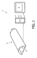
- FIG. 1 schematically illustrates an embodiment of a system 1 for the detection of protrusions in internal parts of a human or animal body, in vivo or in vitro, i.e. a patient.
- This system 1 for example comprises a scanning device 2, for scanning internal parts of a body 3 of a patient, a computer 4 and a display 5.
- the computer 4 comprises a processing circuit 6 and a storage arrangement 7.
- the storage arrangement 7 is loaded with a computer program product.
- the display 5 is configured to display medical data, in particular three dimensional digital data.
- the scanning device 2 sends two and/or three dimensional data to the computer 4.
- the computer 4, in particular the processing circuit 6, further processes the received data such that it is converted into said three dimensional medical data to be displayed on the display 5.
- the computer 4 may receive scanning data via any means, for example via a wired or wireless network, any data carrier such as for example a CD, DVD, hard disk, etc. or via the scanning device 2.
- Scanning device 2 is configured to obtain data of an internal part of the body 3.
- the internal part comprises a colon wall.
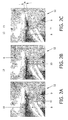
- Three dimensional medical data is obtained from the scanning process, from which a digital image 12 is derived, see for example figure 2C .
- This digital image 12 may for example form a screenshot of a virtual representation of internal parts of the body 3 of the patient, which representation may for example comprise moving images and/or have three dimensional animation-like features.
- Said digital image 12 can for example be studied by a doctor and a certain area 11 that needs special attention from the doctor can be marked by the computer 4. For example, when a lesion 11 is present in a colon wall, it is detected by the computer 3 from the digital image 12, as can be seen in figure 2A and 2C .
- the protrusion can be marked, for example with a distinguishable color 13 or a darker or lighter tan, as shown in figure 2C .
- the doctor may decide directly or in a separate session if medical action should be taken.
- a suspicious surface region 9 comprising a lesion is deformed, i.e. flattened or smoothened, preferably gradually.
- an image 12 can be obtained of what the colon wall would look like without having said lesion.
- Figure 2A illustrates a three dimensional digital image 12 of a colon wall 8 with a fold 10.
- a suspicious surface region 9 is situated that comprises a lesion 11.
- the system 1 may consequently detect the lesion 11 by a method that will be described into more detail below.
- the suspicious surface region 9 is digitally deformed into a deformed surface region 9'.
- the suspicious surface region 9 comprising the lesion 11 is smoothened as compared to the surrounding colon wall to a deformed surface region 9', which represents the colon wall 8 as if no lesion 11 was there.
- the wall 8 comprises a fold 10 on which fold the lesion 11 is situated.
- the deformation from an image according to figure 2A to one according to figure 2B can necessitate a number of steps in between. It can be an iterative process. Consequently, at the site of the fold, the colon wall 8 is smoothened in such a way that the fold 10 looks as if no lesion is present.
- the extent of deformation corresponds to an estimation of a total amount of protrusion of the lesion 11.
- the amount of protrusion can for example be represented by any of the dimension parameters volume, length, width, surface area, base area, height h, etc. Any of these parameters can be calculated and/or displayed separately, next to or instead of the digital image 12. If the value of any of these parameters is above a specific threshold, a computer may classify the corresponding protrusion.
- a protrusion is detected by detecting at least one point p on the colon wall surface 8 where the normal curvature values are above zero.
- equations are used that apply curvature values k1 and k2 of first and second principle curvatures, respectively, wherein k1 and k2 are above zero. All adjacent points p that have a k1 and k2 above zero form a surface region that is a protrusion.
- a protrusion may be almost flat but still have a k1 and k2 above zero.
- the method according to the invention will lead to the possibility of a quantitative assessment of the protrusion and permits intuitive judgment of the candidate protrusions by the specialist, as will follow from the description.
- a protrusion is directed towards the inside 14 of a colon and has at least one point p with normal curvature values above zero, see also figure 3 .
- a said point p, the normal N has a positive direction towards the inside of the colon 14, in the direction of the arrow a.
- the outside 15 is the tissue which forms the colon wall 8 and beyond. Dents in a colon wall 8 are directed towards the outside 15 and are assumed to have at least one point with a k1 and k2 below zero.
- protrusions may also have a k1 and k2 below zero and dents may have a k1 and k2 above zero.
- a normal would be directed in the opposite direction than the normal N that is indicated in figure 3 .
- k1 and k2 are defined as the maximum and minimum values, respectively, of the normal curvatures through point p. Normal curvatures and principal curvatures are common knowledge for those skilled in the art [1].
- the surface region 9 where k1>k2 > 0 is deformed such that k2 is deformed to a value closer to zero and p is translated to p1, as can be seen from figure 4 .
- k1 changes.
- a new second principal curve value k2 1 is calculated.
- This step can be repeated for example n times until the deformed second principal curvature value k2n is approximately zero for all points p n on the deformed surface region 9'.
- protrusions curved in two positive directions, like lesions 11, are deformed such that the colon wall 8 is deformed so that it looks as if no protrusion is there while folds will be unaffected.
- the concave regions 16 and the convex regions 17 of the protrusion will be taken into account for the total deformation and calculation of the amount of protrusion.
- the modified second principle curvature k2 n can be brought to a value lower than zero, for example when the protrusion that is to be deformed is located in a concave fold 18 of the colon wall 8. This is illustrated in figure 5 .
- the extent of deformation to arrive at the deformed surface region 9' as compared to the original surface region 9 is a measure of the amount of protrusion.
- different parameters may indicate the extent of deformation.
- the maximum height of a protrusion can for example be detected by measuring the height h of the largest displacement of a point p to a point p n on the surface region 9. Since the height h of the protrusion may be an indication of the possible risk that a detected protrusion is a harmful lesion 11, the threshold for selecting a protrusion can be set at a specific height h, for example one, three or five mm's. When the protrusion exceeds this threshold, it is selected to be a conspicuous lesion 11. The height of the threshold can also determine how sensitive the lesion selection process is.
- second and/or multiple thresholds can be set. For example, hysteresis thresholding can be applied, wherein adjacent regions of a selected surface region are also included in the protrusion. For example, regions that surround a protrusion that exceeds a first threshold are compared with a second threshold that is lower than the first threshold. For example, a protrusion is found wherein the height h of the protrusion exceeds a first threshold of 1, 3 or 5 millimeters. Thereafter, the surrounding regions are compared to a second threshold of 0.5, 1.5 or 2 mm, respectively, for example.
- the surrounding regions that exceed the second threshold are included in the protrusion and the full protrusion is considered to be the whole region of the surface which exceeds the second threshold and includes an, oftentimes smaller, region that exceeds the first threshold.
- Multiple surface regions and adjacent regions may be included in the protrusion, for example because they overlap or because they are within a specific distance of the protrusion.
- this example of hysteresis thresholding serves merely as an illustration and multiple methods of hysteresis thresholding can be applied as known in the art.
- the volume between the original surface region 9 where k1>k2>0 and the deformed surface region 9' can be a measure of the amount of protrusion so that a threshold may be applied to volume.
- Other parameters that can be used for a threshold and to measure the protrusion are for example width, length, area, surface area and base length.
- a technique for deforming an original surface region 9 to a deformed surface region 9' may use the following principles.
- a system for protrusion detection employs surface deformation on triangle meshes.
- a triangle mesh is generated by a marching cubes algorithm applied to three dimensional CT data using a threshold of -750 Hu (Hounsfield units).
- Hu Heunsfield units
- a typical mesh size comprises around 10 6 vertices, for example.
- the matrix M I- ⁇ dt L is sparse and its structure is given by the mesh one-ring relations, X is a vector containing all mesh points and I is the identity matrix. This system can be solved efficiently using the bi-conjugate gradient method.
- the 'force field' F initially balances the displacement prescribed by the Laplacian and leaves the mesh unaltered.
- F ⁇ t + 1 F ⁇ t - ⁇ 2 ⁇ A 1 ⁇ ring 2 ⁇ ⁇ ⁇ n ⁇
- a 1 ring is the surface area of the 1-ring neighborhood and n is the vertex normal.
- the last term can be interpreted as a correction term. Note that if ⁇ 2 is positive ⁇ F ⁇ should be relaxed.
- the magnitude of the reduction term additionally depends on the sampling density of the mesh. If the sampling is dense and A 1 ring small the magnitude of the correction term should be small.
- the resulting displacement of the mesh points yields a deformed mesh which is an estimate of how the colon wall looks like in the absence of protrusions.
- the total amount of displacement of each mesh point (e.g. in millimeters) is an example of a quantitative measure of the amount of protrusion.
- Candidate objects are generated by applying a threshold on the displacement field.
- multiple types of scanning devices 2 may be suitable to be able to obtain three dimensional digital data of an internal part of the body.
- the scanning device 2 may be any device that is able to obtain an image of the intern*als of the body from the outside or from within.
- the scanning device 2 may for example comprise a CT scanning device, MRI (magnetic resonance imaging) device, US (ultrasound)/sonar scanning device, intravascular probe or other scanning device 2 to obtain three dimensional digital data of an internal part of the body.
- Other imaging techniques may for example include X-ray devices, body invasive probes, cameras, such as CCD (charge coupled device) cameras, television techniques, analogue cameras and/or combinations of those or other techniques.
- From the scanning device 2, data is send to the computer 3.
- This data may comprise three dimensional digital data of an internal part of the body or may comprise two dimensional images or any data that needs to be processed by a computer 3 for conversion to three dimensional digital data.
- a method, system and computer program product according to the invention are suitable for the possible detection and/or prevention of lesions in colons
- applications may include any application wherein a protrusion such as a deformation and/or element has to be detected on the surface of an internal part of the body, particularly a tubular and bended part of the inside of the body.
- Such parts can be colons, airways, arteries or veins, for example.
- Elements and deformations that may be detected with a method, system and/or computer program according to the invention include, but are not limited to, polyps, nodules, cysts, endobronchial lesions, endovascular lesions, emboli, tumors or other lesions, plaques in arteries or veins, stenosis and other elements and/or deformations that may be present inside a tubular and bended part of the body.
- the protrusions may for example comprise any element that may cause blocking of the blood flow in an artery or vein.
Landscapes
- Engineering & Computer Science (AREA)
- Theoretical Computer Science (AREA)
- General Physics & Mathematics (AREA)
- Physics & Mathematics (AREA)
- Nuclear Medicine, Radiotherapy & Molecular Imaging (AREA)
- Computer Vision & Pattern Recognition (AREA)
- General Health & Medical Sciences (AREA)
- Medical Informatics (AREA)
- Multimedia (AREA)
- Radiology & Medical Imaging (AREA)
- Quality & Reliability (AREA)
- Health & Medical Sciences (AREA)
- Measuring And Recording Apparatus For Diagnosis (AREA)
- Apparatus For Radiation Diagnosis (AREA)
- Ultra Sonic Daignosis Equipment (AREA)
- Length Measuring Devices With Unspecified Measuring Means (AREA)
- Magnetic Resonance Imaging Apparatus (AREA)
- Image Processing (AREA)
- Measurement Of The Respiration, Hearing Ability, Form, And Blood Characteristics Of Living Organisms (AREA)
Claims (12)
- Verfahren zum Erkennen einer Protrusion zumindest eines Teils einer Fläche eines inneren Teils eines menschlichen oder tierischen Körpers anhand von dreidimensionalen digitalen Daten, die die genannte Fläche oder den genannten Teil davon darstellen, wobei das Verfahren die folgenden Schritte umfasst:Erkennen eines Flächenbereichs anhand der dreidimensionalen digitalen Daten, wobei der Flächenbereich mindestens einen Punkt aufweist, an dem sich eine erste und eine zweite Normalkrümmung schneiden, die beide einen ursprünglichen Krümmungswert größer als Null oder einen ursprünglichen Krümmungswert kleiner als Null haben, wobei die zweite Normalkrümmung einen Krümmungswert aufweist, der näher an Null liegt als der Krümmungswert der ersten Normalkrümmung,digitales Ändern der zweiten Normalkrümmung derart, dass sie einen geänderten Krümmungswert aufweist, der näher an Null liegt als ihr ursprünglicher Krümmungswert, wobei durch diese Änderung der Flächenbereich digital deformiert wird, so dass ein deformierter Flächenbereich gebildet wird, wobei das Ausmaß der Deformation des deformierten Flächenbereichs im Vergleich zum erkannten Flächenbereich mit einem Grad der Protrusion in Zusammenhang steht, und wobei der Flächenbereich so lange deformiert wird, bis der geänderte Krümmungswert der geänderten zweiten Normalkrümmung des deformierten Flächenbereichs ungefähr Null beträgt, und wobei das Gesamtausmaß der Deformation des deformierten Flächenbereichs einen Gesamtgrad der Protrusion angibt.
- Verfahren, wobei die Schritte des Verfahrens nach Anspruch 1 für den deformierten Flächenbereich wiederholt werden, wobei das Ausmaß der Deformation des deformierten Flächenbereichs nach jeder Wiederholung zunimmt.
- Verfahren nach einem der vorhergehenden Ansprüche, wobei für den Fall, dass der ursprüngliche Krümmungswert der ersten und der zweiten Normalkrümmung größer als Null ist, die erste und die zweite Normalkrümmung erste bzw. zweite Hauptkrümmungen sind, und wobei für den Fall, dass der ursprüngliche Krümmungswert der ersten und der zweiten Normalkrümmung kleiner als Null ist, die erste und die zweite Normalkrümmung zweite bzw. erste Hauptkrümmungen sind.
- Verfahren nach einem der vorhergehenden Ansprüche, wobei das Ausmaß der Deformation eine Höhe der Protrusion umfasst.
- Verfahren nach einem der vorhergehenden Ansprüche, wobei die dreidimensionalen digitalen Daten der deformierten Fläche dargestellt werden.
- Verfahren nach einem der vorhergehenden Ansprüche, wobei das Ausmaß der Deformation mit einem Schwellenwert verglichen wird, wobei ein Signal ausgegeben wird, falls das Ausmaß der Deformation den Schwellenwert übersteigt.
- Verfahren nach Anspruch 6, wobei das Signal eine Kennzeichnung des erkannten Flächenbereiches, vorzugsweise mit einer Farbe, umfasst.
- Verfahren nach einem der vorhergehenden Ansprüche, wobei das genannte innere Teil eine im Wesentlichen schlauchförmige und/oder gebogene Form hat.
- Verfahren nach einem der vorhergehenden Ansprüche, wobei die Protrusion eine Läsion umfasst.
- Protrusionserkennungssystem, das mit einer Schaltung und einer Anzeigevorrichtung versehen oder verbunden ist, wobei die Schaltung so programmiert ist, dass sie ein Verfahren nach einem der vorhergehenden Ansprüche durchführt, und wobei die Anzeigevorrichtung so ausgelegt ist, dass sie die genannten Flächenbereiche anzeigt.
- Computerprogrammprodukt, das so konfiguriert ist, dass es, wenn es in einen Computer geladen ist und aktiviert wird, eine Protrusion von mindestens einem Teil einer Fläche eines inneren Teils eines menschlichen oder tierischen Körpers anhand dreidimensionaler digitaler Daten erkennt, wobei es die Schritte eines Verfahren nach einem der Ansprüche 1 bis 9 umfasst.
- Verfahren zum Erkennen von Protrusionen an der Innenfläche eines schlauchförmigen Teils eines menschlichen oder tierischen Körpers, wobei das Verfahren die folgenden Schritte umfasst:Scannen des Körpers oder von Teilen des Körpers mit dem Ziel, dreidimensionale digitale Daten von mindestens einem Teil einer Innenfläche des schlauchförmigen Teils zu erhalten;Erkennen eines Flächenbereichs anhand der dreidimensionalen digitalen Daten, wobei der Flächenbereich mindestens einen Punkt aufweist, an dem sich eine erste und eine zweite Normalkrümmung schneiden, die beide einen ursprünglichen Krümmungswert größer als Null haben, wobei die zweite Normalkrümmung einen geringeren Krümmungswert als die erste Normalkrümmung aufweist;digitales Ändern der zweiten Normalkrümmung derart, dass sie einen geänderten Krümmungswert hat, der geringer als ihr ursprünglicher Krümmungswert ist, wobei durch diese Änderung der Flächenbereich digital deformiert wird, so dass ein deformierter Flächenbereich gebildet wird, wobei das Ausmaß der Deformation des deformierten Flächenbereichs im Vergleich zu dem erkannten Flächenbereich mit einem Grad der Protrusion in Zusammenhang steht, und wobei der Flächenbereich so lange deformiert wird, bis der geänderte Krümmungswert der geänderten zweiten Normalkrümmung des deformierten Flächenbereichs ungefähr Null beträgt, und wobei das Gesamtausmaß der Deformation des deformierten Flächenbereichs einen Gesamtgrad der Protrusion angibt.
Applications Claiming Priority (2)
| Application Number | Priority Date | Filing Date | Title |
|---|---|---|---|
| NL1032602A NL1032602C2 (nl) | 2006-09-29 | 2006-09-29 | Werkwijzen, systeem en computerprogrammaproduct voor het detecteren van een protrusie. |
| PCT/IB2007/053885 WO2008038222A2 (en) | 2006-09-29 | 2007-09-25 | Methods, system and computer program product for detecting a protrusion |
Publications (2)
| Publication Number | Publication Date |
|---|---|
| EP2074591A2 EP2074591A2 (de) | 2009-07-01 |
| EP2074591B1 true EP2074591B1 (de) | 2010-06-02 |
Family
ID=37923481
Family Applications (1)
| Application Number | Title | Priority Date | Filing Date |
|---|---|---|---|
| EP07826528A Not-in-force EP2074591B1 (de) | 2006-09-29 | 2007-09-25 | Verfahren, system und computerprogrammprodukt zur erkennung einer protrusion |
Country Status (9)
| Country | Link |
|---|---|
| US (1) | US8712117B2 (de) |
| EP (1) | EP2074591B1 (de) |
| JP (1) | JP5551935B2 (de) |
| CN (1) | CN101558429B (de) |
| AT (1) | ATE470203T1 (de) |
| DE (1) | DE602007006976D1 (de) |
| NL (1) | NL1032602C2 (de) |
| RU (1) | RU2459261C2 (de) |
| WO (1) | WO2008038222A2 (de) |
Families Citing this family (4)
| Publication number | Priority date | Publication date | Assignee | Title |
|---|---|---|---|---|
| JP5595750B2 (ja) * | 2010-02-17 | 2014-09-24 | 株式会社東芝 | 医用画像処理装置、及び医用画像処理プログラム |
| US9299139B2 (en) * | 2012-03-26 | 2016-03-29 | The Cleveland Clinic Foundation | Volumetric analysis of pathologies |
| US20180064342A1 (en) * | 2015-03-18 | 2018-03-08 | Imricor Medical Systems, Inc. | System and method for enhanced magnetic resonance imaging of tissue |
| CN109965840B (zh) * | 2019-04-09 | 2024-07-23 | 广东唯仁医疗科技有限公司 | 利用频谱oct成像进行定位黄斑的装置 |
Family Cites Families (27)
| Publication number | Priority date | Publication date | Assignee | Title |
|---|---|---|---|---|
| DE3734979A1 (de) * | 1986-10-16 | 1988-04-28 | Olympus Optical Co | Endoskop |
| US7194117B2 (en) * | 1999-06-29 | 2007-03-20 | The Research Foundation Of State University Of New York | System and method for performing a three-dimensional virtual examination of objects, such as internal organs |
| US6343936B1 (en) * | 1996-09-16 | 2002-02-05 | The Research Foundation Of State University Of New York | System and method for performing a three-dimensional virtual examination, navigation and visualization |
| US6119574A (en) * | 1998-07-02 | 2000-09-19 | Battelle Memorial Institute | Blast effects suppression system |
| EP1133257A4 (de) * | 1998-11-25 | 2007-12-26 | Univ Wake Forest | Virtuelle endoskopie mit verbesserter bildsegmentierung und läsionsdetektion |
| RU2152174C1 (ru) * | 1999-10-05 | 2000-07-10 | Центральный научно-исследовательский рентгено-радиологический институт | Способ диагностики подслизистых опухолей желудочно-кишечного тракта |
| WO2001078005A2 (en) * | 2000-04-11 | 2001-10-18 | Cornell Research Foundation, Inc. | System and method for three-dimensional image rendering and analysis |
| US7379572B2 (en) * | 2001-10-16 | 2008-05-27 | University Of Chicago | Method for computer-aided detection of three-dimensional lesions |
| JP2003260052A (ja) * | 2002-03-08 | 2003-09-16 | Hitachi Medical Corp | 画像診断支援装置 |
| CN101219058B (zh) * | 2002-03-14 | 2012-01-11 | Netkisr有限公司 | 分析和显示计算机体层摄影术数据的系统和方法 |
| WO2003088151A2 (en) * | 2002-04-16 | 2003-10-23 | Koninklijke Philips Electronics N.V. | Medical viewing system and image processing method for visualisation of folded anatomical portions of object surfaces |
| JP4336083B2 (ja) * | 2002-06-20 | 2009-09-30 | 株式会社日立メディコ | 画像診断支援装置、画像診断支援方法 |
| US7260250B2 (en) * | 2002-09-30 | 2007-08-21 | The United States Of America As Represented By The Secretary Of The Department Of Health And Human Services | Computer-aided classification of anomalies in anatomical structures |
| US20040097791A1 (en) * | 2002-11-13 | 2004-05-20 | Olympus Corporation | Endoscope |
| JP4421203B2 (ja) | 2003-03-20 | 2010-02-24 | 株式会社東芝 | 管腔状構造体の解析処理装置 |
| US7391893B2 (en) * | 2003-06-27 | 2008-06-24 | Siemens Medical Solutions Usa, Inc. | System and method for the detection of shapes in images |
| JP4416736B2 (ja) * | 2003-08-01 | 2010-02-17 | 株式会社日立メディコ | 医用画像診断支援装置及びプログラム |
| US7412084B2 (en) * | 2003-08-13 | 2008-08-12 | Siemens Medical Solutions Usa, Inc. | Method of analysis of local patterns of curvature distributions |
| WO2005031635A1 (en) * | 2003-09-25 | 2005-04-07 | Paieon, Inc. | System and method for three-dimensional reconstruction of a tubular organ |
| EP1735750A2 (de) * | 2004-04-12 | 2006-12-27 | The General Hospital Corporation | Verfahren und vorrichtung zum verarbeiten von bildern in einem daumsubtraktionssystem |
| JP5048233B2 (ja) * | 2004-10-08 | 2012-10-17 | ゼネラル・エレクトリック・カンパニイ | Cadシステムにおける解剖学的形状の検出のための方法及びシステム |
| US8317327B2 (en) * | 2005-03-16 | 2012-11-27 | Lc Technologies, Inc. | System and method for eyeball surface topography as a biometric discriminator |
| US8577101B2 (en) * | 2006-03-13 | 2013-11-05 | Kitware, Inc. | Change assessment method |
| US8900124B2 (en) * | 2006-08-03 | 2014-12-02 | Olympus Medical Systems Corp. | Image display device |
| JP4528322B2 (ja) * | 2007-09-28 | 2010-08-18 | 富士フイルム株式会社 | 画像表示装置、画像表示方法、および画像表示プログラム |
| US8483803B2 (en) * | 2008-09-15 | 2013-07-09 | Varian Medical Systems, Inc. | Systems and methods for tracking and targeting object in a patient using imaging techniques |
| AU2010298333B2 (en) * | 2009-09-23 | 2014-04-17 | Lightlab Imaging, Inc. | Lumen morphology and vascular resistance measurements data collection systems, apparatus and methods |
-
2006
- 2006-09-29 NL NL1032602A patent/NL1032602C2/nl not_active IP Right Cessation
-
2007
- 2007-09-25 AT AT07826528T patent/ATE470203T1/de not_active IP Right Cessation
- 2007-09-25 US US12/442,764 patent/US8712117B2/en active Active
- 2007-09-25 WO PCT/IB2007/053885 patent/WO2008038222A2/en not_active Ceased
- 2007-09-25 DE DE602007006976T patent/DE602007006976D1/de active Active
- 2007-09-25 CN CN200780036170.3A patent/CN101558429B/zh not_active Expired - Fee Related
- 2007-09-25 JP JP2009529821A patent/JP5551935B2/ja not_active Expired - Fee Related
- 2007-09-25 RU RU2009116272/08A patent/RU2459261C2/ru active
- 2007-09-25 EP EP07826528A patent/EP2074591B1/de not_active Not-in-force
Also Published As
| Publication number | Publication date |
|---|---|
| RU2459261C2 (ru) | 2012-08-20 |
| RU2009116272A (ru) | 2010-11-10 |
| EP2074591A2 (de) | 2009-07-01 |
| NL1032602C2 (nl) | 2008-04-01 |
| US20110235882A1 (en) | 2011-09-29 |
| WO2008038222A2 (en) | 2008-04-03 |
| CN101558429B (zh) | 2015-07-29 |
| JP2010504794A (ja) | 2010-02-18 |
| JP5551935B2 (ja) | 2014-07-16 |
| CN101558429A (zh) | 2009-10-14 |
| US8712117B2 (en) | 2014-04-29 |
| DE602007006976D1 (de) | 2010-07-15 |
| ATE470203T1 (de) | 2010-06-15 |
| WO2008038222A3 (en) | 2009-02-05 |
Similar Documents
| Publication | Publication Date | Title |
|---|---|---|
| US9418420B2 (en) | System and method for automated detection of lung nodules in medical images | |
| Kostis et al. | Three-dimensional segmentation and growth-rate estimation of small pulmonary nodules in helical CT images | |
| US8121373B2 (en) | Image diagnostic processing device and image diagnostic processing program | |
| EP2869261B1 (de) | Verfahren zur Verarbeitung von Daten, die ein dreidimensionales Volumen repräsentieren | |
| US20090123049A1 (en) | Nodule Detection | |
| JPH1156828A (ja) | 異常陰影候補検出方法および装置 | |
| KR101162599B1 (ko) | 흉부 방사선 이미지 분석을 통한 심비대 자동탐지 방법 및 그 기록매체 | |
| EP2074591B1 (de) | Verfahren, system und computerprogrammprodukt zur erkennung einer protrusion | |
| van Wijk et al. | Detection of protrusions in curved folded surfaces applied to automated polyp detection in CT colonography | |
| Jerebko et al. | Polyp segmentation method for CT colonography computer-aided detection | |
| EP2005389B1 (de) | Automatische herzbanderkennung bei mamma-mri | |
| JP2004195080A (ja) | 異常陰影検出装置 | |
| JPH08287230A (ja) | 計算機支援画像診断装置 | |
| JP3596792B2 (ja) | 異常陰影候補領域を含む局所領域の抽出方法および装置 | |
| JPH10108859A (ja) | 異常陰影候補の検出方法および装置 | |
| JP2002133397A (ja) | 異常陰影候補検出装置 | |
| JP2005046527A (ja) | 超音波肝画像を用いた定量的肝組織性状診断方法 | |
| JP4252744B2 (ja) | 異常陰影検出装置 | |
| JP5132559B2 (ja) | デジタル画像のセグメント化方法およびコンピュータ読み取り可能なプログラム記憶装置 | |
| JP2005224428A (ja) | 石灰化陰影判定方法、石灰化陰影判定装置およびそのプログラム | |
| JPH10118053A (ja) | 異常陰影候補の検出方法および装置 | |
| JP2001222703A (ja) | 異常陰影候補の検出処理方法および装置 | |
| Bankhele et al. | Detection of Protrusion on Curved Folded Surface In Colon Capsule Endoscopy | |
| JPH08272931A (ja) | 画像処理方法 |
Legal Events
| Date | Code | Title | Description |
|---|---|---|---|
| PUAI | Public reference made under article 153(3) epc to a published international application that has entered the european phase |
Free format text: ORIGINAL CODE: 0009012 |
|
| AK | Designated contracting states |
Kind code of ref document: A2 Designated state(s): AT BE BG CH CY CZ DE DK EE ES FI FR GB GR HU IE IS IT LI LT LU LV MC MT NL PL PT RO SE SI SK TR |
|
| AX | Request for extension of the european patent |
Extension state: AL BA HR MK RS |
|
| 17P | Request for examination filed |
Effective date: 20090805 |
|
| RBV | Designated contracting states (corrected) |
Designated state(s): AT BE BG CH CY CZ DE DK EE ES FI FR GB GR HU IE IS IT LI LT LU LV MC MT NL PL PT RO SE SI SK TR |
|
| GRAP | Despatch of communication of intention to grant a patent |
Free format text: ORIGINAL CODE: EPIDOSNIGR1 |
|
| DAX | Request for extension of the european patent (deleted) | ||
| GRAS | Grant fee paid |
Free format text: ORIGINAL CODE: EPIDOSNIGR3 |
|
| GRAA | (expected) grant |
Free format text: ORIGINAL CODE: 0009210 |
|
| AK | Designated contracting states |
Kind code of ref document: B1 Designated state(s): AT BE BG CH CY CZ DE DK EE ES FI FR GB GR HU IE IS IT LI LT LU LV MC MT NL PL PT RO SE SI SK TR |
|
| REG | Reference to a national code |
Ref country code: GB Ref legal event code: FG4D |
|
| REG | Reference to a national code |
Ref country code: CH Ref legal event code: EP |
|
| REG | Reference to a national code |
Ref country code: IE Ref legal event code: FG4D |
|
| REF | Corresponds to: |
Ref document number: 602007006976 Country of ref document: DE Date of ref document: 20100715 Kind code of ref document: P |
|
| REG | Reference to a national code |
Ref country code: NL Ref legal event code: VDEP Effective date: 20100602 |
|
| PG25 | Lapsed in a contracting state [announced via postgrant information from national office to epo] |
Ref country code: SE Free format text: LAPSE BECAUSE OF FAILURE TO SUBMIT A TRANSLATION OF THE DESCRIPTION OR TO PAY THE FEE WITHIN THE PRESCRIBED TIME-LIMIT Effective date: 20100602 Ref country code: LT Free format text: LAPSE BECAUSE OF FAILURE TO SUBMIT A TRANSLATION OF THE DESCRIPTION OR TO PAY THE FEE WITHIN THE PRESCRIBED TIME-LIMIT Effective date: 20100602 |
|
| LTIE | Lt: invalidation of european patent or patent extension |
Effective date: 20100602 |
|
| PG25 | Lapsed in a contracting state [announced via postgrant information from national office to epo] |
Ref country code: LV Free format text: LAPSE BECAUSE OF FAILURE TO SUBMIT A TRANSLATION OF THE DESCRIPTION OR TO PAY THE FEE WITHIN THE PRESCRIBED TIME-LIMIT Effective date: 20100602 Ref country code: SI Free format text: LAPSE BECAUSE OF FAILURE TO SUBMIT A TRANSLATION OF THE DESCRIPTION OR TO PAY THE FEE WITHIN THE PRESCRIBED TIME-LIMIT Effective date: 20100602 Ref country code: AT Free format text: LAPSE BECAUSE OF FAILURE TO SUBMIT A TRANSLATION OF THE DESCRIPTION OR TO PAY THE FEE WITHIN THE PRESCRIBED TIME-LIMIT Effective date: 20100602 Ref country code: FI Free format text: LAPSE BECAUSE OF FAILURE TO SUBMIT A TRANSLATION OF THE DESCRIPTION OR TO PAY THE FEE WITHIN THE PRESCRIBED TIME-LIMIT Effective date: 20100602 |
|
| PG25 | Lapsed in a contracting state [announced via postgrant information from national office to epo] |
Ref country code: PL Free format text: LAPSE BECAUSE OF FAILURE TO SUBMIT A TRANSLATION OF THE DESCRIPTION OR TO PAY THE FEE WITHIN THE PRESCRIBED TIME-LIMIT Effective date: 20100602 Ref country code: CY Free format text: LAPSE BECAUSE OF FAILURE TO SUBMIT A TRANSLATION OF THE DESCRIPTION OR TO PAY THE FEE WITHIN THE PRESCRIBED TIME-LIMIT Effective date: 20100616 |
|
| PG25 | Lapsed in a contracting state [announced via postgrant information from national office to epo] |
Ref country code: EE Free format text: LAPSE BECAUSE OF FAILURE TO SUBMIT A TRANSLATION OF THE DESCRIPTION OR TO PAY THE FEE WITHIN THE PRESCRIBED TIME-LIMIT Effective date: 20100602 Ref country code: NL Free format text: LAPSE BECAUSE OF FAILURE TO SUBMIT A TRANSLATION OF THE DESCRIPTION OR TO PAY THE FEE WITHIN THE PRESCRIBED TIME-LIMIT Effective date: 20100602 |
|
| PG25 | Lapsed in a contracting state [announced via postgrant information from national office to epo] |
Ref country code: IS Free format text: LAPSE BECAUSE OF FAILURE TO SUBMIT A TRANSLATION OF THE DESCRIPTION OR TO PAY THE FEE WITHIN THE PRESCRIBED TIME-LIMIT Effective date: 20101002 Ref country code: CZ Free format text: LAPSE BECAUSE OF FAILURE TO SUBMIT A TRANSLATION OF THE DESCRIPTION OR TO PAY THE FEE WITHIN THE PRESCRIBED TIME-LIMIT Effective date: 20100602 Ref country code: BE Free format text: LAPSE BECAUSE OF FAILURE TO SUBMIT A TRANSLATION OF THE DESCRIPTION OR TO PAY THE FEE WITHIN THE PRESCRIBED TIME-LIMIT Effective date: 20100602 Ref country code: SK Free format text: LAPSE BECAUSE OF FAILURE TO SUBMIT A TRANSLATION OF THE DESCRIPTION OR TO PAY THE FEE WITHIN THE PRESCRIBED TIME-LIMIT Effective date: 20100602 Ref country code: RO Free format text: LAPSE BECAUSE OF FAILURE TO SUBMIT A TRANSLATION OF THE DESCRIPTION OR TO PAY THE FEE WITHIN THE PRESCRIBED TIME-LIMIT Effective date: 20100602 Ref country code: PT Free format text: LAPSE BECAUSE OF FAILURE TO SUBMIT A TRANSLATION OF THE DESCRIPTION OR TO PAY THE FEE WITHIN THE PRESCRIBED TIME-LIMIT Effective date: 20101004 |
|
| PG25 | Lapsed in a contracting state [announced via postgrant information from national office to epo] |
Ref country code: IT Free format text: LAPSE BECAUSE OF FAILURE TO SUBMIT A TRANSLATION OF THE DESCRIPTION OR TO PAY THE FEE WITHIN THE PRESCRIBED TIME-LIMIT Effective date: 20100602 |
|
| PLBE | No opposition filed within time limit |
Free format text: ORIGINAL CODE: 0009261 |
|
| STAA | Information on the status of an ep patent application or granted ep patent |
Free format text: STATUS: NO OPPOSITION FILED WITHIN TIME LIMIT |
|
| PG25 | Lapsed in a contracting state [announced via postgrant information from national office to epo] |
Ref country code: DK Free format text: LAPSE BECAUSE OF FAILURE TO SUBMIT A TRANSLATION OF THE DESCRIPTION OR TO PAY THE FEE WITHIN THE PRESCRIBED TIME-LIMIT Effective date: 20100602 Ref country code: MC Free format text: LAPSE BECAUSE OF NON-PAYMENT OF DUE FEES Effective date: 20100930 |
|
| 26N | No opposition filed |
Effective date: 20110303 |
|
| PG25 | Lapsed in a contracting state [announced via postgrant information from national office to epo] |
Ref country code: GR Free format text: LAPSE BECAUSE OF FAILURE TO SUBMIT A TRANSLATION OF THE DESCRIPTION OR TO PAY THE FEE WITHIN THE PRESCRIBED TIME-LIMIT Effective date: 20100903 |
|
| REG | Reference to a national code |
Ref country code: DE Ref legal event code: R097 Ref document number: 602007006976 Country of ref document: DE Effective date: 20110302 |
|
| REG | Reference to a national code |
Ref country code: FR Ref legal event code: ST Effective date: 20110531 |
|
| PG25 | Lapsed in a contracting state [announced via postgrant information from national office to epo] |
Ref country code: FR Free format text: LAPSE BECAUSE OF NON-PAYMENT OF DUE FEES Effective date: 20100930 Ref country code: IE Free format text: LAPSE BECAUSE OF NON-PAYMENT OF DUE FEES Effective date: 20100925 |
|
| PG25 | Lapsed in a contracting state [announced via postgrant information from national office to epo] |
Ref country code: MT Free format text: LAPSE BECAUSE OF FAILURE TO SUBMIT A TRANSLATION OF THE DESCRIPTION OR TO PAY THE FEE WITHIN THE PRESCRIBED TIME-LIMIT Effective date: 20100602 |
|
| REG | Reference to a national code |
Ref country code: CH Ref legal event code: PL |
|
| GBPC | Gb: european patent ceased through non-payment of renewal fee |
Effective date: 20110925 |
|
| PG25 | Lapsed in a contracting state [announced via postgrant information from national office to epo] |
Ref country code: LI Free format text: LAPSE BECAUSE OF NON-PAYMENT OF DUE FEES Effective date: 20110930 Ref country code: CH Free format text: LAPSE BECAUSE OF NON-PAYMENT OF DUE FEES Effective date: 20110930 |
|
| PG25 | Lapsed in a contracting state [announced via postgrant information from national office to epo] |
Ref country code: GB Free format text: LAPSE BECAUSE OF NON-PAYMENT OF DUE FEES Effective date: 20110925 |
|
| PG25 | Lapsed in a contracting state [announced via postgrant information from national office to epo] |
Ref country code: HU Free format text: LAPSE BECAUSE OF FAILURE TO SUBMIT A TRANSLATION OF THE DESCRIPTION OR TO PAY THE FEE WITHIN THE PRESCRIBED TIME-LIMIT Effective date: 20101203 Ref country code: LU Free format text: LAPSE BECAUSE OF NON-PAYMENT OF DUE FEES Effective date: 20100925 Ref country code: BG Free format text: LAPSE BECAUSE OF FAILURE TO SUBMIT A TRANSLATION OF THE DESCRIPTION OR TO PAY THE FEE WITHIN THE PRESCRIBED TIME-LIMIT Effective date: 20100602 |
|
| PG25 | Lapsed in a contracting state [announced via postgrant information from national office to epo] |
Ref country code: TR Free format text: LAPSE BECAUSE OF FAILURE TO SUBMIT A TRANSLATION OF THE DESCRIPTION OR TO PAY THE FEE WITHIN THE PRESCRIBED TIME-LIMIT Effective date: 20100602 |
|
| PG25 | Lapsed in a contracting state [announced via postgrant information from national office to epo] |
Ref country code: BG Free format text: LAPSE BECAUSE OF FAILURE TO SUBMIT A TRANSLATION OF THE DESCRIPTION OR TO PAY THE FEE WITHIN THE PRESCRIBED TIME-LIMIT Effective date: 20100902 |
|
| PG25 | Lapsed in a contracting state [announced via postgrant information from national office to epo] |
Ref country code: ES Free format text: LAPSE BECAUSE OF FAILURE TO SUBMIT A TRANSLATION OF THE DESCRIPTION OR TO PAY THE FEE WITHIN THE PRESCRIBED TIME-LIMIT Effective date: 20100913 |
|
| REG | Reference to a national code |
Ref country code: DE Ref legal event code: R082 Ref document number: 602007006976 Country of ref document: DE Representative=s name: MEISSNER, BOLTE & PARTNER GBR, DE |
|
| REG | Reference to a national code |
Ref country code: DE Ref legal event code: R082 Ref document number: 602007006976 Country of ref document: DE Representative=s name: MEISSNER BOLTE PATENTANWAELTE RECHTSANWAELTE P, DE Effective date: 20140328 Ref country code: DE Ref legal event code: R082 Ref document number: 602007006976 Country of ref document: DE Representative=s name: MEISSNER, BOLTE & PARTNER GBR, DE Effective date: 20140328 Ref country code: DE Ref legal event code: R081 Ref document number: 602007006976 Country of ref document: DE Owner name: KONINKLIJKE PHILIPS N.V., NL Free format text: FORMER OWNER: KONINKLIJKE PHILIPS ELECTRONICS N.V., EINDHOVEN, NL Effective date: 20140328 |
|
| PGFP | Annual fee paid to national office [announced via postgrant information from national office to epo] |
Ref country code: DE Payment date: 20230928 Year of fee payment: 17 |
|
| REG | Reference to a national code |
Ref country code: DE Ref legal event code: R119 Ref document number: 602007006976 Country of ref document: DE |
|
| PG25 | Lapsed in a contracting state [announced via postgrant information from national office to epo] |
Ref country code: DE Free format text: LAPSE BECAUSE OF NON-PAYMENT OF DUE FEES Effective date: 20250401 |