EP2092343B1 - Nachweiskonjugat - Google Patents
Nachweiskonjugat Download PDFInfo
- Publication number
- EP2092343B1 EP2092343B1 EP07852077.2A EP07852077A EP2092343B1 EP 2092343 B1 EP2092343 B1 EP 2092343B1 EP 07852077 A EP07852077 A EP 07852077A EP 2092343 B1 EP2092343 B1 EP 2092343B1
- Authority
- EP
- European Patent Office
- Prior art keywords
- detection
- filaments
- recognition elements
- recognition
- reporter molecules
- Prior art date
- Legal status (The legal status is an assumption and is not a legal conclusion. Google has not performed a legal analysis and makes no representation as to the accuracy of the status listed.)
- Not-in-force
Links
- 238000001514 detection method Methods 0.000 title claims description 50
- 210000003632 microfilament Anatomy 0.000 claims description 59
- 238000009739 binding Methods 0.000 claims description 43
- 238000000034 method Methods 0.000 claims description 32
- 239000002096 quantum dot Substances 0.000 claims description 17
- 108091034117 Oligonucleotide Proteins 0.000 claims description 16
- 239000012491 analyte Substances 0.000 claims description 14
- 102000029749 Microtubule Human genes 0.000 claims description 12
- 108091022875 Microtubule Proteins 0.000 claims description 12
- 210000004292 cytoskeleton Anatomy 0.000 claims description 12
- 210000004688 microtubule Anatomy 0.000 claims description 12
- 244000052769 pathogen Species 0.000 claims description 12
- JLCPHMBAVCMARE-UHFFFAOYSA-N [3-[[3-[[3-[[3-[[3-[[3-[[3-[[3-[[3-[[3-[[3-[[5-(2-amino-6-oxo-1H-purin-9-yl)-3-[[3-[[3-[[3-[[3-[[3-[[5-(2-amino-6-oxo-1H-purin-9-yl)-3-[[5-(2-amino-6-oxo-1H-purin-9-yl)-3-hydroxyoxolan-2-yl]methoxy-hydroxyphosphoryl]oxyoxolan-2-yl]methoxy-hydroxyphosphoryl]oxy-5-(5-methyl-2,4-dioxopyrimidin-1-yl)oxolan-2-yl]methoxy-hydroxyphosphoryl]oxy-5-(6-aminopurin-9-yl)oxolan-2-yl]methoxy-hydroxyphosphoryl]oxy-5-(6-aminopurin-9-yl)oxolan-2-yl]methoxy-hydroxyphosphoryl]oxy-5-(6-aminopurin-9-yl)oxolan-2-yl]methoxy-hydroxyphosphoryl]oxy-5-(6-aminopurin-9-yl)oxolan-2-yl]methoxy-hydroxyphosphoryl]oxyoxolan-2-yl]methoxy-hydroxyphosphoryl]oxy-5-(5-methyl-2,4-dioxopyrimidin-1-yl)oxolan-2-yl]methoxy-hydroxyphosphoryl]oxy-5-(4-amino-2-oxopyrimidin-1-yl)oxolan-2-yl]methoxy-hydroxyphosphoryl]oxy-5-(5-methyl-2,4-dioxopyrimidin-1-yl)oxolan-2-yl]methoxy-hydroxyphosphoryl]oxy-5-(5-methyl-2,4-dioxopyrimidin-1-yl)oxolan-2-yl]methoxy-hydroxyphosphoryl]oxy-5-(6-aminopurin-9-yl)oxolan-2-yl]methoxy-hydroxyphosphoryl]oxy-5-(6-aminopurin-9-yl)oxolan-2-yl]methoxy-hydroxyphosphoryl]oxy-5-(4-amino-2-oxopyrimidin-1-yl)oxolan-2-yl]methoxy-hydroxyphosphoryl]oxy-5-(4-amino-2-oxopyrimidin-1-yl)oxolan-2-yl]methoxy-hydroxyphosphoryl]oxy-5-(4-amino-2-oxopyrimidin-1-yl)oxolan-2-yl]methoxy-hydroxyphosphoryl]oxy-5-(6-aminopurin-9-yl)oxolan-2-yl]methoxy-hydroxyphosphoryl]oxy-5-(4-amino-2-oxopyrimidin-1-yl)oxolan-2-yl]methyl [5-(6-aminopurin-9-yl)-2-(hydroxymethyl)oxolan-3-yl] hydrogen phosphate Polymers Cc1cn(C2CC(OP(O)(=O)OCC3OC(CC3OP(O)(=O)OCC3OC(CC3O)n3cnc4c3nc(N)[nH]c4=O)n3cnc4c3nc(N)[nH]c4=O)C(COP(O)(=O)OC3CC(OC3COP(O)(=O)OC3CC(OC3COP(O)(=O)OC3CC(OC3COP(O)(=O)OC3CC(OC3COP(O)(=O)OC3CC(OC3COP(O)(=O)OC3CC(OC3COP(O)(=O)OC3CC(OC3COP(O)(=O)OC3CC(OC3COP(O)(=O)OC3CC(OC3COP(O)(=O)OC3CC(OC3COP(O)(=O)OC3CC(OC3COP(O)(=O)OC3CC(OC3COP(O)(=O)OC3CC(OC3COP(O)(=O)OC3CC(OC3COP(O)(=O)OC3CC(OC3COP(O)(=O)OC3CC(OC3COP(O)(=O)OC3CC(OC3CO)n3cnc4c(N)ncnc34)n3ccc(N)nc3=O)n3cnc4c(N)ncnc34)n3ccc(N)nc3=O)n3ccc(N)nc3=O)n3ccc(N)nc3=O)n3cnc4c(N)ncnc34)n3cnc4c(N)ncnc34)n3cc(C)c(=O)[nH]c3=O)n3cc(C)c(=O)[nH]c3=O)n3ccc(N)nc3=O)n3cc(C)c(=O)[nH]c3=O)n3cnc4c3nc(N)[nH]c4=O)n3cnc4c(N)ncnc34)n3cnc4c(N)ncnc34)n3cnc4c(N)ncnc34)n3cnc4c(N)ncnc34)O2)c(=O)[nH]c1=O JLCPHMBAVCMARE-UHFFFAOYSA-N 0.000 claims description 11
- 230000002776 aggregation Effects 0.000 claims description 10
- 238000004220 aggregation Methods 0.000 claims description 10
- 208000037265 diseases, disorders, signs and symptoms Diseases 0.000 claims description 7
- 150000007523 nucleic acids Chemical class 0.000 claims description 7
- 201000010099 disease Diseases 0.000 claims description 6
- 108020004707 nucleic acids Proteins 0.000 claims description 6
- 102000039446 nucleic acids Human genes 0.000 claims description 6
- 238000005119 centrifugation Methods 0.000 claims description 4
- 239000002122 magnetic nanoparticle Substances 0.000 claims description 4
- 230000001717 pathogenic effect Effects 0.000 claims description 4
- 108091023037 Aptamer Proteins 0.000 claims description 3
- -1 fluorochromes Substances 0.000 claims description 3
- PCHJSUWPFVWCPO-UHFFFAOYSA-N gold Chemical compound [Au] PCHJSUWPFVWCPO-UHFFFAOYSA-N 0.000 claims description 3
- 229910052737 gold Inorganic materials 0.000 claims description 3
- 239000010931 gold Substances 0.000 claims description 3
- 239000003550 marker Substances 0.000 claims description 3
- 238000004416 surface enhanced Raman spectroscopy Methods 0.000 claims description 3
- 238000002198 surface plasmon resonance spectroscopy Methods 0.000 claims description 3
- 239000003053 toxin Substances 0.000 claims description 3
- 231100000765 toxin Toxicity 0.000 claims description 3
- 108700012359 toxins Proteins 0.000 claims description 3
- 102000004190 Enzymes Human genes 0.000 claims description 2
- 108090000790 Enzymes Proteins 0.000 claims description 2
- 102000001706 Immunoglobulin Fab Fragments Human genes 0.000 claims description 2
- 108010054477 Immunoglobulin Fab Fragments Proteins 0.000 claims description 2
- 108090001090 Lectins Proteins 0.000 claims description 2
- 102000004856 Lectins Human genes 0.000 claims description 2
- 238000000149 argon plasma sintering Methods 0.000 claims description 2
- 108020001507 fusion proteins Proteins 0.000 claims description 2
- 102000037865 fusion proteins Human genes 0.000 claims description 2
- 239000002523 lectin Substances 0.000 claims description 2
- 108020003175 receptors Proteins 0.000 claims description 2
- 102000005962 receptors Human genes 0.000 claims description 2
- 229910052709 silver Inorganic materials 0.000 claims description 2
- 239000004332 silver Substances 0.000 claims description 2
- 239000000126 substance Substances 0.000 claims description 2
- 230000000063 preceeding effect Effects 0.000 claims 3
- GNFTZDOKVXKIBK-UHFFFAOYSA-N 3-(2-methoxyethoxy)benzohydrazide Chemical compound COCCOC1=CC=CC(C(=O)NN)=C1 GNFTZDOKVXKIBK-UHFFFAOYSA-N 0.000 claims 2
- 238000007689 inspection Methods 0.000 claims 2
- 238000004458 analytical method Methods 0.000 claims 1
- 238000001917 fluorescence detection Methods 0.000 claims 1
- 238000000338 in vitro Methods 0.000 claims 1
- 108090000765 processed proteins & peptides Proteins 0.000 claims 1
- 102000004196 processed proteins & peptides Human genes 0.000 claims 1
- 238000002798 spectrophotometry method Methods 0.000 claims 1
- 239000012634 fragment Substances 0.000 description 40
- 238000002372 labelling Methods 0.000 description 18
- 239000000523 sample Substances 0.000 description 16
- 108020004999 messenger RNA Proteins 0.000 description 13
- YBJHBAHKTGYVGT-ZKWXMUAHSA-N (+)-Biotin Chemical compound N1C(=O)N[C@@H]2[C@H](CCCCC(=O)O)SC[C@@H]21 YBJHBAHKTGYVGT-ZKWXMUAHSA-N 0.000 description 12
- 150000001875 compounds Chemical class 0.000 description 11
- 230000035945 sensitivity Effects 0.000 description 9
- KPKZJLCSROULON-QKGLWVMZSA-N Phalloidin Chemical compound N1C(=O)[C@@H]([C@@H](O)C)NC(=O)[C@H](C)NC(=O)[C@H](C[C@@](C)(O)CO)NC(=O)[C@H](C2)NC(=O)[C@H](C)NC(=O)[C@@H]3C[C@H](O)CN3C(=O)[C@@H]1CSC1=C2C2=CC=CC=C2N1 KPKZJLCSROULON-QKGLWVMZSA-N 0.000 description 8
- 108060008487 Myosin Proteins 0.000 description 7
- 102000003505 Myosin Human genes 0.000 description 7
- 241000700605 Viruses Species 0.000 description 7
- 229960002685 biotin Drugs 0.000 description 7
- 239000011616 biotin Substances 0.000 description 7
- 230000000295 complement effect Effects 0.000 description 7
- 239000000178 monomer Substances 0.000 description 7
- 235000018102 proteins Nutrition 0.000 description 7
- 102000004169 proteins and genes Human genes 0.000 description 7
- 108090000623 proteins and genes Proteins 0.000 description 7
- 239000000243 solution Substances 0.000 description 7
- 108010085238 Actins Proteins 0.000 description 6
- 102000007469 Actins Human genes 0.000 description 6
- 235000020958 biotin Nutrition 0.000 description 6
- 238000002965 ELISA Methods 0.000 description 5
- 230000015572 biosynthetic process Effects 0.000 description 5
- 238000010168 coupling process Methods 0.000 description 5
- 230000004927 fusion Effects 0.000 description 5
- 241000894006 Bacteria Species 0.000 description 4
- TWRXJAOTZQYOKJ-UHFFFAOYSA-L Magnesium chloride Chemical compound [Mg+2].[Cl-].[Cl-] TWRXJAOTZQYOKJ-UHFFFAOYSA-L 0.000 description 4
- 108010009711 Phalloidine Proteins 0.000 description 4
- 108010090804 Streptavidin Proteins 0.000 description 4
- 238000005859 coupling reaction Methods 0.000 description 4
- 239000000203 mixture Substances 0.000 description 4
- 239000002105 nanoparticle Substances 0.000 description 4
- 239000002245 particle Substances 0.000 description 4
- 210000001519 tissue Anatomy 0.000 description 4
- 238000005199 ultracentrifugation Methods 0.000 description 4
- 208000032791 BCR-ABL1 positive chronic myelogenous leukemia Diseases 0.000 description 3
- 208000010833 Chronic myeloid leukaemia Diseases 0.000 description 3
- 208000033761 Myelogenous Chronic BCR-ABL Positive Leukemia Diseases 0.000 description 3
- XUIMIQQOPSSXEZ-UHFFFAOYSA-N Silicon Chemical compound [Si] XUIMIQQOPSSXEZ-UHFFFAOYSA-N 0.000 description 3
- 230000008878 coupling Effects 0.000 description 3
- 239000000284 extract Substances 0.000 description 3
- 239000011521 glass Substances 0.000 description 3
- 230000003993 interaction Effects 0.000 description 3
- 235000013372 meat Nutrition 0.000 description 3
- 244000005700 microbiome Species 0.000 description 3
- 238000000386 microscopy Methods 0.000 description 3
- 239000002953 phosphate buffered saline Substances 0.000 description 3
- 238000000746 purification Methods 0.000 description 3
- 239000010703 silicon Substances 0.000 description 3
- 229910052710 silicon Inorganic materials 0.000 description 3
- 108091032973 (ribonucleotides)n+m Proteins 0.000 description 2
- FPQQSJJWHUJYPU-UHFFFAOYSA-N 3-(dimethylamino)propyliminomethylidene-ethylazanium;chloride Chemical compound Cl.CCN=C=NCCCN(C)C FPQQSJJWHUJYPU-UHFFFAOYSA-N 0.000 description 2
- 108010043137 Actomyosin Proteins 0.000 description 2
- 241000283690 Bos taurus Species 0.000 description 2
- 208000002979 Influenza in Birds Diseases 0.000 description 2
- 102000007066 Prostate-Specific Antigen Human genes 0.000 description 2
- 108010072866 Prostate-Specific Antigen Proteins 0.000 description 2
- FOIXSVOLVBLSDH-UHFFFAOYSA-N Silver ion Chemical compound [Ag+] FOIXSVOLVBLSDH-UHFFFAOYSA-N 0.000 description 2
- 206010064097 avian influenza Diseases 0.000 description 2
- 239000011324 bead Substances 0.000 description 2
- 210000004556 brain Anatomy 0.000 description 2
- 239000008366 buffered solution Substances 0.000 description 2
- 239000006227 byproduct Substances 0.000 description 2
- 210000004027 cell Anatomy 0.000 description 2
- IJOOHPMOJXWVHK-UHFFFAOYSA-N chlorotrimethylsilane Chemical compound C[Si](C)(C)Cl IJOOHPMOJXWVHK-UHFFFAOYSA-N 0.000 description 2
- 230000003436 cytoskeletal effect Effects 0.000 description 2
- 238000010828 elution Methods 0.000 description 2
- 230000007613 environmental effect Effects 0.000 description 2
- 206010022000 influenza Diseases 0.000 description 2
- 229910001629 magnesium chloride Inorganic materials 0.000 description 2
- 238000002156 mixing Methods 0.000 description 2
- 210000003205 muscle Anatomy 0.000 description 2
- 239000002070 nanowire Substances 0.000 description 2
- 239000002773 nucleotide Substances 0.000 description 2
- 125000003729 nucleotide group Chemical group 0.000 description 2
- 239000008188 pellet Substances 0.000 description 2
- 238000003752 polymerase chain reaction Methods 0.000 description 2
- 210000002027 skeletal muscle Anatomy 0.000 description 2
- 238000010561 standard procedure Methods 0.000 description 2
- 241000712461 unidentified influenza virus Species 0.000 description 2
- NVKAWKQGWWIWPM-ABEVXSGRSA-N 17-β-hydroxy-5-α-Androstan-3-one Chemical compound C1C(=O)CC[C@]2(C)[C@H]3CC[C@](C)([C@H](CC4)O)[C@@H]4[C@@H]3CC[C@H]21 NVKAWKQGWWIWPM-ABEVXSGRSA-N 0.000 description 1
- 208000024827 Alzheimer disease Diseases 0.000 description 1
- 241000271566 Aves Species 0.000 description 1
- 241000193738 Bacillus anthracis Species 0.000 description 1
- 108030001720 Bontoxilysin Proteins 0.000 description 1
- OKTJSMMVPCPJKN-UHFFFAOYSA-N Carbon Chemical compound [C] OKTJSMMVPCPJKN-UHFFFAOYSA-N 0.000 description 1
- 241000588724 Escherichia coli Species 0.000 description 1
- 102000004878 Gelsolin Human genes 0.000 description 1
- 108090001064 Gelsolin Proteins 0.000 description 1
- 208000034951 Genetic Translocation Diseases 0.000 description 1
- 241000941423 Grom virus Species 0.000 description 1
- 241000590002 Helicobacter pylori Species 0.000 description 1
- 102000018071 Immunoglobulin Fc Fragments Human genes 0.000 description 1
- 108010091135 Immunoglobulin Fc Fragments Proteins 0.000 description 1
- 102000010638 Kinesin Human genes 0.000 description 1
- 108010063296 Kinesin Proteins 0.000 description 1
- NQTADLQHYWFPDB-UHFFFAOYSA-N N-Hydroxysuccinimide Chemical compound ON1C(=O)CCC1=O NQTADLQHYWFPDB-UHFFFAOYSA-N 0.000 description 1
- 206010028980 Neoplasm Diseases 0.000 description 1
- 206010060862 Prostate cancer Diseases 0.000 description 1
- 208000000236 Prostatic Neoplasms Diseases 0.000 description 1
- 102000004243 Tubulin Human genes 0.000 description 1
- 108090000704 Tubulin Proteins 0.000 description 1
- 238000010521 absorption reaction Methods 0.000 description 1
- 239000003570 air Substances 0.000 description 1
- 230000004075 alteration Effects 0.000 description 1
- 125000000539 amino acid group Chemical group 0.000 description 1
- 230000003321 amplification Effects 0.000 description 1
- 238000013459 approach Methods 0.000 description 1
- 238000005452 bending Methods 0.000 description 1
- 108091006004 biotinylated proteins Proteins 0.000 description 1
- 230000006287 biotinylation Effects 0.000 description 1
- 238000007413 biotinylation Methods 0.000 description 1
- 210000001124 body fluid Anatomy 0.000 description 1
- 239000010839 body fluid Substances 0.000 description 1
- 229940053031 botulinum toxin Drugs 0.000 description 1
- 201000011510 cancer Diseases 0.000 description 1
- 210000000234 capsid Anatomy 0.000 description 1
- 239000002041 carbon nanotube Substances 0.000 description 1
- 229910021393 carbon nanotube Inorganic materials 0.000 description 1
- 125000003178 carboxy group Chemical group [H]OC(*)=O 0.000 description 1
- 230000001413 cellular effect Effects 0.000 description 1
- 238000007385 chemical modification Methods 0.000 description 1
- 238000006243 chemical reaction Methods 0.000 description 1
- 238000004132 cross linking Methods 0.000 description 1
- 210000004748 cultured cell Anatomy 0.000 description 1
- 235000018417 cysteine Nutrition 0.000 description 1
- 125000000151 cysteine group Chemical class N[C@@H](CS)C(=O)* 0.000 description 1
- 238000001446 dark-field microscopy Methods 0.000 description 1
- 230000007423 decrease Effects 0.000 description 1
- 230000001419 dependent effect Effects 0.000 description 1
- 230000014670 detection of bacterium Effects 0.000 description 1
- 230000010460 detection of virus Effects 0.000 description 1
- 238000001152 differential interference contrast microscopy Methods 0.000 description 1
- LOKCTEFSRHRXRJ-UHFFFAOYSA-I dipotassium trisodium dihydrogen phosphate hydrogen phosphate dichloride Chemical compound P(=O)(O)(O)[O-].[K+].P(=O)(O)([O-])[O-].[Na+].[Na+].[Cl-].[K+].[Cl-].[Na+] LOKCTEFSRHRXRJ-UHFFFAOYSA-I 0.000 description 1
- 208000035475 disorder Diseases 0.000 description 1
- 239000000975 dye Substances 0.000 description 1
- 230000000694 effects Effects 0.000 description 1
- 210000003608 fece Anatomy 0.000 description 1
- 239000007850 fluorescent dye Substances 0.000 description 1
- 239000000383 hazardous chemical Substances 0.000 description 1
- 229940037467 helicobacter pylori Drugs 0.000 description 1
- 230000003301 hydrolyzing effect Effects 0.000 description 1
- 238000005286 illumination Methods 0.000 description 1
- 230000002427 irreversible effect Effects 0.000 description 1
- 238000002955 isolation Methods 0.000 description 1
- 238000012544 monitoring process Methods 0.000 description 1
- 210000004165 myocardium Anatomy 0.000 description 1
- 239000002071 nanotube Substances 0.000 description 1
- 238000003199 nucleic acid amplification method Methods 0.000 description 1
- 238000001556 precipitation Methods 0.000 description 1
- 230000002265 prevention Effects 0.000 description 1
- 150000003839 salts Chemical class 0.000 description 1
- 230000011664 signaling Effects 0.000 description 1
- 239000002689 soil Substances 0.000 description 1
- 239000007787 solid Substances 0.000 description 1
- 238000004611 spectroscopical analysis Methods 0.000 description 1
- 230000006641 stabilisation Effects 0.000 description 1
- 238000011105 stabilization Methods 0.000 description 1
- 238000003860 storage Methods 0.000 description 1
- 239000006228 supernatant Substances 0.000 description 1
- 231100000331 toxic Toxicity 0.000 description 1
- 231100000167 toxic agent Toxicity 0.000 description 1
- 230000002588 toxic effect Effects 0.000 description 1
- 239000003440 toxic substance Substances 0.000 description 1
- 239000005051 trimethylchlorosilane Substances 0.000 description 1
- 230000003612 virological effect Effects 0.000 description 1
- XLYOFNOQVPJJNP-UHFFFAOYSA-N water Substances O XLYOFNOQVPJJNP-UHFFFAOYSA-N 0.000 description 1
Images

Classifications
-
- G—PHYSICS
- G01—MEASURING; TESTING
- G01N—INVESTIGATING OR ANALYSING MATERIALS BY DETERMINING THEIR CHEMICAL OR PHYSICAL PROPERTIES
- G01N33/00—Investigating or analysing materials by specific methods not covered by groups G01N1/00 - G01N31/00
- G01N33/48—Biological material, e.g. blood, urine; Haemocytometers
- G01N33/50—Chemical analysis of biological material, e.g. blood, urine; Testing involving biospecific ligand binding methods; Immunological testing
- G01N33/58—Chemical analysis of biological material, e.g. blood, urine; Testing involving biospecific ligand binding methods; Immunological testing involving labelled substances
- G01N33/585—Chemical analysis of biological material, e.g. blood, urine; Testing involving biospecific ligand binding methods; Immunological testing involving labelled substances with a particulate label, e.g. coloured latex
- G01N33/586—Liposomes, microcapsules or cells
-
- C—CHEMISTRY; METALLURGY
- C07—ORGANIC CHEMISTRY
- C07K—PEPTIDES
- C07K14/00—Peptides having more than 20 amino acids; Gastrins; Somatostatins; Melanotropins; Derivatives thereof
- C07K14/435—Peptides having more than 20 amino acids; Gastrins; Somatostatins; Melanotropins; Derivatives thereof from animals; from humans
- C07K14/46—Peptides having more than 20 amino acids; Gastrins; Somatostatins; Melanotropins; Derivatives thereof from animals; from humans from vertebrates
- C07K14/47—Peptides having more than 20 amino acids; Gastrins; Somatostatins; Melanotropins; Derivatives thereof from animals; from humans from vertebrates from mammals
- C07K14/4701—Peptides having more than 20 amino acids; Gastrins; Somatostatins; Melanotropins; Derivatives thereof from animals; from humans from vertebrates from mammals not used
- C07K14/4716—Muscle proteins, e.g. myosin, actin
-
- C—CHEMISTRY; METALLURGY
- C07—ORGANIC CHEMISTRY
- C07K—PEPTIDES
- C07K19/00—Hybrid peptides, i.e. peptides covalently bound to nucleic acids, or non-covalently bound protein-protein complexes
-
- G—PHYSICS
- G01—MEASURING; TESTING
- G01N—INVESTIGATING OR ANALYSING MATERIALS BY DETERMINING THEIR CHEMICAL OR PHYSICAL PROPERTIES
- G01N33/00—Investigating or analysing materials by specific methods not covered by groups G01N1/00 - G01N31/00
- G01N33/48—Biological material, e.g. blood, urine; Haemocytometers
- G01N33/50—Chemical analysis of biological material, e.g. blood, urine; Testing involving biospecific ligand binding methods; Immunological testing
- G01N33/53—Immunoassay; Biospecific binding assay; Materials therefor
- G01N33/536—Immunoassay; Biospecific binding assay; Materials therefor with immune complex formed in liquid phase
-
- G—PHYSICS
- G01—MEASURING; TESTING
- G01N—INVESTIGATING OR ANALYSING MATERIALS BY DETERMINING THEIR CHEMICAL OR PHYSICAL PROPERTIES
- G01N33/00—Investigating or analysing materials by specific methods not covered by groups G01N1/00 - G01N31/00
- G01N33/48—Biological material, e.g. blood, urine; Haemocytometers
- G01N33/50—Chemical analysis of biological material, e.g. blood, urine; Testing involving biospecific ligand binding methods; Immunological testing
- G01N33/68—Chemical analysis of biological material, e.g. blood, urine; Testing involving biospecific ligand binding methods; Immunological testing involving proteins, peptides or amino acids
Definitions
- the invention relates to methods encompassing a detection conjugate comprising a filament fragment and reporter molecules and recognition elements bound to this fragment.
- ELISA Enzyme-linked immunosorbent Assay
- PCR polymerase chain reaction
- EIA enzyme-linked immunoassay
- the said detection methods are often preceded by cultivation of the microorganism, e.g. virus or bacteria. Whereas this procedure increases the sensitivity of the method it also significantly decreases the speed of detection.
- an antibody-microtublin conjugate has been used for capturing virus particles, where the detection conjugate as such is attached to a solid surface, see for example Bachand et al., Small (2006), 2, 381-385 .
- the invention relates to a novel approach with a new type of detection conjugates suspended in a solution.
- This method enables for the first time the possibility to use very small sample volumes as well as increasing the sensitivity to the compound/analyte to be detected as well as a possibility to detect different analytes in one and the same sample.
- this method unlike most previous biosensing methods does not rely on the binding of recognition elements to artifical surfaces.
- the invention is a low cost method, wherein the recognition elements, such as antibodies are immobilized to filamentous binding fragment(s) which might be cytoskeletal filaments such as actin filaments or microtubules.
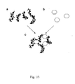
- the invention relates to the finding that binding fragments such as actin filaments and microtubules can bind recognition elements, such as antibodies and nucleic acid oligomers and at the same time bind reporter molecules (e.g. fluorophores) forming one or more detection conjugate(s).
- a first aspect relates to a detection conjugate comprising a binding fragment selected from the group consisting of cytoskeletal filaments and fragments and chemically modified versions thereof and at least one recognition element linked to said binding fragment as defined by the specific application.
- a second aspect relates to a detection conjugate with increased sensitivity accomplished by attaching (a) reporter molecule(s) to the detection conjugate.
- a third aspect relates to One or more detection conjugate(s) , wherein said conjugates become linked to each other via their bound recognition element(s) in the presence of analyte and thereby cross-link and aggregate.
- a fourth aspect relates to a kit comprising several of said detection conjugates.
- a fifth aspect relates to a method in which said detection conjugates are used for the detection of one or more compounds/analytes.
- a sixth aspect relates to the use of said detection conjugates, kit or method for the detection of one or more analytes in a sample.
- the detection conjugate(s) may identify a specific compound (analyte) in a sample and upon binding of the analyte to said binding fragment aggregation of the detection conjugates will occur wherein said aggregates can be detected by the use of different detection methods such as for example microscopy. Due to aggregation of detection conjugates an amplification of the sensitivity occurs and small amounts down to individual molecules can be detected. Moreover, several different components/compounds may be detected in one and the same sample if the proportions between different recognition elements on the binding fragments co-vary with the proportions of different reporter molecules.
- the invention relates to a a detection conjugate comprising a binding fragment being a cytoskeletal filament, fragments and parts thereof, variants thereof, synthetic versions thereof and chemically modified versions thereof and at least one recognition element coupled to said fragment covalently or via non-covalent specific affinity coupling.
- binding fragments are actin filaments and microtubules and prokaryotic analogues.
- Said recognition element may be selected from the group consisting of polyclonal and monoclonal antibodies, which may be genetically engineered monoclonal antibodies, such as chimeric or humanised monoclonal antibodies, other chimeric proteins, Fab-fragments of the mentioned types of antibodies, lectins, receptor proteins, nucleic acids, olignucleotides, aptamers, peptides, chemical compounds or fragments thereof. These may be coupled to the binding fragments covalently or via non-covalent specific affinity coupling (e.g. streptavidin-biotin).
- the filaments generally have a eukaryotic origin mainly from muscle (e.g.
- bovine heart but may also be from other tissues such as skeletal muscle or brain (microtubules) or from cultured cells or tissues.
- filaments or filament analogues expressed in prokaryotes are other examples of binding fragments.
- the filaments may also be of mixed origin and they may also have a mixture of different lengths and be branched. The specific characteristic of different binding fragments is dependent, on among different things, the analyte to be detected and needs to be evaluated for each case, being obvious for a person skilled in the art.
- Monoclonal antibodies as recognition elements may be used in the detection of bacteria and virus particles since these have multiple copies of given epitopes (e.g. viral capsids) to which the same monoclonal antibodies can bind.
- the use of monoclonal antibodies is possible also for the detection of proteins having at least two epitopes to which the monoclonal antibodies can bind resulting in aggregation of at least two actin filaments.
- polyclonal antibodies For the detection of proteins of small or average size ( ⁇ 10 nm diameter; ⁇ 200 kDa) it is probably better to use polyclonal antibodies but it might, however, also be possible with monoclonal antibodies.
- monoclonal antibodies directed against different epitopes on one and the same protein thereby enabling aggregation of the binding fragments and also increasing the sensitivity and specificity, such as having one monoclonal antibody on one binding fragment and another one on the other binding fragment. If the molecule of interest to detect is very small it is possible to use aptamers in combination with antibodies to increase the sensitivity.
- the recognition element is one or more oligonucleotides or nucleic acid fragments to be used for the detection of nucleic acids
- the binding fragments will be coupled with different parts of the complementary strand directed against the molecule to be identified.
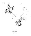
- some binding fragments will be coupled with oligonucleotides complementary to one part of the mRNA to be detected and other binding fragments will be coupled with oligonucleotides complementary to another part of the mRNA to be detected.
- the mRNA to be identified When the mRNA to be identified is added it will cross-link the oligonucleotides on two different binding fragments and thus form aggregates of these (see fig 2 ).
- Actin filaments are less rigid compared to microtubules in connection with torsion and bending 11 . This increases the possibility that recognition elements on different filaments will cross link the different filaments upon binding to a molecule and for formation of aggregates. When the cross linking has occurred it will be more or less irreversible if no external forces are present. As actin filaments are short (usually ⁇ 20 ⁇ m) they can easily be used in small tubes/wells etc.
- Said binding fragment is selected from the group consisting of actin filaments and microtubules or their prokaryotic analogues or mixtures thereof. These fragments may also be chemically modified or synthetically produced.
- each fragment is composed of a large number of identical binding units, the monomers.
- each actin filament consists of ⁇ 400 identical monomers per 1 ⁇ m length of the filament.
- the conditions for formation of the filaments e.g. different concentration of monomers, different salt concentrations and the presence of the actin severing protein gelsolin 12
- the length can be varied between less than 100 nm and more than 50 ⁇ m.
- there is at least one binding site for recognition elements and 1 binding site for reporter molecules on each monomer this opens for the labelling of each actin filament with between 40 and >20 000 recognition elements and reporter molecules.
- Said reporter molecules and said recognition elements may be evenly distributed along said binding fragment, e.g. along one side (e.g. Examples 6-7) or, alternatively in a helical arrangement along the helical protofilaments of the filament.
- Said binding fragments of said detection conjugate may have the same or different lengths varying between 100 nm and more than 50 ⁇ m, such as 1-3, 2- 5, 3-6, 3- 7, 4-7, 6-10, 10-15, 14 - 20 or > 20 um.
- Said recognition elements may be directed against a pathogen, toxin or disease marker, such as a specific protein or nucleotide sequence.
- pathogens are viruses (e.g. human or avian influenza) and bacteria (e.g. Eschericia coli and Helicobacter pylori).
- toxins are anthrax and botulinum toxin.
- relevant disease markers are prostate specific antigen (PSA) for prostate cancer and ABL-BCR fusion mRNA for chronic myeloid leukemia.
- Said reporter molecules are selected from the group consisting of coloured substances, fluorochromes, quantum dots, enzymes, and gold, silver and magnetic nanoparticles.
- the reporter molecule(s) may be bound to said binding fragments through phalloidin-biotin-streptavidin coupling or other similar known coupling methods.
- binding fragment(s) are actin filaments, which easily to low cost can be obtained from muscles (meat), from for example by-products of the meat industry.
- the actin filaments can also be stored in a freezer for a subset of years without any alteration of their properties. It is also easy to couple biotin along the actin filaments through phalloidin-coupling and even to couple nano-particles to these biotin molecules 8 .
- the invention aims at analysing/detecting one or more molecule(s) or microorganism(s) in a sample comprising the steps of: Providing a sample, which may be plasma, other body fluids or extracts thereof, cell or tissue extracts, environmental samples from e.g. air, soil or water, extracts from faeces of birds (monitoring of avian influenza) etc., adding said sample to said detection conjugates as defined above leading to aggregation of said detection conjugates signalling the presence of an analyte. Quantitative information about concentration of analyte may be obtained from the amount of aggregates.
- Detection of the aggregates can be performed in a number of ways such as by microscopy, light scattering, pelleting by centrifugation etc. Detection using a fluorescence microscope is trivial since even individual fluorescence labelled filaments are readily observed (cf. 13 ). However, the formation of large aggregates may also be detected by dark field microscopy due to increased amounts of scattered light or by using differential interference contrast (DIC) microscopy due to increased phase changes of light passing through and by the large filament aggregates. Furthermore, if gold or silver nanoparticles are attached along the filaments surface enhanced Raman scattering and localized surface plasmon resonance effects may be the basis for detection (cf. 2 ).
- the aggregates may also be detected through precipitation, such as if the aggregates are large enough to precipitate without centrifugation they may be detectable by the naked eye. If they are not large enough to precipitate they can be pelleted by centrifugation at different centrifugal forces depending on the size of the aggregates and then be detected by the naked eye, with or without illumination at specific wavelengths, or by microscopy. By ultracentrifugation it is even possible to pellet individual actin filaments 14 .
- the determination of the molecules to be detected may be enhanced by different means if there is a need. This may be performed by attaching a higher amount of reporter molecules to said binding fragments which will increase the sensitivity. By increasing the amount of reporter molecules as defined above the detection could be performed by the use of for example absorption spectometry, fluoroscensce spectrometry, surface plasmon resonance and possibly surface enhanced Raman scattering as mentioned above.
- cytoskeletal fragments being labelled with different reporter molecules (e.g. one which emits green light and one which emits red light) in varied proportions the system/method may be developed to detect several different molecules in one and the same sample.
- Actin filaments are obtained from G-actin monomers purified from skeletal or heart muscle (e.g. Bovine heart obtained as a by-product in the meat industry). The purification of actin and the formation of filamentous form of the protein are performed according to standard procedures (e.g. 13, 15 ).
- actin filaments Following the isolation of actin filaments as described above these may be stored at -80 °C for several years without loss of function 13 .
- the actin filaments can be stabilized by mixing with an equimolar (on monomer basis) concentration of phalloidin as described previously 16 .
- Tubulin is purified from porcine brain using standard protocols 17 .
- Small fluorophores suitable as reporter molecules are e.g. tetramethylrhodamine-isothiocyanate-phalloidin or one of the AlexaTM-series of dyes coupled to phalloidin 13 .
- the labelling with these fluorescent dyes is performed using standard protocols 13, 16 .
- labelling can be performed by direct covalent attachment of fluorophores to e.g. cysteines or other amino acid residues on each actin (cf. 18 ).
- Quantum dots (from Molecuar Probes Invitrogen or other commercial sources) are attached to a majority of the actin monomers on each given actin filament. Suitable quantum dots characterized by their sharply defined emission wavelengths are used to identify the filaments as labelled with certain specific recognition elements. The labelling aims to produce as many quantum dots per actin filament as possible.
- Actin filaments are biotinylated via phalloidin as described in REF, which stabilizes the filaments. Moreover, as shown in reference 13 the biotinylation does not prevent binding to myosin. Neither does it affect the ATP hydrolyzing interaction cycle between actin and myosin where the actomyosin connection is broken by high concentration of ATP.
- Actin filaments are captured by myosin coated silicon beads in a suitable column or on the walls of a glass capillary silanized with trimethylchlorosilane 19 and subsequently eluted by high ATP concentration in a solution of high ionic strength 14 .
- biotinylated actin filaments After addition of the biotinylated actin filaments to the myosin coated column streptavidin-coated quantum dots of suitable emission properties are added at nanomolar concentration to the same column. The reaction is allowed to proceed until the quantum dot concentration of the efflux from the column does not differ from the concentration of the added quantum dot solution. Next, biotinylated protein A is added followed by 1 ⁇ M biotin to block free biotin-binding sites on streptavidin. Finally, the actin filaments are eluted from the column by addition of ATP in a high ionic strength solution thus breaking the actomyosin interaction. Following this elution the actin filaments are pelleted by ultracentrifugation at 200 000 g for 40 min and subsequently dissolved to a considerably higher concentration as desired.
- the actin filaments are labelled with suitable antibodies that bind, via their Fc-fragments to protein A.
- This procedure follows that described in reference 20 with mixing of suitable antibodies at 0.01 - 1 g/l with the filaments (0.01 - 1 ⁇ M) in phosphate buffered saline for 2 h to obtain different degree of coverage of the filaments with antibodies.
- Labelling of filaments with qold or silver nanoparticles or with magnetic nanoparticles can be achieved by a similar method as that described here for the labelling with quantum dots.
- Biotinylated actin filaments are labeled with streptavidin-coated quantum dots in a myosin coated column as in Example 6.
- biotinylated sequence specific oligonucleotides e.g. 10-20 nucleotides long biotin labelled by e.g. the 3'EndTag DNA Labelling System or the 5'EndTag Nucleic Acid Labelling System, Vector laboratories
- 1 ⁇ M biotin to block free biotin-bindings sites on streptavidin.
- the oligonucleotide sequence is designed to be complementary to one part of a specific mRNA to be detected.
- actin filaments are similarly labelled with another oligonucleotide with a sequence complementary to another part of a specific mRNA to be detected.
- actin filaments are eluted from the columns by addition of ATP in a high ionic strength solution as described in Example 6. Following this elution the actin filaments are pelleted by ultracentrifugation at 200 000 g for 40 min and subsequently dissolved at a desired concentration.
- This labelling has the same goals and is performed as described for the actin filaments above but instead of the myosin column a kinesin column is used.
- Recognition elements in the form of antibodies are attached to actin filaments or microtubules subsequent to labelling with small-molecular fluorescent reporter molecules according to standard procedures (Ref, REf).
- carboxyl groups on the surface of the filaments are activated by treatment with 20 mM N- hydroxysuccinimide (NHS) and 10 mM N -(3-dimethylaminopropyl)- N'- ethylcarbodiimide hydrochloride (EDC) dissolved in a buffered solution (pH 7.4) containing 2 mM MgCl 2 and 50 mM KCl.
- filaments (1 ⁇ M) are immersed in a solution with Protein A (10 mg/l) in 10 mM PBS, pH 7.4 for 2 h (cf. 20 ).
- ProteinA-coated filaments are next incubated in a solution with suitable IgG antibodies at concentrations from 0.1-1 g/l in PBS for 2h.
- the antibody-labelled filaments are collected by ultracentrifugation at 200000 g for 40 min. The supernatant is discarded whereas the pellet is re-dissolved in a suitable buffered solution e.g. containing 0.1 mM ATP, 2 mM MgCl 2 and 150 mM KCl for actin filaments.
- Example 10 Detection of the ABL-BCR transcript as a disease marker for chronic myeloid leukemia
- a chimeric ABL-BCR fusion mRNA is expressed as a result of a chromosomal translocation leading to the fusion of parts of the two genes ABL and BCR 21 .
- Actin filaments are labelled as in Example 7 such that some filaments carry an oligonucleotide complementary to part of the ABL mRNA whereas other filaments carry an oligonucleotide complementary to part of the BCR mRNA.
- RNA is extracted from cells or tissue to be studied e.g. as described in reference 22 . The isolated RNA is added to a mixture of actin filaments carrying the two different oligonucleotides. If ABL-BCR fusion mRNA is present actin filaments will be cross-linked by the fusion mRNA and aggregate. If only normal ABL and BCR mRNAs are present no aggregation of filaments will occur.
- Example 11 A method to detect any one of five different influenza virus types
- Actin filaments are labelled with reporter molecules and recognition elements using any of the techniques described in the examples above.
- the actin filaments are labelled in such a way that each individual filament contains only one of five different antibodies which are directed against five different virus types.
- the five different types of actin filaments (labelled with the five different antibodies) are labelled with different proportions of two different reporter molecules (e.g. two different Alexa compounds emitting red and green fluorescence respectively).
- two different reporter molecules e.g. two different Alexa compounds emitting red and green fluorescence respectively.
- actin filaments labelled with antibody #1 emit only green fluorescence
- actin filaments labelled with antibody #2 emit only red fluorescence
- actin filaments labelled with antibody #3 emit red and green fluorescence in a ratio 50/50
- actin filaments labelled with antibody #4 emit red and green fluorescence in a ratio 25/75
- actin filaments labelled with antibody #5 emit red and green fluorescence in a ratio 75/25.
- the five different types of actin filaments are mixed in equal proportions.
- a sample containing an influenza virus is added.
- the actin filaments containing antibodies directed against this virus type will aggregate. By measuring the ratio of green and red fluorescence in the aggregate the virus type will be determined.
- Example 12 A method to detect any combinations of seven different pathogens
- Actin filaments are labelled with quantum dots and antibodies as described in Example 6.
- the actin filaments are labelled in such a way that each individual filament contains only one of seven different antibodies directed against seven different pathogens.
- the seven different types of actin filaments (labelled with the seven different antibodies) are labelled with different quantum dots that emit fluorescence of different wavelengths.
- each actin filament has a unique combination of recognition element and reporter molecule and will only recognize and report (by aggregation) one specific pathogen.
- the seven different types of actin filaments are mixed in equal proportions. A mixture of pathogens is added and actin filaments will aggregate according to which pathogens are present in the sample. The types of pathogens present is determined by the fluorescence at different wavelengths of the aggregated filaments or alternatively by comparing the fluorescence of unaggregated filaments with the fluorescence of filaments before addition of pathogens.
Landscapes
- Health & Medical Sciences (AREA)
- Life Sciences & Earth Sciences (AREA)
- Chemical & Material Sciences (AREA)
- Molecular Biology (AREA)
- Engineering & Computer Science (AREA)
- Biochemistry (AREA)
- Immunology (AREA)
- Organic Chemistry (AREA)
- General Health & Medical Sciences (AREA)
- Medicinal Chemistry (AREA)
- Hematology (AREA)
- Biomedical Technology (AREA)
- Urology & Nephrology (AREA)
- Proteomics, Peptides & Aminoacids (AREA)
- Cell Biology (AREA)
- Genetics & Genomics (AREA)
- Biophysics (AREA)
- Analytical Chemistry (AREA)
- Physics & Mathematics (AREA)
- General Physics & Mathematics (AREA)
- Pathology (AREA)
- Biotechnology (AREA)
- Microbiology (AREA)
- Food Science & Technology (AREA)
- Toxicology (AREA)
- Gastroenterology & Hepatology (AREA)
- Zoology (AREA)
- Measuring Or Testing Involving Enzymes Or Micro-Organisms (AREA)
- Investigating, Analyzing Materials By Fluorescence Or Luminescence (AREA)
- Investigating Or Analysing Biological Materials (AREA)
- Investigating Or Analyzing Materials By The Use Of Electric Means (AREA)
Claims (7)
- In vitro Verfahren zur Detektion eines oder mehrerer Analyten, welches die folgenden Schritte umfasst:a) Bereitstellen aufgereinigter Zytoskelettfilamente, ausgewählt aus Aktinfilamenten, Mikrotubuli oder Kombinationen hiervon;b) Bereitstellen eines oder mehrerer Reportermoleküle, wobei die Reportermoleküle ausgewählt sind aus der Gruppe, bestehend aus farbigen Substanzen, Fluorochromen, Quantendots, Enzymen, Gold, Silber oder magnetischen Nanopartikeln;c) Bereitstellen eines oder mehrerer Erkennungselemente, wobei die Erkennungselemente ausgewählt sind aus der Gruppe, bestehend aus monoklonalen Antikörpern, polyklonalen Antikörpern, Fab-Fragmenten, chimären Proteinen, Lektinen, Rezeptorproteinen, Nukleinsäuren, Oligonukleotiden, Aptameren und Peptiden;d) Binden der Erkennungselemente und der Reportermoleküle an die Zytoskelettfilamente, dadurch Erhalten von Detektionskonjugaten, wobei die Erkennungselemente so ausgewählt sind, dass die Konjugate in der Gegenwart von Analyt durch ihre gebundenen Erkennungselementen miteinander verbunden werden und dadurch vernetzen und aggregieren;e) Bereitstellen der Detektionskonjugate in einer Lösung;f) Inkubieren der Detektionskonjugate in einer Lösung mit einer Probe, die einen oder mehrere Analyten enthält,g) Detektieren der Aggregation der Detektionskonjugate, und,h) optional, Analysieren der gebildeten Aggregate.
- Verfahren gemäß Anspruch 1, wobei die Bindung in Schritt d) darin resultiert, dass Erkennungsmoleküle und Reportermoleküle gleichmäßig entlang dem Zytoskelettfilament verteilt sind.
- Verfahren gemäß Anspruch 1 oder 2, wobei die Detektion in Schritt g) oder die Analyse in Schritt h) mittels Inaugenscheinnahme, Mikroskop, Lichtstreuung, Pelletieren durch Zentrifugation, Spektralphotometrie, Fluoreszenzdetektion, oberflächenverbesserte RamanStreuung oder Oberflächen-Plasmon-Resonanz geschieht.
- Verfahren gemäß einem der vorangehenden Ansprüche, wobei Schritt g) der Detektion der Aggregation aus Inaugenscheinnahme und Feststellen der Anwesenheit oder Abwesenheit von Aggregation besteht.
- Verfahren gemäß einem der vorangehenden Ansprüche, wobei das Erkennungselement ein Erkennungselement für ein Pathogen, Toxin oder Krankheitsmarker ist.
- Verfahren gemäß einem der vorangehenden Ansprüche zur Detektion mehrerer verschiedener Analyten in einer Probe, wobei die Schritte a) bis d) gemäß dem Verfahren von Anspruch 1 mithilfe von verschiedenen Reportermolekülen und mehreren verschiedenen Erkennungselementen durchgeführt werden, resultierend in mehreren verschiedenen Detektionskonjugaten, jedes mit einem eigenen Erkennungselement und ReportermolekülProfil.
- Verfahren gemäß Anspruch 6, wobeia) das eine oder die mehreren Reportermolekülprofile mehrere verschiedene Kombinationen von Fluoreszenz sind, undb) die Erkennungselemente aus mehreren verschiedenen Antikörpern bestehen.
Applications Claiming Priority (2)
| Application Number | Priority Date | Filing Date | Title |
|---|---|---|---|
| SE0602571A SE531135C2 (sv) | 2006-12-01 | 2006-12-01 | Känslig detektion av gifter, patogener och sjukdomsmarkörer genom aggregation av cytoskeletala filament |
| PCT/SE2007/001071 WO2008066463A1 (en) | 2006-12-01 | 2007-11-30 | Detection conjugate |
Publications (3)
| Publication Number | Publication Date |
|---|---|
| EP2092343A1 EP2092343A1 (de) | 2009-08-26 |
| EP2092343A4 EP2092343A4 (de) | 2010-06-02 |
| EP2092343B1 true EP2092343B1 (de) | 2014-08-20 |
Family
ID=39468162
Family Applications (1)
| Application Number | Title | Priority Date | Filing Date |
|---|---|---|---|
| EP07852077.2A Not-in-force EP2092343B1 (de) | 2006-12-01 | 2007-11-30 | Nachweiskonjugat |
Country Status (4)
| Country | Link |
|---|---|
| US (1) | US8658381B2 (de) |
| EP (1) | EP2092343B1 (de) |
| SE (1) | SE531135C2 (de) |
| WO (1) | WO2008066463A1 (de) |
Families Citing this family (7)
| Publication number | Priority date | Publication date | Assignee | Title |
|---|---|---|---|---|
| US9909167B2 (en) | 2014-06-23 | 2018-03-06 | The Board Of Trustees Of The Leland Stanford Junior University | On-slide staining by primer extension |
| US12625134B2 (en) | 2014-10-31 | 2026-05-12 | Massachusetts Institute Of Technology | Complex liquid crystal droplets |
| US10370698B2 (en) | 2016-07-27 | 2019-08-06 | The Board Of Trustees Of The Leland Stanford Junior University | Highly-multiplexed fluorescent imaging |
| US20190170737A1 (en) * | 2016-09-19 | 2019-06-06 | Massachusetts Institute Of Technology | Systems including janus droplets |
| CN120795059A (zh) | 2018-08-27 | 2025-10-17 | 瑞泽恩制药公司 | 拉曼光谱在下游纯化中的应用 |
| US20220163537A1 (en) * | 2019-03-26 | 2022-05-26 | Fast Bt Ug | Detection and Quantification of Molecular Species |
| WO2021055092A1 (en) | 2019-09-18 | 2021-03-25 | Massachusetts Institute Of Technology | Systems and methods for affecting interactions of electromagnetic radiation with janus droplets for sensitive detection of species |
-
2006
- 2006-12-01 SE SE0602571A patent/SE531135C2/sv not_active IP Right Cessation
-
2007
- 2007-11-30 US US12/517,148 patent/US8658381B2/en not_active Expired - Fee Related
- 2007-11-30 WO PCT/SE2007/001071 patent/WO2008066463A1/en not_active Ceased
- 2007-11-30 EP EP07852077.2A patent/EP2092343B1/de not_active Not-in-force
Non-Patent Citations (1)
| Title |
|---|
| WU X ET AL: "IMMUNOFLUORESCENT LABELING OF CANCER MARKER HER2 AND OTHER CELLULAR TARGETS WITH SEMICONDUCTOR QUANTUM DOTS", NATURE BIOTECHNOLOGY, NATURE PUBLISHING GROUP, NEW YORK, NY, US, vol. 21, 1 January 2003 (2003-01-01), pages 41 - 46, XP008053284, ISSN: 1087-0156, DOI: 10.1038/NBT764 * |
Also Published As
| Publication number | Publication date |
|---|---|
| EP2092343A4 (de) | 2010-06-02 |
| SE531135C2 (sv) | 2008-12-23 |
| WO2008066463A1 (en) | 2008-06-05 |
| US20100248212A1 (en) | 2010-09-30 |
| SE0602571L (sv) | 2008-06-02 |
| US8658381B2 (en) | 2014-02-25 |
| EP2092343A1 (de) | 2009-08-26 |
Similar Documents
| Publication | Publication Date | Title |
|---|---|---|
| Nguyen et al. | Highly sensitive sandwich-type SPR based detection of whole H5Nx viruses using a pair of aptamers | |
| EP4281775B1 (de) | Systeme und verfahren zur biomolekülherstellung | |
| US8658381B2 (en) | Detection conjugate | |
| CN102066941B (zh) | 用于生物传感器的新型Au/Ag核-壳复合材料 | |
| JP5597615B2 (ja) | 可溶性検体の検出および増幅 | |
| Ling et al. | Energy transfer with gold nanoparticles for analytical applications in the fields of biochemical and pharmaceutical sciences | |
| Liu et al. | A novel aptamer-mediated CuInS 2 quantum dots@ graphene oxide nanocomposites-based fluorescence “turn off–on” nanosensor for highly sensitive and selective detection of kanamycin | |
| WO2005010031A1 (ja) | チタン、銀、シリコンに結合能を有するペプチド | |
| WO2017180889A1 (en) | Methods and systems for determining states of molecule folding, conformation, or interaction | |
| EP1211514A1 (de) | Verfahren zum analysieren von wechselwirkungen zwischen proteinen und molekülen | |
| Mahmoud et al. | Advanced procedures for labeling of antibodies with quantum dots | |
| EP3619539B1 (de) | Verfahren zur bestimmung der anwesenheit oder abwesenheit eines zielanalyten mit einem reporter-polynukleotid und einer transmembranpore | |
| JP6940532B2 (ja) | 二本鎖核酸信号プローブおよびこれを用いた標的分子の検出方法 | |
| Zhang et al. | Label-free sensing of thrombin based on quantum dots and thrombin binding aptamer | |
| US20250164503A1 (en) | Engineered recombinant protein-binding domains as detection reagents | |
| Yokoo et al. | Direct and selective immobilization of proteins by means of an inorganic material-binding peptide: discussion on functionalization in the elongation to material-binding peptide | |
| JP6873483B2 (ja) | 融合タンパク質及びそれを用いた抗原の検出方法 | |
| KR20110130384A (ko) | 효소가 집적된 미세튜브를 이용한 정량분석법 | |
| KR101551925B1 (ko) | T7 박테리오파지를 이용한 표적-특이적 프로브 및 이를 이용한 바이오마커의 탐지 | |
| Zhao et al. | Fluorescent nanoparticle for bacteria and DNA detection | |
| KR100877187B1 (ko) | 질병마커 인지 에피토프와 연결된 단백질 나노입자를포함하는 진단용 단백질 칩과 그의 초고감도 검출 방법 | |
| WO2008144817A1 (en) | Fluorescent protein particles | |
| Eriksen et al. | A novel complex: a quantum dot conjugated to an active T7 RNA polymerase | |
| Kozak et al. | Improving the signal-to-noise performance of molecular diagnostics with PEG-lysine copolymer dendrons | |
| US20060003339A1 (en) | Two-hybrid system |
Legal Events
| Date | Code | Title | Description |
|---|---|---|---|
| PUAI | Public reference made under article 153(3) epc to a published international application that has entered the european phase |
Free format text: ORIGINAL CODE: 0009012 |
|
| 17P | Request for examination filed |
Effective date: 20090609 |
|
| AK | Designated contracting states |
Kind code of ref document: A1 Designated state(s): AT BE BG CH CY CZ DE DK EE ES FI FR GB GR HU IE IS IT LI LT LU LV MC MT NL PL PT RO SE SI SK TR |
|
| DAX | Request for extension of the european patent (deleted) | ||
| A4 | Supplementary search report drawn up and despatched |
Effective date: 20100216 |
|
| RIC1 | Information provided on ipc code assigned before grant |
Ipc: C07K 19/00 20060101ALI20100210BHEP Ipc: G01N 33/536 20060101ALI20100210BHEP Ipc: C07K 14/47 20060101ALI20100210BHEP Ipc: G01N 33/68 20060101AFI20080619BHEP |
|
| RA4 | Supplementary search report drawn up and despatched (corrected) |
Effective date: 20100414 |
|
| 17Q | First examination report despatched |
Effective date: 20131004 |
|
| GRAP | Despatch of communication of intention to grant a patent |
Free format text: ORIGINAL CODE: EPIDOSNIGR1 |
|
| INTG | Intention to grant announced |
Effective date: 20140313 |
|
| GRAS | Grant fee paid |
Free format text: ORIGINAL CODE: EPIDOSNIGR3 |
|
| GRAA | (expected) grant |
Free format text: ORIGINAL CODE: 0009210 |
|
| AK | Designated contracting states |
Kind code of ref document: B1 Designated state(s): AT BE BG CH CY CZ DE DK EE ES FI FR GB GR HU IE IS IT LI LT LU LV MC MT NL PL PT RO SE SI SK TR |
|
| REG | Reference to a national code |
Ref country code: GB Ref legal event code: FG4D |
|
| REG | Reference to a national code |
Ref country code: CH Ref legal event code: EP |
|
| REG | Reference to a national code |
Ref country code: AT Ref legal event code: REF Ref document number: 683734 Country of ref document: AT Kind code of ref document: T Effective date: 20140915 |
|
| REG | Reference to a national code |
Ref country code: IE Ref legal event code: FG4D |
|
| REG | Reference to a national code |
Ref country code: DE Ref legal event code: R096 Ref document number: 602007038264 Country of ref document: DE Effective date: 20141002 |
|
| REG | Reference to a national code |
Ref country code: AT Ref legal event code: MK05 Ref document number: 683734 Country of ref document: AT Kind code of ref document: T Effective date: 20140820 |
|
| REG | Reference to a national code |
Ref country code: NL Ref legal event code: VDEP Effective date: 20140820 |
|
| REG | Reference to a national code |
Ref country code: LT Ref legal event code: MG4D |
|
| PG25 | Lapsed in a contracting state [announced via postgrant information from national office to epo] |
Ref country code: ES Free format text: LAPSE BECAUSE OF FAILURE TO SUBMIT A TRANSLATION OF THE DESCRIPTION OR TO PAY THE FEE WITHIN THE PRESCRIBED TIME-LIMIT Effective date: 20140820 Ref country code: BG Free format text: LAPSE BECAUSE OF FAILURE TO SUBMIT A TRANSLATION OF THE DESCRIPTION OR TO PAY THE FEE WITHIN THE PRESCRIBED TIME-LIMIT Effective date: 20141120 Ref country code: LT Free format text: LAPSE BECAUSE OF FAILURE TO SUBMIT A TRANSLATION OF THE DESCRIPTION OR TO PAY THE FEE WITHIN THE PRESCRIBED TIME-LIMIT Effective date: 20140820 Ref country code: PT Free format text: LAPSE BECAUSE OF FAILURE TO SUBMIT A TRANSLATION OF THE DESCRIPTION OR TO PAY THE FEE WITHIN THE PRESCRIBED TIME-LIMIT Effective date: 20141222 Ref country code: GR Free format text: LAPSE BECAUSE OF FAILURE TO SUBMIT A TRANSLATION OF THE DESCRIPTION OR TO PAY THE FEE WITHIN THE PRESCRIBED TIME-LIMIT Effective date: 20141121 Ref country code: FI Free format text: LAPSE BECAUSE OF FAILURE TO SUBMIT A TRANSLATION OF THE DESCRIPTION OR TO PAY THE FEE WITHIN THE PRESCRIBED TIME-LIMIT Effective date: 20140820 Ref country code: SE Free format text: LAPSE BECAUSE OF FAILURE TO SUBMIT A TRANSLATION OF THE DESCRIPTION OR TO PAY THE FEE WITHIN THE PRESCRIBED TIME-LIMIT Effective date: 20140820 |
|
| PG25 | Lapsed in a contracting state [announced via postgrant information from national office to epo] |
Ref country code: IS Free format text: LAPSE BECAUSE OF FAILURE TO SUBMIT A TRANSLATION OF THE DESCRIPTION OR TO PAY THE FEE WITHIN THE PRESCRIBED TIME-LIMIT Effective date: 20141220 Ref country code: AT Free format text: LAPSE BECAUSE OF FAILURE TO SUBMIT A TRANSLATION OF THE DESCRIPTION OR TO PAY THE FEE WITHIN THE PRESCRIBED TIME-LIMIT Effective date: 20140820 Ref country code: LV Free format text: LAPSE BECAUSE OF FAILURE TO SUBMIT A TRANSLATION OF THE DESCRIPTION OR TO PAY THE FEE WITHIN THE PRESCRIBED TIME-LIMIT Effective date: 20140820 |
|
| PG25 | Lapsed in a contracting state [announced via postgrant information from national office to epo] |
Ref country code: NL Free format text: LAPSE BECAUSE OF FAILURE TO SUBMIT A TRANSLATION OF THE DESCRIPTION OR TO PAY THE FEE WITHIN THE PRESCRIBED TIME-LIMIT Effective date: 20140820 |
|
| PG25 | Lapsed in a contracting state [announced via postgrant information from national office to epo] |
Ref country code: SK Free format text: LAPSE BECAUSE OF FAILURE TO SUBMIT A TRANSLATION OF THE DESCRIPTION OR TO PAY THE FEE WITHIN THE PRESCRIBED TIME-LIMIT Effective date: 20140820 Ref country code: RO Free format text: LAPSE BECAUSE OF FAILURE TO SUBMIT A TRANSLATION OF THE DESCRIPTION OR TO PAY THE FEE WITHIN THE PRESCRIBED TIME-LIMIT Effective date: 20140820 Ref country code: EE Free format text: LAPSE BECAUSE OF FAILURE TO SUBMIT A TRANSLATION OF THE DESCRIPTION OR TO PAY THE FEE WITHIN THE PRESCRIBED TIME-LIMIT Effective date: 20140820 Ref country code: CZ Free format text: LAPSE BECAUSE OF FAILURE TO SUBMIT A TRANSLATION OF THE DESCRIPTION OR TO PAY THE FEE WITHIN THE PRESCRIBED TIME-LIMIT Effective date: 20140820 Ref country code: IT Free format text: LAPSE BECAUSE OF FAILURE TO SUBMIT A TRANSLATION OF THE DESCRIPTION OR TO PAY THE FEE WITHIN THE PRESCRIBED TIME-LIMIT Effective date: 20140820 Ref country code: DK Free format text: LAPSE BECAUSE OF FAILURE TO SUBMIT A TRANSLATION OF THE DESCRIPTION OR TO PAY THE FEE WITHIN THE PRESCRIBED TIME-LIMIT Effective date: 20140820 |
|
| REG | Reference to a national code |
Ref country code: DE Ref legal event code: R097 Ref document number: 602007038264 Country of ref document: DE |
|
| PG25 | Lapsed in a contracting state [announced via postgrant information from national office to epo] |
Ref country code: PL Free format text: LAPSE BECAUSE OF FAILURE TO SUBMIT A TRANSLATION OF THE DESCRIPTION OR TO PAY THE FEE WITHIN THE PRESCRIBED TIME-LIMIT Effective date: 20140820 |
|
| PLBE | No opposition filed within time limit |
Free format text: ORIGINAL CODE: 0009261 |
|
| STAA | Information on the status of an ep patent application or granted ep patent |
Free format text: STATUS: NO OPPOSITION FILED WITHIN TIME LIMIT |
|
| PG25 | Lapsed in a contracting state [announced via postgrant information from national office to epo] |
Ref country code: MC Free format text: LAPSE BECAUSE OF FAILURE TO SUBMIT A TRANSLATION OF THE DESCRIPTION OR TO PAY THE FEE WITHIN THE PRESCRIBED TIME-LIMIT Effective date: 20140820 Ref country code: LU Free format text: LAPSE BECAUSE OF FAILURE TO SUBMIT A TRANSLATION OF THE DESCRIPTION OR TO PAY THE FEE WITHIN THE PRESCRIBED TIME-LIMIT Effective date: 20141130 Ref country code: BE Free format text: LAPSE BECAUSE OF NON-PAYMENT OF DUE FEES Effective date: 20141130 |
|
| REG | Reference to a national code |
Ref country code: CH Ref legal event code: PL |
|
| 26N | No opposition filed |
Effective date: 20150521 |
|
| PG25 | Lapsed in a contracting state [announced via postgrant information from national office to epo] |
Ref country code: LI Free format text: LAPSE BECAUSE OF NON-PAYMENT OF DUE FEES Effective date: 20141130 Ref country code: CH Free format text: LAPSE BECAUSE OF NON-PAYMENT OF DUE FEES Effective date: 20141130 |
|
| PG25 | Lapsed in a contracting state [announced via postgrant information from national office to epo] |
Ref country code: SI Free format text: LAPSE BECAUSE OF FAILURE TO SUBMIT A TRANSLATION OF THE DESCRIPTION OR TO PAY THE FEE WITHIN THE PRESCRIBED TIME-LIMIT Effective date: 20140820 |
|
| REG | Reference to a national code |
Ref country code: FR Ref legal event code: PLFP Year of fee payment: 9 |
|
| PG25 | Lapsed in a contracting state [announced via postgrant information from national office to epo] |
Ref country code: CY Free format text: LAPSE BECAUSE OF FAILURE TO SUBMIT A TRANSLATION OF THE DESCRIPTION OR TO PAY THE FEE WITHIN THE PRESCRIBED TIME-LIMIT Effective date: 20140820 |
|
| PG25 | Lapsed in a contracting state [announced via postgrant information from national office to epo] |
Ref country code: MT Free format text: LAPSE BECAUSE OF FAILURE TO SUBMIT A TRANSLATION OF THE DESCRIPTION OR TO PAY THE FEE WITHIN THE PRESCRIBED TIME-LIMIT Effective date: 20140820 Ref country code: BE Free format text: LAPSE BECAUSE OF FAILURE TO SUBMIT A TRANSLATION OF THE DESCRIPTION OR TO PAY THE FEE WITHIN THE PRESCRIBED TIME-LIMIT Effective date: 20140820 Ref country code: HU Free format text: LAPSE BECAUSE OF FAILURE TO SUBMIT A TRANSLATION OF THE DESCRIPTION OR TO PAY THE FEE WITHIN THE PRESCRIBED TIME-LIMIT; INVALID AB INITIO Effective date: 20071130 Ref country code: TR Free format text: LAPSE BECAUSE OF FAILURE TO SUBMIT A TRANSLATION OF THE DESCRIPTION OR TO PAY THE FEE WITHIN THE PRESCRIBED TIME-LIMIT Effective date: 20140820 |
|
| REG | Reference to a national code |
Ref country code: FR Ref legal event code: PLFP Year of fee payment: 10 |
|
| PGFP | Annual fee paid to national office [announced via postgrant information from national office to epo] |
Ref country code: IE Payment date: 20161130 Year of fee payment: 10 Ref country code: DE Payment date: 20161201 Year of fee payment: 10 Ref country code: GB Payment date: 20161130 Year of fee payment: 10 Ref country code: FR Payment date: 20161129 Year of fee payment: 10 |
|
| REG | Reference to a national code |
Ref country code: DE Ref legal event code: R119 Ref document number: 602007038264 Country of ref document: DE |
|
| GBPC | Gb: european patent ceased through non-payment of renewal fee |
Effective date: 20171130 |
|
| REG | Reference to a national code |
Ref country code: FR Ref legal event code: ST Effective date: 20180731 |
|
| REG | Reference to a national code |
Ref country code: IE Ref legal event code: MM4A |
|
| PG25 | Lapsed in a contracting state [announced via postgrant information from national office to epo] |
Ref country code: FR Free format text: LAPSE BECAUSE OF NON-PAYMENT OF DUE FEES Effective date: 20171130 Ref country code: DE Free format text: LAPSE BECAUSE OF NON-PAYMENT OF DUE FEES Effective date: 20180602 Ref country code: IE Free format text: LAPSE BECAUSE OF NON-PAYMENT OF DUE FEES Effective date: 20171130 |
|
| PG25 | Lapsed in a contracting state [announced via postgrant information from national office to epo] |
Ref country code: GB Free format text: LAPSE BECAUSE OF NON-PAYMENT OF DUE FEES Effective date: 20171130 |