EP2095762A1 - Dispositif d'investigation micro-invasif in vivo comprenant un guide métallique - Google Patents
Dispositif d'investigation micro-invasif in vivo comprenant un guide métallique Download PDFInfo
- Publication number
- EP2095762A1 EP2095762A1 EP08290179A EP08290179A EP2095762A1 EP 2095762 A1 EP2095762 A1 EP 2095762A1 EP 08290179 A EP08290179 A EP 08290179A EP 08290179 A EP08290179 A EP 08290179A EP 2095762 A1 EP2095762 A1 EP 2095762A1
- Authority
- EP
- European Patent Office
- Prior art keywords
- metal guide
- substrate
- specific
- metal
- antibody
- Prior art date
- Legal status (The legal status is an assumption and is not a legal conclusion. Google has not performed a legal analysis and makes no representation as to the accuracy of the status listed.)
- Granted
Links
Images




Classifications
-
- A—HUMAN NECESSITIES
- A61—MEDICAL OR VETERINARY SCIENCE; HYGIENE
- A61B—DIAGNOSIS; SURGERY; IDENTIFICATION
- A61B5/00—Measuring for diagnostic purposes; Identification of persons
-
- A—HUMAN NECESSITIES
- A61—MEDICAL OR VETERINARY SCIENCE; HYGIENE
- A61B—DIAGNOSIS; SURGERY; IDENTIFICATION
- A61B5/00—Measuring for diagnostic purposes; Identification of persons
- A61B5/145—Measuring characteristics of blood in vivo, e.g. gas concentration or pH-value ; Measuring characteristics of body fluids or tissues, e.g. interstitial fluid or cerebral tissue
- A61B5/14542—Measuring characteristics of blood in vivo, e.g. gas concentration or pH-value ; Measuring characteristics of body fluids or tissues, e.g. interstitial fluid or cerebral tissue for measuring blood gases
-
- A—HUMAN NECESSITIES
- A61—MEDICAL OR VETERINARY SCIENCE; HYGIENE
- A61B—DIAGNOSIS; SURGERY; IDENTIFICATION
- A61B5/00—Measuring for diagnostic purposes; Identification of persons
- A61B5/145—Measuring characteristics of blood in vivo, e.g. gas concentration or pH-value ; Measuring characteristics of body fluids or tissues, e.g. interstitial fluid or cerebral tissue
- A61B5/1468—Measuring characteristics of blood in vivo, e.g. gas concentration or pH-value ; Measuring characteristics of body fluids or tissues, e.g. interstitial fluid or cerebral tissue using chemical or electrochemical methods, e.g. by polarographic means
- A61B5/1473—Measuring characteristics of blood in vivo, e.g. gas concentration or pH-value ; Measuring characteristics of body fluids or tissues, e.g. interstitial fluid or cerebral tissue using chemical or electrochemical methods, e.g. by polarographic means invasive, e.g. introduced into the body by a catheter
-
- A—HUMAN NECESSITIES
- A61—MEDICAL OR VETERINARY SCIENCE; HYGIENE
- A61B—DIAGNOSIS; SURGERY; IDENTIFICATION
- A61B5/00—Measuring for diagnostic purposes; Identification of persons
- A61B5/40—Detecting, measuring or recording for evaluating the nervous system
- A61B5/4076—Diagnosing or monitoring particular conditions of the nervous system
-
- A—HUMAN NECESSITIES
- A61—MEDICAL OR VETERINARY SCIENCE; HYGIENE
- A61B—DIAGNOSIS; SURGERY; IDENTIFICATION
- A61B5/00—Measuring for diagnostic purposes; Identification of persons
- A61B5/68—Arrangements of detecting, measuring or recording means, e.g. sensors, in relation to patient
- A61B5/6846—Arrangements of detecting, measuring or recording means, e.g. sensors, in relation to patient specially adapted to be brought in contact with an internal body part, i.e. invasive
- A61B5/6847—Arrangements of detecting, measuring or recording means, e.g. sensors, in relation to patient specially adapted to be brought in contact with an internal body part, i.e. invasive mounted on an invasive device
- A61B5/6851—Guide wires
-
- A—HUMAN NECESSITIES
- A61—MEDICAL OR VETERINARY SCIENCE; HYGIENE
- A61B—DIAGNOSIS; SURGERY; IDENTIFICATION
- A61B2562/00—Details of sensors; Constructional details of sensor housings or probes; Accessories for sensors
- A61B2562/02—Details of sensors specially adapted for in-vivo measurements
-
- A—HUMAN NECESSITIES
- A61—MEDICAL OR VETERINARY SCIENCE; HYGIENE
- A61B—DIAGNOSIS; SURGERY; IDENTIFICATION
- A61B2562/00—Details of sensors; Constructional details of sensor housings or probes; Accessories for sensors
- A61B2562/04—Arrangements of multiple sensors of the same type
- A61B2562/043—Arrangements of multiple sensors of the same type in a linear array
Definitions
- the present invention relates to the functionalization of a transparietal investigation device, a method of ex vivo analysis of a substrate using a functionalized device of the invention, and the use of such a device for the manufacture of a device. tool for diagnosing cancer, infection, inflammation, or transplant rejection in a patient.
- Such devices take the form of a rigid tube endoscope type or a catheter consisting of a flexible tube which is inserted into the body, including the natural pathways or vessels, and which makes it possible to reach an organ or a specific tissue.
- These devices allow in particular the elimination of blood clots or, when they are associated with optical fibers, the visualization and the in vivo control of the state of a system, such as the digestive tract, or of an organ, like the colon.
- the trauma to the patient resulting from the use of such devices is then minimized but still needs to be improved.
- the patent EP 1 358 481 describes an in vivo analysis or treatment device comprising (i) a micro-system for investigating a substrate other than by analyzing a fluorescent signal, (ii) a flexible rod at one end of which is fixed said microsystem and whose other end is intended for the operation of said micro-system, (iii) a medical instrument having an internal lumen in which said flexible rod can slide, (iv) a sliding system for protecting the removable micro-system at of the substrate, and (v) at the level of said micro-system, a tissue or cell dilaceration system, optionally associated with one or more devices selected from a remote monitoring device by sensory receptors (tactile, optical, physical). chemical and especially electronic or computerized digitized), performing biopsy, treatment, local injection of biological or chemical products.
- the micro-system is then associated with another more rigid system providing this function, preferably in a distal position.
- the investigational device is most often directed to the target organ or tissue using the endovascular or endocavitary route.
- the coupling of the micro-system with a dilaceration system then increases significantly the diameter at the end of the device described. The latter then proves to be of a complex use to be properly directed to its target site without simultaneously altering the bloodstream.
- the coupling of multiple elements adversely affects the overall flexibility of the device, and therefore its correct guidance, as well as obtaining the rigidity necessary to allow the perforation of an organ or tissue.
- a device comprising a metal guide at a perforating end which is directly coupled reactive groups, including antibodies or antibody fragments, specific for a substrate to test, and whose other end is intended for the operation of said insertion site guide to the site of micro-analysis and / or micro-sampling of said substrate.
- Said guide may be inserted into a removable protection system, for example a flexible catheter, thus making it possible to protect the functionalized end of said guide to the site of micro-analysis and / or micro-sampling of the test substrate constituting the tissue , organ, or cells thereof.
- Said “wells” can be produced by various methods, for example by focussed ion beam lithography (FIB or "Focused Ion beam”: Xie et al., Nuclear Instruments & Methods in Physics Research B-beam Section Interactions with Materials and Atoms, 211 (3): 363-368, 2003 ), by laser lithography followed by electrochemical etching and laser ablation.
- FIB focussed ion beam lithography
- the structuring of the metal guide may be carried out in order to define locations on said guide, for example at least one groove in which is associated at least one linear, circular or ribbon miniaturized biochip where the reactive groups will be deposited. and where the biochemical reactions will take place.
- Said device may also be inserted in a medical instrument having an internal lumen in which said metal guide can slide, and in particular in a transparietal puncture needle, in particular transcutaneous or transmucosal, or an endoscope, including an endovascular navigation system.
- Said device may also be associated with an optical fiber, or the metal guide of said device may be replaced by an optical fiber at a piercing end of which is associated a metal ring on which reactive groups, in particular antibodies or antibody fragments, specific for a substrate to be tested, thus allowing a fine in situ visualization, with a view to the in vivo capture of the elements necessary for diagnosis and possibly for prognostic evaluation.
- the properties of the optical fiber are used for imaging and device placement purposes from in situ visualization.
- the device according to the invention due to its simplicity has, while remaining micro-invasive, improved flexibility or elasticity compared to the devices of the prior art. The latter can then be used much more effectively for an endocavitary investigation, especially endovascular, or transparietal, including not transcutaneous or transmutaneous.
- the device according to the invention due to the use of a metal guide, one end of which is coupled to specific reagents, has sufficient rigidity at its functionalized end to effectively perforate a tissue or an organ.
- the diameter of the device according to the invention at its end is sufficiently small to allow simplified navigation in the bloodstream or in the natural cavities, and especially minimize the trauma in the tissue or organ that it must perforated.
- the device has an improved signal in imaging (arteriography, ultrasound, scanner, MRI, etc ). This property makes it possible to considerably simplify the radio guidance of the device according to the invention during the intervention to the target tissue or organ. It is the same for the coupling of said device with an optical fiber for identification imaging purposes, and which allows fine visualization in situ .
- a first object of the invention is a device for the analysis of a substrate, characterized in that it comprises a micro-invasive investigation system and / or micro-sampling said substrate, said system being consisting of at least one metal guide at one end Ea of which is provided at least one series of wells to which is directly coupled at least one specific reactive group of said substrate, said functionalized end Ea being perforating, and the other end Em is intended at the operation of said metal guide and possibly associated with a suction system.
- the device according to the invention because of the low trauma that it generates in the patient, makes it possible to perform several micro-analyzes or micro-samples in a patient at regular intervals (for example, analysis and / or staged samples in the context of the prostate). Said analyzes and / or successive samples thus make it possible, in addition to the diagnosis, to follow the evolution of a cancer, an inflammation, an infection, or the proper taking of an organ transplant in a patient.
- the functionalized end Ea may have, over a length of approximately 0.5 to 2 cm, at least a series of 1 to 25 wells, preferably 2 ⁇ 25 wells, having an average diameter of approximately 30 to 80 ⁇ m, preferably about 40 to 60 ⁇ m, and most preferably about 50 ⁇ m, a depth of about 20 to 30 ⁇ m, preferably 25 ⁇ m, said wells being spaced from each other by about 60 ⁇ m. at 120 ⁇ m.
- the wells have a smooth or rough wall, an oval or round shape, with a flat or concave bottom.
- said device further comprises a removable protection system at the functionalized end Ea.
- said device further comprises a medical instrument having an internal lumen in which said at least one metal guide can slide.
- metal guide is meant, for example, a flexible solid metal rod or a rigid hollow metal rod, having a diameter ranging from 0.2 to 3.5 mm and a length ranging from 5 ⁇ 10 -2 to 2 m, and which may be inserted into a blood vessel, a small cavity or through an organ or tissue, so that it can be directed from the insertion site to the site of microanalysis and / or microdissection in situ .
- a flexible solid metal rod may consist of an optical fiber at one end Ea of which is associated a metal ring provided with at least one series of wells to which is directly coupled at least one specific reactive group of said substrate, said functional end Ea being perforating.
- said metal ring has a width of approximately 0.5 to 2 cm, and may have at least a series of 1 to 25 wells, preferably 2 ⁇ 25 wells, having an average diameter of approximately 30 to 80 ⁇ m, preferably from about 40 to 60 ⁇ m, and most preferably about 50 ⁇ m, a depth of about 20 to 30 ⁇ m, preferably 25 ⁇ m, said wells being spaced from each other by about 60 to 120 ⁇ m.
- the wells have a smooth wall or rough, oval or round, with a flat or concave bottom.
- Metal guide means, in addition, a flexible or hollow, rigid or flexible solid rod made in whole or in part of a metal alloy whose characteristics of flexibility, stiffness, oxidation and immunogenicity are compatible with a such use in the living being and in particular in animals, and especially in humans.
- biocompatible alloys can be identified simply by one skilled in the art in view of his general knowledge and include stainless steels, alloys based on titanium, nickel, cobalt, or mixtures thereof.
- the inventor has been able to demonstrate that a guide based on an alloy of titanium and nickel (Nitinol alloy) has particularly interesting properties in terms of overall flexibility and rigidity at its end to be used effectively for endovascular or endocavitary and also to effectively perforate a tissue or an organ while minimizing trauma (the size of the perforation at said tissue or organ is for example of the order of 0.05 to 0.5 mm 2 , and preferably of the order of 0.07 mm 2 ).
- the metal guide is based on an alloy of nickel and titanium, preferably based on Nitinol (metal guides marketed by Euroflex).
- Said metal guide may be covered, with the exception of the functionalized end Ea, of a hydrophilic polymer, preferably a hydrogel, or a diaper porous protective polymer having a thickness of from about 0.1 to 51 ⁇ m.
- the protective polymer consists of a film of parylene, TiO 2 or OptoDex® (Arrayon Biotechnology, Switzerland), and more preferably a parylene film.
- the removable protective system into which the metal guide is inserted can take many forms, and in particular that of a flexible catheter, which can be determined simply by those skilled in the art, for example forms that can be used for the endovascular, endocavitary route. , transparietal, and especially transcutaneous.
- the removable shielding system can be inserted endovascularly to reach the vessels of the heart, brain, lung, pancreas, kidney, and liver.
- Said removable protection system can be inserted by endocavitary access, in particular through an endoscope, orally, anal, urogenital and respiratory, or transmucosal ORL, or by transcutaneous means through a puncture at the level of the skin, to reach for example the mammary gland, and in particular to the kidney or in a joint or transparietal in the liver, lung or kidney, but also transmucosal, especially for the digestive tract.
- the device according to the invention then makes it possible to reach tissues or organs, normally difficult to access, by the endovascular, endocavitary or transparietal, and in particular transmucosal or transcutaneous, routes conventionally used.
- the device according to the invention makes it possible, because of its specific characteristics of overall flexibility and of rigidity at its end, to reach and perforate by the transparietal (transcutaneous, transmucosal) route.
- said removable protection system into which the metal guide is inserted takes the form of a flexible catheter adapted for the endovascular or endocavitary route.
- the device according to the invention is then particularly suitable for carrying out an investigation, for example, in the arteries and veins, the vessels of the heart, the prostate, the mammary gland, the pancreas, the kidney, the cardiac muscle, central nervous system and its cavities or ducts, brain or liver.
- the removable protection system in which is inserted the metal guide is itself inserted into an endoscope.
- the device according to the invention is then particularly suitable for administration by the endocavitary route.
- the device according to the invention is then particularly suitable for investigating, for example, the tracheobronchial system (including lung), the digestive system of the pharynx in the rectum (including liver and pancreas), the urogenital system (including bladder, kidney, prostate, testicle, ovary and mammary gland), the ophthalmic system (lacrimal ducts), the ENT system (including the ear and nasopharynx), the osteo-articular system, or the brain or mammary gland by the endogalactophoric pathway.
- the tracheobronchial system including lung
- the digestive system of the pharynx in the rectum including liver and pancreas
- the urogenital system including bladder, kidney, prostate, testicle, ovary and mammary gland
- the ophthalmic system lacrimal ducts
- the ENT system including the ear and nasopharynx
- the osteo-articular system or the brain or mammary gland by the end
- the metal guide consisting of a thin needle for transparietal puncture, and in particular for transcutaneous or transmucosal puncture, can be inserted into a removable protection system, for example a flexible catheter.
- a removable protection system for example a flexible catheter.
- the device according to the invention is then particularly suitable for administration specifically by the transcutaneous or transmucosal route.
- the device according to the invention is then particularly suitable for carrying out an investigation, for example, at the level of the integuments (skin, scalp, etc.), of the breast, of the kidney, of the lung, of the liver, of the muscle, of the osteo-muscular or osteo-articular apparatus, or endocrine glands (in particular thyroid, parathyroid, adrenal, testes, mammary glands or ovaries).
- integuments skin, scalp, etc.
- endocrine glands in particular thyroid, parathyroid, adrenal, testes, mammary glands or ovaries.
- the functionalized end Ea of the device is provided with at least a series of wells to which specific reactive groups of a substrate to be tested are directly coupled.
- telomere sequence a nucleic acid sequence complementary to a nucleic acid sequence to be detected, a specific antigen of an antibody to be detected or an antibody or an antibody fragment specific for an antigen to be detected, preferably an antibody or an antibody fragment.
- said specific reactive groups are arranged in the microwells of the functionalized end of the device according to the invention in an increasing or decreasing range.
- said range is of the order of 50 to 500 ⁇ g / ml, preferably of 10 to 100 ⁇ g / ml, for a reagent, in particular an antibody, having an affinity for its substrate, in particular an antigen, of the order of 10 -9 .
- antibody is preferably meant a mammalian immunoglobulin, especially human, and particularly preferably an IgG.
- antibody fragments fragments of antibodies capable of maintaining a specific binding of their antigen.
- antibody fragments mention may be made of Fab, Fab ', F (ab') 2 or Fv fragments.
- Coupling of the antibody or antibody fragment to this layer of functional molecules means a covalent bond (such as disulfide bridges between the free thiol groups of the alkanethiols) or non-covalent bond (such as a streptavidin-biotin bond between the biotin of a pyroll / biotin complex polymer layer and streptavidin of a streptavidin / antibody or antibody fragment) streptavidin).
- a covalent bond such as disulfide bridges between the free thiol groups of the alkanethiols
- non-covalent bond such as a streptavidin-biotin bond between the biotin of a pyroll / biotin complex polymer layer and streptavidin of a streptavidin / antibody or antibody fragment
- the device according to the invention comprises at least one metal guide whose one end Ea is coupled with at least one reactive group, preferably an antibody or an antibody fragment, specific for a marker ( antigen) of cancer, including breast cancer, ovarian, prostate, colon, intra-abdominal, kidney, brain, liver, lung, pancreas or endocrine gland (including thyroid, testes or ovaries).
- a marker antigen of cancer
- CA 15-3 marker Carcinoma-Associated Antigen 15-3; Duffy MJ, Shering S, Sherry F, McDermott E, O'Higgins N, Int J Biol Markers, 2000 Oct-Dec; 15 (4): 330-3
- CA 27-29 Carcinoma-Associated Antigen 27-29; Kaohsiung J, J Med Sci, 1999 Sep; 15 (9): 520-8
- CEA Carcinoembryonic antigen; Soletormos G, Nielsen D, Schioler V, Mouridsen H, Dombernowsky P, Eur J Cancer, 2004 Mar; 40 (4): 481-6
- TPA Antigen Tissue Polypeptide
- TPS Tissue Polypeptide Specific Antigen; Given M, Scott M, Mc Grath JP, Given HF, Breast, 2000 Oct; 9 (5): 277-80
- HER2 Fehm T, Jager W, Kramer S, Sohn C, Solomay
- Ki-67 cell proliferation-associated antigen of Ki-67 antibody; Schlüter C, Duchrow M, Wohlenberg C, Becker MH, Key G, Flad HD, Gerda J, J Cell Biol, 1993 Nov; 123 (3): 513-22
- UPA Ultrakinase Plasmogen Activator; Duffy MJ, Crit Rev Sci Lab Lab, 2001 Jun; 38 (3): 225-62 ).
- CA125 marker Carcinom Antigen 125; Moss EL, Hollingworth J, Reynolds TM, Pathol J, 2005 Mar; 58 (3): 308-12
- CA 15-3 and CEA Valenzuela P, Mateos S, Tello E, Lopez-Bueno MJ, Garrido N, Gaspar MJ, Eur J Gyn Oncol, 2003; 24 (1): 60-2 ).
- prostate cancer marker As an example of prostate cancer marker, mention may be made of the PSA marker (Prostate-Specific Antigen; Gray MA, Clin Lab., 2005; 51 (3-4): 127-33 ); PMSA (Prostate-Specific Membrane Antigen) and AR (Androgen Receptor; Birtle AJ, Freeman A, JR Masters, Payne HA, SJ Harland, BJU Int, 2005 Aug; 96 (3): 303-7 ).
- a marker of colon cancer mention may be made of the CEA marker ( Duffy MJ, Clin Chem, 2001 Apr; 47 (4): 624-30 ), CA 19-9 (Carcinom Antigen 19-9), CA242 (Carcinom Antigen 242), CA 72-4 (Carcinom Antigen 72-4), TPA, TPS ( Duffy MJ, van Dalen A, Haglund C, Hansson L, Klapdor R, Lamerz R, Nilsson O, Sturgeon C, Topolcan O, Eur J Cancer, 2003 Apr; 39 (6): 718-27 ).
- an intraabdominal cancer marker mention may be made of the CEA or CA 19-9 marker ( Coban E, Samur M, Bozcuk H, Ozdogan M, Int J Biol Markers, 2003 Jul-Sep; 18 (3): 177-81 ).
- TA90-IC a 90-kDa immunogens Tumor-associated Antigen
- CA-19-9 Chung MH, Gupta RK, AJ Bilchik, Ye W, Yee R, Morton DL, Curr Surg, 2002 March-April; 59 (2): 194-198
- GST HCG beta (hCG beta, Human Chorionic Gonadotropin beta)
- CA 72-4 CEA, CA 19-9, CA 242 ( Louhimo J, Alfthan H, Stenman UH, Haglund C, Oncology, 2004; 66 (2): 126-31 ).
- liver cancer marker there may be mentioned alpha-fetoprotein marker.
- lung cancer markers include Cyfra A41 (Cytokeratin fragment 41), SCC (Squamous Cell Carcinoma antigen), ACE (Angiotensin Converting Enzyme), CA 19-9, CA 125, NSE ( Neuron Specific Enolase), Chromogranin A, CYFRA 21-1 (Cytokeratin fragment 21-1) CA 15-3.
- the device according to the invention comprises at least one metal guide whose end Ea is coupled with at least one reagent, preferably an antibody or an antibody fragment, specific for a specific marker of inflammation, including rheumatoid arthritis.
- IL-1 ⁇ As an example of a marker for rheumatoid arthritis, there may be mentioned in particular IL-1 ⁇ , IL-1R ⁇ , IL-2, IL-2R, IL-4, IL -5, IL-6, IL7, IL8, IL10, IL12p40P70, IL-13, IL-15, IL-17, TNF ⁇ , IFN ⁇ , IFN ⁇ , GM-CSF, MIP-1, IP-10, MIG, Eotaxine, RANTES and MCP-1 ( COCKRUM et al., Lab Automation, BTi, October 2005, p: 19-21 ).
- the device according to the invention comprises at least one metal guide whose end Ea is coupled with at least one reagent, preferably an antibody or an antibody fragment, specific for a specific marker of an infection, including a viral, bacterial or parasitic infection.
- at least one reagent preferably an antibody or an antibody fragment, specific for a specific marker of an infection, including a viral, bacterial or parasitic infection.
- the device according to the invention comprises at least one metal guide whose end Ea is coupled with at least one reagent, preferably an antibody or an antibody fragment, specific for a specific marker of the transplant rejection.
- antibodies or antibody fragments that can be used in the device according to the invention.
- such antibodies include the antibodies available from TEBU or AXXORA. Those skilled in the art may also obtain such antibodies by well known methods of immunization.
- the device according to the invention at least in its terminal portion functionalized in contact with the substrate to be analyzed, has a level of assurance of sterility (SAL for Sterility Assurance Level) of the order of 10 -6 .
- SAL Sterility Assurance Level
- Different alternatives are possible to reach this level of sterility.
- One possibility is to sterilize the device in the absence of the specific reactive groups of the substrate to be detected and then to add the latter under sterile conditions.
- Another possibility is to sterilize the device after the addition of the reactive groups, which requires the use of sterilization techniques that do not significantly reduce the activity of said reactive groups (for example sterilization by ethylene oxide or radiation) .
- the incubation step is carried out for a time sufficient for the detection agent in solution, in particular an antibody, to bind specifically to the substrate (marker, antigen, antibody, etc.), in particular antigen, optionally present at the end of the device.
- the person skilled in the art can simply determine, using his general knowledge and routine experiments, this incubation time as a function of the affinity of the detection agent in solution, in particular an antibody, for its substrate. , especially an antigen.
- This incubation time is also a function of the temperature of the solution during this incubation.
- the incubation time is of the order of 1 minute to 2 hours, preferably of 5 minutes to 1 hour, and particularly preferably of 10 to 30 minutes, for a temperature of between 20 ° C. C (room temperature) and 37 ° C.
- the detection agent in solution is different from the specific reagent coupled to the functionalized end of the device according to the invention.
- the detection agent is an antibody.
- the antibody in solution and the antibody coupled to the functionalized end of the device according to the invention are each a polyclonal antibody, preferably said antibodies are identical.
- the antibody in solution and the antibody coupled to the functionalized end of the device according to the invention are each a monoclonal antibody, preferably said antibodies are different.
- the antibody in solution is labeled, and in particular is coupled to an enzyme, for example to peroxidase or alkaline phosphatase.
- the method further comprises a step a ') of incubating said end in a solution comprising at least one detection agent specific for the detection agent of the step a), interspersed between steps a) and b).
- the antibodies adapted to the process according to the invention By way of example, it is possible to use in this second step an antibody specifically recognizing mouse immunoglobulins, if such mouse immunoglobulins directed specifically against the substrate (marker, antigen, antibody, etc.) to be identified. are used in step a).
- the method according to the invention comprises a washing step following incubation step a), and optionally step a '), which step washing allows the removal of antibodies that have not specifically attached to the marker (the antigen).
- Such a washing step is again part of the general knowledge or can be determined simply by routine experiments.
- a solution comprising a higher or lower concentration of detergent (from 0.05 to 1%), such as TRITON X100® or TWEEN 20®, and this depending on the affinity of the antibody in solution for its specific antigen.
- the detection step is carried out by demonstrating an activity, in particular enzymatic, coupled to the antibody used in step a) or possibly in step a ').
- the protocol used for this detection step is a function of the label used, and in particular of the enzyme used, for example peroxidase and alkaline phosphatase, and is part of the general knowledge of those skilled in the art.
- This detection step makes it possible to deduce the amount of specific substrate (for example of antigen) attached to the functionalized end of the device and finally the amount of specific substrate present at the organ or tissue where has been performed. ) micro-analysis and / or micro-sampling.
- specific substrate for example of antigen
- the various reactive groups that can be used to carry out the process according to the invention are well known to those skilled in the art and include, in particular, the reagents used for the enzyme immunoassay or immunofluorescence assay technique, for example on a solid support [ELISA technique]. protein chip (ESPINA et al., cited above, 2004)].
- a third object of the invention is the use of a device according to the invention, for the manufacture of a tool for the diagnosis of cancer, inflammation, infection or rejection. of a transplant in a patient.
- said diagnostic tool may comprise at least one metal guide inserted into a flexible catheter inserted into an endoscope.
- said diagnostic tool may comprise at least one metal guide consisting of a transparietal puncture needle, and in particular a transcutaneous or transmucosal puncture, which can be inserted in a removable protection system, by example a flexible catheter.
- a removable protection system by example a flexible catheter.
- the removable protection system and the metal guide cooperate to allow contacting the functionalized end Ea of said guide with the site of micro-analysis and / or micro-sampling.
- said at least one metal guide may be associated over at least a portion of its length with an optical fiber for identification and positioning.
- said diagnostic tool is administered endocavity.
- the tool thus makes it possible to carry out a micro-analysis and / or micro-sampling at the level of the digestive system of the pharynx in the rectum (including liver and pancreas), of the urogenital system (including bladder, urethra, kidney, prostate), tracheobronchial system (including lung), of the ENT system (of which ear and rhinopharynx), of the osteo-articular system (including synovial cavities).
- said diagnostic tool is administered by the transparietal route, in particular transmucosally or transcutaneously.
- Such a diagnostic tool thus makes it possible to analyze tissues or organs difficult to reach by the endocavitary or endovascular route usually used.
- Such a diagnostic tool thus makes it possible to carry out a micro-analysis and / or a micro-sampling by transparietal way in the skin, the testes, the prostate, the ovary or the mammary glands, but also the kidney or the liver, the nervous system (especially the brain) and the endocrine system (eg the thyroid).
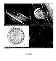
- Nitinol-based metal guide (Euroflex) is structured in order to define locations, for example wells, where the reactive groups will be deposited and where the biochemical interactions will take place ( Figure 1 ).
- Said "wells” can be produced by various methods, for example by focussed ion beam lithography (FIB or "Focused Ion beam”: Xie et al., Nuclear Instruments & Methods in Physics Research B-beam Section Interactions with Materials and Atoms, 211 (3): 363-368, 2003 ), by laser lithography followed by electrochemical etching and laser ablation.
- FIB focussed ion beam lithography
- the machine creates an ion beam, which is focused on the surface to be structured. Under the mechanical action of the ion beam, the atoms of the surface material are removed from the surface. Holes with a diameter of 20 ⁇ m can be formed with the FIB technique in a reasonable time with a bite factor of 8 ⁇ m 3 s -1 under a beam current of 20 nA.
- the Figure 2 shows holes with diameters of 5, 20 and 40 ⁇ m with a depth of 10 and 20 ⁇ m. The bottom surface of the hole is rough due to the re-deposit of the material sprayed during the attack.
- the bite factor was measured at 200 nm min -1 over a circular area of 40 ⁇ m in diameter and with a beam current of 20 nA. This gives a bite factor of 0.2 ⁇ m 3 nC -1 (approximately 5 ⁇ m 3 s -1 ), which corresponds to a procedure time of 20 min to make a hole 20 ⁇ m in diameter and 20 ⁇ m deep. .
- a fluorite-assisted milling technique XeF 2
- XeF 2 fluorite-assisted milling technique
- the technique of laser lithography and electrochemical etching consists in a first step in covering the surface with a polymer layer.
- the polymer layer is shaped using laser ablation.
- the surface is etched using isotropic electrochemical etching through the opening made in the polymer layer ( Figure 4 ).
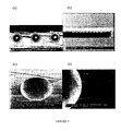
- the Figure 5 presents the results of different structuring tests on metal guides based on Nitinol.
- Nitinol-based metal guides that are used in vivo are usually treated by electrochemical polishing, which replaces the native NiTi oxide layer with a biocompatible TiO 2 layer. Shaped guides with holes are subjected to this process to evaluate the influence of the process on the hole structure ( Figure 6 ).
- Another way of preparing the cavities on the surfaces of the Nitinol-based guides is laser ablation.
- the use of short laser pulses allows local evaporation of the metal without affecting the surrounding metal due to the heat generated.
- the smallest reported dimensions are of the order of 20 microns.
- metal micro-guides (MTI 0.012 "Silver speed) were used, which metal guides were inserted into micro-catheters.
- the device was introduced in pigs, under general anesthesia, at the level of a puncture and then at the scarpa to the kidney by the endovascular route (via the femoral artery). This guidance was ensured by the follow-up of said device in the femoral artery by arteriography.
- the device was introduced into the kidney by endoarterial break-in. This penetration into the tissue was made to a depth of a few millimeters and said device was held there for about ten minutes.
- the animals were then euthanized and the kidneys of the latter were removed to assess the state of the latter after penetration of the device according to the invention.
- the device according to the invention thus allows access to an organ while being very weakly invasive.
- Metal micro-guides (MTI 0.012 "Silver speed) were used, which metal micro-guides were placed in a fiberscope in contrast to Example 1.
- the device was introduced in pigs, under general anesthesia, at the level of a puncture in the scarpa and then to the liver by endoarterial navigation (via the femoral artery). This guidance was ensured by the follow-up of said device in the femoral artery by arteriography.
- the device was introduced into it. This penetration into the fabric was made to a depth of a few millimeters and said device was held there for a further ten minutes.
- the removal of the liver after the operation made it possible to judge the aggressiveness of the intervention on the organ.
- hemorrhagic lesions were minimal: two minor macroscopic lesions could be observed without any destruction of the parenchymal cells and with a simple congestion of the capillaries and centrilobular veins.
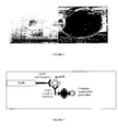
- the device uses the principle of the ELISA technique to highlight the ACE antigen.
- Two monoclonal antibodies recognizing different epitopes on this antigen were used for capture (AcM1) and revelation of ACE antigen (AcM2).
- These monoclonal antibodies having the same isotype (IgG1), the revelation of the ACE antigen was carried out using a monoclonal antibody coupled to biotin and a streptavidin-peroxidase complex ( Figure 7 ).
- PBS-BSA 3% bovine serum albumin
- the reaction was then stopped by adding 50 ⁇ l of 1M sulfuric acid per well.
- the absorbance was read at 492 nm on a plate reader (ref: ELX.800 UV).
- rigid plastic supports in the form of rods 2 to 3 cm in length and 0.5 to 1 mm in diameter were activated.
- the supports thus activated were placed in 1 ml hemolysis microtubes (Fisher) and were functionalized with a monoclonal antibody directed against the ACE antigen (clone 5910 produced in mice and marketed by Medix Biochemical). and diluted 1/50, 1/100, 1/250, 1/500 in carbonate / bicarbonate buffer (250 ⁇ l / tube) for 1 hour at 37 ° C.
- a negative control was made by replacing the monoclonal antibody with carbonate / bicarbonate buffer.
- the saturation was performed with 500 ⁇ l of 3% PBS-BSA overnight at + 4 ° C.
- the supports were then incubated with 250 ⁇ l of ACE antigen serum diluted 1/10, 1/100 in PBS or with the serum of a "healthy" subject (ACE antigen negative) at the same dilution for 1 hour. hour at 37 ° C.
- a monoclonal antibody against the ACE antigen (clone 5909 produced in mice and marketed by Medix Biochemical, which differs from clone 5910 by its affinity constant and recognized epitopes) purified at 1 mg / ml, was dialyzed overnight at 4 ° C against 0.1 M borate buffer pH 8.8. A solution of biotin at 10 mg / ml in DMSO was then added at a rate of 50 ⁇ g / mg of antibody. After incubation for 4 hours at room temperature and with stirring, 1 M ammonium chloride was added, at a rate of 20 ⁇ l / 250 ⁇ g of biotin, and the solution obtained was incubated again for 1 minute at room temperature. After blocking the reaction, the labeled antibody was dialyzed for 24 hours at + 4 ° C against PBS and this labeled antibody was stored as aliquots at -20 ° C.
- the supports were incubated with 250 ⁇ l of the biotinylated antibody 5909 and diluted 1/500 in PBS-Tween for 1 hour at 37 ° C.
- biotin ester of 6-biotinamidocaproylamido-caproic acid and N-hydroxysuccinimide, Sigma
- streptavidin-peroxidase complex Amersham Biosciences
- the revelation of the enzymatic activity was carried out by adding 750 ⁇ l per tube of the substrate mixture (H 2 O 2 ) and chromogenic (OPD, Sigma) in citrate-phosphate buffer (pH 5).
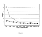
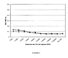
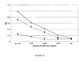
- the results show that the ODs are 7 to 10 times higher with ACE antigen-positive serum than those obtained with ACE antigen negative serum.
- the ACE antigen concentration having been the best detected corresponds to the serum of the patient diluted 1/100 to 6 IU / ml (close to the level considered "normal”: ⁇ 5 IU / ml) and when the dilution of the antibody Detection monoclonal 5909 is at 1/500.
- plastic supports makes it possible to validate the specificity and the sensitivity of the immunocapture processes on a metal rod functionalized according to the protocol described previously.
- an alkanethiol layer is adsorbed on one of the ends of Nitinol-based metal guides (Euroflex) in a first step.
- the free thiol functions of this layer allow the formation of disulfide bridges with a monoclonal antibody directed against the ACE antigen.
- the metal guide obtained is then introduced into a biopsy needle adapted for use in animals or humans.
- an extemporaneous anatomopathological examination is performed using this device on an operative part (mammary tumor), after its removal in a patient suffering from breast cancer.
- a micro-incision in the breast is performed under local anesthesia or general in a patient with breast cancer.
- the needle in which is inserted the metal guide coupled to the antibody directed against the ACE antigen, is introduced into the tumor or by the micro-incision and then directed to the tumor following its progression by imaging, including by ultrasound.
- Said micro-invasive guidance system then makes it possible to remove the end of the metal guide coupled to the antibody directed against the ACE antigen.
- the end of the metal guide is then introduced into the tumor (by perforation) to a depth of the order of a few millimeters. After a short waiting time, of the order of ten minutes, which allows the immunocapture of the ACE antigen possibly expressed by the tumor, the device is removed.
- Micro-sampling is limited to immunocapture of the analyte in vivo and does not require biopsy.
- ACE marker ELISA assay is performed on the end of the device with a monoclonal antibody directed against the ACE antigen which differentiates from the capture antibody by its affinity constant vis-a-vis antigen ACE and recognized epitopes, which is coupled with biotin.
- an alkanethiol layer was adsorbed on the end of a Nitinol-based metal guide (Euroflex) in a first step.
- the free thiol functions of this layer allowed the formation of disulfide bridges with a monoclonal antibody directed against the FAP (Fibroblast-activation protein; RETTIG et al., Proc. Natl. Acad. Sci. USA, vol.85, p: 3110, 1988 ).
- the metallic guide obtained is then introduced at the level of a cutaneous tumor in the animal or in the man when the medical ethical conditions are met, or at the level of a cutaneous tumor in a patient suffering from a cancer of the skin. skin after it has been removed for classical or extemporaneous pathological examination.
- micro-sampling is limited to immunocapture in vivo and again does not require a specific biopsy.
- the device is removed and an ELISA FAP marker assay is performed on the end of the device with a monoclonal antibody directed against the peroxidase-coupled FAP marker.
- the revelation of the peroxidase activity makes it possible to conclude the expression of the FAP marker by the tumor and to modulate accordingly the therapy to be used to treat the patient as best as possible.
Landscapes
- Health & Medical Sciences (AREA)
- Life Sciences & Earth Sciences (AREA)
- Physics & Mathematics (AREA)
- General Health & Medical Sciences (AREA)
- Veterinary Medicine (AREA)
- Engineering & Computer Science (AREA)
- Biomedical Technology (AREA)
- Heart & Thoracic Surgery (AREA)
- Medical Informatics (AREA)
- Molecular Biology (AREA)
- Surgery (AREA)
- Animal Behavior & Ethology (AREA)
- Biophysics (AREA)
- Public Health (AREA)
- Pathology (AREA)
- Optics & Photonics (AREA)
- Neurology (AREA)
- Chemical & Material Sciences (AREA)
- Chemical Kinetics & Catalysis (AREA)
- General Chemical & Material Sciences (AREA)
- Neurosurgery (AREA)
- Physiology (AREA)
- Investigating Or Analysing Biological Materials (AREA)
- Measurement Of The Respiration, Hearing Ability, Form, And Blood Characteristics Of Living Organisms (AREA)
- Endoscopes (AREA)
- Apparatus Associated With Microorganisms And Enzymes (AREA)
Abstract
Description
- La présente invention concerne la fonctionnalisation d'un dispositif d'investigation transpariétal, un procédé d'analyse ex vivo d'un substrat utilisant un dispositif fonctionnalisé de l'invention, et l'utilisation d'un tel dispositif pour la fabrication d'un outil destiné au diagnostic d'un cancer, d'une infection, d'une inflammation, ou d'un rejet de greffe chez un patient.
- Il est connu dans l'art antérieur des dispositifs d'investigation ou de traitement in vivo. De tels dispositifs prennent la forme d'un tube rigide de type endoscope ou d'un cathéter constitué d'un tube flexible qui est inséré dans l'organisme, notamment par les voies naturelles ou les vaisseaux, et qui permet d'atteindre un organe ou un tissu spécifique. Ces dispositifs permettent notamment l'élimination de caillots de sang ou, lorsqu'ils sont associés à des fibres optiques, la visualisation et le contrôle in vivo de l'état d'un système, comme le tube digestif, ou d'un organe, comme le colon. Le traumatisme pour le patient résultant de l'utilisation de tels dispositifs est alors minimisé mais reste encore à améliorer. Toutefois, il n'est pas toujours possible d'effectuer une analyse des organes ou des tissus chez un patient avec ces dispositifs. Une telle impossibilité peut résulter de l'accessibilité réduite dudit tissu ou organe au regard de la circulation sanguine ou des voies naturelles, ou alors de la difficulté à effectuer un diagnostic fiable sans recourir à une étude fine des cellules dudit organe ou tissu. Dans ces cas, il est utilisé de façon courante dans l'art antérieur et à ce jour, le prélèvement d'un fragment dudit tissu ou organe (biopsies) afin de contrôler ex vivo la morphologie de ces tissus ou organes, ou plus finement celle des cellules qui les constituent, notamment à l'aide d'aiguilles fines (FNA ou « Fine Needle Aspiration » : Engelstein et al., Br. J. Urol., 7 : 210-213, 1994 ; Rodrigues et al., J. Am. Acad. Dermatol., 42 : 735-740, 2000 ; Ariga et al., Am. J. Surg., 184 : 410-413, 2002 ; Pérez-Guillermo et al., Diagn. Cytopathol., 32 : 315-320, 2005 ; Fernandez-Esparrach et al., Arch. Bronconeomol., 43 : 219-224, 2007). En outre, il est également possible d'analyser le statut d'expression d'un certain nombre de marqueurs dont l'expression est corrélée à un état pathologique spécifique (cancer, inflammation ou infection notamment).
- Toutefois, ces différentes méthodes, du fait du prélèvement d'une biopsie ou d'aspiration de cellules in situ, font subir un traumatisme parfois important audit tissu ou organe et, par voie de conséquence, au patient. L'organisme de ce dernier peut ainsi être fortement éprouvé du fait d'hémorragies ou encore de la cicatrisation consécutive au prélèvement, en particulier pour certains organes (cerveau, pancréas, foie ou poumon).
- Il existe donc encore aujourd'hui un besoin pour l'identification de nouvelles méthodologies d'investigations permettant d'effectuer un diagnostic fiable et précis, notamment au regard de l'expression de marqueurs associés spécifiquement à différentes pathologies comme notamment le cancer, une inflammation ou une infection, tout en limitant les traumatismes infligés au patient.
- Le brevet
EP 1 358 481 décrit un dispositif d'analyse ou de traitement in vivo comprenant (i) un micro-système d'investigation d'un substrat autre que par analyse d'un signal fluorescent, (ii) une tige souple à une extrémité de laquelle est fixé ledit microsystème et dont l'autre extrémité est destinée à la manoeuvre dudit micro-système, (iii) un instrument médical possédant une lumière interne dans laquelle ladite tige souple peut coulisser, (iv) un système coulissant de protection du micro-système amovible au niveau du substrat, et (v) au niveau dudit micro-système, un système de dilacération de tissu ou de cellule, éventuellement associé à un ou plusieurs dispositifs choisis parmi un dispositif de surveillance à distance par des récepteurs sensoriels (tactiles, optiques, physico-chimiques et notamment électroniques ou informatiques numérisés), de réalisation de biopsie, de traitement, d'injection locale de produits biologiques ou chimiques. - Pour permettre l'effraction de vaisseaux sanguins et la dilacération de tissu ou de cellules, le micro-système est alors associé à un autre système plus rigide assurant cette fonction, de préférence en position distale.
- Toutefois, dans le cas de dispositif d'investigation in vivo, le dispositif d'investigation est le plus souvent dirigé vers l'organe ou le tissu cible en utilisant la voie endovasculaire ou endocavitaire. Le couplage du micro-système avec un système de dilacération augmente alors de façon non négligeable le diamètre à l'extrémité du dispositif décrit. Ce dernier se révèle alors être d'un usage complexe pour être correctement dirigé jusqu'à son site cible sans altérer simultanément les voies de circulation sanguine. Simultanément, le couplage de multiples éléments nuisent à la flexibilité d'ensemble du dispositif, et donc à son guidage correct, ainsi qu'à l'obtention de la rigidité nécessaire pour permettre la perforation d'un organe ou d'un tissu.
- À la suite d'importantes recherches, l'inventeur a maintenant réussi à développer un dispositif comprenant un guide métallique à une extrémité perforante duquel sont directement couplés des groupements réactifs, notamment des anticorps ou des fragments d'anticorps, spécifiques d'un substrat à tester, et dont l'autre extrémité est destinée à la manoeuvre dudit guide du site d'insertion jusqu'au site de micro-analyse et/ou de micro-prélèvement dudit substrat. Ledit guide peut être inséré dans un système de protection amovible, par exemple un cathéter souple, permettant ainsi de protéger l'extrémité fonctionnalisée dudit guide jusqu'au site de micro-analyse et/ou de micro-prélèvement du substrat à tester constitutif du tissu, de l'organe, ou des cellules de ceux-ci.
- Il s'agit, conformément à la présente invention, de structurer la surface du guide dans le but de définir des endroits sur le guide, par exemple des puits, où les groupements réactifs seront déposés et où les interactions biochimiques auront lieu. Lesdits « puits » peuvent être réalisés par différents procédés comme par exemple par lithographie par faisceau d'ions focalisés (FIB ou pour « Focused Ion beam » : Xie et al., Nuclear Instruments & Methods in Physics research Section B-beam Interactions with Materials and Atoms, 211(3) : 363-368, 2003), par lithographie par laser suivie par une attaque électrochimique et une ablation laser.
- Alternativement, la structuration du guide métallique peut être réalisée dans le but de définir des endroits sur ledit guide, par exemple au moins un sillon dans lequel est solidairement associée au moins une biopuce miniaturisée linéaire, circulaire ou en ruban, où les groupements réactifs seront déposés et où les réactions biochimiques auront lieu.
- Ledit dispositif peut en outre être inséré dans un instrument médical possédant une lumière interne dans laquelle ledit guide métallique peut coulisser, et notamment dans une aiguille de ponction transpariétale, notamment transcutanée ou transmuqueuse, ou un endoscope, y compris un système de navigation endovasculaire.
- Ledit dispositif peut en outre être associé à une fibre optique, ou le guide métallique dudit dispositif peut être remplacé par une fibre optique à une extrémité perforante de laquelle est associée une bague métallique sur laquelle sont directement couplés des groupements réactifs, notamment des anticorps ou des fragments d'anticorps, spécifiques d'un substrat à tester, permettant ainsi une visualisation fine in situ, en vue de la capture in vivo des éléments nécessaires au diagnostic et éventuellement à l'évaluation pronostique. Les propriétés de la fibre optique sont utilisées à des fins d'imagerie de repérage et de mise en place du dispositif à partir de la visualisation in situ.
- Le dispositif selon l'invention du fait de sa simplification présente, tout en restant micro-invasif, une flexibilité ou élasticité améliorée par rapport aux dispositifs de l'art antérieur. Ce dernier peut alors être utilisé bien plus efficacement pour une investigation par voie endocavitaire, notamment par voie endovasculaire, ou par voie transpariétale, notamment pas voie transcutanée ou transmuqeuse. De surcroît, le dispositif selon l'invention du fait de l'utilisation d'un guide métallique, dont une extrémité est couplée à des réactifs spécifiques, présente une rigidité suffisante à son extrémité fonctionnalisée pour perforer efficacement un tissu ou un organe. Enfin, le diamètre du dispositif selon l'invention à son extrémité est suffisamment faible pour permettre une navigation simplifiée dans la circulation sanguine ou dans les cavités naturelles, et surtout minimiser le traumatisme au niveau du tissu ou de l'organe que celui-ci doit perforer.
- En outre, et après retrait du dispositif, il est alors possible d'identifier ex vivo à l'aide de techniques classiques, comme un dosage immunoenzymatique ou par immunofluorescence, par exemple sur support solide [technique ELISA, puce à protéines (ESPINA et al., J. Immunol. Methods, vol.290, p :121-133, 2004)], la présence et la concentration relative dudit ou desdits substrats spécifiques à l'extrémité du dispositif selon l'invention et donc au niveau du site de micro-analyse et/ou de micro-prélèvement (organe ou tissu).
- Enfin, et du fait de la nature métallique du guide, le dispositif présente un signal amélioré en imagerie (artériographie, échographie, scanner, IRM, etc...). Cette propriété permet de simplifier considérablement le radioguidage du dispositif selon l'invention lors de l'intervention jusqu'au tissu ou organe ciblé. Il en est de même pour le couplage dudit dispositif avec une fibre optique à des fins d'imagerie de repérage, et qui permet la visualisation fine in situ.
- En conséquence, un premier objet de l'invention est un dispositif pour l'analyse d'un substrat, caractérisé en ce qu'il comprend un système d'investigation micro-invasif et/ou de micro-prélèvement dudit substrat, ledit système étant constitué d'au moins un guide métallique à une extrémité Ea duquel est pourvue au moins une série de puits auxquels est directement couplé au moins un groupement réactif spécifique dudit substrat, ladite extrémité fonctionnalisée Ea étant perforante, et dont l'autre extrémité Em est destinée à la manoeuvre dudit guide métallique et éventuellement associée à un système d'aspiration.
- Le dispositif selon l'invention, du fait du faible traumatisme qu'il engendre chez le patient, permet de réaliser plusieurs micro-analyses ou micro-prélèvements chez un patient à intervalles réguliers (par exemple, analyse et/ou prélèvements étagés dans le cadre de la prostate). Lesdites analyses et/ou prélèvements successifs permettent ainsi, outre le diagnostic, de suivre l'évolution d'un cancer, d'une inflammation, d'une infection, ou la bonne prise d'une greffe d'organe chez un patient.
- Avantageusement, l'extrémité fonctionnalisée Ea peut présenter sur une longueur d'environ 0,5 à 2 cm, au moins une série de 1 à 25 puits, de préférence 2x25 puits, ayant un diamètre moyen d'environ 30 à 80 µm, de préférence d'environ 40 à 60 µm, et de manière tout à fait préférée d'environ 50 µm, une profondeur d'environ 20 à 30 µm, de préférence de 25 µm, lesdits puits étant espacés les uns des autres d'environ 60 à 120 µm. De préférence, les puits ont une paroi lisse ou rugueuse, une forme ovale ou ronde, avec un fond plat ou concave.
- Selon un mode de réalisation particulier du dispositif selon l'invention, ledit dispositif comprend en outre un système de protection amovible au niveau de l'extrémité fonctionnalisée Ea.
- Selon encore un mode de réalisation particulier du dispositif selon l'invention, ledit dispositif comprend en outre un instrument médical possédant une lumière interne dans laquelle ledit au moins un guide métallique peut coulisser.
- Par « guide métallique », on entend par exemple une tige métallique pleine souple ou une tige métallique creuse rigide, ayant un diamètre allant de 0,2 à 3,5 mm et une longueur allant de 5x10⁻² à 2 m, et pouvant être insérée dans un vaisseau sanguin, une petite cavité ou à travers un organe ou un tissu, afin de pouvoir être dirigé depuis le site d'insertion jusqu'au site de micro-analyse et/ou de micro-prélèvement in situ.
- En particulier, une tige métallique pleine souple peut être constituée d'une fibre optique à une extrémité Ea de laquelle est associée une bague métallique pourvue d'au moins une série de puits auxquels est directement couplé au moins un groupement réactif spécifique dudit substrat, ladite extrémité fonctionnalisée Ea étant perforante.
- Avantageusement, ladite bague métallique a une largeur d'environ 0,5 à 2 cm, et peut présenter au moins une série de 1 à 25 puits, de préférence 2x25 puits, ayant un diamètre moyen d'environ 30 à 80 µm, de préférence d'environ 40 à 60 µm, et de manière tout à fait préférée d'environ 50 µm, une profondeur d'environ 20 à 30 µm, de préférence de 25 µm, lesdits puits étant espacés les uns des autres d'environ 60 à 120 µm. De préférence, les puits ont une paroi lisse ou rugueuse, une forme ovale ou ronde, avec un fond plat ou concave.
- Par « guide métallique », on entend en outre une tige pleine souple ou creuse, rigide ou flexible constituée en tout ou partie d'un alliage métallique dont les caractéristiques de flexibilité, de rigidité, d'oxydation et d'immunogénicité sont compatibles avec une telle utilisation chez l'être vivant et en particulier chez l'animal, et tout particulièrement chez l'homme. De tels alliages biocompatibles peuvent être identifiés simplement par l'homme du métier au regard de ses connaissances générales et comprennent notamment les aciers inoxydables, les alliages à base de titane, de nickel, de cobalt, ou des mélanges de ceux-ci.
- L'inventeur a pu démontrer qu'un guide à base d'un alliage de titane et de nickel (alliage de Nitinol) présentait des propriétés particulièrement intéressantes en terme de flexibilité d'ensemble et de rigidité à son extrémité pour être utilisé efficacement pour les voies endovasculaires ou endocavitaires et également pour perforer efficacement un tissu ou un organe tout en minimisant le traumatisme (la taille de la perforation au niveau dudit tissu ou organe est par exemple de l'ordre de 0,05 à 0,5 mm2, et de préférence de l'ordre de 0,07 mm2).
- Avantageusement, le guide métallique est à base d'un alliage de nickel et de titane, de préférence à base de Nitinol (guides métalliques commercialisés par la société Euroflex).
- Ledit guide métallique peut être recouvert, à l'exception de l'extrémité fonctionnalisée Ea, d'un polymère hydrophile, de préférence un hydrogel, ou d'une couche polymère protectrice poreuse, ayant une épaisseur allant d'environ 0,1 à 51 µm. Avantageusement, le polymère protecteur est constitué d'un film de parylène, de TiO2 ou d'OptoDex® (Arrayon Biotechnology, Suisse), et plus préférentiellement d'un film de parylène.
- Le système de protection amovible dans lequel est inséré le guide métallique peut prendre de multiples formes, et notamment celle d'un cathéter souple, qui peuvent être déterminées simplement par l'homme du métier, par exemple des formes utilisables pour la voie endovasculaire, endocavitaire, transpariétale, et notamment transcutanée.
- Ledit système de protection amovible peut être inséré par voie endovasculaire notamment pour atteindre les vaisseaux du coeur, le cerveau, le poumon, le pancréas, le rein, et le foie.
- Ledit système de protection amovible peut être inséré par des accès endocavitaires, notamment par le biais d'un endoscope, par voie orale, anale, urogénitale et respiratoire, ou ORL par voie transmuqueuse, ou encore par voie transcutanée par le biais d'une ponction au niveau de la peau, pour atteindre par exemple la glande mammaire, et notamment jusqu'au rein ou dans une articulation ou en transpariétal dans le foie, le poumon ou le rein, mais également par voie transmuqueuse, notamment pour le tube digestif.
- L'inventeur a ainsi mis en évidence que le dispositif selon l'invention permet alors d'atteindre des tissus ou organes, normalement difficiles d'accès, par les voies endovasculaire, endocavitaire ou transpariétale, et notamment transmuqueuse ou transcutanée, utilisées classiquement.
- Plus généralement, l'inventeur a mis en évidence que le dispositif selon l'invention permet du fait de ses caractéristiques spécifiques de flexibilité d'ensemble et de rigidité à son extrémité d'atteindre et de perforer par voie transpariétale (transcutanée, transmuqueuse), endovasculaire ou endocavitaire certains organes et tissus appartenant au système digestif de l'oropharynx au rectum (y compris foie et pancréas), au système urogénital (dont vessie, rein, prostate, testicule, ovaire et glande mammaire), au système trachéo-bronchique (dont poumon), au système ORL (dont oreille et rhinopharynx), au système ostéo-articulaire (dont cavités synoviales), au système endocrinien, au système neurocérébral ou au système tégumentaire, rendant ainsi possible la réalisation de diagnostics sur des pathologies qui nécessitaient jusqu'alors la réalisation de biopsies voir des ponctions profondes agressives comme les biopsies trans-hépatiques, y compris le prélèvement de cellules par aspiration à l'aide d'aiguilles fines (technique dite de « fine needle aspiration » ou FNA).
- Avantageusement, ledit système de protection amovible dans lequel est inséré le guide métallique prend la forme d'un cathéter souple adapté pour la voie endovasculaire ou endocavitaire.
- Le dispositif selon l'invention est alors particulièrement adapté pour effectuer une investigation, par exemple, au niveau des artères et des veines, des vaisseaux du coeur, de la prostate, de la glande mammaire, du pancréas, du rein, du muscle cardiaque, du système nerveux central et ses cavités ou canaux, du cerveau ou du foie.
- Selon un premier mode de réalisation particulier du dispositif selon l'invention, le système de protection amovible dans lequel est inséré le guide métallique est lui-même inséré dans un endoscope. Le dispositif selon l'invention est alors particulièrement adapté pour une administration par la voie endocavitaire.
- Le dispositif selon l'invention est alors particulièrement adapté pour effectuer une investigation, par exemple, au niveau du système trachéo-bronchique (dont poumon), du système digestif du pharynx au rectum (y compris foie et pancréas), du système urogénital (dont vessie, rein, prostate, testicule, ovaire et glande mammaire), du système ophtalmique (canaux lacrymaux), du système ORL (dont oreille et rhinopharynx), du système ostéo-articulaire, ou au niveau du cerveau ou de la glande mammaire par la voie endogalactophorique.
- Selon un second mode de réalisation particulier du dispositif selon l'invention, le guide métallique constitué d'une aiguille fine de ponction transpariétale, et notamment de ponction transcutanée ou transmuqueuse, peut être inséré dans un système de protection amovible, par exemple un cathéter souple. Le dispositif selon l'invention est alors particulièrement adapté pour une administration spécifiquement par la voie transcutanée ou transmuqueuse.
- Le dispositif selon l'invention est alors particulièrement adapté pour effectuer une investigation, par exemple, au niveau des téguments (peau, cuir chevelu, etc...), du sein, du rein, du poumon, du foie, du muscle, de l'appareil ostéo-musculaire ou ostéo-articulaire, ou des glandes endocrines (notamment thyroïde, parathyroïde, surrénales, testicules, glandes mammaires ou ovaires).
- En ce qui concerne l'extrémité Ea fonctionnalisée du dispositif selon l'invention, elle est pourvue d'au moins une série de puits auxquels sont directement couplés des groupements réactifs spécifiques d'un substrat à tester.
- Par « groupements réactifs spécifiques », on entend par exemple une séquence d'acide nucléique [ADN (amplifiat, fragment de gène, EST, SNP) ou ARN] complémentaire d'une séquence d'acide nucléique à détecter, un antigène spécifique d'un anticorps à détecter ou un anticorps ou un fragment d'anticorps spécifique d'un antigène à détecter, de préférence un anticorps ou un fragment d'anticorps.
- Avantageusement, lesdits groupements réactifs spécifiques sont disposés dans les micro-puits de l'extrémité fonctionnalisée du dispositif selon l'invention selon une gamme croissante ou décroissante. L'homme du métier peut déterminer simplement, à l'aide de ses connaissances générales et d'expériences de routine, ladite gamme en fonction de l'affinité du groupement réactif pour son substrat. À titre d'exemple, la gamme du groupement réactif est de l'ordre de 50 à 500 µg/ml, de préférence de 10 à 100 µg/ml, pour un réactif, notamment un anticorps, ayant une affinité pour son substrat, notamment un antigène, de l'ordre de 10-9.
- Par « anticorps », on entend de préférence une immunoglobuline de mammifère, notamment humaine, et de manière particulièrement préférée une IgG.
- Par « fragments d'anticorps », on entend des fragments d'anticorps capables de maintenir une fixation spécifique de leur antigène. À titre d'exemple de tels fragments d'anticorps, on peut citer les fragments Fab, Fab', F(ab')2 ou Fv.
- Des méthodes de couplage de réactifs, notamment de protéines sur un support métallique sont bien connues de l'homme du métier. De telles méthodes, du fait de la faible réactivité chimique des acides aminés, nécessitent en général l'activation de la surface métallique soit par des mécanismes d'oxydation, soit en recouvrant cette dernière d'au moins une couche de molécules de liaisons, le plus souvent de polymères (celle-ci peut présenter par exemple des groupements thiols, acides carboxyliques et/ou des amines).À titre d'exemple de telles méthodes, on peut citer l'adsorption sur des supports métalliques de molécules fonctionnelles s'organisant en monocouches auto-assemblées (self-assembled monolayer SAMs) et notamment les alcanethiols (voir notamment WITTSTOCK and SCHUHMANN, Anal. Chem., vol.69, p :5059-5066, 1997 ; et la demande internationale
WO 03/006948 - Par couplage de l'anticorps ou du fragment d'anticorps à cette couche de molécules fonctionnelles, on entend une liaison covalente (comme des ponts disulfures entre les groupements thiols libres des alcanethiols) ou non covalente (comme une liaison streptavidine-biotine entre la biotine d'une couche de polymères d'un complexe pyrolle/biotine et la streptavidine d'un complexe streptavidine/anticorps ou fragment d'anticorps) streptavidine).
- Selon un premier mode de réalisation préféré, le dispositif selon l'invention comprend au moins un guide métallique dont une extrémité Ea est couplée avec au moins un groupement réactif, de préférence un anticorps ou un fragment d'anticorps, spécifique d'un marqueur (antigène) d'un cancer, notamment du cancer du sein, de l'ovaire, de la prostate, du colon, intra-abdominal, du rein, du cerveau, du foie, du poumon, du pancréas ou d'une glande endocrine (notamment thyroïde, testicules ou ovaires).
- À titre d'exemple de marqueur du cancer du sein, on peut citer le marqueur CA 15-3 (Carcinoma-Associated Antigen 15-3 ; Duffy MJ, Shering S, Sherry F, McDermott E, O'Higgins N, Int J Biol Markers, 2000 Oct-Dec ;15(4) : 330-3), CA 27-29 (Carcinoma-Associated Antigen 27-29 ; Kaohsiung J, J Med Sci, 1999 Sep ; 15(9) : 520-8), CEA (Carcinoembryonic antigen ; Soletormos G, Nielsen D, Schioler V, Mouridsen H, Dombernowsky P, Eur J Cancer, 2004 Mar ;40 (4) : 481-6) ; TPA (Tissue Polypeptide Antigen) ; TPS (Tissue Polypeptide Specific Antigen; Given M, Scott M, Mc Grath JP, Given HF, Breast, 2000 Oct; 9 (5) : 277-80), HER2 (Fehm T, Jager W, Kramer S, Sohn C, Solomayer E, Wallwiener D, Gebauer G., Anticancer Research, 2004 May-Jun ; 24 (3b) : 1987-92), ER (Estrogene Receptor ; Platet N, Cathiard AM, Gleizes M, Garcia M., Crit Rev Oncol Hematol, 2004 Jul ; 51(1) : 55-67), PR (Progesterone Receptor ; Duffy MJ, Clin Chem, 2005 Mar ;51(3) : 494-503. Epub 2005 Jan 6.), Ki-67 (cell proliferation-associated antigen of antibody Ki-67 ; Schlüter C, Duchrow M, Wohlenberg C, Becker MH, Key G, Flad HD, Gerdes J, J Cell Biol, 1993 Nov ; 123(3) : 513-22) et UPA (Urokinase Plasmogen Activator ; Duffy MJ, Crit Rev Clin Lab Sci, 2001 Jun ;38(3) : 225-62).
- À titre d'exemple de marqueur du cancer de l'ovaire, on peut citer le marqueur CA125 (Carcinom Antigen 125; Moss EL, Hollingworth J, Reynolds TM, J Clin Pathol, 2005 Mar ; 58(3) : 308-12), CA 15-3 et CEA ( Valenzuela P, Mateos S, Tello E, Lopez-Bueno MJ, Garrido N, Gaspar MJ, Eur J Gyn Oncol, 2003 ; 24(1) : 60-2).
- À titre d'exemple de marqueur du cancer de la prostate, on peut citer le marqueur PSA (Prostate-Specific Antigen; Gray MA, Clin Lab.,2005 ; 51(3-4) :127-33) ; PMSA (Prostate-Specific Membrane Antigen) et AR (Androgen Receptor ; Birtle AJ, Freeman A, Masters JR, Payne HA, Harland SJ, BJU Int, 2005 Aug ; 96(3) : 303-7).
- À titre d'exemple de marqueur du cancer du colon, on peut citer le marqueur CEA (Duffy MJ, Clin Chem, 2001 Apr ; 47(4) : 624-30), CA 19-9 (Carcinom Antigen 19-9), CA242 (Carcinom Antigen 242), CA 72-4 (Carcinom Antigen 72-4), TPA, TPS (Duffy MJ, van Dalen A, Haglund C, Hansson L, Klapdor R, Lamerz R, Nilsson O, Sturgeon C, Topolcan O, Eur J Cancer,2003 Apr ; 39 (6) : 718-27).
- À titre d'exemple de marqueur du cancer intraabdominal, on peut citer le marqueur CEA ou CA 19-9 (Coban E, Samur M, Bozcuk H, Ozdogan M, Int J Biol Markers, 2003 Jul-Sep ; 18(3) : 177-81).
- À titre d'exemple de marqueur du cancer du pancréas, on peut citer le marqueur TA90-IC (a 90-kDa immugenic Tumor-associated Antigen), CA-19-9 (Chung MH, Gupta RK, Bilchik AJ, Ye W, Yee R, Morton DL, Curr Surg,2002 March-April ;59(2) : 194-198), TPS, HCG beta (hCG beta, Human Chorionic Gonadotropin beta), CA 72-4, CEA, CA 19-9, CA 242 (Louhimo J, Alfthan H, Stenman UH, Haglund C, Oncology,2004 ; 66(2) : 126-31).
- À titre d'exemple de marqueur du cancer du foie, on peut citer le marqueur alpha-foetoprotéine.
- À titre d'exemple de marqueur du cancer du poumon, on peut citer le marqueur Cyfra A41 (Cytokeratin fragment 41), SCC (Squamous Cell Carcinoma antigen), ACE (Angiotensin Converting Enzyme ), CA 19-9, CA 125, NSE (Neuron Specific Enolase), chromogranine A, CYFRA 21-1 (Cytokeratin fragment 21-1) CA 15-3.
- Selon un second mode de réalisation préféré, le dispositif selon l'invention comprend au moins un guide métallique dont une extrémité Ea est couplée avec au moins un réactif, de préférence un anticorps ou un fragment d'anticorps, spécifique d'un marqueur spécifique d'une inflammation, notamment de l'arthrite rhumatoïde.
- À titre d'exemple de marqueur de l'arthrite rhumatoïde, on peut citer notamment l'IL-1β, l'IL-1Rα, l'IL-2, l'IL-2R, l'IL-4, l'IL-5, l'IL-6, l'IL7, l'IL8, l'IL10, l'IL12p40P70, l'IL-13, l'IL-15, l'IL-17, le TNFα, l'IFNα, l'IFNy, le GM-CSF, le MIP-1, l'IP-10, le MIG, l'Eotaxine, le RANTES et le MCP-1 (COCKRUM et al., Lab Automation, BTi, October 2005, p :19-21).
- Selon un troisième mode de réalisation préféré, le dispositif selon l'invention comprend au moins un guide métallique dont une extrémité Ea est couplée avec au moins un réactif, de préférence un anticorps ou un fragment d'anticorps, spécifique d'un marqueur spécifique d'une infection, notamment d'une infection virale, bactérienne ou parasitaire.
- De nombreux marqueurs infectieux sont connus de l'homme du métier et ce dernier pourra identifier sans difficulté le ou les marqueurs spécifiques associés à une infection donnée.
- Selon un quatrième mode de réalisation préféré, le dispositif selon l'invention comprend au moins un guide métallique dont une extrémité Ea est couplée avec au moins un réactif, de préférence un anticorps ou un fragment d'anticorps, spécifique d'un marqueur spécifique du rejet de greffe.
- De nombreux marqueurs du rejet de greffe sont connus de l'homme du métier. À titre d'exemple, on peut citer MIP-1β et la VE-cadhérine pour la greffe du coeur (ROUSSOULIÈRES et al., Circulation, vol.111(20), p :2636-2644, 2005).
- Au regard des marqueurs spécifiques qui sont décrits précédemment, l'homme du métier au regard de ses connaissances générales pourra identifier facilement et sans expérimentation excessives les anticorps ou fragments d'anticorps spécifiques pouvant être utilisés dans le dispositif selon l'invention. À titre d'exemple, de tels anticorps, on peut citer les anticorps disponibles chez TEBU ou AXXORA. L'homme du métier pourra également obtenir de tels anticorps par des méthodes d'immunisation bien connues.
- De même, l'homme du métier pourra identifier sans difficulté les acides nucléiques spécifiques adaptés pouvant être utilisés dans le dispositif selon l'invention.
- Avantageusement, le dispositif selon l'invention, tout au moins en sa partie terminale fonctionnalisée en contact avec le substrat à analyser, présente un niveau d'assurance de stérilité (SAL pour Sterility Assurance Level) de l'ordre de 10-6. Différentes alternatives sont envisageables afin d'atteindre ce niveau de stérilité. Une possibilité est de stériliser le dispositif en l'absence des groupements réactifs spécifiques du substrat à détecter puis d'ajouter ces derniers en conditions stériles. Une autre possibilité est de stériliser le dispositif après l'ajout des groupements réactifs, ce qui nécessite d'utiliser des techniques de stérilisation ne réduisant pas significativement l'activité desdits groupements réactifs (par exemple stérilisation par l'oxyde d'éthylène ou radiation).
- Un second objet de l'invention est un procédé de détection ex-vivo d'un substrat présent dans un tissu ou organe, caractérisé en ce qu'il comprend les étapes suivantes :
- a) l'incubation de l'extrémité fonctionnalisée Ea d'un dispositif selon l'invention, avec une solution comprenant au moins un agent de détection spécifique dudit substrat, après que ladite extrémité ait été mise en contact avec ledit tissu ou organe devant être examiné.
- b) la détection dudit substrat.
- L'étape d'incubation est effectuée pendant un temps suffisant pour que l'agent de détection en solution, notamment un anticorps, puisse se fixer spécifiquement au substrat (marqueur, antigène, anticorps, etc...), notamment u antigène, éventuellement présent à l'extrémité du dispositif. L'homme du métier peut déterminer simplement à l'aide de ses connaissances générales et d'expériences de routine, ce temps d'incubation en fonction de l'affinité de l'agent de détection en solution, notamment un anticorps, pour son substrat, notamment un antigène. Ce temps d'incubation est également fonction de la température de la solution lors de cette incubation. À titre d'exemple, le temps d'incubation est de l'ordre de 1 minute à 2 heures, de préférence de 5 minutes à 1 heure, et de manière particulièrement préférée de 10 à 30 minutes, pour une température comprise entre 20°C (température ambiante) et 37°C.
- Avantageusement, l'agent de détection en solution est différent du réactif spécifique couplé à l'extrémité fonctionnalisée du dispositif selon l'invention.
- De préférence, l'agent de détection est un anticorps.
- Avantageusement, l'anticorps en solution et l'anticorps couplé à l'extrémité fonctionnalisée du dispositif selon l'invention sont chacun un anticorps polyclonal, de préférence lesdits anticorps sont identiques.
- Avantageusement, l'anticorps en solution et l'anticorps couplé à l'extrémité fonctionnalisée du dispositif selon l'invention sont chacun un anticorps monoclonal, de préférence lesdits anticorps sont différents.
- Avantageusement, l'anticorps en solution est marqué, et notamment est couplé à une enzyme, par exemple à la peroxydase ou à la phosphatase alcaline.
- Selon un premier mode de réalisation particulier du procédé selon l'invention, le procédé comprend en outre une étape a') d'incubation de ladite extrémité dans une solution comprenant au moins un agent de détection spécifique de l'agent de détection de l'étape a), intercalée entre les étapes a) et b).
- L'homme du métier pourra identifier simplement et à l'aide de ses connaissances générales, les anticorps adaptés au procédé selon l'invention. À titre d'exemple, il est possible d'utiliser dans cette deuxième étape un anticorps reconnaissant spécifiquement des immunoglobulines de souris, si de telles immunoglobulines de souris dirigées spécifiquement contre le substrat (marqueur, antigène, anticorps, etc...) à identifier sont utilisés à l'étape a).
- Selon un second mode de réalisation particulier du procédé selon l'invention, le procédé selon l'invention comprend une étape de lavage à la suite de l'étape d'incubation a), et éventuellement de l'étape a'), laquelle étape de lavage permet d'éliminer les anticorps qui ne se sont pas fixés spécifiquement au marqueur (l'antigène).
- Le protocole d'une telle étape de lavage fait là encore partie des connaissances générales ou peut être déterminé simplement par des expériences de routine. À titre d'exemple, une telle étape est effectuée avec une solution comprenant une concentration plus ou moins élevée de détergent (de 0,05 à 1%), comme le TRITON X100® ou le TWEEN 20®, et ceci en fonction de l'affinité de l'anticorps en solution pour son antigène spécifique.
- L'étape de détection est effectuée en mettant en évidence une activité, notamment enzymatique, couplée à l'anticorps utilisé à l'étape a) ou, éventuellement à l'étape a').
- Le protocole utilisé pour cette étape de détection est fonction du marqueur utilisé, et notamment de l'enzyme utilisée, par exemple la peroxydase et la phosphatase alcaline, et fait partie des connaissances générales de l'homme du métier.
- Cette étape de détection permet de déduire la quantité de substrat spécifique (par exemple d'antigène) fixé à l'extrémité fonctionnalisée du dispositif et finalement la quantité de substrat spécifique présent au niveau de l'organe ou du tissu où a été effectué(e) la micro-analyse et/ou le micro-prélèvement.
- Finalement, les différents groupements réactifs utilisables pour réaliser le procédé selon l'invention sont bien connus de l'homme du métier et incluent, notamment, les réactifs utilisés pour la technique de dosage immunoenzymatique ou par immunofluorescence, par exemple sur support solide [technique ELISA, puce à protéines (ESPINA et al., précité, 2004)].
- Un troisième objet de l'invention consiste en l'utilisation d'un dispositif selon l'invention, pour la fabrication d'un outil destiné au diagnostic d'un cancer, d'une inflammation, d'une infection ou d'un rejet de greffe chez un patient.
- Selon un mode de réalisation particulier de l'invention, ledit outil de diagnostic peut comprendre au moins un guide métallique inséré dans un cathéter souple inséré dans un endoscope.
- Selon un autre mode de réalisation particulier de l'invention, ledit outil de diagnostic peut comprendre au moins un guide métallique consistant en une aiguille de ponction transpariétale, et notamment de ponction transcutanée ou transmuqueuse, pouvant être inséré dans un système de protection amovible, par exemple un cathéter souple. En outre, le système de protection amovible et le guide métallique coopèrent de manière à permettre la mise en contact de l'extrémité Ea fonctionnalisée dudit guide avec le site de micro-analyse et/ou de micro-prélèvement.
- Dans ces deux modes de réalisation particuliers de l'invention, ledit au moins un guide métallique peut être associé sur au moins une partie de sa longueur à une fibre optique en vue de repérage et de positionnement.
- Avantageusement, ledit outil de diagnostic est administré par voie endocavitaire.
- Ledit outil permet ainsi d'effectuer une micro-analyse et/ou un micro-prélèvement au niveau du système digestif du pharynx au rectum (y compris foie et pancréas), du système urogénital (dont vessie, urètre, rein, prostate), du système trachéo-bronchique (dont poumon), du système ORL (dont oreille et rhinopharynx), du système ostéo-articulaire (dont cavités synoviales).
- De manière préférée, ledit outil de diagnostic est administré par voie transpariétale, notamment par voie transmuqueuse ou transcutanée.
- Un tel outil de diagnostic permet ainsi d'analyser des tissus ou organes difficiles à atteindre par la voie endocavitaire ou endovasculaire utilisée habituellement. Un tel outil de diagnostic permet ainsi d'effectuer une micro-analyse et/ou un micro-prélèvement par voie transpariétale au niveau de la peau, des testicules, de la prostate, de l'ovaire ou des glandes mammaires, mais également du rein ou du foie, du système nerveux (notamment du cerveau) ainsi que du système endocrinien (par exemple la thyroïde).
- Les exemples qui suivent permettent d'illustrer l'invention et sont donnés à titre non-limitatif.
-
- La
Figure 1 représente différentes possibilités pour la structuration des guides métalliques. - La
Figure 2 représente des photographies de microscopie électronique à balayage de différents trous réalisés par FIB (la grande rugosité de surface due aux inhomogénéités de gravure peut être observée). - La
Figure 3 représente une explication et une observation de l'effet d'ombrage de la technique de fraisage assistée par la fluorine. - La
Figure 4 représente la suite des opérations de microfaçonnage. - La
Figure 5 représente (a) des trous hémisphériques observés par microscope optique (b) des séries de trous observés sur un guide à base de Nitinol par microscopie électronique à balayage (c) détail d'une cavité (d) comparaison de la rugosité de surface dans la cavité avec la rugosité de la surface du guide. - La
Figure 6 représente des photos de microscopie électronique à balayage montrant le même trou hémisphérique que dans laFigure 5 après le traitement de polissage électrochimique. - La
Figure 7 représente un schéma du dispositif de l'invention pour l'immunocapture avec un permier anticorps monoclonal (AcM 1) et la révélation de l'antigène ACE avec un second anticorps monoclonal (AcM 2). - La
Figure 8 représente les résultats d'ELISA avec l'anticorps de capture 5910 et la révélation avec l'anticorps 5909 (absorbance obtenue avec un sérum positif en antigène ACE) - La
Figure 9 représente les résultats d'ELISA avec l'anticorps de capture 5910 et la révélation avec l'anticorps 5909 (absorbance obtenue avec un sérum négatif en antigène ACE) - La
Figure 10 représente les résultats d'ELISA avec l'anticorps de capture 5905 et la révélation avec l'anticorps 5909 (absorbance obtenue avec un sérum positif en antigène ACE) - La
Figure 11 représente les résultats d'ELISA avec l'anticorps de capture 5905 et la révélation avec l'anticorps 5909 (absorbance obtenue avec un sérum négatif en antigène ACE) - La
Figure 12 représente les résultats d'ELISA des tiges plastiques rigides avec l'anticorps de capture 5910 et la révélation avec l'anticorps 5909 (absorbance obtenue avec un sérum positif en antigène ACE) - Ces figures sont des illustrations de la sensibilité et de la spécificité de la méthode utilisée.
- La surface d'un guide métallique à base de Nitinol (Euroflex) est structurée dans le but de définir des endroits, par exemple des puits, où les groupements réactifs seront déposés et où les interactions biochimiques auront lieu (
Figure 1 ). - Lesdits « puits » peuvent être réalisés par différents procédés comme par exemple par lithographie par faisceau d'ions focalisés (FIB ou pour « Focused Ion beam » : Xie et al., Nuclear Instruments & Methods in Physics research Section B-beam Interactions with Materials and Atoms, 211(3) : 363-368, 2003), par lithographie par laser suivie par une gravure électrochimique et une ablation laser.
- Avec la technique FIB, la machine crée un faisceau d'ions, qui est focalisé sur la surface qui doit être structurée. Sous l'action mécanique du faisceau d'ions, les atomes du matériau de surface sont éliminés de la surface. Des trous d'un diamètre de 20 µm peuvent être formés avec la technique FIB dans un délai raisonnable avec un facteur de morsure de 8 µm3s-1 sous un courant de faisceau de 20 nA. La
Figure 2 montre des trous de diamètres de 5, 20 et 40 µm avec une profondeur de 10 et 20 µm. La surface du fond du trou est rugueuse du fait du re-dépôt du matériel pulvérisé au cours de l'attaque. Le facteur de morsure a été mesuré à 200 nm min-1 sur une aire circulaire de 40 µm de diamètre et avec un courant de faisceau de 20 nA. Cela donne un facteur de morsure de 0,2 µm3 nC-1 (environ 5 µm3s-1), qui correspond à un temps de procédure de 20 min pour effectuer un trou de 20 µm de diamètre et de 20 µm de profondeur. Afin d'améliorer la rugosité de la surface, une technique de fraisage assistée par fluorine (XeF2) a été utilisée ; une très faible rugosité de surface a alors été obtenue, mais comme la source de XeF2 n'était pas exactement dans l'axe du faisceau de gravure, un effet d'ombrage a été observé (Figure 3 ). - La technique de lithographie par laser et de gravure électrochimique consiste dans une première étape à recouvrir la surface avec une couche de polymère. Dans une seconde étape, la couche polymère est façonnée en utilisant l'ablation laser. Dans une troisième étape, la surface est gravée en utilisant une gravure électrochimique isotrope à travers l'ouverture faite dans la couche polymère (
Figure 4 ). LaFigure 5 présente les résultats de différents tests de structuration sur des guides métalliques à base de Nitinol. - Par ailleurs, les guides métalliques à base de Nitinol qui sont utilisés in vivo sont habituellement traités par polissage électrochimique, qui remplace la couche d'oxyde natif NiTi avec une couche TiO2 biocompatible. Les guides façonnés avec des trous sont soumis à ce procédé afin d'évaluer l'influence du procédé sur la structure des trous (
Figure 6 ). - Une autre façon de préparer les cavités sur les surfaces des guides à base de Nitinol recourt à l'ablation laser. L'utilisation de courtes impulsions laser permet l'évaporation locale du métal sans affecter le métal environnant du fait de la chaleur générée. Les plus petites dimensions rapportées sont de l'ordre de 20 µm.
- Si les trois procédés décrits ci-dessus permettent la réalisation de puits, c'est le procédé par gravure électrochimique qui donne les meilleurs résultats.
- Pour ces expériences, des micro-guides métalliques (MTI 0,012" Silver speed) ont été utilisés, lesquels guides métalliques étaient insérés dans des micro-cathéters.
- Le dispositif a été introduit chez des porcs, sous anesthésie générale, au niveau d'une ponction puis au niveau du scarpa jusqu'au rein par la voie endovasculaire (via l'artère fémorale). Ce guidage a été assuré par le suivi dudit dispositif dans l'artère fémorale par artériographie.
- Une fois positionné à l'entrée du rein, le dispositif a été introduit dans le rein par effraction endoartérielle. Cette pénétration dans le tissu s'est faite à une profondeur de quelques millimètres et ledit dispositif y a été maintenu pendant une dizaine de minutes.
- Finalement, le dispositif a alors été retiré.
- Les animaux ont alors été euthanasiés et les reins de ces derniers ont été prélevés pour évaluer l'état de ceux-ci après pénétration du dispositif selon l'invention.
- Les résultats ont montré qu'aucune hémorragie importante du rein n'était associée à l'effraction. La lésion la plus importante constatée présentait une dimension de 3 x 1 mm au niveau du site d'effraction.
- Le dispositif selon l'invention permet donc d'accéder à un organe tout en étant très faiblement invasif.
- Des micro-guides métalliques (MTI 0,012" Silver speed) ont été utilisés, lesquels micro-guides métalliques étaient placés dans un fibroscope à la différence de l'exemple 1.
- Le dispositif a été introduit chez des porcs, sous anesthésie générale, au niveau d'une ponction au niveau du scarpa puis jusqu'au foie par navigation endoartérielle (via l'artère fémorale). Ce guidage a été assuré par le suivi dudit dispositif dans l'artère fémorale par artériographie.
- Une fois positionné à proximité du foie, le dispositif a été introduit dans celui-ci. Cette pénétration dans le tissu s'est faite à une profondeur de quelques millimètres et ledit dispositif y a été maintenu là encore pendant une dizaine de minutes.
- Finalement, le dispositif a alors été retiré.
- Comme précédemment, le prélèvement du foie après l'opération a permis de juger de l'agressivité de l'intervention sur l'organe.
- Aucune lésion macroscopiquement visible n'a été observée à la surface du foie. A la coupe, la présence de deux foyers hémorragiques intra-parenchymateux de siège sous-capsulaire de 1,5x0,4 cm et de 1,8x0,5 cm ont été observés. Histologiquement, l'architecture hépatique est en tout point conservée avec une congestion des sinusoïdes, des veinules portes et des veines centrolobulaires sans aucune autre anomalie notable.
- Les résultats ont montré que les lésions hémorragiques sont minimes : deux lésions macroscopiques mineures ont pu être observées sans aucune destruction des cellules parenchymateuses et avec une simple congestion des capillaires et des veines centro-lobulaires.
- L'utilisation d'un guide métallique permet donc d'obtenir un traumatisme mineur et, dans tous les cas, largement inférieur à celui résultant d'une biopsie.
- Le dispositif utilise le principe de la technique ELISA permettant de mettre en évidence l'antigène ACE. Deux anticorps monoclonaux reconnaissant sur cet antigène des épitopes différents, ont été utilisés pour la capture (AcM1) et la révélation de l'antigène ACE (AcM2). Ces anticorps monoclonaux ayant le même isotype (IgG1), la révélation de l'antigène ACE a été réalisée à l'aide d'un anticorps monoclonal couplé à la biotine et d'un complexe streptavidne-peroxydase (
Figure 7 ). - Deux types de support ont été utilisés, soit des plaques pour ELISA, soit des tiges plastiques rigides.
- 100 µl d'un anticorps monoclonal dirigé contre l'antigène ACE (clone 5910 ou clone 5905, produits chez la souris et commercialisés par Medix Biochemical) dilué (1/5000 au 1/128000) en tampon carbonate/bicarbonate ont été déposés par puits, et la plaque a été placée pendant 1 heure à 37°C. UN témoin négatif a été réalisée en remplaçant l'anticorps par du tampon carbonate/bicarbonate.
- Après trois lavages avec 250 µl par puits de PBS les sites libres de la plaque ont été saturés par 200 ml de PBS-BSA (sérum albumine bovine) 3% pendant 2 heures à 37°C.
- Les puits ont ensuite été lavés trois fois par 250 µl de PBS-Tween à 0,5% avant l'ajout de 100 µl par puits d'un sérum positif en antigène ACE dilué au 1/10, 1/100, 1/1000 en PBS-Tween, et la plaque a été incubée pendant 1 heure à 37°C.
- Trois lavages de 250 µl par puits ont été effectués en PBS-Tween avant l'addition de 100 µl par puits d'un anticorps monoclonal dirigé contre l'antigène ACE (clone 5909 produit chez la souris et commercialisés par Medix Biochemical, qui diffère des précédents anticorps de capture utilisés par sa constante d'affinité et par les épitopes reconnus) biotinylé au 1/500 en PBS-Tween, et la plaque a été de nouveau incubée pendant 1 heure à 37°C.
- Après trois lavages au PBS-Tween, 100 µl de conjugué streptavidine couplé à la peroxydase dilué au 1/2000 ont été ajoutés dans chaque puits et incubés pendant 1 heure à 37°C.
- Après trois lavages au PBS-Tween, la révélation a été effectuée par addition de 200 µl par puits du mélange substrat (H2O2) et chromogène (OPD, Sigma) en tampon citrate-phosphate (pH 5).
- En parallèle, la même opération a été réalisée en utilisant comme antigène, un sérum de patient « normal » (témoin négatif avec un dosage d'ACE <5 UI/ml).
- La réaction a ensuite été stoppée par ajout de 50 µl d'acide sulfurique à 1M par puits. L'absorbance a été lue à 492 nm sur un lecteur de plaques (réf : ELX.800 UV).
- Les résultats obtenus en utilisant l'anticorps monoclonal 5910 pour l'immunocapture (dilué au 1/500 puis de demi en demi jusqu'au 1/128000) et la révélation par l'anticorps monoclonal 5909 biotinylé sont présentés dans la
Figure 8 pour le sérum positif en antigène ACE, et dans laFigure 9 pour le sérum négatif en ACE. - Les résultats obtenus en utilisant l'anticorps monoclonal 5905 pour l'immunocapture (dilué au 1/100, 1/200, 1/500 puis de demi en demi jusqu'au 1/32000) et la révélation par l'anticorps monoclonal 5909 biotinylé sont présentés dans la
Figure 10 pour le sérum positif en antigène ACE, et dans laFigure 11 pour le sérum négatif en ACE. - Légendes des
Figures 8 à 11 : - Ordonnée : absorbance (DO) à 492 nm
- Abscisse : dilutions de l'anticorps de capture (5910 ou 5905)
- ◆ = dilution sérum positif en ACE au 1/10
- ■ = dilution sérum positif en ACE au 1/100
- Δ = dilution sérum positif en ACE au 1/1000
- × = pas de sérum
- Les résultats montrent que le sérum positif en antigène ACE au 1/10 donne une absorbance (DO) supérieure à 0,5 lorsque l'anticorps monoclonal de capture est utilisé au 1/500 (
Figure 8 ). Dans les mêmes conditions, le sérum négatif en antigène ACE donne une DO inférieure à 0,15 (Figure 9 ). - Toutefois, il est à noter que de meilleurs résultats ont été obtenus avec le couple anticorps monoclonal 5905 de capture et anticorps monoclonal 5909 de détection (
Figures 10 et11 ) qu'avec le couple anticorps monoclonal 5910 de capture et anticorps monoclonal 5909 de détection (Figures 8 et9 ). En effet, une DO de 1 a été observée avec le sérum positif en antigène ACE dilué au 1/10 (Figure 10 ) alors que le sérum négatif en antigène ACE donne dans les mêmes conditions une DO de 0,1 (Figure 11 ). Ces résultats ont été confirmés en utilisant l'anticorps monoclonal 5910 de capture à différentes dilutions (données non représentées). - Dans une première étape, des supports plastiques rigides sous la forme de tiges de 2 à 3 cm de longueur et de 0,5 à 1 mm de diamètre ont été activés.
- Dans une seconde étape, les supports ainsi activés ont été placés dans des microtubes à hémolyse de 1 ml (Fisher) et ont été fonctionnalisés avec un anticorps monoclonal dirigé contre l'antigène ACE (clone 5910 produit chez la souris et commercialisé par Medix Biochemical) et dilué au 1/50, 1/100, 1/250, 1/500 en tampon carbonate/bicarbonate (250 µl/tube) pendant 1 heure à 37°C. Un témoin négatif a été réalisé en remplaçant l'anticorps monoclonal par du tampon carbonate/bicarbonate. Après fixation et lavages, la saturation a été effectuée avec 500 µl de PBS-BSA 3% pendant une nuit à +4°C.
- Les supports ont ensuite été incubés avec 250 µl de sérum positif en antigène ACE dilué au 1/10, 1/100 en PBS ou avec du sérum d'un sujet « sain » (témoin négatif en antigène ACE) à la même dilution pendant 1 heure à 37°C.
- Un anticorps monoclonal dirigé contre l'antigène ACE (clone 5909 produit chez la souris et commercialisé par Medix Biochemical, qui diffère du clone 5910 par sa constante d'affinité et par les épitopes reconnus) purifié, à 1 mg/ml, a été dialysé une nuit à 4°C contre du tampon borate 0,1 M pH 8,8. Une solution de biotine à 10 mg/ml en DMSO a alors été ajoutée à raison de 50 µg/mg d'anticorps. Après incubation de 4 heures à température ambiante et sous agitation, du chlorure d'ammonium 1 M a été ajouté, à raison de 20 µl/ 250 µg de biotine, et la solution obtenue a été de nouveau incubée durant 1 minute à température ambiante. Après blocage de la réaction, l'anticorps marqué a été dialysé 24 heures à +4°C contre du PBS et cet anticorps marqué a été conservé sous forme d'aliquotes à -20°C.
- Après 3 lavages en PBS-Tween, les supports ont été incubés avec 250 µl de l'anticorps 5909 biotinylé et dilué au 1/500 en PBS-Tween pendant 1 heure à 37°C.
- La détection de la biotine (ester de l'acide 6-biotinamidocaproylamido-caproïque et de N-hydroxysuccinimide, Sigma) a été mise en évidence à l'aide d'un complexe streptavidine-peroxydase (Amersham Biosciences) dilué au 1/2000 en PBS pendant 1 heure à 37°C.
- La révélation de l'activité enzymatique a été réalisée par addition de 750 µl par tube du mélange substrat (H2O2) et chromogène (OPD, Sigma) en tampon citrate-phosphate (pH 5).
- La réaction a ensuite été stoppée par ajout d'acide sulfurique à 1M. L'absorbance a été lue à 492 nm.
- Les résultats d'ELISA sur tiges plastiques sont présentés dans la
Figure 12 . - Légendes de la
Figure 12 : - Ordonnée : absorbance (DO) à 492 nm
- Abscisse : dilutions de l'anticorps 5910 de capture
- ◆ = dilution antigène ACE au 1/10
- ■ = dilution antigène ACE au 1/100
- Δ = dilution témoin négatif au 1/10
- × = dilution témoin négatif au 1/100
- En général, les résultats montrent que les DO sont 7 à 10 fois plus élevées avec le sérum positif en antigène ACE que celles obtenues avec le sérum négatif en antigène ACE.
- Les meilleurs résultats ont été obtenus avec les supports plastiques rigides sur lesquels ont été fixés l'anticorps monoclonal 5910 de capture dilué au 1/50 ou au 1/100.
- La concentration en antigène ACE ayant été la mieux détectée, correspond au sérum du patient dilué au 1/100 soit à 6 UI/ml (proche du taux considéré comme « normal » : < 5UI/ml) et lorsque la dilution de l'anticorps monoclonal 5909 de détection est au 1/500.
- L'utilisation des supports plastiques permet de valider la spécificité et la sensibilité des processus d'immunocapture sur tige métallique fonctionnalisée selon le protocole décrit précédemment.
- Les bons résultats obtenus pour la détection de l'antigène ACE avec les techniques d'immunocapture et de révélation in vitro, valident l'évaluation des dispositifs « tiges fonctionnalisées » permettant la capture in vivo de l'antigène ACE suivie d'une révélation ex vivo.
- Selon le protocole décrit dans la demande PCT
WO 03/006948 - Le guide métallique obtenu est alors introduit dans une aiguille à biopsie adaptée en vue de son utilisation chez l'animal ou chez l'être humain.
- Un examen anatomopathologique extemporané est réalisé à l'aide de ce dispositif sur une pièce opératoire (tumeur mammaire), après son retrait chez une patiente souffrant d'un cancer du sein. Alternativement, lorsque les conditions éthiques médicales sont réunies, une micro-incision au niveau du sein est réalisée sous anesthésie locale ou générale chez une patiente souffrant d'un cancer du sein. L'aiguille, dans laquelle est inséré le guide métallique couplé à l'anticorps dirigé contre l'antigène ACE, est introduite dans la tumeur ou par la micro-incision puis dirigée jusqu'à la tumeur en suivant sa progression par imagerie, et notamment par échographie.
- Ledit système de guidage micro-invasif permet alors de sortir l'extrémité du guide métallique couplée à l'anticorps dirigé contre l'antigène ACE. L'extrémité du guide métallique est alors introduite dans la tumeur (par perforation) à une profondeur de l'ordre de quelques millimètres. Après un faible temps d'attente, de l'ordre d'une dizaine de minutes, qui permet l'immunocapture de l'antigène ACE éventuellement exprimé par la tumeur, le dispositif est retiré.
- Le micro-prélèvement se limite à une immunocapture de l'analyte in vivo et ne nécessite pas de biopsie.
- Finalement, le dispositif est retiré puis un dosage ELISA du marqueur ACE est réalisé sur l'extrémité du dispositif avec un anticorps monoclonal dirigé contre l'antigène ACE qui se différencie de l'anticorps de capture par sa constante d'affinité vis-à-vis de l'antigène ACE et par les épitopes reconnus, et qui est couplé à la biotine.
- La révélation de l'activité enzymatique à l'aide d'un complexe streptavidine-peroxydase permet de conclure à l'expression du marqueur ACE par la tumeur et de moduler en conséquence la thérapie à utiliser pour traiter au mieux la patiente.
- Selon le protocole décrit dans la demande
PCT WO 03/006948 - Le guide métallique obtenu est alors introduit au niveau d'une tumeur cutanée chez l'animal ou chez l'homme lorsque les conditions éthiques médicales sont réunies, ou encore au niveau d'une tumeur cutanée chez un patient souffrant d'un cancer de la peau après son ablation pour un examen anatomopathologique classique ou extemporané.
- Le micro-prélèvement se limite à une immunocapture in vivo et ne nécessite là encore pas de biopsie spécifique.
- Finalement, le dispositif est retiré et un dosage ELISA du marqueur FAP est réalisé sur l'extrémité du dispositif avec un anticorps monoclonal dirigé contre le marqueur FAP couplé à la peroxydase.
- La révélation de l'activité péroxydase permet de conclure à l'expression du marqueur FAP par la tumeur et de moduler en conséquence la thérapie à utiliser pour traiter au mieux le patient.
Claims (27)
- Dispositif d'analyse, caractérisé en ce qu'il comprend un système d'investigation micro-invasif et/ou de micro-prélèvement d'un substrat, ledit système étant constitué d'au moins un guide métallique à une extrémité Ea duquel est pourvue au moins une série de puits auxquels est directement couplé au moins un groupement réactif spécifique dudit substrat, ladite extrémité Ea étant perforante, et dont l'autre extrémité Em est destinée à la manoeuvre dudit guide métallique et éventuellement associée à un système d'aspiration.
- Dispositif selon la revendication 1 comprenant en outre un système de protection amovible au niveau de l'extrémité Ea.
- Dispositif selon la revendication 2, dans lequel ledit système de protection amovible est un cathéter souple.
- Dispositif selon l'une quelconque des revendications 1 à 3 comprenant en outre un instrument médical possédant une lumière interne dans laquelle ledit au moins un guide métallique peut coulisser.
- Dispositif selon la revendication 4, dans lequel ledit instrument médical est choisi dans le groupe comprenant une aiguille de ponction transpariétale, et/ou un endoscope, y compris un système de navigation endovasculaire.
- Dispositif selon la revendication 5, dans lequel ladite aiguille de ponction transpariétale est une aiguille de ponction transcutanée ou transmuqueuse.
- Dispositif selon l'une quelconque des revendications 1 à 6, dans lequel ledit au moins un guide métallique est choisi dans le groupe comprenant une ou plusieurs tiges métalliques pleines souples et/ou une ou plusieurs tiges métalliques creuses rigides, ayant un diamètre allant de 0,3 à 3,5 mm et une longueur allant de 5x10⁻² à 2 m.
- Dispositif selon l'une quelconque des revendications 1 à 7, dans lequel ledit au moins un guide métallique est associé sur au moins une partie de sa longueur à un système de visualisation.
- Dispositif selon la revendication 8, dans lequel le système de visualisation est une fibre optique.
- Dispositif selon la revendication 7, dans lequel ladite au moins une tige métallique pleine souple est une fibre optique à une extrémité Ea de laquelle est associée une bague métallique pourvue d'au moins une série de puits auxquels est directement couplé au moins un groupement réactif spécifique dudit substrat, ladite extrémité Ea étant perforante.
- Dispositif selon l'une quelconque des revendications 1 à 10, dans lequel ledit au moins un guide métallique est constitué en tout ou partie d'un alliage métallique choisi dans le groupe comprenant des aciers inoxydables, des alliages à base de titane, de nickel, de cobalt, ou d'un mélange de ceux-ci.
- Dispositif selon la revendication 11, dans lequel ledit guide métallique est constitué en tout ou partie d'un alliage de titane et de nickel.
- Dispositif selon l'une quelconque des revendications 1 à 12, dans lequel ledit au moins un guide métallique est recouvert, à l'exception de l'extrémité Ea, d'une couche polymère protectrice ayant une épaisseur allant de 2x10⁻³ à 1 µm.
- Dispositif selon l'une quelconque des revendications 1 à 13, dans lequel ledit au moins un groupement réactif est spécifique d'un substrat ou antigène spécifique du cancer, d'une inflammation, d'une infection ou du rejet de greffe.
- Dispositif selon l'une quelconque des revendications 1 à 14, dans lequel ledit au moins un groupement réactif spécifique d'un substrat est un anticorps ou un fragment d'anticorps choisi dans le groupe constitué des fragments Fab, Fab', F(ab')2 et Fv.
- Utilisation d'un dispositif selon l'une quelconque des revendications 1 à 15, pour la fabrication d'un outil destiné au diagnostic d'un cancer, d'une inflammation, d'une infection ou d'un rejet de greffe.
- Utilisation selon la revendication 16, dans laquelle ledit outil comprend au moins un guide métallique inséré dans un cathéter souple inséré dans un endoscope, et est destiné au diagnostic d'un cancer, d'une inflammation, d'une infection ou d'un rejet de greffe.
- Utilisation selon la revendication 16, dans laquelle ledit outil comprend au moins un guide métallique constitué d'une aiguille de ponction transpariétale et inséré dans un cathéter souple, et est destiné au diagnostic d'un cancer, d'une inflammation, d'une infection ou d'un rejet de greffe.
- Utilisation selon la revendication 18, dans laquelle ledit au moins un guide métallique et le cathéter souple coopèrent ensemble de manière à mettre en contact l'extrémité fonctionnalisée Ea avec le site de micro-analyse et/ou de micro-prélèvement.
- Utilisation selon l'une quelconque des revendications 17 à 19, dans laquelle la voie transpariétale est choisie dans le groupe constitué des voies transmuqueuse et transcutanée.
- Utilisation selon l'une quelconque des revendications 17 à 20, dans laquelle ledit au moins un guide métallique est associé sur au moins une partie de sa longueur à une fibre optique.
- Procédé de détection ex-vivo d'un substrat présent dans un tissu ou organe, caractérisé en ce qu'il comprend les étapes suivantes :a) l'incubation de l'extrémité fonctionnalisée Ea d'un dispositif selon l'une quelconque des revendications 1 à 15, avec une solution comprenant au moins un agent de détection spécifique dudit substrat, après que ladite extrémité ait été mise en contact avec ledit tissu ou organe devant être examiné.b) la détection dudit substrat.
- Procédé selon la revendication 22, dans lequel ledit au moins un agent de détection de l'étape a) est un anticorps éventuellement marqué spécifique dudit substrat.
- Procédé selon l'une quelconque des revendications 22 ou 23, dans lequel est intercalé entre les étapes a) et b), une étape a') d'incubation de ladite extrémité dans une solution comprenant au moins un agent de détection spécifique de l'agent de détection de l'étape a).
- Procédé selon la revendication 24, dans lequel ledit au moins un agent de détection de l'étape a') est un anticorps marqué spécifique de l'agent de détection de l'étape a).
- Procédé selon l'une quelconque des revendications 22 ou 23, dans lequel une étape de lavage suit l'étape a).
- Procédé selon l'une quelconque des revendications 24 ou 25, dans lequel une étape de lavage suit l'étape a) et/ou l'étape a').
Priority Applications (11)
| Application Number | Priority Date | Filing Date | Title |
|---|---|---|---|
| EP08290179A EP2095762B1 (fr) | 2008-02-26 | 2008-02-26 | Dispositif d'investigation micro-invasif in vivo comprenant un guide métallique |
| AT08290179T ATE508679T1 (de) | 2008-02-26 | 2008-02-26 | Vorrichtung zur mikroinvasiven in-vivo- untersuchung, die einen metallischen leiter umfasst |
| ES08290179T ES2369472T3 (es) | 2008-02-26 | 2008-02-26 | Dispositivo de investigación micro-invasiva en vivo que comprende una guía metálica. |
| US12/919,655 US8969010B2 (en) | 2008-02-26 | 2009-02-24 | In vivo micro-invasive investigation device including a metal guide |
| CA2716751A CA2716751C (fr) | 2008-02-26 | 2009-02-24 | Dispositif d'investigation micro-invasif in vivo comprenant un guide metallique |
| CN200980112079.4A CN102014734B (zh) | 2008-02-26 | 2009-02-24 | 包括金属导向装置的体内微创检查设备 |
| JP2010547125A JP5620277B2 (ja) | 2008-02-26 | 2009-02-24 | 金属ガイドを含む生体内低侵襲性検査デバイス |
| CN201410136896.6A CN103932678B (zh) | 2008-02-26 | 2009-02-24 | 包括金属导向装置的体内微创检查设备 |
| KR1020107021340A KR101624755B1 (ko) | 2008-02-26 | 2009-02-24 | 금속 가이드를 포함하는 체내 미세-침습성 조사 장치 |
| PCT/EP2009/001308 WO2009106295A1 (fr) | 2008-02-26 | 2009-02-24 | Dispositif d'investigation micro-invasif in vivo comprenant un guide metallique |
| HK11110420.6A HK1155928B (en) | 2008-02-26 | 2009-02-24 | In vivo micro-invasive investigation device including a metal guide |
Applications Claiming Priority (1)
| Application Number | Priority Date | Filing Date | Title |
|---|---|---|---|
| EP08290179A EP2095762B1 (fr) | 2008-02-26 | 2008-02-26 | Dispositif d'investigation micro-invasif in vivo comprenant un guide métallique |
Publications (2)
| Publication Number | Publication Date |
|---|---|
| EP2095762A1 true EP2095762A1 (fr) | 2009-09-02 |
| EP2095762B1 EP2095762B1 (fr) | 2011-05-11 |
Family
ID=39830196
Family Applications (1)
| Application Number | Title | Priority Date | Filing Date |
|---|---|---|---|
| EP08290179A Active EP2095762B1 (fr) | 2008-02-26 | 2008-02-26 | Dispositif d'investigation micro-invasif in vivo comprenant un guide métallique |
Country Status (9)
| Country | Link |
|---|---|
| US (1) | US8969010B2 (fr) |
| EP (1) | EP2095762B1 (fr) |
| JP (1) | JP5620277B2 (fr) |
| KR (1) | KR101624755B1 (fr) |
| CN (2) | CN102014734B (fr) |
| AT (1) | ATE508679T1 (fr) |
| CA (1) | CA2716751C (fr) |
| ES (1) | ES2369472T3 (fr) |
| WO (1) | WO2009106295A1 (fr) |
Families Citing this family (8)
| Publication number | Priority date | Publication date | Assignee | Title |
|---|---|---|---|---|
| JP3040061B2 (ja) | 1995-03-28 | 2000-05-08 | 重秋 軍司 | 物干竿吊り用具及び物干竿 |
| JP3024057B2 (ja) | 1995-04-05 | 2000-03-21 | 功 清野 | 物干し装置 |
| ATE508679T1 (de) * | 2008-02-26 | 2011-05-15 | Biostems Ltd | Vorrichtung zur mikroinvasiven in-vivo- untersuchung, die einen metallischen leiter umfasst |
| DE102013211837A1 (de) * | 2013-06-21 | 2014-12-24 | Gilupi Gmbh | Katheter mit Detektionsvorrichtung zum Echtzeitnachweis eines Probenmaterials |
| CN104970806B (zh) * | 2015-07-12 | 2017-08-04 | 北京泱深生物信息技术有限公司 | 一种用于癌症复发实时动态监测的智能设备 |
| ES2722802B2 (es) * | 2018-02-14 | 2019-12-18 | Fund De Neurociencias | Dispositivo para la eliminacion selectiva de moleculas de tejidos o fluidos |
| CN110755116A (zh) * | 2019-11-29 | 2020-02-07 | 南京大学 | 一种从母体中在体原位捕获胎儿真核红细胞的装置及其应用 |
| CN111956269B (zh) * | 2020-08-20 | 2023-11-28 | 上海蓓蕊医疗科技有限公司 | 一种皮肤保护器 |
Citations (3)
| Publication number | Priority date | Publication date | Assignee | Title |
|---|---|---|---|---|
| US5938595A (en) * | 1996-05-24 | 1999-08-17 | The Regents Of The University Of California | Fiber optic D dimer biosensor |
| EP1358481A2 (fr) | 2000-03-17 | 2003-11-05 | Alain Pompidou | Dispositif d'analyse et/ou de traitement avec une tige souple |
| US7291497B2 (en) * | 2003-09-11 | 2007-11-06 | Theranos, Inc. | Medical device for analyte monitoring and drug delivery |
Family Cites Families (16)
| Publication number | Priority date | Publication date | Assignee | Title |
|---|---|---|---|---|
| DE3332924A1 (de) | 1983-09-13 | 1985-03-21 | Bayer Ag, 5090 Leverkusen | Kerbschlagzaehe, in der schmelze leichtfliessende polyamidformmassen |
| US4710623A (en) * | 1986-02-27 | 1987-12-01 | Eli Lilly And Company | Optical fiber catheter with fiber-contained reactive element |
| US7622294B2 (en) * | 1997-03-14 | 2009-11-24 | Trustees Of Tufts College | Methods for detecting target analytes and enzymatic reactions |
| US6689604B1 (en) * | 1998-03-20 | 2004-02-10 | National Research Council Of Canada | Lipopolysaccharide α-2,3 sialyltransferase of Campylobacter jejuni and its uses |
| US7378056B2 (en) * | 2000-11-09 | 2008-05-27 | Sicel Technologies, Inc. | Circuits for in vivo detection of biomolecule concentrations using fluorescent tags |
| FR2817067B1 (fr) * | 2000-11-21 | 2003-02-21 | Cyber Comm | Procede et dispositif d'authentification de documents electroniques au moyen d'une signature numerique |
| CA2453141A1 (fr) * | 2001-07-09 | 2003-01-23 | Arizona Board Of Regents A Body Corporate Acting On Behalf Of Arizona State University | Biocapteur d'affinite destine a la surveillance de procedes biologiques |
| US7025734B1 (en) * | 2001-09-28 | 2006-04-11 | Advanced Cardiovascular Systmes, Inc. | Guidewire with chemical sensing capabilities |
| US7259019B2 (en) * | 2002-03-11 | 2007-08-21 | Pawliszyn Janusz B | Multiple sampling device and method for investigating biological systems |
| CA2497282A1 (fr) * | 2002-07-26 | 2004-02-05 | Stuart Brown | Dispositif d'echantillonnage de tissu et de fluide |
| US20050153309A1 (en) * | 2003-12-22 | 2005-07-14 | David Hoon | Method and apparatus for in vivo surveillance of circulating biological components |
| JP4493009B2 (ja) * | 2004-04-30 | 2010-06-30 | 尚 吉武 | 脳内高分子のサンプリング方法 |
| FR2881339B1 (fr) * | 2005-02-02 | 2009-07-10 | Commissariat Energie Atomique | Dispositif de prelevement moleculaire par contact |
| DE102005005007B3 (de) * | 2005-02-03 | 2006-09-07 | Lts Lohmann Therapie-Systeme Ag | Biopsienadel zur histologischen Untersuchung von Körpergewebe |
| US20070287991A1 (en) * | 2006-06-08 | 2007-12-13 | Mckay William F | Devices and methods for detection of markers of axial pain with or without radiculopathy |
| ATE508679T1 (de) * | 2008-02-26 | 2011-05-15 | Biostems Ltd | Vorrichtung zur mikroinvasiven in-vivo- untersuchung, die einen metallischen leiter umfasst |
-
2008
- 2008-02-26 AT AT08290179T patent/ATE508679T1/de not_active IP Right Cessation
- 2008-02-26 EP EP08290179A patent/EP2095762B1/fr active Active
- 2008-02-26 ES ES08290179T patent/ES2369472T3/es active Active
-
2009
- 2009-02-24 CN CN200980112079.4A patent/CN102014734B/zh not_active Expired - Fee Related
- 2009-02-24 CA CA2716751A patent/CA2716751C/fr active Active
- 2009-02-24 WO PCT/EP2009/001308 patent/WO2009106295A1/fr not_active Ceased
- 2009-02-24 US US12/919,655 patent/US8969010B2/en active Active
- 2009-02-24 KR KR1020107021340A patent/KR101624755B1/ko not_active Expired - Fee Related
- 2009-02-24 CN CN201410136896.6A patent/CN103932678B/zh not_active Expired - Fee Related
- 2009-02-24 JP JP2010547125A patent/JP5620277B2/ja not_active Expired - Fee Related
Patent Citations (4)
| Publication number | Priority date | Publication date | Assignee | Title |
|---|---|---|---|---|
| US5938595A (en) * | 1996-05-24 | 1999-08-17 | The Regents Of The University Of California | Fiber optic D dimer biosensor |
| EP1358481A2 (fr) | 2000-03-17 | 2003-11-05 | Alain Pompidou | Dispositif d'analyse et/ou de traitement avec une tige souple |
| EP1358481B1 (fr) * | 2000-03-17 | 2004-06-30 | Alain Pompidou | Dispositif d'analyse et/ou de traitement avec une tige souple |
| US7291497B2 (en) * | 2003-09-11 | 2007-11-06 | Theranos, Inc. | Medical device for analyte monitoring and drug delivery |
Non-Patent Citations (4)
| Title |
|---|
| ARIGA ET AL., AM. J. SURG., vol. 184, 2002, pages 410 - 413 |
| FERNANDEZ-ESPARRACH ET AL., ARCH. BRONCONEOMOL., vol. 43, 2007, pages 219 - 224 |
| PÉREZ-GUILLERMO ET AL., DIAGN. CYTOPATHOL., vol. 32, 2005, pages 315 - 320 |
| RODRIGUES ET AL., J. AM. ACAD. DERMATOL., vol. 42, 2000, pages 735 - 740 |
Also Published As
| Publication number | Publication date |
|---|---|
| JP5620277B2 (ja) | 2014-11-05 |
| WO2009106295A1 (fr) | 2009-09-03 |
| US8969010B2 (en) | 2015-03-03 |
| KR101624755B1 (ko) | 2016-05-26 |
| CA2716751C (fr) | 2016-03-08 |
| CN103932678A (zh) | 2014-07-23 |
| JP2011514815A (ja) | 2011-05-12 |
| US20110092783A1 (en) | 2011-04-21 |
| HK1155928A1 (en) | 2012-06-01 |
| CN102014734A (zh) | 2011-04-13 |
| KR20110005685A (ko) | 2011-01-18 |
| EP2095762B1 (fr) | 2011-05-11 |
| CN103932678B (zh) | 2017-10-24 |
| ES2369472T3 (es) | 2011-12-01 |
| CN102014734B (zh) | 2014-05-07 |
| CA2716751A1 (fr) | 2009-09-03 |
| ATE508679T1 (de) | 2011-05-15 |
Similar Documents
| Publication | Publication Date | Title |
|---|---|---|
| EP2095762B1 (fr) | Dispositif d'investigation micro-invasif in vivo comprenant un guide métallique | |
| Dardano et al. | Microneedles-based electrochemical sensors: New tools for advanced biosensing | |
| US10987037B2 (en) | Method and apparatus for in vivo surveillance of circulating biological components | |
| WO2021232109A1 (fr) | Détection et surveillance de bioanalyte | |
| Zhou et al. | Biocompatible core–shell microneedle sensor filled with zwitterionic polymer hydrogel for rapid continuous transdermal monitoring | |
| US20050153379A1 (en) | Method and apparatus for in vivo collection of circulating biological components | |
| Dervisevic et al. | High-density microneedle array-based wearable electrochemical biosensor for detection of insulin in interstitial fluid | |
| US9921163B2 (en) | Method and device for detecting analytes | |
| EP1358481B1 (fr) | Dispositif d'analyse et/ou de traitement avec une tige souple | |
| Córcoles et al. | Biosensors and invasive monitoring in clinical applications | |
| Liu et al. | Intraoperative assessment of microimplantation-induced acute brain inflammation with titanium oxynitride-based plasmonic biosensor | |
| Raz et al. | Transdermal Minimally Invasive Optical Multiplex Detection of Protein Biomarkers by Nanopillars Array-Embedded Microneedles | |
| Atwa et al. | Integrating nanosensors into stem cells technologies and regenerative medicine | |
| JP5889487B2 (ja) | センサーチップ、検出方法および検出装置 | |
| HK1195231B (en) | In vivo micro-invasive investigation device including a metal guide | |
| EP1711623A4 (fr) | Procede et dispositif permettant de prelever et de surveiller in vivo la circulation de composants biologiques | |
| HK1195231A (en) | In vivo micro-invasive investigation device including a metal guide | |
| Zameer et al. | Research Progress in Wearable Microneedle Sensors for Health Analysis | |
| HK1155928B (en) | In vivo micro-invasive investigation device including a metal guide | |
| Yoon et al. | Semi-implantable polyimide/PTFE needle-shaped biosensor for continuous glucose monitoring | |
| Liu et al. | Supramolecular Interface Engineering of Three-Dimensional Electrode for Dual-Mode Action Potentials Regulation and Recording | |
| Mason-King | Surface Modification Techniques for Improving Longevity of EAB Sensors | |
| Ahmed | Microfabricated Stainless Steel Neural Probes and Electro-optic Multiplexing for Ultra-High-density Neural Recording | |
| Deshpande | Development of a nanowire based titanium needle probe sensor for glucose monitoring | |
| Seger | Nanoscale electrical and optical sensing of vertebrate and invertebrate cultured neural networks in vitro |
Legal Events
| Date | Code | Title | Description |
|---|---|---|---|
| PUAI | Public reference made under article 153(3) epc to a published international application that has entered the european phase |
Free format text: ORIGINAL CODE: 0009012 |
|
| 17P | Request for examination filed |
Effective date: 20080307 |
|
| AK | Designated contracting states |
Kind code of ref document: A1 Designated state(s): AT BE BG CH CY CZ DE DK EE ES FI FR GB GR HR HU IE IS IT LI LT LU LV MC MT NL NO PL PT RO SE SI SK TR |
|
| AX | Request for extension of the european patent |
Extension state: AL BA MK RS |
|
| GRAP | Despatch of communication of intention to grant a patent |
Free format text: ORIGINAL CODE: EPIDOSNIGR1 |
|
| AKX | Designation fees paid |
Designated state(s): AT BE BG CH CY CZ DE DK EE ES FI FR GB GR HR HU IE IS IT LI LT LU LV MC MT NL NO PL PT RO SE SI SK TR |
|
| GRAS | Grant fee paid |
Free format text: ORIGINAL CODE: EPIDOSNIGR3 |
|
| GRAA | (expected) grant |
Free format text: ORIGINAL CODE: 0009210 |
|
| AK | Designated contracting states |
Kind code of ref document: B1 Designated state(s): AT BE BG CH CY CZ DE DK EE ES FI FR GB GR HR HU IE IS IT LI LT LU LV MC MT NL NO PL PT RO SE SI SK TR |
|
| REG | Reference to a national code |
Ref country code: GB Ref legal event code: FG4D Free format text: NOT ENGLISH |
|
| REG | Reference to a national code |
Ref country code: CH Ref legal event code: EP |
|
| REG | Reference to a national code |
Ref country code: IE Ref legal event code: FG4D |
|
| REG | Reference to a national code |
Ref country code: DE Ref legal event code: R096 Ref document number: 602008006841 Country of ref document: DE Effective date: 20110622 |
|
| REG | Reference to a national code |
Ref country code: NL Ref legal event code: T3 |
|
| REG | Reference to a national code |
Ref country code: SE Ref legal event code: TRGR |
|
| PG25 | Lapsed in a contracting state [announced via postgrant information from national office to epo] |
Ref country code: NO Free format text: LAPSE BECAUSE OF FAILURE TO SUBMIT A TRANSLATION OF THE DESCRIPTION OR TO PAY THE FEE WITHIN THE PRESCRIBED TIME-LIMIT Effective date: 20110811 Ref country code: LT Free format text: LAPSE BECAUSE OF FAILURE TO SUBMIT A TRANSLATION OF THE DESCRIPTION OR TO PAY THE FEE WITHIN THE PRESCRIBED TIME-LIMIT Effective date: 20110511 |
|
| PG25 | Lapsed in a contracting state [announced via postgrant information from national office to epo] |
Ref country code: GR Free format text: LAPSE BECAUSE OF FAILURE TO SUBMIT A TRANSLATION OF THE DESCRIPTION OR TO PAY THE FEE WITHIN THE PRESCRIBED TIME-LIMIT Effective date: 20110812 Ref country code: AT Free format text: LAPSE BECAUSE OF FAILURE TO SUBMIT A TRANSLATION OF THE DESCRIPTION OR TO PAY THE FEE WITHIN THE PRESCRIBED TIME-LIMIT Effective date: 20110511 Ref country code: IS Free format text: LAPSE BECAUSE OF FAILURE TO SUBMIT A TRANSLATION OF THE DESCRIPTION OR TO PAY THE FEE WITHIN THE PRESCRIBED TIME-LIMIT Effective date: 20110911 Ref country code: LV Free format text: LAPSE BECAUSE OF FAILURE TO SUBMIT A TRANSLATION OF THE DESCRIPTION OR TO PAY THE FEE WITHIN THE PRESCRIBED TIME-LIMIT Effective date: 20110511 Ref country code: CY Free format text: LAPSE BECAUSE OF FAILURE TO SUBMIT A TRANSLATION OF THE DESCRIPTION OR TO PAY THE FEE WITHIN THE PRESCRIBED TIME-LIMIT Effective date: 20110511 Ref country code: FI Free format text: LAPSE BECAUSE OF FAILURE TO SUBMIT A TRANSLATION OF THE DESCRIPTION OR TO PAY THE FEE WITHIN THE PRESCRIBED TIME-LIMIT Effective date: 20110511 Ref country code: SI Free format text: LAPSE BECAUSE OF FAILURE TO SUBMIT A TRANSLATION OF THE DESCRIPTION OR TO PAY THE FEE WITHIN THE PRESCRIBED TIME-LIMIT Effective date: 20110511 |
|
| REG | Reference to a national code |
Ref country code: ES Ref legal event code: FG2A Ref document number: 2369472 Country of ref document: ES Kind code of ref document: T3 Effective date: 20111201 |
|
| REG | Reference to a national code |
Ref country code: IE Ref legal event code: FD4D |
|
| REG | Reference to a national code |
Ref country code: CH Ref legal event code: NV Representative=s name: NOVAGRAAF SWITZERLAND SA |
|
| PG25 | Lapsed in a contracting state [announced via postgrant information from national office to epo] |
Ref country code: EE Free format text: LAPSE BECAUSE OF FAILURE TO SUBMIT A TRANSLATION OF THE DESCRIPTION OR TO PAY THE FEE WITHIN THE PRESCRIBED TIME-LIMIT Effective date: 20110511 Ref country code: IE Free format text: LAPSE BECAUSE OF FAILURE TO SUBMIT A TRANSLATION OF THE DESCRIPTION OR TO PAY THE FEE WITHIN THE PRESCRIBED TIME-LIMIT Effective date: 20110511 Ref country code: CZ Free format text: LAPSE BECAUSE OF FAILURE TO SUBMIT A TRANSLATION OF THE DESCRIPTION OR TO PAY THE FEE WITHIN THE PRESCRIBED TIME-LIMIT Effective date: 20110511 |
|
| PG25 | Lapsed in a contracting state [announced via postgrant information from national office to epo] |
Ref country code: RO Free format text: LAPSE BECAUSE OF FAILURE TO SUBMIT A TRANSLATION OF THE DESCRIPTION OR TO PAY THE FEE WITHIN THE PRESCRIBED TIME-LIMIT Effective date: 20110511 Ref country code: SK Free format text: LAPSE BECAUSE OF FAILURE TO SUBMIT A TRANSLATION OF THE DESCRIPTION OR TO PAY THE FEE WITHIN THE PRESCRIBED TIME-LIMIT Effective date: 20110511 Ref country code: DK Free format text: LAPSE BECAUSE OF FAILURE TO SUBMIT A TRANSLATION OF THE DESCRIPTION OR TO PAY THE FEE WITHIN THE PRESCRIBED TIME-LIMIT Effective date: 20110511 Ref country code: PL Free format text: LAPSE BECAUSE OF FAILURE TO SUBMIT A TRANSLATION OF THE DESCRIPTION OR TO PAY THE FEE WITHIN THE PRESCRIBED TIME-LIMIT Effective date: 20110511 |
|
| PLBE | No opposition filed within time limit |
Free format text: ORIGINAL CODE: 0009261 |
|
| STAA | Information on the status of an ep patent application or granted ep patent |
Free format text: STATUS: NO OPPOSITION FILED WITHIN TIME LIMIT |
|
| 26N | No opposition filed |
Effective date: 20120214 |
|
| PG25 | Lapsed in a contracting state [announced via postgrant information from national office to epo] |
Ref country code: HR Free format text: LAPSE BECAUSE OF FAILURE TO SUBMIT A TRANSLATION OF THE DESCRIPTION OR TO PAY THE FEE WITHIN THE PRESCRIBED TIME-LIMIT Effective date: 20111123 |
|
| REG | Reference to a national code |
Ref country code: DE Ref legal event code: R097 Ref document number: 602008006841 Country of ref document: DE Effective date: 20120214 |
|
| BERE | Be: lapsed |
Owner name: BIOSTEMS LTD. Effective date: 20120228 |
|
| PG25 | Lapsed in a contracting state [announced via postgrant information from national office to epo] |
Ref country code: MC Free format text: LAPSE BECAUSE OF NON-PAYMENT OF DUE FEES Effective date: 20120229 |
|
| PG25 | Lapsed in a contracting state [announced via postgrant information from national office to epo] |
Ref country code: BE Free format text: LAPSE BECAUSE OF NON-PAYMENT OF DUE FEES Effective date: 20120228 |
|
| PG25 | Lapsed in a contracting state [announced via postgrant information from national office to epo] |
Ref country code: BG Free format text: LAPSE BECAUSE OF FAILURE TO SUBMIT A TRANSLATION OF THE DESCRIPTION OR TO PAY THE FEE WITHIN THE PRESCRIBED TIME-LIMIT Effective date: 20110811 |
|
| PG25 | Lapsed in a contracting state [announced via postgrant information from national office to epo] |
Ref country code: MT Free format text: LAPSE BECAUSE OF FAILURE TO SUBMIT A TRANSLATION OF THE DESCRIPTION OR TO PAY THE FEE WITHIN THE PRESCRIBED TIME-LIMIT Effective date: 20110511 |
|
| PG25 | Lapsed in a contracting state [announced via postgrant information from national office to epo] |
Ref country code: HR Free format text: LAPSE BECAUSE OF FAILURE TO SUBMIT A TRANSLATION OF THE DESCRIPTION OR TO PAY THE FEE WITHIN THE PRESCRIBED TIME-LIMIT Effective date: 20110511 |
|
| PG25 | Lapsed in a contracting state [announced via postgrant information from national office to epo] |
Ref country code: LU Free format text: LAPSE BECAUSE OF NON-PAYMENT OF DUE FEES Effective date: 20120226 |
|
| PG25 | Lapsed in a contracting state [announced via postgrant information from national office to epo] |
Ref country code: HU Free format text: LAPSE BECAUSE OF FAILURE TO SUBMIT A TRANSLATION OF THE DESCRIPTION OR TO PAY THE FEE WITHIN THE PRESCRIBED TIME-LIMIT Effective date: 20080226 |
|
| REG | Reference to a national code |
Ref country code: FR Ref legal event code: PLFP Year of fee payment: 9 |
|
| REG | Reference to a national code |
Ref country code: FR Ref legal event code: PLFP Year of fee payment: 10 |
|
| REG | Reference to a national code |
Ref country code: FR Ref legal event code: PLFP Year of fee payment: 11 |
|
| PGFP | Annual fee paid to national office [announced via postgrant information from national office to epo] |
Ref country code: TR Payment date: 20230824 Year of fee payment: 16 Ref country code: NL Payment date: 20230830 Year of fee payment: 16 Ref country code: IT Payment date: 20230829 Year of fee payment: 16 Ref country code: GB Payment date: 20230830 Year of fee payment: 16 Ref country code: ES Payment date: 20230829 Year of fee payment: 16 Ref country code: CH Payment date: 20230831 Year of fee payment: 16 |
|
| PGFP | Annual fee paid to national office [announced via postgrant information from national office to epo] |
Ref country code: SE Payment date: 20230830 Year of fee payment: 16 Ref country code: PT Payment date: 20230824 Year of fee payment: 16 Ref country code: FR Payment date: 20230830 Year of fee payment: 16 Ref country code: DE Payment date: 20230829 Year of fee payment: 16 |
|
| PGFP | Annual fee paid to national office [announced via postgrant information from national office to epo] |
Ref country code: SE Payment date: 20230830 Year of fee payment: 16 |
|
| REG | Reference to a national code |
Ref country code: DE Ref legal event code: R119 Ref document number: 602008006841 Country of ref document: DE |
|
| REG | Reference to a national code |
Ref country code: CH Ref legal event code: PL |
|
| REG | Reference to a national code |
Ref country code: SE Ref legal event code: EUG |
|
| REG | Reference to a national code |
Ref country code: NL Ref legal event code: MM Effective date: 20240301 |
|
| PG25 | Lapsed in a contracting state [announced via postgrant information from national office to epo] |
Ref country code: PT Free format text: LAPSE BECAUSE OF NON-PAYMENT OF DUE FEES Effective date: 20240826 |
|
| PG25 | Lapsed in a contracting state [announced via postgrant information from national office to epo] |
Ref country code: CH Free format text: LAPSE BECAUSE OF NON-PAYMENT OF DUE FEES Effective date: 20240229 |
|
| GBPC | Gb: european patent ceased through non-payment of renewal fee |
Effective date: 20240226 |
|
| PG25 | Lapsed in a contracting state [announced via postgrant information from national office to epo] |
Ref country code: PT Free format text: LAPSE BECAUSE OF NON-PAYMENT OF DUE FEES Effective date: 20240826 Ref country code: CH Free format text: LAPSE BECAUSE OF NON-PAYMENT OF DUE FEES Effective date: 20240229 |
|
| PG25 | Lapsed in a contracting state [announced via postgrant information from national office to epo] |
Ref country code: NL Free format text: LAPSE BECAUSE OF NON-PAYMENT OF DUE FEES Effective date: 20240301 |
|
| PG25 | Lapsed in a contracting state [announced via postgrant information from national office to epo] |
Ref country code: NL Free format text: LAPSE BECAUSE OF NON-PAYMENT OF DUE FEES Effective date: 20240301 |
|
| PG25 | Lapsed in a contracting state [announced via postgrant information from national office to epo] |
Ref country code: DE Free format text: LAPSE BECAUSE OF NON-PAYMENT OF DUE FEES Effective date: 20240903 |
|
| PG25 | Lapsed in a contracting state [announced via postgrant information from national office to epo] |
Ref country code: GB Free format text: LAPSE BECAUSE OF NON-PAYMENT OF DUE FEES Effective date: 20240226 |
|
| PG25 | Lapsed in a contracting state [announced via postgrant information from national office to epo] |
Ref country code: FR Free format text: LAPSE BECAUSE OF NON-PAYMENT OF DUE FEES Effective date: 20240229 |
|
| PG25 | Lapsed in a contracting state [announced via postgrant information from national office to epo] |
Ref country code: GB Free format text: LAPSE BECAUSE OF NON-PAYMENT OF DUE FEES Effective date: 20240226 Ref country code: FR Free format text: LAPSE BECAUSE OF NON-PAYMENT OF DUE FEES Effective date: 20240229 Ref country code: DE Free format text: LAPSE BECAUSE OF NON-PAYMENT OF DUE FEES Effective date: 20240903 |
|
| PG25 | Lapsed in a contracting state [announced via postgrant information from national office to epo] |
Ref country code: IT Free format text: LAPSE BECAUSE OF NON-PAYMENT OF DUE FEES Effective date: 20240226 |
|
| REG | Reference to a national code |
Ref country code: ES Ref legal event code: FD2A Effective date: 20250331 |
|
| PG25 | Lapsed in a contracting state [announced via postgrant information from national office to epo] |
Ref country code: ES Free format text: LAPSE BECAUSE OF NON-PAYMENT OF DUE FEES Effective date: 20240227 |
|
| PG25 | Lapsed in a contracting state [announced via postgrant information from national office to epo] |
Ref country code: SE Free format text: LAPSE BECAUSE OF NON-PAYMENT OF DUE FEES Effective date: 20240227 |