EP2097010B1 - Kombiniertes photoakustisches und ultraschall-darstellungssystem - Google Patents
Kombiniertes photoakustisches und ultraschall-darstellungssystem Download PDFInfo
- Publication number
- EP2097010B1 EP2097010B1 EP07859456A EP07859456A EP2097010B1 EP 2097010 B1 EP2097010 B1 EP 2097010B1 EP 07859456 A EP07859456 A EP 07859456A EP 07859456 A EP07859456 A EP 07859456A EP 2097010 B1 EP2097010 B1 EP 2097010B1
- Authority
- EP
- European Patent Office
- Prior art keywords
- ultrasound
- photoacoustic
- image
- signals
- images
- Prior art date
- Legal status (The legal status is an assumption and is not a legal conclusion. Google has not performed a legal analysis and makes no representation as to the accuracy of the status listed.)
- Not-in-force
Links
Images
Classifications
-
- A—HUMAN NECESSITIES
- A61—MEDICAL OR VETERINARY SCIENCE; HYGIENE
- A61B—DIAGNOSIS; SURGERY; IDENTIFICATION
- A61B8/00—Diagnosis using ultrasonic, sonic or infrasonic waves
- A61B8/52—Devices using data or image processing specially adapted for diagnosis using ultrasonic, sonic or infrasonic waves
- A61B8/5215—Devices using data or image processing specially adapted for diagnosis using ultrasonic, sonic or infrasonic waves involving processing of medical diagnostic data
- A61B8/5238—Devices using data or image processing specially adapted for diagnosis using ultrasonic, sonic or infrasonic waves involving processing of medical diagnostic data for combining image data of patient, e.g. merging several images from different acquisition modes into one image
- A61B8/5261—Devices using data or image processing specially adapted for diagnosis using ultrasonic, sonic or infrasonic waves involving processing of medical diagnostic data for combining image data of patient, e.g. merging several images from different acquisition modes into one image combining images from different diagnostic modalities, e.g. ultrasound and X-ray
-
- A—HUMAN NECESSITIES
- A61—MEDICAL OR VETERINARY SCIENCE; HYGIENE
- A61B—DIAGNOSIS; SURGERY; IDENTIFICATION
- A61B5/00—Measuring for diagnostic purposes; Identification of persons
- A61B5/0033—Features or image-related aspects of imaging apparatus, e.g. for MRI, optical tomography or impedance tomography apparatus; Arrangements of imaging apparatus in a room
- A61B5/0035—Features or image-related aspects of imaging apparatus, e.g. for MRI, optical tomography or impedance tomography apparatus; Arrangements of imaging apparatus in a room adapted for acquisition of images from more than one imaging mode, e.g. combining MRI and optical tomography
-
- A—HUMAN NECESSITIES
- A61—MEDICAL OR VETERINARY SCIENCE; HYGIENE
- A61B—DIAGNOSIS; SURGERY; IDENTIFICATION
- A61B5/00—Measuring for diagnostic purposes; Identification of persons
- A61B5/0059—Measuring for diagnostic purposes; Identification of persons using light, e.g. diagnosis by transillumination, diascopy, fluorescence
-
- A—HUMAN NECESSITIES
- A61—MEDICAL OR VETERINARY SCIENCE; HYGIENE
- A61B—DIAGNOSIS; SURGERY; IDENTIFICATION
- A61B5/00—Measuring for diagnostic purposes; Identification of persons
- A61B5/0093—Detecting, measuring or recording by applying one single type of energy and measuring its conversion into another type of energy
- A61B5/0095—Detecting, measuring or recording by applying one single type of energy and measuring its conversion into another type of energy by applying light and detecting acoustic waves, i.e. photoacoustic measurements
-
- A—HUMAN NECESSITIES
- A61—MEDICAL OR VETERINARY SCIENCE; HYGIENE
- A61B—DIAGNOSIS; SURGERY; IDENTIFICATION
- A61B5/00—Measuring for diagnostic purposes; Identification of persons
- A61B5/72—Signal processing specially adapted for physiological signals or for diagnostic purposes
- A61B5/7203—Signal processing specially adapted for physiological signals or for diagnostic purposes for noise prevention, reduction or removal
- A61B5/7207—Signal processing specially adapted for physiological signals or for diagnostic purposes for noise prevention, reduction or removal of noise induced by motion artifacts
-
- A—HUMAN NECESSITIES
- A61—MEDICAL OR VETERINARY SCIENCE; HYGIENE
- A61B—DIAGNOSIS; SURGERY; IDENTIFICATION
- A61B8/00—Diagnosis using ultrasonic, sonic or infrasonic waves
- A61B8/13—Tomography
- A61B8/14—Echo-tomography
-
- A—HUMAN NECESSITIES
- A61—MEDICAL OR VETERINARY SCIENCE; HYGIENE
- A61B—DIAGNOSIS; SURGERY; IDENTIFICATION
- A61B8/00—Diagnosis using ultrasonic, sonic or infrasonic waves
- A61B8/44—Constructional features of the ultrasonic, sonic or infrasonic diagnostic device
- A61B8/4416—Constructional features of the ultrasonic, sonic or infrasonic diagnostic device related to combined acquisition of different diagnostic modalities, e.g. combination of ultrasound and X-ray acquisitions
-
- A—HUMAN NECESSITIES
- A61—MEDICAL OR VETERINARY SCIENCE; HYGIENE
- A61B—DIAGNOSIS; SURGERY; IDENTIFICATION
- A61B8/00—Diagnosis using ultrasonic, sonic or infrasonic waves
- A61B8/48—Diagnostic techniques
- A61B8/488—Diagnostic techniques involving Doppler signals
-
- A—HUMAN NECESSITIES
- A61—MEDICAL OR VETERINARY SCIENCE; HYGIENE
- A61B—DIAGNOSIS; SURGERY; IDENTIFICATION
- A61B8/00—Diagnosis using ultrasonic, sonic or infrasonic waves
- A61B8/52—Devices using data or image processing specially adapted for diagnosis using ultrasonic, sonic or infrasonic waves
- A61B8/5215—Devices using data or image processing specially adapted for diagnosis using ultrasonic, sonic or infrasonic waves involving processing of medical diagnostic data
- A61B8/5238—Devices using data or image processing specially adapted for diagnosis using ultrasonic, sonic or infrasonic waves involving processing of medical diagnostic data for combining image data of patient, e.g. merging several images from different acquisition modes into one image
-
- A—HUMAN NECESSITIES
- A61—MEDICAL OR VETERINARY SCIENCE; HYGIENE
- A61B—DIAGNOSIS; SURGERY; IDENTIFICATION
- A61B8/00—Diagnosis using ultrasonic, sonic or infrasonic waves
- A61B8/52—Devices using data or image processing specially adapted for diagnosis using ultrasonic, sonic or infrasonic waves
- A61B8/5269—Devices using data or image processing specially adapted for diagnosis using ultrasonic, sonic or infrasonic waves involving detection or reduction of artifacts
- A61B8/5276—Devices using data or image processing specially adapted for diagnosis using ultrasonic, sonic or infrasonic waves involving detection or reduction of artifacts due to motion
-
- G—PHYSICS
- G01—MEASURING; TESTING
- G01N—INVESTIGATING OR ANALYSING MATERIALS BY DETERMINING THEIR CHEMICAL OR PHYSICAL PROPERTIES
- G01N21/00—Investigating or analysing materials by the use of optical means, i.e. using sub-millimetre waves, infrared, visible or ultraviolet light
- G01N21/17—Systems in which incident light is modified in accordance with the properties of the material investigated
-
- G—PHYSICS
- G01—MEASURING; TESTING
- G01N—INVESTIGATING OR ANALYSING MATERIALS BY DETERMINING THEIR CHEMICAL OR PHYSICAL PROPERTIES
- G01N21/00—Investigating or analysing materials by the use of optical means, i.e. using sub-millimetre waves, infrared, visible or ultraviolet light
- G01N21/17—Systems in which incident light is modified in accordance with the properties of the material investigated
- G01N21/1702—Systems in which incident light is modified in accordance with the properties of the material investigated with opto-acoustic detection, e.g. for gases or analysing solids
-
- G—PHYSICS
- G01—MEASURING; TESTING
- G01S—RADIO DIRECTION-FINDING; RADIO NAVIGATION; DETERMINING DISTANCE OR VELOCITY BY USE OF RADIO WAVES; LOCATING OR PRESENCE-DETECTING BY USE OF THE REFLECTION OR RERADIATION OF RADIO WAVES; ANALOGOUS ARRANGEMENTS USING OTHER WAVES
- G01S15/00—Systems using the reflection or reradiation of acoustic waves, e.g. sonar systems
- G01S15/88—Sonar systems specially adapted for specific applications
- G01S15/89—Sonar systems specially adapted for specific applications for mapping or imaging
- G01S15/8906—Short-range imaging systems; Acoustic microscope systems using pulse-echo techniques
- G01S15/899—Combination of imaging systems with ancillary equipment
-
- G—PHYSICS
- G01—MEASURING; TESTING
- G01S—RADIO DIRECTION-FINDING; RADIO NAVIGATION; DETERMINING DISTANCE OR VELOCITY BY USE OF RADIO WAVES; LOCATING OR PRESENCE-DETECTING BY USE OF THE REFLECTION OR RERADIATION OF RADIO WAVES; ANALOGOUS ARRANGEMENTS USING OTHER WAVES
- G01S7/00—Details of systems according to groups G01S13/00, G01S15/00, G01S17/00
- G01S7/52—Details of systems according to groups G01S13/00, G01S15/00, G01S17/00 of systems according to group G01S15/00
-
- G—PHYSICS
- G01—MEASURING; TESTING
- G01S—RADIO DIRECTION-FINDING; RADIO NAVIGATION; DETERMINING DISTANCE OR VELOCITY BY USE OF RADIO WAVES; LOCATING OR PRESENCE-DETECTING BY USE OF THE REFLECTION OR RERADIATION OF RADIO WAVES; ANALOGOUS ARRANGEMENTS USING OTHER WAVES
- G01S7/00—Details of systems according to groups G01S13/00, G01S15/00, G01S17/00
- G01S7/52—Details of systems according to groups G01S13/00, G01S15/00, G01S17/00 of systems according to group G01S15/00
- G01S7/52017—Details of systems according to groups G01S13/00, G01S15/00, G01S17/00 of systems according to group G01S15/00 particularly adapted to short-range imaging
- G01S7/52053—Display arrangements
- G01S7/52057—Cathode ray tube displays
- G01S7/52074—Composite displays, e.g. split-screen displays; Combination of multiple images or of images and alphanumeric tabular information
-
- A—HUMAN NECESSITIES
- A61—MEDICAL OR VETERINARY SCIENCE; HYGIENE
- A61B—DIAGNOSIS; SURGERY; IDENTIFICATION
- A61B8/00—Diagnosis using ultrasonic, sonic or infrasonic waves
- A61B8/08—Clinical applications
Definitions
- the present disclosure relates to systems and methods related to photoacoustic and ultrasound imaging.
- Photoacoustic (PA) tomography is an emerging medical imaging modality.
- a short laser pulse is fired at an object of interest (for example, human or animal tissue).
- Laser energy is absorbed by structures within the object, causing a rapid temperature increase and thermal expansion.
- This thermal expansion causes ultrasound waves to propagate through the object, where they are received by ultrasound transducers positioned on the surface of the object.
- These signals can be beamformed in order to produce an image of the object's absorption at the wavelength of the laser. Since the laser radiation is scattered within the object, the illumination is not strongly focused, and an image can be formed from a single laser pulse. In order to increase the signal to noise ratio (SNR), several of these images may be averaged.
- SNR signal to noise ratio
- Ultrasound imaging is an established medical imaging modality. Images are formed by transmitting focused pulses of ultrasound energy into the body. The pulses are reflected by boundaries between structures within the body. The reflections propagate back to the ultrasound transducer and are then beamformed to create one A-line. Each transmission is used to form one line of the ultrasound image. An ultrasound image is therefore formed by multiple transmissions.
- the frame rate of the PA image is limited by the pulse repetition rate of the laser and the (possible) need to average several pulses to achieve a sufficient signal-to-noise ratio.
- the pulse repetition rate for a typical laser is 10 Hz. This is therefore the maximum frame rate of the PA images. Averaging will reduce it. This is a significantly lower rate than the ultrasound.
- Ultrasound frame rates are typically 60 Hz for an imaging depth of 10 cm and 128 image lines. If the PA images and ultrasound images are acquired in an interleaved fashion, then the rates may be reduced accordingly. For example, if PA images are acquired at 10Hz, then the ultrasound frame rate would be reduced by 5Hz to 55 Hz.
- the relatively low frame rate of PA images can make combining different PA frames difficult. This needs to occur when several PA frames are averaged, or PA frames acquired with different wavelengths are compared.
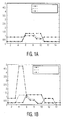
- the object being imaged (for example, the human body or a small animal) may move between the times when the two frames are acquired. If the frames are being averaged, this will reduce the spatial resolution of the resulting image. If the frames correspond to different laser wavelengths, then the motion will cause misregistration and possibly artifacts, as shown in Figure 1(b) .
- EP 1 561 424 discloses a non-invasive diagnostic imaging method and apparatus with light containing a specific wavelength and ultrasonic waves.
- Motion compensation algorithm for non-invasive two-dimensional temperature estimation using diagnostic pulse-echo ultrasound discloses an imaging system based on pulse-echo ultrasound scanning.
- a system associated with the present disclosure includes: (a) a means for generating photoacoustic signals; (b) at least a first transducer adapted to: (i) transmit ultrasound waves; (ii) receive ultrasound signals generated from the ultrasound waves; and (iii) receive photoacoustic signals generated from the photoacoustic signal means; (c) a motion estimator adapted to estimate motion based on the ultrasound signals; and (d) an image combiner adapted to receive and combine ultrasound data, photoacoustic data and motion estimator data generated from the received ultrasound and photoacoustic signals and the motion estimator and correct for motion to generate at least a photoacoustic image.
- the photoacoustic image may be corrected for motion by the image combiner using the motion estimator data.
- An exemplary image combiner is adapted to receive and combine ultrasound data, photoacoustic data and motion estimator data generated from the received ultrasound and photoacoustic signals and the motion estimator to generate a combined image.
- combined images are received by a frame buffer adapted: (i) to store combined image output generated from the image combiner, and (ii) transmit the combined image to a display means.
- the photoacoustic signal means is accomplished by an illumination system adapted to generate a photoacoustic signal within a sample.
- the illumination system is characterized by an energy beam, such as a laser.
- the display means can be any display system, typically used for medical imaging such as an LCD or a CRT.
- the frame buffer is adapted to transmit a series of combined images at a rate of about 55 Hz.
- the received ultrasound signals are beamformed by an ultrasound beamformer to create a series of radio frequency signals.
- a portion of the radio frequency signals are received by the motion estimator and the remaining portion are detected and pass through an ultrasound scan converter to generate an ultrasound image adapted to be received by the image combiner.
- all the radio frequency signals are received by the motion estimator when generating only PA images to correct for motion distortion in the PA imaging.
- the ultrasound signal pathway includes passing through a series of stages including filtering, detection and compounding.
- the motion estimator is adapted to generate a compensation signal to be received by the image combiner.
- An exemplary image combiner associated with the present disclosure is adapted to receive ultrasound image signals at a rate of about 55 Hz.
- the received photoacoustic signals pass through a photoacoustic scan converter adapted to generate a photoacoustic image to be received by the image combiner.
- An exemplary image combiner according to the present disclosure is adapted to receive photoacoustic image signals at a rate of about 10 Hz.
- the motion estimator is adapted to estimate motion of an object based on the portion of radio frequency signals.
- ultrasound signals can be generated in a pulsed Doppler mode.
- An exemplary image combiner is adapted to generate a sequence of combined frames containing data resulting from the received ultrasound signals and data resulting from the received photoacoustic signals.
- the sequence of combined frames is transmitted to a frame buffer.
- the image combiner should be adapted to perform spatial and temporal interpolation and resampling of the received photoacoustic signals.
- the image combiner is adapted to generate an output frame based on a combination of pixel values of at least one ultrasound image and at least one photoacoustic image.
- the display means is adapted to allow different images to be spatially registered, such that corresponding anatomical features in each image can be identified.
- the display means is adapted to display images in a displaying option selected from the group consisting of: displaying photoacoustic images only, displaying photoacoustic images and ultra sound images next to one another, overlaying a photoacoustic image on to an ultrasound image, using Doppler information to select which photoacoustic pixels are displayed, and combining Doppler and photoacoustic oxygenation information.
- the present disclosure describes a method of combining images of a sample including: (a) illuminating a sample with an illumination system adapted to generate photoacoustic signals; (b) transmitting ultrasound waves to the sample with an ultrasound transmitting means adapted to receive ultrasound signals and photoacoustic signals; (c) generating ultrasound images from a portion of the received ultrasound signals via an ultrasound imaging means; (d) generating a motion estimation from a remaining portion of the received ultrasound signals via a motion estimator; (e) generating photoacoustic images from the received photoacoustic signals via a photoacoustic imaging means; and (f) combining the ultrasound images, the motion estimation and the photoacoustic images in an image combiner adapted to transmit the combined image to a display means.
- the present disclosure relates to systems and methods that combine photoacoustic (PA) and ultrasound images.
- Such systems and methods are capable of generating images using PA or ultrasound image generating means.
- the acquisition of these images can be interleaved so that, from the point-of-view of the user, they appear to be acquired simultaneously.
- the two imaging modalities rely on different contrast mechanisms, and they will therefore generate different information.
- an ultrasound image shows boundaries between different tissues with different acoustic impedances
- a PA image shows absorption of laser energy at the associated optical wavelength used.
- An exemplary embodiment of a system associated with the present disclosure includes an image combiner that performs spatial and temporal interpolation of two images (PA and ultrasound) before generating a combined image.
- the combined image is then displayed on a display means such as a CRT and/or an LCD.
- the combined image can also be transmitted as data to a data storage or processing means such as a printer, a hard disk, a compact disk, and/or a flash drive.
- An exemplary image combiner according to the present disclosure is able to use motion estimates obtained from ultrasound data to enhance a photoacoustic image: increasing its apparent frame rate, registering consecutive frames in order to reduce artifacts.
- An exemplary combined system may be capable of generating combined ultrasound and PA images that are registered spatially and temporally.
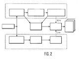
- FIG. 2 a schematic of an exemplary image combiner for a combined PA and ultrasound imaging system is shown.
- An exemplary system according to the present disclosure is used in conjunction with an ultrasound signal pathway, a PA signal pathway, a motion estimator, a frame buffer and a display.
- Ultrasound images are typically formed using a transducer, ultrasound beamformer and an ultrasound scan converter. These components may be the same as those typically found in an existing modem ultrasound machine.
- Ultrasound energy is transmitted in a series of focused beams using an ultrasound beamformer and a transducer. The energy received by the transducer is then beamformed to create a corresponding series of radio frequency (RF) signals, known as A-lines. These signals are detected and then scan converted to form a B-mode ultrasound image.
- RF radio frequency
- PA signals are generated by illuminating an object with a short laser pulse.
- the signals are received by a transducer that is common to PA and ultrasound signal pathways.
- the signals pass through a PA beamformer, which allows them to be spatially localized.
- the PA scan converter is then used to resample the signals and produce a PA image.
- a single transducer is deployed to transmit ultrasound waves, receive ultrasound signals generated by the transmitted ultrasound waves and receive PA signals.
- systems associated with the present disclosure also include embodiments having multiple transducers.
- a first transducer transmits ultrasound waves and a second transducer receives ultrasound signals generated by the transmitted ultrasound waves and receives the PA signals.
- a motion estimator is used to estimate the motion of the object using the ultrasound data. To accomplish motion estimation, it is necessary to compare the signals received at different times from the same image location. This can be done using the RF A-line signals before they are detected and scan converted. Accordingly, it is possible to estimate the axial motion very accurately.
- Such motion estimation methods have previously been used in elastography. (See, e.g., E. E. Konofagou, T. Harrigan, and J. Ophir, "Shear strain estimation and lesion mobility assessment in elastography,” Ultrasonics, Vol. 38, No. 1-8, pp. 400-4, 2000 ; J. Ophir, S. K. Alam, B. Garra, F. Kallel, E. Konofagou, T.
- the motion estimator receives signals from the ultrasound beamformer and the ultrasound images to generate motion estimation.
- a further alternative estimation method includes using Doppler information derived from specific Doppler sequences. (See, e.g., D. A. Christensen, Ultrasonic Bioinstrumentation: John Wiley & Sons, 1988 .) It is understood that ultrasound image generators do not need to contain contrasting structures in order to perform motion estimation. The motion estimation can be performed on regions of uniform speckle texture.
- Motion information extracted from the ultrasound signals may be displayed on the ultrasound image.
- Commercial ultrasound scanners typically include continuous wave Doppler, color Doppler, power Doppler and pulsed Doppler modes. Some also feature elastography, Doppler strain rate imaging and tissue Doppler imaging.
- An exemplary image combiner may perform at least the following two functions:
- the output of the image combiner is typically a sequence of combined frames containing both ultrasound and PA information. This information is typically stored in a frame buffer.
- the frame buffer includes a plurality of frames adapted to be sequentially displayed on a display means.
- Exemplary display means include but are not limited to a CRT, LCD, or any other type of information display system.
- Systems according to the present disclosure are adapted to allow for images acquired by ultrasound and PA modalities to be displayed on a single displaying means. Moreover, exemplary systems allow these images to be displayed at the same rate, even though they are acquired at different rates. Exemplary systems allow for different images to be spatially registered, such that corresponding anatomical features in the two images can be identified. Further aspects associated with the present disclosure include allowing for different PA images to be registered before being combined, thus reducing misregistration artifacts.
- a combined imaging system according to the present disclosure can be adapted to accomplish several displaying options including but not limited to:
- Examples of possible uses for an exemplary system associated with the present disclosure include but are not limited to medical imaging of humans, or small animals.
- An exemplary imaging system can be added to an existing modem ultrasound machine, such as, for example, a Philips iU22 or iE33.
Landscapes
- Health & Medical Sciences (AREA)
- Life Sciences & Earth Sciences (AREA)
- Engineering & Computer Science (AREA)
- Physics & Mathematics (AREA)
- General Health & Medical Sciences (AREA)
- Pathology (AREA)
- Veterinary Medicine (AREA)
- Public Health (AREA)
- Biomedical Technology (AREA)
- Heart & Thoracic Surgery (AREA)
- Medical Informatics (AREA)
- Molecular Biology (AREA)
- Surgery (AREA)
- Animal Behavior & Ethology (AREA)
- Biophysics (AREA)
- Nuclear Medicine, Radiotherapy & Molecular Imaging (AREA)
- Radiology & Medical Imaging (AREA)
- Remote Sensing (AREA)
- Radar, Positioning & Navigation (AREA)
- General Physics & Mathematics (AREA)
- Computer Vision & Pattern Recognition (AREA)
- Acoustics & Sound (AREA)
- Computer Networks & Wireless Communication (AREA)
- Signal Processing (AREA)
- Analytical Chemistry (AREA)
- Immunology (AREA)
- Biochemistry (AREA)
- Chemical & Material Sciences (AREA)
- Artificial Intelligence (AREA)
- Physiology (AREA)
- Psychiatry (AREA)
- Ultra Sonic Daignosis Equipment (AREA)
- Investigating Or Analyzing Materials By The Use Of Ultrasonic Waves (AREA)
- Investigating Or Analysing Materials By Optical Means (AREA)
Claims (15)
- Kombiniertes Bildgebungssystem, das Folgendes umfasst:(a) Mittel zum Erzeugen von photoakustischen Signalen;(b) mindestens einen ersten Wandler, der vorgesehen ist, um (i) Ultraschallwellen auszusenden; (ii) von den Ultraschallwellen erzeugte Ultraschallsignale zu empfangen; und (iii) von den photoakustischen Signalmitteln erzeugte photoakustische Signale zu empfangen;
dadurch gekennzeichnet, dass
das System weiterhin Folgendes umfasst:(c) eine Bewegungsschätzeinheit, die vorgesehen ist, um Bewegung basierend auf Ultraschallsignalen zu schätzen; und(d) eine Bildkombiniereinheit, die vorgesehen ist, um Ultraschalldaten, photoakustische Daten und Bewegungsschätzeinheitdaten zu empfangen und zu kombinieren, die von den empfangenen Ultraschallsignalen und photoakustischen Signalen und der Bewegungsschätzeinheit erzeugt wurden und bezüglich Bewegung zu korrigieren, um ein kombiniertes Signal zu erzeugen. - System nach Anspruch 1, wobei das kombinierte Signal durch einen Einzelbildpuffer empfangen wird, der vorgesehen ist, um (i) von der Bildkombiniereinheit erzeugte kombinierte Bildausgabe zu speichern und (ii) das kombinierte Bild an ein Anzeigemittel und/oder Datenspeichermittel zu senden.
- System nach Anspruch 1, wobei das photoakustische Signalmittel durch ein Beleuchtungssystem geschaffen wird, das vorgesehen ist, um ein photoakustisches Signal innerhalb einer Probe zu erzeugen.
- System nach Anspruch 3, wobei das Beleuchtungssystem durch ein Energiestrahlenbündel gekennzeichnet ist.
- System nach Anspruch 2, wobei das Anzeigemittel ausgewählt wird aus der Gruppe bestehend aus einer Flüssigkristallanzeige (LCD) und einer Kathodenstrahlröhre (CRT) und die Datenspeichermittel ausgewählt werden aus der Gruppe bestehend aus einem Drucker, einer Festplatte, einer Compact Disc und einem Flash-Laufwerk.
- System nach Anspruch 2, wobei der Einzelbildpuffer vorgesehen ist, um eine Reihe von kombinierten Bildern mit einer Rate von ca. 55 Hz zu senden.
- System nach Anspruch 3, wobei die empfangenen Ultraschallsignale durch einen Ultraschall-Strahlformer strahlgeformt werden, um eine Reihe von Hochfrequenzsignalen zu erzeugen.
- System nach Anspruch 3, wobei die empfangenen photoakustischen Signale durch einen photoakustischen Abtastumsetzer geleitet werden, der vorgesehen ist, um ein durch die Bildkombiniereinheit zu empfangendes photoakustisches Bild zu erzeugen.
- System nach Anspruch 1, wobei die Ultraschallsignale in einem gepulsten Dopplermodus erzeugt werden.
- System nach Anspruch 2, wobei die Bildkombiniereinheit vorgesehen ist, um eine an den Einzelbildpuffer zu sendende Folge von kombinierten Einzelbildern zu erzeugen, welche aus den empfangenen Ultraschallsignalen resultierende Daten und aus den empfangenen photoakustischen Signalen resultierende Daten enthalten.
- System nach Anspruch 1, wobei die Bildkombiniereinheit vorgesehen ist, um räumliche und/oder zeitliche Interpolation durchzuführen und die empfangenen photoakustischen Signale erneut abzutasten.
- System nach Anspruch 2, wobei das Anzeigemittel vorgesehen ist, um die räumliche Registrierung von verschiedenen Bildern zu ermöglichen, so dass entsprechende anatomische Merkmale in jedem Bild identifiziert werden können.
- System nach Anspruch 2, wobei das Anzeigemittel vorgesehen ist, um Bilder in einer Anzeigeoption anzuzeigen, die ausgewählt wird aus der Gruppe bestehend aus: Anzeigen von nur photoakustischen Bildern, Anzeigen von photoakustischen Bildern und Ultraschallbildern nebeneinander, Überlagern eines Ultraschallbildes durch ein photoakustischen Bild, Nutzen von Dopplerinformationen, um auszuwählen, welche photoakustischen Pixel angezeigt werden, und Kombinieren von Doppler- und photoakustischen Oxygenationsinformationen.
- Verfahren zum Kombinieren von Bildern einer Probe, das Folgendes umfasst:(a) Beleuchten einer Probe mit einem Beleuchtungssystem, das vorgesehen ist, um photoakustische Signale zu erzeugen;(b) Senden von Ultraschallwellen an die Probe mit einem Ultraschallsendemittel, das vorgesehen ist, um Ultraschallsignale und photoakustische Signale zu empfangen;(c) Erzeugen von mindestens einem Ultraschallbild anhand von einen Teil der empfangenen Ultraschallsignale über ein Ultraschallbildgebungsmittel;(d) Erzeugen einer Bewegungsschätzung anhand von einem verbleibenden Teil der empfangenen Ultraschallsignale über eine Bewegungsschätzeinheit;(e) Erzeugen von mindestens einem photoakustischen Bild anhand der empfangenen photoakustischen Signale über ein photoakustisches Bildgebungsmittel; und(f) Kombinieren des Ultraschallbildes, der Bewegungsschätzung und des photoakustischen Bildes und Korrigieren bezüglich Bewegung in einer Bildkombiniereinheit, die vorgesehen ist, um das kombinierte Bild an ein Anzeigemittel zu senden.
- Kombiniertes Bildgebungssystem, das Folgendes umfasst:(a) Mittel zum Erzeugen von photoakustischen Signalen;(b) mindestens einen ersten Wandler, der vorgesehen ist, um Ultraschallwellen zu senden;(c) mindestens einen zweiten Wandler, der vorgesehen ist, um (i) von den Ultraschallwellen erzeugte Ultraschallsignale zu empfangen; und (ii) von den photoakustischen Signalmitteln erzeugte photoakustische Signale zu empfangen;(d) eine Bewegungsschätzeinheit, die vorgesehen ist, um Bewegung basierend auf Ultraschallsignalen zu schätzen; und(e) eine Bildkombiniereinheit, die vorgesehen ist, um Ultraschalldaten, photoakustische Daten und Bewegungsschätzeinheitdaten zu empfangen und zu kombinieren, die von den empfangenen Ultraschallsignalen und photoakustischen Signalen und der Bewegungsschätzeinheit erzeugt wurden und bezüglich Bewegung zu korrigieren, um ein kombiniertes Signal zu erzeugen.
Applications Claiming Priority (2)
| Application Number | Priority Date | Filing Date | Title |
|---|---|---|---|
| US87071306P | 2006-12-19 | 2006-12-19 | |
| PCT/IB2007/055231 WO2008075299A1 (en) | 2006-12-19 | 2007-12-19 | Combined photoacoustic and ultrasound imaging system |
Publications (2)
| Publication Number | Publication Date |
|---|---|
| EP2097010A1 EP2097010A1 (de) | 2009-09-09 |
| EP2097010B1 true EP2097010B1 (de) | 2011-10-05 |
Family
ID=39323807
Family Applications (1)
| Application Number | Title | Priority Date | Filing Date |
|---|---|---|---|
| EP07859456A Not-in-force EP2097010B1 (de) | 2006-12-19 | 2007-12-19 | Kombiniertes photoakustisches und ultraschall-darstellungssystem |
Country Status (8)
| Country | Link |
|---|---|
| US (2) | US9561017B2 (de) |
| EP (1) | EP2097010B1 (de) |
| JP (1) | JP5506395B2 (de) |
| KR (1) | KR20090088909A (de) |
| CN (1) | CN101563035B (de) |
| AT (1) | ATE526882T1 (de) |
| RU (1) | RU2480147C2 (de) |
| WO (1) | WO2008075299A1 (de) |
Families Citing this family (106)
| Publication number | Priority date | Publication date | Assignee | Title |
|---|---|---|---|---|
| WO2007084981A2 (en) * | 2006-01-19 | 2007-07-26 | The Regents Of The University Of Michigan | System and method for photoacoustic imaging and monitoring of laser therapy |
| US7750536B2 (en) | 2006-03-02 | 2010-07-06 | Visualsonics Inc. | High frequency ultrasonic transducer and matching layer comprising cyanoacrylate |
| WO2008100386A2 (en) * | 2007-02-09 | 2008-08-21 | Board Of Regents, The University Of Texas System | Intravascular photoacoustic and ultrasound echo imaging |
| US20080221647A1 (en) * | 2007-02-23 | 2008-09-11 | The Regents Of The University Of Michigan | System and method for monitoring photodynamic therapy |
| JP5460000B2 (ja) | 2008-08-20 | 2014-04-02 | キヤノン株式会社 | イメージング装置およびイメージング方法 |
| JP5241465B2 (ja) * | 2008-12-11 | 2013-07-17 | キヤノン株式会社 | 光音響イメージング装置および光音響イメージング方法 |
| JP5518096B2 (ja) * | 2009-12-17 | 2014-06-11 | キヤノン株式会社 | 測定システム、画像形成方法及びプログラム |
| JP5818444B2 (ja) * | 2010-02-04 | 2015-11-18 | キヤノン株式会社 | 機能情報取得装置、機能情報取得方法、及びプログラム |
| US8904871B2 (en) * | 2010-07-23 | 2014-12-09 | Board Of Regents, The University Of Texas System | Temperature dependent photoacoustic imaging |
| US9289191B2 (en) | 2011-10-12 | 2016-03-22 | Seno Medical Instruments, Inc. | System and method for acquiring optoacoustic data and producing parametric maps thereof |
| US8686335B2 (en) | 2011-12-31 | 2014-04-01 | Seno Medical Instruments, Inc. | System and method for adjusting the light output of an optoacoustic imaging system |
| US20130338501A1 (en) * | 2012-06-13 | 2013-12-19 | Seno Medical Instruments, Inc. | System and method for storing data associated with the operation of a dual modality optoacoustic/ultrasound system |
| US8839672B2 (en) * | 2010-10-19 | 2014-09-23 | Board Of Regents, The University Of Texas System | Combined ultrasound and photoacoustic imaging of metal objects |
| JP6010306B2 (ja) * | 2011-03-10 | 2016-10-19 | 富士フイルム株式会社 | 光音響計測装置 |
| JP5796896B2 (ja) * | 2011-03-10 | 2015-10-21 | 富士フイルム株式会社 | 断層画像生成装置及び方法 |
| JP2012196308A (ja) * | 2011-03-22 | 2012-10-18 | Fujifilm Corp | 光音響画像生成装置及び方法 |
| JP2016209725A (ja) * | 2011-05-12 | 2016-12-15 | キヤノン株式会社 | 被検体情報取得装置及び被検体情報取得方法 |
| JP5864905B2 (ja) * | 2011-05-20 | 2016-02-17 | キヤノン株式会社 | 被検体情報取得装置及び被検体情報取得方法 |
| JP5719242B2 (ja) * | 2011-06-27 | 2015-05-13 | 富士フイルム株式会社 | ドプラ画像表示方法および装置 |
| JP5599761B2 (ja) * | 2011-07-14 | 2014-10-01 | 富士フイルム株式会社 | 光音響画像生成装置及び方法 |
| JP5694991B2 (ja) * | 2011-07-14 | 2015-04-01 | 富士フイルム株式会社 | 光音響画像化方法および装置 |
| JP5713968B2 (ja) * | 2011-07-29 | 2015-05-07 | 富士フイルム株式会社 | 光音響画像生成装置及び音響波ユニット |
| JP5885437B2 (ja) * | 2011-09-07 | 2016-03-15 | キヤノン株式会社 | 光音響装置及び処理方法 |
| US9733119B2 (en) | 2011-11-02 | 2017-08-15 | Seno Medical Instruments, Inc. | Optoacoustic component utilization tracking |
| US9445785B2 (en) * | 2011-11-02 | 2016-09-20 | Seno Medical Instruments, Inc. | System and method for normalizing range in an optoacoustic imaging system |
| US9445786B2 (en) | 2011-11-02 | 2016-09-20 | Seno Medical Instruments, Inc. | Interframe energy normalization in an optoacoustic imaging system |
| US20140005544A1 (en) * | 2011-11-02 | 2014-01-02 | Seno Medical Instruments, Inc. | System and method for providing selective channel sensitivity in an optoacoustic imaging system |
| US11191435B2 (en) | 2013-01-22 | 2021-12-07 | Seno Medical Instruments, Inc. | Probe with optoacoustic isolator |
| US20130116538A1 (en) | 2011-11-02 | 2013-05-09 | Seno Medical Instruments, Inc. | Optoacoustic imaging systems and methods with enhanced safety |
| US11287309B2 (en) | 2011-11-02 | 2022-03-29 | Seno Medical Instruments, Inc. | Optoacoustic component utilization tracking |
| US9757092B2 (en) * | 2011-11-02 | 2017-09-12 | Seno Medical Instruments, Inc. | Method for dual modality optoacoustic imaging |
| US9743839B2 (en) * | 2011-11-02 | 2017-08-29 | Seno Medical Instruments, Inc. | Playback mode in an optoacoustic imaging system |
| US20130338475A1 (en) | 2012-06-13 | 2013-12-19 | Seno Medical Instruments, Inc. | Optoacoustic imaging system with fiber optic cable |
| US9814394B2 (en) | 2011-11-02 | 2017-11-14 | Seno Medical Instruments, Inc. | Noise suppression in an optoacoustic system |
| US20130289381A1 (en) | 2011-11-02 | 2013-10-31 | Seno Medical Instruments, Inc. | Dual modality imaging system for coregistered functional and anatomical mapping |
| US10433732B2 (en) | 2011-11-02 | 2019-10-08 | Seno Medical Instruments, Inc. | Optoacoustic imaging system having handheld probe utilizing optically reflective material |
| US9730587B2 (en) | 2011-11-02 | 2017-08-15 | Seno Medical Instruments, Inc. | Diagnostic simulator |
| KR101273585B1 (ko) | 2011-12-05 | 2013-06-11 | 삼성전자주식회사 | 초음파 영상 장치 및 초음파 영상 표시방법 |
| CN104023635A (zh) * | 2011-12-30 | 2014-09-03 | 皇家飞利浦有限公司 | 用于在us成像中使用pa效应进行针导航的系统和方法 |
| US11160456B2 (en) * | 2012-01-23 | 2021-11-02 | Tomowave Laboratories, Inc. | Laser optoacoustic ultrasonic imaging system (LOUIS) and methods of use |
| JP5881582B2 (ja) * | 2012-02-07 | 2016-03-09 | 富士フイルム株式会社 | 超音波探触子の製造方法 |
| JP6132466B2 (ja) * | 2012-02-07 | 2017-05-24 | キヤノン株式会社 | 被検体情報取得装置及び被検体情報取得方法 |
| EP2822471B1 (de) * | 2012-03-09 | 2023-03-08 | Seno Medical Instruments, Inc. | Statistisches mapping in einem optoakustischen bildgebungssystem |
| WO2013134772A2 (en) * | 2012-03-09 | 2013-09-12 | Seno Medical Instruments, Inc. | Statistical mapping in an optoacoustic imaging system |
| US10762628B2 (en) * | 2012-03-09 | 2020-09-01 | Seno Medical Instruments, Inc. | Statistical mapping in an optoacoustic imaging system |
| KR101298935B1 (ko) * | 2012-04-13 | 2013-08-23 | 서강대학교산학협력단 | 초음파 영상 및 광음향 영상 생성 방법 및 장치 |
| JP2013226335A (ja) * | 2012-04-27 | 2013-11-07 | Fujifilm Corp | 音響波診断装置および画像表示方法 |
| KR101974580B1 (ko) * | 2012-05-03 | 2019-05-02 | 삼성전자주식회사 | 레이저 유도 초음파 장치 및 이를 이용한 영상 생성 방법 |
| US9610043B2 (en) | 2012-06-13 | 2017-04-04 | Seno Medical Instruments, Inc. | System and method for producing parametric maps of optoacoustic data |
| WO2013188708A1 (en) * | 2012-06-13 | 2013-12-19 | Seno Medical Instruments, Inc. | System and method for storing data associated with the operation of a dual modality optoacoustic / ultrasound system |
| WO2014056134A1 (zh) * | 2012-10-08 | 2014-04-17 | 财团法人工业技术研究院 | 结合超音波与光声影像的成像方法与成像装置 |
| JP6292836B2 (ja) * | 2012-12-28 | 2018-03-14 | キヤノン株式会社 | 被検体情報取得装置、表示方法、プログラム、処理装置 |
| JP6103931B2 (ja) * | 2012-12-28 | 2017-03-29 | キヤノン株式会社 | 被検体情報取得装置、被検体情報取得方法 |
| JP6112861B2 (ja) * | 2012-12-28 | 2017-04-12 | キヤノン株式会社 | 被検体情報取得装置、信号処理装置および表示装置 |
| WO2014115214A1 (en) * | 2012-12-28 | 2014-07-31 | Canon Kabushiki Kaisha | Combined photoacoustic and ultrasound imaging apparatus and method |
| JP5936559B2 (ja) * | 2013-01-18 | 2016-06-22 | 富士フイルム株式会社 | 光音響画像生成装置および光音響画像生成方法 |
| JP6177530B2 (ja) * | 2013-01-18 | 2017-08-09 | 富士フイルム株式会社 | ドプラ計測装置およびドプラ計測方法 |
| AU2014237034A1 (en) | 2013-03-15 | 2015-09-24 | Seno Medical Instruments, Inc. | System and method for diagnostic vector classification support |
| JP6238539B2 (ja) * | 2013-03-21 | 2017-11-29 | キヤノン株式会社 | 処理装置、被検体情報取得装置、および、処理方法 |
| US10143381B2 (en) * | 2013-04-19 | 2018-12-04 | Canon Kabushiki Kaisha | Object information acquiring apparatus and control method therefor |
| KR101418405B1 (ko) * | 2013-04-25 | 2014-07-09 | 포항공과대학교 산학협력단 | 고배율 표면 이미지에 광음향 단층 이미지를 포함하는 증강 현실 이미지를 중첩시켜 출력하는 수술용 현미경 시스템 및 그를 위한 증강 현실 이미지 제공 장치 |
| US20140373599A1 (en) * | 2013-06-25 | 2014-12-25 | Texas Instruments Incorporated | Detection and locking to the absorption spectra of gasses using quartz enhanced photoacoustic sprectroscopy |
| KR101533591B1 (ko) * | 2013-07-08 | 2015-07-06 | 삼성메디슨 주식회사 | 의료 영상 장치 및 의료 영상 제공 방법 |
| CN103385758B (zh) * | 2013-07-22 | 2015-12-09 | 深圳先进技术研究院 | 一种血管内光声超声双模成像系统及其成像方法 |
| EP3015068A4 (de) * | 2013-08-01 | 2017-06-21 | Sogang University Research Foundation | Vorrichtung und verfahren zur erfassung eines fusionsbildes |
| JP2015073577A (ja) * | 2013-10-04 | 2015-04-20 | キヤノン株式会社 | 光音響装置、光音響装置の作動方法、およびプログラム |
| EP3054853A4 (de) | 2013-10-11 | 2017-05-17 | Seno Medical Instruments, Inc. | Systeme und verfahren zur trennung von komponenten bei der medizinischen bildgebung |
| US10258241B2 (en) | 2014-02-27 | 2019-04-16 | Seno Medical Instruments, Inc. | Probe adapted to control blood flow through vessels during imaging and method of use of same |
| US10265047B2 (en) | 2014-03-12 | 2019-04-23 | Fujifilm Sonosite, Inc. | High frequency ultrasound transducer having an ultrasonic lens with integral central matching layer |
| JP6498036B2 (ja) * | 2014-06-13 | 2019-04-10 | キヤノン株式会社 | 光音響装置、信号処理方法、及びプログラム |
| JP6371847B2 (ja) * | 2014-06-30 | 2018-08-08 | 富士フイルム株式会社 | 光音響画像生成装置、信号処理装置、及び光音響画像生成方法 |
| KR101620458B1 (ko) | 2014-07-04 | 2016-05-24 | 포항공과대학교 산학협력단 | 광음향 촬영 장치 및 이를 이용한 산소포화도 측정방법 |
| EP3212065B1 (de) | 2014-10-30 | 2022-03-30 | Seno Medical Instruments, Inc. | System zur optoakustischen bildgebung mit detektion der relativen ausrichtung einer lichtquelle und eines akustischen empfängers unter verwendung von akustischen wellen |
| JP6436442B2 (ja) | 2015-04-10 | 2018-12-12 | キヤノン株式会社 | 光音響装置および画像処理方法 |
| CN104771192A (zh) | 2015-04-20 | 2015-07-15 | 无锡海斯凯尔医学技术有限公司 | 组织形态和弹性信息的处理方法和弹性检测设备 |
| KR101638588B1 (ko) | 2015-04-23 | 2016-07-12 | 포항공과대학교 산학협력단 | 소화기관의 비침습적 이미징 장치 |
| EP3090682A1 (de) * | 2015-05-08 | 2016-11-09 | Universiteit Twente | Reduktion von artefakten in vorrichtungen für photoakustische und thermoakustische abbildung |
| CN105232004A (zh) * | 2015-11-16 | 2016-01-13 | 华南师范大学 | 一种精准测量黑色素瘤厚度的光声超声联合成像装置和成像方法 |
| CN105395170B (zh) * | 2015-12-15 | 2018-07-27 | 同济大学 | 一种光声超声双模态同步成像系统 |
| KR102576682B1 (ko) * | 2016-02-05 | 2023-09-07 | 전북대학교산학협력단 | 혈전 탐지 시스템 및 방법 |
| JP6475369B2 (ja) | 2016-02-08 | 2019-02-27 | 富士フイルム株式会社 | 音響波画像生成装置および音響波画像生成方法 |
| JP2017164198A (ja) * | 2016-03-15 | 2017-09-21 | キヤノン株式会社 | 情報処理システムおよび表示制御方法 |
| JP2017192569A (ja) * | 2016-04-20 | 2017-10-26 | キヤノン株式会社 | 被検体情報取得装置およびその制御方法 |
| US12036000B2 (en) * | 2016-05-27 | 2024-07-16 | The Regents Of The University Of Michigan | Method of photoacoustic imaging intraocular tumors including back-projection reconstruction to measure heterogeneity |
| CN107582096A (zh) * | 2016-07-08 | 2018-01-16 | 佳能株式会社 | 用于获取信息的装置、方法和存储介质 |
| WO2018008439A1 (en) * | 2016-07-08 | 2018-01-11 | Canon Kabushiki Kaisha | Apparatus, method and program for displaying ultrasound image and photoacoustic image |
| US20180055369A1 (en) * | 2016-08-31 | 2018-03-01 | Qualcomm Incorporated | Layered sensing including rf-acoustic imaging |
| JP7129158B2 (ja) * | 2016-11-24 | 2022-09-01 | キヤノン株式会社 | 情報処理装置、情報処理方法、情報処理システムおよびプログラム |
| JP6443866B2 (ja) * | 2017-03-16 | 2018-12-26 | キヤノン株式会社 | 被検体情報取得装置および被検体情報取得方法 |
| JP6513121B2 (ja) * | 2017-04-19 | 2019-05-15 | キヤノン株式会社 | 処理装置、被検体情報取得装置、光音響画像の表示方法、及びプログラム |
| US11596313B2 (en) | 2017-10-13 | 2023-03-07 | Arizona Board Of Regents On Behalf Of Arizona State University | Photoacoustic targeting with micropipette electrodes |
| US12161295B2 (en) | 2018-03-16 | 2024-12-10 | Arizona Board Of Regents On Behalf Of Arizona State University | Deep brain stimulation electrode with photoacoustic and ultrasound imaging capabilities |
| CN108888236A (zh) * | 2018-04-23 | 2018-11-27 | 深圳迈瑞生物医疗电子股份有限公司 | 一种多模态成像系统及方法 |
| WO2020082270A1 (zh) * | 2018-10-24 | 2020-04-30 | 中国医学科学院北京协和医院 | 一种成像方法以及成像系统 |
| JP2019136520A (ja) * | 2019-04-10 | 2019-08-22 | キヤノン株式会社 | 処理装置、光音響画像の表示方法、及びプログラム |
| US11768182B2 (en) | 2019-04-26 | 2023-09-26 | Arizona Board Of Regents On Behalf Of Arizona State University | Photoacoustic and optical microscopy combiner and method of generating a photoacoustic image of a sample |
| US11975327B2 (en) | 2019-06-19 | 2024-05-07 | Arizona Board Of Regents On Behalf Of Arizona State University | Integrated container adapter for photoacoustic microscopy |
| US11683829B2 (en) * | 2021-05-31 | 2023-06-20 | Clarius Mobile Health Corp. | Systems and methods for improving quality of service when transmitting ultrasound image data over a wireless connection |
| KR102636714B1 (ko) * | 2021-08-10 | 2024-02-14 | 주식회사 옵티코 | 광음향 및 초음파 이미징 장치 및 이미지 형성 방법 |
| KR102698306B1 (ko) * | 2022-01-14 | 2024-08-23 | 주식회사 옵티코 | 다파장 광음향 영상 및 초음파 영상을 이용한 종양 분류 방법 및 분석장치 |
| KR102758410B1 (ko) | 2022-05-30 | 2025-01-23 | 부산대학교 산학협력단 | 초음파 기반 호흡 보정을 이용한 3차원 광음향 영상 구현을 위한 장치 및 방법 |
| WO2023235332A1 (en) * | 2022-05-31 | 2023-12-07 | The Regents Of The University Of California | System and method of dual-mode eat/us-guided electroporation |
| KR102808984B1 (ko) * | 2022-11-14 | 2025-05-15 | 국립부경대학교 산학협력단 | 광음향 영상 및 초음파 영상 획득장치 및 그 방법 |
| KR102921272B1 (ko) * | 2022-11-14 | 2026-01-30 | 국립부경대학교 산학협력단 | 광음향 영상 및 초음파 영상의 합성영상 입력장치 및 그 방법 |
| CN116942200B (zh) * | 2023-09-20 | 2024-02-06 | 杭州励影光电成像有限责任公司 | 一种非复用式超声多模态成像系统及方法 |
| WO2026060502A1 (pt) * | 2024-09-23 | 2026-03-26 | Klautau Leite Ana Paula | Sistema de diagnóstico por imagem da endometriose profunda e/ou adenomiose e método de análise de exames de imagem |
Family Cites Families (16)
| Publication number | Priority date | Publication date | Assignee | Title |
|---|---|---|---|---|
| US1561424A (en) * | 1924-11-06 | 1925-11-10 | Exel George | Helicopter |
| US7028899B2 (en) * | 1999-06-07 | 2006-04-18 | Metrologic Instruments, Inc. | Method of speckle-noise pattern reduction and apparatus therefore based on reducing the temporal-coherence of the planar laser illumination beam before it illuminates the target object by applying temporal phase modulation techniques during the transmission of the plib towards the target |
| US5977538A (en) * | 1998-05-11 | 1999-11-02 | Imarx Pharmaceutical Corp. | Optoacoustic imaging system |
| RU2153366C1 (ru) * | 1999-01-05 | 2000-07-27 | Жаров Владимир Павлович | Устройство для комплексного лечения заболеваний предстательной железы |
| AU6894500A (en) * | 1999-08-06 | 2001-03-05 | Board Of Regents, The University Of Texas System | Optoacoustic monitoring of blood oxygenation |
| GR1004180B (el) * | 2000-03-28 | 2003-03-11 | ����������� ����� ��������� (����) | Μεθοδος και συστημα χαρακτηρισμου και χαρτογραφησης αλλοιωσεων των ιστων |
| US20050085725A1 (en) * | 2001-08-09 | 2005-04-21 | Ron Nagar | Photoacoustic assay and imaging system |
| DE10159721B4 (de) * | 2001-12-05 | 2004-07-22 | Bruker Optik Gmbh | Digitales FTIR-Spektrometer |
| US6638228B1 (en) * | 2002-04-26 | 2003-10-28 | Koninklijke Philips Electronics N.V. | Contrast-agent enhanced color-flow imaging |
| US7245789B2 (en) * | 2002-10-07 | 2007-07-17 | Vascular Imaging Corporation | Systems and methods for minimally-invasive optical-acoustic imaging |
| JP4406226B2 (ja) | 2003-07-02 | 2010-01-27 | 株式会社東芝 | 生体情報映像装置 |
| JP4643153B2 (ja) * | 2004-02-06 | 2011-03-02 | 株式会社東芝 | 非侵襲生体情報映像装置 |
| DE102005017492B4 (de) | 2005-04-15 | 2007-04-19 | Siemens Ag | Verfahren zum rechnerischen Kompensieren einer periodischen Bewegung eines Organs sowie Bildaufnahmesystem |
| EP2754388B1 (de) * | 2013-01-15 | 2020-09-09 | Helmholtz Zentrum München Deutsches Forschungszentrum für Gesundheit und Umwelt GmbH | System und Verfahren zur optoakustischen Abbildung eines Objekts mit verbesserter Qualität und hoher Rate |
| US10143381B2 (en) * | 2013-04-19 | 2018-12-04 | Canon Kabushiki Kaisha | Object information acquiring apparatus and control method therefor |
| JP6498036B2 (ja) * | 2014-06-13 | 2019-04-10 | キヤノン株式会社 | 光音響装置、信号処理方法、及びプログラム |
-
2007
- 2007-12-19 KR KR1020097012565A patent/KR20090088909A/ko not_active Ceased
- 2007-12-19 AT AT07859456T patent/ATE526882T1/de not_active IP Right Cessation
- 2007-12-19 CN CN2007800474063A patent/CN101563035B/zh not_active Expired - Fee Related
- 2007-12-19 JP JP2009542368A patent/JP5506395B2/ja not_active Expired - Fee Related
- 2007-12-19 US US12/520,157 patent/US9561017B2/en not_active Expired - Fee Related
- 2007-12-19 RU RU2009127791/14A patent/RU2480147C2/ru not_active IP Right Cessation
- 2007-12-19 EP EP07859456A patent/EP2097010B1/de not_active Not-in-force
- 2007-12-19 WO PCT/IB2007/055231 patent/WO2008075299A1/en not_active Ceased
-
2017
- 2017-01-09 US US15/401,245 patent/US10631830B2/en active Active
Also Published As
| Publication number | Publication date |
|---|---|
| US9561017B2 (en) | 2017-02-07 |
| JP2010512929A (ja) | 2010-04-30 |
| CN101563035A (zh) | 2009-10-21 |
| US20100049044A1 (en) | 2010-02-25 |
| RU2480147C2 (ru) | 2013-04-27 |
| US20170112474A1 (en) | 2017-04-27 |
| CN101563035B (zh) | 2012-08-08 |
| JP5506395B2 (ja) | 2014-05-28 |
| EP2097010A1 (de) | 2009-09-09 |
| RU2009127791A (ru) | 2011-01-27 |
| KR20090088909A (ko) | 2009-08-20 |
| US10631830B2 (en) | 2020-04-28 |
| WO2008075299A1 (en) | 2008-06-26 |
| ATE526882T1 (de) | 2011-10-15 |
Similar Documents
| Publication | Publication Date | Title |
|---|---|---|
| EP2097010B1 (de) | Kombiniertes photoakustisches und ultraschall-darstellungssystem | |
| Jensen et al. | Ultrasound vector flow imaging—Part II: Parallel systems | |
| EP2030570B1 (de) | Bildverarbeitungsvorrichtung | |
| US12310795B2 (en) | Ultrasound controller unit and method | |
| US9332962B2 (en) | Ultrasound ARFI displacement imaging using an adaptive time instance | |
| US9883852B2 (en) | Ultrasound systems, methods and computer program products for estimating tissue deformation with harmonic signals | |
| EP2082688A1 (de) | Synchronisiertes Kombinieren für mit Kontrastmittel verstärkte Ultraschallbildgebung zur medizinischen Diagnose | |
| WO2008108922A1 (en) | Inter-frame processing for contrast agent enhanced medical diagnostic ultrasound imaging | |
| Ivancevich et al. | Real-time 3-D contrast-enhanced transcranial ultrasound and aberration correction | |
| US8668648B2 (en) | Contrast agent destruction effectiveness determination for medical diagnostic ultrasound imaging | |
| US8926516B2 (en) | Ultrasound imaging apparatus and method of ultrasound imaging | |
| US20240111046A1 (en) | Method and system for flow processing on channel data for application of nonlinear beamforming | |
| JP5457062B2 (ja) | 弾性映像を形成する超音波システム及び方法 | |
| Li et al. | High sensitivity liver vasculature visualization using a real-time coherent flow power Doppler (CFPD) imaging system: A pilot clinical study | |
| JP4875397B2 (ja) | 超音波診断装置 | |
| KR20100119480A (ko) | 고유벡터를 설정하는 초음파 시스템 및 방법 | |
| Obruchkov | The technology and performance of 4D ultrasound | |
| Huber | Temporal SNR Guided Adaptive B-mode and Color Doppler Transmit Parameter Selection | |
| Trahey et al. | 2J-6 Parallel Tracking and Other Methods for Real-Time ARFI Imaging Systems | |
| KR20090105461A (ko) | 탄성 영상을 형성하는 초음파 시스템 및 방법 |
Legal Events
| Date | Code | Title | Description |
|---|---|---|---|
| PUAI | Public reference made under article 153(3) epc to a published international application that has entered the european phase |
Free format text: ORIGINAL CODE: 0009012 |
|
| 17P | Request for examination filed |
Effective date: 20090720 |
|
| AK | Designated contracting states |
Kind code of ref document: A1 Designated state(s): AT BE BG CH CY CZ DE DK EE ES FI FR GB GR HU IE IS IT LI LT LU LV MC MT NL PL PT RO SE SI SK TR |
|
| DAX | Request for extension of the european patent (deleted) | ||
| 17Q | First examination report despatched |
Effective date: 20100407 |
|
| GRAP | Despatch of communication of intention to grant a patent |
Free format text: ORIGINAL CODE: EPIDOSNIGR1 |
|
| GRAS | Grant fee paid |
Free format text: ORIGINAL CODE: EPIDOSNIGR3 |
|
| GRAA | (expected) grant |
Free format text: ORIGINAL CODE: 0009210 |
|
| AK | Designated contracting states |
Kind code of ref document: B1 Designated state(s): AT BE BG CH CY CZ DE DK EE ES FI FR GB GR HU IE IS IT LI LT LU LV MC MT NL PL PT RO SE SI SK TR |
|
| REG | Reference to a national code |
Ref country code: GB Ref legal event code: FG4D |
|
| REG | Reference to a national code |
Ref country code: CH Ref legal event code: EP |
|
| REG | Reference to a national code |
Ref country code: IE Ref legal event code: FG4D |
|
| REG | Reference to a national code |
Ref country code: DE Ref legal event code: R096 Ref document number: 602007017685 Country of ref document: DE Effective date: 20111208 |
|
| REG | Reference to a national code |
Ref country code: NL Ref legal event code: VDEP Effective date: 20111005 |
|
| PG25 | Lapsed in a contracting state [announced via postgrant information from national office to epo] |
Ref country code: SI Free format text: LAPSE BECAUSE OF FAILURE TO SUBMIT A TRANSLATION OF THE DESCRIPTION OR TO PAY THE FEE WITHIN THE PRESCRIBED TIME-LIMIT Effective date: 20111005 |
|
| LTIE | Lt: invalidation of european patent or patent extension |
Effective date: 20111005 |
|
| REG | Reference to a national code |
Ref country code: AT Ref legal event code: MK05 Ref document number: 526882 Country of ref document: AT Kind code of ref document: T Effective date: 20111005 |
|
| PG25 | Lapsed in a contracting state [announced via postgrant information from national office to epo] |
Ref country code: BE Free format text: LAPSE BECAUSE OF FAILURE TO SUBMIT A TRANSLATION OF THE DESCRIPTION OR TO PAY THE FEE WITHIN THE PRESCRIBED TIME-LIMIT Effective date: 20111005 Ref country code: IS Free format text: LAPSE BECAUSE OF FAILURE TO SUBMIT A TRANSLATION OF THE DESCRIPTION OR TO PAY THE FEE WITHIN THE PRESCRIBED TIME-LIMIT Effective date: 20120205 Ref country code: LT Free format text: LAPSE BECAUSE OF FAILURE TO SUBMIT A TRANSLATION OF THE DESCRIPTION OR TO PAY THE FEE WITHIN THE PRESCRIBED TIME-LIMIT Effective date: 20111005 |
|
| PG25 | Lapsed in a contracting state [announced via postgrant information from national office to epo] |
Ref country code: SE Free format text: LAPSE BECAUSE OF FAILURE TO SUBMIT A TRANSLATION OF THE DESCRIPTION OR TO PAY THE FEE WITHIN THE PRESCRIBED TIME-LIMIT Effective date: 20111005 Ref country code: PT Free format text: LAPSE BECAUSE OF FAILURE TO SUBMIT A TRANSLATION OF THE DESCRIPTION OR TO PAY THE FEE WITHIN THE PRESCRIBED TIME-LIMIT Effective date: 20120206 Ref country code: NL Free format text: LAPSE BECAUSE OF FAILURE TO SUBMIT A TRANSLATION OF THE DESCRIPTION OR TO PAY THE FEE WITHIN THE PRESCRIBED TIME-LIMIT Effective date: 20111005 Ref country code: GR Free format text: LAPSE BECAUSE OF FAILURE TO SUBMIT A TRANSLATION OF THE DESCRIPTION OR TO PAY THE FEE WITHIN THE PRESCRIBED TIME-LIMIT Effective date: 20120106 Ref country code: LV Free format text: LAPSE BECAUSE OF FAILURE TO SUBMIT A TRANSLATION OF THE DESCRIPTION OR TO PAY THE FEE WITHIN THE PRESCRIBED TIME-LIMIT Effective date: 20111005 |
|
| PG25 | Lapsed in a contracting state [announced via postgrant information from national office to epo] |
Ref country code: CY Free format text: LAPSE BECAUSE OF FAILURE TO SUBMIT A TRANSLATION OF THE DESCRIPTION OR TO PAY THE FEE WITHIN THE PRESCRIBED TIME-LIMIT Effective date: 20111005 |
|
| PG25 | Lapsed in a contracting state [announced via postgrant information from national office to epo] |
Ref country code: DK Free format text: LAPSE BECAUSE OF FAILURE TO SUBMIT A TRANSLATION OF THE DESCRIPTION OR TO PAY THE FEE WITHIN THE PRESCRIBED TIME-LIMIT Effective date: 20111005 Ref country code: EE Free format text: LAPSE BECAUSE OF FAILURE TO SUBMIT A TRANSLATION OF THE DESCRIPTION OR TO PAY THE FEE WITHIN THE PRESCRIBED TIME-LIMIT Effective date: 20111005 Ref country code: MC Free format text: LAPSE BECAUSE OF NON-PAYMENT OF DUE FEES Effective date: 20111231 Ref country code: BG Free format text: LAPSE BECAUSE OF FAILURE TO SUBMIT A TRANSLATION OF THE DESCRIPTION OR TO PAY THE FEE WITHIN THE PRESCRIBED TIME-LIMIT Effective date: 20120105 Ref country code: SK Free format text: LAPSE BECAUSE OF FAILURE TO SUBMIT A TRANSLATION OF THE DESCRIPTION OR TO PAY THE FEE WITHIN THE PRESCRIBED TIME-LIMIT Effective date: 20111005 Ref country code: CZ Free format text: LAPSE BECAUSE OF FAILURE TO SUBMIT A TRANSLATION OF THE DESCRIPTION OR TO PAY THE FEE WITHIN THE PRESCRIBED TIME-LIMIT Effective date: 20111005 |
|
| REG | Reference to a national code |
Ref country code: CH Ref legal event code: PL |
|
| PLBE | No opposition filed within time limit |
Free format text: ORIGINAL CODE: 0009261 |
|
| STAA | Information on the status of an ep patent application or granted ep patent |
Free format text: STATUS: NO OPPOSITION FILED WITHIN TIME LIMIT |
|
| PG25 | Lapsed in a contracting state [announced via postgrant information from national office to epo] |
Ref country code: RO Free format text: LAPSE BECAUSE OF FAILURE TO SUBMIT A TRANSLATION OF THE DESCRIPTION OR TO PAY THE FEE WITHIN THE PRESCRIBED TIME-LIMIT Effective date: 20111005 Ref country code: PL Free format text: LAPSE BECAUSE OF FAILURE TO SUBMIT A TRANSLATION OF THE DESCRIPTION OR TO PAY THE FEE WITHIN THE PRESCRIBED TIME-LIMIT Effective date: 20111005 |
|
| 26N | No opposition filed |
Effective date: 20120706 |
|
| REG | Reference to a national code |
Ref country code: IE Ref legal event code: MM4A |
|
| PG25 | Lapsed in a contracting state [announced via postgrant information from national office to epo] |
Ref country code: IE Free format text: LAPSE BECAUSE OF NON-PAYMENT OF DUE FEES Effective date: 20111219 Ref country code: CH Free format text: LAPSE BECAUSE OF NON-PAYMENT OF DUE FEES Effective date: 20111231 Ref country code: LI Free format text: LAPSE BECAUSE OF NON-PAYMENT OF DUE FEES Effective date: 20111231 |
|
| REG | Reference to a national code |
Ref country code: DE Ref legal event code: R097 Ref document number: 602007017685 Country of ref document: DE Effective date: 20120706 |
|
| PG25 | Lapsed in a contracting state [announced via postgrant information from national office to epo] |
Ref country code: AT Free format text: LAPSE BECAUSE OF FAILURE TO SUBMIT A TRANSLATION OF THE DESCRIPTION OR TO PAY THE FEE WITHIN THE PRESCRIBED TIME-LIMIT Effective date: 20111005 |
|
| PG25 | Lapsed in a contracting state [announced via postgrant information from national office to epo] |
Ref country code: MT Free format text: LAPSE BECAUSE OF FAILURE TO SUBMIT A TRANSLATION OF THE DESCRIPTION OR TO PAY THE FEE WITHIN THE PRESCRIBED TIME-LIMIT Effective date: 20111005 |
|
| PGFP | Annual fee paid to national office [announced via postgrant information from national office to epo] |
Ref country code: IT Payment date: 20121220 Year of fee payment: 6 |
|
| PG25 | Lapsed in a contracting state [announced via postgrant information from national office to epo] |
Ref country code: ES Free format text: LAPSE BECAUSE OF FAILURE TO SUBMIT A TRANSLATION OF THE DESCRIPTION OR TO PAY THE FEE WITHIN THE PRESCRIBED TIME-LIMIT Effective date: 20120116 |
|
| PG25 | Lapsed in a contracting state [announced via postgrant information from national office to epo] |
Ref country code: LU Free format text: LAPSE BECAUSE OF NON-PAYMENT OF DUE FEES Effective date: 20111219 |
|
| PG25 | Lapsed in a contracting state [announced via postgrant information from national office to epo] |
Ref country code: FI Free format text: LAPSE BECAUSE OF FAILURE TO SUBMIT A TRANSLATION OF THE DESCRIPTION OR TO PAY THE FEE WITHIN THE PRESCRIBED TIME-LIMIT Effective date: 20111005 |
|
| PG25 | Lapsed in a contracting state [announced via postgrant information from national office to epo] |
Ref country code: HU Free format text: LAPSE BECAUSE OF FAILURE TO SUBMIT A TRANSLATION OF THE DESCRIPTION OR TO PAY THE FEE WITHIN THE PRESCRIBED TIME-LIMIT Effective date: 20111005 |
|
| PGFP | Annual fee paid to national office [announced via postgrant information from national office to epo] |
Ref country code: TR Payment date: 20131210 Year of fee payment: 7 |
|
| REG | Reference to a national code |
Ref country code: DE Ref legal event code: R082 Ref document number: 602007017685 Country of ref document: DE Representative=s name: MEISSNER, BOLTE & PARTNER GBR, DE |
|
| REG | Reference to a national code |
Ref country code: DE Ref legal event code: R081 Ref document number: 602007017685 Country of ref document: DE Owner name: KONINKLIJKE PHILIPS N.V., NL Free format text: FORMER OWNER: KONINKLIJKE PHILIPS ELECTRONICS N.V., EINDHOVEN, NL Effective date: 20140328 Ref country code: DE Ref legal event code: R082 Ref document number: 602007017685 Country of ref document: DE Representative=s name: MEISSNER BOLTE PATENTANWAELTE RECHTSANWAELTE P, DE Effective date: 20140328 Ref country code: DE Ref legal event code: R082 Ref document number: 602007017685 Country of ref document: DE Representative=s name: MEISSNER, BOLTE & PARTNER GBR, DE Effective date: 20140328 |
|
| REG | Reference to a national code |
Ref country code: FR Ref legal event code: CD Owner name: KONINKLIJKE PHILIPS ELECTRONICS N.V., NL Effective date: 20141126 Ref country code: FR Ref legal event code: CA Effective date: 20141126 |
|
| PG25 | Lapsed in a contracting state [announced via postgrant information from national office to epo] |
Ref country code: IT Free format text: LAPSE BECAUSE OF NON-PAYMENT OF DUE FEES Effective date: 20131231 |
|
| REG | Reference to a national code |
Ref country code: FR Ref legal event code: PLFP Year of fee payment: 9 |
|
| PG25 | Lapsed in a contracting state [announced via postgrant information from national office to epo] |
Ref country code: IT Free format text: LAPSE BECAUSE OF NON-PAYMENT OF DUE FEES Effective date: 20131219 |
|
| REG | Reference to a national code |
Ref country code: FR Ref legal event code: PLFP Year of fee payment: 10 |
|
| REG | Reference to a national code |
Ref country code: FR Ref legal event code: PLFP Year of fee payment: 11 |
|
| PGFP | Annual fee paid to national office [announced via postgrant information from national office to epo] |
Ref country code: FR Payment date: 20211227 Year of fee payment: 15 |
|
| PG25 | Lapsed in a contracting state [announced via postgrant information from national office to epo] |
Ref country code: TR Free format text: LAPSE BECAUSE OF NON-PAYMENT OF DUE FEES Effective date: 20161219 |
|
| PG25 | Lapsed in a contracting state [announced via postgrant information from national office to epo] |
Ref country code: FR Free format text: LAPSE BECAUSE OF NON-PAYMENT OF DUE FEES Effective date: 20221231 |
|
| PGFP | Annual fee paid to national office [announced via postgrant information from national office to epo] |
Ref country code: GB Payment date: 20231219 Year of fee payment: 17 |
|
| PGFP | Annual fee paid to national office [announced via postgrant information from national office to epo] |
Ref country code: DE Payment date: 20231227 Year of fee payment: 17 |
|
| REG | Reference to a national code |
Ref country code: DE Ref legal event code: R119 Ref document number: 602007017685 Country of ref document: DE |
|
| GBPC | Gb: european patent ceased through non-payment of renewal fee |
Effective date: 20241219 |
|
| PG25 | Lapsed in a contracting state [announced via postgrant information from national office to epo] |
Ref country code: DE Free format text: LAPSE BECAUSE OF NON-PAYMENT OF DUE FEES Effective date: 20250701 |
|
| PG25 | Lapsed in a contracting state [announced via postgrant information from national office to epo] |
Ref country code: GB Free format text: LAPSE BECAUSE OF NON-PAYMENT OF DUE FEES Effective date: 20241219 |