EP2107375A1 - Verwendungen von WARS-Protein bei der Krebsprognose - Google Patents
Verwendungen von WARS-Protein bei der Krebsprognose Download PDFInfo
- Publication number
- EP2107375A1 EP2107375A1 EP08153813A EP08153813A EP2107375A1 EP 2107375 A1 EP2107375 A1 EP 2107375A1 EP 08153813 A EP08153813 A EP 08153813A EP 08153813 A EP08153813 A EP 08153813A EP 2107375 A1 EP2107375 A1 EP 2107375A1
- Authority
- EP
- European Patent Office
- Prior art keywords
- wars
- sample
- protein
- affinity ligand
- prognosis
- Prior art date
- Legal status (The legal status is an assumption and is not a legal conclusion. Google has not performed a legal analysis and makes no representation as to the accuracy of the status listed.)
- Withdrawn
Links
Images


Classifications
-
- G—PHYSICS
- G01—MEASURING; TESTING
- G01N—INVESTIGATING OR ANALYSING MATERIALS BY DETERMINING THEIR CHEMICAL OR PHYSICAL PROPERTIES
- G01N33/00—Investigating or analysing materials by specific methods not covered by groups G01N1/00 - G01N31/00
- G01N33/48—Biological material, e.g. blood, urine; Haemocytometers
- G01N33/50—Chemical analysis of biological material, e.g. blood, urine; Testing involving biospecific ligand binding methods; Immunological testing
- G01N33/53—Immunoassay; Biospecific binding assay; Materials therefor
- G01N33/575—Immunoassay; Biospecific binding assay; Materials therefor for cancer
- G01N33/57535—Immunoassay; Biospecific binding assay; Materials therefor for cancer of the large intestine, e.g. colon, rectum or anus
-
- G—PHYSICS
- G01—MEASURING; TESTING
- G01N—INVESTIGATING OR ANALYSING MATERIALS BY DETERMINING THEIR CHEMICAL OR PHYSICAL PROPERTIES
- G01N2333/00—Assays involving biological materials from specific organisms or of a specific nature
- G01N2333/90—Enzymes; Proenzymes
- G01N2333/9015—Ligases (6)
-
- G—PHYSICS
- G01—MEASURING; TESTING
- G01N—INVESTIGATING OR ANALYSING MATERIALS BY DETERMINING THEIR CHEMICAL OR PHYSICAL PROPERTIES
- G01N2800/00—Detection or diagnosis of diseases
- G01N2800/56—Staging of a disease; Further complications associated with the disease
Definitions
- the present invention relates to the field of cancer prognostics.
- it provides new means for establishing a prognosis for a patient having a colorectal cancer.
- Cancer is one of the most common causes of disease and death in the western world. In general, incidence rates increase with age for most forms of cancer. As human populations continue to live longer, due to an increase of the general health status, cancer will affect an increasing number of individuals. The cause of most common cancer types is still at large unknown, although there is an increasing body of knowledge providing a link between environmental factors (dietary, tobacco smoke, UV radiation etc) as well as genetic factors (germ line mutations in "cancer genes" such as p53, APC, BRCA1, XP etc) and the risk for development of cancer.
- cancer is essentially a cellular disease and defined as a transformed cell population with net cell growth and anti-social behavior.
- Malignant transformation represents the transition to a malignant phenotype based on irreversible genetic alterations. Although this has not been formally proven, malignant transformation is believed to take place in one cell, from which a subsequently developed tumor originates (the "clonality of cancer" dogma).
- Carcinogenesis is the process by which cancer is generated and is generally accepted to include multiple events that ultimately lead to growth of a malignant tumor. This multi-step process includes several rate-limiting steps, such as addition of mutations and possibly also epigenetic events, leading to formation of cancer following stages of precancerous proliferation.
- the stepwise changes involve accumulation of errors (mutations) in vital regulatory pathways that determine cell division, asocial behavior and cell death.
- mutations errors
- Each of these changes may provide a selective Darwinian growth advantage compared to surrounding cells, resulting in a net growth of the tumor cell population. It is important to emphasize that a malignant tumor does not only consist of the transformed tumor cells themselves but also surrounding normal cells which act as a supportive stroma.
- This recruited cancer stroma consists of connective tissue, blood vessels and various other normal cells, e.g. inflammatory cells, which act in concert to supply the transformed tumor cells with signals necessary for continued tumor growth.
- cancers arise in somatic cells and are predominantly of epithelial origin, e.g. prostate, breast, colon, urothelial and skin, followed by cancers originating from the hematopoetic lineage, e.g. leukemia and lymphoma, neuroectoderm, e.g. malignant gliomas, and soft tissue tumors, e.g. sarcomas.
- epithelial origin e.g. prostate, breast, colon, urothelial and skin
- cancers originating from the hematopoetic lineage e.g. leukemia and lymphoma
- neuroectoderm e.g. malignant gliomas
- soft tissue tumors e.g. sarcomas.
- Microscopic evaluation of a tissue section taken from a tumor remains the golden standard for determining a diagnosis of cancer.
- biopsy material from suspected tumors is collected and examined under the microscope.
- the tumor tissue is fixated in formalin, histo-processed and paraffin embedded.
- tissue sections can be produced and stained using both histochemical, i.e. hematoxylin-eosin staining, and immunohistochemical methods.
- the surgical specimen is then evaluated with pathology techniques, including gross and microscopic analysis. This analysis forms the basis for assigning a specific diagnosis, i.e. classifying the tumor type and grading the degree of malignancy, of a tumor.
- TMM tumor-node-metastasis
- NMM tumor-node-metastasis
- N stage describes the local extent of the primary tumor, i.e. how far the tumor has invaded and imposed growth into surrounding normal tissues
- N stage and M stage describe how the tumor has developed into metastasis, with the N stage describing spread of tumor to lymph nodes and the M stage describing growth of tumor in other distant organs.
- Early stages include: T0-1, N0, M0, representing localized tumors with negative lymph nodes.
- More advanced stages include: T1-4, N0-4, M0, localized tumors with more widespread growth and T1-4, N1-4, M0, tumors that have metastasized to lymph nodes and T1-4, N1-4, M1, tumors with a metastasis detected in a distant organ. Staging of tumors is often based on several forms of examinations, including surgical, radiological and histopathological analyses. In addition to the staging, there is also a classification system to grade the level of malignancy for most tumor types. The grading systems rely on morphological assessment of a tumor tissue sample and are based on the microscopic features found in a given tumor. These grading systems may be based on the degree of differentiation, proliferation and atypical appearance of the tumor cells. Examples of generally employed grading systems include Gleason grading for prostatic carcinomas and Elston-Ellis grading for breast carcinomas.
- IHC immunohistochemical staining
- IHC can be important to support the accurate diagnosis, including staging and grading, of a primary tumor as well as in the diagnostics of metastases of unknown origin.
- the most commonly used antibodies in clinical practice today include antibodies against cell type "specific" proteins, e.g. PSA (prostate), MelanA (melanocytes), Thyroglobulin (thyroid gland) and antibodies recognizing intermediate filaments (epithelial, mesenchymal, glial) cluster of differentiation (CD) antigens (hematopoetic, sub-classification of lympoid cells) and markers of malignant potential, e.g. Ki67 (proliferation), p53 (commonly mutated tumor suppressor gene) and HER-2 (growth factor receptor).
- PSA prostate
- MelanA melanocytes
- Thyroglobulin thyroid gland
- CD cluster of differentiation
- Ki67 proliferation
- p53 commonly mutated tumor suppressor gene
- HER-2 growth factor receptor
- Adenocarcinomas from colon and rectum colon and rectum (colorectal cancer)
- Colorectal cancer a malignant epithelial tumor that presents as an adenocarcinoma, is one of the most common forms of human cancer worldwide.
- Data from the GLOBOCAM 2002 database presented by Parkin et al show that around 1 million new cases of colorectal cancer are identified yearly ( Parkin DM et al (2005) CA Cancer J Clin 55, 74-108 ). Further, the incidence of colorectal cancer in the world is approximately 9.4 % of all cancers, and colorectal cancer constitutes the second most common cause of death in the western world.
- the five-year survival rate of colorectal cancer is approximately 60 % in the western world but as low as 30 % in Eastern Europe and India.
- Symptoms depend on where in the distal gastrointestinal tract the tumor is located, and include bowel distress, diarrhea, constipation, pain and anemia (secondary to bleeding from the tumor into the bowel).
- Current diagnostics are based on patient history, clinical and endoscopic examination (rectoscopy and colonoscopy), optionally followed by radiological mapping to determine extensiveness of tumor growth.
- tissue biopsies are performed from dubious lesions.
- cytokeratin 20 an intermediate filament marker abundant in the glandular cells of the GI-tract, is commonly used to diagnose primary tumors in the GI-tract including colorectal cancer.
- the CK20 marker is not ideal as several other adenocarcinomas also can be positive for CK20 antibodies, whereas not all colorectal cancers are positive.
- Prognostic information is mainly obtained from tumor staging classification as there are no accepted grading systems or protein markers that provide additional prognostic data.
- WARS Tryptophanyl-tRNA synthetase
- Several studied have shown up-regulation of the transcript in response to interferon treatment ( Rubin BY et al (1991) J Biol Chem 25(266)24245-8 , Fleckner J et al (1991) PNAS 88:11520-4 ).
- Interferon is a cytokine known to induce genes associated with angiogenesis ( Beatty G and Paterson Y (2001) J Immunol. 166(4):2276-82 ).
- More recent studied have reviled that splice variants of WARS acts as inhibitors of angiogenesis. ( Tzima E and Schimmel P (2006) Trends Biochem Sci 1:7-10 ).
- the present invention provides, in its different aspects, new means for determining a prognosis for a subject having a colorectal cancer, and for the treatment thereof.
- the present invention is defined by the appending claims.
- the present invention provides a method for determining whether a prognosis for a mammalian subject having or suspected of having a colorectal cancer is worse than or equal to a reference prognosis, comprising the steps of:
- a method for determining a prognosis for a mammalian subject having a colorectal cancer comprising the steps of:
- any one of the above methods may comprise the additional step:
- a method for determining whether a prognosis for a mammalian subject having a colorectal cancer is: worse than or equal to a reference prognosis; or better than the reference prognosis comprising the steps of:
- step b) of the above methods an increase in the amount of WARS protein typically results in an increase in the sample value, and not the other way around.
- This first aspect of the present invention is based on the previously unrecognized fact that the amount of WARS protein present in samples earlier obtained from a subject having a colorectal cancer may serve as a disease status indicator in the subject. More particularly, the present invention identifies for the first time, in subjects suffering from a colorectal cancer, a correlation between an amount of WARS protein on the one hand and a prognosis for survival on the other. Typically, low WARS protein values have been shown to correlate with a poor prognosis in these subjects, probably due to a more aggressive or high-risk form of the cancer. The present invention based on WARS protein expression as a colorectal cancer status indicator has a number of benefits.
- the WARS protein as a marker for which a certain level of expression is correlated with a certain pattern of disease progression, has a great potential for example in a panel for differential diagnostics of a primary tumor.
- sample values corresponding to various prognoses are presented.
- a low sample value is associated with a poorer prognosis than a high sample value.
- the sample value is compared to a reference value, and if the sample value is equal to, or lower than, the reference value, it is concluded that the prognosis for the subject is equal to, or worse than, a reference prognosis associated with the reference value.
- the above method may be adapted to a reference value.
- a reference value which is equal to, or higher than, the given sample value, may be selected.
- a reference prognosis being associated with that reference value may be established.
- the person skilled in the art understands how to establish a reference prognosis which corresponds to a given reference value. For example, the relation between sample values and survival data in a group of cancer patients may be examined as in Examples, Section 4, below, and the procedure described therein may be adapted to a given reference value. Then, a prognosis corresponding to the given reference value may be selected as the reference prognosis.
- the above method may be adapted to a given reference prognosis.
- a corresponding reference value may be established.
- the person skilled in the art understands how to establish a reference value which corresponds to a given reference prognosis.
- the relation between sample values and survival data in a group of cancer patients may be examined as in Examples, Section 4, below, but the procedure described therein is adapted to establish reference values corresponding to a given reference prognosis. For example, different reference values may be tested until one which correlates with the given reference prognosis is found.
- the reference prognosis may be based on a previously established prognosis, e.g. obtained by an examination of the same subject or population of subjects.
- the reference prognosis may be adapted to a background risk in the general population, a statistical prognosis/risk or an assumption based on an examination of the subject.
- Such examination may comprise the subject's age, general condition, sex, race and/or medical status and history, such as cancer history or colorectal cancer status.
- a physician may adapt the reference prognosis to the subject's cancer history, the stage of the tumor, the morphology of the tumor, the location of the tumor, the presence and spread of metastases and/or further cancer characteristics.
- step b an increase in the amount of WARS protein typically results in an increase in the sample value, and not the other way around.
- establishing a prognosis refers to establishing a specific prognosis or a prognosis interval.
- the sample may be an earlier obtained sample.
- step c) refers to any way of associating survival data to the obtained sample value so as to establish a prognosis for the subject.
- a method of treatment of a subject being in need thereof, wherein the subject is having a colorectal cancer comprising:
- step b) of the above method an increase in the amount of WARS protein typically results in an increase in the sample value, and not the other way around.
- the method may comprise the additional step:
- the reference value of step c) may be associated with a reference prognosis and said colorectal cancer treatment regimen of step d) may be adapted to a prognosis which is worse than or equal to the reference prognosis.
- the method may comprise the additional step: e) and if said sample value is higher than said reference value, treating said subject with a treatment regimen adapted to a prognosis which is better than the reference prognosis.
- the treatment regimen of the second aspect may be selected from chemotherapy, neo-adjuvant therapy and combinations thereof.
- the treatment regimen may be neo-adjuvant therapy.
- neo-adjuvant therapy may consist of radiation therapy only or radiation therapy in combination with chemotherapy.
- the treatment may be chemotherapy.
- the physician responsible for the treatment may take several parameters into account, such as the result of an immunohistochemical evaluation, patient age, hormone receptor status, general condition and medical history, such as colorectal cancer history.
- the physician may perform WARS protein test, or order a WARS protein test performed, according to an embodiment of one of the method aspects presented above.
- prognosis refers to the prediction of the course or outcome of a disease and its treatment.
- prognosis may also refer to a determination of chance of survival or recovery from a disease, as well as to a prediction of the expected survival time of a subject.
- a prognosis may, specifically, involve establishing the likelihood for survival of a subject during a period of time into the future, such as three years, five years, ten years or any other period of time.
- "earlier obtained” refers to obtained before the inventive method is performed. Consequently, if a sample earlier obtained from a subject is provided in a method, the method does not involve obtaining the sample from the subject, i.e. the sample was previously obtained from the subject in a step separate from the method.
- a mammalian subject having a colorectal cancer refers to a mammalian subject having a primary or secondary colorectal tumor or a mammalian subject which has had a tumor removed from the colon and/or rectum, wherein the removal of the tumor refers to killing or removing the tumor by any appropriate type of surgery or therapy.
- a mammalian subject having a colorectal cancer also includes the cases wherein the mammalian subject is suspected of having a colorectal at the time of the performance of the use or method and the colorectal cancer diagnosis is established later.
- the "reference value” refers to a predetermined value found to be relevant for making decisions, or drawing conclusions, regarding the prognosis, or a suitable treatment strategy, for the subject.
- a reference value being "associated" with a reference prognosis refers to the reference value being assigned a corresponding reference prognosis, based on empirical data and/or clinically relevant assumptions.
- Step b) of the methods of the above aspects involve evaluating the amount of WARS protein present in at least part of the sample, and determining a sample value corresponding to the amount.
- the "at least part of the sample” refers to a relevant part, or relevant parts, of the sample for establishing the prognosis or drawing conclusions regarding suitable treatments.
- the person skilled in the art understands which part or parts that are relevant under the circumstances present when performing the method. For example, if evaluating at a sample comprising cells, the skilled person may only consider the tumor cells, or only the cytoplasms of tumor cells, of the sample.
- step b) an amount is evaluated and a sample value corresponding to the amount is determined. Consequently, an exact measurement of the amount of WARS protein is not required for obtaining the sample value.
- the amount of WARS protein may be evaluated by visual inspection of a stained tissue sample and the sample value may then be categorized as a e.g. high or low based on the evaluated amount.
- evaluation and determination may be performed as described below in connection with "quantification of WARS protein expression in a sample”.
- the reference prognosis may for example be a reference probability of survival, such as five-year survival, ten-year survival or 15-year survival.
- the survival may be an overall survival.
- the prognosis for said subject of the methods of the above aspect may also be a probability of survival, such as five-year survival, ten-year survival or 15-year survival, wherein the survival may for example be an overall survival. Consequently, the prognosis being worse than or equal to the reference prognosis may correspond to a probability of survival which is lower than or equal to a reference probability of survival.
- the sample is a body fluid sample.
- the body fluid sample may be selected from the group consisting of blood, plasma, serum, cerebral fluid, urine, semen and exudate.
- the sample may be a cytology sample or a stool sample.
- the sample may be a tissue sample, such as a colorectal tissue sample, e.g. a sample derived from the colon or rectum.
- the sample may be a tumor tissue sample.
- the sample may comprise glandular cells from said subject. Consequently, in addition to tissue samples, e.g. a stool sample or blood sample may also comprise such cells, which expression of WARS protein may be evaluated.
- the evaluation of WARS expression in a sample may be limited to an analysis of cytoplasmic expression in tumor cells present in the sample, e.g. an earlier obtained tumor tissue biopsy material or specimen from a surgical removal of a colorectal cancer.
- the evaluation of step b) of the above methods may be limited to evaluating the amount WARS protein in the cytoplasms of tumor cells, such as cells originating from epithelial cells, e.g. glandular cells, of said sample.
- the subject may have, or be suspected of having, colorectal cancer in different forms and/or stages.
- the colorectal cancer in question is a node-negative colorectal cancer, i.e. colorectal cancer that has not progressed to the lymph node metastazing stage.
- the colorectal cancer in question is characterized as being in either Dukes' stage A or B.
- the colorectal cancer in question is colorectal adenoma or colorectal carcinoma.
- determining that the subject exhibits low WARS expression may be of great value for the prognosis of future progression of the disease and thus form the basis for an informed decision with regard to future disease management.
- the colorectal cancer is in Dukes' stage A.
- said colorectal cancer is in T1-2, N0 and M0 according to the TNM staging system described above.
- the colorectal cancer in question is metastazing colorectal cancer. In other similar embodiments, the colorectal cancer in question is characterized as being in Dukes' stage C.
- WARS protein high A sample value of WARS protein being higher than the reference value, or a subject from which such sample value is obtained, is sometimes referred to herein as "WARS protein high”. Further, a sample value of WARS protein being lower than, or equal to, the reference value, or a subject from which such sample value is obtained, is sometimes referred to herein as "WARS protein low”.
- sample value and “reference value” are to be interpreted broadly.
- the quantification of WARS protein to obtain these values may be done via automatic means, via a scoring system based on visual or microscopic inspection of samples, or via combinations thereof.
- a skilled person such as a person skilled in the art of histopathology, to determine the sample and reference values merely by inspection, e.g. of tissue slides that have been stained for WARS protein expression.
- the determination of the sample value being lower than or equal to the reference value may thus correspond to the determination, upon visual or microscopic inspection, that a sample tissue slide is less densely stained and/or exhibit a smaller fraction of stained cells than is the case for a reference tissue slide.
- the sample value may also be compared to a reference value given by a literal reference, such as a reference value described in wording. In this case, the sample and reference values are thought of as mental values that the skilled person determines upon inspection and comparison.
- the skilled person may categorize a sample as being WARS protein high or low, wherein the sample is categorized as high if it contains more WARS protein than a previously inspected reference sample and low if it contains less or equally much. Such evaluation may be assisted by staining the sample, and, if necessary, a reference sample, with a staining solution comprising e.g. antibodies selective for WARS protein.
- a reference value found to be relevant for the provision of a prognosis for a subject having a colorectal cancer, or for making treatment decisions regarding such subjects, for use as comparison with the sample value from the subject, may be provided in various ways. With the knowledge of the teachings of the present disclosure, the skilled artisan can, without undue burden, provide relevant reference values for performing the methods of the above aspects.
- the person performing the methods of the above aspects may, for example, adapt the reference value to desired prognostic information.
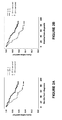
- the reference value may be adapted to yield the most significant prognostic information, e.g. the largest separation between the WARS protein high survival curve and the WARS protein low survival curve (see the figures).
- Examples of reference values that may yield a large separation is a cytoplasmic fraction (CF) of ⁇ 2 % or an absent cytoplasmic intensity (CI).
- CF cytoplasmic fraction
- CI absent cytoplasmic intensity
- the reference value may be adapted to identify a group of subjects having a predetermined prognosis, e.g. the group of subjects having a probability of five year overall survival of less than 70 %.
- the reference value may correspond to the amount of WARS protein expression in a healthy tissue, such as healthy colorectal or stroma tissue, of the subject of the method.
- the reference value may be provided by the amount of WARS protein expression measured in a standard sample of normal tissue from another, comparable subject.
- the reference value may be provided by the amount of WARS protein expression measured in a reference sample comprising tumor cells, such as a reference sample of tumor tissue.
- the reference value may be provided by the amount of WARS protein measured in a reference sample comprising cells expressing a predetermined amount of WARS protein.
- the reference value may be provided by the amount of WARS protein expression measured in a reference sample comprising cell lines, such as cancer cell lines, expressing a predetermined, or controlled, amount of WARS protein.
- cell lines such as cancer cell lines
- a predetermined, or controlled, amount of WARS protein The person skilled in the art understands how to provide such cell lines, for example guided by the disclosure of Rhodes et al. (2006) The biomedical scientist, p 515-520 .
- the reference value may be a predetermined value corresponding to the amount of WARS protein expression in a reference sample.
- the sample value of step b) may be either 1, corresponding to detectable WARS protein in the sample, or 0, corresponding to no detectable WARS protein in the sample. Consequently, in such embodiment, the evaluation of the sample is digital: WARS protein is considered to be either present or not.
- no detectable WARS protein refers to an amount of WARS protein that is so small that it is not, during normal operational circumstances, detectable by a person or an apparatus performing the invention according to any one of its different aspects.
- the "normal operational circumstances” refer to the laboratory methods and techniques a person skilled in the art would find appropriate for performing the invention.
- the reference value of step c) may be provided by a reference sample, such as a tissue sample, having no detectable WARS protein expression.
- the reference value of step c) may be 0, corresponding to no detectable WARS protein.
- the fraction may for example be: a "cellular fraction", wherein the WARS protein expression of the whole cells is taken into account; a "cytoplasmic fraction”, wherein the WARS protein expression of only the cytoplasms of the cells is taken into account, or the "nuclear fraction", wherein the WARS protein expression of only the nuclei of the cells is taken into account.
- the cytoplasmic fraction may for example be classified as ⁇ 2 %, 2 - 25 %, > 25 - 75 % or > 75 % immunoreactive cells of the relevant cell population.
- cytoplasmic fraction corresponds to the percentage of relevant cells in a sample that exhibits a positive staining in the cytoplasm, wherein a medium or distinct and strong immunoreactivity in the cytoplasm is considered positive and no or faint immunoreactivity in the cytoplasm is considered negative.
- the person skilled in the art of pathology understands which cells that are relevant under the conditions present when performing the method and may determine a cytoplasmic fraction based on his general knowledge and the teachings of the present disclosure.
- the relevant cells may for example be tumor cells. Further, the skilled artisan understands how to perform corresponding measurements employing the "cellular fraction" or the "nuclear fraction”.
- the intensity may for example be: a "cellular intensity”, wherein the WARS protein expression of the whole cells is taken into account; a "cytoplasmic intensity”, wherein the WARS protein expression of only the cytoplasms of the cells is taken into account, or the "nuclear intensity”, wherein the WARS protein expression of only the nuclei of the cells is taken into account. Cytoplasmic intensity is subjectively evaluated in accordance to standards used in clinical histopathological diagnostics.
- the person skilled in the art understands which cells that are relevant under the conditions present when performing the method and may determine a cytoplasmic intensity based on his general knowledge and the teachings of the present disclosure.
- the relevant cells may for example be tumor cells. This determination may for example be performed as described below in the Examples, Section 4, definition of "cytoplasmic intensity”. Further, the skilled artisan understands how to perform corresponding measurements employing the "cellular intensity" or the "nuclear intensity”.
- the reference value may be a cytoplasmic fraction, a cytoplasmic intensity or a combination thereof.
- the sample value may be a cytoplasmic fraction, a cytoplasmic intensity or a combination thereof.
- the sample value and the reference value are both the same type of value. Accordingly, in embodiments of the methods of the above aspects, the sample value and the reference value may each be a cytoplasmic fraction, a cytoplasmic intensity or a combinations thereof.
- the criterion for the conclusion in step d) is a sample value for the cytoplasmic fraction of WARS protein positive cells, i.e. a "cytoplasmic fraction", which is lower than or equal to the reference value of 75 %, such as lower than or equal to 50 %, such as lower than or equal to 40 %, such as lower than or equal to 30 %, such as lower than or equal to 25 %, such as lower than or equal to 20 %, such as lower than or equal to 15 %, such as lower than or equal to 10 %, such as lower than or equal to 5 %, such as lower than 2 %, such as equal to 0%.
- a cytoplasmic fraction which is lower than or equal to the reference value of 75 %, such as lower than or equal to 50 %, such as lower than or equal to 40 %, such as lower than or equal to 30 %, such as lower than or equal to 25 %, such as lower than or equal to 20 %, such as lower than or equal to 15 %, such
- the reference value of step c) is a cytoplasmic fraction of 75 % WARS positive cells or lower, such as 50 % WARS positive cells or lower, such as 40 % WARS positive cells or lower, such as 30 % WARS positive cells or lower, such as 25 % WARS positive cells or lower, such as 20 % WARS positive cells or lower, such as 10 % WARS positive cells or lower, such as 5 % WARS positive cells or lower, such as ⁇ 2 % WARS positive cells, such as 0 % WARS positive cells.
- the reference value of step c) is a cytoplasmic fraction of 0 - 25 %, such as 0 - 20 %, such as 0 - 15 %, such as 0 - 10 %, such as 0 - 5 %, such as 0 - ⁇ 2 %.
- the criterion for the conclusion in step d) may be a sample value for staining intensity of a sample, i.e. a cytoplasmic intensity, which is equal to, or lower than, a moderate cytoplasmic intensity, such as equal to, or lower than, a weak cytoplasmic intensity, such as equal to an absent cytoplasmic intensity.
- the reference value of step c) may be a moderate cytoplasmic intensity of WARS protein expression, or lower, such as a weak cytoplasmic intensity of WARS protein expression, or lower, such as an absent cytoplasmic intensity of WARS protein expression.
- the reference value may be constituted of two values, wherein the criterion for the conclusion in step d) is a sample value being lower than or equal to any one of these two values.
- the reference value may be a combination of a fraction value and an intensity value, such as a cytoplasmic fraction value and a cytoplasmic intensity value.
- the reference value may be a function of a cytoplasmic fraction value and a cytoplasmic intensity value.
- a function may be a staining score.
- the "staining score" is calculated as described in Examples, Section 3 and table 1 below.
- the reference value may be a staining score of 2 or lower, such as 1 or lower, such as 0.
- a person skilled in the art e.g. a pathologist, understands how to perform the evaluation yielding a fraction, such as a cellular, cytoplasmic or nuclear fraction, or an intensity, such as a cellular, cytoplasmic or nuclear intensity.
- a fraction such as a cellular, cytoplasmic or nuclear fraction
- an intensity such as a cellular, cytoplasmic or nuclear intensity.
- the skilled artisan may use a reference sample comprising a predetermined amount of WARS protein for establishing the appearance of a certain fraction or intensity.
- a reference sample may not only be used for the provision of the actual reference value, but also for the provision of an example of a sample with an amount of WARS protein that is higher than the amount corresponding to the reference value.
- histochemical staining such as in immunohistochemical staining
- the skilled artisan may use a reference sample for establishing the appearance of a stained sample with a high amount of WARS protein, e.g. a positive reference. Subsequently, the skilled artisan may assess the appearances of samples with lower amounts of WARS, such as the appearance of a sample with an amount of WARS corresponding to the reference value.
- the skilled artisan may use a reference sample to create a mental image of a reference value corresponding to an amount of WARS protein which is lower than that of the reference sample.
- the skilled artisan may use another reference sample having a low amount of WARS protein, or essentially lacking WARS protein, for establishing the appearance of such sample, e.g. as a "negative reference”.
- reference sample for establishing the appearance of a sample with a high amount of WARS protein.
- Such reference sample may be a sample comprising tissue expressing a high amount of WARS protein, such as a sample comprising colorectal tumor tissue having a pre-established high expression of WARS protein.
- the reference sample may provide an example of a strong cytoplasmic intensity (CI).
- CI cytoplasmic intensity
- the skilled artisan may then divide samples into the CI categories presented in Examples, Sections 3 and 4, below, i.e. absent, weak, moderate and strong. This division may be further assisted by a reference sample essentially lacking WARS protein (negative reference), i.e. a reference sample providing an absent cytoplasmic intensity.
- the reference sample may provide an example of a sample with a cytoplasmic fraction (CF) of 75 % or higher. With the knowledge of the appearance of a sample with more than 75 % positive cells, the skilled artisan may then evaluate the cytoplasmic fraction of other samples having e.g.
- a reference sample essentially lacking WARS protein negative reference
- a reference sample providing a low CF e.g. ⁇ 5%, such as ⁇ 2%
- cell lines expressing a controlled amount of WARS protein may be used as the reference, in particular as a positive reference.
- the methods according to the above aspects may be adapted to a selected reference value, such as one of the reference values presented above, and the reference prognosis will in such case be a consequence of the selected reference value.
- a selected reference value such as one of the reference values presented above
- the reference prognosis will in such case be a consequence of the selected reference value.
- the corresponding cumulative survival according to the CF > 0 curve may be read from the figure, e.g.
- Other associated reference prognoses may also be derived from the figures. The skilled artisan understands how to identify reference prognoses associated with reference values, and may do so without undue burden.
- the reference prognosis may be a likelihood of five-year survival of lower than 70 %.
- the reference prognosis may be a predetermined average probability of five-year survival for subjects having colorectal cancer, such as the average five-year survival for subjects having colorectal cancer at the time of performing the method.
- the reference value is adapted to the reference prognosis, as discussed above.
- the average five-year survival of a patient diagnosed with colorectal cancer was about 60 % world wide in 2002 ( Parkin et al (2007) CA Cancer J Clin 55, 74-108 ).
- the survival numbers above may be higher in the future, such as in the second or third decade of the 21 st century.
- the subject may be a human.
- the subject may have a cancer.
- the WARS protein has an amino acid sequence which comprises a sequence selected from:
- sequence ii) above is at least 90 % identical, at least 91 % identical, at least 92 % identical, at least 93 % identical, at least 94 % identical, at least 95 % identical, at least 96 % identical, at least 97 % identical, at least 98 % identical or at least 99 % identical to SEQ ID NO:1.
- the WARS protein has an amino acid sequence which comprises a sequence selected from:
- sequence ii) above is at least 90 % identical, at least 91 % identical, at least 92 % identical, at least 93 % identical, at least 94 % identical, at least 95 % identical, at least 96 % identical, at least 97 % identical, at least 98 % identical or at least 99 % identical to SEQ ID NO:2.
- the WARS protein may be detected and/or quantified through the application to a sample of a detectable and/or quantifiable affinity ligand, which is capable of selective interaction with the WARS protein.
- the application of the affinity ligand is performed under conditions that enable binding of the affinity ligand to any WARS protein in the sample.
- step b) may comprise:
- affinity ligand remaining in association with the sample refers to affinity ligand which was not removed in step b2), e.g. the affinity ligand bound to the sample.
- affinity ligands that may prove useful, as well as examples of formats and conditions for detection and/or quantification, are given below for the sake of illustration.
- an affinity ligand is used, which is selected from the group consisting of antibodies, fragments thereof and derivatives thereof, i.e. affinity ligands based on an immunoglobulin scaffold.
- the antibodies may be isolated and/or mono-specific.
- antibodies comprise monoclonal and polyclonal antibodies of any origin, including murine, rabbit, human and other antibodies, as well as chimeric antibodies comprising sequences from different species, such as partly humanized antibodies, e.g. partly humanized mouse antibodies.
- Polyclonal antibodies are produced by immunization of animals with the antigen of choice, whereas monoclonal antibodies of defined specificity can be produced using the hybridoma technology developed by Köhler and Milstein ( Köhler G and Milstein C (1976) Eur. J. Immunol. 6:511-519 ).
- Antibody fragments and derivatives comprise Fab fragments, consisting of the first constant domain of the heavy chain (CH1), the constant domain of the light chain (CL), the variable domain of the heavy chain (VH) and the variable domain of the light chain (VL) of an intact immunoglobulin protein; Fv fragments, consisting of the two variable antibody domains VH and VL ( Skerra A and Plückthun A (1988) Science 240:1038-1041 ); single chain Fv fragments (scFv), consisting of the two VH and VL domains linked together by a flexible peptide linker ( Bird RE and Walker BW (1991) Trends Biotechnol.
- Fab fragments consisting of the first constant domain of the heavy chain (CH1), the constant domain of the light chain (CL), the variable domain of the heavy chain (VH) and the variable domain of the light chain (VL) of an intact immunoglobulin protein
- Fv fragments consisting of the two variable antibody domains VH and VL ( Skerra A and Plück
- the biomolecular diversity needed for selection of affinity ligands may be generated by combinatorial engineering of one of a plurality of possible scaffold molecules, and specific and/or selective affinity ligands are then selected using a suitable selection platform.
- the scaffold molecule may be of immunoglobulin protein origin ( Bradbury AR and Marks JD (2004) J. Immunol. Meths. 290:29-49 ), of non-immunoglobulin protein origin ( Nygren P ⁇ and Skerra A (2004) J. Immunol. Meths. 290:3-28 ), or of an oligonucleotide origin ( Gold L et al (1995) Annu. Rev. Biochem. 64:763-797 ).
- Non-limiting examples of such structures useful for generating affinity ligands against WARS for use according to the present disclosure, are staphylococcal protein A and domains thereof and derivatives of these domains, such as protein Z ( Nord K et al (1997) Nat. Biotechnol. 15:772-777 ); lipocalins ( Beste G et al (1999) Proc. Natl. Acad. Sci. U.S.A. 96:1898-1903 ); ankyrin repeat domains ( Binz HK et al (2003) J. Mol. Biol.
- CBD cellulose binding domains
- GFP green fluorescent protein
- CTL-4 Hufton SE et al (2000) FEBS Lett. 475:225-231 ; Irving RA et al (2001) J. Immunol. Meth.
- protease inhibitors such as Knottin proteins ( Wentzel A et al (2001) J. Bacteriol. 183:7273-7284 ; Baggio R et al (2002) J. Mol. Recognit. 15:126-134 ) and Kunitz domains ( Roberts BL et al (1992) Gene 121:9-15 ; Dennis MS and Lazarus RA (1994) J. Biol. Chem. 269:22137-22144 ); PDZ domains ( Schneider S et al (1999) Nat. Biotechnol.
- peptide aptamers such as thioredoxin ( Lu Z et al (1995) Biotechnology 13:366-372 ; Klevenz B et al (2002) Cell. Mol. Life Sci. 59:1993-1998 ); staphylococcal nuclease ( Norman TC et al (1999) Science 285:591-595 ); tendamistats ( McConell SJ and Hoess RH (1995) J. Mol. Biol. 250:460-479 ; Li R et al (2003) Protein Eng. 16:65-72 ); trinectins based on the fibronectin type III domain ( Koide A et al (1998) J. Mol. Biol.
- non-immunoglobulin protein scaffolds include scaffold proteins presenting a single randomized loop used for the generation of novel binding specificities, protein scaffolds with a rigid secondary structure where side chains protruding from the protein surface are randomized for the generation of novel binding specificities, and scaffolds exhibiting a non-contiguous hyper-variable loop region used for the generation of novel binding specificities.
- oligonucleotides may also be used as affinity ligands.
- Single stranded nucleic acids called aptamers or decoys, fold into well-defined three-dimensional structures and bind to their target with high affinity and specificity.
- aptamers or decoys Single stranded nucleic acids
- the oligonucleotide ligands can be either RNA or DNA and can bind to a wide range of target molecule classes.
- Selection platforms include, but are not limited to, phage display ( Smith GP (1985) Science 228:1315-1317 ), ribosome display ( Hanes J and Plückthun A (1997) Proc. Natl. Acad. Sci. U.S.A.
- yeast two- hybrid system Fields S and Song O (1989) Nature 340:245-246
- yeast display Gai SA and Wittrup KD (2007) Curr Opin Struct Biol 17:467-473
- mRNA display Roberts RW and Szostak JW (1997) Proc. Natl. Acad. Sci. U.S.A.
- an affinity ligand may be used, which is a non-immunoglobulin affinity ligand derived from any of the protein scaffolds listed above, or an oligonucleotide molecule.
- the quantifiable affinity ligand may be capable of selective interaction with a WARS protein having the sequence SEQ ID NO:1.
- an affinity ligand capable of selective interaction with the WARS protein is detectable and/or quantifiable.
- the detection and/or quantification of such an affinity ligand may be accomplished in any way known to the skilled person for detection and/or quantification of binding reagents in assays based on biological interactions.
- any affinity ligand, as described above may be used quantitatively or qualitatively to detect the presence of the WARS protein.
- These "primary" affinity ligands may be labeled themselves with various markers or may in turn be detected by secondary, labeled affinity ligands to allow detection, visualization and/or quantification.
- Non-limiting examples of labels that can be conjugated to primary and/or secondary affinity ligands include fluorescent dyes or metals (e.g. fluorescein, rhodamine, phycoerythrin, fluorescamine), chromophoric dyes (e.g. rhodopsin), chemiluminescent compounds (e.g. luminal, imidazole) and bioluminescent proteins (e.g. luciferin, luciferase), haptens (e.g. biotin).
- fluorescent dyes or metals e.g. fluorescein, rhodamine, phycoerythrin, fluorescamine
- chromophoric dyes e.g. rhodopsin
- chemiluminescent compounds e.g. luminal, imidazole
- bioluminescent proteins e.g. luciferin, luciferase
- haptens e.g. biotin
- Affinity ligands can also be labeled with enzymes (e.g. horseradish peroxidase, alkaline phosphatase, beta-lactamase), radioisotopes (e.g. 3 H, 14 C, 32 P, 35 S or 125 I) and particles (e.g. gold).
- enzymes e.g. horseradish peroxidase, alkaline phosphatase, beta-lactamase
- radioisotopes e.g. 3 H, 14 C, 32 P, 35 S or 125 I
- particles e.g. gold
- particles e.g. gold
- particles e.g. gold
- particles e.g. gold
- particles e.g. gold
- particles e.g. gold
- particles e.g. gold
- particles e.g. gold
- particles e.g. gold
- particles e.g. gold
- particles e.g. gold
- particles e.g. gold
- particles
- the different types of labels can be conjugated to an affinity ligand using various chemistries, e.g. the amine reaction or the thiol reaction. However, other reactive groups than amines and thiols can be used, e.g. aldehydes, carboxylic acids and glutamine.
- the detection, localization and/or quantification of a labeled affinity ligand bound to its WARS protein target may involve visualizing techniques, such as light microscopy or immunofluoresence microscopy. Other methods may involve the detection via flow cytometry or luminometry.
- a biological sample such as a tumor tissue sample (biopsy), for example from colorectal tissue, may be removed from the subject for detection and/or quantification of WARS protein.
- the biological sample such as the biopsy, may be an earlier obtained sample. If using an earlier obtained sample, no steps of any one of the embodiments of the methods of the above aspects are practiced on the human or animal body.
- the affinity ligand may be applied to the biological sample for detection and/or quantification of the WARS marker protein. This procedure enables not only detection of WARS protein, but may in addition show the distribution and relative level of expression thereof.
- the method of visualization of labels on the affinity ligand may include, but is not restricted to, fluorometric, luminometric and/or enzymatic techniques. Fluorescence is detected and/or quantified by exposing fluorescent labels to light of a specific wavelength and thereafter detecting and/or quantifying the emitted light in a specific wavelength region. The presence of a luminescently tagged affinity ligand may be detected and/or quantified by luminescence developed during a chemical reaction. Detection of an enzymatic reaction is due to a color shift in the sample arising from chemical reaction. Those of skill in the art are aware that a variety of different protocols can be modified in order for proper detection and/or quantification.
- a biological sample may be immobilized onto a solid phase support or carrier, such as nitrocellulose or any other solid support matrix capable of immobilizing any WARS protein present in the biological sample applied to it.
- solid state support materials useful in the present invention include glass, carbohydrate (e.g. Sepharose), nylon, plastic, wool, polystyrene, polyethene, polypropylene, dextran, amylase, films, resins, cellulose, polyacrylamide, agarose, alumina, gabbros and magnetite.
- primary affinity ligand specific to WARS may be applied, e.g. as described in Examples, Sections 2 and/or 3, of the present disclosure.
- the supporting matrix may be washed with one or more appropriate buffers known in the art, followed by exposure to a secondary labeled affinity ligand and washed once again with buffers to remove unbound affinity ligands. Thereafter, selective affinity ligands may be detected and/or quantified with conventional methods.
- the binding properties for an affinity ligand may vary from one solid state support to the other, but those skilled in the art will be able to determine operative and optimal assay conditions for each determination by routine experimentation.
- the quantifiable affinity ligand of b1) may be detected using a secondary affinity ligand capable of recognizing the quantifiable affinity ligand.
- the quantification of b3) may thus be carried out by means of a secondary affinity ligand with affinity for the quantifiable affinity ligand.
- the secondary affinity ligand may be an antibody or a fragment or a derivative thereof.
- one available method for detection and/or quantification of the WARS protein is by linking the affinity ligand to an enzyme that can then later be detected and/or quantified in an enzyme immunoassay (such as an EIA or ELISA).
- an enzyme immunoassay such as an EIA or ELISA.
- the biological sample is brought into contact with a solid material or with a solid material conjugated to an affinity ligand against the WARS protein, which is then detected and/or quantified with an enzymatically labeled secondary affinity ligand.
- an appropriate substrate is brought to react in appropriate buffers with the enzymatic label to produce a chemical moiety, which for example is detected and/or quantified using a spectrophotometer, fluorometer, luminometer or by visual means.
- primary and any secondary affinity ligands can be labeled with radioisotopes to enable detection and/or quantification.
- appropriate radiolabels in the current invention are 3 H, 14 C, 32 P, 35 S or 125 I.
- the specific activity of the labeled affinity ligand is dependent upon the half-life of the radiolabel, isotopic purity, and how the label has been incorporated into the affinity ligand.
- Affinity ligands are preferably labeled using well-known techniques ( Wensel TG and Meares CF (1983) in: Radioimmunoimaging and Radioimmunotherapy (Burchiel SW and Rhodes BA eds.) Elsevier, New York, pp 185-196 ).
- a thus radiolabeled affinity ligand can be used to visualize WARS protein by detection of radioactivity in vivo or in vitro.
- Radionuclear scanning with e.g. gamma camera, magnetic resonance spectroscopy or emission tomography function for detection in vivo and in vitro, while gamma/beta counters, scintillation counters and radiographies are also used in vitro.
- kit for carrying out a method according to an embodiment of the above aspects which comprises:
- the affinity ligand is capable of selective interaction with a WARS protein having an amino acid sequence which comprises a sequence selected from:
- sequence ii) above is at least 90 % identical, at least 91 % identical, at least 92 % identical, at least 93 % identical, at least 94 % identical, at least 95 % identical, at least 96 % identical, at least 97 % identical, at least 98 % identical or at least 99 % identical to SEQ ID NO:1.
- the affinity ligand is capable of selective interaction with a WARS protein having an amino acid sequence which comprises a sequence selected from:
- sequence ii) above is at least 90 % identical, at least 91 % identical, at least 92 % identical, at least 93 % identical, at least 94 % identical, at least 95 % identical, at least 96 % identical, at least 97 % identical, at least 98 % identical or at least 99 % identical to SEQ ID NO:2.
- kit according to the kit aspect may be selected and specified as described above in connection with the method aspects of the present disclosure.
- the kit according to the invention comprises an affinity ligand against WARS, as well as other means that help to quantify the specific and/or selective affinity ligand after it has bound specifically and/or selectively to WARS.
- the kit may contain a secondary affinity ligand for detecting and/or quantifying a complex formed by any WARS protein and the affinity ligand capable of selective interaction with a WARS protein.
- the kit may also contain various auxiliary substances other than affinity ligands, to enable the kit to be used easily and efficiently.
- auxiliary substances include solvents for dissolving or reconstituting lyophilized protein components of the kit, wash buffers, substrates for measuring enzyme activity in cases where an enzyme is used as a label, target retrieval solution to enhance the accessibility to antigens in cases where paraffin or formalin-fixed tissue samples are used, and substances such as reaction arresters, e.g. endogenous enzyme block solution to decrease the background staining and/or counterstaining solution to increase staining contrast, that are commonly used in immunoassay reagent kits.
- reaction arresters e.g. endogenous enzyme block solution to decrease the background staining and/or counterstaining solution to increase staining contrast, that are commonly used in immunoassay reagent kits.
- the quantifiable affinity ligand is selected from the group consisting of antibodies, fragments thereof and derivatives thereof.
- such quantifiable affinity ligand may be obtainable by a process comprising a step of immunizing an animal, such as a rabbit, with a protein whose amino acid sequence comprises the sequence SEQ ID NO:1 or a sequence which is at least 85 % identical to SEQ ID NO:1, preferably the sequence SEQ ID NO:1.
- immunization may for example be performed as described in Examples, Section 2, below.
- the antibodies may for example be isolated and/or mono-specific.
- the quantifiable affinity ligand is a protein ligand derived from a scaffold selected from the group consisting of staphylococcal protein A and domains thereof, lipocalins, ankyrin repeat domains, cellulose binding domains, Y crystallines, green fluorescent protein, human cytotoxic T lymphocyte-associated antigen 4, protease inhibitors, PDZ domains, peptide aptamers, staphylococcal nuclease, tendamistats, fibronectin type III domain and zinc fingers.
- the quantifiable affinity ligand is an oligonucleotide molecule.
- the quantifiable affinity ligand may be capable of selective interaction with a WARS protein having the sequence SEQ ID NO:1.
- the detectable affinity ligand may comprise a label selected from the group consisting of fluorescent dyes and metals, chromophoric dyes, chemiluminescent compounds and bioluminescent proteins, enzymes, radioisotopes, particles and quantum dots.
- the reagents necessary for quantifying the amount of the affinity ligand comprise a secondary affinity ligand capable of recognizing the quantifiable affinity ligand.
- the secondary affinity ligand capable of recognizing the quantifiable affinity ligand comprises a label selected from the group consisting of fluorescent dyes or metals, chromophoric dyes, chemiluminescent compounds and bioluminescent proteins, enzymes, radioisotopes, particles and quantum dots.
- the kit according to the kit aspect may also advantageously comprise a reference sample for provision of, or yielding, the reference value to be used for comparison with the sample value.
- the reference sample comprises a predetermined amount of WARS protein.
- a reference sample may for example be constituted by a tissue sample having a predetermined amount of WARS protein, which may then be used by the person of skill in the art to determine the WARS expression status in the sample being studied, by manual, such as ocular, or automated comparison of expression levels in the reference sample and the subject sample.
- the reference sample may comprise cell lines, such as cancer cell lines, expressing a predetermined, or controlled, amount of WARS protein.
- the cell lines may be formalin fixed.
- such formalin fixed cell lines may be paraffin embedded.
- the wording "reference sample for provision of the reference value” is to be interpreted broadly in the context of the kit aspect.
- the reference sample may comprise an amount of WARS protein actually corresponding to the reference value, but it may also comprise an amount of WARS protein corresponding to a value being higher than the reference value.
- the "high" value may be used by a person performing the method as an upper reference (positive reference) for assessing, e.g. the appearance of, a reference value which is lower than the "high” value.
- the person skilled in the art of immunohistochemistry understands how to do such an assessment.
- the skilled person may use another reference sample comprising a low amount of WARS protein for provision of a "low” value in such an assessment, e.g. as a negative reference.
- the reference sample may comprise an amount of WARS protein corresponding to the reference value.
- the reference sample may comprise an amount of WARS protein corresponding to a cytoplasmic fraction of 75 % WARS positive cells or lower, such as 50 % WARS positive cells or lower, such as 40 % WARS positive cells or lower, such as 30 % WARS positive cells or lower, such as 25 % WARS positive cells or lower, such as 20 % WARS positive cells or lower, such as 10 % WARS positive cells or lower, such as 5 % WARS positive cells or lower, such as ⁇ 2 % WARS positive cells, such as 0 % WARS positive cells.
- the reference sample may comprise an amount of WARS protein corresponding to an moderate cytoplasmic intensity of WARS protein expression, or lower, such as a faint cytoplasmic intensity of WARS protein expression, or lower, such as no cytoplasmic intensity of WARS protein expression.
- the reference sample may comprise an amount of WARS protein corresponding a staining score 2 or lower, such as 1 or lower, such as 0.
- cytoplasmic fraction values or cytoplasmic intensity values is discussed above in connection with the method aspects.
- the kit may comprise a reference sample comprising an amount of WARS protein corresponding to a value being higher than the reference value.
- the reference sample may for example comprise an amount of WARS protein corresponding to a cytoplasmic fraction of 75 % or higher and/or a strong cytoplasmic intensity of WARS expression.
- the kit may comprise a reference sample comprising an amount of WARS protein corresponding to a value being lower than the reference value, e.g. an absent cytoplasmic intensity and/or a cytoplasmic fraction of ⁇ 2 % WARS positive cells, such as 0 % WARS positive cells.
- the kit may comprise: a reference sample comprising an amount of WARS protein corresponding to a predetermined reference value; a reference sample comprising an amount of WARS protein corresponding to a value being higher than a predetermined reference value; and/or a reference sample comprising an amount of WARS protein corresponding to a value being lower than a predetermined reference value.
- kits may comprise: a first reference sample comprising an amount of WARS protein being higher than a predetermined reference value; and a second reference sample comprising an amount of WARS protein being lower than the predetermined reference value.
- the reference sample may be a tissue sample, such as a tissue sample adapted to ocular or microscopic evaluation.
- the tissue reference sample may be fixated in paraffin or buffered formalin and/or histo-processed to ⁇ m-thin sections that are mounted on microscopic glass-slides.
- the tissue reference sample may be further adapted to staining with affinity ligands, such as antibodies, for a WARS protein.
- the reference sample may be adapted to directly, or indirectly, provide any relevant reference value, such as any one of the reference values discussed above.
- a WARS protein as a prognostic marker. Also provided is the use of a WARS protein as a prognostic marker for cancer, such as colorectal cancer.
- a “prognostic marker” refers to a product which presence is of value in an establishment of a prognosis.
- WARS protein having the amino acid sequence of SEQ ID NO:1, which has been designed to have desired antigenic properties, may be particularly suitable for such purposes.
- a WARS protein or an antigenically active fragment thereof, for the production, selection or purification of a prognostic agent for establishing a prognosis for a patient having a colorectal cancer.
- an "antigenically active fragment" of a WARS protein is a fragment of sufficient size to be useful for the generation of an affinity ligand, e.g. an antibody, which will interact with a WARS protein comprising the fragment.
- an affinity ligand e.g. an antibody
- a “prognostic agent” refers to an agent having at least one property being valuable in an establishment of a prognosis.
- the prognostic agent may be an affinity ligand capable of selective interaction with the WARS protein, or antigenically active fragment thereof.
- the affinity ligand may preferably be capable of selective interaction with a protein having the sequence SEQ ID NO:1.
- the affinity ligand may preferably be an antibody or a fragment or a derivative thereof.
- the antibody may be isolated and/or mono-specific.
- the use may comprise selection of prognostic agents having specificity for the WARS protein using a soluble matrix in which the WARS protein has been immobilized.
- soluble matrix may for example be a dextran matrix for use in a surface plasmon resonance instrument, such as BiacoreTM instrument and the selection may for example comprise monitoring the affinity for the immobilized WARS protein of a number of prognostic agents.
- the use may comprise immunizing a mammal with the WARS protein in order to produce prognostic agents that are collected from the serum of the immunized mammal.
- the WARS protein may for example comprise a sequence selected from: i) SEQ ID NO:1; and ii) a sequence which is at least 85 % identical to SEQ ID NO:1.
- sequence ii) is at least 90 % identical, at least 91 % identical, at least 92 % identical, at least 93 % identical, at least 94 % identical, at least 95 % identical, at least 96 % identical, at least 97 % identical, at least 98 % identical or at least 99 % identical to SEQ ID NO:1.
- the WARS protein may for example comprise a sequence selected from: i) SEQ ID NO:2; and ii) a sequence which is at least 85 % identical to SEQ ID NO:2.
- sequence ii) is at least 90 % identical, at least 91 % identical, at least 92 % identical, at least 93 % identical, at least 94 % identical, at least 95 % identical, at least 96 % identical, at least 97 % identical, at least 98 % identical or at least 99 % identical to SEQ ID NO:2.
- the use may be in vitro -use.
- the inventors have found a previously unknown correlation between WARS protein and the prognosis for a patient suffering from a colorectal cancer. Further, the inventors have developed an affinity ligand with affinity for the WARS protein.
- the inventive scope is not limited to a single type of affinity ligand, and the inventive idea may be put into practice using any type of affinity ligand capable of selective interaction with a WARS protein.
- an affinity ligand capable of selective interaction with a WARS protein, such as a WARS protein having the amino acid sequence SEQ ID NO:1.
- the affinity ligand is an antibody or a fragment or a derivative thereof.
- the antibody may be isolated and/or mono-specific.
- SEQ ID NO:1 is a WARS protein especially designed for immunizations, e.g. designed to lack transmembrane regions to ensure efficient expression in E . coli, and to lack any signal peptide, since those are cleaved off in the mature protein.
- the antibody, or fragment or derivative thereof may for example be one that is obtainable by a process comprising a step of immunizing an animal, such as a rabbit, with a protein whose amino acid sequence comprises the sequence SEQ ID NO:1 or a sequence which is at least 85 % identical to SEQ ID NO:1, preferably the sequence SEQ ID NO:1.
- the immunization process may comprise primary immunization with the protein in Freund's complete adjuvant.
- the immunization process may further comprise boosting at least two times, in intervals of 2-6 weeks, with the protein in Freund's incomplete adjuvant.
- any of those variants of the WARS protein (e.g. SEQ ID NO:2) or the antigenically active fragment thereof (e.g. SEQ ID NO:1) that are discussed above may, of course, be used in such a process for generating an antibody or a fragment or derivative thereof.
- the present disclosure provides the above affinity ligand for evaluating the amount of WARS protein present in at least part of a sample obtained from a subject having a colorectal cancer. Also, the present disclosure provides the above affinity ligand for establishing a prognosis for a mammalian subject having a colorectal cancer.
- the use of the affinity ligand according to the above aspect as a prognostic agent.
- a preferred embodiment of this use is as a prognostic agent for the prognosis of a cancer, such as a colorectal cancer.
- a use of the affinity ligand in the manufacture of a prognostic agent for the prognosis of a colorectal cancer may be in vitro -uses.
- a specific or selective interaction refers to the extent to which a particular method can be used to determine the presence and/or amount of a specific protein, the target protein or a fragment thereof, under given conditions in the presence of other proteins in a tissue sample or fluid sample of a naturally occurring or processed biological fluid.
- specificity or selectivity is the capacity to distinguish between related proteins.
- Specific and selective are sometimes used interchangeably in the present description.
- the specificity or selectivity of an antibody may be determined as in Examples, section 2, below, wherein analysis is performed using a protein array set-up and a western blot, respectively. Specificity and selectivity determinations are also described in Nilsson P et al (2005) Proteomics 5:4327-4337 .
- a "mono-specific antibody” is one of a population of polyclonal antibodies which has been affinity purified on its own antigen, thereby separating such mono-specific antibodies from other antiserum proteins and non-specific antibodies. This affinity purification results in antibodies that bind selectively to its antigen.
- the polyclonal antisera are purified by a two-step immunoaffinity based protocol to obtain mono-specific antibodies selective for the target protein. Antibodies directed against generic affinity tags of antigen fragments are removed in a primary depletion step, using the immobilized tag protein as the capturing agent.
- the serum is loaded on a second affinity column with the antigen as capturing agent, in order to enrich for antibodies specific for the antigen (see also Nilsson P et al (2005) Proteomics 5:4327-4337 ).
- a suitable fragment of the target protein encoded by the EnsEMBL Gene ID ENSG00000140105 was selected using bioinformatic tools with the human genome sequence as template ( Lindskog M et al (2005) Biotechniques 38:723-727 , EnsEMBL, www.ensembl.org). The fragment was used as template for the production of a 144 amino acid long fragment corresponding to amino acids 34-178 (SEQ ID NO:1) of the WARS protein (SEQ ID NO:2; EnsEMBL entry no. ENSP00000339485).
- a fragment of the WARS gene transcript containing nucleotides 434-866 of EnsEMBL entry number ENST00000344102 was isolated by a SuperscriptTM One-Step RT-PCR amplification kit with Platinum® Taq (Invitrogen) and a human total RNA pool panel as template (Human Total RNA Panel IV, BD Biosciences Clontech). Flanking restriction sites NotI and AscI were introduced into the fragment through the PCR amplification primers, to allow in-frame cloning into the expression vector (forward primer: GATGCCACAGAAGCTGAAG, reverse primer: GGCATTCTCCACAGCATAG).
- downstream primer was biotinylated to allow solid-phase cloning as previously described, and the resulting biotinylated PCR product was immobilized onto Dynabeads M280 Streptavidin (Dynal Biotech) ( Larsson M et al (2000) J. Biotechnol. 80:143-157 ).
- the fragment was released from the solid support by NotI-AscI digestion (New England Biolabs), ligated into the pAff8c vector (Larsson M et al, supra ) in frame with a dual affinity tag consisting of a hexahistidyl tag for immobilized metal ion chromatography (IMAC) purification and an immunopotentiating albumin binding protein (ABP) from streptococcal protein G ( Sjölander A et al (1997) J. Immunol.
- NotI-AscI digestion New England Biolabs
- IMAC immobilized metal ion chromatography
- ABSP immunopotentiating albumin binding protein
- BL21 (DE3) cells harboring the expression vector were inoculated in 100 ml 30 g/l tryptic soy broth (Merck KGaA) supplemented with 5 g/l yeast extract (Merck KGaA) and 50 mg/l kanamycin (Sigma-Aldrich) by addition of 1 ml of an overnight culture in the same culture medium.
- the cell culture was incubated in a 1 liter shake flask at 37 °C and 150 rpm until the optical density at 600 nm reached 0.5-1.5.
- Protein expression was then induced by addition of isopropyl- ⁇ -D-thiogalactopyranoside (Apollo Scientific) to a final concentration of 1 mM, and the incubation was continued overnight at 25 °C and 150 rpm.
- the His 6 -tagged fusion protein was purified by immobilized metal ion affinity chromatography (IMAC) on columns with 1 ml Talon® metal (Co 2+ ) affinity resin (BD Biosciences Clontech) using an automated protein purification procedure ( Steen J et al (2006) Protein Expr. Purif. 46:173-178 ) on an ASPEC XL4TM (Gilson).
- the resin was equilibrated with 20 ml denaturing washing buffer (6 M guanidine hydrochloride, 46.6 mM Na 2 HPO 4 , 3.4 mM NaH 2 PO 4 , 300 mM NaCl, pH 8.0-8.2). Clarified cell lysates were then added to the column.
- the resin was then washed with a minimum of 31.5 ml washing buffer prior to elution in 2.5 ml elution buffer (6 M urea, 50 mM NaH 2 PO 4 , 100 mM NaCl, 30 mM acetic acid, 70 mM Na-acetate, pH 5.0).
- the eluted material was fractioned in three pools of 500, 700 and 1300 ⁇ l.
- the 700 ⁇ l fraction, containing the antigen, and the pooled 500 and 1300 ⁇ l fractions were stored for further use.
- the antigen fraction was diluted to a final concentration of 1 M urea with phosphate buffered saline (PBS; 1.9 mM NaH 2 PO 4 , 8.1 mM Na 2 HPO 4 , 154 mM NaCl) followed by a concentration step to increase the protein concentration using Vivapore 10/20 ml concentrator with molecular weight cut off at 7500 Da (Vivascience AG).
- the protein concentration was determined using a bicinchoninic acid (BCA) micro assay protocol (Pierce) with a bovine serum albumin standard according to the manufacturer's recommendations.
- BCA bicinchoninic acid
- the protein quality was analyzed on a Bioanalyzer instrument using the Protein 50 or 200 assay (Agilent Technologies).
- a gene fragment corresponding to nucleotides 434-866 of the long transcript (SEQ ID NO:3) of the WARS gene and encoding a peptide (SEQ ID NO:1) consisting of amino acids 34 to 178 of the target protein WARS (SEQ ID NO:2) was successfully isolated by RT-PCR from a human RNA pool using primers specific for the protein fragment.
- the 144 amino acid fragment (SEQ ID NO:1) of the target protein (SEQ ID NO:2) was designed to lack transmembrane regions to ensure efficient expression in E. coli, and to lack any signal peptide, since those are cleaved off in the mature protein.
- the protein fragment was designed to consist of a unique sequence with low homology with other human proteins, to minimize cross reactivity of generated affinity reagents, and to be of a suitable size to allow the formation of conformational epitopes and still allow efficient cloning and expression in bacterial systems.
- a clone encoding the correct amino acid sequence was identified, and, upon expression in E. coli, a single protein of the correct size was produced and subsequently purified using immobilized metal ion chromatography. After dilution of the eluted sample to a final concentration of 1 M urea and concentration of the sample to 1 ml, the concentration of the protein fragment was determined to be 5.55 mg/ml and was 97.7 % pure according to purity analysis.
- the purified WARS fragment as obtained above was used as antigen to immunize a rabbit in accordance with the national guidelines (Swedish permit no. A 84-02).
- the rabbit was immunized intramuscularly with 200 ⁇ g of antigen in Freund's complete adjuvant as the primary immunization, and boosted three times in four week intervals with 100 ⁇ g antigen in Freund's incomplete adjuvant.
- Antiserum from the immunized animal was purified by a three-step immunoaffinity based protocol ( Agaton C et al (2004) J. Chromatogr. A 1043:33-40 ; Nilsson P et al (2005) Proteomics 5:4327-4337 ).
- the first step 7 ml of total antiserum was buffered with 10x PBS to a final concentration of 1x PBS (1.9 mM NaH 2 PO 4 , 8.1 mM Na 2 HPO 4 , 154 mM NaCl), filtered using a 0.45 ⁇ m pore-size filter (Acrodisc®, Life Science) and applied to an affinity column containing 5 ml N-hydroxysuccinimide-activated SepharoseTM 4 Fast Flow (GE Healthcare) coupled to the dual affinity tag protein His 6 -ABP (a hexahistidyl tag and an albumin binding protein tag) expressed from the pAff8c vector and purified in the same way as described above for the antigen protein fragment.
- 1x PBS 1.9 mM NaH 2 PO 4 , 8.1 mM Na 2 HPO 4 , 154 mM NaCl
- the flow-through depleted of antibodies against the dual affinity tag His 6 -ABP
- the His 6 -ABP protein and the protein fragment antigen had been coupled to the NHS activated matrix as recommended by the manufacturer.
- Unbound material was washed away with 1 x PBST (1 x PBS, 0.1 % Tween20, pH 7.25), and captured antibodies were eluted using a low pH glycine buffer (0.2 M glycine, 1 mM EGTA, pH 2.5).
- the eluted antibody fraction was collected automatically, and loaded onto two 5 ml HiTrapTM desalting columns (GE Healthcare) connected in series for efficient buffer exchange in the third step.
- the second and third purification steps were run on the ⁇ KTAxpressTM platform (GE Healthcare).
- the antigen selective (mono-specific) antibodies (msAbs) were eluted with PBS buffer, supplemented with glycerol and NaN 3 to final concentrations of 40 % and 0.02 %, respectively, for long term storage at -20 °C ( Nilsson P et al (2005) Proteomics 5:4327-4337 ).
- the specificity and selectivity of the affinity purified antibody fraction were analyzed by binding analysis against the antigen itself and against 94 other human protein fragments in a protein array set-up ( Nilsson P et al (2005) Proteomics 5:4327-4337 ).
- the protein fragments were diluted to 40 ⁇ g/ml in 0.1 M urea and 1x PBS (pH 7.4) and 50 ⁇ l of each were transferred to the wells of a 96-well spotting plate.
- the protein fragments were spotted and immobilized onto epoxy slides (SuperEpoxy, TeleChem) using a pin-and-ring arrayer (Affymetrix 427).
- the slide was washed in 1x PBS (5 min) and the surface was then blocked (SuperBlock®, Pierce) for 30 minutes.
- An adhesive 16-well silicone mask (Schleicher & Schuell) was applied to the glass before the mono-specific antibodies were added (diluted 1:2000 in 1x PBST to appr. 50 ng/ml) and incubated on a shaker for 60 min.
- Affinity tag-specific IgY antibodies were co-incubated with the mono-specific antibodies in order to quantify the amount of protein in each spot.
- the slide was washed with 1x PBST and 1x PBS twice for 10 min each.
- the specificity and selectivity of the affinity-purified antibody were analyzed by Western blot.
- Western blot was performed by separation of total protein extracts from selected human tissues and cell lines on pre-cast 10-20 % SDS-PAGE gradient gels (Bio-Rad Laboratories) under reducing conditions, followed by electro-transfer to PVDF membranes (Bio-Rad Laboratories) according to the manufacturer's recommendations.
- the membranes were blocked (5 % dry milk, 1x TBST; 0.1 M Tris-HCl, 0.5 M NaCl, 0.1 % Tween20) for 1 h at room temperature, incubated with the primary affinity purified antibody (diluted 1:500 in blocking buffer) and washed in TBST.
- the secondary HRP-conjugated antibody (swine anti-rabbit immunoglobulin/HRP, DakoCytomation) was diluted 1:3000 in blocking buffer and chemiluminescence detection was carried out using a ChemidoCTM CCD camera (Bio-Rad Laboratories) and SuperSignal® West Dura Extended Duration substrate (Pierce), according to the manufacturer's protocol.
- a two-color dye labeling system was used, with a combination of primary and secondary antibodies.
- Tag-specific IgY antibodies generated in hen were detected with a secondary goat anti-hen antibody labeled with Alexa 555 fluorescent dye.
- the specific binding of the rabbit msAb to its antigen on the array was detected with a fluorescently Alexa 647 labeled goat anti-rabbit antibody.
- Each protein fragment was spotted in duplicates. The protein array analysis showed that the affinity purified mono-specific antibody against WARS is highly selective to the correct protein fragment and has a very low background to all other protein fragments analyzed on the array.
- the result of the Western blot analysis showed that the antibody specifically detects a single band of approximately 50 kDa in a bladder tumor cell line (RT-4) and a human glioma cell line (U-251 MG sp). In addition, a band was detected a in tonsil tissue samples.
- the theoretical molecular weight of WARS is 49 kDa (as calculated from the WARS amino acid sequence SEQ ID NO:2), corresponding well to the result obtained.
- tissue microarray TMA
- All tissues used as donor blocks for tissue microarray (TMA) production were selected from the archives at the Department of Pathology, University Hospital, Uppsala, in agreement with approval from the local ethical committee. All tissue sections used for TMA analysis were examined to determine diagnosis and to select representative areas in donor blocks. Normal tissue was defined as microscopically normal (non-neoplastic) and was most often selected from specimens collected from the vicinity of surgically removed tumors. Cancer tissue was reviewed for diagnosis and classification. All tissues were formalin fixated, paraffin embedded, and sectioned for diagnostic purposes.
- TMA production was performed essentially as previously described ( Kononen J et al (1998) Nature Med. 4:844-847 ; Kallioniemi OP et al (2001) Hum. Mol. Genet. 10:657-662 ). Briefly, a hole was made in the recipient TMA block and a cylindrical core tissue sample from the donor block was acquired and deposited in the recipient TMA block. This was repeated in an automated tissue arrayer from Beecher Instrument (ATA-27, Beecher Instruments, Sun Prairie, CA, USA) until a complete TMA design was produced. TMA recipient blocks were baked at 42 °C for 2 h prior to sectioning.
- TMA The design of TMA:s was focused on obtaining samples from a large range of representative normal tissues, and on including representative cancer tissues. This has previously been described in detail in Kampf C et al (2004) Clin. Proteomics 1:285-300 . In brief, samples from 48 normal tissues and from 20 of the most common cancer types affecting humans were selected. In total, eight different designs of TMA blocks, each containing 72 cores of tissue with 1 mm diameter, were produced. Two of the TMA:s represented normal tissues, corresponding to 48 different normal tissues in triplicates from different individuals. The remaining 6 TMA:s represented cancer tissue from 20 different types of cancer.
- TMA blocks were sectioned with 4 ⁇ m thickness using a waterfall microtome (Leica), and placed onto SuperFrost® (Roche Applied Science) glass slides for IHC analysis.
- the slides were incubated for 30 min at room temperature with the primary antibody obtained as in Examples, Section 2, followed by incubation for 30 min at room temperature with goat anti-rabbit peroxidase conjugated Envision®. Between all steps, slides were rinsed in wash buffer (DakoCytomation). Finally, diaminobenzidine (DakoCytomation) was used as chromogen and Harris hematoxylin (Sigma-Aldrich) was used for counterstaining. The slides were mounted with Pertex® (Histolab).
- Staining score Staining score Staining intensity Fraction of stained cells 0 absent ⁇ 2% 0 absent 2 - 25% 0 absent > 25 - 75% 0 absent > 75% 0 weak ⁇ 2% 0 weak 2 - 25% 1 weak > 25 - 75% 1 weak > 75% 1 moderate ⁇ 2% 1 moderate 2 - 25% 2 moderate > 25 - 75% 2 moderate > 75% 2 strong ⁇ 2% 2 strong 2 - 25% 3 strong > 25 - 75% 3 strong > 75%
- Bronchus surface epithelial cells Cerebellum cells in granular layer 0 cells in molecular layer 0 purkinje cells 2 Cerebral cortex neuronal cells 2 non-neuronal cells 1 Cervix, uterine glandular cells 2 surface epithelial cells (squamous) 1 Colon glandular cells 0 Duodenum glandular cells 2 Endometrium 1 cells in endometrial stroma/ECM 2 cells in myometrium/ECM 0 glandular cells 2 Endometrium 2 cells in endometrial stroma/ECM 0 cells in myometrium/ECM 0 glandular cells 2 Epididymis glandular cells 2 Esophagus surface epithelial cells 0 Fallopian tube glandular cells 2 Gall bladder glandular cells 1 Heart muscle myocytes 0 Hippocampus neuronal cells 2 non-neuronal cells 1 Kidney cells in glomeruli 1 cells in tubuli 1 Lateral ventricle neuronal cells 2 non-neuronal cells 1 Liver bile duct cells
- ovarian stromal cells 0 Pancreas exocrine pancreas 2 islet cells 2 Parathyroid gland glandular cells 0 Placenta decidual cells N.R. trophoblastic cells 0 Prostate glandular cells 1 Rectum glandular cells 1 Salivary gland glandular cells 2 Seminal vescicle glandular cells 1 Skeletal muscle myocytes 0 Skin adnexal cells N.R.
- WARS protein expression was evaluated in tissue samples from various cancer types. Table 3 shows the level of WARS expression in 12 different colorectal carcinoma tissues samples. All 12 of these samples showed representative tissue, 11 showed positivity and 2 strong expression. Table 3: Expression pattern of WARS in colorectal cancer Cancer type Subject number 1 2 3 4 5 6 7 8 9 10 11 12 Colorectal cancer 3 3 2 2 2 2 2 1 1 1 1 0
- TMA colorectal carcinoma
- All 122 cases of colorectal carcinoma were histopathologically reevaluated on slides stained with hematoxylin and eosin.
- TMA:s were then constructed by sampling 2 x 1.0 mm cores per case from areas representative of invasive cancer.
- the TMA:s were prepared and automated immunohistochemistry was performed as described in section 3 above, using the WARS antibody prepared as described in section 2 above.
- cytoplasmic intensity and cytoplasmic fraction were instead analyzed separately for correlation with survival.
- cytoplasmic intensity corresponds to the overall staining intensity of cytoplasms of glandular/tumor cells in a sample.
- Cytoplasmic fraction corresponds to the percentage of glandular/tumor cells in a sample that exhibits a positive staining intensity in the cytoplasm.
- cytoplasmic intensity was divided into: absent cytoplasmic intensity (0); or weak, moderate or strong cytoplasmic intensity (> 0). Further, the cytoplasmic fraction was divided into: ⁇ 2 % positive cells (0), i.e. less than 2 % of the cells in a sample exhibiting a positive staining in the cytoplasm; or ⁇ 2 % positive cells (1-3), i.e. 2 % or more of the cells in a sample exhibiting a positive staining in the cytoplasm.
- Tissue microarray based analysis of 122 colorectal carcinomas showed that 78 subjects (64 %) were positive for WARS (CF ⁇ 2 %) as seen in figure 1A .
- the figure further reveals that for 58% of the subjects, fractions of stained cells are > 25%.
- the intensity was in 67% of the subjects weak, moderate or strong, as seen in figure 1B .
- Survival analysis based on the cytoplasmic fraction or cytoplasmic intensity for the entire cohort revealed a significantly (p ⁇ 0.03) lower five year overall survival (OS) for patients having tumors with no WARS expression ( figure 2A and 2B ).
- the level of WARS protein expression clearly correlates with probability of survival, such as five-year survival.
- a cancer patient can present symptoms or signs from a tumor growth, focal symptoms including pain and distress from the region where the tumor grows or more general symptoms such as weight loss and fatigue. Signs from growth of a colorectal tumor can also become evident through blood in feces and/or dysfunction, e.g. diarrhea/constipation.
- a tumor tissue sample is obtained.
- the tumor tissue sample may be obtained from a biopsy performed earlier during the diagnosis of the cancer or from a specimen from an earlier surgical removal of the tumor.
- a sample is taken from archival material comprising tissue having low, or essentially lacking, WARS protein expression.
- archival tissue may for example be colorectal cancer tissue having a pre-established low WARS protein expression level or an appropriate tissue having a staining score of 0 in Table 2, such as myocytes from skeletal muscle.
- a sample is taken from archival material comprising tissue having high WARS protein expression, such as colorectal cancer tissue having a pre-established high WARS protein expression level or an appropriate tissue having a staining score of 2 in Table 2, such as normal urothelial tissue.
- tissue having high WARS protein expression such as colorectal cancer tissue having a pre-established high WARS protein expression level or an appropriate tissue having a staining score of 2 in Table 2, such as normal urothelial tissue.
- the sample material is fixated in buffered formalin and histo-processed in order to obtain thin sections (4 ⁇ m) of the of the sample material.
- Immunohistochemistry is performed as described in Examples, Section 3.
- One or more sample sections from each sample are mounted on glass slides that are incubated for 45 min in 60 °C, de-paraffinized in xylene (2 x 15 min) and hydrated in graded alcohols.
- slides are immersed in TRS (Target Retrieval Solution, pH 6.0, DakoCytomation) and boiled for 4 min at 125 °C in a Decloaking chamber® (Biocare Medical). Slides are placed in the Autostainer® (DakoCytomation) and endogenous peroxidase is initially blocked with H 2 O 2 (DakoCytomation).
- TRS Target Retrieval Solution, pH 6.0, DakoCytomation
- H 2 O 2 Decloaking chamber
- a primary WARS specific antibody is added to the slides and incubated for 30 min in room temperature, followed by a 30 min incubation in room temperature with a labeled secondary antibody; e.g. goat-anti-rabbit peroxidase conjugated Envision®.
- a labeled secondary antibody e.g. goat-anti-rabbit peroxidase conjugated Envision®.
- diaminobenzidine (DakoCytomation) is used as chromogen, contrasted with a Harris hematoxylin (Sigma-Aldrich) counterstaining. Between all steps, slides are rinsed in wash buffer (DakoCytomation). The slides are then mounted with Pertex® (Histolab) mounting media.
- two control cell-lines may be used; e.g. one slide with cells expressing WARS (positive cell line) and one slide having cells with indistinct weak or no WARS expression (negative cell line).
- WARS positive cell line
- negative cell line a cell line having cells with indistinct weak or no WARS expression
- the skilled artisan understands how to provide such cell lines, for example guided by the disclosure of Rhodes et al. (2006) The biomedical scientist, p 515-520 .
- the control-line slides may be simultaneously stained in the same procedure as the colorectal cancer slides, i.e. incubated with the same primary and secondary antibodies.
- the colorectal cancer tumor slides, the staining reference slides, and optionally, the slides with control cell-lines may be scanned in a light microscope using a ScanScope T2 automated slide scanning system (Aperio Technologies) at x20 magnification.
- control cell-lines are used, these are inspected to validate the staining procedure. If the cell-lines display staining results outside acceptable criteria, e.g. staining artifacts recognized by the skilled artisan, the staining of the biopsy samples is considered invalid and the whole staining procedure is repeated with new slides. If the positive and negative cell-lines display strong staining intensity and indistinct weak or no staining intensity, respectively, the staining is considered as valid.
- the stained sample slide(s) from the tumor tissue biopsy is/are evaluated manually by visual inspection in accordance to standards used in clinical histo-pathological diagnostics, and the immunoreactivity of the colorectal cancer slide(s) is/are graded as described in Examples, Section 4.
- the person performing the evaluation and grading is aided by visual inspection of the stained reference slides, i.e. the "positive reference” and the "negative reference".
- the sample value(s), i.e. the CI(s) and/or the CF(s), of the sample slide(s) from the tumor tissue biopsy are then compared to a reference value.
- the reference value may for example be an absent CI (no immunoreactivity) and/or a CF ⁇ 2 %. If the sample value(s) are equal to or lower than the reference value, a conclusion is drawn that the prognosis is worse than or equal to a reference prognosis being associated with the reference value. For example, if the reference value is an absent CI, which may be associated with a probability of five-year survival of lower than 70 %, the conclusion may be that the prognosis for the patient is a probability of five-year survival of lower than 70 %.
- the prognosis for the patient is a probability of five-year survival of less than 70 %.
- the prognosis may then form a basis for further decisions relating to the treatment, or non-treatment, of the patient.
Landscapes
- Health & Medical Sciences (AREA)
- Life Sciences & Earth Sciences (AREA)
- Immunology (AREA)
- Engineering & Computer Science (AREA)
- Molecular Biology (AREA)
- Biomedical Technology (AREA)
- Chemical & Material Sciences (AREA)
- Hematology (AREA)
- Urology & Nephrology (AREA)
- Biotechnology (AREA)
- Microbiology (AREA)
- Cell Biology (AREA)
- Food Science & Technology (AREA)
- Medicinal Chemistry (AREA)
- Physics & Mathematics (AREA)
- Analytical Chemistry (AREA)
- Biochemistry (AREA)
- General Health & Medical Sciences (AREA)
- General Physics & Mathematics (AREA)
- Pathology (AREA)
- Investigating Or Analysing Biological Materials (AREA)
- Measuring Or Testing Involving Enzymes Or Micro-Organisms (AREA)
Priority Applications (1)
| Application Number | Priority Date | Filing Date | Title |
|---|---|---|---|
| EP08153813A EP2107375A1 (de) | 2008-03-31 | 2008-03-31 | Verwendungen von WARS-Protein bei der Krebsprognose |
Applications Claiming Priority (1)
| Application Number | Priority Date | Filing Date | Title |
|---|---|---|---|
| EP08153813A EP2107375A1 (de) | 2008-03-31 | 2008-03-31 | Verwendungen von WARS-Protein bei der Krebsprognose |
Publications (1)
| Publication Number | Publication Date |
|---|---|
| EP2107375A1 true EP2107375A1 (de) | 2009-10-07 |
Family
ID=39401143
Family Applications (1)
| Application Number | Title | Priority Date | Filing Date |
|---|---|---|---|
| EP08153813A Withdrawn EP2107375A1 (de) | 2008-03-31 | 2008-03-31 | Verwendungen von WARS-Protein bei der Krebsprognose |
Country Status (1)
| Country | Link |
|---|---|
| EP (1) | EP2107375A1 (de) |
Cited By (4)
| Publication number | Priority date | Publication date | Assignee | Title |
|---|---|---|---|---|
| WO2015137881A1 (en) * | 2014-03-13 | 2015-09-17 | National University Of Singapore | Methods of modulating wars2 |
| CN108449999A (zh) * | 2015-09-01 | 2018-08-24 | Jw生物科学股份有限公司 | 使用色酰胺-tRNA合成酶诊断感染病或其并发症的组合物以及检测诊断标记物的方法 |
| WO2022092644A1 (ko) * | 2020-10-27 | 2022-05-05 | 주식회사 미림진 | Wars 중화항체 및 이의 용도 |
| EP4029882A4 (de) * | 2019-07-18 | 2023-08-16 | JW Bioscience | Spezifisch an das wrs-protein bindende antikörper und verwendung davon |
Citations (3)
| Publication number | Priority date | Publication date | Assignee | Title |
|---|---|---|---|---|
| WO2006015047A2 (en) * | 2004-07-28 | 2006-02-09 | Bayer Healthcare Llc | Differential expression of genes in microsatellite instability |
| JP2007215412A (ja) * | 2006-02-14 | 2007-08-30 | Shizuoka Prefecture | 悪性転移性胃癌の判定方法 |
| WO2007140966A1 (en) * | 2006-06-02 | 2007-12-13 | Atlas Antibodies Ab | Use of protein satb2 as a marker for colorectal cancer |
-
2008
- 2008-03-31 EP EP08153813A patent/EP2107375A1/de not_active Withdrawn
Patent Citations (3)
| Publication number | Priority date | Publication date | Assignee | Title |
|---|---|---|---|---|
| WO2006015047A2 (en) * | 2004-07-28 | 2006-02-09 | Bayer Healthcare Llc | Differential expression of genes in microsatellite instability |
| JP2007215412A (ja) * | 2006-02-14 | 2007-08-30 | Shizuoka Prefecture | 悪性転移性胃癌の判定方法 |
| WO2007140966A1 (en) * | 2006-06-02 | 2007-12-13 | Atlas Antibodies Ab | Use of protein satb2 as a marker for colorectal cancer |
Non-Patent Citations (7)
| Title |
|---|
| DATABASE NCBI [online] NLM; 5 February 2008 (2008-02-05), NCBI: "Tryptophanyl-tRNA synthetase", XP002481989, retrieved from NCBI accession no. P23381.2 Database accession no. 135191 * |
| DATABASE UniProt [online] EBI; 26 February 2008 (2008-02-26), NCBI: "WARS protein", XP002481990, retrieved from EBI accession no. A6NGN1 Database accession no. A6NGN1 * |
| DATABASE WPI Week 200765, Derwent World Patents Index; AN 2007-695659, XP002481991 * |
| IVAKHNO SERHIY S ET AL: "Cytokine-like activities of some aminoacyl-tRNA synthetases and auxiliary p43 cofactor of aminoacylation reaction and their role in oncogenesis.", EXPERIMENTAL ONCOLOGY DEC 2004, vol. 26, no. 4, December 2004 (2004-12-01), pages 250 - 255, XP008092082, ISSN: 1812-9269 * |
| PASCHE B ET AL: "Molecular markers in prognosis of colorectal cancer and prediction of response to treatment", BAILLIERE'S BEST PRACTICE AND RESEARCH. CLINICAL GASTROENTEROLOGY, BAILLIERE TINDALL, LONDON, US, vol. 16, no. 2, 1 April 2002 (2002-04-01), pages 331 - 345, XP002447570, ISSN: 1521-6918 * |
| RUBIN B Y ET AL: "Interferon induces tryptophanyl-tRNA synthetase expression in human fibroblasts.", THE JOURNAL OF BIOLOGICAL CHEMISTRY 25 DEC 1991, vol. 266, no. 36, 25 December 1991 (1991-12-25), pages 24245 - 24248, XP002481987, ISSN: 0021-9258 * |
| RUBIN B Y ET AL: "Production of a monoclonal antibody directed against an interferon-induced 56,000-dalton protein and its use in the study of this protein.", JOURNAL OF VIROLOGY JUN 1988, vol. 62, no. 6, June 1988 (1988-06-01), pages 1875 - 1880, XP002481988, ISSN: 0022-538X * |
Cited By (7)
| Publication number | Priority date | Publication date | Assignee | Title |
|---|---|---|---|---|
| WO2015137881A1 (en) * | 2014-03-13 | 2015-09-17 | National University Of Singapore | Methods of modulating wars2 |
| CN106456721A (zh) * | 2014-03-13 | 2017-02-22 | 新加坡国立大学 | 调节wars2的方法 |
| US10160971B2 (en) | 2014-03-13 | 2018-12-25 | National University Of Singapore | Methods of modulating WARS2 |
| CN108449999A (zh) * | 2015-09-01 | 2018-08-24 | Jw生物科学股份有限公司 | 使用色酰胺-tRNA合成酶诊断感染病或其并发症的组合物以及检测诊断标记物的方法 |
| CN108449999B (zh) * | 2015-09-01 | 2021-08-13 | Jw生物科学股份有限公司 | 使用色酰胺-tRNA合成酶诊断感染病或其并发症的组合物以及检测诊断标记物的方法 |
| EP4029882A4 (de) * | 2019-07-18 | 2023-08-16 | JW Bioscience | Spezifisch an das wrs-protein bindende antikörper und verwendung davon |
| WO2022092644A1 (ko) * | 2020-10-27 | 2022-05-05 | 주식회사 미림진 | Wars 중화항체 및 이의 용도 |
Similar Documents
| Publication | Publication Date | Title |
|---|---|---|
| AU2010311540B9 (en) | PODXL protein in colorectal cancer | |
| EP2211180B1 (de) | Verwendung des Proteins SATB2 als Marker zur Unterscheidung kolorektaler Karzinome von anderen Krebserkrankungen | |
| EP2396346B1 (de) | Rbm3-protein bei der kolorektalkrebs-prognostik | |
| US8728739B2 (en) | RBM3 in colorectal cancer prognostics | |
| CA2734174A1 (en) | Anln protein as an endocrine treatment predictive factor | |
| EP2107375A1 (de) | Verwendungen von WARS-Protein bei der Krebsprognose | |
| US8420788B2 (en) | Epitopes derived from SATB2 and uses thereof | |
| EP2055717A1 (de) | Prognoseverfahren | |
| EP2293070A1 (de) | Mittel und Verfahren zur Prognose von Eierstockkrebs | |
| EP2241889A1 (de) | RBM3-Protein bei Kolorektalkrebsprognosen |
Legal Events
| Date | Code | Title | Description |
|---|---|---|---|
| PUAI | Public reference made under article 153(3) epc to a published international application that has entered the european phase |
Free format text: ORIGINAL CODE: 0009012 |
|
| AK | Designated contracting states |
Kind code of ref document: A1 Designated state(s): AT BE BG CH CY CZ DE DK EE ES FI FR GB GR HR HU IE IS IT LI LT LU LV MC MT NL NO PL PT RO SE SI SK TR |
|
| AX | Request for extension of the european patent |
Extension state: AL BA MK RS |
|
| AKX | Designation fees paid | ||
| REG | Reference to a national code |
Ref country code: DE Ref legal event code: 8566 |
|
| STAA | Information on the status of an ep patent application or granted ep patent |
Free format text: STATUS: THE APPLICATION IS DEEMED TO BE WITHDRAWN |
|
| 18D | Application deemed to be withdrawn |
Effective date: 20100408 |