EP2118664B1 - Peptides marqueurs pour le diagnostic de l'eclampsisme - Google Patents
Peptides marqueurs pour le diagnostic de l'eclampsisme Download PDFInfo
- Publication number
- EP2118664B1 EP2118664B1 EP08705059A EP08705059A EP2118664B1 EP 2118664 B1 EP2118664 B1 EP 2118664B1 EP 08705059 A EP08705059 A EP 08705059A EP 08705059 A EP08705059 A EP 08705059A EP 2118664 B1 EP2118664 B1 EP 2118664B1
- Authority
- EP
- European Patent Office
- Prior art keywords
- protein
- preeclampsia
- calcyclin
- expression level
- marker
- Prior art date
- Legal status (The legal status is an assumption and is not a legal conclusion. Google has not performed a legal analysis and makes no representation as to the accuracy of the status listed.)
- Not-in-force
Links
Images






Classifications
-
- G—PHYSICS
- G01—MEASURING; TESTING
- G01N—INVESTIGATING OR ANALYSING MATERIALS BY DETERMINING THEIR CHEMICAL OR PHYSICAL PROPERTIES
- G01N33/00—Investigating or analysing materials by specific methods not covered by groups G01N1/00 - G01N31/00
- G01N33/48—Biological material, e.g. blood, urine; Haemocytometers
- G01N33/50—Chemical analysis of biological material, e.g. blood, urine; Testing involving biospecific ligand binding methods; Immunological testing
- G01N33/68—Chemical analysis of biological material, e.g. blood, urine; Testing involving biospecific ligand binding methods; Immunological testing involving proteins, peptides or amino acids
- G01N33/6803—General methods of protein analysis not limited to specific proteins or families of proteins
- G01N33/6848—Methods of protein analysis involving mass spectrometry
- G01N33/6851—Methods of protein analysis involving laser desorption ionisation mass spectrometry
-
- G—PHYSICS
- G01—MEASURING; TESTING
- G01N—INVESTIGATING OR ANALYSING MATERIALS BY DETERMINING THEIR CHEMICAL OR PHYSICAL PROPERTIES
- G01N33/00—Investigating or analysing materials by specific methods not covered by groups G01N1/00 - G01N31/00
- G01N33/48—Biological material, e.g. blood, urine; Haemocytometers
- G01N33/50—Chemical analysis of biological material, e.g. blood, urine; Testing involving biospecific ligand binding methods; Immunological testing
- G01N33/53—Immunoassay; Biospecific binding assay; Materials therefor
- G01N33/575—Immunoassay; Biospecific binding assay; Materials therefor for cancer
- G01N33/57557—Immunoassay; Biospecific binding assay; Materials therefor for cancer of other specific parts of the body, e.g. brain
-
- G—PHYSICS
- G01—MEASURING; TESTING
- G01N—INVESTIGATING OR ANALYSING MATERIALS BY DETERMINING THEIR CHEMICAL OR PHYSICAL PROPERTIES
- G01N33/00—Investigating or analysing materials by specific methods not covered by groups G01N1/00 - G01N31/00
- G01N33/48—Biological material, e.g. blood, urine; Haemocytometers
- G01N33/50—Chemical analysis of biological material, e.g. blood, urine; Testing involving biospecific ligand binding methods; Immunological testing
- G01N33/68—Chemical analysis of biological material, e.g. blood, urine; Testing involving biospecific ligand binding methods; Immunological testing involving proteins, peptides or amino acids
- G01N33/6893—Chemical analysis of biological material, e.g. blood, urine; Testing involving biospecific ligand binding methods; Immunological testing involving proteins, peptides or amino acids related to diseases not provided for elsewhere
- G01N33/6896—Neurological disorders, e.g. Alzheimer's disease
-
- G—PHYSICS
- G01—MEASURING; TESTING
- G01N—INVESTIGATING OR ANALYSING MATERIALS BY DETERMINING THEIR CHEMICAL OR PHYSICAL PROPERTIES
- G01N2800/00—Detection or diagnosis of diseases
- G01N2800/28—Neurological disorders
- G01N2800/285—Demyelinating diseases; Multipel sclerosis
-
- G—PHYSICS
- G01—MEASURING; TESTING
- G01N—INVESTIGATING OR ANALYSING MATERIALS BY DETERMINING THEIR CHEMICAL OR PHYSICAL PROPERTIES
- G01N2800/00—Detection or diagnosis of diseases
- G01N2800/36—Gynecology or obstetrics
- G01N2800/368—Pregnancy complicated by disease or abnormalities of pregnancy, e.g. preeclampsia, preterm labour
-
- G—PHYSICS
- G01—MEASURING; TESTING
- G01N—INVESTIGATING OR ANALYSING MATERIALS BY DETERMINING THEIR CHEMICAL OR PHYSICAL PROPERTIES
- G01N2800/00—Detection or diagnosis of diseases
- G01N2800/52—Predicting or monitoring the response to treatment, e.g. for selection of therapy based on assay results in personalised medicine; Prognosis
Definitions
- the present invention is in the field of disease diagnostics.
- the invention provides the use of calcyclin as a marker for the diagnosis, or (therapeutic) monitoring of early-onset preeclampsia.
- Preeclampsia is a pregnancy specific syndrome that is diagnosed by the new appearance of increased blood pressure and proteinuria. It is a leading cause of maternal mortality in developed countries and increases perinatal mortality up to five-fold. Since its etiology is largly unknown, a panoply of pathophysiological abnormalities are described. However, it is evident that abnormal placentation plays an important role. In normal pregnancy spiral arteries undergo striking remodeling. They change from typical muscular arteries to flaccid tubes with no muscularis or elastic lamina with a diameter at least four times greater than that of non-pregnant vessels. Shortly after the invasion of trophoblasts into the superficial endometrium, the maternal erythrocytes can be observed within the precursors of the placental intervillous space.
- Matrix-assisted laser desorption/ionization time-of-flight mass spectrometry (MALDI-TOFMS) recently became available as a flexible tool in the search for disease markers.
- MALDI-FTMS matrix-assisted laser desorption/ionization Fourier transform mass spectrometry
- biomarkers in particular protein or peptide markers that can indicate the risk of early onset preeclampsia or the presence of early onset preeclampsia with great precision at an early stage, preferably between 10 and 34 weeks.
- the invention is defined by the claims.
- the present inventors set out to analyze, identify, and verify specific proteins of villous trophoblast and villous stroma in small numbers of microdissected cells (approximately 125 cells) from seven placentas of women with pregnancies complicated by preeclampsia (cases) and seven uncomplicated pregnancies (controls). Tryptic peptide profiling by MALDI-TOF MS was used for comparison and identification of significantly expressed peptides. The data were analyzed by ClinProTools (Bruker Daltonics) and by principal component analysis. Subsequently, a subset of placental tissues were homogenized and separated on a NanoLC system to obtain sequencing information (MS/MS spectra).
- calcyclin The expression of calcyclin was verified in early onset preeclamptic placental sections by immunohistochemistry. These data suggest that in early onset preeclampsia trophoblastic choriomammotropin regulation is abnormal, possibly through abnormal calcyclin expression and regulation. The present inventors have thus discovered a method to detect early onset preeclampsia, which method involves testing the presence of calcyclin. Further, surfeit locus protein, and/or choriomammotropin A in chorionic villi, preferably trophoblasts may be tested in addition to calcyclin.
- calcyclin refers to the member of the S100 family of proteins containing 2 EF-hand calcium-binding motifs localized in the cytoplasm and/or nucleus of a wide range of cells, and involved in the regulation of a number of cellular processes such as cell cycle progression and differentiation, also known as S100 calcium-binding protein A6 (S100A6), prolactin receptor-associated protein (PRA), Growth factor-inducible protein 2A9, and MLN 4 (UniProtKB/Swiss-Prot entry P06703).
- S100A6 S100 calcium-binding protein A6
- PRA prolactin receptor-associated protein
- Growth factor-inducible protein 2A9 MLN 4 (UniProtKB/Swiss-Prot entry P06703).
- calcyclin refers in particular to the human protein essentially having the amino acid sequence as shown in Fig. 9 .
- peptide fragments of the calcyclin protein can also be detected and serve as markers, such as peptides of the protein obtained by enzymatic (tryptic) digestion of samples of a subject wherein said marker is to be detected.
- suitable peptide fragments are fragments of calcyclin having a length of 7-100 amino acids, preferably 10-30 amino acids.
- surfeit locus protein refers to the protein essentially having the amino acid sequence as shown in Fig. 10 .
- peptide fragments of the surfeit locus protein can also be detected and serve as markers, such as peptides of the protein obtained by enzymatic (tryptic) digestion of samples of a subject wherein said marker is to be detected.
- suitable peptide fragments are fragments of surfeit locus protein having a length of 7-100 amino acids, preferably 10-3d amino acids.
- choriomammotropin A precursor refers to the protein essentially having the amino acid sequence as shown in Fig. 11 .
- peptide fragments of the choriomammotropin A precursor protein can also be detected and serve as markers, such as peptides of the protein obtained by enzymatic (tryptic) digestion of samples of a subject wherein said marker is to be detected.
- suitable peptide fragments are fragments of choriomammotropin A precursor having a length of 7-100 amino acids, preferably 10-30 amino acids.
- OTTHUMP00000018488 refers to the protein essentially having the amino acid sequence as shown in Fig. 12 .
- peptide fragments of the protein OTTHUMP00000018488 can also be detected and serve as markers, such as peptides of the protein obtained by enzymatic (tryptic) digestion of samples of a subject wherein said marker is to be detected.
- suitable peptide fragments are fragments of OTTHUMP00000018488 having a length of 7-100 amino acids, preferably 10-30 amino acids.
- preeclampsia refers to a medical condition where hypertension arises in pregnancy (pregnancy-induced hypertension) in association with significant amounts of protein in the urine.
- the term refers to a set of symptoms rather than any causative factor, and it is established that there can be many different causes for the syndrome. While blood pressure elevation is the most visible sign of the disease, it involves generalized damage to the maternal endothelium, kidneys and liver, with the release of vasopressive factors only secondary to the original damage.
- Preeclampsia may develop from 20 weeks gestation. It is defined as “early onset” when occurring before 34 weeks of gestation, which is associated with increased morbidity, i.e. involves the greatest risk for the foetus. It is defined as "late onset” when occurring after 34 weeks of gestation.
- chorionic villi refers to are villi that sprout from the chorion, a structure consisting of two layers: an outer formed by the primitive ectoderm or trophoblast, and an inner formed by the somatic mesoderm; with this latter the amnion is in contact.
- the trophoblast is made up of an internal layer of cubical or prismatic cells, the cytotrophoblast or layer of Langhans, and an external layer of richly nucleated protoplasm devoid of cell boundaries, the syncytiotrophoblast.
- Chorionic villi as defined herein include primary, secundairy and tertiary chorionic villi, as well as floating villi, floating freely in the intervillous space, and anchoring (stem) villi, acting to stablise mechanical integrity of the placental-maternal interface.
- a sample of chorionic villi refers to a sample obtained by chorionic villus sampling (CVS), including such techniques as transabdominal and transcervical sampling. Such a technique is suitable to be performed at early stage in pregnancy (i.e. before 34 weeks).
- determining the expression level of the protein refers to measuring absolute or relative amounts of protein, and to absolute or relative amounts of transcription products transcribed from the genes encoding said protein. Said measuring involves measurements "in vitro”.
- the present inventors identified several proteins that were specifically expressed in trophoblast cells from women with early onset preeclampsia relative to trophoblast cells in women with preterm delivery due to other or unknown cause (see Example 1) by using a method comprising the following steps:
- microdissected chorionic villi biopsies were used.
- the peptides of the enzymatically digested proteins derived from the small numbers of cells obtained by microdissection, were measured by MALDI-FT mass spectrometry.
- the identification of differentially expresses peptides was achieved by combining nano-LC fractionation of samples with offline MALDI-TOF/TOF and MALDI FTMS measurements. The findings were validated by using specific antibodies. Details of these experiments are described in the Example below.
- the inventors discovered a proteinaceous marker, calcyclin, the expression level of which was indicative for early onset preeclampsia.
- markers indicative for early onset preeclampsia were surfeit locus protein, human protein OTTHUMP00000018:488 and choriomammotropin A precursor, and these markers may be used in addition to calcyclin in exactly the same manner.
- the markers of the present invention are very suitably used in a method for monitoring the disease activity of early-onset preeclampsia or the response of the patient to treatment regimens aimed at ameliorating early-onset preeclampsia.
- a method for monitoring the disease activity of early-onset preeclampsia or the response of the patient to treatment regimens aimed at ameliorating early-onset preeclampsia comprises the step of measuring the expression level of for instance calcyclin in tissue.
- Reference values for markers may be determined as described below using for instance (MALDI) Triple-quad analysis and methods of diagnosis of preeclampsia may be performed as described in the Examples below.
- the peptides of the marker calcyclin are detected with a mass accuracy of around 0.4 and 4 ppm when using the Ultimate NanoLC system and MALDI TOF/TOF method as described in the Example.
- the skilled person will understand that the exact amounts will depend on the marker detected, on the sample type, and on the reference values measured in samples obtained from normal, healthy subjects.
- the skilled artisan is well aware of methods to obtain reference values for diagnostic markers.
- typical reference samples will be obtained from subjects that are clinically well documented and that are free from the disease.
- normal (reference) concentrations of the marker proteins can be determined, for instance by providing the average concentration over the reference samples. In determining the reference concentration of the marker a variety of considerations is taken into regard.
- a group of at least 2 to preferably more than 3 subjects preferably ranked according to the above considerations, for instance from various age categories, are taken as reference group.
- the marker of the present invention is absent in samples wherein no preeclampsia is present. In contrast, the marker is present in samples wherein preeclampsia is confirmed. For instance in the case of early onset preeclampsia, the calcyclin protein can easily be detected at elevated levels in the trophoblast cells of chorionic villi of women with preeclamsia at 13 weeks in gestation by immunological techniques, whereas in chorionic villi of healthy subjects, said marker is present at markedly lower concentrations or completely absent (See Fig. 7 and the Example).
- a level in the concentration of the marker that is increased at least 1.1-10 times, preferably 1.5-5 times, but suitably about 2 times, relative to concentration of the reference value is indicative of the presence of preeclampsia.
- a marker indicative of preeclampsia as defined herein may be present in the diseased condition vs. absent in the normal condition, this is for instance the case with calcyclin. More often however, the level of expression of the marker will be altered, usually enhanced, so that elevated levels of the marker indicate the presence of the disease or even the severity of the disease condition, this is for instance the case for surfeit locus protein and ch. Therefore, in some instances, quantitative detection of the marker and comparison with reference values is necessary in order to draw conclusions.
- the steps which must be taken in order for a diagnosis to be made are generally:
- step iv is generally excluded.
- the methods of the present invention in particular relate to the technical steps of providing samples and providing clinical data on marker concentrations, which steps precede the deductive medical or veterinary decision phase.
- Detection of the marker in a patient sample may be performed by any method available to the artisan. Generally, in order to detect the subtle concentration differences in the expression level of the marker, sophisticated methods are required. The skilled person is well acquainted with the various methods available, and these need not be described in great detail here.
- suitable methods include mass spectrometric methods such as those described and used herein, in particular in the Examples, and immunological detection methods.
- Immunological detection methods for determining the (quantitative) presence of a peptide or protein in a sample are well known to those of skill in the art.
- the markers identified by methods of the present invention can be employed as immunogens for constructing antibodies immunoreactive to a protein of the present invention for such exemplary utilities as immunoassays or protein purification techniques.
- Polyclonal and monoclonal antibodies raised against calcyclin protein or peptide fragments thereof and that bind specifically thereto can be used for detection purpose in the present invention, for example, in immunoassays in which they can be utilized in liquid phase or bound to a solid phase carrier.
- the monoclonal antibodies in these immunoassays can be detectably labeled in various ways.
- a variety of immunoassay formats may be used to select antibodies specifically reactive with a particular peptide or protein marker.
- solid-phase ELISA immunoassays are routinely used to select monoclonal antibodies specifically immunoreactive with a protein.
- immunoassay formats and conditions that can be used to determine selective binding.
- types of immunoassays that can utilize monoclonal antibodies of the invention are competitive and non-competitive immunoassays in either a direct or indirect format.
- examples of such immunoassays are the radioimmunoassay (RIA) and the sandwich (immunometric) assay.
- Detection of the peptide or protein marker using an antibody can be done utilizing immunoassays that are run in either the forward, reverse, or simultaneous modes, including immunological assays on physiological samples. Those of skill in the art will know, or can readily discern, other immunoassay formats without undue experimentation.
- Immunological detection may for instance be performed by ELISA using commercial tests for detection of calcyclin in for instance CSF samples (DakoCytomation, Denmark).
- Antibodies can be bound to many different carriers and used to detect the presence of the disease markers.
- Examples of well-known carriers include glass, polystyrene, polypropylene, polyethylene, dextran, nylon, amylases, natural and modified celluloses, polyacrylamides, agaroses and magnetite.
- the nature of the carrier can be either soluble or insoluble for purposes of the invention. Those skilled in the art will know of other suitable carriers for binding monoclonal antibodies, or will be able to ascertain such using routine experimentation.
- binding of the antibody to the marker of the present invention can be detected in numerous ways that are well known in the art. Binding of the antibody and disease marker forms an immune complex that can be detected directly or indirectly.
- the immune complexes are detected directly, for example, when the antibodies employed are conjugated to a label.
- the immune complex is detected indirectly by examining for the effect of immune complex formation in an assay medium on a signal producing system or by employing a labeled receptor that specifically binds to an antibody.
- Suitable detection techniques that may be applied in concert with the above techniques include autoradiographic detection techniques, detection techniques based on fluorescence, luminescence or phosphorescence or chromogenic detection techniques. These detection techniques are known in the art of detection of biomolecules.
- Use may for instance be made of signal producing systems, involving one or more components, at least one component being a detectable label, which generate a detectable signal that relates to the amount of bound and/or unbound label, i.e. the amount of label bound or not bound to the compound being detected.
- the label is any molecule that produces or can be induced to produce a signal, and preferably is a fluorescer, radio-label, enzyme, chemiluminescer or photosensitizer.
- the signal is detected and/or measured by detecting fluorescence or luminescence, radioactivity, enzyme activity or light absorbance.
- Suitable labels include, by way of illustration and not limitation, enzymes such as alkaline phosphatase, glucose-6-phosphate dehydrogenase ("G6PDH”) and horseradish peroxidase; ribozyme; a substrate for a replicase such as QB replicase; promoters; dyes; fluorescers, such as fluorescein, rhodamine compounds, phycoerythrin, phycocyanin, allophycocyanin, o-phthaldehyde, and fluorescamine; chemiluminescers such as isoluminol; sensitizes; coenzymes; enzyme substrates; radiolabels such as 125 I, 14 O, 3 H, 57 Co and 75 Se; particles such as latex or carbon particles; metal sol; crystallite; liposomes; cells, etc., which may be further labeled with a dye, catalyst or other detectable group.
- enzymes such as alkaline phosphatase, glucose-6-
- Suitable enzymes and coenzymes are disclosed in U.S. Patent No. 4,275,149 ; U.S. Patent No. 4,318,980 ; suitable fluorescers and chemiluminescers are disclosed i.a. in U.S. Patent No. 4,275,149 .
- the label can produce a signal detectable by external means, for example, desirably by visual examination or by electromagnetic radiation, heat, and chemical reagents.
- the label or other signal producing system component can also be bound to a specific binding partner, another molecule or to a support.
- the label can directly produce a signal, and therefore, additional components are not required to produce a signal.
- Numerous organic molecules for example fluorescers, are able to absorb ultraviolet and visible light, where the light absorption transfers energy to these molecules and elevates them to an excited energy state. This absorbed energy is then dissipated by emission of light at a second wavelength.
- Other labels that directly produce a signal include radioactive isotopes and dyes.
- the label may need other components to produce a signal, and the signal producing system would then include all the components required to produce a measurable signal, which may include substrates, coenzymes, enhancers, additional enzymes, substances that react with enzymic products, catalysts, activators, cofactors, inhibitors, scavengers, metal ions, and a specific binding substance required for binding of signal generating substances.
- suitable signal producing systems can be found in U.S. Patent No. 5,185,243 .
- the label can be bound covalently to numerous specific binding partners: an antibody; a receptor for an antibody; a receptor that is capable of binding to a small molecule conjugated to an antibody; or a ligand analog. Bonding of the label to the specific binding partner may be accomplished by chemical reactions which result in replacing a hydrogen atom of the label with a bond to the specific binding partner member or may include a linking group between the label and the specific binding partner.
- Other signal producing system components may also be bound covalently to specific binding partners. For example, two signal producing system components such as a fluorescer and quencher can each be bound to a different antibody that forms a specific complex with the analyte.
- An antibody may also be bound to a first signal producing system component and a detectable label as the second signal producing system components.
- the detectable label is bound to a ligand analog
- the extent of binding of the antibody to the analog can be measured by detecting the signal produced by the interaction of the signal producing system components.
- kits or assays may for example comprise one or more reference markers, one or more reference samples and/or one or more antibodies for any of the markers for the various disease conditions as described herein, and can be used specifically to carry out a method or use.
- Example 1 Specific peptides identified by mass spectrometry in placental tissue from pregnancies complicated by early onset preeclampsia attained by laser capture dissection
- Preeclampsia was defined according to the International Society for the Study of Hypertension in Pregnancy (ISSHP) definition ( Am. J. Obstet. Gynecol. 183, S1-22 (2000 )). The Medical Ethics Review Boards of the Radboud University Nijmegen Medical Center and the Erasmus MC, Rotterdam approved the protocol.
- Placenta trophoblastic tissues were dissected as described recently ( De Groot, C.J., et al. (2005) Proteomics 5, 597-607 ). In brief, placenta sections were directly frozen in liquid nitrogen and liquid iso-pentane (Brunschwig Chemie, Amsterdam, the Netherlands). The frozen tissue was embedded into Cryoblock (Klinipath BV, Duiven, Netherlands) tissue medium and subsequently frozen tissue sections (5 ⁇ m) were made by cryostat (type HM 500 Adamas, Rhenen, The Netherlands). The sections were mounted on a PEN (polyethylene naphthalate) membrane (1.35 ⁇ m) as recommended by the manufacturer (P.A.L.M. Microlaser Technologies AG, Bernried, Germany).
- the sections were placed in 70 % alcohol (99,8 % purity) for 1 minute followed by 10 seconds in Milli-Q water, 1 minute in hematoxylin (2x repeated), 1 minute in 0.5% acetic acid, 1 minute in ammonia water, and eosine for 10 seconds, respectively. Subsequently, the slides were dehydrated in 96 % alcohol and 100 % alcohol each for 1 minute, respectively. Dehydration with 96 % alcohol and 100 % alcohol were repeated twice.
- Trophoblast cells were captured with a P.A.L.M Microlaser capture microdissection (type P-MB, Bernried Germany). The microdissected cells were collected in 0.1 % Rapigest SF reagent (sodium 3-[(2-methyl-2-undecyl-1,3-dioxolan-4-yl)methoxy]-1-propanesulfonate) (Waters Corporation, Milford, MA, USA) in 50 mM ammonium bicarbonate buffer for approximately 1:5 hour before digestion with trypsin.
- Rapigest SF reagent sodium 3-[(2-methyl-2-undecyl-1,3-dioxolan-4-yl)methoxy]-1-propanesulfonate
- Laser microdissected tissues were centrifuged at 10,000 rpm for 10 seconds and subsequently sonified by a Ultrasonic Disruptor Sonifier II Model W-250/W-450 (Bransons Ultrasonics) equipped with a cuphorn for 1 minute at 70% amplitude. Samples were boiled for 5 minutes. After cooling to room temperature, trypsin (Promega Corporation Cat No.: V5280, U.S.A.) solutions were added routinely at a concentration of 0.1 ug/ uL. Digestion was performed for 1.5 hour at 37°C. Subsequently, the digestion reaction was stopped by incubating at 50 mM HCl.
- trypsin Promega Corporation Cat No.: V5280, U.S.A.
- Mass spectrometry was automatically performed on an Ultraflex I MALDI-TOF mass spectrometer (Bruker Daltonics, Billerica, USA), with a laser power of 50 - 60 % and 1,000 laser shots per single spectrum. Calibration occurred with the peptide calibration standard (Bruker Daltonics Part No.: 206195, Bremen, Germany).
- the control spectra obtained from sole PEN membrane treated in the same way as microdissected tissue, were used as background spectra. Independently, microdissection was performed three times and from each sample a triplicate mass spectrometry measurement was performed. Accordingly, 9 spectra were obtained from each of the 17 placentas. The reproducibility of the intensities of the measurements of the complex peptide mixtures ranged between 15% and 30%.
- PCA Principal Component Analysis
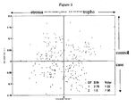
- MALDI-TOF data was performed using ChemoMetricks, an in-house developed statistical analysis toolbox. This toolbox was developed on the Matlab 5.2 (Mathworks, Nantucket MA) platform. All spectra were truncated to 190700 points. Individual spectra were subsequently rebinned in segments of 100 points, yielding 122 spectra of 1907 variables for the preterm data and 255 spectra with 1907 variables in the 7 control versus 7 cases comparison. After reading in the full data the data was normalized and autoscaled prior to PCA. After computing the Principal Components (PC) and the Discriminant functions (DF) all data was plotted using standard Matlab plotting routines.
- PC Principal Components
- DF Discriminant functions
- Placental tissue sections of the samples (3, 10, 17, Table 1) representative for the three groups were separated on an Ultimate NanoLC system (Dionex, Sunnyvale, CA, USA) prior to MALDI-TOF analysis. Assuming that a cell size is 10 x 10 x 10 ⁇ m, the tissue represented approximately 50.000 cells. Tissue samples were prepared in the same way as microdissected tissue. Subsequently, fractionation was performed on 5 ⁇ L tissue sample solutions, for 130 min with a 5 to 95% step wise gradient of solution A (100% H2O, 0.05% TFA) and solution B (20% H2O, 80% ACN, 0.04% TFA).
- a 15 cm C18 column with 75 ⁇ m ID and a guard column were used. Separate fractions were collected using a robotic spotter (Probot Micro Fraction collector, Dionex, Sunnyvale, CA, USA) on a prespotted, with matrix of ⁇ -cyano-4-hydroxycinnimic acid, anchorchip plate (Bruker Daltonics Germany). One ⁇ L of each fraction was spotted. Mass spectrometry analyses were performed in reflectron mode using standard settings for peptide measurements (Bruker default file "Proteomics_HPC.par”) in addition to the MS spectrum a MS/MS spectrum was obtained for each peak using the standard settings for a lift MS/MS measurement.
- LC Warp software (Bruker Daltonics) was used to combine MS/MS spectra of peptides of the same protein. The resulting spectra were analyzed and annotated in Flex analyses 2.2 (Bruker Daltonics). In Biotools 2.2 (Bruker Daltonis) the peak lists from the MS spectra and the MS/MS were combined to create a MGF file. The MGF file was used as input for the Mascot search engine (Matrix Science, London, UK) to search the MSDB human database using a 100 ppm tolerance of the MS spectra and a 0.5 Da tolerance for the fragments in the MS/MS spectra. Identification was significant, if a Mowse score with a probability of p ⁇ 0.05 was observed in the Mascot search engine.
- Prespotted anchorchip plates (Bruker Daltonics, Germany) used for the MS/MS sequencing in the MALDI TOF/TOF were spotted with DHB (10 mg/ mL 0.1%TFA water). Subsequently, for confirmation remains of the fractions were analyzed by MALDI FT ICR MS (APEX Q, Bruker Daltonics) for accurate mass annotation. Identification was confirmed if the calculated mass of the identified differential expressed peptide peaks observed in the MALDI-TOF/TOF analysis did not deviate more than 4 ppm in the MALDI FT-ICR MS analysis from the calculated mass of the peptide identified.
- Figure 5 illustrates the frequency of differentially expressed peptides in women with early compared to late onset preeclampsia.
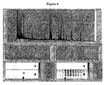
- Figure 6 differences in peptide profiles of trophoblast cells between early onset preeclampsia and preterm delivery with unknown cause are illustrated in Figure 6 .
- Calcyclin was verified by immunostaining, for the other proteins no commercial antibodies were available yet. Immunohistochemistry showed for calcyclin positive staining for trophoblasts from placenta of women with early onset preeclampsia compared with controls ( Fig. 7 ). Table 7 represents the expression levels of calcyclin in the early onset preeclampsia, control and preterm group. By immunohistochemistry, it was evident that choriomammotropin (using an antibody that is not specific for the precursor) is present both in trophoblast of preeclampsia and control placental tissue. Table 7.
- S100A6 calcyclin
- This Example describes different tryptic peptide patterns of villous trophoblast and stroma cells from placentas of women with pregnancies complicated by preeclampsia (cases) compared to uncomplicated pregnancies (controls) in a small number of 125 microdissected cells.
- these peptide patterns are different in placentas from early onset preeclampsia ( ⁇ 34 weeks gestation) compared to a late onset of the disease (> 34 weeks gestation.
- we compared the peptide patterns of women with early onset preeclampsia with women who had a pregnancy complicated by preterm delivery of unknown cause.
- Calcyclin, S100A6 is a member of the S-100 calcium-binding protein family (12). Recently, it has been described that calcyclin stimulates secretion of choriomammotropin (placental lactogen). A dose-dependent release in isolated mice trophoblasts of placental lactogen in response to calcyclin was described by independent researchers. Uterine natural killer (NK) cells were the primary site of calcyclin RNA messengers and calcyclin is expressed in natural killer cells throughout pregnancy. Uterine natural killer cells have been implicated in trophoblast function.
- the present finding is further supported by the reported reduced natural killer cell proportion primarily in pregnancies complicated by impaired placental development (trophoblast infiltration and placental growth) such as preeclampsia. Furthermore, calcyclin secretion is further controlled by cytokines such as interleukin-1. Interleukin-1 is spontaneously synthesized during preeclampsia. This implies a specific and different mechanism in early onset preeclampsia through calcyclin and choriomammotropin.
- Surfeit locus protein 4 is another peptide which is differentially expressed in placentas from women with early onset preeclampsia.
- the surfeit locus genes contain a tight cluster of mammalian genes.
- the surfeit locus 4 gene is encoding an integral membrane protein most likely associated with the endoplasmic reticulum. No association with placentation or human development has hitherto been described.
- Clusterin was identified as over expressed spots on two-dimensional electrophoresis with MALDI-TOF-MS followed by Western blot analysis. Using placenta samples in line with the hypothesis that the responsible trigger for the disease is mostly likely located within the placenta, we could not confirm that clusterin is involved in the pathophysiology of preeclampsia.
Landscapes
- Life Sciences & Earth Sciences (AREA)
- Health & Medical Sciences (AREA)
- Engineering & Computer Science (AREA)
- Molecular Biology (AREA)
- Immunology (AREA)
- Biomedical Technology (AREA)
- Physics & Mathematics (AREA)
- Urology & Nephrology (AREA)
- Hematology (AREA)
- Chemical & Material Sciences (AREA)
- General Health & Medical Sciences (AREA)
- Pathology (AREA)
- Food Science & Technology (AREA)
- Medicinal Chemistry (AREA)
- Biotechnology (AREA)
- Analytical Chemistry (AREA)
- Biochemistry (AREA)
- Microbiology (AREA)
- General Physics & Mathematics (AREA)
- Cell Biology (AREA)
- Bioinformatics & Cheminformatics (AREA)
- Bioinformatics & Computational Biology (AREA)
- Spectroscopy & Molecular Physics (AREA)
- Proteomics, Peptides & Aminoacids (AREA)
- Optics & Photonics (AREA)
- Biophysics (AREA)
- Neurology (AREA)
- Neurosurgery (AREA)
- Other Investigation Or Analysis Of Materials By Electrical Means (AREA)
- Investigating Or Analysing Biological Materials (AREA)
Claims (10)
- Procédé pour détecter une pré-éclampsie à déclenchement précoce chez un sujet comprenant la détermination du niveau d'expression de la protéine calcycline dans un échantillon de villosités choriales dudit sujet.
- Procédé selon la revendication 1 où ledit niveau d'expression est déterminé à un point dans le temps entre 10 et 34 semaines de gestation, de préférence entre 10 et 20 semaines de gestation, de préférence encore entre 10 et 15 semaines de gestation.
- Procédé selon la revendication 1 ou 2 où ledit niveau d'expression est déterminé dans des cellules stromales ou des cellules trophoblastiques, de manière particulièrement préférable dans des trophoblastes.
- Procédé selon l'une quelconque des revendications précédentes où, en plus dudit niveau d'expression de calcycline, également le niveau d'expression d'une ou plusieurs des protéines choisies parmi la protéine surfeit locus, la protéine humaine OTTHUMP00000018488 ayant la séquence d'aminoacides de SEQ ID NO: 4 et le précurseur d'hormone lactogène placentaire A est déterminé.
- Procédé selon l'une quelconque des revendications précédentes où ledit niveau d'expression est déterminé par détection de ladite protéine ou d'un fragment peptidique de celle-ci dans une plage de masse de 800 à 27 000 Da.
- Procédé selon l'une quelconque des revendications précédentes où ladite détection est accomplie par analyse immunohistochimique ou spectrométrie de masse.
- Utilisation d'un procédé selon l'une quelconque des revendications 1-6 où ladite utilisation est pour suivre le processus morbide de pré-éclampsie à déclenchement précoce ou une réponse à une thérapie pendant la maladie.
- Utilisation d'une protéine marqueur ou d'un peptide marqueur pour détecter une pré-éclampsie à déclenchement précoce chez un sujet où ladite protéine marqueur est le niveau d'expression de la protéine calcycline dans un échantillon de villosités choriales dudit sujet et où ledit peptide marqueur est un fragment peptidique de calcycline dans un échantillon de villosités choriales dudit sujet ayant une masse entre 800 à 27 000 Da.
- Utilisation selon la revendication 8 où ledit niveau d'expression est le niveau d'expression à un stade dans la grossesse entre 10 semaines et 34 semaines de gestation.
- Utilisation d'une protéine marqueur selon la revendication 8 ou 9 ou d'un profil marqueur comprenant le niveau d'expression dans un échantillon de villosités choriales d'un sujet d'une première protéine qui est la calcycline ou un peptide de celle-ci, et où ledit profil marqueur comprend en outre au moins un niveau d'expression supplémentaire d'une protéine ou d'un fragment peptidique choisi dans le groupe de la protéine surfeit locus, de la protéine humaine OTTHUMP00000018488 ayant la séquence d'aminoacides de SEQ ID NO: 4 et du précurseur d'hormone lactogène placentaire A pour détecter une pré-éclampsie à déclenchement précoce chez une femme enceinte.
Applications Claiming Priority (2)
| Application Number | Priority Date | Filing Date | Title |
|---|---|---|---|
| PCT/NL2007/050014 WO2008085024A1 (fr) | 2007-01-12 | 2007-01-12 | Identification et détection de peptides associés à des troubles spécifiques |
| PCT/NL2008/000021 WO2008085036A1 (fr) | 2007-01-12 | 2008-01-14 | Peptides marqueurs pour le diagnostic de l'éclampsisme |
Publications (2)
| Publication Number | Publication Date |
|---|---|
| EP2118664A1 EP2118664A1 (fr) | 2009-11-18 |
| EP2118664B1 true EP2118664B1 (fr) | 2012-03-07 |
Family
ID=38171286
Family Applications (2)
| Application Number | Title | Priority Date | Filing Date |
|---|---|---|---|
| EP08705059A Not-in-force EP2118664B1 (fr) | 2007-01-12 | 2008-01-14 | Peptides marqueurs pour le diagnostic de l'eclampsisme |
| EP08705057A Withdrawn EP2118662A1 (fr) | 2007-01-12 | 2008-01-14 | Marqueurs peptidiques pour le diagnostic de l'angiogenese |
Family Applications After (1)
| Application Number | Title | Priority Date | Filing Date |
|---|---|---|---|
| EP08705057A Withdrawn EP2118662A1 (fr) | 2007-01-12 | 2008-01-14 | Marqueurs peptidiques pour le diagnostic de l'angiogenese |
Country Status (7)
| Country | Link |
|---|---|
| US (2) | US20100240088A1 (fr) |
| EP (2) | EP2118664B1 (fr) |
| AT (1) | ATE548658T1 (fr) |
| AU (2) | AU2008203968A1 (fr) |
| CA (2) | CA2674737A1 (fr) |
| NZ (1) | NZ578262A (fr) |
| WO (4) | WO2008085024A1 (fr) |
Families Citing this family (26)
| Publication number | Priority date | Publication date | Assignee | Title |
|---|---|---|---|---|
| EP1937815B1 (fr) | 2005-09-13 | 2015-05-13 | National Research Council of Canada | Méthodes et compositions permettant de moduler l'activité des cellules tumorales |
| EP3358354B1 (fr) | 2008-01-18 | 2020-07-15 | President and Fellows of Harvard College | Procédés de détection de signatures de maladies ou d'états dans des fluides corporels |
| WO2009158423A1 (fr) * | 2008-06-25 | 2009-12-30 | University Of Utah Research Foundation | Identification et quantification de biomarqueurs associés à la prééclampsie |
| IT1392551B1 (it) * | 2008-11-25 | 2012-03-09 | Bioindustry Park Del Canavese S P A | Biomarcatori per la diagnosi e per rilevare la progressione di malattie neurodegenerative, in particolare della sclerosi laterale amiotrofica |
| WO2011063523A1 (fr) | 2009-11-24 | 2011-06-03 | Alethia Biotherapeutics Inc. | Anticorps anti-clustérine et fragments de liaison de l'antigène et leur utilisation dans la réduction du volume d'une tumeur |
| US9595099B2 (en) * | 2010-02-26 | 2017-03-14 | Carolyn M Salafia | Automated placental measurement |
| SG10201505724SA (en) | 2010-07-23 | 2015-09-29 | Harvard College | Methods of detecting diseases or conditions using phagocytic cells |
| MX382244B (es) | 2010-07-23 | 2025-03-13 | Harvard College | Metodos para detectar firmas de enfermedad o condiciones en fluidos corporales |
| US20130203624A1 (en) | 2010-07-23 | 2013-08-08 | President And Fellows Of Harvard College | Methods of Detecting Prenatal or Pregnancy-Related Diseases or Conditions |
| CA2806293A1 (fr) | 2010-07-23 | 2012-01-26 | President And Fellows Of Harvard College | Procedes de detection de maladies/pathologies auto-immunes ou liees au systeme immunitaire |
| KR101449100B1 (ko) | 2011-04-21 | 2014-10-13 | 가톨릭대학교 산학협력단 | 신경세포 데브리스 제거용 오스테오폰틴 |
| US20150004632A1 (en) | 2011-10-14 | 2015-01-01 | Aarhus Universitet | Diagnostic and prognostic use of prombp-complexes |
| WO2013123588A1 (fr) | 2012-02-22 | 2013-08-29 | Alethia Biotherapeutics Inc. | Utilisation conjointe d'un inhibiteur de clusterine et d'un inhibiteur d'egfr pour traiter le cancer |
| EP2836832A4 (fr) * | 2012-04-13 | 2015-10-14 | Univ Wayne State | Dépistage en début de grossesse d'une pré-éclampsie précoce ou tardive |
| CN104603289A (zh) | 2012-06-15 | 2015-05-06 | 哈里·斯泰利 | 检测疾病或病状的方法 |
| SG11201408383SA (en) | 2012-06-15 | 2015-01-29 | Harry Stylli | Methods of detecting diseases or conditions using circulating diseased cells |
| SG11201408652SA (en) | 2012-06-26 | 2015-01-29 | Biodesix Inc | Mass-spectral method for selection, and de-selection, of cancer patients for treatment with immune response generating therapies |
| EP4202441A3 (fr) | 2013-03-09 | 2023-07-26 | Immunis.AI, Inc. | Profil d'expression génétique dans les macrophages pour le diagnostic du cancer |
| WO2014164362A1 (fr) | 2013-03-09 | 2014-10-09 | Harry Stylli | Procédés de détection du cancer de la prostate |
| US10626464B2 (en) | 2014-09-11 | 2020-04-21 | Cell Mdx, Llc | Methods of detecting prostate cancer |
| ES2955883T3 (es) | 2015-06-19 | 2023-12-07 | Sera Prognostics Inc | Pares de biomarcadores para la predicción del parto prematuro |
| FR3038984B1 (fr) * | 2015-07-15 | 2020-01-10 | Universite de Bordeaux | Analyse proteomique de compartiments subcellulaires |
| WO2018151938A2 (fr) * | 2017-02-17 | 2018-08-23 | The Methodist Hospital | Compositions et méthodes de détermination d'un niveau d'infection chez un sujet |
| AU2018317902B2 (en) | 2017-08-18 | 2025-02-20 | Sera Prognostics, Inc. | Pregnancy clock proteins for predicting due date and time to birth |
| AU2018324195B2 (en) | 2017-09-01 | 2024-12-12 | Venn Biosciences Corporation | Identification and use of glycopeptides as biomarkers for diagnosis and treatment monitoring |
| JPWO2019202767A1 (ja) * | 2018-04-17 | 2021-05-13 | 国立大学法人大阪大学 | 抗線維化剤及び線維症のバイオマーカー |
Family Cites Families (6)
| Publication number | Priority date | Publication date | Assignee | Title |
|---|---|---|---|---|
| US5185243A (en) * | 1988-08-25 | 1993-02-09 | Syntex (U.S.A.) Inc. | Method for detection of specific nucleic acid sequences |
| US5436132A (en) * | 1993-02-13 | 1995-07-25 | Amano Pharmaceutical Co., Ltd. | Quantitative determination of tenascin as glioma marker |
| ITMI20011833A1 (it) * | 2001-08-31 | 2003-03-03 | San Raffaele Centro Fond | Agenti che regolano la permeabilita' vascolare |
| CA2519129A1 (fr) * | 2003-03-18 | 2004-09-30 | Biovision Ag | Procede de depistage d'une maladie dementielle chronique progressive, et peptides et reactifs de depistage correspondants |
| CA2565405A1 (fr) * | 2004-06-18 | 2005-12-29 | F. Hoffmann-La Roche Ag | Utilisation d'une proteine cbp2 en tant que marqueur pour un cancer colorectal |
| EP1998789A4 (fr) * | 2006-02-28 | 2009-04-22 | Metabolon Inc | Biomarqueurs pour sclérose latérale amyotrophique |
-
2007
- 2007-01-12 WO PCT/NL2007/050014 patent/WO2008085024A1/fr not_active Ceased
-
2008
- 2008-01-14 US US12/522,880 patent/US20100240088A1/en not_active Abandoned
- 2008-01-14 NZ NZ578262A patent/NZ578262A/en not_active IP Right Cessation
- 2008-01-14 EP EP08705059A patent/EP2118664B1/fr not_active Not-in-force
- 2008-01-14 US US12/522,875 patent/US20100291612A1/en not_active Abandoned
- 2008-01-14 AT AT08705059T patent/ATE548658T1/de active
- 2008-01-14 AU AU2008203968A patent/AU2008203968A1/en not_active Abandoned
- 2008-01-14 AU AU2008203970A patent/AU2008203970A1/en not_active Abandoned
- 2008-01-14 WO PCT/NL2008/000020 patent/WO2008085035A1/fr not_active Ceased
- 2008-01-14 WO PCT/NL2008/000021 patent/WO2008085036A1/fr not_active Ceased
- 2008-01-14 CA CA002674737A patent/CA2674737A1/fr not_active Abandoned
- 2008-01-14 WO PCT/NL2008/000019 patent/WO2008085034A1/fr not_active Ceased
- 2008-01-14 EP EP08705057A patent/EP2118662A1/fr not_active Withdrawn
- 2008-01-14 CA CA002674736A patent/CA2674736A1/fr not_active Abandoned
Also Published As
| Publication number | Publication date |
|---|---|
| CA2674737A1 (fr) | 2008-07-17 |
| WO2008085034A1 (fr) | 2008-07-17 |
| ATE548658T1 (de) | 2012-03-15 |
| NZ578262A (en) | 2012-02-24 |
| EP2118664A1 (fr) | 2009-11-18 |
| US20100291612A1 (en) | 2010-11-18 |
| WO2008085024A1 (fr) | 2008-07-17 |
| AU2008203970A1 (en) | 2008-07-17 |
| CA2674736A1 (fr) | 2008-07-17 |
| US20100240088A1 (en) | 2010-09-23 |
| AU2008203968A1 (en) | 2008-07-17 |
| WO2008085036A1 (fr) | 2008-07-17 |
| EP2118662A1 (fr) | 2009-11-18 |
| WO2008085035A1 (fr) | 2008-07-17 |
Similar Documents
| Publication | Publication Date | Title |
|---|---|---|
| US20100291612A1 (en) | Peptide Markers for Diagnosis of Preeclampsia | |
| US20240318250A1 (en) | Biomarker pairs for predicting preterm birth | |
| US12601744B2 (en) | Biomarkers and methods for predicting preterm birth | |
| US8679769B2 (en) | Detection of endometrial secretion markers for assessment of endometriosis | |
| US20100016173A1 (en) | Maternal serum biomarkers for detection of pre-eclampsia | |
| US10215760B2 (en) | Detection of intraamniotic and/or infection | |
| EP1618388B1 (fr) | Analyse proteomique de fluides biologiques | |
| US20060094039A1 (en) | Diagnosis of fetal aneuploidy | |
| US20100017143A1 (en) | Gestational age dependent proteomic changes of human maternal serum for monitoring maternal and fetal health | |
| JP2013534309A (ja) | 腎機能不全のバイオマーカーとしてのパールカン | |
| TW201905461A (zh) | 用以檢測大腸癌之生物標記 | |
| JP2010522882A (ja) | 卵巣癌のバイオマーカー | |
| EP4430406A2 (fr) | Procédés de préparation et d'analyse d'échantillons pour biomarqueurs associés au placenta accreta | |
| HK40107594A (en) | Biomarker pairs for predicting preterm birth | |
| Kolla | Identification of Screening Biomarkers for Chromosomal Anomalies and Pregnancy-related Disorder Using Quantitative Plasma Proteomics | |
| HK1254669B (en) | Biomarker pairs for predicting preterm birth |
Legal Events
| Date | Code | Title | Description |
|---|---|---|---|
| PUAI | Public reference made under article 153(3) epc to a published international application that has entered the european phase |
Free format text: ORIGINAL CODE: 0009012 |
|
| 17P | Request for examination filed |
Effective date: 20090803 |
|
| AK | Designated contracting states |
Kind code of ref document: A1 Designated state(s): AT BE BG CH CY CZ DE DK EE ES FI FR GB GR HR HU IE IS IT LI LT LU LV MC MT NL NO PL PT RO SE SI SK TR |
|
| DAX | Request for extension of the european patent (deleted) | ||
| 17Q | First examination report despatched |
Effective date: 20100326 |
|
| GRAP | Despatch of communication of intention to grant a patent |
Free format text: ORIGINAL CODE: EPIDOSNIGR1 |
|
| GRAS | Grant fee paid |
Free format text: ORIGINAL CODE: EPIDOSNIGR3 |
|
| GRAA | (expected) grant |
Free format text: ORIGINAL CODE: 0009210 |
|
| AK | Designated contracting states |
Kind code of ref document: B1 Designated state(s): AT BE BG CH CY CZ DE DK EE ES FI FR GB GR HR HU IE IS IT LI LT LU LV MC MT NL NO PL PT RO SE SI SK TR |
|
| REG | Reference to a national code |
Ref country code: GB Ref legal event code: FG4D |
|
| REG | Reference to a national code |
Ref country code: AT Ref legal event code: REF Ref document number: 548658 Country of ref document: AT Kind code of ref document: T Effective date: 20120315 Ref country code: CH Ref legal event code: EP |
|
| REG | Reference to a national code |
Ref country code: IE Ref legal event code: FG4D |
|
| REG | Reference to a national code |
Ref country code: DE Ref legal event code: R096 Ref document number: 602008013940 Country of ref document: DE Effective date: 20120503 |
|
| REG | Reference to a national code |
Ref country code: NL Ref legal event code: VDEP Effective date: 20120307 |
|
| PG25 | Lapsed in a contracting state [announced via postgrant information from national office to epo] |
Ref country code: HR Free format text: LAPSE BECAUSE OF FAILURE TO SUBMIT A TRANSLATION OF THE DESCRIPTION OR TO PAY THE FEE WITHIN THE PRESCRIBED TIME-LIMIT Effective date: 20120307 Ref country code: LT Free format text: LAPSE BECAUSE OF FAILURE TO SUBMIT A TRANSLATION OF THE DESCRIPTION OR TO PAY THE FEE WITHIN THE PRESCRIBED TIME-LIMIT Effective date: 20120307 Ref country code: NO Free format text: LAPSE BECAUSE OF FAILURE TO SUBMIT A TRANSLATION OF THE DESCRIPTION OR TO PAY THE FEE WITHIN THE PRESCRIBED TIME-LIMIT Effective date: 20120607 Ref country code: NL Free format text: LAPSE BECAUSE OF FAILURE TO SUBMIT A TRANSLATION OF THE DESCRIPTION OR TO PAY THE FEE WITHIN THE PRESCRIBED TIME-LIMIT Effective date: 20120307 |
|
| LTIE | Lt: invalidation of european patent or patent extension |
Effective date: 20120307 |
|
| PG25 | Lapsed in a contracting state [announced via postgrant information from national office to epo] |
Ref country code: FI Free format text: LAPSE BECAUSE OF FAILURE TO SUBMIT A TRANSLATION OF THE DESCRIPTION OR TO PAY THE FEE WITHIN THE PRESCRIBED TIME-LIMIT Effective date: 20120307 Ref country code: GR Free format text: LAPSE BECAUSE OF FAILURE TO SUBMIT A TRANSLATION OF THE DESCRIPTION OR TO PAY THE FEE WITHIN THE PRESCRIBED TIME-LIMIT Effective date: 20120608 Ref country code: LV Free format text: LAPSE BECAUSE OF FAILURE TO SUBMIT A TRANSLATION OF THE DESCRIPTION OR TO PAY THE FEE WITHIN THE PRESCRIBED TIME-LIMIT Effective date: 20120307 |
|
| REG | Reference to a national code |
Ref country code: AT Ref legal event code: MK05 Ref document number: 548658 Country of ref document: AT Kind code of ref document: T Effective date: 20120307 |
|
| PG25 | Lapsed in a contracting state [announced via postgrant information from national office to epo] |
Ref country code: CY Free format text: LAPSE BECAUSE OF FAILURE TO SUBMIT A TRANSLATION OF THE DESCRIPTION OR TO PAY THE FEE WITHIN THE PRESCRIBED TIME-LIMIT Effective date: 20120307 |
|
| PG25 | Lapsed in a contracting state [announced via postgrant information from national office to epo] |
Ref country code: RO Free format text: LAPSE BECAUSE OF FAILURE TO SUBMIT A TRANSLATION OF THE DESCRIPTION OR TO PAY THE FEE WITHIN THE PRESCRIBED TIME-LIMIT Effective date: 20120307 Ref country code: PL Free format text: LAPSE BECAUSE OF FAILURE TO SUBMIT A TRANSLATION OF THE DESCRIPTION OR TO PAY THE FEE WITHIN THE PRESCRIBED TIME-LIMIT Effective date: 20120307 Ref country code: EE Free format text: LAPSE BECAUSE OF FAILURE TO SUBMIT A TRANSLATION OF THE DESCRIPTION OR TO PAY THE FEE WITHIN THE PRESCRIBED TIME-LIMIT Effective date: 20120307 Ref country code: SE Free format text: LAPSE BECAUSE OF FAILURE TO SUBMIT A TRANSLATION OF THE DESCRIPTION OR TO PAY THE FEE WITHIN THE PRESCRIBED TIME-LIMIT Effective date: 20120307 Ref country code: SI Free format text: LAPSE BECAUSE OF FAILURE TO SUBMIT A TRANSLATION OF THE DESCRIPTION OR TO PAY THE FEE WITHIN THE PRESCRIBED TIME-LIMIT Effective date: 20120307 Ref country code: CZ Free format text: LAPSE BECAUSE OF FAILURE TO SUBMIT A TRANSLATION OF THE DESCRIPTION OR TO PAY THE FEE WITHIN THE PRESCRIBED TIME-LIMIT Effective date: 20120307 Ref country code: BE Free format text: LAPSE BECAUSE OF FAILURE TO SUBMIT A TRANSLATION OF THE DESCRIPTION OR TO PAY THE FEE WITHIN THE PRESCRIBED TIME-LIMIT Effective date: 20120307 Ref country code: IS Free format text: LAPSE BECAUSE OF FAILURE TO SUBMIT A TRANSLATION OF THE DESCRIPTION OR TO PAY THE FEE WITHIN THE PRESCRIBED TIME-LIMIT Effective date: 20120707 |
|
| PG25 | Lapsed in a contracting state [announced via postgrant information from national office to epo] |
Ref country code: PT Free format text: LAPSE BECAUSE OF FAILURE TO SUBMIT A TRANSLATION OF THE DESCRIPTION OR TO PAY THE FEE WITHIN THE PRESCRIBED TIME-LIMIT Effective date: 20120709 Ref country code: SK Free format text: LAPSE BECAUSE OF FAILURE TO SUBMIT A TRANSLATION OF THE DESCRIPTION OR TO PAY THE FEE WITHIN THE PRESCRIBED TIME-LIMIT Effective date: 20120307 |
|
| PLBE | No opposition filed within time limit |
Free format text: ORIGINAL CODE: 0009261 |
|
| STAA | Information on the status of an ep patent application or granted ep patent |
Free format text: STATUS: NO OPPOSITION FILED WITHIN TIME LIMIT |
|
| PG25 | Lapsed in a contracting state [announced via postgrant information from national office to epo] |
Ref country code: AT Free format text: LAPSE BECAUSE OF FAILURE TO SUBMIT A TRANSLATION OF THE DESCRIPTION OR TO PAY THE FEE WITHIN THE PRESCRIBED TIME-LIMIT Effective date: 20120307 Ref country code: DK Free format text: LAPSE BECAUSE OF FAILURE TO SUBMIT A TRANSLATION OF THE DESCRIPTION OR TO PAY THE FEE WITHIN THE PRESCRIBED TIME-LIMIT Effective date: 20120307 |
|
| 26N | No opposition filed |
Effective date: 20121210 |
|
| PG25 | Lapsed in a contracting state [announced via postgrant information from national office to epo] |
Ref country code: IT Free format text: LAPSE BECAUSE OF FAILURE TO SUBMIT A TRANSLATION OF THE DESCRIPTION OR TO PAY THE FEE WITHIN THE PRESCRIBED TIME-LIMIT Effective date: 20120307 |
|
| REG | Reference to a national code |
Ref country code: DE Ref legal event code: R097 Ref document number: 602008013940 Country of ref document: DE Effective date: 20121210 |
|
| PG25 | Lapsed in a contracting state [announced via postgrant information from national office to epo] |
Ref country code: ES Free format text: LAPSE BECAUSE OF FAILURE TO SUBMIT A TRANSLATION OF THE DESCRIPTION OR TO PAY THE FEE WITHIN THE PRESCRIBED TIME-LIMIT Effective date: 20120618 |
|
| PG25 | Lapsed in a contracting state [announced via postgrant information from national office to epo] |
Ref country code: BG Free format text: LAPSE BECAUSE OF FAILURE TO SUBMIT A TRANSLATION OF THE DESCRIPTION OR TO PAY THE FEE WITHIN THE PRESCRIBED TIME-LIMIT Effective date: 20120607 |
|
| PG25 | Lapsed in a contracting state [announced via postgrant information from national office to epo] |
Ref country code: MC Free format text: LAPSE BECAUSE OF NON-PAYMENT OF DUE FEES Effective date: 20130131 |
|
| REG | Reference to a national code |
Ref country code: CH Ref legal event code: PL |
|
| GBPC | Gb: european patent ceased through non-payment of renewal fee |
Effective date: 20130114 |
|
| REG | Reference to a national code |
Ref country code: IE Ref legal event code: MM4A |
|
| REG | Reference to a national code |
Ref country code: FR Ref legal event code: ST Effective date: 20130930 |
|
| PG25 | Lapsed in a contracting state [announced via postgrant information from national office to epo] |
Ref country code: DE Free format text: LAPSE BECAUSE OF NON-PAYMENT OF DUE FEES Effective date: 20130801 Ref country code: CH Free format text: LAPSE BECAUSE OF NON-PAYMENT OF DUE FEES Effective date: 20130131 Ref country code: LI Free format text: LAPSE BECAUSE OF NON-PAYMENT OF DUE FEES Effective date: 20130131 |
|
| REG | Reference to a national code |
Ref country code: DE Ref legal event code: R119 Ref document number: 602008013940 Country of ref document: DE Effective date: 20130801 |
|
| PG25 | Lapsed in a contracting state [announced via postgrant information from national office to epo] |
Ref country code: GB Free format text: LAPSE BECAUSE OF NON-PAYMENT OF DUE FEES Effective date: 20130114 Ref country code: FR Free format text: LAPSE BECAUSE OF NON-PAYMENT OF DUE FEES Effective date: 20130131 |
|
| PG25 | Lapsed in a contracting state [announced via postgrant information from national office to epo] |
Ref country code: IE Free format text: LAPSE BECAUSE OF NON-PAYMENT OF DUE FEES Effective date: 20130114 |
|
| PG25 | Lapsed in a contracting state [announced via postgrant information from national office to epo] |
Ref country code: MT Free format text: LAPSE BECAUSE OF FAILURE TO SUBMIT A TRANSLATION OF THE DESCRIPTION OR TO PAY THE FEE WITHIN THE PRESCRIBED TIME-LIMIT Effective date: 20120307 |
|
| PG25 | Lapsed in a contracting state [announced via postgrant information from national office to epo] |
Ref country code: TR Free format text: LAPSE BECAUSE OF FAILURE TO SUBMIT A TRANSLATION OF THE DESCRIPTION OR TO PAY THE FEE WITHIN THE PRESCRIBED TIME-LIMIT Effective date: 20120307 |
|
| PG25 | Lapsed in a contracting state [announced via postgrant information from national office to epo] |
Ref country code: HU Free format text: LAPSE BECAUSE OF FAILURE TO SUBMIT A TRANSLATION OF THE DESCRIPTION OR TO PAY THE FEE WITHIN THE PRESCRIBED TIME-LIMIT; INVALID AB INITIO Effective date: 20080114 Ref country code: LU Free format text: LAPSE BECAUSE OF NON-PAYMENT OF DUE FEES Effective date: 20130114 |