EP2119767B1 - Verfahren zur präparation einer knorpelzelle - Google Patents
Verfahren zur präparation einer knorpelzelle Download PDFInfo
- Publication number
- EP2119767B1 EP2119767B1 EP08704109.1A EP08704109A EP2119767B1 EP 2119767 B1 EP2119767 B1 EP 2119767B1 EP 08704109 A EP08704109 A EP 08704109A EP 2119767 B1 EP2119767 B1 EP 2119767B1
- Authority
- EP
- European Patent Office
- Prior art keywords
- chondrocytes
- cell
- cartilage
- perichondrocytes
- tissue
- Prior art date
- Legal status (The legal status is an assumption and is not a legal conclusion. Google has not performed a legal analysis and makes no representation as to the accuracy of the status listed.)
- Not-in-force
Links
Images











Classifications
-
- C—CHEMISTRY; METALLURGY
- C12—BIOCHEMISTRY; BEER; SPIRITS; WINE; VINEGAR; MICROBIOLOGY; ENZYMOLOGY; MUTATION OR GENETIC ENGINEERING
- C12N—MICROORGANISMS OR ENZYMES; COMPOSITIONS THEREOF; PROPAGATING, PRESERVING, OR MAINTAINING MICROORGANISMS; MUTATION OR GENETIC ENGINEERING; CULTURE MEDIA
- C12N5/00—Undifferentiated human, animal or plant cells, e.g. cell lines; Tissues; Cultivation or maintenance thereof; Culture media therefor
- C12N5/06—Animal cells or tissues; Human cells or tissues
-
- C—CHEMISTRY; METALLURGY
- C12—BIOCHEMISTRY; BEER; SPIRITS; WINE; VINEGAR; MICROBIOLOGY; ENZYMOLOGY; MUTATION OR GENETIC ENGINEERING
- C12N—MICROORGANISMS OR ENZYMES; COMPOSITIONS THEREOF; PROPAGATING, PRESERVING, OR MAINTAINING MICROORGANISMS; MUTATION OR GENETIC ENGINEERING; CULTURE MEDIA
- C12N5/00—Undifferentiated human, animal or plant cells, e.g. cell lines; Tissues; Cultivation or maintenance thereof; Culture media therefor
- C12N5/06—Animal cells or tissues; Human cells or tissues
- C12N5/0602—Vertebrate cells
- C12N5/0652—Cells of skeletal and connective tissues; Mesenchyme
- C12N5/0655—Chondrocytes; Cartilage
-
- A—HUMAN NECESSITIES
- A61—MEDICAL OR VETERINARY SCIENCE; HYGIENE
- A61K—PREPARATIONS FOR MEDICAL, DENTAL OR TOILETRY PURPOSES
- A61K35/00—Medicinal preparations containing materials or reaction products thereof with undetermined constitution
- A61K35/12—Materials from mammals; Compositions comprising non-specified tissues or cells; Compositions comprising non-embryonic stem cells; Genetically modified cells
- A61K35/32—Bones; Osteocytes; Osteoblasts; Tendons; Tenocytes; Teeth; Odontoblasts; Cartilage; Chondrocytes; Synovial membrane
-
- A—HUMAN NECESSITIES
- A61—MEDICAL OR VETERINARY SCIENCE; HYGIENE
- A61P—SPECIFIC THERAPEUTIC ACTIVITY OF CHEMICAL COMPOUNDS OR MEDICINAL PREPARATIONS
- A61P19/00—Drugs for skeletal disorders
-
- A—HUMAN NECESSITIES
- A61—MEDICAL OR VETERINARY SCIENCE; HYGIENE
- A61P—SPECIFIC THERAPEUTIC ACTIVITY OF CHEMICAL COMPOUNDS OR MEDICINAL PREPARATIONS
- A61P43/00—Drugs for specific purposes, not provided for in groups A61P1/00-A61P41/00
-
- C—CHEMISTRY; METALLURGY
- C12—BIOCHEMISTRY; BEER; SPIRITS; WINE; VINEGAR; MICROBIOLOGY; ENZYMOLOGY; MUTATION OR GENETIC ENGINEERING
- C12N—MICROORGANISMS OR ENZYMES; COMPOSITIONS THEREOF; PROPAGATING, PRESERVING, OR MAINTAINING MICROORGANISMS; MUTATION OR GENETIC ENGINEERING; CULTURE MEDIA
- C12N2500/00—Specific components of cell culture medium
- C12N2500/05—Inorganic components
- C12N2500/10—Metals; Metal chelators
- C12N2500/20—Transition metals
- C12N2500/24—Iron; Fe chelators; Transferrin
- C12N2500/25—Insulin-transferrin; Insulin-transferrin-selenium
-
- C—CHEMISTRY; METALLURGY
- C12—BIOCHEMISTRY; BEER; SPIRITS; WINE; VINEGAR; MICROBIOLOGY; ENZYMOLOGY; MUTATION OR GENETIC ENGINEERING
- C12N—MICROORGANISMS OR ENZYMES; COMPOSITIONS THEREOF; PROPAGATING, PRESERVING, OR MAINTAINING MICROORGANISMS; MUTATION OR GENETIC ENGINEERING; CULTURE MEDIA
- C12N2501/00—Active agents used in cell culture processes, e.g. differentation
- C12N2501/10—Growth factors
- C12N2501/105—Insulin-like growth factors [IGF]
-
- C—CHEMISTRY; METALLURGY
- C12—BIOCHEMISTRY; BEER; SPIRITS; WINE; VINEGAR; MICROBIOLOGY; ENZYMOLOGY; MUTATION OR GENETIC ENGINEERING
- C12N—MICROORGANISMS OR ENZYMES; COMPOSITIONS THEREOF; PROPAGATING, PRESERVING, OR MAINTAINING MICROORGANISMS; MUTATION OR GENETIC ENGINEERING; CULTURE MEDIA
- C12N2501/00—Active agents used in cell culture processes, e.g. differentation
- C12N2501/10—Growth factors
- C12N2501/115—Basic fibroblast growth factor (bFGF, FGF-2)
-
- C—CHEMISTRY; METALLURGY
- C12—BIOCHEMISTRY; BEER; SPIRITS; WINE; VINEGAR; MICROBIOLOGY; ENZYMOLOGY; MUTATION OR GENETIC ENGINEERING
- C12N—MICROORGANISMS OR ENZYMES; COMPOSITIONS THEREOF; PROPAGATING, PRESERVING, OR MAINTAINING MICROORGANISMS; MUTATION OR GENETIC ENGINEERING; CULTURE MEDIA
- C12N2501/00—Active agents used in cell culture processes, e.g. differentation
- C12N2501/30—Hormones
- C12N2501/38—Hormones with nuclear receptors
- C12N2501/39—Steroid hormones
Definitions
- the present invention relates to a method of preparing chondrocytes. More specifically, the present invention relates to a method of preparing chondrocytes from perichondrocytes.
- Non-Patent Documents Nos. 1-4 methods in which a part of autologous chondrocytes is taken, cultured ex vivo and returned to the deficient site have been developed (Non-Patent Documents Nos. 1-4) and applied clinically (Non-Patent Documents Nos. 5, 6 and 7).
- chondrocytes have two problems: invasion into the donor site and retention of the shape of a regenerated tissue for a long time.
- the first one is a problem that the donor site from which a cartilage tissue has been taken for culture may result in deformities, such as defect or recess, or dysfunction.
- the second one retention of the shape for a long time is a problem whether a tissue regenerated with cultured cartilage can retain its shape for a long time without being absorbed.
- chondrocytes In order to solve these problems, other sources of chondrocytes have been sought for. That is, an idea has been contemplated in which cells other than chondrocytes are differentiated ex vivo into chondrocytes and returned into the living body. Examples of these cells include embryonic stem cells, mesenchymal stem cells, cells derived from the synovial membrane of knee joint, and adipocytes (Non-Patent Documents Nos. 1-4). All of these cells have been confirmed to differentiate into chondrocytes.
- embryonic stem cells have the problems of invasion and differentiation efficiency. Thus, any of these cells has not reached the stage of clinical application.
- Non-Patent Document 11 reports the presence of cartilage progenitor cells in the ear perichondrium of adult rabbits.
- the present inventors have developed a method of using perichondrium covering the outside of auricular cartilage or costicartilage.
- the present inventors have succeeded in producing proteoglycan and type II collagen (matrixes peculiar to chondrocytes) by isolating and proliferating perichondrocytes from perichondrium and differentiating the resultant perichondrocytes into chondrocytes in vivo or ex vivo.
- the present invention may be summarized as follows.
- the method of preparing chondrocytes according to the present invention need not collect a cartilage tissue. Therefore, it is possible to minimize the invasion into donor sites.
- the method of the present invention makes it possible to retain the shape of a regenerated tissue for a long time.
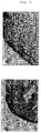
- Fig. 1 Histological Examination of Human Perichondrium at the Time of Collection.
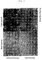
- Fig. 2 Centrifuge Tube Culture of Perichondrocytes
- a cell mass of perichondrocytes forms a cartilage tissue in a centrifuge tube.
- a cartilage tissue regenerated from perichondrocytes
- b cartilage tissue regenerated from chondrocytes. Alcian blue staining. Bar: 200 ⁇ m
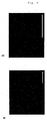
- Fig. 3 The matrix (proteoglycan)-producing capacity of perichondrocytes is enhanced by multi-layering in vitro, in the same manner as seen in chondrocytes.
- a bright field
- b Alcian blue staining.
- Fig. 4 The matrix (proteoglycan)-producing capacity of perichondrocytes is enhanced by multi-layering in vitro, in the same manner as seen in chondrocytes.
- the matrix-producing capacity of perichondrocytes is comparable to that of chondrocytes.
- c mono-layered chondrocytes;
- d multi-layered (triple-layered) chondrocytes. Alcian blue staining.
- Fig. 5 RT-PCR in Multi-Layered Culture of Perichondrocytes and Chondrocytes
- type I collagen decreases and type II collagen increases as a result of multi-layering.
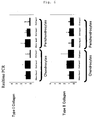
- Fig. 6 Quantitative Determination by Real Time PCR in Multi-Layered Culture of Perichondrocytes and Chondrocytes
- type I collagen tends to decrease and type II collagen tends to increase as a result of multi-layering.
- Fig. 7 Proteoglycan is produced in the supernatant as a result of multi-layering in vitro.
- the proteoglycan-producing capacity of perichondrocytes is almost comparable to that of chondrocytes.
- Fig. 8 Cultured human perichondrocytes form a cartilage tissue in vivo.
- This Figure shows the state after two months of subcutaneous transplanting of cultured perichondrocytes in the back of NOD/SCID mouse. Alcian blue staining. a: magnification 200; b: magnification 400
- Fig. 9 Cultured human perichondrocytes produce type I and type II collagen and form a cartilage tissue in vivo.
- This Figure shows the state after two months of subcutaneous transplanting of cultured perichondrocytes in the back of NOD/SCID mouse. Alcian blue staining. Type I collagen (red) and type II collagen (green). a: magnification 100; b: magnification 400
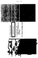
- Fig. 10 shows the layer structure of perichondrium and cartilage such as elastic cartilage and hyaline cartilage.
- Fig. 11 supplements Fig. 1 .
- a human perichondrial tissue is collected in outermost layer and separate two layers of fibroblast layer and innermost layer. Those other than these layers fall under cartilage tissue.
- c cartilage tissue. Alcian blue staining. Magnification: 200
- Fig. 12 The Growth Capacity of Human Perichondrocytes. This Figure shows microscopic comparison of the colony forming activities of perichondrocytes and chondrocytes. Compared to human chondrocytes, human perichondrocytes formed a larger colony after one month of culture. Human chondrocytes (dotted arrow). Human perichondrocytes (solid arrow). Scale bar: 500 ⁇ m
- Fig. 13 The Growth Capacity of Human Perichondrocytes. This Figure shows comparison of the colony-forming activities of human perichondrocytes, cells in the perichondrium-cartilage transition zone, and chondrocytes.
- Perichondrocytes cells in perichondrium-cartilage transition zone and chondrocytes were cultured for 14 days. Comparison of the numbers of colonies formed by 50 cells or more revealed that perichondrocytes have a high colony-forming activity. Thus, it was revealed that perichondrocytes have a high growth capacity localized therein.
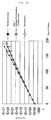
- Fig. 14 The Growth Capacity of Human Perichondrocytes. This Figure shows comparison of the long-term growth capacities of human perichondrocytes, cells in the cartilage-perichondrium transition zone, and chondrocytes.
- Multipotency was examined in vitro.
- Panel A Induction of lipid differentiation: while perichondrocytes formed lipid droplets and were stained with Oil red O (lipid staining reagent), chondrocytes did not form lipid droplets and were not stained with Oil red O.
- Panel B Induction of bone differentiation: while perichondrocytes showed a large number of Ca deposits and were stained with alizarin red, chondrocytes showed no Ca deposits and were not stained with alizarin red. Thus, multipotency to lipid and bone was recognized in perichondrocytes.
- Fig. 16 In Vitro Induction of Differentiation into Cartilage Panel A: Induction of Cartilage Differentiation in Human Perichondrocytes
- the extracellular matrix-producing capacity was increased by (A) mono-layered, (B) double-layered and (C) triple-layered culture of perichondrocytes in a differentiation inducing medium.
- perichondrocytes were stained with type I collagen (red), type II collagen (green) and DAPI (blue), it was also confirmed that the extracellular matrix-producing capacity was increased by (D) mono-layered, (E) double-layered and (F) triple-layered culture.
- a differentiation inducing medium was not used, extracellular matrix was not produced even when (G) mono-layered, (H) double-layered and (I) triple-layered culture was performed.
- A-C, G-I Alcian blue staining; D-F: type I collagen staining (red), type II collagen staining (green) and DAP staining I (blue). Scale bar: 200 ⁇ m
- Panel B Induction of Cartilage Differentiation in Human Chondrocytes
- the extracellular matrix-producing capacity was increased by (A) mono-layered, (B) double-layered and (C) triple-layered culture of chondrocytes in a differentiation inducing medium.
- chondrocytes were stained with type I collagen (red), type II collagen (green) and DAPI (blue), it was also confirmed that the extracellular matrix-producing capacity was increased by (D) mono-layered, (E) double-layered and (F) triple-layered culture.
- A-C, G-I Alcian blue staining; D-F: type I collagen staining (red), type II collagen staining (green) and DAP staining I (blue). Scale bar: 200 ⁇ m
- FIG. 17 Histological Examination of Cartilage Tissues Regenerated in vivo from Human Perichondrocytes and Chondrocytes
- Panel A Human Perichondrocytes-Derived and Human Chondrocytes-Derived Regenerated Cartilage Tissues after One Month of Transplant (A-D: perichondrocytes-derived cartilage tissue; E-H: chondrocytes-derived cartilage tissue)
- Panel B Human Perichondrocytes-Derived and Human Chondrocytes-Derived Regenerated Cartilage Tissues after Three Months of Transplant (A-D: perichondrocytes-derived cartilage tissue; E-H: chondrocytes-derived cartilage tissue)
- A, E HE staining; B, F: Alcian blue staining; C, G: Elastica van Gieson staining; D, H: type I collagen staining (red), type II collagen staining (green) and DAP staining I (blue). Scale bar: 200 ⁇ m
- perichondrocytes regenerate cartilage in the same manner as chondrocytes do. Unlike from the chondrocytes-derived regenerated cartilage, the perichondrocytes-derived regenerated cartilage is covered with perichondrium. This suggests that perichondrocytes-derived regenerated cartilage is superior in long-term shape-retaining ability.
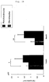
- This Figure shows the cell count per mm 2 of the cartilage portion in tissues removed one month and three months after transplant. While no change in cell count was observed in the perichondrocytes-derived tissue whether one month or three months passed after transplant, a decrease in cell count was observed in the chondrocytes-derived tissue three months after transplant. *: p ⁇ 0.05, **: p ⁇ 0.01 (Mann Whitney U-Test) These results indicate that perichondrocytes are superior to chondrocytes in long-term shape-retaining ability.
- Fig. 19 Comparison of Colony Forming Activities in 10% Human Autoserum Medium between Human Perichondrocytes, Cells in Perichondrium-Cartilage Transition Zone, and Chondrocytes
- Fig. 20 Microscopic Comparison of Human Perichondrocytes, Cells in Perichondrium-Cartilage Transition Zone, and Chondrocytes after culture in 10% Human Autoserum Medium
- cartilage tissue is collected as a mass, which is then treated with enzymes such as collagenase to isolate chondrocytes from the matrix components; the thus isolated chondrocytes are cultured for transplant treatment, especially autotransplantation.
- perichondrium and cartilage such as elastic cartilage and hyaline cartilage are composed of four layers ( Fig. 10 ). They are 1) outermost layer (including capillary vessels); 2) fibroblast layer (mainly consisting of perichondrocytes) (expressed as "perichondrocyte layer” in Fig. 10 ); 3) innermost layer (zone of transition to the cartilage matrix), and 4) mature cartilage layer (surrounded by the cartilage matrix) (see, for example, Bairati A, Comazzi M, Gioria M. et al., Tissue Cell 28: 455-68. (1996 ); Tonna et al., Labor.
- the "outermost layer” is the uppermost of those layers expressing type I collagen but not expressing type II collagen, and includes capillary vessels.
- the "fibroblast layer” is a layer expressing type I collagen but not expressing type II collagen, and is entirely composed of perichondrocytes excluding the outermost layer.
- the “innermost layer” includes the “fibroblast layer” and the cartilage matrix that is expressing type II collagen and proteoglycan.
- the “mature cartilage layer” is a layer that is expressing type II collagen and proteoglycan but not expressing type I collagen.
- a tissue section comprising 1) and 2) above or 1) to 3) above may be collected with sharp tools such as tweezers scissors. Alternatively, dull tools such as raspatories may be used.
- the layer 4) in which chondrocytes occupying the major part of cartilage tissue reside need not be collected. Therefore, it is possible to collect a tissue section with minimum invasion. It is a great advantage of the method of the present invention that no deficiency is caused in the cartilage tissue and thus no ugly appearance will occur.
- the thus obtained tissue section is isolated with collagenase or the like under specific conditions (e.g., 0.1-0.3% collagenase, 37°C, 1-3 hours). This is an isolation method peculiar to perichondrocytes.
- all the four layers 1) to 4) may be collected.
- perichondrocytes and chondrocytes may be separated from each other.
- perichondrocytes are isolated within three hours while chondrocytes need 10 to 16 hours for isolation. Using this time difference, it is possible to separate perichondrocytes and chondrocytes.
- the thus isolated perichondrocytes are plated in culture dishes and cultured for about one week in a growth medium. Subsequently, the cells are centrifuge tube cultured, or mono-layer or multi-layer cultured in a differentiation inducing medium. By these procedures, it is possible to differentiate perichondrocytes into chondrocytes.
- perichondrocytes into chondrocytes by culturing those human perichondrocytes obtained from cartilage-free human perichondrium.
- the present invention provides a cell derived from a perichondrial tissue, which is capable of differentiating into a chondrocyte.
- Alcian blue does not stain said perichondrial tissue.
- the cell of the present invention derived from a perichondrial tissue is believed to be a cartilage stem cell and/or a cartilage progenitor cell.
- the perichondrial tissue is part of the tissue composing cartilage tissue such as auricle or costicartilage, and includes perichondrium.
- the perichondrial tissue is a tissue comprising the outermost layer and the fibroblast layer (perichondrocyte layer) or a tissue comprising the outermost layer, the fibroblast layer (perichondrocyte layer), and the innermost layer.
- the human perichondrial tissue from which the cell of the present invention is derived may consist of the outermost layer and the fibroblast layer.
- the human perichondrial tissue may consist of the outermost layer, the fibroblast layer, and the innermost layer.
- the perichondrial tissue may be a perichondrial tissue section collected from a human, especially a patient in need of cartilage transplant.
- the cell derived from a perichondrial tissue may be a cell isolated from a perichondrial tissue or a cell obtained by subculturing the isolated cell.
- a perichondrial tissue section may be collected and cells may be isolated from the resultant tissue section.
- sharp tools such as tweezers and scissors may be used.
- dull tools such as raspatories may be used.
- the tissue section may be treated with collagenase under specific conditions (e.g., 0.1-0.3% collagenase, 37°C, 1-3 hours) and then centrifuged (e.g., 1500 rpm/5 min; twice). These treatment conditions may be appropriately changed, and such changed conditions are also within the scope of the present invention.
- the cell For primary culturing or subculturing the cell isolated from a perichondrial tissue section, the cell may be cultured in Dulbecco's Modified Eagles's Medium/Nutrient Mixture F12 (DMEM/F12) or Nutrient Mixture F-12 Ham (F-12 Ham), both supplemented with a serum (e.g., 10% fetal bovine serum (FBS) or a serum derived from the patient who is to receive transplant), at about 37°C.
- DMEM/F12 Dulbecco's Modified Eagles's Medium/Nutrient Mixture F12
- F-12 Ham Nutrient Mixture F-12 Ham
- the medium may be exchanged every 2 to 4 days.
- the cells isolated from a perichondrial tissue section are differentiated into chondrocytes
- the cells may be cultured in a medium containing a serum [for example, a medium containing DEME/F12, a serum (e.g., 10% FBS or a serum derived from the patient who is to receive transplant), antibiotics and antimycotics] at about 37°C.
- a serum for example, a medium containing DEME/F12, a serum (e.g., 10% FBS or a serum derived from the patient who is to receive transplant
- antibiotics and antimycotics antibiotic antimycotic solution SIGMA A5955 or the like may be used.
- SIGMA A5955 antibiotic antimycotic solution
- 40-60 ⁇ g/ml dexamethasone and/or 30-60 ⁇ g/ml L-ascorbic acid may be added further.
- an insulin-like growth factor e.g., 5-10 ng/ml insulin-like growth factor-1 (IGF-1) or 5-10 ng/ml basic fibroblast growth factor (bFGF)
- IGF-1 insulin-like growth factor-1
- bFGF basic fibroblast growth factor
- 5-10 ng/ml insulin or the like may be added. The medium may be exchanged every 2 to 3 days.
- a cell mass is formed by culturing the perichondrial tissue-derived cell in a centrifuge tube.
- a cell mass is formed when the cell is cultured in a centrifuge tube in serum-free DMEM/F12 medium containing 5 ng/ml insulin-like growth factor-1 (IGF-1), 5 ng/ml basic fibroblast growth factor (bFGF), 40 ng/ml dexamethasone and L-ascorbic acid, 1% antibiotic antimycotic solution, and insulin/transferrin/serine (ITS) at about 37°C for 2 to 4 weeks.
- IGF-1 insulin-like growth factor-1
- bFGF basic fibroblast growth factor
- ITS insulin/transferrin/serine
- the perichondrial tissue-derived cell may be mono-layered or multi-layered by plate culture.
- the cell when the cell is plate cultured in DMEM/F12 medium containing 10% FBS and 1% antibiotic antimicotic solution or DMEM/F12 medium containing 10% FBS, 1% antibiotic antimicotic solution, 5 ng/ml IGF-1, 5ng/ml bFGF and 40 ng/ml dexamethasone and layered every one week, the matrix (e.g., proteoglycan) producing capacity of the cell is enhanced.
- the matrix e.g., proteoglycan
- composition of the medium and the contents of individual components may be changed appropriately, and such changed composition and contents are also within the scope of the present invention.
- the present invention provides a method of preparing a cell capable of differentiating into a chondrocyte, comprising culturing cells isolated from a perichondrial tissue.
- Alcian blue does not stain said perichondrial tissue.
- the present invention provides a method of preparing chondrocytes, comprising differentiating a cell derived from a perichondrial tissue into a chondrocyte, wherein Alcian blue does not stain said perichondrial tissue.
- perichondrocytes and chondrocytes produce different substances. It is known that perichondrocytes are present in type I collagen whereas chondrocytes are present in type II collagen and proteoglycan, and that they produce these substances. It is possible to discriminate perichondrocytes from chondrocytes by these products they produce.
- chondrocytes prepared by differentiating a cell derived from a perichondrial tissue into a chondrocyte.
- a perichondrial tissue-derived cell capable of differentiating into a chondrocyte By transplanting a perichondrial tissue-derived cell capable of differentiating into a chondrocyte to a living body, it is possible to perform treatment of congenital auricular deformity, treatment of costicartilage defect, treatment of damage to articular cartilage (e.g., knee osteoarthritis), treatment of tracheal cartilage defect, rhinoplasty, genioplasty, plastic surgery of small facial recesses, corrective surgery of facial left-right asymmetry, corrective surgery around eyelids, cosmetic surgery of face, and so on.
- matrixes produced by these cells e.g., type I and type II collagen, proteoglycans such as aggrecan
- matrixes produced by these cells e.g., type I and type II collagen, proteoglycans such as aggrecan
- perichondrial tissue-derived chondrocytes may also be added.
- the perichondrial tissue may be a section collected from a human, especially a patient in need of cartilage transplant.
- a perichondrial tissue may be collected and then cells may be isolated from the collected tissue.
- the latter may be treated with collagenase under specific conditions (e.g., 0.1-0.2% collagenase, 37°C, 10-16 hours).
- chondrocytes are added to a different culture system or to chondrocytes for the purpose of increasing the cell count in primary culture, the cell count necessary for transplant can be prepared in a shorter period of time because the cell count in the primary culture is large from the beginning.
- prepared cells may be transplanted without any additives.
- substances such as type I and type II collagen or proteoglycan may be added.
- Cell transplant may be performed by a method in which a perichondrial tissue-derived cell capable of differentiating into a chondrocyte is injected into an affected area with a syringe.
- a cartilage tissue possessing a three-dimensional structure can be formed.
- scaffold materials include collagen, polylactic acid, polyglycolic acid, copolymer of polylactic acid and polyglycolic acid, and non-absorbable materials such as polyethylene.
- chondrocytes prepared by differentiating a perichondrial tissue-derived cell into a chondrocyte and/or a cartilage tissue formed by these chondrocytes may be used to perform transplant treatment aiming at treatment of congenital auricular deformity, treatment of costicartilage defect, treatment of damage to articular cartilage (e.g., knee osteoarthritis), treatment of tracheal cartilage defect, rhinoplasty, genioplasty, plastic surgery of small facial recesses, corrective surgery of facial left-right asymmetry, corrective surgery around eyelids, cosmetic surgery of face, and so on.
- transplant treatment aiming at treatment of congenital auricular deformity, treatment of costicartilage defect, treatment of damage to articular cartilage (e.g., knee osteoarthritis), treatment of tracheal cartilage defect, rhinoplasty, genioplasty, plastic surgery of small facial recesses, corrective surgery of facial left-right asymmetry, corrective surgery around eye
- chondrocytes e.g., type I and type II collagen, proteoglycan or aggrecan
- matrixes produced by these chondrocytes may also be added.
- chondrocytes derived from a cartilage tissue may also be added.
- the method of obtaining chondrocytes and the advantage of chondrocyte addition are as described above. Further, by placing an absorbable or non-absorbable material at the bottom of a culture vessel during cell culture, it is possible to allow the perichondrocytes to enter the material. The resultant material may directly be transplanted into an affected area.
- Transplant of chondrocytes may be performed by injecting the cells into an affected area with a syringe.
- Transplant of a cartilage tissue using a scaffold may be performed by a surgical transplant operation.
- surgical techniques such as surgery practiced in rhinoplasty or otoplasty may be used.
- the present invention provides a method of preparing a matrix produced by chondrocytes, comprising differentiating perichondrocytes into chondrocytes and allowing the chondrocytes to produce the matrix.
- matrixes produced by chondrocytes include type II collagen and proteoglycan. These matrixes may be used in cosmetics, foods, health foods, pharmaceuticals, and so on.
- peripheral tissue at the time of collection refers histologically to the outermost layer and the fibroblast layer; and the term “tissue of perichondrium-cartilage transition zone” refers to the innermost layer.
- a perichondrial tissue from a cartilage tissue such as auricular cartilage or costilcartilage
- a cartilage tissue such as auricular cartilage or costilcartilage
- peripheral cartilage tissue such as auricular cartilage or costilcartilage
- sharp tools such as tweezers and scissors were used.
- dull tools such as raspatories may be used.
- sharp tools were used, a tissue section was prepared for each individual and examined for confirmation that the perichondrium alone had been collected ( Fig. 1 ).
- every tissue on the perichondrium (such as adipose tissue) was removed. Subsequently, the perichondrium was collected manually with sharp tools such as scissors.
- perichondrial tissue was stained with Alcian blue (staining peculiar to cartilage matrix), and the staining was examined for each individual. While the perichondrial tissue was not at all stained with Alcian blue, a part of the tissue in perichondrium-cartilage transition zone and the entire part of cartilage tissue were stained ( Fig. 11 ).
- perichondrium was cut into pieces with scissors or surgical knives and shaken in 0.1-0.3% collagenase at 37°C for 1-3 hours. Subsequently, the perichondrium was centrifuged (1500 rpm/5 min; twice) and the resultant precipitate was recovered. By these procedures, perichondrocytes could be isolated.
- the perichondrocytes obtained as described above were plate cultured in Dulbecco's Modified Eagles's Medium/Nutrient Mixture F12 (DMEM/F12) supplemented with 10% fetal bovine serum (FBS) at 37°C while exchanging the medium twice a week.
- DMEM/F12 Dulbecco's Modified Eagles's Medium/Nutrient Mixture F12
- FBS fetal bovine serum
- RNA extraction was performed with RAeasy (QIAGEN) according to kit's the protocol. From the resultant RNA, cDNA was obtained using RNA PCR kit (Takara).
- RT-PCR primers F: atgctcagctttgtggatacgcgg (SEQ ID NO: 1) and R: aggaaagccacgagcaccctgtgg (SEQ ID NO: 2) were used for type I collagen;
- F catcattgacattgcacccatg (SEQ ID NO: 3) and R: ttagtttcctgtctctgcctg (SEQ ID NO: 4) were used for type II collagen;
- type I collagen Hs00266273_ml
- type II collagen Hs00164099_ml
- aggrecan Hs00202971_ml of Taqman Gene Expression Assays (Applied Biosystems) were used.
- the cells from the above-described culture b) were harvested with a cell lifter and transplanted subcutaneously at the back of severe immunodeficiency mice (Sankyo, Japan). The cells were collected two months after the transplant and examined histologically. In order to examine the collected tissue histologically, the tissue was thinly sliced to prepare tissue samples. The tissue samples were subjected to Alcian blue staining to stain proteoglycan, a matrix peculiar to cartilage tissue. Further, immunostaining was performed for type II collagen that is a matrix of cartilage tissue and for type I collagen that is a cover around the cartilage.
- the extracellular matrix of cartilage tissue was stained blue in Alcian blue staining; this suggested the presence of proteoglycan, a cartilage marker ( Fig. 8 ). Further, the extracellular matrix of cartilage tissue was stained with type II collagen, and the tissue around the cartilage was stained with type I collagen ( Fig. 9 ). Thus, it was confirmed that cultured perichondrocytes form a cartilage tissue when transplanted.
- Colony assay was performed on the resultant perichondrocytes and chondrocytes. Each type of cells was seeded in 35 mm easy grip cell culture dishes to give a density of 1 cell/cm 2 . Cells were cultured using Dulbecco's modified Eagle's medium and Ham's F-12 medium containing 10 % fetal bovine serum and 1 % antibiotic antimycotic solution in an incubator set at 37°C under 5% CO 2 . Starting 24 hours after the seeding, the medium was exchanged once in every 3 days. After one - month culture, formed colonies were observed under a microscope and photographed. As a result, perichondrocyte colonies were larger than chondrocyte colonies ( Fig. 12 ).
- Colony assay was performed on the resultant perichondrocytes, cells in perichondrium-cartilage transition zone, and chondrocytes. Each type of cells was seeded in 35 mm easy grip cell culture dishes to give a density of 52 cells/cm 2 . Cells were cultured using Dulbecco's modified Eagle's medium and Ham's F-12 medium containing 10 % fetal bovine serum and 1 % antibiotic antimycotic solution in an incubator set at 37°C under 5% CO 2 . Starting 24 hours after the seeding, the medium was exchanged once in every 3 days. Colony numbers were counted after 14 - day culture. A cell population consisting of 50 or more cells was counted as one colony.
- Differentiation into cartilage was induced by layered culture of perichondrocytes and chondrocytes. Each type of cells was adjusted to a density of 2.5 x 10 4 cells/cm 2 and seeded. After 7-day culture, cells were detached using 0.2% collagenase type II-containing Hank's balanced solution. The resultant cells were adjusted to a density of 2.5 x 10 4 cells/cm 2 and seeded over those cells that had been cultured for 7 days. Up to 48 hours after seeding, cells were cultured using Dulbecco's modified Eagle's medium and Ham's F-12 medium containing 10 % fetal bovine serum and 1 % antibiotic antimycotic solution.
- cells were cultured using Dulbecco's modified Eagle's medium and Ham's F-12 medium containing 10 % fetal bovine serum, 1 % antibiotic antimycotic aolution, L-ascorobic acid 2-phosphate, dexamethasone, insuline-like growth factor-1 and basic fibroblast growth factor. After 4-day culture using it, the medium was further seeded with cells at a density of 2.5 x 10 4 cells/cm 2 , which were similarly cultured. These operations were repeated twice at an interval of 1 week. Culture was performed in an incubator set at 37°C and under 5% CO 2 while exchanging the medium once in every 3 days. During these operations, histological examination was performed up to 3 weeks at intervals of 1 week. As a result, perichondrocytes were stained with Alcian blue and type II collagen on the cell culture dish in the same manner as chondrocytes ( Fig. 16A and 16B ).
- Chondrocytes derived from perichondrocytes and chondrocytes that had been induced for differentiation into cartilage using a differentiation inducing medium were detached with a cell lifter.
- the detached cells were collected into a 2.5 ml syringe (Terumo, Japan) equipped with 23G injection needle (Terumo, Japan).
- the cells in the syringe were injected subcutaneously in 1-ml portions into the dehaired back of severely immunodeficient mice (Sankyo, Japan) for transplant.
- the removed tissue was stained with hematoxylin-eosin, Alcian blue, Elastica van Gieson, and type I and II collagen.
- both tissues derived from perichondrocytes and chondrocytes were stained with Alcian blue and Elastica van Gieson (see panels B, C, F and G in both Fig. 17A and 17B ).
- the perichondrocytes-derived tissue was stained with type I and type II collagen (see panel D in both Fig. 17A and 17B ).
- the chondrocytes-derived tissue was stained with type II collagen but not with type I collagen (see panel H in both Fig. 17A and 17B ).
- Human autologous blood obtained from the same patient from whom cartilage had been collected was left to stand at room temperature for 20 min and then centrifuged at 3000 rpm for 10 min. The resultant supernatant was recoverd as human autoserum.
- a human autoserum immobilized at 37°C for 30 min and then sterilized by filtration with a 0.45 ⁇ m filter was used.
- Colony assay was performed on the resultant perichondrocytes, cells in perichondrium-cartilage transition zone, and chondrocytes. Each type of cells was seeded in 35 mm easy grip cell culture dishes at 500 cells/dish. Cells were cultured using Dulbecco's modified Eagle's medium and Ham's F-12 medium containing 10 % human autoserum and 1 % antibiotic antimycotic solution in an incubator set at 37°C under 5% CO 2 . Starting 24 hours after the seeding, the medium was exchanged once in every 7 days. After 9-day culture, formed colonies were photographed ( Fig. 19 ). Human perichondrocytes formed larger colonies than human chondrocytes at day 9 of culture.
- Each type of cells i.e., the resultant perichondrocytes, cells in perichondrium-cartilage transition zone and chondrocytes was seeded in 24-well cell culture plates at 5000 cells/well.
- Cells were cultured using Dulbecco's modified Eagle's medium and Ham's F-12 medium containing 10 % human autoserum and 1 % antibiotic antimycotic solution in an incubator set at 37°C under 5% CO 2 . Starting 24 hours after the seeding, the medium was exchanged once in every 3 days. After 10 days of culture, formed colonies were photographed ( Fig. 20 ).
- Each type of cells cultured in 10% human autoserum medium reached confluency more quickly than in 10% bovine serum medium.
- the present invention it has become possible to culture human chondrocytes by proliferating and differentiating human perichondrocytes. Further, it is possible to obtain a mass of human chondrocytes by culturing human perichondrocytes in centrifuge tubes, culturing such cells three dimensionally with collagen gel or the like, or culturing such cells in a layered form. The resultant cell mass may be transplanted either as it is or after being embedded in a cartilage treatment material such as collagen gel.
- such a cell mass is applicable to cartilage-related diseases (e.g., congenital auricular deformity, costicartilage defect, damage to articular cartilage such as knee osteoarthritis, tracheal cartilage defect, etc.). Further, the cell mass is also useful in treatment for aesthetic improvement in the field of cosmetic surgery, e.g., such as treatment for cartilage transplant in rhinoplasty, genioplasty, plastic surgery of small facial recesses, corrective surgery of facial left-right asymmetry, corrective surgery around eyelids, or cosmetic surgery of face.
- cartilage-related diseases e.g., congenital auricular deformity, costicartilage defect, damage to articular cartilage such as knee osteoarthritis, tracheal cartilage defect, etc.
- the cell mass is also useful in treatment for aesthetic improvement in the field of cosmetic surgery, e.g., such as treatment for cartilage transplant in rhinoplasty, genioplasty, plastic surgery of small facial recesses, corrective surgery
Landscapes
- Health & Medical Sciences (AREA)
- Life Sciences & Earth Sciences (AREA)
- Engineering & Computer Science (AREA)
- Biomedical Technology (AREA)
- Chemical & Material Sciences (AREA)
- Zoology (AREA)
- Biotechnology (AREA)
- Bioinformatics & Cheminformatics (AREA)
- Organic Chemistry (AREA)
- General Health & Medical Sciences (AREA)
- Rheumatology (AREA)
- Cell Biology (AREA)
- Wood Science & Technology (AREA)
- Genetics & Genomics (AREA)
- Pharmacology & Pharmacy (AREA)
- Public Health (AREA)
- Veterinary Medicine (AREA)
- Animal Behavior & Ethology (AREA)
- Medicinal Chemistry (AREA)
- Orthopedic Medicine & Surgery (AREA)
- Developmental Biology & Embryology (AREA)
- Microbiology (AREA)
- Epidemiology (AREA)
- Virology (AREA)
- Biochemistry (AREA)
- General Engineering & Computer Science (AREA)
- Immunology (AREA)
- Chemical Kinetics & Catalysis (AREA)
- General Chemical & Material Sciences (AREA)
- Nuclear Medicine, Radiotherapy & Molecular Imaging (AREA)
- Physical Education & Sports Medicine (AREA)
- Micro-Organisms Or Cultivation Processes Thereof (AREA)
- Materials For Medical Uses (AREA)
- Medicines Containing Material From Animals Or Micro-Organisms (AREA)
Claims (15)
- Aus humanem Perichondrialgewebe stammende Zelle, wobei Alcianblau das Perichondrialgewebe nicht färbt, und die Zelle fähig ist, sich zu einem Chondrozyten zu differenzieren.
- Verfahren zur Herstellung der Zelle gemäß Anspruch 1, wobei das Verfahren Kultivieren von Zellen umfasst, die aus einem humanen Perichondrialgewebe isoliert worden sind, wobei Alcianblau das Perichondrialgewebe nicht färbt.
- Zusammensetzung, umfassend die Zelle gemäß Anspruch 1.
- Zusammensetzung gemäß Anspruch 3, welche ferner umfasst:(a) eine Matrix, produziert durch die Zelle gemäß Anspruch 1, und/oder(b) ein Gerüst.
- Zusammensetzung gemäß Anspruch 3 oder 4, die zur Verwendung bei der Proliferation einer Zelle geeignet ist, die aus einem humanen Perichondrialgewebe stammt, wobei die Zelle fähig ist, sich zu einem Chondrozyten zu differenzieren.
- Zusammensetzung gemäß Anspruch 3 oder 4 zur Verwendung in:(a) einem Zelltransplantat oder(b) einem Zelltransplantat, wobei das Zelltransplantat für eines von der Behandlung von kongenitaler Aurikulardeformität, der Behandlung von Rippenknorpeldefekt, der Behandlung von Artikularknorpelschaden, der Behandlung von Luftröhrenknorpeldefekt, Rhinoplastie, Genioplastie, der plastischen Chirurgie kleiner Gesichtsrezesse, der korrigierenden Chirurgie von Links-rechts-Gesichtsasymmetrie, der korrigierenden Chirurgie um die Augenlider oder kosmetischer Gesichtschirurgie vorgesehen ist.
- Verfahren zur Herstellung von Chondrozyten, umfassend Differenzieren der Zelle gemäß Anspruch 1 zu Chondrozyten.
- Verfahren gemäß Anspruch 7, wobei(a) Zellmassen gebildet werden, indem die Zelle gemäß Anspruch 1 in Zentrifugationsröhrchen kultiviert wird, oder(b) indem die Zelle gemäß Anspruch 1 durch Plattenkultur in eine Mehrfachschicht gebracht wird.
- Verfahren gemäß Anspruch 7 oder 8, wobei die Zelle gemäß Anspruch 1 in einem serumhaltigen Medium proliferiert und/oder differenziert wird.
- Verfahren gemäß Anspruch 9, wobei das Serum bovines Serum oder ein Autoserum ist.
- Verfahren gemäß einem der Ansprüche 7 bis 10, wobei die Zelle gemäß Anspruch 1 in einem Medium zu Chondrozyten differenziert wird, welches DMEM/F12, ein Serum, Antibiotika und Antimykotika enthält.
- Verfahren gemäß Anspruch 11, wobei das Medium ferner Dexamethason und/oder L-Ascorbinsäure enthält.
- Verfahren gemäß Anspruch 11 oder 12, wobei das Medium ferner einen insulinähnlichen Wachstumsfaktor und/oder einen basischen Fibroblastenwachstumsfaktor enthält.
- Verfahren zur Herstellung einer Matrix, die durch Chondrozyten produziert wird, umfassend Differenzieren der Zelle gemäß Anspruch 1 zu Chondrozyten, und die Chondrozyten die Matrix produzieren lassen.
- Verfahren gemäß Anspruch 14, wobei die Matrix Typ-II-Kollagen und/oder Proteoglycan ist.
Applications Claiming Priority (2)
| Application Number | Priority Date | Filing Date | Title |
|---|---|---|---|
| JP2007012160 | 2007-01-23 | ||
| PCT/JP2008/051327 WO2008091013A1 (ja) | 2007-01-23 | 2008-01-23 | 軟骨細胞調製方法 |
Publications (3)
| Publication Number | Publication Date |
|---|---|
| EP2119767A1 EP2119767A1 (de) | 2009-11-18 |
| EP2119767A4 EP2119767A4 (de) | 2011-01-12 |
| EP2119767B1 true EP2119767B1 (de) | 2016-01-20 |
Family
ID=39644582
Family Applications (1)
| Application Number | Title | Priority Date | Filing Date |
|---|---|---|---|
| EP08704109.1A Not-in-force EP2119767B1 (de) | 2007-01-23 | 2008-01-23 | Verfahren zur präparation einer knorpelzelle |
Country Status (6)
| Country | Link |
|---|---|
| US (1) | US9951312B2 (de) |
| EP (1) | EP2119767B1 (de) |
| JP (1) | JP4748222B2 (de) |
| KR (1) | KR101503939B1 (de) |
| CN (1) | CN101657536B (de) |
| WO (1) | WO2008091013A1 (de) |
Families Citing this family (6)
| Publication number | Priority date | Publication date | Assignee | Title |
|---|---|---|---|---|
| EP2468827B1 (de) | 2010-12-21 | 2014-03-12 | Agfa-Gevaert | Dispersion mit metallischen, Metalloxid- oder Metallvorläufernanopartikeln |
| JP5769136B2 (ja) * | 2012-03-29 | 2015-08-26 | 株式会社ジェイ・エム・エス | 間葉系幹細胞の分化促進剤およびそれを用いた分化促進方法 |
| EP2977448B1 (de) | 2013-03-21 | 2019-07-10 | Public University Corporation Yokohama City University | Verfahren zur herstellung von chondrozyten |
| CN104511052A (zh) * | 2014-12-16 | 2015-04-15 | 温州医科大学附属第一医院 | 一种骨膜生物支架与异体种子细胞复合的培养方法 |
| EP3417888A1 (de) | 2017-06-25 | 2018-12-26 | co.don AG | Verfahren zum herstellen von transplantierbarem knorpelgewebe |
| JPWO2023032441A1 (de) * | 2021-08-31 | 2023-03-09 |
Family Cites Families (10)
| Publication number | Priority date | Publication date | Assignee | Title |
|---|---|---|---|---|
| IL122473A (en) * | 1995-06-12 | 2003-11-23 | Yeda Res & Dev | Fgfr3 as a marker for mesenchymal skeletal progenitor cells |
| US5919702A (en) * | 1996-10-23 | 1999-07-06 | Advanced Tissue Science, Inc. | Production of cartilage tissue using cells isolated from Wharton's jelly |
| CA2378079C (en) * | 1999-07-28 | 2008-11-18 | Interface Biotech A/S | In vitro repair of bone and/or cartilage defects |
| US6429013B1 (en) | 1999-08-19 | 2002-08-06 | Artecel Science, Inc. | Use of adipose tissue-derived stromal cells for chondrocyte differentiation and cartilage repair |
| JP4246994B2 (ja) | 2001-01-30 | 2009-04-02 | オーソジーン, インコーポレイテッド | 滑膜由来の組織または細胞を使用する関節軟骨における欠陥または病変の処置および修復のための組成物および方法 |
| JP4119626B2 (ja) | 2001-08-07 | 2008-07-16 | 京セラ株式会社 | 無線電話機用充電装置 |
| US20030109038A1 (en) | 2001-12-07 | 2003-06-12 | Thies R. Scott | Chondrocyte precursors derived from human embryonic stem cells |
| US7931687B2 (en) * | 2002-05-13 | 2011-04-26 | Articular Engineering, Llc | Tissue engineered osteochondral implant |
| JP3680067B2 (ja) | 2003-04-15 | 2005-08-10 | 矢永博子 | 移植用軟骨細胞の製法 |
| JP3913258B2 (ja) | 2005-06-30 | 2007-05-09 | シャープ株式会社 | 半導体記憶装置 |
-
2008
- 2008-01-23 EP EP08704109.1A patent/EP2119767B1/de not_active Not-in-force
- 2008-01-23 KR KR1020097015300A patent/KR101503939B1/ko not_active Expired - Fee Related
- 2008-01-23 US US12/524,359 patent/US9951312B2/en active Active
- 2008-01-23 CN CN200880002777.4A patent/CN101657536B/zh not_active Expired - Fee Related
- 2008-01-23 WO PCT/JP2008/051327 patent/WO2008091013A1/ja not_active Ceased
- 2008-01-23 JP JP2008555127A patent/JP4748222B2/ja not_active Expired - Fee Related
Non-Patent Citations (4)
| Title |
|---|
| BAIRATI A ET AL: "A comparative study of perichondrial tissue in mammalian cartilages.", TISSUE & CELL AUG 1996 LNKD- PUBMED:8760859, vol. 28, no. 4, August 1996 (1996-08-01), pages 455 - 468, ISSN: 0040-8166 * |
| DUYNSTEE MARK L G ET AL: "The dual role of perichondrium in cartilage wound healing.", PLASTIC AND RECONSTRUCTIVE SURGERY 15 SEP 2002, vol. 110, no. 4, 15 September 2002 (2002-09-15), pages 1073 - 1079, ISSN: 0032-1052 * |
| ENGKVIST O ET AL: "The cartilaginous potential of the perichondrium in rabbit ear and rib. A comparative study in vivo and in vitro.", SCANDINAVIAN JOURNAL OF PLASTIC AND RECONSTRUCTIVE SURGERY 1979, vol. 13, no. 2, 1979, pages 275 - 280, ISSN: 0036-5556 * |
| S. KOBAYASHI ET AL: "Reconstruction of human elastic cartilage by a CD44+ CD90+ stem cell in the ear perichondrium", PROCEEDINGS OF THE NATIONAL ACADEMY OF SCIENCES, vol. 108, no. 35, 30 August 2011 (2011-08-30), pages 14479 - 14484, XP055012051, ISSN: 0027-8424, DOI: 10.1073/pnas.1109767108 * |
Also Published As
| Publication number | Publication date |
|---|---|
| WO2008091013A1 (ja) | 2008-07-31 |
| JP4748222B2 (ja) | 2011-08-17 |
| KR20090104833A (ko) | 2009-10-06 |
| CN101657536A (zh) | 2010-02-24 |
| US20090324560A1 (en) | 2009-12-31 |
| US9951312B2 (en) | 2018-04-24 |
| KR101503939B1 (ko) | 2015-03-18 |
| EP2119767A1 (de) | 2009-11-18 |
| EP2119767A4 (de) | 2011-01-12 |
| CN101657536B (zh) | 2016-06-01 |
| JPWO2008091013A1 (ja) | 2010-05-20 |
Similar Documents
| Publication | Publication Date | Title |
|---|---|---|
| EP3397294B1 (de) | Verfahren zur herstellung eines organoiden 3d-knorpelblocks | |
| KR100907248B1 (ko) | 분화된 어린 지방 세포와 생분해성 중합체의 이식에 의한신체의 부피 대체 방법 | |
| WO2006001428A1 (ja) | 羊膜由来細胞の分化誘導方法及びその利用 | |
| EP2977448B1 (de) | Verfahren zur herstellung von chondrozyten | |
| KR20120137404A (ko) | 간엽계 줄기 세포를 포함한 세포 제제 및 그의 제조 방법 | |
| MX2011010367A (es) | Aislamiento de celulas germinales mesenquimales derivadas de la sangre de cordon umbilical humano. | |
| KR101178153B1 (ko) | 신규한 줄기세포주, 이의 이용 및 배양 방법 | |
| US9951312B2 (en) | Method for preparation of cartilage cell | |
| KR101098073B1 (ko) | 이식용 연골세포의 제법 | |
| Zhang et al. | The in vivo chondrogenesis of cartilage stem/progenitor cells from auricular cartilage and the perichondrium | |
| Ghiasi et al. | Use of mesenchymal adult stem cell for cartilage regeneration by hydrogel | |
| WO2023032441A1 (ja) | 造形可能かつ足場不要な軟骨組織の創出法 | |
| CN102002479A (zh) | 一种脐带血间充质干细胞及其制备方法和用途 | |
| CN118804971A (zh) | 扩增毛囊间充质细胞的制造方法及其用途 | |
| KR100827660B1 (ko) | Cd9를 발현하는 인간 중간엽 줄기세포의 선별 및배양방법 | |
| JP6785516B2 (ja) | ヒト臍帯由来間葉系幹細胞から骨芽細胞の製造を目的としたアクチン重合阻害剤による分化誘導技術 | |
| JP2004016109A (ja) | 軟骨細胞様細胞、及びその作製方法 | |
| CN120366202B (zh) | 一种3d软骨组织及其制备方法和应用 | |
| KR20060107897A (ko) | 이식용 연골 조성물 | |
| Huang et al. | Generation of implant-type tissue-engineered cartilage from human embryonic stem cell-derived chondrocytes | |
| TWI622649B (zh) | 從胎兒組織製備親代細胞庫 | |
| KR20250093218A (ko) | 줄기세포 분리용 조성물 및 이의 용도 | |
| JP2005204523A (ja) | 間葉系細胞含有赤血球画分、間葉系細胞の分離方法及び移植用組織の製造方法 | |
| VI | CULTURE AND GROWTH CHARACTERIZATION OF HUMAN MESENCHYMAL STEM CELLS FROM DENTAL PULP | |
| Farzaneh et al. | Use of Mesenchymal Adult Stem Cell for Cartilage Regeneration by Hydrogel |
Legal Events
| Date | Code | Title | Description |
|---|---|---|---|
| PUAI | Public reference made under article 153(3) epc to a published international application that has entered the european phase |
Free format text: ORIGINAL CODE: 0009012 |
|
| 17P | Request for examination filed |
Effective date: 20090818 |
|
| AK | Designated contracting states |
Kind code of ref document: A1 Designated state(s): AT BE BG CH CY CZ DE DK EE ES FI FR GB GR HR HU IE IS IT LI LT LU LV MC MT NL NO PL PT RO SE SI SK TR |
|
| RIN1 | Information on inventor provided before grant (corrected) |
Inventor name: KOBAYASHI, SHINJI Inventor name: TANIGUCHI, HIDEKI |
|
| DAX | Request for extension of the european patent (deleted) | ||
| A4 | Supplementary search report drawn up and despatched |
Effective date: 20101215 |
|
| 17Q | First examination report despatched |
Effective date: 20111125 |
|
| REG | Reference to a national code |
Ref country code: DE Ref legal event code: R079 Ref document number: 602008042103 Country of ref document: DE Free format text: PREVIOUS MAIN CLASS: C12N0005000000 Ipc: C12N0005077000 |
|
| RIC1 | Information provided on ipc code assigned before grant |
Ipc: A61K 35/32 20060101ALI20141001BHEP Ipc: C12N 5/077 20100101AFI20141001BHEP |
|
| GRAP | Despatch of communication of intention to grant a patent |
Free format text: ORIGINAL CODE: EPIDOSNIGR1 |
|
| INTG | Intention to grant announced |
Effective date: 20150909 |
|
| GRAS | Grant fee paid |
Free format text: ORIGINAL CODE: EPIDOSNIGR3 |
|
| GRAA | (expected) grant |
Free format text: ORIGINAL CODE: 0009210 |
|
| AK | Designated contracting states |
Kind code of ref document: B1 Designated state(s): AT BE BG CH CY CZ DE DK EE ES FI FR GB GR HR HU IE IS IT LI LT LU LV MC MT NL NO PL PT RO SE SI SK TR |
|
| REG | Reference to a national code |
Ref country code: GB Ref legal event code: FG4D |
|
| REG | Reference to a national code |
Ref country code: CH Ref legal event code: EP |
|
| REG | Reference to a national code |
Ref country code: IE Ref legal event code: FG4D |
|
| REG | Reference to a national code |
Ref country code: AT Ref legal event code: REF Ref document number: 771743 Country of ref document: AT Kind code of ref document: T Effective date: 20160215 |
|
| REG | Reference to a national code |
Ref country code: DE Ref legal event code: R096 Ref document number: 602008042103 Country of ref document: DE |
|
| REG | Reference to a national code |
Ref country code: LT Ref legal event code: MG4D Ref country code: NL Ref legal event code: MP Effective date: 20160120 |
|
| PG25 | Lapsed in a contracting state [announced via postgrant information from national office to epo] |
Ref country code: BE Free format text: LAPSE BECAUSE OF NON-PAYMENT OF DUE FEES Effective date: 20160131 |
|
| REG | Reference to a national code |
Ref country code: AT Ref legal event code: MK05 Ref document number: 771743 Country of ref document: AT Kind code of ref document: T Effective date: 20160120 |
|
| PG25 | Lapsed in a contracting state [announced via postgrant information from national office to epo] |
Ref country code: NL Free format text: LAPSE BECAUSE OF FAILURE TO SUBMIT A TRANSLATION OF THE DESCRIPTION OR TO PAY THE FEE WITHIN THE PRESCRIBED TIME-LIMIT Effective date: 20160120 |
|
| PG25 | Lapsed in a contracting state [announced via postgrant information from national office to epo] |
Ref country code: IT Free format text: LAPSE BECAUSE OF FAILURE TO SUBMIT A TRANSLATION OF THE DESCRIPTION OR TO PAY THE FEE WITHIN THE PRESCRIBED TIME-LIMIT Effective date: 20160120 Ref country code: HR Free format text: LAPSE BECAUSE OF FAILURE TO SUBMIT A TRANSLATION OF THE DESCRIPTION OR TO PAY THE FEE WITHIN THE PRESCRIBED TIME-LIMIT Effective date: 20160120 Ref country code: GR Free format text: LAPSE BECAUSE OF FAILURE TO SUBMIT A TRANSLATION OF THE DESCRIPTION OR TO PAY THE FEE WITHIN THE PRESCRIBED TIME-LIMIT Effective date: 20160421 Ref country code: NO Free format text: LAPSE BECAUSE OF FAILURE TO SUBMIT A TRANSLATION OF THE DESCRIPTION OR TO PAY THE FEE WITHIN THE PRESCRIBED TIME-LIMIT Effective date: 20160420 Ref country code: FI Free format text: LAPSE BECAUSE OF FAILURE TO SUBMIT A TRANSLATION OF THE DESCRIPTION OR TO PAY THE FEE WITHIN THE PRESCRIBED TIME-LIMIT Effective date: 20160120 Ref country code: ES Free format text: LAPSE BECAUSE OF FAILURE TO SUBMIT A TRANSLATION OF THE DESCRIPTION OR TO PAY THE FEE WITHIN THE PRESCRIBED TIME-LIMIT Effective date: 20160120 |
|
| PG25 | Lapsed in a contracting state [announced via postgrant information from national office to epo] |
Ref country code: AT Free format text: LAPSE BECAUSE OF FAILURE TO SUBMIT A TRANSLATION OF THE DESCRIPTION OR TO PAY THE FEE WITHIN THE PRESCRIBED TIME-LIMIT Effective date: 20160120 Ref country code: LV Free format text: LAPSE BECAUSE OF FAILURE TO SUBMIT A TRANSLATION OF THE DESCRIPTION OR TO PAY THE FEE WITHIN THE PRESCRIBED TIME-LIMIT Effective date: 20160120 Ref country code: PT Free format text: LAPSE BECAUSE OF FAILURE TO SUBMIT A TRANSLATION OF THE DESCRIPTION OR TO PAY THE FEE WITHIN THE PRESCRIBED TIME-LIMIT Effective date: 20160520 Ref country code: PL Free format text: LAPSE BECAUSE OF FAILURE TO SUBMIT A TRANSLATION OF THE DESCRIPTION OR TO PAY THE FEE WITHIN THE PRESCRIBED TIME-LIMIT Effective date: 20160120 Ref country code: IS Free format text: LAPSE BECAUSE OF FAILURE TO SUBMIT A TRANSLATION OF THE DESCRIPTION OR TO PAY THE FEE WITHIN THE PRESCRIBED TIME-LIMIT Effective date: 20160520 Ref country code: LT Free format text: LAPSE BECAUSE OF FAILURE TO SUBMIT A TRANSLATION OF THE DESCRIPTION OR TO PAY THE FEE WITHIN THE PRESCRIBED TIME-LIMIT Effective date: 20160120 Ref country code: SE Free format text: LAPSE BECAUSE OF FAILURE TO SUBMIT A TRANSLATION OF THE DESCRIPTION OR TO PAY THE FEE WITHIN THE PRESCRIBED TIME-LIMIT Effective date: 20160120 |
|
| REG | Reference to a national code |
Ref country code: CH Ref legal event code: PL |
|
| REG | Reference to a national code |
Ref country code: DE Ref legal event code: R097 Ref document number: 602008042103 Country of ref document: DE |
|
| PG25 | Lapsed in a contracting state [announced via postgrant information from national office to epo] |
Ref country code: MC Free format text: LAPSE BECAUSE OF FAILURE TO SUBMIT A TRANSLATION OF THE DESCRIPTION OR TO PAY THE FEE WITHIN THE PRESCRIBED TIME-LIMIT Effective date: 20160120 Ref country code: CH Free format text: LAPSE BECAUSE OF NON-PAYMENT OF DUE FEES Effective date: 20160131 Ref country code: EE Free format text: LAPSE BECAUSE OF FAILURE TO SUBMIT A TRANSLATION OF THE DESCRIPTION OR TO PAY THE FEE WITHIN THE PRESCRIBED TIME-LIMIT Effective date: 20160120 Ref country code: DK Free format text: LAPSE BECAUSE OF FAILURE TO SUBMIT A TRANSLATION OF THE DESCRIPTION OR TO PAY THE FEE WITHIN THE PRESCRIBED TIME-LIMIT Effective date: 20160120 Ref country code: LI Free format text: LAPSE BECAUSE OF NON-PAYMENT OF DUE FEES Effective date: 20160131 |
|
| REG | Reference to a national code |
Ref country code: IE Ref legal event code: MM4A |
|
| PLBE | No opposition filed within time limit |
Free format text: ORIGINAL CODE: 0009261 |
|
| STAA | Information on the status of an ep patent application or granted ep patent |
Free format text: STATUS: NO OPPOSITION FILED WITHIN TIME LIMIT |
|
| PG25 | Lapsed in a contracting state [announced via postgrant information from national office to epo] |
Ref country code: RO Free format text: LAPSE BECAUSE OF FAILURE TO SUBMIT A TRANSLATION OF THE DESCRIPTION OR TO PAY THE FEE WITHIN THE PRESCRIBED TIME-LIMIT Effective date: 20160120 Ref country code: SK Free format text: LAPSE BECAUSE OF FAILURE TO SUBMIT A TRANSLATION OF THE DESCRIPTION OR TO PAY THE FEE WITHIN THE PRESCRIBED TIME-LIMIT Effective date: 20160120 Ref country code: CZ Free format text: LAPSE BECAUSE OF FAILURE TO SUBMIT A TRANSLATION OF THE DESCRIPTION OR TO PAY THE FEE WITHIN THE PRESCRIBED TIME-LIMIT Effective date: 20160120 |
|
| 26N | No opposition filed |
Effective date: 20161021 |
|
| GBPC | Gb: european patent ceased through non-payment of renewal fee |
Effective date: 20160420 |
|
| PG25 | Lapsed in a contracting state [announced via postgrant information from national office to epo] |
Ref country code: BE Free format text: LAPSE BECAUSE OF FAILURE TO SUBMIT A TRANSLATION OF THE DESCRIPTION OR TO PAY THE FEE WITHIN THE PRESCRIBED TIME-LIMIT Effective date: 20160120 |
|
| REG | Reference to a national code |
Ref country code: FR Ref legal event code: ST Effective date: 20161219 |
|
| PG25 | Lapsed in a contracting state [announced via postgrant information from national office to epo] |
Ref country code: IE Free format text: LAPSE BECAUSE OF NON-PAYMENT OF DUE FEES Effective date: 20160123 Ref country code: FR Free format text: LAPSE BECAUSE OF NON-PAYMENT OF DUE FEES Effective date: 20160321 Ref country code: GB Free format text: LAPSE BECAUSE OF NON-PAYMENT OF DUE FEES Effective date: 20160420 |
|
| PG25 | Lapsed in a contracting state [announced via postgrant information from national office to epo] |
Ref country code: SI Free format text: LAPSE BECAUSE OF FAILURE TO SUBMIT A TRANSLATION OF THE DESCRIPTION OR TO PAY THE FEE WITHIN THE PRESCRIBED TIME-LIMIT Effective date: 20160120 Ref country code: BG Free format text: LAPSE BECAUSE OF FAILURE TO SUBMIT A TRANSLATION OF THE DESCRIPTION OR TO PAY THE FEE WITHIN THE PRESCRIBED TIME-LIMIT Effective date: 20160420 |
|
| PG25 | Lapsed in a contracting state [announced via postgrant information from national office to epo] |
Ref country code: MT Free format text: LAPSE BECAUSE OF FAILURE TO SUBMIT A TRANSLATION OF THE DESCRIPTION OR TO PAY THE FEE WITHIN THE PRESCRIBED TIME-LIMIT Effective date: 20160120 |
|
| PG25 | Lapsed in a contracting state [announced via postgrant information from national office to epo] |
Ref country code: CY Free format text: LAPSE BECAUSE OF FAILURE TO SUBMIT A TRANSLATION OF THE DESCRIPTION OR TO PAY THE FEE WITHIN THE PRESCRIBED TIME-LIMIT Effective date: 20160120 Ref country code: HU Free format text: LAPSE BECAUSE OF FAILURE TO SUBMIT A TRANSLATION OF THE DESCRIPTION OR TO PAY THE FEE WITHIN THE PRESCRIBED TIME-LIMIT; INVALID AB INITIO Effective date: 20080123 |
|
| PG25 | Lapsed in a contracting state [announced via postgrant information from national office to epo] |
Ref country code: TR Free format text: LAPSE BECAUSE OF FAILURE TO SUBMIT A TRANSLATION OF THE DESCRIPTION OR TO PAY THE FEE WITHIN THE PRESCRIBED TIME-LIMIT Effective date: 20160120 Ref country code: LU Free format text: LAPSE BECAUSE OF NON-PAYMENT OF DUE FEES Effective date: 20160123 Ref country code: MT Free format text: LAPSE BECAUSE OF FAILURE TO SUBMIT A TRANSLATION OF THE DESCRIPTION OR TO PAY THE FEE WITHIN THE PRESCRIBED TIME-LIMIT Effective date: 20160131 |
|
| PGFP | Annual fee paid to national office [announced via postgrant information from national office to epo] |
Ref country code: DE Payment date: 20230123 Year of fee payment: 16 |
|
| REG | Reference to a national code |
Ref country code: DE Ref legal event code: R119 Ref document number: 602008042103 Country of ref document: DE |
|
| PG25 | Lapsed in a contracting state [announced via postgrant information from national office to epo] |
Ref country code: DE Free format text: LAPSE BECAUSE OF NON-PAYMENT OF DUE FEES Effective date: 20240801 |
|
| PG25 | Lapsed in a contracting state [announced via postgrant information from national office to epo] |
Ref country code: DE Free format text: LAPSE BECAUSE OF NON-PAYMENT OF DUE FEES Effective date: 20240801 |