EP2130929B1 - Détection multiplexe contrôlée en interne et quantification d'acides nucléiques microbiens - Google Patents
Détection multiplexe contrôlée en interne et quantification d'acides nucléiques microbiens Download PDFInfo
- Publication number
- EP2130929B1 EP2130929B1 EP20080104295 EP08104295A EP2130929B1 EP 2130929 B1 EP2130929 B1 EP 2130929B1 EP 20080104295 EP20080104295 EP 20080104295 EP 08104295 A EP08104295 A EP 08104295A EP 2130929 B1 EP2130929 B1 EP 2130929B1
- Authority
- EP
- European Patent Office
- Prior art keywords
- nucleic acid
- microbial
- nucleic acids
- microbial nucleic
- quantitative standard
- Prior art date
- Legal status (The legal status is an assumption and is not a legal conclusion. Google has not performed a legal analysis and makes no representation as to the accuracy of the status listed.)
- Active
Links
Images

Classifications
-
- C—CHEMISTRY; METALLURGY
- C12—BIOCHEMISTRY; BEER; SPIRITS; WINE; VINEGAR; MICROBIOLOGY; ENZYMOLOGY; MUTATION OR GENETIC ENGINEERING
- C12Q—MEASURING OR TESTING PROCESSES INVOLVING ENZYMES, NUCLEIC ACIDS OR MICROORGANISMS; COMPOSITIONS OR TEST PAPERS THEREFOR; PROCESSES OF PREPARING SUCH COMPOSITIONS; CONDITION-RESPONSIVE CONTROL IN MICROBIOLOGICAL OR ENZYMOLOGICAL PROCESSES
- C12Q1/00—Measuring or testing processes involving enzymes, nucleic acids or microorganisms; Compositions therefor; Processes of preparing such compositions
- C12Q1/68—Measuring or testing processes involving enzymes, nucleic acids or microorganisms; Compositions therefor; Processes of preparing such compositions involving nucleic acids
- C12Q1/6876—Nucleic acid products used in the analysis of nucleic acids, e.g. primers or probes
- C12Q1/6888—Nucleic acid products used in the analysis of nucleic acids, e.g. primers or probes for detection or identification of organisms
-
- C—CHEMISTRY; METALLURGY
- C12—BIOCHEMISTRY; BEER; SPIRITS; WINE; VINEGAR; MICROBIOLOGY; ENZYMOLOGY; MUTATION OR GENETIC ENGINEERING
- C12Q—MEASURING OR TESTING PROCESSES INVOLVING ENZYMES, NUCLEIC ACIDS OR MICROORGANISMS; COMPOSITIONS OR TEST PAPERS THEREFOR; PROCESSES OF PREPARING SUCH COMPOSITIONS; CONDITION-RESPONSIVE CONTROL IN MICROBIOLOGICAL OR ENZYMOLOGICAL PROCESSES
- C12Q2600/00—Oligonucleotides characterized by their use
- C12Q2600/16—Primer sets for multiplex assays
-
- C—CHEMISTRY; METALLURGY
- C12—BIOCHEMISTRY; BEER; SPIRITS; WINE; VINEGAR; MICROBIOLOGY; ENZYMOLOGY; MUTATION OR GENETIC ENGINEERING
- C12Q—MEASURING OR TESTING PROCESSES INVOLVING ENZYMES, NUCLEIC ACIDS OR MICROORGANISMS; COMPOSITIONS OR TEST PAPERS THEREFOR; PROCESSES OF PREPARING SUCH COMPOSITIONS; CONDITION-RESPONSIVE CONTROL IN MICROBIOLOGICAL OR ENZYMOLOGICAL PROCESSES
- C12Q2600/00—Oligonucleotides characterized by their use
- C12Q2600/166—Oligonucleotides used as internal standards, controls or normalisation probes
Definitions
- the present invention relates to new methods and uses for the detection and quantification of microbial nucleic acids employing an internal quantitative reference.
- Preferred methods are based on the amplification of nucleic acids, preferably the polymerase chain reaction.
- kits comprising components for performing said methods and uses.
- HBV Human Immunodeficiency Virus
- HBV Hepatitis-B
- HCV C Virus
- the latter comprise a variety of different techniques, the most commonly used one being the Polymerase Chain Reaction (PCR) introduced by Kary Mullis in 1984.
- the amplification is performed most commonly with the polymerase chain reaction which specifically amplifies target nucleic acids to detectable amounts.
- Other possible amplification reactions comprise, among others, the Ligase Chain Reaction, Polymerase Ligase Chain Reaction, Gap-LCR, Repair Chain Reaction, 3SR, NASBA, Strand Displacement Amplification (SDA), Transcription Mediated Amplification (TMA), and Q ⁇ -amplification.
- Detection of a microbial nucleic acid in a biological sample is crucial e.g. for recognizing an infection of an individual.
- an assay for detection of a microbial infection is that false-negative results due to variable sequence regions on a microbial genome caused by mutations are to be avoided.
- individuals infected with HIV are most contagious during the early viremic stages of infection.
- plasma viremia declines.
- the asymptomatic period is characterized by persistent, low level plasma viremia. Mutated or partially mutated sequences within the virus genome that are possibly not amplified and/or detected in combination with the low viral load enhance the possibility of obtaining false-negative results.
- the absolute quantity of a microbial nucleic acid is subsequently determined by comparison of the result obtained with the analyzed sample with said standard function.
- External calibration has the disadvantage that a possible extraction procedure, its varied efficacy, and the possible and often not predictable presence of agents inhibiting the amplification and/or detection reaction are not taken into consideration. This circumstance also applies to any other sample-related effects. Therefore, it might be the case that a sample is judged as negative due to an unsuccessful extraction procedure or other sample-based factors, whereas the microbial nucleic acid to be detected and quantified is actually present in the sample.
- the present invention relates to new methods and uses for the detection and quantification of microbial nucleic acids employing one or more internal quantitative references.
- multiple sequence portions of a microbial nucleic acid are simultaneously amplified and detected along with said internal quantitative reference in the same reaction mixture, allowing quantification of the microbial nucleic acid.
- one subject of the present invention is:
- the method according to the invention adds a number of improvements to the art:
- the life cycle of tests formulated according to the invention is prolonged, since it is able to deal even with new variants generated by microorganisms such as viruses through selective pressure for example as a consequence of new anti- retro viral drugs.
- sample-specific, but also sample-unspecific inhibitory effects possibly interfering with the amplification and detection reactions for quantitative standard nucleic acid and microbial nucleic acid simultaneously (target region-independent inhibition) are leveled resulting in more accurate titers.
- Internal means that the one or more quantitative standard nucleic acids are amplified, detected and quantified within the same reaction mixture as the microbial nucleic acid instead of in a separate experiment.
- amplification efficiency specific for one target sequence portion but not affecting said one or more quantitative standard nucleic acids may result in incorrect (underestimation of) titers (e.g. in the case of mutations in the target nucleic acid at the 3'end of the primers, target specific secondary structure e.g. for HCV genotypes).
- target specific secondary structure e.g. for HCV genotypes.
- the quantitative standard nucleic acid or acids in quantitative tests have a rather high concentration so that they are still amplified and detected in samples with a high concentration of target nucleic acid. Therefore, the monitoring of low positive samples at the limit of detection (LOD) is not stringent, especially if the amplification reaction is partly suppressed. By using multiple different sequence portions for detection, the LOD can be ensured because the probability that two or more different target sequence portions are suppressed is reduced.
- the invention provides the method described above, additionally comprising:
- reaction mixture as used in the present invention comprises at least all components to facilitate a biological or chemical reaction. It is a single volume without any separating compartments, i.e. all components present in said "reaction mixture" are in immediate contact with each other.
- a “biological sample” can be any sample of natural origin.
- a “biological sample” is derived from a human and is a body liquid.
- the "biological sample” is blood.
- nucleoside is a base-sugar combination.
- the base portion of the nucleoside is normally a heterocyclic base.
- the two most common classes of such heterocyclic bases are the purines and the pyrimidines.
- Nucleotides are “nucleosides” that further include a phosphate group covalently linked to the sugar portion of the nucleoside.
- the phosphate group can be linked to either the 2', 3' or 5' hydroxyl moiety of the sugar.
- a “nucleotide” is the “monomeric unit” of an “oligonucleotide”, more generally denoted herein as an “oligomeric compound”, or a "polynucleotide”, more generally denoted as a “polymeric compound”.
- Another general expression for the aforementioned is deoxyribonucleic acid (DNA) and ribonucleic acid (RNA).
- an "oligomeric compound” is a compound consisting of "monomeric units" which may be “nucleotides” alone or “non-natural compounds” (see below), more specifically “modified nucleotides” (or “nucleotide analogs”) or “non-nucleotide compounds", alone or combinations thereof. "Oligonucleotides” and “modified oligonucleotides” (or “oligonucleotide analogs”) are subgroups of "oligomeric compounds" in the context of the invention.
- oligonucleotide refers to "polynucleotides” formed from a plurality of “nucleotides” as the "monomeric unit", i.e. an "oligonucleotide” belongs to a specific subgroup of a “oligomeric compound” or “polymeric compound” of ribonucleic acid (RNA) or deoxyribonucleic acid (DNA) with “monomeric units".
- RNA ribonucleic acid
- DNA deoxyribonucleic acid
- the phosphate groups are commonly referred to as forming the internucleoside backbone of the "oligonucleotide".
- the normal linkage or backbone of RNA and DNA is a 3' to 5' phosphodiester linkage.
- Oligonucleotides and “modified oligonucleotides” (see below) according to the invention may be synthesized as principally described in the art and known to the expert in the field. Methods for preparing oligomeric compounds of specific sequences are known in the art, and include, for example, cloning and restriction of appropriate sequences and direct chemical synthesis. Chemical synthesis methods may include, for example, the phosphotriester method described by Narang S. A. et al., Methods in Enzymology 68 (1979) 90-98 , the phosphodiester method disclosed by Brown E.
- the nucleic acids can be present in double-stranded or single-stranded form whereby the double-stranded nucleic acids are denatured, i.e. made single-stranded, before the method is performed by heating, i.e. thermal denaturing.
- a primer and/ or the probe may be chemically modified, i.e. the primer and/ or the probe comprise a modified nucleotide or a non-nucleotide compound.
- the probe or the primer is then a modified oligonucleotide.
- Modified nucleotides differ from a natural "nucleotide” by some modification but still consist of a base, a pentofuranosyl sugar, a phosphate portion, base-like, pentofuranosyl sugar-like and phosphate-like portion or combinations thereof.
- a "label” may be attached to the base portion of a “nucleotide” whereby a “modified nucleotide” is obtained.
- a natural base in a “nucleotide” may also be replaced by e.g. a 7-deazapurine whereby a "modified nucleotide” is obtained as well.
- modified nucleotide or “nucleotide analog” are used interchangeably in the present application.
- a “modified nucleoside” (or “nucleoside analog”) differs from a natural nucleoside by some modification in the manner as outlined above for a “modified nucleotide” (or a “nucleotide analog”).
- a “non-nucleotide compound” is different from a natural “nucleotide” but is in the sense of this invention still capable - similar to a “nucleotide” - of being a “monomeric unit” of an "oligomeric compound”. Therefore, a “non-nucleotide compound” has to be capable of forming an "oligomeric compound” with “nucleotides”. Even “non-nucleotide compounds” may contain a base-like, pentofuranosyl sugar-like or a phosphate-like portions, however, not all of them are present at the same time in a "non-nucleotide compound".
- modified oligonucleotide belongs to another specific subgroup of the “oligomeric compounds", that possesses one or more “nucleotides", one or more “non-nucleotide compounds” or “modified nucleotides” as “monomeric units".
- modified oligonucleotide refers to structures that function in a manner substantially similar to “oligonucleotides” and are used interchangeably throughout the application.
- a "modified oligonucleotide” (or a “oligonucleotide analog”) can be for example made by chemical modification of "oligonucleotides” by appropriate modification of the phosphate backbone, ribose unit or the nucleotide bases ( Uhlmann and Peyman, Chemical Reviews 90 (1990) 543 ; Verma S., and Eckstein F., Annu. Rev. Biochem. 67 (1998) 99-134 ).
- Representative modifications include phosphorothioate, phosphorodithioate, methyl phosphonate, phosphotriester or phosphoramidate inter-nucleoside linkages in place of phosphodiester inter-nucleoside linkages; deaza- or aza-purines and -pyrimidines in place of natural purine and pyrimidine bases, pyrimidine bases having substituent groups at the 5 or 6 position; purine bases having altered substituent groups at the 2, 6 or 8 positions or 7 position as 7-deazapurines; bases carrying alkyl-, alkenyl-, alkinyl or aryl-moieties, e.g.
- lower alkyl groups such as methyl, ethyl, propyl, butyl, tert-butyl, pentyl, hexyl, heptyl, octyl, nonyl, decyl, or aryl groups like phenyl, benzyl, naphtyl; sugars having substituent groups at, for example, their 2' position; or carbocyclic or acyclic sugar analogs.
- modified oligonucleotides are best described as being functionally interchangeable with, yet structurally different from, natural “oligonucleotides” (or synthetic "oligonucleotides” along natural lines).
- exemplary modifications are disclosed in Verma S., and Eckstein F., Annu. Rev. Biochem. 67 (1998) 99-134 or WO 02/12263 .
- modification can be made wherein nucleoside units are joined through groups that substitute for the internucleoside phosphate or sugar phosphate linkages. Such linkages include those disclosed in Verma S., and Eckstein F., Annu. Rev. Biochem. 67 (1998) 99-134 .
- linkages include those disclosed in Verma S., and Eckstein F., Annu. Rev. Biochem. 67 (1998) 99-134 .
- other than phosphate linkages are utilized to link the nucleoside units, such structures have also been described as "oligonucleosides”.
- a “nucleic acid” as well as the “target nucleic acid” or the “microbial nucleic acid” is a polymeric compound of "nucleotides” as known to the expert skilled in the art.
- “Target nucleic acid” or “microbial nucleic acid” is used herein to denote a “nucleic acid” in a sample which should be analyzed, i.e. the presence, non-presence or amount thereof in a sample should be determined. Therefore, in this case the nucleic acid is the target and can therefore be also denoted as “target nucleic acid”. Since, according to the invention, the target nucleic acid is of microbial origin, the target nucleic acid is also referred to as “microbial nucleic acid”. For example, if it has to be determined whether blood contains HIV, the "target nucleic acid” or “microbial nucleic acid” is the nucleic acid of HIV.
- Microorganism means any virus, bacterium, archaeum, fungus or any unicellular eukaryotic organism.
- Microbial means derived from or belonging to a “microorganism”.
- a “quantitative standard nucleic acid” is a “nucleic acid” and thus a polymeric compound of "nucleotides” as known to the expert skilled in the art.
- the nucleic acid is apt to be and used as a reference in order to determine the quantity of the "target nucleic acid” or “microbial nucleic acid”.
- the “quantitative standard nucleic acid” undergoes all possible sample preparation steps along with the "target nucleic acid” or the “microbial nucleic acid”. Moreover, it is processed throughout the method within the same reaction mixture.
- the “quantitative standard nucleic acid” must generate, directly or indirectly, a detectable signal both in the presence or absence of the target nucleic acid.
- the concentration of the "quantitative standard nucleic acid” has to be carefully optimized in each test in order not to interfere with sensitivity but in order to generate a detectable signal also e.g. at very high target concentrations.
- the concentration range for the "quantitative standard nucleic acid” will comprise a range of 100 copies per reaction to 100 000 copies per reaction (e.g. HIV: 1000 copies / reaction, HCV: 7500 copies/reaction).
- the final concentration of the QS in the reaction mixture is dependent on the quantitative measuring range accomplished.
- the "quantitative standard nucleic acid can be, for example, DNA, RNA or PNA, armored DNA or RNA and modified forms thereof.
- oligomeric compounds primarily to “oligonucleotides”, but also to “modified oligonucleotides” that are able to "prime” DNA synthesis by a template-dependent DNA polymerase, i.e. the 3'-end of the e.g. oligonucleotide provides a free 3'-OH group whereto further "nucleotides” may be attached by a template-dependent DNA polymerase establishing 3' to 5' phosphodiester linkage whereby deoxynucleoside triphosphates are used and whereby pyrophosphate is released. Therefore, there is - except for the intended function - no fundamental difference between a "primer", an "oligonucleotide” or a "probe” as used in the present invention.
- the nucleic acids can be present in double-stranded or single-stranded form whereby the double-stranded nucleic acids are denatured, i.e. made single-stranded, before the method is performed by heating, i.e. thermal denaturing.
- a primer and/ or the probe may be chemically modified, i.e. the primer and/ or the probe comprise a modified nucleotide or a non-nucleotide compound.
- the probe or the primer is then a modified oligonucleotide.
- Labels are generally groups that make a nucleic acid, in particular the “oligomeric compound” or the “modified oligonucleotide”, as well as any nucleic acids bound thereto distinguishable from the remainder of the sample (nucleic acids having attached a “label” can also be termed labeled nucleic acid binding compounds, labeled probes or just probes).
- Preferred labels according to the invention are fluorescent labels, which are e.g. "fluorescent dyes” as a fluorescein dye, a rhodamine dye, a cyanine dye, and a coumarin dye.
- Preferred “fluorescent dyes” according to the invention are FAM, HEX, CY5, JA270, Cyan, CY5.5, LC-Red 640, LC-Red 705.
- a “detectable signal” is a signal “generated”, by a compound such as the "microbial nucleic acid” or the “quantitative standard nucleic acid", rendering said compound distinguishable from the remainder of the sample. According to the invention, said “detectable signal” can be quantified.
- a “detectable signal” can be e.g. radioactive or optical such as luminescent signals.
- Preferred “detectable signals” according to the invention are fluorescent signals emitted by "fluorescent dyes”.
- To generate means to produce, directly or indirectly.
- “to generate” can therefore mean “to produce directly”, e.g. in the case of a fluorescent dye emitting a fluorescent signal, or “to produce indirectly” in the sense of "to evoke” or “to induce”, such as a "microbial nucleic acid” “generating” a “detectable signal” via a “label” such as a “fluorescent dye”, or via a nucleic acid probe carrying a "label” such as a "fluorescent dye”.
- the term "specific" in the context of primers and probes implies that a primer or probe "specific” for a distinct nucleic acid binds to said nucleic acid under stringent conditions.
- the primers and probes used in the method according to the invention are at least 80% identical to sequence portions of the microbial nucleic acid and/or the quantitative standard nucleic acid or acids.
- the primer sequences are at least 12 contiguous nucleotides selected from the group of SEQ ID NO 1-16, 21-27
- the probes are at least 12 contiguous nucleotides selected from the group of SEQ ID NO 17-20, 28, 29 or the corresponding complementary nucleic acid sequences thereof. More preferably, the selected sequences consist of 12 to 60 nucleotides, yet more preferably of 20 to 60 nucleotides, most preferably of the exact sequences selected from said groups of sequences or their complementary nucleic acid sequences.
- PCR Polymerase Chain Reaction
- PCR typically employs two or more oligonucleotide primers that bind to a selected nucleic acid template (e.g. DNA or RNA).
- Primers useful in the present invention include oligonucleotides capable of acting as a point of initiation of nucleic acid synthesis within the nucleic acid sequences of the microbial nucleic acid or quantitative standard nucleic acid.
- a primer can be purified from a restriction digest by conventional methods, or it can be produced synthetically.
- the primer is preferably single-stranded for maximum efficiency in amplification, but the primer can be double-stranded.
- Double-stranded primers are first denatured, i.e., treated to separate the strands.
- One method of denaturing double stranded nucleic acids is by heating.
- a "thermostable polymerase” is a polymerase enzyme that is heat stable, i.e., it is an enzyme that catalyzes the formation of primer extension products complementary to a template and does not irreversibly denature when subjected to the elevated temperatures for the time necessary to effect denaturation of double-stranded template nucleic acids.
- the synthesis is initiated at the 3' end of each primer and proceeds in the 5' to 3' direction along the template strand.
- Thermostable polymerases have been isolated from Thermus flavus, T. ruber, T. thermophilus, T. aquaticus, T. lacteus, T. rubens, Bacillus stearothermophilus, and Methanothermus fervidus. Nonetheless, polymerases that are not thermostable also can be employed in PCR assays provided the enzyme is replenished. If the template nucleic acid is double-stranded, it is necessary to separate the two strands before it can be used as a template in PCR. Strand separation can be accomplished by any suitable denaturing method including physical, chemical or enzymatic means.
- One method of separating the nucleic acid strands involves heating the nucleic acid until it is predominately denatured (e.g., greater than 50%, 60%, 70%, 80%, 90% or 95% denatured).
- the heating conditions necessary for denaturing template nucleic acid will depend, e.g., on the buffer salt concentration and the length and nucleotide composition of the nucleic acids being denatured, but typically range from about 90°C to about 105°C for a time depending on features of the reaction such as temperature and the nucleic acid length. Denaturation is typically performed for about 30 sec to 4 min (e.g., 1 min to 2 min 30 sec, or 1.5 min).
- the reaction mixture is allowed to cool to a temperature that promotes annealing of each primer to its target sequence on the microbial nucleic acid and/or quantitative standard nucleic acid.
- the temperature for annealing is usually from about 35°C to about 65°C (e.g., about 40°C to about 60°C; about 45°C to about 50°C).
- Annealing times can be from about 10 sec to about 1 min (e.g., about 20 sec to about 50 sec; about 30 sec to about 40 sec).
- the reaction mixture is then adjusted to a temperature at which the activity of the polymerase is promoted or optimized, i.e., a temperature sufficient for extension to occur from the annealed primer to generate products complementary to the microbial nucleic acid and/or quantitative standard nucleic acid.
- a temperature sufficient for extension to occur from the annealed primer to generate products complementary to the microbial nucleic acid and/or quantitative standard nucleic acid.
- the temperature should be sufficient to synthesize an extension product from each primer that is annealed to a nucleic acid template, but should not be so high as to denature an extension product from its complementary template (e.g., the temperature for extension generally ranges from about 40° to 80°C (e.g., about 50°C to about 70°C; about 60°C).
- Extension times can be from about 10 sec to about 5 min (e.g., about 30 sec to about 4 min; about 1 min to about 3 min; about 1 min 30 sec to about 2 min).
- the newly synthesized strands form a double-stranded molecule that can be used in the succeeding steps of the reaction.
- the steps of strand separation, annealing, and elongation can be repeated as often as needed to produce the desired quantity of amplification products corresponding to the microbial nucleic acid and/or quantitative standard nucleic acid.
- the limiting factors in the reaction are the amounts of primers, thermostable enzyme, and nucleoside triphosphates present in the reaction.
- the cycling steps i.e., denaturation, annealing, and extension
- cycling steps For use in detection, the number of cycling steps will depend, e.g., on the nature of the sample. If the sample is a complex mixture of nucleic acids, more cycling steps will be required to amplify the target sequence sufficient for detection. Generally, the cycling steps are repeated at least about 20 times, but may be repeated as many as 40, 60, or even 100 times.
- LID Limit of detection
- Probit Analysis is a method of analyzing the relationship between a stimulus (dose) and the quantal (all or nothing) response.
- dose dose
- quantal response experiment groups of animals are given different doses of a drug. The percent dying at each dose level is recorded. These data may then be analyzed using Probit Analysis.
- the Probit Model assumes that the percent response is related to the log dose as the cumulative normal distribution. That is, the log doses may be used as variables to read the percent dying from the cumulative normal. Using the normal distribution, rather than other probability distributions, influences the predicted response rate at the high and low ends of possible doses, but has little influence near the middle.
- hitrate is commonly expressed in percent [%] and indicates the percentage of positive results at a specific concentration of an analyte.
- an LOD can be determined at 95% hitrate, which means that the LOD is calculated for a setting in which 95% of the valid results are positive.
- Nucleic acid amplification reactions apart from PCR comprise the Ligase Chain Reaction (LCR; Wu D. Y. and Wallace R. B., Genomics 4 (1989) 560-69 ; and Barany F., Proc. Natl. Acad. Sci. USA 88 (1991)189-193 ); Polymerase Ligase Chain Reaction ( Barany F., PCR Methods and Applic. 1 (1991) 5-16 ); Gap-LCR ( WO 90/01069 ); Repair Chain Reaction ( EP 0439182 A2 ), 3SR ( Kwoh D. Y. et al., Proc. Natl. Acad. Sci.
- nucleic acid detection methods are known to the expert in the field and are described in standard textbooks as Sambrook J. et al., Molecular Cloning: A Laboratory Manual, Cold Spring Harbor Laboratory Press, Cold Spring Harbor, New York, 1989 and Ausubel F. et al.: Current Protocols in Molecular Biology 1987, J. Wiley and Sons, NY .
- the detection methods may include but are not limited to the binding or intercalating of specific dyes as ethidium bromide which intercalates into the double-stranded DNA and changes its fluorescence thereafter.
- the purified nucleic acid may also be separated by electrophoretic methods optionally after a restriction digest and visualized thereafter. There are also probe-based assays which exploit the oligonucleotide hybridization to specific sequences and subsequent detection of the hybrid. It is also possible to sequence the nucleic acid after further steps known to the expert in the field.
- Preferred template-dependent nucleic acid polymerase is the ZO5 DNA polymerase and mutations thereof.
- Other template-dependent nucleic acid polymerases useful in the invention are Taq polymerase and Tth Polymerase. Yet other nucleic acid polymerases useful for the methods according to the invention are known to the skilled artisan.
- nucleic acids Before nucleic acids may be analyzed in one of the above-mentioned assays, they have to be isolated or purified from biological samples containing complex mixtures of different components. Often, for the first steps, processes are used which allow the enrichment of the nucleic acids. To release the contents of cells or viral particles, they may be treated with enzymes or with chemicals to dissolve, degrade or denature the cellular walls or viral particles. This process is commonly referred to as lysis. The resulting solution containing such lysed material is referred to as lysate. A problem often encountered during lysis is that other enzymes degrading the component of interest, e.g.
- deoxyribonucleases or ribonucleases degrading nucleic acids come into contact with the component of interest during the lysis procedure.
- degrading enzymes may also be present outside the cells or may have been spatially separated in different cellular compartments prior to lysis. As the lysis takes place, the component of interest becomes exposed to said degrading enzymes.
- Other components released during this process may e.g. be endotoxins belonging to the family of lipopolysaccharides which are toxic to cells and can cause problems for products intended to be used in human or animal therapy.
- chaotropic agents such as guanidinium thiocyanate or anionic, cationic, zwitterionic or non-ionic detergents when nucleic acids are intended to be set free.
- proteases which rapidly degrade the previously described enzymes or unwanted proteins. However, this may produce another problem as said substances or enzymes can interfere with reagents or components in subsequent steps.
- Enzymes which can be advantageously used in such lysis or sample preparation processes mentioned above are enzymes which cleave the amide linkages in protein substrates and which are classified as proteases, or (interchangeably) peptidases (see Walsh, 1979, Enzymatic Reaction Mechanisms. W. H. Freeman and Company, San Francisco, Chapter 3 ).
- Proteases used in the prior art comprise alkaline proteases ( WO 98/04730 ) or acid proteases ( US 5,386,024 ).
- a protease which has been widely used for sample preparation in the isolation of nucleic acids in the prior art is proteinase K from Tritirachium album (see e.g. Sambrook J.
- the component of interest is further enriched. If the non-proteinaceous components of interest are e.g. nucleic acids, they are normally extracted from the complex lysis mixtures before they are used in a probe-based assay.
- nucleic acids particularly interesting for extraction purposes is the adsorption of nucleic acids to a glass surface although other surfaces are possible.
- Many procedures for isolating nucleic acids from their natural environment have been proposed in recent years by the use of their binding behavior to glass surfaces. If unmodified nucleic acids are the target, a direct binding of the nucleic acids to a material with a silica surface is preferred because, among other reasons, the nucleic acids do not have to be modified, and even native nucleic acids can be bound.
- the nucleic acids bound to the glass fiber filters are washed and then eluted with a methanol-containing Tris/EDTA buffer.
- a similar procedure for purifying DNA from lambda phages is described in Jakobi R. et al., Anal. Biochem. 175 (1988) 196-201 .
- the procedure entails the selective binding of nucleic acids to glass surfaces in chaotropic salt solutions and separating the nucleic acids from contaminants such as agarose, proteins or cell residue. To separate the glass particles from the contaminants, the particles may be either centrifuged or fluids are drawn through glass fiber filters. This is a limiting step, however, that prevents the procedure from being used to process large quantities of samples.
- nucleic acids are agglutinated along with the magnetic particles.
- the agglutinate is separated from the original solvent by applying a magnetic field and performing a wash step. After one wash step, the nucleic acids are dissolved in a Tris buffer.
- This procedure has a disadvantage, however, in that the precipitation is not selective for nucleic acids. Rather, a variety of solid and dissolved substances are agglutinated as well.
- Magnetic, porous glass is also available on the market that contains magnetic particles in a porous, particular glass matrix and is covered with a layer containing streptavidin.
- This product can be used to isolate biological materials, e.g., proteins or nucleic acids, if they are modified in a complex preparation step so that they bind covalently to biotin.
- Magnetizable particular adsorbents proved to be very efficient and suitable for automatic sample preparation.
- Ferrimagnetic and ferromagnetic as well as superparamagnetic pigments are used for this purpose.
- the most preferred MGPs and methods using magnetic glass particles are those described in WO 01/37291 .
- nucleic acid isolation is the method according to R. Boom et al. (J Clin Microbiol. 28 (1990), 495-503 ). After the purification or isolation of the nucleic acids including the target nucleic acid from their natural surroundings, the target nucleic acid may be detected.
- the method of the invention includes steps to avoid contamination.
- an enzymatic method utilizing uracil-DNA glycosylase is described in U.S. Patent Nos. 5,035,996 , 5,683,896 and 5,945,313 to reduce or eliminate contamination between one thermocycler run and the next.
- standard laboratory containment practices and procedures are desirable when performing the method of the invention. Containment practices and procedures include, but are not limited to, separate work areas for different steps of a method, containment hoods, barrier filter pipette tips and dedicated air displacement pipettes. Consistent containment practices and procedures by personnel are necessary for accuracy in a diagnostic laboratory handling clinical samples.
- the methods set out above are preferably based on Fluorescence Resonance Energy Transfer (FRET) between a donor fluorescent moiety and an acceptor fluorescent moiety.
- FRET Fluorescence Resonance Energy Transfer
- a representative donor fluorescent moiety is fluorescein, and representative corresponding acceptor fluorescent moieties include LC-Red 640, LC-Red 705, Cy5, and Cy5.5.
- the detecting step includes exciting the sample at a wavelength absorbed by the donor fluorescent moiety and visualizing and/or measuring the wavelength emitted by the corresponding acceptor fluorescent moiety.
- the detection is followed by quantitating the FRET.
- the detecting step is performed after each cycling step. Most preferably, the detecting step is performed in real time.
- PCR amplification and detection of the amplification product can be combined in a single closed cuvette with dramatically reduced cycling time. Since detection occurs concurrently with amplification, the real-time PCR methods obviate the need for manipulation of the amplification product, and diminish the risk of cross-contamination between amplification products. Real-time PCR greatly reduces turn-around time and is an attractive alternative to conventional PCR techniques in the clinical laboratory.
- the LightCyclerTM instrument is a rapid thermal cycler combined with a microvolume fluorometer utilizing high quality optics. This rapid thermocycling technique uses thin glass cuvettes as reaction vessels. Heating and cooling of the reaction chamber are controlled by alternating heated and ambient air. Due to the low mass of air and the high ratio of surface area to volume of the cuvettes, very rapid temperature exchange rates can be achieved within the thermal chamber.
- TaqMan ® technology utilizes a single-stranded hybridization probe labeled with two fluorescent moieties.
- a first fluorescent moiety When a first fluorescent moiety is excited with light of a suitable wavelength, the absorbed energy is transferred to a second fluorescent moiety according to the principles of FRET.
- the second fluorescent moiety is generally a quencher molecule.
- Typical fluorescent dyes used in this format are for example, among others, FAM, HEX, CY5, JA270, Cyan and CY5.5.
- the labeled hybridization probe binds to the target nucleic acid (i.e., the amplification product) and is degraded by the 5' to 3' exonuclease activity of the Taq or another suitable polymerase as known by the skilled artisan, such as the preferred ZO5 polymerase, during the subsequent elongation phase.
- the excited fluorescent moiety and the quencher moiety become spatially separated from one another.
- the fluorescence emission from the first fluorescent moiety can be detected.
- the intensity of the emitted signal can be correlated with the number of original target nucleic acid molecules.
- an amplification product can be detected using a double-stranded DNA binding dye such as a fluorescent DNA binding dye (e.g., SYBRGREEN I ® or SYBRGOLD ® (Molecular Probes)).
- a fluorescent DNA binding dye e.g., SYBRGREEN I ® or SYBRGOLD ® (Molecular Probes)
- SYBRGREEN I ® or SYBRGOLD ® fluorescent DNA binding dye
- a double-stranded DNA binding dye such as a nucleic acid intercalating dye also can be used.
- a melting curve analysis is usually performed for confirmation of the presence of the amplification product.
- the method described above comprising providing several probes additionally comprises
- Molecular beacons in conjunction with FRET can also be used to detect the presence of an amplification product using the real-time PCR methods of the invention.
- Molecular beacon technology uses a hybridization probe labeled with a first fluorescent moiety and a second fluorescent moiety.
- the second fluorescent moiety is generally a quencher, and the fluorescent labels are typically located at each end of the probe.
- Molecular beacon technology uses a probe oligonucleotide having sequences that permit secondary structure formation (e.g., a hairpin). As a result of secondary structure formation within the probe, both fluorescent moieties are in spatial proximity when the probe is in solution. After hybridization to the amplification products, the secondary structure of the probe is disrupted and the fluorescent moieties become separated from one another such that after excitation with light of a suitable wavelength, the emission of the first fluorescent moiety can be detected.
- probes comprise a nucleic acid sequence that permits secondary structure formation, wherein said secondary structure formation results in spatial proximity between said first and second fluorescent moiety.
- Efficient FRET can only take place when the fluorescent moieties are in direct local proximity and when the emission spectrum of the donor fluorescent moiety overlaps with the absorption spectrum of the acceptor fluorescent moiety.
- said donor and acceptor fluorescent moieties are within no more than 5 nucleotides of each other on said probe.
- said acceptor fluorescent moiety is a quencher.
- the method according to the invention comprises amplifying one or more of said different sequence portions of the microbial nucleic acid and said one or more quantitative standard nucleic acids with the same primer pair.
- the method according to the invention comprises amplifying said different sequence portions of the microbial nucleic acid and said one or more quantitative standard nucleic acids with different primer pairs.
- the method according to the invention comprises employing one fluorescent dye as a detectable label for the amplification products derived from said two or more different sequence portions of the microbial nucleic acid, and a different fluorescent dye as a detectable label for the amplification product or products derived from said one or more quantitative standard nucleic acids.
- the method according to the invention comprises employing one distinct fluorescent dye as a detectable label for each one of the amplification products derived from the microbial nucleic acid, and a different fluorescent dye as a detectable label for the amplification product or products derived from said one or more quantitative standard nucleic acids, wherein the quantifying step comprises separately quantifying each of the amplified sequence portions of the microbial target sequence.
- the labeled hybridization probe binds to the target nucleic acid (i.e., the amplification product) and is degraded by the 5' to 3' exonuclease activity of the Taq or another suitable polymerase as known by the skilled artisan, such as the preferred ZO5 polymerase, during the subsequent elongation phase.
- amplification employs a polymerase enzyme having 5' to 3' exonuclease activity.
- the method according to the invention can be readily applied to detect the nucleic acids derived from multiple microorganisms, thereby widening the possibilities for a use in a clinical environment with an easy single-tube detection and quantification procedure.
- the method according to the invention comprises simultaneously detecting and quantifying said microbial nucleic acid in multiple different microorganisms.
- the method according to the invention can be advantageously applied on viral nucleic acids.
- the microbial nucleic acid is a viral nucleic acid.
- the method according to the invention can be most advantageously be applied on HIV.
- the microbial nucleic acid is a nucleic acid of HIV.
- Suitable genetic regions as detection and quantification targets within HIV are e.g. GAG, POL, and/or LTR.
- the different sequence portions are sequences of GAG and LTR of HIV.
- a titer is calculated from input data of instrument-corrected fluorescence values from an entire PCR run.
- a set of samples containing a target nucleic acid such as a microbial nucleic acid and a quantitative standard nucleic acid undergo PCR on a thermocycler using a temperature profile as specified.
- a target nucleic acid such as a microbial nucleic acid and a quantitative standard nucleic acid
- thermocycler using a temperature profile as specified.
- At selected temperatures and times during the PCR profile samples are illuminated by filtered light and the filtered fluorescence data are collected for each sample for the target nucleic acid and the quantitative standard nucleic acid.
- the fluorescence readings are processed to yield one set of dye concentration data for the quantitative standard nucleic acid and one set of dye concentration data for the target nucleic acid. Each set of dye concentration data are processed in the same manner.
- the elbow values are calculated for the quantitative standard nucleic acid and the target nucleic acid. The elbow value is defined as the point where the fluorescence of the target nucleic acid or the quantitative standard nucleic acid crosses a predefined threshold (fluorescence concentration).
- Another subject of the invention is the use of a quantitative standard nucleic acid for detecting and quantifying a microbial nucleic acid according to any of the methods described above.
- the invention provides a kit for detecting and quantifying a microbial nucleic acid in a biological sample by any of the methods described above, said kit comprising one or more quantitative standard nucleic acids, two or more primer pairs, said primer pairs being specific for different sequence portions of said microbial nucleic acid, and one or more primer pairs specific for said one or more standard nucleic acids.
- the kit additionally comprises two or more probes specific for different sequence portions of said microbial nucleic acid, and one or more probes specific for said one or more standard nucleic acids.
- kits known in the art further comprise plastics ware which can be used during the sample preparation procedure as e.g. microtiter plates in the 96 or 384 well format or ordinary reaction tubes manufactured e.g. by Eppendorf, Hamburg, Germany and all other reagents for carrying out the method according to the invention. Therefore, the kit can additionally contain a material with an affinity to nucleic acids, preferably the material with an affinity to nucleic acids comprises a material with a silica surface.
- the material with a silica surface is a glass.
- the material with an affinity to nucleic acids is a composition comprising magnetic glass particles.
- the kit can further or additionally comprise a lysis buffer containing e.g.
- kits may further or additionally comprise a washing solution which is suitable for the washing step of the magnetic glass particles when a nucleic acid is bound thereto.
- This washing solution may contain ethanol and/ or chaotropic agents in a buffered solution or solutions with an acidic pH without ethanol and/ or chaotropic agents as described above. Often the washing solution or other solutions are provided as stock solutions which have to be diluted before use.
- the kit may further or additionally comprise an eluent or elution buffer, i.e.
- a solution or a buffer e.g. 10 mM Tris, 1 mM EDTA, pH 8.0
- pure water e.g. 10 mM Tris, 1 mM EDTA, pH 8.0
- additional reagents or buffered solutions may be present which can be used for the purification process of a nucleic acid.
- the kit contains a polymerase enzyme having 5' to 3' exonuclease activity. Also preferred is that the kit contains an enzyme with reverse transcriptase activity.
- the method according to the invention is embedded in a sequence of methods carried out within an analytical system, thereby preferably forming an automatable process.
- such an analytical system for performing the method of the invention preferably comprises
- the sample preparation module can advantageously comprise components for the sample preparation procedures supra, i.e. for example magnetic glass particles and a magnet for separating them from the solution, chaotropic salt solutions and one or more vessels that contain the crude sample and the reagents required for sample preparation.
- the reaction receptacle in the amplification and detection module can for example be a microtiter plate, a centrifugation vial, a lysis tube, or any other type of vessel suitable for containing a reaction mixture according to the invention.
- the analytical system contains a storage module containing the reagents for performing the method of the invention.
- Said storage module can further contain other components useful for the method of the invention, e.g. disposables such as pipet tips or even vessels to be used as reaction receptacles within the amplification and detection module.
- disposables such as pipet tips or even vessels to be used as reaction receptacles within the amplification and detection module.
- the analytical system contains a transfer module for transferring the biological sample from the sample preparation module to the amplification and detection module.
- an automated system wherein the transfer is performed e.g. by a robotic device such e.g. as a motor-driven mobile rack or robotic pivot arm.
- Automatable process means that the steps of the process are suitable to be carried out with an apparatus or machine capable of operating with little or no external control or influence by a human being.
- Automated method means that the steps of the automatable method are carried out with an apparatus or machine capable of operating with little or no external control or influence by a human being. Only the preparation steps for the method may have to be done by hand, e.g. storage containers have to be filled and put into place, the choice of samples has to be performed by a human being and further steps known to the expert in the field, e.g. the operation of a controlling computer.
- the apparatus or machine may e.g. automatically add liquids, mix the samples or carry out incubation steps at specific temperatures.
- such a machine or apparatus is a robot controlled by a computer which carries out a program in which the single steps and commands are specified.
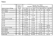
- Test 2 COBAS AmpliPrep / COBAS TaqMan HIV-1 (Test 2) is compared to the test COBAS AMPLICOR HIV-1 MONITOR (Test 1, both tests available from Roche Diagnostics GmbH, Mannheim, Germany) in the context of different studies performed at sites in several European countries as well as at a site in Thailand.
- test 2 sample preparation was carried out using magnetic glass particles and chaotropic reagents, while in the amplification and detection step, the following concentrations were employed:
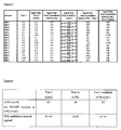
- Test 2 A number of 16 samples underquantitated in Test 2 (COBAS TaqMan HIV-1, targets GAG only) if compared to Test 1 (for log10 difference in titer see column 5) was tested with the modified Test 2 which targets the GAG and the LTR regions of HIV simultaneously. The results are displayed in Figure 3 . Test conditions were the same for both tests, with the exception that additional primers and a probe for LTR were introduced at about a third of the concentration of the oligonucleotides for amplification and detection of GAG.
- Example 2 In essentially the same setting as for Example 2, the LOD was determined for Test 2 and modified Test 2. Additionally, a third experiment was carried out with the oligonucleotides for amplification and detection of LTR but not of GAG (Test 2a). The oligonucleotide concentration in Test 2a was equivalent to the concentration of oligonucleotides for amplification and detection of LTR in modified Test 2.
Landscapes
- Chemical & Material Sciences (AREA)
- Life Sciences & Earth Sciences (AREA)
- Analytical Chemistry (AREA)
- Proteomics, Peptides & Aminoacids (AREA)
- Organic Chemistry (AREA)
- Zoology (AREA)
- Wood Science & Technology (AREA)
- Health & Medical Sciences (AREA)
- Engineering & Computer Science (AREA)
- Microbiology (AREA)
- Immunology (AREA)
- Molecular Biology (AREA)
- Biotechnology (AREA)
- Biophysics (AREA)
- Physics & Mathematics (AREA)
- Biochemistry (AREA)
- Bioinformatics & Cheminformatics (AREA)
- General Engineering & Computer Science (AREA)
- General Health & Medical Sciences (AREA)
- Genetics & Genomics (AREA)
- Measuring Or Testing Involving Enzymes Or Micro-Organisms (AREA)
Claims (12)
- Procédé de détection et de quantification d'un acide nucléique microbien dans un échantillon biologique, ledit procédé comprenant :a) la fourniture d'un mélange réactionnel comprenant un ou plusieurs acides nucléiques étalons quantitatifs, deux paires d'amorces ou plus, lesdites paires d'amorces étant spécifiques de portions différentes de la séquence dudit acide nucléique microbien, et une ou plusieurs paires d'amorces spécifiques desdits un ou plusieurs acides nucléiques étalonsb) l'ajout dudit échantillon biologique audit mélange réactionnelc) la réalisation d'une ou plusieurs étapes de manière cyclique, dans laquelle une étape réalisée de manière cyclique comprend une étape d'amplification, ladite étape d'amplification comprenant la production de deux produits d'amplification différents ou plus, dérivés dudit acide nucléique microbien s'il est présent dans ledit échantillon et la production d'un ou de plusieurs produits d'amplification dérivés desdits un ou plusieurs acides nucléiques étalons quantitatifsd) la détection et la mesure des signaux détectables générés par lesdits produits d'amplification et proportionnels à leur concentration dans ledit mélange réactionnel, dans laquelle la présence ou l'absence du signal ou des signaux détectables générés par ledit acide nucléique microbien est indicatrice de la présence ou de l'absence de l'acide nucléique microbien dans ledit échantillon biologiquee) la détermination de la quantité dudit acide nucléique microbien dans ledit échantillon biologique par comparaison des signaux générés par ledit acide nucléique microbien et lesdits un ou plusieurs acides nucléiques étalons quantitatifs,dans lequel lesdites deux portions de séquence différentes ou plus de l'acide nucléique microbien sont une LTR et GAG du VIH.
- Procédé selon la revendication 1, comprenant en outreà l'étape a) la fourniture de deux sondes ou plus spécifiques des portions de séquence différentes amplifiées par lesdites deux paires d'amorces ou plus spécifiques des portions de séquence différentes dudit acide nucléique microbien, et une ou plusieurs sondes spécifiques de la portion ou des portions de séquence amplifiées par lesdites une ou plusieurs paires d'amorces spécifiques desdits un ou plusieurs acides nucléiques étalons quantitatifs.
- Procédé selon la revendication 2, comprenant en outreà l'étape c) une étape d'hybridation constituée par ladite étape réalisée de manière cyclique, dans laquelle ladite étape d'hybridation comprend une hybridation des portions de séquence différentes amplifiées par lesdites deux paires d'amorces ou plus spécifiques des portions de séquence différentes dudit acide nucléique microbien, si l'acide nucléique microbien est présent dans ledit échantillon, avec lesdites deux sondes ou plus, et l'hybridation de ladite portion ou desdites portions de séquence amplifiées par lesdites une ou plusieurs paires d'amorces spécifiques desdits un ou plusieurs acides nucléiques étalons quantitatifs avec une ou plusieurs sondes, dans laquelle les sondes sont marquées avec une fraction fluorescente donneur et une fraction fluorescente accepteur correspondanteà l'étape d) la détection de la présence ou de l'absence de transfert d'énergie de fluorescence par résonance (FRET) entre ladite fraction fluorescente donneur et ladite fraction fluorescente accepteur de ladite sonde, dans laquelle la présence ou l'absence de fluorescence générée par ledit acide nucléique microbien est indicatrice de la présence ou de l'absence de l'acide nucléique microbien dans ledit échantillon biologiqueà l'étape e) la détermination de la quantité dudit acide nucléique microbien dans ledit échantillon par comparaison des signaux fluorescents générés par ledit acide nucléique microbien et ledit acide nucléique étalon quantitatif.
- Procédé selon l'une quelconque des revendications précédentes, comprenant l'amplification d'une ou plusieurs desdites portions de séquence différentes de l'acide nucléique microbien et desdits un ou plusieurs acides nucléiques étalons quantitatifs avec la même paire d'amorces.
- Procédé selon l'une quelconque des revendications précédentes, comprenant l'amplification desdites portions de séquence différentes de l'acide nucléique microbien et desdits un ou plusieurs acides nucléiques étalons quantitatifs avec des paires d'amorces différentes.
- Procédé selon l'une quelconque des revendications précédentes, comprenant l'utilisation d'un colorant fluorescent comme marqueur détectable pour les produits d'amplification dérivés desdites deux portions de séquence différentes ou plus de l'acide nucléique microbien, et un colorant fluorescent différent comme marqueur détectable pour le produit ou les produits d'amplification dérivés desdits un ou plusieurs acides nucléiques étalons quantitatifs.
- Procédé selon l'une quelconque des revendications 1 à 5, comprenant l'utilisation d'un colorant fluorescent distinct comme marqueur détectable pour chacun des produits d'amplification dérivés de l'acide nucléique microbien, et d'un colorant fluorescent différent comme marqueur détectable pour le produit ou les produits d'amplification dérivés desdits un ou plusieurs acides nucléiques étalons quantitatifs, dans lequel l'étape de quantification comprend la quantification de manière séparée de chacune des portions de séquence amplifiées de la séquence microbienne cible.
- Procédé selon l'une quelconque des revendications précédentes, dans lequel exactement un acide nucléique étalon quantitatif est présent dans le mélange réactionnel.
- Procédé selon l'une quelconque des revendications précédentes, dans lequel les séquences d'acides nucléiques desdites une ou plusieurs amorces sont d'au moins 12 nucléotides contigus choisis dans le groupe de SEQ ID NO 1 à 16 et 21 à 27 ou les séquences d'acides nucléiques complémentaires correspondantes de celles-ci.
- Procédé selon l'une quelconque des revendications 2 ou 3, dans lequel les séquences d'acides nucléiques desdites deux sondes ou plus sont d'au moins 12 nucléotides contigus choisis dans le groupe de SEQ ID NO 17 à 20, 28, 29 ou les séquences d'acides nucléiques complémentaires correspondantes de celles-ci.
- Utilisation d'un acide nucléique étalon quantitatif pour la détection et la quantification d'un acide nucléique microbien conformément au procédé selon l'une quelconque des revendications précédentes.
- Trousse pour la détection et la quantification d'un acide nucléique microbien dans un échantillon biologique par le procédé selon l'une quelconque des revendications précédentes, ladite trousse comprenant un ou plusieurs acides nucléiques étalons quantitatifs, deux paires d'amorces ou plus, lesdites paire d'amorces étant spécifiques de portions de séquence différentes dudit acide nucléique microbien, et une ou plusieurs paires d'amorces spécifiques desdits un ou plusieurs acides nucléiques étalons, dans lequel lesdites deux portions de séquence différentes ou plus de l'acide nucléique microbien sont une LTR et GAG du VIH.
Priority Applications (4)
| Application Number | Priority Date | Filing Date | Title |
|---|---|---|---|
| EP20080104295 EP2130929B1 (fr) | 2008-06-06 | 2008-06-06 | Détection multiplexe contrôlée en interne et quantification d'acides nucléiques microbiens |
| ES08104295T ES2439951T3 (es) | 2008-06-06 | 2008-06-06 | Detección y cuantificación multiplex de ácidos nucleicos microbianos controlada de forma interna |
| EP20110181688 EP2402462A1 (fr) | 2008-06-06 | 2008-06-06 | Détection multiplexe contrôlée en interne et quantification d'acides nucléiques microbiens |
| US12/479,643 US20100041040A1 (en) | 2008-06-06 | 2009-06-05 | Internally Controlled Multiplex Detection and Quantification of Microbial Nucleic Acids |
Applications Claiming Priority (1)
| Application Number | Priority Date | Filing Date | Title |
|---|---|---|---|
| EP20080104295 EP2130929B1 (fr) | 2008-06-06 | 2008-06-06 | Détection multiplexe contrôlée en interne et quantification d'acides nucléiques microbiens |
Related Child Applications (1)
| Application Number | Title | Priority Date | Filing Date |
|---|---|---|---|
| EP11181688.0 Division-Into | 2011-09-16 |
Publications (2)
| Publication Number | Publication Date |
|---|---|
| EP2130929A1 EP2130929A1 (fr) | 2009-12-09 |
| EP2130929B1 true EP2130929B1 (fr) | 2013-10-09 |
Family
ID=40104887
Family Applications (2)
| Application Number | Title | Priority Date | Filing Date |
|---|---|---|---|
| EP20080104295 Active EP2130929B1 (fr) | 2008-06-06 | 2008-06-06 | Détection multiplexe contrôlée en interne et quantification d'acides nucléiques microbiens |
| EP20110181688 Withdrawn EP2402462A1 (fr) | 2008-06-06 | 2008-06-06 | Détection multiplexe contrôlée en interne et quantification d'acides nucléiques microbiens |
Family Applications After (1)
| Application Number | Title | Priority Date | Filing Date |
|---|---|---|---|
| EP20110181688 Withdrawn EP2402462A1 (fr) | 2008-06-06 | 2008-06-06 | Détection multiplexe contrôlée en interne et quantification d'acides nucléiques microbiens |
Country Status (3)
| Country | Link |
|---|---|
| US (1) | US20100041040A1 (fr) |
| EP (2) | EP2130929B1 (fr) |
| ES (1) | ES2439951T3 (fr) |
Families Citing this family (16)
| Publication number | Priority date | Publication date | Assignee | Title |
|---|---|---|---|---|
| GB0701253D0 (en) | 2007-01-23 | 2007-02-28 | Diagnostics For The Real World | Nucleic acid amplification and testing |
| JP5972265B2 (ja) * | 2010-07-29 | 2016-08-17 | エフ.ホフマン−ラ ロシュ アーゲーF. Hoffmann−La Roche Aktiengesellschaft | ジェネリックpcr |
| US8877464B2 (en) | 2010-07-29 | 2014-11-04 | Roche Molecular Systems, Inc. | Qualitative and quantitative detection of microbial nucleic acids |
| US9725754B2 (en) | 2010-07-29 | 2017-08-08 | Sean F. Boyle | Generic sample preparation |
| US9175332B2 (en) | 2010-07-29 | 2015-11-03 | Roche Molecular Systems, Inc. | Generic PCR |
| CN107058617A (zh) * | 2010-07-29 | 2017-08-18 | 霍夫曼-拉罗奇有限公司 | 通用样品制备 |
| CA2802567C (fr) * | 2010-07-29 | 2017-12-05 | F. Hoffmann La-Roche Ag | Acides nucleiques temoins pour de multiples parametres |
| US8609340B2 (en) * | 2010-07-29 | 2013-12-17 | Roche Molecular Systems, Inc. | Control nucleic acids for multiple parameters |
| CN107190056A (zh) * | 2010-07-29 | 2017-09-22 | 霍夫曼-拉罗奇有限公司 | 微生物核酸的定性和定量检测 |
| JP6121591B2 (ja) * | 2010-07-29 | 2017-04-26 | エフ.ホフマン−ラ ロシュ アーゲーF. Hoffmann−La Roche Aktiengesellschaft | 微生物核酸の定性的および定量的検出 |
| EP2426222A1 (fr) * | 2010-09-07 | 2012-03-07 | F. Hoffmann-La Roche AG | Tampon générique pour amplification |
| WO2014018885A2 (fr) | 2012-07-27 | 2014-01-30 | Gen-Probe Incorporated | Procédé d'étalonnage à double référence et système de quantification de polynucléotides |
| ES2659747T3 (es) | 2012-10-18 | 2018-03-19 | F. Hoffmann-La Roche Ag | Ensayo de doble sonda para la detección de poblaciones heterogéneas de amplicón |
| EP2722398B1 (fr) | 2012-10-18 | 2017-07-12 | F. Hoffmann-La Roche AG | Analyse de sonde double pour la détection du VHC |
| US20150176060A1 (en) * | 2013-12-20 | 2015-06-25 | Roche Molecular Systems, Inc. | Method For Coding Of Multiple PCR Reactions For Assay Recognition |
| EP3899035B1 (fr) * | 2018-12-20 | 2025-04-16 | F. Hoffmann-La Roche AG | Détection d'acide nucléique cible par molographie en phase solide |
Family Cites Families (27)
| Publication number | Priority date | Publication date | Assignee | Title |
|---|---|---|---|---|
| US4458066A (en) | 1980-02-29 | 1984-07-03 | University Patents, Inc. | Process for preparing polynucleotides |
| US4683202A (en) | 1985-03-28 | 1987-07-28 | Cetus Corporation | Process for amplifying nucleic acid sequences |
| US4965188A (en) | 1986-08-22 | 1990-10-23 | Cetus Corporation | Process for amplifying, detecting, and/or cloning nucleic acid sequences using a thermostable enzyme |
| US4683195A (en) | 1986-01-30 | 1987-07-28 | Cetus Corporation | Process for amplifying, detecting, and/or-cloning nucleic acid sequences |
| US4800159A (en) | 1986-02-07 | 1989-01-24 | Cetus Corporation | Process for amplifying, detecting, and/or cloning nucleic acid sequences |
| DE3734442A1 (de) | 1987-10-12 | 1989-04-27 | Kernforschungsanlage Juelich | Verfahren und vorrichtung zur bestrahlung grosser flaechen mit ionen |
| US5130238A (en) | 1988-06-24 | 1992-07-14 | Cangene Corporation | Enhanced nucleic acid amplification process |
| ATE138106T1 (de) | 1988-07-20 | 1996-06-15 | David Segev | Verfahren zur amplifizierung und zum nachweis von nukleinsäuresequenzen |
| US5035996A (en) | 1989-06-01 | 1991-07-30 | Life Technologies, Inc. | Process for controlling contamination of nucleic acid amplification reactions |
| US5683896A (en) | 1989-06-01 | 1997-11-04 | Life Technologies, Inc. | Process for controlling contamination of nucleic acid amplification reactions |
| US5219727A (en) * | 1989-08-21 | 1993-06-15 | Hoffmann-Laroche Inc. | Quantitation of nucleic acids using the polymerase chain reaction |
| KR950013953B1 (ko) | 1990-01-26 | 1995-11-18 | 애보트 래보라토리즈 | 리가제 연쇄 반응에 적용가능한 표적 핵산의 증폭 방법 |
| GB9003253D0 (en) | 1990-02-13 | 1990-04-11 | Amersham Int Plc | Precipitating polymers |
| US5474895A (en) | 1990-11-14 | 1995-12-12 | Siska Diagnostics Inc. | Non-isotopic detection of nucleic acids using a polystyrene support-based sandwich hybridization assay and compositions useful therefor |
| US5386024A (en) | 1993-02-10 | 1995-01-31 | Gen-Probe Incorporated | Method to prepare nucleic acids from a biological sample using low pH and acid protease |
| WO1995014109A1 (fr) * | 1993-11-19 | 1995-05-26 | U.S. Department Of The Army | Amplification type pcr quantitative a rendement eleve pour echantillons cliniques a vih |
| DE69739357D1 (de) | 1996-06-04 | 2009-05-28 | Univ Utah Res Found | Überwachung der hybridisierung während pcr |
| EP1442794B1 (fr) | 1996-06-04 | 2006-03-29 | University Of Utah Research Foundation | Système et procédé d'execution et de suivi de processus biologiques |
| US5981235A (en) | 1996-07-29 | 1999-11-09 | Promega Corporation | Methods for isolating nucleic acids using alkaline protease |
| WO2000068436A1 (fr) * | 1999-05-07 | 2000-11-16 | Nationales Zentrum Für Retroviren | Systeme de detection du virus de l'immunodeficience humaine fonde sur l'amplification d'acide nucleique |
| CA2392009C (fr) | 1999-11-17 | 2009-07-14 | Roche Diagnostics Gmbh | Particules de verre magnetiques, procede de preparation et utilisations |
| US7238795B2 (en) | 2000-08-03 | 2007-07-03 | Roche Molecular Systems, Inc. | Nucleic acid binding compounds containing pyrazolo[3,4-d]pyrimidine analogues of purin-2,6-diamine and their uses |
| EP1201753B1 (fr) | 2000-10-31 | 2008-04-23 | Boehringer Mannheim Gmbh | Procédé pour l'analyse de composants non-protéiques en utilisant une protéase provenant d'une souche de Bacillus |
| US20040229211A1 (en) | 2003-05-13 | 2004-11-18 | Yeung Wah Hin Alex | Sensitive diagnostic testing methodology using multiplex real time PCR with one dye (MOD) and its use as in severe acute respiratory syndrome (SARS) |
| EP1625232B1 (fr) * | 2003-05-16 | 2013-09-04 | GOVERNMENT OF THE UNITED STATES OF AMERICA, as represented by THE SECRETARY, DEPARTMENT OF HEALTH AND HUMAN SERVICES | Molecule d'acide nucleique de controle interne pour systemes d'amplification d'acide nucleique |
| EP1602736A1 (fr) * | 2004-06-04 | 2005-12-07 | Bio-Rad Pasteur | Détection du type et du sous-type de virus HIV |
| US7465561B2 (en) * | 2005-06-30 | 2008-12-16 | Roche Molecular Systems, Inc. | Probes and methods for hepatitis C virus typing using single probe analysis |
-
2008
- 2008-06-06 EP EP20080104295 patent/EP2130929B1/fr active Active
- 2008-06-06 EP EP20110181688 patent/EP2402462A1/fr not_active Withdrawn
- 2008-06-06 ES ES08104295T patent/ES2439951T3/es active Active
-
2009
- 2009-06-05 US US12/479,643 patent/US20100041040A1/en not_active Abandoned
Also Published As
| Publication number | Publication date |
|---|---|
| EP2130929A1 (fr) | 2009-12-09 |
| EP2402462A1 (fr) | 2012-01-04 |
| US20100041040A1 (en) | 2010-02-18 |
| ES2439951T3 (es) | 2014-01-27 |
Similar Documents
| Publication | Publication Date | Title |
|---|---|---|
| EP2402462A1 (fr) | Détection multiplexe contrôlée en interne et quantification d'acides nucléiques microbiens | |
| CA2761546C (fr) | Matrice generique pour acides nucleiques de controle | |
| US9719133B2 (en) | Qualitative and quantitative detection of microbial nucleic acids | |
| EP2598655B1 (fr) | Détection qualitative et quantitative d'acides nucléiques microbiens | |
| JP6169489B2 (ja) | 一般的な試料調製 | |
| CA2802567C (fr) | Acides nucleiques temoins pour de multiples parametres | |
| EP2598653B1 (fr) | Pcr générique | |
| US20170283888A1 (en) | Use of rnase h for the selective amplification of viral dna | |
| JP6121591B2 (ja) | 微生物核酸の定性的および定量的検出 | |
| JP6348202B2 (ja) | 一般的な試料調製 |
Legal Events
| Date | Code | Title | Description |
|---|---|---|---|
| PUAI | Public reference made under article 153(3) epc to a published international application that has entered the european phase |
Free format text: ORIGINAL CODE: 0009012 |
|
| AK | Designated contracting states |
Kind code of ref document: A1 Designated state(s): AT BE BG CH CY CZ DE DK EE ES FI FR GB GR HR HU IE IS IT LI LT LU LV MC MT NL NO PL PT RO SE SI SK TR |
|
| AX | Request for extension of the european patent |
Extension state: AL BA MK RS |
|
| 17P | Request for examination filed |
Effective date: 20100609 |
|
| 17Q | First examination report despatched |
Effective date: 20100702 |
|
| AKX | Designation fees paid |
Designated state(s): AT BE BG CH CY CZ DE DK EE ES FI FR GB GR HR HU IE IS IT LI LT LU LV MC MT NL NO PL PT RO SE SI SK TR |
|
| GRAP | Despatch of communication of intention to grant a patent |
Free format text: ORIGINAL CODE: EPIDOSNIGR1 |
|
| INTG | Intention to grant announced |
Effective date: 20130606 |
|
| GRAS | Grant fee paid |
Free format text: ORIGINAL CODE: EPIDOSNIGR3 |
|
| GRAA | (expected) grant |
Free format text: ORIGINAL CODE: 0009210 |
|
| AK | Designated contracting states |
Kind code of ref document: B1 Designated state(s): AT BE BG CH CY CZ DE DK EE ES FI FR GB GR HR HU IE IS IT LI LT LU LV MC MT NL NO PL PT RO SE SI SK TR |
|
| REG | Reference to a national code |
Ref country code: GB Ref legal event code: FG4D |
|
| REG | Reference to a national code |
Ref country code: AT Ref legal event code: REF Ref document number: 635595 Country of ref document: AT Kind code of ref document: T Effective date: 20131015 Ref country code: CH Ref legal event code: EP |
|
| REG | Reference to a national code |
Ref country code: IE Ref legal event code: FG4D |
|
| REG | Reference to a national code |
Ref country code: DE Ref legal event code: R096 Ref document number: 602008027971 Country of ref document: DE Effective date: 20131205 |
|
| REG | Reference to a national code |
Ref country code: ES Ref legal event code: FG2A Ref document number: 2439951 Country of ref document: ES Kind code of ref document: T3 Effective date: 20140127 |
|
| REG | Reference to a national code |
Ref country code: AT Ref legal event code: MK05 Ref document number: 635595 Country of ref document: AT Kind code of ref document: T Effective date: 20131009 |
|
| REG | Reference to a national code |
Ref country code: NL Ref legal event code: VDEP Effective date: 20131009 |
|
| PG25 | Lapsed in a contracting state [announced via postgrant information from national office to epo] |
Ref country code: SI Free format text: LAPSE BECAUSE OF FAILURE TO SUBMIT A TRANSLATION OF THE DESCRIPTION OR TO PAY THE FEE WITHIN THE PRESCRIBED TIME-LIMIT Effective date: 20131009 |
|
| REG | Reference to a national code |
Ref country code: LT Ref legal event code: MG4D |
|
| PG25 | Lapsed in a contracting state [announced via postgrant information from national office to epo] |
Ref country code: NL Free format text: LAPSE BECAUSE OF FAILURE TO SUBMIT A TRANSLATION OF THE DESCRIPTION OR TO PAY THE FEE WITHIN THE PRESCRIBED TIME-LIMIT Effective date: 20131009 Ref country code: LT Free format text: LAPSE BECAUSE OF FAILURE TO SUBMIT A TRANSLATION OF THE DESCRIPTION OR TO PAY THE FEE WITHIN THE PRESCRIBED TIME-LIMIT Effective date: 20131009 Ref country code: FI Free format text: LAPSE BECAUSE OF FAILURE TO SUBMIT A TRANSLATION OF THE DESCRIPTION OR TO PAY THE FEE WITHIN THE PRESCRIBED TIME-LIMIT Effective date: 20131009 Ref country code: IS Free format text: LAPSE BECAUSE OF FAILURE TO SUBMIT A TRANSLATION OF THE DESCRIPTION OR TO PAY THE FEE WITHIN THE PRESCRIBED TIME-LIMIT Effective date: 20140209 Ref country code: NO Free format text: LAPSE BECAUSE OF FAILURE TO SUBMIT A TRANSLATION OF THE DESCRIPTION OR TO PAY THE FEE WITHIN THE PRESCRIBED TIME-LIMIT Effective date: 20140109 Ref country code: SE Free format text: LAPSE BECAUSE OF FAILURE TO SUBMIT A TRANSLATION OF THE DESCRIPTION OR TO PAY THE FEE WITHIN THE PRESCRIBED TIME-LIMIT Effective date: 20131009 Ref country code: HR Free format text: LAPSE BECAUSE OF FAILURE TO SUBMIT A TRANSLATION OF THE DESCRIPTION OR TO PAY THE FEE WITHIN THE PRESCRIBED TIME-LIMIT Effective date: 20131009 Ref country code: BE Free format text: LAPSE BECAUSE OF FAILURE TO SUBMIT A TRANSLATION OF THE DESCRIPTION OR TO PAY THE FEE WITHIN THE PRESCRIBED TIME-LIMIT Effective date: 20131009 |
|
| PG25 | Lapsed in a contracting state [announced via postgrant information from national office to epo] |
Ref country code: CY Free format text: LAPSE BECAUSE OF FAILURE TO SUBMIT A TRANSLATION OF THE DESCRIPTION OR TO PAY THE FEE WITHIN THE PRESCRIBED TIME-LIMIT Effective date: 20131009 Ref country code: AT Free format text: LAPSE BECAUSE OF FAILURE TO SUBMIT A TRANSLATION OF THE DESCRIPTION OR TO PAY THE FEE WITHIN THE PRESCRIBED TIME-LIMIT Effective date: 20131009 Ref country code: PL Free format text: LAPSE BECAUSE OF FAILURE TO SUBMIT A TRANSLATION OF THE DESCRIPTION OR TO PAY THE FEE WITHIN THE PRESCRIBED TIME-LIMIT Effective date: 20131009 Ref country code: LV Free format text: LAPSE BECAUSE OF FAILURE TO SUBMIT A TRANSLATION OF THE DESCRIPTION OR TO PAY THE FEE WITHIN THE PRESCRIBED TIME-LIMIT Effective date: 20131009 |
|
| PG25 | Lapsed in a contracting state [announced via postgrant information from national office to epo] |
Ref country code: PT Free format text: LAPSE BECAUSE OF FAILURE TO SUBMIT A TRANSLATION OF THE DESCRIPTION OR TO PAY THE FEE WITHIN THE PRESCRIBED TIME-LIMIT Effective date: 20140210 |
|
| REG | Reference to a national code |
Ref country code: DE Ref legal event code: R097 Ref document number: 602008027971 Country of ref document: DE |
|
| PG25 | Lapsed in a contracting state [announced via postgrant information from national office to epo] |
Ref country code: EE Free format text: LAPSE BECAUSE OF FAILURE TO SUBMIT A TRANSLATION OF THE DESCRIPTION OR TO PAY THE FEE WITHIN THE PRESCRIBED TIME-LIMIT Effective date: 20131009 |
|
| PLBE | No opposition filed within time limit |
Free format text: ORIGINAL CODE: 0009261 |
|
| STAA | Information on the status of an ep patent application or granted ep patent |
Free format text: STATUS: NO OPPOSITION FILED WITHIN TIME LIMIT |
|
| PG25 | Lapsed in a contracting state [announced via postgrant information from national office to epo] |
Ref country code: CZ Free format text: LAPSE BECAUSE OF FAILURE TO SUBMIT A TRANSLATION OF THE DESCRIPTION OR TO PAY THE FEE WITHIN THE PRESCRIBED TIME-LIMIT Effective date: 20131009 Ref country code: SK Free format text: LAPSE BECAUSE OF FAILURE TO SUBMIT A TRANSLATION OF THE DESCRIPTION OR TO PAY THE FEE WITHIN THE PRESCRIBED TIME-LIMIT Effective date: 20131009 Ref country code: RO Free format text: LAPSE BECAUSE OF FAILURE TO SUBMIT A TRANSLATION OF THE DESCRIPTION OR TO PAY THE FEE WITHIN THE PRESCRIBED TIME-LIMIT Effective date: 20131009 |
|
| 26N | No opposition filed |
Effective date: 20140710 |
|
| PG25 | Lapsed in a contracting state [announced via postgrant information from national office to epo] |
Ref country code: DK Free format text: LAPSE BECAUSE OF FAILURE TO SUBMIT A TRANSLATION OF THE DESCRIPTION OR TO PAY THE FEE WITHIN THE PRESCRIBED TIME-LIMIT Effective date: 20131009 |
|
| REG | Reference to a national code |
Ref country code: DE Ref legal event code: R097 Ref document number: 602008027971 Country of ref document: DE Effective date: 20140710 |
|
| PG25 | Lapsed in a contracting state [announced via postgrant information from national office to epo] |
Ref country code: LU Free format text: LAPSE BECAUSE OF FAILURE TO SUBMIT A TRANSLATION OF THE DESCRIPTION OR TO PAY THE FEE WITHIN THE PRESCRIBED TIME-LIMIT Effective date: 20140606 Ref country code: MC Free format text: LAPSE BECAUSE OF FAILURE TO SUBMIT A TRANSLATION OF THE DESCRIPTION OR TO PAY THE FEE WITHIN THE PRESCRIBED TIME-LIMIT Effective date: 20131009 |
|
| REG | Reference to a national code |
Ref country code: IE Ref legal event code: MM4A |
|
| PG25 | Lapsed in a contracting state [announced via postgrant information from national office to epo] |
Ref country code: IE Free format text: LAPSE BECAUSE OF NON-PAYMENT OF DUE FEES Effective date: 20140606 |
|
| PG25 | Lapsed in a contracting state [announced via postgrant information from national office to epo] |
Ref country code: MT Free format text: LAPSE BECAUSE OF FAILURE TO SUBMIT A TRANSLATION OF THE DESCRIPTION OR TO PAY THE FEE WITHIN THE PRESCRIBED TIME-LIMIT Effective date: 20131009 |
|
| PG25 | Lapsed in a contracting state [announced via postgrant information from national office to epo] |
Ref country code: BG Free format text: LAPSE BECAUSE OF FAILURE TO SUBMIT A TRANSLATION OF THE DESCRIPTION OR TO PAY THE FEE WITHIN THE PRESCRIBED TIME-LIMIT Effective date: 20131009 |
|
| REG | Reference to a national code |
Ref country code: FR Ref legal event code: PLFP Year of fee payment: 9 |
|
| PG25 | Lapsed in a contracting state [announced via postgrant information from national office to epo] |
Ref country code: GR Free format text: LAPSE BECAUSE OF FAILURE TO SUBMIT A TRANSLATION OF THE DESCRIPTION OR TO PAY THE FEE WITHIN THE PRESCRIBED TIME-LIMIT Effective date: 20140110 |
|
| PG25 | Lapsed in a contracting state [announced via postgrant information from national office to epo] |
Ref country code: TR Free format text: LAPSE BECAUSE OF FAILURE TO SUBMIT A TRANSLATION OF THE DESCRIPTION OR TO PAY THE FEE WITHIN THE PRESCRIBED TIME-LIMIT Effective date: 20131009 Ref country code: HU Free format text: LAPSE BECAUSE OF FAILURE TO SUBMIT A TRANSLATION OF THE DESCRIPTION OR TO PAY THE FEE WITHIN THE PRESCRIBED TIME-LIMIT; INVALID AB INITIO Effective date: 20080606 |
|
| REG | Reference to a national code |
Ref country code: FR Ref legal event code: PLFP Year of fee payment: 10 |
|
| REG | Reference to a national code |
Ref country code: FR Ref legal event code: PLFP Year of fee payment: 11 |
|
| PGFP | Annual fee paid to national office [announced via postgrant information from national office to epo] |
Ref country code: ES Payment date: 20250701 Year of fee payment: 18 |
|
| PGFP | Annual fee paid to national office [announced via postgrant information from national office to epo] |
Ref country code: CH Payment date: 20250701 Year of fee payment: 18 |
|
| REG | Reference to a national code |
Ref country code: CH Ref legal event code: U11 Free format text: ST27 STATUS EVENT CODE: U-0-0-U10-U11 (AS PROVIDED BY THE NATIONAL OFFICE) Effective date: 20260701 |
|
| PGFP | Annual fee paid to national office [announced via postgrant information from national office to epo] |
Ref country code: GB Payment date: 20260520 Year of fee payment: 19 |
|
| PGFP | Annual fee paid to national office [announced via postgrant information from national office to epo] |
Ref country code: IT Payment date: 20260520 Year of fee payment: 19 |
|
| PGFP | Annual fee paid to national office [announced via postgrant information from national office to epo] |
Ref country code: DE Payment date: 20260520 Year of fee payment: 19 |
|
| PGFP | Annual fee paid to national office [announced via postgrant information from national office to epo] |
Ref country code: FR Payment date: 20260521 Year of fee payment: 19 |