EP2157979B1 - Protéines de canal de calcium et leur utilisation - Google Patents
Protéines de canal de calcium et leur utilisation Download PDFInfo
- Publication number
- EP2157979B1 EP2157979B1 EP08756325.0A EP08756325A EP2157979B1 EP 2157979 B1 EP2157979 B1 EP 2157979B1 EP 08756325 A EP08756325 A EP 08756325A EP 2157979 B1 EP2157979 B1 EP 2157979B1
- Authority
- EP
- European Patent Office
- Prior art keywords
- calcium
- cells
- stim
- cell
- protein
- Prior art date
- Legal status (The legal status is an assumption and is not a legal conclusion. Google has not performed a legal analysis and makes no representation as to the accuracy of the status listed.)
- Not-in-force
Links
- 102000003922 Calcium Channels Human genes 0.000 title description 6
- 108090000312 Calcium Channels Proteins 0.000 title description 6
- 239000011575 calcium Substances 0.000 claims description 370
- 229910052791 calcium Inorganic materials 0.000 claims description 361
- 210000004027 cell Anatomy 0.000 claims description 361
- OYPRJOBELJOOCE-UHFFFAOYSA-N Calcium Chemical compound [Ca] OYPRJOBELJOOCE-UHFFFAOYSA-N 0.000 claims description 360
- 230000003834 intracellular effect Effects 0.000 claims description 190
- 108090000623 proteins and genes Proteins 0.000 claims description 171
- 150000001875 compounds Chemical class 0.000 claims description 157
- 101100114828 Drosophila melanogaster Orai gene Proteins 0.000 claims description 151
- 230000000694 effects Effects 0.000 claims description 149
- 102000004169 proteins and genes Human genes 0.000 claims description 144
- 238000000034 method Methods 0.000 claims description 98
- 108090000532 Stromal Interaction Molecule 1 Proteins 0.000 claims description 81
- 102000004094 Stromal Interaction Molecule 1 Human genes 0.000 claims description 80
- 239000003795 chemical substances by application Substances 0.000 claims description 59
- 108090000765 processed proteins & peptides Proteins 0.000 claims description 56
- 229920001184 polypeptide Polymers 0.000 claims description 54
- 102000004196 processed proteins & peptides Human genes 0.000 claims description 54
- 101100043696 Drosophila melanogaster Stim gene Proteins 0.000 claims description 52
- 238000012360 testing method Methods 0.000 claims description 44
- 241000282414 Homo sapiens Species 0.000 claims description 32
- 239000012528 membrane Substances 0.000 claims description 29
- 230000003993 interaction Effects 0.000 claims description 24
- 238000005259 measurement Methods 0.000 claims description 24
- 150000007523 nucleic acids Chemical group 0.000 claims description 20
- 102000005935 Stromal Interaction Molecule 2 Human genes 0.000 claims description 16
- 108010030731 Stromal Interaction Molecule 2 Proteins 0.000 claims description 16
- 238000003384 imaging method Methods 0.000 claims description 14
- 210000004962 mammalian cell Anatomy 0.000 claims description 13
- 108091028043 Nucleic acid sequence Proteins 0.000 claims description 10
- 238000002866 fluorescence resonance energy transfer Methods 0.000 claims description 10
- 241000283984 Rodentia Species 0.000 claims description 9
- 230000027455 binding Effects 0.000 claims description 9
- 101150079867 Orai gene Proteins 0.000 claims description 7
- 101150101888 Stim gene Proteins 0.000 claims description 7
- 241000238631 Hexapoda Species 0.000 claims description 4
- 102000040430 polynucleotide Human genes 0.000 claims description 4
- 108091033319 polynucleotide Proteins 0.000 claims description 4
- 239000002157 polynucleotide Substances 0.000 claims description 4
- 230000007831 electrophysiology Effects 0.000 claims description 3
- 238000002001 electrophysiology Methods 0.000 claims description 3
- 239000007850 fluorescent dye Substances 0.000 claims description 3
- 125000003275 alpha amino acid group Chemical group 0.000 claims 4
- 229910001424 calcium ion Inorganic materials 0.000 description 183
- BHPQYMZQTOCNFJ-UHFFFAOYSA-N Calcium cation Chemical compound [Ca+2] BHPQYMZQTOCNFJ-UHFFFAOYSA-N 0.000 description 182
- 230000003185 calcium uptake Effects 0.000 description 137
- 235000018102 proteins Nutrition 0.000 description 132
- 208000037265 diseases, disorders, signs and symptoms Diseases 0.000 description 94
- 108091022898 Calcium release-activated calcium channel Proteins 0.000 description 74
- 102000020167 Calcium release-activated calcium channel Human genes 0.000 description 74
- 230000004913 activation Effects 0.000 description 58
- 108091006146 Channels Proteins 0.000 description 57
- 210000002472 endoplasmic reticulum Anatomy 0.000 description 56
- 239000000203 mixture Substances 0.000 description 54
- 201000010099 disease Diseases 0.000 description 50
- 108091032973 (ribonucleotides)n+m Proteins 0.000 description 49
- 230000014509 gene expression Effects 0.000 description 47
- 230000001086 cytosolic effect Effects 0.000 description 44
- 208000035475 disorder Diseases 0.000 description 44
- 102000040650 (ribonucleotides)n+m Human genes 0.000 description 43
- 210000000170 cell membrane Anatomy 0.000 description 38
- 238000003556 assay Methods 0.000 description 37
- HATRDXDCPOXQJX-UHFFFAOYSA-N Thapsigargin Natural products CCCCCCCC(=O)OC1C(OC(O)C(=C/C)C)C(=C2C3OC(=O)C(C)(O)C3(O)C(CC(C)(OC(=O)C)C12)OC(=O)CCC)C HATRDXDCPOXQJX-UHFFFAOYSA-N 0.000 description 35
- IXFPJGBNCFXKPI-FSIHEZPISA-N thapsigargin Chemical group CCCC(=O)O[C@H]1C[C@](C)(OC(C)=O)[C@H]2[C@H](OC(=O)CCCCCCC)[C@@H](OC(=O)C(\C)=C/C)C(C)=C2[C@@H]2OC(=O)[C@@](C)(O)[C@]21O IXFPJGBNCFXKPI-FSIHEZPISA-N 0.000 description 35
- 108090000695 Cytokines Proteins 0.000 description 32
- 230000009460 calcium influx Effects 0.000 description 32
- 238000011282 treatment Methods 0.000 description 32
- 102000004127 Cytokines Human genes 0.000 description 30
- 239000000243 solution Substances 0.000 description 30
- 210000001744 T-lymphocyte Anatomy 0.000 description 28
- 230000033001 locomotion Effects 0.000 description 27
- 241001465754 Metazoa Species 0.000 description 26
- 230000005764 inhibitory process Effects 0.000 description 26
- 230000001404 mediated effect Effects 0.000 description 26
- 238000009472 formulation Methods 0.000 description 25
- 241000255581 Drosophila <fruit fly, genus> Species 0.000 description 24
- 230000001105 regulatory effect Effects 0.000 description 24
- 150000002500 ions Chemical class 0.000 description 23
- 230000000284 resting effect Effects 0.000 description 23
- 102000004631 Calcineurin Human genes 0.000 description 22
- 108010042955 Calcineurin Proteins 0.000 description 22
- 230000006870 function Effects 0.000 description 22
- 230000004075 alteration Effects 0.000 description 21
- -1 STIM Proteins 0.000 description 20
- 238000002474 experimental method Methods 0.000 description 20
- 230000001419 dependent effect Effects 0.000 description 19
- 230000004941 influx Effects 0.000 description 19
- 210000004698 lymphocyte Anatomy 0.000 description 19
- 241000124008 Mammalia Species 0.000 description 18
- 101150035353 SERCA gene Proteins 0.000 description 18
- 239000008194 pharmaceutical composition Substances 0.000 description 18
- 108010002350 Interleukin-2 Proteins 0.000 description 17
- 102000000588 Interleukin-2 Human genes 0.000 description 17
- 230000001413 cellular effect Effects 0.000 description 17
- 210000000805 cytoplasm Anatomy 0.000 description 17
- 230000004044 response Effects 0.000 description 17
- 101100422645 Drosophila melanogaster Syx5 gene Proteins 0.000 description 16
- 238000012228 RNA interference-mediated gene silencing Methods 0.000 description 16
- 230000009368 gene silencing by RNA Effects 0.000 description 16
- 102000005962 receptors Human genes 0.000 description 16
- 108020003175 receptors Proteins 0.000 description 16
- 230000009467 reduction Effects 0.000 description 16
- 230000002459 sustained effect Effects 0.000 description 16
- BLZVCIGGICSWIG-UHFFFAOYSA-N 2-aminoethoxydiphenylborane Chemical compound C=1C=CC=CC=1B(OCCN)C1=CC=CC=C1 BLZVCIGGICSWIG-UHFFFAOYSA-N 0.000 description 15
- 101100434663 Bacillus subtilis (strain 168) fbaA gene Proteins 0.000 description 15
- 101100315389 Streptomyces azureus tsnR gene Proteins 0.000 description 15
- 150000001768 cations Chemical class 0.000 description 15
- 101150031187 fba gene Proteins 0.000 description 15
- 239000000499 gel Substances 0.000 description 15
- 230000002018 overexpression Effects 0.000 description 15
- 101150066142 tsr gene Proteins 0.000 description 15
- 101710137312 Protein orai Proteins 0.000 description 14
- 241000700159 Rattus Species 0.000 description 14
- 238000004458 analytical method Methods 0.000 description 14
- 230000001965 increasing effect Effects 0.000 description 14
- 238000002347 injection Methods 0.000 description 14
- 239000007924 injection Substances 0.000 description 14
- 210000005061 intracellular organelle Anatomy 0.000 description 14
- 230000007246 mechanism Effects 0.000 description 14
- 230000002829 reductive effect Effects 0.000 description 14
- 230000028327 secretion Effects 0.000 description 14
- 230000000763 evoking effect Effects 0.000 description 13
- 230000002401 inhibitory effect Effects 0.000 description 13
- 208000024891 symptom Diseases 0.000 description 13
- 210000001519 tissue Anatomy 0.000 description 13
- 230000032258 transport Effects 0.000 description 13
- 108010043121 Green Fluorescent Proteins Proteins 0.000 description 12
- 102000004144 Green Fluorescent Proteins Human genes 0.000 description 12
- 102000014384 Type C Phospholipases Human genes 0.000 description 12
- 108010079194 Type C Phospholipases Proteins 0.000 description 12
- 150000001413 amino acids Chemical group 0.000 description 12
- 239000005090 green fluorescent protein Substances 0.000 description 12
- 239000002609 medium Substances 0.000 description 12
- 238000012544 monitoring process Methods 0.000 description 12
- 210000003463 organelle Anatomy 0.000 description 12
- 230000011664 signaling Effects 0.000 description 12
- XLYOFNOQVPJJNP-UHFFFAOYSA-N water Substances O XLYOFNOQVPJJNP-UHFFFAOYSA-N 0.000 description 12
- 108060008682 Tumor Necrosis Factor Proteins 0.000 description 11
- MMWCIQZXVOZEGG-HOZKJCLWSA-N [(1S,2R,3S,4S,5R,6S)-2,3,5-trihydroxy-4,6-diphosphonooxycyclohexyl] dihydrogen phosphate Chemical compound O[C@H]1[C@@H](O)[C@H](OP(O)(O)=O)[C@@H](OP(O)(O)=O)[C@H](O)[C@H]1OP(O)(O)=O MMWCIQZXVOZEGG-HOZKJCLWSA-N 0.000 description 11
- 239000000427 antigen Substances 0.000 description 11
- 230000003139 buffering effect Effects 0.000 description 11
- 230000004663 cell proliferation Effects 0.000 description 11
- 238000003197 gene knockdown Methods 0.000 description 11
- 230000008569 process Effects 0.000 description 11
- 230000001052 transient effect Effects 0.000 description 11
- 101100168832 Drosophila melanogaster Cals gene Proteins 0.000 description 10
- 102000000852 Tumor Necrosis Factor-alpha Human genes 0.000 description 10
- 102000036639 antigens Human genes 0.000 description 10
- 239000000872 buffer Substances 0.000 description 10
- 239000003814 drug Substances 0.000 description 10
- 102000039446 nucleic acids Human genes 0.000 description 10
- 108020004707 nucleic acids Proteins 0.000 description 10
- 239000002773 nucleotide Substances 0.000 description 10
- 125000003729 nucleotide group Chemical group 0.000 description 10
- 210000000056 organ Anatomy 0.000 description 10
- 206010039073 rheumatoid arthritis Diseases 0.000 description 10
- 108091093088 Amplicon Proteins 0.000 description 9
- 102000004190 Enzymes Human genes 0.000 description 9
- 108090000790 Enzymes Proteins 0.000 description 9
- 206010061218 Inflammation Diseases 0.000 description 9
- 241000699666 Mus <mouse, genus> Species 0.000 description 9
- 206010028980 Neoplasm Diseases 0.000 description 9
- 206010029260 Neuroblastoma Diseases 0.000 description 9
- 239000000020 Nitrocellulose Substances 0.000 description 9
- 238000010171 animal model Methods 0.000 description 9
- 108091007433 antigens Proteins 0.000 description 9
- 108010085377 beta-N-Acetylhexosaminidases Proteins 0.000 description 9
- 102000007478 beta-N-Acetylhexosaminidases Human genes 0.000 description 9
- 230000033228 biological regulation Effects 0.000 description 9
- 230000007423 decrease Effects 0.000 description 9
- 239000012634 fragment Substances 0.000 description 9
- 210000003630 histaminocyte Anatomy 0.000 description 9
- 230000028993 immune response Effects 0.000 description 9
- 210000000987 immune system Anatomy 0.000 description 9
- 230000004054 inflammatory process Effects 0.000 description 9
- 239000003112 inhibitor Substances 0.000 description 9
- PGHMRUGBZOYCAA-UHFFFAOYSA-N ionomycin Natural products O1C(CC(O)C(C)C(O)C(C)C=CCC(C)CC(C)C(O)=CC(=O)C(C)CC(C)CC(CCC(O)=O)C)CCC1(C)C1OC(C)(C(C)O)CC1 PGHMRUGBZOYCAA-UHFFFAOYSA-N 0.000 description 9
- PGHMRUGBZOYCAA-ADZNBVRBSA-N ionomycin Chemical compound O1[C@H](C[C@H](O)[C@H](C)[C@H](O)[C@H](C)/C=C/C[C@@H](C)C[C@@H](C)C(/O)=C/C(=O)[C@@H](C)C[C@@H](C)C[C@@H](CCC(O)=O)C)CC[C@@]1(C)[C@@H]1O[C@](C)([C@@H](C)O)CC1 PGHMRUGBZOYCAA-ADZNBVRBSA-N 0.000 description 9
- 229920001220 nitrocellulos Polymers 0.000 description 9
- 101150060735 orai1 gene Proteins 0.000 description 9
- 230000000638 stimulation Effects 0.000 description 9
- 238000003860 storage Methods 0.000 description 9
- 208000023275 Autoimmune disease Diseases 0.000 description 8
- 108010017080 Granulocyte Colony-Stimulating Factor Proteins 0.000 description 8
- 102000004269 Granulocyte Colony-Stimulating Factor Human genes 0.000 description 8
- 108091023040 Transcription factor Proteins 0.000 description 8
- 102000040945 Transcription factor Human genes 0.000 description 8
- 230000018109 developmental process Effects 0.000 description 8
- 239000002552 dosage form Substances 0.000 description 8
- 238000011156 evaluation Methods 0.000 description 8
- 208000017169 kidney disease Diseases 0.000 description 8
- 208000019423 liver disease Diseases 0.000 description 8
- 210000003584 mesangial cell Anatomy 0.000 description 8
- 230000026731 phosphorylation Effects 0.000 description 8
- 238000006366 phosphorylation reaction Methods 0.000 description 8
- 150000003839 salts Chemical class 0.000 description 8
- 208000002491 severe combined immunodeficiency Diseases 0.000 description 8
- 239000000725 suspension Substances 0.000 description 8
- 239000003981 vehicle Substances 0.000 description 8
- 102000004310 Ion Channels Human genes 0.000 description 7
- 108090000862 Ion Channels Proteins 0.000 description 7
- 101150066814 Orai3 gene Proteins 0.000 description 7
- 230000002354 daily effect Effects 0.000 description 7
- 230000006378 damage Effects 0.000 description 7
- 230000003247 decreasing effect Effects 0.000 description 7
- 238000011161 development Methods 0.000 description 7
- 239000003085 diluting agent Substances 0.000 description 7
- 239000001963 growth medium Substances 0.000 description 7
- 210000002865 immune cell Anatomy 0.000 description 7
- 238000000338 in vitro Methods 0.000 description 7
- 239000007788 liquid Substances 0.000 description 7
- 239000003550 marker Substances 0.000 description 7
- 239000000463 material Substances 0.000 description 7
- 239000011148 porous material Substances 0.000 description 7
- 239000000651 prodrug Substances 0.000 description 7
- 229940002612 prodrug Drugs 0.000 description 7
- 239000000047 product Substances 0.000 description 7
- 238000003757 reverse transcription PCR Methods 0.000 description 7
- 239000012453 solvate Substances 0.000 description 7
- 230000001629 suppression Effects 0.000 description 7
- QTBSBXVTEAMEQO-UHFFFAOYSA-N Acetic acid Chemical compound CC(O)=O QTBSBXVTEAMEQO-UHFFFAOYSA-N 0.000 description 6
- IAZDPXIOMUYVGZ-UHFFFAOYSA-N Dimethylsulphoxide Chemical compound CS(C)=O IAZDPXIOMUYVGZ-UHFFFAOYSA-N 0.000 description 6
- LFQSCWFLJHTTHZ-UHFFFAOYSA-N Ethanol Chemical compound CCO LFQSCWFLJHTTHZ-UHFFFAOYSA-N 0.000 description 6
- 108010017213 Granulocyte-Macrophage Colony-Stimulating Factor Proteins 0.000 description 6
- 102100039620 Granulocyte-macrophage colony-stimulating factor Human genes 0.000 description 6
- NTYJJOPFIAHURM-UHFFFAOYSA-N Histamine Chemical compound NCCC1=CN=CN1 NTYJJOPFIAHURM-UHFFFAOYSA-N 0.000 description 6
- 101000914484 Homo sapiens T-lymphocyte activation antigen CD80 Proteins 0.000 description 6
- 208000022559 Inflammatory bowel disease Diseases 0.000 description 6
- 102000004058 Leukemia inhibitory factor Human genes 0.000 description 6
- 108090000581 Leukemia inhibitory factor Proteins 0.000 description 6
- 108010010469 Qa-SNARE Proteins Proteins 0.000 description 6
- 230000006052 T cell proliferation Effects 0.000 description 6
- 102100027222 T-lymphocyte activation antigen CD80 Human genes 0.000 description 6
- 206010052779 Transplant rejections Diseases 0.000 description 6
- 239000000556 agonist Substances 0.000 description 6
- 208000006673 asthma Diseases 0.000 description 6
- 210000003719 b-lymphocyte Anatomy 0.000 description 6
- 201000011510 cancer Diseases 0.000 description 6
- 210000001072 colon Anatomy 0.000 description 6
- 230000004907 flux Effects 0.000 description 6
- 208000014674 injury Diseases 0.000 description 6
- 210000003734 kidney Anatomy 0.000 description 6
- 238000004519 manufacturing process Methods 0.000 description 6
- 108020004999 messenger RNA Proteins 0.000 description 6
- 210000004940 nucleus Anatomy 0.000 description 6
- 230000030648 nucleus localization Effects 0.000 description 6
- 230000036470 plasma concentration Effects 0.000 description 6
- 239000011734 sodium Substances 0.000 description 6
- 238000001890 transfection Methods 0.000 description 6
- 238000012546 transfer Methods 0.000 description 6
- MZOFCQQQCNRIBI-VMXHOPILSA-N (3s)-4-[[(2s)-1-[[(2s)-1-[[(1s)-1-carboxy-2-hydroxyethyl]amino]-4-methyl-1-oxopentan-2-yl]amino]-5-(diaminomethylideneamino)-1-oxopentan-2-yl]amino]-3-[[2-[[(2s)-2,6-diaminohexanoyl]amino]acetyl]amino]-4-oxobutanoic acid Chemical compound OC[C@@H](C(O)=O)NC(=O)[C@H](CC(C)C)NC(=O)[C@H](CCCN=C(N)N)NC(=O)[C@H](CC(O)=O)NC(=O)CNC(=O)[C@@H](N)CCCCN MZOFCQQQCNRIBI-VMXHOPILSA-N 0.000 description 5
- JKMHFZQWWAIEOD-UHFFFAOYSA-N 2-[4-(2-hydroxyethyl)piperazin-1-yl]ethanesulfonic acid Chemical compound OCC[NH+]1CCN(CCS([O-])(=O)=O)CC1 JKMHFZQWWAIEOD-UHFFFAOYSA-N 0.000 description 5
- 102000008186 Collagen Human genes 0.000 description 5
- 108010035532 Collagen Proteins 0.000 description 5
- 208000009386 Experimental Arthritis Diseases 0.000 description 5
- 206010018364 Glomerulonephritis Diseases 0.000 description 5
- 208000009329 Graft vs Host Disease Diseases 0.000 description 5
- 239000007995 HEPES buffer Substances 0.000 description 5
- 108010052285 Membrane Proteins Proteins 0.000 description 5
- 241000699670 Mus sp. Species 0.000 description 5
- 102000015799 Qa-SNARE Proteins Human genes 0.000 description 5
- 108700008625 Reporter Genes Proteins 0.000 description 5
- 208000027418 Wounds and injury Diseases 0.000 description 5
- QOMNQGZXFYNBNG-UHFFFAOYSA-N acetyloxymethyl 2-[2-[2-[5-[3-(acetyloxymethoxy)-2,7-difluoro-6-oxoxanthen-9-yl]-2-[bis[2-(acetyloxymethoxy)-2-oxoethyl]amino]phenoxy]ethoxy]-n-[2-(acetyloxymethoxy)-2-oxoethyl]-4-methylanilino]acetate Chemical compound CC(=O)OCOC(=O)CN(CC(=O)OCOC(C)=O)C1=CC=C(C)C=C1OCCOC1=CC(C2=C3C=C(F)C(=O)C=C3OC3=CC(OCOC(C)=O)=C(F)C=C32)=CC=C1N(CC(=O)OCOC(C)=O)CC(=O)OCOC(C)=O QOMNQGZXFYNBNG-UHFFFAOYSA-N 0.000 description 5
- 239000012190 activator Substances 0.000 description 5
- 150000001669 calcium Chemical class 0.000 description 5
- 230000036755 cellular response Effects 0.000 description 5
- 239000002738 chelating agent Substances 0.000 description 5
- 229920001436 collagen Polymers 0.000 description 5
- 239000002299 complementary DNA Substances 0.000 description 5
- 230000007547 defect Effects 0.000 description 5
- 230000002708 enhancing effect Effects 0.000 description 5
- 210000002950 fibroblast Anatomy 0.000 description 5
- 208000024908 graft versus host disease Diseases 0.000 description 5
- 208000026278 immune system disease Diseases 0.000 description 5
- 230000004807 localization Effects 0.000 description 5
- 238000012423 maintenance Methods 0.000 description 5
- 239000002953 phosphate buffered saline Substances 0.000 description 5
- 238000002360 preparation method Methods 0.000 description 5
- 238000011552 rat model Methods 0.000 description 5
- 210000001908 sarcoplasmic reticulum Anatomy 0.000 description 5
- 238000012216 screening Methods 0.000 description 5
- 239000000126 substance Substances 0.000 description 5
- 230000002195 synergetic effect Effects 0.000 description 5
- 229940124597 therapeutic agent Drugs 0.000 description 5
- 230000002103 transcriptional effect Effects 0.000 description 5
- 238000002054 transplantation Methods 0.000 description 5
- 238000001262 western blot Methods 0.000 description 5
- FTEDXVNDVHYDQW-UHFFFAOYSA-N BAPTA Chemical compound OC(=O)CN(CC(O)=O)C1=CC=CC=C1OCCOC1=CC=CC=C1N(CC(O)=O)CC(O)=O FTEDXVNDVHYDQW-UHFFFAOYSA-N 0.000 description 4
- 201000004624 Dermatitis Diseases 0.000 description 4
- YUXIBTJKHLUKBD-UHFFFAOYSA-N Dibutyl succinate Chemical compound CCCCOC(=O)CCC(=O)OCCCC YUXIBTJKHLUKBD-UHFFFAOYSA-N 0.000 description 4
- WSFSSNUMVMOOMR-UHFFFAOYSA-N Formaldehyde Chemical compound O=C WSFSSNUMVMOOMR-UHFFFAOYSA-N 0.000 description 4
- 239000012981 Hank's balanced salt solution Substances 0.000 description 4
- DGAQECJNVWCQMB-PUAWFVPOSA-M Ilexoside XXIX Chemical compound C[C@@H]1CC[C@@]2(CC[C@@]3(C(=CC[C@H]4[C@]3(CC[C@@H]5[C@@]4(CC[C@@H](C5(C)C)OS(=O)(=O)[O-])C)C)[C@@H]2[C@]1(C)O)C)C(=O)O[C@H]6[C@@H]([C@H]([C@@H]([C@H](O6)CO)O)O)O.[Na+] DGAQECJNVWCQMB-PUAWFVPOSA-M 0.000 description 4
- 108090000176 Interleukin-13 Proteins 0.000 description 4
- 108010002386 Interleukin-3 Proteins 0.000 description 4
- 108090000978 Interleukin-4 Proteins 0.000 description 4
- 108010002616 Interleukin-5 Proteins 0.000 description 4
- 108090001007 Interleukin-8 Proteins 0.000 description 4
- TWRXJAOTZQYOKJ-UHFFFAOYSA-L Magnesium chloride Chemical compound [Mg+2].[Cl-].[Cl-] TWRXJAOTZQYOKJ-UHFFFAOYSA-L 0.000 description 4
- 102000018697 Membrane Proteins Human genes 0.000 description 4
- 241000283973 Oryctolagus cuniculus Species 0.000 description 4
- 102000001424 Ryanodine receptors Human genes 0.000 description 4
- UIIMBOGNXHQVGW-UHFFFAOYSA-M Sodium bicarbonate Chemical compound [Na+].OC([O-])=O UIIMBOGNXHQVGW-UHFFFAOYSA-M 0.000 description 4
- 230000006044 T cell activation Effects 0.000 description 4
- 206010067584 Type 1 diabetes mellitus Diseases 0.000 description 4
- 230000009471 action Effects 0.000 description 4
- 239000002671 adjuvant Substances 0.000 description 4
- 230000006907 apoptotic process Effects 0.000 description 4
- 239000007864 aqueous solution Substances 0.000 description 4
- 230000008901 benefit Effects 0.000 description 4
- 230000004071 biological effect Effects 0.000 description 4
- 230000004094 calcium homeostasis Effects 0.000 description 4
- 230000028956 calcium-mediated signaling Effects 0.000 description 4
- 230000033077 cellular process Effects 0.000 description 4
- 239000003153 chemical reaction reagent Substances 0.000 description 4
- 230000001684 chronic effect Effects 0.000 description 4
- 210000000172 cytosol Anatomy 0.000 description 4
- 230000030609 dephosphorylation Effects 0.000 description 4
- 238000006209 dephosphorylation reaction Methods 0.000 description 4
- 230000000779 depleting effect Effects 0.000 description 4
- 239000006185 dispersion Substances 0.000 description 4
- 238000009826 distribution Methods 0.000 description 4
- 229940079593 drug Drugs 0.000 description 4
- 230000028023 exocytosis Effects 0.000 description 4
- 230000004927 fusion Effects 0.000 description 4
- 231100000283 hepatitis Toxicity 0.000 description 4
- 230000002757 inflammatory effect Effects 0.000 description 4
- 238000011068 loading method Methods 0.000 description 4
- 230000005012 migration Effects 0.000 description 4
- 238000013508 migration Methods 0.000 description 4
- 230000035772 mutation Effects 0.000 description 4
- 230000037361 pathway Effects 0.000 description 4
- 239000000546 pharmaceutical excipient Substances 0.000 description 4
- 210000002381 plasma Anatomy 0.000 description 4
- 239000013641 positive control Substances 0.000 description 4
- 239000000843 powder Substances 0.000 description 4
- 230000002062 proliferating effect Effects 0.000 description 4
- 230000002685 pulmonary effect Effects 0.000 description 4
- 230000002285 radioactive effect Effects 0.000 description 4
- 108091052345 ryanodine receptor (TC 1.A.3.1) family Proteins 0.000 description 4
- 238000007423 screening assay Methods 0.000 description 4
- 230000035945 sensitivity Effects 0.000 description 4
- 229910052708 sodium Inorganic materials 0.000 description 4
- 239000003381 stabilizer Substances 0.000 description 4
- 239000000758 substrate Substances 0.000 description 4
- 230000001225 therapeutic effect Effects 0.000 description 4
- 230000001960 triggered effect Effects 0.000 description 4
- 108010082808 4-1BB Ligand Proteins 0.000 description 3
- 108091006112 ATPases Proteins 0.000 description 3
- 102000057290 Adenosine Triphosphatases Human genes 0.000 description 3
- 241000283690 Bos taurus Species 0.000 description 3
- 102000007499 CD27 Ligand Human genes 0.000 description 3
- 108010046080 CD27 Ligand Proteins 0.000 description 3
- 108010017987 CD30 Ligand Proteins 0.000 description 3
- 108010029697 CD40 Ligand Proteins 0.000 description 3
- 102100032937 CD40 ligand Human genes 0.000 description 3
- 101100043687 Caenorhabditis elegans stim-1 gene Proteins 0.000 description 3
- UXVMQQNJUSDDNG-UHFFFAOYSA-L Calcium chloride Chemical compound [Cl-].[Cl-].[Ca+2] UXVMQQNJUSDDNG-UHFFFAOYSA-L 0.000 description 3
- 102100039319 Calcium release-activated calcium channel protein 1 Human genes 0.000 description 3
- 241000282472 Canis lupus familiaris Species 0.000 description 3
- 241000282693 Cercopithecidae Species 0.000 description 3
- 208000006545 Chronic Obstructive Pulmonary Disease Diseases 0.000 description 3
- BQOHYSXSASDCEA-KEOHHSTQSA-N Cyclic ADP-Ribose Chemical compound C([C@@H]1[C@H]([C@H]([C@@H](O1)N1C=2N=CN3C(C=2N=C1)=N)O)O)OP(O)(=O)OP(O)(=O)OC[C@@H]1[C@@H](O)[C@@H](O)[C@H]3O1 BQOHYSXSASDCEA-KEOHHSTQSA-N 0.000 description 3
- MMWCIQZXVOZEGG-XJTPDSDZSA-N D-myo-Inositol 1,4,5-trisphosphate Chemical compound O[C@@H]1[C@H](O)[C@@H](OP(O)(O)=O)[C@H](OP(O)(O)=O)[C@@H](O)[C@@H]1OP(O)(O)=O MMWCIQZXVOZEGG-XJTPDSDZSA-N 0.000 description 3
- 102000003951 Erythropoietin Human genes 0.000 description 3
- 108090000394 Erythropoietin Proteins 0.000 description 3
- 102000003688 G-Protein-Coupled Receptors Human genes 0.000 description 3
- 108090000045 G-Protein-Coupled Receptors Proteins 0.000 description 3
- 241000287828 Gallus gallus Species 0.000 description 3
- PEDCQBHIVMGVHV-UHFFFAOYSA-N Glycerine Chemical compound OCC(O)CO PEDCQBHIVMGVHV-UHFFFAOYSA-N 0.000 description 3
- 102000008070 Interferon-gamma Human genes 0.000 description 3
- 108010074328 Interferon-gamma Proteins 0.000 description 3
- 102000014150 Interferons Human genes 0.000 description 3
- 108010050904 Interferons Proteins 0.000 description 3
- 108010002352 Interleukin-1 Proteins 0.000 description 3
- 102000000589 Interleukin-1 Human genes 0.000 description 3
- 108090000174 Interleukin-10 Proteins 0.000 description 3
- 102000003814 Interleukin-10 Human genes 0.000 description 3
- 108090000177 Interleukin-11 Proteins 0.000 description 3
- 102000003815 Interleukin-11 Human genes 0.000 description 3
- 108010065805 Interleukin-12 Proteins 0.000 description 3
- 108090000172 Interleukin-15 Proteins 0.000 description 3
- 101800003050 Interleukin-16 Proteins 0.000 description 3
- 108050003558 Interleukin-17 Proteins 0.000 description 3
- 102000013691 Interleukin-17 Human genes 0.000 description 3
- 108090000171 Interleukin-18 Proteins 0.000 description 3
- 108090001005 Interleukin-6 Proteins 0.000 description 3
- 108010002586 Interleukin-7 Proteins 0.000 description 3
- 108010002335 Interleukin-9 Proteins 0.000 description 3
- CKLJMWTZIZZHCS-REOHCLBHSA-N L-aspartic acid Chemical compound OC(=O)[C@@H](N)CC(O)=O CKLJMWTZIZZHCS-REOHCLBHSA-N 0.000 description 3
- 102000004083 Lymphotoxin-alpha Human genes 0.000 description 3
- 108090000542 Lymphotoxin-alpha Proteins 0.000 description 3
- 108090000362 Lymphotoxin-beta Proteins 0.000 description 3
- 102100026894 Lymphotoxin-beta Human genes 0.000 description 3
- OKKJLVBELUTLKV-UHFFFAOYSA-N Methanol Chemical compound OC OKKJLVBELUTLKV-UHFFFAOYSA-N 0.000 description 3
- 101100369993 Mus musculus Tnfsf10 gene Proteins 0.000 description 3
- CVRXLMUYFMERMJ-UHFFFAOYSA-N N,N,N',N'-tetrakis(2-pyridylmethyl)ethylenediamine Chemical compound C=1C=CC=NC=1CN(CC=1N=CC=CC=1)CCN(CC=1N=CC=CC=1)CC1=CC=CC=N1 CVRXLMUYFMERMJ-UHFFFAOYSA-N 0.000 description 3
- 108010018525 NFATC Transcription Factors Proteins 0.000 description 3
- 102000002673 NFATC Transcription Factors Human genes 0.000 description 3
- 238000005481 NMR spectroscopy Methods 0.000 description 3
- 102000017954 Nuclear factor of activated T cells (NFAT) Human genes 0.000 description 3
- 108050007058 Nuclear factor of activated T cells (NFAT) Proteins 0.000 description 3
- 102000004140 Oncostatin M Human genes 0.000 description 3
- 108090000630 Oncostatin M Proteins 0.000 description 3
- 229920002565 Polyethylene Glycol 400 Polymers 0.000 description 3
- DNIAPMSPPWPWGF-UHFFFAOYSA-N Propylene glycol Chemical compound CC(O)CO DNIAPMSPPWPWGF-UHFFFAOYSA-N 0.000 description 3
- 238000010240 RT-PCR analysis Methods 0.000 description 3
- 208000002200 Respiratory Hypersensitivity Diseases 0.000 description 3
- 239000012891 Ringer solution Substances 0.000 description 3
- 206010070834 Sensitisation Diseases 0.000 description 3
- 208000021386 Sjogren Syndrome Diseases 0.000 description 3
- FAPWRFPIFSIZLT-UHFFFAOYSA-M Sodium chloride Chemical compound [Na+].[Cl-] FAPWRFPIFSIZLT-UHFFFAOYSA-M 0.000 description 3
- 102000009292 Stromal interaction molecule Human genes 0.000 description 3
- 108050000143 Stromal interaction molecule Proteins 0.000 description 3
- 102100031988 Tumor necrosis factor ligand superfamily member 6 Human genes 0.000 description 3
- 108050002568 Tumor necrosis factor ligand superfamily member 6 Proteins 0.000 description 3
- 102100032100 Tumor necrosis factor ligand superfamily member 8 Human genes 0.000 description 3
- 102100032101 Tumor necrosis factor ligand superfamily member 9 Human genes 0.000 description 3
- 238000002835 absorbance Methods 0.000 description 3
- 230000010085 airway hyperresponsiveness Effects 0.000 description 3
- 230000000735 allogeneic effect Effects 0.000 description 3
- 206010003246 arthritis Diseases 0.000 description 3
- 230000001363 autoimmune Effects 0.000 description 3
- 230000015572 biosynthetic process Effects 0.000 description 3
- 230000000903 blocking effect Effects 0.000 description 3
- 210000004369 blood Anatomy 0.000 description 3
- 239000008280 blood Substances 0.000 description 3
- 210000001185 bone marrow Anatomy 0.000 description 3
- 239000001110 calcium chloride Substances 0.000 description 3
- 229910001628 calcium chloride Inorganic materials 0.000 description 3
- 239000000969 carrier Substances 0.000 description 3
- 238000006243 chemical reaction Methods 0.000 description 3
- 235000013330 chicken meat Nutrition 0.000 description 3
- 238000013270 controlled release Methods 0.000 description 3
- 230000008878 coupling Effects 0.000 description 3
- 238000010168 coupling process Methods 0.000 description 3
- 238000005859 coupling reaction Methods 0.000 description 3
- 238000010790 dilution Methods 0.000 description 3
- 239000012895 dilution Substances 0.000 description 3
- 239000002270 dispersing agent Substances 0.000 description 3
- 239000000975 dye Substances 0.000 description 3
- 230000008030 elimination Effects 0.000 description 3
- 238000003379 elimination reaction Methods 0.000 description 3
- 210000001163 endosome Anatomy 0.000 description 3
- 108010048367 enhanced green fluorescent protein Proteins 0.000 description 3
- 210000002919 epithelial cell Anatomy 0.000 description 3
- 229940105423 erythropoietin Drugs 0.000 description 3
- DEFVIWRASFVYLL-UHFFFAOYSA-N ethylene glycol bis(2-aminoethyl)tetraacetic acid Chemical compound OC(=O)CN(CC(O)=O)CCOCCOCCN(CC(O)=O)CC(O)=O DEFVIWRASFVYLL-UHFFFAOYSA-N 0.000 description 3
- 238000013265 extended release Methods 0.000 description 3
- 210000001723 extracellular space Anatomy 0.000 description 3
- 238000011049 filling Methods 0.000 description 3
- 210000002683 foot Anatomy 0.000 description 3
- 229940044627 gamma-interferon Drugs 0.000 description 3
- BRZYSWJRSDMWLG-CAXSIQPQSA-N geneticin Chemical compound O1C[C@@](O)(C)[C@H](NC)[C@@H](O)[C@H]1O[C@@H]1[C@@H](O)[C@H](O[C@@H]2[C@@H]([C@@H](O)[C@H](O)[C@@H](C(C)O)O2)N)[C@@H](N)C[C@H]1N BRZYSWJRSDMWLG-CAXSIQPQSA-N 0.000 description 3
- 230000001434 glomerular Effects 0.000 description 3
- 230000036541 health Effects 0.000 description 3
- 208000006454 hepatitis Diseases 0.000 description 3
- 229960001340 histamine Drugs 0.000 description 3
- 210000005260 human cell Anatomy 0.000 description 3
- 229940047124 interferons Drugs 0.000 description 3
- 238000010253 intravenous injection Methods 0.000 description 3
- 239000002502 liposome Substances 0.000 description 3
- 102000006240 membrane receptors Human genes 0.000 description 3
- 210000003470 mitochondria Anatomy 0.000 description 3
- 201000006417 multiple sclerosis Diseases 0.000 description 3
- 208000004296 neuralgia Diseases 0.000 description 3
- 208000021722 neuropathic pain Diseases 0.000 description 3
- JLFNLZLINWHATN-UHFFFAOYSA-N pentaethylene glycol Chemical compound OCCOCCOCCOCCOCCO JLFNLZLINWHATN-UHFFFAOYSA-N 0.000 description 3
- 229920000136 polysorbate Polymers 0.000 description 3
- OXCMYAYHXIHQOA-UHFFFAOYSA-N potassium;[2-butyl-5-chloro-3-[[4-[2-(1,2,4-triaza-3-azanidacyclopenta-1,4-dien-5-yl)phenyl]phenyl]methyl]imidazol-4-yl]methanol Chemical compound [K+].CCCCC1=NC(Cl)=C(CO)N1CC1=CC=C(C=2C(=CC=CC=2)C2=N[N-]N=N2)C=C1 OXCMYAYHXIHQOA-UHFFFAOYSA-N 0.000 description 3
- 239000003755 preservative agent Substances 0.000 description 3
- 230000002335 preservative effect Effects 0.000 description 3
- 238000012545 processing Methods 0.000 description 3
- 230000035755 proliferation Effects 0.000 description 3
- 230000002035 prolonged effect Effects 0.000 description 3
- 230000006916 protein interaction Effects 0.000 description 3
- 230000004850 protein–protein interaction Effects 0.000 description 3
- 230000003248 secreting effect Effects 0.000 description 3
- 230000008313 sensitization Effects 0.000 description 3
- 108091005496 single-pass transmembrane proteins Proteins 0.000 description 3
- 102000035087 single-pass transmembrane proteins Human genes 0.000 description 3
- 239000000375 suspending agent Substances 0.000 description 3
- 208000011580 syndromic disease Diseases 0.000 description 3
- 238000013518 transcription Methods 0.000 description 3
- 230000035897 transcription Effects 0.000 description 3
- 238000005303 weighing Methods 0.000 description 3
- 208000016557 Acute basophilic leukemia Diseases 0.000 description 2
- IJGRMHOSHXDMSA-UHFFFAOYSA-N Atomic nitrogen Chemical compound N#N IJGRMHOSHXDMSA-UHFFFAOYSA-N 0.000 description 2
- 102100026189 Beta-galactosidase Human genes 0.000 description 2
- 208000008439 Biliary Liver Cirrhosis Diseases 0.000 description 2
- 208000033222 Biliary cirrhosis primary Diseases 0.000 description 2
- 102000005701 Calcium-Binding Proteins Human genes 0.000 description 2
- 108010045403 Calcium-Binding Proteins Proteins 0.000 description 2
- 108010017954 Calcium-Transporting ATPases Proteins 0.000 description 2
- 102000004612 Calcium-Transporting ATPases Human genes 0.000 description 2
- 102100029801 Calcium-transporting ATPase type 2C member 1 Human genes 0.000 description 2
- 241000283707 Capra Species 0.000 description 2
- 102000014914 Carrier Proteins Human genes 0.000 description 2
- 108010078791 Carrier Proteins Proteins 0.000 description 2
- 108010001857 Cell Surface Receptors Proteins 0.000 description 2
- 102000000503 Collagen Type II Human genes 0.000 description 2
- 108010041390 Collagen Type II Proteins 0.000 description 2
- 206010009944 Colon cancer Diseases 0.000 description 2
- 206010010099 Combined immunodeficiency Diseases 0.000 description 2
- 206010010744 Conjunctivitis allergic Diseases 0.000 description 2
- 241000699800 Cricetinae Species 0.000 description 2
- 201000003883 Cystic fibrosis Diseases 0.000 description 2
- 206010012438 Dermatitis atopic Diseases 0.000 description 2
- 101100495257 Dictyostelium discoideum anapc8 gene Proteins 0.000 description 2
- 101100065717 Drosophila melanogaster Ets65A gene Proteins 0.000 description 2
- 101100097011 Drosophila melanogaster Syx1A gene Proteins 0.000 description 2
- 108700002096 Drosophila olf186-F Proteins 0.000 description 2
- 238000002965 ELISA Methods 0.000 description 2
- LVGKNOAMLMIIKO-UHFFFAOYSA-N Elaidinsaeure-aethylester Natural products CCCCCCCCC=CCCCCCCCC(=O)OCC LVGKNOAMLMIIKO-UHFFFAOYSA-N 0.000 description 2
- 208000022461 Glomerular disease Diseases 0.000 description 2
- DHMQDGOQFOQNFH-UHFFFAOYSA-N Glycine Chemical compound NCC(O)=O DHMQDGOQFOQNFH-UHFFFAOYSA-N 0.000 description 2
- 208000030836 Hashimoto thyroiditis Diseases 0.000 description 2
- 208000035186 Hemolytic Autoimmune Anemia Diseases 0.000 description 2
- HTTJABKRGRZYRN-UHFFFAOYSA-N Heparin Chemical compound OC1C(NC(=O)C)C(O)OC(COS(O)(=O)=O)C1OC1C(OS(O)(=O)=O)C(O)C(OC2C(C(OS(O)(=O)=O)C(OC3C(C(O)C(O)C(O3)C(O)=O)OS(O)(=O)=O)C(CO)O2)NS(O)(=O)=O)C(C(O)=O)O1 HTTJABKRGRZYRN-UHFFFAOYSA-N 0.000 description 2
- 241000282412 Homo Species 0.000 description 2
- 101000745520 Homo sapiens Calcium release-activated calcium channel protein 1 Proteins 0.000 description 2
- 101000728145 Homo sapiens Calcium-transporting ATPase type 2C member 1 Proteins 0.000 description 2
- 101100456626 Homo sapiens MEF2A gene Proteins 0.000 description 2
- 208000004454 Hyperalgesia Diseases 0.000 description 2
- 208000005615 Interstitial Cystitis Diseases 0.000 description 2
- PIWKPBJCKXDKJR-UHFFFAOYSA-N Isoflurane Chemical compound FC(F)OC(Cl)C(F)(F)F PIWKPBJCKXDKJR-UHFFFAOYSA-N 0.000 description 2
- 108060001084 Luciferase Proteins 0.000 description 2
- 239000005089 Luciferase Substances 0.000 description 2
- 108010040897 Microfilament Proteins Proteins 0.000 description 2
- 102000002151 Microfilament Proteins Human genes 0.000 description 2
- 101100079042 Mus musculus Myef2 gene Proteins 0.000 description 2
- 102100038895 Myc proto-oncogene protein Human genes 0.000 description 2
- 101710135898 Myc proto-oncogene protein Proteins 0.000 description 2
- 102100021148 Myocyte-specific enhancer factor 2A Human genes 0.000 description 2
- 201000002481 Myositis Diseases 0.000 description 2
- 208000036110 Neuroinflammatory disease Diseases 0.000 description 2
- 208000001132 Osteoporosis Diseases 0.000 description 2
- 108010058846 Ovalbumin Proteins 0.000 description 2
- 241001494479 Pecora Species 0.000 description 2
- ISWSIDIOOBJBQZ-UHFFFAOYSA-N Phenol Chemical compound OC1=CC=CC=C1 ISWSIDIOOBJBQZ-UHFFFAOYSA-N 0.000 description 2
- 108010050254 Presenilins Proteins 0.000 description 2
- 102000015499 Presenilins Human genes 0.000 description 2
- 208000012654 Primary biliary cholangitis Diseases 0.000 description 2
- 241000288906 Primates Species 0.000 description 2
- 238000001069 Raman spectroscopy Methods 0.000 description 2
- 108020004511 Recombinant DNA Proteins 0.000 description 2
- 206010039085 Rhinitis allergic Diseases 0.000 description 2
- 102000000583 SNARE Proteins Human genes 0.000 description 2
- 108010041948 SNARE Proteins Proteins 0.000 description 2
- 101100512548 Schizosaccharomyces pombe (strain 972 / ATCC 24843) mcm10 gene Proteins 0.000 description 2
- 206010039710 Scleroderma Diseases 0.000 description 2
- 229940124639 Selective inhibitor Drugs 0.000 description 2
- CDBYLPFSWZWCQE-UHFFFAOYSA-L Sodium Carbonate Chemical compound [Na+].[Na+].[O-]C([O-])=O CDBYLPFSWZWCQE-UHFFFAOYSA-L 0.000 description 2
- 230000005867 T cell response Effects 0.000 description 2
- 108010018242 Transcription Factor AP-1 Proteins 0.000 description 2
- 102100023132 Transcription factor Jun Human genes 0.000 description 2
- 101710150448 Transcriptional regulator Myc Proteins 0.000 description 2
- 206010046914 Vaginal infection Diseases 0.000 description 2
- 201000008100 Vaginitis Diseases 0.000 description 2
- 206010047115 Vasculitis Diseases 0.000 description 2
- 238000010521 absorption reaction Methods 0.000 description 2
- 238000009825 accumulation Methods 0.000 description 2
- 239000004480 active ingredient Substances 0.000 description 2
- 230000001154 acute effect Effects 0.000 description 2
- 230000033289 adaptive immune response Effects 0.000 description 2
- 239000000443 aerosol Substances 0.000 description 2
- 208000002205 allergic conjunctivitis Diseases 0.000 description 2
- 201000010105 allergic rhinitis Diseases 0.000 description 2
- 230000000692 anti-sense effect Effects 0.000 description 2
- 208000024998 atopic conjunctivitis Diseases 0.000 description 2
- 201000008937 atopic dermatitis Diseases 0.000 description 2
- 208000010668 atopic eczema Diseases 0.000 description 2
- 201000000448 autoimmune hemolytic anemia Diseases 0.000 description 2
- 239000011324 bead Substances 0.000 description 2
- 230000006399 behavior Effects 0.000 description 2
- 108010005774 beta-Galactosidase Proteins 0.000 description 2
- 210000000601 blood cell Anatomy 0.000 description 2
- 230000037396 body weight Effects 0.000 description 2
- 210000004556 brain Anatomy 0.000 description 2
- 210000004899 c-terminal region Anatomy 0.000 description 2
- AIYUHDOJVYHVIT-UHFFFAOYSA-M caesium chloride Chemical compound [Cl-].[Cs+] AIYUHDOJVYHVIT-UHFFFAOYSA-M 0.000 description 2
- RYYVLZVUVIJVGH-UHFFFAOYSA-N caffeine Chemical compound CN1C(=O)N(C)C(=O)C2=C1N=CN2C RYYVLZVUVIJVGH-UHFFFAOYSA-N 0.000 description 2
- 230000010221 calcium permeability Effects 0.000 description 2
- 239000002775 capsule Substances 0.000 description 2
- 230000024245 cell differentiation Effects 0.000 description 2
- 230000003915 cell function Effects 0.000 description 2
- 230000010261 cell growth Effects 0.000 description 2
- 230000006727 cell loss Effects 0.000 description 2
- 230000019522 cellular metabolic process Effects 0.000 description 2
- 230000008859 change Effects 0.000 description 2
- 238000012512 characterization method Methods 0.000 description 2
- OSASVXMJTNOKOY-UHFFFAOYSA-N chlorobutanol Chemical compound CC(C)(O)C(Cl)(Cl)Cl OSASVXMJTNOKOY-UHFFFAOYSA-N 0.000 description 2
- HVYWMOMLDIMFJA-DPAQBDIFSA-N cholesterol Chemical compound C1C=C2C[C@@H](O)CC[C@]2(C)[C@@H]2[C@@H]1[C@@H]1CC[C@H]([C@H](C)CCCC(C)C)[C@@]1(C)CC2 HVYWMOMLDIMFJA-DPAQBDIFSA-N 0.000 description 2
- 210000000349 chromosome Anatomy 0.000 description 2
- 206010009887 colitis Diseases 0.000 description 2
- 208000029742 colonic neoplasm Diseases 0.000 description 2
- 238000004624 confocal microscopy Methods 0.000 description 2
- 230000001276 controlling effect Effects 0.000 description 2
- 238000007796 conventional method Methods 0.000 description 2
- DDRJAANPRJIHGJ-UHFFFAOYSA-N creatinine Chemical compound CN1CC(=O)NC1=N DDRJAANPRJIHGJ-UHFFFAOYSA-N 0.000 description 2
- 210000004443 dendritic cell Anatomy 0.000 description 2
- 201000001981 dermatomyositis Diseases 0.000 description 2
- 238000013461 design Methods 0.000 description 2
- 208000037765 diseases and disorders Diseases 0.000 description 2
- 231100000673 dose–response relationship Toxicity 0.000 description 2
- 238000012377 drug delivery Methods 0.000 description 2
- 238000004520 electroporation Methods 0.000 description 2
- 230000001804 emulsifying effect Effects 0.000 description 2
- 239000000839 emulsion Substances 0.000 description 2
- 230000012202 endocytosis Effects 0.000 description 2
- LVGKNOAMLMIIKO-QXMHVHEDSA-N ethyl oleate Chemical compound CCCCCCCC\C=C/CCCCCCCC(=O)OCC LVGKNOAMLMIIKO-QXMHVHEDSA-N 0.000 description 2
- 229940093471 ethyl oleate Drugs 0.000 description 2
- 210000003527 eukaryotic cell Anatomy 0.000 description 2
- 239000013604 expression vector Substances 0.000 description 2
- 102000034287 fluorescent proteins Human genes 0.000 description 2
- 108091006047 fluorescent proteins Proteins 0.000 description 2
- YFHXZQPUBCBNIP-UHFFFAOYSA-N fura-2 Chemical compound CC1=CC=C(N(CC(O)=O)CC(O)=O)C(OCCOC=2C(=CC=3OC(=CC=3C=2)C=2OC(=CN=2)C(O)=O)N(CC(O)=O)CC(O)=O)=C1 YFHXZQPUBCBNIP-UHFFFAOYSA-N 0.000 description 2
- 239000003102 growth factor Substances 0.000 description 2
- 238000013038 hand mixing Methods 0.000 description 2
- 210000003958 hematopoietic stem cell Anatomy 0.000 description 2
- 229960002897 heparin Drugs 0.000 description 2
- 229920000669 heparin Polymers 0.000 description 2
- 238000013537 high throughput screening Methods 0.000 description 2
- 206010020718 hyperplasia Diseases 0.000 description 2
- 230000001900 immune effect Effects 0.000 description 2
- 230000001506 immunosuppresive effect Effects 0.000 description 2
- 238000001727 in vivo Methods 0.000 description 2
- 230000002779 inactivation Effects 0.000 description 2
- 238000010348 incorporation Methods 0.000 description 2
- 238000011534 incubation Methods 0.000 description 2
- 210000000936 intestine Anatomy 0.000 description 2
- 210000004020 intracellular membrane Anatomy 0.000 description 2
- 238000010255 intramuscular injection Methods 0.000 description 2
- 238000001990 intravenous administration Methods 0.000 description 2
- 230000037427 ion transport Effects 0.000 description 2
- 229960002725 isoflurane Drugs 0.000 description 2
- 201000006370 kidney failure Diseases 0.000 description 2
- 210000003127 knee Anatomy 0.000 description 2
- 210000004561 lacrimal apparatus Anatomy 0.000 description 2
- 210000000265 leukocyte Anatomy 0.000 description 2
- 230000000670 limiting effect Effects 0.000 description 2
- 238000012417 linear regression Methods 0.000 description 2
- 210000004185 liver Anatomy 0.000 description 2
- 206010025135 lupus erythematosus Diseases 0.000 description 2
- 210000003712 lysosome Anatomy 0.000 description 2
- 230000001868 lysosomic effect Effects 0.000 description 2
- 210000002540 macrophage Anatomy 0.000 description 2
- 229910001629 magnesium chloride Inorganic materials 0.000 description 2
- 230000036210 malignancy Effects 0.000 description 2
- 238000004949 mass spectrometry Methods 0.000 description 2
- 101150014102 mef-2 gene Proteins 0.000 description 2
- 230000004060 metabolic process Effects 0.000 description 2
- 230000002438 mitochondrial effect Effects 0.000 description 2
- 238000002156 mixing Methods 0.000 description 2
- 230000004048 modification Effects 0.000 description 2
- 238000012986 modification Methods 0.000 description 2
- 230000004001 molecular interaction Effects 0.000 description 2
- 206010028417 myasthenia gravis Diseases 0.000 description 2
- 208000029986 neuroepithelioma Diseases 0.000 description 2
- 210000000440 neutrophil Anatomy 0.000 description 2
- 239000012457 nonaqueous media Substances 0.000 description 2
- 231100000252 nontoxic Toxicity 0.000 description 2
- 230000003000 nontoxic effect Effects 0.000 description 2
- 201000008482 osteoarthritis Diseases 0.000 description 2
- 229940092253 ovalbumin Drugs 0.000 description 2
- 230000000144 pharmacologic effect Effects 0.000 description 2
- 229920003023 plastic Polymers 0.000 description 2
- 239000004033 plastic Substances 0.000 description 2
- BASFCYQUMIYNBI-UHFFFAOYSA-N platinum Chemical compound [Pt] BASFCYQUMIYNBI-UHFFFAOYSA-N 0.000 description 2
- 230000002265 prevention Effects 0.000 description 2
- 230000000644 propagated effect Effects 0.000 description 2
- 230000012846 protein folding Effects 0.000 description 2
- 208000005069 pulmonary fibrosis Diseases 0.000 description 2
- 239000002096 quantum dot Substances 0.000 description 2
- ZAHRKKWIAAJSAO-UHFFFAOYSA-N rapamycin Natural products COCC(O)C(=C/C(C)C(=O)CC(OC(=O)C1CCCCN1C(=O)C(=O)C2(O)OC(CC(OC)C(=CC=CC=CC(C)CC(C)C(=O)C)C)CCC2C)C(C)CC3CCC(O)C(C3)OC)C ZAHRKKWIAAJSAO-UHFFFAOYSA-N 0.000 description 2
- 238000007634 remodeling Methods 0.000 description 2
- 230000000717 retained effect Effects 0.000 description 2
- 230000002441 reversible effect Effects 0.000 description 2
- 238000011808 rodent model Methods 0.000 description 2
- 239000000523 sample Substances 0.000 description 2
- QFJCIRLUMZQUOT-HPLJOQBZSA-N sirolimus Chemical compound C1C[C@@H](O)[C@H](OC)C[C@@H]1C[C@@H](C)[C@H]1OC(=O)[C@@H]2CCCCN2C(=O)C(=O)[C@](O)(O2)[C@H](C)CC[C@H]2C[C@H](OC)/C(C)=C/C=C/C=C/[C@@H](C)C[C@@H](C)C(=O)[C@H](OC)[C@H](O)/C(C)=C/[C@@H](C)C(=O)C1 QFJCIRLUMZQUOT-HPLJOQBZSA-N 0.000 description 2
- 229960002930 sirolimus Drugs 0.000 description 2
- 210000004927 skin cell Anatomy 0.000 description 2
- 235000017557 sodium bicarbonate Nutrition 0.000 description 2
- 229910000030 sodium bicarbonate Inorganic materials 0.000 description 2
- DAEPDZWVDSPTHF-UHFFFAOYSA-M sodium pyruvate Chemical compound [Na+].CC(=O)C([O-])=O DAEPDZWVDSPTHF-UHFFFAOYSA-M 0.000 description 2
- 239000002904 solvent Substances 0.000 description 2
- 241000894007 species Species 0.000 description 2
- 238000010186 staining Methods 0.000 description 2
- 238000010561 standard procedure Methods 0.000 description 2
- 210000000130 stem cell Anatomy 0.000 description 2
- 230000004960 subcellular localization Effects 0.000 description 2
- 238000007920 subcutaneous administration Methods 0.000 description 2
- 238000010254 subcutaneous injection Methods 0.000 description 2
- 238000003786 synthesis reaction Methods 0.000 description 2
- 239000003826 tablet Substances 0.000 description 2
- 230000008685 targeting Effects 0.000 description 2
- 238000002560 therapeutic procedure Methods 0.000 description 2
- 239000002562 thickening agent Substances 0.000 description 2
- 210000001541 thymus gland Anatomy 0.000 description 2
- 206010043778 thyroiditis Diseases 0.000 description 2
- 238000010361 transduction Methods 0.000 description 2
- 230000026683 transduction Effects 0.000 description 2
- 230000009261 transgenic effect Effects 0.000 description 2
- 230000005945 translocation Effects 0.000 description 2
- 238000012384 transportation and delivery Methods 0.000 description 2
- 230000028973 vesicle-mediated transport Effects 0.000 description 2
- MMWCIQZXVOZEGG-UHFFFAOYSA-N 1,4,5-IP3 Natural products OC1C(O)C(OP(O)(O)=O)C(OP(O)(O)=O)C(O)C1OP(O)(O)=O MMWCIQZXVOZEGG-UHFFFAOYSA-N 0.000 description 1
- IIZPXYDJLKNOIY-JXPKJXOSSA-N 1-palmitoyl-2-arachidonoyl-sn-glycero-3-phosphocholine Chemical compound CCCCCCCCCCCCCCCC(=O)OC[C@H](COP([O-])(=O)OCC[N+](C)(C)C)OC(=O)CCC\C=C/C\C=C/C\C=C/C\C=C/CCCCC IIZPXYDJLKNOIY-JXPKJXOSSA-N 0.000 description 1
- JZODKRWQWUWGCD-UHFFFAOYSA-N 2,5-di-tert-butylbenzene-1,4-diol Chemical compound CC(C)(C)C1=CC(O)=C(C(C)(C)C)C=C1O JZODKRWQWUWGCD-UHFFFAOYSA-N 0.000 description 1
- PDURUKZNVHEHGO-UHFFFAOYSA-N 2-[6-[bis(carboxymethyl)amino]-5-(carboxymethoxy)-1-benzofuran-2-yl]-1,3-oxazole-5-carboxylic acid Chemical compound O1C=2C=C(N(CC(O)=O)CC(O)=O)C(OCC(=O)O)=CC=2C=C1C1=NC=C(C(O)=O)O1 PDURUKZNVHEHGO-UHFFFAOYSA-N 0.000 description 1
- VYTVPRWAGRDIIR-UHFFFAOYSA-N 2-aminoethyl diphenyl borate Chemical compound C=1C=CC=CC=1OB(OCCN)OC1=CC=CC=C1 VYTVPRWAGRDIIR-UHFFFAOYSA-N 0.000 description 1
- 102100040973 26S proteasome non-ATPase regulatory subunit 1 Human genes 0.000 description 1
- 238000010600 3H thymidine incorporation assay Methods 0.000 description 1
- FWMNVWWHGCHHJJ-SKKKGAJSSA-N 4-amino-1-[(2r)-6-amino-2-[[(2r)-2-[[(2r)-2-[[(2r)-2-amino-3-phenylpropanoyl]amino]-3-phenylpropanoyl]amino]-4-methylpentanoyl]amino]hexanoyl]piperidine-4-carboxylic acid Chemical compound C([C@H](C(=O)N[C@H](CC(C)C)C(=O)N[C@H](CCCCN)C(=O)N1CCC(N)(CC1)C(O)=O)NC(=O)[C@H](N)CC=1C=CC=CC=1)C1=CC=CC=C1 FWMNVWWHGCHHJJ-SKKKGAJSSA-N 0.000 description 1
- 102000015693 Actin Depolymerizing Factors Human genes 0.000 description 1
- 108010038798 Actin Depolymerizing Factors Proteins 0.000 description 1
- 241000251468 Actinopterygii Species 0.000 description 1
- 208000024893 Acute lymphoblastic leukemia Diseases 0.000 description 1
- 208000014697 Acute lymphocytic leukaemia Diseases 0.000 description 1
- 208000036762 Acute promyelocytic leukaemia Diseases 0.000 description 1
- 108010000239 Aequorin Proteins 0.000 description 1
- 206010071198 Allergic hepatitis Diseases 0.000 description 1
- 239000004382 Amylase Substances 0.000 description 1
- 102000013142 Amylases Human genes 0.000 description 1
- 108010065511 Amylases Proteins 0.000 description 1
- 102000005446 Anaphase-Promoting Complex-Cyclosome Human genes 0.000 description 1
- 108010031677 Anaphase-Promoting Complex-Cyclosome Proteins 0.000 description 1
- 102000052589 Anaphase-Promoting Complex-Cyclosome Apc4 Subunit Human genes 0.000 description 1
- 241000256186 Anopheles <genus> Species 0.000 description 1
- 241000256182 Anopheles gambiae Species 0.000 description 1
- 108010083359 Antigen Receptors Proteins 0.000 description 1
- 108020000948 Antisense Oligonucleotides Proteins 0.000 description 1
- 102000007370 Ataxin2 Human genes 0.000 description 1
- 241000271566 Aves Species 0.000 description 1
- 208000032791 BCR-ABL1 positive chronic myelogenous leukemia Diseases 0.000 description 1
- LSNNMFCWUKXFEE-UHFFFAOYSA-M Bisulfite Chemical compound OS([O-])=O LSNNMFCWUKXFEE-UHFFFAOYSA-M 0.000 description 1
- 206010005003 Bladder cancer Diseases 0.000 description 1
- 208000006386 Bone Resorption Diseases 0.000 description 1
- 108030001720 Bontoxilysin Proteins 0.000 description 1
- 206010006187 Breast cancer Diseases 0.000 description 1
- 208000026310 Breast neoplasm Diseases 0.000 description 1
- 208000011691 Burkitt lymphomas Diseases 0.000 description 1
- 241000244203 Caenorhabditis elegans Species 0.000 description 1
- 101100326430 Caenorhabditis elegans bub-1 gene Proteins 0.000 description 1
- 101100039010 Caenorhabditis elegans dis-3 gene Proteins 0.000 description 1
- 101100030889 Caenorhabditis elegans rpt-4 gene Proteins 0.000 description 1
- 101100478890 Caenorhabditis elegans smo-1 gene Proteins 0.000 description 1
- 206010006956 Calcium deficiency Diseases 0.000 description 1
- 102000000584 Calmodulin Human genes 0.000 description 1
- 108010041952 Calmodulin Proteins 0.000 description 1
- 108091005462 Cation channels Proteins 0.000 description 1
- 241000700198 Cavia Species 0.000 description 1
- 108010067225 Cell Adhesion Molecules Proteins 0.000 description 1
- 102000016289 Cell Adhesion Molecules Human genes 0.000 description 1
- 241000122205 Chamaeleonidae Species 0.000 description 1
- 102000019034 Chemokines Human genes 0.000 description 1
- 108010012236 Chemokines Proteins 0.000 description 1
- 108010077544 Chromatin Proteins 0.000 description 1
- 208000010833 Chronic myeloid leukaemia Diseases 0.000 description 1
- 102000016726 Coat Protein Complex I Human genes 0.000 description 1
- 108010092897 Coat Protein Complex I Proteins 0.000 description 1
- 108091026890 Coding region Proteins 0.000 description 1
- 206010009900 Colitis ulcerative Diseases 0.000 description 1
- 229940126062 Compound A Drugs 0.000 description 1
- 108010062580 Concanavalin A Proteins 0.000 description 1
- 101150059484 CycT gene Proteins 0.000 description 1
- 108010045171 Cyclic AMP Response Element-Binding Protein Proteins 0.000 description 1
- 102000005636 Cyclic AMP Response Element-Binding Protein Human genes 0.000 description 1
- CNZIQHGDUXRUJS-UHFFFAOYSA-N Cyclopiazonic acid Natural products CC(=C/1C(=O)C2C3C(Cc4cccc5[nH]cc3c45)C(C)(C)N2C1=O)O CNZIQHGDUXRUJS-UHFFFAOYSA-N 0.000 description 1
- PMATZTZNYRCHOR-CGLBZJNRSA-N Cyclosporin A Chemical compound CC[C@@H]1NC(=O)[C@H]([C@H](O)[C@H](C)C\C=C\C)N(C)C(=O)[C@H](C(C)C)N(C)C(=O)[C@H](CC(C)C)N(C)C(=O)[C@H](CC(C)C)N(C)C(=O)[C@@H](C)NC(=O)[C@H](C)NC(=O)[C@H](CC(C)C)N(C)C(=O)[C@H](C(C)C)NC(=O)[C@H](CC(C)C)N(C)C(=O)CN(C)C1=O PMATZTZNYRCHOR-CGLBZJNRSA-N 0.000 description 1
- FBPFZTCFMRRESA-FSIIMWSLSA-N D-Glucitol Natural products OC[C@H](O)[C@H](O)[C@@H](O)[C@H](O)CO FBPFZTCFMRRESA-FSIIMWSLSA-N 0.000 description 1
- FBPFZTCFMRRESA-JGWLITMVSA-N D-glucitol Chemical compound OC[C@H](O)[C@@H](O)[C@H](O)[C@H](O)CO FBPFZTCFMRRESA-JGWLITMVSA-N 0.000 description 1
- RGHNJXZEOKUKBD-SQOUGZDYSA-M D-gluconate Chemical compound OC[C@@H](O)[C@@H](O)[C@H](O)[C@@H](O)C([O-])=O RGHNJXZEOKUKBD-SQOUGZDYSA-M 0.000 description 1
- 108020004414 DNA Proteins 0.000 description 1
- 102100035925 DNA methyltransferase 1-associated protein 1 Human genes 0.000 description 1
- 238000012270 DNA recombination Methods 0.000 description 1
- 241000252212 Danio rerio Species 0.000 description 1
- 206010011968 Decreased immune responsiveness Diseases 0.000 description 1
- 229920002307 Dextran Polymers 0.000 description 1
- 101100327311 Dictyostelium discoideum anapc6 gene Proteins 0.000 description 1
- 108700023573 Drosophila Stim Proteins 0.000 description 1
- 241000255601 Drosophila melanogaster Species 0.000 description 1
- 101100248168 Drosophila melanogaster Gnf1 gene Proteins 0.000 description 1
- 101100332451 Drosophila melanogaster Hel25E gene Proteins 0.000 description 1
- 101100153807 Drosophila melanogaster Nipped-A gene Proteins 0.000 description 1
- 101100140192 Drosophila melanogaster Roc1a gene Proteins 0.000 description 1
- 101100471690 Drosophila melanogaster RpL40 gene Proteins 0.000 description 1
- 101100424628 Drosophila melanogaster alphaTub84D gene Proteins 0.000 description 1
- 101100045495 Drosophila melanogaster alphaTub85E gene Proteins 0.000 description 1
- 101100098878 Drosophila melanogaster betaTub56D gene Proteins 0.000 description 1
- 101100296145 Drosophila melanogaster pAbp gene Proteins 0.000 description 1
- 101100429090 Drosophila melanogaster xmas-2 gene Proteins 0.000 description 1
- 239000006144 Dulbecco’s modified Eagle's medium Substances 0.000 description 1
- 102000016675 EF-hand domains Human genes 0.000 description 1
- 108050006297 EF-hand domains Proteins 0.000 description 1
- 238000012286 ELISA Assay Methods 0.000 description 1
- 238000008157 ELISA kit Methods 0.000 description 1
- 206010014733 Endometrial cancer Diseases 0.000 description 1
- 206010014759 Endometrial neoplasm Diseases 0.000 description 1
- 229940118365 Endothelin receptor antagonist Drugs 0.000 description 1
- 241000283086 Equidae Species 0.000 description 1
- 241001125671 Eretmochelys imbricata Species 0.000 description 1
- 208000031637 Erythroblastic Acute Leukemia Diseases 0.000 description 1
- 208000036566 Erythroleukaemia Diseases 0.000 description 1
- 108010087819 Fc receptors Proteins 0.000 description 1
- 102000009109 Fc receptors Human genes 0.000 description 1
- 241000282326 Felis catus Species 0.000 description 1
- 206010016654 Fibrosis Diseases 0.000 description 1
- OZLGRUXZXMRXGP-UHFFFAOYSA-N Fluo-3 Chemical compound CC1=CC=C(N(CC(O)=O)CC(O)=O)C(OCCOC=2C(=CC=C(C=2)C2=C3C=C(Cl)C(=O)C=C3OC3=CC(O)=C(Cl)C=C32)N(CC(O)=O)CC(O)=O)=C1 OZLGRUXZXMRXGP-UHFFFAOYSA-N 0.000 description 1
- OUVXYXNWSVIOSJ-UHFFFAOYSA-N Fluo-4 Chemical compound CC1=CC=C(N(CC(O)=O)CC(O)=O)C(OCCOC=2C(=CC=C(C=2)C2=C3C=C(F)C(=O)C=C3OC3=CC(O)=C(F)C=C32)N(CC(O)=O)CC(O)=O)=C1 OUVXYXNWSVIOSJ-UHFFFAOYSA-N 0.000 description 1
- 108091006027 G proteins Proteins 0.000 description 1
- 102000030782 GTP binding Human genes 0.000 description 1
- 108091000058 GTP-Binding Proteins 0.000 description 1
- 108010010803 Gelatin Proteins 0.000 description 1
- 206010018370 Glomerulonephritis membranoproliferative Diseases 0.000 description 1
- 239000004471 Glycine Substances 0.000 description 1
- 241000282575 Gorilla Species 0.000 description 1
- 101150053603 HMGCR gene Proteins 0.000 description 1
- NLDMNSXOCDLTTB-UHFFFAOYSA-N Heterophylliin A Natural products O1C2COC(=O)C3=CC(O)=C(O)C(O)=C3C3=C(O)C(O)=C(O)C=C3C(=O)OC2C(OC(=O)C=2C=C(O)C(O)=C(O)C=2)C(O)C1OC(=O)C1=CC(O)=C(O)C(O)=C1 NLDMNSXOCDLTTB-UHFFFAOYSA-N 0.000 description 1
- 102000000543 Histamine Receptors Human genes 0.000 description 1
- 108010002059 Histamine Receptors Proteins 0.000 description 1
- 241001272567 Hominoidea Species 0.000 description 1
- 101000612655 Homo sapiens 26S proteasome non-ATPase regulatory subunit 1 Proteins 0.000 description 1
- 101000930289 Homo sapiens DNA methyltransferase 1-associated protein 1 Proteins 0.000 description 1
- 101000861452 Homo sapiens Forkhead box protein P3 Proteins 0.000 description 1
- 101000869747 Homo sapiens Mitochondrial carrier protein SCaMC-3L Proteins 0.000 description 1
- 101000950695 Homo sapiens Mitogen-activated protein kinase 8 Proteins 0.000 description 1
- 101000979342 Homo sapiens Nuclear factor NF-kappa-B p105 subunit Proteins 0.000 description 1
- 101001064774 Homo sapiens Peroxidasin-like protein Proteins 0.000 description 1
- 208000029462 Immunodeficiency disease Diseases 0.000 description 1
- 206010062016 Immunosuppression Diseases 0.000 description 1
- 206010065390 Inflammatory pain Diseases 0.000 description 1
- 102000015696 Interleukins Human genes 0.000 description 1
- 108010063738 Interleukins Proteins 0.000 description 1
- LPHGQDQBBGAPDZ-UHFFFAOYSA-N Isocaffeine Natural products CN1C(=O)N(C)C(=O)C2=C1N(C)C=N2 LPHGQDQBBGAPDZ-UHFFFAOYSA-N 0.000 description 1
- UETNIIAIRMUTSM-UHFFFAOYSA-N Jacareubin Natural products CC1(C)OC2=CC3Oc4c(O)c(O)ccc4C(=O)C3C(=C2C=C1)O UETNIIAIRMUTSM-UHFFFAOYSA-N 0.000 description 1
- 208000031671 Large B-Cell Diffuse Lymphoma Diseases 0.000 description 1
- 108090001090 Lectins Proteins 0.000 description 1
- 102000004856 Lectins Human genes 0.000 description 1
- 239000000232 Lipid Bilayer Substances 0.000 description 1
- 206010067125 Liver injury Diseases 0.000 description 1
- 102000006830 Luminescent Proteins Human genes 0.000 description 1
- 108010047357 Luminescent Proteins Proteins 0.000 description 1
- 206010058467 Lung neoplasm malignant Diseases 0.000 description 1
- 208000005777 Lupus Nephritis Diseases 0.000 description 1
- 208000004451 Membranoproliferative Glomerulonephritis Diseases 0.000 description 1
- 206010066453 Mesangioproliferative glomerulonephritis Diseases 0.000 description 1
- AFVFQIVMOAPDHO-UHFFFAOYSA-N Methanesulfonic acid Chemical compound CS(O)(=O)=O AFVFQIVMOAPDHO-UHFFFAOYSA-N 0.000 description 1
- 102100037808 Mitogen-activated protein kinase 8 Human genes 0.000 description 1
- 108010006519 Molecular Chaperones Proteins 0.000 description 1
- 102000005431 Molecular Chaperones Human genes 0.000 description 1
- 101100273648 Mus musculus Ccna2 gene Proteins 0.000 description 1
- 101100402535 Mus musculus Mrgbp gene Proteins 0.000 description 1
- 101710150912 Myc protein Proteins 0.000 description 1
- 241001049988 Mycobacterium tuberculosis H37Ra Species 0.000 description 1
- 208000033761 Myelogenous Chronic BCR-ABL Positive Leukemia Diseases 0.000 description 1
- 102000004390 N-Ethylmaleimide-Sensitive Proteins Human genes 0.000 description 1
- 108010081735 N-Ethylmaleimide-Sensitive Proteins Proteins 0.000 description 1
- 108010057466 NF-kappa B Proteins 0.000 description 1
- 102000003945 NF-kappa B Human genes 0.000 description 1
- 241000244206 Nematoda Species 0.000 description 1
- 102100023050 Nuclear factor NF-kappa-B p105 subunit Human genes 0.000 description 1
- 108700027851 ORAI1 Proteins 0.000 description 1
- 108091074751 Orai family Proteins 0.000 description 1
- 102000041751 Orai family Human genes 0.000 description 1
- 206010033128 Ovarian cancer Diseases 0.000 description 1
- 206010061535 Ovarian neoplasm Diseases 0.000 description 1
- 102000003697 P-type ATPases Human genes 0.000 description 1
- 108090000069 P-type ATPases Proteins 0.000 description 1
- 241000282579 Pan Species 0.000 description 1
- 102000007074 Phospholipase C beta Human genes 0.000 description 1
- 108010047834 Phospholipase C beta Proteins 0.000 description 1
- 102000004422 Phospholipase C gamma Human genes 0.000 description 1
- 108010056751 Phospholipase C gamma Proteins 0.000 description 1
- 102000004160 Phosphoric Monoester Hydrolases Human genes 0.000 description 1
- 108090000608 Phosphoric Monoester Hydrolases Proteins 0.000 description 1
- 108091000080 Phosphotransferase Proteins 0.000 description 1
- 108010040945 Plasma Membrane Calcium-Transporting ATPases Proteins 0.000 description 1
- 102000002141 Plasma Membrane Calcium-Transporting ATPases Human genes 0.000 description 1
- 108010041472 Poly(A)-Binding Protein II Proteins 0.000 description 1
- 239000002202 Polyethylene glycol Substances 0.000 description 1
- 229920001213 Polysorbate 20 Polymers 0.000 description 1
- 208000006664 Precursor Cell Lymphoblastic Leukemia-Lymphoma Diseases 0.000 description 1
- 208000009052 Precursor T-Cell Lymphoblastic Leukemia-Lymphoma Diseases 0.000 description 1
- 208000017414 Precursor T-cell acute lymphoblastic leukemia Diseases 0.000 description 1
- 206010060862 Prostate cancer Diseases 0.000 description 1
- 208000000236 Prostatic Neoplasms Diseases 0.000 description 1
- 102000001253 Protein Kinase Human genes 0.000 description 1
- 201000004681 Psoriasis Diseases 0.000 description 1
- 238000002123 RNA extraction Methods 0.000 description 1
- 241000700157 Rattus norvegicus Species 0.000 description 1
- 102000004278 Receptor Protein-Tyrosine Kinases Human genes 0.000 description 1
- 108090000873 Receptor Protein-Tyrosine Kinases Proteins 0.000 description 1
- 208000015634 Rectal Neoplasms Diseases 0.000 description 1
- 201000000582 Retinoblastoma Diseases 0.000 description 1
- 229930001406 Ryanodine Natural products 0.000 description 1
- FWLPKVQUECFKSW-UHFFFAOYSA-N SKF-96365 hydrochloride Chemical compound Cl.C1=CC(OC)=CC=C1CCCOC(C=1C=CC(OC)=CC=1)CN1C=NC=C1 FWLPKVQUECFKSW-UHFFFAOYSA-N 0.000 description 1
- 240000004808 Saccharomyces cerevisiae Species 0.000 description 1
- 101000912093 Saccharomyces cerevisiae (strain ATCC 204508 / S288c) Cell division control protein 24 Proteins 0.000 description 1
- 101100408688 Schizosaccharomyces pombe (strain 972 / ATCC 24843) pmt3 gene Proteins 0.000 description 1
- 206010040047 Sepsis Diseases 0.000 description 1
- 238000012300 Sequence Analysis Methods 0.000 description 1
- MTCFGRXMJLQNBG-UHFFFAOYSA-N Serine Natural products OCC(N)C(O)=O MTCFGRXMJLQNBG-UHFFFAOYSA-N 0.000 description 1
- 108020004459 Small interfering RNA Proteins 0.000 description 1
- ANLMVXSIPASBFL-UHFFFAOYSA-N Streptamin D Natural products NC1C(O)C(N)C(O)C(O)C1O ANLMVXSIPASBFL-UHFFFAOYSA-N 0.000 description 1
- 238000000692 Student's t-test Methods 0.000 description 1
- 229930006000 Sucrose Natural products 0.000 description 1
- CZMRCDWAGMRECN-UGDNZRGBSA-N Sucrose Chemical compound O[C@H]1[C@H](O)[C@@H](CO)O[C@@]1(CO)O[C@@H]1[C@H](O)[C@@H](O)[C@H](O)[C@@H](CO)O1 CZMRCDWAGMRECN-UGDNZRGBSA-N 0.000 description 1
- 241000282887 Suidae Species 0.000 description 1
- 241000282898 Sus scrofa Species 0.000 description 1
- 108010057722 Synaptosomal-Associated Protein 25 Proteins 0.000 description 1
- 102100030552 Synaptosomal-associated protein 25 Human genes 0.000 description 1
- 102000050389 Syntaxin Human genes 0.000 description 1
- 201000001322 T cell deficiency Diseases 0.000 description 1
- 108091008874 T cell receptors Proteins 0.000 description 1
- 102000016266 T-Cell Antigen Receptors Human genes 0.000 description 1
- 208000029052 T-cell acute lymphoblastic leukemia Diseases 0.000 description 1
- 208000027912 T-cell immunodeficiency Diseases 0.000 description 1
- IQFYYKKMVGJFEH-XLPZGREQSA-N Thymidine Chemical compound O=C1NC(=O)C(C)=CN1[C@@H]1O[C@H](CO)[C@@H](O)C1 IQFYYKKMVGJFEH-XLPZGREQSA-N 0.000 description 1
- 239000007983 Tris buffer Substances 0.000 description 1
- 239000013504 Triton X-100 Substances 0.000 description 1
- 229920004890 Triton X-100 Polymers 0.000 description 1
- 102000004142 Trypsin Human genes 0.000 description 1
- 108090000631 Trypsin Proteins 0.000 description 1
- 102100040247 Tumor necrosis factor Human genes 0.000 description 1
- 208000025865 Ulcer Diseases 0.000 description 1
- 201000006704 Ulcerative Colitis Diseases 0.000 description 1
- 102000037089 Uniporters Human genes 0.000 description 1
- 108091006293 Uniporters Proteins 0.000 description 1
- 208000007097 Urinary Bladder Neoplasms Diseases 0.000 description 1
- 206010046851 Uveitis Diseases 0.000 description 1
- 241000251539 Vertebrata <Metazoa> Species 0.000 description 1
- 101150063202 Vha68-2 gene Proteins 0.000 description 1
- 241000269370 Xenopus <genus> Species 0.000 description 1
- 108010084455 Zeocin Proteins 0.000 description 1
- MMWCIQZXVOZEGG-LKPKBOIGSA-N [(1r,2s,3s,4s,5s,6r)-2,3,5-trihydroxy-4,6-diphosphonooxycyclohexyl] dihydrogen phosphate Chemical compound O[C@H]1[C@H](O)[C@@H](OP(O)(O)=O)[C@H](OP(O)(O)=O)[C@@H](O)[C@H]1OP(O)(O)=O MMWCIQZXVOZEGG-LKPKBOIGSA-N 0.000 description 1
- 210000000683 abdominal cavity Anatomy 0.000 description 1
- 230000001594 aberrant effect Effects 0.000 description 1
- DPXJVFZANSGRMM-UHFFFAOYSA-N acetic acid;2,3,4,5,6-pentahydroxyhexanal;sodium Chemical compound [Na].CC(O)=O.OCC(O)C(O)C(O)C(O)C=O DPXJVFZANSGRMM-UHFFFAOYSA-N 0.000 description 1
- OIPILFWXSMYKGL-UHFFFAOYSA-N acetylcholine Chemical compound CC(=O)OCC[N+](C)(C)C OIPILFWXSMYKGL-UHFFFAOYSA-N 0.000 description 1
- 229960004373 acetylcholine Drugs 0.000 description 1
- 230000003213 activating effect Effects 0.000 description 1
- 208000021841 acute erythroid leukemia Diseases 0.000 description 1
- 239000000654 additive Substances 0.000 description 1
- 239000011543 agarose gel Substances 0.000 description 1
- GZCGUPFRVQAUEE-SLPGGIOYSA-N aldehydo-D-glucose Chemical compound OC[C@@H](O)[C@@H](O)[C@H](O)[C@@H](O)C=O GZCGUPFRVQAUEE-SLPGGIOYSA-N 0.000 description 1
- 230000000961 alloantigen Effects 0.000 description 1
- CNZIQHGDUXRUJS-DQYPLSBCSA-N alpha-cyclopiazonic acid Natural products CC(O)=C1C(=O)[C@@H]2[C@@H]3[C@@H](Cc4cccc5[nH]cc3c45)C(C)(C)N2C1=O CNZIQHGDUXRUJS-DQYPLSBCSA-N 0.000 description 1
- 235000019418 amylase Nutrition 0.000 description 1
- 238000000540 analysis of variance Methods 0.000 description 1
- 210000003423 ankle Anatomy 0.000 description 1
- 239000003242 anti bacterial agent Substances 0.000 description 1
- 230000000844 anti-bacterial effect Effects 0.000 description 1
- 239000003429 antifungal agent Substances 0.000 description 1
- 229940121375 antifungal agent Drugs 0.000 description 1
- 210000000612 antigen-presenting cell Anatomy 0.000 description 1
- 239000000074 antisense oligonucleotide Substances 0.000 description 1
- 238000012230 antisense oligonucleotides Methods 0.000 description 1
- 230000034720 apoptotic signaling pathway Effects 0.000 description 1
- 239000008365 aqueous carrier Substances 0.000 description 1
- 239000007900 aqueous suspension Substances 0.000 description 1
- 239000008135 aqueous vehicle Substances 0.000 description 1
- 229940009098 aspartate Drugs 0.000 description 1
- CKLJMWTZIZZHCS-REOHCLBHSA-L aspartate group Chemical class N[C@@H](CC(=O)[O-])C(=O)[O-] CKLJMWTZIZZHCS-REOHCLBHSA-L 0.000 description 1
- 101150102670 atx-2 gene Proteins 0.000 description 1
- 230000006472 autoimmune response Effects 0.000 description 1
- 230000005784 autoimmunity Effects 0.000 description 1
- 230000004888 barrier function Effects 0.000 description 1
- 210000003651 basophil Anatomy 0.000 description 1
- 230000009286 beneficial effect Effects 0.000 description 1
- 230000008827 biological function Effects 0.000 description 1
- 230000005540 biological transmission Effects 0.000 description 1
- 230000002051 biphasic effect Effects 0.000 description 1
- 230000036772 blood pressure Effects 0.000 description 1
- 238000010322 bone marrow transplantation Methods 0.000 description 1
- 230000024279 bone resorption Effects 0.000 description 1
- GJPICJJJRGTNOD-UHFFFAOYSA-N bosentan Chemical compound COC1=CC=CC=C1OC(C(=NC(=N1)C=2N=CC=CN=2)OCCO)=C1NS(=O)(=O)C1=CC=C(C(C)(C)C)C=C1 GJPICJJJRGTNOD-UHFFFAOYSA-N 0.000 description 1
- 229960003065 bosentan Drugs 0.000 description 1
- 229940053031 botulinum toxin Drugs 0.000 description 1
- 210000003678 bronchial smooth muscle cell Anatomy 0.000 description 1
- 239000008366 buffered solution Substances 0.000 description 1
- ZKSIPEYIAHUPNM-ZEQRLZLVSA-N butobendine Chemical compound C([C@H](CC)N(C)CCN(C)[C@@H](CC)COC(=O)C=1C=C(OC)C(OC)=C(OC)C=1)OC(=O)C1=CC(OC)=C(OC)C(OC)=C1 ZKSIPEYIAHUPNM-ZEQRLZLVSA-N 0.000 description 1
- 229960001948 caffeine Drugs 0.000 description 1
- VJEONQKOZGKCAK-UHFFFAOYSA-N caffeine Natural products CN1C(=O)N(C)C(=O)C2=C1C=CN2C VJEONQKOZGKCAK-UHFFFAOYSA-N 0.000 description 1
- 235000011148 calcium chloride Nutrition 0.000 description 1
- 239000001506 calcium phosphate Substances 0.000 description 1
- 229910000389 calcium phosphate Inorganic materials 0.000 description 1
- 235000011010 calcium phosphates Nutrition 0.000 description 1
- 102000006889 calcium:sodium antiporter activity proteins Human genes 0.000 description 1
- 108040000797 calcium:sodium antiporter activity proteins Proteins 0.000 description 1
- 230000000987 capacitive calcium entry Effects 0.000 description 1
- 239000001768 carboxy methyl cellulose Substances 0.000 description 1
- 230000009084 cardiovascular function Effects 0.000 description 1
- 210000000845 cartilage Anatomy 0.000 description 1
- 230000015556 catabolic process Effects 0.000 description 1
- 230000020411 cell activation Effects 0.000 description 1
- 238000000423 cell based assay Methods 0.000 description 1
- 238000004113 cell culture Methods 0.000 description 1
- 230000030833 cell death Effects 0.000 description 1
- 230000006037 cell lysis Effects 0.000 description 1
- 239000013522 chelant Substances 0.000 description 1
- 239000002820 chemotaxin Substances 0.000 description 1
- 229960004926 chlorobutanol Drugs 0.000 description 1
- 230000001713 cholinergic effect Effects 0.000 description 1
- 210000003483 chromatin Anatomy 0.000 description 1
- 238000004587 chromatography analysis Methods 0.000 description 1
- 208000037976 chronic inflammation Diseases 0.000 description 1
- 230000006020 chronic inflammation Effects 0.000 description 1
- 238000002983 circular dichroism Methods 0.000 description 1
- 230000007882 cirrhosis Effects 0.000 description 1
- 208000019425 cirrhosis of liver Diseases 0.000 description 1
- 238000010367 cloning Methods 0.000 description 1
- 238000003501 co-culture Methods 0.000 description 1
- 230000004186 co-expression Effects 0.000 description 1
- 239000011248 coating agent Substances 0.000 description 1
- 238000000576 coating method Methods 0.000 description 1
- 230000000112 colonic effect Effects 0.000 description 1
- 238000002648 combination therapy Methods 0.000 description 1
- 238000003271 compound fluorescence assay Methods 0.000 description 1
- 238000007906 compression Methods 0.000 description 1
- 239000003636 conditioned culture medium Substances 0.000 description 1
- 238000012790 confirmation Methods 0.000 description 1
- 239000000039 congener Substances 0.000 description 1
- 108091036078 conserved sequence Proteins 0.000 description 1
- 238000011109 contamination Methods 0.000 description 1
- 230000008602 contraction Effects 0.000 description 1
- 229940109239 creatinine Drugs 0.000 description 1
- 108010057085 cytokine receptors Proteins 0.000 description 1
- 102000003675 cytokine receptors Human genes 0.000 description 1
- 210000004292 cytoskeleton Anatomy 0.000 description 1
- 238000007405 data analysis Methods 0.000 description 1
- 230000002950 deficient Effects 0.000 description 1
- 230000006735 deficit Effects 0.000 description 1
- 238000006731 degradation reaction Methods 0.000 description 1
- 230000001934 delay Effects 0.000 description 1
- 239000003405 delayed action preparation Substances 0.000 description 1
- 230000003111 delayed effect Effects 0.000 description 1
- 230000002939 deleterious effect Effects 0.000 description 1
- 210000004207 dermis Anatomy 0.000 description 1
- 230000001627 detrimental effect Effects 0.000 description 1
- 206010012601 diabetes mellitus Diseases 0.000 description 1
- 238000010586 diagram Methods 0.000 description 1
- 238000000502 dialysis Methods 0.000 description 1
- 235000014113 dietary fatty acids Nutrition 0.000 description 1
- UGMCXQCYOVCMTB-UHFFFAOYSA-K dihydroxy(stearato)aluminium Chemical compound CCCCCCCCCCCCCCCCCC(=O)O[Al](O)O UGMCXQCYOVCMTB-UHFFFAOYSA-K 0.000 description 1
- 239000000539 dimer Substances 0.000 description 1
- LOKCTEFSRHRXRJ-UHFFFAOYSA-I dipotassium trisodium dihydrogen phosphate hydrogen phosphate dichloride Chemical compound P(=O)(O)(O)[O-].[K+].P(=O)(O)([O-])[O-].[Na+].[Na+].[Cl-].[K+].[Cl-].[Na+] LOKCTEFSRHRXRJ-UHFFFAOYSA-I 0.000 description 1
- 208000014793 distal colitis Diseases 0.000 description 1
- 230000007783 downstream signaling Effects 0.000 description 1
- 239000008298 dragée Substances 0.000 description 1
- 230000009977 dual effect Effects 0.000 description 1
- 230000004064 dysfunction Effects 0.000 description 1
- 101150018333 eIF3d1 gene Proteins 0.000 description 1
- 230000002500 effect on skin Effects 0.000 description 1
- 239000012636 effector Substances 0.000 description 1
- 210000003162 effector t lymphocyte Anatomy 0.000 description 1
- 230000002121 endocytic effect Effects 0.000 description 1
- 239000002308 endothelin receptor antagonist Substances 0.000 description 1
- 238000005516 engineering process Methods 0.000 description 1
- 238000007824 enzymatic assay Methods 0.000 description 1
- 230000002255 enzymatic effect Effects 0.000 description 1
- 229940088598 enzyme Drugs 0.000 description 1
- 210000003979 eosinophil Anatomy 0.000 description 1
- 210000001339 epidermal cell Anatomy 0.000 description 1
- BEFDCLMNVWHSGT-UHFFFAOYSA-N ethenylcyclopentane Chemical compound C=CC1CCCC1 BEFDCLMNVWHSGT-UHFFFAOYSA-N 0.000 description 1
- 230000003203 everyday effect Effects 0.000 description 1
- 230000029142 excretion Effects 0.000 description 1
- 210000002907 exocrine cell Anatomy 0.000 description 1
- 210000003499 exocrine gland Anatomy 0.000 description 1
- 239000000284 extract Substances 0.000 description 1
- 239000000194 fatty acid Substances 0.000 description 1
- 229930195729 fatty acid Natural products 0.000 description 1
- 239000010685 fatty oil Substances 0.000 description 1
- 230000002550 fecal effect Effects 0.000 description 1
- 230000004720 fertilization Effects 0.000 description 1
- 238000001506 fluorescence spectroscopy Methods 0.000 description 1
- 238000001502 gel electrophoresis Methods 0.000 description 1
- 239000008273 gelatin Substances 0.000 description 1
- 229920000159 gelatin Polymers 0.000 description 1
- 235000019322 gelatine Nutrition 0.000 description 1
- 235000011852 gelatine desserts Nutrition 0.000 description 1
- 230000030279 gene silencing Effects 0.000 description 1
- 238000012226 gene silencing method Methods 0.000 description 1
- 102000034356 gene-regulatory proteins Human genes 0.000 description 1
- 230000009395 genetic defect Effects 0.000 description 1
- 230000002068 genetic effect Effects 0.000 description 1
- 231100000852 glomerular disease Toxicity 0.000 description 1
- 206010061989 glomerulosclerosis Diseases 0.000 description 1
- 229940050410 gluconate Drugs 0.000 description 1
- ZDXPYRJPNDTMRX-UHFFFAOYSA-N glutamine Natural products OC(=O)C(N)CCC(N)=O ZDXPYRJPNDTMRX-UHFFFAOYSA-N 0.000 description 1
- 210000004517 glycocalyx Anatomy 0.000 description 1
- 239000008187 granular material Substances 0.000 description 1
- 230000012010 growth Effects 0.000 description 1
- 210000003128 head Anatomy 0.000 description 1
- 201000010536 head and neck cancer Diseases 0.000 description 1
- 208000014829 head and neck neoplasm Diseases 0.000 description 1
- 230000003862 health status Effects 0.000 description 1
- 230000002440 hepatic effect Effects 0.000 description 1
- 231100000753 hepatic injury Toxicity 0.000 description 1
- 238000004128 high performance liquid chromatography Methods 0.000 description 1
- 229940088597 hormone Drugs 0.000 description 1
- 239000005556 hormone Substances 0.000 description 1
- 230000002209 hydrophobic effect Effects 0.000 description 1
- XLYOFNOQVPJJNP-UHFFFAOYSA-M hydroxide Chemical compound [OH-] XLYOFNOQVPJJNP-UHFFFAOYSA-M 0.000 description 1
- 239000012729 immediate-release (IR) formulation Substances 0.000 description 1
- 230000003053 immunization Effects 0.000 description 1
- 238000002649 immunization Methods 0.000 description 1
- 230000002998 immunogenetic effect Effects 0.000 description 1
- 238000002513 implantation Methods 0.000 description 1
- 230000006872 improvement Effects 0.000 description 1
- 238000000099 in vitro assay Methods 0.000 description 1
- 230000006698 induction Effects 0.000 description 1
- 208000015181 infectious disease Diseases 0.000 description 1
- 208000027866 inflammatory disease Diseases 0.000 description 1
- 238000001802 infusion Methods 0.000 description 1
- 239000004615 ingredient Substances 0.000 description 1
- 229910052816 inorganic phosphate Inorganic materials 0.000 description 1
- 238000003780 insertion Methods 0.000 description 1
- 230000037431 insertion Effects 0.000 description 1
- 229940047122 interleukins Drugs 0.000 description 1
- 238000007918 intramuscular administration Methods 0.000 description 1
- 239000007927 intramuscular injection Substances 0.000 description 1
- 230000009545 invasion Effects 0.000 description 1
- 239000002555 ionophore Substances 0.000 description 1
- 230000000236 ionophoric effect Effects 0.000 description 1
- 238000002955 isolation Methods 0.000 description 1
- FZWBNHMXJMCXLU-BLAUPYHCSA-N isomaltotriose Chemical compound O[C@@H]1[C@@H](O)[C@H](O)[C@@H](CO)O[C@@H]1OC[C@@H]1[C@@H](O)[C@H](O)[C@@H](O)[C@@H](OC[C@@H](O)[C@@H](O)[C@H](O)[C@@H](O)C=O)O1 FZWBNHMXJMCXLU-BLAUPYHCSA-N 0.000 description 1
- 239000007951 isotonicity adjuster Substances 0.000 description 1
- 210000003125 jurkat cell Anatomy 0.000 description 1
- 210000002510 keratinocyte Anatomy 0.000 description 1
- 230000002045 lasting effect Effects 0.000 description 1
- 239000000787 lecithin Substances 0.000 description 1
- 229940067606 lecithin Drugs 0.000 description 1
- 235000010445 lecithin Nutrition 0.000 description 1
- 239000002523 lectin Substances 0.000 description 1
- 238000011694 lewis rat Methods 0.000 description 1
- 238000001638 lipofection Methods 0.000 description 1
- 201000005202 lung cancer Diseases 0.000 description 1
- 208000020816 lung neoplasm Diseases 0.000 description 1
- 210000001165 lymph node Anatomy 0.000 description 1
- 230000000527 lymphocytic effect Effects 0.000 description 1
- 230000000873 masking effect Effects 0.000 description 1
- 201000001441 melanoma Diseases 0.000 description 1
- 230000028161 membrane depolarization Effects 0.000 description 1
- 108020004084 membrane receptors Proteins 0.000 description 1
- 230000008172 membrane trafficking Effects 0.000 description 1
- 201000008265 mesangial proliferative glomerulonephritis Diseases 0.000 description 1
- 239000002207 metabolite Substances 0.000 description 1
- 229910052751 metal Inorganic materials 0.000 description 1
- 239000002184 metal Substances 0.000 description 1
- NZWOPGCLSHLLPA-UHFFFAOYSA-N methacholine Chemical compound C[N+](C)(C)CC(C)OC(C)=O NZWOPGCLSHLLPA-UHFFFAOYSA-N 0.000 description 1
- 229960002329 methacholine Drugs 0.000 description 1
- 238000002493 microarray Methods 0.000 description 1
- 244000005700 microbiome Species 0.000 description 1
- 238000000386 microscopy Methods 0.000 description 1
- 235000013336 milk Nutrition 0.000 description 1
- 239000008267 milk Substances 0.000 description 1
- 210000004080 milk Anatomy 0.000 description 1
- 230000003278 mimic effect Effects 0.000 description 1
- 210000001700 mitochondrial membrane Anatomy 0.000 description 1
- 239000003226 mitogen Substances 0.000 description 1
- 230000002297 mitogenic effect Effects 0.000 description 1
- 230000010551 mitotic anaphase Effects 0.000 description 1
- 238000010369 molecular cloning Methods 0.000 description 1
- 239000003068 molecular probe Substances 0.000 description 1
- 210000001616 monocyte Anatomy 0.000 description 1
- 210000005087 mononuclear cell Anatomy 0.000 description 1
- 229940126619 mouse monoclonal antibody Drugs 0.000 description 1
- 210000003205 muscle Anatomy 0.000 description 1
- 238000002703 mutagenesis Methods 0.000 description 1
- 231100000350 mutagenesis Toxicity 0.000 description 1
- 210000000066 myeloid cell Anatomy 0.000 description 1
- XPRZIORDEVHURQ-UHFFFAOYSA-N n-[4-[3,5-bis(trifluoromethyl)pyrazol-1-yl]phenyl]-4-methylthiadiazole-5-carboxamide Chemical compound N1=NSC(C(=O)NC=2C=CC(=CC=2)N2C(=CC(=N2)C(F)(F)F)C(F)(F)F)=C1C XPRZIORDEVHURQ-UHFFFAOYSA-N 0.000 description 1
- 210000000822 natural killer cell Anatomy 0.000 description 1
- 210000005036 nerve Anatomy 0.000 description 1
- 210000000653 nervous system Anatomy 0.000 description 1
- 230000001537 neural effect Effects 0.000 description 1
- 230000002981 neuropathic effect Effects 0.000 description 1
- 239000002858 neurotransmitter agent Substances 0.000 description 1
- 230000003472 neutralizing effect Effects 0.000 description 1
- 229910052757 nitrogen Inorganic materials 0.000 description 1
- 102000037979 non-receptor tyrosine kinases Human genes 0.000 description 1
- 108091008046 non-receptor tyrosine kinases Proteins 0.000 description 1
- 239000003921 oil Substances 0.000 description 1
- 235000019198 oils Nutrition 0.000 description 1
- 238000006384 oligomerization reaction Methods 0.000 description 1
- 238000002515 oligonucleotide synthesis Methods 0.000 description 1
- 239000004006 olive oil Substances 0.000 description 1
- 235000008390 olive oil Nutrition 0.000 description 1
- 210000000287 oocyte Anatomy 0.000 description 1
- 230000003287 optical effect Effects 0.000 description 1
- 230000008816 organ damage Effects 0.000 description 1
- 150000002895 organic esters Chemical class 0.000 description 1
- 201000008968 osteosarcoma Diseases 0.000 description 1
- 239000005022 packaging material Substances 0.000 description 1
- 238000007911 parenteral administration Methods 0.000 description 1
- 230000036961 partial effect Effects 0.000 description 1
- 239000002245 particle Substances 0.000 description 1
- 230000008506 pathogenesis Effects 0.000 description 1
- 230000035699 permeability Effects 0.000 description 1
- 230000037050 permeability transition Effects 0.000 description 1
- 230000002085 persistent effect Effects 0.000 description 1
- 239000000825 pharmaceutical preparation Substances 0.000 description 1
- 239000002831 pharmacologic agent Substances 0.000 description 1
- 229960003742 phenol Drugs 0.000 description 1
- 208000028591 pheochromocytoma Diseases 0.000 description 1
- CWCMIVBLVUHDHK-ZSNHEYEWSA-N phleomycin D1 Chemical compound N([C@H](C(=O)N[C@H](C)[C@@H](O)[C@H](C)C(=O)N[C@@H]([C@H](O)C)C(=O)NCCC=1SC[C@@H](N=1)C=1SC=C(N=1)C(=O)NCCCCNC(N)=N)[C@@H](O[C@H]1[C@H]([C@@H](O)[C@H](O)[C@H](CO)O1)O[C@@H]1[C@H]([C@@H](OC(N)=O)[C@H](O)[C@@H](CO)O1)O)C=1N=CNC=1)C(=O)C1=NC([C@H](CC(N)=O)NC[C@H](N)C(N)=O)=NC(N)=C1C CWCMIVBLVUHDHK-ZSNHEYEWSA-N 0.000 description 1
- 150000003906 phosphoinositides Chemical class 0.000 description 1
- 102000020233 phosphotransferase Human genes 0.000 description 1
- 230000035479 physiological effects, processes and functions Effects 0.000 description 1
- 239000002504 physiological saline solution Substances 0.000 description 1
- 239000006187 pill Substances 0.000 description 1
- 210000004180 plasmocyte Anatomy 0.000 description 1
- 229910052697 platinum Inorganic materials 0.000 description 1
- 229920001983 poloxamer Polymers 0.000 description 1
- 239000008389 polyethoxylated castor oil Substances 0.000 description 1
- 229920001223 polyethylene glycol Polymers 0.000 description 1
- 229920005862 polyol Polymers 0.000 description 1
- 150000003077 polyols Chemical class 0.000 description 1
- 239000000256 polyoxyethylene sorbitan monolaurate Substances 0.000 description 1
- 235000010486 polyoxyethylene sorbitan monolaurate Nutrition 0.000 description 1
- 230000003389 potentiating effect Effects 0.000 description 1
- 238000011533 pre-incubation Methods 0.000 description 1
- 238000001556 precipitation Methods 0.000 description 1
- DBABZHXKTCFAPX-UHFFFAOYSA-N probenecid Chemical compound CCCN(CCC)S(=O)(=O)C1=CC=C(C(O)=O)C=C1 DBABZHXKTCFAPX-UHFFFAOYSA-N 0.000 description 1
- 229960003081 probenecid Drugs 0.000 description 1
- 230000001737 promoting effect Effects 0.000 description 1
- 230000000069 prophylactic effect Effects 0.000 description 1
- 229960004063 propylene glycol Drugs 0.000 description 1
- 235000013772 propylene glycol Nutrition 0.000 description 1
- 230000001012 protector Effects 0.000 description 1
- 235000004252 protein component Nutrition 0.000 description 1
- 238000002331 protein detection Methods 0.000 description 1
- 108060006633 protein kinase Proteins 0.000 description 1
- 230000020978 protein processing Effects 0.000 description 1
- 238000001243 protein synthesis Methods 0.000 description 1
- 230000000541 pulsatile effect Effects 0.000 description 1
- 238000000746 purification Methods 0.000 description 1
- 150000003217 pyrazoles Chemical class 0.000 description 1
- 238000012205 qualitative assay Methods 0.000 description 1
- 238000012207 quantitative assay Methods 0.000 description 1
- 101150024395 rab1A gene Proteins 0.000 description 1
- 238000009790 rate-determining step (RDS) Methods 0.000 description 1
- 239000011541 reaction mixture Substances 0.000 description 1
- 230000009257 reactivity Effects 0.000 description 1
- 206010038038 rectal cancer Diseases 0.000 description 1
- 201000001275 rectum cancer Diseases 0.000 description 1
- 230000021014 regulation of cell growth Effects 0.000 description 1
- 230000025053 regulation of cell proliferation Effects 0.000 description 1
- 230000005073 regulation of store-operated calcium entry Effects 0.000 description 1
- 210000003289 regulatory T cell Anatomy 0.000 description 1
- 230000027425 release of sequestered calcium ion into cytosol Effects 0.000 description 1
- 230000008085 renal dysfunction Effects 0.000 description 1
- 230000010076 replication Effects 0.000 description 1
- 210000004994 reproductive system Anatomy 0.000 description 1
- 230000000241 respiratory effect Effects 0.000 description 1
- 230000016914 response to endoplasmic reticulum stress Effects 0.000 description 1
- 201000006845 reticulosarcoma Diseases 0.000 description 1
- 210000003660 reticulum Anatomy 0.000 description 1
- 208000029922 reticulum cell sarcoma Diseases 0.000 description 1
- 201000009410 rhabdomyosarcoma Diseases 0.000 description 1
- 238000005096 rolling process Methods 0.000 description 1
- JJSYXNQGLHBRRK-SFEDZAPPSA-N ryanodine Chemical compound O([C@@H]1[C@]([C@@]2([C@]3(O)[C@]45O[C@@]2(O)C[C@]([C@]4(CC[C@H](C)[C@H]5O)O)(C)[C@@]31O)C)(O)C(C)C)C(=O)C1=CC=CN1 JJSYXNQGLHBRRK-SFEDZAPPSA-N 0.000 description 1
- 210000003296 saliva Anatomy 0.000 description 1
- 210000003079 salivary gland Anatomy 0.000 description 1
- 239000012266 salt solution Substances 0.000 description 1
- 239000012723 sample buffer Substances 0.000 description 1
- 238000005070 sampling Methods 0.000 description 1
- 238000002821 scintillation proximity assay Methods 0.000 description 1
- 230000001235 sensitizing effect Effects 0.000 description 1
- 238000002864 sequence alignment Methods 0.000 description 1
- 125000003607 serino group Chemical group [H]N([H])[C@]([H])(C(=O)[*])C(O[H])([H])[H] 0.000 description 1
- 239000008159 sesame oil Substances 0.000 description 1
- 235000011803 sesame oil Nutrition 0.000 description 1
- 230000035939 shock Effects 0.000 description 1
- 230000004905 short-term response Effects 0.000 description 1
- 238000004904 shortening Methods 0.000 description 1
- 230000019491 signal transduction Effects 0.000 description 1
- 239000002002 slurry Substances 0.000 description 1
- 239000004055 small Interfering RNA Substances 0.000 description 1
- 229910000029 sodium carbonate Inorganic materials 0.000 description 1
- 235000019812 sodium carboxymethyl cellulose Nutrition 0.000 description 1
- 229920001027 sodium carboxymethylcellulose Polymers 0.000 description 1
- 239000011780 sodium chloride Substances 0.000 description 1
- 239000001509 sodium citrate Substances 0.000 description 1
- NLJMYIDDQXHKNR-UHFFFAOYSA-K sodium citrate Chemical compound O.O.[Na+].[Na+].[Na+].[O-]C(=O)CC(O)(CC([O-])=O)C([O-])=O NLJMYIDDQXHKNR-UHFFFAOYSA-K 0.000 description 1
- 229940054269 sodium pyruvate Drugs 0.000 description 1
- 159000000000 sodium salts Chemical class 0.000 description 1
- 239000007787 solid Substances 0.000 description 1
- 239000004334 sorbic acid Substances 0.000 description 1
- 229940075582 sorbic acid Drugs 0.000 description 1
- 235000010199 sorbic acid Nutrition 0.000 description 1
- 239000000600 sorbitol Substances 0.000 description 1
- 210000001032 spinal nerve Anatomy 0.000 description 1
- 210000000952 spleen Anatomy 0.000 description 1
- 230000000087 stabilizing effect Effects 0.000 description 1
- 238000003153 stable transfection Methods 0.000 description 1
- 238000007619 statistical method Methods 0.000 description 1
- 238000011476 stem cell transplantation Methods 0.000 description 1
- 239000008223 sterile water Substances 0.000 description 1
- ANLMVXSIPASBFL-FAEUDGQSSA-N streptamine Chemical compound N[C@H]1[C@H](O)[C@@H](N)[C@H](O)[C@@H](O)[C@@H]1O ANLMVXSIPASBFL-FAEUDGQSSA-N 0.000 description 1
- 210000003699 striated muscle Anatomy 0.000 description 1
- 210000002536 stromal cell Anatomy 0.000 description 1
- 239000007929 subcutaneous injection Substances 0.000 description 1
- 210000001913 submandibular gland Anatomy 0.000 description 1
- 239000005720 sucrose Substances 0.000 description 1
- 235000000346 sugar Nutrition 0.000 description 1
- 150000008163 sugars Chemical class 0.000 description 1
- NCEXYHBECQHGNR-QZQOTICOSA-N sulfasalazine Chemical compound C1=C(O)C(C(=O)O)=CC(\N=N\C=2C=CC(=CC=2)S(=O)(=O)NC=2N=CC=CC=2)=C1 NCEXYHBECQHGNR-QZQOTICOSA-N 0.000 description 1
- 229960001940 sulfasalazine Drugs 0.000 description 1
- NCEXYHBECQHGNR-UHFFFAOYSA-N sulfasalazine Natural products C1=C(O)C(C(=O)O)=CC(N=NC=2C=CC(=CC=2)S(=O)(=O)NC=2N=CC=CC=2)=C1 NCEXYHBECQHGNR-UHFFFAOYSA-N 0.000 description 1
- 239000006228 supernatant Substances 0.000 description 1
- 230000000153 supplemental effect Effects 0.000 description 1
- 238000002198 surface plasmon resonance spectroscopy Methods 0.000 description 1
- 239000004094 surface-active agent Substances 0.000 description 1
- 238000013268 sustained release Methods 0.000 description 1
- 239000012730 sustained-release form Substances 0.000 description 1
- 239000006188 syrup Substances 0.000 description 1
- 235000020357 syrup Nutrition 0.000 description 1
- 230000009885 systemic effect Effects 0.000 description 1
- RTKIYNMVFMVABJ-UHFFFAOYSA-L thimerosal Chemical compound [Na+].CC[Hg]SC1=CC=CC=C1C([O-])=O RTKIYNMVFMVABJ-UHFFFAOYSA-L 0.000 description 1
- 229940033663 thimerosal Drugs 0.000 description 1
- 238000011200 topical administration Methods 0.000 description 1
- 230000000699 topical effect Effects 0.000 description 1
- 231100000419 toxicity Toxicity 0.000 description 1
- 230000001988 toxicity Effects 0.000 description 1
- 230000009466 transformation Effects 0.000 description 1
- 230000014616 translation Effects 0.000 description 1
- 230000008733 trauma Effects 0.000 description 1
- 238000011269 treatment regimen Methods 0.000 description 1
- 150000003626 triacylglycerols Chemical class 0.000 description 1
- QORWJWZARLRLPR-UHFFFAOYSA-H tricalcium bis(phosphate) Chemical compound [Ca+2].[Ca+2].[Ca+2].[O-]P([O-])([O-])=O.[O-]P([O-])([O-])=O QORWJWZARLRLPR-UHFFFAOYSA-H 0.000 description 1
- LENZDBCJOHFCAS-UHFFFAOYSA-N tris Chemical compound OCC(N)(CO)CO LENZDBCJOHFCAS-UHFFFAOYSA-N 0.000 description 1
- 239000012588 trypsin Substances 0.000 description 1
- 210000004881 tumor cell Anatomy 0.000 description 1
- 230000007306 turnover Effects 0.000 description 1
- 230000036269 ulceration Effects 0.000 description 1
- 201000005112 urinary bladder cancer Diseases 0.000 description 1
- 230000002485 urinary effect Effects 0.000 description 1
- 238000010200 validation analysis Methods 0.000 description 1
- 235000015112 vegetable and seed oil Nutrition 0.000 description 1
- 239000008158 vegetable oil Substances 0.000 description 1
- 238000005406 washing Methods 0.000 description 1
- 238000009736 wetting Methods 0.000 description 1
- 238000002424 x-ray crystallography Methods 0.000 description 1
Images
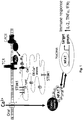


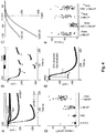
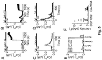
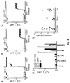
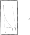


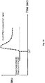

Classifications
-
- G—PHYSICS
- G01—MEASURING; TESTING
- G01N—INVESTIGATING OR ANALYSING MATERIALS BY DETERMINING THEIR CHEMICAL OR PHYSICAL PROPERTIES
- G01N33/00—Investigating or analysing materials by specific methods not covered by groups G01N1/00 - G01N31/00
- G01N33/48—Biological material, e.g. blood, urine; Haemocytometers
- G01N33/50—Chemical analysis of biological material, e.g. blood, urine; Testing involving biospecific ligand binding methods; Immunological testing
- G01N33/5005—Chemical analysis of biological material, e.g. blood, urine; Testing involving biospecific ligand binding methods; Immunological testing involving human or animal cells
- G01N33/5008—Chemical analysis of biological material, e.g. blood, urine; Testing involving biospecific ligand binding methods; Immunological testing involving human or animal cells for testing or evaluating the effect of chemical or biological compounds, e.g. drugs, cosmetics
-
- G—PHYSICS
- G01—MEASURING; TESTING
- G01N—INVESTIGATING OR ANALYSING MATERIALS BY DETERMINING THEIR CHEMICAL OR PHYSICAL PROPERTIES
- G01N33/00—Investigating or analysing materials by specific methods not covered by groups G01N1/00 - G01N31/00
- G01N33/48—Biological material, e.g. blood, urine; Haemocytometers
- G01N33/50—Chemical analysis of biological material, e.g. blood, urine; Testing involving biospecific ligand binding methods; Immunological testing
- G01N33/5005—Chemical analysis of biological material, e.g. blood, urine; Testing involving biospecific ligand binding methods; Immunological testing involving human or animal cells
- G01N33/5008—Chemical analysis of biological material, e.g. blood, urine; Testing involving biospecific ligand binding methods; Immunological testing involving human or animal cells for testing or evaluating the effect of chemical or biological compounds, e.g. drugs, cosmetics
- G01N33/5076—Chemical analysis of biological material, e.g. blood, urine; Testing involving biospecific ligand binding methods; Immunological testing involving human or animal cells for testing or evaluating the effect of chemical or biological compounds, e.g. drugs, cosmetics involving cell organelles, e.g. Golgi complex, endoplasmic reticulum
-
- G—PHYSICS
- G01—MEASURING; TESTING
- G01N—INVESTIGATING OR ANALYSING MATERIALS BY DETERMINING THEIR CHEMICAL OR PHYSICAL PROPERTIES
- G01N33/00—Investigating or analysing materials by specific methods not covered by groups G01N1/00 - G01N31/00
- G01N33/48—Biological material, e.g. blood, urine; Haemocytometers
- G01N33/50—Chemical analysis of biological material, e.g. blood, urine; Testing involving biospecific ligand binding methods; Immunological testing
- G01N33/68—Chemical analysis of biological material, e.g. blood, urine; Testing involving biospecific ligand binding methods; Immunological testing involving proteins, peptides or amino acids
- G01N33/6872—Intracellular protein regulatory factors and their receptors, e.g. including ion channels
-
- G—PHYSICS
- G01—MEASURING; TESTING
- G01N—INVESTIGATING OR ANALYSING MATERIALS BY DETERMINING THEIR CHEMICAL OR PHYSICAL PROPERTIES
- G01N2333/00—Assays involving biological materials from specific organisms or of a specific nature
- G01N2333/435—Assays involving biological materials from specific organisms or of a specific nature from animals; from humans
- G01N2333/705—Assays involving receptors, cell surface antigens or cell surface determinants
Definitions
- compositions and uses related to Ca 2+ release-activated Ca 2+ (CRAC) channel activity are described herein.
- the regulation of intracellular calcium is a key element in the transduction of signals into and within cells.
- Cellular responses to growth factors, neurotransmitters, hormones and a variety of other signal molecules are initiated through calcium-dependent processes.
- Cytosolic Ca 2+ signals control a wide array of cellular functions ranging from short-term responses such as contraction and secretion to longer-term regulation of cell growth and proliferation. Usually, these signals involve some combination of release of Ca 2+ from intracellular stores, such as the endoplasmic reticulum (ER), and influx of Ca 2+ across the plasma membrane.
- ER endoplasmic reticulum
- influx of Ca 2+ across the plasma membrane influx of Ca 2+ across the plasma membrane.
- cell activation begins with an agonist binding to a surface membrane receptor, coupled to phospholipase C (PLC) through a G-protein mechanism.
- PLC phospholipase C
- IP 3 inositol 1,4,5-triphosphate
- SOC plasma membrane store-operated calcium
- Store-operated calcium (SOC) influx is a process in cellular physiology that controls such diverse functions such as, but not limited to, refilling of intracellular Ca 2+ stores ( Putney et al. Cell, 75, 199-201, 1993 ), activation of enzymatic activity ( Fagan et al., J. Biol. Chem. 275:26530-26537, 2000 ), gene transcription ( Lewis, Annu. Rev. Immunol. 19:497-521, 2001 ), cell proliferation ( Nunez et al., J. Physiol. 571.1, 57-73, 2006 ), and release of cytokines ( Winslow et al., Curr. Opin. Immunol. 15:299-307, 2003 ).
- SOC Store-operated calcium
- SOC influx occurs through calcium release-activated calcium (CRAC) channels, a type of SOC channel.
- CRAC calcium release-activated calcium
- J.C.Mercer et al., J. Biol. Chemistry, 281, 24979-24990 (2006 ) relates to the large store-operated calcium selective currects due to co-expression of orai1 or orai2 with the intracellular calcium sensor, stim1*.
- C. Peinelt et al., Nature Cell Biology, 8, 771-776 (2006 ), relates to amplification of CRAC current by STIM1 and CRACM1 (orai1).
- Disclosed herein are methods of identifying an agent that modulates intracellular calcium comprising: contacting one or more test cells comprising a stromal interacting molecule (STIM) protein or a portion thereof and an Orai protein or a portion thereof, assessing the effect(s) of the agent on intracellular calcium, and identifying an agent that has an effect on intracellular calcium.
- STIM protein and/or the Orai protein is a mammalian protein.
- the STIM protein is STIM1 or STIM2, or a portion thereof.
- the Orai protein is Orail, or a portion thereof.
- the agent modulating intracellular calcium levels acts by decreasing intracellular calcium levels.
- the modulation of the intracellular calcium levels is monitored with ion-flux analysis.
- the ion-flux analysis measures electrophysiological changes or changes in fluorescent content of the test cells.
- the methods disclosed herein further comprise a fluorescent protein or luminescent protein that is used in monitoring or measuring intracellular calcium levels.
- the fluorescent protein is an aequorin-like protein or a chameleon or chameleon-like protein.
- the methods disclosed hereinfurther comprise a calcium depleting agent that provides for reduction of calcium levels in an intracellular calcium store.
- the calcium depleting agent is thapsigargin.
- the method includes an that agent modulates an activity of, modulates an interaction of, or modulates the level of, or binds to, or interacts with STIM1, Orai 1 or a combination thereof.
- the agent further modulates a cytokine expression or secretion.
- the cytokine is selected from IL-2, IL-3, IL-4, IL-5, IL-6, IL-7, IL-8, IL-9, IL-10, IL-11, IL-12, IL-13, IL-15, IL-16, IL-17, IL-18, IL-1 ⁇ , IL-1 ⁇ , IL-1 RA, granulocyte colony stimulating factor (G-CSF), granulocyte-macrophage colony stimulating factor (GM-CSF), oncostatin M, erythropoietin, leukemia inhibitory factor (LIF), interferons, gamma-interferon ( ⁇ -IFN), B7.1 (CD80), B7.2 (B70, CD86), TNF- ⁇ , TNF- ⁇ , LT- ⁇ , CD40 ligand, Fas ligand, CD27 ligand, CD30 ligand, 4-1BBL, Trail,
- a method is described to identify an agent that modulates intracellular calcium comprising: contacting one or more test cells comprising a first amino acid sequence at least 95% identical to an amino acid sequence of a STIM protein and a second amino acid sequence at least 95% identical to an amino acid sequence of an Orai protein, assessing the effect(s) of the agent on intracellular calcium, and identifying an agent that has an effect on intracellular calcium.
- a method is described to identify an agent that modulates intracellular calcium comprising: contacting one or more test cells comprising a nucleotide sequence at least 95% identical to a nucleic acid sequence of a STIM protein and a second nucleotide sequence at least 95% identical to a nucleic sequence of an Orai protein, assessing the effect(s) of the agent on intracellular calcium, and identifying an agent that has an effect on intracellular calcium.
- a method is described of identifying an agent that modulates cytokine expression or secretion, comprising: contacting one or more test cells comprising a stromal interacting molecule (STIM) protein or a portion thereof and an Orai protein or a portion thereof, assessing the effect(s) of the agent on cytokine expression or secretion, and identifying an agent that has an effect on cytokine expression or secretion.
- STIM stromal interacting molecule
- a mammalian cell which comprises a stromal interacting molecule (STIM) protein or a portion thereof, and an Orai protein or a portion thereof.
- STIM stromal interacting molecule
- the STIM protein of the mammalian cell is STIM1 or STIM2, or a portion thereof.
- the Orai protein is Orail, or a portion thereof.
- a mammalian cell comprising a first amino acid sequence at least 95% identical to the amino acid sequence of STIM1 and a second amino acid sequence at least 95% identical to the amino acid sequence of Orail.
- a mammalian cell comprising a nucleotide sequence at least 95% identical to the nucleic acid sequence of STIM1 and a second nucleotide sequence at least 95% identical to the nucleic acid sequence of Orail.
- composition comprising a cell comprising a first amino acid sequence at least 95% identical to the amino acid sequence of a STIM protein, and a second amino acid sequence at least 95% identical to the amino acid sequence of an Orai protein, and an agent that provides for reduction of calcium levels in an intracellular calcium store.
- a method of treating a disease, disorder or condition in a mammal where the mammal would benefit from inhibition of store operated calcium channel activity, the method comprising administering a compound capable of modulating a STIM protein and/or an Orai protein, or pharmaceutically acceptable salt, pharmaceutically acceptable solvate, or pharmaceutically acceptable prodrug thereof.
- a method of decreasing cytokine expression by inhibiting a store-operated calcium entry activation in a mammal is described, the method comprising administering a compound capable of modulating a STIM protein and/or an Orai protein, or pharmaceutically acceptable salt, pharmaceutically acceptable solvate, or pharmaceutically acceptable prodrug thereof.
- an article of manufacture comprising packaging material, a cell comprising a stromal interacting molecule (STIM) protein or a portion thereof, and an Orai protein or a portion thereof is described.
- STIM stromal interacting molecule
- the STIM protein of the article of manufacture is STIM1 or STIM2
- the Orai protein is Orail.
- Described herein is a method of modulating store-operated calcium (SOC) channel activity comprising contacting the store-operated calcium (SOC) channel complex, or portion thereof, with a compound that inhibits intracellular calcium levels, or pharmaceutically acceptable salt, pharmaceutically acceptable solvate, or pharmaceutically acceptable prodrug thereof.
- the contacting occurs in vitro.
- the contacting occurs in vivo.
- the intracellular calcium modulating compounds identified and described herein modulate an activity of, modulates an interaction of, or modulates the level of, or binds to, or interacts with at least one portion of the store operated calcium channel complex selected from stromal interaction molecules (STIM) or Orai family of proteins.
- STIM stromal interaction molecules
- the intracellular calcium modulating compound modulates an activity of, modulates an interaction of, or modulates the level of, or binds to, or interacts with at least one portion of STIM1 or STIM2. In another case the intracellular calcium modulating compound modulates an activity of, modulates an interaction of, or modulates the level of, or binds to, or interacts with at least one portion of Orail.
- modulating store operated calcium channel activity with an intracellular calcium modulating compound inhibits store-operated calcium entry (SOCE).
- the store operated calcium channel complex is calcium-release activated calcium (CRAC) channel complex.
- modulating calcium release activated calcium (CRAC) activity with a compound capable of modulating a STIM protein and/or an Orai protein inhibits the electrophysiological current (I CRAC ) directly associated with activated CRAC channels.
- intracellular calcium modulating compound modulates the activity of, modulates an interaction of, or binds to, or interacts with a mammalian STIM1 protein, or a mammalian STIM2 protein.
- intracellular calcium modulating compound modulates the activity of, modulates an interaction of, or binds to, or interacts with a mammalian Orail protein.
- the intracellular calcium modulating compound modulates the activity of, modulates an interaction of, or binds to, or interacts with a mammalian STIM1 protein and a mammalian Orail protein.
- the disease, disorder or condition in a mammal is selected from diseases/disorders involving inflammation, glomerulonephritis, uveitis, hepatic diseases or disorders, renal diseases or disorders, chronic obstructive pulmonary disease, rheumatoid arthritis, inflammatory bowel disease, vasculitis, dermatitis, osteoarthritis, inflammatory muscle disease, allergic rhinitis, vaginitis, interstitial cystitis, scleroderma, osteoporosis, eczema, allogeneic or xenogeneic transplantation, graft rejection, graft-versus-host disease, lupus erythematosus, type I diabetes, pulmonary fibrosis, dermatomyositis, thyroiditis, myasthenia gravis, autoimmune hemolytic anemia, cystic fibrosis, chronic relapsing hepatitis, primary biliary cirrhosis, allergic conjun
- Also described herein is a method of inhibiting store-operated calcium entry (SOCE) activation of nuclear factor of activated T cells (NFAT) in a mammal comprising administering a compound capable of modulating a STIM protein and/or an Orai protein, or pharmaceutically acceptable salt, pharmaceutically acceptable solvate, or pharmaceutically acceptable prodrug thereof.
- the compounds identified and described herein modulate an interaction of, or modulates the level of, or binds to, or interacts with a mammalian STIM1 protein, or a mammalian STIM2 protein.
- Also described herein is a method of decreasing cytokine expression by inhibiting the store-operated calcium entry activation of NFAT in a mammal comprising administering a compound capable of modulating a STIM protein and/or an Orai protein levels, or pharmaceutically acceptable salt, pharmaceutically acceptable solvate, or pharmaceutically acceptable prodrug thereof.
- the compounds disclosed herein modulate an interaction of, or modulates the level of, or binds to, or interacts with a mammalian STIM1 protein or a mammalian STIM2 protein.
- the cytokine is selected from IL-2, IL-3, IL-4, IL-5, IL-6, IL-7, IL-8, IL-9, IL-10, IL-11, IL-12, IL-13, IL-15, IL-16, IL-17, IL-18, IL-1 ⁇ , IL-1 ⁇ , IL-1 RA, granulocyte colony stimulating factor (G-CSF), granulocyte-macrophage colony stimulating factor (GM-CSF), oncostatin M, erythropoietin, leukemia inhibitory factor (LIF), interferons, gamma-interferon ( ⁇ -IFN), B7.1 (CD80), B7.2 (B70, CD86), TNF- ⁇ , TNF- ⁇ , LT- ⁇ , CD40 ligand, Fas ligand, CD27 ligand, CD30 ligand, 4-1BBL, Trail, beta-hexosaminidase, and migration inhibitory factor (MIF).
- G-CSF
- the invention provides a stably transfected recombinant cell comprising a recombinant orai gene or a polynucleotide that is at least 95% identical to a nucleic acid sequence of the orai gene and a recombinant stim gene or a polynucleotide that is at least 95% identical to a nucleic acid sequence of the orai STIM gene, wherein the orai gene and stim gene are over-expressed.
- the invention further provides the recombinant cell of claim 1, wherein the cell is a mammalian, human, rodent, or insect cell.
- the invention further provides a method of identifying a compound that modulates intracellular calcium levels and binds to or modulates an activity of an orai/stim polypeptide complex, the method comprising: (a) contacting a stably transfected recombinant cell of any of claims 1 to 7 comprising the polypeptide complex with a test compound; and (b) detecting binding of the test compound to the complex, or modulation of the activity of the polypeptide complex by the test compound compared to a control compound using patch clamp electrophysiology, capacitance measurements, calcium imaging using fluorescent dyes, fluorescence resonance energy transfer (FRET), or a molecular biology method, thereby identifying the compound that binds to or modulates the activity of the complex.
- a test compound comprising: (a) contacting a stably transfected recombinant cell of any of claims 1 to 7 comprising the polypeptide complex with a test compound; and (b) detecting binding of the test compound to the complex, or modulation of the activity of the
- the invention further provides the method of claim 8, wherein the STIM protein is STIM1 or STIM2.
- RNAi medium-throughput RNA interference
- STIM1 and STIM2 also were identified in an independently performed screen of HeLa cells by using the Drosophila enzyme Dicer to generate small interfering RNA species from dsRNA.
- Drosophila Stim and the mammalian homolog STIM1 appear to play dual roles in the CRAC channel activation sequence, sensing the luminal Ca 2+ store content through an EF hand motif and trafficking from an endoplasmic reticulum (ER)-like localization to the plasma membrane to trigger CRAC channel activity.
- ER endoplasmic reticulum
- Stim and its mammalian homolog STIM1 are unlikely to form the CRAC channel itself.
- a genome-wide screen on S2 cells was devised and performed based on a fluorescence assay of Ca 2+ influx.
- the library at Harvard's Drosophila RNAi Screening Center (DRSC) of 23,845 dsRNA amplicons has been used in several functional screens.
- a genetic defect was identified in patients with severe combined immune deficiency (SCID).
- SCID severe combined immune deficiency
- the screen in the genome-wide study made use of the ability of thapsigargin (TG) to send GFP-tagged nuclear factor of activated T cells (NFAT) to the nucleus in S2 cells, providing an assay for disruption of signaling anywhere in the cascade from elevated [Ca 2+ ] i to calcineurin activation and nuclear relocalization of NFAT.
- the fly gene olf186-F (named Orai ) was identified in the screen, and a human homolog on chromosome 12 was shown to be mutated in SCID patients, resulting in the loss of CRAC channel activity.
- Heterologous expression of the wild-type human homolog which was named Orail, restored CRAC channel activity in SCID T cell lines.
- olf186-F knockdown of olf186-F did not produce a severe cell growth phenotype (data not shown).
- the olf186-F gene is a member of a highly conserved gene family that contains three homologs in mammals, two in chicken, three in zebrafish, and one member only in fly and worm (see Fig. 8 A ).
- C09F5.2 the only homolog in Caenorhabditis elegans, is expressed in intestine, hypodermis, and reproductive system as well as some neuron-like cells in the head and tail regions (www.wormbase.org). Worms under RNAi treatment against C09F5.2 are sterile.
- Ca-P60A has been proposed to be the only Drosophila SERCA gene.
- the ER pump function was validated by showing that ionomycin did not induce significant store release from S2 cells pretreated with dsRNA against Ca-P60A, consistent with a previous report.
- the elevation in resting [Ca 2+ ] i and rapidly changing Ca 2+ transients during changes in external Ca 2+ before addition of TG indicates a low level of constitutive CRAC channel activity induced by store depletion.
- SERCA knockdown inhibited CRAC channel activity after passive store depletion in whole-cell patch recordings.
- RNAi effects of Syx5 was verified at the single-cell level and demonstrated strong suppression of CRAC channel activity as well as the SOC influx.
- tsr regulates SOC influx indirectly by controlling cell metabolism because RNAi of tsr did not significantly influence CRAC current density in whole-cell patch-clamp experiments.
- Membrane trafficking previously has been demonstrated to play a role in SOC channel activity in Xenopus oocytes, based on inhibition by botulinum toxin or by a dominant-negative SNAP-25 construct, and our results further support a requirement for syntaxins and SNARE-complex formation, possibly to mediate translocation of Stim to the plasma membrane.
- the screen also revealed three other groups of hits that influence calcium dynamics.
- a agent inhibiting either the STIM or Orai polypeptides, or both affects a larger inhibitory effect on intracellular calcium levels or calcium influx, then by the presence of the STIM or Orai polypeptide alone.
- the STIM protein a single-pass transmembrane protein localized in the endoplasmic reticulum, functions as a sensor of luminal Ca 2+ store content through an EF hand motif. Upon decrease of intracellular calcium levels, STIM migrates to the plasma membrane to interact with the Orai polypeptide, triggering CRAC channel activity.
- compositions that comprise a labeled STIM polypeptide and a labeled Orai polypeptide, wherein the labeled STIM and Orai polypeptide when in proximity of each other emit a second energy frequency (e.g.
- the STIM polypeptide and the Orai polypeptide are mammalian.
- the STIM polypeptide is STIM1 or STIM2.
- the Orai polypeptide is Orail.
- nucleotides encoding the STIM and Orai polypeptides are transiently transfected.
- nucleotides encoding the polypeptides are stably transfected.
- nucleotides encoding the polypeptides are overexpressed.
- the label is fluorescent or radioactive.
- compositions that comprise a STIM polypeptide and Orai polypeptide, and the synergistic activity of both polypeptides in regulating SOC and calcium influx into a cell.
- the STIM polypeptide and the Orai polypeptide are mammalian.
- the STIM polypeptide is STIM1 or STIM2.
- the Orai polypeptide is Orail.
- nucleotides encoding the STIM and Orai polypeptides are transiently transfected.
- nucleotides encoding the polypeptides are stably transfected.
- nucleotides encoding the polypeptides are overexpressed.
- compositions described herein to identify agents that affect intracellular calcium levels.
- intracellular calcium levels are monitored via ion flux analysis (e.g. patch clamp analysis), or by monitoring the influx of radioactive or fluorescent calcium tracers into the cell in response to the depletion of calcium stores in the cell.
- thapsigargin treatment depletes the calcium stores.
- treatment with thapsigargin depletes intracellular calcium stores, which triggers the STIM Ca 2+ sensor and subsequent activation of the STIM/Orai CRAC channel.
- methods of identifying agents that affect intracellular calcium levels by treating test cells comprising a STIM polypeptide and an Orai polypeptide, and monitoring intracellular calcium levels as a result of treatment of the test cells with said agent.
- compounds that inhibit the STIM/Orai CRAC-channel mediated calcium influx are optionally used, for example, to modulate cytokine levels by the modulation of the intracellular calcium stores in immunological cells, e.g. T-cells.
- subject or “patient” encompasses mammals and non-mammals.
- mammals include, but are not limited to, any member of the Mammalian class: humans, non-human primates such as chimpanzees, and other apes and monkey species; farm animals such as cattle, horses, sheep, goats, swine; domestic animals such as rabbits, dogs, and cats; laboratory animals including rodents, such as rats, mice and guinea pigs, and the like.
- non-mammals include, but are not limited to, birds, fish and the like.
- the mammal is a human.
- treat include alleviating, abating or ameliorating a disease, disorder or condition symptoms, preventing additional symptoms, ameliorating or preventing the underlying causes of symptoms, inhibiting the disease, disorder or condition, e.g., arresting the development of the disease, disorder or condition, relieving the disease, disorder or condition, causing regression of the disease, disorder or condition, relieving a condition caused by the disease, disorder or condition, or stopping the symptoms of the disease, disorder or condition either prophylactically and/or therapeutically.
- target protein refers to a protein or a portion of a protein capable of being bound by, or interacting with a compound described herein, such as a compound capable of modulating a SITM protein and/or an Orai protein.
- a target protein is a STIM protein.
- a target protein is an Orai protein, and in yet other cases, the compound targets both STIM and Orai proteins.
- STIM protein includes but is not limited to, mammalian STIM-1, such as human and rodent (e.g., mouse) STIM-1, Drosophila melanogaster D-STIM, C. elegans C-STIM, Anopheles gambiae STIM and mammalian STIM-2, such as human and rodent (e.g., mouse) STIM-2.
- mammalian STIM-1 such as human and rodent (e.g., mouse) STIM-1
- Drosophila melanogaster D-STIM such as human and rodent (e.g., mouse) STIM-1
- C. elegans C-STIM C. elegans C-STIM
- Anopheles gambiae STIM gambiae STIM
- mammalian STIM-2 such as human and rodent (e.g., mouse) STIM-2.
- such proteins have been identified as being involved in, participating in and/or providing for store-operated calcium entry or modulation thereof,
- an "Orai protein” includes Orail (SEQ ID NO: 1 as described in WO 07/081804 ), Orai2 (SEQ ID NO: 2 as described in WO 07/081804 ), or Orai3 (SEQ ID NO: 3 as described in WO 07/081804 ).
- Orail nucleic acid sequence corresponds to GenBank accession number NM_032790
- Orai2 nucleic acid sequence corresponds to GenBank accession number BC069270
- Orai3 nucleic acid sequence corresponds to GenBank accession number NM_152288.
- Orai refers to any one of the Orai genes, e.g., Orail, Orai2, Orai3 (see Table I of WO 07/081804 ). As described herein, such proteins have been identified as being involved in, participating in and/or providing for store-operated calcium entry or modulation thereof, cytoplasmic calcium buffering and/or modulation of calcium levels in or movement of calcium into, within or out of intracellular calcium stores (e.g., endoplasmic reticulum). In alternative cases, an Orai protein may be labeled with a tag molecule, by way of example only, is an enzyme fragment, a protein (e.g.
- c-myc or other tag protein or fragment thereof an enzyme tag, a fluorescent tag, a fluorophore tag, a chromophore tag, a Raman-activated tag, a chemiluminescent tag, a quantum dot marker, an antibody, a radioactive tag, or combinations thereof.
- fragment or “derivative” when referring to a protein (e.g. STIM, Orai) means proteins or polypeptides which retain essentially the same biological function or activity in at least one assay as the native protein(s).
- the fragments or derivatives of the referenced protein maintains at least about 50% of the activity of the native proteins, at least 75%, at least about 95% of the activity of the native proteins, as determined e.g. by a calcium influx assay.
- amelioration of the symptoms of a particular disease, disorder or condition by administration of a particular compound or pharmaceutical composition refers to any lessening of severity, delay in onset, slowing of progression, or shortening of duration, whether permanent or temporary, lasting or transient that are attributed to or associated with administration of the compound or composition.
- module means to interact with a target protein either directly or indirectly so as to alter the activity of the target protein, including, by way of example only, to inhibit the activity of the target, or to limit or reduce the activity of the target.
- a modulator refers to a compound that alters an activity of a target. For example, in some cases, a modulator causes an increase or decrease in the magnitude of a certain activity of a target compared to the magnitude of the activity in the absence of the modulator. In certain cases, a modulator is an inhibitor, which decreases the magnitude of one or more activities of a target. In certain cases, an inhibitor completely prevents one or more activities of a target.
- modulation with reference to intracellular calcium refers to any alteration or adjustment in intracellular calcium including but not limited to alteration of calcium concentration in the cytoplasm and/or intracellular calcium storage organelles, e.g ., endoplasmic reticulum, and alteration of the kinetics of calcium fluxes into, out of and within cells. In case, modulation refers to reduction.
- inhibitors refer to inhibition of store operated calcium channel activity or calcium release activated calcium channel activity.
- pharmaceutically acceptable refers a material, such as a carrier or diluent, which does not abrogate the biological activity or properties of the compound, and is relatively nontoxic, i.e., the material is administered to an individual without causing undesirable biological effects or interacting in a deleterious manner with any of the components of the composition in which it is contained.
- composition refers to a mixture of a compound capable of modulating a STIM protein and/or an Orai protein as described herein with other chemical components, such as carriers, stabilizers, diluents, dispersing agents, suspending agents, thickening agents, and/or excipients.
- the pharmaceutical composition facilitates administration of the compound to an organism. Multiple techniques of administering a compound exist including, but not limited to: intravenous, oral, aerosol, parenteral, ophthalmic, pulmonary and topical administration.
- an "effective amount” or “therapeutically effective amount,” as used herein, refer to a sufficient amount of an agent or a compound being administered which will relieve to some extent one or more of the symptoms of the disease or condition being treated. The result is reduction and/or alleviation of the signs, symptoms, or causes of a disease, or any other desired alteration of a biological system.
- an "effective amount” for therapeutic uses is the amount of the composition that includes a compound of capable of modulating a STIM protein and/or an Orai protein as described herein required to provide a clinically significant decrease in disease symptoms.
- an appropriate "effective" amount in any individual case is determined using techniques, such as a dose escalation study.
- an “enhance” or “enhancing,” as used herein, means to increase or prolong either in potency or duration a desired effect.
- the term “enhancing” refers to the ability to increase or prolong, either in potency or duration, the effect of other therapeutic agents on a system.
- An “enhancing-effective amount,” as used herein, refers to an amount adequate to enhance the effect of another therapeutic agent in a desired system.
- carrier refers to relatively nontoxic chemical compounds or agents that facilitate the incorporation of a compound into cells or tissues.
- diluent refers to chemical compounds that are used to dilute the compound of interest prior to delivery. In some cases, diluents are used to stabilize compounds because they provide a more stable environment. Salts dissolved in buffered solutions (which also provide pH control or maintenance) are utilized as diluents, including, but not limited to a phosphate buffered saline solution.
- a “metabolite” of a compound disclosed herein is a derivative of that compound that is formed when the compound is metabolized.
- Bioavailability refers to the percentage of the weight of the compound disclosed herein (e.g. compound capable of modulating intracellular calcium that is delivered into the general circulation of the animal or human being studied). The total exposure (AUC(0- ⁇ )) of a drug when administered intravenously is usually defined as 100% bioavailable (F%).
- Oral bioavailability refers to the extent to which a compound disclosed herein, is absorbed into the general circulation when the pharmaceutical composition is taken orally as compared to intravenous injection.
- Blood plasma concentration refers to the concentration of a compound capable of modulating a STIM protein and/or an Orai protein as disclosed herein, in the plasma component of blood of a subject. It is understood that the plasma concentration of compounds described herein likely varies significantly between subjects, due to variability with respect to metabolism and/or possible interactions with other therapeutic agents. In accordance with one case disclosed herein, the blood plasma concentration of the compounds disclosed herein varies from subject to subject. Likewise, in some cases, values such as maximum plasma concentration (C max ) or time to reach maximum plasma concentration (T max ), or total area under the plasma concentration time curve (AUC(0- ⁇ )) varies from subject to subject. Due to this variability, the amount necessary to constitute "a therapeutically effective amount" of a compound will vary from subject to subject.
- calcium homeostasis refers to the maintenance of an overall balance in intracellular calcium levels and movements, including calcium signaling, within a cell.
- intracellular calcium refers to calcium located in a cell without specification of a particular cellular location.
- cytosolic or “cytoplasmic” with reference to calcium refers to calcium located in the cell cytoplasm.
- an effect on intracellular calcium is any alteration of any case of intracellular calcium, including but not limited to, an alteration in intracellular calcium levels and location and movement of calcium into, out of or within a cell or intracellular calcium store or organelle.
- an effect on intracellular calcium is an alteration of the properties, such as, for example, the kinetics, sensitivities, rate, amplitude, and electrophysiological characteristics, of calcium flux or movement that occurs in a cell or portion thereof.
- an effect on intracellular calcium is an alteration in any intracellular calcium-modulating process, including, store-operated calcium entry, cytosolic calcium buffering, and calcium levels in or movement of calcium into, out of or within an intracellular calcium store.
- any of these cases are assessed in a variety of ways including, but not limited to, evaluation of calcium or other ion (particularly cation) levels, movement of calcium or other ion (particularly cation), fluctuations in calcium or other ion (particularly cation) levels, kinetics of calcium or other ion (particularly cation) fluxes and/or transport of calcium or other ion (particularly cation) through a membrane.
- An alteration is any such change that is statistically significant.
- intracellular calcium in a test cell and a control cell is said to differ, such differences are a statistically significant difference.
- intracellular calcium or intracellular calcium regulation means that when expression or activity of the protein in a cell is reduced, altered or eliminated, there is a concomitant or associated reduction, alteration or elimination of one or more cases of intracellular calcium or intracellular calcium regulation. Such an alteration or reduction in expression or activity occurs by virtue of an alteration of expression of a gene encoding the protein or by altering the levels of the protein.
- a protein involved in an case of intracellular calcium such as, for example, store-operated calcium entry, thus, are one that provides for or participates in an case of intracellular calcium or intracellular calcium regulation.
- a protein that provides for store-operated calcium entry are a STIM protein and/or an Orai protein.
- a protein that is a component of a calcium channel is a protein that participates in multiprotein complex that forms the channel.
- cation entry or “calcium entry” into a cell refers to entry of cations, such as calcium, into an intracellular location, such as the cytoplasm of a cell or into the lumen of an intracellular organelle or storage site.
- cation entry is, for example, the movement of cations into the cell cytoplasm from the extracellular medium or from an intracellular organelle or storage site, or the movement of cations into an intracellular organelle or storage site from the cytoplasm or extracellular medium. Movement of calcium into the cytoplasm from an intracellular organelle or storage site is also referred to as "calcium release" from the organelle or storage site.
- protein that modulates intracellular calcium refers to any cellular protein that is involved in regulating, controlling and/or altering intracellular calcium. For example, in some cases, such a protein is involved in altering or adjusting intracellular calcium in a number of ways, including, but not limited to, through the maintenance of resting or basal cytoplasmic calcium levels, or through involvement in a cellular response to a signal that is transmitted in a cell through a mechanism that includes a deviation in intracellular calcium from resting or basal states.
- a "cellular" protein is one that is associated with a cell, such as, for example, a cytoplasmic protein, a plasma membrane-associated protein or an intracellular membrane protein. Proteins that modulate intracellular calcium include, but are not limited to, ion transport proteins, calcium-binding proteins and regulatory proteins that regulate ion transport proteins.
- amelioration refers to an improvement in a disease or condition or at least a partial relief of symptoms associated with a disease or condition.
- cell response refers to any cellular response that results from ion movement into or out of a cell or within a cell.
- the cell response is associated with any cellular activity that is dependent, at least in part, on ions such as, for example, calcium.
- activities optionally include, for example, cellular activation, gene expression, endocytosis, exocytosis, cellular trafficking and apoptotic cell death.
- immune cells include cells of the immune system and cells that perform a function or activity in an immune response, such as, but not limited to, T-cells, B-cells, lymphocytes, macrophages, dendritic cells, neutrophils, eosinophils, basophils, mast cells, plasma cells, white blood cells, antigen presenting cells and natural killer cells.
- cytokine refers to small soluble proteins secreted by cells that in some cases, alter the behavior or properties of the secreting cell or another cell. Cytokines bind to cytokine receptors and trigger a behavior or property within the cell, for example, cell proliferation, death or differentiation.
- cytokines include, but are not limited to, interleukins (e.g., IL-2, IL-3, IL-4, IL-5, IL-6, IL-7, IL-8, IL-9, IL-10, IL-11, IL-12, IL-13, IL-15, IL-16, IL-17, IL-18, IL-1 ⁇ , IL-1 ⁇ , and IL-1 RA), granulocyte colony stimulating factor (G-CSF), granulocyte-macrophage colony stimulating factor (GM-CSF), oncostatin M, erythropoietin, leukemia inhibitory factor (LIF), interferons, B7.1 (also known as CD80), B7.2 (also known as B70, CD86), TNF family members (TNF- ⁇ , TNF- ⁇ , LT- ⁇ , CD40 ligand, Fas ligand, CD27 ligand, CD30 ligand, 4-1BBL, Trail), and MIF.
- interleukins e.g
- Selective inhibitor of SOC channel activity means that the inhibitor is selective for SOC channels and does not substantially affect the activity of other types of ion channels.
- “Selective inhibitor of CRAC channel activity” means that the inhibitor is selective for CRAC channels and does not substantially affect the activity of other types of ion channels and/or other SOC channels.
- Cellular calcium homeostasis is a result of the summation of regulatory systems involved in the control of intracellular calcium levels and movements.
- Cellular calcium homeostasis is achieved, at least in part, by calcium binding and by movement of calcium into and out of the cell across the plasma membrane and within the cell by movement of calcium across membranes of intracellular organelles including, for example, the endoplasmic reticulum, sarcoplasmic reticulum, mitochondria and endocytic organelles including endosomes and lysosomes.
- Movement of calcium across cellular membranes is carried out by specialized proteins.
- calcium from the extracellular space enters the cell through various calcium channels and a sodium/calcium exchanger and is actively extruded from the cell by calcium pumps and sodium/calcium exchangers.
- Calcium is also released from internal stores through inositol trisphosphate or ryanodine receptors and is likely taken up by these organelles by means of calcium pumps.
- VOC voltage-operated calcium
- SOC store-operated calcium
- sodium/calcium exchangers operating in reverse mode.
- VOC channels are activated by membrane depolarization and are found in excitable cells like nerve and muscle and are for the most part not found in nonexcitable cells.
- Ca 2+ also enters cells via Na + -Ca 2+ exchangers operating in reverse mode.
- Endocytosis provides another process by which cells take up calcium from the extracellular medium through endosomes.
- some cells e.g., exocrine cells, release calcium via exocytosis.
- Cytosolic calcium concentration is tightly regulated with resting levels usually estimated at approximately 0.1 ⁇ M in mammalian cells, whereas the extracellular calcium concentration is typically about 2 mM. This tight regulation facilitates transduction of signals into and within cells through transient calcium flux across the plasma membrane and membranes of intracellular organelles.
- the principal components involved in maintaining basal calcium levels are calcium pumps and leaks in the endoplasmic reticulum and plasma membrane. Disturbance of resting cytosolic calcium levels effects transmission of such signals and give rise to defects in a number of cellular processes. For example, cell proliferation involves a prolonged calcium signaling sequence. Other cellular processes include, but are not limited to, secretion, signaling, and fertilization, involve calcium signaling.
- SOC store operated calcium
- CRAC calcium release-activated calcium
- SOCE Store-operated Ca 2+ entry
- SOCE is the process in which the emptying of Ca 2+ stores itself activates Ca 2+ channels in the plasma membrane to help refill the stores ( Putney, Cell Calcium, 7, 1-12, 1986 ; Parekh et al., Physiol.Rev. 757-810; 2005 ).
- SOCE does more than simply provide Ca 2+ for refilling stores, but itself generates sustained Ca 2+ signals that control such essential functions as gene expression, cell metabolism and exocytosis ( Parekh and Putney, Physiol. Rev. 85, 757-810 (2005 ).
- NFAT a phosphatase that regulates the transcription factor NFAT.
- NFAT is phosphorylated and resides in the cytoplasm, but when dephosphorylated by calcineurin, NFAT translocates to the nucleus and activates different genetic programmes depending on stimulation conditions and cell type.
- NFAT In response to infections and during transplant rejection, NFAT partners with the transcription factor AP-1 (Fos-Jun) in the nucleus of "effector" T cells, thereby transactivating cytokine genes, genes that regulate T cell proliferation and other genes that orchestrate an active immune response ( Rao et al., Annu Rev Immunol., 1997;15:707-47 ). In contrast, in T cells recognizing self antigens, NFAT is activated in the absence of AP-1, and activates a transcriptional programme otherwise known as "anergy” that suppresses autoimmune responses ( Macian et al., Transcriptional mechanisms underlying lymphocyte tolerance. Cell. 2002 Jun 14;109(6):719-31 ).
- NFAT In a subclass of T cells, known as regulatory T cells which suppress autoimmunity mediated by self-reactive effector T cells, NFAT partners with the transcription factor FOXP3 to activate genes responsible for suppressor function ( Wu et al., Cell, 2006 Jul 28;126(2):375-87 ; Rudensky AY, Gavin M, Zheng Y. Cell. 2006 Jul 28;126(2):253-256 ).
- the endoplasmic reticulum carries out a variety processes.
- the ER has a role as both an agonist-sensitive Ca 2+ store and sink, protein folding/processing takes place within its lumen.
- numerous Ca 2+ -dependent chaperone proteins ensure that newly synthesized proteins are folded correctly and sent off to the appropriate destination.
- the ER is also involved in vesicle trafficking, release of stress signals, regulation of cholesterol metabolism, and apoptosis. Many of these processes require intraluminal Ca 2+ , and protein misfolding, ER stress responses, and apoptosis are all likely induced by depleting the ER of Ca 2+ for prolonged periods of time.
- I CRAC Ca 2+ release-activated Ca 2+ current
- I CRAC is non-voltage activated, inwardly rectifying, and remarkably selective for Ca 2+ . It is found in several cell types mainly of hemapoietic origin. I CRAC is not the only store-operated current, and it is now apparent that store-operated influx encompasses a family of Ca 2+ -permeable channels, with different properties in different cell types. I CRAC was the first store-operated Ca 2+ current to be described and remains a popular model for studying store-operated influx.
- TPEN lowers free intraluminal Ca 2+ concentration without changing total store Ca 2+ such that the store depletion-dependent signal is generated.
- STIM Stromal interaction molecule
- CCE capacitative calcium entry
- I SOC store-operated current
- I CRAC calcium release-activated current
- Electrophysiological analysis of store-operated or calcium release-activated currents reveals distinct biophysical properties of these currents.
- the current is activated by depletion of intracellular calcium stores (e.g., by nonphysiological activators such as thapsigargin, CPA, ionomycin and BAPTA, and physiological activators such as IP 3 ) and are likely selective for divalent cations, such as calcium, over monovalent ions in physiological solutions or conditions, are influenced by changes in cytosolic calcium levels, and generally shows altered selectivity and conductivity in the presence of low extracellular concentrations of divalent cations.
- the current is also blocked or enhanced by 2-APB (depending on concentration) and blocked by SKF96365 and Gd 3+ and generally are described as a calcium current that is not strictly voltage-gated.
- CRAC entry mechanism as an ion channel with distinctive biophysical characteristics, including a high selectivity for Ca 2+ paired with an exceedingly low conductance. Furthermore, the CRAC channel was shown to fulfill the rigorous criteria for being store-operated, which is the activation solely by the reduction of Ca 2+ in the ER rather than by cytosolic Ca 2+ or other messengers generated by PLC.
- Intracellular calcium stores are characterized by sensitivity to agents, which are physiological or pharmacological, which activate release of calcium from the stores or inhibit uptake of calcium into the stores.
- agents which are physiological or pharmacological, which activate release of calcium from the stores or inhibit uptake of calcium into the stores.
- Different cells have been studied in characterization of intracellular calcium stores, and stores have been characterized as sensitive to various agents, including, but not limited to, IP 3 and compounds that effect the IP 3 receptor, thapsigargin, ionomycin and/or cyclic ADP-ribose (cADPR).
- SR endoplasmic reticulum and sarcoplasmic reticulum (SR; a specialized version of the endoplasmic reticulum in striated muscle) storage organelles is achieved through sarcoplasmic-endoplasmic reticulum calcium ATPases (SERCAs), commonly referred to as calcium pumps.
- SERCAs sarcoplasmic-endoplasmic reticulum calcium ATPases
- endoplasmic reticulum calcium is replenished by the SERCA pump with cytoplasmic calcium that has entered the cell from the extracellular medium.
- IP 3 receptor-mediated calcium release is triggered by IP 3 formed in the break down of plasma membrane phosphoinositides through the action of phospholipase C activated by binding of an agonist to a plasma membrane G protein-coupled receptor.
- Ryanodine receptor-mediated calcium release is triggered by an increase in cytoplasmic calcium and is referred to as calcium-induced calcium release (CICR).
- CICR calcium-induced calcium release
- the calcium levels in the stores, and in the cytoplasm fluctuate.
- ER free calcium generally decrease from a range of about 60-400 ⁇ M to about 1-50 ⁇ M when HeLa cells are treated with histamine, an agonist of PLC-linked histamine receptors ( Miyawaki et al. (1997) Nature 388:882-887 ).
- Store-operated calcium entry is activated as the free calcium concentration of the intracellular stores is reduced. Depletion of store calcium, as well as a concomitant increase in cytosolic calcium concentration, likely thus regulate store-operated calcium entry into cells.
- Agonist activation of signaling processes in cells generally involve dramatic increases in the calcium permeability of the endoplasmic reticulum, for example, through opening of IP 3 receptor channels, and the plasma membrane through store-operated calcium entry. These increases in calcium permeability are associated with an increase in cytosolic calcium concentration that are separated into two components: a "spike" of calcium release from the endoplasmic reticulum during activation of the IP 3 receptor and a plateau phase which is a sustained elevation of calcium levels resulting from entry of calcium into the cytoplasm from the extracellular medium. Upon stimulation, the resting intracellular free calcium concentration of about 100 nM generally rise globally to greater than 1 ⁇ M.
- the cell modulates these calcium signals with endogenous calcium buffers, including physiological buffering by organelles such as mitochondria, endoplasmic reticulum and Golgi.
- organelles such as mitochondria, endoplasmic reticulum and Golgi.
- Mitochondrial uptake of calcium through a uniporter in the inner membrane is driven by the large negative mitochondrial membrane potential, and the accumulated calcium is released slowly through sodium-dependent and -independent exchangers, and, under some circumstances, the permeability transition pore (PTP).
- PTP permeability transition pore
- mitochondria generally act as calcium buffers by taking up calcium during periods of activation and slowly releasing it later. Uptake of calcium into the endoplasmic reticulum is regulated by the sarcoplasmic and endoplasmic reticulum calcium ATPase (SERCA).
- SERCA sarcoplasmic and endoplasmic reticulum calcium ATPase
- Uptake of calcium into the Golgi is mediated by a P-type calcium transport ATPase (PMR1/ATP2C1). Additionally, there is evidence that a significant amount of the calcium released upon IP 3 receptor activation is extruded from the cell through the action of the plasma membrane calcium ATPase.
- plasma membrane calcium ATPases provide the dominant mechanism for calcium clearance in human T cells and Jurkat cells, although sodium/calcium exchange also contributes to calcium clearance in human T cells.
- calcium ions are bound to specialized calcium-buffering proteins, such as, for example, calsequestrins, calreticulins and calnexins.
- cytoplasmic calcium buffering allows for sustained calcium influx through SOC channels. Large increases in cytoplasmic Ca2+ or store refilling deactivate SOCE.
- store-operated calcium entry affects a multitude of events that are consequent to or in addition to the store-operated changes.
- Ca 2+ influx results in the activation of a large number of calmodulin-dependent enzymes including the serine phosphatase calcineurin.
- Activation of calcineurin by an increase in intracellular calcium results in acute secretory processes such as mast cell degranulation.
- Activated mast cells release preformed granules containing histamine, heparin, TNF ⁇ and enzymes such as ⁇ -hexosaminidase.
- Some cellular events, such as B and T cell proliferation require sustained calcineurin signaling, which requires a sustained increase in intracellular calcium.
- NFAT neurotrophic factor of activated T cells
- MEF2 calcineurin
- NFAT transcription factors play important roles in many cell types, including immune cells.
- immune cells NFAT mediates transcription of a large number of molecules, including cytokines, chemokines and cell surface receptors.
- Transcriptional elements for NFAT have been found within the promoters of cytokines such as IL-2, IL-3, IL-4, IL-5, IL-8, IL-13, as well as tumor necrosis factor alpha (TNF ⁇ ), granulocyte colony-stimulating factor (G-CSF), and gamma-interferon ( ⁇ -IFN).
- TNF ⁇ tumor necrosis factor alpha
- G-CSF granulocyte colony-stimulating factor
- ⁇ -IFN gamma-interferon
- NFAT proteins The activity of NFAT proteins is regulated by their phosphorylation level, which in turn is regulated by both calcineurin and NFAT kinases.
- Activation of calcineurin by an increase in intracellular calcium levels results in dephosphorylation of NFAT and entry into the nucleus.
- Rephosphorylation of NFAT masks the nuclear localization sequence of NFAT and prevents its entry into the nucleus. Because of its strong dependence on calcineurin-mediated dephosphorylation for localization and activity, NFAT is a sensitive indicator of intracellular calcium levels.
- CRAC channel a type of SOC channel
- SOCE contributes directly to the elevation of cytosolic Ca 2+ levels ([Ca 2+ ] i ), as in T lymphocytes where CRAC channels generate the sustained Ca 2+ signals needed to drive gene expression underlying T cell activation by antigen.
- Sustained calcium entry is needed for lymphocyte activation and adpative immune response. Calcium entry into lymphocytes occurs primarily through the CRAC channels. Increased calcium levels lead to NFAT activation and expression of cytokines required for immune response.
- the CRAC channel has a distinctive biophysical fingerprint, quantifiable store-dependence, and essential function in T cells. Studies have shown that CRAC channels are formed from two component proteins, which interact to form CRAC channels.
- the CRAC channel is assembled by two functional components, STIM1 and Orail.
- STIM1 stromal interaction molecule 1
- Orai1/CRACM1 was identified as a component of the mammalian CRAC channel.
- STIM1 is the sensor of Ca 2+ within ER Ca 2+ stores, moving in response to store depletion into ER puncta close to the plasma membrane.
- Orail is a pore forming CRAC channel subunit in the plasma membrane. The two membrane proteins STIM1 and Orail have each been shown to be essential for the activation of CRAC channels.
- STIM proteins are thought to mediate Ca 2+ store-sensing and endoplasmic reticulum-plasma membrane coupling with no intrinsic channel properties.
- Orail contributes the plasma membrane channel component responsible for Ca 2+ entry. The suppression of CRAC channel function by Orail overexpression reflects a required stoichiometry between STIM1 and Orail.
- RNAi screen in Drosophila S2 cells using thapsigargin-activated Ca 2+ entry as a marker for store-operated channels one gene gave a substantially reduced Ca 2+ entry, coding for the protein stromal interaction molecule (Stim).
- Stim protein stromal interaction molecule
- STIM1 and STIM2 both of which appear to be distributed ubiquitously.
- STIM1 is the ER Ca 2+ sensor for store-operated Ca 2+ entry.
- STIM1 is a 77 kDa type I membrane protein with multiple predicted protein interaction or signaling domains and is located predominantly in the ER, but also to a limited extent in the plasma membrane.
- STIM1 Knockdown of STIM1 by RNAi substantially reduced I CRAC in Jurkat T cells, and store-operated Ca 2+ entry in HEK293 epithelial cells and SH-SY5Y neuroblastoma cells. However, knockdown of the closely related STIM2 had no effect. These results indicate an essential role of STIM (Drosophila) and STIM1 (mammals) in the mechanism of activation of store-operated channels. It is unlikely that STIM1 is the store-operated channel itself. It has no channel-like sequence, and overexpression of the protein only modestly enhances Ca 2+ entry. STIM1 is located both on the plasma and intracellular membranes, such as the ER.
- STIM1 spans the membrane once, with its NH 2 terminus oriented toward the lumen of the ER or the extracellular space.
- the NH 2 terminus contains an EF-hand domain, and functions as the Ca 2+ sensor in the ER.
- the protein also contains protein-protein interaction domains, notably coiled-coiled domains in the cytoplasm and a sterile motif (SAM) in the ER (or extracellular space), both near the predicted transmembrane domain.
- SAM sterile motif
- Total internal reflection fluorescence (TIRF) and confocal microscopy reveal that STIM1 is distributed throughout the ER when Ca 2+ stores are full, but redistributes into discrete puncta near the plasma membrane on store depletion. Although the redistribution of STIM1 into junctional ER regions is slow, it does precede the opening of CRAC channels by several and is therefore rapid enough to be an essential step in the activation of CRAC channels.
- STIM1 Store depletion, e.g. by treatment with thapsigargin, causes the insertion of STIM1 into the plasma membrane where STIM1 controls store operated calcium entry through the CRAC channels.
- STIM1 as the Ca 2+ sensor for SOCE is that mutation of predicted Ca 2+ -binding residues of the EF hand structural motif, expected to reduce its affinity for Ca 2+ and hence mimic the store-depleted state, causes STIM1 to redistribute spontaneously into puncta and trigger constitutive Ca 2+ influx through SOCs even when stores are full.
- Orail also known as CRACM1
- CRACM1 CRACM1
- 33 kDa plasma membrane protein with 4 transmembrane domains and a lack of significant sequence homology to other ion channels.
- Orai2 and Orai3 Other mammalian Orai homologues exist, e.g. Orai2 and Orai3, however their function is not clearly defined. Orai2 and Orai3 generally exhibits SOC channel activity when overexpressed with STIM1 in HEK cells.
- CRAC channel activity is triggered by the loss of Ca 2+ from the ER lumen, which is coupled to the opening of CRAC channels in the plasma membrane through the actions of STIM1 and Orai1. Depletion of Ca 2+ is sensed by STIM1, causing it to accumulate in junctional ER adjacent to the plasma membrane.
- STIM1 and Orai1 Depletion of Ca 2+ is sensed by STIM1, causing it to accumulate in junctional ER adjacent to the plasma membrane.
- TIRF-based Ca 2+ -imaging study to map the locations of open CRAC channels, [Ca 2+ ] i elevations were seen to co-localize with STIM1 puncta, showing directly that CRAC channels open only in extreme proximity to these sites.
- CRAC channels are formed by apposed clusters of STIM1 in the ER and Orail in the plasma membrane, separated by a narrow gap of cytosol.
- the junctional gap (about 10-25 nm) is likely small enough to permit protein-protein interactions. This is supported by the fact that overexpressed STIM1 and Orail have been co-immunoprecipitated.
- STIM1 and Orail interact either directly or as members of a multiprotein complex. Support for this was observed when the expression of the cytosolic portion of STIM1 by itself was sufficient to activate CRAC channels in one study, and the effects of deleting the ERM/coiled-coil and other C-terminal domains point to roles in STIM1 clustering and SOC channel activation.
- the isolated EF-SAM region forms dimers and higher-order multimers on removal of Ca 2+ in vitro, indicating that STIM1 oligomerization is likely an early step in store operated calcium activation.
- Compounds disclosed herein that are capable of modulating a STIM protein and/or an Orai protein such as, inhibition or reduction of SOCE and/or I CRAC .
- the modulation by compounds disclosed herein that are capable of modulating intracellular calcium levels result from a variety of effects, such as, but not limited to, binding to a protein, interaction with a protein, or modulation of interactions, activities, levels or any physical, structural or other property of a protein involved in modulating intracellular calcium (e.g. a STIM protein and/or Orai protein).
- methods for assessing binding or interaction of a test agent with a protein involved in modulating a STIM protein and/or an Orai protein include NMR, mass spectroscopy, fluorescence spectroscopy, scintillation proximity assays, surface plasmon resonance assays and others.
- methods for assessing modulation of interactions, activities, levels or any physical, structural or other property of a protein involved in modulating a STIM protein and/or an Orai protein include, but are not limited to, FRET assays to assess effects on protein interactions, NMR, X-ray crystallography and circular dichroism to assess effects on protein interactions and on physical and structural properties of a protein, and activity assays suitable for assessing a particular activity of a protein.
- monitoring or assessing the effect of compounds or agents on intracellular calcium in any of the screening/identification methods described herein a direct or indirect evaluation or measurement of cellular (including cytosolic and intracellular organelle or compartment) calcium and/or movement of ions into, within or out of a cell, organelle, calcium store or portions thereof (e.g., a membrane) are conducted.
- cellular including cytosolic and intracellular organelle or compartment
- movement of ions into, within or out of a cell, organelle, calcium store or portions thereof e.g., a membrane
- a variety of methods are described herein for evaluating calcium levels and ion movements or flux. The particular method used and the conditions employed depend on whether a particular case of intracellular calcium is being monitored or assessed.
- reagents and conditions are used for specifically evaluating store-operated calcium entry, resting cytosolic calcium levels, calcium buffering and calcium levels and uptake by or release from intracellular organelles and calcium stores.
- the effect of a compound or agent on intracellular calcium is monitored or assessed using, for example, a cell, an intracellular organelle or calcium storage compartment, a membrane (including, e.g., a detached membrane patch or a lipid bilayer) or a cell-free assay system (e.g., outside-out membrane vesicle).
- a membrane including, e.g., a detached membrane patch or a lipid bilayer
- a cell-free assay system e.g., outside-out membrane vesicle.
- intracellular calcium is monitored or assessed in the presence of test agent and compared to a control, e.g., intracellular calcium in the absence of test agent.
- modulation of intracellular calcium is any alteration or adjustment in intracellular calcium including but not limited to alteration of calcium concentration or level in the cytoplasm and/or intracellular calcium storage organelles, e.g., endoplasmic reticulum, alteration in the movement of calcium into, out of and within a cell or intracellular calcium store or organelle, alteration in the location of calcium within a cell, and alteration of the kinetics, or other properties, of calcium fluxes into, out of and within cells.
- intracellular calcium modulation involves alteration or adjustment, e.g.
- modulation of intracellular calcium involves an alteration or adjustment in receptor-mediated ion (e.g., calcium) movement, second messenger-operated ion (e.g., calcium) movement, calcium influx into or efflux out of a cell, and/or ion (e.g., calcium) uptake into or release from intracellular compartments, including, for example, endosomes and lysosomes.
- receptor-mediated ion e.g., calcium
- second messenger-operated ion e.g., calcium
- ion e.g., calcium
- compounds described herein modulate intracellular calcium, such as but not limited to, modulation (e.g. reduction or inhibition) of SOC channel activity, such as inhibition of CRAC channel activity (e.g. inhibition of I CRAC , inhibition of SOCE), in an immune system cell (e.g., a lymphocyte, white blood cell, T cell, B cell), a fibroblast (or a cell derived from a fibroblast), or an epidermal, dermal or skin cell (e.g., a keratinocyte).
- an immune system cell e.g., a lymphocyte, white blood cell, T cell, B cell
- a fibroblast or a cell derived from a fibroblast
- an epidermal, dermal or skin cell e.g., a keratinocyte
- the step of modulating one or more proteins involved in modulating intracellular calcium e.g.
- a STIM protein and/or Orai protein involves, for example, reducing the level, expression of, an activity of, function of and/or molecular interactions of a protein. For instance, if a cell exhibits an increase in calcium levels or lack of regulation of an case of intracellular calcium modulation, e.g., store-operated calcium entry, then in other cases, modulating involves reducing the level of, expression of, an activity or function of, or a molecular interaction of a protein, e.g. a STIM protein and/or Orai protein.
- Compounds described herein modulate intracellular calcium and are used in the treatment of diseases, disorders or conditions where modulation of intracellular calcium has a beneficial effect.
- compounds described herein inhibit store operated calcium entry.
- compounds capable of modulating intracellular calcium levels interrupt the assembly of SOCE units.
- compounds capable of modulating intracellular calcium levels alter the functional interactions of proteins that form store operated calcium channel complexes.
- compounds capable of modulating intracellular calcium levels alter the functional interactions of STIM1 with Orail.
- compounds capable of modulating intracellular calcium levels are SOC channel pore blockers.
- compounds capable of modulating intracellular calcium levels are CRAC channel pore blockers.
- compounds capable of modulating intracellular calcium levels inhibit the electrophysiological current (I SOC ) directly associated with activated SOC channels. In one case, compounds capable of modulating intracellular calcium levels inhibit the electrophysiological current (I CRAC ) directly associated with activated CRAC channels.
- the diseases, conditions or disorders that benefit from modulation of intracellular calcium include, but are not limited to, an immune system-related disease (e.g., an autoimmune disease), a disease or disorder involving inflammation (e.g., asthma, chronic obstructive pulmonary disease, rheumatoid arthritis, inflammatory bowel disease, glomerulonephritis, neuroinflammatory diseases, multiple sclerosis, and disorders of the immune system), cancer or other proliferative disease, kidney disease and liver disease.
- an immune system-related disease e.g., an autoimmune disease
- a disease or disorder involving inflammation e.g., asthma, chronic obstructive pulmonary disease, rheumatoid arthritis, inflammatory bowel disease, glomerulonephritis, neuroinflammatory diseases, multiple sclerosis, and disorders of the immune system
- cancer or other proliferative disease e.g., kidney disease and liver disease.
- compounds described herein are used as immunosuppresants to prevent transplant graft rejections, all
- Compounds described herein modulate an activity of, modulate an interaction of, or binds to, or interacts with at least one portion of a protein in the store operated calcium channel complex. In one case, compounds described herein modulate an activity of, modulate an interaction of, or binds to, or interacts with at least one portion of a protein in the calcium release activated calcium channel complex. In one case, compounds described herein reduce the level of functional store operated calcium channel complexes. In one case, compounds described herein reduce the level of activated store operated calcium channel complexes. In one case, store operated calcium channel complexes are calcium release activated calcium channel complexes.
- Compounds capable of modulating intracellular calcium levels for treatment of a disease or disorder when administered to a subject having a disease or disorder effectively reduces, ameliorates or eliminates a symptom or manifestation of the disease, condition or disorder.
- compounds described herein also are administered to a subject predisposed to a disease, condition or disorder that does not yet manifest a symptom of the disease, condition or disorder, prevents or delays development of the symptoms.
- the agent has such effects alone or in combination with other agents, or functions to enhance a therapeutic effect of another agent.
- Inhibition of CRAC channel activity with the compounds that modulate intracellular calcium levels provide a means for providing immunosuppresive therapy as demonstrated by the elimiation of store-operated calcium entry noted in patients with severe-combined immunodeficiency (SCID).
- SCID severe-combined immunodeficiency
- T cells, fibroblasts, and in some cases B cells, from patients with T cell immunodeficiency or SCID having a principal defect in T cell activation show a strong defect in store-operated calcium entry.
- SCID patients lack adaptive immune response, but without any impairment or toxicity in major organs. The SCID patient phenotype indicates that inhibition of CRAC channels is an effective strategy for immunosuppression.
- diseases, disorders or conditions that are treated or prevented using compounds disclosed herein that are capable of modulating intracellular calcium levels, compositions thereof, and methods provided herein to identify compounds capable of modulating intracellular calcium levels include diseases, conditions or disorders involving inflammation and/or that are related to the immune system. These diseases include but are not limited to asthma, chronic obstructive pulmonary disease, rheumatoid arthritis, inflammatory bowel disease, glomerulonephritis, neuroinflammatory diseases such as multiple sclerosis, and disorders of the immune system.
- PMN neutrophils
- Store-operated calcium influx in particular is thought to play an important role in PMN activation. It has been shown that trauma increases PMN store-operated calcium influx and that prolonged elevations of cytosolic calcium concentration due to enhanced store-operated calcium influx likely alters stimulus-response coupling to chemotaxins and contribute to PMN dysfunction after injury. Modulation of PMN cytosolic calcium concentration through store-operated calcium channels might therefore be useful in regulating PMN-mediated inflammation and spare cardiovascular function after injury, shock or sepsis.
- NFAT nuclear factor of activated T cells
- JNK1 nuclear factor of activated T cells
- MEF2 a transcriptional regulator of the IL-2 (and other cytokine) genes.
- a sustained elevation of intracellular calcium level is required to keep NFAT in a transcriptionally active state, and is dependent on store-operated calcium entry. Reduction or blocking of store-operated calcium entry in lymphocytes blocks calcium-dependent lymphocyte activation.
- modulation of a STIM protein and/or an Orai protein, and particularly store-operated calcium entry is a method for treating immune and immune-related disorders, including, for example, chronic immune diseases/disorders, acute immune diseases/disorders, autoimmune and immunodeficiency diseases/disorders, diseases/disorders involving inflammation, organ transplant graft rejections and graft-versus-host disease and altered (e.g., hyperactive) immune responses.
- treatment of an automimmune disease/disorder involves reducing, blocking or eliminating store-operated calcium entry in lymphocytes.
- immune disorders include psoriasis, rheumatoid arthritis, vasculitis, inflammatory bowel disease, dermatitis, osteoarthritis, asthma, inflammatory muscle disease, allergic rhinitis, vaginitis, interstitial cystitis, scleroderma, osteoporosis, eczema, allogeneic or xenogeneic transplantation (organ, bone marrow, stem cells and other cells and tissues) graft rejection, graft-versus-host disease, lupus erythematosus, inflammatory disease, type I diabetes, pulmonary fibrosis, dermatomyositis, Sjogren's syndrome, thyroiditis (e.g., Hashimoto's and autoimmune thyroiditis), myasthenia gravis, autoimmune hemolytic anemia, multiple sclerosis, cystic fibrosis, chronic relapsing hepatitis, primary biliary cirrhosis, allergic conjunctivitis
- compounds disclosed herein that are capable of modulating intracellular calcium levels, compositions thereof, and methods provided herein to identify compounds capable of modulating intracellular calcium levels are used in connection with treatment of malignancies, including, but not limited to, malignancies of lymphoreticular origin, bladder cancer, breast cancer, colon cancer, endometrial cancer, head and neck cancer, lung cancer, melanoma, ovarian cancer, prostate cancer and rectal cancer.
- malignancies including, but not limited to, malignancies of lymphoreticular origin, bladder cancer, breast cancer, colon cancer, endometrial cancer, head and neck cancer, lung cancer, melanoma, ovarian cancer, prostate cancer and rectal cancer.
- Store-operated calcium entry is thought to play an important role in cell proliferation in cancer cells.
- Inhibition of SOCE is sufficient to prevent tumor cell proliferation.
- the pyrazole derivative BTP-2, a direct I CRAC blocker inhibits SOCE and proliferation in Jurkat cells and in colon cancer cells.
- sustained SOCE requires mitochonrial Ca 2+ uptake and that prevention of mitochondrial Ca 2+ uptake leads to SOCE inhibition.
- Stimulation of Jurkat cells induces sustained SOCE and activation of the Ca 2+ -dependent phosphatase calcineurin that dephosphorylates NFAT, promoting expression of interleukin-2 and proliferation.
- compounds capable of modulating intracellular calcium levels inhibit SOCE and are used in the treatment of cancer or other proliferative diseases or conditions.
- diseases, disorders or conditions that are treated or prevented using compounds disclosed herein that are capable of modulating intracellular calcium levels, compositions thereof, and methods provided herein to identify compounds capable of modulating intracellular calcium levels include hepatic or liver diseases and disorders. These diseases, conditions or disorders include but are not limited to liver injury, for example, due to transplantation, hepatitis and cirrhosis.
- diseases, conditions or disorders that are treated or prevented using the compounds disclosed herein that are capable of modulating intracellular calcium levels, compositions thereof, and methods provided herein to identify compounds capable of modulating intracellular calcium levels include kidney or renal diseases and disorders.
- Mesangial cell hyperplasia is often a key feature of such diseases and disorders.
- diseases and disorders are caused by immunological or other mechanisms of injury, including IgAN, membranoproliferative glomerulonephritis or lupus nephritis. Imbalances in the control of mesangial cell replication also appear to play a key role in the pathogenesis of progressive renal failure.
- mesangial hyperplasia due to elevated proliferation rate or reduced cell loss of mesangial cells.
- mesangial cell proliferation is induced without cell loss, for example due to mitogenic stimulation, mesangioproliferative glomerulonephritis does result.
- regulators of mesangial cell growth are thought to act by regulating store-operated calcium channels.
- modulators of store-operated calcium influx aids in the treatment of glomerular diseases by inhibiting mesangial cell proliferation.
- compositions are formulated in a conventional manner using one or more physiologically acceptable carriers including excipients and auxiliaries which facilitate processing of the active compounds into preparations which are used pharmaceutically. Proper formulation is dependent upon the route of administration chosen. In some cases, a summary of pharmaceutical compositions described herein are found, for example, in Remington: The Science and Practice of Pharmacy, Nineteenth Ed (Easton, Pa.: Mack Publishing Company, 1995 ); Hoover, John E., Remington's Pharmaceutical Sciences, Mack Publishing Co., Easton, Pennsylvania 1975 ; Liberman, H.A. and Lachman, L., Eds., Pharmaceutical Dosage Forms, Marcel Decker, New York, N.Y., 1980 ; and Pharmaceutical Dosage Forms and Drug Delivery Systems, Seventh Ed. (Lippincott Williams & Wilkins1999 ).
- a pharmaceutical composition refers to a mixture of a compound capable of modulating intracellular calcium levels as described herein, with other chemical components, such as carriers, stabilizers, diluents, dispersing agents, suspending agents, thickening agents, and/or excipients.
- the pharmaceutical composition facilitates administration of the compound to an organism.
- therapeutically effective amounts of compounds described herein are administered in a pharmaceutical composition to a mammal having a disease, disorder, or condition to be treated.
- the mammal is a human.
- a therapeutically effective amount varies widely depending on the severity of the disease, the age and relative health of the subject, the potency of the compound used and other factors.
- the compounds capable of modulating intracellular calcium levels are used singly or in combination with one or more therapeutic agents as components of mixtures (as in combination therapy).
- the pharmaceutical formulations described herein are administered to a subject by multiple administration routes, including but not limited to, oral, parenteral (e.g., intravenous, subcutaneous, intramuscular), intranasal, buccal, topical, rectal, or transdermal administration routes.
- parenteral e.g., intravenous, subcutaneous, intramuscular
- intranasal e.g., buccal
- topical e.g., topical, rectal, or transdermal administration routes.
- the pharmaceutical compositions described herein which include a compound capable of modulating intracellular calcium levels described herein, are formulated into any suitable dosage form, including but not limited to, aqueous oral dispersions, liquids, gels, syrups, elixirs, slurries, suspensions, aerosols, controlled release formulations, fast melt formulations, effervescent formulations, lyophilized formulations, tablets, powders, pills, dragees, capsules, delayed release formulations, extended release formulations, pulsatile release formulations, multiparticulate formulations, and mixed immediate release and controlled release formulations.
- aqueous oral dispersions liquids, gels, syrups, elixirs, slurries, suspensions, aerosols, controlled release formulations, fast melt formulations, effervescent formulations, lyophilized formulations, tablets, powders, pills, dragees, capsules, delayed release formulations, extended release formulations, pulsatile release formulations, multiparticulate formulation
- the compounds and/or compositions are administered in a local rather than systemic manner, for example, via injection of the compound directly into an organ or tissue, often in a depot preparation or sustained release formulation.
- such long acting formulations are administered by implantation (for example subcutaneously or intramuscularly) or by intramuscular injection.
- the drug is administered in a targeted drug delivery system, for example, in a liposome coated with organ-specific antibody. The liposomes will be targeted to and taken up selectively by the organ.
- the drug is provided in the form of a rapid release formulation, in the form of an extended release formulation, or in the form of an intermediate release formulation.
- compositions including a compound described herein is manufactured in a conventional manner, such as, by way of example only, by means of conventional mixing, dissolving, granulating, dragee-making, levigating, emulsifying, encapsulating, entrapping or compression processes.
- formulations suitable for intramuscular, subcutaneous, or intravenous injection include physiologically acceptable sterile aqueous or non-aqueous solutions, dispersions, suspensions or emulsions, and sterile powders for reconstitution into sterile injectable solutions or dispersions.
- suitable aqueous and non-aqueous carriers, diluents, solvents, or vehicles including water, ethanol, polyols (propyleneglycol, polyethylene-glycol, glycerol, cremophor and the like), suitable mixtures thereof, vegetable oils (such as olive oil) and injectable organic esters such as ethyl oleate.
- formulations suitable for subcutaneous injection also contain additives such as preserving, wetting, emulsifying, and dispensing agents. Prevention of the growth of microorganisms is ensured by various antibacterial and antifungal agents, such as parabens, chlorobutanol, phenol, sorbic acid, and the like.
- An additional case includes isotonic agents, such as sugars, sodium chloride, and the like. Prolonged absorption of the injectable pharmaceutical form are brought about by the use of agents delaying absorption, such as aluminum monostearate and gelatin.
- aqueous solutions In some cases, are intravenous injections, compounds formulated in aqueous solutions; in some cases, in physiologically compatible buffers such as Hank's solution, Ringer's solution, or physiological saline buffer.
- physiologically compatible buffers such as Hank's solution, Ringer's solution, or physiological saline buffer.
- penetrants appropriate to the barrier to be permeated are used in the formulation
- appropriate formulations optionally includes aqueous or nonaqueous solutions; in other cases, with physiologically compatible buffers or excipients.
- parenteral injections involve bolus injection or continuous infusion.
- formulations for injection are presented in unit dosage form, e.g., in ampoules or in multi-dose containers, with an added preservative.
- the pharmaceutical compositions described herein are in a form suitable for parenteral injection as a sterile suspensions, solutions or emulsions in oily or aqueous vehicles, and contain formulatory agents such as suspending, stabilizing and/or dispersing agents.
- Pharmaceutical formulations for parenteral administration include aqueous solutions of the active compounds in water-soluble form. Additionally, in other cases, suspensions of the active compounds are prepared as appropriate oily injection suspensions.
- Suitable lipophilic solvents or vehicles include fatty oils such as sesame oil, or synthetic fatty acid esters, such as ethyl oleate or triglycerides, or liposomes.
- aqueous injection suspensions contain substances which increase the viscosity of the suspension, such as sodium carboxymethyl cellulose, sorbitol, or dextran.
- the suspension also contains suitable stabilizers or agents which increase the solubility of the compounds to allow for the preparation of highly concentrated solutions.
- the active ingredient is in powder form for constitution with a suitable vehicle, e.g., sterile pyrogen-free water, before use.
- the compounds described herein are used in the preparation of medicaments for the modulation of a STIM protein and/or an Orai protein, or for the treatment of diseases, disorders or conditions that would benefit, at least in part, from modulation of a STIM protein and/or an Orai protein.
- a method for treating any of the diseases, disorders or conditions described herein in a subject in need of such treatment involves administration of pharmaceutical compositions containing at least one compound described herein, or a pharmaceutically acceptable salt, pharmaceutically acceptable prodrug, or pharmaceutically acceptable solvate thereof, in therapeutically effective amounts to said subject.
- compositions containing the compound(s) described herein are administered for prophylactic and/or therapeutic treatments.
- the compositions are administered to a patient already suffering from a disease, disorder or condition, in an amount sufficient to cure or at least partially arrest the symptoms of the disease, disorder or condition. Amounts effective for this use will depend on the severity and course of the disease, disorder or condition, previous therapy, the patient's health status, weight, and response to the drugs, and the judgment of the treating physician.
- the amount of a given agent varies depending upon factors such as the particular compound, disease, disorder or condition and its severity, the identity (e.g., weight) of the subject or host in need of treatment, but is nevertheless determined in a manner according to the particular circumstances surrounding the case, including, e.g., the specific agent being administered, the route of administration, the condition being treated, and the subject or host being treated.
- doses employed for adult human treatment are typically in the range of about 0.02 to about 5000 mg per day, in other cases, about 1 to about 1500 mg per day.
- the desired dose is conveniently presented in a single dose or as divided doses administered simultaneously (or over a short period of time) or at appropriate intervals, for example as two, three, four or more sub-doses per day.
- the pharmaceutical composition described herein is in unit dosage forms suitable for single administration of precise dosages.
- the formulation is divided into unit doses containing appropriate quantities of one or more compound.
- the unit dosage is in the form of a package containing discrete quantities of the formulation.
- Non-limiting examples are packaged tablets or capsules, and powders in vials or ampoules.
- aqueous suspension compositions are packaged in single-dose non-reclosable containers.
- multiple-dose reclosable containers are used, in which case it is typical to include a preservative in the composition.
- formulations for parenteral injection are presented in unit dosage form, which include, but are not limited to ampoules, or in multi-dose containers, with an added preservative.
- the daily dosages appropriate for the compounds described herein described herein are from about 0.01 mg/kg to about 20 mg/kg. In one case, the daily dosages are from about 0.1 mg/kg to about 10 mg/kg.
- An indicated daily dosage in the larger mammal, including, but not limited to, humans, is in the range from about 0.5 mg to about 1000 mg, conveniently administered in a single dose or in divided doses, including, but not limited to, up to four times a day or in extended release form.
- Suitable unit dosage forms for oral administration include from about 1 to about 500 mg active ingredient. In one case, the unit dosage is about 1 mg, about 5 mg, about, 10 mg, about 20 mg, about 50 mg, about 100 mg, about 200 mg, about 250 mg, about 400 mg, or about 500 mg.
- assays include in vitro cell based assays as well as in vivo animal models.
- assays that detect, monitor or measure an effect on intracellular calcium, including calcium entry-mediated events.
- assays include, but are not limited to, assays monitoring, measuring and/or detecting intracellular calcium levels, modulation of calcium levels, and movement of calcium into, out of or within cells and intracellular organelles.
- assays which also include monitoring, measuring and/or detecting calcium entry-mediated events and molecules involved in calcium entry-mediated events such as, but not limited to, signal transduction molecules, transcription factors, secreted molecules and other molecules that are affected by changes in calcium homeostasis.
- Assays include, but are not limited to, those described herein and those described in US patent publication no. 2007/0031814 and WO 07/081804 , herein incorporated by reference.
- the cell is one in which store-operated calcium entry occurs or that is manipulated such that store-operated calcium entry occurs in the cell.
- the cell contains one or more proteins involved in modulating intracellular calcium (and, in particular, is involved in, participates in and/or provides for store-operated calcium entry, movement of calcium into, out of or within an intracellular organelle or calcium store, modulation of calcium levels in an intracellular organelle or calcium store (e.g., endoplasmic reticulum) and/or calcium buffering), such as those provided herein.
- the protein(s) include a STIM proteins (including STIM1, STIM2, DSTIM and CSTIM protein) and/or Orai proteins (Orail, Orai2, Orai3).
- STIM proteins including STIM1, STIM2, DSTIM and CSTIM protein
- Orai proteins Oprail, Orai2, Orai3.
- the cell optionally endogenously expresses the protein(s) or recombinantly express the protein(s).
- cells for use in the methods are of any species.
- the cells are eukaryotic cells.
- the cells are yeast, insect (e.g., Drosophila or Anopheles), or mammalian cells.
- Mammalian cells include, but are not limited to, rodent (e.g., mouse, rat and hamster), primate, monkey, dog, bovine, rabbit and human cells.
- rodent e.g., mouse, rat and hamster
- primate monkey, dog, bovine, rabbit and human cells.
- a variety of cell types are used in the methods, including, for example, neuronal, nervous system, brain, immune system cells, e.g., T lymphocytes and B cells, primary cells, blood and hematopoietic cells, stromal cells, myeloid cells, lymphoid cells, and a variety of tumor and cancer cells.
- Particular cells include Drosophila Schneider 2 or S2 cells, human embryonic kidney (HEK293) cells, rat basophilic leukemia (RBL-2H3) cells, Jurkat cells, epithelial cells, rhabdomyosarcoma cells, rhabdoid cells, retinoblastoma cells, neuroepithelioma cells, neuroblastoma cells, osteosarcoma cells, fibroblasts, bone marrow stroma cells, erythroleukemia cells and lymphoblast cells.
- Other cell lines include HEK 293 and 293T, CHO (including CHO-K1), LTK-, N2A, H6, and HGB. Many such cells and cell lines are available through cell depositories such as, for example, the American Type Culture Collection (ATCC, Manassas, Va.). In further cases, primary cells are obtained by isolation from tissue sources.
- cells from an established cell line are used, such as neuroblastoma SH-SY5Y cells, pheochromocytoma PC12 cells, neuroblastoma SK-N-BE(2)C or SK-N-SH cells, human SK-N-MC neuroepithelioma cells, SMS-KCNR cells, human LAN-5 neuroblastoma cells, human GI-CA-N neuroblastoma cells, human GOTO neuroblastoma cells, mouse Neuro 2a (N2A) neuroblastoma cells and/or human IMR 32 neuroblastoma cells, chronic myeloid leukemia cells (e.g., human K562 cells), promyelocytic leukemia cells (e.g., HL60 cells) and histiocytic lymphoma cells (e.g., U937 cells), Burkitt's lymphoma cells (e.g., CA46 cells), B-cells (e.g., NALM6), acute lymphoblastic leuk
- a cell for use in an in vitro assay to test the modulation of intracellular calcium by compounds capable of modulating intracellular calcium levels involve several considerations, including, for example, a particular protein that is being used in the method and a particular case or activity of intracellular calcium modulation that is being monitored or assessed in the method.
- the modulation of intracellular calcium by compounds capable of modulating intracellular calcium levels is examined by monitoring or assessing the effect on store-operated calcium entry.
- Cells typically used in such methods exhibit store-operated calcium entry either naturally or through manipulation of the cells.
- cells that endogenously exhibit store-operated calcium entry include some excitable cells and most nonexcitable cells and are identified using methods described herein.
- a cell that contains components of signaling and messenger systems that effects release of calcium from intracellular stores For example, cells containing components of receptor-mediated phospholipase C (PLC) activation systems are used for physiological activation (via generation of IP 3 ) of store depletion to facilitate monitoring of store-operated calcium entry.
- PLC receptor-mediated phospholipase C
- Receptor-mediated PLC activation occurs through distinct coupling mechanisms: PLC- ⁇ activation by G protein-coupled receptors (GPCRs) and PLC- ⁇ activation by tyrosine kinase receptors and nonreceptor tyrosine kinases.
- GPCRs G protein-coupled receptors
- tyrosine kinase receptors and nonreceptor tyrosine kinases are distinct coupling mechanisms.
- an assessment of intracellular calcium after treatment with compounds capable of modulating intracellular calcium levels is made under a variety of conditions. Conditions are selected to evaluate the effect of test agent on a specific case of intracellular calcium. For example, in some cases reagents and conditions are used for specifically evaluating store-operated calcium entry, resting cytosolic calcium levels, calcium buffering, and calcium levels of and calcium uptake by or release from intracellular organelles. in further cases, resting cytosolic calcium levels, intracellular organelle calcium levels and cation movement are assessed using any of the methods described herein.
- Such methods of assessing modulation in intracellular calcium include, but are not limited to, calcium-sensitive indicator-based measurements, such as fluo-3, mag-fura 2 and ER-targeted aequorin, labeled calcium (such as 45 Ca 2+ )-based measurements, and electrophysiological measurements.
- Particular cases of ion flux that are assessed include, but are not limited to, a reduction (including elimination) in the amount of ion flux, altered biophysical properties of the ion current, and altered sensitivities of the flux to activators or inhibitors of calcium flux processes, such as, for example, store-operated calcium entry.
- Reagents and conditions for use in specifically evaluating receptor-mediated calcium movement and second messenger-operated calcium movement are also available.
- Compounds may be added to cells to determine if they are capable of modulating STIM/Orai interaction at the plasma membrane.
- cells are transfected with a STIM nucleic acid and an Orai nucleic acid that are expressed, or alternatively, over-expressed in the test cell.
- the STIM nucleic acid is labeled with a tag molecule upon expression in the cell.
- the Orai nucleic acid is labeled with a tag molecule upon expression in the cell.
- the STIM and Orai expressed proteins in the cell are unlabeled.
- STIM and Orai protein expression levels are monitored in the cell after transfection, for example, using Western blot analysis, ELISA quantitative and/or qualitative assays, 2-D gel analysis or protein/reporter gene conjugates (e.g. green fluorescent protein (GFP) quantitation), or a combination thereof.
- GFP green fluorescent protein
- cells are transfected with a STIM nucleic acid and an Orai nucleic acid that, upon expression, are both, or singly, labeled with a tag molecule.
- the tag molecule is an enzyme fragment (see, e.g. US Patent Application No. 2007/0105160 , incorporated by reference herein in its entirety), a protein (e.g. c-myc or other tag protein or fragment thereof), an enzyme tag, a fluorescent tag, a fluorophore tag, a chromophore tag, a Raman-activated tag, a chemiluminescent tag, a quantum dot marker, an antibody, a radioactive tag, or combinations thereof.
- a STIM polypeptide and an Orai polypeptide labeled tag molecule are introduced into cells for incorporation into the plasma and endoplasmic reticulum membrane.
- the cells are recombinant cells with stably incorporate tagged STIM and Orai nucleic acids.
- the tag marker activity level is changed when a STIM polypeptide migrates and comes within close proximity of the Orai polypeptide, for example, with FRET energy transfer.
- Enzyme activity may be monitored before and after treatment to determine if enzyme activity is modulated by treatment with test compounds or agents.
- fluorescent activity is monitored before and treatment to determine if marker levels, for example, FRET-induced fluorescent levels, are modulated by treatment with test compounds or agents.
- the marker activity is monitored to determine if the test agent of compound is capable of modulating the marker activity level with treatment.
- Compounds capable of modulating intracellular calcium levels may be added to cells under conditions that permit store-operated calcium entry to occur in order to assess the effects of compounds capable of modulating intracellular calcium levels on store-operated calcium entry.
- cells are treated to reduce the calcium levels of intracellular calcium stores and then analyzed for evidence of ion (e.g., calcium) influx in response thereto in the presence of a compound capable of modulating intracellular calcium levels.
- evidence of ion e.g., calcium
- Techniques for reducing calcium levels of intracellular stores and for analyzing cells for evidence of ion (e.g., calcium) influx are described herein.
- electrophysiological analysis of currents across a cell-detached plasma membrane patch or an outside-out membrane vesicle are used to detect or monitor store-operated channel currents (e.g., I SOC , I CRAC ) in the presence of a compound capable of modulating intracellular calcium levels.
- store-operated channel currents e.g., I SOC , I CRAC
- a number of molecules involved in calcium-regulated pathways have been identified. Evaluation of molecules involved in calcium-entry mediated events are used to monitor intracellular calcium, by way of example only, in screening assays described herein to monitor the effects of compounds capable of modulating intracellular calcium levels. Examples of assays include but are not limited to assays which detect, or determine the presence, levels, alteration of levels, production, modification (such as phosphorylation and dephosphorylation), translocation, degradation and activity of molecules involved in calcium-entry mediated events (see for example, Trevillyan et al. (2001) J. Biol. Chem. 276:48118-26 ).
- the assays described herein are used with cells that have been treated with or contacted with compounds capable of modulating intracellular calcium levels, or that express an altered amount of a test molecule (such as a protein involved in calcium regulation, including a STIM protein, Orai protein), or with control cells.
- a test molecule such as a protein involved in calcium regulation, including a STIM protein, Orai protein
- the assays are also conducted in cells that have been stimulated with a physiological or non-physiological activator, or in unstimulated cells.
- the following are representative assays for molecules involved in calcium-entry mediated events and are meant to be exemplary only. Other assays for these molecules and assays for other molecules involved in calcium-entry mediated events are also employed in any of the screening and/or modulation methods described herein.
- ⁇ -hexosaminidase In mast cells, Ca 2+ influx results in degranulation and release of inflammatory mediators such as heparin, histamine and enzymes such as ⁇ -hexosaminidase.
- detecting and/or measuring release of such molecules is used to monitor intracellular calcium.
- media from mast cells are collected.
- suitable substrate for ⁇ -hexosaminidase e.g. p-nitrophenyl-acetyl-glucosamide
- suitable substrate for ⁇ -hexosaminidase is then added and the absorbance of the resulting mixture assessed to measure the relative amount of ⁇ -hexosaminidase activity in the samples.
- CaN activity is assessed by incubating purified CaN and a CaN substrate, for example a radiolabeled peptide corresponding to a sequence in the RII subunit of cAMP-dependent kinase, either with or without compounds capable of modulating intracellular calcium levels (see, Trevillyan et al. (2001) J. Biol. Chem 276:48118-26 ).
- the level of radiolabeled peptide and/or the amount of free inorganic phosphate released is measured to assess CaN dephosphorylation activity.
- the NFAT (nuclear factor of activated T cells) transcription factor regulates a number of genes in response to intracellular calcium levels.
- NFAT proteins regulate the transcription of cytokine genes involved in the immune response.
- promoters from NFAT-regulated genes, and/or regulatory regions and elements from these genes are used to monitor NFAT regulated expression and thereby monitor intracellular calcium.
- reporter gene fusions are constructed with NFAT regulated promoters or NFAT-regulated elements operably linked to a reporter gene such as luciferase, ⁇ -galactosidase, green fluorescent protein (GFP) or any other established reporter system (see for example, Published U.S. Application no. 2002-0034728 ).
- the amount of reporter protein or activity is a measure of NFAT activity.
- NFAT activation is regulated primarily through its phosphorylation, which in turn regulates its subcellular localization.
- NFAT is a hyperphosphorylated cytosolic protein.
- An elevation in intracellular Ca 2+ induced by a variety of mechanisms, increases the activity of the Ca 2+ -calmodulin-dependent phosphatase, calcineurin.
- calcineurin Activated calcineurin dephosphorylates multiple serine residues within the regulatory region of the NFAT molecule.
- NFAT is rephosphorylated in response to decreases in Ca 2+ levels or CaN inhibition.
- the phosphorylation state of NFAT is monitored for example, by expressing a detectably tagged NFAT protein in cells, such as a His6 tagged-NFAT.
- Tagged NFAT is purified from cells using Ni 2+ chromatography and subjected to gel electrophoresis and staining or western blotting. More highly phosphorylated forms of NFAT are distinguished by their slower migration.
- the state of phosphorylated NFAT is used as a measure of NFAT activation (see, Trevillyan et al. (2001) J. Biol. Chem 276:48118-26 ).
- NFAT localization between the cytoplasm and nucleus is regulated by the phosphorylation state of NFAT. Phosphorylation of NFAT prevents nuclear localization by masking the nuclear localization sequence. NFAT nuclear localization are monitored, for example, by expressing fluorescently tagged NFAT, for example, GFP-NFAT, in cells. In further cases, confocal microscopy is used to monitor nuclear localization of the tagged NFAT (see, Trevillyan et al. (2001) J. Biol. Chem 276:48118-26 ).
- cytokine secretion such as IL-2 secretion
- protein detection assays For example, supernatant is collected from immune cells.
- an ELISA assay or other suitable format with IL-2 antibodies is used to detect and/or measure the amount of IL-2 secreted as compared to control cells.
- Secretion of other cytokines, for example, TNF- ⁇ is also detected in similar assays.
- cytokines such as, but not limited to IL-2
- an IL-2 promoter are operably linked to a reporter gene such as luciferase or ⁇ -galactosidase, and the reporter construct introduced into cells.
- reporter gene expression is monitored and compared to gene expression in control cells (see, Trevillyan et al. (2001) J. Biol. Chem 276:48118-26 ).
- expression of endogenous or recombinant IL-2 mRNA or protein is assessed.
- Cytokines such as IL-2 are necessary for T-cell proliferation in response to mitogen or alloantigen stimulation, and thus T-cell proliferation is altered by changes in cytokine expression or secretion.
- T cells are induced, such as with concanavalin A or alloreactive lymphocytes and T cell proliferation measured, for example, by subjecting cells to a pulse of 3 H-thymidine and measuring 3 H-thymidine incorporation (see, Trevillyan et al. (2001) J. Biol. Chem 276:48118-26 ).
- the modulation (e.g. inhibition or reduction) of SOCE by compounds capable of modulating intracellular calcium levels is determined by evaluation of any of the following criteria:
- Animal models that are used in cases of the methods further include animals, such as, but not limited to non-human animals, which have, in at least some of their cells, an alteration or defect in, or aberrant functioning of, a cellular process which relies on or is regulated by intracellular calcium.
- Cellular processes that rely on or are regulated by intracellular calcium include, for example, cellular activation, gene expression, cellular trafficking, and apoptosis.
- diseases/disorders that involve defects that are at least partially compensated for by modulation of intracellular calcium include, but are not limited to: autoimmune disorders, including rheumatoid arthritis, inflammatory bowel disease, Sjogren's syndrome (cytokines associated with lymphocyte invasion of salivary epithelial cells generally reduce calcium mobilization in parotid cells; also, T-cell activation, including activation of transcription factors, cytokine gene expression and cell proliferation, depends on sustained elevation of intracellular calcium level provided by store-operated calcium influx), asthma (store-operated calcium entry also plays an important role in mediating bronchial chonstriction and bronchial smooth muscle cell proliferation), glomerulonephritis and glomerular inflammation (changes in intracellular calcium, such as by store-operated calcium entry, signal monocyte adhesion in a co-culture model of glomerular inflammation).
- autoimmune disorders including rheumatoid arthritis, inflammatory bowel disease, Sjogren's syndrome (cytokines associated with lymphocyte
- Types of animal models include, but are not limited to, non-human animals, such as non-human invertebrates and vertebrates and non-human mammals, rodents (e.g., mice, rat and hamster), cows, chickens, pigs, goats, dogs, sheep, insects, Drosophila, nematodes, worms, C. elegans, monkeys, gorillas, and other primates.
- rodents e.g., mice, rat and hamster
- cows chickens, pigs, goats, dogs, sheep, insects, Drosophila, nematodes, worms, C. elegans, monkeys, gorillas, and other primates.
- Animal models include transgenic and non-transgenic animals.
- AHR airway hyperresponsiveness
- This model are generated, for example, by sensitization through immunization with ovalbumin followed by exposure to aerosolized ovalbumin and challenge by cholinergic stimulation (e.g., via administration of methacholine or acetylcholine) (see, e.g., Xu et al. (2002) J. Appl. Physiol. 93:1833-1840 ; Humbles et al (2002) Proc. Natl. Acad. Sci. 99:1479-1484 ).
- Airway hyperresponsiveness (which in some cases are evaluated using methods such as, for e.g., using barometric plethysmography to record respiratory pressure curves and through measurement of pulmonary parameters such as pulmonary conductance and pulmonary compliance) are assessed and compared in animals treated and not treated with compounds capable of modulating intracellular calcium levels.
- a further example of an animal model that is used in cases of the methods is a rodent model of mesangial proliferative glomerulonephritis, which is generated, for example, by administration of anti-Thy1.1 antibody (see, e.g., Jefferson and Johnson (1999) J. Nephrol. 12:297-307 ).
- any number of parameters indicative of glomerulonephritis or renal dysfunction are in some cases, evaluated and compared in animals treated with and not treated with test agent.
- the non-obese diabetic (NOD) mouse an inbred mouse strain that spontaneously develops an autoimmune diabetes that shares many immunogenetic features with Type 1 diabetes mellitus, is another example of an animal model that is used in one case of the methods.
- mice also manifest many characteristics of autoimmune exocrinopathy (such as Sjorgen's syndrome) including declining exocrine tissue secretory function (see, e.g., Humphreys-Beher and Peck (1999) Arch. Oral Biol. 44 Suppl 1: S21-25 and Brayer et al. (2000) J Rheumatol. 27:1896-1904 ).
- autoimmune exocrinopathy such as Sjorgen's syndrome
- Characteristics relevant to Sjorgen's syndrome e.g., lymphocytic infiltrates in exocrine glands (e.g., salivary and lacrimal glands), presence of dendritic cells and macrophages in submandibular glands, integrity of the lacrimal gland by measurement of basal and stimulated tear secretion, saliva flow rates and amylase activity
- lymphocytic infiltrates in exocrine glands e.g., salivary and lacrimal glands
- dendritic cells and macrophages in submandibular glands e.g., salivary and lacrimal glands
- integrity of the lacrimal gland by measurement of basal and stimulated tear secretion saliva flow rates and amylase activity
- an animal e.g., rodent model of autoimmune disease is also used in particular cases of the methods.
- Such animals include rat models available through the National Institutes of Health (NIH) Autoimmune Rat Model Repository and Development Center (Bethesda, Md.; accessible at www.ors.od.nih.gov/dirs/vrp/ratcenter).
- NASH National Institutes of Health
- RA rheumatoid arthritis
- CIA collagen-induced arthritis
- Characteristic phenotypes of autoimmune disease e.g.
- altered levels of immune reactivity to self-antigens, chronic inflammation of autoantigen-expressing target organs, and activation and participation of invading mononuclear cells and tissue fibroblasts in organ damage are in some cases, evaluated and compared in animals treated with and not treated with compounds capable of modulating intracellular calcium levels.
- an animal (e.g., rodent) model of neuropathic or inflammatory pain is also used in one case of the methods.
- one rat model of neuropathic pain involves development of tactile allodynia (exaggerated response to otherwise innocuous stimuli) after ligation of lumbar spinal nerves (see, e.g., Chaplan et al. (1994) J. Neurosci.
- Tactile allodynia one characteristic feature of neuropathic pain, are evaluated (e.g., by evaluating paw withdrawal threshold in response to application of pressure) and compared in animals treated and not treated with compounds capable of modulating intracellular calcium levels.
- Example 1 Identification of Protein Components of the CRAC channels: Synergistic Action of Stim and Orai Transfected Proteins in Drosophila cells
- Drosophila S2 cells were cultured in 384-well plates containing ⁇ 0.25 ⁇ g of dsRNA ( ⁇ 10 4 cells per well). Each plate included a well with dsRNA targeting Stim as a positive control. After 5 days, cells were loaded with a [Ca 2+ ] i indicator fluo-4/AM (10 ⁇ M; Molecular Probes); free dye then was washed by Ringer solution containing 2 mM Ca 2+ (see Table 1).
- Fig. 9A Three fluorescence measurements were systematically performed: basal (resting intracellular free Ca 2+ ), CCE (TG-dependent Ca 2+ influx assessed 4 min after addition of TG), and F max (maximal fluorescence 15 min after addition of Triton X-100 to a final concentration of ⁇ 2% to detect changes in cell number).
- basal resting intracellular free Ca 2+
- CCE TG-dependent Ca 2+ influx assessed 4 min after addition of TG
- F max maximal fluorescence 15 min after addition of Triton X-100 to a final concentration of ⁇ 2% to detect changes in cell number.
- Fig. 9A A schematic diagram is shown in Fig. 9A. Values of "basal/ F max " were calculated for each well to indicate the normalized resting [Ca 2+ ] i level, and values of "CCE/basal" were computed to represent the relative CCE levels. The screen was carried out in duplicate.
- Ringer solutions were used for [Ca 2+ ] i imaging; external solutions were used in patch-clamp experiments. Concentrations are in mM, and osmolality is in mOsm/kg. S2 Ringer solutions contained 2.5 mM probenecid. Ca 2+ -free Ringer and external solutions contained 1 mM EGTA. All Ringer and external solutions contained 10 mM d-glucose. High-Ca 2+ external solution contained 10 mM sucrose. Internal solutions contained 12 mM BAPTA. pH was adjusted with the appropriate hydroxide.
- Hits in the screen defined by values of >3 standard deviations from the mean (z-score ⁇ -3 or >3) fell into four categories: ( i ) decreased resting [Ca 2+ ] i ; ( ii ) increased resting [Ca 2+ ] i ; ( iii ) decreased CCE (Table 2); and ( iv ) increased CCE.
- putative hits with a z-score of F max ⁇ -2, or with more than five off-targets were generally filtered out from the lists. Overlapping hits between groups i and iv and groups ii and iii were removed from group iv and iii , respectively.
- Drosophila S2 cells used in the RNAi screen, single cell imaging, and patch-clamp experiments were propagated in Schneider's medium (Invitrogen) supplemented with 10% FBS (Invitrogen) at 24°C. Cells were seeded at a density of 10 6 cells per ml and passaged when the cells achieved a density of ⁇ 6 ⁇ 10 6 cells per ml. S2 cells were transfected (see clones described later) using a Nucleofector (Amaxa, Gaithersburg, MD) following the manufacturer's protocol. Forty-eight hours after transfection, cells were used for patch-clamp experiments or processed for RT-PCR analysis.
- PCR templates for dsRNA synthesis were either from the Drosophila RNAi Screening Center (DRSC) stock or were analyzed by RT-PCR from cultured S2 cells ( olf186-F ). Primers were designed based on the original amplicon sequences to produce ⁇ 500-bp fragments with T7 polymerase binding sites on both sense and antisense strands. For PCR primer pairs, see Table 2.
- the MEGAscript RNAi kits (Ambion, Austin, TX) were used to synthesize the dsRNA according to manufacturer's protocol. The concentration of dsRNA was determined by optical density at 260 nm. Table 2.
- RNAi in Drosophila S2 Cells were adapted from the protocols described by Worby et al.. Drosophila S2 cells (0.5 ⁇ 10 6 ) were seeded in T-25 flasks in 2 ml of complete S2 medium. The next day, medium was removed and replaced with 2 ml of serum-free S2 medium. Twenty micrograms of dsRNA was added, and cells were incubated at room temperature for 45 min with gentle rocking. Four milliliters of S2 medium was added, and cells were incubated for 5 days at 24°C. Cells then were harvested and either plated for single-cell Ca 2+ imaging and patch-clamp experiments or processed for RT-PCR analysis.
- RNA Isolation and RT-PCR RNA was isolated using TRIzol (Invitrogen) following the manufacturer's protocols. The total RNA yield was calculated from the OD 260 of the RNA preparation. RNA quality was determined from the absorbance ratio OD 260 /OD 280 (>1.8). In each sample, total RNA (3 ⁇ g) was reverse-transcribed using the Superscript Preamplification System (Invitrogen). The sense and antisense primers were specifically designed from the coding regions of our targeted genes (Table 4). The fidelity and specificity of the sense and antisense oligonucleotides were examined using the BLAST program.
- PCR reactions were performed by DNA thermal cycler (Bio-Rad) using Platinum PCR Supermix High Fidelity (Invitrogen).
- the first-strand cDNA reaction mixture (1 ⁇ l) was used in a 50- ⁇ l PCR reaction consisting of 0.2 ⁇ M paired primers.
- the cDNA samples were amplified under the following conditions: the mixture was denatured at 94°C (30 s), annealed at 55°C (30 s), and extended at 68°C (30 s) for 25-27 cycles, followed by a final extension at 72°C (10 min) to ensure complete product extension.
- the PCR products were electrophoresed through a 1.5% agarose gel, and amplified cDNA bands were visualized by GelStar (Cambrex, East Rutherford, NJ) staining.
- Ratiometric [Ca 2+ ] i imaging was performed as described in ref. 3, using solution recipes described in Table 3.
- Transfected cells were recognized by coexpressed enhanced GFP (EGFP), using filters to avoid contamination of Fura-2 fluorescence by bleedthrough of GFP fluorescence.
- EGFP enhanced GFP
- Data were analyzed with METAFLUOR software (Universal Imaging, Downington, PA) and ORIGINPRO 7.5 software (OriginLab, Northampton, MA) and are expressed as means ⁇ SEM.
- Bioinformatics The PHI-BLAST server at the National Center for Biotechnology Information was used to look for homologous proteins of the Drosophila olf186-F gene product. The criteria used were: E value ⁇ 1 ⁇ 10 -20 , and the length of homology regions must be at least 2/3 of the full proteins. The sequences of all family members identified were clustered using CLUSTALW, and a phylogenetic tree (phylogram) was generated according to the mutual similarity among the members.
- top 10 hits were selected for further evaluation (Fig. 2B ; see also Table 3).
- Table 3 Top 10 hits involved in store-operated Ca 2+ entry DRSC amplicon Target gene CCE/ basal basal/ F max Z of F max Predicted TM segments Putative function Potential off-targets DRSC11164 Ets65A 1.16 0.23 -0.35 0 Transcription factor 0 DRSC04600 Ca-P60A 1.23 0.37 0.43 8 SERCA pump 0 DRSC20158 Stim 1.26 0.28 -1.03 1 Putative ER Ca 2+ sensor for SOC activation 0 DRSC04718 tsr 1.28 0.37 0.56 0 Actin binding protein 0 DRSC02708 cdc23 1.30 0.35 -1.69 0 Component of anaphase-promoting complex for mitotic anaphase 1 DRSC22061 olf186-F 1.31
- Stim is unlikely to constitute the CRAC channel, because multiple transmembrane segments are found in all identified ion-channel pore-forming subunits.
- the protein product of sec61alpha is a subunit of the translocon complex, which recognizes and delivers newly synthesized membrane proteins into ER, and is likely a hit in this screen by altering synthesis or localization of other essential components.
- Ca-P60A is the SERCA pump gene in fly, whose products are located in the ER for filling/refilling the Ca 2+ store.
- Syx5 generates a single transmembrane-soluble N -ethylmaleimide-sensitive (NSF) attachment receptor (SNARE) protein (Syntaxin 5), which is essential for vesicle fusion and likely modulates CCE by altered protein trafficking rather than serving as the channel pore.
- NSF N -ethylmaleimide-sensitive
- SNARE N -ethylmaleimide-sensitive protein attachment receptor
- 3D clearly demonstrates that suppression of olf186-F effectively inhibits both the early and sustained components of Ca 2+ entry evoked by TG at the single-cell level. Comparable inhibition was obtained in cells pretreated with Stim dsRNA as a positive control (data not shown).
- CRAC current normally develops after establishing the whole-cell recording configuration as the cytoplasm is dialyzed by a pipette solution containing a strong Ca 2+ chelator to reduce cytosolic [Ca 2+ ] i and deplete internal stores. With this method of "passive stores depletion," current increases after an initial delay to a maximum value before declining slowly. However, in the majority of cells pretreated with olf186-F dsRNA, CRAC current was completely suppressed, as illustrated by the representative traces in Fig. 3E and by a chart of CRAC current densities ( Fig. 3F ). Stim, olf186-F expression is required for normal CRAC channel activity.
- olf186-F To examine further the function of olf186-F , we cloned its full-length cDNA from S2 cells and inserted it into a Drosophila expression vector. The olf186-F clone was overexpressed with or without a cotransfected Stim clone in S2 cells, by using a cotransfected GFP construct for identification of transfected cells. Increased expression levels of olf186-F and Stim after separate transfections or cotransfection were verified by RT-PCR (see Fig. 6A ). Fig. 4A illustrates the time course of current development after break-in to achieve whole-cell recording in four representative cells.
- FIG. 4D illustrates CRAC current densities for individual cells in each group of transfected cells.
- Overexpression of olf186-F increased the average current density 3-fold, and although Stim by itself did not alter current density, cotransfection with olf186-F produced a remarkable 8-fold enhancement.
- cotransfection with Stim also decreased the initial delay to the onset of current development ( Fig. 4A , E, and F).
- overexpression of olf186-F is sufficient to increase CRAC current density, that coexpression with Stim produces a further enhancement, and that interaction with Stim is likely a rate-limiting step for channel activation.
- the Ca 2+ -selective current in cells cotransfected with olf186-F and Stim exhibited biophysical properties that were indistinguishable from native CRAC currents.
- Monovalent ion selectivity upon removal of external Ca 2+ (divalent-free), Na + current inactivation, and potentiation of Ca 2+ current upon readdition of external Ca 2+ were similar to that described for native CRAC current in lymphocytes and S2 cells (see Fig. 5A ).
- Current-voltage relations for the monovalent Na + current also showed inward rectification and a reversal potential of +45 mV ( Fig.
- knockdown of SERCA has a strong Ca 2+ phenotype, raising resting [Ca 2+ ] i , reducing release transients, and suppressing influx evoked by TG.
- patch-clamp experiments demonstrated that CRAC currents also were suppressed when stores were depleted passively by dialysis of a Ca 2+ chelator, confirming a requirement of Ca-P60A for activation of functional CRAC channels.
- sx5 is a syntaxin, several of which have been implicated in SNARE complexes that regulate vesicle trafficking; and tsr is referred to as an actin-binding protein that regulates cytoskeleton remodeling.
- a putative role of its human homolog, cofilin, has been reported in activation of store-operated calcium entry in platelets. Both Syx5 and tsr dsRNA preincubation caused significant and selective lowering of mRNA levels ( Fig.
- Fig. 6C and D summarizes the inhibition of TG-dependent Ca 2+ influx in S2 cells, without altering the resting [Ca 2+ ] i or store release.
- Fig. 6D summarizes the inhibition of TG-evoked [Ca 2+ ] i influx when Syx5 or tsr expression was knocked down. Patch-clamp experiments confirmed that CRAC currents were indeed suppressed during passive stores depletion when Syx5 was knocked down, but effects of tsr knockdown on CRAC currents did not achieve statistical significance ( Fig. 6E ).
- Jurkat T-cells are maintained in DMEM (Invitrogen cat# 11960051), 10% FBS (Invitrogen cat# 10082147), 1% Hepes (Invitrogen cat# 15630080), 1% Sodium Pyruvate (Invitrogen cat# 11360070), 1% Pen/Strep/Glutamine 100X (Invitrogen cat# 10378016), 1mL/L MEM NEAA 100X (Invitrogen cat# 11140050), 1.5g/L Sodium Bicarbonate (EMD cat# EM-SX0320-1), 2.5mL/L Zeocin (Invitrogen cat# R25001), 20mL/L Geneticin (G418) (Invitrogen cat# 10131035).
- DMEM Invitrogen cat# 11960051
- FBS Invitrogen cat# 10082147
- Hepes Invitrogen cat# 15630080
- 1% Sodium Pyruvate Invitrogen cat# 11360070
- Cells are thawed by removing vials from liquid nitrogen and placing the vials in a 37°C water bath until the frozen cells just begin to melt.
- the cells are diluted 1:10 with culture medium and transferred to a poly-D-lysine-(PDL, Sigma P-6407-5mg) coated T-75 flask (coat flasks for 5 min with a solution of 1 mg PDL diluted into 50 mL water, then aspirate). Cells are Incubated at 37°C/6% CO 2 overnight.
- the cells are rinsed cells with 5-10 mLs D-PBS (Invitrogen cat# 14190250), aspirated, and then added to 10 mLs fresh culture medium (to remove residual DMSO).
- the cells are monitored dailty until the cells reach ⁇ 70% confluence ( ⁇ 1 week).
- the cells are expanded to PDL-coated T-150 flasks (pre-coated with 15 mLs PDL, as above).
- the cells are passaged 1:3 every three days (when cells reach ⁇ 70% confluence). The cells should not go above 70% confluence, as the cells become unhealthy at higher confluence.
- the culture medium is aspirated and the cells rinsed with D-PBS.
- the cells are trypsinized in T-150 flask with 5 ml 0.25% trypsin (Invitrogen cat # 15050065) and incubated for 5 minutes at 37°C/6% CO2. 5 mL culture medium is added to the flasks to inactivate the trysin, and the trypsinized cells triturated. 3.3 mL cells are transferred to 17 mL culture medium contained in a fresh PDL-coated T-150 flask.
- Jurkat T-cells are stably co-transfected with a STIM1 nucleic acid and an Orail nucleic acid using calcium phosphate precipitation or electroporation methods.
- the co-transfected cells are monitored for co-expression of STIM1 and Orail, isolated and propagated to form clonal populations.
- Western blot analysis is used to monitor protein STIM and Orai protein expression in stably transfected cells.
- Stim1 protein expression is monitored using a rabbit polyclonal antibody specific to STIM1.
- Orail protein expression is monitored using a mouse monoclonal antibody to a myc protein fragment, expressed as a tag on the C-terminal end of the expressed Orail protein.
- 4-20% acrylamide-15-well gels are prepared and used for Western blot analysis. Each well holds up to ⁇ 17 ⁇ l sample. Samples are prepared by adding extracts to 2X sample buffer. The prepared gel wells are flushed by repeated pipetting and the samples loaded to each gel well. Benchmark Pre-stained Protein Ladder (5 ul/well) are loaded into one well for molecular weight reference. The gels are run at 150V (constant voltage) for 90 minutes. 2 Whatman paper and pre-cut nitrocellulose membrane (Invitrogen, Carlsbad, California) are pre-soaked in transfer buffer (3.03 g Tris base, 14.1 g glycine, 20% MeOH, water to 1 liter).
- the gel plates are pried apart and Whatman paper placed over the gel; the gel is peeled from the plastic gel plate and placed onto a sponge, gel side up.
- the pre-wet nitrocellulose membrane is placed on top of the gel, taking care to remove any air bubbles by rolling a pencil or pipet piece over the 'gel sandwich'.
- the transfer cassette is closed and placed in the gel transfer apparatus by first filling the interior of the transfer cassette with transfer buffer, and filling the gel apparatus with water to cool unit while transferring.
- the proteins are transferred to nitrocellulose membrane at 25V (constant voltage) for 2 hours.
- the transferred nitrocellulose membrane is first soaked in PonceauS stain 5 minutes, and the nitrocellulose membrane rinsed with water. The protein loading is then documented by photocopying or by scanning.
- the membrane is soaked in BLOTTO (PBS Tween (1L PBS + 0.05% Tween-20) + 5 g instant milk/100 ml PBS-Tween, stored at 4°C) for 30 minutes at room temperature on a place rocker.
- BLOTTO PBS Tween (1L PBS + 0.05% Tween-20) + 5 g instant milk/100 ml PBS-Tween, stored at 4°C
- For a 10cm plate 10 ml of BLOTTO is used; for a 150mm plate, 25 ml is used.
- the BLOTTO blocker is replaced with fresh BLOTTO, and the antibody added to the nitrocellulose membrane at the appropriate dilution.
- Anti-GAPDH Fragerald 1:5000 (mouse monoclonal); Anti-STIMl 1:2000 (rabbit polyclonal); Anti-myc 1:1000 (mouse monoclonal).
- the membrane is incubated at 4°C overnight on a plate rocker.
- the nitrocellulose membrane is then washed 3X with BLOTTO, 10 minutes each wash at room temperature.
- Fresh BLOTTO is added and the appropriate secondary antibody (i.e. goat-anti-mouse for monoclonals; goat-anti-rabbit for polyclonals) at a dilution of 1:2500.
- the secondary antibod(ies) are incubated for 1 hour at room temperature on a plate rocker.
- the nitrocellulose membrane blot is washed 3-times with PBS-Tween, 10 minutes each wash at room temperature.
- the nitrocellulose membrane blot is placed on a plastic wrap or page protector, and the excess liquid removed.
- the signal is developed with an enhanced chemiluminescent ECL Western blot substrate (Amersham ECL or Pierce SuperSignal).
- Cells are plated at 30K cells/50 uL media/well in PDL-coated 384-well plate (Greiner Cell Coat; VWR cat# 82051-358) 24 hours before experiment.
- 20 ⁇ M Fluo-4-AM (Invitrogen cat# F-14201) are prepared, resuspended with 10 ⁇ l F-127, 20% pluronic solution in DMSO (Invitrogen cat# P3000MP), and added to 2.3 mL culture medium.
- Cells are loaded with dye by adding 5 ⁇ l of the 20 ⁇ M Fluo-4-AM solution to each well of the 384-well plate that already contained 50 ⁇ L of culture medium. The plates are kept in dark, RT, for approximately1 hour.
- HBSS/1% HEPES/0.2g/L MgCl 2 was prepared and cells transferredto a plate washer to remove dye-loading solution from cells. The cells are washed with HBSS/1% HEPES/0.2g/L MgCl 2 , being careful not to dislodge the cells. The final volume of buffer left on the cells should equal 40 ⁇ L. The cell plate is transferred to FLIPR monitoring instrument (Molecular Devices, Sunnyvale, California), and 10 ⁇ L compound is added and incubated for 30 minutes. Data points (1/sec) are acquired prior to the addition of CaCl2 to establish basal readings.
- FLIPR 384 analysis of stably transfected Jurkat T-cells demonstrates the enhanced Ca2+ entry signal of hSTIM1 and hOrai1 as compared to cells stably transfected with hSTIM1 alone. See Figure 9 . Stable transfection of STIM1 alone increased levels to approximately 2-fold, in contrast to the 9-fold increase with stably co-transfected hSTIM1 and hOrai1. Moreover, as seen in Figure 10 , the RFU (relative fluorescent unit) mapped over time allows the assay to map and detect multiple parameters that define CRAC-channel mediated Ca2 + entry.
- Fluorescence-based assays are used for screening the compounds described herein, which modulate intracellular calcium.
- RBL-2H3 cells plated in 384-well plates are loaded for 45 min with FLUO-4-AM (2 ⁇ M final concentration) in a Hanks-buffered salt solution. Cells are washed and placed in a nominally Ca 2+ - and Mg 2+ -free Hanks solution. One minute later, a test agent or vehicle is added. After a 15 minute incubation period, 1 ⁇ M thapsigargin (Tg) is added to inhibit the ER Ca 2+ pump and discharge intracellular Ca 2+ stores. Fifteen minutes after addition of Tg, store-operated calcium entry is initiated by adding external Ca 2+ to a final concentration of 1.8 mM and the cells monitored for a further 10-15 minutes. Calcium levels are monitored throughout the assay using a FLIPR 384 (Molecular Devices fluorimetric imaging plate reader for high throughput screening).
- FLIPR 384 Molecular Devices fluorimetric imaging plate reader for high throughput screening.
- Tg 1 ⁇ M Tg is added to the SH-SY5Y cells. Fifteen minutes after addition of Tg, test compound or vehicle is added, followed by another 15 minute incubation in Ca 2+ -free buffer. Store-operated calcium entry is then initiated by adding external Ca 2+ to a final concentration of 1.8 mM and the response monitored for a further 10-15 minutes.
- the screening assay alternatively uses external Ba 2+ (final concentration of 10 mM) in place of external Ca 2+ .
- external Ba 2+ final concentration of 10 mM
- thapsigargin-induced store-operated Ba 2+ entry serves as a surrogate for store-operated Ca 2+ entry.
- the kinetic data from the FLIPR 384 are analyzed and stored in a relational database (ActivityBase; IDBS).
- ActivityBase relational database
- Ten quantitative parameters are calculated that define various cases of the store-operated calcium entry response. These parameters are as follows: Mean Basal: basal fluorescence (relative fluorescence units, RFU) readings averaged over 30 seconds prior to addition of Ca 2+ to initiate store-operated calcium entry.
- Up slope linear regression of the increase in RFU from 2 to 30 sec after addition of Ca 2+ .
- Up K the rate constant derived from first-order association of RFUs from 2 seconds to peak response.
- Peak the peak RFU (single point) achieved after addition of Ca 2+ .
- Time to peak the time at which the peak RFU is achieved.
- Peak/Basal the difference between peak and mean basal RFU.
- Decay slope linear regression of the decrease in RFU from the peak to the end of the measurement period.
- Decay rate constant (Decay K): the rate constant derived from first-order decay of RFUs from the peak to the end of the measurement period.
- AUC Area under the curve
- Combinations of these parameters are used to characterize the compounds capable of modulating intracellular calcium levels. Compounds are retested under identical conditions to confirm their activity. Compounds with confirmed activity are then analyzed for concentration-dependent effects, and subsequently, those compounds displaying concentration-dependent effects are categorized as compounds that modulate intracellular calcium.
- Cmpd A and Cmpd B Two compounds, Cmpd A and Cmpd B, were found to inhibit CRAC-channel mediated Ca2+ entry in stably transfected STIM1/Orai1 cells.
- Cmpd A and Cmpd B which was isolated via high throughput screening are both fluorobenzamido compounds, and are represented by the following structures:
- the IC 50 for compound A was 3.3 ⁇ M, with Cmpd B at 1.9 ⁇ M, demonstrating the efficacy of inhibiting calcium inflow at low micromolar levels.
- RBL-2H3 cells are plated and stimulated with 20 nM thapsigargin/20 nM TPA for 20 hr in the presence or absence of compounds capable of modulating intracellular calcium levels. Media is collected and assayed for the release of the inflammatory mediator ⁇ -hexosaminidase or for the release of the cytokine TNF- ⁇ .
- the ⁇ -hexosaminidase enzymatic assay is performed by adding 200 ⁇ L 1 mM p-nitrophenyl-acetyl-glucosamide substrate (Sigma #N9376) in 0.05M sodium citrate (pH 4.5) to 50 ⁇ L of conditioned medium, incubating for 60 min at 37 °C, then adding 500 ⁇ L 0.05M sodium carbonate, 0.05M sodium bicarbonate pH 10.5, mixing thoroughly, and reading the absorbance at 405 nm in a BioRad plate reader.
- the TNF- ⁇ release assay is performed using the Rat Tumor Necrosis Factor- ⁇ Ultrasensitive ELISA Kit from BioSource.
- Example 7 Modulation of Intracellular Calcium by a SOCE Inhibitor in STIM1 and Orail-Overexpressing Cells
- overexpression of STIM1 in HEK293 cells confers a CRAC-like property to constitutive Ca 2+ entry measured in HEK293 cells. Accordingly, assays to identify agents that modulate intracellular calcium are optionally performed in cells overexpressing STIM1 in the absence of intracellular calcium depletion protocols.
- IL-2 secretion from Jurkat T cells cells are plated in a 96 well plate at a density of 1.5 x 10 5 cells/well. Cells are stimulated with 2.5 ⁇ g/ml PHA lectin + 80 nM TPA for 20 hr in the presence or absence of a compounds capable of modulating intracellular calcium levels. The medium is then collected and analyzed for IL-2 levels by ELISA (BioSource) according to the manufacturer's protocols.
- Purpose Determine dose-response effects of Test Compound on mBSA induced DTH response in foot pads when dosing is done during the sensitization as well as induction phase.
- Methylated BSA (Sigma) Freund's complete adjuvant (Difco) plus supplemental M. tuberculosis H37 RA (Difco).
- mice are anesthetized with Isoflurane and given intradermal antigen injections of 0.1 ml at the base of the tail (D0, D07).
- Antigen is prepared by making a 4 mg/ml solution in sterile water. Equal volumes of antigen and Freund's complete adjuvant to which 4 mg/ml MTB are added (sonicate for 5 minutes after adding MTB to oil), are emulsified by hand mixing until a bead of this material holds its form when placed in water. Treatment is initiated on day 0, qd (24 hr intervals) and continued through day 10 when challenge is done.
- mice On day 10 animals are injected into the right hind footpad with 20 ⁇ l of 10mg/ml mBSA. Five unsensitized males are injected with mBSA into the footpad. Twenty-four hours later (day 11) the right and left hind paws are transected at the medial and lateral malleolus and weighed and the weight difference induced by injection of antigen is determined.
- Paw weights (mean ⁇ SE) for each group are analyzed for differences using a Student's t test or ANOVA with Dunnett's post test. Statistical significance is set at p ⁇ 0.05.
- Table 5 Treatment Groups Males Group N Treatment 10 ml/kg qd, po 1 5 Normal controls (no sensitization) Inject mBSA into right only 2 8 DTH+Vehicle (70% PEG400/30%Water) 3 8 DTH+ Test Compound (50 mg/kg, po, qd) 4 8 DTH+ Test Compound (100 mg/kg, po, qd) 5 8 DTH+ Test Compound (200 mg/kg, po, qd) 6 8 DTH+ Test Compound (300 mg/kg, po, qd) 7 8 DTH+ CSA (100 mg/kg qd, ip) 8 8 DTH+Rapamycin (5 mg/kg qd, ip)
- Purpose Determine efficacy of Test Compound administered by oral dosing qd, in inhibiting the inflammation, cartilage destruction and bone resorption of developing type II collagen arthritis in rats.
- Animals 44 Female Lewis rats (Charles River#7246950), weighing 125-150 g at the start of the study. 40 rats are injected with collagen to get 40 solid responders on days 10,11 for 4 groups of 10. Four nonimmunized animals serve as normal controls.
- Test Compound sodium salt
- PEG400 as liquid
- Type II collagen Type II collagen
- Freund's incomplete adjuvant acetic acid
- Test Compound is prepared at a concentration of up to 100 mg/ml in 70% PEG400 / 30% water.
- Collagen is prepared by making a 4 mg/ml solution in 0.01N Acetic acid. Equal volumes of collagen and Freund's incomplete adjuvant, are emulsified by hand mixing until a bead of this material holds its form when placed in water.
- Test Compound at 24 hour intervals is initiated on Day 0 using a dose volume of 5 ml/kg for oral solutions. Rats are weighed on Days 0, 3, 6, and 9-17 of arthritis, and caliper measurements of ankles taken every day beginning on Day 9. Final body weights are taken on Day 17 of arthritis. On Day 17, all animals are anesthetized for terminal blood draw and then euthanized. Subsequently, hind paws and knees are removed, the hind paws are weighed and then (with knees) placed in formalin for processing for microscopy. Livers, spleen and thymus and kidneys are also removed, trimmed of extraneous tissue and weighed. Kidneys are retained in formalin for histopathology.
- the animals are sacrificed 12 hours after the final bid dose and 24 hours after the final daily dose and the colon is removed and weighed. During the experiment, body weight, fecal occult blood and stool consistency are monitored daily. Furthermore, when the abdominal cavity is opened before removal of the colon, adhesions between the colon and other organs are noted as is the presence of colonic ulceration after removal and weighing of each colon (a macroscopic damage score is recorded according to established score criteria).
- the colon-to-body weight ratio is calculated according to the formula: Colon (g)/BW x 100.
- the "Net" increase in ratio of Vehicle-control + DNBS group relative to Vehicle-control group is used as a base for comparison with individual treated groups and expressed as "Dec. (%)" (percent decrease). A 30% or more ( ⁇ 30%) reduction in colon-to-body weight ratio, relative to the vehicle treated control group, is considered significant.
- Sulfasalazine is used the standard test agent.
- Hogaboam CM et al., An orally active non-selective endothelin receptor antagonist, bosentan, markedly reduces injury in a rat model of colitis. Eur J Pharmacol. 309: 261-269, 1996 ; Yue G, et al., In some cases, the 21-aminosteroid tirilazid mesylate ameliorates inflammatory bowel disease in rats. J Pharmacol Exp Ther. 276: 265-270, 1996 .)
Landscapes
- Health & Medical Sciences (AREA)
- Life Sciences & Earth Sciences (AREA)
- Engineering & Computer Science (AREA)
- Biomedical Technology (AREA)
- Immunology (AREA)
- Molecular Biology (AREA)
- Chemical & Material Sciences (AREA)
- Hematology (AREA)
- Urology & Nephrology (AREA)
- Cell Biology (AREA)
- Pathology (AREA)
- Biochemistry (AREA)
- General Physics & Mathematics (AREA)
- Biotechnology (AREA)
- General Health & Medical Sciences (AREA)
- Food Science & Technology (AREA)
- Medicinal Chemistry (AREA)
- Physics & Mathematics (AREA)
- Analytical Chemistry (AREA)
- Microbiology (AREA)
- Bioinformatics & Cheminformatics (AREA)
- Toxicology (AREA)
- Tropical Medicine & Parasitology (AREA)
- Proteomics, Peptides & Aminoacids (AREA)
- Medicines That Contain Protein Lipid Enzymes And Other Medicines (AREA)
- Measuring Or Testing Involving Enzymes Or Micro-Organisms (AREA)
- Pharmaceuticals Containing Other Organic And Inorganic Compounds (AREA)
- Micro-Organisms Or Cultivation Processes Thereof (AREA)
Claims (11)
- Cellule recombinante isolée stablement transfectée avec un gène orai recombinant ou un polynucléotide qui est au moins 95 % identique à une séquence d'acides nucléiques du gène orai et un gène STIM recombinant ou un polynucléotide qui est au moins 95 % identique à une séquence d'acides nucléiques du gène STIM, les gènes orai et STIM étant surexprimés.
- Cellule recombinante selon la revendication 1, laquelle cellule est une cellule mammifère, humaine, de rongeur ou d'insecte.
- Cellule recombinante selon la revendication 1, dans laquelle le gène orai et le gène stim sont exprimés dans la cellule pour produire des polypeptides orai et stim recombinants.
- Cellule recombinante selon la revendication 1, dans laquelle le gène orai recombinant et le gène stim recombinant sont exprimés dans la cellule formant une interaction entre les polypeptides orai et stim recombinants dans la cellule, ou dans une membrane de la cellule.
- Cellule mammifère isolée stablement transfectée avec un polypeptide recombinant de molécule d'interaction stromale (STIM) ou un polypeptide qui est au moins 95 % identique à une séquence d'acides nucléiques du polypeptide STIM, et un polypeptide Orai recombinant ou un polypeptide qui est au moins 95 % identique à une séquence d'acides nucléiques du polypeptide Orai, un ou deux des polypeptides étant surexprimés.
- Cellule mammifère selon la revendication 5, dans laquelle ledit polypeptide STIM est STIM1 ou STIM2.
- Cellule mammifère selon la revendication 5, dans laquelle ledit polypeptide Orai est Orai1.
- Procédé d'identification d'un composé qui module les concentrations en calcium intracellulaire et qui se lie à ou module une activité d'un complexe de polypeptides orai/stim, le procédé comprenant les étapes consistant à :(a) mettre en contact une cellule recombinante stablement transfectée selon l'une quelconque des revendications 1 à 7 comprenant le complexe de polypeptides avec un composé d'essai ; et(b) détecter la liaison du composé d'essai au complexe, ou la modulation de l'activité du complexe de polypeptides par le composé d'essai par comparaison avec un composé de contrôle en utilisant l'électrophysiologie patch-clamp, les mesures de capacitance, l'imagerie calcique à l'aide de colorants fluorescents, le transfert d'énergie par résonance de fluorescence (FRET) ou un procédé de biologie moléculaire, pour ainsi identifier le composé qui se lie à ou qui module l'activité du complexe.
- Procédé d'identification d'un agent qui module le calcium intracellulaire comprenant les étapes consistant à : mettre en contact une ou plusieurs cellules d'essai recombinantes stablement transfectées comprenant une protéine recombinante de molécule d'interaction stromale (STIM) ou un polypeptide qui est au moins 95 % identique à une séquence d'acides nucléiques du polypeptide STIM, et une protéine Orai recombinante ou un polypeptide qui est au moins 95 % identique à une séquence d'acides nucléiques du polypeptide Orai, avec un ou plusieurs agents, évaluer le ou les effets de l'agent sur le calcium intracellulaire à l'aide d'une mesure basée sur un indicateur sensible au calcium, d'une mesure basée sur le calcium marqué ou d'une mesure électrophysiologique, et identifier un agent qui a un effet sur le calcium intracellulaire.
- Procédé selon la revendication 8, dans lequel la protéine STIM est STIM1 ou STIM2.
- Procédé selon la revendication 8, dans lequel la protéine Orai est Orai1.
Applications Claiming Priority (2)
| Application Number | Priority Date | Filing Date | Title |
|---|---|---|---|
| US93992207P | 2007-05-24 | 2007-05-24 | |
| PCT/US2008/064915 WO2008148108A1 (fr) | 2007-05-24 | 2008-05-27 | Protéines de canal de calcium et leur utilisation |
Publications (3)
| Publication Number | Publication Date |
|---|---|
| EP2157979A1 EP2157979A1 (fr) | 2010-03-03 |
| EP2157979A4 EP2157979A4 (fr) | 2010-06-23 |
| EP2157979B1 true EP2157979B1 (fr) | 2018-07-11 |
Family
ID=40072773
Family Applications (1)
| Application Number | Title | Priority Date | Filing Date |
|---|---|---|---|
| EP08756325.0A Not-in-force EP2157979B1 (fr) | 2007-05-24 | 2008-05-27 | Protéines de canal de calcium et leur utilisation |
Country Status (5)
| Country | Link |
|---|---|
| US (1) | US8507269B2 (fr) |
| EP (1) | EP2157979B1 (fr) |
| CA (1) | CA2688417C (fr) |
| DK (1) | DK2157979T3 (fr) |
| WO (1) | WO2008148108A1 (fr) |
Families Citing this family (32)
| Publication number | Priority date | Publication date | Assignee | Title |
|---|---|---|---|---|
| JP5881270B2 (ja) | 2006-01-05 | 2016-03-09 | チルドレンズ メディカル センター コーポレーション | Nfatの制御因子 |
| US20080039392A1 (en) * | 2006-05-26 | 2008-02-14 | The Regents Of The University Of California | CRAC channel and modulator screening methods |
| CA2688417C (fr) | 2007-05-24 | 2017-04-25 | Calcimedica, Inc. | Proteines de canal de calcium et leur utilisation |
| US8263641B2 (en) * | 2007-09-10 | 2012-09-11 | Calcimedica, Inc. | Compounds that modulate intracellular calcium |
| CA2733930A1 (fr) * | 2008-08-14 | 2010-02-18 | The Uab Research Foundation | Agents anti-arythmie, procedes pour leur utilisation, procedes pour leur identification et kits pertinents |
| US20120165265A1 (en) * | 2009-02-26 | 2012-06-28 | Dolmetsch Ricardo E | Calcium Signaling Modulators Involving STIM and ORAI Proteins |
| US20120264231A1 (en) * | 2009-06-19 | 2012-10-18 | Immune Disease Institute, Inc. | Assay for orai calcium channel regulators |
| US8394778B1 (en) | 2009-10-08 | 2013-03-12 | Immune Disease Institute, Inc. | Regulators of NFAT and/or store-operated calcium entry |
| US8377970B2 (en) * | 2009-10-08 | 2013-02-19 | Rhizen Pharmaceuticals Sa | Modulators of calcium release-activated calcium channel |
| US8993612B2 (en) | 2009-10-08 | 2015-03-31 | Rhizen Pharmaceuticals Sa | Modulators of calcium release-activated calcium channel and methods for treatment of non-small cell lung cancer |
| US20120231006A1 (en) | 2009-11-20 | 2012-09-13 | Amgen Inc. | Anti-orai1 antigen binding proteins and uses thereof |
| US10703722B2 (en) | 2010-04-27 | 2020-07-07 | Calcimedica, Inc. | Compounds that modulate intracellular calcium |
| EP2563759B1 (fr) | 2010-04-27 | 2022-04-06 | Calcimedica, Inc. | Composés qui modulent le calcium intracellulaire |
| JP6112486B2 (ja) | 2010-04-27 | 2017-04-12 | カルシメディカ,インク. | 細胞内カルシウムを調節する化合物 |
| EP2609095A4 (fr) | 2010-08-27 | 2014-06-18 | Calcimedica Inc | Composés modulant le calcium intracellulaire |
| EP2619200A4 (fr) * | 2010-09-22 | 2014-10-15 | Calcimedica Inc | Composés qui modulent le calcium intracellulaire |
| WO2012048316A2 (fr) | 2010-10-08 | 2012-04-12 | Immune Disease Institute, Inc. | Régulateurs du nfat et/ou des entrées calciques dites capacitives (soce) |
| US20120316182A1 (en) | 2011-06-10 | 2012-12-13 | Calcimedica, Inc. | Compounds that modulate intracellular calcium |
| WO2013059677A1 (fr) | 2011-10-19 | 2013-04-25 | Calcimedica, Inc. | Composés qui modulent le calcium intracellulaire |
| MX369389B (es) * | 2012-09-27 | 2019-11-07 | Quincy Bioscience Llc | Apoaecuorina para usarse en aliviar un síntoma asociado con esclerosis múltiple en un sujeto. |
| US9512116B2 (en) | 2012-10-12 | 2016-12-06 | Calcimedica, Inc. | Compounds that modulate intracellular calcium |
| WO2014120790A1 (fr) * | 2013-01-30 | 2014-08-07 | The Regents Of The University Of California | Petites molécules pour bloquer la voie de signalisation |
| PL2982982T3 (pl) * | 2014-08-06 | 2018-03-30 | Centre Hospitalier Regional Et Univ De Brest | Sposób badania przesiewowego związków przy użyciu błonowego STIM1 |
| EP3062105A1 (fr) * | 2015-02-26 | 2016-08-31 | Université de Bretagne Occidentale (U.B.O.) | Procédés pour le diagnostic, le pronostic et la surveillance de la progression de la leucémie lymphoïde chronique (llc) et/ou du lupus érythémateux systémique (sle) au moyen d'une membrane stim 1 |
| EP3778595B1 (fr) | 2015-02-27 | 2021-08-25 | Calcimedica, Inc. | Traitement de la pancréatite |
| CA2995094A1 (fr) | 2015-08-07 | 2017-02-16 | Calcimedica, Inc. | Utilisation d'inhibiteurs du canal crac pour le traitement d'un accident vasculaire cerebral et d'une lesion cerebrale traumatique |
| CN107779501B (zh) * | 2016-08-24 | 2021-02-09 | 中央民族大学 | 一种基于stim1基因检测人细胞内钙内流(soce)水平的方法及其专用试剂盒 |
| CN108938625B (zh) * | 2017-05-27 | 2021-06-29 | 北京大学 | 2-芳甲酰胺基噻吩-3-羧酸类衍生物作为ano1蛋白抑制剂的用途及制备方法 |
| WO2020053834A1 (fr) | 2018-09-14 | 2020-03-19 | Rhizen Pharmaceuticals Sa | Compositions comprenant un inhibiteur de crac et un corticostéroïde ainsi que leurs méthodes d'utilisation |
| CA3115230A1 (fr) * | 2018-10-04 | 2020-04-09 | The Trustees Of Indiana University | Procedes de traitement de troubles renaux utilisant des inhibiteurs de canaux calciques |
| JP2023528537A (ja) | 2020-03-20 | 2023-07-04 | カルシメディカ,インク. | 急性肺傷害及び急性呼吸窮迫症候群を処置するための方法及び組成物 |
| CA3198798A1 (fr) | 2020-11-13 | 2022-05-19 | Kenneth Stauderman | Synthese amelioree d'inhibiteurs de canal crac |
Family Cites Families (14)
| Publication number | Priority date | Publication date | Assignee | Title |
|---|---|---|---|---|
| US6506559B1 (en) | 1997-12-23 | 2003-01-14 | Carnegie Institute Of Washington | Genetic inhibition by double-stranded RNA |
| EP0976823A1 (fr) | 1998-07-22 | 2000-02-02 | Helge Dr. Völkel | Système d'expression recombinante et essai biologique à grand débit pour l'utilisation thérapeutique de calcineurine-A-alpha, calcineurine-A-beta, calcineurine-A-gamma et Cu/Zn-superoxide dismutase respectivement pour l'identification des compositions pharmaceutiques |
| EP1143013A1 (fr) | 2000-04-03 | 2001-10-10 | Warner-Lambert Company | Procédés et compositions de criblage des modulateurs d' ICRAC |
| WO2002030976A1 (fr) | 2000-10-11 | 2002-04-18 | The University Of Melbourne | Proteines et acides nucleiques de regulation cellulaire |
| AU2002364699A1 (en) | 2001-11-13 | 2003-06-17 | Incyte Genomics, Inc. | Receptors and membrane-associated proteins |
| US7645588B2 (en) | 2003-03-04 | 2010-01-12 | Calcimedica, Inc. | Composition comprising a cell comprising a STIM1 protein and an agent that modulates intracellular calcium and methods of use |
| WO2005019258A2 (fr) | 2003-08-11 | 2005-03-03 | Genentech, Inc. | Compositions et methodes de traitement de maladies relatives au systeme immunitaire |
| EP1815021A2 (fr) | 2004-11-03 | 2007-08-08 | Almac Diagnostics Limited | Technologie de micro-réseaux de transcriptome et procédés d'utilisation de celle-ci |
| EP1938104A2 (fr) | 2005-10-17 | 2008-07-02 | Institute for Systems Biology | Glycoproteines derivees de tissus et du serum et leurs methodes d'utilisation |
| CA2627022A1 (fr) | 2005-10-24 | 2007-05-03 | Discoverx, Inc. | Detection de complexes enzymatiques intracellulaires |
| JP5881270B2 (ja) | 2006-01-05 | 2016-03-09 | チルドレンズ メディカル センター コーポレーション | Nfatの制御因子 |
| AU2007238225A1 (en) | 2006-04-10 | 2007-10-25 | Beth Israel Deaconess Medical Center | CRAC modulators and use of same for drug discovery |
| US20080039392A1 (en) | 2006-05-26 | 2008-02-14 | The Regents Of The University Of California | CRAC channel and modulator screening methods |
| CA2688417C (fr) | 2007-05-24 | 2017-04-25 | Calcimedica, Inc. | Proteines de canal de calcium et leur utilisation |
-
2008
- 2008-05-27 CA CA2688417A patent/CA2688417C/fr active Active
- 2008-05-27 WO PCT/US2008/064915 patent/WO2008148108A1/fr not_active Ceased
- 2008-05-27 EP EP08756325.0A patent/EP2157979B1/fr not_active Not-in-force
- 2008-05-27 DK DK08756325.0T patent/DK2157979T3/en active
- 2008-05-27 US US12/127,729 patent/US8507269B2/en active Active
Non-Patent Citations (2)
| Title |
|---|
| PEINELT CHRISTINE ET AL: "Amplification of CRAC current by STIM1 and CRACM1 (Orai1)", NATURE CELL BIOLOGY, MACMILLAN MAGAZINES LTD, vol. 8, no. 7, 1 July 2006 (2006-07-01), pages 771 - 773,1, XP002489913, ISSN: 1465-7392, [retrieved on 20060530], DOI: 10.1038/NCB1435 * |
| R TSIEN ET AL: "Mechanisms of calcium channel modulation by [beta]-adrenergic agents and dihydropyridine calcium agonists", JOURNAL OF MOLECULAR AND CELLULAR CARDIOLOGY, vol. 18, no. 7, 1 July 1986 (1986-07-01), pages 691 - 710, XP055080963, ISSN: 0022-2828, DOI: 10.1016/S0022-2828(86)80941-5 * |
Also Published As
| Publication number | Publication date |
|---|---|
| US8507269B2 (en) | 2013-08-13 |
| EP2157979A1 (fr) | 2010-03-03 |
| CA2688417A1 (fr) | 2008-12-04 |
| EP2157979A4 (fr) | 2010-06-23 |
| WO2008148108A1 (fr) | 2008-12-04 |
| US20080293092A1 (en) | 2008-11-27 |
| DK2157979T3 (en) | 2018-08-27 |
| CA2688417C (fr) | 2017-04-25 |
Similar Documents
| Publication | Publication Date | Title |
|---|---|---|
| EP2157979B1 (fr) | Protéines de canal de calcium et leur utilisation | |
| US7645588B2 (en) | Composition comprising a cell comprising a STIM1 protein and an agent that modulates intracellular calcium and methods of use | |
| Muroyama et al. | Divergent regulation of functionally distinct γ-tubulin complexes during differentiation | |
| Gicquiaux et al. | Rapid internalization and recycling of the human neuropeptide Y Y1 receptor | |
| Luo et al. | Native polycystin 2 functions as a plasma membrane Ca2+-permeable cation channel in renal epithelia | |
| Mulligan et al. | Secreted Wingless-interacting molecule (Swim) promotes long-range signaling by maintaining Wingless solubility | |
| Trester-Zedlitz et al. | Mass spectrometric analysis of agonist effects on posttranslational modifications of the β-2 adrenoceptor in mammalian cells | |
| Tiscione et al. | Disease-associated mutations in Niemann-Pick type C1 alter ER calcium signaling and neuronal plasticity | |
| Schmitz et al. | A cell-permeable inhibitor to trap Gαq proteins in the empty pocket conformation | |
| Schneider et al. | Molecular mechanisms of centrosome and cytoskeleton anchorage at the nuclear envelope | |
| Pronin et al. | Teaching an old drug new tricks: agonism, antagonism, and biased signaling of pilocarpine through M3 muscarinic acetylcholine receptor | |
| Dadi et al. | Selective small molecule activators of TREK-2 channels stimulate dorsal root ganglion c-fiber nociceptor two-pore-domain potassium channel currents and limit calcium influx | |
| Guo et al. | Disruption of ER ion homeostasis maintained by an ER anion channel CLCC1 contributes to ALS-like pathologies | |
| Zeng et al. | Evidence for endocytosis of ROMK potassium channel via clathrin-coated vesicles | |
| Titus et al. | A new homogeneous high-throughput screening assay for profiling compound activity on the human ether-a-go-go-related gene channel | |
| Sirkis et al. | Widespread dysregulation of peptide hormone release in mice lacking adaptor protein AP-3 | |
| Näsman et al. | The orexin OX1 receptor regulates Ca2+ entry via diacylglycerol-activated channels in differentiated neuroblastoma cells | |
| Venglovecz et al. | Pathophysiological relevance of apical large-conductance Ca2+-activated potassium channels in pancreatic duct epithelial cells | |
| Feinshreiber et al. | Non-conducting function of the Kv2. 1 channel enables it to recruit vesicles for release in neuroendocrine and nerve cells | |
| Hamann et al. | Human ClCa1 modulates anionic conduction of calcium‐dependent chloride currents | |
| Mant et al. | N-glycosylation-dependent control of functional expression of background potassium channels K2P3. 1 and K2P9. 1 | |
| Janovick et al. | Refolding of misfolded mutant GPCR: post-translational pharmacoperone action in vitro | |
| Yuasa et al. | Development of in vitro model of insulin receptor cleavage induced by high glucose in HepG2 cells | |
| Coufal et al. | Discovery of a novel small-molecule targeting selective clearance of mutant huntingtin fragments | |
| Mao et al. | Pharmacologic approach to defective protein trafficking in the E637K-hERG mutant with PD-118057 and thapsigargin |
Legal Events
| Date | Code | Title | Description |
|---|---|---|---|
| PUAI | Public reference made under article 153(3) epc to a published international application that has entered the european phase |
Free format text: ORIGINAL CODE: 0009012 |
|
| 17P | Request for examination filed |
Effective date: 20091217 |
|
| AK | Designated contracting states |
Kind code of ref document: A1 Designated state(s): AT BE BG CH CY CZ DE DK EE ES FI FR GB GR HR HU IE IS IT LI LT LU LV MC MT NL NO PL PT RO SE SI SK TR |
|
| AX | Request for extension of the european patent |
Extension state: AL BA MK RS |
|
| A4 | Supplementary search report drawn up and despatched |
Effective date: 20100525 |
|
| DAX | Request for extension of the european patent (deleted) | ||
| 17Q | First examination report despatched |
Effective date: 20120404 |
|
| GRAJ | Information related to disapproval of communication of intention to grant by the applicant or resumption of examination proceedings by the epo deleted |
Free format text: ORIGINAL CODE: EPIDOSDIGR1 |
|
| GRAP | Despatch of communication of intention to grant a patent |
Free format text: ORIGINAL CODE: EPIDOSNIGR1 |
|
| STAA | Information on the status of an ep patent application or granted ep patent |
Free format text: STATUS: EXAMINATION IS IN PROGRESS |
|
| GRAP | Despatch of communication of intention to grant a patent |
Free format text: ORIGINAL CODE: EPIDOSNIGR1 |
|
| STAA | Information on the status of an ep patent application or granted ep patent |
Free format text: STATUS: GRANT OF PATENT IS INTENDED |
|
| INTG | Intention to grant announced |
Effective date: 20170522 |
|
| GRAS | Grant fee paid |
Free format text: ORIGINAL CODE: EPIDOSNIGR3 |
|
| GRAJ | Information related to disapproval of communication of intention to grant by the applicant or resumption of examination proceedings by the epo deleted |
Free format text: ORIGINAL CODE: EPIDOSDIGR1 |
|
| GRAL | Information related to payment of fee for publishing/printing deleted |
Free format text: ORIGINAL CODE: EPIDOSDIGR3 |
|
| STAA | Information on the status of an ep patent application or granted ep patent |
Free format text: STATUS: EXAMINATION IS IN PROGRESS |
|
| INTC | Intention to grant announced (deleted) | ||
| GRAP | Despatch of communication of intention to grant a patent |
Free format text: ORIGINAL CODE: EPIDOSNIGR1 |
|
| STAA | Information on the status of an ep patent application or granted ep patent |
Free format text: STATUS: GRANT OF PATENT IS INTENDED |
|
| INTG | Intention to grant announced |
Effective date: 20171127 |
|
| GRAJ | Information related to disapproval of communication of intention to grant by the applicant or resumption of examination proceedings by the epo deleted |
Free format text: ORIGINAL CODE: EPIDOSDIGR1 |
|
| GRAL | Information related to payment of fee for publishing/printing deleted |
Free format text: ORIGINAL CODE: EPIDOSDIGR3 |
|
| STAA | Information on the status of an ep patent application or granted ep patent |
Free format text: STATUS: EXAMINATION IS IN PROGRESS |
|
| INTC | Intention to grant announced (deleted) | ||
| GRAR | Information related to intention to grant a patent recorded |
Free format text: ORIGINAL CODE: EPIDOSNIGR71 |
|
| STAA | Information on the status of an ep patent application or granted ep patent |
Free format text: STATUS: GRANT OF PATENT IS INTENDED |
|
| GRAA | (expected) grant |
Free format text: ORIGINAL CODE: 0009210 |
|
| STAA | Information on the status of an ep patent application or granted ep patent |
Free format text: STATUS: THE PATENT HAS BEEN GRANTED |
|
| INTG | Intention to grant announced |
Effective date: 20180522 |
|
| AK | Designated contracting states |
Kind code of ref document: B1 Designated state(s): AT BE BG CH CY CZ DE DK EE ES FI FR GB GR HR HU IE IS IT LI LT LU LV MC MT NL NO PL PT RO SE SI SK TR |
|
| REG | Reference to a national code |
Ref country code: GB Ref legal event code: FG4D |
|
| REG | Reference to a national code |
Ref country code: CH Ref legal event code: EP |
|
| REG | Reference to a national code |
Ref country code: AT Ref legal event code: REF Ref document number: 1016200 Country of ref document: AT Kind code of ref document: T Effective date: 20180715 |
|
| REG | Reference to a national code |
Ref country code: IE Ref legal event code: FG4D |
|
| REG | Reference to a national code |
Ref country code: DE Ref legal event code: R096 Ref document number: 602008055961 Country of ref document: DE |
|
| REG | Reference to a national code |
Ref country code: DK Ref legal event code: T3 Effective date: 20180821 |
|
| REG | Reference to a national code |
Ref country code: NL Ref legal event code: FP |
|
| REG | Reference to a national code |
Ref country code: SE Ref legal event code: TRGR |
|
| REG | Reference to a national code |
Ref country code: LT Ref legal event code: MG4D |
|
| REG | Reference to a national code |
Ref country code: AT Ref legal event code: MK05 Ref document number: 1016200 Country of ref document: AT Kind code of ref document: T Effective date: 20180711 |
|
| PG25 | Lapsed in a contracting state [announced via postgrant information from national office to epo] |
Ref country code: BG Free format text: LAPSE BECAUSE OF FAILURE TO SUBMIT A TRANSLATION OF THE DESCRIPTION OR TO PAY THE FEE WITHIN THE PRESCRIBED TIME-LIMIT Effective date: 20181011 Ref country code: AT Free format text: LAPSE BECAUSE OF FAILURE TO SUBMIT A TRANSLATION OF THE DESCRIPTION OR TO PAY THE FEE WITHIN THE PRESCRIBED TIME-LIMIT Effective date: 20180711 Ref country code: LT Free format text: LAPSE BECAUSE OF FAILURE TO SUBMIT A TRANSLATION OF THE DESCRIPTION OR TO PAY THE FEE WITHIN THE PRESCRIBED TIME-LIMIT Effective date: 20180711 Ref country code: GR Free format text: LAPSE BECAUSE OF FAILURE TO SUBMIT A TRANSLATION OF THE DESCRIPTION OR TO PAY THE FEE WITHIN THE PRESCRIBED TIME-LIMIT Effective date: 20181012 Ref country code: PL Free format text: LAPSE BECAUSE OF FAILURE TO SUBMIT A TRANSLATION OF THE DESCRIPTION OR TO PAY THE FEE WITHIN THE PRESCRIBED TIME-LIMIT Effective date: 20180711 Ref country code: IS Free format text: LAPSE BECAUSE OF FAILURE TO SUBMIT A TRANSLATION OF THE DESCRIPTION OR TO PAY THE FEE WITHIN THE PRESCRIBED TIME-LIMIT Effective date: 20181111 Ref country code: NO Free format text: LAPSE BECAUSE OF FAILURE TO SUBMIT A TRANSLATION OF THE DESCRIPTION OR TO PAY THE FEE WITHIN THE PRESCRIBED TIME-LIMIT Effective date: 20181011 |
|
| PG25 | Lapsed in a contracting state [announced via postgrant information from national office to epo] |
Ref country code: LV Free format text: LAPSE BECAUSE OF FAILURE TO SUBMIT A TRANSLATION OF THE DESCRIPTION OR TO PAY THE FEE WITHIN THE PRESCRIBED TIME-LIMIT Effective date: 20180711 Ref country code: ES Free format text: LAPSE BECAUSE OF FAILURE TO SUBMIT A TRANSLATION OF THE DESCRIPTION OR TO PAY THE FEE WITHIN THE PRESCRIBED TIME-LIMIT Effective date: 20180711 Ref country code: HR Free format text: LAPSE BECAUSE OF FAILURE TO SUBMIT A TRANSLATION OF THE DESCRIPTION OR TO PAY THE FEE WITHIN THE PRESCRIBED TIME-LIMIT Effective date: 20180711 |
|
| REG | Reference to a national code |
Ref country code: DE Ref legal event code: R097 Ref document number: 602008055961 Country of ref document: DE |
|
| PG25 | Lapsed in a contracting state [announced via postgrant information from national office to epo] |
Ref country code: EE Free format text: LAPSE BECAUSE OF FAILURE TO SUBMIT A TRANSLATION OF THE DESCRIPTION OR TO PAY THE FEE WITHIN THE PRESCRIBED TIME-LIMIT Effective date: 20180711 Ref country code: IT Free format text: LAPSE BECAUSE OF FAILURE TO SUBMIT A TRANSLATION OF THE DESCRIPTION OR TO PAY THE FEE WITHIN THE PRESCRIBED TIME-LIMIT Effective date: 20180711 Ref country code: RO Free format text: LAPSE BECAUSE OF FAILURE TO SUBMIT A TRANSLATION OF THE DESCRIPTION OR TO PAY THE FEE WITHIN THE PRESCRIBED TIME-LIMIT Effective date: 20180711 Ref country code: CZ Free format text: LAPSE BECAUSE OF FAILURE TO SUBMIT A TRANSLATION OF THE DESCRIPTION OR TO PAY THE FEE WITHIN THE PRESCRIBED TIME-LIMIT Effective date: 20180711 |
|
| PLBE | No opposition filed within time limit |
Free format text: ORIGINAL CODE: 0009261 |
|
| STAA | Information on the status of an ep patent application or granted ep patent |
Free format text: STATUS: NO OPPOSITION FILED WITHIN TIME LIMIT |
|
| PG25 | Lapsed in a contracting state [announced via postgrant information from national office to epo] |
Ref country code: SK Free format text: LAPSE BECAUSE OF FAILURE TO SUBMIT A TRANSLATION OF THE DESCRIPTION OR TO PAY THE FEE WITHIN THE PRESCRIBED TIME-LIMIT Effective date: 20180711 |
|
| 26N | No opposition filed |
Effective date: 20190412 |
|
| PGFP | Annual fee paid to national office [announced via postgrant information from national office to epo] |
Ref country code: DE Payment date: 20190530 Year of fee payment: 12 |
|
| PG25 | Lapsed in a contracting state [announced via postgrant information from national office to epo] |
Ref country code: SI Free format text: LAPSE BECAUSE OF FAILURE TO SUBMIT A TRANSLATION OF THE DESCRIPTION OR TO PAY THE FEE WITHIN THE PRESCRIBED TIME-LIMIT Effective date: 20180711 |
|
| PGFP | Annual fee paid to national office [announced via postgrant information from national office to epo] |
Ref country code: FR Payment date: 20190527 Year of fee payment: 12 |
|
| PGFP | Annual fee paid to national office [announced via postgrant information from national office to epo] |
Ref country code: GB Payment date: 20190528 Year of fee payment: 12 |
|
| REG | Reference to a national code |
Ref country code: CH Ref legal event code: PL |
|
| REG | Reference to a national code |
Ref country code: DK Ref legal event code: EBP Effective date: 20190531 |
|
| REG | Reference to a national code |
Ref country code: NL Ref legal event code: MM Effective date: 20190601 |
|
| PG25 | Lapsed in a contracting state [announced via postgrant information from national office to epo] |
Ref country code: LI Free format text: LAPSE BECAUSE OF NON-PAYMENT OF DUE FEES Effective date: 20190531 Ref country code: MC Free format text: LAPSE BECAUSE OF FAILURE TO SUBMIT A TRANSLATION OF THE DESCRIPTION OR TO PAY THE FEE WITHIN THE PRESCRIBED TIME-LIMIT Effective date: 20180711 Ref country code: SE Free format text: LAPSE BECAUSE OF NON-PAYMENT OF DUE FEES Effective date: 20190528 Ref country code: CH Free format text: LAPSE BECAUSE OF NON-PAYMENT OF DUE FEES Effective date: 20190531 Ref country code: FI Free format text: LAPSE BECAUSE OF NON-PAYMENT OF DUE FEES Effective date: 20190527 |
|
| REG | Reference to a national code |
Ref country code: BE Ref legal event code: MM Effective date: 20190531 |
|
| PG25 | Lapsed in a contracting state [announced via postgrant information from national office to epo] |
Ref country code: LU Free format text: LAPSE BECAUSE OF NON-PAYMENT OF DUE FEES Effective date: 20190527 |
|
| PG25 | Lapsed in a contracting state [announced via postgrant information from national office to epo] |
Ref country code: TR Free format text: LAPSE BECAUSE OF FAILURE TO SUBMIT A TRANSLATION OF THE DESCRIPTION OR TO PAY THE FEE WITHIN THE PRESCRIBED TIME-LIMIT Effective date: 20180711 |
|
| PG25 | Lapsed in a contracting state [announced via postgrant information from national office to epo] |
Ref country code: IE Free format text: LAPSE BECAUSE OF NON-PAYMENT OF DUE FEES Effective date: 20190527 Ref country code: NL Free format text: LAPSE BECAUSE OF NON-PAYMENT OF DUE FEES Effective date: 20190601 Ref country code: DK Free format text: LAPSE BECAUSE OF NON-PAYMENT OF DUE FEES Effective date: 20190531 |
|
| REG | Reference to a national code |
Ref country code: SE Ref legal event code: EUG |
|
| PG25 | Lapsed in a contracting state [announced via postgrant information from national office to epo] |
Ref country code: BE Free format text: LAPSE BECAUSE OF NON-PAYMENT OF DUE FEES Effective date: 20190531 |
|
| PG25 | Lapsed in a contracting state [announced via postgrant information from national office to epo] |
Ref country code: PT Free format text: LAPSE BECAUSE OF FAILURE TO SUBMIT A TRANSLATION OF THE DESCRIPTION OR TO PAY THE FEE WITHIN THE PRESCRIBED TIME-LIMIT Effective date: 20181111 |
|
| REG | Reference to a national code |
Ref country code: DE Ref legal event code: R119 Ref document number: 602008055961 Country of ref document: DE |
|
| GBPC | Gb: european patent ceased through non-payment of renewal fee |
Effective date: 20200527 |
|
| PG25 | Lapsed in a contracting state [announced via postgrant information from national office to epo] |
Ref country code: FR Free format text: LAPSE BECAUSE OF NON-PAYMENT OF DUE FEES Effective date: 20200531 Ref country code: GB Free format text: LAPSE BECAUSE OF NON-PAYMENT OF DUE FEES Effective date: 20200527 |
|
| PG25 | Lapsed in a contracting state [announced via postgrant information from national office to epo] |
Ref country code: CY Free format text: LAPSE BECAUSE OF FAILURE TO SUBMIT A TRANSLATION OF THE DESCRIPTION OR TO PAY THE FEE WITHIN THE PRESCRIBED TIME-LIMIT Effective date: 20180711 Ref country code: DE Free format text: LAPSE BECAUSE OF NON-PAYMENT OF DUE FEES Effective date: 20201201 |
|
| PG25 | Lapsed in a contracting state [announced via postgrant information from national office to epo] |
Ref country code: MT Free format text: LAPSE BECAUSE OF FAILURE TO SUBMIT A TRANSLATION OF THE DESCRIPTION OR TO PAY THE FEE WITHIN THE PRESCRIBED TIME-LIMIT Effective date: 20180711 Ref country code: HU Free format text: LAPSE BECAUSE OF FAILURE TO SUBMIT A TRANSLATION OF THE DESCRIPTION OR TO PAY THE FEE WITHIN THE PRESCRIBED TIME-LIMIT; INVALID AB INITIO Effective date: 20080527 |