EP2201899A1 - Instrument destiné à l'opération d'une fracture de l'os zygomatique, ensemble formé de plusieurs instruments de ce type et procédé de fabrication d'un tel instrument - Google Patents
Instrument destiné à l'opération d'une fracture de l'os zygomatique, ensemble formé de plusieurs instruments de ce type et procédé de fabrication d'un tel instrument Download PDFInfo
- Publication number
- EP2201899A1 EP2201899A1 EP08172680A EP08172680A EP2201899A1 EP 2201899 A1 EP2201899 A1 EP 2201899A1 EP 08172680 A EP08172680 A EP 08172680A EP 08172680 A EP08172680 A EP 08172680A EP 2201899 A1 EP2201899 A1 EP 2201899A1
- Authority
- EP
- European Patent Office
- Prior art keywords
- instrument
- instruments
- spoon
- end portion
- orbital
- Prior art date
- Legal status (The legal status is an assumption and is not a legal conclusion. Google has not performed a legal analysis and makes no representation as to the accuracy of the status listed.)
- Withdrawn
Links
- 238000004519 manufacturing process Methods 0.000 title claims abstract description 8
- 239000002184 metal Substances 0.000 claims abstract description 3
- 210000003625 skull Anatomy 0.000 claims description 11
- 210000004556 brain Anatomy 0.000 abstract description 8
- 210000004279 orbit Anatomy 0.000 description 18
- 206010017076 Fracture Diseases 0.000 description 15
- 210000001508 eye Anatomy 0.000 description 15
- 208000010392 Bone Fractures Diseases 0.000 description 14
- 210000000988 bone and bone Anatomy 0.000 description 12
- 210000005252 bulbus oculi Anatomy 0.000 description 11
- 101100390736 Danio rerio fign gene Proteins 0.000 description 8
- 101100390738 Mus musculus Fign gene Proteins 0.000 description 8
- 210000001328 optic nerve Anatomy 0.000 description 5
- 210000004872 soft tissue Anatomy 0.000 description 5
- 208000027418 Wounds and injury Diseases 0.000 description 3
- 210000000577 adipose tissue Anatomy 0.000 description 3
- 210000002454 frontal bone Anatomy 0.000 description 3
- 208000014674 injury Diseases 0.000 description 3
- 210000002050 maxilla Anatomy 0.000 description 3
- 208000007825 Orbital Fractures Diseases 0.000 description 2
- 208000006735 Periostitis Diseases 0.000 description 2
- 230000006978 adaptation Effects 0.000 description 2
- 210000003484 anatomy Anatomy 0.000 description 2
- 230000002490 cerebral effect Effects 0.000 description 2
- 210000004714 cranial suture Anatomy 0.000 description 2
- 238000005520 cutting process Methods 0.000 description 2
- 230000006378 damage Effects 0.000 description 2
- 230000018109 developmental process Effects 0.000 description 2
- 210000001031 ethmoid bone Anatomy 0.000 description 2
- 210000001847 jaw Anatomy 0.000 description 2
- 238000000465 moulding Methods 0.000 description 2
- 210000003205 muscle Anatomy 0.000 description 2
- 210000005036 nerve Anatomy 0.000 description 2
- 210000003460 periosteum Anatomy 0.000 description 2
- 230000007704 transition Effects 0.000 description 2
- 201000004569 Blindness Diseases 0.000 description 1
- 235000009854 Cucurbita moschata Nutrition 0.000 description 1
- 240000001980 Cucurbita pepo Species 0.000 description 1
- 235000009852 Cucurbita pepo Nutrition 0.000 description 1
- 208000003164 Diplopia Diseases 0.000 description 1
- 208000002021 Enophthalmos Diseases 0.000 description 1
- 206010016042 Facial bones fracture Diseases 0.000 description 1
- 241000124008 Mammalia Species 0.000 description 1
- 208000012287 Prolapse Diseases 0.000 description 1
- 201000010814 Synostosis Diseases 0.000 description 1
- 239000000956 alloy Substances 0.000 description 1
- 229910045601 alloy Inorganic materials 0.000 description 1
- 210000004204 blood vessel Anatomy 0.000 description 1
- 210000000027 corpus adiposum orbitae Anatomy 0.000 description 1
- 210000000806 cranial fontanelle Anatomy 0.000 description 1
- 238000002224 dissection Methods 0.000 description 1
- 238000009826 distribution Methods 0.000 description 1
- 239000003814 drug Substances 0.000 description 1
- 230000000694 effects Effects 0.000 description 1
- 210000000744 eyelid Anatomy 0.000 description 1
- 239000011888 foil Substances 0.000 description 1
- 210000001214 frontal sinus Anatomy 0.000 description 1
- 238000003780 insertion Methods 0.000 description 1
- 230000037431 insertion Effects 0.000 description 1
- 210000004561 lacrimal apparatus Anatomy 0.000 description 1
- 208000018769 loss of vision Diseases 0.000 description 1
- 231100000864 loss of vision Toxicity 0.000 description 1
- 210000004086 maxillary sinus Anatomy 0.000 description 1
- 230000032724 odontogenesis Effects 0.000 description 1
- 230000005868 ontogenesis Effects 0.000 description 1
- 230000035515 penetration Effects 0.000 description 1
- 238000002360 preparation method Methods 0.000 description 1
- 230000001681 protective effect Effects 0.000 description 1
- 235000020354 squash Nutrition 0.000 description 1
- 238000011477 surgical intervention Methods 0.000 description 1
- 238000001356 surgical procedure Methods 0.000 description 1
- 238000002560 therapeutic procedure Methods 0.000 description 1
- 238000003325 tomography Methods 0.000 description 1
- 230000008733 trauma Effects 0.000 description 1
- 230000004393 visual impairment Effects 0.000 description 1
- 210000000216 zygoma Anatomy 0.000 description 1
Images






Classifications
-
- A—HUMAN NECESSITIES
- A61—MEDICAL OR VETERINARY SCIENCE; HYGIENE
- A61B—DIAGNOSIS; SURGERY; IDENTIFICATION
- A61B17/00—Surgical instruments, devices or methods
- A61B17/02—Surgical instruments, devices or methods for holding wounds open, e.g. retractors; Tractors
- A61B17/0231—Surgical instruments, devices or methods for holding wounds open, e.g. retractors; Tractors for eye surgery
-
- A—HUMAN NECESSITIES
- A61—MEDICAL OR VETERINARY SCIENCE; HYGIENE
- A61B—DIAGNOSIS; SURGERY; IDENTIFICATION
- A61B17/00—Surgical instruments, devices or methods
- A61B17/56—Surgical instruments or methods for treatment of bones or joints; Devices specially adapted therefor
- A61B17/58—Surgical instruments or methods for treatment of bones or joints; Devices specially adapted therefor for osteosynthesis, e.g. bone plates, screws or setting implements
- A61B17/68—Internal fixation devices, including fasteners and spinal fixators, even if a part thereof projects from the skin
- A61B17/80—Cortical plates, i.e. bone plates; Instruments for holding or positioning cortical plates, or for compressing bones attached to cortical plates
- A61B17/8085—Cortical plates, i.e. bone plates; Instruments for holding or positioning cortical plates, or for compressing bones attached to cortical plates with pliable or malleable elements or having a mesh-like structure, e.g. small strips
-
- A—HUMAN NECESSITIES
- A61—MEDICAL OR VETERINARY SCIENCE; HYGIENE
- A61B—DIAGNOSIS; SURGERY; IDENTIFICATION
- A61B90/00—Instruments, implements or accessories specially adapted for surgery or diagnosis and not covered by any of the groups A61B1/00 - A61B50/00, e.g. for luxation treatment or for protecting wound edges
- A61B90/08—Accessories or related features not otherwise provided for
- A61B2090/0817—Spatulas or spatula like extensions
Definitions
- the present invention relates to an instrument for the operation of an orbital floor fracture according to claim 1, d. H. an instrument usable in such an operation, a set of a plurality of such instruments, and a method of manufacturing such an instrument.
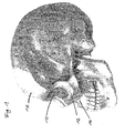
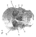
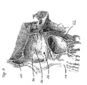
- FIGS. 1 to 3 which is shown by the example of a human skull with the kind permission of 3B Scientific GmbH, Hamburg (www.3bscientific.com) to illustrate the facts, forms an orbita 10 the protective, bony enclosure for eyeball (Bulbus oculi) with optic nerve (optic nerve), other nerves and blood vessels and lacrimal gland (not shown). These structures are surrounded by orbital adipose tissue (Corpus adiposum orbitae).
- the eye socket 10 has the shape of a slightly outwardly and downwardly diverging funnel, with the six eye muscles at the tip of the funnel originating around the optic nerve and attaching to the eyeball, thus moving in the eye socket as in a joint cavity.
- the bony entrance of the eye socket 10 forms a stable ring of zygomatic bone 12, upper maxilla (maxillary bone) 14 and frontal bone (frontal bone) 16.
- the remaining bony boundary consists of partially very thin bone lamellae 18. These bone lamellae 18 also form the Border to surrounding cavities such as the frontal sinus (sinus frontalis) 20 or the jaw cavity (sinus maxillaris) 22.
- the orbital floor (Paries inferior) 24 is formed by two bones, the maxilla 14 and the zygomatic bone 12, which is approximately in the center of the lower Orbital entrance to the sutura zygomaticomaxillaris called cranial suture 26 abut each other, with the upper jaw 14 at the transition to the medial orbital wall 28 in front of the lacrimal bone (Os lacrimale) 30 and further inside the ethmoid bone (Os ethmoidale) 32 join.
- a fracture of the floor 24 of the eye socket 10 is caused, for example, by direct trauma (eg, by a squash or tennis ball or a punch ) or in connection with a more complex fracture, such as the zygomatic bone 12.
- direct trauma eg, by a squash or tennis ball or a punch
- a more complex fracture such as the zygomatic bone 12.
- surgical intervention is usually considered to be indicated, as, for example, herniation in the jaw cavity 22, a fall back of the eyeball into the eye socket (enophthalmos) or the occurrence of double images (diplopia) connected.
- surgical reconstruction of the orbit is not without controversy, as it may involve high risks. In a few cases - according to a study of 20,500 patients in 0.04% of all cases - even a loss of vision has occurred postoperatively.
- the eye socket 10 is opened surgically according to the present invention from the anterior, with a cut in the area of the lower eyelid (palpebral) to the edge of the eye socket 10 received, the bone or leg skin (periosteum) incised to visualize the orbital edge, and then, with a so-called freer, push the soft tissue (eyeball, eye muscles, adipose tissue, periosteum, not shown) all the way over the fracture from the orbital floor 24. Subsequently, the Orbita soft tissue is raised and a dissolving film inserted for support.
- a cerebral spatula is not an optimal tool for surgical treatment of orbital floor fractures, as it is - nomen est omen - not designed for this purpose.
- an instrument for operating an orbital floor fracture comprises a spoon-shaped end portion whose shape conforms to a shape of a medial orbital floor and a handle portion connected at one end to the spoon-like end portion.
- this spoon-shaped end portion is thus adapted in size and shape to that of the area of an eye socket 10, at which the instrument is to be used according to the invention, wherein the shape was determined by molding the orbital floor of a skull.
- the instrument with its spoon-shaped end portion of anterior substantially in the region of the upper jaw 14, partially overlapping in the region of the zygomatic bone 12 on the one hand and in the area of lacrimal bone and ethmoid bone 32 on the other hand under the as described above from the orbital floor dissolved orbital soft parts in the eye socket 10 is introduced to lift the eyeball with its appendages and keep it from fracture.
- any orbital soft tissues protruding into the antrum are removed from the fracture, the fracture is covered with a dissolving foil and the wound is subsequently closed.
- the shape of the instrument according to the invention is thus defined and defined herein by the complicated shape of the eye socket 10, since it eludes a satisfactory conceptual description.
- conventionally used brain spatula which have a viewed over their length more or less the same and relatively small width
- at least in a longitudinally central portion of the spoon-shaped end portion wider and adapted to the anatomy of the orbital floor 24 form of the instrument according to the invention exercises the spoon-shaped end portion by raising the eyeball, although larger forces however, the distribution of force acting on the caudal / caudal medial surface of the eyeball corresponds largely to that exerted by the bone structure in the normally-positioned eye.
- the eyeball as a whole is slightly lifted and not locally deformed or pressed in, as is easily the case in conventional brain spatulas because of their slim shape, so that the risk of injury is reduced.
- it is in conventionally used and generally much narrower trained brain blades, by which the eyeball is pressed much more tempted to "nachzuactu", whereby the risk exists that the cerebral spatula inserted too deeply and the nerve structure (especially the optic nerve), the after emerges behind, gets injured.
- the first portion of the instrument according to the invention tapers towards its end, on the other hand, it widened significantly away from this end, thus taking approximately the shape of a tablespoon in this respect, so that a conventional one formed in comparison to the case of a "paddle-shaped" Brain spatula deeper penetration into the eye socket 10 and the risk of injury to the optic nerve is reduced.
- the invention is preferably intended for use in human medicine, veterinary use, especially in injured mammals of comparable anatomy, is quite conceivable.
- the instrument has a second end portion which is connected to the other end of the handle portion and formed as a brain spatula tapering away from the grip portion.
- the grip portion is plastically deformable. This allows optimal positioning and / or insertion of the respective end portion for the operation, such that the surgeon's view is not obscured by his hand or that of an assisting physician at any time during the operation.
- the instrument may preferably be bent manually at the transition of the handle portion to the working part formed as a spoon-like end portion to achieve optimal effects.
- the ratio of the length of the handle portion to that of the working portion is 2: 1, for example.
- the spoon-like end portion is formed asymmetrically, which is advantageous in view of the asymmetrical shape of the eye socket 10 and results from the manufacture of the instrument by molding the orbital floor.
- asymmetrically means the absence of any form of symmetry plane or point.
- This advantageous embodiment entails that there are two variants of the instrument according to the invention: one for the left eye, one mirror image for the right eye.
- an adaptation to the age of the patient is expedient, since not only the size but also the shape of the eye socket changes in the course of ontogenesis, the development of the individual. In particular, such a change is during the so-called synostosis, d. H.
- the present invention provides at least two sizes, for adults and children.
- the instrument is formed in one piece from a metal, in particular a medical alloy.
- an instrument set comprising a plurality of instruments according to the present invention comprises two instruments for the left and right eye of an adult and / or two instruments for the left and right eyes of a child described above, the "adult instruments” differ in size and shape from the "children's instruments”.
- the invention is not limited to two sizes / shapes.
- an ethnological adaptation is conceivable because the skull shapes of the individual peoples are very different.
- a method of manufacturing an instrument according to any one of claims 1 to 5 comprises the steps of: creating an impression by directly impressing an orbital floor of a skull, and fabricating the instrument as a standard instrument by means of the impression.
- the impression is taken as a wax impression or the like in order to obtain an anatomically precise instrument.
- a standard instrument is made.
- standard instruments are made in two or more sizes. By the achieved standardization it can be avoided to make a separate instrument for each individual patient.
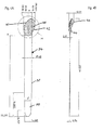
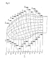
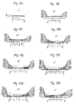
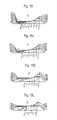
- FIGS. 4A to 60 10 show views of an instrument for operation of an orbital floor fracture according to a first embodiment of the present invention, ie for a left eye of an adult. Since the shape of the instrument according to the invention is identical except for the differences mentioned below, the following description is limited to this variant.
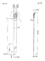
- FIG. 10 is a side view and a plan view, respectively, of an instrument 34 for a left eye of an adult according to a first embodiment of the present invention.
- Fig. 4A is clearly seen the asymmetrical shape of an end portion 36 which is connected to a straight handle 38.
- One side edge 40 is formed to conform to the contour of the medial orbital wall, while the opposite side edge 42 is not replicated to any orbital wall; it is located in the middle of the orbit floor during the operation.
- Fig. 4B is a facing towards the viewer first high vault 44 and facing away from the viewer second high vault 46 can be seen.
- the bumps 44 and 46 correspond to the anatomical shape of the orbital floor; Its shape was obtained experimentally by direct impression of an orbital floor on a skull and is ideal for the removal of the orbital soft tissues. This form covers the area of the orbit that most frequently breaks, and also serves as a guide to knowing how far one can "prepare” safely into the eye socket.
- the lines drawn in the end section 36 should visualize its three-dimensional shape.
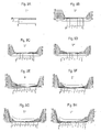
- FIGS. 6A-60 show sectional views corresponding to the cutting progressions A to O in Fig. 5 looking towards the handle 38. Especially in the FIGS. 6N and 60 is clearly the strongly different expression of the first high vault 20 (right) compared to the second high vault 22 (left) to recognize.
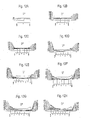
- FIGS. 4A and 4B for a left eye A comparison of FIGS. 4A and 4B for a left eye with the FIGS. 7A or 7B for a right eye clearly shows the mirror image of the instrument according to the invention. Furthermore, the dimensions show for example in the FIGS. 4A and 4B for an adult compared to those in the FIGS. 10A or 10B for a child resizing to the patient's age.
- the instrument 34 according to the invention on the end portion 36 opposite second end portion 48, which is in the form of a tapered away from the handle 38 brain spatula.
Landscapes
- Health & Medical Sciences (AREA)
- Surgery (AREA)
- Life Sciences & Earth Sciences (AREA)
- Biomedical Technology (AREA)
- Nuclear Medicine, Radiotherapy & Molecular Imaging (AREA)
- Engineering & Computer Science (AREA)
- Ophthalmology & Optometry (AREA)
- Heart & Thoracic Surgery (AREA)
- Medical Informatics (AREA)
- Molecular Biology (AREA)
- Animal Behavior & Ethology (AREA)
- General Health & Medical Sciences (AREA)
- Public Health (AREA)
- Veterinary Medicine (AREA)
- Surgical Instruments (AREA)
Priority Applications (1)
| Application Number | Priority Date | Filing Date | Title |
|---|---|---|---|
| EP08172680A EP2201899A1 (fr) | 2008-12-23 | 2008-12-23 | Instrument destiné à l'opération d'une fracture de l'os zygomatique, ensemble formé de plusieurs instruments de ce type et procédé de fabrication d'un tel instrument |
Applications Claiming Priority (1)
| Application Number | Priority Date | Filing Date | Title |
|---|---|---|---|
| EP08172680A EP2201899A1 (fr) | 2008-12-23 | 2008-12-23 | Instrument destiné à l'opération d'une fracture de l'os zygomatique, ensemble formé de plusieurs instruments de ce type et procédé de fabrication d'un tel instrument |
Publications (1)
| Publication Number | Publication Date |
|---|---|
| EP2201899A1 true EP2201899A1 (fr) | 2010-06-30 |
Family
ID=40671026
Family Applications (1)
| Application Number | Title | Priority Date | Filing Date |
|---|---|---|---|
| EP08172680A Withdrawn EP2201899A1 (fr) | 2008-12-23 | 2008-12-23 | Instrument destiné à l'opération d'une fracture de l'os zygomatique, ensemble formé de plusieurs instruments de ce type et procédé de fabrication d'un tel instrument |
Country Status (1)
| Country | Link |
|---|---|
| EP (1) | EP2201899A1 (fr) |
Cited By (6)
| Publication number | Priority date | Publication date | Assignee | Title |
|---|---|---|---|---|
| DE102010054333A1 (de) | 2010-12-13 | 2012-06-14 | Stryker Leibinger Gmbh & Co. Kg | Chirurgischer Retraktor |
| US20180317902A1 (en) * | 2017-05-03 | 2018-11-08 | University Of Virginia Patent Foundation | Illuminated suction retractor device |
| RU2699624C2 (ru) * | 2014-04-11 | 2019-09-06 | КАРЛ ЛЯЙБИНГЕР МЕДИЦИНТЕХНИК ГМБХ И Ко. КГ | Предварительно сформованный имплантат для вправления и фиксации центролатерального перелома средней части лица |
| WO2020150485A1 (fr) * | 2019-01-16 | 2020-07-23 | Poriferous, LLC | Calibreur, dispositif d'introduction et dispositif de gabarit |
| CN112957173A (zh) * | 2021-02-08 | 2021-06-15 | 青岛大学附属医院 | 一种用于眼眶骨手术的组织撑开装置 |
| CN113423345A (zh) * | 2019-02-11 | 2021-09-21 | 新宁研究院 | 深部眼眶入路牵开器 |
Citations (4)
| Publication number | Priority date | Publication date | Assignee | Title |
|---|---|---|---|---|
| AT384945B (de) * | 1986-08-06 | 1988-01-25 | Lixl Georg | Heilbehelf zur reposition des augenhoehlenbodens |
| US6440065B1 (en) * | 2001-10-01 | 2002-08-27 | The Nemours Foundation | Single-use disposable eyelid speculum, eye examination kit, and method for examining a patient's eye |
| US20020161374A1 (en) * | 1999-12-09 | 2002-10-31 | Cohen Steven R. | Completely resorbable connective tissue distraction devices and techniques |
| US20080081952A1 (en) * | 2006-09-29 | 2008-04-03 | Josephberg Robert G | Scleral depressor |
-
2008
- 2008-12-23 EP EP08172680A patent/EP2201899A1/fr not_active Withdrawn
Patent Citations (4)
| Publication number | Priority date | Publication date | Assignee | Title |
|---|---|---|---|---|
| AT384945B (de) * | 1986-08-06 | 1988-01-25 | Lixl Georg | Heilbehelf zur reposition des augenhoehlenbodens |
| US20020161374A1 (en) * | 1999-12-09 | 2002-10-31 | Cohen Steven R. | Completely resorbable connective tissue distraction devices and techniques |
| US6440065B1 (en) * | 2001-10-01 | 2002-08-27 | The Nemours Foundation | Single-use disposable eyelid speculum, eye examination kit, and method for examining a patient's eye |
| US20080081952A1 (en) * | 2006-09-29 | 2008-04-03 | Josephberg Robert G | Scleral depressor |
Cited By (14)
| Publication number | Priority date | Publication date | Assignee | Title |
|---|---|---|---|---|
| WO2012079743A1 (fr) | 2010-12-13 | 2012-06-21 | Stryker Leibinger Gmbh & Co. Kg | Rétracteur chirurgical |
| US9717489B2 (en) | 2010-12-13 | 2017-08-01 | Stryker European Holdings I, Llc | Surgical retractor |
| DE102010054333B4 (de) | 2010-12-13 | 2019-05-29 | Stryker European Holdings I, LLC (n.d. Ges. d. Staates Delaware) | Chirurgischer Retraktor |
| DE102010054333A1 (de) | 2010-12-13 | 2012-06-14 | Stryker Leibinger Gmbh & Co. Kg | Chirurgischer Retraktor |
| EP3033019B1 (fr) * | 2014-04-11 | 2021-08-18 | Karl Leibinger Medizintechnik Gmbh & Co. Kg | Implant préformé pour le repositionnement et la fixation d'une fracture du visage médian centrolatéral |
| RU2699624C2 (ru) * | 2014-04-11 | 2019-09-06 | КАРЛ ЛЯЙБИНГЕР МЕДИЦИНТЕХНИК ГМБХ И Ко. КГ | Предварительно сформованный имплантат для вправления и фиксации центролатерального перелома средней части лица |
| US20180317902A1 (en) * | 2017-05-03 | 2018-11-08 | University Of Virginia Patent Foundation | Illuminated suction retractor device |
| US11253245B2 (en) * | 2017-05-03 | 2022-02-22 | University Of Virginia Patent Foundation | Illuminated suction retractor device |
| WO2020150485A1 (fr) * | 2019-01-16 | 2020-07-23 | Poriferous, LLC | Calibreur, dispositif d'introduction et dispositif de gabarit |
| EP3911279A4 (fr) * | 2019-01-16 | 2022-09-14 | Poriferous, LLC | Calibreur, dispositif d'introduction et dispositif de gabarit |
| US11491029B2 (en) | 2019-01-16 | 2022-11-08 | Poriferous, LLC | Sizer, introducer and template device |
| AU2020209223B2 (en) * | 2019-01-16 | 2024-05-02 | Poriferous, LLC | Sizer, introducer and template device |
| CN113423345A (zh) * | 2019-02-11 | 2021-09-21 | 新宁研究院 | 深部眼眶入路牵开器 |
| CN112957173A (zh) * | 2021-02-08 | 2021-06-15 | 青岛大学附属医院 | 一种用于眼眶骨手术的组织撑开装置 |
Similar Documents
| Publication | Publication Date | Title |
|---|---|---|
| DE69920694T2 (de) | Minimalinvasives messinstrument für einen herzventilverankerungsring | |
| DE69416588T2 (de) | Intervertebrale scheibenprothese | |
| DE69032527T2 (de) | Längenverstellbare, vertikale rippenprothese | |
| DE602004013281T2 (de) | Artikulierbares implantatsystem | |
| EP1475056B1 (fr) | Implant pour écarter les ailes du nez | |
| EP3033019B1 (fr) | Implant préformé pour le repositionnement et la fixation d'une fracture du visage médian centrolatéral | |
| DE60128834T2 (de) | Vorrichtung und bausatz für zahn extraktionen | |
| EP3370637B1 (fr) | Procédé de réalisation d'un distracteur pour fixation au squelette d'une mâchoire | |
| DE202018101432U1 (de) | Periostabstreifer mit hakenförmigen Kammvorsprüngen | |
| EP3064167B1 (fr) | Kit d'expansion comprenant un dispositif d'expansion ou d'élongation de segments osseux | |
| EP0969772B1 (fr) | Spatule a dissequer pourvue de mors ecartables | |
| EP4138738B1 (fr) | Méthode de conception d'une prothèse de l'articulation temporo-mandibulaire mise en oeuvre par ordinateur, procédé de fabrication, système informatique, produit logiciel, support de stockage de données, flux de données et serveur web correspondant | |
| DE202021105388U1 (de) | Endoskopisches Instrumentarium für die Bandscheibenchirurgie | |
| DE102021209981A1 (de) | Starres, gebogenes Laryngoskop | |
| DE60124212T2 (de) | Vorrichtung zur Befestigung einer Schädelklappe an der Schädelhöhle | |
| WO2011092161A1 (fr) | Implant de cloison nasale et trousse d'opération pour corriger une faiblesse du cartilage du nez humain | |
| DE202023102729U1 (de) | Drehschneidender Knochenmeißel | |
| DE112015006850B4 (de) | Kanüle mit einem elastischen Filament mit Formgedächtnis zum Abtragen von Sinusbodenschleimhaut sowie Zusammenstellung derartiger Kanülen mit elastischen Filamenten mit Formgedächtnis | |
| DE102021113317B4 (de) | Distraktionssystem | |
| DE19718708A1 (de) | Hämorrhoidenresektionsinstrument | |
| RU2775213C1 (ru) | Способ двусторонней чрезбровной супраорбитальной краниотомии | |
| DE202023102731U1 (de) | Endoskopischer Knochenmeißel | |
| DE202023102728U1 (de) | Schaft für Ligament-Punch | |
| DE102017123801B4 (de) | Membranen für Alveolarkämme | |
| DE202023102721U1 (de) | Hülse für endoskopische posteriore Fusionsoperationen und Endoskopanordnung mit der Hülse |
Legal Events
| Date | Code | Title | Description |
|---|---|---|---|
| PUAI | Public reference made under article 153(3) epc to a published international application that has entered the european phase |
Free format text: ORIGINAL CODE: 0009012 |
|
| AK | Designated contracting states |
Kind code of ref document: A1 Designated state(s): AT BE BG CH CY CZ DE DK EE ES FI FR GB GR HR HU IE IS IT LI LT LU LV MC MT NL NO PL PT RO SE SI SK TR |
|
| AX | Request for extension of the european patent |
Extension state: AL BA MK RS |
|
| 17P | Request for examination filed |
Effective date: 20101227 |
|
| AKX | Designation fees paid |
Designated state(s): AT BE BG CH CY CZ DE DK EE ES FI FR GB GR HR HU IE IS IT LI LT LU LV MC MT NL NO PL PT RO SE SI SK TR |
|
| RAP1 | Party data changed (applicant data changed or rights of an application transferred) |
Owner name: HIBRAND ESTABLISHMENT |
|
| GRAP | Despatch of communication of intention to grant a patent |
Free format text: ORIGINAL CODE: EPIDOSNIGR1 |
|
| GRAS | Grant fee paid |
Free format text: ORIGINAL CODE: EPIDOSNIGR3 |
|
| STAA | Information on the status of an ep patent application or granted ep patent |
Free format text: STATUS: THE APPLICATION IS DEEMED TO BE WITHDRAWN |
|
| 18D | Application deemed to be withdrawn |
Effective date: 20120703 |