EP2206501A2 - HMGA bindende Nukleinsäuren - Google Patents
HMGA bindende Nukleinsäuren Download PDFInfo
- Publication number
- EP2206501A2 EP2206501A2 EP09016125A EP09016125A EP2206501A2 EP 2206501 A2 EP2206501 A2 EP 2206501A2 EP 09016125 A EP09016125 A EP 09016125A EP 09016125 A EP09016125 A EP 09016125A EP 2206501 A2 EP2206501 A2 EP 2206501A2
- Authority
- EP
- European Patent Office
- Prior art keywords
- hmga
- helix
- nucleic acid
- nucleotides
- section
- Prior art date
- Legal status (The legal status is an assumption and is not a legal conclusion. Google has not performed a legal analysis and makes no representation as to the accuracy of the status listed.)
- Withdrawn
Links
- 150000007523 nucleic acids Chemical class 0.000 title claims abstract description 324
- 102000039446 nucleic acids Human genes 0.000 title claims abstract description 314
- 108020004707 nucleic acids Proteins 0.000 title claims abstract description 314
- 230000027455 binding Effects 0.000 title claims abstract description 163
- 102000009012 HMGA Proteins Human genes 0.000 title claims abstract description 161
- 108010049069 HMGA Proteins Proteins 0.000 title claims abstract description 161
- NPOAOTPXWNWTSH-UHFFFAOYSA-N 3-hydroxy-3-methylglutaric acid Chemical compound OC(=O)CC(O)(C)CC(O)=O NPOAOTPXWNWTSH-UHFFFAOYSA-N 0.000 title claims description 127
- 238000000034 method Methods 0.000 claims abstract description 55
- 239000005557 antagonist Substances 0.000 claims abstract description 37
- 239000000556 agonist Substances 0.000 claims abstract description 36
- 238000012360 testing method Methods 0.000 claims abstract description 8
- 239000012071 phase Substances 0.000 claims abstract description 5
- 239000007790 solid phase Substances 0.000 claims abstract description 5
- 239000002773 nucleotide Substances 0.000 claims description 215
- 125000003729 nucleotide group Chemical group 0.000 claims description 193
- 108700039142 HMGA1a Proteins 0.000 claims description 84
- 102000049983 HMGA1a Human genes 0.000 claims description 84
- 208000037265 diseases, disorders, signs and symptoms Diseases 0.000 claims description 48
- 108700039144 HMGA1b Proteins 0.000 claims description 47
- 102100029009 High mobility group protein HMG-I/HMG-Y Human genes 0.000 claims description 44
- 101000986380 Homo sapiens High mobility group protein HMG-I/HMG-Y Proteins 0.000 claims description 44
- 201000010099 disease Diseases 0.000 claims description 43
- 108700039143 HMGA2 Proteins 0.000 claims description 31
- 101150073387 Hmga2 gene Proteins 0.000 claims description 29
- 239000003814 drug Substances 0.000 claims description 29
- 238000001514 detection method Methods 0.000 claims description 27
- 238000009396 hybridization Methods 0.000 claims description 26
- 102000040945 Transcription factor Human genes 0.000 claims description 20
- 108091023040 Transcription factor Proteins 0.000 claims description 20
- 238000011282 treatment Methods 0.000 claims description 19
- 238000003745 diagnosis Methods 0.000 claims description 16
- 239000003795 chemical substances by application Substances 0.000 claims description 9
- 239000000032 diagnostic agent Substances 0.000 claims description 9
- 229940039227 diagnostic agent Drugs 0.000 claims description 9
- 230000002265 prevention Effects 0.000 claims description 8
- 238000012216 screening Methods 0.000 claims description 8
- 108091028043 Nucleic acid sequence Proteins 0.000 claims description 4
- 238000010276 construction Methods 0.000 claims description 3
- 102100028999 High mobility group protein HMGI-C Human genes 0.000 claims 1
- 230000003834 intracellular effect Effects 0.000 abstract description 64
- 102000027411 intracellular receptors Human genes 0.000 abstract description 54
- 108091008582 intracellular receptors Proteins 0.000 abstract description 54
- 239000000203 mixture Substances 0.000 abstract description 51
- 230000000694 effects Effects 0.000 abstract description 27
- 239000013543 active substance Substances 0.000 abstract description 16
- 230000008569 process Effects 0.000 abstract description 7
- 238000013518 transcription Methods 0.000 abstract description 7
- 230000035897 transcription Effects 0.000 abstract description 7
- 230000010534 mechanism of action Effects 0.000 abstract description 3
- 230000001085 cytostatic effect Effects 0.000 abstract description 2
- 230000002401 inhibitory effect Effects 0.000 abstract description 2
- 230000003110 anti-inflammatory effect Effects 0.000 abstract 1
- 238000004166 bioassay Methods 0.000 abstract 1
- 239000000824 cytostatic agent Substances 0.000 abstract 1
- 239000012873 virucide Substances 0.000 abstract 1
- 108091027076 Spiegelmer Proteins 0.000 description 206
- 210000004027 cell Anatomy 0.000 description 166
- 206010028980 Neoplasm Diseases 0.000 description 121
- 108090000623 proteins and genes Proteins 0.000 description 87
- 229920002873 Polyethylenimine Polymers 0.000 description 74
- 108091023037 Aptamer Proteins 0.000 description 72
- 201000011510 cancer Diseases 0.000 description 72
- 239000000693 micelle Substances 0.000 description 57
- 102000004169 proteins and genes Human genes 0.000 description 54
- 235000018102 proteins Nutrition 0.000 description 53
- 241000282414 Homo sapiens Species 0.000 description 50
- 230000014509 gene expression Effects 0.000 description 47
- 239000003981 vehicle Substances 0.000 description 40
- 102000005962 receptors Human genes 0.000 description 36
- 108020003175 receptors Proteins 0.000 description 36
- 210000001519 tissue Anatomy 0.000 description 34
- 239000000872 buffer Substances 0.000 description 33
- 238000003556 assay Methods 0.000 description 32
- 229920001223 polyethylene glycol Polymers 0.000 description 31
- 108020004414 DNA Proteins 0.000 description 30
- 102000049982 HMGA2 Human genes 0.000 description 30
- 108090000765 processed proteins & peptides Proteins 0.000 description 29
- 239000002202 Polyethylene glycol Substances 0.000 description 27
- 239000000562 conjugate Substances 0.000 description 26
- 230000003993 interaction Effects 0.000 description 26
- 239000000543 intermediate Substances 0.000 description 24
- 238000009826 distribution Methods 0.000 description 22
- 230000035755 proliferation Effects 0.000 description 22
- 101100184710 Xenopus laevis cdc25-1-a gene Proteins 0.000 description 21
- 210000000170 cell membrane Anatomy 0.000 description 21
- 210000002381 plasma Anatomy 0.000 description 20
- 239000000523 sample Substances 0.000 description 20
- 150000001875 compounds Chemical class 0.000 description 19
- 230000005764 inhibitory process Effects 0.000 description 19
- 108010029485 Protein Isoforms Proteins 0.000 description 18
- 102000001708 Protein Isoforms Human genes 0.000 description 18
- 102000004196 processed proteins & peptides Human genes 0.000 description 18
- 239000000243 solution Substances 0.000 description 18
- 108091032973 (ribonucleotides)n+m Proteins 0.000 description 17
- 241001465754 Metazoa Species 0.000 description 17
- 108091034117 Oligonucleotide Proteins 0.000 description 17
- 238000013459 approach Methods 0.000 description 17
- 230000004048 modification Effects 0.000 description 17
- 238000012986 modification Methods 0.000 description 17
- YBJHBAHKTGYVGT-ZKWXMUAHSA-N (+)-Biotin Chemical compound N1C(=O)N[C@@H]2[C@H](CCCCC(=O)O)SC[C@@H]21 YBJHBAHKTGYVGT-ZKWXMUAHSA-N 0.000 description 16
- FAPWRFPIFSIZLT-UHFFFAOYSA-M Sodium chloride Chemical compound [Na+].[Cl-] FAPWRFPIFSIZLT-UHFFFAOYSA-M 0.000 description 16
- 210000000172 cytosol Anatomy 0.000 description 16
- 229940079593 drug Drugs 0.000 description 16
- 229920001184 polypeptide Polymers 0.000 description 16
- SVUJNSGGPUCLQZ-FQQAACOVSA-N tenofovir alafenamide fumarate Chemical compound OC(=O)\C=C\C(O)=O.O([P@@](=O)(CO[C@H](C)CN1C2=NC=NC(N)=C2N=C1)N[C@@H](C)C(=O)OC(C)C)C1=CC=CC=C1.O([P@@](=O)(CO[C@H](C)CN1C2=NC=NC(N)=C2N=C1)N[C@@H](C)C(=O)OC(C)C)C1=CC=CC=C1 SVUJNSGGPUCLQZ-FQQAACOVSA-N 0.000 description 16
- 108700020796 Oncogene Proteins 0.000 description 15
- 230000004614 tumor growth Effects 0.000 description 15
- 230000015572 biosynthetic process Effects 0.000 description 14
- 150000002632 lipids Chemical class 0.000 description 14
- 206010061289 metastatic neoplasm Diseases 0.000 description 14
- JLCPHMBAVCMARE-UHFFFAOYSA-N [3-[[3-[[3-[[3-[[3-[[3-[[3-[[3-[[3-[[3-[[3-[[5-(2-amino-6-oxo-1H-purin-9-yl)-3-[[3-[[3-[[3-[[3-[[3-[[5-(2-amino-6-oxo-1H-purin-9-yl)-3-[[5-(2-amino-6-oxo-1H-purin-9-yl)-3-hydroxyoxolan-2-yl]methoxy-hydroxyphosphoryl]oxyoxolan-2-yl]methoxy-hydroxyphosphoryl]oxy-5-(5-methyl-2,4-dioxopyrimidin-1-yl)oxolan-2-yl]methoxy-hydroxyphosphoryl]oxy-5-(6-aminopurin-9-yl)oxolan-2-yl]methoxy-hydroxyphosphoryl]oxy-5-(6-aminopurin-9-yl)oxolan-2-yl]methoxy-hydroxyphosphoryl]oxy-5-(6-aminopurin-9-yl)oxolan-2-yl]methoxy-hydroxyphosphoryl]oxy-5-(6-aminopurin-9-yl)oxolan-2-yl]methoxy-hydroxyphosphoryl]oxyoxolan-2-yl]methoxy-hydroxyphosphoryl]oxy-5-(5-methyl-2,4-dioxopyrimidin-1-yl)oxolan-2-yl]methoxy-hydroxyphosphoryl]oxy-5-(4-amino-2-oxopyrimidin-1-yl)oxolan-2-yl]methoxy-hydroxyphosphoryl]oxy-5-(5-methyl-2,4-dioxopyrimidin-1-yl)oxolan-2-yl]methoxy-hydroxyphosphoryl]oxy-5-(5-methyl-2,4-dioxopyrimidin-1-yl)oxolan-2-yl]methoxy-hydroxyphosphoryl]oxy-5-(6-aminopurin-9-yl)oxolan-2-yl]methoxy-hydroxyphosphoryl]oxy-5-(6-aminopurin-9-yl)oxolan-2-yl]methoxy-hydroxyphosphoryl]oxy-5-(4-amino-2-oxopyrimidin-1-yl)oxolan-2-yl]methoxy-hydroxyphosphoryl]oxy-5-(4-amino-2-oxopyrimidin-1-yl)oxolan-2-yl]methoxy-hydroxyphosphoryl]oxy-5-(4-amino-2-oxopyrimidin-1-yl)oxolan-2-yl]methoxy-hydroxyphosphoryl]oxy-5-(6-aminopurin-9-yl)oxolan-2-yl]methoxy-hydroxyphosphoryl]oxy-5-(4-amino-2-oxopyrimidin-1-yl)oxolan-2-yl]methyl [5-(6-aminopurin-9-yl)-2-(hydroxymethyl)oxolan-3-yl] hydrogen phosphate Polymers Cc1cn(C2CC(OP(O)(=O)OCC3OC(CC3OP(O)(=O)OCC3OC(CC3O)n3cnc4c3nc(N)[nH]c4=O)n3cnc4c3nc(N)[nH]c4=O)C(COP(O)(=O)OC3CC(OC3COP(O)(=O)OC3CC(OC3COP(O)(=O)OC3CC(OC3COP(O)(=O)OC3CC(OC3COP(O)(=O)OC3CC(OC3COP(O)(=O)OC3CC(OC3COP(O)(=O)OC3CC(OC3COP(O)(=O)OC3CC(OC3COP(O)(=O)OC3CC(OC3COP(O)(=O)OC3CC(OC3COP(O)(=O)OC3CC(OC3COP(O)(=O)OC3CC(OC3COP(O)(=O)OC3CC(OC3COP(O)(=O)OC3CC(OC3COP(O)(=O)OC3CC(OC3COP(O)(=O)OC3CC(OC3COP(O)(=O)OC3CC(OC3CO)n3cnc4c(N)ncnc34)n3ccc(N)nc3=O)n3cnc4c(N)ncnc34)n3ccc(N)nc3=O)n3ccc(N)nc3=O)n3ccc(N)nc3=O)n3cnc4c(N)ncnc34)n3cnc4c(N)ncnc34)n3cc(C)c(=O)[nH]c3=O)n3cc(C)c(=O)[nH]c3=O)n3ccc(N)nc3=O)n3cc(C)c(=O)[nH]c3=O)n3cnc4c3nc(N)[nH]c4=O)n3cnc4c(N)ncnc34)n3cnc4c(N)ncnc34)n3cnc4c(N)ncnc34)n3cnc4c(N)ncnc34)O2)c(=O)[nH]c1=O JLCPHMBAVCMARE-UHFFFAOYSA-N 0.000 description 13
- 239000008194 pharmaceutical composition Substances 0.000 description 13
- 102000004190 Enzymes Human genes 0.000 description 12
- 108090000790 Enzymes Proteins 0.000 description 12
- 206010027476 Metastases Diseases 0.000 description 12
- 108010076504 Protein Sorting Signals Proteins 0.000 description 12
- 238000004458 analytical method Methods 0.000 description 12
- 210000001163 endosome Anatomy 0.000 description 12
- 229940088598 enzyme Drugs 0.000 description 12
- 238000001890 transfection Methods 0.000 description 12
- 230000032258 transport Effects 0.000 description 12
- 108091003079 Bovine Serum Albumin Proteins 0.000 description 11
- 102000018802 High Mobility Group Proteins Human genes 0.000 description 11
- 108010052512 High Mobility Group Proteins Proteins 0.000 description 11
- 102000043276 Oncogene Human genes 0.000 description 11
- 150000001720 carbohydrates Chemical class 0.000 description 11
- 235000014633 carbohydrates Nutrition 0.000 description 11
- 239000012894 fetal calf serum Substances 0.000 description 11
- 239000000499 gel Substances 0.000 description 11
- 238000000338 in vitro Methods 0.000 description 11
- 238000001727 in vivo Methods 0.000 description 11
- 230000001404 mediated effect Effects 0.000 description 11
- 239000012528 membrane Substances 0.000 description 11
- 108020004999 messenger RNA Proteins 0.000 description 11
- 230000002503 metabolic effect Effects 0.000 description 11
- 101001093899 Homo sapiens Retinoic acid receptor RXR-alpha Proteins 0.000 description 10
- 101100189471 Mus musculus Pbx1 gene Proteins 0.000 description 10
- 101000595993 Phyllomedusa sauvagei Phylloseptin-S1 Proteins 0.000 description 10
- 229920001213 Polysorbate 20 Polymers 0.000 description 10
- 102100035178 Retinoic acid receptor RXR-alpha Human genes 0.000 description 10
- 230000008859 change Effects 0.000 description 10
- 230000000875 corresponding effect Effects 0.000 description 10
- 230000008878 coupling Effects 0.000 description 10
- 238000010168 coupling process Methods 0.000 description 10
- 238000005859 coupling reaction Methods 0.000 description 10
- 230000006870 function Effects 0.000 description 10
- 210000004072 lung Anatomy 0.000 description 10
- 230000001394 metastastic effect Effects 0.000 description 10
- IJGRMHOSHXDMSA-UHFFFAOYSA-N nitrogen Substances N#N IJGRMHOSHXDMSA-UHFFFAOYSA-N 0.000 description 10
- 235000010486 polyoxyethylene sorbitan monolaurate Nutrition 0.000 description 10
- 239000000256 polyoxyethylene sorbitan monolaurate Substances 0.000 description 10
- 238000004904 shortening Methods 0.000 description 10
- 239000000126 substance Substances 0.000 description 10
- 102100027468 Anion exchange protein 2 Human genes 0.000 description 9
- 102000003960 Ligases Human genes 0.000 description 9
- 108090000364 Ligases Proteins 0.000 description 9
- 241000700605 Viruses Species 0.000 description 9
- 230000033228 biological regulation Effects 0.000 description 9
- 229960002685 biotin Drugs 0.000 description 9
- 239000011616 biotin Substances 0.000 description 9
- 210000003527 eukaryotic cell Anatomy 0.000 description 9
- RAXXELZNTBOGNW-UHFFFAOYSA-N imidazole Natural products C1=CNC=N1 RAXXELZNTBOGNW-UHFFFAOYSA-N 0.000 description 9
- 238000004519 manufacturing process Methods 0.000 description 9
- 229910052757 nitrogen Inorganic materials 0.000 description 9
- 210000000056 organ Anatomy 0.000 description 9
- 238000004806 packaging method and process Methods 0.000 description 9
- 210000000496 pancreas Anatomy 0.000 description 9
- 206010003210 Arteriosclerosis Diseases 0.000 description 8
- 101710163270 Nuclease Proteins 0.000 description 8
- 229940024606 amino acid Drugs 0.000 description 8
- 235000001014 amino acid Nutrition 0.000 description 8
- 150000001413 amino acids Chemical class 0.000 description 8
- 208000011775 arteriosclerosis disease Diseases 0.000 description 8
- 235000020958 biotin Nutrition 0.000 description 8
- 239000006143 cell culture medium Substances 0.000 description 8
- 239000012634 fragment Substances 0.000 description 8
- 108020001507 fusion proteins Proteins 0.000 description 8
- 102000037865 fusion proteins Human genes 0.000 description 8
- 238000011534 incubation Methods 0.000 description 8
- 239000002105 nanoparticle Substances 0.000 description 8
- 238000000159 protein binding assay Methods 0.000 description 8
- 239000011780 sodium chloride Substances 0.000 description 8
- 201000009030 Carcinoma Diseases 0.000 description 7
- 102000053602 DNA Human genes 0.000 description 7
- 101710088194 Dehydrogenase Proteins 0.000 description 7
- 102100032864 General transcription factor IIH subunit 2 Human genes 0.000 description 7
- 102100031181 Glyceraldehyde-3-phosphate dehydrogenase Human genes 0.000 description 7
- 101000666405 Homo sapiens General transcription factor IIH subunit 1 Proteins 0.000 description 7
- 101000655398 Homo sapiens General transcription factor IIH subunit 2 Proteins 0.000 description 7
- 101000655391 Homo sapiens General transcription factor IIH subunit 3 Proteins 0.000 description 7
- 101000655406 Homo sapiens General transcription factor IIH subunit 4 Proteins 0.000 description 7
- 101000655402 Homo sapiens General transcription factor IIH subunit 5 Proteins 0.000 description 7
- 108010006519 Molecular Chaperones Proteins 0.000 description 7
- 230000000692 anti-sense effect Effects 0.000 description 7
- 210000000481 breast Anatomy 0.000 description 7
- 108020004445 glyceraldehyde-3-phosphate dehydrogenase Proteins 0.000 description 7
- 201000010260 leiomyoma Diseases 0.000 description 7
- 239000002502 liposome Substances 0.000 description 7
- 201000008806 mesenchymal cell neoplasm Diseases 0.000 description 7
- 239000000047 product Substances 0.000 description 7
- 230000010837 receptor-mediated endocytosis Effects 0.000 description 7
- 238000003757 reverse transcription PCR Methods 0.000 description 7
- 208000005623 Carcinogenesis Diseases 0.000 description 6
- 229920000858 Cyclodextrin Polymers 0.000 description 6
- 241000713772 Human immunodeficiency virus 1 Species 0.000 description 6
- 206010024612 Lipoma Diseases 0.000 description 6
- 206010025323 Lymphomas Diseases 0.000 description 6
- TWRXJAOTZQYOKJ-UHFFFAOYSA-L Magnesium chloride Chemical compound [Mg+2].[Cl-].[Cl-] TWRXJAOTZQYOKJ-UHFFFAOYSA-L 0.000 description 6
- 206010029260 Neuroblastoma Diseases 0.000 description 6
- 108090000608 Phosphoric Monoester Hydrolases Proteins 0.000 description 6
- 102000004160 Phosphoric Monoester Hydrolases Human genes 0.000 description 6
- PLXBWHJQWKZRKG-UHFFFAOYSA-N Resazurin Chemical compound C1=CC(=O)C=C2OC3=CC(O)=CC=C3[N+]([O-])=C21 PLXBWHJQWKZRKG-UHFFFAOYSA-N 0.000 description 6
- 102100023132 Transcription factor Jun Human genes 0.000 description 6
- 206010046798 Uterine leiomyoma Diseases 0.000 description 6
- 230000004075 alteration Effects 0.000 description 6
- 239000011575 calcium Substances 0.000 description 6
- 230000036952 cancer formation Effects 0.000 description 6
- 231100000504 carcinogenesis Toxicity 0.000 description 6
- 208000016160 chondroid hamartoma Diseases 0.000 description 6
- 210000001072 colon Anatomy 0.000 description 6
- 230000000295 complement effect Effects 0.000 description 6
- 229940097362 cyclodextrins Drugs 0.000 description 6
- 238000009510 drug design Methods 0.000 description 6
- 239000000975 dye Substances 0.000 description 6
- 208000016018 endometrial polyp Diseases 0.000 description 6
- 230000007717 exclusion Effects 0.000 description 6
- 238000002474 experimental method Methods 0.000 description 6
- 238000009472 formulation Methods 0.000 description 6
- 239000011777 magnesium Substances 0.000 description 6
- QJGQUHMNIGDVPM-UHFFFAOYSA-N nitrogen group Chemical group [N] QJGQUHMNIGDVPM-UHFFFAOYSA-N 0.000 description 6
- 208000002154 non-small cell lung carcinoma Diseases 0.000 description 6
- 210000004940 nucleus Anatomy 0.000 description 6
- 208000004333 pleomorphic adenoma Diseases 0.000 description 6
- 210000001236 prokaryotic cell Anatomy 0.000 description 6
- 210000002307 prostate Anatomy 0.000 description 6
- 230000009467 reduction Effects 0.000 description 6
- 230000003252 repetitive effect Effects 0.000 description 6
- 210000003079 salivary gland Anatomy 0.000 description 6
- 239000000758 substrate Substances 0.000 description 6
- 230000001225 therapeutic effect Effects 0.000 description 6
- 210000001685 thyroid gland Anatomy 0.000 description 6
- 201000007954 uterine fibroid Diseases 0.000 description 6
- 206010046811 uterine polyp Diseases 0.000 description 6
- 201000000153 Angiomyoma Diseases 0.000 description 5
- 208000037260 Atherosclerotic Plaque Diseases 0.000 description 5
- 102100032912 CD44 antigen Human genes 0.000 description 5
- 102000052510 DNA-Binding Proteins Human genes 0.000 description 5
- 108700020911 DNA-Binding Proteins Proteins 0.000 description 5
- 208000021994 Diffuse astrocytoma Diseases 0.000 description 5
- 208000002966 Giant Cell Tumor of Bone Diseases 0.000 description 5
- 108010054147 Hemoglobins Proteins 0.000 description 5
- 102000001554 Hemoglobins Human genes 0.000 description 5
- 101000936546 Homo sapiens Anion exchange protein 2 Proteins 0.000 description 5
- 101000868273 Homo sapiens CD44 antigen Proteins 0.000 description 5
- 101000736088 Homo sapiens PC4 and SFRS1-interacting protein Proteins 0.000 description 5
- 101000962473 Homo sapiens Transcription factor MafG Proteins 0.000 description 5
- VEXZGXHMUGYJMC-UHFFFAOYSA-N Hydrochloric acid Chemical compound Cl VEXZGXHMUGYJMC-UHFFFAOYSA-N 0.000 description 5
- 206010058467 Lung neoplasm malignant Diseases 0.000 description 5
- 241000699670 Mus sp. Species 0.000 description 5
- 102100023050 Nuclear factor NF-kappa-B p105 subunit Human genes 0.000 description 5
- 102100036220 PC4 and SFRS1-interacting protein Human genes 0.000 description 5
- 229910019142 PO4 Inorganic materials 0.000 description 5
- 102100023606 Retinoic acid receptor alpha Human genes 0.000 description 5
- 108091006260 SLC4A2 Proteins 0.000 description 5
- 102100029904 Signal transducer and activator of transcription 1-alpha/beta Human genes 0.000 description 5
- 208000000453 Skin Neoplasms Diseases 0.000 description 5
- 108010090804 Streptavidin Proteins 0.000 description 5
- 102100039188 Transcription factor MafG Human genes 0.000 description 5
- 102100035559 Transcriptional activator GLI3 Human genes 0.000 description 5
- 208000036142 Viral infection Diseases 0.000 description 5
- 230000002378 acidificating effect Effects 0.000 description 5
- 239000011230 binding agent Substances 0.000 description 5
- 239000000090 biomarker Substances 0.000 description 5
- 210000004899 c-terminal region Anatomy 0.000 description 5
- 210000003679 cervix uteri Anatomy 0.000 description 5
- 238000006243 chemical reaction Methods 0.000 description 5
- 208000020719 chondrogenic neoplasm Diseases 0.000 description 5
- 230000001419 dependent effect Effects 0.000 description 5
- 208000035475 disorder Diseases 0.000 description 5
- 231100000673 dose–response relationship Toxicity 0.000 description 5
- 238000004520 electroporation Methods 0.000 description 5
- 238000010828 elution Methods 0.000 description 5
- 230000012202 endocytosis Effects 0.000 description 5
- 201000001169 fibrillary astrocytoma Diseases 0.000 description 5
- 238000001415 gene therapy Methods 0.000 description 5
- 230000012010 growth Effects 0.000 description 5
- 239000003102 growth factor Substances 0.000 description 5
- 230000010189 intracellular transport Effects 0.000 description 5
- 208000032839 leukemia Diseases 0.000 description 5
- 230000009401 metastasis Effects 0.000 description 5
- 210000001672 ovary Anatomy 0.000 description 5
- 230000002018 overexpression Effects 0.000 description 5
- NBIIXXVUZAFLBC-UHFFFAOYSA-K phosphate Chemical compound [O-]P([O-])([O-])=O NBIIXXVUZAFLBC-UHFFFAOYSA-K 0.000 description 5
- 239000010452 phosphate Substances 0.000 description 5
- 238000002360 preparation method Methods 0.000 description 5
- 210000000664 rectum Anatomy 0.000 description 5
- 108091008726 retinoic acid receptors α Proteins 0.000 description 5
- 210000002784 stomach Anatomy 0.000 description 5
- 239000006228 supernatant Substances 0.000 description 5
- 238000002560 therapeutic procedure Methods 0.000 description 5
- 230000009385 viral infection Effects 0.000 description 5
- 238000005406 washing Methods 0.000 description 5
- 206010006187 Breast cancer Diseases 0.000 description 4
- 208000026310 Breast neoplasm Diseases 0.000 description 4
- 208000011691 Burkitt lymphomas Diseases 0.000 description 4
- 101100026251 Caenorhabditis elegans atf-2 gene Proteins 0.000 description 4
- 230000004568 DNA-binding Effects 0.000 description 4
- 101710096438 DNA-binding protein Proteins 0.000 description 4
- 101100352278 Danio rerio pitx2 gene Proteins 0.000 description 4
- IAZDPXIOMUYVGZ-UHFFFAOYSA-N Dimethylsulphoxide Chemical compound CS(C)=O IAZDPXIOMUYVGZ-UHFFFAOYSA-N 0.000 description 4
- KCXVZYZYPLLWCC-UHFFFAOYSA-N EDTA Chemical compound OC(=O)CN(CC(O)=O)CCN(CC(O)=O)CC(O)=O KCXVZYZYPLLWCC-UHFFFAOYSA-N 0.000 description 4
- LFQSCWFLJHTTHZ-UHFFFAOYSA-N Ethanol Chemical compound CCO LFQSCWFLJHTTHZ-UHFFFAOYSA-N 0.000 description 4
- WQZGKKKJIJFFOK-GASJEMHNSA-N Glucose Natural products OC[C@H]1OC(O)[C@H](O)[C@@H](O)[C@@H]1O WQZGKKKJIJFFOK-GASJEMHNSA-N 0.000 description 4
- 102100022103 Histone-lysine N-methyltransferase 2A Human genes 0.000 description 4
- 102100022599 Homeobox protein Hox-C6 Human genes 0.000 description 4
- 102100029394 Homeobox protein PKNOX1 Human genes 0.000 description 4
- 101001045846 Homo sapiens Histone-lysine N-methyltransferase 2A Proteins 0.000 description 4
- 101001045154 Homo sapiens Homeobox protein Hox-C6 Proteins 0.000 description 4
- 101001125957 Homo sapiens Homeobox protein PKNOX1 Proteins 0.000 description 4
- 101000572976 Homo sapiens POU domain, class 2, transcription factor 3 Proteins 0.000 description 4
- 101001050288 Homo sapiens Transcription factor Jun Proteins 0.000 description 4
- 101150117406 Mafk gene Proteins 0.000 description 4
- 108010057466 NF-kappa B Proteins 0.000 description 4
- 101150114527 Nkx2-5 gene Proteins 0.000 description 4
- 108020005187 Oligonucleotide Probes Proteins 0.000 description 4
- 102100026466 POU domain, class 2, transcription factor 3 Human genes 0.000 description 4
- 102100038831 Peroxisome proliferator-activated receptor alpha Human genes 0.000 description 4
- 108010044012 STAT1 Transcription Factor Proteins 0.000 description 4
- 108010017324 STAT3 Transcription Factor Proteins 0.000 description 4
- 102100024040 Signal transducer and activator of transcription 3 Human genes 0.000 description 4
- 102100024026 Transcription factor E2F1 Human genes 0.000 description 4
- 102100039190 Transcription factor MafK Human genes 0.000 description 4
- 102000004243 Tubulin Human genes 0.000 description 4
- 108090000704 Tubulin Proteins 0.000 description 4
- 241000145297 Vanni Species 0.000 description 4
- 108700005077 Viral Genes Proteins 0.000 description 4
- 101100460507 Xenopus laevis nkx-2.5 gene Proteins 0.000 description 4
- 238000009825 accumulation Methods 0.000 description 4
- 230000022131 cell cycle Effects 0.000 description 4
- 238000001516 cell proliferation assay Methods 0.000 description 4
- 230000002759 chromosomal effect Effects 0.000 description 4
- 239000000412 dendrimer Substances 0.000 description 4
- 229920000736 dendritic polymer Polymers 0.000 description 4
- 238000013461 design Methods 0.000 description 4
- 238000010494 dissociation reaction Methods 0.000 description 4
- 230000005593 dissociations Effects 0.000 description 4
- 238000005516 engineering process Methods 0.000 description 4
- 238000012869 ethanol precipitation Methods 0.000 description 4
- 230000000799 fusogenic effect Effects 0.000 description 4
- 239000008103 glucose Substances 0.000 description 4
- -1 hydroxymethylglutaryl Chemical group 0.000 description 4
- 238000002347 injection Methods 0.000 description 4
- 239000007924 injection Substances 0.000 description 4
- 210000003734 kidney Anatomy 0.000 description 4
- 210000004185 liver Anatomy 0.000 description 4
- 201000005202 lung cancer Diseases 0.000 description 4
- 208000020816 lung neoplasm Diseases 0.000 description 4
- 210000003712 lysosome Anatomy 0.000 description 4
- 230000001868 lysosomic effect Effects 0.000 description 4
- 238000005259 measurement Methods 0.000 description 4
- 230000007246 mechanism Effects 0.000 description 4
- 239000011859 microparticle Substances 0.000 description 4
- 239000002751 oligonucleotide probe Substances 0.000 description 4
- 239000008188 pellet Substances 0.000 description 4
- 125000002467 phosphate group Chemical group [H]OP(=O)(O[H])O[*] 0.000 description 4
- 150000003904 phospholipids Chemical class 0.000 description 4
- 230000002285 radioactive effect Effects 0.000 description 4
- 238000000926 separation method Methods 0.000 description 4
- 230000035939 shock Effects 0.000 description 4
- 201000000849 skin cancer Diseases 0.000 description 4
- 150000003384 small molecules Chemical class 0.000 description 4
- 238000003786 synthesis reaction Methods 0.000 description 4
- 238000012546 transfer Methods 0.000 description 4
- 230000001960 triggered effect Effects 0.000 description 4
- 238000002604 ultrasonography Methods 0.000 description 4
- 238000001262 western blot Methods 0.000 description 4
- HBAQYPYDRFILMT-UHFFFAOYSA-N 8-[3-(1-cyclopropylpyrazol-4-yl)-1H-pyrazolo[4,3-d]pyrimidin-5-yl]-3-methyl-3,8-diazabicyclo[3.2.1]octan-2-one Chemical class C1(CC1)N1N=CC(=C1)C1=NNC2=C1N=C(N=C2)N1C2C(N(CC1CC2)C)=O HBAQYPYDRFILMT-UHFFFAOYSA-N 0.000 description 3
- USFZMSVCRYTOJT-UHFFFAOYSA-N Ammonium acetate Chemical compound N.CC(O)=O USFZMSVCRYTOJT-UHFFFAOYSA-N 0.000 description 3
- 239000005695 Ammonium acetate Substances 0.000 description 3
- 241000894006 Bacteria Species 0.000 description 3
- 102100026810 Cyclin-dependent kinase 7 Human genes 0.000 description 3
- 101150096607 Fosl2 gene Proteins 0.000 description 3
- 102100033840 General transcription factor IIF subunit 1 Human genes 0.000 description 3
- 102100033295 Glial cell line-derived neurotrophic factor Human genes 0.000 description 3
- 102100025110 Homeobox protein Hox-A5 Human genes 0.000 description 3
- 102100029423 Homeobox protein Hox-B8 Human genes 0.000 description 3
- 102100021086 Homeobox protein Hox-D4 Human genes 0.000 description 3
- 101001077568 Homo sapiens Homeobox protein Hox-A5 Proteins 0.000 description 3
- 101000988994 Homo sapiens Homeobox protein Hox-B8 Proteins 0.000 description 3
- 101001041136 Homo sapiens Homeobox protein Hox-D4 Proteins 0.000 description 3
- 101000588302 Homo sapiens Nuclear factor erythroid 2-related factor 2 Proteins 0.000 description 3
- 101000572986 Homo sapiens POU domain, class 3, transcription factor 2 Proteins 0.000 description 3
- 101000756346 Homo sapiens RE1-silencing transcription factor Proteins 0.000 description 3
- 101000665894 Homo sapiens Replication initiator 1 Proteins 0.000 description 3
- 101000640876 Homo sapiens Retinoic acid receptor RXR-beta Proteins 0.000 description 3
- 101000723920 Homo sapiens Zinc finger protein 40 Proteins 0.000 description 3
- 241000700588 Human alphaherpesvirus 1 Species 0.000 description 3
- 101150026829 JUNB gene Proteins 0.000 description 3
- 101150021395 JUND gene Proteins 0.000 description 3
- 102100022819 MHC class II regulatory factor RFX1 Human genes 0.000 description 3
- 102000008109 Mixed Function Oxygenases Human genes 0.000 description 3
- 108010074633 Mixed Function Oxygenases Proteins 0.000 description 3
- 241000699660 Mus musculus Species 0.000 description 3
- 108010074852 NF-kappa B p52 Subunit Proteins 0.000 description 3
- 102000008125 NF-kappa B p52 Subunit Human genes 0.000 description 3
- 101150006407 NRF1 gene Proteins 0.000 description 3
- 102100022165 Nuclear factor 1 B-type Human genes 0.000 description 3
- 102100031701 Nuclear factor erythroid 2-related factor 2 Human genes 0.000 description 3
- 102000004316 Oxidoreductases Human genes 0.000 description 3
- 108090000854 Oxidoreductases Proteins 0.000 description 3
- 102100026459 POU domain, class 3, transcription factor 2 Human genes 0.000 description 3
- 102000035195 Peptidases Human genes 0.000 description 3
- 108091005804 Peptidases Proteins 0.000 description 3
- 108091000080 Phosphotransferase Proteins 0.000 description 3
- 239000004365 Protease Substances 0.000 description 3
- 102100022940 RE1-silencing transcription factor Human genes 0.000 description 3
- 239000012980 RPMI-1640 medium Substances 0.000 description 3
- 108010030933 Regulatory Factor X1 Proteins 0.000 description 3
- 102100034253 Retinoic acid receptor RXR-beta Human genes 0.000 description 3
- 108010038912 Retinoid X Receptors Proteins 0.000 description 3
- 102100032741 SET-binding protein Human genes 0.000 description 3
- 101150063267 STAT5B gene Proteins 0.000 description 3
- 102100024474 Signal transducer and activator of transcription 5B Human genes 0.000 description 3
- 210000001744 T-lymphocyte Anatomy 0.000 description 3
- 239000006180 TBST buffer Substances 0.000 description 3
- 239000007983 Tris buffer Substances 0.000 description 3
- 102100028440 Zinc finger protein 40 Human genes 0.000 description 3
- UDMBCSSLTHHNCD-KQYNXXCUSA-N adenosine 5'-monophosphate Chemical compound C1=NC=2C(N)=NC=NC=2N1[C@@H]1O[C@H](COP(O)(O)=O)[C@@H](O)[C@H]1O UDMBCSSLTHHNCD-KQYNXXCUSA-N 0.000 description 3
- 229940043376 ammonium acetate Drugs 0.000 description 3
- 235000019257 ammonium acetate Nutrition 0.000 description 3
- 230000003321 amplification Effects 0.000 description 3
- 238000010171 animal model Methods 0.000 description 3
- 230000001745 anti-biotin effect Effects 0.000 description 3
- 230000001093 anti-cancer Effects 0.000 description 3
- 230000006907 apoptotic process Effects 0.000 description 3
- 230000008033 biological extinction Effects 0.000 description 3
- 230000000903 blocking effect Effects 0.000 description 3
- 210000004204 blood vessel Anatomy 0.000 description 3
- 210000001124 body fluid Anatomy 0.000 description 3
- 239000010839 body fluid Substances 0.000 description 3
- 230000015556 catabolic process Effects 0.000 description 3
- 125000002091 cationic group Chemical group 0.000 description 3
- 238000004113 cell culture Methods 0.000 description 3
- 230000001413 cellular effect Effects 0.000 description 3
- 239000003153 chemical reaction reagent Substances 0.000 description 3
- 210000000349 chromosome Anatomy 0.000 description 3
- 230000002860 competitive effect Effects 0.000 description 3
- 230000001276 controlling effect Effects 0.000 description 3
- 230000009260 cross reactivity Effects 0.000 description 3
- 230000013020 embryo development Effects 0.000 description 3
- 230000002255 enzymatic effect Effects 0.000 description 3
- 210000000232 gallbladder Anatomy 0.000 description 3
- 238000011813 knockout mouse model Methods 0.000 description 3
- 239000003446 ligand Substances 0.000 description 3
- 230000004807 localization Effects 0.000 description 3
- 229910001629 magnesium chloride Inorganic materials 0.000 description 3
- 230000009149 molecular binding Effects 0.000 description 3
- 230000001613 neoplastic effect Effects 0.000 description 3
- 238000003199 nucleic acid amplification method Methods 0.000 description 3
- 238000011580 nude mouse model Methods 0.000 description 3
- 239000002245 particle Substances 0.000 description 3
- 230000006320 pegylation Effects 0.000 description 3
- 102000020233 phosphotransferase Human genes 0.000 description 3
- 239000013612 plasmid Substances 0.000 description 3
- 238000002264 polyacrylamide gel electrophoresis Methods 0.000 description 3
- 239000002243 precursor Substances 0.000 description 3
- 238000004445 quantitative analysis Methods 0.000 description 3
- 108700042226 ras Genes Proteins 0.000 description 3
- 239000011541 reaction mixture Substances 0.000 description 3
- 230000002441 reversible effect Effects 0.000 description 3
- 238000012552 review Methods 0.000 description 3
- 229920002477 rna polymer Polymers 0.000 description 3
- 210000000329 smooth muscle myocyte Anatomy 0.000 description 3
- 230000006641 stabilisation Effects 0.000 description 3
- 238000011105 stabilization Methods 0.000 description 3
- 238000010254 subcutaneous injection Methods 0.000 description 3
- 239000000725 suspension Substances 0.000 description 3
- 108010014677 transcription factor TFIIE Proteins 0.000 description 3
- LENZDBCJOHFCAS-UHFFFAOYSA-N tris Chemical compound OCC(N)(CO)CO LENZDBCJOHFCAS-UHFFFAOYSA-N 0.000 description 3
- 230000003612 virological effect Effects 0.000 description 3
- 239000011534 wash buffer Substances 0.000 description 3
- 239000002023 wood Substances 0.000 description 3
- HCUOEKSZWPGJIM-YBRHCDHNSA-N (e,2e)-2-hydroxyimino-6-methoxy-4-methyl-5-nitrohex-3-enamide Chemical compound COCC([N+]([O-])=O)\C(C)=C\C(=N/O)\C(N)=O HCUOEKSZWPGJIM-YBRHCDHNSA-N 0.000 description 2
- 102000004899 14-3-3 Proteins Human genes 0.000 description 2
- KSXTUUUQYQYKCR-LQDDAWAPSA-M 2,3-bis[[(z)-octadec-9-enoyl]oxy]propyl-trimethylazanium;chloride Chemical compound [Cl-].CCCCCCCC\C=C/CCCCCCCC(=O)OCC(C[N+](C)(C)C)OC(=O)CCCCCCC\C=C/CCCCCCCC KSXTUUUQYQYKCR-LQDDAWAPSA-M 0.000 description 2
- LRFVTYWOQMYALW-UHFFFAOYSA-N 9H-xanthine Chemical compound O=C1NC(=O)NC2=C1NC=N2 LRFVTYWOQMYALW-UHFFFAOYSA-N 0.000 description 2
- 108091006112 ATPases Proteins 0.000 description 2
- 108010085238 Actins Proteins 0.000 description 2
- 102000007469 Actins Human genes 0.000 description 2
- 102000057234 Acyl transferases Human genes 0.000 description 2
- 108700016155 Acyl transferases Proteins 0.000 description 2
- 102000057290 Adenosine Triphosphatases Human genes 0.000 description 2
- 102100036664 Adenosine deaminase Human genes 0.000 description 2
- 108020005544 Antisense RNA Proteins 0.000 description 2
- 101100004644 Arabidopsis thaliana BAT1 gene Proteins 0.000 description 2
- 201000001320 Atherosclerosis Diseases 0.000 description 2
- 108090001008 Avidin Proteins 0.000 description 2
- 101001074429 Bacillus subtilis (strain 168) Polyketide biosynthesis acyltransferase homolog PksD Proteins 0.000 description 2
- 101000936617 Bacillus velezensis (strain DSM 23117 / BGSC 10A6 / FZB42) Polyketide biosynthesis acyltransferase homolog BaeD Proteins 0.000 description 2
- 102000004506 Blood Proteins Human genes 0.000 description 2
- 108010017384 Blood Proteins Proteins 0.000 description 2
- 102100028226 COUP transcription factor 2 Human genes 0.000 description 2
- 102100024156 Cadherin-12 Human genes 0.000 description 2
- 101710196882 Cadherin-12 Proteins 0.000 description 2
- 102000004031 Carboxy-Lyases Human genes 0.000 description 2
- 108090000489 Carboxy-Lyases Proteins 0.000 description 2
- 108010078791 Carrier Proteins Proteins 0.000 description 2
- 108090000994 Catalytic RNA Proteins 0.000 description 2
- 102000053642 Catalytic RNA Human genes 0.000 description 2
- 101000850966 Cavia porcellus Eosinophil granule major basic protein 1 Proteins 0.000 description 2
- ZEOWTGPWHLSLOG-UHFFFAOYSA-N Cc1ccc(cc1-c1ccc2c(n[nH]c2c1)-c1cnn(c1)C1CC1)C(=O)Nc1cccc(c1)C(F)(F)F Chemical compound Cc1ccc(cc1-c1ccc2c(n[nH]c2c1)-c1cnn(c1)C1CC1)C(=O)Nc1cccc(c1)C(F)(F)F ZEOWTGPWHLSLOG-UHFFFAOYSA-N 0.000 description 2
- 101000986346 Chironomus tentans High mobility group protein I Proteins 0.000 description 2
- VEXZGXHMUGYJMC-UHFFFAOYSA-M Chloride anion Chemical compound [Cl-] VEXZGXHMUGYJMC-UHFFFAOYSA-M 0.000 description 2
- 108010077544 Chromatin Proteins 0.000 description 2
- 108020004705 Codon Proteins 0.000 description 2
- 102000008186 Collagen Human genes 0.000 description 2
- 108010035532 Collagen Proteins 0.000 description 2
- 102000016736 Cyclin Human genes 0.000 description 2
- 108050006400 Cyclin Proteins 0.000 description 2
- 108010068192 Cyclin A Proteins 0.000 description 2
- 108010068106 Cyclin T Proteins 0.000 description 2
- 102100025191 Cyclin-A2 Human genes 0.000 description 2
- 102100028675 DNA polymerase subunit gamma-2, mitochondrial Human genes 0.000 description 2
- 239000003298 DNA probe Substances 0.000 description 2
- 230000033616 DNA repair Effects 0.000 description 2
- 102100022812 DNA-binding protein RFX2 Human genes 0.000 description 2
- 102100020986 DNA-binding protein RFX5 Human genes 0.000 description 2
- 102100021044 DNA-binding protein RFXANK Human genes 0.000 description 2
- 102100022872 Deoxyribonuclease-1-like 1 Human genes 0.000 description 2
- 101710094552 Deoxyribonuclease-1-like 1 Proteins 0.000 description 2
- 102100031702 Endoplasmic reticulum membrane sensor NFE2L1 Human genes 0.000 description 2
- 102100031690 Erythroid transcription factor Human genes 0.000 description 2
- 108010041356 Estrogen Receptor beta Proteins 0.000 description 2
- 102100038595 Estrogen receptor Human genes 0.000 description 2
- 101710196141 Estrogen receptor Proteins 0.000 description 2
- 102100029951 Estrogen receptor beta Human genes 0.000 description 2
- 102100034553 Fanconi anemia group J protein Human genes 0.000 description 2
- 102100023593 Fibroblast growth factor receptor 1 Human genes 0.000 description 2
- 101710182386 Fibroblast growth factor receptor 1 Proteins 0.000 description 2
- 102100023600 Fibroblast growth factor receptor 2 Human genes 0.000 description 2
- 101710182389 Fibroblast growth factor receptor 2 Proteins 0.000 description 2
- 102100035134 Forkhead box protein J2 Human genes 0.000 description 2
- 102100023374 Forkhead box protein M1 Human genes 0.000 description 2
- 102000003817 Fos-related antigen 1 Human genes 0.000 description 2
- 108090000123 Fos-related antigen 1 Proteins 0.000 description 2
- 102100039818 Frizzled-5 Human genes 0.000 description 2
- 101710140951 Frizzled-5 Proteins 0.000 description 2
- 102000001390 Fructose-Bisphosphate Aldolase Human genes 0.000 description 2
- 108010068561 Fructose-Bisphosphate Aldolase Proteins 0.000 description 2
- 241001295925 Gegenes Species 0.000 description 2
- 102100035184 General transcription and DNA repair factor IIH helicase subunit XPD Human genes 0.000 description 2
- 108091010837 Glial cell line-derived neurotrophic factor Proteins 0.000 description 2
- DHMQDGOQFOQNFH-UHFFFAOYSA-N Glycine Chemical compound NCC(O)=O DHMQDGOQFOQNFH-UHFFFAOYSA-N 0.000 description 2
- 102000000849 HMGB Proteins Human genes 0.000 description 2
- 108010001860 HMGB Proteins Proteins 0.000 description 2
- 108010044429 HMGN Proteins Proteins 0.000 description 2
- 102000006491 HMGN Proteins Human genes 0.000 description 2
- 101150032268 HOXB7 gene Proteins 0.000 description 2
- 101150089759 HOXB8 gene Proteins 0.000 description 2
- 101150004541 HOXC8 gene Proteins 0.000 description 2
- 102100032606 Heat shock factor protein 1 Human genes 0.000 description 2
- 108010068377 Hemoglobin M Proteins 0.000 description 2
- 108010020382 Hepatocyte Nuclear Factor 1-alpha Proteins 0.000 description 2
- 102100037907 High mobility group protein B1 Human genes 0.000 description 2
- 102100022846 Histone acetyltransferase KAT2B Human genes 0.000 description 2
- 108050002855 Histone-lysine N-methyltransferase 2A Proteins 0.000 description 2
- 102100038970 Histone-lysine N-methyltransferase EZH2 Human genes 0.000 description 2
- 101150107737 Hmga1 gene Proteins 0.000 description 2
- 102100028404 Homeobox protein Hox-B4 Human genes 0.000 description 2
- 102100029240 Homeobox protein Hox-B5 Human genes 0.000 description 2
- 102100025056 Homeobox protein Hox-B6 Human genes 0.000 description 2
- 101000882335 Homo sapiens Alpha-enolase Proteins 0.000 description 2
- 101001016178 Homo sapiens Cilium assembly protein DZIP1L Proteins 0.000 description 2
- 101000756799 Homo sapiens DNA-binding protein RFX2 Proteins 0.000 description 2
- 101001075432 Homo sapiens DNA-binding protein RFX5 Proteins 0.000 description 2
- 101001075464 Homo sapiens DNA-binding protein RFXANK Proteins 0.000 description 2
- 101000848171 Homo sapiens Fanconi anemia group J protein Proteins 0.000 description 2
- 101000907578 Homo sapiens Forkhead box protein M1 Proteins 0.000 description 2
- 101000876511 Homo sapiens General transcription and DNA repair factor IIH helicase subunit XPD Proteins 0.000 description 2
- 101100395337 Homo sapiens HMGA2 gene Proteins 0.000 description 2
- 101000867525 Homo sapiens Heat shock factor protein 1 Proteins 0.000 description 2
- 101001047006 Homo sapiens Histone acetyltransferase KAT2B Proteins 0.000 description 2
- 101000882127 Homo sapiens Histone-lysine N-methyltransferase EZH2 Proteins 0.000 description 2
- 101000839788 Homo sapiens Homeobox protein Hox-B4 Proteins 0.000 description 2
- 101000840553 Homo sapiens Homeobox protein Hox-B5 Proteins 0.000 description 2
- 101001077542 Homo sapiens Homeobox protein Hox-B6 Proteins 0.000 description 2
- 101000614841 Homo sapiens Myocyte-specific enhancer factor 2A Proteins 0.000 description 2
- 101001109689 Homo sapiens Nuclear receptor subfamily 4 group A member 3 Proteins 0.000 description 2
- 101001000773 Homo sapiens POU domain, class 2, transcription factor 2 Proteins 0.000 description 2
- 101001094741 Homo sapiens POU domain, class 4, transcription factor 1 Proteins 0.000 description 2
- 101000612089 Homo sapiens Pancreas/duodenum homeobox protein 1 Proteins 0.000 description 2
- 101000598778 Homo sapiens Protein OSCP1 Proteins 0.000 description 2
- 101001075466 Homo sapiens Regulatory factor X-associated protein Proteins 0.000 description 2
- 101000666775 Homo sapiens T-box transcription factor TBX3 Proteins 0.000 description 2
- 101000800488 Homo sapiens T-cell leukemia homeobox protein 1 Proteins 0.000 description 2
- 101000879604 Homo sapiens Transcription factor E4F1 Proteins 0.000 description 2
- 101000723923 Homo sapiens Transcription factor HIVEP2 Proteins 0.000 description 2
- 101001023770 Homo sapiens Transcription factor NF-E2 45 kDa subunit Proteins 0.000 description 2
- 101000818569 Homo sapiens Zinc finger and BTB domain-containing protein 24 Proteins 0.000 description 2
- 101000964706 Homo sapiens Zinc finger protein 398 Proteins 0.000 description 2
- 101000785605 Homo sapiens Zinc finger protein 653 Proteins 0.000 description 2
- 108010001336 Horseradish Peroxidase Proteins 0.000 description 2
- 206010020460 Human T-cell lymphotropic virus type I infection Diseases 0.000 description 2
- 241000714260 Human T-lymphotropic virus 1 Species 0.000 description 2
- 241000701044 Human gammaherpesvirus 4 Species 0.000 description 2
- 102100026720 Interferon beta Human genes 0.000 description 2
- 108090000890 Interferon regulatory factor 1 Proteins 0.000 description 2
- 102000004289 Interferon regulatory factor 1 Human genes 0.000 description 2
- 108090000467 Interferon-beta Proteins 0.000 description 2
- 102000003815 Interleukin-11 Human genes 0.000 description 2
- 108090000177 Interleukin-11 Proteins 0.000 description 2
- 102000004195 Isomerases Human genes 0.000 description 2
- 108090000769 Isomerases Proteins 0.000 description 2
- AHLPHDHHMVZTML-BYPYZUCNSA-N L-Ornithine Chemical compound NCCC[C@H](N)C(O)=O AHLPHDHHMVZTML-BYPYZUCNSA-N 0.000 description 2
- 102000004317 Lyases Human genes 0.000 description 2
- 108090000856 Lyases Proteins 0.000 description 2
- 102100030200 Matrix metalloproteinase-16 Human genes 0.000 description 2
- 108090000561 Matrix metalloproteinase-16 Proteins 0.000 description 2
- 102000016397 Methyltransferase Human genes 0.000 description 2
- 108060004795 Methyltransferase Proteins 0.000 description 2
- 241000713869 Moloney murine leukemia virus Species 0.000 description 2
- 102100025751 Mothers against decapentaplegic homolog 2 Human genes 0.000 description 2
- 101710143123 Mothers against decapentaplegic homolog 2 Proteins 0.000 description 2
- 102100025748 Mothers against decapentaplegic homolog 3 Human genes 0.000 description 2
- 101710143111 Mothers against decapentaplegic homolog 3 Proteins 0.000 description 2
- 102100025725 Mothers against decapentaplegic homolog 4 Human genes 0.000 description 2
- 101710143112 Mothers against decapentaplegic homolog 4 Proteins 0.000 description 2
- 101100071843 Mus musculus Hoxb1 gene Proteins 0.000 description 2
- 101100018257 Mus musculus Hoxb3 gene Proteins 0.000 description 2
- 101001067395 Mus musculus Phospholipid scramblase 1 Proteins 0.000 description 2
- 102100038895 Myc proto-oncogene protein Human genes 0.000 description 2
- 101710135898 Myc proto-oncogene protein Proteins 0.000 description 2
- 102100021148 Myocyte-specific enhancer factor 2A Human genes 0.000 description 2
- 108010018525 NFATC Transcription Factors Proteins 0.000 description 2
- 102000002673 NFATC Transcription Factors Human genes 0.000 description 2
- 238000005481 NMR spectroscopy Methods 0.000 description 2
- 101100410801 Neurospora crassa (strain ATCC 24698 / 74-OR23-1A / CBS 708.71 / DSM 1257 / FGSC 987) pxr-1 gene Proteins 0.000 description 2
- 101150095442 Nr1h2 gene Proteins 0.000 description 2
- 108010077850 Nuclear Localization Signals Proteins 0.000 description 2
- 101710205482 Nuclear factor 1 A-type Proteins 0.000 description 2
- 101710170464 Nuclear factor 1 B-type Proteins 0.000 description 2
- 101710140810 Nuclear factor 1 X-type Proteins 0.000 description 2
- 102100022673 Nuclear receptor subfamily 4 group A member 3 Human genes 0.000 description 2
- 208000008589 Obesity Diseases 0.000 description 2
- AHLPHDHHMVZTML-UHFFFAOYSA-N Orn-delta-NH2 Natural products NCCCC(N)C(O)=O AHLPHDHHMVZTML-UHFFFAOYSA-N 0.000 description 2
- UTJLXEIPEHZYQJ-UHFFFAOYSA-N Ornithine Natural products OC(=O)C(C)CCCN UTJLXEIPEHZYQJ-UHFFFAOYSA-N 0.000 description 2
- 102000007354 PAX6 Transcription Factor Human genes 0.000 description 2
- 108010032788 PAX6 Transcription Factor Proteins 0.000 description 2
- 102100035591 POU domain, class 2, transcription factor 2 Human genes 0.000 description 2
- 102100035395 POU domain, class 4, transcription factor 1 Human genes 0.000 description 2
- 108010016731 PPAR gamma Proteins 0.000 description 2
- 108090000029 Peroxisome Proliferator-Activated Receptors Proteins 0.000 description 2
- 102100027584 Protein c-Fos Human genes 0.000 description 2
- 108010071563 Proto-Oncogene Proteins c-fos Proteins 0.000 description 2
- 102000000850 Proto-Oncogene Proteins c-rel Human genes 0.000 description 2
- 108010001859 Proto-Oncogene Proteins c-rel Proteins 0.000 description 2
- 102100026869 RNA-binding protein MEX3B Human genes 0.000 description 2
- 241000700159 Rattus Species 0.000 description 2
- 101100480477 Rattus norvegicus Taar7d gene Proteins 0.000 description 2
- 102100021043 Regulatory factor X-associated protein Human genes 0.000 description 2
- 102100038242 Replication initiator 1 Human genes 0.000 description 2
- 102100025192 Retinoblastoma-binding protein 5 Human genes 0.000 description 2
- 101710157309 Retinoblastoma-binding protein 5 Proteins 0.000 description 2
- 102100033912 Retinoic acid receptor gamma Human genes 0.000 description 2
- 101710194492 SET-binding protein Proteins 0.000 description 2
- 101150058731 STAT5A gene Proteins 0.000 description 2
- 206010039491 Sarcoma Diseases 0.000 description 2
- 102100024481 Signal transducer and activator of transcription 5A Human genes 0.000 description 2
- 241000700584 Simplexvirus Species 0.000 description 2
- 108020004459 Small interfering RNA Proteins 0.000 description 2
- 108010019965 Spectrin Proteins 0.000 description 2
- 102000005890 Spectrin Human genes 0.000 description 2
- 108010074436 Sterol Regulatory Element Binding Protein 1 Proteins 0.000 description 2
- 108010014480 T-box transcription factor 5 Proteins 0.000 description 2
- 102100038409 T-box transcription factor TBX3 Human genes 0.000 description 2
- 102100024755 T-box transcription factor TBX5 Human genes 0.000 description 2
- 102100033111 T-cell leukemia homeobox protein 1 Human genes 0.000 description 2
- 102100040296 TATA-box-binding protein Human genes 0.000 description 2
- 108010022394 Threonine synthase Proteins 0.000 description 2
- 101001023030 Toxoplasma gondii Myosin-D Proteins 0.000 description 2
- 108090000340 Transaminases Proteins 0.000 description 2
- 102100040396 Transcobalamin-1 Human genes 0.000 description 2
- 101710124861 Transcobalamin-1 Proteins 0.000 description 2
- 108010048992 Transcription Factor 4 Proteins 0.000 description 2
- 108010018242 Transcription Factor AP-1 Proteins 0.000 description 2
- 102100023489 Transcription factor 4 Human genes 0.000 description 2
- 102100037331 Transcription factor E4F1 Human genes 0.000 description 2
- 102100028438 Transcription factor HIVEP2 Human genes 0.000 description 2
- 102100035412 Transcription factor NF-E2 45 kDa subunit Human genes 0.000 description 2
- 102100028604 Transcription initiation factor IIA subunit 2 Human genes 0.000 description 2
- 101710150448 Transcriptional regulator Myc Proteins 0.000 description 2
- 108010030743 Tropomyosin Proteins 0.000 description 2
- 102000005937 Tropomyosin Human genes 0.000 description 2
- 108700025716 Tumor Suppressor Genes Proteins 0.000 description 2
- 102000044209 Tumor Suppressor Genes Human genes 0.000 description 2
- ISAKRJDGNUQOIC-UHFFFAOYSA-N Uracil Chemical compound O=C1C=CNC(=O)N1 ISAKRJDGNUQOIC-UHFFFAOYSA-N 0.000 description 2
- HCHKCACWOHOZIP-UHFFFAOYSA-N Zinc Chemical compound [Zn] HCHKCACWOHOZIP-UHFFFAOYSA-N 0.000 description 2
- 102100040827 Zinc finger protein 398 Human genes 0.000 description 2
- QWXOJIDBSHLIFI-UHFFFAOYSA-N [3-(1-chloro-3'-methoxyspiro[adamantane-4,4'-dioxetane]-3'-yl)phenyl] dihydrogen phosphate Chemical compound O1OC2(C3CC4CC2CC(Cl)(C4)C3)C1(OC)C1=CC=CC(OP(O)(O)=O)=C1 QWXOJIDBSHLIFI-UHFFFAOYSA-N 0.000 description 2
- 239000004480 active ingredient Substances 0.000 description 2
- OIRDTQYFTABQOQ-KQYNXXCUSA-N adenosine Chemical compound C1=NC=2C(N)=NC=NC=2N1[C@@H]1O[C@H](CO)[C@@H](O)[C@H]1O OIRDTQYFTABQOQ-KQYNXXCUSA-N 0.000 description 2
- 210000000577 adipose tissue Anatomy 0.000 description 2
- 239000011543 agarose gel Substances 0.000 description 2
- 210000004102 animal cell Anatomy 0.000 description 2
- 125000000129 anionic group Chemical group 0.000 description 2
- 238000000137 annealing Methods 0.000 description 2
- 239000012131 assay buffer Substances 0.000 description 2
- 230000008901 benefit Effects 0.000 description 2
- 210000004369 blood Anatomy 0.000 description 2
- 239000008280 blood Substances 0.000 description 2
- 230000037396 body weight Effects 0.000 description 2
- 230000009172 bursting Effects 0.000 description 2
- 208000035269 cancer or benign tumor Diseases 0.000 description 2
- YCIMNLLNPGFGHC-UHFFFAOYSA-N catechol Chemical compound OC1=CC=CC=C1O YCIMNLLNPGFGHC-UHFFFAOYSA-N 0.000 description 2
- 230000004663 cell proliferation Effects 0.000 description 2
- 230000007541 cellular toxicity Effects 0.000 description 2
- HVYWMOMLDIMFJA-DPAQBDIFSA-N cholesterol Chemical compound C1C=C2C[C@@H](O)CC[C@]2(C)[C@@H]2[C@@H]1[C@@H]1CC[C@H]([C@H](C)CCCC(C)C)[C@@]1(C)CC2 HVYWMOMLDIMFJA-DPAQBDIFSA-N 0.000 description 2
- WLNARFZDISHUGS-MIXBDBMTSA-N cholesteryl hemisuccinate Chemical compound C1C=C2C[C@@H](OC(=O)CCC(O)=O)CC[C@]2(C)[C@@H]2[C@@H]1[C@@H]1CC[C@H]([C@H](C)CCCC(C)C)[C@@]1(C)CC2 WLNARFZDISHUGS-MIXBDBMTSA-N 0.000 description 2
- 210000003483 chromatin Anatomy 0.000 description 2
- 229920001436 collagen Polymers 0.000 description 2
- 238000012875 competitive assay Methods 0.000 description 2
- 230000009918 complex formation Effects 0.000 description 2
- 238000009833 condensation Methods 0.000 description 2
- 230000005494 condensation Effects 0.000 description 2
- OPTASPLRGRRNAP-UHFFFAOYSA-N cytosine Chemical compound NC=1C=CNC(=O)N=1 OPTASPLRGRRNAP-UHFFFAOYSA-N 0.000 description 2
- 230000003436 cytoskeletal effect Effects 0.000 description 2
- 230000006378 damage Effects 0.000 description 2
- 239000007857 degradation product Substances 0.000 description 2
- 238000006731 degradation reaction Methods 0.000 description 2
- 238000012217 deletion Methods 0.000 description 2
- 230000037430 deletion Effects 0.000 description 2
- 238000011161 development Methods 0.000 description 2
- 230000018109 developmental process Effects 0.000 description 2
- MTHSVFCYNBDYFN-UHFFFAOYSA-N diethylene glycol Chemical compound OCCOCCO MTHSVFCYNBDYFN-UHFFFAOYSA-N 0.000 description 2
- 238000010790 dilution Methods 0.000 description 2
- 239000012895 dilution Substances 0.000 description 2
- 239000003937 drug carrier Substances 0.000 description 2
- 230000028023 exocytosis Effects 0.000 description 2
- 239000007850 fluorescent dye Substances 0.000 description 2
- 101150051296 foxj2 gene Proteins 0.000 description 2
- 238000001502 gel electrophoresis Methods 0.000 description 2
- 238000010353 genetic engineering Methods 0.000 description 2
- ZDXPYRJPNDTMRX-UHFFFAOYSA-N glutamine Natural products OC(=O)C(N)CCC(N)=O ZDXPYRJPNDTMRX-UHFFFAOYSA-N 0.000 description 2
- 210000002288 golgi apparatus Anatomy 0.000 description 2
- 108010047389 hemoglobin D Proteins 0.000 description 2
- 125000000487 histidyl group Chemical group [H]N([H])C(C(=O)O*)C([H])([H])C1=C([H])N([H])C([H])=N1 0.000 description 2
- 102000043323 human PSIP1 Human genes 0.000 description 2
- 230000002209 hydrophobic effect Effects 0.000 description 2
- 230000008676 import Effects 0.000 description 2
- 239000003112 inhibitor Substances 0.000 description 2
- 238000003780 insertion Methods 0.000 description 2
- 230000037431 insertion Effects 0.000 description 2
- 238000011835 investigation Methods 0.000 description 2
- 230000000670 limiting effect Effects 0.000 description 2
- 239000007788 liquid Substances 0.000 description 2
- 108090000865 liver X receptors Proteins 0.000 description 2
- 102000004311 liver X receptors Human genes 0.000 description 2
- 210000002540 macrophage Anatomy 0.000 description 2
- 210000004962 mammalian cell Anatomy 0.000 description 2
- 239000003550 marker Substances 0.000 description 2
- 239000000463 material Substances 0.000 description 2
- 230000004060 metabolic process Effects 0.000 description 2
- 210000003632 microfilament Anatomy 0.000 description 2
- 239000013642 negative control Substances 0.000 description 2
- 210000005170 neoplastic cell Anatomy 0.000 description 2
- 238000013421 nuclear magnetic resonance imaging Methods 0.000 description 2
- 235000020824 obesity Nutrition 0.000 description 2
- 231100000590 oncogenic Toxicity 0.000 description 2
- 230000002246 oncogenic effect Effects 0.000 description 2
- 229960003104 ornithine Drugs 0.000 description 2
- PXQPEWDEAKTCGB-UHFFFAOYSA-N orotic acid Chemical compound OC(=O)C1=CC(=O)NC(=O)N1 PXQPEWDEAKTCGB-UHFFFAOYSA-N 0.000 description 2
- 230000003204 osmotic effect Effects 0.000 description 2
- 208000008443 pancreatic carcinoma Diseases 0.000 description 2
- 230000008506 pathogenesis Effects 0.000 description 2
- 239000000863 peptide conjugate Substances 0.000 description 2
- 108091008725 peroxisome proliferator-activated receptors alpha Proteins 0.000 description 2
- 239000002953 phosphate buffered saline Substances 0.000 description 2
- 230000007505 plaque formation Effects 0.000 description 2
- 229920000642 polymer Polymers 0.000 description 2
- 229920000136 polysorbate Polymers 0.000 description 2
- 238000010837 poor prognosis Methods 0.000 description 2
- 239000013641 positive control Substances 0.000 description 2
- 238000001556 precipitation Methods 0.000 description 2
- 125000002924 primary amino group Chemical group [H]N([H])* 0.000 description 2
- 238000004393 prognosis Methods 0.000 description 2
- 108010008929 proto-oncogene protein Spi-1 Proteins 0.000 description 2
- 230000005588 protonation Effects 0.000 description 2
- 238000000746 purification Methods 0.000 description 2
- 230000037425 regulation of transcription Effects 0.000 description 2
- 238000011160 research Methods 0.000 description 2
- 108091008760 retinoic acid receptors γ Proteins 0.000 description 2
- 108091092562 ribozyme Proteins 0.000 description 2
- 229940071089 sarcosinate Drugs 0.000 description 2
- FSYKKLYZXJSNPZ-UHFFFAOYSA-N sarcosine Chemical compound C[NH2+]CC([O-])=O FSYKKLYZXJSNPZ-UHFFFAOYSA-N 0.000 description 2
- 239000002904 solvent Substances 0.000 description 2
- 230000009870 specific binding Effects 0.000 description 2
- 239000007858 starting material Substances 0.000 description 2
- 239000007929 subcutaneous injection Substances 0.000 description 2
- 230000008961 swelling Effects 0.000 description 2
- 231100000331 toxic Toxicity 0.000 description 2
- 230000002588 toxic effect Effects 0.000 description 2
- 102000014898 transaminase activity proteins Human genes 0.000 description 2
- 108010014678 transcription factor TFIIF Proteins 0.000 description 2
- 108091006108 transcriptional coactivators Proteins 0.000 description 2
- 230000007723 transport mechanism Effects 0.000 description 2
- 208000029729 tumor suppressor gene on chromosome 11 Diseases 0.000 description 2
- 241001529453 unidentified herpesvirus Species 0.000 description 2
- 210000004291 uterus Anatomy 0.000 description 2
- 210000004509 vascular smooth muscle cell Anatomy 0.000 description 2
- XLYOFNOQVPJJNP-UHFFFAOYSA-N water Substances O XLYOFNOQVPJJNP-UHFFFAOYSA-N 0.000 description 2
- 239000011701 zinc Substances 0.000 description 2
- 229910052725 zinc Inorganic materials 0.000 description 2
- YDRYQBCOLJPFFX-REOHCLBHSA-N (2r)-2-amino-3-(1,1,2,2-tetrafluoroethylsulfanyl)propanoic acid Chemical compound OC(=O)[C@@H](N)CSC(F)(F)C(F)F YDRYQBCOLJPFFX-REOHCLBHSA-N 0.000 description 1
- TYBFYWFTPNZNIS-DKWTVANSSA-N (2s)-2-aminobutanedioic acid;phosphoric acid Chemical compound OP(O)(O)=O.OC(=O)[C@@H](N)CC(O)=O TYBFYWFTPNZNIS-DKWTVANSSA-N 0.000 description 1
- VQPIHIGAMRSAAN-WMUFLLRFSA-N (3S)-3-[[(2S)-6-amino-2-[[2-[[(2S)-2-[[(2S)-6-amino-2-[[2-[[(2S)-2-[[(2S)-2-[[(2S)-2-[[2-[[(2S)-5-amino-2-[[(2S)-2-[[(2S)-6-amino-2-[[(2S)-2-[[(2S)-2-[[(2S)-2-amino-5-carbamimidamidopentanoyl]amino]-4-methylpentanoyl]amino]-3-(4-hydroxyphenyl)propanoyl]amino]hexanoyl]amino]-3-phenylpropanoyl]amino]-5-oxopentanoyl]amino]acetyl]amino]propanoyl]amino]-4-methylpentanoyl]amino]-4-methylpentanoyl]amino]acetyl]amino]hexanoyl]amino]-3-phenylpropanoyl]amino]acetyl]amino]hexanoyl]amino]-4-[[(2S)-1-[[(2S)-4-amino-1-[[(2S)-1-[[(2S)-1-[[(2S)-6-amino-1-[[(2S)-6-amino-1-[[(2S)-1-[[(2S)-1-[[(2S)-1-[[(2S)-1-[[(2S)-1-[[(2S)-1-[[(2S)-1-[[2-[[(2S)-1-[[(2S)-1-[[(2S)-1-[[(2S)-1-[[2-[[2-[[(2S)-1-[[(2S)-1-[[(2S,3R)-1-[[(2S)-5-amino-1-[[(2S)-1-[[(2S,3R)-1-[[(1S)-1-carboxy-2-phenylethyl]amino]-3-hydroxy-1-oxobutan-2-yl]amino]-3-methyl-1-oxobutan-2-yl]amino]-1,5-dioxopentan-2-yl]amino]-3-hydroxy-1-oxobutan-2-yl]amino]-3-hydroxy-1-oxopropan-2-yl]amino]-1-oxopropan-2-yl]amino]-2-oxoethyl]amino]-2-oxoethyl]amino]-4-methyl-1-oxopentan-2-yl]amino]-3-carboxy-1-oxopropan-2-yl]amino]-4-methyl-1-oxopentan-2-yl]amino]-1-oxopropan-2-yl]amino]-2-oxoethyl]amino]-3-(4-hydroxyphenyl)-1-oxopropan-2-yl]amino]-4-methyl-1-oxopentan-2-yl]amino]-3-carboxy-1-oxopropan-2-yl]amino]-3-hydroxy-1-oxopropan-2-yl]amino]-3-methyl-1-oxobutan-2-yl]amino]-3-methyl-1-oxobutan-2-yl]amino]-3-methyl-1-oxobutan-2-yl]amino]-1-oxohexan-2-yl]amino]-1-oxohexan-2-yl]amino]-3-(4-hydroxyphenyl)-1-oxopropan-2-yl]amino]-3-methyl-1-oxobutan-2-yl]amino]-1,4-dioxobutan-2-yl]amino]-3-(4-hydroxyphenyl)-1-oxopropan-2-yl]amino]-4-oxobutanoic acid Chemical compound CC(C)C[C@H](NC(=O)[C@H](CC(C)C)NC(=O)[C@H](C)NC(=O)CNC(=O)[C@H](CCC(N)=O)NC(=O)[C@H](Cc1ccccc1)NC(=O)[C@H](CCCCN)NC(=O)[C@H](Cc1ccc(O)cc1)NC(=O)[C@H](CC(C)C)NC(=O)[C@@H](N)CCCNC(N)=N)C(=O)NCC(=O)N[C@@H](CCCCN)C(=O)N[C@@H](Cc1ccccc1)C(=O)NCC(=O)N[C@@H](CCCCN)C(=O)N[C@@H](CC(O)=O)C(=O)N[C@@H](Cc1ccc(O)cc1)C(=O)N[C@@H](CC(N)=O)C(=O)N[C@@H](C(C)C)C(=O)N[C@@H](Cc1ccc(O)cc1)C(=O)N[C@@H](CCCCN)C(=O)N[C@@H](CCCCN)C(=O)N[C@@H](C(C)C)C(=O)N[C@@H](C(C)C)C(=O)N[C@@H](C(C)C)C(=O)N[C@@H](CO)C(=O)N[C@@H](CC(O)=O)C(=O)N[C@@H](CC(C)C)C(=O)N[C@@H](Cc1ccc(O)cc1)C(=O)NCC(=O)N[C@@H](C)C(=O)N[C@@H](CC(C)C)C(=O)N[C@@H](CC(O)=O)C(=O)N[C@@H](CC(C)C)C(=O)NCC(=O)NCC(=O)N[C@@H](C)C(=O)N[C@@H](CO)C(=O)N[C@@H]([C@@H](C)O)C(=O)N[C@@H](CCC(N)=O)C(=O)N[C@@H](C(C)C)C(=O)N[C@@H]([C@@H](C)O)C(=O)N[C@@H](Cc1ccccc1)C(O)=O VQPIHIGAMRSAAN-WMUFLLRFSA-N 0.000 description 1
- UTZAFOQPCXRRFF-RKBILKOESA-N (beta-D-glucosyl)-O-mycofactocinone Chemical compound CC1(C(NC(=O)C1=O)CC2=CC=C(C=C2)O[C@H]3[C@@H]([C@H]([C@@H]([C@H](O3)CO)O)O)O)C UTZAFOQPCXRRFF-RKBILKOESA-N 0.000 description 1
- ZKSVYBRJSMBDMV-UHFFFAOYSA-N 1,3-diphenyl-2-benzofuran Chemical compound C1=CC=CC=C1C1=C2C=CC=CC2=C(C=2C=CC=CC=2)O1 ZKSVYBRJSMBDMV-UHFFFAOYSA-N 0.000 description 1
- GZCWLCBFPRFLKL-UHFFFAOYSA-N 1-prop-2-ynoxypropan-2-ol Chemical compound CC(O)COCC#C GZCWLCBFPRFLKL-UHFFFAOYSA-N 0.000 description 1
- KVUXYQHEESDGIJ-UHFFFAOYSA-N 10,13-dimethyl-2,3,4,5,6,7,8,9,11,12,14,15,16,17-tetradecahydro-1h-cyclopenta[a]phenanthrene-3,16-diol Chemical compound C1CC2CC(O)CCC2(C)C2C1C1CC(O)CC1(C)CC2 KVUXYQHEESDGIJ-UHFFFAOYSA-N 0.000 description 1
- FUFLCEKSBBHCMO-UHFFFAOYSA-N 11-dehydrocorticosterone Natural products O=C1CCC2(C)C3C(=O)CC(C)(C(CC4)C(=O)CO)C4C3CCC2=C1 FUFLCEKSBBHCMO-UHFFFAOYSA-N 0.000 description 1
- PXFBZOLANLWPMH-UHFFFAOYSA-N 16-Epiaffinine Natural products C1C(C2=CC=CC=C2N2)=C2C(=O)CC2C(=CC)CN(C)C1C2CO PXFBZOLANLWPMH-UHFFFAOYSA-N 0.000 description 1
- LDGWQMRUWMSZIU-LQDDAWAPSA-M 2,3-bis[(z)-octadec-9-enoxy]propyl-trimethylazanium;chloride Chemical compound [Cl-].CCCCCCCC\C=C/CCCCCCCCOCC(C[N+](C)(C)C)OCCCCCCCC\C=C/CCCCCCCC LDGWQMRUWMSZIU-LQDDAWAPSA-M 0.000 description 1
- AKWUNZFZIXEOPV-UHFFFAOYSA-N 2-[4-[[3-[7-chloro-1-(oxan-4-ylmethyl)indol-3-yl]-1,2,4-oxadiazol-5-yl]methyl]piperazin-1-yl]acetamide Chemical compound C1CN(CC(=O)N)CCN1CC1=NC(C=2C3=CC=CC(Cl)=C3N(CC3CCOCC3)C=2)=NO1 AKWUNZFZIXEOPV-UHFFFAOYSA-N 0.000 description 1
- QKNYBSVHEMOAJP-UHFFFAOYSA-N 2-amino-2-(hydroxymethyl)propane-1,3-diol;hydron;chloride Chemical compound Cl.OCC(N)(CO)CO QKNYBSVHEMOAJP-UHFFFAOYSA-N 0.000 description 1
- 101710112984 20 kDa protein Proteins 0.000 description 1
- 108020005345 3' Untranslated Regions Proteins 0.000 description 1
- HRGUSFBJBOKSML-UHFFFAOYSA-N 3',5'-di-O-methyltricetin Chemical compound COC1=C(O)C(OC)=CC(C=2OC3=CC(O)=CC(O)=C3C(=O)C=2)=C1 HRGUSFBJBOKSML-UHFFFAOYSA-N 0.000 description 1
- 102100033747 39S ribosomal protein L15, mitochondrial Human genes 0.000 description 1
- FWBHETKCLVMNFS-UHFFFAOYSA-N 4',6-Diamino-2-phenylindol Chemical compound C1=CC(C(=N)N)=CC=C1C1=CC2=CC=C(C(N)=N)C=C2N1 FWBHETKCLVMNFS-UHFFFAOYSA-N 0.000 description 1
- 108010060511 4-Aminobutyrate Transaminase Proteins 0.000 description 1
- 102100035923 4-aminobutyrate aminotransferase, mitochondrial Human genes 0.000 description 1
- 101710169336 5'-deoxyadenosine deaminase Proteins 0.000 description 1
- 102100022464 5'-nucleotidase Human genes 0.000 description 1
- 108010088547 ARNTL Transcription Factors Proteins 0.000 description 1
- 102100038511 AT-rich interactive domain-containing protein 3A Human genes 0.000 description 1
- 208000006678 Abdominal Neoplasms Diseases 0.000 description 1
- 108010000700 Acetolactate synthase Proteins 0.000 description 1
- 108010003902 Acetyl-CoA C-acyltransferase Proteins 0.000 description 1
- 102000004672 Acetyl-CoA C-acyltransferase Human genes 0.000 description 1
- 102000000452 Acetyl-CoA carboxylase Human genes 0.000 description 1
- 108010016219 Acetyl-CoA carboxylase Proteins 0.000 description 1
- 102100033639 Acetylcholinesterase Human genes 0.000 description 1
- 108010022752 Acetylcholinesterase Proteins 0.000 description 1
- 108010051457 Acid Phosphatase Proteins 0.000 description 1
- 102000013563 Acid Phosphatase Human genes 0.000 description 1
- 108010009924 Aconitate hydratase Proteins 0.000 description 1
- 102000006488 Acyl-Carrier Protein S-Malonyltransferase Human genes 0.000 description 1
- 108010058912 Acyl-Carrier Protein S-Malonyltransferase Proteins 0.000 description 1
- 102100025976 Adenosine deaminase 2 Human genes 0.000 description 1
- 241000673185 Aeolus Species 0.000 description 1
- NRCXNPKDOMYPPJ-HYORBCNSSA-N Aflatoxin P1 Chemical compound C=1([C@@H]2C=CO[C@@H]2OC=1C=C(C1=2)O)C=2OC(=O)C2=C1CCC2=O NRCXNPKDOMYPPJ-HYORBCNSSA-N 0.000 description 1
- 102100036475 Alanine aminotransferase 1 Human genes 0.000 description 1
- 108010082126 Alanine transaminase Proteins 0.000 description 1
- 108010021809 Alcohol dehydrogenase Proteins 0.000 description 1
- 102000007698 Alcohol dehydrogenase Human genes 0.000 description 1
- 102000016912 Aldehyde Reductase Human genes 0.000 description 1
- 108010053754 Aldehyde reductase Proteins 0.000 description 1
- 108020004774 Alkaline Phosphatase Proteins 0.000 description 1
- 102000002260 Alkaline Phosphatase Human genes 0.000 description 1
- 102100039160 Amiloride-sensitive amine oxidase [copper-containing] Human genes 0.000 description 1
- 108010028700 Amine Oxidase (Copper-Containing) Proteins 0.000 description 1
- 102000052866 Amino Acyl-tRNA Synthetases Human genes 0.000 description 1
- 108700028939 Amino Acyl-tRNA Synthetases Proteins 0.000 description 1
- 101710191958 Amino-acid acetyltransferase Proteins 0.000 description 1
- 102000013455 Amyloid beta-Peptides Human genes 0.000 description 1
- 108010090849 Amyloid beta-Peptides Proteins 0.000 description 1
- 101710137189 Amyloid-beta A4 protein Proteins 0.000 description 1
- 102100022704 Amyloid-beta precursor protein Human genes 0.000 description 1
- 101710151993 Amyloid-beta precursor protein Proteins 0.000 description 1
- 102000000412 Annexin Human genes 0.000 description 1
- 108050008874 Annexin Proteins 0.000 description 1
- 108020000948 Antisense Oligonucleotides Proteins 0.000 description 1
- 101000745634 Aplysia californica Cytoplasmic polyadenylation element-binding protein Proteins 0.000 description 1
- 102100021987 Apoptosis-stimulating of p53 protein 1 Human genes 0.000 description 1
- 102100021986 Apoptosis-stimulating of p53 protein 2 Human genes 0.000 description 1
- 102000007347 Apyrase Human genes 0.000 description 1
- 108010007730 Apyrase Proteins 0.000 description 1
- 101100339431 Arabidopsis thaliana HMGB2 gene Proteins 0.000 description 1
- 101000719121 Arabidopsis thaliana Protein MEI2-like 1 Proteins 0.000 description 1
- 101000797612 Arabidopsis thaliana Protein MEI2-like 3 Proteins 0.000 description 1
- 102000004452 Arginase Human genes 0.000 description 1
- 108700024123 Arginases Proteins 0.000 description 1
- 102000009042 Argininosuccinate Lyase Human genes 0.000 description 1
- 108010078554 Aromatase Proteins 0.000 description 1
- 102000014654 Aromatase Human genes 0.000 description 1
- 102100027839 Aryl hydrocarbon receptor nuclear translocator 2 Human genes 0.000 description 1
- 102100037211 Aryl hydrocarbon receptor nuclear translocator-like protein 1 Human genes 0.000 description 1
- 102100021661 Aryl hydrocarbon receptor nuclear translocator-like protein 2 Human genes 0.000 description 1
- 102000009133 Arylsulfatases Human genes 0.000 description 1
- 101150010353 Ascl1 gene Proteins 0.000 description 1
- 102100021631 B-cell lymphoma 6 protein Human genes 0.000 description 1
- 101150104873 BARHL1 gene Proteins 0.000 description 1
- 102000036365 BRCA1 Human genes 0.000 description 1
- 108700020463 BRCA1 Proteins 0.000 description 1
- 101150072950 BRCA1 gene Proteins 0.000 description 1
- 108050000749 BTB/POZ domains Proteins 0.000 description 1
- 102000008836 BTB/POZ domains Human genes 0.000 description 1
- 101150057523 Barhl2 gene Proteins 0.000 description 1
- 206010060999 Benign neoplasm Diseases 0.000 description 1
- 102000015735 Beta-catenin Human genes 0.000 description 1
- 108060000903 Beta-catenin Proteins 0.000 description 1
- 108010018763 Biotin carboxylase Proteins 0.000 description 1
- 108010029692 Bisphosphoglycerate mutase Proteins 0.000 description 1
- 102100021390 C-terminal-binding protein 1 Human genes 0.000 description 1
- 101710178052 C-terminal-binding protein 1 Proteins 0.000 description 1
- 102100021411 C-terminal-binding protein 2 Human genes 0.000 description 1
- 239000002126 C01EB10 - Adenosine Substances 0.000 description 1
- 102000019063 CCAAT-Binding Factor Human genes 0.000 description 1
- 108010026988 CCAAT-Binding Factor Proteins 0.000 description 1
- 102100034800 CCAAT/enhancer-binding protein epsilon Human genes 0.000 description 1
- 101710153441 CCAAT/enhancer-binding protein epsilon Proteins 0.000 description 1
- 108010014064 CCCTC-Binding Factor Proteins 0.000 description 1
- 102100033849 CCHC-type zinc finger nucleic acid binding protein Human genes 0.000 description 1
- 101710116319 CCHC-type zinc finger nucleic acid binding protein Proteins 0.000 description 1
- 101150017002 CD44 gene Proteins 0.000 description 1
- 102000006277 CDX2 Transcription Factor Human genes 0.000 description 1
- 108010083123 CDX2 Transcription Factor Proteins 0.000 description 1
- 108010065376 COUP Transcription Factor I Proteins 0.000 description 1
- 108010020650 COUP Transcription Factor II Proteins 0.000 description 1
- 102100028228 COUP transcription factor 1 Human genes 0.000 description 1
- 108010018842 CTF-1 transcription factor Proteins 0.000 description 1
- 101100170001 Caenorhabditis elegans ddb-1 gene Proteins 0.000 description 1
- 101100227322 Caenorhabditis elegans fli-1 gene Proteins 0.000 description 1
- 101100454128 Caenorhabditis elegans ksr-1 gene Proteins 0.000 description 1
- 101100280477 Caenorhabditis elegans lbp-1 gene Proteins 0.000 description 1
- 101100129500 Caenorhabditis elegans max-2 gene Proteins 0.000 description 1
- 101100518995 Caenorhabditis elegans pax-3 gene Proteins 0.000 description 1
- 101100258233 Caenorhabditis elegans sun-1 gene Proteins 0.000 description 1
- 102000055006 Calcitonin Human genes 0.000 description 1
- 108060001064 Calcitonin Proteins 0.000 description 1
- OYPRJOBELJOOCE-UHFFFAOYSA-N Calcium Chemical compound [Ca] OYPRJOBELJOOCE-UHFFFAOYSA-N 0.000 description 1
- 102000000584 Calmodulin Human genes 0.000 description 1
- 108010041952 Calmodulin Proteins 0.000 description 1
- 102000007590 Calpain Human genes 0.000 description 1
- 108010032088 Calpain Proteins 0.000 description 1
- 102100029968 Calreticulin Human genes 0.000 description 1
- 108090000549 Calreticulin Proteins 0.000 description 1
- 102100027313 Calsenilin Human genes 0.000 description 1
- 101100422204 Candida albicans (strain SC5314 / ATCC MYA-2876) CDC68 gene Proteins 0.000 description 1
- 101100341660 Canis lupus familiaris KRT1 gene Proteins 0.000 description 1
- 102100037403 Carbohydrate-responsive element-binding protein Human genes 0.000 description 1
- BVKZGUZCCUSVTD-UHFFFAOYSA-L Carbonate Chemical compound [O-]C([O-])=O BVKZGUZCCUSVTD-UHFFFAOYSA-L 0.000 description 1
- 102000007132 Carboxyl and Carbamoyl Transferases Human genes 0.000 description 1
- 108010072957 Carboxyl and Carbamoyl Transferases Proteins 0.000 description 1
- 102000014914 Carrier Proteins Human genes 0.000 description 1
- 102000008122 Casein Kinase I Human genes 0.000 description 1
- 108010049812 Casein Kinase I Proteins 0.000 description 1
- 102000052052 Casein Kinase II Human genes 0.000 description 1
- 108010010919 Casein Kinase II Proteins 0.000 description 1
- 102100035882 Catalase Human genes 0.000 description 1
- 108010053835 Catalase Proteins 0.000 description 1
- 102000004225 Cathepsin B Human genes 0.000 description 1
- 108090000712 Cathepsin B Proteins 0.000 description 1
- 102000004172 Cathepsin L Human genes 0.000 description 1
- 108090000624 Cathepsin L Proteins 0.000 description 1
- 102000005600 Cathepsins Human genes 0.000 description 1
- 108010084457 Cathepsins Proteins 0.000 description 1
- 101000850997 Cavia porcellus Eosinophil granule major basic protein 2 Proteins 0.000 description 1
- 101150096994 Cdx1 gene Proteins 0.000 description 1
- 102100039095 Chromatin-remodeling ATPase INO80 Human genes 0.000 description 1
- 102100032920 Chromobox protein homolog 2 Human genes 0.000 description 1
- 101150065749 Churc1 gene Proteins 0.000 description 1
- 102100032247 Cilium assembly protein DZIP1L Human genes 0.000 description 1
- 102100034229 Citramalyl-CoA lyase, mitochondrial Human genes 0.000 description 1
- 102100026191 Class E basic helix-loop-helix protein 40 Human genes 0.000 description 1
- 101710130550 Class E basic helix-loop-helix protein 40 Proteins 0.000 description 1
- 102100026190 Class E basic helix-loop-helix protein 41 Human genes 0.000 description 1
- 102000005853 Clathrin Human genes 0.000 description 1
- 108010019874 Clathrin Proteins 0.000 description 1
- 108091026890 Coding region Proteins 0.000 description 1
- 102100023709 Coiled-coil domain-containing protein 71 Human genes 0.000 description 1
- 206010009944 Colon cancer Diseases 0.000 description 1
- 208000001333 Colorectal Neoplasms Diseases 0.000 description 1
- 108010079362 Core Binding Factor Alpha 3 Subunit Proteins 0.000 description 1
- MFYSYFVPBJMHGN-ZPOLXVRWSA-N Cortisone Chemical compound O=C1CC[C@]2(C)[C@H]3C(=O)C[C@](C)([C@@](CC4)(O)C(=O)CO)[C@@H]4[C@@H]3CCC2=C1 MFYSYFVPBJMHGN-ZPOLXVRWSA-N 0.000 description 1
- MFYSYFVPBJMHGN-UHFFFAOYSA-N Cortisone Natural products O=C1CCC2(C)C3C(=O)CC(C)(C(CC4)(O)C(=O)CO)C4C3CCC2=C1 MFYSYFVPBJMHGN-UHFFFAOYSA-N 0.000 description 1
- 101001120236 Crotalus durissus cumanensis Basic phospholipase A2 10 Proteins 0.000 description 1
- 229920001651 Cyanoacrylate Polymers 0.000 description 1
- 101710095468 Cyclase Proteins 0.000 description 1
- 108010045171 Cyclic AMP Response Element-Binding Protein Proteins 0.000 description 1
- 102000005636 Cyclic AMP Response Element-Binding Protein Human genes 0.000 description 1
- 102100023580 Cyclic AMP-dependent transcription factor ATF-4 Human genes 0.000 description 1
- 102100023582 Cyclic AMP-dependent transcription factor ATF-5 Human genes 0.000 description 1
- 102100023583 Cyclic AMP-dependent transcription factor ATF-6 alpha Human genes 0.000 description 1
- 108010068150 Cyclin B Proteins 0.000 description 1
- 102000002427 Cyclin B Human genes 0.000 description 1
- 102000002435 Cyclin T Human genes 0.000 description 1
- 102100024112 Cyclin-T2 Human genes 0.000 description 1
- 102000001493 Cyclophilins Human genes 0.000 description 1
- 108010068682 Cyclophilins Proteins 0.000 description 1
- 102100026846 Cytidine deaminase Human genes 0.000 description 1
- 108010031325 Cytidine deaminase Proteins 0.000 description 1
- 108010015742 Cytochrome P-450 Enzyme System Proteins 0.000 description 1
- 102000003849 Cytochrome P450 Human genes 0.000 description 1
- 108060006006 Cytochrome-c peroxidase Proteins 0.000 description 1
- 108090000695 Cytokines Proteins 0.000 description 1
- 102000004127 Cytokines Human genes 0.000 description 1
- 102100039868 Cytoplasmic aconitate hydratase Human genes 0.000 description 1
- 150000008574 D-amino acids Chemical class 0.000 description 1
- 230000005778 DNA damage Effects 0.000 description 1
- 231100000277 DNA damage Toxicity 0.000 description 1
- 102100029145 DNA damage-inducible transcript 3 protein Human genes 0.000 description 1
- 101710156077 DNA damage-inducible transcript 3 protein Proteins 0.000 description 1
- 101100083446 Danio rerio plekhh1 gene Proteins 0.000 description 1
- 102100028559 Death domain-associated protein 6 Human genes 0.000 description 1
- 101710085792 Defensin-like protein 1 Proteins 0.000 description 1
- 102000000541 Defensins Human genes 0.000 description 1
- 108010002069 Defensins Proteins 0.000 description 1
- 101000667272 Dictyostelium discoideum Homeobox protein Wariai Proteins 0.000 description 1
- 102000016680 Dioxygenases Human genes 0.000 description 1
- 108010028143 Dioxygenases Proteins 0.000 description 1
- 102000004648 Distal-less homeobox proteins Human genes 0.000 description 1
- 108010003661 Distal-less homeobox proteins Proteins 0.000 description 1
- 102100021212 Double homeobox protein 1 Human genes 0.000 description 1
- 102100021158 Double homeobox protein 4 Human genes 0.000 description 1
- 101000831686 Drosophila melanogaster Protein cycle Proteins 0.000 description 1
- 101100422210 Drosophila melanogaster dre4 gene Proteins 0.000 description 1
- 206010013883 Dwarfism Diseases 0.000 description 1
- 102100038913 E1A-binding protein p400 Human genes 0.000 description 1
- 102100034597 E3 ubiquitin-protein ligase TRIM22 Human genes 0.000 description 1
- 101150059401 EGR2 gene Proteins 0.000 description 1
- 101150000195 EGR3 gene Proteins 0.000 description 1
- 238000002965 ELISA Methods 0.000 description 1
- 108010032363 ERRalpha estrogen-related receptor Proteins 0.000 description 1
- 101150033269 ESRRG gene Proteins 0.000 description 1
- 102100039578 ETS translocation variant 4 Human genes 0.000 description 1
- 102100035079 ETS-related transcription factor Elf-3 Human genes 0.000 description 1
- 108700037623 ETS-related transcription factor Elf-3 Proteins 0.000 description 1
- 102100039247 ETS-related transcription factor Elf-4 Human genes 0.000 description 1
- 102000016942 Elastin Human genes 0.000 description 1
- 108010014258 Elastin Proteins 0.000 description 1
- 102100030801 Elongation factor 1-alpha 1 Human genes 0.000 description 1
- 102100030209 Elongin-B Human genes 0.000 description 1
- 101710191209 Elongin-B Proteins 0.000 description 1
- 102100037114 Elongin-C Human genes 0.000 description 1
- 108050009447 Elongin-C Proteins 0.000 description 1
- 241000196324 Embryophyta Species 0.000 description 1
- 102100038132 Endogenous retrovirus group K member 6 Pro protein Human genes 0.000 description 1
- 101710157062 Endoplasmic reticulum membrane sensor NFE2L1 Proteins 0.000 description 1
- 102100036448 Endothelial PAS domain-containing protein 1 Human genes 0.000 description 1
- 102100032450 Endothelial differentiation-related factor 1 Human genes 0.000 description 1
- 101710182961 Endothelial differentiation-related factor 1 Proteins 0.000 description 1
- 101710198510 Enoyl-[acyl-carrier-protein] reductase [NADH] Proteins 0.000 description 1
- 101710091045 Envelope protein Proteins 0.000 description 1
- 108010008655 Epstein-Barr Virus Nuclear Antigens Proteins 0.000 description 1
- 101710100588 Erythroid transcription factor Proteins 0.000 description 1
- 102100031855 Estrogen-related receptor gamma Human genes 0.000 description 1
- 101150031329 Ets1 gene Proteins 0.000 description 1
- 102100028166 FACT complex subunit SSRP1 Human genes 0.000 description 1
- 101150043847 FOXD1 gene Proteins 0.000 description 1
- 208000035126 Facies Diseases 0.000 description 1
- 102000008857 Ferritin Human genes 0.000 description 1
- 108050000784 Ferritin Proteins 0.000 description 1
- 238000008416 Ferritin Methods 0.000 description 1
- 102000018233 Fibroblast Growth Factor Human genes 0.000 description 1
- 108050007372 Fibroblast Growth Factor Proteins 0.000 description 1
- 102100028043 Fibroblast growth factor 3 Human genes 0.000 description 1
- 108010010285 Forkhead Box Protein L2 Proteins 0.000 description 1
- 102100021109 Forkhead box protein B1 Human genes 0.000 description 1
- 102100021084 Forkhead box protein C1 Human genes 0.000 description 1
- 102100021083 Forkhead box protein C2 Human genes 0.000 description 1
- 102100037057 Forkhead box protein D1 Human genes 0.000 description 1
- 102100037062 Forkhead box protein D2 Human genes 0.000 description 1
- 102100037060 Forkhead box protein D3 Human genes 0.000 description 1
- 102100037043 Forkhead box protein D4 Human genes 0.000 description 1
- 102100037042 Forkhead box protein E1 Human genes 0.000 description 1
- 102100020855 Forkhead box protein E3 Human genes 0.000 description 1
- 102100020856 Forkhead box protein F1 Human genes 0.000 description 1
- 102100020871 Forkhead box protein G1 Human genes 0.000 description 1
- 102100041002 Forkhead box protein H1 Human genes 0.000 description 1
- 102100041001 Forkhead box protein I1 Human genes 0.000 description 1
- 102100035128 Forkhead box protein J3 Human genes 0.000 description 1
- 102100035130 Forkhead box protein K1 Human genes 0.000 description 1
- 102100035120 Forkhead box protein L1 Human genes 0.000 description 1
- 102100035137 Forkhead box protein L2 Human genes 0.000 description 1
- 102100023371 Forkhead box protein N1 Human genes 0.000 description 1
- 102100023360 Forkhead box protein N2 Human genes 0.000 description 1
- 102100023359 Forkhead box protein N3 Human genes 0.000 description 1
- 102100035421 Forkhead box protein O3 Human genes 0.000 description 1
- 102100035416 Forkhead box protein O4 Human genes 0.000 description 1
- 102100028122 Forkhead box protein P1 Human genes 0.000 description 1
- 102100028115 Forkhead box protein P2 Human genes 0.000 description 1
- 102100027581 Forkhead box protein P3 Human genes 0.000 description 1
- 102100027579 Forkhead box protein P4 Human genes 0.000 description 1
- 102100027570 Forkhead box protein Q1 Human genes 0.000 description 1
- 101710088110 Forkhead box protein Q1 Proteins 0.000 description 1
- 102100029346 Forkhead box protein S1 Human genes 0.000 description 1
- 108010036781 Fumarate Hydratase Proteins 0.000 description 1
- 102100036160 Fumarate hydratase, mitochondrial Human genes 0.000 description 1
- 102100035205 GA-binding protein subunit beta-1 Human genes 0.000 description 1
- 101710082961 GATA-binding factor 2 Proteins 0.000 description 1
- 102000008412 GATA5 Transcription Factor Human genes 0.000 description 1
- 108010021779 GATA5 Transcription Factor Proteins 0.000 description 1
- 102000002464 Galactosidases Human genes 0.000 description 1
- 108010093031 Galactosidases Proteins 0.000 description 1
- 102000030902 Galactosyltransferase Human genes 0.000 description 1
- 108060003306 Galactosyltransferase Proteins 0.000 description 1
- 101001066288 Gallus gallus GATA-binding factor 3 Proteins 0.000 description 1
- 101000597041 Gallus gallus Transcriptional enhancer factor TEF-3 Proteins 0.000 description 1
- 208000001613 Gambling Diseases 0.000 description 1
- 102100039928 Gamma-interferon-inducible protein 16 Human genes 0.000 description 1
- 101710192437 Gamma-interferon-inducible protein 16 Proteins 0.000 description 1
- 108010010803 Gelatin Proteins 0.000 description 1
- 102000013382 Gelatinases Human genes 0.000 description 1
- 108010026132 Gelatinases Proteins 0.000 description 1
- 102000004878 Gelsolin Human genes 0.000 description 1
- 108090001064 Gelsolin Proteins 0.000 description 1
- 102100038073 General transcription factor II-I Human genes 0.000 description 1
- 101710144827 General transcription factor II-I Proteins 0.000 description 1
- 102100034936 General transcription factor IIE subunit 1 Human genes 0.000 description 1
- 101710202045 General transcription factor IIF subunit 1 Proteins 0.000 description 1
- 102100033299 Glia-derived nexin Human genes 0.000 description 1
- 102100041011 Glucocorticoid modulatory element-binding protein 1 Human genes 0.000 description 1
- 102000053187 Glucuronidase Human genes 0.000 description 1
- 108010060309 Glucuronidase Proteins 0.000 description 1
- 108010073324 Glutaminase Proteins 0.000 description 1
- 102000009127 Glutaminase Human genes 0.000 description 1
- 239000004471 Glycine Substances 0.000 description 1
- 229920002527 Glycogen Polymers 0.000 description 1
- 102100034228 Grainyhead-like protein 1 homolog Human genes 0.000 description 1
- 108700040186 HMGA1c Proteins 0.000 description 1
- 108700010013 HMGB1 Proteins 0.000 description 1
- 101150021904 HMGB1 gene Proteins 0.000 description 1
- 108700010012 HMGN1 Proteins 0.000 description 1
- 108010081348 HRT1 protein Hairy Proteins 0.000 description 1
- 208000002927 Hamartoma Diseases 0.000 description 1
- 102100023855 Heart- and neural crest derivatives-expressed protein 1 Human genes 0.000 description 1
- 102100034049 Heat shock factor protein 2 Human genes 0.000 description 1
- 102100027529 Heat shock factor-binding protein 1 Human genes 0.000 description 1
- 102100034051 Heat shock protein HSP 90-alpha Human genes 0.000 description 1
- 102100039165 Heat shock protein beta-1 Human genes 0.000 description 1
- 101710100504 Heat shock protein beta-1 Proteins 0.000 description 1
- 102100031880 Helicase SRCAP Human genes 0.000 description 1
- 102100021889 Helix-loop-helix protein 2 Human genes 0.000 description 1
- 208000006050 Hemangiopericytoma Diseases 0.000 description 1
- 108010085682 Hemoglobin A Proteins 0.000 description 1
- 102000007513 Hemoglobin A Human genes 0.000 description 1
- 108091005880 Hemoglobin F Proteins 0.000 description 1
- 102000009824 Hepatocyte Nuclear Factor 1-alpha Human genes 0.000 description 1
- 108010038661 Hepatocyte Nuclear Factor 3-alpha Proteins 0.000 description 1
- 102000010818 Hepatocyte Nuclear Factor 3-alpha Human genes 0.000 description 1
- 108010087745 Hepatocyte Nuclear Factor 3-beta Proteins 0.000 description 1
- 102000009094 Hepatocyte Nuclear Factor 3-beta Human genes 0.000 description 1
- 108010055480 Hepatocyte Nuclear Factor 3-gamma Proteins 0.000 description 1
- 102000000155 Hepatocyte Nuclear Factor 3-gamma Human genes 0.000 description 1
- 102100022057 Hepatocyte nuclear factor 1-alpha Human genes 0.000 description 1
- 102000005646 Heterogeneous-Nuclear Ribonucleoprotein K Human genes 0.000 description 1
- 108010084680 Heterogeneous-Nuclear Ribonucleoprotein K Proteins 0.000 description 1
- 102000005548 Hexokinase Human genes 0.000 description 1
- 108700040460 Hexokinases Proteins 0.000 description 1
- 101710168537 High mobility group protein B1 Proteins 0.000 description 1
- 102100022128 High mobility group protein B2 Human genes 0.000 description 1
- 108010014095 Histidine decarboxylase Proteins 0.000 description 1
- 102100037095 Histidine decarboxylase Human genes 0.000 description 1
- 102100039996 Histone deacetylase 1 Human genes 0.000 description 1
- 102100039999 Histone deacetylase 2 Human genes 0.000 description 1
- 102100021455 Histone deacetylase 3 Human genes 0.000 description 1
- 102100021454 Histone deacetylase 4 Human genes 0.000 description 1
- 102100021453 Histone deacetylase 5 Human genes 0.000 description 1
- 108010016918 Histone-Lysine N-Methyltransferase Proteins 0.000 description 1
- 102100032742 Histone-lysine N-methyltransferase SETD2 Human genes 0.000 description 1
- 102100039489 Histone-lysine N-methyltransferase, H3 lysine-79 specific Human genes 0.000 description 1
- 108010033040 Histones Proteins 0.000 description 1
- 101150068639 Hnf4a gene Proteins 0.000 description 1
- 102100030941 Homeobox even-skipped homolog protein 1 Human genes 0.000 description 1
- 102100026342 Homeobox protein BarH-like 2 Human genes 0.000 description 1
- 102100031800 Homeobox protein ESX1 Human genes 0.000 description 1
- 102100027817 Homeobox protein GBX-1 Human genes 0.000 description 1
- 102100029019 Homeobox protein HMX1 Human genes 0.000 description 1
- 102100030309 Homeobox protein Hox-A1 Human genes 0.000 description 1
- 102100030339 Homeobox protein Hox-A10 Human genes 0.000 description 1
- 102100030308 Homeobox protein Hox-A11 Human genes 0.000 description 1
- 102100030307 Homeobox protein Hox-A13 Human genes 0.000 description 1
- 102100039541 Homeobox protein Hox-A3 Human genes 0.000 description 1
- 102100025116 Homeobox protein Hox-A4 Human genes 0.000 description 1
- 102100022649 Homeobox protein Hox-A6 Human genes 0.000 description 1
- 102100022650 Homeobox protein Hox-A7 Human genes 0.000 description 1
- 102100021088 Homeobox protein Hox-B13 Human genes 0.000 description 1
- 102100034862 Homeobox protein Hox-B2 Human genes 0.000 description 1
- 102100025061 Homeobox protein Hox-B7 Human genes 0.000 description 1
- 102100029433 Homeobox protein Hox-B9 Human genes 0.000 description 1
- 102100029426 Homeobox protein Hox-C10 Human genes 0.000 description 1
- 102100020766 Homeobox protein Hox-C11 Human genes 0.000 description 1
- 102100020758 Homeobox protein Hox-C12 Human genes 0.000 description 1
- 102100020761 Homeobox protein Hox-C13 Human genes 0.000 description 1
- 102100020759 Homeobox protein Hox-C4 Human genes 0.000 description 1
- 102100020762 Homeobox protein Hox-C5 Human genes 0.000 description 1
- 102100022601 Homeobox protein Hox-C8 Human genes 0.000 description 1
- 102100039544 Homeobox protein Hox-D10 Human genes 0.000 description 1
- 102100039545 Homeobox protein Hox-D11 Human genes 0.000 description 1
- 102100040227 Homeobox protein Hox-D13 Human genes 0.000 description 1
- 102100040228 Homeobox protein Hox-D3 Human genes 0.000 description 1
- 102100034858 Homeobox protein Hox-D8 Human genes 0.000 description 1
- 102100034864 Homeobox protein Hox-D9 Human genes 0.000 description 1
- 102100024208 Homeobox protein MIXL1 Human genes 0.000 description 1
- 102100027875 Homeobox protein Nkx-2.5 Human genes 0.000 description 1
- 102100029330 Homeobox protein PKNOX2 Human genes 0.000 description 1
- 102100035081 Homeobox protein TGIF1 Human genes 0.000 description 1
- 102100035082 Homeobox protein TGIF2 Human genes 0.000 description 1
- 102100039704 Homeobox protein VENTX Human genes 0.000 description 1
- 102100033791 Homeobox protein aristaless-like 3 Human genes 0.000 description 1
- 102100030234 Homeobox protein cut-like 1 Human genes 0.000 description 1
- 102100030231 Homeobox protein cut-like 2 Human genes 0.000 description 1
- 102100038145 Homeobox protein goosecoid-2 Human genes 0.000 description 1
- 241000282412 Homo Species 0.000 description 1
- 101000760079 Homo sapiens 14-3-3 protein epsilon Proteins 0.000 description 1
- 101000964898 Homo sapiens 14-3-3 protein zeta/delta Proteins 0.000 description 1
- 101000733945 Homo sapiens 39S ribosomal protein L15, mitochondrial Proteins 0.000 description 1
- 101000808887 Homo sapiens AT-rich interactive domain-containing protein 3A Proteins 0.000 description 1
- 101000720051 Homo sapiens Adenosine deaminase 2 Proteins 0.000 description 1
- 101000971171 Homo sapiens Apoptosis regulator Bcl-2 Proteins 0.000 description 1
- 101000752722 Homo sapiens Apoptosis-stimulating of p53 protein 1 Proteins 0.000 description 1
- 101000752711 Homo sapiens Apoptosis-stimulating of p53 protein 2 Proteins 0.000 description 1
- 101000768838 Homo sapiens Aryl hydrocarbon receptor nuclear translocator 2 Proteins 0.000 description 1
- 101000740484 Homo sapiens Aryl hydrocarbon receptor nuclear translocator-like protein 1 Proteins 0.000 description 1
- 101000971234 Homo sapiens B-cell lymphoma 6 protein Proteins 0.000 description 1
- 101100382122 Homo sapiens CIITA gene Proteins 0.000 description 1
- 101000860860 Homo sapiens COUP transcription factor 2 Proteins 0.000 description 1
- 101000726098 Homo sapiens Calsenilin Proteins 0.000 description 1
- 101000952179 Homo sapiens Carbohydrate-responsive element-binding protein Proteins 0.000 description 1
- 101001033682 Homo sapiens Chromatin-remodeling ATPase INO80 Proteins 0.000 description 1
- 101000797586 Homo sapiens Chromobox protein homolog 2 Proteins 0.000 description 1
- 101000980066 Homo sapiens Cilia- and flagella-associated protein 410 Proteins 0.000 description 1
- 101000765033 Homo sapiens Class E basic helix-loop-helix protein 41 Proteins 0.000 description 1
- 101000978332 Homo sapiens Coiled-coil domain-containing protein 71 Proteins 0.000 description 1
- 101000974934 Homo sapiens Cyclic AMP-dependent transcription factor ATF-2 Proteins 0.000 description 1
- 101000905743 Homo sapiens Cyclic AMP-dependent transcription factor ATF-4 Proteins 0.000 description 1
- 101000905746 Homo sapiens Cyclic AMP-dependent transcription factor ATF-5 Proteins 0.000 description 1
- 101000905751 Homo sapiens Cyclic AMP-dependent transcription factor ATF-6 alpha Proteins 0.000 description 1
- 101000911952 Homo sapiens Cyclin-dependent kinase 7 Proteins 0.000 description 1
- 101000837415 Homo sapiens DNA polymerase subunit gamma-2, mitochondrial Proteins 0.000 description 1
- 101001132307 Homo sapiens DNA repair protein RAD51 homolog 2 Proteins 0.000 description 1
- 101000915428 Homo sapiens Death domain-associated protein 6 Proteins 0.000 description 1
- 101000968544 Homo sapiens Double homeobox protein 1 Proteins 0.000 description 1
- 101000968549 Homo sapiens Double homeobox protein 4 Proteins 0.000 description 1
- 101000636713 Homo sapiens E3 ubiquitin-protein ligase NEDD4 Proteins 0.000 description 1
- 101000848629 Homo sapiens E3 ubiquitin-protein ligase TRIM22 Proteins 0.000 description 1
- 101000813747 Homo sapiens ETS translocation variant 4 Proteins 0.000 description 1
- 101000813135 Homo sapiens ETS-related transcription factor Elf-4 Proteins 0.000 description 1
- 101001056455 Homo sapiens Endosome-associated-trafficking regulator 1 Proteins 0.000 description 1
- 101000697353 Homo sapiens FACT complex subunit SSRP1 Proteins 0.000 description 1
- 101001060280 Homo sapiens Fibroblast growth factor 3 Proteins 0.000 description 1
- 101000818727 Homo sapiens Forkhead box protein B1 Proteins 0.000 description 1
- 101000818310 Homo sapiens Forkhead box protein C1 Proteins 0.000 description 1
- 101000818305 Homo sapiens Forkhead box protein C2 Proteins 0.000 description 1
- 101001029314 Homo sapiens Forkhead box protein D2 Proteins 0.000 description 1
- 101001029308 Homo sapiens Forkhead box protein D3 Proteins 0.000 description 1
- 101001029302 Homo sapiens Forkhead box protein D4 Proteins 0.000 description 1
- 101001029304 Homo sapiens Forkhead box protein E1 Proteins 0.000 description 1
- 101000931489 Homo sapiens Forkhead box protein E3 Proteins 0.000 description 1
- 101000931494 Homo sapiens Forkhead box protein F1 Proteins 0.000 description 1
- 101000931482 Homo sapiens Forkhead box protein F2 Proteins 0.000 description 1
- 101000931525 Homo sapiens Forkhead box protein G1 Proteins 0.000 description 1
- 101000892840 Homo sapiens Forkhead box protein H1 Proteins 0.000 description 1
- 101000892875 Homo sapiens Forkhead box protein I1 Proteins 0.000 description 1
- 101001023387 Homo sapiens Forkhead box protein J3 Proteins 0.000 description 1
- 101001023398 Homo sapiens Forkhead box protein K1 Proteins 0.000 description 1
- 101001023352 Homo sapiens Forkhead box protein L1 Proteins 0.000 description 1
- 101000907576 Homo sapiens Forkhead box protein N1 Proteins 0.000 description 1
- 101000907593 Homo sapiens Forkhead box protein N2 Proteins 0.000 description 1
- 101000907594 Homo sapiens Forkhead box protein N3 Proteins 0.000 description 1
- 101000877681 Homo sapiens Forkhead box protein O3 Proteins 0.000 description 1
- 101000877683 Homo sapiens Forkhead box protein O4 Proteins 0.000 description 1
- 101001059893 Homo sapiens Forkhead box protein P1 Proteins 0.000 description 1
- 101001059881 Homo sapiens Forkhead box protein P2 Proteins 0.000 description 1
- 101000861452 Homo sapiens Forkhead box protein P3 Proteins 0.000 description 1
- 101000861403 Homo sapiens Forkhead box protein P4 Proteins 0.000 description 1
- 101001062403 Homo sapiens Forkhead box protein S1 Proteins 0.000 description 1
- 101001022098 Homo sapiens GA-binding protein subunit beta-1 Proteins 0.000 description 1
- 101000926140 Homo sapiens Gem-associated protein 2 Proteins 0.000 description 1
- 101000997803 Homo sapiens Glia-derived nexin Proteins 0.000 description 1
- 101000997829 Homo sapiens Glial cell line-derived neurotrophic factor Proteins 0.000 description 1
- 101001069933 Homo sapiens Grainyhead-like protein 1 homolog Proteins 0.000 description 1
- 101000905239 Homo sapiens Heart- and neural crest derivatives-expressed protein 1 Proteins 0.000 description 1
- 101001016883 Homo sapiens Heat shock factor protein 2 Proteins 0.000 description 1
- 101001080298 Homo sapiens Heat shock factor-binding protein 1 Proteins 0.000 description 1
- 101001016865 Homo sapiens Heat shock protein HSP 90-alpha Proteins 0.000 description 1
- 101000704158 Homo sapiens Helicase SRCAP Proteins 0.000 description 1
- 101001081105 Homo sapiens Helicase-like transcription factor Proteins 0.000 description 1
- 101000897691 Homo sapiens Helix-loop-helix protein 1 Proteins 0.000 description 1
- 101000897700 Homo sapiens Helix-loop-helix protein 2 Proteins 0.000 description 1
- 101001045791 Homo sapiens High mobility group protein B2 Proteins 0.000 description 1
- 101000986379 Homo sapiens High mobility group protein HMGI-C Proteins 0.000 description 1
- 101001046967 Homo sapiens Histone acetyltransferase KAT2A Proteins 0.000 description 1
- 101001035024 Homo sapiens Histone deacetylase 1 Proteins 0.000 description 1
- 101001035011 Homo sapiens Histone deacetylase 2 Proteins 0.000 description 1
- 101000899282 Homo sapiens Histone deacetylase 3 Proteins 0.000 description 1
- 101000899259 Homo sapiens Histone deacetylase 4 Proteins 0.000 description 1
- 101000899255 Homo sapiens Histone deacetylase 5 Proteins 0.000 description 1
- 101001028782 Homo sapiens Histone-lysine N-methyltransferase EZH1 Proteins 0.000 description 1
- 101000654725 Homo sapiens Histone-lysine N-methyltransferase SETD2 Proteins 0.000 description 1
- 101000963360 Homo sapiens Histone-lysine N-methyltransferase, H3 lysine-79 specific Proteins 0.000 description 1
- 101000938552 Homo sapiens Homeobox even-skipped homolog protein 1 Proteins 0.000 description 1
- 101000766187 Homo sapiens Homeobox protein BarH-like 2 Proteins 0.000 description 1
- 101000920856 Homo sapiens Homeobox protein ESX1 Proteins 0.000 description 1
- 101000859749 Homo sapiens Homeobox protein GBX-1 Proteins 0.000 description 1
- 101000986308 Homo sapiens Homeobox protein HMX1 Proteins 0.000 description 1
- 101001083156 Homo sapiens Homeobox protein Hox-A1 Proteins 0.000 description 1
- 101001083164 Homo sapiens Homeobox protein Hox-A10 Proteins 0.000 description 1
- 101001083158 Homo sapiens Homeobox protein Hox-A11 Proteins 0.000 description 1
- 101000962622 Homo sapiens Homeobox protein Hox-A3 Proteins 0.000 description 1
- 101001077578 Homo sapiens Homeobox protein Hox-A4 Proteins 0.000 description 1
- 101001045083 Homo sapiens Homeobox protein Hox-A6 Proteins 0.000 description 1
- 101001045116 Homo sapiens Homeobox protein Hox-A7 Proteins 0.000 description 1
- 101001041145 Homo sapiens Homeobox protein Hox-B13 Proteins 0.000 description 1
- 101001019752 Homo sapiens Homeobox protein Hox-B2 Proteins 0.000 description 1
- 101001077539 Homo sapiens Homeobox protein Hox-B7 Proteins 0.000 description 1
- 101000989000 Homo sapiens Homeobox protein Hox-B9 Proteins 0.000 description 1
- 101000989027 Homo sapiens Homeobox protein Hox-C10 Proteins 0.000 description 1
- 101001003015 Homo sapiens Homeobox protein Hox-C11 Proteins 0.000 description 1
- 101001002991 Homo sapiens Homeobox protein Hox-C12 Proteins 0.000 description 1
- 101001002988 Homo sapiens Homeobox protein Hox-C13 Proteins 0.000 description 1
- 101001002994 Homo sapiens Homeobox protein Hox-C4 Proteins 0.000 description 1
- 101001002966 Homo sapiens Homeobox protein Hox-C5 Proteins 0.000 description 1
- 101001045158 Homo sapiens Homeobox protein Hox-C8 Proteins 0.000 description 1
- 101000962573 Homo sapiens Homeobox protein Hox-D10 Proteins 0.000 description 1
- 101000962591 Homo sapiens Homeobox protein Hox-D11 Proteins 0.000 description 1
- 101001037168 Homo sapiens Homeobox protein Hox-D13 Proteins 0.000 description 1
- 101001037158 Homo sapiens Homeobox protein Hox-D3 Proteins 0.000 description 1
- 101001019776 Homo sapiens Homeobox protein Hox-D8 Proteins 0.000 description 1
- 101001019766 Homo sapiens Homeobox protein Hox-D9 Proteins 0.000 description 1
- 101001052462 Homo sapiens Homeobox protein MIXL1 Proteins 0.000 description 1
- 101000632197 Homo sapiens Homeobox protein Nkx-2.5 Proteins 0.000 description 1
- 101001125949 Homo sapiens Homeobox protein PKNOX2 Proteins 0.000 description 1
- 101000596925 Homo sapiens Homeobox protein TGIF1 Proteins 0.000 description 1
- 101000596938 Homo sapiens Homeobox protein TGIF2 Proteins 0.000 description 1
- 101000667986 Homo sapiens Homeobox protein VENTX Proteins 0.000 description 1
- 101000779611 Homo sapiens Homeobox protein aristaless-like 3 Proteins 0.000 description 1
- 101000726740 Homo sapiens Homeobox protein cut-like 1 Proteins 0.000 description 1
- 101000726714 Homo sapiens Homeobox protein cut-like 2 Proteins 0.000 description 1
- 101001032616 Homo sapiens Homeobox protein goosecoid-2 Proteins 0.000 description 1
- 101001021527 Homo sapiens Huntingtin-interacting protein 1 Proteins 0.000 description 1
- 101001046870 Homo sapiens Hypoxia-inducible factor 1-alpha Proteins 0.000 description 1
- 101000852539 Homo sapiens Importin-5 Proteins 0.000 description 1
- 101001033233 Homo sapiens Interleukin-10 Proteins 0.000 description 1
- 101001053430 Homo sapiens Iroquois-class homeodomain protein IRX-3 Proteins 0.000 description 1
- 101001006895 Homo sapiens Krueppel-like factor 11 Proteins 0.000 description 1
- 101001046564 Homo sapiens Krueppel-like factor 13 Proteins 0.000 description 1
- 101001046599 Homo sapiens Krueppel-like factor 15 Proteins 0.000 description 1
- 101001046593 Homo sapiens Krueppel-like factor 16 Proteins 0.000 description 1
- 101001139146 Homo sapiens Krueppel-like factor 2 Proteins 0.000 description 1
- 101001139134 Homo sapiens Krueppel-like factor 4 Proteins 0.000 description 1
- 101001139130 Homo sapiens Krueppel-like factor 5 Proteins 0.000 description 1
- 101001139117 Homo sapiens Krueppel-like factor 7 Proteins 0.000 description 1
- 101001139112 Homo sapiens Krueppel-like factor 9 Proteins 0.000 description 1
- 101001003569 Homo sapiens LIM domain only protein 3 Proteins 0.000 description 1
- 101001022957 Homo sapiens LIM domain-binding protein 1 Proteins 0.000 description 1
- 101001038339 Homo sapiens LIM homeobox transcription factor 1-alpha Proteins 0.000 description 1
- 101000984044 Homo sapiens LIM homeobox transcription factor 1-beta Proteins 0.000 description 1
- 101001020548 Homo sapiens LIM/homeobox protein Lhx1 Proteins 0.000 description 1
- 101001020544 Homo sapiens LIM/homeobox protein Lhx2 Proteins 0.000 description 1
- 101000619914 Homo sapiens LIM/homeobox protein Lhx5 Proteins 0.000 description 1
- 101001044093 Homo sapiens Lipopolysaccharide-induced tumor necrosis factor-alpha factor Proteins 0.000 description 1
- 101100456961 Homo sapiens MEX3B gene Proteins 0.000 description 1
- 101001005664 Homo sapiens Mastermind-like protein 1 Proteins 0.000 description 1
- 101001000302 Homo sapiens Max-interacting protein 1 Proteins 0.000 description 1
- 101001028019 Homo sapiens Metastasis-associated protein MTA2 Proteins 0.000 description 1
- 101001033726 Homo sapiens Methyl-CpG-binding protein 2 Proteins 0.000 description 1
- 101000958041 Homo sapiens Musculin Proteins 0.000 description 1
- 101001017254 Homo sapiens Myb-binding protein 1A Proteins 0.000 description 1
- 101001128495 Homo sapiens Myeloid zinc finger 1 Proteins 0.000 description 1
- 101001023043 Homo sapiens Myoblast determination protein 1 Proteins 0.000 description 1
- 101000589002 Homo sapiens Myogenin Proteins 0.000 description 1
- 101000979909 Homo sapiens NMDA receptor synaptonuclear signaling and neuronal migration factor Proteins 0.000 description 1
- 101000634538 Homo sapiens Neuronal PAS domain-containing protein 1 Proteins 0.000 description 1
- 101000634537 Homo sapiens Neuronal PAS domain-containing protein 2 Proteins 0.000 description 1
- 101000785705 Homo sapiens Neurotrophin receptor-interacting factor homolog Proteins 0.000 description 1
- 101001111328 Homo sapiens Nuclear factor 1 A-type Proteins 0.000 description 1
- 101000973211 Homo sapiens Nuclear factor 1 B-type Proteins 0.000 description 1
- 101000979347 Homo sapiens Nuclear factor 1 X-type Proteins 0.000 description 1
- 101000588303 Homo sapiens Nuclear factor erythroid 2-related factor 3 Proteins 0.000 description 1
- 101000973177 Homo sapiens Nuclear factor interleukin-3-regulated protein Proteins 0.000 description 1
- 101000602930 Homo sapiens Nuclear receptor coactivator 2 Proteins 0.000 description 1
- 101000974340 Homo sapiens Nuclear receptor corepressor 1 Proteins 0.000 description 1
- 101000582254 Homo sapiens Nuclear receptor corepressor 2 Proteins 0.000 description 1
- 101000603323 Homo sapiens Nuclear receptor subfamily 0 group B member 1 Proteins 0.000 description 1
- 101000633516 Homo sapiens Nuclear receptor subfamily 2 group F member 6 Proteins 0.000 description 1
- 101001109698 Homo sapiens Nuclear receptor subfamily 4 group A member 2 Proteins 0.000 description 1
- 101000934489 Homo sapiens Nucleosome-remodeling factor subunit BPTF Proteins 0.000 description 1
- 101001000780 Homo sapiens POU domain, class 2, transcription factor 1 Proteins 0.000 description 1
- 101000572981 Homo sapiens POU domain, class 3, transcription factor 1 Proteins 0.000 description 1
- 101000572989 Homo sapiens POU domain, class 3, transcription factor 3 Proteins 0.000 description 1
- 101000572950 Homo sapiens POU domain, class 3, transcription factor 4 Proteins 0.000 description 1
- 101001094737 Homo sapiens POU domain, class 4, transcription factor 3 Proteins 0.000 description 1
- 101001094700 Homo sapiens POU domain, class 5, transcription factor 1 Proteins 0.000 description 1
- 101000738966 Homo sapiens POU domain, class 6, transcription factor 1 Proteins 0.000 description 1
- 101001072590 Homo sapiens POZ-, AT hook-, and zinc finger-containing protein 1 Proteins 0.000 description 1
- 101000896765 Homo sapiens Peregrin Proteins 0.000 description 1
- 101000583156 Homo sapiens Pituitary homeobox 1 Proteins 0.000 description 1
- 101000595669 Homo sapiens Pituitary homeobox 2 Proteins 0.000 description 1
- 101000595674 Homo sapiens Pituitary homeobox 3 Proteins 0.000 description 1
- 101001096159 Homo sapiens Pituitary-specific positive transcription factor 1 Proteins 0.000 description 1
- 101001047090 Homo sapiens Potassium voltage-gated channel subfamily H member 2 Proteins 0.000 description 1
- 101000611663 Homo sapiens Prolargin Proteins 0.000 description 1
- 101000619116 Homo sapiens Proline-rich protein 12 Proteins 0.000 description 1
- 101001069749 Homo sapiens Prospero homeobox protein 1 Proteins 0.000 description 1
- 101000892360 Homo sapiens Protein AF-17 Proteins 0.000 description 1
- 101000761460 Homo sapiens Protein CASP Proteins 0.000 description 1
- 101000721172 Homo sapiens Protein DBF4 homolog A Proteins 0.000 description 1
- 101000716750 Homo sapiens Protein SCAF11 Proteins 0.000 description 1
- 101000640050 Homo sapiens Protein strawberry notch homolog 1 Proteins 0.000 description 1
- 101001133957 Homo sapiens Putative POU domain, class 5, transcription factor 1B Proteins 0.000 description 1
- 101000968552 Homo sapiens Putative double homeobox protein 3 Proteins 0.000 description 1
- 101000629813 Homo sapiens RNA-binding protein MEX3B Proteins 0.000 description 1
- 101000640882 Homo sapiens Retinoic acid receptor RXR-gamma Proteins 0.000 description 1
- 101000703463 Homo sapiens Rho GTPase-activating protein 35 Proteins 0.000 description 1
- 101000703425 Homo sapiens Rho GTPase-activating protein 39 Proteins 0.000 description 1
- 101000703439 Homo sapiens Rho GTPase-activating protein 42 Proteins 0.000 description 1
- 101000687474 Homo sapiens Rhombotin-1 Proteins 0.000 description 1
- 101001111742 Homo sapiens Rhombotin-2 Proteins 0.000 description 1
- 101000650547 Homo sapiens Ribosome production factor 1 Proteins 0.000 description 1
- 101000857677 Homo sapiens Runt-related transcription factor 1 Proteins 0.000 description 1
- 101000857682 Homo sapiens Runt-related transcription factor 2 Proteins 0.000 description 1
- 101000694550 Homo sapiens RuvB-like 1 Proteins 0.000 description 1
- 101000711466 Homo sapiens SAM pointed domain-containing Ets transcription factor Proteins 0.000 description 1
- 101000713317 Homo sapiens SLC2A4 regulator Proteins 0.000 description 1
- 101000585484 Homo sapiens Signal transducer and activator of transcription 1-alpha/beta Proteins 0.000 description 1
- 101000703681 Homo sapiens Single-minded homolog 1 Proteins 0.000 description 1
- 101000616761 Homo sapiens Single-minded homolog 2 Proteins 0.000 description 1
- 101000688996 Homo sapiens Ski-like protein Proteins 0.000 description 1
- 101000897669 Homo sapiens Small RNA 2'-O-methyltransferase Proteins 0.000 description 1
- 101000851696 Homo sapiens Steroid hormone receptor ERR2 Proteins 0.000 description 1
- 101000653635 Homo sapiens T-box transcription factor TBX18 Proteins 0.000 description 1
- 101000713602 Homo sapiens T-box transcription factor TBX21 Proteins 0.000 description 1
- 101000625913 Homo sapiens T-box transcription factor TBX4 Proteins 0.000 description 1
- 101000655119 Homo sapiens T-cell leukemia homeobox protein 3 Proteins 0.000 description 1
- 101000596086 Homo sapiens TATA box-binding protein-associated factor RNA polymerase I subunit D Proteins 0.000 description 1
- 101000702545 Homo sapiens Transcription activator BRG1 Proteins 0.000 description 1
- 101000663444 Homo sapiens Transcription elongation factor SPT4 Proteins 0.000 description 1
- 101000702364 Homo sapiens Transcription elongation factor SPT5 Proteins 0.000 description 1
- 101000891371 Homo sapiens Transcription elongation regulator 1 Proteins 0.000 description 1
- 101000701142 Homo sapiens Transcription factor ATOH1 Proteins 0.000 description 1
- 101000837845 Homo sapiens Transcription factor E3 Proteins 0.000 description 1
- 101000837841 Homo sapiens Transcription factor EB Proteins 0.000 description 1
- 101000837837 Homo sapiens Transcription factor EC Proteins 0.000 description 1
- 101000843556 Homo sapiens Transcription factor HES-1 Proteins 0.000 description 1
- 101000962469 Homo sapiens Transcription factor MafF Proteins 0.000 description 1
- 101000756787 Homo sapiens Transcription factor RFX3 Proteins 0.000 description 1
- 101001075434 Homo sapiens Transcription factor RFX4 Proteins 0.000 description 1
- 101000652332 Homo sapiens Transcription factor SOX-1 Proteins 0.000 description 1
- 101000825079 Homo sapiens Transcription factor SOX-13 Proteins 0.000 description 1
- 101000652326 Homo sapiens Transcription factor SOX-18 Proteins 0.000 description 1
- 101000652337 Homo sapiens Transcription factor SOX-21 Proteins 0.000 description 1
- 101000687911 Homo sapiens Transcription factor SOX-3 Proteins 0.000 description 1
- 101000596093 Homo sapiens Transcription initiation factor TFIID subunit 1 Proteins 0.000 description 1
- 101000894871 Homo sapiens Transcription regulator protein BACH1 Proteins 0.000 description 1
- 101001074042 Homo sapiens Transcriptional activator GLI3 Proteins 0.000 description 1
- 101000657352 Homo sapiens Transcriptional adapter 2-alpha Proteins 0.000 description 1
- 101000653735 Homo sapiens Transcriptional enhancer factor TEF-1 Proteins 0.000 description 1
- 101000597045 Homo sapiens Transcriptional enhancer factor TEF-3 Proteins 0.000 description 1
- 101000597043 Homo sapiens Transcriptional enhancer factor TEF-5 Proteins 0.000 description 1
- 101000669432 Homo sapiens Transducin-like enhancer protein 1 Proteins 0.000 description 1
- 101000796673 Homo sapiens Transformation/transcription domain-associated protein Proteins 0.000 description 1
- 101000971144 Homo sapiens Tyrosine-protein kinase BAZ1B Proteins 0.000 description 1
- 101000671637 Homo sapiens Upstream stimulatory factor 1 Proteins 0.000 description 1
- 101000671649 Homo sapiens Upstream stimulatory factor 2 Proteins 0.000 description 1
- 101000747867 Homo sapiens Upstream-binding protein 1 Proteins 0.000 description 1
- 101000854931 Homo sapiens Visual system homeobox 2 Proteins 0.000 description 1
- 101000791652 Homo sapiens YY1-associated factor 2 Proteins 0.000 description 1
- 101100377226 Homo sapiens ZBTB16 gene Proteins 0.000 description 1
- 101100321437 Homo sapiens ZFAT gene Proteins 0.000 description 1
- 101100489441 Homo sapiens ZNF281 gene Proteins 0.000 description 1
- 101000723833 Homo sapiens Zinc finger E-box-binding homeobox 2 Proteins 0.000 description 1
- 101000964478 Homo sapiens Zinc finger and BTB domain-containing protein 17 Proteins 0.000 description 1
- 101000785559 Homo sapiens Zinc finger and SCAN domain-containing protein 26 Proteins 0.000 description 1
- 101000976577 Homo sapiens Zinc finger protein 124 Proteins 0.000 description 1
- 101000976580 Homo sapiens Zinc finger protein 133 Proteins 0.000 description 1
- 101000759226 Homo sapiens Zinc finger protein 143 Proteins 0.000 description 1
- 101000964587 Homo sapiens Zinc finger protein 174 Proteins 0.000 description 1
- 101000760175 Homo sapiens Zinc finger protein 35 Proteins 0.000 description 1
- 101000964392 Homo sapiens Zinc finger protein 354A Proteins 0.000 description 1
- 101000964713 Homo sapiens Zinc finger protein 395 Proteins 0.000 description 1
- 101000760180 Homo sapiens Zinc finger protein 43 Proteins 0.000 description 1
- 101000760183 Homo sapiens Zinc finger protein 44 Proteins 0.000 description 1
- 101000964726 Homo sapiens Zinc finger protein 76 Proteins 0.000 description 1
- 101000964789 Homo sapiens Zinc finger protein 83 Proteins 0.000 description 1
- 101000743811 Homo sapiens Zinc finger protein 85 Proteins 0.000 description 1
- 101000857273 Homo sapiens Zinc finger protein GLIS2 Proteins 0.000 description 1
- 101000730643 Homo sapiens Zinc finger protein PLAGL1 Proteins 0.000 description 1
- 101000730644 Homo sapiens Zinc finger protein PLAGL2 Proteins 0.000 description 1
- 101000976643 Homo sapiens Zinc finger protein ZIC 2 Proteins 0.000 description 1
- 101000788690 Homo sapiens Zinc fingers and homeoboxes protein 1 Proteins 0.000 description 1
- 101000873785 Homo sapiens mRNA-decapping enzyme 1A Proteins 0.000 description 1
- 101000687642 Homo sapiens snRNA-activating protein complex subunit 1 Proteins 0.000 description 1
- 101000687648 Homo sapiens snRNA-activating protein complex subunit 2 Proteins 0.000 description 1
- 101000825856 Homo sapiens snRNA-activating protein complex subunit 3 Proteins 0.000 description 1
- 101000825848 Homo sapiens snRNA-activating protein complex subunit 4 Proteins 0.000 description 1
- 101150085741 Hoxa2 gene Proteins 0.000 description 1
- 102000003839 Human Proteins Human genes 0.000 description 1
- 108090000144 Human Proteins Proteins 0.000 description 1
- 241001502974 Human gammaherpesvirus 8 Species 0.000 description 1
- 241000341655 Human papillomavirus type 16 Species 0.000 description 1
- 102100035957 Huntingtin-interacting protein 1 Human genes 0.000 description 1
- 102000004157 Hydrolases Human genes 0.000 description 1
- 108090000604 Hydrolases Proteins 0.000 description 1
- 102100031612 Hypermethylated in cancer 1 protein Human genes 0.000 description 1
- 101710133850 Hypermethylated in cancer 1 protein Proteins 0.000 description 1
- 102100022875 Hypoxia-inducible factor 1-alpha Human genes 0.000 description 1
- 101150027427 ICP4 gene Proteins 0.000 description 1
- 108010087227 IMP Dehydrogenase Proteins 0.000 description 1
- 102000006674 IMP dehydrogenase Human genes 0.000 description 1
- 102000018071 Immunoglobulin Fc Fragments Human genes 0.000 description 1
- 108010091135 Immunoglobulin Fc Fragments Proteins 0.000 description 1
- 108010075418 Immunoglobulin J Recombination Signal Sequence Binding Protein Proteins 0.000 description 1
- 102000008047 Immunoglobulin J Recombination Signal Sequence Binding Protein Human genes 0.000 description 1
- 102100036340 Importin-5 Human genes 0.000 description 1
- 206010061216 Infarction Diseases 0.000 description 1
- 206010061218 Inflammation Diseases 0.000 description 1
- 108090000191 Inhibitor of growth protein 1 Proteins 0.000 description 1
- 102000003781 Inhibitor of growth protein 1 Human genes 0.000 description 1
- 102100027636 Insulin-like growth factor-binding protein 1 Human genes 0.000 description 1
- 108090000957 Insulin-like growth factor-binding protein 1 Proteins 0.000 description 1
- 108010032038 Interferon Regulatory Factor-3 Proteins 0.000 description 1
- 108010032036 Interferon Regulatory Factor-7 Proteins 0.000 description 1
- 102100029838 Interferon regulatory factor 2 Human genes 0.000 description 1
- 108090000908 Interferon regulatory factor 2 Proteins 0.000 description 1
- 102100029843 Interferon regulatory factor 3 Human genes 0.000 description 1
- 102100030126 Interferon regulatory factor 4 Human genes 0.000 description 1
- 102100030131 Interferon regulatory factor 5 Human genes 0.000 description 1
- 101710157897 Interferon regulatory factor 5 Proteins 0.000 description 1
- 102100030130 Interferon regulatory factor 6 Human genes 0.000 description 1
- 101710157822 Interferon regulatory factor 6 Proteins 0.000 description 1
- 102100038070 Interferon regulatory factor 7 Human genes 0.000 description 1
- 102100038069 Interferon regulatory factor 8 Human genes 0.000 description 1
- 102100038251 Interferon regulatory factor 9 Human genes 0.000 description 1
- 101710157824 Interferon regulatory factor 9 Proteins 0.000 description 1
- 108090001005 Interleukin-6 Proteins 0.000 description 1
- 102000004310 Ion Channels Human genes 0.000 description 1
- 108090000862 Ion Channels Proteins 0.000 description 1
- 102100024374 Iroquois-class homeodomain protein IRX-3 Human genes 0.000 description 1
- 108020003285 Isocitrate lyase Proteins 0.000 description 1
- 108091036429 KCNQ1OT1 Proteins 0.000 description 1
- 108010076876 Keratins Proteins 0.000 description 1
- 102000011782 Keratins Human genes 0.000 description 1
- 102100022251 Kinesin-like protein KIFC2 Human genes 0.000 description 1
- 101710134602 Kinesin-like protein KIFC2 Proteins 0.000 description 1
- 102100022248 Krueppel-like factor 1 Human genes 0.000 description 1
- 102100027797 Krueppel-like factor 11 Human genes 0.000 description 1
- 102100022254 Krueppel-like factor 13 Human genes 0.000 description 1
- 102100022328 Krueppel-like factor 15 Human genes 0.000 description 1
- 102100022324 Krueppel-like factor 16 Human genes 0.000 description 1
- 102100020675 Krueppel-like factor 2 Human genes 0.000 description 1
- 102100020677 Krueppel-like factor 4 Human genes 0.000 description 1
- 102100020680 Krueppel-like factor 5 Human genes 0.000 description 1
- 102100020679 Krueppel-like factor 6 Human genes 0.000 description 1
- 102100020692 Krueppel-like factor 7 Human genes 0.000 description 1
- 102100020684 Krueppel-like factor 9 Human genes 0.000 description 1
- 108010049058 Kruppel-Like Factor 6 Proteins 0.000 description 1
- 102000015335 Ku Autoantigen Human genes 0.000 description 1
- 108010025026 Ku Autoantigen Proteins 0.000 description 1
- 150000008575 L-amino acids Chemical class 0.000 description 1
- 102000003855 L-lactate dehydrogenase Human genes 0.000 description 1
- 108700023483 L-lactate dehydrogenases Proteins 0.000 description 1
- ROHFNLRQFUQHCH-YFKPBYRVSA-N L-leucine Chemical compound CC(C)C[C@H](N)C(O)=O ROHFNLRQFUQHCH-YFKPBYRVSA-N 0.000 description 1
- 102100026460 LIM domain only protein 3 Human genes 0.000 description 1
- 102100035114 LIM domain-binding protein 1 Human genes 0.000 description 1
- 102100040290 LIM homeobox transcription factor 1-alpha Human genes 0.000 description 1
- 102100025457 LIM homeobox transcription factor 1-beta Human genes 0.000 description 1
- 102100036133 LIM/homeobox protein Lhx1 Human genes 0.000 description 1
- 102100036132 LIM/homeobox protein Lhx2 Human genes 0.000 description 1
- 102100022139 LIM/homeobox protein Lhx5 Human genes 0.000 description 1
- 108010063045 Lactoferrin Proteins 0.000 description 1
- 102100032241 Lactotransferrin Human genes 0.000 description 1
- 101150085715 Lbx1 gene Proteins 0.000 description 1
- ROHFNLRQFUQHCH-UHFFFAOYSA-N Leucine Natural products CC(C)CC(N)C(O)=O ROHFNLRQFUQHCH-UHFFFAOYSA-N 0.000 description 1
- 108010028275 Leukocyte Elastase Proteins 0.000 description 1
- 102100021607 Lipopolysaccharide-induced tumor necrosis factor-alpha factor Human genes 0.000 description 1
- 108090001030 Lipoproteins Proteins 0.000 description 1
- 102000004895 Lipoproteins Human genes 0.000 description 1
- 102000003820 Lipoxygenases Human genes 0.000 description 1
- 108090000128 Lipoxygenases Proteins 0.000 description 1
- 208000028018 Lymphocytic leukaemia Diseases 0.000 description 1
- 102100022699 Lymphoid enhancer-binding factor 1 Human genes 0.000 description 1
- 108090001093 Lymphoid enhancer-binding factor 1 Proteins 0.000 description 1
- 101150022636 MAFB gene Proteins 0.000 description 1
- 102100026371 MHC class II transactivator Human genes 0.000 description 1
- 108700002010 MHC class II transactivator Proteins 0.000 description 1
- 108010064699 MSH Release-Inhibiting Hormone Proteins 0.000 description 1
- 101150116999 MXD1 gene Proteins 0.000 description 1
- FYYHWMGAXLPEAU-UHFFFAOYSA-N Magnesium Chemical compound [Mg] FYYHWMGAXLPEAU-UHFFFAOYSA-N 0.000 description 1
- 108010026217 Malate Dehydrogenase Proteins 0.000 description 1
- 102000013460 Malate Dehydrogenase Human genes 0.000 description 1
- 108020004687 Malate Synthase Proteins 0.000 description 1
- 206010064912 Malignant transformation Diseases 0.000 description 1
- 102000001696 Mannosidases Human genes 0.000 description 1
- 108010054377 Mannosidases Proteins 0.000 description 1
- 102100025129 Mastermind-like protein 1 Human genes 0.000 description 1
- 102100035880 Max-interacting protein 1 Human genes 0.000 description 1
- NOOJLZTTWSNHOX-UWVGGRQHSA-N Melanostatin Chemical compound NC(=O)CNC(=O)[C@H](CC(C)C)NC(=O)[C@@H]1CCCN1 NOOJLZTTWSNHOX-UWVGGRQHSA-N 0.000 description 1
- 102000012750 Membrane Glycoproteins Human genes 0.000 description 1
- 108010090054 Membrane Glycoproteins Proteins 0.000 description 1
- 102100037511 Metastasis-associated protein MTA2 Human genes 0.000 description 1
- 108010061951 Methemoglobin Proteins 0.000 description 1
- MWCLLHOVUTZFKS-UHFFFAOYSA-N Methyl cyanoacrylate Chemical compound COC(=O)C(=C)C#N MWCLLHOVUTZFKS-UHFFFAOYSA-N 0.000 description 1
- 102000006890 Methyl-CpG-Binding Protein 2 Human genes 0.000 description 1
- 108010072388 Methyl-CpG-Binding Protein 2 Proteins 0.000 description 1
- 108010050345 Microphthalmia-Associated Transcription Factor Proteins 0.000 description 1
- 102100030157 Microphthalmia-associated transcription factor Human genes 0.000 description 1
- 102100025744 Mothers against decapentaplegic homolog 1 Human genes 0.000 description 1
- 102100030610 Mothers against decapentaplegic homolog 5 Human genes 0.000 description 1
- 101710143113 Mothers against decapentaplegic homolog 5 Proteins 0.000 description 1
- 102100030590 Mothers against decapentaplegic homolog 6 Human genes 0.000 description 1
- 101710143114 Mothers against decapentaplegic homolog 6 Proteins 0.000 description 1
- 102100030608 Mothers against decapentaplegic homolog 7 Human genes 0.000 description 1
- 101710159910 Movement protein Proteins 0.000 description 1
- 101150118570 Msx2 gene Proteins 0.000 description 1
- 102000016943 Muramidase Human genes 0.000 description 1
- 108010014251 Muramidase Proteins 0.000 description 1
- 101100434616 Mus musculus Akna gene Proteins 0.000 description 1
- 101100220214 Mus musculus Cdx4 gene Proteins 0.000 description 1
- 101100444898 Mus musculus Egr1 gene Proteins 0.000 description 1
- 101100445099 Mus musculus Emx1 gene Proteins 0.000 description 1
- 101100445103 Mus musculus Emx2 gene Proteins 0.000 description 1
- 101100281205 Mus musculus Fli1 gene Proteins 0.000 description 1
- 101100013973 Mus musculus Gata4 gene Proteins 0.000 description 1
- 101100121434 Mus musculus Gcm1 gene Proteins 0.000 description 1
- 101100507946 Mus musculus Hoxc9 gene Proteins 0.000 description 1
- 101100289867 Mus musculus Lyl1 gene Proteins 0.000 description 1
- 101100024583 Mus musculus Mtf1 gene Proteins 0.000 description 1
- 101100518987 Mus musculus Pax1 gene Proteins 0.000 description 1
- 101100518992 Mus musculus Pax2 gene Proteins 0.000 description 1
- 101100518997 Mus musculus Pax3 gene Proteins 0.000 description 1
- 101100351020 Mus musculus Pax5 gene Proteins 0.000 description 1
- 101100351033 Mus musculus Pax7 gene Proteins 0.000 description 1
- 101100462885 Mus musculus Pax9 gene Proteins 0.000 description 1
- 101100519086 Mus musculus Pcgf2 gene Proteins 0.000 description 1
- 101100521345 Mus musculus Prop1 gene Proteins 0.000 description 1
- 101100366231 Mus musculus Sox12 gene Proteins 0.000 description 1
- 101100310648 Mus musculus Sox17 gene Proteins 0.000 description 1
- 101100043050 Mus musculus Sox4 gene Proteins 0.000 description 1
- 101100043062 Mus musculus Sox7 gene Proteins 0.000 description 1
- 101100043067 Mus musculus Sox8 gene Proteins 0.000 description 1
- 102100038169 Musculin Human genes 0.000 description 1
- 102100034005 Myb-binding protein 1A Human genes 0.000 description 1
- 101710101468 Myb-binding protein 1A Proteins 0.000 description 1
- 102100034711 Myb-related protein A Human genes 0.000 description 1
- 101710115158 Myb-related protein A Proteins 0.000 description 1
- 102100034670 Myb-related protein B Human genes 0.000 description 1
- 101710115153 Myb-related protein B Proteins 0.000 description 1
- 102100038893 Myelin transcription factor 1 Human genes 0.000 description 1
- 101710104371 Myelin transcription factor 1 Proteins 0.000 description 1
- 102000047831 Myeloid Ecotropic Viral Integration Site 1 Human genes 0.000 description 1
- 108700041619 Myeloid Ecotropic Viral Integration Site 1 Proteins 0.000 description 1
- 102100031827 Myeloid zinc finger 1 Human genes 0.000 description 1
- 108010075393 Myeloid-Lymphoid Leukemia Protein Proteins 0.000 description 1
- 102100038610 Myeloperoxidase Human genes 0.000 description 1
- 108090000235 Myeloperoxidases Proteins 0.000 description 1
- 102100035077 Myoblast determination protein 1 Human genes 0.000 description 1
- 108091005975 Myofilaments Proteins 0.000 description 1
- 102100038380 Myogenic factor 5 Human genes 0.000 description 1
- 101710099061 Myogenic factor 5 Proteins 0.000 description 1
- 102100038379 Myogenic factor 6 Human genes 0.000 description 1
- 102100032970 Myogenin Human genes 0.000 description 1
- 108010034119 Myosin Subfragments Proteins 0.000 description 1
- 108010062010 N-Acetylmuramoyl-L-alanine Amidase Proteins 0.000 description 1
- 108700026495 N-Myc Proto-Oncogene Proteins 0.000 description 1
- 102100030124 N-myc proto-oncogene protein Human genes 0.000 description 1
- 102000004960 NAD(P)H dehydrogenase (quinone) Human genes 0.000 description 1
- 108020000284 NAD(P)H dehydrogenase (quinone) Proteins 0.000 description 1
- 102000002250 NAD+ Nucleosidase Human genes 0.000 description 1
- 108010000193 NAD+ Nucleosidase Proteins 0.000 description 1
- ACFIXJIJDZMPPO-NNYOXOHSSA-N NADPH Chemical compound C1=CCC(C(=O)N)=CN1[C@H]1[C@H](O)[C@H](O)[C@@H](COP(O)(=O)OP(O)(=O)OC[C@@H]2[C@H]([C@@H](OP(O)(O)=O)[C@@H](O2)N2C3=NC=NC(N)=C3N=C2)O)O1 ACFIXJIJDZMPPO-NNYOXOHSSA-N 0.000 description 1
- 108010071380 NF-E2-Related Factor 1 Proteins 0.000 description 1
- 102000018745 NF-KappaB Inhibitor alpha Human genes 0.000 description 1
- 108010052419 NF-KappaB Inhibitor alpha Proteins 0.000 description 1
- 101150111110 NKX2-1 gene Proteins 0.000 description 1
- 102100024546 NMDA receptor synaptonuclear signaling and neuronal migration factor Human genes 0.000 description 1
- 102000048850 Neoplasm Genes Human genes 0.000 description 1
- 108700019961 Neoplasm Genes Proteins 0.000 description 1
- 206010029098 Neoplasm skin Diseases 0.000 description 1
- 208000009869 Neu-Laxova syndrome Diseases 0.000 description 1
- 102100032063 Neurogenic differentiation factor 1 Human genes 0.000 description 1
- 108050000588 Neurogenic differentiation factor 1 Proteins 0.000 description 1
- 102100029052 Neuronal PAS domain-containing protein 1 Human genes 0.000 description 1
- 102100029045 Neuronal PAS domain-containing protein 2 Human genes 0.000 description 1
- 101100043306 Neurospora crassa (strain ATCC 24698 / 74-OR23-1A / CBS 708.71 / DSM 1257 / FGSC 987) ctc-2 gene Proteins 0.000 description 1
- 101100445499 Neurospora crassa (strain ATCC 24698 / 74-OR23-1A / CBS 708.71 / DSM 1257 / FGSC 987) erg-1 gene Proteins 0.000 description 1
- 101100133350 Neurospora crassa (strain ATCC 24698 / 74-OR23-1A / CBS 708.71 / DSM 1257 / FGSC 987) nhp-1 gene Proteins 0.000 description 1
- 102100033174 Neutrophil elastase Human genes 0.000 description 1
- 239000000020 Nitrocellulose Substances 0.000 description 1
- 101150013760 Nkx6-1 gene Proteins 0.000 description 1
- 102000007999 Nuclear Proteins Human genes 0.000 description 1
- 108010089610 Nuclear Proteins Proteins 0.000 description 1
- 108090001146 Nuclear Receptor Coactivator 1 Proteins 0.000 description 1
- 108090001145 Nuclear Receptor Coactivator 3 Proteins 0.000 description 1
- 108010016592 Nuclear Respiratory Factor 1 Proteins 0.000 description 1
- 102100024006 Nuclear factor 1 A-type Human genes 0.000 description 1
- 102100023049 Nuclear factor 1 X-type Human genes 0.000 description 1
- 102100031700 Nuclear factor erythroid 2-related factor 3 Human genes 0.000 description 1
- 102100022163 Nuclear factor interleukin-3-regulated protein Human genes 0.000 description 1
- 102100035413 Nuclear factor of activated T-cells 5 Human genes 0.000 description 1
- 108050006769 Nuclear factor of activated T-cells 5 Proteins 0.000 description 1
- 102100037223 Nuclear receptor coactivator 1 Human genes 0.000 description 1
- 102100037226 Nuclear receptor coactivator 2 Human genes 0.000 description 1
- 102100022883 Nuclear receptor coactivator 3 Human genes 0.000 description 1
- 102100022929 Nuclear receptor coactivator 6 Human genes 0.000 description 1
- 101710115514 Nuclear receptor coactivator 6 Proteins 0.000 description 1
- 102100022935 Nuclear receptor corepressor 1 Human genes 0.000 description 1
- 102100030569 Nuclear receptor corepressor 2 Human genes 0.000 description 1
- 102100039019 Nuclear receptor subfamily 0 group B member 1 Human genes 0.000 description 1
- 102100023171 Nuclear receptor subfamily 1 group D member 2 Human genes 0.000 description 1
- 102100028470 Nuclear receptor subfamily 2 group C member 1 Human genes 0.000 description 1
- 102100029528 Nuclear receptor subfamily 2 group F member 6 Human genes 0.000 description 1
- 102100022676 Nuclear receptor subfamily 4 group A member 2 Human genes 0.000 description 1
- 102100034408 Nuclear transcription factor Y subunit alpha Human genes 0.000 description 1
- 101710115878 Nuclear transcription factor Y subunit alpha Proteins 0.000 description 1
- 108020004711 Nucleic Acid Probes Proteins 0.000 description 1
- 108010025568 Nucleophosmin Proteins 0.000 description 1
- 102100025062 Nucleosome-remodeling factor subunit BPTF Human genes 0.000 description 1
- IGROSIFOXXJJJJ-QMMMGPOBSA-N OCN([C@@H](CCSC)C(=O)O)C(CCCC(=O)O)=O Chemical compound OCN([C@@H](CCSC)C(=O)O)C(CCCC(=O)O)=O IGROSIFOXXJJJJ-QMMMGPOBSA-N 0.000 description 1
- 101150092239 OTX2 gene Proteins 0.000 description 1
- 102100026073 Oligodendrocyte transcription factor 1 Human genes 0.000 description 1
- 101710195940 Oligodendrocyte transcription factor 1 Proteins 0.000 description 1
- 108010058765 Oncogene Protein pp60(v-src) Proteins 0.000 description 1
- 108010078627 Oncogene Protein v-crk Proteins 0.000 description 1
- 108010041236 Oncogene Proteins v-abl Proteins 0.000 description 1
- 108010072293 Oncogene Proteins v-mos Proteins 0.000 description 1
- 108010062618 Oncogene Proteins v-rel Proteins 0.000 description 1
- 102100021079 Ornithine decarboxylase Human genes 0.000 description 1
- 108700005126 Ornithine decarboxylases Proteins 0.000 description 1
- 101150041192 Otx1 gene Proteins 0.000 description 1
- 102000004020 Oxygenases Human genes 0.000 description 1
- 108090000417 Oxygenases Proteins 0.000 description 1
- 102000017946 PGC-1 Human genes 0.000 description 1
- 108700038399 PGC-1 Proteins 0.000 description 1
- 101150062967 PHOX2A gene Proteins 0.000 description 1
- 102100030476 POU domain class 2-associating factor 1 Human genes 0.000 description 1
- 101710114665 POU domain class 2-associating factor 1 Proteins 0.000 description 1
- 102100035593 POU domain, class 2, transcription factor 1 Human genes 0.000 description 1
- 102100026458 POU domain, class 3, transcription factor 1 Human genes 0.000 description 1
- 102100026456 POU domain, class 3, transcription factor 3 Human genes 0.000 description 1
- 102100026450 POU domain, class 3, transcription factor 4 Human genes 0.000 description 1
- 102100035398 POU domain, class 4, transcription factor 3 Human genes 0.000 description 1
- 102100035423 POU domain, class 5, transcription factor 1 Human genes 0.000 description 1
- 102100037483 POU domain, class 6, transcription factor 1 Human genes 0.000 description 1
- 102100036665 POZ-, AT hook-, and zinc finger-containing protein 1 Human genes 0.000 description 1
- 102000000536 PPAR gamma Human genes 0.000 description 1
- 102100024894 PR domain zinc finger protein 1 Human genes 0.000 description 1
- 101150012195 PREB gene Proteins 0.000 description 1
- 102100041030 Pancreas/duodenum homeobox protein 1 Human genes 0.000 description 1
- 102000016387 Pancreatic elastase Human genes 0.000 description 1
- 108010067372 Pancreatic elastase Proteins 0.000 description 1
- 206010061902 Pancreatic neoplasm Diseases 0.000 description 1
- 229930040373 Paraformaldehyde Natural products 0.000 description 1
- 108010049977 Peptide Elongation Factor Tu Proteins 0.000 description 1
- 102100020739 Peptidyl-prolyl cis-trans isomerase FKBP4 Human genes 0.000 description 1
- 108010020062 Peptidylprolyl Isomerase Proteins 0.000 description 1
- 102000009658 Peptidylprolyl Isomerase Human genes 0.000 description 1
- 102100021698 Peregrin Human genes 0.000 description 1
- 102000003992 Peroxidases Human genes 0.000 description 1
- 102100038824 Peroxisome proliferator-activated receptor delta Human genes 0.000 description 1
- 102100038825 Peroxisome proliferator-activated receptor gamma Human genes 0.000 description 1
- 108030002254 Phosphate acyltransferases Proteins 0.000 description 1
- 108010069394 Phosphatidate Phosphatase Proteins 0.000 description 1
- 102000001107 Phosphatidate Phosphatase Human genes 0.000 description 1
- 102000001105 Phosphofructokinases Human genes 0.000 description 1
- 108010069341 Phosphofructokinases Proteins 0.000 description 1
- 102000009569 Phosphoglucomutase Human genes 0.000 description 1
- 102000011755 Phosphoglycerate Kinase Human genes 0.000 description 1
- 102000011025 Phosphoglycerate Mutase Human genes 0.000 description 1
- 102100026918 Phospholipase A2 Human genes 0.000 description 1
- 102000011420 Phospholipase D Human genes 0.000 description 1
- 108090000553 Phospholipase D Proteins 0.000 description 1
- 108010058864 Phospholipases A2 Proteins 0.000 description 1
- 102000007982 Phosphoproteins Human genes 0.000 description 1
- 108010089430 Phosphoproteins Proteins 0.000 description 1
- 102000009097 Phosphorylases Human genes 0.000 description 1
- 108010073135 Phosphorylases Proteins 0.000 description 1
- ZYFVNVRFVHJEIU-UHFFFAOYSA-N PicoGreen Chemical compound CN(C)CCCN(CCCN(C)C)C1=CC(=CC2=[N+](C3=CC=CC=C3S2)C)C2=CC=CC=C2N1C1=CC=CC=C1 ZYFVNVRFVHJEIU-UHFFFAOYSA-N 0.000 description 1
- IDDMFNIRSJVBHE-UHFFFAOYSA-N Piscigenin Natural products COC1=C(O)C(OC)=CC(C=2C(C3=C(O)C=C(O)C=C3OC=2)=O)=C1 IDDMFNIRSJVBHE-UHFFFAOYSA-N 0.000 description 1
- 102100030345 Pituitary homeobox 1 Human genes 0.000 description 1
- 102100036090 Pituitary homeobox 2 Human genes 0.000 description 1
- 102100036088 Pituitary homeobox 3 Human genes 0.000 description 1
- 102100037914 Pituitary-specific positive transcription factor 1 Human genes 0.000 description 1
- 102100038124 Plasminogen Human genes 0.000 description 1
- 108010051456 Plasminogen Proteins 0.000 description 1
- 239000004952 Polyamide Substances 0.000 description 1
- 108010021757 Polynucleotide 5'-Hydroxyl-Kinase Proteins 0.000 description 1
- 102000008422 Polynucleotide 5'-hydroxyl-kinase Human genes 0.000 description 1
- 208000037062 Polyps Diseases 0.000 description 1
- 108010013381 Porins Proteins 0.000 description 1
- 102000017033 Porins Human genes 0.000 description 1
- 108010009975 Positive Regulatory Domain I-Binding Factor 1 Proteins 0.000 description 1
- 108010012271 Positive Transcriptional Elongation Factor B Proteins 0.000 description 1
- 102000019014 Positive Transcriptional Elongation Factor B Human genes 0.000 description 1
- ZLMJMSJWJFRBEC-UHFFFAOYSA-N Potassium Chemical compound [K] ZLMJMSJWJFRBEC-UHFFFAOYSA-N 0.000 description 1
- 102100022807 Potassium voltage-gated channel subfamily H member 2 Human genes 0.000 description 1
- 206010036790 Productive cough Diseases 0.000 description 1
- 101710117467 Prolargin Proteins 0.000 description 1
- 102100040659 Prolargin Human genes 0.000 description 1
- 102100022637 Proline-rich protein 12 Human genes 0.000 description 1
- 108700003766 Promyelocytic Leukemia Zinc Finger Proteins 0.000 description 1
- 102100038567 Properdin Human genes 0.000 description 1
- 108010005642 Properdin Proteins 0.000 description 1
- 108700017836 Prophet of Pit-1 Proteins 0.000 description 1
- 102100033880 Prospero homeobox protein 1 Human genes 0.000 description 1
- 108090000459 Prostaglandin-endoperoxide synthases Proteins 0.000 description 1
- 102000004005 Prostaglandin-endoperoxide synthases Human genes 0.000 description 1
- 102100040638 Protein AF-17 Human genes 0.000 description 1
- 102100025198 Protein DBF4 homolog A Human genes 0.000 description 1
- 108090000315 Protein Kinase C Proteins 0.000 description 1
- 102000003923 Protein Kinase C Human genes 0.000 description 1
- 102100020876 Protein SCAF11 Human genes 0.000 description 1
- 102100027171 Protein SET Human genes 0.000 description 1
- 101710148582 Protein SET Proteins 0.000 description 1
- 101710188315 Protein X Proteins 0.000 description 1
- 102000004022 Protein-Tyrosine Kinases Human genes 0.000 description 1
- 108090000412 Protein-Tyrosine Kinases Proteins 0.000 description 1
- 108010014651 Proto-Oncogene Protein c-ets-1 Proteins 0.000 description 1
- 102000017012 Proto-Oncogene Protein c-ets-1 Human genes 0.000 description 1
- 108010014644 Proto-Oncogene Protein c-ets-2 Proteins 0.000 description 1
- 102000017006 Proto-Oncogene Protein c-ets-2 Human genes 0.000 description 1
- 102000009096 Proto-Oncogene Proteins c-myb Human genes 0.000 description 1
- 108010087776 Proto-Oncogene Proteins c-myb Proteins 0.000 description 1
- 102100034145 Putative POU domain, class 5, transcription factor 1B Human genes 0.000 description 1
- 102100021168 Putative double homeobox protein 3 Human genes 0.000 description 1
- 108020005115 Pyruvate Kinase Proteins 0.000 description 1
- 102000013009 Pyruvate Kinase Human genes 0.000 description 1
- WHBMMWSBFZVSSR-UHFFFAOYSA-N R3HBA Natural products CC(O)CC(O)=O WHBMMWSBFZVSSR-UHFFFAOYSA-N 0.000 description 1
- 108091008730 RAR-related orphan receptors β Proteins 0.000 description 1
- 108091008773 RAR-related orphan receptors γ Proteins 0.000 description 1
- 108091008103 RNA aptamers Proteins 0.000 description 1
- 102100023544 Ras-responsive element-binding protein 1 Human genes 0.000 description 1
- 101710132554 Ras-responsive element-binding protein 1 Proteins 0.000 description 1
- 101100065098 Rattus norvegicus Egr4 gene Proteins 0.000 description 1
- 101100016889 Rattus norvegicus Hes2 gene Proteins 0.000 description 1
- 101100247004 Rattus norvegicus Qsox1 gene Proteins 0.000 description 1
- 101100431670 Rattus norvegicus Ybx3 gene Proteins 0.000 description 1
- 108700025701 Retinoblastoma Genes Proteins 0.000 description 1
- 102100034262 Retinoic acid receptor RXR-gamma Human genes 0.000 description 1
- 108091008770 Rev-ErbAß Proteins 0.000 description 1
- 102100030676 Rho GTPase-activating protein 35 Human genes 0.000 description 1
- 102100030748 Rho GTPase-activating protein 39 Human genes 0.000 description 1
- 102100030757 Rho GTPase-activating protein 42 Human genes 0.000 description 1
- 108091078243 Rho family Proteins 0.000 description 1
- 102000042463 Rho family Human genes 0.000 description 1
- 102100024869 Rhombotin-1 Human genes 0.000 description 1
- 102100023876 Rhombotin-2 Human genes 0.000 description 1
- 108091028664 Ribonucleotide Proteins 0.000 description 1
- 108010041388 Ribonucleotide Reductases Proteins 0.000 description 1
- 102000000505 Ribonucleotide Reductases Human genes 0.000 description 1
- 102100027482 Ribosome production factor 1 Human genes 0.000 description 1
- 102100025373 Runt-related transcription factor 1 Human genes 0.000 description 1
- 102100025368 Runt-related transcription factor 2 Human genes 0.000 description 1
- 102100025369 Runt-related transcription factor 3 Human genes 0.000 description 1
- 102100027160 RuvB-like 1 Human genes 0.000 description 1
- 230000018199 S phase Effects 0.000 description 1
- 102100034018 SAM pointed domain-containing Ets transcription factor Human genes 0.000 description 1
- 102100036901 SLC2A4 regulator Human genes 0.000 description 1
- 101700032040 SMAD1 Proteins 0.000 description 1
- 101700026522 SMAD7 Proteins 0.000 description 1
- 101700031501 SMAD9 Proteins 0.000 description 1
- 102000052049 SMARCB1 Human genes 0.000 description 1
- 101150020367 SOX11 gene Proteins 0.000 description 1
- 101150073471 SOX14 gene Proteins 0.000 description 1
- 101150106167 SOX9 gene Proteins 0.000 description 1
- 101150097243 SPT16 gene Proteins 0.000 description 1
- 102000004265 STAT2 Transcription Factor Human genes 0.000 description 1
- 108010081691 STAT2 Transcription Factor Proteins 0.000 description 1
- 102000005886 STAT4 Transcription Factor Human genes 0.000 description 1
- 108010019992 STAT4 Transcription Factor Proteins 0.000 description 1
- 102000013968 STAT6 Transcription Factor Human genes 0.000 description 1
- 108010011005 STAT6 Transcription Factor Proteins 0.000 description 1
- 102100024793 SWI/SNF complex subunit SMARCC1 Human genes 0.000 description 1
- 101710169053 SWI/SNF complex subunit SMARCC1 Proteins 0.000 description 1
- 101710199691 SWI/SNF-related matrix-associated actin-dependent regulator of chromatin subfamily B member 1 Proteins 0.000 description 1
- 102100024777 SWI/SNF-related matrix-associated actin-dependent regulator of chromatin subfamily D member 1 Human genes 0.000 description 1
- 101710188233 SWI/SNF-related matrix-associated actin-dependent regulator of chromatin subfamily D member 1 Proteins 0.000 description 1
- 101100333774 Saccharomyces cerevisiae (strain ATCC 204508 / S288c) ERR3 gene Proteins 0.000 description 1
- 101000767160 Saccharomyces cerevisiae (strain ATCC 204508 / S288c) Intracellular protein transport protein USO1 Proteins 0.000 description 1
- 101100122755 Saccharomyces cerevisiae (strain ATCC 204508 / S288c) NPA3 gene Proteins 0.000 description 1
- 101100528938 Schizosaccharomyces pombe (strain 972 / ATCC 24843) ker1 gene Proteins 0.000 description 1
- MTCFGRXMJLQNBG-UHFFFAOYSA-N Serine Natural products OCC(N)C(O)=O MTCFGRXMJLQNBG-UHFFFAOYSA-N 0.000 description 1
- 108010034546 Serratia marcescens nuclease Proteins 0.000 description 1
- 108010016797 Sickle Hemoglobin Proteins 0.000 description 1
- 102100031980 Single-minded homolog 1 Human genes 0.000 description 1
- 102100021825 Single-minded homolog 2 Human genes 0.000 description 1
- 102100024451 Ski-like protein Human genes 0.000 description 1
- 102000049870 Smad8 Human genes 0.000 description 1
- 102100021887 Small RNA 2'-O-methyltransferase Human genes 0.000 description 1
- 101150117830 Sox5 gene Proteins 0.000 description 1
- 108010071698 Spermine synthase Proteins 0.000 description 1
- 102100037616 Spermine synthase Human genes 0.000 description 1
- 102000005782 Squalene Monooxygenase Human genes 0.000 description 1
- 108020003891 Squalene monooxygenase Proteins 0.000 description 1
- 229920002472 Starch Polymers 0.000 description 1
- 108010049356 Steroid 11-beta-Hydroxylase Proteins 0.000 description 1
- 102100036832 Steroid hormone receptor ERR1 Human genes 0.000 description 1
- 102100036831 Steroid hormone receptor ERR2 Human genes 0.000 description 1
- 102000008078 Sterol Regulatory Element Binding Protein 1 Human genes 0.000 description 1
- 108010074438 Sterol Regulatory Element Binding Protein 2 Proteins 0.000 description 1
- 102100026839 Sterol regulatory element-binding protein 1 Human genes 0.000 description 1
- 102100026841 Sterol regulatory element-binding protein 2 Human genes 0.000 description 1
- 238000000692 Student's t-test Methods 0.000 description 1
- 102000011929 Succinate-CoA Ligases Human genes 0.000 description 1
- 108010075728 Succinate-CoA Ligases Proteins 0.000 description 1
- 102000019197 Superoxide Dismutase Human genes 0.000 description 1
- 108010012715 Superoxide dismutase Proteins 0.000 description 1
- 102100029848 T-box transcription factor TBX18 Human genes 0.000 description 1
- 102100036840 T-box transcription factor TBX21 Human genes 0.000 description 1
- 102100024754 T-box transcription factor TBX4 Human genes 0.000 description 1
- 102100032568 T-cell leukemia homeobox protein 3 Human genes 0.000 description 1
- 102100035207 TATA box-binding protein-associated factor RNA polymerase I subunit D Human genes 0.000 description 1
- 101150059272 TBX6 gene Proteins 0.000 description 1
- 101150009163 Tada3 gene Proteins 0.000 description 1
- 108010030965 Tartrate dehydrogenase Proteins 0.000 description 1
- 101001099217 Thermotoga maritima (strain ATCC 43589 / DSM 3109 / JCM 10099 / NBRC 100826 / MSB8) Triosephosphate isomerase Proteins 0.000 description 1
- 102100036407 Thioredoxin Human genes 0.000 description 1
- 108060008245 Thrombospondin Proteins 0.000 description 1
- 102000002938 Thrombospondin Human genes 0.000 description 1
- 102000005497 Thymidylate Synthase Human genes 0.000 description 1
- 102000010793 Transcription Factor Brn-3B Human genes 0.000 description 1
- 108010063400 Transcription Factor Brn-3B Proteins 0.000 description 1
- 108010083262 Transcription Factor TFIIA Proteins 0.000 description 1
- 108010083268 Transcription Factor TFIID Proteins 0.000 description 1
- 108010068068 Transcription Factor TFIIIA Proteins 0.000 description 1
- 102100031027 Transcription activator BRG1 Human genes 0.000 description 1
- 102100038997 Transcription elongation factor SPT4 Human genes 0.000 description 1
- 102100030402 Transcription elongation factor SPT5 Human genes 0.000 description 1
- 102100040393 Transcription elongation regulator 1 Human genes 0.000 description 1
- 102100033121 Transcription factor 21 Human genes 0.000 description 1
- 101710119687 Transcription factor 21 Proteins 0.000 description 1
- 108050005285 Transcription factor 7-like 1 Proteins 0.000 description 1
- 102100026155 Transcription factor A, mitochondrial Human genes 0.000 description 1
- 101710106416 Transcription factor A, mitochondrial Proteins 0.000 description 1
- 102100029373 Transcription factor ATOH1 Human genes 0.000 description 1
- 102100038313 Transcription factor E2-alpha Human genes 0.000 description 1
- 108050002596 Transcription factor E2F5 Proteins 0.000 description 1
- 102100028507 Transcription factor E3 Human genes 0.000 description 1
- 102100028502 Transcription factor EB Human genes 0.000 description 1
- 102100028503 Transcription factor EC Human genes 0.000 description 1
- 102100030798 Transcription factor HES-1 Human genes 0.000 description 1
- 102100028509 Transcription factor IIIA Human genes 0.000 description 1
- 102100039187 Transcription factor MafF Human genes 0.000 description 1
- 102100027654 Transcription factor PU.1 Human genes 0.000 description 1
- 102100022821 Transcription factor RFX3 Human genes 0.000 description 1
- 102100020984 Transcription factor RFX4 Human genes 0.000 description 1
- 102100030248 Transcription factor SOX-1 Human genes 0.000 description 1
- 102100022435 Transcription factor SOX-13 Human genes 0.000 description 1
- 102100030249 Transcription factor SOX-18 Human genes 0.000 description 1
- 102100030247 Transcription factor SOX-21 Human genes 0.000 description 1
- 102100024276 Transcription factor SOX-3 Human genes 0.000 description 1
- 102000004408 Transcription factor TFIIB Human genes 0.000 description 1
- 108090000941 Transcription factor TFIIB Proteins 0.000 description 1
- 101710145409 Transcription initiation factor IIA subunit 2 Proteins 0.000 description 1
- 102100034904 Transcription initiation factor IIE subunit beta Human genes 0.000 description 1
- 101710165271 Transcription initiation factor IIF subunit alpha Proteins 0.000 description 1
- 102100035222 Transcription initiation factor TFIID subunit 1 Human genes 0.000 description 1
- 102100029898 Transcriptional enhancer factor TEF-1 Human genes 0.000 description 1
- 102100035148 Transcriptional enhancer factor TEF-3 Human genes 0.000 description 1
- 102100035147 Transcriptional enhancer factor TEF-5 Human genes 0.000 description 1
- 102100027671 Transcriptional repressor CTCF Human genes 0.000 description 1
- 102100039362 Transducin-like enhancer protein 1 Human genes 0.000 description 1
- 102000004357 Transferases Human genes 0.000 description 1
- 108090000992 Transferases Proteins 0.000 description 1
- 102100032762 Transformation/transcription domain-associated protein Human genes 0.000 description 1
- 102100033459 Transforming growth factor beta-1-induced transcript 1 protein Human genes 0.000 description 1
- 101710188061 Transforming growth factor beta-1-induced transcript 1 protein Proteins 0.000 description 1
- 102100027172 Transposon Hsmar1 transposase Human genes 0.000 description 1
- 108010075344 Tryptophan synthase Proteins 0.000 description 1
- 102000000504 Tumor Suppressor p53-Binding Protein 1 Human genes 0.000 description 1
- 208000035896 Twin-reversed arterial perfusion sequence Diseases 0.000 description 1
- 108010083176 Twist-Related Protein 2 Proteins 0.000 description 1
- 102100031720 Twist-related protein 2 Human genes 0.000 description 1
- 108010079194 Type C Phospholipases Proteins 0.000 description 1
- 102100021575 Tyrosine-protein kinase BAZ1B Human genes 0.000 description 1
- 108010065282 UDP xylose-protein xylosyltransferase Proteins 0.000 description 1
- 102100040105 Upstream stimulatory factor 1 Human genes 0.000 description 1
- 102100040103 Upstream stimulatory factor 2 Human genes 0.000 description 1
- 102100040065 Upstream-binding protein 1 Human genes 0.000 description 1
- 206010046865 Vaccinia virus infection Diseases 0.000 description 1
- 208000024248 Vascular System injury Diseases 0.000 description 1
- 208000012339 Vascular injury Diseases 0.000 description 1
- 108020005202 Viral DNA Proteins 0.000 description 1
- 101710090398 Viral interleukin-10 homolog Proteins 0.000 description 1
- 102100020676 Visual system homeobox 2 Human genes 0.000 description 1
- 208000027418 Wounds and injury Diseases 0.000 description 1
- 108010035430 X-Box Binding Protein 1 Proteins 0.000 description 1
- 102100038151 X-box-binding protein 1 Human genes 0.000 description 1
- 238000002441 X-ray diffraction Methods 0.000 description 1
- 102100036973 X-ray repair cross-complementing protein 5 Human genes 0.000 description 1
- 101710124921 X-ray repair cross-complementing protein 5 Proteins 0.000 description 1
- 102100036976 X-ray repair cross-complementing protein 6 Human genes 0.000 description 1
- 101710124907 X-ray repair cross-complementing protein 6 Proteins 0.000 description 1
- 101000988661 Xenopus laevis Hepatocyte nuclear factor 1-alpha-A Proteins 0.000 description 1
- 101000988662 Xenopus laevis Hepatocyte nuclear factor 1-alpha-B Proteins 0.000 description 1
- 101001057161 Xenopus laevis MDS1 and EVI1 complex locus protein EVI1-A Proteins 0.000 description 1
- 101100351021 Xenopus laevis pax5 gene Proteins 0.000 description 1
- 102000010199 Xylosyltransferases Human genes 0.000 description 1
- 102100027644 YY1-associated factor 2 Human genes 0.000 description 1
- 108010016200 Zinc Finger Protein GLI1 Proteins 0.000 description 1
- 108010088665 Zinc Finger Protein Gli2 Proteins 0.000 description 1
- 102100040314 Zinc finger and BTB domain-containing protein 16 Human genes 0.000 description 1
- 102100040761 Zinc finger and BTB domain-containing protein 17 Human genes 0.000 description 1
- 102100021129 Zinc finger and BTB domain-containing protein 24 Human genes 0.000 description 1
- 101710096165 Zinc finger and BTB domain-containing protein 24 Proteins 0.000 description 1
- 102100026583 Zinc finger and SCAN domain-containing protein 26 Human genes 0.000 description 1
- 102100023573 Zinc finger protein 124 Human genes 0.000 description 1
- 102100023575 Zinc finger protein 133 Human genes 0.000 description 1
- 102100023389 Zinc finger protein 143 Human genes 0.000 description 1
- 102100040812 Zinc finger protein 174 Human genes 0.000 description 1
- 102100026316 Zinc finger protein 281 Human genes 0.000 description 1
- 102100024672 Zinc finger protein 35 Human genes 0.000 description 1
- 102100040317 Zinc finger protein 354A Human genes 0.000 description 1
- 101710160586 Zinc finger protein 37 Proteins 0.000 description 1
- 102100023552 Zinc finger protein 37 homolog Human genes 0.000 description 1
- 102100040733 Zinc finger protein 395 Human genes 0.000 description 1
- 102100024666 Zinc finger protein 43 Human genes 0.000 description 1
- 102100024660 Zinc finger protein 44 Human genes 0.000 description 1
- 102100040710 Zinc finger protein 76 Human genes 0.000 description 1
- 102100040639 Zinc finger protein 83 Human genes 0.000 description 1
- 102100039050 Zinc finger protein 85 Human genes 0.000 description 1
- 102100035535 Zinc finger protein GLI1 Human genes 0.000 description 1
- 102100035558 Zinc finger protein GLI2 Human genes 0.000 description 1
- 102100025884 Zinc finger protein GLIS2 Human genes 0.000 description 1
- 102100032570 Zinc finger protein PLAGL1 Human genes 0.000 description 1
- 102100032571 Zinc finger protein PLAGL2 Human genes 0.000 description 1
- 102100023492 Zinc finger protein ZIC 2 Human genes 0.000 description 1
- 102100025105 Zinc fingers and homeoboxes protein 1 Human genes 0.000 description 1
- ZKHQWZAMYRWXGA-KNYAHOBESA-N [[(2r,3s,4r,5r)-5-(6-aminopurin-9-yl)-3,4-dihydroxyoxolan-2-yl]methoxy-hydroxyphosphoryl] dihydroxyphosphoryl hydrogen phosphate Chemical compound C1=NC=2C(N)=NC=NC=2N1[C@@H]1O[C@H](COP(O)(=O)OP(O)(=O)O[32P](O)(O)=O)[C@@H](O)[C@H]1O ZKHQWZAMYRWXGA-KNYAHOBESA-N 0.000 description 1
- 230000002159 abnormal effect Effects 0.000 description 1
- 125000000218 acetic acid group Chemical group C(C)(=O)* 0.000 description 1
- 229940022698 acetylcholinesterase Drugs 0.000 description 1
- 230000003213 activating effect Effects 0.000 description 1
- 230000004913 activation Effects 0.000 description 1
- 229960005305 adenosine Drugs 0.000 description 1
- 108060000200 adenylate cyclase Proteins 0.000 description 1
- 102000030621 adenylate cyclase Human genes 0.000 description 1
- 210000001789 adipocyte Anatomy 0.000 description 1
- 239000000443 aerosol Substances 0.000 description 1
- 102000019199 alpha-Mannosidase Human genes 0.000 description 1
- 108010012864 alpha-Mannosidase Proteins 0.000 description 1
- 125000003277 amino group Chemical group 0.000 description 1
- DZHSAHHDTRWUTF-SIQRNXPUSA-N amyloid-beta polypeptide 42 Chemical compound C([C@@H](C(=O)N[C@@H](C)C(=O)N[C@@H](CCC(O)=O)C(=O)N[C@@H](CC(O)=O)C(=O)N[C@H](C(=O)NCC(=O)N[C@@H](CO)C(=O)N[C@@H](CC(N)=O)C(=O)N[C@@H](CCCCN)C(=O)NCC(=O)N[C@@H](C)C(=O)N[C@H](C(=O)N[C@@H]([C@@H](C)CC)C(=O)NCC(=O)N[C@@H](CC(C)C)C(=O)N[C@@H](CCSC)C(=O)N[C@@H](C(C)C)C(=O)NCC(=O)NCC(=O)N[C@@H](C(C)C)C(=O)N[C@@H](C(C)C)C(=O)N[C@@H]([C@@H](C)CC)C(=O)N[C@@H](C)C(O)=O)[C@@H](C)CC)C(C)C)NC(=O)[C@H](CC=1C=CC=CC=1)NC(=O)[C@@H](NC(=O)[C@H](CC(C)C)NC(=O)[C@H](CCCCN)NC(=O)[C@H](CCC(N)=O)NC(=O)[C@H](CC=1N=CNC=1)NC(=O)[C@H](CC=1N=CNC=1)NC(=O)[C@@H](NC(=O)[C@H](CCC(O)=O)NC(=O)[C@H](CC=1C=CC(O)=CC=1)NC(=O)CNC(=O)[C@H](CO)NC(=O)[C@H](CC(O)=O)NC(=O)[C@H](CC=1N=CNC=1)NC(=O)[C@H](CCCNC(N)=N)NC(=O)[C@H](CC=1C=CC=CC=1)NC(=O)[C@H](CCC(O)=O)NC(=O)[C@H](C)NC(=O)[C@@H](N)CC(O)=O)C(C)C)C(C)C)C1=CC=CC=C1 DZHSAHHDTRWUTF-SIQRNXPUSA-N 0.000 description 1
- 230000033115 angiogenesis Effects 0.000 description 1
- 239000002870 angiogenesis inducing agent Substances 0.000 description 1
- 229940121369 angiogenesis inhibitor Drugs 0.000 description 1
- 239000004037 angiogenesis inhibitor Substances 0.000 description 1
- 230000003042 antagnostic effect Effects 0.000 description 1
- 230000003466 anti-cipated effect Effects 0.000 description 1
- 230000000840 anti-viral effect Effects 0.000 description 1
- 239000000074 antisense oligonucleotide Substances 0.000 description 1
- 238000012230 antisense oligonucleotides Methods 0.000 description 1
- 230000001640 apoptogenic effect Effects 0.000 description 1
- 239000007864 aqueous solution Substances 0.000 description 1
- 101150024767 arnT gene Proteins 0.000 description 1
- 238000003491 array Methods 0.000 description 1
- 238000010420 art technique Methods 0.000 description 1
- 229940009098 aspartate Drugs 0.000 description 1
- 230000001580 bacterial effect Effects 0.000 description 1
- 208000036815 beta tubulin Diseases 0.000 description 1
- 108091008324 binding proteins Proteins 0.000 description 1
- 229920002988 biodegradable polymer Polymers 0.000 description 1
- 239000004621 biodegradable polymer Substances 0.000 description 1
- 239000012472 biological sample Substances 0.000 description 1
- 238000001574 biopsy Methods 0.000 description 1
- 210000004556 brain Anatomy 0.000 description 1
- 201000003149 breast fibroadenoma Diseases 0.000 description 1
- BBBFJLBPOGFECG-VJVYQDLKSA-N calcitonin Chemical compound N([C@H](C(=O)N[C@@H](CC(C)C)C(=O)NCC(=O)N[C@@H](CCCCN)C(=O)N[C@@H](CC(C)C)C(=O)N[C@@H](CO)C(=O)N[C@@H](CCC(N)=O)C(=O)N[C@@H](CCC(O)=O)C(=O)N[C@@H](CC(C)C)C(=O)N[C@@H](CC=1NC=NC=1)C(=O)N[C@@H](CCCCN)C(=O)N[C@@H](CC(C)C)C(=O)N[C@@H](CCC(N)=O)C(=O)N[C@@H]([C@@H](C)O)C(=O)N[C@@H](CC=1C=CC(O)=CC=1)C(=O)N1[C@@H](CCC1)C(=O)N[C@@H](CCCNC(N)=N)C(=O)N[C@@H]([C@@H](C)O)C(=O)N[C@@H](CC(N)=O)C(=O)N[C@@H]([C@@H](C)O)C(=O)NCC(=O)N[C@@H](CO)C(=O)NCC(=O)N[C@@H]([C@@H](C)O)C(=O)N1[C@@H](CCC1)C(N)=O)C(C)C)C(=O)[C@@H]1CSSC[C@H](N)C(=O)N[C@@H](CO)C(=O)N[C@@H](CC(N)=O)C(=O)N[C@@H](CC(C)C)C(=O)N[C@@H](CO)C(=O)N[C@@H]([C@@H](C)O)C(=O)N1 BBBFJLBPOGFECG-VJVYQDLKSA-N 0.000 description 1
- 229960004015 calcitonin Drugs 0.000 description 1
- 229910052791 calcium Inorganic materials 0.000 description 1
- 238000004364 calculation method Methods 0.000 description 1
- 210000000234 capsid Anatomy 0.000 description 1
- 239000000969 carrier Substances 0.000 description 1
- 230000003197 catalytic effect Effects 0.000 description 1
- 150000001768 cations Chemical class 0.000 description 1
- 230000001364 causal effect Effects 0.000 description 1
- 101150073031 cdk2 gene Proteins 0.000 description 1
- 230000021164 cell adhesion Effects 0.000 description 1
- 230000006369 cell cycle progression Effects 0.000 description 1
- 230000024245 cell differentiation Effects 0.000 description 1
- 230000032823 cell division Effects 0.000 description 1
- 230000004709 cell invasion Effects 0.000 description 1
- 230000009087 cell motility Effects 0.000 description 1
- 210000003855 cell nucleus Anatomy 0.000 description 1
- 230000007248 cellular mechanism Effects 0.000 description 1
- 230000036755 cellular response Effects 0.000 description 1
- 230000005754 cellular signaling Effects 0.000 description 1
- 230000004700 cellular uptake Effects 0.000 description 1
- 210000001175 cerebrospinal fluid Anatomy 0.000 description 1
- 210000003756 cervix mucus Anatomy 0.000 description 1
- 238000012512 characterization method Methods 0.000 description 1
- 125000003636 chemical group Chemical group 0.000 description 1
- 235000012000 cholesterol Nutrition 0.000 description 1
- 230000010428 chromatin condensation Effects 0.000 description 1
- 229930193282 clathrin Natural products 0.000 description 1
- 238000003776 cleavage reaction Methods 0.000 description 1
- 238000010367 cloning Methods 0.000 description 1
- 201000010989 colorectal carcinoma Diseases 0.000 description 1
- 239000002299 complementary DNA Substances 0.000 description 1
- 238000004590 computer program Methods 0.000 description 1
- 239000012468 concentrated sample Substances 0.000 description 1
- 230000001143 conditioned effect Effects 0.000 description 1
- 238000013270 controlled release Methods 0.000 description 1
- 230000002596 correlated effect Effects 0.000 description 1
- 229960004544 cortisone Drugs 0.000 description 1
- 238000004132 cross linking Methods 0.000 description 1
- 101150052649 ctbp2 gene Proteins 0.000 description 1
- 210000004748 cultured cell Anatomy 0.000 description 1
- IERHLVCPSMICTF-XVFCMESISA-N cytidine 5'-monophosphate Chemical compound O=C1N=C(N)C=CN1[C@H]1[C@H](O)[C@H](O)[C@@H](COP(O)(O)=O)O1 IERHLVCPSMICTF-XVFCMESISA-N 0.000 description 1
- 230000002559 cytogenic effect Effects 0.000 description 1
- 229940104302 cytosine Drugs 0.000 description 1
- 230000001086 cytosolic effect Effects 0.000 description 1
- 231100000433 cytotoxic Toxicity 0.000 description 1
- 230000001472 cytotoxic effect Effects 0.000 description 1
- 230000003013 cytotoxicity Effects 0.000 description 1
- 231100000135 cytotoxicity Toxicity 0.000 description 1
- 230000034994 death Effects 0.000 description 1
- 230000007423 decrease Effects 0.000 description 1
- 239000005547 deoxyribonucleotide Substances 0.000 description 1
- 125000002637 deoxyribonucleotide group Chemical group 0.000 description 1
- 238000001212 derivatisation Methods 0.000 description 1
- 206010012601 diabetes mellitus Diseases 0.000 description 1
- 235000005911 diet Nutrition 0.000 description 1
- 230000037213 diet Effects 0.000 description 1
- 235000014113 dietary fatty acids Nutrition 0.000 description 1
- 230000004069 differentiation Effects 0.000 description 1
- KCFYHBSOLOXZIF-UHFFFAOYSA-N dihydrochrysin Natural products COC1=C(O)C(OC)=CC(C2OC3=CC(O)=CC(O)=C3C(=O)C2)=C1 KCFYHBSOLOXZIF-UHFFFAOYSA-N 0.000 description 1
- 102000004419 dihydrofolate reductase Human genes 0.000 description 1
- XPPKVPWEQAFLFU-UHFFFAOYSA-J diphosphate(4-) Chemical compound [O-]P([O-])(=O)OP([O-])([O-])=O XPPKVPWEQAFLFU-UHFFFAOYSA-J 0.000 description 1
- 235000011180 diphosphates Nutrition 0.000 description 1
- 229940042399 direct acting antivirals protease inhibitors Drugs 0.000 description 1
- 238000012377 drug delivery Methods 0.000 description 1
- 238000009509 drug development Methods 0.000 description 1
- 238000007876 drug discovery Methods 0.000 description 1
- 229920002549 elastin Polymers 0.000 description 1
- 230000005684 electric field Effects 0.000 description 1
- 230000009881 electrostatic interaction Effects 0.000 description 1
- 239000012149 elution buffer Substances 0.000 description 1
- 210000004696 endometrium Anatomy 0.000 description 1
- 108010018033 endothelial PAS domain-containing protein 1 Proteins 0.000 description 1
- 230000003511 endothelial effect Effects 0.000 description 1
- 108010089558 erythroid Kruppel-like factor Proteins 0.000 description 1
- 238000011156 evaluation Methods 0.000 description 1
- 208000021045 exocrine pancreatic carcinoma Diseases 0.000 description 1
- 238000000605 extraction Methods 0.000 description 1
- 239000011536 extraction buffer Substances 0.000 description 1
- 229930195729 fatty acid Natural products 0.000 description 1
- 239000000194 fatty acid Substances 0.000 description 1
- 229940126864 fibroblast growth factor Drugs 0.000 description 1
- 125000001153 fluoro group Chemical group F* 0.000 description 1
- 101150064107 fosB gene Proteins 0.000 description 1
- 238000004108 freeze drying Methods 0.000 description 1
- 125000000524 functional group Chemical group 0.000 description 1
- 230000002538 fungal effect Effects 0.000 description 1
- 230000004927 fusion Effects 0.000 description 1
- 239000008273 gelatin Substances 0.000 description 1
- 229920000159 gelatin Polymers 0.000 description 1
- 235000019322 gelatine Nutrition 0.000 description 1
- 235000011852 gelatine desserts Nutrition 0.000 description 1
- 230000008303 genetic mechanism Effects 0.000 description 1
- 150000002305 glucosylceramides Chemical class 0.000 description 1
- 125000000404 glutamine group Chemical group N[C@@H](CCC(N)=O)C(=O)* 0.000 description 1
- 108010048527 glycerol-2-phosphatase Proteins 0.000 description 1
- 229940096919 glycogen Drugs 0.000 description 1
- 239000003630 growth substance Substances 0.000 description 1
- 101150008103 hal gene Proteins 0.000 description 1
- 210000002216 heart Anatomy 0.000 description 1
- 238000010438 heat treatment Methods 0.000 description 1
- 108010072499 hemoglobin Barcelona Proteins 0.000 description 1
- 108010051149 hemoglobin Bart's Proteins 0.000 description 1
- 108010089521 hemoglobin Bunbury Proteins 0.000 description 1
- 108010063014 hemoglobin Cowtown Proteins 0.000 description 1
- 108010092588 hemoglobin Creteil Proteins 0.000 description 1
- 108010078059 hemoglobin D Punjab Proteins 0.000 description 1
- 108010011344 hemoglobin Gower Proteins 0.000 description 1
- 108010042919 hemoglobin Indianapolis Proteins 0.000 description 1
- 108010009903 hemoglobin Kansas Proteins 0.000 description 1
- 108010068520 hemoglobin Kariya Proteins 0.000 description 1
- 108010032553 hemoglobin Kempsey Proteins 0.000 description 1
- 108010022474 hemoglobin Kenya Proteins 0.000 description 1
- 108010057485 hemoglobin Lepore Proteins 0.000 description 1
- 108010052844 hemoglobin M Iwate Proteins 0.000 description 1
- 108010093438 hemoglobin Osler Proteins 0.000 description 1
- 108010087897 hemoglobin Philly Proteins 0.000 description 1
- 108010093067 hemoglobin Quong Sze Proteins 0.000 description 1
- 108010066846 hemoglobin Raleigh Proteins 0.000 description 1
- 108010052880 hemoglobin Saskatoon Proteins 0.000 description 1
- 108010038707 hemoglobin Sealy Proteins 0.000 description 1
- 108010041854 hemoglobin Seattle Proteins 0.000 description 1
- 108010025849 hemoglobin St. Louis Proteins 0.000 description 1
- 108010071245 hemoglobin Titusville Proteins 0.000 description 1
- 108010079102 hemoglobin Torino Proteins 0.000 description 1
- 108010090136 hemoglobin Wayne Proteins 0.000 description 1
- 108010080975 hemoglobin York Proteins 0.000 description 1
- 108010059775 hemoglobin Zurich Proteins 0.000 description 1
- 208000002672 hepatitis B Diseases 0.000 description 1
- 238000013537 high throughput screening Methods 0.000 description 1
- 235000014304 histidine Nutrition 0.000 description 1
- 108010021685 homeobox protein HOXA13 Proteins 0.000 description 1
- 101150118036 hoxa9a gene Proteins 0.000 description 1
- 101150019766 hoxa9b gene Proteins 0.000 description 1
- 102000051196 human CFAP410 Human genes 0.000 description 1
- 102000044400 human DZIP1L Human genes 0.000 description 1
- 102000056688 human ENTR1 Human genes 0.000 description 1
- 102000053413 human GT-IC Human genes 0.000 description 1
- 108700042383 human GT-IC Proteins 0.000 description 1
- 102000051789 human HMGA2 Human genes 0.000 description 1
- 102000049285 human KMT2A Human genes 0.000 description 1
- 102000057381 human MYBBP1A Human genes 0.000 description 1
- 102000048703 human PRELP Human genes 0.000 description 1
- 102000048577 human RAD51B Human genes 0.000 description 1
- 102000043752 human REPIN1 Human genes 0.000 description 1
- 102000051381 human SLC4A2 Human genes 0.000 description 1
- 102000054983 human ZBTB24 Human genes 0.000 description 1
- 210000005260 human cell Anatomy 0.000 description 1
- 125000004435 hydrogen atom Chemical group [H]* 0.000 description 1
- 125000002887 hydroxy group Chemical group [H]O* 0.000 description 1
- 206010020718 hyperplasia Diseases 0.000 description 1
- 238000009802 hysterectomy Methods 0.000 description 1
- 230000028993 immune response Effects 0.000 description 1
- 230000002163 immunogen Effects 0.000 description 1
- 230000000415 inactivating effect Effects 0.000 description 1
- 230000006882 induction of apoptosis Effects 0.000 description 1
- 230000007574 infarction Effects 0.000 description 1
- 230000004054 inflammatory process Effects 0.000 description 1
- 239000004615 ingredient Substances 0.000 description 1
- 208000014674 injury Diseases 0.000 description 1
- 230000008611 intercellular interaction Effects 0.000 description 1
- 108010051920 interferon regulatory factor-4 Proteins 0.000 description 1
- 108010051621 interferon regulatory factor-8 Proteins 0.000 description 1
- 230000006525 intracellular process Effects 0.000 description 1
- 238000001990 intravenous administration Methods 0.000 description 1
- 238000010253 intravenous injection Methods 0.000 description 1
- 230000009545 invasion Effects 0.000 description 1
- 238000003874 inverse correlation nuclear magnetic resonance spectroscopy Methods 0.000 description 1
- 150000002500 ions Chemical class 0.000 description 1
- CSSYQJWUGATIHM-IKGCZBKSSA-N l-phenylalanyl-l-lysyl-l-cysteinyl-l-arginyl-l-arginyl-l-tryptophyl-l-glutaminyl-l-tryptophyl-l-arginyl-l-methionyl-l-lysyl-l-lysyl-l-leucylglycyl-l-alanyl-l-prolyl-l-seryl-l-isoleucyl-l-threonyl-l-cysteinyl-l-valyl-l-arginyl-l-arginyl-l-alanyl-l-phenylal Chemical compound C([C@H](N)C(=O)N[C@@H](CCCCN)C(=O)N[C@@H](CS)C(=O)N[C@@H](CCCNC(N)=N)C(=O)N[C@@H](CCCNC(N)=N)C(=O)N[C@@H](CC=1C2=CC=CC=C2NC=1)C(=O)N[C@@H](CCC(N)=O)C(=O)N[C@@H](CC=1C2=CC=CC=C2NC=1)C(=O)N[C@@H](CCCNC(N)=N)C(=O)N[C@@H](CCSC)C(=O)N[C@@H](CCCCN)C(=O)N[C@@H](CCCCN)C(=O)N[C@@H](CC(C)C)C(=O)NCC(=O)N[C@@H](C)C(=O)N1CCC[C@H]1C(=O)N[C@@H](CO)C(=O)N[C@@H]([C@@H](C)CC)C(=O)N[C@@H]([C@@H](C)O)C(=O)N[C@@H](CS)C(=O)N[C@@H](C(C)C)C(=O)N[C@@H](CCCNC(N)=N)C(=O)N[C@@H](CCCNC(N)=N)C(=O)N[C@@H](C)C(=O)N[C@@H](CC=1C=CC=CC=1)C(O)=O)C1=CC=CC=C1 CSSYQJWUGATIHM-IKGCZBKSSA-N 0.000 description 1
- 229940078795 lactoferrin Drugs 0.000 description 1
- 235000021242 lactoferrin Nutrition 0.000 description 1
- 230000003902 lesion Effects 0.000 description 1
- 238000011068 loading method Methods 0.000 description 1
- 201000005296 lung carcinoma Diseases 0.000 description 1
- 210000002751 lymph Anatomy 0.000 description 1
- 208000003747 lymphoid leukemia Diseases 0.000 description 1
- 229960000274 lysozyme Drugs 0.000 description 1
- 235000010335 lysozyme Nutrition 0.000 description 1
- 239000004325 lysozyme Substances 0.000 description 1
- 102100035856 mRNA-decapping enzyme 1A Human genes 0.000 description 1
- 229920002521 macromolecule Polymers 0.000 description 1
- 229910052749 magnesium Inorganic materials 0.000 description 1
- 229940049920 malate Drugs 0.000 description 1
- BJEPYKJPYRNKOW-UHFFFAOYSA-N malic acid Chemical compound OC(=O)C(O)CC(O)=O BJEPYKJPYRNKOW-UHFFFAOYSA-N 0.000 description 1
- 230000036212 malign transformation Effects 0.000 description 1
- 230000003211 malignant effect Effects 0.000 description 1
- 208000015486 malignant pancreatic neoplasm Diseases 0.000 description 1
- 239000011159 matrix material Substances 0.000 description 1
- 201000001441 melanoma Diseases 0.000 description 1
- 101150029117 meox2 gene Proteins 0.000 description 1
- ONCZDRURRATYFI-QTCHDTBASA-N methyl (2z)-2-methoxyimino-2-[2-[[(e)-1-[3-(trifluoromethyl)phenyl]ethylideneamino]oxymethyl]phenyl]acetate Chemical compound CO\N=C(/C(=O)OC)C1=CC=CC=C1CO\N=C(/C)C1=CC=CC(C(F)(F)F)=C1 ONCZDRURRATYFI-QTCHDTBASA-N 0.000 description 1
- 239000004005 microsphere Substances 0.000 description 1
- 230000005012 migration Effects 0.000 description 1
- 238000013508 migration Methods 0.000 description 1
- 230000003278 mimic effect Effects 0.000 description 1
- 210000003470 mitochondria Anatomy 0.000 description 1
- 230000002438 mitochondrial effect Effects 0.000 description 1
- 238000002156 mixing Methods 0.000 description 1
- 102000035118 modified proteins Human genes 0.000 description 1
- 108091005573 modified proteins Proteins 0.000 description 1
- 230000004899 motility Effects 0.000 description 1
- 238000010172 mouse model Methods 0.000 description 1
- 108010084677 myogenic factor 6 Proteins 0.000 description 1
- 125000001419 myristoyl group Chemical group O=C([*])C([H])([H])C([H])([H])C([H])([H])C([H])([H])C([H])([H])C([H])([H])C([H])([H])C([H])([H])C([H])([H])C([H])([H])C([H])([H])C([H])([H])C([H])([H])[H] 0.000 description 1
- 208000009091 myxoma Diseases 0.000 description 1
- UPSFMJHZUCSEHU-JYGUBCOQSA-N n-[(2s,3r,4r,5s,6r)-2-[(2r,3s,4r,5r,6s)-5-acetamido-4-hydroxy-2-(hydroxymethyl)-6-(4-methyl-2-oxochromen-7-yl)oxyoxan-3-yl]oxy-4,5-dihydroxy-6-(hydroxymethyl)oxan-3-yl]acetamide Chemical compound CC(=O)N[C@@H]1[C@@H](O)[C@H](O)[C@@H](CO)O[C@H]1O[C@H]1[C@H](O)[C@@H](NC(C)=O)[C@H](OC=2C=C3OC(=O)C=C(C)C3=CC=2)O[C@@H]1CO UPSFMJHZUCSEHU-JYGUBCOQSA-N 0.000 description 1
- MRWXACSTFXYYMV-FDDDBJFASA-N nebularine Chemical compound O[C@@H]1[C@H](O)[C@@H](CO)O[C@H]1N1C2=NC=NC=C2N=C1 MRWXACSTFXYYMV-FDDDBJFASA-N 0.000 description 1
- 230000001338 necrotic effect Effects 0.000 description 1
- 239000002547 new drug Substances 0.000 description 1
- 229930027945 nicotinamide-adenine dinucleotide Natural products 0.000 description 1
- 229920001220 nitrocellulos Polymers 0.000 description 1
- 231100000957 no side effect Toxicity 0.000 description 1
- 230000009871 nonspecific binding Effects 0.000 description 1
- 231100000252 nontoxic Toxicity 0.000 description 1
- 230000003000 nontoxic effect Effects 0.000 description 1
- 238000010606 normalization Methods 0.000 description 1
- 108010010765 nuclear factor-jun Proteins 0.000 description 1
- 239000002853 nucleic acid probe Substances 0.000 description 1
- 229920001542 oligosaccharide Polymers 0.000 description 1
- 238000011275 oncology therapy Methods 0.000 description 1
- 230000008520 organization Effects 0.000 description 1
- 201000008968 osteosarcoma Diseases 0.000 description 1
- 201000002528 pancreatic cancer Diseases 0.000 description 1
- 208000003154 papilloma Diseases 0.000 description 1
- 229920002866 paraformaldehyde Polymers 0.000 description 1
- 108010018738 parvovirus initiation factor p96 subunit Proteins 0.000 description 1
- 230000009057 passive transport Effects 0.000 description 1
- 230000007918 pathogenicity Effects 0.000 description 1
- 230000009589 pathological growth Effects 0.000 description 1
- 101150098999 pax8 gene Proteins 0.000 description 1
- 230000000149 penetrating effect Effects 0.000 description 1
- 239000000137 peptide hydrolase inhibitor Substances 0.000 description 1
- 108040007629 peroxidase activity proteins Proteins 0.000 description 1
- 108091008765 peroxisome proliferator-activated receptors β/δ Proteins 0.000 description 1
- 239000008177 pharmaceutical agent Substances 0.000 description 1
- 108010078226 phenylalanine oxidase Proteins 0.000 description 1
- 150000004713 phosphodiesters Chemical group 0.000 description 1
- 108010064607 phosphoglucokinase Proteins 0.000 description 1
- 108091000115 phosphomannomutase Proteins 0.000 description 1
- 150000008300 phosphoramidites Chemical class 0.000 description 1
- 108010024646 phosphoribomutase Proteins 0.000 description 1
- 238000000053 physical method Methods 0.000 description 1
- 239000002504 physiological saline solution Substances 0.000 description 1
- 230000036470 plasma concentration Effects 0.000 description 1
- 229920000729 poly(L-lysine) polymer Polymers 0.000 description 1
- 229920002401 polyacrylamide Polymers 0.000 description 1
- 229920002647 polyamide Polymers 0.000 description 1
- 231100000683 possible toxicity Toxicity 0.000 description 1
- 239000011591 potassium Substances 0.000 description 1
- 229910052700 potassium Inorganic materials 0.000 description 1
- 244000144977 poultry Species 0.000 description 1
- 230000003449 preventive effect Effects 0.000 description 1
- 238000011321 prophylaxis Methods 0.000 description 1
- 125000001436 propyl group Chemical group [H]C([*])([H])C([H])([H])C([H])([H])[H] 0.000 description 1
- 108010043671 prostatic acid phosphatase Proteins 0.000 description 1
- 239000012268 protein inhibitor Substances 0.000 description 1
- 229940121649 protein inhibitor Drugs 0.000 description 1
- 239000002212 purine nucleoside Substances 0.000 description 1
- JUJWROOIHBZHMG-UHFFFAOYSA-O pyridinium Chemical compound C1=CC=[NH+]C=C1 JUJWROOIHBZHMG-UHFFFAOYSA-O 0.000 description 1
- ZLIBICFPKPWGIZ-UHFFFAOYSA-N pyrimethanil Chemical compound CC1=CC(C)=NC(NC=2C=CC=CC=2)=N1 ZLIBICFPKPWGIZ-UHFFFAOYSA-N 0.000 description 1
- 238000011002 quantification Methods 0.000 description 1
- 238000000163 radioactive labelling Methods 0.000 description 1
- 239000011535 reaction buffer Substances 0.000 description 1
- 230000006798 recombination Effects 0.000 description 1
- 238000005215 recombination Methods 0.000 description 1
- 230000007115 recruitment Effects 0.000 description 1
- 230000002829 reductive effect Effects 0.000 description 1
- 230000022983 regulation of cell cycle Effects 0.000 description 1
- 230000022532 regulation of transcription, DNA-dependent Effects 0.000 description 1
- 230000001105 regulatory effect Effects 0.000 description 1
- 108091008146 restriction endonucleases Proteins 0.000 description 1
- 210000001525 retina Anatomy 0.000 description 1
- 108010059301 retinoic acid receptor gamma Proteins 0.000 description 1
- 102000003702 retinoic acid receptors Human genes 0.000 description 1
- 108090000064 retinoic acid receptors Proteins 0.000 description 1
- 108091008761 retinoic acid receptors β Proteins 0.000 description 1
- 201000009410 rhabdomyosarcoma Diseases 0.000 description 1
- 239000002336 ribonucleotide Substances 0.000 description 1
- 125000002652 ribonucleotide group Chemical group 0.000 description 1
- 210000003705 ribosome Anatomy 0.000 description 1
- 210000003296 saliva Anatomy 0.000 description 1
- 239000012723 sample buffer Substances 0.000 description 1
- 230000007017 scission Effects 0.000 description 1
- 230000035945 sensitivity Effects 0.000 description 1
- 238000012163 sequencing technique Methods 0.000 description 1
- 230000009919 sequestration Effects 0.000 description 1
- UQDJGEHQDNVPGU-UHFFFAOYSA-N serine phosphoethanolamine Chemical compound [NH3+]CCOP([O-])(=O)OCC([NH3+])C([O-])=O UQDJGEHQDNVPGU-UHFFFAOYSA-N 0.000 description 1
- 210000002966 serum Anatomy 0.000 description 1
- 235000020183 skimmed milk Nutrition 0.000 description 1
- 210000003491 skin Anatomy 0.000 description 1
- 102000030938 small GTPase Human genes 0.000 description 1
- 108060007624 small GTPase Proteins 0.000 description 1
- 102100024840 snRNA-activating protein complex subunit 1 Human genes 0.000 description 1
- 102100024838 snRNA-activating protein complex subunit 2 Human genes 0.000 description 1
- 102100022779 snRNA-activating protein complex subunit 3 Human genes 0.000 description 1
- 102100022780 snRNA-activating protein complex subunit 4 Human genes 0.000 description 1
- 239000002689 soil Substances 0.000 description 1
- 101150077014 sox10 gene Proteins 0.000 description 1
- 125000006850 spacer group Chemical group 0.000 description 1
- 238000012421 spiking Methods 0.000 description 1
- 210000003802 sputum Anatomy 0.000 description 1
- 208000024794 sputum Diseases 0.000 description 1
- 230000000087 stabilizing effect Effects 0.000 description 1
- 238000010186 staining Methods 0.000 description 1
- 235000019698 starch Nutrition 0.000 description 1
- 239000008107 starch Substances 0.000 description 1
- 238000007619 statistical method Methods 0.000 description 1
- 238000003860 storage Methods 0.000 description 1
- 235000000346 sugar Nutrition 0.000 description 1
- 108060007951 sulfatase Proteins 0.000 description 1
- 230000003319 supportive effect Effects 0.000 description 1
- 230000001629 suppression Effects 0.000 description 1
- 238000013268 sustained release Methods 0.000 description 1
- 239000012730 sustained-release form Substances 0.000 description 1
- 208000024891 symptom Diseases 0.000 description 1
- 230000002194 synthesizing effect Effects 0.000 description 1
- 238000007910 systemic administration Methods 0.000 description 1
- 108010067247 tacrolimus binding protein 4 Proteins 0.000 description 1
- 101150115978 tbx5 gene Proteins 0.000 description 1
- 238000004809 thin layer chromatography Methods 0.000 description 1
- 108020002982 thioesterase Proteins 0.000 description 1
- RYYWUUFWQRZTIU-UHFFFAOYSA-K thiophosphate Chemical compound [O-]P([O-])([O-])=S RYYWUUFWQRZTIU-UHFFFAOYSA-K 0.000 description 1
- 108060008226 thioredoxin Proteins 0.000 description 1
- 229940094937 thioredoxin Drugs 0.000 description 1
- DSNBHJFQCNUKMA-SCKDECHMSA-N thromboxane A2 Chemical compound OC(=O)CCC\C=C/C[C@@H]1[C@@H](/C=C/[C@@H](O)CCCCC)O[C@@H]2O[C@H]1C2 DSNBHJFQCNUKMA-SCKDECHMSA-N 0.000 description 1
- 231100000419 toxicity Toxicity 0.000 description 1
- 230000001988 toxicity Effects 0.000 description 1
- 230000002103 transcriptional effect Effects 0.000 description 1
- 230000037317 transdermal delivery Effects 0.000 description 1
- 238000003151 transfection method Methods 0.000 description 1
- 230000009466 transformation Effects 0.000 description 1
- 230000007704 transition Effects 0.000 description 1
- 238000002054 transplantation Methods 0.000 description 1
- BMCJATLPEJCACU-UHFFFAOYSA-N tricin Natural products COc1cc(OC)c(O)c(c1)C2=CC(=O)c3c(O)cc(O)cc3O2 BMCJATLPEJCACU-UHFFFAOYSA-N 0.000 description 1
- 210000004881 tumor cell Anatomy 0.000 description 1
- 230000005748 tumor development Effects 0.000 description 1
- 239000000439 tumor marker Substances 0.000 description 1
- 208000025443 tumor of adipose tissue Diseases 0.000 description 1
- 230000003827 upregulation Effects 0.000 description 1
- 229940035893 uracil Drugs 0.000 description 1
- 210000002700 urine Anatomy 0.000 description 1
- 208000007089 vaccinia Diseases 0.000 description 1
- 238000010200 validation analysis Methods 0.000 description 1
- 230000007733 viral latency Effects 0.000 description 1
- 238000003260 vortexing Methods 0.000 description 1
- 229940075420 xanthine Drugs 0.000 description 1
Images


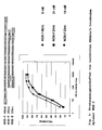

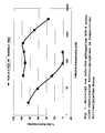

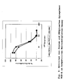


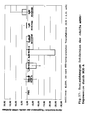


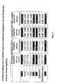
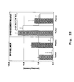
Classifications
-
- C—CHEMISTRY; METALLURGY
- C07—ORGANIC CHEMISTRY
- C07H—SUGARS; DERIVATIVES THEREOF; NUCLEOSIDES; NUCLEOTIDES; NUCLEIC ACIDS
- C07H21/00—Compounds containing two or more mononucleotide units having separate phosphate or polyphosphate groups linked by saccharide radicals of nucleoside groups, e.g. nucleic acids
- C07H21/04—Compounds containing two or more mononucleotide units having separate phosphate or polyphosphate groups linked by saccharide radicals of nucleoside groups, e.g. nucleic acids with deoxyribosyl as saccharide radical
-
- C—CHEMISTRY; METALLURGY
- C12—BIOCHEMISTRY; BEER; SPIRITS; WINE; VINEGAR; MICROBIOLOGY; ENZYMOLOGY; MUTATION OR GENETIC ENGINEERING
- C12N—MICROORGANISMS OR ENZYMES; COMPOSITIONS THEREOF; PROPAGATING, PRESERVING, OR MAINTAINING MICROORGANISMS; MUTATION OR GENETIC ENGINEERING; CULTURE MEDIA
- C12N15/00—Mutation or genetic engineering; DNA or RNA concerning genetic engineering, vectors, e.g. plasmids, or their isolation, preparation or purification; Use of hosts therefor
- C12N15/09—Recombinant DNA-technology
- C12N15/11—DNA or RNA fragments; Modified forms thereof; Non-coding nucleic acids having a biological activity
- C12N15/115—Aptamers, i.e. nucleic acids binding a target molecule specifically and with high affinity without hybridising therewith ; Nucleic acids binding to non-nucleic acids, e.g. aptamers
-
- A—HUMAN NECESSITIES
- A61—MEDICAL OR VETERINARY SCIENCE; HYGIENE
- A61K—PREPARATIONS FOR MEDICAL, DENTAL OR TOILETRY PURPOSES
- A61K48/00—Medicinal preparations containing genetic material which is inserted into cells of the living body to treat genetic diseases; Gene therapy
-
- A—HUMAN NECESSITIES
- A61—MEDICAL OR VETERINARY SCIENCE; HYGIENE
- A61P—SPECIFIC THERAPEUTIC ACTIVITY OF CHEMICAL COMPOUNDS OR MEDICINAL PREPARATIONS
- A61P19/00—Drugs for skeletal disorders
- A61P19/02—Drugs for skeletal disorders for joint disorders, e.g. arthritis, arthrosis
-
- A—HUMAN NECESSITIES
- A61—MEDICAL OR VETERINARY SCIENCE; HYGIENE
- A61P—SPECIFIC THERAPEUTIC ACTIVITY OF CHEMICAL COMPOUNDS OR MEDICINAL PREPARATIONS
- A61P31/00—Antiinfectives, i.e. antibiotics, antiseptics, chemotherapeutics
-
- A—HUMAN NECESSITIES
- A61—MEDICAL OR VETERINARY SCIENCE; HYGIENE
- A61P—SPECIFIC THERAPEUTIC ACTIVITY OF CHEMICAL COMPOUNDS OR MEDICINAL PREPARATIONS
- A61P31/00—Antiinfectives, i.e. antibiotics, antiseptics, chemotherapeutics
- A61P31/12—Antivirals
-
- A—HUMAN NECESSITIES
- A61—MEDICAL OR VETERINARY SCIENCE; HYGIENE
- A61P—SPECIFIC THERAPEUTIC ACTIVITY OF CHEMICAL COMPOUNDS OR MEDICINAL PREPARATIONS
- A61P35/00—Antineoplastic agents
-
- A—HUMAN NECESSITIES
- A61—MEDICAL OR VETERINARY SCIENCE; HYGIENE
- A61P—SPECIFIC THERAPEUTIC ACTIVITY OF CHEMICAL COMPOUNDS OR MEDICINAL PREPARATIONS
- A61P35/00—Antineoplastic agents
- A61P35/02—Antineoplastic agents specific for leukemia
-
- A—HUMAN NECESSITIES
- A61—MEDICAL OR VETERINARY SCIENCE; HYGIENE
- A61P—SPECIFIC THERAPEUTIC ACTIVITY OF CHEMICAL COMPOUNDS OR MEDICINAL PREPARATIONS
- A61P35/00—Antineoplastic agents
- A61P35/04—Antineoplastic agents specific for metastasis
-
- A—HUMAN NECESSITIES
- A61—MEDICAL OR VETERINARY SCIENCE; HYGIENE
- A61P—SPECIFIC THERAPEUTIC ACTIVITY OF CHEMICAL COMPOUNDS OR MEDICINAL PREPARATIONS
- A61P43/00—Drugs for specific purposes, not provided for in groups A61P1/00-A61P41/00
-
- A—HUMAN NECESSITIES
- A61—MEDICAL OR VETERINARY SCIENCE; HYGIENE
- A61P—SPECIFIC THERAPEUTIC ACTIVITY OF CHEMICAL COMPOUNDS OR MEDICINAL PREPARATIONS
- A61P9/00—Drugs for disorders of the cardiovascular system
- A61P9/10—Drugs for disorders of the cardiovascular system for treating ischaemic or atherosclerotic diseases, e.g. antianginal drugs, coronary vasodilators, drugs for myocardial infarction, retinopathy, cerebrovascula insufficiency, renal arteriosclerosis
-
- C—CHEMISTRY; METALLURGY
- C12—BIOCHEMISTRY; BEER; SPIRITS; WINE; VINEGAR; MICROBIOLOGY; ENZYMOLOGY; MUTATION OR GENETIC ENGINEERING
- C12N—MICROORGANISMS OR ENZYMES; COMPOSITIONS THEREOF; PROPAGATING, PRESERVING, OR MAINTAINING MICROORGANISMS; MUTATION OR GENETIC ENGINEERING; CULTURE MEDIA
- C12N2310/00—Structure or type of the nucleic acid
- C12N2310/10—Type of nucleic acid
- C12N2310/16—Aptamers
-
- G—PHYSICS
- G01—MEASURING; TESTING
- G01N—INVESTIGATING OR ANALYSING MATERIALS BY DETERMINING THEIR CHEMICAL OR PHYSICAL PROPERTIES
- G01N2500/00—Screening for compounds of potential therapeutic value
- G01N2500/04—Screening involving studying the effect of compounds C directly on molecule A (e.g. C are potential ligands for a receptor A, or potential substrates for an enzyme A)
Definitions
- the present invention relates in one aspect to a new use of aptmers.
- the present invention relates to HMG protein binding aptmers.
- the target molecules can basically be divided into two groups.
- the first group comprises target molecules which are present extracellularly and thus in principle be brought into contact with it by administering an active substance into a body fluid or a body cavity containing the target molecule.
- the first group of target molecules are also referred to herein as extracellular target molecules.
- the second group of target molecules includes target molecules that are present in cells, which cells are related to the disease to be treated or the disposition for the disease. It is not necessary that the target molecule is directly responsible for the disease state or is directly related to the disposition for the disease.
- the second group of target molecules are also referred to herein as intracellular target molecules.
- target molecule d. H. extracellular or intracellular target molecule
- the type of target molecule determines the class of compounds that can be used to provide the necessary interaction between the drug, typically the pharmaceutical agent, and the target molecule for therapeutic or preventive action.
- small molecules can be used, ie. H. chemical compounds having a molecular weight of typically 1000 or less daltons. These molecules can interact directly with extracellular but also with intracellular target molecules as desired.
- the other new classes of drugs in particular antisense molecules and siRNA molecules, must be introduced as a result of their mechanism of action into the respective cell containing the target molecule or the gene coding therefor.
- the targeted delivery of the active substance also referred to as delivery, is also the currently limiting factor for clinical use for these substance classes.
- aptamers and spiegelmers d. H. functional nucleic acids with a defined three-dimensional structure, which allows the specific interaction with the respective target molecules.
- the use of aptamers to target intracellular target molecules uses genetic engineering methods, more specifically, gene therapy.
- the aptamers directed against an intracellular target molecule, also called intramers are introduced into the respective target cells by means of genetic engineering methods.
- intracellular expression of intracellular expression of an intracellular nucleic acid coding for the respective aptamer is in principle closed to the Spiegelmers since no biological system exists, which Spiegelmers, d. H. L-nucleotide aptamers would be able to build up.
- the object of the present invention is therefore to provide a class of substances which is capable of specific with intracellular target molecules, d. H. Target molecules that are present in a cell to interact.
- the object is achieved in a first aspect by the use of an L-nucleic acid as intracellular active agent.
- the L-nucleic acid is a spiegelmer.
- the L-nucleic acid interacts with an intracellular receptor.
- the intracellular receptor is selected from the group comprising molecular receptors, enzymes, chaperone molecules, signal peptides, intracellular structures and metabolic intermediates.
- the intracellular receptor is selected from the group comprising polypeptides, carbohydrates, nucleic acids, lipids, and combinations thereof.
- the L-nucleic acid interacts with an intracellular receptor within a cell.
- the intracellular receptor is selected from the group comprising transcription factors and an AT hook-binding DNA-binding protein.
- the intracellular receptor is selected from the group comprising the HMG proteins, preferably from the group comprising HMGA1, HMGA1a, HMGA1b, and HMGA2.
- incubation is under conditions such that the L-nucleic acid binds to the intracellular receptor in the cell.
- the L-nucleic acid is a spiegelmer.
- a third embodiment of the second aspect which is also an embodiment of the first and second embodiments, after incubation of the cell with the L-nucleic acid, it is determined whether binding, in particular intracellular binding, of the L-nucleic acid occurs at the intracellular receptor is.
- the intracellular receptor is selected from the group comprising molecular receptors, metabolic intermediates and enzymes.
- the intracellular receptor is selected from the group comprising polypeptides, carbohydrates, nucleic acid, lipids and combinations thereof.
- the intracellular receptor is selected from the group comprising transcription factors and an AT hook-binding DNA-binding protein.
- the intracellular receptor is selected from the group comprising HMG proteins, preferably selected from the group comprising HMGA1, HMGA1a, HMGA1b and HMGA2.
- the object is achieved in a third aspect by the use of an L-nucleic acid for the production of a medicament for the treatment and / or prevention of a disease, wherein the target molecule of the medicament is an intracellular target molecule.
- the intracellular receptor is selected from the group comprising molecular receptors, enzymes, chaperone molecules, signal peptides, intracellular structures and metabolic intermediates.
- the intracellular receptor is selected from the group comprising polypeptides, carbohydrates, nucleic acids, lipids, and combinations thereof.
- the target molecule is selected from the group comprising transcription factors and an AT hook-binding DNA-binding protein.
- the target molecule is selected from the group comprising HMG proteins, preferably selected from the group comprising HMGA1, HMGA1a, HMGA1b and HMGA2.
- the disease is selected from the group comprising tumor diseases, viral infections and arteriosclerosis.
- the tumor disease is selected from the group comprising mesenchymal tumors, epithelial tumors, benign tumors, malignant tumors, and metastatic tumors.
- the target molecule HMGA and the disease are selected from the group comprising carcinomas of the prostate, pancreas, thyroid, cervix, stomach, breast, colon / Rectum, ovaries; neuroblastomas; Lymphomas, uterine leiomyomas; lipomas; endometrial polyps; chondroid hamartomas of the lung; pleomorphic adenomas of the salivary glands; Hämangiopericytome; chondromatous tumors; aggressive angiomyomas; diffuse astrocytomas; osteoclastomas; Skin cancer; Burkitts lymphoma; Lewis lung cancer; Leukemia; non-small cell lung carcinoma; and in each case metastases and / or metastatic forms thereof.
- Atherosclerosis is triggered or caused by HMGA1, HMGA1a, HMG1b and / or HMGA2 mediated formation of atherosclerotic plaques.
- the intracellular target molecule is intracellular.
- the object is achieved in a fourth aspect by the use of an L-nucleic acid for the production of a diagnostic agent for diagnosis, wherein the target molecule of the diagnostic agent is an intracellular target molecule.
- the intracellular receptor is selected from the group comprising molecular receptors, enzymes, chaperone molecules, signal peptides, intracellular structures and metabolic intermediates.
- the intracellular receptor is selected from the group comprising polypeptides, carbohydrates, nucleic acids, lipids, and combinations thereof.
- the target molecule is selected from the group comprising transcription factors and an AT hook-binding DNA-binding protein.
- the target molecule is selected from the group comprising HMG proteins, preferably selected from the group comprising HMGA, HMGA1a, HMGA1b and HMGA2.
- the disease is selected from the group comprising tumor diseases, viral infections and arteriosclerosis.
- the tumor disease is selected from the group comprising mesenchymal tumors, epithelial tumors, benign tumors, malignant tumors, and metastatic tumors.
- the target molecule is HMGA and the disease is selected from the group comprising carcinomas of the prostate, pancreas, thyroid, cervix, stomach, breast, Colon / rectum, ovaries; neuroblastomas; Lymphomas, uterine leiomyomas; lipomas; endometrial polyps; chondroid hamartomas of the lung; pleomorphic adenomas of the salivary glands; Hämangiopericytome; chondromatous tumors; aggressive angiomyomas; diffuse astrocytomas; osteoclastomas; Skin cancer; Burkitts lymphoma; Lewis lung cancer; Leukemia; non-small cell lung carcinoma; and in each case metastases and / or metastatic forms thereof.
- arteriosclerosis is triggered by HMGA1, HMGA1a, HMG1b and / or HMGA2-mediated formation of atherosclerotic plaques.
- the intracellular target molecule is intracellular.
- the object is achieved in a fifth aspect by a composition comprising an L-nucleic acid binding to an intracellular target molecule and a delivery vehicle.
- the delivery vehicle is a delivery vehicle suitable for intracellular delivery of the L-nucleic acid.
- the delivery vehicle is selected from the group comprising vehicles, conjugates and physical measures.
- the delivery vehicle is a vehicle, wherein the vehicle is selected from the group comprising liposomes, nanoparticles, microparticles, cyclodextrins or dendrimers, or a vesicle consisting of polypeptides, Polyethyleneimine and / or amphipathic molecules.
- the delivery vehicle is a conjugate, wherein the conjugate is a receptor-mediated endocytosis conjugate, a fusogenic peptide conjugate, a signal peptide conjugate, a conjugate with a nucleic acid, preferably a conjugate with a aptmer or a lipophilic conjugate.
- the conjugate is a receptor-mediated endocytosis conjugate, a fusogenic peptide conjugate, a signal peptide conjugate, a conjugate with a nucleic acid, preferably a conjugate with a aptmer or a lipophilic conjugate.
- the delivery vehicle is a physical measure, the measure being preferably selected from the group comprising electroporation, iontophoresis, pressure, ultrasound, and shock waves.
- the delivery vehicle comprises polyethyleneimine.
- the polyethyleneimine is a branched polyethyleneimine having a molecular weight of about 25 kDa.
- the polyethyleneimine forms a micelle or a micelle-like structure.
- the L-nucleic acid is a spiegelmer.
- the aptmer carries a modification, wherein the modification is selected from the group comprising PEG residues.
- the PEG moiety has a molecular weight of about 1,000 to 10,000 Da, preferably a molecular weight of about 1,500 to 2,500 Da, and most preferably a molecular weight of about 2,000 Da ,
- the modification is attached to the 5'-terminus or the 3'-terminus of the L-nucleic acid.
- the ratio of the total number of the nitrogen groups of the polyethyleneimine and the total number of the phosphate groups of the nucleic acid contained in the composition is about 1 to 20, preferably about 1.5 to 10, more preferably about 2 to 5, and most preferably about 2 to 3.
- the composition provides the L-nucleic acid intracellularly.
- the object is achieved in a sixth aspect by the pharmaceutical composition
- the pharmaceutical composition comprising a composition according to the fifth aspect, and a pharmaceutically acceptable carrier.
- the L-nucleic acid is a composition according to the fifth aspect.
- the L-nucleic acid is a composition according to the fifth aspect.
- the L-nucleic acid is a composition according to the fifth aspect.
- the L-nucleic acid is a composition according to the fifth aspect.
- an HMGA-binding nucleic acid characterized in that the nucleic acid comprises a section box A1 and a section box A2, wherein the section box A1 and the section box A2 are interconnected by an intermediate section and wherein Box A1 and Box A2 are individually and independently selected from the group comprising GGGCG, GGGUG and GGGAG.
- the intermediate section consists of either an intermediate section Z1 comprising six or seven nucleotides or an intermediate section Z2 comprising 12 to 25 nucleotides.
- the nucleic acid has a first portion at the 5'-end of the portion Box A1 and a second portion at the 3'-end of the portion Box A2, preferably both Sections independently comprise four to eight nucleotides each.
- the two sections are at least partially or completely hybridized with each other, wherein the hybridization extends over four to eight nucleotide pairs.
- the nucleic acid has a section Helix A1 at the 5 'end of the section Box A1 and a section Helix A2 at the 3' end of the section Box A2 , wherein preferably the section helix A1 comprises four to eight nucleotides and preferably the section helix A2 comprises four to eight nucleotides, and wherein preferably the section helix A1 represents the first section at the 5 'end of the section box A1 or a part thereof, and wherein preferably the section Helix A2 represents the second section at the 3 'end of the section Box A2 or a part thereof, wherein the length of the section Helix A1 is independent of the length of the section Helix A2.
- the sections Helix A1 and Helix A2 are at least partially or completely hybridized with each other, wherein the hybridization extends over four to eight nucleotide pairs.
- a section Helix B2 is arranged, wherein preferably the length of the section Helix B1 and Helix B2 individually and independently comprises a length of four to eight nucleotides.
- the sections Helix B1 and Helix B2 are at least partially or completely hybridized with each other, wherein the hybridization extends over four to eight nucleotide pairs.
- nucleotides are disposed between the 3 'end of the helix A1 portion and the 5' end of the helix B1 portion.
- nucleotides are arranged between the 3 'end of the section helix A1 and the 5' end of the section helix B1.
- nucleotides are interposed between the 3 'end of the section Helix B2 and the 5' end of the section Helix A2.
- a nucleotide is between the 3 'end of the helix B2 portion and the 5' end of the helix A2 portion arranged.
- the sum of the nucleotides of section Helix A1 and section Helix B1 is 10 to 12 nucleotides and the sum of the nucleotides from section Helix A2 and from section Helix B2 10 to 12 nucleotides.
- the sum of the hybridized nucleotides from the hybridization of section Helix A1 with section Helix A2 and section Helix B1 with section Helix B2 is 10 to 12 nucleotide pairs.
- the nucleic acid does not include a portion of helix A1 and helix A2, thereby 5 'end of the nucleic acid section Helix B1 and at the 3' end the helix B2 is arranged, wherein preferably the length of the section helix B1 and helix B2 each individually and independently comprises a length of four to eight nucleotides.
- the sections Helix B1 and Helix B2 are at least partially or completely hybridized with each other, wherein the hybridization extends over four to eight nucleotide pairs.
- between the 3 'end of the section helix A1 and the 5' end of the section box A1 are one to five nucleotides and between the 3 ' End of the section box A2 and the 5 'end of the section Helix A2 one to three nucleotides arranged.
- a seventeenth embodiment of the seventh aspect which is also an embodiment of the sixth, seventh, eighth, ninth, tenth, eleventh, twelfth, thirteenth, fourteenth, and fifteenth embodiments, between the 3 'end of the section helix B1 and the 5' End of the section box A1 two nucleotides and arranged between the 3 'end of the section box A2 and the 5' end of the section helix B2 one to seven nucleotides.
- an eighteenth embodiment of the seventh aspect which is also an embodiment of the first, second, third, fourth, fifth, sixth, seventh, eighth and tenth embodiments insofar as it is an embodiment of the sixth, seventh and eighth embodiments, the twelfth and thirteenth embodiments Embodiment, as far as these embodiments of the sixth, seventh, eighth and tenth embodiments, the fourteenth and fifteenth embodiment, as far as these embodiments of the sixth, seventh, eighth, tenth, twelfth and thirteenth embodiments, or the seventeenth embodiment, as far as these embodiments of the sixth , eighth, tenth, twelfth, thirteenth and fifteenth embodiments, each within the scope limited herein, includes the intermediate section Z1 six or seven nucleotides.
- the nucleic acid comprises a section box A1 and a section box A2, with the 3 'end of the section box A1 directly connected to the 5' end of the intermediate section Z1 is connected, and the 3 'end of the intermediate section Z1 is connected directly to the 5' end of the section box A2.
- the nucleic acid comprises a section helix B1 and a section helix B2.
- the portions Helix B1 and Helix B2 individually and independently of each other comprise four to eight nucleotides, which are preferably fully or partially hybridized with each other.
- a twenty-fourth embodiment of the seventh aspect which is also an embodiment of the twenty-first, twenty-second, and twenty-third embodiments, there is no nucleotide between the 3 'end of the portion Box A2 and the 5' end of the portion Helix B2, or in the 5 ' 3 'direction, the nucleotide sequence GN y arranged, wherein N y comprises zero to six nucleotides, preferably 0 or 6 nucleotides.
- the nucleic acid comprises a portion of helix A1 and helix A2.
- the sections Helix A1 and Helix A2 each independently and independently of each other comprise four to eight nucleotides, wherein preferably the sections Helix A1 and Helix A2 are completely or partially hybridized with each other.
- a nucleotide sequence N x is disposed between the 3 'end of the helix A1 portion and the 5' end of the helix B1 portion, where N x zero to five nucleotides.
- a nucleotide sequence N z is disposed between the 3 'end of the helix B2 portion and the 5' end of the helix A2 portion N z is zero to six nucleotides.
- a twenty-ninth embodiment of the seventh aspect which is also an embodiment of the twenty-first, twenty-second, twenty-third, twenty-fourth, twenty-fifth, twenty-sixth, twenty-seventh and twenty-eighth embodiments, the sum of the hybridized nucleotides from the hybridization of section Helix A1 with section Helix A2 and of Section Helix B1 with section Helix B2 10 to 12 nucleotide pairs.
- nucleotide sequence GN y arranged, wherein N y comprises zero to six nucleotides, preferably 0 or 6 nucleotides.
- the HMGA-binding nucleic acid comprises a sequence selected from the group comprising SEQ. ID. No. 1, SEQ. ID. No. 2, SEQ. ID. No. 3, SEQ. ID. No. 5, SEQ. ID. No. 6, SEQ. ID. No. 7 and SEQ. ID. No. 13th
- the 3 'end of the section Box A2 immediately adjoins the 5' end of the section Helix B2 ,
- the HMGA-binding nucleic acid comprises a sequence comprising SEQ. ID. No. Third
- the HMGA-binding nucleic acid has the following structure
- the HMGA-binding nucleic acid has the following structure
- the HMGA-binding nucleic acid comprises a sequence selected from the group comprising SEQ. ID. No. 15 and SEQ. ID. No. 16th
- FIG HGMA-binding nucleic acid comprises an intermediate section Z 2 of 12 to 25 nucleotides.
- the HGMA binding nucleic acid comprises an intermediate portion Z2, a portion Helix C1, and a portion Helix C2.
- a central portion N c is disposed between the helix C1 portion and the helical C2 portion of the HMGA-binding nucleic acid.
- the length of the helix C1 and helix C2 portion of the HGMA-binding nucleic acid is the same length.
- the length of the helix C1 and helix C2 portion of the HGMA-binding nucleic acid is individually and independently three to six nucleotides.
- helix C1 and helix C2 portions of the HMGA-binding nucleic acid are fully or partially hybridized with each other.
- the central portion N c of the HMGA-binding nucleic acid comprises three to five nucleotides.
- the HMGA-binding nucleic acid comprises a section box A1 and a section box A2, wherein between the 3'- End of the section box A1 and the 5 'end of the section Helix C1 a nucleotide sequence N b is arranged and comprises three nucleotides.
- the HMGA-binding nucleic acid comprises a section box A1 and a section box A2, wherein between the third 'End of the section Helix C2 and the 5' end of the section box A2 a nucleotide sequence N d is arranged and comprises two to five nucleotides.
- the HMGA-binding nucleic acid comprises a section helix A1 and a section helix A2.
- the sections Helix A1 and Helix A2 of the HMGA-binding nucleic acid each individually and independently of each other comprise five to six nucleotides, preferably the section Helix A1 and the section Helix A2 completely or partially hybridized with each other.
- a nucleotide sequence N a is arranged between the 3 'end of the section helix A1 and the 5' end of the section box A1 of the HMGA binding nucleic acid, wherein N a comprises one to five nucleotides.
- a fifty-first embodiment of the seventh aspect which is also an embodiment of the forty-eighth, forty-ninth and fiftieth embodiments, between the 3 'end of the section box A2 and the 5' end of the section helix A2 is the HMGA-binding nucleic acid arranged in the 5'-3 'direction a nucleotide sequence GN e , where N e comprises one to two nucleotides, preferably A or UU.
- the helix C1 and helix C2 portions of the HMGA-binding nucleic acid are each independently and independently of one another five or six nucleotides in length , wherein preferably the sections Helix C1 and Helix C2 are completely or partially hybridized with each other.
- the HMGA-binding nucleic acid comprises a sequence selected from the group comprising SEQ. ID. No. 8, SEQ. ID. No. 9, SEQ. ID. No. 10, SEQ. ID. No. 11, SEQ. ID. No. 14, SEQ. ID. No. 22 and SEQ. ID. No. 24th
- the nucleic acid comprises a portion of box A1 and a portion of helix C1 of the HMGA-binding nucleic acid, with between 3'- End of the section box A1 and the 5 'end of the section Helix C1 is a nucleotide A.
- the HMGA-binding nucleic acid comprises a section helix C2 and a section box A2, wherein between the 3 'end of the section helix C2 and the 5' end of the Section box A2 a nucleotide G is arranged.
- the central portion N c of the HMGA-binding nucleic acid comprises four nucleotides, wherein N c is preferably GAUG.
- the HMGA-binding nucleic acid comprises a section Helix B1 and a section Helix B2.
- the sections Helix B1 and Helix B2 of the HMGA-binding nucleic acid individually and independently of each other comprise five nucleotides, wherein preferably the section Helix B1 is hybridized with the section Helix B2 ,
- a sixtieth embodiment of the seventh aspect which is also an embodiment of the fifty-eighth or fifty-ninth embodiment, between the 3 'end of the helix B1 portion and the 5' end of the box A1 portion of the HMGA binding nucleic acid is a nucleotide sequence comprising two nucleotides N j , where N j is preferably AG.
- a nucleotide G is located between the 3 'end of the box A2 and the 5' end of helix B2 of the HMGA-binding nucleic acid.
- the HGMA-binding nucleic acid comprises a section helix A1 and a section helix A2.
- the sections Helix A1 and Helix A2 of the HMGA-binding nucleic acid individually and independently of each other each comprise six nucleotides, and preferably the section Helix A1 and the section Helix A2 are hybridized with each other ,
- a nucleotide sequence comprising two nucleotides N i is disposed between the 3 'end of the helix A1 portion and the 5' end of the helix B1 portion N i is preferably CA.
- a nucleotide A is disposed between the 3 'end of the helix B2 portion and the 5' end of the helix A2 portion.
- the sections include Helix C1 and Helix C2 each have three nucleotides, wherein preferably the sections Helix C1 and Helix C2 are hybridized with each other.
- the HMGA-binding nucleic acid comprises a sequence selected from the group comprising SEQ. ID. No. 12th
- the nucleic acid is one that binds to transcription factors, particularly transcription factors that have an AT-hook.
- the object is achieved in an eighth aspect by a nucleic acid which binds to a transcription factor comprising an AT hook, wherein the nucleic acid comprises a construction according to the seventh aspect.
- the L-nucleic acid is a nucleic acid according to the seventh aspect.
- the L-nucleic acid is a nucleic acid according to the seventh aspect.
- the L-nucleic acid is a nucleic acid according to the seventh aspect.
- the L-nucleic acid is a nucleic acid according to the seventh aspect.
- the L-nucleic acid is a nucleic acid according to the seventh aspect.
- the determination is made to test whether the nucleic acid is being replaced by the candidate HMGA agonist or by the candidate HMGA antagonist.
- the object is achieved in an eleventh aspect by a kit for the detection of HMGA comprising a nucleic acid according to the seventh aspect.
- the object is achieved in a twelfth aspect by an HMGA antagonist obtainable by a method according to the tenth aspect.
- the object is achieved in a thirteenth aspect by an HMGA agonist obtainable by a method according to the tenth aspect.
- the object is achieved in a fourteenth aspect by a complex comprising an HMGA protein and a nucleic acid according to the seventh aspect.
- the present invention is based on the surprising finding that contrary to the prior art it is possible to use L-nucleic acids and in particular aptmers to address intracellular target molecules.
- the intracellular target molecules are preferably target molecules present in a cell. It allows the functional L-nucleic acids by their structure of L-nucleotides inherent properties such as high specificity of the interaction with their target molecules with high stability and lack of toxic or immunologically active degradation products when applied in biological systems and in particular in the animal and human body not to use the cellular mechanisms to encode, as in the case of the intramers, L-nucleic acids from a plasmid or generally a vector, thereby providing the actual functional nucleic acid through the intracellular process of transcription.
- the present inventors assume that functional L-nucleic acids per se can overcome the cytoplasmic membrane, or in the case of involvement of endosomal transport mechanisms in overcoming the cytoplasmic membrane, ingesting them from the vesicle structures that are thereby formed a two- or three-dimensional structure that allows the specific interaction of the functional nucleic acid with its target molecule.
- the present teachings deliberately turn away from the aptamer-based principle of utilizing intracellular transcription mechanisms to produce aptamers in the cell and, for the first time, provide means for the application of functional L-nucleic acids, and in particular aptmers, to cells.
- the term functional nucleic acids refers to those nucleic acids other than structural, in particular naturally occurring structural nucleic acids such as rRNAs or coding nucleic acids such as mRNAs.
- functional nucleic acids are nucleic acids which, due to their two- and / or three-dimensional structure, are capable of binding to target molecules.
- binding to the target molecule is not by hybridization or base pairing based on Watson-Crick base pairings or Hoogsteen base pairing.
- Particularly preferred functional nucleic acids are aptamers and aptamers.
- an L-nucleic acid is a nucleic acid which is completely, substantially or partially constructed of L-nucleotides. It is particularly preferred if the L-nucleic acid consists entirely of L-nucleotides.
- the term "essentially” designates an embodiment in which that part of the L-nucleic acid which is responsible for the interaction with the target molecule or the part which mediates the binding to the target molecule consists of L-nucleotides or these is constructed.
- a functional L-nucleic acid is a functional nucleic acid that is wholly, substantially or partially constructed of L-nucleotides.
- L-nucleic acids The synthesis of L-nucleic acids is known to those skilled in the art and is described, for example, in US Pat Nolte et al., Nat. Biotech, 14, 1116-1119, 1996 . and Klussmann et al., Nat. Biotechnol, 14, 1112-1115, 1996 ,
- aptamers The basic process for producing aptamers is described, for example, in US Pat Tuerck et al. Science, 248, 505-510, 1990 ; or Ellington et al. Nature, 346, 818-822, 1990 , the basic method for generating Spiegelmeren is described for example in Nolte et al., Nat. Biotech, 14, 1116-1119, 1996 . or Klussmann et al., Nat. Biotechnol, 14, 1112-1115, 1996 Gambling Gelmere are thus aptamers, which consist of L-nucleotides instead of D-nucleotides.
- target molecule designates that molecule which is used in the selection process for producing the aptamers or spiegelmers or the molecule which is ultimately bound by the aptamer or the aptamer.
- an intracellularly active agent is a chemical compound capable of binding to a molecule in a cell. It is particularly preferred when the cell is a cell isolated in a tissue or organ, but more preferably not in a human or animal body. If the intracellularly active agent is a aptmer, then it is preferably an intracellularly active agent if it is capable of binding to an intracellular target molecule. Alternatively, the aptmer is an intracellularly active agent when it is capable of binding to its target molecule under conditions such as are present in a cell. Assays to determine these properties are known to those skilled in the art and include, for example, equilibrium binding assays under buffer conditions as they are intracellular (ionic strength and composition, pH, temperature) as disclosed in Example 1.
- the target molecule of the L-nucleic acid, in particular of the functional L-nucleic acid is an intracellular receptor.
- An intracellular receptor as used herein is preferably a chemical compound or a chemical structure, or a portion thereof, with which the functional L-nucleic acid interacts, and preferably one to which the functional L-nucleic acid binds the intracellular receptor, ie the chemical compound or the chemical structure or a part thereof, intracellular, d. H. is present in a cell, as it is preferably described in the preceding paragraph, is present. It is within the scope of the present invention that the intracellular receptor is the target molecule in the production of the functional nucleic acid, in particular of the functional L-nucleic acid.
- the term receptor refers to any interaction partner, preferably a specifically binding interaction partner of the functional nucleic acid, ie, an interaction partner interacting with the functional nucleic acid having a particular spatial structure, charge distribution, hydrophobicity distribution, etc.
- the interaction partner corresponds to the target molecule of the functional nucleic acid as used in the production of the functional nucleic acid. It is within the scope of the present invention that a receptor can also be different from the target molecule used in the production of the functional nucleic acid, but the specific interaction by a cross-reactivity of the functional nucleic acid between the interaction partner and the target molecule used in the production of the functional nucleic acid conditional.
- the term "intracellular receptor” refers to a receptor present in a cell, or a receptor that may be present in a cell, naturally occurring in a cell, or present among such in a cell. It is particularly preferred if the cell is a cell isolated in a tissue or organ, but more preferably not in a human or animal body. However, as used herein, the term “intracellular receptor” also refers to a receptor that is present under conditions such as are present in a cell.
- the term cell refers to a cell selected from the group comprising prokaryotic and eukaryotic cells.
- the eukaryotic cell is selected from the group comprising fungal cells, plant cells, animal cells and human cells.
- the term cell herein generally refers to a compartment bounded by a phospholipid double membrane, which in a preferred embodiment corresponds to a cytoplasmic membrane, which is separated from the environment by the membrane. The separation from the environment is not a complete separation, but allows an energy exchange and a mass transfer between the cell and the environment. Preferably, the mass transfer is limited.
- the term "cell” herein also encompasses vesicles and / or compartments of a prokaryotic or eukaryotic cell as defined herein that are both present or may be present within a prokaryotic or eukaryotic cell as well as outside such a prokaryotic or eukaryotic cell, for example as a vesicle or parts of a prokaryotic or eukaryotic cell surrounded by a cytoplasmic membrane, which in one embodiment may be present in a body fluid.
- the conditions within such a cell substantially correspond to those present in a prokaryotic or eukaryotic cell, in particular the factors which affect the binding of the functional nucleic acid to it Influence the target molecule.
- the body fluid is selected from the group consisting of blood, urine, cerebrospinal fluid, lymph, serum, plasma, vaginal secretions, saliva, and sperm.
- the receptor is defined by its function in a cell. Accordingly, the receptor may be selected from the group consisting of molecular receptors, enzymes, metabolic intermediates, signal peptides, chaperone molecules, and intracellular structures such as ribosomes, mitochondria, cytoskeletal elements such as cytoskeletal cells. Tubulin and actin filaments, endosomal particles, lysosomes, other intracellular structures such as vesicles, in particular intracellular vesicles comprises.
- the term molecular receptor refers to a molecule that receives and propagates information within a cell, tissue, organ or organism. The information is typically mediated by a molecule that interacts with the molecular receptor.
- the molecular receptor is able to produce a signal.
- a signal may be due to or manifest in the change in conformation and / or activity of the receptor.
- the signal itself is able to relay the recorded or represented information in a different form.
- the signal may preferably be a chemical, biochemical or electrical signal.
- the molecular receptor is part of a reaction cascade, more preferably a signal cascade.
- the information transmitted by a molecular receptor may be quantitative and / or qualitative information, for example regarding the presence of a compound and / or its concentration.
- metabolic intermediates refers to all those compounds that occur through metabolic activities in a cell as components of catabolism as well as anabolism.
- the receptor is defined by its chemical nature.
- the receptor is selected from the group comprising polypeptides, carbohydrates, nucleic acid, lipids, and combinations thereof.
- polypeptide preferably refers to any polymer of two or more amino acids.
- the amino acids are L-amino acids, but D-amino acids can also be used in the context of the embodiment.
- nucleic acids preferably refers to a polymer of two or more nucleotides or nucleotide analogs known to those skilled in the art, which are either D nucleotides or L nucleotides or mixtures thereof.
- Preferred combinations include glycosylated polypeptides and glycosylated lipids
- a particular group of intracellular receptors are transcription factors and DNA-binding proteins that bind to an AT-hook.
- Exemplary transcription factors are given in the following Table 1: Table 1: Transcription factors gamma) OBP ALL-1 AP 2beta (STAT5A) 4 alpha-CBF AP 2gamma 120 kDa CRE alpha-CP2b AP 2rep binding protein alphaH0 AP-3 (1) 14-3-3 epsilon alphaH2-alphaH3 AP-3 (2) 14-3-3 zeta ALX3 AP-4 50-55K protein Alx-4 AP-5 53BP1 AMEF-2 APC 70-75K protein AML1 AR 80-90K protein AML1a Arnt AAF AML1b Arnt (774 AA Form) ABF-1 AML1c ARNT2 ADA2 AML1DeltaN ASC-2 Ada3 AML2 ASPP1 ADA NF1 AML3 ASPP2 AFP1 AML3a ATBF1-A Ah
- Intracellular target molecules "Long-chain” -Fettklaren- Acetyl-CoA citrate malate Amyloid precursor protein CoA ligase synthase Ankarin "major basic” protein Acetylglucosaminyl- arginase "mixed function” transferase Argininosuccinate- oxygenase Acetylspermin- synthetase 11 ⁇ -Hydroxylase (EC deacetylase Argininosuccinate lyase 1.14.15.4) acetyl transacylase aromatase 18-hydroxylase aconitase arylsulfatase 1-acylglycerol-3- actin aspartate phosphate acyltransferase Adenosine deaminase aminotransferase 2,3 Adenosylhomocysteine- aspartate Oxidos
- HMG proteins are the HMG proteins, as for example in the international patent application PCT / EP96 / 00716 described and especially the HMGA proteins.
- HMGA proteins preferably denotes, in summary, the following proteins: HMGA1, HMGA1a, HMGA1b, and HMGA2.
- HMGA proteins are modular and each have three DNA-binding domains, called “AT-hooks” or “AT-hooks”, and as DBD1 to DBD3 in Fig. 2 and a very acidic C-terminal region. It is obvious to those skilled in the art that antagonists that bind to one of the "AT-hooks” not only recognize the HMGA1 proteins and thereby the two splice variants HMGA1a and HMGA1b (see Fig. 2 ), but also have cross-reactivity with similar DNA-binding molecules as HMGA2. In addition to HMGA2, many other proteins have sequences similar to "AT-hooks" and are in any case other receptors.
- Such proteins include those listed in Table 3: Table 3: Column 1: protein database access codes; Column 2: protein name Q9UKB0 Human HMG protein R Q9UKY1 ZHX1_Human zinc finger and homeobox protein 1 P55198 AF17_HUMAN AF-17 protein [MLLT6] Q59F28 Human Trithorax homologue (fragment) Q6PJQ2 Human ZNF406 protein (fragment) Q75PJ9 Human ZFAT-1 protein Q75PJ7 Human ZFAT-3 protein Q75PJ6 Human TR-ZFAT protein Q9ULG1 Human KIAA1259 protein Q9NUK2 Human Hypothetical Protein FLJ11314 Q9NTG6 Human hypothetical protein DKFZp434B0616 Q8IX01 SFR14_HUMAN Suspected splice factor Q9H5J8 Human Hypothetical Protein FLJ23363 Q6I9Y6 Human MGC5306 protein Q8IX01-2 Splice isoform 2 of Q8IX01 Q8IX01-3 Splice iso
- the present invention also relates to L-nucleic acids and in particular Spiegelmers which are directed against any of the target molecules mentioned in Tables 1 to 3.
- L-nucleic acid as an intracellularly active agent, particularly within a cell, to bind to an intracellular receptor, intracellularly various forms of interactions between the intracellular receptor and its interaction partners are influenced.
- the intracellular use of L-nucleic acids thus makes it possible to influence interactions of proteins, nucleic acids, lipids, carbohydrates or combinations of proteins, nucleic acids, lipids, carbohydrates with one another and with one another.
- an L-nucleic acid in particular a Spiegelmer
- this is preferably an in vitro application or an in vitro method.
- the target molecule is an intracellular target molecule.
- the intracellular target molecule is one which is causally or non-causally involved in the disease or disease to be prevented, treated or diagnosed, but in each case results in its binding to an L-nucleic acid specifically binding thereto; that in the case of a drug, the disease is alleviated, prevented, or cured and / or in the case of a diagnostic agent, the disease or disposition can be detected or diagnosed.
- diagnosis includes a first diagnosis as well as subsequent diagnoses, particularly diagnoses or examinations, to, for example, track or determine the course of the disease or the stages of the disease.
- the target molecule is an intracellular receptor as described herein, in particular a transcription factor, an intracellular target molecule or an HMG protein. It is very particularly preferred in the context of the present invention that the target molecule is present intracellularly, ie within a cell and the interaction influencing the disease and / or diagnosis between the L-nucleic acid and in particular the spiegelmer and the target molecule, ie the receptor, intracellularly. However, it is also within the scope of the present invention if the target molecule is outside of a cell and the interaction between the L-nucleic acid and especially the aptmer and the target molecule, ie the receptor, extracellularly.
- HMGA proteins are known to be associated with carcinomas (including the breast, lung, skin, thyroid), as well as leukemias and lymphomas, as well as other malignant tumors such as the like.
- Sarcomas rhabdomyosarcoma, osteosarcoma
- mesenchymal tumors i.a.
- HMGA Hamartomas (breast and lung), adipose tissue tumors (lipomas), pleomorphic adenomas of the salivary glands, uterine leiomyomas, angiomyxomas, breast fibroadenomas, polyps of the endometrium, and atherosclerotic plaques.
- HMGA is an interesting therapeutic target.
- Blockade of HMGA may be a suitable starting point for the control of cancer and prevent its metastatic spread.
- L-nucleic acids directed against HMGA proteins are also useful for the diagnosis and / or therapy of viral diseases and arteriosclerosis due to the involvement of the HMGA proteins in the regulation of transcription of a variety of viral genes or the strong expression of HMGA and in particular HMGA1 in the atherosclerosis affected tissue associated with neointimal vascular smooth muscle cells, macrophages and new blood vessels
- nucleic acids preferably L-nucleic acids and preferably spiegelmers are capable of penetrating as such a phospholipid double membrane as a cytoplasmic membrane and then being intracellularly functional in the sense of - specific - interaction with the intracellular receptor
- the efficacy of the introduction of L-nucleic acid can be influenced and in particular increased by the use of different techniques. These techniques include the use of chemical compounds or molecules as well as the use of physical measures. Whatever the nature, these techniques are generally referred to herein as delivery vehicles. It is within the scope of the present invention that the inventors have also found that aptamers also have this property and how the aptamers thus also together with the invention Composition can be used or used for basically the same purposes, applications and uses.
- nucleic acid When using chemical compounds or molecules, it is further possible to distinguish whether the nucleic acid has to be modified for delivery or whether it does not have to be modified.
- Modification for purposes of using a delivery vehicle is typically not required when the delivery vehicle is or comprises a vesicle, such as in liposomes, polypeptide vehicles, cyclodextrins, dendrimers, nano- and microparticles, and polyethyleneimine.
- modification for purposes of using a delivery vehicle is typically required when the delivery vehicle uses receptor-mediated endocytosis, fusogenic peptides, signal peptides, or lipophilic conjugates.
- the group of physical techniques includes in particular electroporation and iontophoresis.
- Liposomes consist of artificial cationic lipids such as N- [1- (2,3-dioleoyloxy) propyl] -N, N, N-trimethylammonium chloride (DOTMA) and N- [1- (2,3-dioleoyloxy) propyl] -N, N, N-trimethylammonium sulfate (DOTAP), in which the cationic groups interact with the negatively charged nucleic acids and neutralize their anionic charge. Transport takes place via endocytosis ( PNAS, 93: 11493-11498, 1996 ).
- DOTMA N- [1- (2,3-dioleoyloxy) propyl] -N, N, N-trimethylammonium chloride
- DOTAP N- [1- (2,3-dioleoyloxy) propyl] -N, N, N-trimethylammonium sulfate
- Transport takes place via endocytosis ( PNAS,
- cationic liposomes are cytotoxic, especially at higher concentrations, limiting their use in vitro and in vivo ( Biochem Biophys Res Commun, 197: 818, 1993 ; Biochem Biophys Res Commun, 1372: 55-68, 1998 ).
- a non-toxic formulation is the amphiphilic pyridinium-based lipid SAINT-2 ( Nucleic Acids Res, 29: 2079-2087, 2001 ).
- An alternative is also pH-sensitive liposomes, which consist of amphipatic molecules such as cholesteryl hemisuccinate (CHEMS) and dioleylphosphatidylethanolamine (DOPE) ( J Pharmacol Exp Ther, 297: 1129-1136, 2001 ).
- CHEMS cholesteryl hemisuccinate
- DOPE dioleylphosphatidylethanolamine
- Receptor-mediated endocytosis utilizes transport mechanisms that are already present in the cell membrane.
- the nucleic acid for example, via a poly-L-lysine (PPL) linker covalently linked to a transporter protein ("carrier" protein).
- PPL poly-L-lysine
- carrier protein transporter protein
- the selection of the transporter protein is dependent on the ability to bind to certain receptors of the cell membrane and to accumulate in the cell by endocytosis.
- a cell-specific transport can be realized.
- an antisense phosphorothioate directed against c- myc could be introduced into M-14 human melanoma cells ( Anticancer Res, 17: 29-35, 1997 ).
- RME Reactive lipoprotein
- the selected ligand must be inactive or fortifying relative to the therapeutic result to avoid possible toxicity of the delivery vehicle.
- the selection and ubiquitous distribution of the selected receptor in vivo is critical to successful RME-based transport.
- a sequestration of nucleic acids in endosomal compartments was observed in RME-based transport, which makes this method not very promising for intracellular transport or intracellular delivery or delivery.
- the link between the receptor and the nucleic acid must be chosen such that the function of one or the other is not reduced ( J Pharmaceutical Science, 92 (8): 1559-1573, 2003 ).
- Fusogenic peptides have been used to allow peptide-oligonucleotide conjugates to fuse with the cell membrane and thus to facilitate transport into the cell ( Bioconjug Chem, 9: 466-475, 1998 ; Bioconjug Chem. 6: 43-53, 1995 ; Nucleic Acids Research, 25: 2730-2736, 1997 ).
- NLS nuclear localization signals
- vesicles can be formed analogously to viral capsids which serve as potential delivery vehicles for intracellular transport ( Nat Materials, 3 (4): 244-8, 2004 ).
- lipophilic conjugates provide a way to increase the ability of oligonucleotides to bind to lipoproteins and thereby improve intracellular delivery.
- the best studied conjugate is cholesterol ( Antisense and Nucleic Acid Drug Development, 12: 103-128, 2002 ).
- Cyclodextrins are cyclic oligosaccharides that have a central hydrophobic cavity and multiple hydroxyl groups on the outer surface. Thus, cyclodextrins have already been used for the transport of antisense oligonucleotides into human T cell lines ( Antisense Res Dev, 5: 185-192, 1995 ) and also in vivo for intracellular transport or intracellular delivery or delivery of immunogenic CpG sequences ( Biochem Pharmacol, 52: 1537-1544, 1996 ). Various formulations of cyclodextrins can be found in the review article Davis and Brewster (Nature Reviews Drug Discovery 3: 1023-1035, 2004 ).
- Dendrimers are highly branched macromolecules composed of repetitive units of - typically - polyamides.
- the molecules carry on their surface functional groups such as primary amino groups, which interact with other molecules through electrostatic interaction.
- a complex formation takes place rapidly and highly reproducibly, which leads to complexes with weak cytotoxicity ( Nucleic Acids Research, 28: 4225-4231, 2000 ; Clin Cancer Res, 7: 3606-3612, 2001 ;)
- Cyanoacrylate nanoparticles have been tested since the early 1990's for delivery or delivery of oligonucleotides.
- the interaction of oligonucleotides to the nanoparticles is carried out by ion pairs of the anionic charge of the oligonucleotides with different hydrophobic cations, primarily with charged nanoparticles.
- PIHCA polyisohexyl
- PIBCA polyisobutyl
- PHCA polyhexycyanoacrylates
- Microparticles or "microspheres” are typically biodegradable polymers of poly (d, l-lactide-co-glycolide [P (LA-GA)] and used for the sustained release of oligonucleotides ( J Pharm Sci, 91: 790-799; 2000 ; J Controlled Release, 69: 197-207, 2000 ; J Drug Traget, 5: 291-302, 1998 )
- Electroporation is a transport technology that uses a strong electric field to destabilize the lipid double membrane, thereby permeabilizing the cell membrane and thus allowing transport of the substance to be administered, which may also be ionized (iontophoresis), into the cell. Electroporation has already been successfully used to allow transdermal delivery of oligonucleotides ex vivo as well as in vivo ( Int J Pharm, 184: 147-156, 1999 ; J Drug Traget, 5: 275-289, 1998 ; Pharm Res. 15: 1596-1602, 1998 ; Int J Cancer, 85: 260-266, 2000 ; Biochem Biophys Res. Commun., 212: 286-292, 1995 ; Blood, 88: 731-741, 1996 ).
- shock waves high-pressure acoustic pulses
- Ultrasound is also an acoustic technology similar to the shock waves, but uses higher frequency ranges (MHz instead of Hz) and shorter use times (seconds to minutes) and has already been supportive for gene therapy Use approaches ( Hum Gene Ther, 7: 1339-1346, 1996 ; Invest Radiol, 32: 723-727, 1997 ; Ultrasound Med Bio, 25: 1451-1457, 1999 ).
- a novel delivery vehicle which is particularly suitable for the transport of functional nucleic acids such as aptamers, preferably functional L-nucleic acids and most preferably Spiegelmeren.
- the delivery vehicle is a micelle-like or liposomal-like structure based on polyethyleneimine.
- polyethyleneimine may, in principle, be present as a linear or branched polyethylenimine and also be used, with branched polyethylenimine in the branched form being particularly preferred.
- polyethyleneimine can be present as a high molecular weight or low molecular weight polyethyleneimine and also be used.
- high molecular weight polyethyleneimine has a molecular weight of about 800 kDa and low molecular weight polyethyleneimine has a molecular weight of about 3 kDa.
- a polyethyleneimine having an average molecular weight of about 25 kDa is preferred, which is more preferably a branched polyethylenimine having a molecular weight of about 25 kDa.
- the nucleic acid to be delivered itself also carries a modification. It is preferred if the modification is selected from the group comprising PEG radicals. It is further preferred that the PEG moiety has a molecular weight of about 1,000 to 10,000 Da, more preferably about 1,200 to 5,000 Da, and more preferably about 1,500 to 2,500 Da, and most preferably about 2,000 Da.
- a quotient of the total number of nitrogen groups of the polyethyleneimine and the total number of phosphate groups of the nucleic acid to be delivered or packaged with or by the delivery vehicle is from about 1 to 20, preferably from about 1.5 to 10, more preferably from 2 to 5, and most preferably from about 2 to 3.
- the delivery vehicle according to the invention thus also makes it possible to use the mechanism of the intracellular transport of nucleic acids via condensation or packaging with charged particles or reagents and associated change in the charge of the overall complex, also for functional nucleic acids such as aptamers and in particular L-nucleic acids such as Spiegelmers.
- This complex is easily absorbed via endocytosis and thus enters the cytosol of the cell.
- a disadvantage of this method is the stability of the DNA / RNA or the release of the nucleic acid from the endosomal compartment. In the cytosol of the cell, a lysosome develops rapidly from the constricted endosome by the introduction of proteases or nucleases and by protonation of the compartment.
- Nucleases digest the nucleic acids there. However, this does not apply to aptmers, as they are nuclease-stable due to their unnatural configuration.
- nucleic acids are not stable in the acidic environment of the lysosome. However, this applies more to nucleic acids from DNA, less to nucleic acids made up of RNA. Through exocytosis or degradation in the Golgi apparatus, the entire complex is quickly transported back out of the cell and thus only a few nucleic acids enter the cell.
- One of the challenges for a suitable transfection system is thus the stabilization, as well as the release of the nucleic acid from the endosomes into the cytosol.
- RNA spiegelmers have ideal properties for transfection of eukaryotic cells since they are not cleaved by enzymes as enantiomers.
- the use according to the invention of the L-nucleic acids and in particular in connection with the composition according to the invention is of particular importance for this class of drugs, since their mechanism of action is based on a stoichiometric approach and not on a catalytic approach in which the intracellular delivery of a few molecules is already sufficient to achieve the desired effect.
- the present invention satisfies a need hitherto unsatisfied by the prior art techniques.
- the transfection system according to the invention provided or designed with the delivery vehicles according to the invention is based on the formation of micelles from nucleic acids and branched polyethyleneimine (PEI).
- the phosphodiester backbone of the nucleic acids interacts with the free nitrogen positions of the PEI and cross-branches to form small micelles that have a positive charge due to the PEI.
- These micelles are easily taken up by constricting the plasma membrane as endosomes from a cell.
- the PEI now buffers incoming protons, whereupon many chloride ions inside the endosome lead to a swelling of the compartment due to the osmotic pressure.
- composition according to the invention is administered as an aerosol.
- spiegelmers can be derivatized with signal peptides for intracellular as well as intranuclear delivery as well as for organ specific delivery. Coupling of signal peptides directly to the polyethyleneimine can be used for site localization to organs or within the cell.
- the present invention relates to L-nucleic acids, particularly aptmers, and more preferably RNA aptmers directed against HMGA proteins.
- the disclosed herein Spiegelmer against HMGA proteins are particularly examples of the present application also underlying finding that L-nucleic acids and Spiegelmers in particular are able to overcome a phospholipid double membrane or a cytoplasmic membrane of a cell and intracellularly with the intracellular To bind receptor on the specific binding of which they have been selected.
- the one disclosed herein for the intracellular use of L-nucleic acid also applies in connection with the present aspect of the invention (and vice versa) and is used to avoid unnecessary repetitions at this point Reference repeated.
- the HMG (high mobility group) family of DNA-binding phosphoproteins are ubiquitous in mammalian cells as non-histone components of chromatin (Grosschedl et al., 1994).
- the basic HMG proteins are divided into three different families - the HMGB (formerly HMG-1 / -2), the HMGN (formerly HMG-14 / -17), and the HMGA family (formerly HMG). IY / C).
- Each HMG family has its own unique functional sequence motif: the "HMG-box” (HMGB family), the "nucleosomal binding domain” (HMGN family), and the "AT-hook” (HMGA family).
- the HMGA family includes two genes, HMGA1 and HMGA2. From HMGA1 , three different proteins can be expressed by alternative splicing (HMGA1a [formerly HMG-I], HMGA1b [formerly HMG-Y], HMGA1c [formerly HMG-I / R]), HMGA2 only one (HMGA2 [earlier : HMGI-C]).
- HMGA1a, HMGA1b and HMGA2 are polypeptides of approximately 100 amino acids in length and have a modular sequence organization: they have three strongly basic regions ("AT-hooks") which bind the narrow minor groove of double-stranded AT-rich DNA (Reeves & Nissen 1990) , The C-terminus, on the other hand, contains many acidic amino acids.
- the proteins have no stable secondary structure in solution and only assume a defined conformation when in complex with DNA or other proteins (Huth et al 1997).
- HMGA proteins are among the most highly modified proteins in the mammalian cell nucleus and are phosphorylated, acetylated, and methylated (Reeves & Beckerbauer 2001).
- HMGA proteins do not have any transcriptional activity per se but, as so-called architectural transcription factors, through their protein-protein and protein-DNA interactions, organize the formation of the nucleoprotein-DNA transcription complex (Wolffe 1994). They thus have an activating or inhibitory influence on the expression of a multiplicity of genes.
- the most prominent example of positive regulation is the involvement of HMGA1 in the regulation of IFN- ⁇ (Thanos & Maniatis, 1992).
- the HMGA1b stimulates the binding of NF- ⁇ B and ATF-2 to the DNA double helix and simultaneously alters the DNA structure such that NF- ⁇ B and ATF-2 may interact with each other and presumably also with the rest of the transcription machinery (Thanos & Maniatis 1992, Du et al 1993).
- Another transcriptional-activating effect associated with arteriosclerotic pathogenesis is HMGA1-induced CD44 gene regulation (Foster et al 1998).
- CD44 is a cell surface glycoprotein and involved in the migration and proliferation of smooth muscle cells after endothelial injury (Jain et al 1996, Cuff et al 2001).
- CD44 Transcriptional regulation of CD44 is induced by binding of c-Fos and c-Jun to the AP-1 binding site in the CD44 promoter and enhanced by binding of HMGA1.
- Studies in rats have shown that CD44 overexpression leads to an increased recruitment of smooth muscle cells and has a direct influence on the development of arteriosclerotic lesions (Pellacani et al 1999, Foster et al., 1998, 2000).
- HMGAI gene located in the chromosomal band 6p21.3 and the HMGA2 gene localized in the region 12q14-15 showed that they are mainly active in processes of cellular differentiation. Accordingly, strong expression of these genes is found during embryonic development and in undifferentiated cells (Chiappetta et al 1996) as well as in growth factor-stimulated cells (Friedman et al 1993, Johnson et al 1990, Ogram et al 1995, Holth et al 1997).
- HMGA1 In the adult, differentiated tissue HMGA1 is only strongly expressed in the retina, while in the other tissues HMGA2 is not found at all and HMGA1 is found only in very low concentrations (Bussemakers et al 1991, Chiappetta et al 1996, Rogalla et al 1996, Zhou et al 1995; Chau et al 2000).
- HMGA proteins in differentiated normal tissue is associated with the growth and differentiation of adipocytes (Zhou et al 1995, Anand & Chada 2000, Melillo et al 2001), smooth muscle cell proliferation in the blood vessels after vascular injury (Chin et al 1999), the immune response in inflammatory reactions (Pellacani et al 1999), as well as apoptotic processes (Diana et al 2001, Sgarra et al 2003).
- the amount of HMGA1 varies depending on the proliferation rate of the cells (Johnson et al 1990).
- HMGA1 expression is concentrated on specific organs of ecto, meso or endodermal origin, while HMGA2 is restricted to mesenchymal tissues. So far, no information is available on the phenotype of HMGA1 knockout mice, possibly because the lack of this factor damages embryonic development too much.
- HMGA2- knockout mice are dwarfed and have low levels of adipose tissue (Zhou et al 1995) and are also resistant to diet-induced obesity (Anand & Chada 2000).
- HMGA2 or HMGA1b expression is undetectable in the adipose tissue of normal mice, but dramatically increased in the fat of fat or diabetic mice (Chada et al., 2004), suggesting an association between obesity and HMGA expression
- HMGA1 Abnormal regulation of HMGA1 could therefore lead to general alterations in gene expression, thereby significantly contributing to the formation of transformed and / or metastatic phenotypes.
- HMGA proteins appear to play different roles in mesenchymal or epithelial tumors: in malignant epithelial tumors, HMGA expression is more associated with later stages of carcinogenesis, while benign - rarely-converting mesenchymal tumors - already express HMGA in early hyperplasia. This indicates that HMGA proteins in tissues of different embryonic origins perform different functions, which immediately results in the corresponding applications the L-nucleic acids according to the invention in the diagnosis and / or treatment of - corresponding - diseases, as also shown in more detail below darg Dead.
- HMGA1 Animal models investigated the expression of HMGA1 in various human and animal neoplasms. The role of HMGA-1 has been demonstrated in animal models of tumorigenesis (Leman et al 2003, Ram et al 1993) and neoplastic progression (Bussemakers et al 1991, Nestl et al 2001, Ram et al 1993).
- HMGA proteins Aberrations in the genetic mechanisms that control growth and proliferation are the primary cause of carcinogenesis.
- the expression of HMGA proteins is strongly associated with tumor development, as shown in a number of papers (Giancotti et al., 1987, 1989, 1993).
- HMGA2 expression was found in chemically or virally generated as well as in spontaneously obtained tumors, whereas in non-transformed cells or healthy tissue this protein could not be detected (Giancotti et al., 1989).
- oncogenic retrovirus-infected cells in which the synthesis of HMGA2 expression had been specifically blocked lacked various phenotypic markers for transformation (Berlingieri et al., 1995).
- HMGA2 knockout mice show dwarfism, ie the animals are about 60% smaller than wild-type mice. However, these dwarf mice have a high resistance to chemically induced skin tumors.
- the main cause of mortality and morbidity of cancer patients is the metastatic spread of the primary neoplasm in the body. Metastasis is not a simple process, as successful colonization of distant organs by scattered neoplastic cells must pass through many stages. Neoplastic cells must detach from the primary neoplasm, enter the bloodstream, extravagate at distant locations, and finally proliferate again in the parenchyma of the corresponding organ. Many genes are involved in the different stages of this highly complex metastatic cascade express proteins such as proteases, adhesion molecules, motility factors and angiogenic factors.
- HMGA1 HMGA1 gene as one of the most important factors controlling this process is a valid candidate for this.
- the gene products of HMGA1 affect the transcription of many genes that are important for successful metastasis. For example, it has already been shown that other metastasis-associated genes in turn are less expressed in suppression of HMGA1 expression (Battista 1998, Vallone 1997).
- HMGA1 is therefore an interesting therapeutic target molecule.
- the blockade of HMGA1 is therefore in principle suitable for controlling the cancer and preventing its metastatic spread (Evans 2004, Sgarra 2004).
- antisense RNAs directed against HMGA transcripts in cancer cells in vitro, it was possible to reduce cell proliferation or even to induce apoptosis (Masciullo 2003, Scala 2000, Chau 2003).
- the animal model has shown that gene therapy (adoenoviral expression of antisense RNAs directed against HMGA transcripts) drastically reduces the growth of various pancreatic cancer xenografts (Trapasso et al 2004).
- HMGA1 could continue to be used as a prognostic diagnostic marker to determine which patients would benefit from more aggressive cancer therapy. There is a close correlation between the degree of malignant transformation and the amount of HMGA1 expressed. This in turn can be correlated with a poor prognosis in many human cancers, such as prostate (Tamimi 1996, Bussemakers 1991) and colorectal carcinoma (Abe 1999) or neuroblastoma (Giannini 2000).
- HMGA proteins are implicated in the regulation of transcription of a variety of viral genes in a host cell. Examples include the regulation of the expression of the early and late expressed genes of the human papovavirus JC (Leger et al., 1995), regulation of EBNA1 (Epstein-Barr virus nuclear antigen 1) gene of the Ebstein-Bar virus (EBV), which is responsible for the control of viral latency is, (Schaefer et al., 1997), regulation of the IE-3 (immediate-early) gene of herpes simplex virus-1 (HSV-1), which encodes the early expressed protein ICP4 (Panagiotidis et al., 1999 ), Regulation of the latent phase active promoter 2 of HSV-1 (French et al., 1996) and regulation of the LTR (long terminal repeats) promoter of the human HIV-1 virus (Henderson et al., 2000).
- EBNA1 Epstein-Barr virus nuclear antigen 1
- HSV-1 herpe
- HMGA1 also appears to play a decided role as an architectural cofactor in integrating the viral DNA of the HIV-1 virus, the Moloney murine leukemia virus (MoMuLv) and sarcastic poultry flu virus (ASV) into the human genome and thus appears to be an interesting therapeutic approach of antiviral Therapy (Van Maele et al 2006, Li et al 1998, Hindmarsh et al., 1999)
- inhibitors of HMGA proteins are also suitable for the treatment and diagnosis of viral infections (Reeves & Beckerbauer 2002).
- HMGA proteins in various diseases or their suitability as diagnostic markers
- directed L-nucleic acid and in particular aptmers can be used for the prevention, therapy and diagnosis of precisely these diseases.
- Particularly preferred aptmers are the aptmer described herein.
- the individual aptmers have been generated for a given HMGA protein, as a result of the domain approach illustrated in Example 2, these also allow cross-reactivity with other HMGA proteins, as can be seen in US Pat in Fig. 2 shown alignment results.
- nucleic acids of the invention have a number of structural motifs that define a class of aptmers that bind to HMGA proteins as intracellular receptors.
- the various structural motifs are further detailed in Example 1.
- the nucleic acids according to the invention also comprise those nucleic acids which are substantially homologous to the sequences specifically disclosed herein.
- the term "substantially homologous” herein is preferably understood to mean that the homology is at least 75%, preferably 85%, more preferably 90%, and most preferably more than 95, 96, 97, 98 or 99%.
- nucleic acids or nucleic acids according to the present invention should furthermore also include those nucleic acids which comprise nucleic acid sequences as disclosed herein or parts thereof, preferably to the extent that the nucleic acids or the said parts thereof are involved in binding to HMGAs. Proteins are involved.
- nucleic acid can be derived from those disclosed herein, e.g. B. by shortening or truncation.
- a truncation may refer to either or both ends of the nucleic acids as disclosed herein.
- a truncation may also involve the inner sequence of nucleotides, i. H. it may relate to nucleotide (s) between the 5 'and 3' terminal nucleotides, respectively.
- truncation should also refer to the deletion of as little as a single nucleotide from the sequence of the nucleic acids disclosed herein. Shortening may also refer to more than one region of the nucleic acid (s) of the invention, wherein each of these regions may be as small as one nucleotide in length.
- the nucleic acids according to the present invention may furthermore be either D-nucleic acids or L-nucleic acids.
- the nucleic acids according to the invention are L-nucleic acids.
- one or more parts of the nucleic acid are present as D-nucleic acids, or at least one or more parts of the nucleic acids are L-nucleic acids.
- the term "part" of the nucleic acids is not intended to denote any nucleotide. Such nucleic acids are generally referred to herein as D- and L-nucleic acids, respectively.
- the nucleic acids according to the present invention of L-nucleotides and comprise at least one D-nucleotide.
- a D-nucleotide is preferably attached to a part other than the region (s) defining the nucleic acids according to the present invention, preferably those parts thereof which interact with other parts of the nucleic acids.
- a D-nucleotide is attached to the end of each region or nucleic acid according to the present invention.
- such D-nucleotides may function as a spacer or a linker which preferably binds modifications such as PEG and HES to the nucleic acids of the present invention.
- the nucleic acids according to the invention also include those which are part of a longer nucleic acid, wherein these longer nucleic acids can comprise several parts, wherein at least one part is a nucleic acid according to the present invention or a part thereof.
- the other part or parts of these longer nucleic acids may be either a D-nucleic acid or an L-nucleic acid. Any combination may be used in connection with the present invention and for the purposes or uses as disclosed for the nucleic acids of the invention herein.
- This other part or parts of the longer nucleic acid may have a function other than binding and in particular binding to HMGA protein.
- One possible function is to interact with other molecules, e.g. B. for purposes of immobilization, cross-linking, detection, amplification or modification or molecular weight increase to allow.
- L-nucleic acids as used herein are nucleic acids consisting of L-nucleotides, preferably consisting entirely of L-nucleotides.
- D-nucleic acids are nucleic acids consisting of D-nucleotides, preferably consisting entirely of D-nucleotides.
- the nucleic acid according to the invention consists of D-nucleotides, L-nucleotides or a combination thereof of both, wherein the combination, for. B., is a random combination or sequence of regions consisting of at least one L-nucleotide and at least one D-nucleic acid
- the nucleic acid may consist of one or more deoxyribonucleotides, ribonucleotides, and combinations thereof.
- the present invention relates to a pharmaceutical composition which consists of at least one of the nucleic acids according to the invention in combination with one or more other nucleic acids, wherein the other nucleic acids preferably bind / bind to target molecules other than HMGA protein or one of performs / exercise various functions on the nucleic acids of the invention.
- L-nucleic acids are enantiomers of naturally occurring nucleic acids.
- D-nucleic acids are not very stable in aqueous solutions and especially in biological systems and in biological samples due to the extensive presence of nucleases.
- Naturally occurring nucleases, especially animal cell nucleases are unable to degrade L-nucleic acids.
- the biological half-life of L-nucleic acid in such a system, including the human and animal body is significantly increased. Due to the lack of degradability of L-nucleic acids no nuclease degradation products are produced and thus no side effects are observed.
- This aspect effectively delineates the L-nucleic acid from all other compounds used in the therapy of diseases and / or disorders, and the presence of HMGA or its causal involvement comprises L-nucleic acids that specifically bind to a target molecule through a mechanism, which is different from the Watson-Crick base pairing, or aptamers consisting partly or wholly of L-nucleotides, especially where those portions of the aptamer involved in the binding of the aptamer to the target molecule are termed Spiegelmers.
- nucleic acids according to the invention are single-stranded or double-stranded Nucleic acids may be present.
- the nucleic acids according to the invention are single-stranded nucleic acids which, as a consequence of the primary sequence, have defined secondary structures and can thus also form tertiary structures.
- the nucleic acids according to the invention can also be double-stranded in the sense that two strands which are complementary or partially complementary to one another are hybridized with one another. This confers stability to the nucleic acids, which will be particularly advantageous when the nucleic acid is present in the naturally occurring D-form rather than the L-form.
- nucleic acids according to the invention can be modified. Such modifications may involve single nucleotides of the nucleic acid and are well known in the art. Examples of such a modification are, inter alia, described in Venkatesan N. et al. (2003) Curr Med Chem. Oct; 10 (19): 1973-91 ; Kusser, W. (2000) J. Biotechnol. 74: 27-38 ; Aurup, H. et al. (1994) Nucleic Acids Res, 22, 20-4 ; Cummins, LL et al, (1995) Nucleic Acids Res, 23, 2019-24 ; Eaton, BE et al.
- nucleic acid according to the present invention may comprise at least one LNA nucleotide.
- the nucleic acid according to the present invention consists of LNA nucleotides, preferably entirely of LNA nucleotides.
- the nucleic acids according to the present invention may be a multi-part nucleic acid.
- a multi-part nucleic acid as used herein is a nucleic acid consisting of at least two nucleic acid strands. These at least two nucleic acid strands form a functional unit, wherein the functional unit is a ligand for a target molecule.
- the at least two strands of nucleic acid can be derived from one of the nucleic acids of the invention either by cleaving the nucleic acid to generate two strands, or by synthesizing one nucleic acid corresponding to a first part of the invention, ie total nucleic acid, and another nucleic acid corresponding to the second part of the total nucleic acid , It is recognized that both cleavage and synthesis can be used to generate a multi-part nucleic acid where more than the two strands may be exemplified above.
- the at least two strands of nucleic acid are preferably different from two strands that are complementary to each other and hybridize to each other, although there may be some degree of complementarity between the different nucleic acid parts.
- the present inventors have found that the nucleic acids according to the present invention have a very advantageous K D value range or dissociation value range and thus a very advantageous binding constant.
- An example to determine the binding constant is the use of an equilibrium binding assay as described in Example 1.
- the K D value of the nucleic acids according to the invention is preferably less than 1 ⁇ M.
- a K D value of about one ⁇ M should be characteristic of non-specific binding of a nucleic acid to a target.
- the K D value of a group of compounds, such as the nucleic acids according to the present invention will be within a certain range.
- the above-mentioned K D of about 1 ⁇ M is a more preferred upper limit of the K D value.
- the preferred lower limit for the K D of the target molecule-binding nucleic acids may be about picomolar or lower. It is within the scope of the present invention that the K D values of individual nucleic acids which bind to HMGA are preferably within this range.
- Preferred ranges can be selected by selecting a first number within this range and a second number within that range.
- Preferred upper values are 0.25 ⁇ M, 0.1 ⁇ M, preferred lower values are 100 nM, 10 nM, 1 nM and 0.05 nM.
- the nucleic acids according to the invention preferably bind to HMGA1b at 37 ° C. in solution with a dissociation constant K D ⁇ 20 nM, as shown in Example 2.
- the nucleic acids according to the present invention may be of any length provided that they are still capable of binding to the target molecule. It will be appreciated in the art that certain lengths of the nucleic acids according to the present invention are preferred. Typically, the length is between 15 and 120 nucleotides. It will be further appreciated by those skilled in the art that any integer between 15 and 120 is a preferred length of potential for the nucleic acids of the present invention. Preferred ranges for the length of the nucleic acids according to the present invention are lengths of about 20 to 100 nucleotides, about 20 to 80 nucleotides, about 20 to 60 nucleotides, about 20 to 50 nucleotides and about 30 to 50 nucleotides.
- the nucleic acids according to the invention may be modified.
- a particularly preferred form of modification is pegylation. This is the modification of the nucleic acids according to the invention by coupling with polyethylene glycol (PEG) or other groups.
- PEG polyethylene glycol
- nucleic acids according to the invention Due to the high stability of the nucleic acids according to the invention, in particular in the embodiment that they are present as L-nucleic acids, a direct administration of the nucleic acids according to the invention is possible for the treatment of a patient in need of such treatment.
- the nucleic acids of the invention are provided as a physiological solution for local or systemic administration.
- the pharmaceutical composition according to the present invention thus comprises at least one of the nucleic acids according to the present invention and preferably a pharmaceutically acceptable binder.
- a binder may be any known or known binder in the art.
- such a binder is any binder as described in connection with the preparation of the medicament as disclosed herein.
- the pharmaceutical composition comprises another pharmaceutically active agent. It is within the scope of the present invention that the medicament described herein is the pharmaceutical composition as described herein.
- the pharmaceutical composition is for intravenous administration.
- such pharmaceutical compositions can be administered intramuscularly, intraperitoneally, or subcutaneously.
- Other routes of administration are oral or intranasal, with the preferred mode of administration being the least invasive, while retaining the efficacy of the pharmaceutical composition or pharmaceutically active agent.
- the nucleic acids of the invention are preferably contained or dissolved as such or in association with the pharmaceutical composition of the invention in a pharmaceutically acceptable solvent.
- solvents are especially those selected from the group consisting of water, physiological saline, PBS, or a glucose solution, especially a 5% glucose solution.
- a carrier can, for. B., water, buffer, PBS, glucose solution, preferably a 5% glucose solution (iso-osmotic), starch, sugar, gelatin or any other acceptable vehicle.
- Such carriers are generally known to those skilled in the art.
- the pharmaceutical composition contains at least one of the nucleic acids according to the invention in their various embodiments, including, but not limited to, the nucleic acid as a conjugate, as described herein.
- the medicament comprises another pharmaceutically active agent.
- Such further pharmaceutically active agents are, for example, protease inhibitors, proliferation inhibitors and angiogenesis inhibitors and / or agents which have a cystostatic effect.
- another pharmaceutically active agent is another nucleic acid according to the present invention.
- the medicament comprises at least one or more nucleic acids that bind to a target molecule other than HMGA or having a function different from any of the nucleic acids of the present invention.
- composition according to the present invention may be used for the treatment, diagnosis and / or prevention of any of the diseases or disorders described herein.
- the present invention relates to a method of treating a subject in need of such treatment, the method comprising administering a pharmaceutically active amount of at least one of the nucleic acids according to the present invention.
- the subject is suffering from or at risk of contracting such a disorder, the disorder being any of those disclosed herein, in particular, any disorder associated with the use of any of the nucleic acids according to the present invention present invention for the preparation of a medicament is described.
- HMGA proteins and their genes have been increasingly associated with the diagnosis and prognosis of neoplastic diseases and have been proposed as potential biomarkers.
- the level of expression of HMGA1a / b proteins is very low, if detectable at all.
- Increased HMGA1a / b protein expression is characteristic for the phenotype of a variety of tumors and metastases of various cancers (Sarhadi et al., 2006, Balcercak et al., 2005, Briese et al., 2006, Chang et al., 2005, Peters et al., 2005, Sato et al., 2005, Chiappetta et al., 2004, Li et al., 2004, Chuma et al., 2004, Donato et al., 2004, Czyz et al., 2004, Kettunen et al., 2004, Lee et al., 2004, Chen et al 2004, Abe et al 2003, Blacerczak et al 2003, Flohr et al 2003, Masciullo et al 2003, Nam et al 2003, Pierantoni et al 2003).
- HMGA protein expression correlates significantly with poor prognosis and metastatic disease.
- the detection of the HMGA1a / b expression level in biopsies and their histological characterization represents a diagnostic approach for the early detection, prognosis and identification of neoplastic diseases, in particular the diseases and conditions described above.
- HMGA1 regulates CD44, one of the major target genes for plaque formation.
- CD44 the major target genes for plaque formation.
- the affected regions such as neointimal, vascular smooth muscle cells, macrophages and new blood vessels show a high expression of HMGA1.
- HMGA1 seems to be one of the mediators of plaque formation, thus providing a target for diagnostics.
- the biotinylated HMGA1a / b binding aptmer recognizes 5'-bio-NOX-A50 in the Western blot HMGA1a / b as a single band in cancer cell lines. Furthermore, as described in Example 2, recombinantly expressed HMGA1b protein could be detected. The detection of the biotinylated aptmer takes place, for example, via an anti-biotin antibody conjugated by means of Horse Raddish Peroxidase (HRP).
- HRP Horse Raddish Peroxidase
- HMGA1a / b Another approach is the in vivo diagnosis of HMGA1a / b, in which the nucleic acids according to the invention can be used. Tumors and metastases are often embedded by necrotic tumor cells, which release HMGA1a / b into the surrounding tissue. The detection of the extracellular HMGA1a / b- represents an approach to the diagnosis of tumors and metastases embedded in healthy tissue.
- a diagnostic or diagnostic agent or diagnostic agent is capable of detecting, either directly or indirectly, an HMGA protein, preferably HMGA1a / b as described herein, and more preferably HMGA1a / b as described herein in connection with the various Faults and Diseases.
- the diagnostic agent is suitable for detection and / or aftercare for any of the diseases or diseases described herein. Such detection is possible by the binding of the nucleic acids according to the present invention to HMGA1a / b. Such binding can be detected either directly or indirectly.
- the corresponding methods and means are known to those skilled in the art.
- the nucleic acids according to the present invention may comprise a label which allows the detection of the nucleic acids according to the present invention, preferably the nucleic acid which is bound or capable of binding to HMGA protein and preferably HMGA1a / b.
- a label is preferably selected from the group consisting of radioactive, enzymatic and fluorescent labels.
- all known assays that have been developed for antibodies can be adapted for the nucleic acids according to the present invention, wherein the target molecule-binding antibody is replaced by a target molecule-binding nucleic acid.
- detection is preferably by a secondary antibody modified with radioactive, enzymatic or fluorescent labels and binding to the target-binding antibody on its Fc fragment.
- a nucleic acid preferably a nucleic acid according to the present invention
- the nucleic acid is modified with such a label, whereby preferably such a label is selected from the group consisting of biotin, CY-3 and CY-5, and such a label is detected by an antibody directed against such label, e.g.
- an anti-biotin antibody, an anti-CY-3 antibody or an anti-CY5 antibody or in the case that the label is biotin the label is detected by streptavidin or avidin, which binds naturally to biotin.
- streptavidin or avidin is in turn preferably modified with a corresponding label, e.g. As a radioactive, enzymatic or fluorescent label, analogous to a secondary antibody.
- the nucleic acids according to the present invention are detected or analyzed by a second detection means, wherein the detection means is a molecular beacon.
- the technique of molecular lighthouses is known to those skilled in the art. Briefly, these molecular lighthouses are nucleic acid probes that are a reverse complement to the one to be detected Nucleic acid sample, and therefore hybridize with a portion of the nucleic acid sample to be detected, it must be detected. After binding of the nucleic acid sample, the fluorophore groups of the molecular beacon are separated from each other, resulting in the change of the fluorescence signal, preferably a change in intensity. This change correlates with the amount of nucleic acid sample present.
- nucleic acids according to the invention can be used correspondingly as L-nucleic acids within the scope of the various aspects disclosed herein.
- the nucleic acids according to the invention can furthermore be used as starting material for the design of pharmaceutical active substances (English drug design). Basically, there are two possible approaches. One approach is to screen libraries of compounds, such libraries of compounds preferably being libraries of low molecular weight compounds (small or small molecules). Such libraries are known to those skilled in the art. In one embodiment, the screening is high throughput screening. Preferably, high-level screening is fast, efficient, and is performed as a trial-and-error evaluation of drugs in a target-molecule-based assay.
- the nucleic acids can be used for the rational design of drugs.
- the rational design of drugs is the design of a pharmaceutical drug candidate.
- the target molecule which is usually determined by methods such as X-ray crystallographic analysis or nuclear magnetic resonance spectroscopy (NMR)
- NMR nuclear magnetic resonance spectroscopy
- computer programs are used to search databases with structures of a variety of different chemical compounds. The selection is carried out by the computer. The selected compounds are additionally tested in the laboratory.
- the rational design of active ingredients may here be based on any of the nucleic acids according to the present invention and comprises a structure, in particular a three-dimensional structure, which is similar to the structure of the nucleic acid (s) according to the invention. is or is identical to that part of the structure of the nucleic acid (s) of the invention which mediates binding to HMG proteins. In any case, such a structure still shows the same or at least similar binding behavior as the nucleic acid (s) according to the invention.
- the preferably three-dimensional structure of those parts of the HMG protein-binding nucleic acids is mimicked by chemical groups, which are preferably different from nucleotides and nucleic acids.
- a compound can be constructed which is different from the nucleic acid or nucleic acids used as starting materials for the rational design of the active ingredient.
- a compound or drug is preferably a small molecule or a peptide.
- HMG analogs In the case of screening of compound libraries using competitive assays known to those skilled in the art, appropriate HMG analogs, HMG agonists, and HMG antagonists can be found.
- Such competitive assays can be constructed as follows.
- the nucleic acid according to the invention preferably a spiegelmer, d. H. an L-nucleic acid binding the target molecule is coupled to a preferably solid phase.
- labeled HMG protein is added to the test system.
- the HMG protein could also be coupled to a solid phase and the nucleic acid according to the invention could be labeled.
- a potential analogue or potential agonist or antagonist would compete with the HMG molecules that bind to the aptmer, which would be accompanied by a decrease in the signal received from the corresponding label. Screening for agonists or antagonists may involve the use of a cell culture assay system known to those skilled in the art.
- the nucleic acids according to the invention can be used owing to their characteristic binding behavior to HMG protein for the target validation ("target” is English for "target molecule”).
- the nucleic acids of the invention can be used in an ex vivo organ model to study the function of HMG protein. Basically ex vivo models exist in which HMG agonists / antagonists can be tested.
- a kit according to the present invention may comprise at least one or more of the nucleic acids of the invention.
- the kit may include at least one or more positive or negative controls.
- positive controls can z.
- HMG protein against which the nucleic acid of the invention has been screened or to which it binds, preferably in liquid form, may be used.
- a negative control among other things, a peptide can be used, which behaves in terms of its biophysical properties similar to HMG protein, which is not recognized by the nucleic acids of the invention or a peptide with the same amino acid composition but of HMG protein of different sequence.
- the kit may comprise one or more buffers.
- the various ingredients may be present in the kit in dry or lyophilized form, or dissolved in a liquid.
- the kit may include one or more containers, which in turn may contain one or more of the components of the kit.
- the vessels contain reaction mixtures as required for a one-time experiment using one or more components of the kit.
- hybridizing the two sections is understood herein to mean that the sections can hybridize in vitro due to general base pairing rules, or that the sections can hybridize or hybridize under the conditions of use, but not necessarily together are hybridized or hybridized under the conditions of use.
- nucleic acids can be freely selected according to the technical teaching disclosed herein, d. H. can be chosen so that they show the required binding behavior to the respective target molecule and / or the structures described herein, in particular secondary structures are able to form.
- HMGA1a / b-binding RNA tikmers were generated by in vitro selection against D-21AS-HMGA1a / b and subsequent truncation steps.
- the generated aptamers that bind the 21AS-HMGA1a / b domain are in Fig. 1A shown.
- the different clones were prepared as aptamers (D-RNA) by standard phosphoramidite synthesis and radiolabeled at the 5 'end by kinase (see below). The clones were then analyzed for their affinity and activity at two levels of D-bio-21aa HMGA1a / b by equilibrium binding assay.
- Radioactive labeling by kinase ⁇ / u> substance [final] RNA 5 ⁇ M T4 Forward Reaction Buffer (Invitrogen) 1x T4 polynucleotide kinase (Invitrogen) 10 U / 10 ⁇ l reaction mixture [ ⁇ - 32 P] -ATP 1 ⁇ l / 10 ⁇ l reaction mixture
- the reaction ran for one hour at 37 ° C and was then stopped by heating (10 min at 65 ° C).
- the separation of radioactive nucleotides from labeled oligonucleotides was carried out by analytical polyacrylamide gel electrophoresis (PAGE) (see below). This was followed by a crush-and-soak gel elution with ammonium acetate and ethanol precipitation (see below).
- the amount of purified RNA was estimated by the radioactivity of the pellet (after precipitation) relative to the radioactivity of the excised band.
- oligonucleotides were precipitated at -80 ° C. for 30 minutes and centrifuged at 16,100 ⁇ g, 4 ° C. for 30 minutes. The pellet was washed 1x with 70% ethanol and centrifuged for 5 min at 16,100 x g, 4 ° C.
- the background control was a solution without Biotinylated-D-HMGA1a / b-21mer. Peptide and complexes were then immobilized with 10 ⁇ l Streptavidin Ultra-Link gel within 30 min at 37 ° C. The radioactivity of the suspension was decided. The supernatant was removed. Then the matrix was washed once with 100 ⁇ l of selection buffer and then filled up with selection buffer. By measuring the radioactivity, the aptamer content present in the complex with biotinyl-D-HMGA1a / b-21mer was determined for each peptide concentration. Graphical application and Fit (GraFit, version 4.0.10, Erithacus software) were used to determine the dissociation constant of the active species and the proportion of active molecules.
- a dissociation constant for binding to the 21 amino acid D fragment of HMGA1a / b (biotinyl-D-HMGA1a / b-21mer) of 8-22 nM was determined in the equilibrium binding assay ( Fig. 1A ).
- Fig. 3 shows exemplarily on the candidate NOX-f (132-B3) the shortening of the aptamer based on the stabilizing Stem structure, which in extended form in the candidates 132-C3, 132-B3, 132-C4, 132-E2, 132-A2 , 132-H1, 132-F1, 122-G2, 122-E2 (see Fig. 1A ) can be found.
- sequences 132-G2, 122-A1, 122-C1, 122-B2 and 122-B4 have a markedly shorter stem at the 5'-end and 3'-end of the repetitive sequence motif (GGGCG and GGGAG, respectively) Structure on. Shortening them led to a loss of binding. A possible shortening of the longer for these sequences, central region between the repetitive sequence motif (GGGCG or GGGUG or GGGAG) was not made.
- the SEQ IDs of the aptamer sequences of the HMGA-binding nucleic acids disclosed herein are as follows: Seq. ID Internal reference RNA / peptide 34 132-C3, NOX-h D-RNA (aptamer) 35 132-B3, NOX-f (48nt) D-RNA (aptamer) 36 132-C4 D-RNA (aptamer) 37 132-E2 D-RNA (aptamer) 38 132-A2 D-RNA (aptamer) 39 132-H1, NOX-i D-RNA (aptamer) 40 132-F1 D-RNA (aptamer) 41 132-G2, NOX-g D-RNA (aptamer) 42 122-A1, NOX-A D-RNA (aptamer) 43 122-C1, NOX-B D-RNA (aptamer) 44 122-B2 D-RNA (aptamer) 45 122-E2, NOX-C D-RNA (aptamer) 46 122-G2, NOX
- the L-nucleotide enantiomer of an aptamer i. a D-nucleic acid generated against a D-peptide to the mirror-image enantiomer of the D-peptide, i. the L-peptide, as it naturally occurs.
- This L-nucleic acid is also referred to herein as Spiegelmer and otherwise has basically the same binding properties as the aptamer.
- Characteristic of all Spiegelmers that bind to HMGA1a / b is a repetitive sequence element of the sequence GGGCG or GGGUG or GGGAG. This sequence element appears twice in HMGA1a / b-binding Spiegelmeren ( Fig. 1A and 1B ).
- the sequence element closer to the 5 'end of the aptmer is referred to herein as box A1.
- the sequence element closer to the 3 'end of the spiegelmer is referred to herein as box A2.
- Box A1 and box A2 and their arrangement to each other probably represent the crucial feature of HMGA1a / b-binding Spiegelmers.
- box A1 and box A2 are either a sequence section with a length of six to seven nucleotides or 12 to 22 nucleotides ( Fig. 1A and 1B ). Since these sequence sections differ not only in their length, they are dealt with separately.
- sequence section lying between box A1 and box A2 has a length of six nucleotides, then the sequence section has the sequence UGGUUG, UGGCUG, CGGUUG, AGGUUG or GUGUAA. Insertion of one nucleotide (uracil) into the sequence CGGUUG, resulting in the sequence CGGUUUG, has neither a negative nor a positive influence on the binding properties of the Spiegelmers.
- sequence section lying between box A1 and box A2 has a length of 12 to 22 nucleotides, then this sequence section has two sequence regions of equal length, the optionally hybridize with each other (Helix C).
- the Hybidleiter is accomplished thereby in each case three to six nucleotides. Between the nucleotides forming the helix C are three to five unpaired nucleotides. Between the 3 'end of box A1 and the 5' end of helix C, one to three nucleotides are unpaired. Between the 3 'end of Helix C and the 5' end of Box A2, one to five nucleotides can be unpaired.
- All HMGA1a / b-binding Spigelmers excel at their 5 'and 3' ends by sequence sections which can hybridize to each other (Helix A1 and Helix A2, ( Fig. 1A and 1B ).
- the number of each hybridizing nucleotides can vary from four to eight. In this case, this supposedly double-stranded region can approach the 5 'end of box A1 and the 3' end of box A2. If this is not the case, then box A1 and box A2 can be flanked by additionally hybridizing nucleotides (helix B1 and helix B2). These may be regions of four to eight nucleotides each ( Fig. 1A and 1B ).
- Helix A1 and helix A2 in each case four to eight nucleotides which hybridize completely or partially with one another, the sum of the mutually hybridizing nucleotides composed of helix A1 and helix A2 and helix B1 and helix B2 being 10 to 12 nucleotides;
- Helix B1 and Helix B2 four to eight nucleotides each, which hybridize with each other, wherein the sum of each hybridizing nucleotides of helix A1 and helix A2 and helix B1 and helix B2 is 10 to 12 nucleotides.
- the molecules are also active after shortening at the 5 'and at the 3' end. After removal of the helices A1 and A2 as well as the regions N 6 N7 and GNy, the truncated molecules retain their binding properties. This was shown for the shortened variants 132-B3 32nt (NOX-f 32nt) and 132-B3 33nt (NOX-f 33nt) (see Fig. 3 and 4 ).
- the BD-Freedom TM ORF clone GH00552L1.0 (high mobility group AT hookl) with the HMGA1b coding sequence was purchased from BioCat, Heidelberg. The sequence is already altered to convert the stop codon to a leucine-encoding codon to allow C-terminal fusions. The sequence of the clone most closely matches the sequence stored in the RefSeq database numbered NM002131.
- the primers HMG_fwd1 (TCGACACCATGGGTGAGTC, Seq.ID 34) and HMG_rev1 (GTCTAGAAAGCTTCCCAACTG, Seq.ID 35) were used to amplify the sequence encoding HMGA1b using standard PCR.
- the base was changed from A to G after the ATG to introduce an NcoI site.
- the PCR product was used according to the manufacturer's instructions with the restriction enzymes NcoI and HindIII Both from NEB, Frankfurt am Main; Germany) cut and cleaned over an agarose gel.
- the vector pHO2d Fasshauer et al. (1997) J. Biol. Chem. 272: 28036-28041 ) was also cut with NcoI and HindIII and purified on an agarose gel.
- the vector pHO2d allows the expression of a protein fused at the C-terminal end to a sequence of 6 histidine residues (His6-tag) under the control of a T7 promoter ( Fasshauer et al., 1997, JBC 272: 28036 ).
- the purified and cut PCR product was ligated into the prepared vector overnight at 15 ° C. using a T4 ligase according to the manufacturer's instructions (MBI Fermentas, St. Leon-Roth, Germany). Bacteria were transformed from strain DH5 ⁇ with the ligation product. Correctness of the plasmids from obtained colonies was ensured by sequencing.
- the fusion protein HMGA1b-His6 encoded by pHO2d / HMGA1b has followed a glycine (G) instead of serine (S) at position 2 in comparison to the natural HMGA1b protein and a leucine (L) (see above) after the C-terminal glutamine (Q) of 5 additional amino acids (GSLNS) (encoded by vector) followed by the six histidines (H).
- G glycine
- S serine
- L leucine
- Q C-terminal glutamine
- GSLNS C-terminal glutamine
- fusion protein For the expression of the fusion protein, bacteria of strain BL21 were transformed with the plasmid pHO2d / HMGA1b. Expression of the fusion protein was induced with isopropylthio- ⁇ -D-galactoside (IPTG). After 4 h, the bacteria were centrifuged off for 15 min at 10,000 ⁇ g and the pellet was stored at -20 ° C. until further use.
- IPTG isopropylthio- ⁇ -D-galactoside
- extraction buffer 1% n-octyl- ⁇ , D-thioglucopyranoside (OTG) in 50 mM Na x PO 4 buffer, pH 8.0, 250 mM NaCl, 10 mM Imidazole and MiniProteaselnhibitoren tablets (Roche, Mannheim, Germany) (5 St./50 ml)
- 5 .mu.l Benzonase grade 1, MERCK, Darmstadt, Germany
- the supernatant was clarified by a pleated filter and then applied to a HIS-Select column equilibrated with washing buffer (50 mM Na x PO 4 buffer, pH 8.0, 250 mM NaCl, 10 mM imidazole, all MERCK, Darmstadt, Germany) (HIS assay).
- washing buffer 50 mM Na x PO 4 buffer, pH 8.0, 250 mM NaCl, 10 mM imidazole, all MERCK, Darmstadt, Germany
- SELECT Cartridge Sigma, Deisenhofen, Germany
- the fusion protein was eluted with elution buffer (250 mM imidazole in wash buffer) in 0.5 - 1 ml fractions. Protein-containing fractions were analyzed by gel electrophoresis (16% polyacrylamide gel after Shuger & Jagow, 1987, Anal.
- HMGA1a / b spiegelmers For a more detailed analysis of the affinity of the HMGA1a / b aptmers for HMGA1b, a test based on the 96-well format was used. In this assay, binding of the HMGA1a / b aptmers to HMGA1b-His6 prevents its interaction with a DNA oligonucleotide that has a binding site for HMGA1a / b.
- This DNA oligonucleotide (dsDNA AT-Hook) (Fashena et al., 1992) is labeled on a strand with a biotin molecule through which it can be attached to streptavidin-coated plates.
- spiegelmers at various concentrations and HMGA1b-His6 (0.36 ⁇ g / ml, about 30 nM) are incubated in a shallow bottom plate in a total volume of 100 ⁇ l for 10 min at room temperature with shaking.
- the incubation solution also contains: 25 mM Tris / HCl, pH 7.0 (Ambion, Austin, TX, USA), 140 mM KCl (Ambion, Austin, Tex., USA), 12 mM NaCl (Ambion, Austin, TX, USA), 0 , 8 mM MgCl 2 (Ambion, Austin, TX, USA), 0.25 mg / ml BSA (Roche, Mannheim, Germany), 1 mM DTT (Invitrogen, Düsseldorf, Germany), 18-20 ⁇ g / ml poly (dGdC) (Sigma, Deisenhofen, Germany)), 0.05% Tween 20 (Roche, Mannheim, Germany).
- biotinylated DNA oligonucleotide dsDNA AT-Hook (equimolar mixture of 5'biotin-TCGAAAAAAGCAAAAAAAAAAAACTGGC (34nt) and 5'GCCAGTTTTTTTTTTTTTTTTTTTTTGCTTTTTTTT (31 nt); 75 ⁇ M in 150 mM NaCl (Ambion, Austin, TX)) are added and incubated for a further 10 min with shaking at RT. Subsequently, the mixtures are transferred to a streptavidin-coated black 96-well plate (ReactiBind v. Pierce, Bonn, Germany) and incubated for 30 min with gentle shaking at RT.
- TBSTCM 20 mM Tris / HCl, pH 7.6 (Ambion, Austin, TX), 137 mM NaCl (Ambion, Austin, TX, USA), 1 mM MgCl2 (Ambion, Austin, TX, USA), 1 mM CaCl 2 (Sigma, Deisenhofen, Germany), 0.05% Tween20 (Roche, Mannheim, Germany)).
- HMGA1b The recombinantly expressed HMGA1b was separated by gel electrophoresis on a 16% PAA Tricin gel and transferred by electroblotting onto nitrocellulose membranes. The membrane was subsequently blocked with 5% skimmed milk and 100 nM unspecific aptmer in 1xTBST (20 mM Tris / HCl pH 7.6, 137 mM NaCl, 0.1% Tween) for 1 hour and washed 3 times for 10 minutes with 1xTBST. Detection of the recombinant HMGA1b was carried out with 5'-end biotinylated Spiegelmer NOX-A (5'bioNOX-A).
- 5'bioNOX-A was diluted in 1xTBST with 1 mM calcium and magnesium (TBST + Ca / Mg) and 100 nM unspecific aptmer and incubated for 1.5 hours. The blot was then washed 3 times for 10 minutes with 1x TBST + Ca / Mg and the bound biotinylated aptmer incubated with an anti-biotin antibody in TBST + Ca / Mg for 45 minutes. Subsequently, the blot was washed 5 times for 10 minutes with IxTBST + Ca / Mg and the coupled horseradish peroxidase (HRP) secondary antibody was detected by means of LumiGLO detection reagent (Cell Signaling Technology).
- HRP horseradish peroxidase
- the target molecule HMGA1a / b is expressed in the cytosol and, as a transcription factor, finds its natural binding partner, the double-stranded DNA in the cell nucleus.
- the Spiegelmer By binding of the Spiegelmer to HMGA1a / b in the cytosol and competition of HMGA1a / b bound by the AT-hook on the DNA in the nucleus of the cell, the HMGA1a / b-mediated cellular responses are to be antagonized. Due to the negative charge of the plasma membrane, DNA and RNA molecules are poorly absorbed by passive transport from a cell.
- One of the approaches of intracellular transport of nucleic acids is the condensation or packaging with charged particles or reagents and associated change in the charge of the entire complex.
- This complex is easily absorbed via endocytosis and thus enters the cytosol of the cell.
- Disadvantages of this method is the stability of the DNA / RNA or the release of the nucleic acid from the endosomal compartments.
- a lysosome develops rapidly from the constricted endosome by the introduction of proteases or nucleases and by protonation of the compartment.
- Nucleases digest the nucleic acids there and, moreover, the nucleic acid is not stable in an acidic environment. Through exocytosis or degradation in the Golgi apparatus, the entire complex is quickly transported back out of the cell and thus only a few nucleic acids enter the cell.
- RNA spiegelmers have ideal properties for transfection of eukaryotic cells because they are not cleaved by enzymes as unnatural enantiomers.
- the selected transfection system is based on the formation of micelles of branched polyethylenimine (PEI) nucleic acids.
- PEI polyethylenimine
- the phosphate backbone of the nucleic acids interacts with the free nitrogen positions of the PEI and cross-branches to form small micelles, which have a positive charge due to the PEI.
- This is PEI with a Molecular weight of 3 to 800 kDa used.
- the use of 25 kDa cross-branched PEI results in the addition of nucleic acids to polyplexes of size of 100 to 500 nm, but typically to polyplexes of size 100 to 200 nm.
- a nitrogen / phosphate ratio of 2: 1 to 5: 1 is used; sometimes even up to 20: 1.
- the packaging of the nucleic acid in micelles results in a change in the zeta potential of the complex to ⁇ (+) 21mV for an N / P 3. It is known that with increasing positive zeta potential of complexes, toxicity for cultured cells increases. However, these micelles are easily taken up by constricting the plasma membrane as endosomes from a cell. The PEI now buffers incoming protons, whereupon many chloride ions inside the endosome lead to a swelling of the compartment due to the osmotic pressure.
- nucleic acid-PEI complex due to a strong positive charge, tends to interact and aggregate with serum proteins as well as to the cell toxicity already described.
- the solution to the problem is the derivatization of the nucleic acid with 2kDa polyethylene glycol (PEG). These residues attach like a shield around the micelles and prevent binding to serum proteins ( Ogris et al., Gene Therapy, 1999, 6 (595-605 ).
- the zeta potential is lowered to +/- OmV, resulting in lower cell toxicity with constant buffer capacity of the PEI with respect to the proton sponge effect.
- Lead candidates NOX-A and NOX-f were prepared synthetically as aptamer and aptamer with a 3'-terminal amino group and subsequently PEGylated via the amino residue.
- PEGylation has no influence on the binding properties of the aptamers to the HMGA1a / b fragment.
- competition assays with recombinant full-length HMGA1a / b It is also shown that the binding of Spiegelmeren to the full-length HMGA1a / b is independent of the 3'-terminal PEGylation ( Fig. 9 ).
- PEGylated spiegelmer packaging was carried out in PBS by adding 25kDa branched-chain polyethylenimine (PEI) (ALDRICH, Cat .: 40,872-7) in a ratio of 2.5: 1 absolute PEI to absolute phosphate ribonucleic acid backbone (N / P 2,5).
- PEI branched-chain polyethylenimine
- the sterile, autoclaved PEI solution had a concentration of 200 mM free nitrogen groups and was adjusted to pH 7.4 with 1 M hydrochloric acid.
- the sterile filtered aptmer was initially introduced at a concentration of up to 700 ⁇ M in 1xPBS with Ca / Mg and incubated after the addition of sterile-filtered PEI for 30-60 minutes at room temperature.
- the complex formation proceeds with the lowest possible concentration of aptmer since high concentrations of aptmer lead to nonspecifically large aggregates.
- the formation of aptmer micelles was measured by a dye exclusion assay. It was determined how much aptmer can be detected by dye before and after packaging in micelles. The dye used was RiboGreen (M. Probes) and the fluorescence was determined with an ELISA reader. 1 ⁇ M aptmer was initially introduced in 100 ⁇ l 1xPBS and increasing amounts of PEI were added.
- spiegelmer micelles were under in Fig. 11 stored conditions.
- the stability of aptmer micelles was measured by dye exclusion assays described in section 3.3.
- a stability study of the micelles showed that the storage of micelles in different media, as well as at different temperatures has no influence on the Spiegelmer micelles.
- Also described in the literature is the freeze-drying of ribozyme / PEI complexes without loss of the properties of the ribozyme ( Brus-C et al., J. Control Release, 2004, Feb, 20, 95 (1), 199-31 )
- the preparations were again washed with 3x1ml 1xPBS, incubated for staining of the chromatin in the nucleus for a further 10-20 seconds with a DAPI solution (1 ⁇ l stick to 10ml 1xPBS), washed again and covered with a Paramount solution.
- the prepared preparations were analyzed on a fluorescence microscope (emission 488nm / extinction 514-522nm).
- the transfection efficiency was> 95% of the total cells and showed no influence on the morphology of the cells.
- the 5'-FITC-coupled Spiegelmer was found mainly in the cytosol and associated to the plasma membrane.
- the punctiform distribution pattern indicates inclusion in compartments and the diffuse pattern indicates liberated spiegelmer. In the nucleus, only a small signal could be detected at Spiegelmer.
- the selective distribution of the aptmere in the cytosol and perinuclear space of the H578T cells indicates an accumulation in compartments of the cells, eg endosomes.
- the distribution pattern of individual strongly enlarged cells was analyzed ( Figure 13 ).
- a diffuse distribution pattern in the cytosol and at the plasma membrane could be detected, which is an indication of the endosomal release of aptmers. This pattern could not be determined with "naked" Spiegelmer.
- HMGA1a / b The potential role of HMGA1a / b in cell division was examined by proliferation assays.
- aptmer was added as a "naked" nucleic acid into the cell culture medium at a high dose and the growth of the cells was monitored over time.
- the breast cancer cell line MCF-7 was selected as a model, since in these cells a low (antagonizing) expression of HMGA1a / b was found, or the role of HMGA1a / b in the proliferation of these cells has already been described in the literature. Reeves et al.
- HMGA1a / b leads to increased proliferation in MCF-7 cells and inhibition of HMGA1a / b by expressed antisense constructs inhibits the proliferation of MCF-7 cells.
- 0.5 x 10 4 MCF-7 cells were seeded in flat-bottomed 96 well plates (Costar) and cultured for 16-24 hours in 100 ⁇ l of RPMI 1640 medium containing 10% fetal calf serum (FCS) , Subsequently, the cells were washed with PBS and cultured for a further 48 hours with standard cell culture medium with the direct addition of sterile-filtered aptmer. This was followed by the addition of 10 ⁇ l of a resazurin solution (0.44 mM in PBS) to the respective batches and further incubation for 2 hours at 37 ° C. The conversion of resazurin through the metabolism of the cell correlates directly with the cell number.
- FCS fetal calf serum
- the color change was measured in a Fluostar Optima multidetection plate reader (BMG) (emission 544 nm, extinction 590 nm). Each value was determined as a triplicate per experiment and related to the values of untreated control cells.
- BMG Fluostar Optima multidetection plate reader
- the maximum inhibition of proliferation to about 30% of the value of untreated Cells were measured at 40 ⁇ M.
- concentrations up to 40 ⁇ M no nonspecific effect of the inverse control aptmer could be detected.
- NCI-H-1299 cells lung carcinoma cells, ATCC
- NCI-H-1299 cells lung carcinoma cells, ATCC
- RPMI 1640 medium 1 ml RPMI 1640 medium with 10% FCS.
- the cells were washed 2x with PBS and cultured for a further 3 days with cell culture medium containing 1% FCS and aptamer micelles.
- PEGylated spiegelmer packaging was previously carried out in PBS by adding 25kDa branched-chain polyethylenimine (PEI) (Sigma) in a ratio of 2.5: 1 of the absolute nitrogen content of the PEI to the absolute phosphate of the ribonucleic acid backbone (N / P2.5).
- PEI branched-chain polyethylenimine
- the sterile aptmer was initially introduced at a concentration of 30 ⁇ M and incubated after the addition of the PEI for 30-60 minutes at room temperature. Subsequently, the aptmer micelles were diluted to 1 ⁇ M with cell culture medium containing 1% FCS, added directly to the washed cells and incubated for 3 days at 37 ° C This was followed by the addition of 100 ⁇ l resazurin solution to the respective batches and further incubation for 2 hours at 37 ° C. The conversion of resazurin through the metabolism of the cell correlates directly with the cell number.
- HMGA1a / b induces the expression of a variety of genes.
- cell cycle and growth factors up to 100-fold overexpressed such as the cdc25a (cell-division-cycle 25a-phosphatase) identified as a potential oncogene, are crucial for controlling the transition from G1 to S-phase of the cell cycle Role play.
- cdc25a and HMGA1a / b could be detected in MCF-7 and H-1299 cells.
- MCF-7 cells also showed low expression of cdc25a with low expression of HMGA1a / b , whereas in H-1299 cells HMGA1a / b and cdc25a were strongly expressed.
- Transfection of H-1299 cells for 2 days with HMGA1a / b binding Spiegelmers® led to a significant, dose-dependent reduction in the expression of Cdc25 mRNA ( Fig. 16 and Figure 17 ).
- a control aptmer showed up to 4 ⁇ M no nonspecific effect, neither on the GAPDH nor on the cdc25a mRNA expression. It can be concluded that the HMGA1a / b-induced overexpression of the potential oncogene cdc25a can be inhibited by means of aptmers .
- HMGA1a / b-binding Spiegelmers inhibit the growth of tumors in vivo
- a xenograft model was developed for the pancreatic carcinoma cells PSN-1 expressing strong HMGA1a / b. Based on this model, a treatment trial with 2mg / kg NOX-A Spiegelmer micelles was performed on an N / P 2.5 (see Example 3, Section 3.3).
- the tissues were homogenized in hybridization buffer (0.5 ⁇ SSC pH 7.0, 0.5% (w / v) SD sarcosinate) and centrifuged at 4000 ⁇ g for 10 minutes. The supernatants were stored at -20 ° C until further use.
- hybridization buffer 0.5 ⁇ SSC pH 7.0, 0.5% (w / v) SD sarcosinate
- the amount of aptmer in the plasma samples and in the tissue homogenates was investigated by means of a hybridization assay ( Drolet et al. (2000) Pharm. 17: 1503 ).
- a streptavidin-alkaline phosphatase conjugate is bound in a further step to the complex.
- Immobilization of the oligonucleotide probe 100 ⁇ l of capture probe (0.75 pmol / ml in coupling buffer: 500 mM Na 2 HPO 4 pH 8.5, 0.5 mM EDTA) was added per well (well of a plate) in DNA BIND Plates (Corning Costar) and incubated overnight at 4 ° C. The mixture was then washed 3 ⁇ with 200 ⁇ l of coupling buffer and incubated for 1 h at 37 ° C. with 200 ⁇ l each of blocking buffer (0.5% (w / v) BSA in coupling buffer). After washing again with 200 ⁇ l of coupling buffer and 3 ⁇ 200 ⁇ l hybridization buffer, the plates can be used for detection.
- Hybridization and detection 10 ⁇ l EDTA plasma or tissue homogenate were mixed with 90 ⁇ l detection buffer (2 pmol / ⁇ l detector probe in hybridization buffer) and centrifuged. Depending on requirements, further dilutions were made. Subsequently, the batches were denatured at 95 ° C for 10 min, transferred to the appropriately prepared DNA BIND wells (see above) and incubated for 45 min at about 40 ° C. This was followed by washes: 2 ⁇ 200 ⁇ l hybridization buffer and 3 ⁇ 200 ⁇ l 1 ⁇ TBS / Tween20 (20 mM Tris-Cl pH 7.6, 137 mM NaCl, 0.1% (v / v) Tween 20).
- 1 ⁇ l of streptavidin-alkaline phosphatase conjugate (Promega) was diluted with 5 ml of 1 ⁇ TBS / Tween 20. 100 ⁇ l of the diluted conjugate was added per well and incubated for one hour at room temperature. Washing steps followed: 2 ⁇ 200 ⁇ l 1 ⁇ TBS / Tween 20 and 2 ⁇ 200 ⁇ l 1 ⁇ assay buffer (20 mM Tris-Cl pH 9.8, 1 mM MgCl 2 ).
- Example 6 Comparison of in vivo tissue distribution of packaged and unpackaged aptamer
- the non-functional aptmer POC-3'PEG was packed into micelles as described above by addition of PEI in a ratio of N / P 2.5. As a control for the non-PEI-mediated introduction of spiegelmer served non-micelle packed aptmer POC-3'PEG. 100 ⁇ l of the aptmer-micellar suspension or aptmer solution (corresponding to 1500 nmol / kg or 2000 nmol / kg) were injected subcutaneously each day near the tumor.
- the tissues were homogenized in hybridization buffer (0.5 ⁇ SSC pH 7.0, 0.5% (w / v) SD sarcosinate) and centrifuged at 4000 ⁇ g for 10 minutes. The supernatants were stored at -20 ° C until further use.
- hybridization buffer 0.5 ⁇ SSC pH 7.0, 0.5% (w / v) SD sarcosinate
- the amount of aptamer in the plasma samples and in the tissue homogenates was examined by means of a hybridization assay (Drolet et al. (2000) Pharm. Res. 17: 1503).
- a streptavidin-alkaline phosphatase conjugate is bound in a further step to the complex. After adding a chemiluminescent substrate, light is generated and measured in a lumi
- Immobilization of the oligonucleotide probe 100 ⁇ l of the POC capture probe (0.75 pmol / ml in coupling buffer: 500 mM Na 2 HPO 4 pH 8.5, 0.5 mM EDTA) were incubated per well (well of a plate) in DNA BIND plates (Corning Costar) and incubated overnight at 4 ° C. The mixture was then washed 3 ⁇ with 200 ⁇ l of coupling buffer and incubated for 1 h at 37 ° C. with 200 ⁇ l each of blocking buffer (0.5% (w / v) BSA in coupling buffer). After washing again with 200 ⁇ l of coupling buffer and 3 ⁇ 200 ⁇ l hybridization buffer, the plates can be used for detection.
- Hybridization and detection 10 ⁇ l EDTA plasma or tissue homogenate were mixed with 90 ⁇ l detection buffer (2 pmol / ⁇ l POC detector probe in hybridization buffer) and centrifuged. Depending on requirements, further dilutions were made. Subsequently, the batches were denatured at 95 ° C for 10 min, transferred to the appropriately prepared DNA BIND wells (see above) and incubated for 45 min at about 40 ° C. This was followed by washes: 2x200 ⁇ l hybridization buffer and 3 ⁇ 200 ⁇ l 1 ⁇ TBS / Tween20 (20 mM Tris-C1 pH 7.6, 137 mM NaCl, 0.1% (v / v) Tween 20).
- 1 ⁇ l of streptavidin-alkaline phosphatase conjugate (Promega) was diluted with 5 ml of 1 ⁇ TBS / Tween 20. 100 ⁇ l of the diluted conjugate was added per well and incubated for one hour at room temperature. Washing steps followed: 2 ⁇ 200 ⁇ l 1 ⁇ TBS / Tween 20 and 2 ⁇ 200 ⁇ l 1 ⁇ assay buffer (20 mM Tris-Cl pH 9.8, 1 mM MgCl 2 ).
- spiegelmer micelles have a significantly longer residence time in plasma and tumor compared to unpackaged material (Fig 21B) and thus represent a promising approach for the use of aptmers against intracellular target molecules.
Landscapes
- Health & Medical Sciences (AREA)
- Life Sciences & Earth Sciences (AREA)
- Chemical & Material Sciences (AREA)
- Engineering & Computer Science (AREA)
- Organic Chemistry (AREA)
- General Health & Medical Sciences (AREA)
- Genetics & Genomics (AREA)
- Medicinal Chemistry (AREA)
- Pharmacology & Pharmacy (AREA)
- Animal Behavior & Ethology (AREA)
- Public Health (AREA)
- Veterinary Medicine (AREA)
- Bioinformatics & Cheminformatics (AREA)
- Nuclear Medicine, Radiotherapy & Molecular Imaging (AREA)
- General Chemical & Material Sciences (AREA)
- Chemical Kinetics & Catalysis (AREA)
- Molecular Biology (AREA)
- Biomedical Technology (AREA)
- Biotechnology (AREA)
- Zoology (AREA)
- Wood Science & Technology (AREA)
- General Engineering & Computer Science (AREA)
- Biochemistry (AREA)
- Oncology (AREA)
- Microbiology (AREA)
- Plant Pathology (AREA)
- Biophysics (AREA)
- Physics & Mathematics (AREA)
- Communicable Diseases (AREA)
- Urology & Nephrology (AREA)
- Virology (AREA)
- Vascular Medicine (AREA)
- Physical Education & Sports Medicine (AREA)
- Rheumatology (AREA)
- Orthopedic Medicine & Surgery (AREA)
- Immunology (AREA)
- Heart & Thoracic Surgery (AREA)
- Hematology (AREA)
- Cardiology (AREA)
- Epidemiology (AREA)
Abstract
Description
- Die vorliegende Erfindung betrifft in einem Aspekt eine neue Verwendung von Spiegelmeren. In einem anderen Aspekt betrifft die vorliegende Erfindung HMG-Proteine bindende Spiegelmere.
- Mit den Fortschritten in der molekularen Medizin ist es möglich geworden, an einer Erkrankung oder einem Krankheitszustand beteiligte Zielmoleküle zu identifizieren und auf diese spezifisch einzuwirken, um dadurch die Erkrankung oder den Krankheitszustand zu behandeln, zu verhindern oder zumindest die damit verbundenen Symptome zu lindern. Die Zielmoleküle können grundsätzlich in zwei Gruppen eingeteilt werden. Die erste Gruppe umfasst Zielmoleküle, die extrazellulär vorhanden sind und somit grundsätzlich durch Gabe eines Wirkstoffes in eine Körperflüssigkeit oder eine Körperkavität, die das Zielmolekül enthält, mit diesem in Kontakt gebracht werden. Die erste Gruppe von Zielmolekülen werden hierin auch als extrazelluläre Zielmoleküle bezeichnet. Die zweite Gruppe von Zielmolekülen umfasst Zielmoleküle, die in Zellen vorhanden sind, wobei die Zellen mit der zu behandelnden Erkrankung oder der Disposition für die Erkrankung im Zusammenhang stehen. Es ist dabei nicht erforderlich, dass das Zielmolekül unmittelbar für den Krankheitszustand verantwortlich ist oder unmittelbar mit der Disposition für die Erkrankung im Zusammenhang steht. Vielmehr ist es ausreichend, dass das jeweilige Zielmolekül an einer Wirkkaskade beteiligt ist, die in ihrem Ablauf durch den Wirkstoff beeinflusst wird, mit der Folge, dass der Wirkstoff für die Behandlung oder Prävention der Erkrankung geeignet ist. Die zweite Gruppe von Zielmolekülen wird hierin auch als intrazelluläre Zielmoleküle bezeichnet.
- Die Art des Zielmoleküls, d. h. extrazelluläres oder intrazelluläres Zielmolekül, bestimmt grundsätzlich die Verbindungsklasse, mit der versucht werden kann, die für die therapeutische oder präventive Wirkung erforderliche Wechselwirkung zwischen dem Wirkstoff, typischerweise dem pharmazeutischen Wirkstoff, und dem Zielmolekül bereitzustellen. Nahezu ubiquitär lassen sich sog. small molecules einsetzen, d. h. chemische Verbindungen mit einem Molekulargewicht von typischerweise 1000 oder weniger Dalton. Diese Moleküle können direkt mit extrazellulären, aber auch mit intrazellulären Zielmolekülen in erwünschter Weise wechselwirken.
- Vor diesem Hintergrund wurden von der biotechnologischen Industrie neue Wirkstoffklassen entwickelt, wie beispielsweise Antikörper, insbesondere monoklonale Antikörper, Antisense-Moleküle, siRNA-Moleküle, Aptamere und Spiegelmere. Obgleich einige dieser Molekülklassen noch am Beginn von klinischen Untersuchungen stehen, existieren zumindest im Falle von Antikörpern und Antisense-Molekülen bereits in der Klinik eingesetzte Produkte. Gleichwohl bestehen auch mit diesen neuen Substanzklassen erhebliche Probleme, wenn es gilt, intrazelluläre Zielmoleküle zu adressieren. So ist beispielsweise die intrazelluläre Verwendung von Antikörpern derzeit immer noch nicht möglich, zumindest nicht in einem Umfang oder in einer Art und Weise, die eine routinemäßige Anwendung von gegen intrazelluläre Zielmoleküle gerichteten Antikörpern am Patienten zur Therapie und/oder Prophylaxe erlaubt. Auch die anderen neuen Wirkstoffklassen, insbesondere Antisense-Moleküle und siRNA-Moleküle, müssen infolge ihres Wirkmechanismus' in die jeweilige, das Zielmolekül oder das dafür codierende Gen enthaltende Zelle eingebracht werden. Die zielgerichtete Abgabe des Wirkstoffes, auch als Delivery bezeichnet, ist auch für diese Substanzklassen der derzeit limitierende Faktor für eine klinische Anwendung.
- Gleiches gilt auch für Aptamere und Spiegelmere, d. h. funktionale Nukleinsäuren mit einer definierten dreidimensionalen Struktur, die die spezifische Wechselwirkung mit den jeweiligen Zielmolekülen ermöglicht. Die Anwendung von Aptameren, um intrazellulär vorliegende Zielmoleküle zu adressieren, bedient sich dabei Methoden der Gentechnologie, genauer der Gentherapie. Die gegen ein intrazelluläres Zielmolekül gerichteten, auch als Intramere bezeichneten Aptamere, werden vermittels gentechnologischer Verfahren in die jeweilige Zielzelle eingeschleust. Ein derartiger Ansatz ist jedoch ebenfalls mit erheblichen Beschränkungen verbunden, nicht zuletzt wegen der fehlenden Akzeptanz von Behandlungsansätzen auf der Grundlage der Gentherapie. Speziell der bei Intrameren beschrittene Weg der intrazellulären Expression einer für das jeweilige Aptamer intrazellulär codierenden Nukleinsäure ist den Spiegelmeren grundsätzlich verschlossen, da kein biologisches System existiert, welches Spiegelmere, d. h. aus L-Nukleotiden bestehende Aptamere, aufzubauen in der Lage wäre.
- Aufgabe der vorliegenden Erfindung ist somit, eine Substanzklasse bereitzustellen, die in der Lage ist, spezifisch mit intrazellulären Zielmolekülen, d. h. Zielmolekülen, die in einer Zelle vorhanden sind, zu wechselwirken.
- Erfindungsgemäß wird diese Aufgabe gelöst durch den Gegenstand der beigefügten unabhängigen Ansprüche. Bevorzugte Ausführungsformen ergeben sich aus den Unteransprüchen.
- Gemäß der vorliegenden Erfindung wird die zugrundeliegende Aufgabe durch den Gegenstand der unabhängigen Ansprüche gelöst. Bevorzugte Ausführungsformen ergeben sich aus den Unteransprüchen.
- Erfindungsgemäß wird die Aufgabe gelöst in einem ersten Aspekt durch die Verwendung einer L-Nukleinsäure als intrazelluläres aktives Agens.
- In einer ersten Ausführungsform des ersten Aspektes ist die L-Nukleinsäure ein Spiegelmer.
- In einer zweiten Ausführungsform des ersten Aspektes, die auch eine Ausführungsform der ersten Ausführungsform ist, tritt die L-Nukleinsäure in Wechselwirkung mit einem intrazellulären Rezeptor.
- In einer dritten Ausführungsform des ersten Aspektes, die auch eine Ausführungsform der zweiten Ausführungsform ist, ist der intrazelluläre Rezeptor ausgewählt aus der Gruppe umfassend molekulare Rezeptoren, Enzyme, Chaperon-Moleküle, Signalpeptide, intrazelluläre Strukturen und Stoffwechselintermediate.
- In einer vierten Ausführungsform des ersten Aspektes, die auch eine Ausführungsform der zweiten Ausführungsform ist, ist der intrazelluläre Rezeptor ausgewählt aus der Gruppe umfassend Polypeptide, Kohlenhydrate, Nukleinsäuren, Lipide und Kombinationen davon.
- In einer fünften Ausführungsform des ersten Aspektes, die auch eine Ausführungsform der zweiten, dritten und vierten Ausführungsform ist, tritt die L-Nukleinsäure mit einem intrazellulären Rezeptor innerhalb einer Zelle in Wechselwirkung.
- In einer sechsten Ausführungsform des ersten Aspektes, die auch eine Ausführungsform der zweiten, dritten, vierten und fünften Ausführungsform ist, ist der intrazelluläre Rezeptor ausgewählt aus der Gruppe umfassend Transkriptionsfaktoren und einen AT-Haken bindende DNA-bindende Proteine.
- In einer siebten Ausführungsform des ersten Aspektes, die auch eine Ausführungsform der sechsten Ausführungsform ist, ist der intrazelluläre Rezeptor ausgewählt aus der Gruppe umfassend die HMG-Proteine, bevorzugterweise aus der Gruppe umfassend HMGA1, HMGA1a, HMGA1b, und HMGA2.
- Erfindungsgemäß wird die Aufgabe gelöst in einem zweiten Aspekt durch ein Verfahren zum Binden eines intrazellulären Rezeptors umfassend:
- Bereitstellen einer Zelle enthaltend mindestens den einen intrazellulären Rezeptor,
- Bereitstellen einer L-Nukleinsäure, und
- Inkubieren der Zelle mit der L-Nukleinsäure.
- In einer ersten Ausführungsform des zweiten Aspekts erfolgt das Inkubieren unter Bedingungen, so dass die L-Nukleinsäure an den intrazellulären Rezeptor in der Zelle bindet.
- In einer zweiten Ausführungsform des zweiten Aspekts, die auch eine Ausführungsform der ersten Ausführungsform ist, ist die L-Nukleinsäure ein Spiegelmer.
- In einer dritten Ausführungsform des zweiten Aspekts, die auch eine Ausführungsform der ersten und zweiten Ausführungsform ist, wird nach der Inkubation der Zelle mit der L-Nukleinsäure bestimmt, ob eine Bindung, insbesondere eine intrazelluläre Bindung, der L-Nukleinsäure an dem intrazellulären Rezeptor erfolgt ist.
- In einer vierten Ausführungsform des zweiten Aspekts, die auch eine Ausftihrungsform der ersten, zweiten und dritten Ausführungsform ist, ist der intrazelluläre Rezeptor ausgewählt aus der Gruppe umfassend molekulare Rezeptoren, Stoffwechselintermediate und Enzyme.
- In einer fünften Ausführungsform des zweiten Aspekts, die auch eine Ausführungsform der ersten, zweiten, dritten und vierten Ausführungsform ist, ist der intrazelluläre Rezeptor ausgewählt aus der Gruppe umfassend Polypeptide, Kohlenhydrate, Nukleinsäure, Lipide und Kombinationen davon.
- In einer sechsten Ausführungsform des zweiten Aspekts, die auch eine Ausführungsform der ersten, zweiten, dritten, vierten und fünften Ausführungsform ist, ist der intrazelluläre Rezeptor ausgewählt aus der Gruppe umfassend Transkriptionsfaktoren und einen AT-Haken bindende DNA-bindende Proteine.
- In einer siebten Ausführungsform des zweiten Aspekts, die auch eine Ausführungsform der sechsten Ausführungsform ist, ist der intrazelluläre Rezeptor ausgewählt aus der Gruppe umfassend HMG-Proteine, bevorzugterweise ausgewählt aus der Gruppe umfassend HMGA1, HMGA1a, HMGA1b und HMGA2.
- Erfindungsgemäß wird die Aufgabe gelöst in einem dritten Aspekt durch die Verwendung einer L-Nukleinsäure zur Herstellung eines Medikamentes zur Behandlung und/oder Prävention einer Erkrankung, wobei das Zielmolekül des Medikamentes ein intrazelluläres Zielmolekül ist.
- In einer ersten Ausführungsform des dritten Aspekts ist der intrazelluläre Rezeptor ausgewählt aus der Gruppe umfassend molekulare Rezeptoren, Enzyme, Chaperon-Moleküle, Signalpeptide, intrazelluläre Strukturen und Stoffwechselintermediate.
- In einer zweiten Ausführungsform des dritten Aspekts, die auch eine Ausführungsform der ersten Ausführungsform ist, ist der intrazelluläre Rezeptor ausgewählt aus der Gruppe umfassend Polypeptide, Kohlenhydrate, Nukleinsäuren, Lipide und Kombinationen davon.
- In einer dritten Ausführungsform des dritten Aspekts, die auch eine Ausführungsform der ersten und zweiten Ausführungsform ist, ist das Zielmolekül ausgewählt aus der Gruppe, die Transkriptionsfaktoren und einen AT-Haken bindende DNA-bindende Proteine umfasst.
- In einer vierten Ausführungsform des dritten Aspekts, die auch eine Ausführungsform der dritten Ausführungsform ist, ist das Zielmolekül ausgewählt aus der Gruppe umfassend HMG-Proteine, bevorzugterweise ausgewählt aus der Gruppe umfassend HMGA1, HMGA1a, HMGA1b und HMGA2.
- In einer fünften Ausführungsform des dritten Aspekts, die auch eine Ausführungsform der dritten und vierten Ausführungsform ist, ist die Erkrankung ausgewählt aus der Gruppe umfassend Tumorerkrankungen, Virusinfektionen und Arteriosklerose.
- In einer sechsten Ausführungsform des dritten Aspekts, die auch eine Ausführungsform der fünften Ausführungsform ist, ist die Tumorerkrankung ausgewählt aus der Gruppe umfassend mesenchymale Tumoren, epitheliale Tumoren, benigne Tumoren, maligne Tumoren und metastasierende Tumoren.
- In einer siebten Ausführungsform des dritten Aspekts, die auch eine Ausführungsform der dritten, vierten, fünften und sechsten Ausführungsform ist, sind das Zielmolekül HMGA und die Erkrankung ausgewählt aus der Gruppe umfassend Karzinome von Prostata, Pankreas, Schilddrüse, Cervix, Magen, Brust, Colon/Rektum, Ovarien; Neuroblastome; Lymphome, Uterusleiomyome; Lipome; Endometriumpolypen; chondroide Hamartome der Lunge; pleomorphe Adenome der Kopfspeicheldrüsen; Hämangiopericytome; chondromatöse Tumore; aggressive Angiomyxome; diffuse Astrocytome; Osteoklastome; Hautkrebs; Burkitts Lymphom; Lewis-Lungenkrebs; Leukämie; nicht-kleinzelliges Lungenkarzinom; sowie jeweils Metastasen und/oder metastasierende Formen davon.
- In einer achten Ausführungsform des dritten Aspekts, die auch eine Ausführungsform der fünften Ausführungsform ist, ist die Arteriosklerose durch HMGA1, HMGA1a, HMG1b und/oder HMGA2 vermittelte Bildung von artierosklerotischen Plaques ausgelöst oder bedingt.
- In einer neunten Ausführungsform des dritten Aspekts, die auch eine Ausführungsform der ersten, zweiten, dritten, vierten, fünften, sechsten, siebten und achten Ausführungsform ist, liegt das intrazelluläre Zielmolekül intrazellulär vor.
- Erfindungsgemäß wird die Aufgabe gelöst in einem vierten Aspekt durch die Verwendung einer L-Nukleinsäure zur Herstellung eines diagnostischen Mittels zur Diagnose, wobei das Zielmolekül des diagnostischen Mittels ein intrazelluläres Zielmolekül ist.
- In einer ersten Ausführungsform des vierten Aspekts ist der intrazelluläre Rezeptor ausgewählt aus der Gruppe umfassend molekulare Rezeptoren, Enzyme, Chaperon-Moleküle, Signalpeptide, intrazelluläre Strukturen und Stoffwechselintermediate.
- In einer zweiten Ausführungsform des vierten Aspekts, die auch eine Ausführungsform der ersten Ausführungsform ist, ist der intrazelluläre Rezeptor ausgewählt aus der Gruppe umfassend Polypeptide, Kohlenhydrate, Nukleinsäuren, Lipide und Kombinationen davon.
- In einer dritten Ausführungsform des vierten Aspekts, die auch eine Ausführungsform der ersten und zweiten Ausführungsform ist, ist das Zielmolekül ausgewählt aus der Gruppe, die Transkriptionsfaktoren und einen AT-Haken bindende DNA-bindende Proteine umfasst.
- In einer vierten Ausführungsform des vierten Aspekts, die auch eine Ausführungsform der dritten Ausführungsform ist, ist das Zielmolekül ausgewählt aus der Gruppe umfassend HMG-Proteine, bevorzugterweise ausgewählt aus der Gruppe umfassend HMGA, HMGA1a, HMGA1b und HMGA2.
- In einer fünften Ausführungsform des vierten Aspekts, die auch eine Ausführungsform der dritten und vierten Ausführungsform ist, ist die Erkrankung ausgewählt aus der Gruppe umfassend Tumorerkrankungen, Virusinfektionen und Arteriosklerose.
- In einer sechsten Ausführungsform des vierten Aspekts, die auch eine Ausführungsform der fünften Ausführungsform ist, ist die Tumorerkrankung ausgewählt aus der Gruppe umfassend mesenchymale Tumoren, epitheliale Tumoren, benigne Tumoren, maligne Tumoren und metastatisierende Tumoren.
- In einer siebten Ausführungsform des vierten Aspekts, die auch eine Ausführungsform der dritten, vierten, fünften und sechsten Ausführungsform ist, ist das Zielmolekül HMGA und die Erkrankung ist ausgewählt aus der Gruppe umfassend Karzinome von Prostata, Pankreas, Schilddrüse, Cervix, Magen, Brust, Colon/Rektum, Ovarien; Neuroblastome; Lymphome, Uterusleiomyome; Lipome; Endometriumpolypen; chondroide Hamartome der Lunge; pleomorphe Adenome der Kopfspeicheldrüsen; Hämangiopericytome; chondromatöse Tumore; aggressive Angiomyxome; diffuse Astrocytome; Osteoklastome; Hautkrebs; Burkitts Lymphom; Lewis-Lungenkrebs; Leukämie; nicht-kleinzelliges Lungenkarzinom; sowie jeweils Metastasen und/oder metastasierende Formen davon.
- In einer achten Ausführungsform des vierten Aspekts, die auch eine Ausführungsform der fünften Ausführungsform ist, wird die Arteriosklerose durch HMGA1, HMGA1a, HMG1b und/oder HMGA2 vermittelte Bildung von artierosklerotischen Plaques ausgelöst.
- In einer neunten Ausführungsform des vierten Aspekts, die auch eine Ausführungsform der ersten, zweiten, dritten, vierten, fünften, sechsten und siebten Ausführungsform ist, liegt das intrazelluläre Zielmolekül intrazellulär vor.
- Erfindungsgemäß wird die Aufgabe gelöst in einem fünften Aspekt durch eine Zusammensetzung umfassend eine an ein intrazelluläres Zielmolekül bindende L-Nukleinsäure und ein Abgabevehikel.
- In einer ersten Ausführungsform des fünften Aspekts ist das Abgabevehikel ein für die intrazelluläre Abgabe der L-Nukleinsäure geeignetes Abgabevehikel.
- In einer zweiten Ausführungsform des fünften Aspekts, die auch eine Ausführungsform der ersten Ausführungsform ist, ist das Abgabevehikel ausgewählt aus der Gruppe umfassend Vehikel, Konjugate und physikalische Maßnahmen.
- In einer dritten Ausführungsform des fünften Aspekts, die auch eine Ausführungsform der zweiten Ausführungsform ist, ist das Abgabevehikel ein Vehikel, wobei das Vehikel ausgewählt ist aus der Gruppe umfassend Liposome, Nanopartikel, Mikropartikel, Cyclodextrine oder Dendrimere, oder ein Vesikel, das aus Polypeptiden, Polyethylenimin und/oder amphipathischen Molekülen besteht.
- In einer vierten Ausführungsform des fünften Aspekts, die auch eine Ausführungsform der zweiten Ausführungsform ist, ist das Abgabevehikel ein Konjugat, wobei das Konjugat ein Konjugat für die Rezeptor-vermittelte Endozytose, ein Konjugat mit einem fusogenen Peptid, ein Konjugat mit einem Signalpeptid, ein Konjugat mit einer Nukleinsäure, bevorzugterweise ein Konjugat mit einem Spiegelmer oder ein lipophiles Konjugat ist.
- In einer fünften Ausführungsform des fünften Aspekts, die auch eine Ausführungsform der zweiten Ausführungsform ist, ist das Abgabevehikel eine physikalische Maßnahme, wobei die Maßnahme bevorzugterweise ausgewählt ist aus der Gruppe umfassend Elektroporation, Iontophorese, Druck, Ultraschall und Schockwellen.
- In einer sechsten Ausführungsform des fünften Aspekts, die auch eine Ausführungsform der dritten Ausführungsform ist, umfasst das Abgabevehikel Polyethylenimin.
- In einer siebten Ausführungsform des fünften Aspekts, die auch eine Ausführungsform der sechsten Ausführungsform ist, ist das Polyethylenimin ein verzweigtes Polyethylenimin mit einem Molekulargewicht von etwa 25 kDa.
In einer achten Ausführungsform des fünften Aspekts, die auch eine Ausführungsform der sechsten und siebten Ausführungsform ist, bildet das Polyethylenimin eine Mizelle oder eine mizellenartige Struktur aus. - In einer neunten Ausführungsform des fünften Aspekts, die auch eine Ausführungsform der ersten, zweiten, dritten, vierten, fünften, sechsten, siebten und achten Ausführungsform ist, ist die L-Nukleinsäure ein Spiegelmer.
- In einer zehnten Ausführungsform des fünften Aspekts, die auch eine Ausführungsform der neunten Ausführungsform ist, trägt das Spiegelmer eine Modifikation, wobei die Modifikation ausgewählt ist aus der Gruppe umfassend PEG-Reste.
- In einer elften Ausführungsform des fünften Aspekts, die auch eine Ausführungsform der zehnten Ausführungsform ist, weist der PEG-Rest ein Molekulargewicht von etwa 1.000 bis 10.000 Da auf, bevorzugterweise ein Molekulargewicht von etwa 1.500 bis 2.500 Da und am bevorzugtesten ein Molekulargewicht von etwa 2.000 Da.
- In einer zwölften Ausführungsform des fünften Aspekts, die auch eine Ausführungsform der zehnten und elften Ausführungsform ist, ist die Modifikation am 5'-Terminus oder am 3'-Terminus der L-Nukleinsäure gebunden.
- In einer dreizehnten Ausführungsform des fünften Aspekts, die auch eine Ausführungsform der neunten, zehnten, elften und zwölften Ausführungsform ist, beträgt in der Zusammensetzung das Verhältnis aus Gesamtanzahl der Stickstoffgruppen des Polyethylenimins und Gesamtanzahl der Phosphatgruppen der in der Zusammensetzung enthaltenen Nukleinsäure etwa 1 bis 20, bevorzugterweise etwa 1,5 bis 10, bevorzugtererweise etwa 2 bis 5 und am bevorzugtesten etwa 2 bis 3.
- In einer vierzehnten Ausführungsform des fünften Aspekts, die auch eine Ausführungsform der ersten, zweiten, dritten, vierten, fünften, sechsten, siebten achten, neunten, zehnten, elften, zwölften und dreizehnten Ausführungsform ist, stellt die Zusammensetzung die L-Nukleinsäure intrazellulär bereit.
- Erfindungsgemäß wird die Aufgabe gelöst in einem sechsten Aspekt durch die pharmazeutische Zusammensetzung umfassend eine Zusammensetzung nach dem fünften Aspekt, und einen pharmazeutisch akzeptablen Träger.
- In einer Ausführungsform der Verwendung nach dem ersten Aspekt ist die L-Nukleinsäure eine Zusammensetzung nach dem fünften Aspekt.
- In einer Ausführungsform des Verfahrens nach dem zweiten Aspekt ist die L-Nukleinsäure eine Zusammensetzung nach dem fünften Aspekt.
- In einer Ausführungsform der Verwendung nach dem dritten Aspekt ist die L-Nukleinsäure eine Zusammensetzung nach dem fünften Aspekt.
- In einer Ausführungsform der Verwendung nach dem vierten Aspekt ist die L-Nukleinsäure eine Zusammensetzung nach dem fünften Aspekt.
- Erfindungsgemäß wird die Aufgabe gelöst in einem siebten Aspekt durch eine HMGA bindende Nukleinsäure, dadurch gekennzeichnet, dass die Nukleinsäure einen Abschnitt Box A1 und einen Abschnitt Box A2 umfasst, wobei der Abschnitt Box A1 und der Abschnitt Box A2 durch einen Zwischenabschnitt miteinander verbunden sind und wobei Box A1 und Box A2 einzeln und unabhängig voneinander aus der Gruppe ausgewählt sind, die GGGCG, GGGUG und GGGAG umfasst.
- In einer ersten Ausführungsform des siebten Aspekts besteht der Zwischenabschnitt entweder aus einem Zwischenabschnitt Z1 umfassend sechs oder sieben Nukleotide oder aus einem Zwischenabschnitt Z2 umfassend 12 bis 25 Nukleotide.
- In einer zweiten Ausführungsform des siebten Aspekts, die auch eine Ausführungsform der ersten Ausführungsform ist, weist die Nukleinsäure am 5'-Ende des Abschnittes Box A1 einen ersten Abschnitt und am 3'-Ende des Abschnittes Box A2 einen zweiten Abschnitt auf, wobei bevorzugterweise beide Abschnitte unabhängig voneinander je vier bis acht Nukleotide umfassen.
- In einer dritten Ausführungsform des siebten Aspekts, die auch eine Ausführungsform der zweiten Ausführungsform ist, sind die beiden Abschnitte wenigstens teilweise oder vollständig miteinander hybridisiert, wobei sich die Hybridisierung über vier bis acht Nukleotidpaare erstreckt.
- In einer vierten Ausführungsform des siebten Aspekts, die auch eine Ausführungsform der zweiten und dritten Ausführungsform ist, weist die Nukleinsäure am 5'-Ende des Abschnittes Box A1 einen Abschnitt Helix A1 und am 3'-Ende des Abschnittes Box A2 einen Abschnitt Helix A2 auf, wobei bevorzugterweise der Abschnitt Helix A1 vier bis acht Nukleotide umfasst und bevorzugterweise der Abschnitt Helix A2 vier bis acht Nukleotide umfasst, und wobei bevorzugterweise der Abschnitt Helix A1 den ersten Abschnitt am 5'-Ende des Abschnittes Box A1 oder einen Teil davon darstellt, und wobei bevorzugterweise der Abschnitt Helix A2 den zweiten Abschnitt am 3'-Ende des Abschnittes Box A2 oder einen Teil davon darstellt, wobei die Länge des Abschnittes Helix A1 unabhängig von der Länge des Abschnittes Helix A2 ist.
- In einer fünften Ausführungsform des siebten Aspekts, die auch eine Ausführungsform der vierten Ausführungsform ist, sind die Abschnitte Helix A1 und Helix A2 wenigstens teilweise oder vollständig miteinander hybridisiert, wobei sich die Hybridisierung über vier bis acht Nukleotidpaare erstreckt.
- In einer sechsten Ausführungsform des siebten Aspekts, die auch eine Ausführungsform der vierten und fünften Ausführungsform ist, ist zwischen dem 3'-Ende des Abschnittes Helix A1 und dem 5'-Ende des Abschnittes Box A1 ein Abschnitt Helix B1 und zwischen dem 3'-Ende des Abschnittes Box A2 und dem 5'-Ende des Abschnittes Helix A2 ein Abschnitt Helix B2 angeordnet, wobei bevorzugterweise die Länge des Abschnittes Helix B1 und Helix B2 jeweils einzeln und unabhängig eine Länge von vier bis acht Nukleotiden umfasst.
- In einer siebten Ausführungsform des siebten Aspekts, die auch eine Ausführungsform der sechsten Ausführungsform ist, sind die Abschnitte Helix B1 und Helix B2 wenigstens teilweise oder vollständig miteinander hybridisiert, wobei sich die Hybridisierung über vier bis acht Nukleotidpaare erstreckt.
- In einer achten Ausführungsform des siebten Aspekts, die auch eine Ausführungsform der sechsten und siebten Ausführungsform ist, sind zwischen dem 3'-Ende des Abschnittes Helix A1 und dem 5'-Ende des Abschnittes Helix B1 null bis fünf Nukleotide angeordnet.
- In einer neunten Ausführungsform des siebten Aspekts, die auch eine Ausführungsform der achten Ausführungsform ist, sind zwischen dem 3'-Ende des Abschnittes Helix A1 und dem 5'-Ende des Abschnittes Helix B1 zwei Nukleotide angeordnet.
- In einer zehnten Ausführungsform des siebten Aspekts, die auch eine Ausführungsform der sechsten, siebten, achten und neunten Ausführungsform ist, sind zwischen dem 3'-Ende des Abschnittes Helix B2 und dem 5'-Ende des Abschnittes Helix A2 null bis sechs Nukleotide angeordnet.
- In einer elften Ausführungsform des siebten Aspekts, die auch eine Ausführungsform der zehnten Ausführungsform ist, bevorzugterweise insoweit diese eine Ausführungsform der neunten Ausführungsform ist, ist zwischen dem 3'-Ende des Abschnittes Helix B2 und dem 5'-Ende des Abschnittes Helix A2 ein Nukleotid angeordnet.
- In einer zwölften Ausführungsform des siebten Aspekts, die auch eine Ausführungsform der sechsten, siebten, achten, neunten, zehnten und elften Ausführungsform ist, beträgt die Summe der Nukleotide von Abschnitt Helix A1 und von Abschnitt Helix B1 10 bis 12 Nukleotide und die Summe der Nukleotide von Abschnitt Helix A2 und von Abschnitt Helix B2 10 bis 12 Nukleotide.
- In einer dreizehnten Ausführungsform des siebten Aspekts, die auch eine Ausführungsform der zwölften Ausführungsform ist, beträgt die Summe der hybridisierten Nukleotide aus der Hybridisierung von Abschnitt Helix A1 mit Abschnitt Helix A2 und von Abschnitt Helix B1 mit Abschnitt Helix B2 10 bis 12 Nukleotidpaare.
- In einer vierzehnten Ausführungsform des siebten Aspekts, die auch eine Ausführungsform der sechsten, siebten, achten, neunten, zehnten, elften, zwölften und dreizehnten Ausführungsform ist, bevorzugterweise der sechsten oder siebten, umfasst die Nukleinsäure keinen Abschnitt Helix A1 und Helix A2, wodurch am 5'-Ende der Nukleinsäure der Abschnitt Helix B1 und am 3'-Ende die Helix B2 angeordnet ist, wobei bevorzugterweise die Länge des Abschnittes Helix B1 und Helix B2 jeweils einzeln und unabhängig eine Länge von vier bis acht Nukleotiden umfasst.
- In einer fünfzehnten Ausführungsform des siebten Aspekts, die auch eine Ausführungsform der vierzehnten Ausführungsform ist, sind die Abschnitte Helix B1 und Helix B2 wenigstens teilweise oder vollständig miteinander hybridisiert, wobei sich die Hybridisierung über vier bis acht Nukleotidpaare erstreckt.
- In einer sechzehnten Ausführungsform des siebten Aspekts, die auch eine Ausführungsform der vierten und fünften Ausführungsform ist, sind zwischen dem 3'-Ende des Abschnittes Helix A1 und dem 5'-Ende des Abschnittes Box A1 ein bis fünf Nukleotide und zwischen dem 3'-Ende des Abschnittes Box A2 und dem 5'-Ende des Abschnittes Helix A2 ein bis drei Nukleotide angeordnet.
- In einer siebzehnten Ausführungsform des siebten Aspekts, die auch eine Ausführungsform der sechsten, siebten, achten, neunten, zehnten, elften, zwölften, dreizehnten, vierzehnten und fünfzehnten Ausführungsform ist, sind zwischen dem 3'-Ende des Abschnittes Helix B1 und dem 5'-Ende des Abschnittes Box A1 zwei Nukleotide und zwischen dem 3'-Ende des Abschnittes Box A2 und dem 5'-Ende des Abschnittes Helix B2 eins bis sieben Nukleotide angeordnet.
- In einer achtzehnten Ausführungsform des siebten Aspekts, die auch eine Ausführungsform der ersten, zweiten, dritten, vierten, fünften, sechsten, siebten, achten und zehnten Ausführungsform ist, soweit diese eine Ausführungsform der sechsten, siebten und achten Ausführungsform ist, der zwölften und dreizehnten Ausführungsform, soweit diese Ausführungsformen der sechsten, siebten, achten und zehnten Ausführungsform sind, der vierzehnten und fünfzehnten Ausführungsform, soweit diese Ausführungsformen der sechsten, siebten, achten, zehnten, zwölften und dreizehnten Ausführungsform sind, oder der siebzehnten Ausführungsform, soweit diese Ausführungsformen der sechsten, achten, zehnten, zwölften, dreizehnten und fünfzehnten Ausführungsform sind, jeweils in dem hierin eingeschränkten Umfang, umfasst der Zwischenabschnitt Z1 sechs oder sieben Nukleotide.
- In einer neunzehnten Ausführungsform des siebten Aspekts, die auch eine Ausführungsform der achtzehnten Ausführungsform ist, weist der Zwischenabschnitt Z1 die Sequenz N1N2GN8N3N4N5 auf, wobei
- N1 = U, C, A oder G ist;
- N2 = G oder U ist;
- N3 = U oder C ist;
- N4 = U oder A ist;
- N5 = G oder A ist; und
- N8 = U ist oder fehlt.
- In einer zwanzigsten Ausführungsform des siebten Aspekts, die auch eine Ausführungsform der neunzehnten Ausführungsform ist, umfasst die Nukleinsäure einen Abschnitt Box A1 und einen Abschnitt Box A2, wobei das 3'-Ende des Abschnittes Box A1 direkt mit dem 5'-Ende des Zwischenabschnittes Z1 verbunden ist, und das 3'-Ende des Zwischenabschnittes Z1 direkt mit dem 5'-Ende des Abschnittes Box A2 verbunden ist.
- In einer einundzwanzigsten Ausführungsform des siebten Aspekts, die auch eine Ausführungsform der achtzehnten, neunzehnten und zwanzigsten Ausführungsform ist, insbesondere der zwanzigsten, umfasst die Nukleinsäure einen Abschnitt Helix B1 und einen Abschnitt Helix B2.
- In einer zweiundzwanzigsten Ausführungsform des siebten Aspekts, die auch eine Ausführungsform der einundzwanzigsten Ausführungsform ist, umfassen die Abschnitte Helix B1 und Helix B2 jeweils einzeln und unabhängig voneinander vier bis acht Nukleotide, die bevorzugterweise vollständig oder teilweise miteinander hybridisiert sind.
- In einer dreiundzwanzigsten Ausführungsform des siebten Aspekts, die auch eine Ausführungsform der einundzwanzigsten und zweiundzwanzigsten Ausführungsform ist, sind zwischen dem 3'-Ende des Abschnittes Helix B1 und dem 5'-Ende des Abschnittes Box A1 in 5'-3'-Richtung zwei Nukleotide N6, N7 angeordnet, wobei N6 G, A oder U ist, und N7 = G oder U ist.
- In einer vierundzwanzigsten Ausführungsform des siebten Aspekts, die auch eine Ausführungsform der einundzwanzigsten, zweiundzwanzigsten und dreiundzwanzigsten Ausführungsform ist, ist zwischen dem 3'-Ende des Abschnittes Box A2 und dem 5'-Ende des Abschnittes Helix B2 kein Nukleotid, oder in 5'-3'-Richtung die Nukleotidsequenz GNy angeordnet, wobei Ny null bis sechs Nukleotide umfasst, bevorzugterweise 0 oder 6 Nukleotide.
- In einer fünfundzwanzigsten Ausführungsform des siebten Aspekts, die auch eine Ausführungsform der achtzehnten, neunzehnten, zwanzigsten, einundzwanzigsten, zweiundzwanzigsten, dreiundzwanzigsten und vierundzwanzigsten Ausführungsform ist, umfasst die Nukleinsäure einen Abschnitt Helix A1 und Helix A2.
- In einer sechsundzwanzigsten Ausführungsform des siebten Aspekts, die auch eine Ausführungsform der funfundzwanzigsten Ausführungsform ist, umfassen die Abschnitte Helix A1 und Helix A2 jeweils und unabhängig voneinander vier bis acht Nukleotide, wobei bevorzugterweise die Abschnitte Helix A1 und Helix A2 vollständig oder teilweise miteinander hybridisiert sind.
- In einer siebenundzwanzigsten Ausführungsform des siebten Aspekts, die auch eine Ausführungsform der fünfundzwanzigsten und sechsundzwanzigsten Ausführungsform ist, umfasst zwischen dem 3'-Ende des Abschnittes Helix A1 und dem 5'-Ende des Abschnittes Helix B1 eine Nukleotidsequenz Nx angeordnet ist, wobei Nx null bis fünf Nukleotide.
- In einer achtundzwanzigsten Ausführungsform des siebten Aspekts, die auch eine Ausführungsform der fünfundzwanzigsten, sechsundzwanzigsten und siebenundzwanzigsten Ausführungsform ist, umfasst zwischen dem 3'-Ende des Abschnittes Helix B2 und dem 5'-Ende des Abschnittes Helix A2 eine Nukleotidsequenz Nz angeordnet ist, wobei Nz null bis sechs Nukleotide.
- In einer neunundzwanzigsten Ausführungsform des siebten Aspekts, die auch eine Ausführungsform der einundzwanzigsten, zweiundzwanzigsten, dreiundzwanzigsten, vierundzwanzigsten, fünfundzwanzigsten, sechsundzwanzigsten, siebenundzwanzigsten und achtundzwanzigsten Ausführungsform ist, beträgt die Summe der hybridisierten Nukleotide aus der Hybridisierung von Abschnitt Helix A1 mit Abschnitt Helix A2 und von Abschnitt Helix B1 mit Abschnitt Helix B2 10 bis 12 Nukleotidpaare.
- In einer dreißigsten Ausführungsform des siebten Aspekts, die auch eine Ausführungsform der vierundzwanzigsten, fünfundzwanzigsten, sechsundzwanzigsten, siebenundzwanzigsten, achtundzwanzigsten und neunundzwanzigsten Ausführungsform ist, ist zwischen dem 3'-Ende des Abschnittes Box A2 und dem 5'-Ende des Abschnittes Helix B2 in 5'-3'-Richtung die Nukleotidsequenz GNy angeordnet, wobei Ny null bis sechs Nukleotide umfasst, bevorzugterweise 0 oder 6 Nukleotide.
- In einer einunddreißigsten Ausführungsform des siebten Aspekts, die auch eine Ausführungsform der dreißigsten Ausführungsform ist, umfasst die HMGA bindende Nukleinsäure die folgende Strukturwobei

- N1 = U, C, A oder G ist;
- N2 = G oder U ist;
- N3 = U oder C ist;
- N4 = U oder A ist;
- N5 = G oder A ist;
- N6 = G, A oder U ist;
- N7 = G oder U ist;
- N8 = U oder kein Nukleotid ist;
- Nx = null bis fünf Nukleotide ist;
- Ny = null oder sechs Nukleotide ist; und
- Nz = null bis sechs Nukleotide ist;
- In einer zweiunddreißigsten Ausführungsform des siebten Aspekts, die auch eine Ausführungsform der dreißigsten und einunddreißigsten Ausführungsform ist, umfasst die HMGA bindende Nukleinsäure eine Sequenz ausgewählt aus der Gruppe umfassend SEQ. ID. No. 1, SEQ. ID. No. 2, SEQ. ID. No. 3, SEQ. ID. No. 5, SEQ. ID. No. 6, SEQ. ID. No. 7 und SEQ. ID. No. 13.
- In einer dreiunddreißigsten Ausführungsform des siebten Aspekts, die auch eine Ausführungsform der vierundzwanzigsten, fünfundzwanzigsten, sechsundzwanzigsten, siebenundzwanzigsten, achtundzwanzigsten und neunundzwanzigsten Ausführungsform ist, schließt sich das 3'-Ende des Abschnittes Box A2 unmittelbar an das 5'-Ende des Abschnittes Helix B2 an.
- In einer vierunddreißigsten Ausführungsform des siebten Aspekts, die auch eine Ausführungsform der dreiunddreißigsten Ausführungsform ist, umfasst die HMGA bindende Nukleinsäure die folgende Strukturwobei

- N1 = U, C, A oder G ist;
- N2 = G oder U ist;
- N3 = U oder C ist;
- N4 = U oder A ist;
- N5 = G oder A ist;
- N6 = G, A oder U ist;
- N7 = G oder U ist;
- N8 = U oder kein Nukleotid ist;
- Nx = null bis fünf Nukleotide ist; und
- Nz = null bis sechs Nukleotide ist;
- In einer fünfunddreißigsten Ausführungsform des siebten Aspekts, die auch eine Ausführungsform der dreiunddreißigsten und vierunddreißigsten Ausführungsform ist, umfasst die HMGA bindende Nukleinsäure eine Sequenz umfassend SEQ. ID. No. 3.
- In einer sechsunddreißigsten Ausführungsform des siebten Aspekts, die auch eine Ausführungsform der einunddreißigsten Ausführungsform ist, umfasst die HMGA bindende Nukleinsäure die folgende Struktur

- In einer siebenunddreißigsten Ausführungsform des siebten Aspekts, die auch eine Ausführungsform der vierunddreißigsten Ausführungsform ist, umfasst die HMGA bindende Nukleinsäure die folgende Struktur

- In einer achtunddreißigsten Ausführungsform des siebten Aspekts, die auch eine Ausführungsform der sechsunddreißigsten Ausführungsform ist, umfasst die HMGA bindende Nukleinsäure eine Sequenz, die ausgewählt ist aus der Gruppe umfassend SEQ. ID. No. 15 und SEQ. ID. No. 16.
- In einer neununddreißigsten Ausführungsform des siebten Aspekts, die auch eine Ausführungsform der ersten, zweiten, dritten, vierten, fünften, sechsten, siebten, achten, neunten, zehnten, elften, zwölften, dreizehnten und sechzehnten oder siebzehnten Ausführungsform des siebten Aspekts ist, umfasst die HGMA bindende Nukleinsäure einen Zwischenabschnitt Z2 der 12 bis 25 Nukleotide umfasst.
- In einer vierzigsten Ausführungsform des siebten Aspekts, die auch eine Ausführungsform der neununddreißigsten Ausführungsform ist, umfassend die HGMA bindende Nukleinsäure einen Zwischenabschnitt Z2 einen Abschnitt Helix C1 und einen Abschnitt Helix C2.
- In einer einundvierzigsten Ausführungsform des siebten Aspekts, die auch eine Ausführungsform der vierzigsten Ausführungsform ist, ist zwischen dem Abschnitt Helix C1 und dem Abschnitt Helix C2 der HMGA bindenden Nukleinsäure ein zentraler Abschnitt Nc angeordnet.
- In einer zweiundvierzigsten Ausführungsform des siebten Aspekts, die auch eine Ausführungsform der vierzigsten oder einundvierzigsten Ausführungsform ist, ist die Länge des Abschnittes Helix C1 und Helix C2 der HGMA bindenden Nukleinsäure gleich lang.
- In einer dreiundvierzigsten Ausführungsform des siebten Aspekts, die auch eine Ausführungsform der vierzigsten, einundvierzigsten und zweiundvierzigsten Ausführungsform ist, ist die Länge des Abschnittes Helix C1 und Helix C2 der HGMA bindenden Nukleinsäure einzeln und unabhängig drei bis sechs Nukleotide.
- In einer vierundvierzigsten Ausführungsform des siebten Aspekts, die auch eine Ausführungsform der vierzigsten, einundvierzigsten, zweiundvierzigsten und dreiundvierzigsten Ausführungsform ist, sind die Abschnitte Helix C1 und Helix C2 der HMGA bindenden Nukleinsäure vollständig oder teilweise miteinander hybridisiert.
- In einer fünfundvierzigsten Ausführungsform des siebten Aspekts, die auch eine Ausführungsform der neununddreißigsten, vierzigsten, einundvierzigsten, zweiundvierzigsten, dreiundvierzigsten und vierundvierzigsten Ausführungsform ist, umfasst der zentrale Abschnitt Nc der HMGA bindenden Nukleinsäure drei bis fünf Nukleotide.
- In einer sechsundvierzigsten Ausführungsform des siebten Aspekts, die auch eine Ausführungsform der neununddreißigsten, vierzigsten, einundvierzigsten, zweiundvierzigsten, dreiundvierzigsten, vierundvierzigsten und fünfundvierzigsten Ausführungsform ist, umfasst die HMGA bindende Nukleinsäure einen Abschnitt Box A1 und einen Abschnitt Box A2, wobei zwischen dem 3'-Ende des Abschnittes Box A1 und dem 5'-Ende des Abschnittes Helix C1 eine Nukleotidsequenz Nb angeordnet ist und drei Nukleotide umfasst.
- In einer siebenundvierzigsten Ausführungsform des siebten Aspekts, die auch eine Ausführungsform der neununddreißigsten, vierzigsten, einundvierzigsten, zweiundvierzigsten, dreiundvierzigsten, vierundvierzigsten, fünfundvierzigsten und sechsundvierzigsten Ausführungsform ist, umfasst die HMGA bindende Nukleinsäure einen Abschnitt Box A1 und einen Abschnitt Box A2, wobei zwischen dem 3'-Ende des Abschnittes Helix C2 und dem 5'-Ende des Abschnittes Box A2 eine Nukleotidsequenz Nd angeordnet ist und zwei bis fünf Nukleotide umfasst.
- In einer achtundvierzigsten Ausführungsform des siebten Aspekts, die auch eine Ausführungsform der neununddreißigsten, vierzigsten, einundvierzigsten, zweiundvierzigsten, dreiundvierzigsten, vierundvierzigsten, fünfundvierzigsten, sechsundvierzigsten und siebenundvierzigsten Ausführungsform ist, umfasst die HMGA bindende Nukleinsäure einen Abschnitt Helix A1 und einen Abschnitt Helix A2.
- In einer neunundvierzigsten Ausführungsform des siebten Aspekts, die auch eine Ausführungsform der achtundvierzigsten Ausführungsform ist, umfassen die Abschnitte Helix A1 und Helix A2 der HMGA bindenden Nukleinsäure jeweils einzeln und unabhängig voneinander fünf bis sechs Nukleotide, wobei bevorzugterweise der Abschnitt Helix A1 und der Abschnitt Helix A2 vollständig oder teilweise miteinander hybridisiert sind.
- In einer fünfzigsten Ausführungsform des siebten Aspekts, die auch eine Ausführungsform der achtundvierzigsten und neunundvierzigsten Ausführungsform ist, ist zwischen dem 3'-Ende des Abschnittes Helix A1 und dem 5'-Ende des Abschnittes Box A1 der HMGA bindenden Nukleinsäure eine Nukleotidsequenz Na angeordnet, wobei Na ein bis fünf Nukleotide umfasst.
- In einer einundfünfzigsten Ausführungsform des siebten Aspekts, die auch eine Ausführungsform der achtundvierzigsten, neunundvierzigsten und fünfzigsten Ausführungsform ist, ist zwischen dem 3'-Ende des Abschnittes Box A2 und dem 5'-Ende des Abschnittes Helix A2 der HMGA bindenden Nukleinsäure in 5'-3'-Richtung eine Nukleotidsequenz GNe angeordnet, wobei Ne eins bis zwei Nukleotide, bevorzugterweise A oder UU umfasst.
- In einer zweiundfünfzigsten Ausführungsform des siebten Aspekts, die auch eine Ausführungsform der achtundvierzigsten, neunundvierzigsten, fünfzigsten und einundfünfzigsten Ausführungsform ist, weisen der Abschnitt Helix C1 und der Abschnitt Helix C2 der HMGA bindenden Nukleinsäure jeweils einzeln und unabhängig voneinander eine Länge von fünf oder sechs Nukleotiden auf, wobei bevorzugterweise die Abschnitte Helix C1 und Helix C2 vollständig oder teilweise miteinander hybridisiert sind.
- In einer dreiundfünfzigsten Ausführungsform des siebten Aspekts, die auch eine Ausführungsform der zweiundfünfzigsten Ausführungsform ist, umfasst die HMGA bindende Nukleinsäure die folgende Struktur:wobei

- Na = ein bis fünf Nukleotide ist;
- Nb = drei Nukleotide ist;
- Nc = drei bis fünf Nukleotide ist;
- Nd = zwei bis fünf Nukleotide ist; und
- Ne = eins bis zwei Nukleotide, bevorzugterweise A oder UU ist;
- In einer vierundfünfzigsten Ausführungsform des siebten Aspekts, die auch eine Ausführungsform der dreiundfünfzigsten Ausführungsform ist, umfasst die HMGA bindende Nukleinsäure eine Sequenz, die ausgewählt ist aus der Gruppe umfassend SEQ. ID. No. 8, SEQ. ID. No. 9, SEQ. ID. No. 10, SEQ. ID. No. 11, SEQ. ID. No. 14, SEQ. ID. No. 22 und SEQ. ID. No. 24.
- In einer fünfundfünfzigsten Ausführungsform des siebten Aspekts, die auch eine Ausführungsform der neununddreißigsten, vierzigsten, einundvierzigsten, zweiundvierzigsten, dreiundvierzigsten und vierundvierzigsten Ausführungsform ist, umfasst die Nukleinsäure einen Abschnitt Box A1 und einen Abschnitt Helix C1 der HMGA bindenden Nukleinsäure, wobei zwischen dem 3'-Ende des Abschnittes Box A1 und dem 5'-Ende des Abschnittes Helix C1 ein Nukleotid A angeordnet ist.
- In einer sechsundfünfzigsten Ausführungsform des siebten Aspekts, die auch eine Ausführungsform der fünfundfünfzigsten Ausführungsform ist, umfasst die HMGA bindende Nukleinsäure einen Abschnitt Helix C2 und einen Abschnitt Box A2, wobei zwischen dem 3'-Ende des Abschnittes Helix C2 und dem 5'-Ende des Abschnittes Box A2 ein Nukleotid G angeordnet ist.
- In einer siebenundfünfzigsten Ausführungsform des siebten Aspekts, die auch eine Ausführungsform der fünfundfünfzigsten oder sechsundfünfzigsten Ausführungsform ist, umfasst der zentrale Abschnitt Nc der HMGA bindenden Nukleinsäure vier Nukleotide, wobei Nc bevorzugterweise GAUG ist.
- In einer achtundfünfzigsten Ausführungsform des siebten Aspekts, die auch eine Ausführungsform der fünfundfünfzigsten, sechsundfünfzigsten und siebenundfünfzigsten Ausführungsform ist, umfasst die HMGA bindende Nukleinsäure einen Abschnitt Helix B1 und einen Abschnitt Helix B2.
- In einer neunundfünfzigsten Ausführungsform des siebten Aspekts, die auch eine Ausführungsform der achtundfünfzigsten Ausführungsform ist, umfassen die Abschnitte Helix B1 und Helix B2 der HMGA bindenden Nukleinsäure einzeln und unabhängig voneinander jeweils fünf Nukleotide, wobei bevorzugterweise der Abschnitt Helix B1 mit dem Abschnitt Helix B2 hybridisiert ist.
- In einer sechzigsten Ausführungsform des siebten Aspekts, die auch eine Ausführungsform der achtundfünfzigsten oder neunundfünfzigsten Ausführungsform ist, sind zwischen dem 3'-Ende des Abschnittes Helix B1 und dem 5'-Ende des Abschnittes Box A1 der HMGA bindenden Nukleinsäure eine Nukleotidsequenz umfassend zwei Nukleotide Nj angeordnet, wobei Nj bevorzugterweise AG ist.
- In einer einundsechzigsten Ausführungsform des siebten Aspekts, die auch eine Ausführungsform der achtundfünfzigsten, neunundfünfzigsten und sechzigsten Ausführungsform ist, ist zwischen dem 3'-Ende des Abschnittes Box A2 und dem 5'-Ende von Helix B2 der HMGA bindenden Nukleinsäure ein Nukleotid G angeordnet.
- In einer zweiundsechzigsten Ausführungsform des siebten Aspekts, die auch eine Ausführungsform der fünfundfünfzigsten, sechsundfünfzigsten, siebenundfünfzigsten, achtundfünfzigsten, neunundfünfzigsten, sechzigsten und einundsechzigsten Ausführungsform ist, umfasst die HGMA bindende Nukleinsäure einen Abschnitt Helix A1 und einen Abschnitt Helix A2.
- In einer dreiundsechzigsten Ausführungsform des siebten Aspekts, die auch eine Ausführungsform der zweiundsechzigsten Ausführungsform ist, umfassen die Abschnitte Helix A1 und Helix A2 der HMGA bindenden Nukleinsäure einzeln und unabhängig voneinander jeweils sechs Nukleotide und sind bevorzugterweise der Abschnitt Helix A1 und der Abschnitt Helix A2 miteinander hybridisiert.
- In einer vierundsechzigsten Ausführungsform des siebten Aspekts, die auch eine Ausführungsform der zweiundsechzigsten und dreiundsechzigsten Ausführungsform ist, ist zwischen dem 3'-Ende des Abschnittes Helix A1 und dem 5'-Ende des Abschnittes Helix B1 eine Nukleotidsequenz umfassend zwei Nukleotide Ni angeordnet, wobei Ni bevorzugterweise CA ist.
- In einer fünfundsechzigsten Ausführungsform des siebten Aspekts, die auch eine Ausführungsform der zweiundsechzigsten, dreiundsechzigsten und vierundsechzigsten Ausführungsform ist, ist zwischen dem 3'-Ende des Abschnittes Helix B2 und dem 5'-Ende des Abschnittes Helix A2 ein Nukleotid A angeordnet.
- In einer sechsundsechzigsten Ausführungsform des siebten Aspekts, die auch eine Ausführungsform der fünfundfünfzigsten bis fünfundsechzigsten Ausführungsform ist, umfassen die Abschnitte Helix C1 und Helix C2 jeweils drei Nukleotide, wobei bevorzugterweise die Abschnitte Helix C1 und Helix C2 miteinander hybridisiert sind.
- In einer siebenundsechzigsten Ausführungsform des siebten Aspekts, die auch eine Ausführungsform der sechsundsechzigsten Ausführungsform ist, umfasst die HMGA bindende Nukleinsäure die folgende Struktur:wobei

- Ni = zwei Nukleotide umfasst, bevorzugterweise CA ist;
- Nj = zwei Nukleotide umfasst, bevorzugterweise AG ist;
- Nc = vier Nukleotide umfasst, bevorzugterweise GAUG ist;
- In einer achtundsechzigsten Ausführungsform des siebten Aspekts, die auch eine Ausführungsform der siebenundsechzigsten Ausführungsform ist, umfasst die HMGA bindende Nukleinsäure eine Sequenz, die ausgewählt ist aus der Gruppe umfassend SEQ. ID. No. 12.
- In einer neunundsechzigsten Ausführungsform des siebten Aspekts, die auch eine Ausführungsform der zweiten bis siebenundsechzigsten Ausführungsform ist, ist die Nukleinsäure eine solche, die an Transkriptionsfaktoren bindet, insbesondere Transkriptionsfaktoren, die einen AT-Haken aufweisen.
- Erfindungsgemäß wird die Aufgabe gelöst in einem achten Aspekt durch eine Nukleinsäure , die an einen Transkriptionsfaktor umfassend einen AT-Haken bindet, wobei die Nukleinsäure einen Aufbau gemäß dem siebten Aspekt umfasst.
- In einer Ausführungsform der Zusammensetzung nach dem sechsten Aspekt ist die L-Nukleinsäure eine Nukleinsäure nach dem siebten Aspekt.
- In einer Ausführungsform der Verwendung nach dem ersten Aspekt ist die L-Nukleinsäure eine Nukleinsäure nach dem siebten Aspekt.
- In einer Ausführungsform des Verfahrens nach dem zweiten Aspekt ist die L-Nukleinsäure eine Nukleinsäure nach dem siebten Aspekt.
- In einer Ausführungsform der Verwendung nach dem dritten Aspekt ist die L-Nukleinsäure eine Nukleinsäure nach dem siebten Aspekt.
- In einer Ausführungsform des Verfahrens nach dem vierten Aspekt ist die L-Nukleinsäure eine Nukleinsäure nach dem siebten Aspekt.
- Erfindungsgemäß wird die Aufgabe gelöst in einem neunten Aspekt durch ein Verfahren zum Screenen eines HMGA-Antagonisten oder HMGA-Agonisten umfassend die folgenden Schritte:
- Bereitstellen eines Kandidaten-HMGA-Antagonisten und/oder eines Kandidaten-HMGA-Agonisten,
- Bereitstellen einer Nukleinsäure nach dem siebten Aspekt,
- Bereitstellen eines Testsystems, welches ein Signal in Gegenwart eines HMGA-Antagonisten und/oder eines HMGA-Agonisten liefert, und
- Bestimmen, ob der Kandidaten-HMGA-Antagonist ein HMGA-Antagonist ist und/oder ob der Kandidaten-HMGA-Agonist ein HMGA-Agonist ist.
- Erfindungsgemäß wird die Aufgabe gelöst in einem zehnten Aspekt durch ein Verfahren zum Screenen eines HMGA-Agonisten und/oder eines HMGA-Antagonisten umfassend die folgenden Schritte:
- Bereitstellen eines an einer Phase, bevorzugterweise einer Festphase, imobilisierten HMGA,
- Bereitstellen einer Nukleinsäure gemäß dem siebten Aspekt, bevorzugterweise eine Nukleinsäure nach dem siebten Aspekt, die markiert ist,
- Zugeben eines Kandidaten-HMGA-Agonisten und/oder eines Kandidaten-HMGA-Antagonisten, und
- Bestimmen, ob der Kandidaten-HMGA-Agonist ein HMGA-Agonist ist und/oder ob der Kandidaten-HMGA-Antagonist ein HMGA-Antagonist ist.
- In einer Ausführungsform des zehnten Aspekts ist vorgesehen, dass die Bestimmung so durchgeführt wird, dass getestet wird, ob die Nukleinsäure durch den Kandidaten-HMGA-Agonisten oder durch den Kandidaten-HMGA-Antagonisten ersetzt wird.
- Erfindungsgemäß wird die Aufgabe gelöst in einem elften Aspekt durch einen Kit für den Nachweis von HMGA, umfassend eine Nukleinsäure nach dem siebten Aspekt.
- Erfindungsgemäß wird die Aufgabe gelöst in einem zwölften Aspekt durch einen HMGA-Antagonisten, der durch ein Verfahren nach dem zehnten Aspekt erhältlich ist.
- Erfindungsgemäß wird die Aufgabe gelöst in einem dreizehnten Aspekt durch einen HMGA-Agonisten, der durch ein Verfahren nach dem zehnten Aspekt erhältlich ist.
- Erfindungsgemäß wird die Aufgabe gelöst in einem vierzehnten Aspekt durch einen Komplex umfassend ein HMGA-Protein und eine Nukleinsäure nach dem siebten Aspekt.
- Der vorliegenden Erfindung liegt die überraschende Erkenntnis zugrunde, dass es entgegen der Auffassung im Stand der Technik möglich ist, L-Nukleinsäuren und insbesondere Spiegelmere zu verwenden, um intrazelluläre Zielmoleküle zu adressieren. Bei den intrazellulären Zielmolekülen handelt es sich bevorzugterweise um Zielmoleküle, die in einer Zelle vorliegen. Die den funktionalen L-Nukleinsäuren durch ihren Aufbau aus L-Nukleotiden inhärenten Eigenschaften wie hohe Spezifität der Wechselwirkung mit ihren Zielmolekülen bei gleichzeitig hoher Stabilität und Fehlen von toxischen oder immunologisch aktiven Abbauprodukten bei Anwendung derselben in biologischen Systemen und insbesondere im tierischen und menschlichen Körper erlaubt es nicht, die zellulären Mechanismen zu nutzen, um, wie im Falle der Intramere, L-Nukleinsäuren von einem Plasmid oder allgemein einem Vektor codieren zu lassen und damit die eigentlich funktionale Nukleinsäure durch den intrazellulär ablaufenden Prozess der Transkription bereitzustellen.
- Dieses unentrinnbare Dilemma wird mit der vorliegenden Erfindung gelöst: Funktionale L-Nukleinsäuren und insbesondere Spiegelmere können unter Beibehaltung ihrer Spezifität was ihre Bindung an ihr Zielmolekül anbelangt, und ihrer Aktivität über eine Cytoplasmamembran transportiert werden. Diese Durchgängigkeit der funktionalen L-Nukleinsäuren ist den Spiegelmeren inhärent und kann durch die Verwendung von Abgabevehikeln oder Abgabetechniken noch verstärkt werden. Ohne im folgenden darauf festgelegt sein zu wollen, gehen die vorliegenden Erfinder davon aus, dass funktionale L-Nukleinsäuren an sich die Cytoplasmamembran überwinden können, bzw. bei Beteiligung von endosomalen Transportmechanismen bei der Überwindung der Cytoplasmamembran, diese aus den sich dabei ausbildenden Vesikelstrukturen unter Einnahme einer zwei- bzw. dreidimensionalen Struktur zu befreien in der Lage sind, die die spezifische Wechselwirkung der funktionalen Nukleinsäure mit ihrem Zielmolekül erlaubt. Mit der hierin gegebenen technischen Lehre wendet man sich bewusst von dem für Aptamere entwickelten Prinzip der Nutzung von intrazellulären Transkriptionsmechanismen zur Erzeugung von Aptameren in der Zelle ab und stellt erstmalig Mittel für die Anwendung von funktionalen L-Nukleinsäuren und insbesondere von Spiegelmeren in Zellen bereit.
- Wie hierin in einer bevorzugten Ausführungsform verwendet, bezeichnet der Begriff der funktionalen Nukleinsäuren solche Nukleinsäuren, die verschieden sind von strukturellen, insbesondere natürlich vorkommenden strukturellen Nukleinsäuren wie rRNAs oder von codierenden Nukleinsäuren wie mRNAs. Insbesondere sind funktionale Nukleinsäuren Nukleinsäuren, die aufgrund ihrer zwei- und/oder dreidimensionalen Struktur in der Lage sind, an Zielmoleküle zu binden. In einer besonders bevorzugten Ausführungsform erfolgt die Bindung an das Zielmolekül nicht durch Hybridisierung oder Basenpaarung auf der Grundlage von Watson-Crick-Basenpaarungen oder einer Hoogsteen-Basenpaarung. Besonders bevorzugte funktionale Nukleinsäuren sind Aptamere und Spiegelmere.
- Eine L-Nukleinsäure ist in einer bevorzugten Ausführungsform eine Nukleinsäure, die vollständig, im wesentlichen oder teilweise aus L-Nukleotiden aufgebaut ist. Besonders bevorzugt ist, wenn die L-Nukleinsäure vollständig aus L-Nukleotiden besteht. Der Begriff "im wesentlichen" bezeichnet dabei eine Ausführungsform, bei der derjenige Teil der L-Nukleinsäure, der für die Wechselwirkung mit dem Zielmolekül verantwortlich ist bzw. derjenige Teil, der die Bindung an das Zielmolekül vermittelt, aus L-Nukleotiden besteht oder aus diesen aufgebaut ist.
- Wie hierin verwendet ist eine funktionale L-Nukleinsäure eine funktionale Nukleinsäure, die vollständig, im wesentlichen oder teilweise aus L-Nukleotiden aufgebaut ist.
- Die Synthese von L-Nukleinsäuren ist den Fachleuten auf dem Gebiet bekannt und beispielsweise beschrieben in Nolte et al., Nat. Biotech, 14, 1116-1119, 1996.; und Klussmann et al., Nat. Biotechnol, 14, 1112-1115, 1996.
- Das grundsätzliche Verfahren zur Erzeugung von Aptameren ist beispielsweise beschrieben in Tuerck et al. Science, 248, 505-510, 1990; oder Ellington et al. Nature, 346, 818-822, 1990, das grundsätzliche Verfahren zur Erzeugung von Spiegelmeren ist beispielsweise beschrieben in Nolte et al., Nat. Biotech, 14, 1116-1119, 1996.; oder Klussmann et al., Nat. Biotechnol, 14, 1112-1115, 1996 Spielgelmere sind somit Aptamere, die aus L-Nukleotiden statt aus D-Nukleotiden bestehen. Im Zusammenhang mit der Erzeugung von Aptameren bzw. Spiegelmeren bezeichnet der Begriff Zielmolekül dasjenige Molekül, das im Selektionsprozess zur Erzeugung der Aptamere bzw. Spiegelmere verwendet wird bzw. dasjenige Molekül, das letztlich durch das Aptamer bzw. das Spiegelmer gebunden wird.
- In einer bevorzugten Ausführungsform ist ein intrazellulär aktives Agens eine chemische Verbindung, die in einer Zelle an ein Molekül zu binden in der Lage ist. Dabei ist besonders bevorzugt, wenn die Zelle eine Zelle ist, die isoliert in einem Gewebe oder einem Organ vorliegt, aber bevorzugtererweise nicht in einem menschlichen oder tierischen Körper. Ist das intrazellulär aktive Agens ein Spiegelmer, so ist es bevorzugterweise dann ein intrazellulär aktives Agens, wenn es in der Lage ist, an ein intrazelluläres Zielmolekül zu binden. Alternativ ist das Spiegelmer dann ein intrazellulär aktives Agens, wenn es in der Lage ist, an sein Zielmolekül unter Bedingungen zu binden, wie sie in einer Zelle vorliegen. Tests, um diese Eigenschaften zu bestimmen, sind den Fachleuten auf dem Gebiet bekannt und umfassen z.B. Gleichgewichtsanbindungsassays unter Pufferbedingungen, wie sie intrazellulär vorliegen (Ionenstärke und Solutzusammensetzung, pH, Temperatur) wie in Beispiel 1 offenbart.
- In einer bevorzugten Ausführungsform ist das Zielmolekül der L-Nukleinsäure, insbesondere der funktionalen L-Nukleinsäure ein intrazellulärer Rezeptor. Ein intrazellulärer Rezeptor, wie hierin verwendet, ist bevorzugterweise eine chemische Verbindung oder eine chemische Struktur oder jeweils ein Teil davon, mit der/dem die funktionale L-Nukleinsäure in Wechselwirkung tritt und bevorzugterweise eine solche, an die die funktionale L-Nukleinsäure bindet, wobei der intrazelluläre Rezeptor, d.h. die chemische Verbindung oder die chemische Struktur oder jeweils ein Teil davon, intrazellulär, d. h. in einer Zelle vorliegt, wie sie bevorzugt in dem vorstehenden Absatz beschrieben ist, vorliegt. Dabei ist es im Rahmen der vorliegenden Erfindung, dass der intrazelluläre Rezeptor das Zielmolekül bei der Erzeugung der funktionalen Nukleinsäure, insbesondere der funktionalen L-Nukleinsäure, ist.
- In einer Ausführungsform bezeichnet der Begriff des Rezeptors einen jeglichen Wechselwirkungspartner, bevorzugterweise einen spezifisch bindenden Wechselwirkungspartner der funktionalen Nukleinsäure, d.h. einen mit der funktionalen Nukleinsäure interagierenden Wechselwirkungspartner, der eine bestimmte räumliche Struktur, Ladungsverteilung, Hydrophobizitätsverteilung etc. aufweisen. In einer besonders bevorzugten Ausführungsform entspricht der Wechselwirkungspartner dem Zielmolekül der funktionalen Nukleinsäure, wie es bei der Erzeugung der funktionalen Nukleinsäure verwendet wurde. Dabei ist es im Rahmen der vorliegenden Erfindung, dass ein Rezeptor auch von dem bei der Erzeugung der funktionalen Nukleinsäure verwendeten Zielmolekül verschieden sein kann, jedoch die spezifische Wechselwirkung durch eine Kreuzreaktivität der funktionalen Nukleinsäure zwischen dem Wechselwirkungspartner und dem bei der Erzeugung der funktionalen Nukleinsäure verwendeten Zielmolekül bedingt wird.
- In einer bevorzugten Ausführungsform bezeichnet der Begriff "intrazellulärer Rezeptor" einen Rezeptor, der in einer Zelle vorliegt, oder einen Rezeptor, der in einer Zelle vorliegen kann, unter natürlichen Umständen in einer Zelle vorkommt oder unter solchen in einer Zelle vorhanden ist. Dabei ist besonders bevorzugt, wenn die Zelle eine Zelle ist, die isoliert in einem Gewebe oder einem Organ vorliegt, aber bevorzugtererweise nicht in einem menschlichen oder tierischen Körper. Wie hierin verwendet bezeichnet das Begriff "intrazellulärer Rezeptor" aber auch einen Rezeptor, der unter Bedingungen vorliegt, wie sie in einer Zelle vorliegen.
- In einer bevorzugten Ausführungsform bezeichnet der Begriff der Zelle eine Zelle, die ausgewählt ist aus der Gruppe, die prokaryotische und eukaryotische Zellen umfasst. Bevorzugterweise ist die eukaryotische Zelle ausgewählt aus der Gruppe, die Pilzzellen, pflanzliche Zellen, tierische Zellen und menschliche Zellen umfasst. In einer alternativen Ausführungsform bezeichnet der Begriff der Zelle hierin allgemein ein durch eine Phospholipiddoppelmembran, die in einer bevorzugten Ausführungsform einer Cytoplasmamembran entspricht, begrenztes Kompartiment, das durch die Membran von der Umgebung abgetrennt ist. Die Abtrennung von der Umgebung ist dabei nicht eine vollständige Abtrennung, sondern erlaubt einen Energieaustausch und einen Stoffaustausch zwischen der Zelle und der Umgebung. Bevorzugterweise ist der Stoffaustausch beschränkt. Die Beschränkung des Stoffaustausches wird im Falle der Abgrenzung der Zelle gegenüber der Umgebung durch eine Cytoplasmamembran oder durch eine einer Cytoplasmamembran ähnlichen Membran durch die Transporteigenschaften der Membran definiert. In einer Ausführungsform umfasst der Begriff der Zelle hierin somit auch Vesikel und/oder Kompartimente von einer prokaryotischen oder eukaryotischen Zellen wie hierin definiert, die sowohl wiederum innerhalb einer prokaryotischen oder eukaryotischen Zelle als auch ausserhalb einer derartigen prokaryotischen oder eukaryotischen Zelle vorliegen oder vorliegen können, beispielsweise als Vesikel oder von einer Cytoplasmamembran umgebene Teile einer prokaryotischen oder eukaryotischen Zelle, die in einer Ausführungsform in einer Körperflüssigkeit vorliegen kann. In einer bevorzugten Ausführungsform ist in einer Zelle gemäß der zweiten alternativen Ausführungsform vorgesehen, dass die Bedingungen innerhalb einer solchen Zelle im wesentlichen jenen entsprechen, wie sie in einer prokaryotischen oder eukaryotischen Zelle vorliegen, insbesondere hinischtlich der Faktoren, die die Bindung der funktionalen Nukleinsäure an ihr Zielmolekül beeinflussen.
- In einer Bevorzugten Ausführungsform ist die Körperflüssigkeit aus der Gruppe ausgewählt, die Blut, Urin, Liquor, Lymphflüssigkeit, Serum, Plasma, Vaginalsekret, Speichel und Sperma umfasst.
- In einer Ausführungsform ist der Rezeptor über seine Funktion in einer Zelle definiert. Entsprechend kann der Rezeptor aus der Gruppe ausgewählt sein, die molekulare Rezeptoren, Enzyme, Stoffwechselintermediate, Signalpeptide, Chaperon-Moleküle und intrazelluläre Strukturen wie beispielsweise Ribosomen, Mitochondrien, Elemente des Zytoskeletts wie z.B. Tubulin- und Aktinfilamente, endosomale Partikel, Lysosomen, sonstige intrazelluläre Strukturen wie Vesikel, insbesondere intrazelluläre Vesikel umfasst. Wie hierin verwendet bezeichnet der Begriff des molekularen Rezeptors in einer bevorzugten Ausführungsform ein Molekül, welches Information(en) aufnimmt und innerhalb einer Zelle, einem Gewebe, einem Organ oder einem Organismus weiterleitet. Die Information wird typischerweise durch ein Molekül vermittelt, das mit dem molekularen Rezeptor in Wechselwirkung tritt. Infolge der Wechselwirkung ist der molekulare Rezeptor in der Lage, ein Signal zu erzeugen. Ein derartiges Signal kann in der Änderung der Konformation und/oder der Aktivität des Rezeptors begründet sein oder sich darin manifestieren. Das Signal selbst ist in der Lage, die aufgenommene oder von ihm dargestellte Information in einer anderen Form weiterzuleiten. Infolge der Änderung der Konformation bzw. der Aktivität des molekularen Rezeptors kann das Signal bevorzugterweise ein chemisches, biochemisches oder ein elektrisches Signal sein. Bevorzugterweise ist der molekulare Rezeptor Teil einer Reaktionskaskade, noch bevorzugtererweise einer Signalkaskade. Die von einem molekularen Rezeptor übertragene Information kann eine quantitative und/oder eine qualitative Information sein, beispielsweise betreffend die Anwesenheit einer Verbindung und/oder deren Konzentration.
- In einer bevorzugten Ausführungsform bezeichnet der Begriff der Stoffwechselintermediate all jene Verbindungen, die durch metabolische Aktivitäten in einer Zelle als Bestandteile des Katabolismus ebenso wie des Anabolismus auftreten.
- In einer weiteren Ausführungsform ist der Rezeptor über seine chemische Natur definiert. Bevorzugterweise ist der Rezeptor aus der Gruppe ausgewählt, die Polypeptide, Kohlenhydrate, Nukleinsäure, Lipide und Kombinationen davon umfasst. Wie hierin verwendet bezeichnet der Begriff Polypeptid bevorzugterweise ein jegliches Polymer aus zwei oder mehr Aminosäuren. Bevorzugterweise handelt es sich bei den Aminosäuren um L-Aminosäuren, wobei jedoch auch D-Aminosäuren im Rahmen der Ausführungsform verwendet werden können. Wie hierin verwendet bezeichnet der Begriff Nukleinsäuren bevorzugterweise ein Polymer aus zwei oder mehreren Nukleotiden oder Nukleotidanaloga, die den Fachleuten auf dem Gebiet bekannt sind, wobei es sich um entweder D-Nukleotide oder um L-Nukleotide oder Mischungen davon handelt. Bevorzugte Kombinationen umfassen glykosilierte Polypeptide und glykosilierte Lipide
- Eine besondere Gruppe von intrazellulären Rezeptoren sind Transkriptionsfaktoren und DNA-bindende Proteine, die an einen AT-Haken binden. Beispielhafte Transkriptionsfaktoren sind in der folgenden Tabelle 1 angebeben:
Tabelle 1: Transkriptionsfaktoren gamma)OBP ALL-1 AP-2beta (STAT5A)4 alpha-CBF AP-2gamma 120-kDa CRE- alpha-CP2b AP-2rep Bindungsprotein alphaH0 AP-3 (1) 14-3-3 epsilon alphaH2-alphaH3 AP-3 (2) 14-3-3 zeta ALX3 AP-4 50-55K Protein Alx-4 AP-5 53BP1 aMEF-2 APC 70-75K Protein AML1 AR 80-90K Protein AML1a Arnt AAF AML1b Arnt (774 AA Form) ABF-1 AML1c ARNT2 ADA2 AML1DeltaN ASC-2 ADA3 AML2 ASPP1 ADA-NF1 AML3 ASPP2 AFP1 AML3a ATBF1-A AhR AML3b ATBF1-B AhR:Arnt AMY-1L ATF AllN3 A-Myb ATF-1 Aiolos ANF ATF2 AIRE AP-1 ATF-2 AKNA AP-2alphaA ATF-2:c-Jun ALF AP-2alphaB ATF3 ATF3 deltaZIP BRIP1 CDX2 ATF4 Brm Cdx-4 ATF5 BTEB1 c-Ets-1 ATF6 BTEB2 c-Ets-2 ATF-a BTEB3 CFF ATF-adelta BTEB4 c-Fos ATOH1 B-TFIID ChCh ATPF1 C/EBPalpha CHOP-10 Bach1 C/EBPbeta Chx10 Bach1t C/EBPdelta CIITA Bach2 C/EBPepsilon c-Jun BAF155 C/EBPgamma c-Jun:JunD BAF47 CA150 CL1M1 BAF53a c-abl CLIM2 BAF60A CACCC-Bindungsfaktor CLOCK Barhl1 CAR c-Myb Barhl2 CAR:RXR-alpha c-Myc Barx1 Cart-1 C-Myc 1 Barx2 CBAF c-Myc:Max Bcl-3 CBF (4) CNBP BCL-6 CBF (5) CoS beta-catenin CBP COUP-TF1 Bin1 CCAAT- Bindungsfa- COUP-TF2 BMAL2 ktor CP1A B-Myb CCF CP1C BNC CCG1 CP2 BP1 CCK-1a CPBP BP2 CCK-1b CPE Bindungsprotein BR140 CD28RC CREB Brachyury Cdc5 CRE-BPa BRCA1 cdk2 c-Rel BRCA2 cdk9 c-Rel:RelA BRG1 Cdx-1 CREMalpha CREST DbpB DSIF-p14 CRF DCoHm DSIF-p160 Crx DDB DTF CSA DDB-1 DUX1 CSB DDB-2 DUX2 CSBP-1 DEC1 DUX3 CSEN DEC2 DUX4 c-Ski DEF E CtBP1 deltaCREB E12 CtBP2 deltaFosB E2F CTCF deltaMax E2F+E4 CTF DeltaN p63beta E2F+p107 CTF-1 DeltaN p73alpha E2F-1 CTF-2 DeltaN p73beta E2F-1:DP-1 CTF-3 DeltaN p73gamma E2F-1:DP-2 CTF-5 DeltaNp63alpha E2F-2 CTF-7 DeltaNp63gamma E2F-3a CUP Dermo-1 E2F-4 CUTL1 DF-1 E2F-4:DP-1 CUTL2 DF-2 E2F-4: DP-2 Cx DF-3 E2F-5 cyclin A Dlx-1 E2F-6 cyclin T1 Dlx-2 E2F-7 cyclin T2 Dlx-3 E47 cyclin T2a Dlx-4 (lange Isoform) E4BP4 cyclin T2b Dlx-4 (kurze Isoform) E4F DAP Dlx-5 E4F1 DAX1 Dlx-6 E4TF2 DB1 DP-1 E7; HPV-16, Papilloma DBF4 DP-2 Virus Typ 16 DBP DPBF EAR2 DbpA DRIL1 EBF DbpAv DSIF EBP-80 EC2 ER-beta5 f-EBP EF1 ERF FEV Egr-1 Erg-1 Fgf3 Egr-2 Erg-2 FKBP59 Egr-3 ERM FKHL18 Egr-4 ERR1 FKHRL1P2 EllaE-A ERR2 FKLF EllaE-B ERR3 Fli-1 EllaE-Calpha ERR3-1 FosB EllaE-Cbeta ERR3-2 FOXB1 EivF ERR3-3 FOXC1 EKLF ERRalpha1 FOXC2 ELF-1 ESE-1 FOXD1 ELFR ESE-1a FOXD2 elios ESE-1b FOXD3 Elk-1 ESE-2 FOXD4 Emx-1 ESE-2a FOXE1 Emx-2 ESE-2b FOXE3 En-1 ESE-3 FOXF1 En-2 ESE-3a FOXF2 ENH-Bindungsprotein ESE-3b FOXG1a ENKTF-1 ESXR1 FOXG1b EP400 ETF FOXG1c EPAS1 Ets-1 deltaVII FOXH1 Epicardin Evi-1 FOXI1 epsilonF1 EVX1 FOXJ1a ER-alpha EZF-2 FOXJ1b ER-alpha:ER-beta EZH1 FOXJ2 (lange Isoform) ER-beta EZH2 FOXJ2 (kurze Isoform) ER-beta1 F2F FOXJ3 ER-beta2 FAC1 FOXK1 ER-beta3 factor 2 FOXK2a ER-beta4 FBP FOXK2b FOXK2c GABPB GT-IC FOXL1 GABP-beta1 GT-IIA FOXL2 GABP-beta2 GT-IIBalpha FOXM1a GAF GT-IIBbeta FOXM1b gammaCAAT H1TF1 FOXM1c gammaCAC1 H1TF2 FOXN1 gammaCAC2 H1TF2A FOXN2 GATA-1 H4TF-1 FOXN3 GATA-2 H4TF-2 FOXO1a GATA-3 HAF FOXO1b GATA-4 HAND1 FOXO2 GATA-5 HAND2 FOXO3a GATA-6 HB9 FOXO3b Gbx1 HDAC1 FOXO4 Gbx2 HDAC2 FOXP1 GCF HDAC3 FOXP2 GCMa HDAC4 FOXP3 GCN5 HDAC5 FOXP4 GCNF-1 hDaxx Fra-1 GCNF-2 HDBP1 Fra-2 GF1 HDBP2 FTF GKLF Hitze-induzierter Faktor FTS GLI1 HEB FXR GLI2 HEB1-p67 FXR:RXR-alpha GLI3 HEB1-p94 FXR-alpha GLIS2 HEF-1B FXR-beta1 GMEB-1 HEF-1T FXR-beta2 GR HEF-4C G factor GR-alpha HEN1 G6 factor GR-beta HEN2 GAAP-1 GRF-1 HES-1 GABP Gsc HES-2 GABP-alpha Gscl Hesx1 Hex HNF-1beta-A HOXB2 Hey1 HNF-1beta-B HOXB3 Hey2 HNF-1beta-C HOXB4 HeyL HNF-3 HOXB5 HFH-1 HNF-3alpha HOXB6 HIC-1 HNF-3beta HOXB7 Hic-5 HNF-3gamma HOXB8 HIF-1 HNF-4 HOXB9 HIF-1alpha HNF-4alpha HOXC10 HiNF-A HNF-4alpha1 HOXC11 HiNF-B HNF-4alpha2 HOXC12 HiNF-C HNF-4alpha3 HOXC13 HiNF-D HNF-4alpha4 HOXC4 HiNF-D3 HNF-4alpha7 HOXC5 HiNF-E HNF-4gamma HOXC6 HiNF-P HNF-6alpha HOXC8 HIP1 hnRNP K HOXC9 HIV-EP2 HOX11 HOXD10 Hlf HOXA1 HOXD11 HLTF HOXA10 HOXD12 HLTF (Met123) HOXA10 PL2 HOXD13 HLX HOXA11 HOXD3 HMBP HOXA13 HOXD4 HMG I HOXA2 HOXD8 HMG I(Y) HOXA3 HOXD9 HMG Y HOXA4 Hp55 HMGB1 HOXA5 Hp65 HMGB2 HOXA6 HPX42B HMGI-C HOXA7 HrpF HMX1 HOXA9A HSBP1 HNF-1alpha-A HOXA9B HSF HNF-1alpha-B HOXB1 HSF1 (lang) HNF-1alpha-C HOXB13 HSF1 (kurz) HSF2 ING1b JunD HSF4a INSAF JunD:Fra-2 HSF4b IPCS-BF kappaY FaKtor HSF4c IPF1 KBP-1 hsp56 IPF1:Pbx KER1 Hsp90 IRF-1 KER-1 IA-1 IRF-1:C/EBPbeta KLF15 iASPP IRF-2 KLF7 IASPP-RAI IRF-3 Kox1 IB1 IRF-4 KR3 IBP-1 IRF-5 KRF-1 ICER-II IRF-6 KRN ICER-Ilgamma IRF-7A KSR-1 Id1 IRF-7B Ku Autoantigen Id1H IRF-7H Ku70 Id2 IRF-8 Ku80 Id3 IRF-9 KUP Id3 / Heir-1 irlB LAF-4 IF1 IRX-1 LANA; KSHV, Kaposi's IFI-16 IRX2a sarcoma-associated IgPE-1 Irx-3 herpesvirus (herpesvi- IgPE-2 Irx-4 rus 8) IgPE-3 ISGF-1 LBP-1 Ik-1 ISGF-3 LBP-1a IkappaB ISGF-3alpha LBP-1d IkappaB-alpha Isl-1alpha LBP-32 IkappaB-beta ITF LBP-9 IkappaBR ITF-1 LBX1 II-1 RF ITF-2 LCR-F1 IL-10E1 JRF LEF-1 IL-6 RE-BP JunB LEF-1B II-6 RF JunB:Fra-1 LF-A1 ING1 JunB:Fra-2 LHX1 LHX2 MASH-1 Meis-3 LHX3a Max Mel-18 LHX3b Max1 Meox1 LHX5 Max2 Meox1a LHX6.1a MAZ Meox2 LHX6.1b MAZi MHox (K-2) LIT-1 MAZR MIF-1 LITAF MBF1 MITF LKLF MBF2 MIXL1 Lmo1 MBF3 Miz-1 Lmo2 MBP-1 (1) MLX LMO3 MBP-1 (2) MM-1 LMX1A MBP-2 MondoA LMX1B MDBP MOP3 L-Myc-1 (lange Form) MECP-2 MR L-Myc-1 (kurze Form) MEF-2A MRF-2 L-Myc-2 MEF-2B1 Msx-1 LUN-1 MEF-2C Msx-2 LUN-2 MEF-2C/delta32 MTA1-L1 LXR-alpha MEF-2C/delta8 MTB-Zf LXR-alpha:RXR-alpha MEF-2C/delta8,32 MTF-1 LXR-beta MEF-2D00 mtTFA LXR-beta:RXR-alpha MEF-2D0B Mxi1 Lyl-1 MEF-2DA0 Myf-3 M factor MEF-2DA'0 Myf-4 Mad1 MEF-2DAB Myf-5 Maf MEF-2DA'B Myf-6 MafB Meis-1 Myocardin, Splice Form MafF Meis-2a 1 MafG Meis-2b MyoD MafG:MafG Meis-2c MyoD:E12 MafK Meis-2d MyT1 MAML1 Meis-2e MZF-1 NC1 NFdeltaE3A NF-muE1 NC2 NFdeltaE3B NF-muE2 NCOR1 NFdeltaE3C NF-muE3 NCOR2 NFdeltaE4A NF-S NCX NFdeltaE4B NF-X NELF NFdeltaE4C NF-X1 NERF NFe NF-X2 NERF-1a NF-E NF-X3 NERF-1b NF-E2 NF-Xc NERF-2 NF-E2 p45 NF-Y Net NF-E3 NF-YA NeuroD1 NFE-6 NF-Zc NEUROD-2 NF-GMa NF-Zz NEUROD-3 NF-GMb NGN3 NF III-a NFI/CTF NHP-1 NF III-c NFIA NHP-2 NF III-e NFIB NHP3 NF-1 NF-IL-2A NHP4 NF-4FA NF-IL-2B Nkx2-1 NF-4FB NFIX Nkx2-2 NF-4FC NF-jun Nkx2-3 NF-AB NF-kappaB Nkx2-5 NF-AT1 NF-kappaB(-ähnlich) Nkx2-8 NF-AT1 NF-kappaB1 Nkx3-1 NF-AT2 NF-kappaB1 Vorläufer Nkx3-1 v1 NF-AT2-alpha NF-kappaB2 Nkx3-1 v2 NF-AT2-beta NF-kappaB2 (p49) Nkx3-1 v3 NF-AT3 NF-kappaB2 Vorläufer Nkx3-1 v4 NF-AT4 NF-kappaE1 Nkx3-2 NF-AT5 NF-kappaE2 Nkx6-1 NFbetaA NF-kappaE3 Nkx6-2 NF-CLE0a NF-MHCIIA Nmi NF-CLE0b NF-MHCIIB N-Myc N-Oct-2alpha Octamer- p73alpha N-Oct-2beta Bindungsfaktor p73beta N-Oct-4 Oct-B1 p73delta NOR1 oct-B2 p73epsilon NOR1/MINOR oct-B3 p73eta NPA3 OLIG2 p73gamma NPAS1 Oligo1 p73kappa NPAS2 Otx1 p73zeta NP-TCII Otx2 Pax-1 NRF Otx3 Pax-2 Nrf1 OZF Pax-3 NRF-1 p107 Pax-3A Nrf1:MafG p130 Pax-3B Nrf1:MafK p160MBP Pax-4a Nrf2 p28 Modulator Pax-5 Nrf2:MafG p300 Pax-6 Nrf2:MafK p38erg Pax-6 / Pd-5a NRF-2beta1 p40x; HTLV-I, T-Zell Pax-7 NRF-2gamma1 Lymphotropischer Virus Pax-8 Nrf3 Typ I Pax-8a Nrf3:MafK p45 Pax-8b N RL p49erg Pax-8c NRSF p50:c-Rel Pax-8d NRSF Form 1 p53 Pax-8e NRSF Form 2 p55 Pax-8f NTF p55erg Pax-9 Nur77 p63 Pbx NURR1 p63alpha Pbx1 OAZ p63beta Pbx1:HoxB1 OC-2 p63delta Pbx1:HoxB2 OCA-B p63gamma Pbx1:HoxB3 Octa-factor p65delta Pbx1:HoxB4 p73 Pbx1:HoxB5 Pbx1:HoxB6 PEBP2beta POU3F1 Pbx1: HoxB8 PGC-1 POU3F2 Pbx1:PKNOX1 PITX1 POU3F2 (N-Oct-5a) Pbx1:Tcl3 PITX2 POU3F2 (N-Oct-5b) Pbx1a PITX2A POU3F3 Pbx1A:HoxA5 PITX2A: Nkx2.5 POU3F4 Pbx1a:Hoxb7 PITX2B POU4F1(I) Pbx1a:Hoxb8 PITX2B:Nkx2.5 POU4F1(s) Pbx1a:Hoxc6 PITX2C POU4F2 Pbx1A:HoxC8 PITX2C:Nkx2.5 POU4F3 Pbx1A:HoxD4 PITX3 POU5F1 Pbx1a:IPF1 PKNOX1 POU5F1A Pbx1b PKNOX2 POU5F1B Pbx1B:HoxA5 PLAGL1 POU5F1C Pbx1B:HoxB7 PLAGL2 POU6F1 Pbx1 B:HoxB8 PLZF PPAR-alpha Pbx1B:HoxC8 PML PPAR-alpha:RXR- Pbx1B:HoxD4 PML-3 alpha Pbx1b:PKNOX1 Pmx2a PPAR-beta Pbx2 Pmx2b PPAR-gamma1 Pbx2:HoxB8 PNR PPAR-gamma2 Pbx2:Hoxc6 PO-B PPAR-gamma3 Pbx2:PKNOX1 Pontin52 PPAR-gamma4 Pbx3a POU1F1 PPUR Pbx3a:Hoxc6 POU2F1 PR Pbx3b POU2F2 PRA PC2 POU2F2 (Oct-2.1) PR B PC4 POU2F2B pRb PC5 POU2F2C PRDI-BF1 PCAF POU2F3 PRDI-BFc PDEF POU2F3, Isoform a Preb PEA3 POU2F3, Isoform d1 Prop-1 PEBP2alpha POU2F3, Isoform d2 PROX1 PSE1 RAR-gamma:RXR- RSRFC4 P-TEFb alpha RSRFC9 PTF RAR-gamma1 RVF PTFalpha Rb:E2F-1:DP-1 RX PTFbeta RBP60 RXR-alpha PTFdelta RBP-Jkappa RXR-beta PTFgamma Ref-1 RXR-gamma Pu box Bindungsfaktor RelA SAP-1a Pu box Bindungsfaktor RelB SAP-1b (BJA-B) REVERB-alpha SF-1 PU.1 REVERB-beta SHOX2a PuF RFX1 SHOX2b Pur factor RFX1:RFX2 SHOXa pX; HBV, Hepatitis B RFX1:RFX3 SHOXb Virus RFX2 SHP PXR-1 RFX3 SIII-p110 PXR-1: RXR-alpha RFX4 SIII-p15 PXR-1 :RXR-beta RFX5 SIII-p18 PXR-2 RFX5:RFXAP:RFXANK SIM1 R1 RFXANK SIM2 R2 RFXAP SIP1 RAR-alpha RFX-B-delta5 Six-1 RAR-alpha:RXR-alpha RF-Y Six-2 RAR-alpha:RXR-beta RORalpha1 Six-3 RAR-alpha:RXR- RORalpha2 Six-4 gamma RORalpha3 Six-5 RAR-alpha1 RORbeta Six-6 RAR-alpha2 RORgamma SKIP RAR-beta Rox SLUG RAR-beta:RXR-alpha RP58 Smad1 RAR-beta2 RPF1 Smad2 RAR-gamma RPGalpha Smad2 (437 Ami- RREB-1 nosäuren) Smad3 Sp1 SXR:RXR-alpha Smad3:Smad4 Sp2 SYT Smad4 Sp3 T3R-alpha Smad4delta3 Sp4 T3R-alpha:RXR-alpha Smad4delta4 Spi-B T3R-alpha1 Smad4delta4-6 SPT16 T3R-alpha2 Smad4delta4-7 SRC-1 T3R-beta1 Smad4delta5-6 SRC-3 T3R-beta2 Smad4delta6 SRCAP TAF(I)110 Smad5 SREBP-1a TAF(I)48 Smad6 SREBP-1b TAF(I)63 Smad7 SREBP-1c TAF(II)100 Smad8 SREBP-2 TAF(II)125 SMIF SRE-ZBP TAF(II)135 Sna SRF TAF(II)170 SnoN SRF:SRF TAF(II)18 Sox1 SRY TAF(II)20 Sox10 SSRP1 TAF(II)250 Sox11 Staf-50 TAF(II)250Delta Sox12 STAT1 TAF(II)28 Sox13 STAT1:STAT1 TAF(II)30 Sox14 STAT1:STAT3 TAF(II)31 Sox17 STAT1alpha TAF(II)55 Sox18 STAT1beta TAF(II)70-alpha Sox2 STAT2 TAF(I I)70-beta Sox20 STAT3 TAF(II)70-gamma Sox21 STAT3:STAT3 TAF-I Sox3 STAT4 TAF-II Sox4 STAT5A TAF-L Sox5 STAT5B Tal-1 Sox7 STAT5B:STAT5B Tal-1beta Sox8 STAT6 Tal-2 Sox9 SXR TAR factor tat; HIV-1, Immunode- TCF-1F TFIIE-alpha fizienz Virus Typ 1 TCF-1G TFIIE-beta Tax; HTLV-I, T-Zell TCF-2alpha TFIIF Lymphotropisches Vi- TCF-3 TFIIF-alpha rus Typ I TCF-4 TFIIF-beta T-bet TCF-4(K) TFIIH TBP TCF-4B TFIIH* Tbr-1 TCF-4E TFIIH-CAK TBR2 TEF TFIIH-cyclin H TBX18 TEF-1 TFIIH-MAT1 TBX19 TEF-2 TFIIH-MO15 TBX1A TEF-3 TFIIH-p34 TBX1B TEF-5 TFIIH-p44 TBX2 TEL1 TFIIH-p62 TBX20 Tel-2a TFIIH-p80 Tbx22 Tel-2b TFIIH-p80:CAK TBX3 (722 Ami- Tel-2c TFIIH-p90 nosäuren) Tel-2d TFII-I TBX3 (742 Ami- Tel-2e TFIIIA nosäuren) Tel-2f Tf-LF1 TBX4 TFE3 Tf-LF2 TBX5 (lange Isoform) TFEB TFP-95 TBX5 (kurze Isoform) TFEB-A TGIF Tbx5:Nkx2.5 TFEC TGIF2 TBX6 TFIIA TGT3 TCF TFIIA-alpha/beta Vor- TIEG-1 TCF-1 läufer (Hauptform) TIF1a TCF17 TFIIA-alpha/beta Vor- TIF1g TCF-1A läufer (Nebenform) TIF2 TCF-1B TFIIA-gamma TLE1 TCF-1C TFIIB TLX TCF-1D TFIID TLX3 TCF-1E TFIIE TMF TR2-11 Vax-2 ZER6 p52 TR2-5 VDR ZER6 p71 TR2-9 VITF; Vacciniavirus/, ZF1 TR4 Homo sapiens ZF2 TRAP Vpr; HIV-1, Immunode- ZFP-37 TREB-1 fizienz Virus Typ 1 ZFX TREB-2 WBSCR14 ZFY TREB-3 WSTF ZHX1 TREF1 WT1 ZIC2 TREF2 WT1 ZID TRF (2) WT1 I-KTS ZNF11a TRRAP WT1 I-del2 ZNF124 TWIST WT1-KTS ZNF133 TxRE BP WT1-del2 ZNF143 TxREF XBP-1 ZNF174 UBF XW,V ZNF-20 UBP-1 YAF2 ZNF-24 UEF-1 YB-1 ZNF33a UEF-2 YEBP ZNF35 UEF-3 YL-1 ZNF43 UEF-4 YY1 ZNF44 USF1 ZAC ZNF45 USF1:USF2 ZBP89 ZNF7 USF2 ZBP99 ZNF76 USF2b ZEB (1124 AA) ZNF83 Vav ZEB (1154 AA) ZNF85 - Eine weitere Gruppe von intrazellulären Rezeptoren sind die in der nachfolgenden Tabelle 2 dargestellten intrazellulären Zielmoleküle.
Tabelle 2: Intrazelluläre Zielmoleküle "long-chain"-Fettsäuren- Acetyl-CoA-Malat-Citrat- Amyloid-Vorläuferprotein CoA-Ligase Synthase Ankarin "major basic"-Protein Acetylglucosaminyl- Arginase "mixed function"- Transferase Argininosuccinate- Oxygenase Acetylspermin- Synthetase 11β-Hydroxylase (EC Deacetylase Argininosuccinat-Lyase 1.14.15.4) Acetyltransacylase Aromatase 18-Hydroxylase Aconitase Arylsulfatase 1-Acylglycerol-3- Actin Aspartat- phosphate Acyltransferase Adenosin-Deaminase Aminotransferase 2,3- Adenosylhomocysteine- Aspartat- Oxidosqualenlanosterol- Hydrolase Transcarbamoylase Cyclase Adenosylmethionine- ATPase 21-Steroid-Hydroxylase Decarboxylase ATP-Diphosphohydrolase (EC 1.14.99.10) Adenylatcyclase bcl-2 Onkogen-Protein 24,28-Sterolreduktase Adenylatdeaminase Bindegewebeaktivieren- 3-Hydroxybutyrat- Adenylatkinase des Peptid Dehydrogenase Adenylsuccinate-Lyase C5a-inaktivierender Fac- 3-Ketothiolase Adenylsuccinate-Synthase tor 3-β-Hydroxy-steroid- Alanin-Aminotransferase Calcitonin Dehydrogenase Aldolase Calmodulin (EC5.3.3.1) Aldose-Reductase Calpain I 5'-Nucleotidase Alkalische Phosphatase Calreticulin 8-Oxoguanosin- Alkohol-Dehydrogenase Carbamoylphosphat- Deglykosylase Amidophosphoribosyla- Synthetase abl Onkogen-Protein min-Transferase Carbonat-Anhydrase Acetolactat-Synthase AMP- Caseinkinase 1 Acetylcholinesterase Phosphodiestererase Caseinkinase 2 Acetyl-CoA-Carboxylase Amyloid β / A4-Protein Catalase Catechol- Dihydrouracil- Glycerinphosphat- Methyltransferase Dehydrogenase Acyltransferase Cathepsin Dioxygenase Glycerinphosphat- Cathepsin B and L Dopaminmonooxygenase Dehydrogenase cdc 10 Dynenin Glycinamide- cdc 13 p60 Elastase Ribonucleotidtransfor- cdc 2 p34 Elastin mylase cdc 25 p80 Elongation factor Tu GTP bindendes Protein Chaparonin Endo-Rhamosidase Hämoglobin A Cholesterin-Esterase Enolase Hämoglobin A1 Cholesterin- Enoyl-ACP-Hydratase Hämoglobin Barcelona Monooxygenase Enoyl-ACP-Reductase Hämoglobin Barts Citratsynthetase ets Onkogen-Protein Hämoglobin Beth Isreal Clathrin Ferritin Hämoglobin Bunbury Collagenase Ferrodoxin Hämoglobin Cochin-Port Cortison-Dehydrogenase Fettsäuresynthetase Royal crk Onkogen-Protein fgr Onkogen-Protein Hämoglobin Cowtown Cyclin A and B fps Onkogen-Protein Hämoglobin Cranston Cyclophilin Fructosebisphosphat- Hämoglobin Creteil Cytidin-Deaminase Aldolase Hämoglobin D Cytidylate-Deaminase Fumarase Hämoglobin D-Los Ange- Cytochrom C-Peroxidase GABA-Aminotransferase les Cytochrom P450 Galactosidase Hämoglobin D-Punjab Cytosin- Gelatinase Hämoglobin F Methyltransferase Gelsolin Hämoglobin Gower dbl Onkogen-Protein Glucophosphat-Isomerase Hämoglobin Hammers- Defensin Glucosylceramid- mith Diacylglycerol- Galactosyltransferase Hämoglobin Hiroshima Acyltransferase Glutaminase Hämoglobin Indianapolis Dihydrofolate-Reductase Glutamin- Hämoglobin Kansas Dihydroorotatase Phosphoribosylpyropho- Hämoglobin Kariya Dihydroorotat- sphat-Amidotransferase Hämoglobin Kempsey Dehydrogenase Hämoglobin Kenya Hämoglobin Lepore Hydroxymethylglutaryl- Myeloperoxidase Hämoglobin M CoA-Reductase Myofilament Hämoglobin M Hyde Hydroxymethylglutaryl- Myristoyltransferase Park CoA-Sythetase Na / K ATPase Hämoglobin M Iwate Hydroxysteroid- N-Acetylglucuronidase Hämoglobin M Saskatoon Dehydrogenase NAD-abhängige Sterol-4- Hämoglobin Nancy Hypoxanthine-Guanine- carboxylase Hämoglobin Philly Phosphoribosyltransferase NADase Hämoglobin Quong Sze IMP-Dehydrogenase NADPH-abhängige 3- Hämoglobin Raleigh Indollyase Oxosteroidreductase Hämoglobin Ranier Inositolphosphat- Nexin Hämoglobin S Phosphatase N-ras Onkogen-Protein Hämoglobin Sealy int-1 Onkogen-Protein Nukleolusprotein B23 Hämoglobin Seattle Isocitratlyase Nukleosiddiphosphat- Hämoglobin St. Louis Kininbildendes Enzym Kinase Hämoglobin St. Mande Ki-ras Onkogen-Protein Omithin- Hämoglobin Titusville Lactat-Dehydrogenase Aminotransferase Hämoglobin Torino Lactoferrin Omithin- Hämoglobin Wayne Laminin Carbamoyltransferase Hämoglobin York Leukozyten-Elastase Ornithin-Decarboxylase Hämoglobin Zurich Lipocortin Orotate-Decarboxylase Ha-ras Onkogen-Protein Lipoxygenase Orotat- Hexokinase L-myc Onkogen-Protein Phosphoribosyltransferase Histaminase Lysozym p53 Histidin-Decarboxylase Malat-Dehydrogenase Peptidylamidoglycolate- HSP 27 Malat-Synthase Lyase Hydropyrimidine- Malonyltransacylase Peptidyl-Prolyl-Isomerase Hydrolase Mannosidase PF4 Hydroxyacyl-CoA- met Onkogen-Protein Phenylalanine- Dehydrogenase Methämoglobin Hydroxylase Hydroxymethylglutaryl- Methionin- Phosphatidat-Phosphatase CoA Spaltendes Enzyme Adenosyltransferase Phosphoenolpyruvate- mos Onkogen-Protein Carboxykinase Phosphofructokinase rel Onkogen-Protein tRNA-Synthetase Phosphoglucokinase Ribonucleotid-Reduktase Tropomyosin Phosphoglucomutase Ribosephosphate- Tryptophan-Synthase Phosphoglycerate-Kinase Pyrophosphat-Kinase Tubulin Phosphoglyceromutase Ricintropoelastin Tyrosinkinase Phospholipase A2 Saure Phosphatase Ubioquinon-Reduktase Phospholipase C Saure Protease UPA Phospholipase CG1 Schweres Meromyosin Uridinmonophosphat- Phospholipase D Serine / Threonin-Kinase Kinase Phospholipase S Spectrin Vitamin K-Reduktase Phosphoribomutase Spermine-Synthase wee-1 Genproduct Phosphoribosylphosphat- Squalene-Epoxidase Xanthindehydrogenase Transferase Squalen-Monooxygenase Xanthinoxidase pim Onkogen-Protein src Onkogen-Protein Xylosyltransferase Plasminogenaktivator- Sterolmethyltransferase yes Onkogen-Protein Inhibitor suc 1 p13 α-Actin Porin Succinyl-CoA-Synthetase α-Mannosidase pRB (Retinoblastomgen- Superoxid-Dismutase α-Melogenin Produkt) Tartrat-Dehydrogenase α-Tubulin pRb Retinablastom- Thioesterase β-Actin Genprodukt Thioredoxin β-Glucuronidase Properdin Thrombospondin β-Glycerophosphatase Prostaglandin-Synthase Thromboxane-A2- β-Ketoacyl-ACP- Proteinkinase C Synthetase Reduktase Purinnucleosid- Thymidylat-synthetase β-Ketoacyl-ACP- Phosphorylase Transacylase Synthetase PyruvateDehydrogenase Triosephophat-Isomerase β-Spectrin Pyruvate-Kinase Triosephosphate- β-Tropomyosin raf Onkogen-Protein Dehyrogenase β-Tubulin - Eine weitere besonders bevorzugte Gruppe von intrazellulären Rezeptoren sind die HMG-Proteine wie beispielsweise in der internationalen Patentanmeldung
PCT/EP96/00716 beschrieben und insbesondere die HMGA-Proteine. Wie hierin verwendet bezeichnet der Begriff der HMGA-Proteine bevorzugterweise summarisch die folgenden Proteine: HMGA1, HMGA1a, HMGA1b und HMGA2. - Die HMGA-Proteine sind modular aufgebaut und besitzen je drei DNA-bindende Domänen, die als "AT-hooks" oder als "AT-Haken" bezeichnet werden und als DBD1 bis DBD3 in
Fig. 2 dargestellt sind, sowie einen sehr sauren C-terminalen Bereich. Es ist für den Fachmann offenkundig, dass Antagonisten, die an einen der "AT-hooks" binden, nicht nur die HMGA1-Proteine und dabei die beiden Splice-Varianten HMGA1a und HMGA1b erkennen (sieheFig. 2 ), sondern auch Kreuzreaktivität mit ähnlichen DNA-bindenden Molekülen wie HMGA2 aufweisen. Außer HMGA2 besitzen noch viele weitere Proteine den "AT-hooks" ähnliche Sequenzen und stellen in jedem Falle weitere Rezeptoren dar. Solche Proteine sind unter anderen die in Tabelle 3 angegebenen:Tabelle 3: Spalte 1: Proteindatenbank-Zugriffscodes; Spalte 2: Proteinbezeichnung Q9UKB0 Humanes HMG-Protein-R Q9UKY1 ZHX1_Humanes Zinkfinger- und Homöobox-Protein 1 P55198 AF17_HUMAN AF-17 Protein [MLLT6] Q59F28 Humanes Trithorax Homologon (Fragment) Q6PJQ2 Humanes ZNF406 Protein (Fragment) Q75PJ9 Humanes ZFAT-1 Protein Q75PJ7 Humanes ZFAT-3 Protein Q75PJ6 Humanes TR-ZFAT Protein Q9ULG1 Humanes KIAA1259 Protein Q9NUK2 Humanes Hypothetisches Protein FLJ11314 Q9NTG6 Humanes Hypothetisches Protein DKFZp434B0616 Q8IX01 SFR14_HUMAN Mutmaßlicher Spleißfaktor Q9H5J8 Humanes Hypothetisches Protein FLJ23363 Q6I9Y6 Humanes MGC5306 Protein Q8IX01-2 Spleiß-Isoform 2 von Q8IX01 Q8IX01-3 Spleiß-Isoform 3 von Q8IX01 Q8IX01-4 Spleiß-Isoform 4 von Q8IX01 Q15291 RBBP5_HUMAN Retinoblastom-bindendes Protein 5 (RBBP-5) P51608 MECP2_HUMAN Methyl-CpG-bindendes Protein 2 Q6IPE2 Humanes FLJ12800 Protein Q6QHH9 Humanes Methyl-CpG-bindendes Protein 2, Isoform B Q9H8H4 Humanes Hypothetisches Protein FLJ13629 Q7Z384 Humanes Hypothetisches Protein DKFZp686A24160 042043 ENK7_HUMAN HERV-K_1q23.3 Provirus P61569 ENK16_HUMAN HERV-K_10p14 Provirus Q86VM3 Humanes MYB bindendes Protein 1a [MYBBP1A] Q9UNW3 Humanes Hüllprotein RIC-2 Q9BWE0 Humanes REPIN1 Protein (Hypothetisches Protein ZNF464) Q9ULL5 Humanes KIAA1205 Protein Q9NZH2 Humanes Dhfr Oribeta-bindendes Protein RIP60 Q9NZI3 Humaner Linensepithel-enthaltener Wachstumsfaktor p52 Q9NY27 Humane regulatorische Untereinheit 2 der Proteinphosphatase-4 Q86U91 Humanes HMGA2/RAD51L1 Fusionsprotein 095368 Humaner Transkriptioneller Koaktivator p52 Q9P015 Humanes HSPC145 (Mitochondriales Ribosomenprotein L15) Q5U071 Humanes HMG Protein 'box 2' Q9H0Y1 Humanes Hypothetisches Protein DKFZp564I206 Q6ZP45 Humanes Hypothetisches Protein FLJ26517 P17096-2 Spleiß-Isoform HMG-Y von P17096 [HMGA1] Q9Y6X0 SETBP_HUMAN SET-bindendes Protein (SEB) [SETBP1] Q8TEK3 DOT1L_HUMAN Histon-Lysine N-Methyltransferase Q8TEK3-2 Spleiß-Isoform 1 von Q8TEK3 [DOT1L] Q03164 HRX_HUMAN Zinkfinger-Protein HRX (ALL-1) Q86YP1 Humaner Transkriptionsfaktor MLL UPN96240 Q86YN9 Humaner Transkriptionsfaktor MLL UPN95022 Q03164-2 Spleiß-Isoform 14P-18B von Q03164 [MLL] P04920 B3A2_HUMAN Anionenaustauscher-Protein 2 Q59GF1 Humane Anionenaustauscher-2 Typ a-Variante Q8TAG3 Humanes SLC4A2 Protein Q6P391 Humanes PSIP1 Protein 075475 Humaner Linensepithel-enthaltener Wachstumsfaktor p75 Q9UEY6 Humaner Anionenaustauscher-2 Typ a [SLC4A2] Q9UEY5 Humaner Anionenaustauscher-2 Typ b2 [SLC4A2] Q9UEY4 Humaner Anionenaustauscher-2 Typ b1[SLC4A2] Q9UER6 Humaner Transkriptioneller Koaktivator p75 000256 Humanes DFS70 P04920-2 Spleiß-Isoform B1 von P04920 [SLC4A2] Q9BTB1 Humanes Hypothetisches Protein MGC10561 Q9UKB0 Humanes HMG Protein-R 043167 ZBT24_HUMAN Zinkfinger- und BTB-Domänen enthaltendes Protein Q8N455 Humanes ZBTB24 Protein [ZBTB24] Q5TED5 Humanes Zinkfinger-Protein 450 [ZNF450] Q96CK0 Humanes Zinkfinger-Protein 653 Q96AS7 Humanes Zinkfinger-Protein 653 P51888 PRELP_HUMAN Prolargin-Vorläufer Q5JPC9 Humanes Hypothetisches Protein DKFZp667H216 Q6FHG6 Humanes PRELP-Potein Q6ZR44 Humanes Hypothetisches Protein FLJ46672 Q8NEZ4 MLL3_HUMAN Myeloid/lymphoid-Leukämieprotein 3 Homologon Q96AC6 KIFC2_HUMAN Kinesin-ähnliches Protein KIFC2 Q9C0H5 K1688_HUMAN Protein KIAA1688 P52926 HMGIC_HUMAN HMG Protein I-C Q9UKV3 ACINU_HUMAN Induktor apoptotischer Chromatinkondensation Q59F82 Humane C21orf2-Proteinvariante Q5VYT7 Humanes OTTHUMP00000021181 Q96M56 Humanes Hypothetisches Protein FLJ32810 Q69YJ6 Humanes Hypothetisches Protein DKFZp667N107 Q8NEY3 SPAT4_HUMAN Spermatogenes-Assoziiertes Protein 4 Q12809 KCNH2_HUMAN Kalium Potential-gesteuerte Ionenkanal-Subfamilie Q8IYY4 Humanes dem DAZ-interagierenden Protein 1-ähnliches Protein [DZIP1L] Q6ZN04 Humanes Hypothetisches Protein FLJ16544 Q5SXN7 Humanes Serologisch definiertes Kolonkrebs-Antigen 3 Q8IVG2 Humanes KIAA2009 Protein (Fragment) [RKHD3] Q75VX8 Humanes KIAA2038 Protein (Fragment) [KIAA2038] Q12809-2 Spleiß-Isoform 2 von Q12809 [KCNH2] - Vor diesem Hintergrund betrifft die vorliegende Erfindung auch L-Nukleinsäuren und insbesondere Spiegelmere, die gegen ein jegliches der in den Tabellen 1 bis 3 genannten Zielmoleküle gerichtet sind.
- Indem die L-Nukleinsäure als intrazellulär aktives Agens insbesondere innerhalb einer Zelle verwendet wird, um dort an einen intrazellulären Rezeptor zu binden, können intrazellulär verschiedene Formen der Wechselwirkungen zwischen dem intrazellulären Rezeptor und seinen Wechselwirkungspartnern beeinflusst werden. In Abhängigkeit von der Art der Wechselwirkungspartner des intrazellulären Rezeptors erlaubt somit die intrazelluäre Verwendung von L-Nukleinsäuren die Beeinflussung von Wechselwirkungen von Proteinen, Nukleinsäuren, Lipiden, Kohlenhydraten, oder Kombinationen von Proteinen, Nukleinsäuren, Lipiden, Kohlenhydraten miteinander und untereinander.
- Im Zusammenhang mit der erfindungsgemäßen Verwendung einer L-Nukleinsäure, insbesondere eines Spiegelmers, als intrazelluläres Agens bzw. dem Verfahren zum Binden eines intrazellulärem Rezeptors ist beachtlich, dass es sich dabei bevorzugterweise im eine in vitro Anwendung bzw. ein in vitro Verfahren handelt.
- Im Zusammenhang mit der erfindungsgemäßem Verwendung einer L-Nukleinsäure, insbesondere eines Spiegelmers, zur Herstellung eines Medikamentes für die Behandlung und/oder Prävention einer Erkrankung und/oder zur Herstellung eines Mittels zur Diagnose ist das Zielmolekül ein intrazelluläres Zielmolekül. In diesem Zusammenhang ist das intrazelluläre Zielmolekül ein solches, das an der zu verhindernden, zu therapierenden oder zu diagnostizierenden Erkrankung oder Krankheit kausal oder nicht-kausal beteiligt ist, in einem jeden Fall aber dessen Bindung an eine daran spezifisch bindende L-Nukleinsäure dazu führt, dass im Falle eines Medikamentes die Erkrankung gelindert, verhindert, oder geheilt wird und/oder im Falle eines diagnostischen Mittels die Erkrankung oder eine Disposition festgestellt oder diagnostiziert werden kann. Wie hierin verwendet umfasst der Begriff der Diagnose eine Erstdiagnose ebenso wie nachfolgende Diagnosen, insbesondere Diagnosen oder Untersuchungen, um, bspw., den Verlauf der Erkrankung oder die Stadien der Erkrankung zu verfolgen oder zu bestimmen. Es ist im Rahmen der Erfindung, dass das Zielmolekül ein intrazellulärer Rezeptor wie hierin beschrieben ist, insbesondere ein Transkriptionsfaktor, ein intrazelluläres Zielmolekül oder ein HMG-Protein. Es ist im Rahmen der vorliegenden Erfindung ganz besonders bevorzugt, dass das Zielmolekül intrazellulär vorliegt, d.h. innerhalb einer Zelle und die auf die Erkrankung und/oder Diagnostik Einfluss nehmende Wechselwirkung zwischen der L-Nukleinsäure und insbesondere dem Spiegelmer und dem Zielmolekül, d. h. dem Rezeptor, intrazellulär erfolgt. Es ist jedoch auch im Rahmen der vorliegenden Erfindung, wenn das Zielmolekül außerhalb einer Zelle vorliegt und die Wechselwirkung zwischen der L-Nukleinsäure und insbesondere dem Spiegelmer und dem Zielmolekül, d. h. dem Rezeptor, extrazellulär erfolgt.
- Die indikationsmäßige Anwendung des unter Verwendung einer L-Nukleinsäure hergestellten Medikamentes, wobei die Nukleinsäure gegen ein intrazelluläres Zielmolekül gerichtet ist, ergibt sich für den Fachmann aus der Beteiligung des intrazellulären Zielmoleküls an dem der jeweiligen Indikation zugrundeliegenden Pathogenitätsmechanismus. So ist bspw. für HMGA-Proteine bekannt, dass diese mit Karzinomen (u.a. von Brust, Lunge, Haut, Schilddrüse) sowie Leukämien und Lymphomen sowie anderen malignen Tumoren wie u.a. Sarkomen (Rhabdomyosarkom, Osteosarkom) assoziiert sind. Ausserdem werden sie in vielerlei mesenchymalen Tumoren, u.a. Hamartome (Brust und Lunge), Fettgewebstumoren (Lipome), pleomorphen Adenomen der Kopfspeicheldrüsen, Uterus-Leiomyomen, Angiomyxomen, Fibroadenomen der Brust, Polypen des Endometriums und atherosklerotischen Plaques exprimiert. HMGA ist ein interessantes therapeutisches Target. Blockade von HMGA könnte ein geeigneter Ansatzpunkt zur Kontrolle des Krebses sein und seine metastatische Verbreitung verhindern. Wie detaillierter hierin beschrieben sind gegen HMGA-Proteine gerichtete L-Nukleinsäuren auch für die Diagnose und/oder Therapie von Viruserkrankungen und Arteriosklerose geeignet infolge der Beteiligung der HMGA-Proteine an der Regulation der Transkription einer Vielzahl viraler Gene bzw. der starken Expression von HMGA und insbesondere HMGA1 in dem von Areteriosklerose betroffenen Gewebe, das mit neointimalen, vaskuläre glatten Muskelzellen, Makrophagen und neuen Blutgefäßen assoziiert ist
- Obwohl - wie von den vorliegenden Erfindern überraschend festgestellt - Nukleinsäuren, bevorzugt L-Nukleinsäuren und bevorzugterweise Spiegelmere in der Lage sind, als solches eine Phospholipid-Doppelmembran wie eine Cytoplasmamembran zu durchdringen und dann intrazellulär funktional zu sein im Sinne der - spezifischen - Wechselwirkung mit dem intrazellulären Rezeptor, kann die Wirksamkeit der Einschleusung der L-Nukleinsäure durch die Verwendung verschiedener Techniken beeinflusst und insbesondere erhöht werden. Zu diesen Techniken gehört die Verwendung von chemischen Verbindungen oder Molekülen ebenso wie die Verwendung von physikalischen Maßnahmen. Unabhängig von der Art werden diese Techniken hierin allgemein als Abgabevehikel bezeichnet. Es ist im Rahmen der vorliegenden Erfindung, dass die Erfinder ebenfalls festgestellt haben, dass auch Aptamere diese Eigenschaft aufweisen und wie die somit Spiegelmere ebenfalls zusammen mit der erfindungsgemäßen Zusammensetzung zu den grundsätzlich gleichen Zwecken, Anwendungen und Verwendungen verwendet oder herangezogen werden können.
- Bei der Verwendung von chemischen Verbindungen bzw. Molekülen kann weiter unterschieden werden, ob die Nukleinsäure für die Abgabe modifiziert werden muss, oder ob sie nicht modifiziert werden muss. Eine Modifikation zu Zwecken der Verwendung eines Abgabevehikels ist typischerweise nicht erforderlich, wenn das Abgabevehikel ein Vesikel darstellt oder umfasst, wie beispielsweise bei Liposomen, Polypeptid-Vehikeln, Cyclodextrinen, Dendrimeren, Nano- und Mikropartikeln sowie Polyethylenimin. Eine Modifikation zu Zwecken der Verwendung eines Abgabevehikels ist dagegen typischerweise erforderlich wenn das Abgabevehikel Rezeptor-vermittelte Endozytose, fusogene Peptide, Signalpeptide oder lipophile Konjugate verwendet. Die Gruppe der physikalischen Techniken umfasst insbesondere Elektroporation und Iontophorese. Es wird anerkannt werden, dass den Fachleuten auf dem Gebiet weitere Techniken bekannt sind, um eine Verbindung über eine Phospholipid-Doppelmembran wie eine Cytoplasmamembran zu transportieren, die grundsätzlich auch für den Transfer eine funktionalen Nukleinsäure wie beispielsweise eines Aptamers und/oder eines Spielgelmers geeignet sind.
- Im folgenden sollen die einzelnen Abgabevehikel, die im Rahmen der verschiedenen Aspekte der vorliegenden Erfindung verwendet werden können, detaillierter beschrieben werden.
- Liposomen bestehen aus künstlichen kationischen Lipiden wie N-[1-(2,3-dioleoyloxy) propyl]-N,N,N-trimethylammoniumchlorid (DOTMA) und N-[1-(2,3-dioleoyloxy) propyl]-N,N,N-trimethylammoniumsulfat (DOTAP), in denen die kationischen Gruppen mit den negativ geladenen Nukleinsäuren wechselwirken und deren anionischen Ladung neutralisieren.
Der Transport erfolgt über Endozytose (PNAS, 93:11493-11498, 1996). Allerdings sind kationische Liposomen zytotoxisch, insbesondere in höheren Konzentrationen, was ihre Anwendung in vitro und in vivo begrenzt (Biochem Biophys Res Commun, 197:818, 1993; Biochem Biophys Res Commun, 1372:55-68, 1998). Eine nicht toxische Formulierung stellt dagegen das amphiphile Pyridinium-basierte Lipid SAINT-2 dar (Nucleic Acids Res, 29:2079-2087, 2001). Eine Alternative stellen auch pH-sensitive Liposomen dar, die aus amphipatischen Molekülen wie Cholesterylhemisuccinat (CHEMS) und Dioleylphosphatidylethanolamin (DOPE) bestehen (J Pharmacol Exp Ther, 297:1129-1136, 2001). Verschiedenste Formulierungen von Liposomen finden sich in den Übersichtsartikeln von Dass und Torchili (Drug Delivery, 9:169-180, 2002; Nat Rev Drug Disc, 4:145-160, 2005). - Bei der Rezeptor-vermittelten Endozytose (RME) werden Transportmechanismen, die bereits in der Zellmembran vorhanden sind, genutzt. Dazu wird die Nukleinsäure beispielsweise über einen Poly-L-lysin (PPL)-Linker kovalent mit einem Transporter-Protein ("Carrier"-Protein) verknüpft. Die Auswahl des Transporter-Proteins ist dabei abhängig von der Fähigkeit an bestimmte Rezeptoren der Zellmembran zu binden und in der Zelle durch Endozytose zu akkumulieren. So kann ein zellspezifischer Transport realisiert werden. So konnte beispielsweise ein gegen c-myc gerichtetes Antisense-Phosphorothioat in M-14 humane Melanom-Zellen geschleust werden (Anticancer Res, 17:29-35, 1997). Allerdings hängt ein effektiver Transport mittels RME dabei nicht nur von der Affinität des Rezeptors für den Liganden sondern auch von der Limitierung des ausgewählten Rezeptors bezüglich der Zellen - speziell in vivo - ab. Des weiteren muss der ausgewählte Ligand inaktiv oder verstärkend bezogen auf das therapeutischen Ergebnis sein, um eine mögliche Toxizität des Transportvehikels zu vermeiden. So ist die Auswahl und die ubiquitäre Verbreitung des ausgewählten Rezeptors in vivo entscheidend für einen erfolgreichen RME-basierten Transport. Des weiteren konnte eine Sequestration von Nukleinsäuren in endosomalen Kompartments bei RME-basiertem Transport beobachtet werden, was diese Methode für einen intrazellulären Transport bzw. eine intrazelluläre Abgabe oder Delivery wenig vielversprechend erscheinen lässt. Zuallerletzt muss die Verknüpfung zwischen Rezeptor und Nukleinsäure so gewählt werden, dass sowohl die Funktion des einen oder des anderen nicht reduziert wird (J Pharmaceutical Science, 92 (8):1559-1573, 2003).
- Fusogene Peptide sind verwendet worden, um Peptid-Oligonukleotid-Konjugaten die Fusion mit der Zellmembran zu ermöglichen und so den Transport in die Zelle zu realisieren (Bioconjug Chem, 9: 466-475, 1998; Bioconjug Chem, 6:43-53, 1995; Nucleic Acids Research, 25:2730-2736, 1997).
- Der selektive Import von Kernproteinen aus dem Zytosol in den Nukleus wird durch kurze Peptidsequenzen, die "Nuclear localization signals" (NLS) genannt werden, vermittelt. So können verschiedene NLS-Peptidderivate verwendet werden, um Nukleinsäuren in den Nukleus zu transportieren (Bioconjug Chem, 10:1005-1012, 1999; Bioconjug Chem, 10:598-606, 1999; Bioconjug Chem, 6:101-108, 1995). Zudem gibt es auch sogenannte "Signal import peptides" (IP), die die zelluläre Aufnahme von Nukleinsäuren befördern können und beispielsweise aus dem Kaposi's Fibroblastenwachstumsfaktor (K-FGF) abgeleitet werden konnten (Adv Drug Deliv Rev, 44:35-49, 2000).
- Durch Blöcke von Polypeptiden können Vesikel analog zu viralen Kapsiden ausgebildet werden, die als mögliche Transportvehikel für einen intrazellulären Transport dienen (Nat Materials, 3(4):244-8, 2004).
- Der hydrophile Charakter von Oligonukleotiden und das anionische Phosphodiester-Rückgrat reduzieren die zelluläre Permeation. Daher stellen lipophile Konjugate eine Möglichkeit dar, die Fähigkeit von Oligonukleotiden zu erhöhen, an Lipoproteine zu binden und dadurch die intrazelluläre Delivery zu verbessern. Das am besten untersuchte Konjugat ist das Cholesterin (Antisense and Nucleic Acid Drug Development, 12:103-128, 2002).
- Cyclodextrine sind zyklische Oligosaccharide, die eine zentrale hydrophobe Kavität und multiple Hydroxylgruppen an der äußeren Oberfläche aufweisen. Cyclodextrine konnten so bereits für den Transport von Antisense-Oligonukleotiden in humane T- Zelllinien (Antisense Res Dev, 5:185-192, 1995) und auch in vivo zum intrazellulären Transport bzw. zur intrazellulären Abgabe oder Delivery von immunogenen CpG-Sequenzen verwendet werden (Biochem Pharmacol, 52:1537-1544, 1996). Verschiedenste Formulierungen von Cycloodextrinen finden sich in dem Übersichtsartikel von Davis und Brewster (Nature Reviews Drug Discovery 3:1023-1035, 2004).
- Dendrimere sind hochverzweigte Makromoleküle, die sich aus repetitiven Einheiten von - typischerweise - Polyamiden zusammensetzen. Die Moleküle tragen auf ihrer Oberfläche funktionelle Gruppen wie primäre Aminogruppen, die mit anderen Molekülen durch elektrostatische Interaktion wechselwirken. Dadurch findet schnell und hoch reproduzierbar eine komplexe Formbildung statt, die zu Komplexen mit schwacher Zytotoxizität führt (Nucleic Acids Research, 28:4225-4231, 2000; Clin Cancer Res, 7:3606-3612, 2001;)
- Zyanacrylatnanopartikel sind seit Beginn der 1990er Jahre für die Abgabe oder Delivery von Oligonukleotiden getestet worden. Die Wechselwirkung von Oligonukleotiden zu den Nanopartikeln erfolgt durch Ionen-Paare der anionischen Ladung der Oligonukleotide mit verschiedenen hydrophoben Kationen vorrangig mit geladenden Nanopartikeln. Typischerweise werden zur Bildung von Nanopartiklen Polyisohexyl-(PIHCA), Polyisobutyl-(PIBCA) oder Polyhexycyanoacrylate (PHCA) verwendet, obwohl auch eine Vielzahl von lipophilen Kation-Oligonukleotid-Paaren getestet wurde (Pharm Res., 1:1370-1378, 1994; PNAS, 91:10460-10464, 1994; Pharm Res, 9:441-449, 1992). Auch zur in vivo Anwendung wurden Nanopartikel bereits eingesetzt (Biochem Biophys Res Commun, 279:401-406, 2000; Pharm Res, 13:38-43, 1996).
- Mikropartikel oder sogenannte Mikrokugeln ("microspheres") sind typischerweise aus bioabbaubaren Polymeren Poly(d,l-laktid-co-glykolide [P(LA-GA)] und werden zum verzögerten Abgabe von Oligonukleotiden eingesetzt (J Pharm Sci, 91:790-799; 2000; J Controlled Release, 69:197-207, 2000; J Drug Traget, 5:291-302, 1998)
- Elektroporation ist eine Transporttechnologie, die ein starkes elektrisches Feld benutzt, um die Lipiddoppelmembran zu destabilisieren, dadurch die Zellmembran zu permeabilisieren und so einen Transport der zu verabreichenden Substanz, die auch ionisiert vorliegen kann (Iontophorese), in die Zelle zu ermöglichen. Elektroporation ist bereits erfolgreich verwendet worden, um transdermalen Transport von Oligonukleotiden ex vivo sowie in vivo zu ermöglichen (Int J Pharm, 184:147-156, 1999; J Drug Traget, 5:275-289, 1998; Pharm Res, 15:1596-1602, 1998; Int J Cancer, 85:260-266, 2000; Biochem Biophys Res Commun, 212:286-292, 1995; Blood, 88:731-741, 1996).
- Durch Aufwendung von hohem Druck kann die Aufnahme "nackter" Oligonukleotide in Zellen in vitro und ex vivo verbessert werden. Die Notwendigkeit geschlossener Systeme zur Verwendung dieser Technologie lässt nur eine Verwendung für ex vivo Anwendungen zu (PNAS, 96:6411-6416, 1999; Hum Gene Ther, 10:2355-2664, 1999).
- Auch die Verwendung von Schockwellen, akustischen Hochdruckpulsen, ermöglicht den Transport von Oligonukleotiden in Zellen (J Mol Med, 79:306-313, 2001; Cancer Res, 58:219-221, 1998). Ultraschall ist ebenfalls eine akustische Technolgie vergleichbar zu den Schockwellen, benutzt allerdings höhere Frequenzbereiche (MHz statt Hz) und kürzere Verwendungszeiten (Sekunden bis Minuten) und wurde bereits unterstützend für gentherapeutische Ansätze verwende (Hum Gene Ther, 7:1339-1346, 1996; Invest Radiol, 32:723-727, 1997; Ultrasound Med Bio, 25:1451-1457, 1999).
- In einem weiteren Aspekt der vorliegenden Erfindung wird ein neues Abgabevehikel bereitgestellt, das insbesondere für den Transport von funktionalen Nukleinsäuren wie Aptameren, bevorzugt funktionalen L-Nukleinsäuren und ganz besonders bevorzugt Spiegelmeren geeignet ist. Das Abgabevehikel ist dabei eine mizellenartige oder liposomenartige Struktur auf der Grundlage von Polyethylenimin. Ohne darauf im folgenden festgelegt sein zu wollen, gehen die vorliegenden Erfinder derzeit davon aus, dass die Nukleinsäure in der micellenartigen oder liposomenartigen Struktur eingebettet oder enthalten vorliegt. Polyethylenimin kann grundsätzlich als lineares oder verzweigtes Polyethylenimin vorliegen und auch verwendet werden, wobei verzweigtes Polyethylenimin in der verzweigten Form besonders bevorzugt ist. Des weiteren kann Polyethylenimin als hochmolekulares oder niedermolekulares Polyethylenimin vorliegen und auch verwendet werden. Bevorzugterweise weist hochmolekulares Polyethylenimin ein Molekulargewicht von etwa 800 kDa auf und niedermolekulares Polyethylenimin ein Molekulargewicht von etwa 3 kDa auf. Im Rahmen der vorliegenden Erfindung wird ein Polyethylenimin mit einem mittleren Molekulargewicht von etwa 25 kDa bevorzugt, wobei es sich dabei bevorzugtererweise um ein verzweigtes Polyethylenimin mit einem Molekulargewicht von etwa 25 kDa handelt.
- Obgleich es für ein Funktionieren nicht obligatorisch, ist es doch bevorzugt, dass bei dem erfindungsgemäßen Abgabevehikel die abzugebende Nukleinsäure selbst auch eine Modifikation trägt. Dabei ist es bevorzugt, wenn die Modifikation aus der Gruppe ausgewählt ist, die PEG-Reste umfasst. Dabei ist weiter bevorzugt, wenn der PEG-Rest ein Molekulargewicht von etwa 1000 bis 10000 Da, bevorzugtererweise etwa von 1200 bis 5000 Da und weiter bevorzugtererweise etwa von 1500 bis 2500 Da und am bevorzugtesten von etwa 2000 Da aufweist.
- Bei der Mischung der Nukleinsäure mit dem Abgabevehikel zur Herstellung einer erfindungsgemäßen Zusammensetzung wird ein Quotient aus Gesamtanzahl der Stickstoffgruppen des Polyethylenimins und Gesamtanzahl der Phoshpatgruppen der mit oder durch das Abgabevehikel abzugebenden oder zu verpackenden Nukleinsäure von etwa 1 bis 20, bevorzugterweise von etwa 1,5 bis 10, bevorzugtererweise von 2 bis 5 und am bevorzugtesten von etwa 2 bis 3 eingestellt bzw. realisiert.
- Das erfindungsgemäße Abgabevehikel erlaubt somit, auch für funktionale Nukleinsäure wie Aptamere und insbesondere L-Nukleinsäuren wie Spiegelmere den Mechanismus des intrazellulären Transports von Nukleinsäuren über Kondensation bzw. Verpackung mit geladenen Partikeln oder Reagenzien und damit verbundene Änderung der Ladung des Gesamtkomplexes zu nutzen. Dieser Komplex wird leicht über Endozytose aufgenommen und gelangt somit in das Zytosol der Zelle. Ein Nachteil dieser Methode ist die Stabilität der DNA/ RNA bzw. das Freisetzen der Nukleinsäure aus dem endosomalen Kompartiment. Im Zytosol der Zelle entsteht aus dem eingeschnürten Endosom durch das Einbringen von Proteasen bzw. Nukleasen und durch Protonierung des Kompartiments schnell ein Lysosom. Nukleasen verdauen dort die Nukleinsäuren. Das gilt jedoch nicht für Spiegelmere, da diese durch ihre unnatürliche Konfiguration nukleasestabil sind. Zudem sind Nukleinsäuren im saurem Milieu des Lysosoms nicht stabil. Allerdings gilt dies eher für Nukleinsäuren aus DNA, weniger für Nukleinsäuren, die aus RNA aufgebaut sind. Über Exozytose bzw. den Abbau im Golgi-Apparat wird der gesamte Komplex schnell wieder aus der Zelle transportiert und somit gelangen nur wenige Nukleinsäuren in die Zelle. Eine der Herausforderung an ein geeignetes Transfektionssystem ist somit die Stabilisierung, sowie die Freisetzung der Nukleinsäure aus den Endosomen in das Zytosol. Hinsichtlich der Stabilität haben RNA-Spiegelmere ideale Eigenschaften für eine Transfektion von eukaryotischen Zellen, da sie als Enantiomere nicht von Enzymen gespalten werden.
- Die erfindungsgemäße Verwendung der L-Nukleinsäuren und insbesondere im Zusammenhang mit der erfindungsgemäßen Zusammensetzung ist gerade für diese Wirkstoffklasse von Bedeutung, da deren Wirkmechanismus auf einem stöchiometrischen Ansatz und nicht auf einem katalytischen Ansatz beruht, bei dem bereits die intrazelluläre Abgabe von einigen wenigen Molekülen ausreichend ist, um die gewünschte Wirkung zu erzielen. Insoweit wird mit der vorliegenden Erfindung ein bisher durch die Techniken des Standes der Technik nicht befriedigter Bedarf befriedigt.
- Das mit den erfindungsgemäßen Abgabevehikeln bereitgestellte bzw. ausgebildete erfindungsgemäße Transfektionssystem basiert auf der Bildung von Mizellen aus Nukleinsäuren und verzweigtem Polyethylenimin (PEI). Das Phosphodiesterrückgrat der Nukleinsäuren interagiert mit den freien Stickstoff-Positionen des PEI und bildet über Querverzweigung kleine Mizellen, welche aufgrund des PEI eine positive Ladung haben. Diese Mizellen werden leicht durch Einschnürung der Plasmamembran als Endosomen von einer Zelle aufgenommen. Das PEI puffert nun einströmende Protonen ab, woraufhin viele Chlorid-Ionen im Inneren des Endosoms zu einem Anschwellen des Kompartiments aufgrund des osmotischen Drucks führen. Dieser Effekt des PEI wird als Protonen-Schwamm-Effekt in der Literatur beschrieben und führt letztlich zum Platzen des Endosoms und zur Freisetzung der Spiegelmere in das Zytosol. (Pharm Res, 22 (3): 373-80, 2005; Eur J Cell Biol 83 (3): 97-111, 2004; Gene Ther 9(24):1700-7, 2002).
- Es ist im Rahmen der vorliegenden Erfindung, dass die erfindungsgemäße Zusammensetzung als Aerosol verabreicht wird.
- Des Weiteren können Spiegelmere mit Signalpeptiden für die intrazelluläre, sowie intranukleäre Abgabe (engl. "Delivery"), als auch für die Organ spezifische Delivery derivatisiert werden. Eine Kopplung von Signalpeptiden direkt an das Polyethylenimin kann für eine gerichte Lokalisation zu Organen oder innerhalb der Zelle verwendet werden.
- In einem noch weiteren Aspekt betrifft die vorliegende Erfindung L-Nukleinsäuren, insbesondere Spiegelmere und noch bevorzugter RNA-Spiegelmere, die gegen HMGA-Proteine gerichtet sind. Die hierin offenbarten Spiegelmere gegen HMGA-Proteine stellen insbesondere Beispiele dar für die der vorliegenden Anmeldung ebenfalls zugrundeliegende Erkenntnis, dass L-Nukleinsäuren und insbesondere Spiegelmere in der Lage sind, eine Phospholipid-Doppelmembran bzw. eine Cytoplasmamembran einer Zelle zu überwinden und intrazellulär mit dem intrazellulären Rezeptor, auf dessen spezifische Bindung hin sie selektiert worden sind, zu binden. Hinsichtlich der Ausgestaltung der HMGA-Proteine und der dagegen gerichteten L-Nukleinsäuren gilt das hierin zu der intrazellulären Verwendung von L-Nukleinsäure offenbarte auch im Zusammenhang mit dem vorliegenden Aspekt der Erfindung (und vice versa) und wird zur Vermeidung unnötiger Wiederholungen an dieser Stelle durch Bezugnahme wiederholt.
- Die HMG (high mobility group)-Familie von DNA-bindenden Phosphoproteinen sind als Nicht-Histon-Komponenten des Chromatins ubiquitär in Säugerzellen vorhanden (Grosschedl et al. 1994). Die basischen HMG-Proteine werden in drei verschiedene Familien eingeteilt - die HMGB- (früher: HMG-1/-2), die HMGN- (früher: HMG-14/-17), und die HMGA-Familie (früher: HMG-IY/C). Jede HMG-Familie besitzt ihr charakteristisches funktionelles Sequenzmotiv: die "HMG-box" (HMGB-Familie), die "nucleosomal binding domain" (HMGN-Familie), bzw. den "AT-hook" (HMGA-Familie).
- Zum gegenwärtigen Stand umfasst die HMGA-Familie zwei Gene, HMGA1 und HMGA2. Von HMGA1 können durch alternatives splicing drei verschiedene Proteine exprimiert werden (HMGA1a [früher: HMG-I], HMGA1b [früher: HMG-Y], HMGA1c [früher: HMG-I/R]), von HMGA2 nur eines (HMGA2 [früher: HMGI-C]). HMGA1a, HMGA1b und HMGA2 sind Polypeptide von ungefähr 100 Aminosäuren Länge und weisen eine modulare Sequenzorganisation auf: sie besitzen drei stark basische Regionen ("AT-hook") die die schmale kleine Furche von doppelsträngiger AT-reicher DNA binden (Reeves & Nissen 1990). Der C-Terminus dagegen enthält viele saure Aminosäuren. Die Proteine besitzen frei in Lösung keine stabile Sekundärstruktur und nehmen erst eine definierte Konformation an, wenn sie im Komplex mit DNA oder anderen Proteinen vorliegen (Huth et al 1997). HMGA-Proteine gehören zu den am stärksten modifizierten Proteinen im Säugerzellkern und werden phosphoryliert, acetyliert, und methyliert (Reeves & Beckerbauer 2001).
- Die HMGA-Proteine besitzen per se keine transkriptionelle Aktivität, organisieren aber als s.g. architektonische Transkriptionsfaktoren durch ihre Protein-Protein sowie Protein-DNA-Interaktionen die Ausbildung des Nukleoprotein-DNA-Transkriptionskomplexes (Wolffe 1994). Sie nehmen somit regulatorisch auf die Expression einer Vielzahl von Genen einen aktivierenden oder inhibitorischen Einfluß. Das prominenteste Beispiel einer positiven Regulation ist die Involvierung von HMGA1 in die Regulation des IFN-β (Thanos & Maniatis, 1992). So stimuliert beispielsweise im Falle des IFN-β-Promotors das HMGA1b die Bindung von NF-κB und ATF-2 an die DNA-Doppelhelix und verändert gleichzeitig die DNA-Struktur dergestalt, dass NF-κB und ATF-2 miteinander und vermutlich auch mit der restlichen Transkriptionsmaschinerie interagieren können (Thanos & Maniatis 1992, Du et al 1993). Ein weiterer transkriptionsaktivierender Effekt im Zusammenhang mit der arteriosklerotischen Pathogenese ist die durch HMGA1 induzierte CD44-Genregulation (Foster et al 1998). CD44 ist ein Zelloberflächen-Glykoprotein und in die Migration und Proliferation glatter Muskelzellen nach Endothelverletzung involviert (Jain et al 1996, Cuff et al 2001). Die transkriptionelle Regulation von CD44 wird durch die Bindung von c-Fos und c-Jun an die AP-1 Bindungsstelle im CD44-Promotor induziert und durch die Bindung von HMGA1 verstärkt. Untersuchungen in Ratten zeigten, daß es durch CD44-Überexpression zu einer verstärkten Rekrutierung glatter Muskelzellen kommt und dies direkten Einfluß auf die Entstehung von arteriosklerotischen Läsionen hat (Pellacani et al 1999; Foster et al. 1998; 2000).
- Untersuchungen zur Expression des in der chromosomalen Bande 6p21.3 lokalisierten HMGAI-Gens und des in der Region 12q14-15 lokalisierten HMGA2- Gens zeigten, dass diese hauptsächlich bei Prozessen zellulärer Differenzierung aktiv sind. Dementsprechend ist eine starke Expression dieser Gene während der Embryonalentwicklung und in undifferenzierten Zellen (Chiappetta et al 1996) sowie in Wachstumsfaktor-stimulierten Zellen zu finden (Friedman et al 1993; Johnson et al 1990; Ogram et al 1995; Holth et al 1997). Im adulten, differenzierten Gewebe ist lediglich in der Retina HMGA1 stark exprimiert, während in den übrigen Geweben HMGA2 gar nicht und HMGA1 nur noch in sehr geringen Konzentrationen zu finden ist (Bussemakers et al 1991; Chiappetta et al 1996; Rogalla et al 1996; Zhou et al 1995; Chau et al 2000). Eine reaktivierte Expression von HMGA-Proteinen in differenziertem Normalgewebe wird dabei mit dem Wachstum und der Differenzierung von Adipozyten (Zhou et al 1995; Anand & Chada 2000; Melillo et al 2001), der Proliferation glatter Muskelzellen in den Blutgefäßen nach Gefäßverletzungen (Chin et al 1999), der Immunantwort bei entzündlichen Reaktionen (Pellacani et al 1999) sowie mit apoptotischen Prozessen (Diana et al 2001; Sgarra et al 2003) assoziiert. Die Menge an HMGA1 variiert dabei in Abhängigkeit von der Proliferationsrate der Zellen (Johnson et al 1990).
- Im Verlauf der Embryonalentwicklung konzentriert sich die HMGA1-Expression auf spezifische Organe ekto-, meso- oder endodermalen Ursprungs, während HMGA2 auf mesenchymale Gewebe beschränkt ist. Bislang liegen keine Informationen über den Phänotyp von HMGA1-knockout Mäusen vor, möglicherweise weil das Fehlen dieses Faktors die embryonale Entwicklung zu sehr schädigt. HMGA2-knockout Mäuse dagegen sind zwergwüchsig und weisen besonders wenig Fettgewebe auf (Zhou et al 1995) und sind darüber hinaus gegen diätinduzierte Fettleibigkeit resistent (Anand & Chada 2000).
- Schließlich ist HMGA2- bzw. HMGA1b-Expression nicht nachweisbar im Fettgewebe von normalen Mäusen, jedoch dramatisch erhöht im Fett von fetten oder diabetischen Mäusen (Chada et al. 2004), was auf einen Zusammenhang von Adipositas und HMGA-Expression hindeutet
- Überexpression von HMGA1 beeinflusst im Einzelnen (Reeves et al 2001):
- Regulatoren von Zellzyklus und Wachstum wie cdc25A,
- Intermediäre Filamentmarker wie Zytokeratin, Typ 1
- Apoptoseregulatoren wie TRAR15
- Onkogene und Tumorsuppressor-Gene wie MET
- Gene für DNA-Reparatur und -Rekombination wie DNase X
- Regulatoren von Zellschicksal und -entwicklung wie frizzled-5
- Rezeptoren wie FGFR1
- Zelladhäsions-, Motilitäts- und Invasionsgene wie collagen type 1
- Angiogenese-Regulatoren wie FGFR2
- Invasionsregulatoren wie MMP-16
- kleinen GTPasen der Rho-Familie und ihre Regulatoren wie RhoC
- Zell-Zellinteraktionsgene wie cadherin 12
- Wachstumsfaktoren und Zytokine wie IL-11
- Abnormale Regulation von HMGA1 könnte daher zu generellen Alterationen der Genexpression führen und dadurch signifikant zur Bildung von transformierten und/oder metastatischen Phänotypen beitragen.
- HMGA-Proteine scheinen in mesenchymalen bzw. epithelialen Tumoren unterschiedliche Rollen zu spielen: in malignen epithelialen Tumoren ist HMGA-Expression eher mit späteren Stadien der Karzinogenese assoziiert, während die benignen - eher selten konvertierenden mesenchymalen Tumoren - bereits in früher Hyperplasie HMGA exprimieren. Dies deutet darauf hin, dass HMGA-Proteine in Geweben von unterschiedlicher embryonaler Herkunft verschiedene Funktionen erfüllen, woraus sich auch unmittelbar die entsprechenden Anwendungen der erfindungsgemäßen L-Nukleinsäuren in der Diagnose und/oder Therapie von - entsprechenden - Erkrankungen ergeben, wie auch im Folgenden auch detaillierter dargstellt.
- In Tiermodellen wurde die Expression von HMGA1 in verschiedenen menschlichen und tierischen Neoplasmen untersucht. Die Rolle von HMGA-1 konnte in Tiermodellen bezüglich Tumorigenese (Leman et al 2003; Ram et al 1993) sowie neoplastischer Progression gezeigt werden (Bussemakers et al 1991; Nestl et al 2001; Ram et al 1993).
- Erhöhte Expression des HMGA1-Gens ist belegt für Karzinome von
- Prostata (Bussemaker et al 1991; Tamimi et al 1996, Leman et al 2003; Nestl et al 2001)
- Pankreas (Nestl et al 2001; Abe et al 2000, 2002; Tarbe et al 2001)
- Schilddrüse (Chiappetta et al 1998, 1995)
- Zervix (Bandiera et al 1998)
- Magen (Xiang et al 1997)
- Brust (Holth et al 1997; Baldassarre et al 2003; Reeves et al 2001; Nestl et al 2001; Ram et al 1993; Dolde et al 2002)
- Colon/Rektum (Fedele et al 1996; Abe et al 1999; Kim et al 1999; Chiapetta et al 2001)
- Ovarien (Masciullo et al 2003)
- und weiterhin für
- Neuroblastome (Giannini et al 2000; 1999) sowie
- Lymphome (Wood et al 2000a; b).
- Die genaue Basis für die erhöhte Expression und die Rolle des HMGAI-Gens in der Pathogenese des Tumors und des Prozesses der Metastasierung ist zwar noch nicht ganz geklärt. Verschiedene Studien legen jedoch nahe, dass die Stärke der HMGA1-Expression des jeweiligen Tumors als prognostischer Marker mit dessen Metastasierungspotential korreliert und damit ein charakteristisches Merkmal für eine maligne transformierte Zelle darstellt (Giancotti et al 1987).
- Weitere HMGA1-assozüerte - in diesem Falle benigne, mesenchymale Tumore - sind charakterisiert durch chromosomale Veränderungen in der chromosomalen HMGA1-Region 6p21.3. Derartige Aberrationen wurden bislang unter anderem für
- Uterus-Leiomyome (Mark et al 1988; Ozisik et al 1993)
- Lipome (Sreekantaiah et al 1990)
- Endometriumpolypen (Fletcher et al 1992; Dal Cin et al 1995) sowie
- chondroide Hamartome der Lunge (Fletcher et al 1991; Johansson et al 1992, 1993)
beschrieben. - Aberrationen in den genetischen Mechanismen, die Wachstum und Proliferation kontrollieren, sind die primäre Ursache für Karzinogenese. Die Expression von HMGA-Proteinen ist stark mit Tumorentwicklung assoziiert, wie in einer Anzahl von Arbeiten gezeigt werden konnte (Giancotti et al. 1987, 1989, 1993). So wurde eine beträchtliche HMGA2-Expression in chemisch oder viral erzeugten wie auch in spontan erhaltenen Tumoren gefunden, während in nicht-transformierten Zellen bzw. gesundem Gewebe dieses Protein nicht nachgewiesen werden konnte (Giancotti et al. 1989). In Einklang damit fehlten mit onkogenen Retroviren infizierten Zellen, in denen die Synthese von HMGA2-Expression spezifisch blockiert worden war, verschiedene phänotypische Marker für Transformation (Berlingieri et al. 1995).
- Die Schlüsselrolle der HMGA-Proteine für normales wie auch pathologisches Wachstum konnte in Mausmodellen herausgearbeitet werden: HMGA2 knockout-Mäuse zeigen Zwergwuchs, d. h. die Tiere sind ca. 60% kleiner als Wildtyp-Mäuse. Diese Zwergmäuse jedoch weisen eine hohe Resistenz gegen chemisch induzierte Hauttumoren auf.
- Mit Hilfe zytogenetischer Untersuchungen wurden in den letzten Jahren für eine ganze Reihe von benignen Tumoren mesenchymalen Ursprungs - hierbei handelt es sich um die größte Gruppe gutartiger Neoplasien des Menschen - strukturelle Aberrationen der chromosomalen Region 12q14-15 das HMGA2-Gen betreffend gefunden. Trotz einer Vielzahl an Aberrationen (Schoenmakers et al 1995; Kottickal et al 1998; Klotzbüchel et al 1999) haben die veränderten Formen aber immer eine Gemeinsamkeit: sie beinhalten alle die drei DNA-bindenden Domänen, gleichzeitig verlieren sie aber sowohl die saure C-terminale Domäne als auch auf Ebene der RNA die Informationen des 3'UTRs.
- Solche Veränderungen wurden bereits für viele (meist gutartige) mesenchymalen HMGAassoziierte Tumore gefunden:
- Leiomyome des Uterus, die häufigsten Unterleibstumoren bei Frauen und Indikation für über 200.000 jährliche Hysterektomien in den USA (Heim et al 1988; Turc-Carel et al 1986; Vanni et al 1988)
- Lipome (Heim et al 1988; Turc-Carel et al 1986; Mandahl et al 1987; Sreekantaiah et al 1991; Belge et al 1992)
- Endometriumpolypen (Walter et al 1989; Vanni et al 1993; Dal Cin et al 1995)
- chondroide Hamartome der Lunge (Fletcher et al 1991, 1995; Dal Cin et al 1993)
- pleomorphe Adenome der Kopfspeicheldrüsen (Mark et al 1980, 1986; Bullerdiek et al 1987)
- Hämangiopericytome (Mandahl et al 1993)
- chondromatöse Tumore (Mandahl et al 1989; Bridge et al 1992)
- benigne Tumore der Brust (Birdsal et al 1992; Rohen et al 1995; Staats et al 1996)
- aggressive Angiomyxome (Kazmierczak et al 1995)
- diffuse Astrocytome
- Osteoklastome (Nuguera et al 1989)
- Die Hauptursache für Mortalität und Morbidität von Krebspatienten ist die metastatische Verbreitung des primären Neoplasmas im Körper. Metastasierung ist ein kein einfacher Vorgang, da eine erfolgreiche Kolonisierung von entfernten Organen durch ausgestreute neoplastische Zellen viele Stadien durchlaufen muss. Neoplastische Zellen müssen sich vom primären Neoplasma lösen, in den Blutkreislauf eintreten, an entfernten Orten wieder extravadieren, und schliesslich im Parenchym des entsprechenden Organs wieder proliferieren. Bei den verschiedenen Stadien dieser hochkomplexen metastatischen Kaskade sind viele Gene beteiligt die Proteine wie Proteasen, Adhäsionsmoleküle, Motilitätsfaktoren und angiogene Faktoren exprimieren.
- Welches dieser Gene letztendlich das ausschlaggebende für die Metastasierung ist, ist nicht bekannt. Das HMGA1-Gen als einer der wichtigsten Faktoren, der diesen Prozess kontrolliert, ist jedoch ein valider Kandidat dafür. Die Genprodukte von HMGA1 beeinflussen die Transkription vieler Gene, die für erfolgreiche Metastasierung wichtig sind. So wurde bereits gezeigt, dass andere Metastase-assoziierte Gene bei Suppression von HMGA1-Expression ihrerseits vermindert exprimiert werden (Battista 1998; Vallone 1997).
- HMGA1 ist daher ein interessantes therapeutisches Zielmolekül. Die Blockade von HMGA1 ist somit grundsätzlich geeignet den Krebs zu kontrollieren und seine metastatische Verbreitung zu verhindern (Evans 2004; Sgarra 2004). So konnte beispielsweise durch Einsatz von gegen HMGA-Transkripte gerichtete Antisense-RNAs in Krebszellen in vitro die Zellproliferation verringert oder die Zellen sogar zur Apoptose gebracht werden (Masciullo 2003; Scala 2000; Chau 2003). Im Tiermodell wurde gezeigt, dass durch Gentherapie (adoenovirale Expression von gegen HMGA-Transkripte gerichtete Antisense-RNAs) das Wachstum von verschiedenen Pankreaskrebs-Xenografts drastisch reduziert wird (Trapasso et al 2004).
- HMGA1 könnte weiterhin als prognostischer diagnostischer Marker verwendet werden, um zu bestimmen, welche Patienten von einer aggressiveren Krebstherapie profitieren würden. Es besteht eine enge Korrelation zwischen dem Grad der malignen Transformation und der Menge an exprimiertem HMGA1. Dies kann wiederum mit einer schlechten Prognose bei vielen menschlichen Krebstypen korreliert werden, wie Prostata- ( Tamimi 1996; Bussemakers 1991) und kolorektale Karzinome (Abe 1999) bzw. Neuroblastome (Giannini 2000).
- HMGA-Proteine werden von vielen Viren als von der Wirtszelle bereitgestellte Kontrollfaktoren für die Expression viraler Gene oder als Cofaktoren, unter anderem von
- humanem Papovavirus JC (Leger et al 1995)
- Epstein-Barr-Virus (Schaefer et al 1997)
- Herpes-Simplex-Virus (Panagiotidis 1999 ; French et al 1996)
- HIV-1 Virus (Henderson et al 2000)
verwendet. - Insbesondere werden HMGA Proteine mit der Regulation der Transkription einer Vielzahl viraler Gene in einer Wirtszelle in Verbindung gebracht. Beispiele hierfür sind die Regulation der Expression der frühen und spät expremierten Gene des humanen Papovavirus JC (Leger et al. 1995), Regulation des EBNA1 (Epstein-Barr virus nuclear antigen 1) Gens des Ebstein-Bar Virus (EBV), welches mitverantwortlich für die Kontrolle viraler Latenz ist, (Schaefer et al. 1997), Regulation des IE-3 (immediate-early) Gens des Herpes Simplex Virus-1 (HSV-1), welches das frühzeitig expremierte Protein ICP4 codiert (Panagiotidis et al. 1999), Regulation des während der Latenzphase aktiven Promotors 2 des HSV-1 (French et al. 1996) und Regulation des LTR (long terminal repeats) -Promotors des humanen HIV-1 Virus (Henderson et al 2000).
- Die Requirierung von HMGA seitens der Wirtszelle im Rahmen viraler Erkrankungen ist nicht nur auf virale Gen-Regulation beschränkt. HMGA1 scheint auch eine entschiedene Rolle als architektonischer Kofaktor bei der Integration der viralen DNA des HIV-1 Virus, des Moloney murinen Leukämie Virus (MoMuLv) und sarkomen Geflügelgrippe Virus (ASV) in das menschliche Genom zu spielen und scheint somit ein interessanter therapeutischer Ansatz antiviraler Therapie zu sein (Van Maele et al. 2006, Li et al 1998, Hindmarsh et al. 1999)
- Somit sind Inhibitoren von HMGA Proteinen auch zur Behandlung und der Diagnose von Virusinfektionen geeignet (Reeves & Beckerbauer 2002).
- Infolge der vorstehend dargelegten Beteiligung von HMGA-Proteinen an verschiedenen Erkrankungen bzw. ihrer Eignung als diagnostische Marker, können dagegen gerichtete L-Nukleinsäure und insbesondere Spiegelmere für die Prävention, Therapie und Diagnose von eben diesen Erkrankungen verwendet werden. Besonders bevorzugte Spiegelmere sind dabei die hierin beschriebenen Spiegelmeren. In diesem Zusammenhang wird von den Fachleuten anerkannt werden, dass obwohl die einzelnen Spiegelmere für ein bestimmtes HMGA-Protein generiert worden sind, infolge des in Beispiel 2 dargestellten Domänen-Ansatzes diese auch eine Kreuzreaktivität mit anderen HMGA-Proteinen erlauben, wie sich auch aus dem in
Fig. 2 dargestellten Alignment ergibt. - Weiterhin wird von den Fachleuten auf dem Gebiet anerkannt werden, dass die erfindungsgemäßen Nukleinsäuren eine Reihe von Strukturmotiven aufweisen, die eine Klasse von Spiegelmeren definieren, die gegen HMGA-Proteine als intrazelluläre Rezeptoren binden. Die verschiedenen Strukturmotive werden detaillierter in Beispiel 1 weiter dargestellt.
- Die erfindungsgemäßen Nukleinsäuren umfassen in einer bevorzugten Ausführungsform auch solche Nukleinsäuren, die im Wesentlichen homolog zu den spezifisch hierin offenbarten Sequenzen sind. Der Begriff "im Wesentlichen homolog" soll hierin bevorzugterweise so verstanden werden, dass die Homologie wenigstens 75 %, bevorzugterweise 85 %, bevorzugtererweise 90 % und am bevorzugtesten mehr als 95, 96, 97, 98 oder 99 % beträgt.
- Der Begriff der erfindungsgemäßen Nukleinsäuren oder Nukleinsäuren gemäß der vorliegenden Erfindung soll weiterhin auch jene Nukleinsäuren umfassen, die Nukleinsäuresequenzen umfassen, wie sie hierin offenbart sind oder Teile davon, bevorzugterweise in dem Umfang, dass die Nukleinsäuren oder die besagten Teile davon an der Bindung an HMGA-Proteine beteiligt sind. Eine derartige Nukleinsäure kann von den hierin offenbarten abgeleitet werden, z. B. durch Verkürzung oder Trunkierung. Eine Verkürzung kann entweder ein oder beide Enden der Nukleinsäuren, wie sie hierin offenbart sind, betreffen. Eine Verkürzung kann auch die innere Sequenz aus Nukleotiden betreffen, d. h. sie kann Nukleotid(e) zwischen dem 5'- bzw. dem 3'-terminalen Nukleotid betreffen. Darüber hinaus soll der Begriff Verkürzung auch die Deletion von so wenig wie einem einzelnen Nukleotid von der Sequenz der hierin offenbarten Nukleinsäuren betreffen. Verkürzung kann auch mehr als einen Bereich der erfindungsgemäßen Nukleinsäure(n) betreffen, wobei ein jeder dieser Bereiche so klein wie ein Nukleotid lang sein kann.
- Die Nukleinsäuren gemäß der vorliegenden Erfindung können darüber hinaus entweder D-Nukleinsäuren oder L-Nukleinsäuren sein. Bevorzugterweise sind die erfindungsgemäßen Nukleinsäuren L-Nukleinsäuren. Zusätzlich ist es möglich, dass ein oder mehrere Teile der Nukleinsäure als D-Nukleinsäuren vorliegt, oder wenigstens ein oder mehrere Teile der Nukleinsäuren L-Nukleinsäuren sind. Der Begriff "Teil" der Nukleinsäuren soll sowenig wie ein Nukleotid bezeichnen. Derartige Nukleinsäuren werden allgemein hierin als D- bzw. L-Nukleinsäuren bezeichnet. Deshalb bestehen in einer bevorzugten Ausführungsform die Nukleinsäuren gemäß der vorliegenden Erfindung aus L-Nukleotiden und umfassen wenigstens ein D-Nukleotid. Ein derartiges D-Nukleotid ist bevorzugterweise an einem Teil befestigt, der von dem/den Bereich(en) verschieden ist, die die Nukleinsäuren gemäß der vorliegenden Erfindung definieren, bevorzugterweise jenen Teilen davon, mit denen eine Wechselwirkung mit anderen Teilen der Nukleinsäuren erfolgt. Bevorzugterweise ist ein derartiges D-Nukleotid am Ende eines jeden Bereiches bzw. einer jeden Nukleinsäure gemäß der vorliegenden Erfindung befestigt. In einer bevorzugten Ausführungsform können derartige D-Nukleotide als ein Spacer oder ein Linker fungieren, der bevorzugterweise Modifikationen wie PEG und HES an die Nukleinsäuren gemäß der vorliegenden Erfindung bindet.
- Im Rahmen der vorliegenden Erfindung umfassen in einer Ausführungsform die erfindungsgemäßen Nukleinsäuren auch solche, die Teil einer längeren Nukleinsäure sind, wobei diese längeren Nukleinsäuren mehrere Teile umfassen können, wobei wenigstens ein Teil eine Nukleinsäure gemäß der vorliegenden Erfindung oder ein Teil davon ist. Der andere Teil oder die anderen Teile dieser längeren Nukleinsäuren können entweder eine D-Nukleinsäure oder eine L-Nukleinsäure sein. Eine jegliche Kombination kann in Verbindung mit der vorliegenden Erfindung und zu den Zwecken bzw. Verwendungen verwendet werden, wie sie für die erfindungsgemäßen Nukleinsäuren hierin offenbart sind. Dieser andere Teil bzw. diese anderen Teile der längeren Nukleinsäure können eine Funktion aufweisen, die vom Binden und insbesondere vom Binden an HMGA-Protein, verschieden ist. Eine mögliche Funktion besteht darin, eine Wechselwirkung mit anderen Molekülen, z. B. zu Zwecken der Immobilisierung, Kreuzvernetzung, des Nachweises, der Amplifikation oder Modifikation oder Molekulargewichtserhöhung zu erlauben.
- Insbesondere in diesem Zusammenhang sind L-Nukleinsäuren, wie hierin verwendet, Nukleinsäuren, die aus L-Nukleotiden bestehen, bevorzugterweise vollständig aus L-Nukleotiden bestehen.
- Entsprechend sind insbesondere D-Nukleinsäuren, wie hierin verwendet, Nukleinsäuren, die aus D-Nukleotiden bestehen, bevorzugterweise vollständig aus D-Nukleotiden bestehen.
- Unabhängig davon, ob die erfindungsgemäße Nukleinsäure aus D-Nukleotiden, L-Nukleotiden oder einer Kombination davon aus beiden besteht, wobei die Kombination, z. B., eine zufällige Kombination oder eine definierte Sequenz aus Bereichen ist, die aus wenigstens einem L-Nukleotid und wenigstens einer D-Nukleinsäure bestehen, kann die Nukleinsäure aus einem oder mehreren Desoxyribonukleotiden, Ribonukleotiden und Kombinationen davon bestehen.
- In einem weiteren Aspekt betrifft die vorliegende Erfindung eine pharmazeutische Zusammensetzung, die aus mindestens einer der erfindungsgemäßen Nukleinsäuren in Kombination mit einer oder mehreren anderen Nukleinsäuren besteht, wobei die andere(n) Nukleinsäuren bevorzugterweise an von HMGA-Protein verschiedene Zielmoleküle bindet/binden oder eine von den erfindungsgemäßen Nukleinsäuren verschiedene Funktion ausübt/ausüben.
- Die Konstruktion der erfindungsgemäßen Nukleinsäuren als L-Nukleinsäure ist aus mehreren Gründen vorteilhaft. L-Nukleinsäuren sind Enantiomere von natürlicher Weise auftretenden Nukleinsäuren. D-Nukleinsäuren sind jedoch in wässrigen Lösungen und insbesondere in biologischen Systemen und in biologischen Proben infolge des umfänglichen Vorhandenseins von Nukleasen nicht sehr stabil. Natürlicherweise auftretende Nukleasen, insbesondere Nukleasen aus tierischen Zellen, sind nicht in der Lage, L-Nukleinsäuren abzubauen. Infolge dessen ist die biologische Halbwertszeit der L-Nukleinsäure in einem derartigen System, einschließlich des menschlichen und tierischen Körpers, signifikant erhöht. Infolge der fehlenden Abbaubarkeit von L-Nukleinsäuren werden keine Nuklease-Abbauprodukte erzeugt und somit werden keine dadurch bedingten Nebenwirkungen beobachtet. Dieser Aspekt grenzt die L-Nukleinsäure von faktisch allen anderen Verbindungen ab, die bei der Therapie von Erkrankungen und/oder Störungen verwendet werden und die Anwesenheit von HMGA oder dessen kausale Involvierung umfassen L-Nukleinsäuren, die spezifisch an ein Zielmolekül durch einen Mechanismus binden, der von dem Watson-Crick-Basenpaarung unterschiedlich ist, oder Aptamere, die teilweise oder vollständig aus L-Nukleotiden bestehen, insbesondere wobei jene Teile des Aptamers, die an der Bindung des Aptamers an das Zielmolekül beteiligt sind, werden als Spiegelmere bezeichnet.
- Es ist auch im Rahmen der vorliegenden Erfindung, dass die erfindungsgemäßen Nukleinsäuren, unabhängig davon, ob sie als D-Nukleinsäuren, L-Nukleinsäuren oder D-L-Nukleinsäuren vorliegen, und ob sie DNA oder RNA sind, als einzelsträngige oder doppelsträngige Nukleinsäuren vorhanden sein können. Typischerweise sind die erfindungsgemäßen Nukleinsäuren einzelsträngige Nukleinsäuren, die infolge der Primärsequenz definierte Sekundärstrukturen aufweisen und somit auch Tertiärstrukturen ausbilden können. Die erfindungsgemäßen Nukleinsäuren können jedoch auch doppelsträngig sein in dem Sinne, dass zwei Stränge, die komplementär oder teilweise komplementär zueinander sind, miteinander hybridisiert sind. Dies verleiht den Nukleinsäuren Stabilität, was insbesondere vorteilhaft sein wird, wenn die Nukleinsäure in der natürlicherweise auftretenden D-Form statt der L-Form vorliegt.
- Die erfindungsgemäßen Nukleinsäuren können modifiziert werden. Derartige Modifikationen können einzelne Nukleotide der Nukleinsäure betreffen und sind in der Technik gut bekannt. Beispiele für eine derartige Modifikation sind, unter anderem, beschrieben in Venkatesan N. et al. (2003) Curr Med Chem. Oct;10(19):1973-91; Kusser, W.(2000) J Biotechnol, 74: 27-38; Aurup, H. et al. (1994) Nucleic Acids Res, 22, 20-4; Cummins, L.L. et al, (1995) Nucleic Acids Res, 23, 2019-24; Eaton, B.E. et al. (1995) Chem Biol, 2, 633-8; Green, L.S. et al., (1995) Chem Biol, 2, 683-95; Kawasaki, A.M. et al., (1993) J Med Chem, 36, 831-41; Lesnik, E.A. et al., (1993) Biochemistry, 32, 7832-8; Miller, L.E. et al., (1993) J Physiol, 469, 213-43. Eine derartige Modifikation kann beispielhaft ein H-Atom, ein F-Atom oder eine O-CH3-Gruppe oder NH2-Gruppe an der 2'-Position eines einzelnen Nukleotids sein, das in der Nukleinsäure enthalten ist. Darüberhinaus kann die Nukleinsäure gemäß der vorliegenden Erfindung wenigstens ein LNA-Nukleotid umfassen. In einer Ausführungsform besteht die Nukleinsäure gemäß der vorliegenden Erfindung aus LNA-Nukleotiden, bevorzugterweise vollständig aus LNA-Nukleotiden.
- In einer Ausführungsform können die Nukleinsäuren gemäß der vorliegenden Erfindung eine mehrteilige Nukleinsäure sein. Eine mehrteilige Nukleinsäure, wie hierin verwendet, ist eine Nukleinsäure, die aus wenigstens zwei Nukleinsäuresträngen besteht. Diese wenigstens zwei Nukleinsäurestränge bilden eine funktionale Einheit, wobei die funktionale Einheit ein Ligand für ein Zielmolekül ist. Die wenigstens zwei Nukleinsäurestränge können von einer der erfindungsgemäßen Nukleinsäuren abgeleitet werden entweder durch Spalten der Nukleinsäure, um zwei Stränge zu erzeugen, oder durch Synthetisieren von einer Nukleinsäure entsprechend einem ersten Teil der erfindungsgemäßen, d. h. Gesamtnukleinsäure, und einer weiteren Nukleinsäure entsprechend dem zweiten Teil der Gesamtnukleinsäure. Es wird anerkannt, dass sowohl die Spaltung als auch die Synthese verwendet werden kann, um eine mehrteilige Nukleinsäure zu erzeugen, wo mehr als die beiden beispielhaft oben beschriebenen Stränge vorhandensein können. Mit anderen Worten, die wenigstens zwei Nukleinsäurestränge sind bevorzugterweise verschieden von zwei Strängen, die komplementär zueinander sind und miteinander hybridisieren, obwohl eine Komplementarität in einem bestimmten Umfang zwischen den verschiedenen Nukleinsäureteilen bestehen kann.
- Die vorliegenden Erfinder haben festgestellt, dass die Nukleinsäuren gemäß der vorliegenden Erfindung einen sehr vorteilhaften KD-Wertebereich oder Dissoziationswertebereich und damit eine sehr vorteilhafte Bindungskonstante aufweisen. Ein Beispiel, um die Bindungskonstante zu bestimmen, ist die Verwendung eines Gleichgewichtsanbindungsassay , wie er in Beispiel 1 beschrieben ist.
- Der KD-Wert der erfindungsgemäßen Nukleinsäuren beträgt bevorzugterweise weniger als 1 µM. Ein KD-Wert von etwa ein µM soll für eine nicht-spezifische Bindung einer Nukleinsäure an ein Ziel charakteristisch sein. Wie von den Fachleuten anerkannt werden wird bewegt sich der KD-Wert einer Gruppe von Verbindungen wie beispielsweise der Nukleinsäuren gemäß der vorliegenden Erfindung, innerhalb eines bestimmten Bereiches. Der oben erwähnte KD von etwa 1 µM ist ein bevorzugterer oberer Grenzwert für den KD-Wert. Der bevorzugte untere Grenzwert für den KD von das Zielmolekül bindenden Nukleinsäuren kann etwa pikomolar oder niedriger sein. Es ist im Rahmen der vorliegenden Erfindung, dass die KD-Werte von einzelnen Nukleinsäuren, die an HMGA binden, bevorzugterweise innerhalb dieses Bereiches liegen. Bevorzugte Bereiche können ausgewählt werden, indem eine erste Zahl innerhalb dieses Bereiches ausgewählt wird, und eine zweite Zahl innerhalb dieses Bereiches. Bevorzugte obere Werte sind 0,25 µM, 0,1 µM, bevorzugte untere Werte sind 100 nM, 10 nM, 1 nM und 0,05 nM.
- Die erfindungsgemäßen Nukleinsäuren binden bevorzugterweise an HMGA1b bei 37° C in Lösung mit einer Dissoziationskonstante KD<20 nM , wie in Beispiel 2 dargestellt.
- Die Nukleinsäuren gemäß der vorliegenden Erfindung können eine beliebige Länge aufweisen unter der Voraussetzung, dass sie noch in der Lage sind, an das Zielmolekül zu binden. Es wird in der Technik anerkannt werden, dass bestimmte Längen der Nukleinsäuren gemäß der vorliegenden Erfindung bevorzugt sind. Typischerweise beträgt die Länge zwischen 15 und 120 Nukleotiden. Es wird von den Fachleuten weiterhin anerkannt werden, dass irgendeine ganze Zahl zwischen 15 und 120 eine bevorzugte mögliche Länge für die Nukleinsäuren gemäß der vorliegenden Erfindung ist. Bevorzugterere Bereiche für die Länge der Nukleinsäuren gemäß der vorliegenden Erfindung sind Längen von etwa 20 bis 100 Nukleotiden, etwa 20 bis 80 Nukleotiden, etwa 20 bis 60 Nukleotiden, etwa 20 bis 50 Nukleotiden und etwa 30 bis 50 Nukleotiden.
- In einer Ausführungsform können die erfindungsgemäßen Nukleinsäuren modifiziert vorliegen. Eine besonders bevorzugte Form der Modifizierung ist die PEGylierung. Dabei handelt es sich um die Modifizierung der erfindungsgemäßen Nukleinsäuren durch Kopplung mit Polyethylenglycol (PEG) oder anderer Gruppen.
- Infolge der hohen Stabilität der erfindungsgemäßen Nukleinsäuren, insbesondere in der Ausführungsform, dass diese als L-Nukleinsäuren vorliegen, ist eine direkte Gabe der erfindungsgemäßen Nukleinsäuren zur Behandlung eines Patienten, der einer solchen Behandlung bedarf, möglich. Bevorzugterweise werden die erfindungsgemäßen Nukleinsäuren als physiologische Lösung zur lokalen oder systemischen Verabreichung bereitgestellt.
- Neben der direkten Verwendung der erfindungsgemäßen Nukleinsäuren zur Behadnlung, Prävention und Diagnostik der hierin beschrieben Erkrankungen kann diese einzeln oder in Kombination mit anderen in einer pharmazeutischen Zusammensetzumng vorliegen bzw. verwendet werden. Die pharmazeutische Zusammensetzung gemäß der vorliegenden Erfindung umfasst somit wenigstens eine der Nukleinsäuren gemäß der vorliegenden Erfindung und bevorzugterweise einen pharmazeutisch akzeptablen Binder. Ein derartiger Binder kann ein jeglicher bekannter oder auf dem Gebiet bekannter Binder sein. Insbesondere ist ein derartiger Binder irgendein Binder, wie er im Zusammenhang mit der Herstellung des Medikaments beschrieben ist, wie hierin offenbart. In einer weiteren Ausführungsform umfasst die pharmazeutische Zusammensetzung ein weiteres pharmazeutisch aktives Agens. Es ist im Rahmen der vorliegenden Erfindung, dass das hierin beschriebene Medikament die pharmazeutische Zusammensetzung darstellt, wie sie hierin beschrieben ist.
- Bevorzugterweise ist die pharmazeutische Zusammensetzung für die intravenöse Verabreichung. Es ist jedoch auch im Rahmen der vorliegenden Erfindung, dass derartige pharmazeutische Zusammensetzungen intramuskulär, intraperitoneal, oder subkutan verabreicht werden können. Andere Wege der Verabreichung sind per orum oder intranasal, wobei diejenige Verabreichungsart bevorzugt ist, die am wenigsten invasiv ist, unter gleichzeitiger Beibehaltung der Wirksamkeit der pharmazeutischen Zusammensetzung bzw. des pharmazeutisch aktiven Agens.
- Die erfindungsgemäßen Nukleinsäuren sind bevorzugterweise als solches oder im Zusammenhang mit der erfindungsgemäßen pharmazeutischen Zusammensetzung in einem pharmazeutisch akzeptablen Lösungsmittel enthalten oder gelöst. Derartige Lösungsmittel sind insbesondere jene, die aus der Gruppe ausgewählt sind, Wasser, physiologische Kochsalzlösung, PBS, oder eine Glukoselösung, insbesondere eine 5% Glukose-Lösung umfasst. Ein derartiger Träger kann, z. B., Wasser, Puffer, PBS, Glucoselösung, bevorzugterweise eine 5% Glukoselösung (iso-osmotisch), Stärke, Zucker, Gelatine oder irgendeine andere akzeptable Trägersubstanz sein. Derartige Träger sind den Fachleuten auf dem Gebiet im allgemeinen bekannt.
- Es ist im Rahmen der vorliegenden Erfindung, dass die pharmazeutische Zusammensetzung mindestens eine der erfindungsgemäßen Nukleinsäuren in ihren verschiedenen Ausführungsformen enthält, einschließlich, aber nicht darauf beschränkt, die Nukleinsäure als Konjugat, wie hierin beschrieben.
- In einer weiteren Ausführungsform umfasst das Medikament ein weiteres pharmazeutisch aktives Agens. Derartige weitere pharmazeutische aktive Agenzien sind beispielsweise Proteasehemmer, Proliferationsinhibitoren und Angiogenesehemmer und/ oder Agenzien die zystostatsich wirken. Alternativ oder zusätzlich ist ein derartiges weiteres pharmazeutisch aktives Agens eine weitere Nukleinsäure gemäß der vorliegenden Erfindung. Alternativ umfasst das Medikament wenigstens eine oder mehrere Nukleinsäuren, die an ein Zielmolekül binden, das von HMGA verschieden ist oder eine Funktion aufweist, die verschieden ist von einer der Nukleinsäuren gemäß der vorliegenden Erfindung.
- Die pharmazeutische Zusammensetzung gemäß der vorliegenden Erfindung kann für die Behandlung, Diagnose und/oder Prävention einer jeglichen der hierin beschriebenen Erkrankungen oder Störungen verwendet werden.
- In einem weiteren Aspekt betrifft die vorliegende Erfindung ein Verfahren für die Behandlung eines Lebewesens, das einer derartiger Behandlung bedarf, wobei das Verfahren die Verabreichung einer pharmazeutisch aktiven Menge von wenigstens einer der Nukleinsäuren gemäß der vorliegenden Erfindung umfasst. In einer Ausführungsform leidet das Lebewesen unter einer Erkrankung oder es besteht das Risiko, dass es an einer derartigen Erkrankung erkrankt, wobei die Erkrankung eine jede der hierin offenbarten ist, insbesondere eine jede Erkrankung, die im Zusammenhang mit der Verwendung von einer der Nukleinsäuren gemäß der vorliegenden Erfindung für die Herstellung eines Medikaments beschrieben ist.
- Obgleich sich die Verwendung der erfindungsgemäßen Nukleinsäuren bereits aus der oben dargestellten Involvierung der HMGA-Proteine bei den verschiedenen Erkrankungen und Zuständen ergibt, soll auf diesen Aspekt im folgenden zur Veranschaulichung weiter eingegangen werden.
- HMGA Proteine und deren Gene wurden insbesondere vermehrt mit der Diagnose und Prognose neoplastischer Erkrankungen in Verbindung gebracht und als potentielle Biomarker vorgeschlagen. In gesundem Gewebe ist das Expressionsniveau von HMGA1a/b Proteinen sehr niedrig, wenn überhaupt detektierbar. Erhöhte HMGA1a/b Proteinexpression ist charakteristisch für den Phänotyp einer Vielzahl von Tumoren und Metastasen unterschiedlichster Krebserkrankungen (Sarhadi et al. 2006, Balcercak et al. 2005, Briese et al. 2006, Chang et al. 2005, Peters et al. 2005, Sato et al. 2005, Chiappetta et al.2004, Li et al. 2004 , Chuma et al. 2004 , Donato et al. 2004, Czyz et al. 2004, Kettunen et al. 2004, Lee et al. 2004, Chen et al. 2004, Abe et al. 2003, Blacerczak et al. 2003, Flohr et al. 2003 , Masciullo et al. 2003, Nam et al. 2003, Pierantoni et al. 2003). Hohe HMGA-Proteinexpression korreliert signifikant mit einer schlechten Prognose und der Entstehung von Metastasen. Die Detektion des HMGA1a/b Expressionsniveaus in Biopsien und deren histologischen Charakterisierung stellt einen diagnostischen Ansatz zur Früherkennung, Prognose und Identifizierung von neoplastischen Erkrankungen dar, insbesondere den vorstehend dargestellten Erkrankugnen und Zuständen.
- Des Weiteren ist in der Literatur eine Assoziation von HMGA1 Proteinen mit arteriosklerotischen Plaques beschrieben (Schlueter et al. 2005.). HMGA1 reguliert CD44, eines der hauptsächlichen Zielgene für die Entstehung von Plaques. Dabei zeigte im Vergleich zum umliegenden Gewebe, dass die betroffene Regionen wie neointimale, vaskuläre glatte Muskelzellen, Makrophagen und neue Blutgefäßen eine starke Expression von HMGA1 aufweisen. HMGA1 scheint hierbei einer der Mediatoren bei der Entstehung von Plaques zu sein und stellt so ein Zielmolekül für die Diagnostik dar.
- Die hier beschriebenen L-Nukleinsäuren und insbesondere die Spiegelmer, die HMGA1a/b binden, können im Rahmen der dem Fachmann bekannten Methoden ähnlich Antikörpern eingesetzt werden. Bisher sind nur sehr wenig spezifische (differenzierende) und affine Antikörper gegen HMGA1 identifiziert worden und kommerziell erhältlich. Dies scheint durch die nicht vorhandene Sekundärstruktur von HMGA1 bedingt zu sein, welche kein geeignetes Ziel für den MHC Komplex bei der Generierung von Antikörpern darstellt.
- Vor diesem Hintergrund konnte hierin überraschenderweise gezeigt werden, dass das biotinylierte HMGA1a/b bindende Spiegelmer 5'-bio-NOX-A50 im Western Blot HMGA1a/b als einzelne Bande in Krebszelllinien erkennt. Des Weiteren konnte - wie in Beispiel 2 beschrieben - rekombinant expremiertes HMGA1b Protein detektiert werden. Die Detektion des biotinylierten Spiegelmers erfolgt beispielsweise über einen anti-Biotin Antikörper konjugiert mittels Horse Raddish Peroxidase (HRP).
- Einen weiteren Ansatz stellt in die in-vivo Diagnostik vom HMGA1a/b dar, bei der die erfiundungsgemäßen Nukleinsäuren verwendet werden können. Tumore und Metastasen sind oft eingebettet von nekrotischen Tumorzellen, welche HMGA1a/b in das umliegende Gewebe abgeben. Die Detektion des extrazellulären HMGA1a/b- stellt einen Ansatz der Diagnose vom in gesunden Gewebe eingebetteten Tumoren und Metastasen dar.
- Wie bevorzugterweise hierin verwendet ist ein Diagnostikum oder diagnostisches Agens oder diagnostisches Mittel in der Lage, entweder direkt oder indirekt ein HMGA-Protein, bevorzugterweise HMGA1a/b, wie hierin beschrieben, nachzuweisen und bevorzugtererweise HMGA1a/b, wie hierin beschrieben im Zusammenhang mit den verschiedenen Störungen und Erkrankungen. Das Diagnostikum ist geeignet für den Nachweis und/oder die Nachsorge für eine jegliche der hierin beschriebenen Krankheiten bzw. Erkrankungen. Ein derartiger Nachweis ist möglich durch die Bindung der Nukleinsäuren gemäß der vorliegenden Erfindung an HMGA1a/b. Ein derartiges Binden kann entweder direkt oder indirekt nachgewiesen werden. Die entsprechenden Verfahren und Mittel sind den Fachleuten auf dem Gebiet bekannt. Unter anderem können die Nukleinsäuren gemäß der vorliegenden Erfindung eine Markierung umfassen, die den Nachweis der Nukleinsäuren gemäß der vorliegenden Erfindung erlaubt, bevorzugterweise der Nukleinsäure, die an HMGA-Protein und bevorzugterweise HMGA1a/b gebunden ist oder binden kann. Eine derartige Markierung ist bevorzugterweise ausgewählt aus der Gruppe bestehend aus radioaktiven, enzymatischen und Fluoreszenz-Markierungen. Im Prinzip können alle bekannten Tests, die für Antikörper entwickelt worden sind, für die Nukleinsäuren gemäß der vorliegenden Erfindung angepasst werden, wobei der Zielmolekül-bindende Antikörper durch eine Zielmolekül-bindende Nukleinsäure ersetzt ist. In Antikörper-Tests, die unmarkierte Zielmolekül-bindende Antikörper verwenden, erfolgt der Nachweis bevorzugterweise durch einen Sekundärantikörper, der mit radioaktiven, enzymatischen oder Fluoreszenz-Markierungen modifiziert ist und an den Zielmolekül-bindenden Antikörper an seinem Fc-Fragment bindet. Im Falle einer Nukleinsäure, bevorzugterweise einer Nukleinsäure gemäß der vorliegenden Erfindung, ist die Nukleinsäure mit einer derartigen Markierung modifiziert, wobei bevorzugterweise eine derartige Markierung ausgewählt ist aus der Gruppe bestehend aus Biotin, CY-3 und CY-5, und eine derartige Markierung nachgewiesen wird durch einen Antikörper, der gegen eine derartige Markierung gerichtet ist, z. B. ein anti-Biotin-Antikörper, ein anti-CY-3-Antikörper oder ein anti-CY5-Antikörper oder in dem Fall, dass die Markierung Biotin ist, wird die Markierung durch Streptavidin oder Avidin nachgewiesen, das natürlicherweise an Biotin bindet. Ein derartiger Antikörper, Streptavidin oder Avidin ist wiederum bevorzugterweise mit einer entsprechenden Markierung modifiziert, z. B. einer radioaktiven, enzymatischen oder Fluoreszenz-Markierung, analog einem Sekundärantikörper.
- In einer weiteren Ausführungsform werden die Nukleinsäuren gemäß der vorliegenden Erfindung nachgewiesen oder analysiert durch ein zweites Nachweismittel, wobei das Nachweismittel ein molekularer Leuchtturm (engl. "molecular beacon") ist. Die Technik der molekularen Leuchttürme ist den Fachleuten auf dem Gebiet bekannt. Kurz gesagt sind diese molekularen Leuchttürme Nukleinsäuresonden, die ein reverses Komplement zu der nachzuweisenden Nukleinsäureprobe sind, und hybridisieren deshalb mit einem Teil der nachzuweisenden Nukleinsäureprobe, die es nachzuweisen gilt. Nach Binden der Nukleinsäureprobe werden die Fluorophorgruppen des molekularen Leuchtturms voneinander getrennt, was zur Änderung des Fluoreszenzsignals führt, bevorzugterweise eine Änderung der Intensität. Diese Änderung korreliert mit der Menge an vorhandener Nukleinsäureprobe.
- Es ist im Rahmen der vorliegenden Erfindung, dass die erfindungsgemäßen Nukleinsäuren im Rahmen der verschiedenen hierin offenbarten Aspekte entsprechend als L-Nukleinsäuren verwendet werden können.
- Die erfindungsgemäßen Nukleinsäuren können weiterhin als Ausgangsmaterial für das Design von pharmazeutischen Wirkstoffen (engl. drug design) verwendet werden. Grundsätzlich gibt es hierzu zwei mögliche Ansätze. Ein Ansatz besteht in dem Screening von Bibliotheken von Verbindungen, wobei derartige Bibliotheken von Verbindungen bevorzugterweise Bibliotheken von niedermolekularen Verbindungen (engl. low oder small molecules) sind. Derartige Bibliotheken sind den Fachleuten auf diesem Gebiet bekannt. In einer Ausführungsform, ist das Screening ein Hochdurchsatzscreening. Bevorzugterweise ist Hochdruchsatzscreening schnell, effizient und wird als Versuch-und-Fehler-Evaluation ("Trial-and-Error-Evaluation") von Wirkstoffen in einem Zielmolekül-basierten Assay durchgeführt.
- Alternativ können gemäß der vorliegenden Erfindung die Nukleinsäuren für das rationale Design von Wirkstoffen verwendet werden. Bevorzugterweise ist das rationale Design von Wirkstoffen das Design eines pharmazeutischen Wirkstoffkandidaten. Beginnend von der dreidimensionalen Struktur des Zielmoleküls, die üblicherweise durch Methoden wie Röngtenstrukturanalyse oder Magnetresonanzspektroskopie (NMR = nuclear magnetic resonance spectroscopy) bestimmt wird, werden Computerprogramme verwendet, um Datenbanken mit Strukturen einer Vielzahl verschiedener chemischer Verbindungen zu durchsuchen. Die Auswahl wird dabei von dem Computer durchgeführt. Die ausgewählten Verbindungen werden zusätzlich im Labor getestet.
- Das rationale Design von Wirkstoffen kann dabei seinen Ausgangspunkt von irgendeiner der Nukleinsäuren gemäß der vorliegenden Erfindung nehmen und umfaßt eine Struktur, insbesondere eine dreidimensionale Struktur, die ähnlich der Struktur der erfindungsgemäßen Nukleinsäure(n) ist oder identisch ist zu dem Teil der Struktur der erfindungsgemäßen Nukleinsäure(n), die die Bindung an HMG-Proteine vermittelt. In einem jeden Fall zeigt eine derartige Struktur noch das gleiche oder ein zumindest ähnliches Bindungsverhalten, wie die erfindungsgemäße(n) Nukleinsäure(n). In entweder einem weiteren Schritt oder als ein alternativer Schritt wird bei dem rationalen Design von Wirkstoffen die bevorzugterweise dreidimensionale Struktur jener Teile der an HMG-Proteine bindenden Nukleinsäuren durch chemische Gruppen nachgeahmt, die bevorzugterweise verschieden sind von Nukleotiden und Nukleinsäuren. Durch dieses Nachahmen, auch als Mimikry bezeichnet, kann eine Verbindung konstruiert werden, die von der Nukleinsäure bzw. den Nukleinsäuren verschieden ist, die als Ausgangsmaterialien für das rationale Design des Wirkstoffes verwendet wurde. Eine derartige Verbindung oder Wirkstoff ist bevorzugterweise ein kleines Molekül (engl. small molecule) oder ein Peptid.
- Im Falle des Screenings von Verbindungsbibliotheken unter Verwendung kompetitiver Tests, die den Fachleuten auf dem Gebiet bekannt sind, können geeignete HMG-Analoga, HMG-Agonisten und HMG-Antagonisten gefunden werden. Derartige kompetitive Assays können wie folgt aufgebaut sein. Die erfindungsgemäße Nukleinsäure, bevorzugterweise ein Spiegelmer, d. h. eine das Zielmolekül bindende L-Nukleinsäure, wird an eine bevorzugterweise feste Phase gekoppelt. Um HMG-Analoga zu identifizieren, wird mit einer Markierung versehenes HMG-Protein zu dem Testsystem hinzugegeben. Alternativ könnte auch das HMG-Protein an eine feste Phase gekoppelt werden und die erfindungsgemäße Nukleinsäure könnte markiert sein. Ein potentielles Analogon bzw. ein potentieller Agonist oder Antagonist würde mit den HMG-Molekülen, die an das Spiegelmer binden, kompetitieren, was mit einer Abnahme des Signals, das von der entsprechenden Markierung erhalten wird, einhergehen würde. Das Screenen auf Agonisten oder Antagonisten kann die Verwendung eines Zellkultur-Testsystems umfassen, das den Fachleuten auf dem Gebiet bekannt ist.
- In einem weiteren Aspekt können die erfindungsgemäßen Nukleinsäuren infolge ihres charakteristischen Bindungsverhaltens an HMG-Protein für die Targetvalidierung ("Target" ist englisch für "Zielmolekül") verwendet werden. Die erfindungsgemäßen Nukleinsäuren können in einem ex vivo-Organmodell verwendet werden, um die Funktion von HMG-Protein zu studieren. Grundsätzlich existieren ex vivo Modelle, in denen HMG-Agonisten/Antagonisten getestet werden können.
- Ein Kit gemäß der vorliegenden Erfindung kann wenigstens eine oder mehrere der erfindungsgemäßen Nukleinsäuren umfassen. Zusätzlich kann der Kit wenigstens eine oder mehrere Positiv- oder Negativkontrollen umfassen. Als Positivkontrollen kann z. B. HMG-Protein verwendet werden, gegen das die erfindungsgemäße Nukleinsäure gescreent wurde, oder an die diese bindet, bevorzugterweise in flüssiger Form. Als Negativkontrolle kann unter anderem ein Peptid verwendet werden, welches sich hinsichtlich seiner biophysikalischen Eigenschaften ähnlich wie HMG-Protein verhält, welches jedoch nicht durch die erfindungsgemäßen Nukleinsäuren erkannt wird oder ein Peptid mit gleicher Aminosäurezusammensetzung aber von HMG-Protein verschiedener Sequenz.
- Weiterhin kann der Kit einen oder mehrere Puffer umfassen. Die verschiedenen Bestandteile können in dem Kit in trockener oder lyophilisierter Form, oder gelöst in einer Flüssigkeit vorliegen. Der Kit kann eine oder mehrere Behältnisse umfassen, die wiederum ein oder mehrere der Bestandteile des Kits enthalten können. Bevorzugterweise enthalten die Gefäße Reaktionsansätze, wie sie für eine einmalige Durchführung eines Experimentes unter Verwendung einer oder mehrerer Bestandteile des Kits erforderlich sind.
- Es wird anerkannt werden, dass, sofern nicht gegenteiliges angegeben ist, die hierin angeführten Sequenzen in 5'-3'-Richtung angegeben sind. Weiterhin wird anerkannt werden, dass der Begriff, das zwei Abschnitt miteinander hybridisieren, hierin so verstanden wird, dass die Abschnitte in vitro aufgrund von allgemeinen Basenpaarungsregeln hybridisieren können, oder dass die Abschnitte unter den Bedingungen der Anwendung hybridisieren oder hybridisieren können, jedoch nicht notwendigerweise miteinander unter den Bedingungen der Anwendung hybridisiert sind bzw. hybridisiert vorliegen.
- Die verschiendenen SEQ.ID., der chemische Aufbau der Nukleinsäuren wie hierin offenbart und das Zielmolekül HMGA1a/1b wie hierin verwendet, die tatsächlichen Sequenzen und die interne Referenz sind in der folgenden Tabelle zusammengefasst.
Seq. ID Interne Referenz RNA/Peptid Sequenz 1 132-C3, NOX-h L-RNA (Spiegelmer) GCUGCUGCAAAUUGACGGGGGCGUGGUUGGGGCGGGUCGAUUGCAGC 2 132-B3, NOX-f (48nt) L-RNA (Spiegelmer) GCUGAAUGAGGAUCGCAGGGGCGUGGCUGGGGUGGGCGACCGUUCAGC 3 132-C4 L-RNA (Spiegelmer) GCUGCGCAAGGAGAGGGGCGCGGUUGGGGAGGCUCUAAGCGCUGCAGC 4 132-E2 L-RNA (Spiegelmer) GCUGGCGCUAUAGGACAGGGGUGCGGUUGGGGCGGUCCGCUGUCAGC 5 132-A2 L-RNA (Spiegelmer) GCUGGAUAGAACGCAGGGGUGCGGUUUGGGGUGGGCGUGAUAUGCAGC 6 132-H1, NOX-i L-RNA (Spiegelmer) GCUGCCGUAAAGAGGGGUGAGGUUGGGGAGGCUUUACGGUUUCAGC 7 132-F1 L-RNA (Spiegelmer) GCUGCAUGCCGCGAUCAGGGGAGCGGUUGGGGCGGGAUCCGGCUCAGC 8 132-G2, NOX-g L-RNA (Spiegelmer) GCUGCGAGGGAGGUAGCGGCUCUGCGCCGUGACGUGGGUGGAUGCAGC 9 122-A1, NOX-A L-RNA (Spiegelmer) GGCUGAUACGUGGGUGGAUAUGGGGCAGUUCCAUGUGGGUGGUUUCAGCC 10 122-C1, NOX-B L-RNA (Spiegelmer) GGCUGAUACGUGGGUGAAUAUGGGGCAGUUCCAUGUGGGUGGUUUCAGCC 11 122-B2 L-RNA (Spiegelmer) GGCUGAUACGUGGGAGGAAAGGUGUAACUACCUGUGGGAGGUUUCAGCC 12 122-E2, NOX-C L-RNA (Spiegelmer) GGCUGGCACUCGCAGGGGUGAAGUGAUGAUUGGGGUGGGCGAGACCAGCC 13 122-G2, NOX-E L-RNA (Spiegelmer) GGCUGCCGAGUGGUUGGGUGGUGUAAGGGAGGUGGAAUCCGCGGGCAGCC 14 122-B4, NOX-D L-RNA (Spiegelmer) GGCUGUUCGUGGGAGGAAGGCUCUUGGAUAGAGUCGUGGGUGGUUCAGCC 15 132-B3 32nt, NOX-f 32nt L-RNA (Spiegelmer) GGAUCGCAGGGGCGUGGCUGGGGUGGGCGACC 16 132-B3 33nt, NOX-f 33nt L-RNA (Spiegelmer) GGAUCGCAGGGGCGUGGCUGGGGUGGGCGAUCC 17 HMGA1a/b Zielmolekül Domäne, Biotinyl-D-HMGA1a/b-21mer D-Peptid Biotin-EPSEVPTPKRPRGRPKGSKNK 18 HMGA1a (human) L-Peptid 
19 HMGA1b (human) L-Peptid 
20 HMGA2 human L-Peptid 
21 50 bio-dsDNA (AT hook) D-DNA 5'biotin-TCGAAAAAAGCAAAAAAAAAAAAAAAAAACTGGC) und 5'GCCAGTTTTTTTTTTTTTTTTTTGCTTTTTT 22 NOX-A-3'PEG, NOX-A-3'PEG2000, NOX-A-2kDa PEG, NOX-A PEG L-RNA GGCUGAUACGUGGGUGGAUAUGGGGCAGUUCCAUGUGGGUGGUUUCAGCC-2kDA-PEG 23 INVERSE-3'-PEG INV 3'-PEG L-RNA CCGACUUUGGUGGGUGUACCUUGACGGGGUAUAGGUGGGUGCAUAGUCGG-2kDA-PEG 24 5'-Biotin-NOX-A L-RNA Biotin-GGCUGAUACGUGGGUGGAUAUGGGGCAGUUCCAUGUGGGUGGUUUCAGCC 25 5'-Biotin-NOX-A inverse L-RNA Biotin-CCGACUUUGGUGGGUGUACCUUGACGGGGUAUAGGUGGGUGCAUAGUCGG 26 INVERSE L-RNA CCGACUUUGGUGGGUGUACCUUGACGGGGUAUAGGUGGGUGCAUAGUCGG 27 POC-3'-PEG L-RNA UAAGGAAACUCGGUCUGAUGCGGUAGCGCUGUGCAGAGCU-2kDA-PEG 28 Capture-Sonde NOX-A L-RNA CCCATATCCACCCACGTATCAGCCTTTTTTTT-NH2 29 Detektor-Sonde NOX-A L-RNA Biotin-TTTTTTTTGGCTGAAACCACCCACATGG 30 Capture-Sonde POC L-RNA NH2(C7)-TTTTTTTTTAGCTCTGCACAGCGCT 31 Detektor-Sonde POC L-RNA CCGCATCAGACCGAGTTTCCTTATTTTTTTT-Biotin 32 HMG_fwd1 Primer D-DNA TCGACACCATGGGTGAGTC 33 HMG_rev1 Primer D-DNA GTCTAGAAAGCTTCCCAACTG 34 132-C3, NOX-h D-RNA (Aptamer) GCUGCUGCAAAUUGACGGGGGCGUGGUUGGGGCGGGUCGAUUGCAGC 35 132-B3, NOX-f (48nt) D-RNA (Aptamer) GCUGAAUGAGGAUCGCAGGGGCGUGGCUGGGGUGGGCGACCGUUCAGC 36 132-C4 D-RNA (Aptamer) GCUGCGCAAGGAGAGGGGCGCGGUUGGGGAGGCUCUAAGCGCUGCAGC 37 132-E2 D-RNA (Aptamer) GCUGGCGCUAUAGGACAGGGGUGCGGUUGGGGCGGUCCGCUGUCAGC 38 132-A2 D-RNA (Aptamer) GCUGGAUAGAACGCAGGGGUGCGGUUUGGGGUGGGCGUGAUAUGCAGC 39 132-H1, NOX-i D-RNA (Aptamer) GCUGCCGUAAAGAGGGGUGAGGUUGGGGAGGCUUUACGGUUUCAGC 40 132-F1 D-RNA (Aptamer) GCUGCAUGCCGCGAUCAGGGGAGCGGUUGGGGCGGGAUCCGGCUCAGC 41 132-G2, NOX-g D-RNA (Aptamer) GCUGCGAGGGAGGUAGCGGCUCUGCGCCGUGACGUGGGUGGAUGCAGC 42 122-A1, NOX-A D-RNA (Aptamer) GGCUGAUACGUGGGUGGAUAUGGGGCAGUUCCAUGUGGGUGGUUUCAGCC 43 122-C1, NOX-B D-RNA (Aptamer) GGCUGAUACGUGGGUGAAUAUGGGGCAGUUCCAUGUGGGUGGUUUCAGCC 44 122-B2 D-RNA (Aptamer) GGCUGAUACGUGGGAGGAAAGGUGUAACUACCUGUGGGAGGUUUCAGCC 45 122-E2, NOX-C D-RNA (Aptamer) GGCUGGCACUCGCAGGGGUGAAGUGAUGAUUGGGGUGGGCGAGACCAGCC 46 122-G2, NOX-E D-RNA (Aptamer) GGCUGCCGAGUGGUUGGGUGGUGUAAGGGAGGUGGAAUCCGCGGGCAGCC 47 122-B4, NOX-D D-RNA (Aptamer) GGCUGUUCGUGGGAGGAAGGCUCUUGGAUAGAGUCGUGGGUGGUUCAGCC 48 132-B3 32nt, NOX-f 32nt D-RNA (Aptamer) GGAUCGCAGGGGCGUGGCUGGGGUGGGCGACC 49 132-B333nt,NOX-f33nt D-RNA (Aptamer) GGAUCGCAGGGGCGUGGCUGGGGUGGGCGAUCC - Es ist im Rahmen der vorliegenden Erfindung, dass, sofern für die einzelnen Abschnitte der erfindungsgemäßen Nukleinsäuren keine Sequenzen explizit angegeben sind, diese gemäß der hierin offenbarten technischen Lehre frei gewählt werden können, d. h. so gewählt werden können, dass sie das erforderliche Bindungsverhalten an das jeweilige Zielmolekül zeigen und/oder die hierin beschriebenen Strukturen, insbesondere Sekundärstrukturen auszubilden in der Lage sind.
- Weiterhin ist es im Rahmen von bevorzugten Ausführungsformen der vorliegenden Erfindung, dass für den Fall, dass in Sequenzen, die als RNA-Sequenzen bezeichnet werden, T statt U angegeben sind, T für U stehen soll.
- Die vorliegende Erfindung wird weiter anhand der folgenden Figuren und Beispiele erläutert, aus denen sich weitere Merkmale, Ausführungsformen und Vorteile ergeben. Dabei zeigt
-
Fig. 1A durch in vitro Selektion gegen D-21AS-HMGA1a/b erzeugte Aptamere, die die 21AS-HMGA1a/b-Domäne binden; -
Fig. 1B eine Darstellung der identifizierten, wiederholt auftretenden Sequenzbereiche der durch in vitro Selektion gegen D-21AS-HMGA1a/b erzeugten Aptamere, die die 21AS-HMGA1a/b-Domäne binden; -
Fig. 2 einen Sequenzvergleich von HMGA1a/b und HMGA2; -
Fig.3 eine Verkürzung von HMGA1a/b-bindendem Aptamer NOX-f; -
Fig. 4 die Bindungseigenschaften von verkürzten HMGA1a/b-bindendem Aptamer NOX-f; -
Fig. 5 einen Kompetitionsassay zur Messung der Bindung von HMGA an die doppelsträngige natürliche Ziel-DNA im Multiwellplatten-Assay; die Bindung des Spiegelmers kompetiert mit der Bindung des rekombinanten HMGA1b an die biotinylierte dsDNA (AT-hook Motiv). Die Detektion des gebundenen HMGA1b erfolgt über den His-Tag per Nickel-HRP, welche ein Substrat in fluoreszierendes Signal umsetzt; -
Fig. 6 einen Vergleich von Spiegelmer NOX-A und Spiegelmmer NOX-f (48nt; 33nt) im kompetitiven Multiwellplatten-Assay; Spiegelmer NOX-A, sowie Spiegelmer NOX-f und dessen verkürzte Variante Spiegelmer NOX-f33 verhindern im Plattenassay die Bindung von rekombinaten HMGA1b an seinen natürlichen vorkommenden Bindungspartner im niedrig nanomolaren Bereich. -
Fig. 7 die Aktivität von 2kDa-PEG-gekoppeltem Spiegelmer NOX-A sowie nichtfunktionalem Kontrollspiegelmer im kompetitiven Multiwellplatten-Assay; das PEGylierte Spiegelmer NOX-A kompetiert die Bindung von rekombinantem HMGA1b an das AT-Hook-Motiv der dsDNA mit einer IC50 von 15nM; das inverse Kontroll-Spiegelmer von NOX-A zeigt bei hohen Spiegelmerkonzentrationen eine unspezifische Interaktion mit HMGA1b; -
Fig. 8 eine Western Blot; Nachweis von immobilisiertem HMGA1b durch biotinyliertes Spiegelmer; rekombinantes HMGA1b wandert im elektrophoretischen Feld wie ein 20kDa großes Protein und kann bei niedrigen Konzentrationen (3nM) durch das biotinylierte Spiegelmer (hier am Beispiel von NOX-A) erkannt werden; ein inverses Kontroll Spiegelmer konnte HGMGA1b nicht erkennen; -
Fig. 9 : die Aktivität von freiem und PEGyliertem Spiegelmer NOX-A im kompetitiven Multiwellplatten-Assay; -
Fig. 10 : eine Untersuchung der Verpackung von PEGyliertem Spiegelmer in Mizellen im "RiboGreen exclusion assay"; -
Fig. 11 die Stabilität von PEI-Spiegelmer-Mizellen im "RiboGreen exclusion assay"; -
Fig. 12 die effiziente Aufnahme von in PEI-Mizellen verpacktem Spiegelmer, genauer einen Vergleich der Transfektion von "nacktem" Spiegelmer im Vergleich zu in Mizellen verpackten Spiegelmeren am Beispiel von Spiegelmer NOX-A-3'PEG2kDa.; die Zellen die mit Spiegelmer-Mizellen transfiziert wurden, zeigten bei geringerer Einstellung der Sensitivität der Kamera (camera gain) eine stärkere Fluoreszenz im Zytosol verglichen mit Zellen die nur mit reinem Spiegelmer inkubiert wurden; die Effizienz lag bei beiden Transfektionsmethoden bei >95%; -
Fig. 13 die Freisetzung von Spiegelmer aus dem endosomalen Kompartiment; Spiegelmer Mizellen zeigten eine deutlich höhere Fluoreszenz im Vergleich zu reinem Spiegelmer; Spiegelmer-Mizellen zeigten ein punktuelles perinukleäres, sowie zytoplasmatisches Verteilungsmuster; die punktuelle Verteilung weist auf eine Lokalisation in endosomalen Kompartimenten hin; die diffuse Verteilung im Zytosol und an der Plasmamembran deutet auf aus Endosomen freigesetztes Spiegelmer hin; -
Fig. 14 einen Proliferationsassay mit "nacktem" Spiegelmer; dosisabhängige Hemmung der Proliferation von MCF-7 Zellen bei hohen Spiegelmerkonzentrationen nach 2 Tagen im Zellkulturmedium (Quantifizierung über Resazurin); -
Fig. 15 die Proliferation von H1299-Zellen ("non-small cell lung cancer") nach Behandlung mit PEI-verpacktem NOX-A-2kDa PEG; Hemmung der Proliferation von H-1299 Zellen bei 1µM Spiegelmer, appliziert als PEI-Spiegelmer-Mizellen (N/P 2,5); NOX-A zeigte eine geringe Hemmung der Proliferation im Vergleich zum Kontroll-Spiegelmer ; -
Fig. 16 die Inhibierung der HMGA1a/b-induzierten cdc25a-Genexpression, nachgewiesen durch quantitative RT-PCR; Bestimmung der spezifischen Hemmung der cdc25a mRNA Expression in H-1299 Zellen durch 1µM NOX-A Spiegelmer-Mizellen (N/P 2,5) mittels RT-PCR; -
Fig. 17 Dosisabhängige Inhibition der cdc25a mRNA-Expression durch Spiegelmer NOX-A; Quantifizierung der Dosis-abhängigen Hemmung der cdc25a mRNA Expression in H1299 Zellen mittels RT-PCR; NOX-A Spiegelmer-Mizellen (N/P 2,5) zeigten ab 250nM eine spezifische Hemmung der cdc25a mRNA Expression; bei einer Konzentration >4µM zeigte sich ein unspezischer Effekt des Kontroll-Spiegelmers, sowie toxische Effekte des Polyethylenimins (PEI) bei > 10µM (Daten nicht gezeigt); -
Fig. 18 die Hemmung des Tumorwachstums im Xenograft-Modell in Nacktmäusen durch das Spiegelmer NOX-A; Inhibition des Tumorwachstums nach subkutaner Injektion von PSN-1 Zellen durch 2mg/kg Spiegelmer-Mizellen (N/P 2,5). Spiegelmer NOX-A zeigte eine deutliche Reduktion des Tumorwachstums; -
Fig. 19 die statistische Analyse der Daten aus dem Xenograft-Versuchs; Inhibition des Tumorwachstums nach subkutaner Injektion von PSN-1 Zellen durch 2mg/kg Spiegelmer-Mizellen (N/P 2,5); Endpunktanalyse und Darstellung als Box-andWhisker-plot. NOX-A bewirkte eine hoch signifikante Verringerung des Tumorwachstums (p=0,0098 gegenüber PBS und p=0,022 gegenüber inversem Kontroll-Spiegelmer); -
Fig. 20 : die Gewebeverteilung von Spiegelmer NOX-A im Xenograft-Versuch; quantitative Analyse der Verteilung von Spiegelmer NOX-A in Plasma und Geweben; Im Tumorgewebe war im Vergleich zu den anderen Geweben und Plasma eine hohe Konzentration an Spiegelmer NOX-A nachweisbar. -
Fig. 21 : Gewebeverteilung von in Mizellen verpacktem und unverpacktem Spiegelmer 24 und 96 Stunden nach der letzten Injektion im Xenograft-Versuch; quantitative Analyse der Verteilung eines unfunktionalen Spieglemers in Plasma und Geweben; im Tumorgewebe war im Falle eines in Mizellen verpackten Spiegelmers im Vergleich zu den anderen Geweben und Plasma eine deutlich erhöhte Konzentration an Spiegelmer sowohl nach 24 als auch nach 96 Stunden nachweisbar. -
Fig. 22 : Verteilung von in Mizellen verpacktem und unverpacktem Spiegelmer im Plasma und im Tumor 24 und 96 Stunden nach der letzten Injektion im Xenograft-Versuch; quantitative Analyse der Verteilung eines unfunktionalen Spieglemers in Plasma und Tumor; Im Tumorgewebe war im Falle eines in Mizellen verpackten Spiegelmers im Vergleich zu unverpackten Spiegelmers eine deutlich erhöhte Konzentration an Spiegelmer sowohl nach 24 als auch nach 96 Stunden nachweisbar. - Die HMGA1a/b-bindenden RNA-Spiegelmere wurden durch in vitro Selektion gegen D-21AS-HMGA1a/b und nachfolgende Verkürzungschritte generiert. Die erzeugten Aptamere, die die 21AS-HMGA1a/b-Domäne binden, sind in
Fig. 1A dargestellt. - Die unterschiedlichen Klone (siehe
Fig . 1A ) wurden als Aptamere (D-RNA) mittels Standard-Phosphoramidit-Synthese hergestellt und am 5'-Ende durch Kinasierung radioaktiv markiert (siehe unten). Die Klone wurden anschließend bei zwei Konzentrationen an D-bio-21aa HMGA1a/b mittels Gleichgewichtsanbindungsassay hinsichtlich Ihrer Affinität und Aktivität analysiert.Radioaktive Markierung durch Kinasierung: Substanz [final] RNA 5 µM T4 Forward Reaction Buffer (Invitrogen) 1x T4 Polynucleotide Kinase (Invitrogen) 10 U/10 µlReaktionsansatz [γ-32P]-ATP 1 µl/10 µlReaktionsansatz - Die Reaktion lief für eine Stunde bei 37°C und wurde dann durch Erhitzen (10 min bei 65°C) gestoppt. Die Abtrennung radioaktiver Nukleotide von markierten Oligonukleotiden erfolgte über eine analytische Polyacrylamid-Gelelektrophorese (PAGE) (siehe folgend). Danach erfolgte eine "Crush-and-Soak"-Gel-Elution mit Ammoniumacetat und eine Ethanolfällung (siehe folgend). Die Menge der aufgereinigten RNA wurde über die Radioaktivtät des Pellets (nach der Fällung) im Verhältnis zur Radiokativität der ausgeschnittenen Bande abgeschätzt.
- Zur präparativen Reinigung von Oligonukleotiden wurden den Reaktionsansätzen ½ bis 2 Volumina konzentrierten Probenpuffers für denaturierende PAGE hinzugefügt. Zusätzlich wurden bei Bedarf Größenstandards vorbereitet (je 250 pmol) und in Probenpuffer aufgenommen. Die Ansätze wurden 5 min bei 95°C denaturiert und auf Eis abgekühlt. Ein präparatives, denaturierendes 7 %iges oder 10 %iges PAA-Gel (200 x 200 x 1,5 mm) wurde durch Anlegen der Spannung von maximal 600 V bei 40-50 W vortemperiert (ca. 1 h). Nach Spülen der Taschen mit 1x TBE wurden die Proben aufgetragen. Nach Abschluß der Trennung (50 min bei 50 W) wurde das Gel auf eine durch Klarsichtfolie geschützte, fluoreszierende Dünnschicht-Chromatographie-Platte (Farbstoff 60F254) gelegt. Mittels UV-Licht (254 nm) konnten die Banden als Schatten visualisiert werden ("UV-Shadowing") und mit einem Skalpell ausgeschnitten werden. Danach erfolgte eine "Crush-and-Soak"-Gel-Elution mit Ammoniumacetat.
- Zur Elution von Oligonukleotiden aus PAA-Gelen wurde nach Zerkleinerung der ausgeschnittenen PAA-Gel-Banden mit einer Pipettenspitze oder einem Spatel 500 µl Ammoniumacetat (2 M) zugegeben. Die "Crush-and-Soak"-Elution wurde 2 x 1,5 h bei 68°C in einem Thermoschüttler (1000 U/min) durchgeführt. Die Überstände wurden durch "Ultrafree-MC"-Säulchen (Millipore/Amicon, Schwalbach, Germany) in einer Tischzentrifuge (16.100 x g) von Gelresten befreit. Die so eluierte RNA wurde dann mittels einer Ethanolfällung entsalzt.
- Zur Ethanolfällung wurden 1-2 µl Glycogen als Fällungshilfsmittel eingesetzt. Nach Zugabe von 2,5 Volumen absoluten Ethanols und "Vortexen" wurden die Oligonukleotide 30 min bei -80°C gefällt und 30 min bei 16.100 x g, 4°C abzentrifugiert. Das Pellet wurde 1x mit 70 %igem Ethanol gewaschen und 5 min bei 16.100 x g, 4°C zentrifugiert.
- Je 2 pmol der 5'-radioaktiv markierten Aptamere wurden in Biotinyl-D-HMGA1a/b-21mer (EPSEVPTPKRPRGRPKGSKNK [Seq. ID. 17]; siehe
Fig. 2 ), wurde hergestellt von Bachem (Weil am Rhein, Deutschland). Lösungen im Konzentrationsbereich von 1 - 3000 nM (oder zur Zwei-Punkt-Mesung mit 300 nM und 30 nM oder 100 nM und 10 nM Peptid) für eine Stunde bei 37°C in Selektionspuffer (10mM Tris HCl pH7,4, 5mM KCI, 0,8mM MgCl2, 0,1% Tween) inkubiert. Als Hintergrundkontrolle diente eine Lösung ohne Biotinyliertes-D-HMGA1a/b-21mer. Peptid und Komplexe wurden anschließend mit 10 µl Streptavidin Ultra-Link Gel innerhalb von 30 min bei 37°C immobilisiert. Die Radioaktivität der Suspension wurde bestimmt. Der Überstand wurde abgenommen. Dann wurde die Matrix einmal mit 100 µl Selektionspuffer gewaschen und dann mit Selektionspuffer aufgefüllt. Durch Messung der Radioaktivität wurde für jede Peptidkonzentration der mit Biotinyl-D-HMGA1a/b-21mer im Komplex vorliegende Aptamer-Anteil ermittelt. Durch grafische Auftragung und Fit (GraFit, Version 4.0.10, Erithacus Software) konnten die Dissoziationskonstante der aktiven Spezies und der Anteil aktiver Moleküle ermittelt werden. - Für alle als Aptamere (D-RNA) synthetisierten Klone wurde im Gleichgewichtsanbindungsassay eine Dissoziationskonstante für die Bindung an das 21 Aminosäure lange D-Fragment von HMGA1a/b (Biotinyl-D-HMGA1a/b-21mer) von 8 - 22 nM ermittelt (
Fig. 1A ). - Alle Kandidaten der Selektionen zeigten ein repetitiv auftretendes Sequenzmotiv GGGCG bzw. GGGUG bzw. GGGAG, welches am 5'und am 3'-Ende von einem Helix/ Stem-Motiv stabilisiert wird (
Fig. 3 ). - Eine Analyse der voraussichtlichen Struktur und Faltung der RNA-Aptamere nach Zuker (Nucleic Acids Res. 2003 Jul 1;31(13):3406-15) ergab, dass sich die vorgegebene Stem/ Helix-Struktur in einigen verlängert hat (132-C3, 132-B3, 132-C4, 132-E2, 132-A2, 132-H1, 132-F1, 122-G2, 122-E2, siehe
Fig. 1A ). Diese Stem-Helix-Struktur stellte die Grundlage für die weitere Verkürzung dieser Kandidaten dar. Diese weitere Verkürzung der Kandidaten erfolgte über die Identifizierung und Stabilisierung des minimalen Bindungsmotivs über Faltungs- und anschließende Deletionsanalyse der synthetischen D-RNAs im Hinblick auf die Bindung an das HMGA1a/b Fragment. Die Bestimmung dieser Bindungseigenschaften erfolgte per Gleichgewichtsanbindungsassay.Fig. 3 zeigt exemplarisch am Kandidaten NOX-f (132-B3) die Verkürzung des Aptamers auf Grundlage der stabilisierenden Stem-Struktur, die in verlängerter Form bei den Kandidaten 132-C3, 132-B3, 132-C4, 132-E2, 132-A2, 132-H1, 132-F1, 122-G2, 122-E2 (sieheFig. 1A ) zu finden ist. Eine Verkürzung auf eine 32 Nukleotide lange Aptamer-Variante von NOX-f mit einem 6 Nukleotide langen Stem (NOX-f 32nt) führte zu keinem Verlust der Bindungseigenschaften an das 21aa HMGA1a/b Fragment (Fig. 3 und4 ). Das artifizielle Einfügen eines Adenosins an der dritten Position des 5'-ständigen Stem führte zu einer theoretischen Ausbildung eines 7 Nukleotid langen Stems ohne ausgeloopten Bereich und diente zur Komplettierung des Stems im 3'-Bereich (NOX-f 33nt,Fig. 3 und4 ). Die Messung der Bindungseigenschaften (Affinität und Aktivität) mittels Gleichgewichtsanbindungsassay an die 21 Aminosäure lange Domäne von HMGA1a/b wurde nicht von diesen Veränderungen beeinflusst. - Die Sequenzen 132-G2, 122-A1, 122-C1, 122-B2 und 122-B4 weisen dagegen am 5'-Ende und 3'-Ende des repetitiv aufretenden Sequenzmotivs (GGGCG bzw. GGGUG bzw. GGGAG) eine deutlich kürzere Stem-Struktur auf. Eine Verkürzung derselben führte zu einem Verlust an Bindung. Eine mögliche Verkürzung des für diese Sequenzen längeren, zentralen Bereichs zwischen dem repetitiven Sequenzmotivs (GGGCG bzw. GGGUG bzw. GGGAG) wurde nicht vorgenommen.
- Die Seq.-ID's der Aptamersequenzen der hierin offenbarten HMGA-bindenden Nukleinsäuren sind wie folgt:
Seq. ID Interne Referenz RNA/Peptid 34 132-C3, NOX-h D-RNA (Aptamer) 35 132-B3, NOX-f (48nt) D-RNA (Aptamer) 36 132-C4 D-RNA (Aptamer) 37 132-E2 D-RNA (Aptamer) 38 132-A2 D-RNA (Aptamer) 39 132-H1, NOX-i D-RNA (Aptamer) 40 132-F1 D-RNA (Aptamer) 41 132-G2, NOX-g D-RNA (Aptamer) 42 122-A1, NOX-A D-RNA (Aptamer) 43 122-C1, NOX-B D-RNA (Aptamer) 44 122-B2 D-RNA (Aptamer) 45 122-E2, NOX-C D-RNA (Aptamer) 46 122-G2, NOX-E D-RNA (Aptamer) 47 122-B4, NOX-D D-RNA (Aptamer) 48 132-B3 32nt, NOX-f 32nt D-RNA (Aptamer) 49 132-B3 33nt, NOX-f 33nt D-RNA (Aptamer) - Wie hierin bereits dargelegt und den Fachleuten auf dem Gebiet bekannt, bindet das aus L-Nukleotiden bestehende Enantiomer eines Aptamers, d.h. einer D-Nukleinsäure, das gegen ein D-Peptid erzeugt wurde, an das spiegelbildliche Enantiomer des D-Peptids, d.h. das L-Peptid, wie es natürlicherweise vorkommt. Diese L-Nukleinsäure wird hierin auch als Spiegelmer bezeichnet und weist ansonsten grundsätzlich die gleichen Bindungseigenschaften wie das Aptamer auf.
- Charakteristisch für alle Spiegelmere, die gegen HMGA1a/b binden, ist ein repetitives Sequenzelement der Sequenz GGGCG bzw. GGGUG bzw. GGGAG. Dieses Sequenzelement taucht zweimal in HMGA1a/b-bindenden Spiegelmeren auf (
Fig. 1A und1B ). Das dem 5'-Ende der Spiegelmere näher liegende Sequenzelement wird hierin als Box A1 bezeichnet. Das dem 3'-Ende der Spiegelmere näher liegende Sequenzelement wird dagegen hierin als Box A2 bezeichnet. Box A1 und Box A2 und ihre Anordnung zueinander stellen wahrscheinlich das entscheidende Merkmal HMGA1a/b-bindender Spiegelmere dar. - Zwischen Box A1 und Box A2 liegt entweder ein Sequenzabschnitt mit einer Länge von sechs bis sieben Nukleotiden oder 12 bis 22 Nukleotiden (
Fig. 1A und1B ). Da sich diese Sequenzabschnitte nicht nur in ihrer Länge unterscheiden, werden sie getrennt abgehandelt. - Hat der zwischen Box A1 und Box A2 liegende Sequenzabschnitt eine Länge von sechs Nukleotiden, so weist der Sequenzabschnitt die Sequenz UGGUUG, UGGCUG, CGGUUG, AGGUUG oder GUGUAA auf. Ein Einschub von einem Nukleotid (Uracil) in die Sequenz CGGUUG, was zu der Sequenz CGGUUUG führt, hat weder einen negativen noch einen positiven Einfluß auf die Bindungseigenschaften der Spiegelmere.
- Hat der zwischen Box A1 und Box A2 liegende Sequenzabschnitt eine Länge von 12 bis 22 Nukleotiden, so weist dieser Sequenzabschnitt zwei Sequenzbereiche gleicher Länge auf, die miteinander ggf. hybridisieren können (Helix C). Die Hybidisierung wird dabei von jeweils drei bis sechs Nukleotiden bewerkstelligt. Zwischen den die Helix C ausbildenden Nukleotiden liegen drei bis fünf ungepaarte Nukleotide vor. Zwischen dem 3'-Ende von Box A1 und dem 5'-Ende von Helix C liegen eins bis drei Nukleotide ungepaart vor. Zwischem dem 3'-Ende von Helix C und dem 5'-Ende von Box A2 können eins bis fünf Nukleotide ungepaart vorliegen.
- Alle HMGA1a/b-bindenden Spigelmere zeichnen an ihren 5'- und 3'-Enden durch Sequenzabschnitte aus, die miteinander hybridisieren können (Helix A1 und Helix A2, (
Fig. 1A und1B ). Die Anzahl der jeweils der miteinander hybridisierenden Nukleotide kann dabei von vier bis acht variieren. Dabei kann dieser vermeintlich doppelsträngige Bereich an das 5'-Ende von Box A1 und das 3'-Ende von Box A2 heranreichen. Sollte dies nicht der Fall sein, so können Box A1 und Box A2 von zusätzlich miteinander hybridisierenden Nukleotiden (Helix B1 und Helix B2) flankiert sein. Dabei kann es sich um Bereiche von jeweils vier bis acht Nukleotiden handeln (Fig. 1A und1B ). - Im Rahmen der der vorliegenden Anmeldung zugrundeliegenden Erfindung wurden verschiedenen Klassen von Nukleinsäuren und insbesondere L-Nukleinsäuren identifiziert, die an das Zielmolekül binden. Die folgende Darstellung und Beschreibung dieser Klassen, die hierin auch als Fälle bezeichnet werden, stellt insoweit einen integralen Bestandteil der vorliegenden Anmeldung dar. Im folgenden werden für eine jede Klasse deren prinzipieller Aufbau und beispielhafte L-Nukleinsäuren für diese Klasse unter Verwendung der jeweiligen Kurzbezeichnungen der L-Nukleinsäuren angegeben.
- bzw.


- N1 = U, C, A, G;
- N2 = G, U;
- N3 = U, C;
- N4 = U, A;
- N5 = G, A;
- N6 = G, A, U;
- N7 = G, U;
- N8 = U oder kein Nukleotid;
- Nx = null bis fünf Nukleotide;
- Ny = null oder sechs Nukleotide;
- Nz = null bis sechs Nukleotide;

- Helix A1 und Helix A2 = jeweils vier bis acht Nukleotide, die vollständig oder teilweise miteinander hybridisieren, wobei die Summe der jeweils miteinander hybridisierenden Nukleotide aus Helix A1 und Helix A2 und Helix B1 und Helix B2 10 bis 12 Nukleotide beträgt;
- Helix B1 und Helix B2 = jeweils vier bis acht Nukleotide, die miteinander hybridisieren,
wobei die Summe der jeweils miteinander hybridisierenden Nukleotide aus Helix A1 und Helix A2 und Helix B1 und Helix B2 10 bis 12 Nukleotide beträgt. - Die Moleküle sind auch nach der Verkürzung am 5'- und am 3'-Ende aktiv. Nach der Entfernung der Helix A1 und A2 sowie sowie der Bereiche N6N7 und GNy behalten die verkürzten Moleküle ihre Bindungseigenschaften bei. Gezeigt wurde dies für die verkürzten Varianten 132-B3 32nt (NOX-f 32nt) und 132-B3 33nt (NOX-f 33nt) (siehe
Fig. 3 und4 ). -

- Na = ein bis fünf Nukleotide
- Nb = drei Nukleotide
- Nc = drei bis fünf Nukleotide
- Nd = zwei bis fünf Nukleotide
- Ne = eins bis zwei Nukleotide, bevorzugterweise A oder UU
Helix A1 und Helix A2 = jeweils fünf bis sechs Nukleotide, die vollständig oder teilweise miteinander hybridisieren,

Helix C1 und Helix C2 = jeweils fünf bis sechs Nukleotide, die miteinander hybridisieren, -

- Ni = zwei Nukleotide, bevorzugterweise CA
- Nj = zwei Nukleotide, bevorzugterweise AG
- Nc = vier Nukleotide, bevorzugterweise GAUG
Helix A1 und Helix A2 = jeweils sechs Nukleotide, die miteinander hybridisieren,

Helix B1 und Helix B2 = jeweils fünf Nukleotide, die miteinander hybridisieren
Helix C1 und Helix C2 = jeweils drei Nukleotide, die miteinander hybridisieren - Der BD-Freedom™ ORF Klon GH00552L1.0 (high mobility group AT hookl) mit der für HMGA1b kodierenden Sequenz wurde von BioCat, Heidelberg gekauft. Die Sequenz ist darin bereits so verändert, dass das Stop-Codon in ein für Leucin kodierendes Codon umgewandelt ist, um C-terminale Fusionen zu ermöglichen. Die Sequenz des Klons entspricht am meisten der in der RefSeq-Datenbank mit der Nummer NM002131 gespeicherten Sequenz. Mit den primern HMG_fwd1 (TCGACACCATGGGTGAGTC, Seq.ID 34) und HMG_rev1 (GTCTAGAAAGCTTCCCAACTG, Seq.ID 35) wurde mit Hilfe einer Standard-PCR die für HMGA1b kodierende Sequenz amplifiziert. Dabei wurde die Base nach dem ATG von A nach G verändert, um damit eine Ncol-Schnittstelle einzuführen. Das PCR-Produkt wurde entsprechend der Herstellerangaben mit den Restriktionsenzymen NcoI und HindIII Beide von NEB, Frankfurt am Main; Deutschland) geschnitten und über ein Agarosegel gereinigt. Der Vektor pHO2d (Fasshauer et al. (1997) J.Biol.Chem. 272:28036 - 28041) ) wurde ebenfalls mit Ncol und HindIII geschnitten und über ein Agarosegel gereinigt. Der Vektor pHO2d erlaubt die Expression eines am C-terminalen Ende mit einer Sequenz aus 6 Histidinresten (His6-tag) fusionierten Proteins unter Kontrolle eines T7-Promotors (Fasshauer et al., 1997, JBC 272:28036).
- Das gereinigte und geschnittene PCR-Produkt wurde über Nacht bei 15°C mit Hilfe einer T4-Ligase entsprechend der Herstellerangaben (MBI Fermentas, St. Leon-Roth, Deustchland) in den vorbereiteten Vektor ligiert. Mit dem Ligationsprodukt wurden Bakterien vom Stamm DH5□ transformiert. Die Korrektheit der Plasmide aus erhaltenen Kolonien wurde durch Sequenzierung sichergestellt. Das von pHO2d/HMGA1b kodierte Fusionsprotein HMGA1b-His6 hat gegenüber dem natürlichen HMGA1b-Protein ein Glycin (G) statt Serin (S) an Position 2 und nach dem C-terminalen Glutamin (Q) ein Leucin (L) (s.o.), gefolgt von 5 weiteren Aminosäuren (G S L N S) (vom Vektor kodiert), an die sich die sechs Histidine (H) anschließen.
- Für die Expression des Fusionsproteins wurden Bakterien vom Stamm BL21 mit dem Plasmid pHO2d/HMGA1b transformiert. Die Expression des Fusionsproteins wurde mit Isopropylthio-β-D-Galaktosid (IPTG) induziert. Nach 4 h wurden die Bakterien für 15 min bei 10.000 x g abzentrifugiert und das Pellet bis zur weiteren Verwendung bei -20°C aufbewahrt.
- Für die Extraktion des Fusionsproteins wurden zu einem gefrorenem Bakterienpellet von 500 ml Kultur 25 ml Extraktionspuffer (1% n-Octyl-β,D-Thioglucopyranosid (OTG) in 50 mM NaxPO4-Puffer, pH 8.0, 250 mM NaCl, 10 mM Imidazol und MiniProteaselnhibitoren-Tabletten (Roche, Mannheim, Deutschland) (5 St./50 ml)) gegeben, mit 5 µl Benzonase (grade 1; MERCK, Darmstadt, Deutschland) versetzt, durch Auf- und Abpipettieren homogenisiert und 5 min bei RT inkubiert. Danach wurde 15 min bei 10.000 x g (RT) zentrifugiert. Der Überstand wurde durch einen Faltenfilter geklärt und dann auf eine mit Waschpuffer (50 mM NaxPO4-Puffer, pH 8.0, 250 mM NaCl, 10 mM Imidazol, alle MERCK, Darmstadt, Deutschland) äquilibrierte HIS-Select-Säule (HIS-SELECT Cartridge, Sigma, Deisenhofen, Deutschland) aufgetragen. Nach Waschen der Säule mit 10 - 15 ml Waschpuffer wurde das Fusionsprotein mit Elutionspuffer (250 mM Imidazol in Waschpuffer) in 0,5 - 1 ml großen Fraktionen eluiert. Proteinhaltige Fraktionen wurden mittels Gelelektrophorese (16% Polyacrylamidgel nach Schäger & Jagow, 1987, Anal.Biochem. 166:368-379) auf Reinheit überprüft. Fraktionen mit Fusionsprotein wurden vereinigt, ggf. gegen einen geeigneten Puffer dialysiert und nach Proteinbestimmung nochmals auf Reinheit überprüft. Das gereinigte Fusionsprotein wurde in Aliquots bei -20°C gelagert.
- Zur genaueren Analyse der Affinität der HMGA1a/b-Spiegelmere zu HMGA1b wurde ein auf dem 96-well Format basierender Test verwendet. In diesem Test verhindert die Bindung der HMGA1a/b-Spiegelmere an HMGA1b-His6 dessen Interaktion mit einem DNA-Oligonukleotid, das eine Bindungsstelle für HMGA1a/b besitzt. Dieses DNA-Oligonukleotid (dsDNA AT-Hook) (Fashena et al., 1992) ist an einem Strang mit einem Biotin-Molekül markiert, über das es an mit Streptavidin beschichtete Platten gebunden werden kann. Der Nachweis von an DNA gebundenem HMGA1b-His6 erfolgt über mit Nickel modifizierter Meerrettichperoxidase (Nickel-HRP), welche ein fluorogenes Substrat umsetzt. Das Spiegelmer verdrängt in diesem Assay das rekombinante HMGA1b von seinem natürlichen Bindungspartner. Auf Grund der 1:1:1 Stöchometrie von Spiegelmer/ rHMGA1b/ dsDNA AT-Hook lassen sich direkt Aussagen über die Affinität der Spiegelmere gegenüber HMGA1b treffen. Das Prinzip des Assays ist in
Fig. 5 dargestellt. - Zur Durchführung dieses Tests werden in einer Spitzbodenplatte Spiegelmere in verschiedenen Konzentrationen und HMGA1b-His6 (0,36µg/ml; ca. 30 nM) in einem Gesamtvolumen von 100 µl 10 min bei Raumtemperatur unter Schütteln inkubiert. Die Inkubationslösung enthält ausserdem: 25 mM Tris/HCl, pH 7.0 (Ambion, Austin, TX, USA), 140 mM KCl (Ambion, Austin, TX, USA), 12 mM NaCl (Ambion, Austin, TX, USA), 0,8 mM MgCl2 (Ambion, Austin, TX, USA), 0,25 mg/ml BSA (Roche, Mannheim, Deutschland), 1 mM DTT (Invitrogen, Karlsruhe, Deutschland), 18 - 20 µg/ml poly(dGdC) (Sigma, Deisenhofen, Deutschland)), 0,05 % Tween 20 (Roche, Mannheim, Deutschland). Dann werden 2 µl biotinyliertes DNA-Oligonukleotid dsDNA AT-Hook (äquimolare Mischung von 5'biotin-TCGAAAAAAGCAAAAAAAAAAAAAAAAAACTGGC (34nt) und 5'GCCAGTTTTTTTTTTTTTTTTTTGCTTTTTT (31 nt); 75 µM in 150 mM NaCl (Ambion, Austin, TX, USA)) zugegeben und weitere 10 min unter Schütteln bei RT inkubiert. Anschließend werden die Ansätze in eine mit Streptavidin beschichtete schwarze 96-well Platte (ReactiBind v. Pierce, Bonn, Deustchland)) überführt und 30 min unter leichtem Schütteln bei RT inkubiert. Danach werden die Vertiefungen der Platte drei mal mit je 200 µl TBSTCM (20 mM Tris/HCl, pH 7,6 (Ambion, Austin, TX, USA); 137 mM NaCl (Ambion, Austin, TX, USA), 1 mM MgCl2 (Ambion, Austin, TX, USA), 1 mM CaCl2 (Sigma, Deisenhofen, Deustchland), 0,05% Tween20 (Roche, Mannheim, Deustchland)) gewaschen.
In jede Vertiefung werden 50 µl einer verdünnten Nickel-HRP-Lösung gegeben (ExpressDetector Nickel-HRP, (Medac, Hamburg, Deutschland) 1:1000 in 10 mg/ml BSA (Roche, Mannheim, Deustchland)in TBSTCM) und 1 h unter leichtem Schütteln bei RT inkubiert. Danach werden die Vertiefungen wieder drei mal mit je 200 µl TBSTCM gewaschen. Zu jeder Vertiefung werden dann 100 µl des fluorogenen HRP Substrats (QuantaBlue, Pierce, Bonn, Deutschland) gegeben und nach 15 min die Fluoreszenz gemessen (ex: 340/em: 405 nm). - Es konnte gezeigt werden, dass die Spiegelmere NOX-A (50nt), NOX-f (33nt) und NOX-f (48nt) konzentrationsabhängig mit der Bindung von HMGA1b-His6 an das biotinylierte DNA-Oligonukleotid kompetieren (
Fig. 6 ). Für Spiegelmer NOX-A ergibt sich eine IC50 von ca. 15 nM.
Im Gegensatz zu dem aktiven Spiegelmer zeigte ein Kontrollspiegelmer mit zu NOX-A inverser Sequenz bis zu einer Konzentration von 0,5 µM keinen Effekt auf die Bindung von HMGA1b-His6 an das DNA-Oligonukleotid hat und erst bei Konzentrationen über 1 µM unspezifische Interaktionen mit HMGA1b-His6 auftreten (Fig. 7 ). - Das rekombinant exprimierte HMGA1b wurde über Gelelektrophorese auf einem 16% PAA-Tricin-Gel aufgetrennt und mittels Elektroblottings auf Nitrozellulosemembranen transferiert. Die Membran wurde nachfolgend mit 5% Magermilch und 100nM unspezifischen Spiegelmer in 1xTBST (20 mM Tris/HCl ph 7.6, 137 mM NaCl, 0.1% Tween) für 1 Stunde geblockt und 3mal für 10 Minuten mit 1xTBST gewaschen. Die Detektion des rekombinaten HMGA1b erfolgte mit am 5'-Ende biotinyliertem Spiegelmer NOX-A (5'bioNOX-A). 5'bioNOX-A wurde in 1xTBST mit je 1mM Kalzium und Magnesium (TBST+Ca/Mg)und 100nM unspezifischem Spiegelmer verdünnt und für 1,5 Stunden inkubiert. Der Blot wurde anschließend 3mal für 10 Minuten mit 1x TBST+ Ca/ Mg gewaschen und das gebundene biotinylierte Spiegelmer mit einem anti-Biotin-Antikörper in TBST+ Ca/Mg für 45 Minuten inkubiert. Anschließend wurde der Blot 5mal für 10 Minuten mit IxTBST+Ca/Mg gewaschen und der mit gekoppelter Meerrettichperoxidase (HRP) Sekündarantikörper mittels LumiGLO Nachweisreagens (Cell Signaling Technology) detektiert.
- Mit Hilfe des beschriebenen Verfahrens konnte die Bindung eines 5'-terminal biotinylierten Spiegelmers an das rekombinant exprimierte HMGA1b gezeigt werden. Ähnlich einer auf Antikörper basierenden Detektion wurden 5µg HMGA1b nach Transfer auf eine Blotmembran mit 3nM bio-NOX-A detektiert. Das inverse Spiegelmer von NOX-A konnte HMGA1b nicht erkennen, was die spezifische Bindung von NOX-A belegt (
Fig. 8 ). - Das Zielmolekül HMGA1a/b wird im Zytosol exprimiert und findet als Transkriptionsfaktor seinen natürlichen Bindungspartner, die doppelsträngige DNA im Zellkern. Durch Bindung des Spiegelmers an HMGA1a/b im Zytosol und Kompetition des durch die AT-Hooks auf der DNA gebundenen HMGA1a/b im Zellkern der Zelle sollen die HMGA1a/b-vermittelten zellulären Antworten antagonisiert werden. Aufgrund der negativen Ladung der Plasmamembran werden DNA- und RNA-Moleküle schlecht durch passiven Transport von einer Zelle aufgenommen. Einer der Ansätze des intrazellulären Transports vom Nukleinsäuren ist die Kondensation bzw. Verpackung mit geladenen Partikeln oder Reagenzien und damit verbundene Änderung der Ladung des Gesamtkomplexes. Dieser Komplex wird leicht über Endozytose aufgenommen und gelangt somit in das Zytosol der Zelle. Nachteile dieser Methode ist die Stabilität der DNA/ RNA bzw. das Freisetzen der Nukleinsäure aus dem endosomalen Kompartementen. Im Zytosol der Zelle entsteht aus dem eingeschnürten Endosom durch das Einbringen von Proteasen bzw. Nukleasen und durch Protonierung des Kompartiments schnell ein Lysosom. Nukleasen verdauen dort die Nukleinsäuren und zudem ist die Nukleinsäure im saurem Milieu nicht stabil. Über Exozytose bzw. Abbau im Golgi Apparat wird der gesamte Komplex schnell wieder aus der Zelle transportiert und somit gelangen nur wenige Nukleinsäuren in die Zelle. Eine der Herausforderung an ein geeignetes Transfektionssystem ist somit die Stabilisierung, sowie die Freisetzung der Nukleinsäure aus den Endosomen in das Zytosol. Hinsichtlich der Stabilität haben RNA-Spiegelmere ideale Eigenschaften für eine Transfektion von eukaryotischen Zellen, da sie als unnatürliche Enantiomere nicht von Enzymen gespalten werden.
- Das ausgewählte Transfektionssystem basisiert auf der Bildung von Mizellen von Nukleinsäuren mit verzweigtem Polyethylenimin (PEI). Das Phosphatrückrat der Nukleinsäuren interagiert mit den freien Stickstoff-Positionen des PEI und bildet über Querverzweigung kleine Mizellen, welche aufgrund des PEI eine positive Ladung haben. Dabei wird PEI mit einem Molekulargewicht von 3 bis 800 kDa verwendet. Je kleiner das PEI gewählt wird desto kleiner sind die gebildeten Mizellen. Die Verwendung von 25 kDa querverzweigtem PEI (Sigma-Aldrich Cat. No. 40;872-7) führt bei der Zugabe von Nukleinsäuren zu Polyplexen von einer Größe von 100 bis zu 500nm, typischerweise jedoch zu Polyplexen einer Größe von 100-200nm. In der Regel wird eine Stickstoff/ Phosphat-ratio von 2:1 bis 5:1 verwendet; teilweise sogar bis zu 20:1. Die Verpackung der Nukleinsäure in Mizellen hat eine Änderung des zeta-Potential des Komplexes auf ∼ (+)21mV bei einer N/P 3 zur Folge. Es ist bekannt, dass mit zunehmenden, positiven zeta-Potential von Komplexen die Toxizität für kultivierte Zellen steigt. Diese Mizellen werden jedoch leicht durch Einschnürung der Plasmamembran als Endosomen von einer Zelle aufgenommen. Das PEI puffert nun einströmende Protonen ab, woraufhin viele Chlorid-Ionen im Inneren des Endosoms zu einem Anschwellen des Kompartiments aufgrund des osmotischen Drucks führen. Dieser Effekt des PEI wird als Protonen-Schwamm-Effekt in der Literatur beschrieben (Sonawane et al., JBC, 2003, Vol.278; No.45(7) pp.44826-44831) und führt letztlich zum Platzen des Endosoms und zur Freisetzung der Spiegelmere in das Zytosol.
- Der Nukleinsäure-PEI Komplex neigt aufgrund einer stark positiven Ladung zur Interaktion und Aggregation mit Serumproteinen, sowie zur bereits beschriebenen Zelltoxizität. So wurde in der Literatur beschrieben, dass hohe Dosen an Nukleinsäure-PEI Mizellen nach subkutaner und intravenöser Injektion in Ratten schnell zu einer Akkumulation in der Lunge und somit zu Embolien/ Infarkten führen kann. Die Lösung für das Problem stellt die Derivatisierung der Nukleinsäure mit 2kDa Polyethylenglycol (PEG) dar. Diese Reste legen sich wie ein Schild um die Mizellen und verhindern die Bindung an Serumproteine ( Ogris et al., Gene Therapy, 1999, 6(595-605). Des Weiteren wird das zeta-Potential auf +/- OmV abgesenkt, was zu einer geringeren Zelltoxizität bei gleichbleibender Pufferkapazität des PEI hinsichtlich des protonsponge-effect führt.
- Die Leitkandidaten NOX-A und NOX-f wurden synthetisch als Aptamer und Spiegelmer mit 3'-terminaler Aminogruppe hergestellt und anschließend über den Amino-Rest PEGyliert. Mittels Gleichgewichtsanbindungsassays konnte gezeigt werden, dass die PEGylierung keinen Einfluss auf die Bindungseigenschaften der Aptamere an das HMGA1a/b Fragment hat. Des Weiteren konnte mittels Kompetitionsassays mit rekombinantem Volllänge- HMGA1a/b gezeigt werden, dass auch die Bindung von Spiegelmeren an das Volllänge- HMGA1a/b unabhängig von der 3'-terminalen PEGylierung ist (
Fig. 9 ). - Die Verpackung von sterilem, PEGyliertem Spiegelmer erfolgte in PBS durch Zugabe von 25kDa querverzweigtem Polyethylenimine (PEI) (ALDRICH, Cat.: 40,872-7) in einem Verhältnis von 2,5:1 des absoluten Stickstoffanteils des PEI zum absoluten Phosphat des Ribonukleinsäurerückgrats (N/P 2,5). Die sterile, autoklavierte PEI Lösung hatte eine Konzentration von 200mM freien Stickstoffgruppen und wurde mit 1 M Salzsäure auf einen pH von 7,4 eingestellt. Das sterile filtrierte Spiegelmer wurde dabei mit einer Konzentration von bis zu 700 µM in 1xPBS mit Ca/Mg vorgelegt und nach der Zugabe von steril filtriertem PEI für 30-60 Minuten bei Raumtemperatur inkubiert. Idealerweise läuft die Komplexbildung bei möglichst geringer vorgelegter Konzentration an Spiegelmer ab, da hohe Konzentration an Spiegelmer zu unspezifisch großen Aggregaten führen. Die Bildung von Spiegelmer-Mizellen wurde mittels eines Farbstoff-Ausschluss-Assays gemessen. Dabei wurde bestimmt, wie viel Spiegelmer sich per Farbstoff vor und nach Verpackung in Mizellen nachweisen lässt. Als Farbstoff wurde RiboGreen (M. Probes) verwendet und die Fluoreszenz mit einem ELISA-Reader bestimmt. Je 1µM Spiegelmer wurde in 100µl 1xPBS vorlegt und steigende Mengen an PEI zugegeben. In einer fluoreszenz-geeigneten 96-well Mikrotiterplatte wurden 100µl 0,2µg/µl RiboGreen vorgelegt und nach einer Inkubationzeit des Mizellenansatzes von 30 Minuten bei Raumtemperatur je 10µl in die Mikrotiterplatte pipettiert. Ab einem N/P Verhältnis von 2 lagen mehr als 90% der Spiegelmere als Mizellen vor (
Fig.10 ). PEI allein hatte dabei keinen Einfluss auf die Fluoreszenz des Farbstoffs - 1µM Spiegelmermizellen wurden unter in
Fig. 11 aufgeführten Bedingungen gelagert. Die Stabilität von Spiegelmer-Mizellen wurde mit unter Absatz 3.3 beschriebenen Farbstoff-Ausschluss-Assays gemessen. Eine Stabilitätsstudie der Mizellen zeigte, dass die Lagerung von Mizellen in unterschiedlichen Medien, sowie bei verschiedenen Temperaturen keinen Einfluss auf die Spiegelmer-Mizellen hat. In der Literatur beschieben ist auch die Gefriertrocknung von Ribozym/ PEI-Komplexen ohne Verlust der Eigenschaften des Ribozyms (Brus-C et al., J. Control Release, 2004, Feb, 20, 95(1), 199-31) - Die intrazelluläre Aufnahme von Spiegelmer Mizellen wurde am Zellkultursystem von HS578T Zellen etabliert. 1x10⁴ HS578T Zellen wurden auf sterile 20mm Deckgläschenwurden bis zu einer Konfluenz von 30-40% wachsen gelassen. 5'-markiertes Spiegelmer NOX-A-3'-PEG wurde mit einer N/P Ratio von 2,5:1 in Mizellen verpackt, in einer Konzentration von 1µM zu den Zellen gegeben und für 16 Stunden bei 37°C inkubiert. Als Kontrolle für die passive Aufnahme von Spiegelmeren wurde je 1µM reines Fluoreszenz-markiertes Spiegelmer mit den Zellen inkubiert. Die Zellen wurden anschließend 3x1ml 1xPBS gewaschen und für 30 Minuten mit 3% Paraformaldehyd fixiert. Die Präparate wurden wiederum mit 3x1ml 1xPBS gewaschen, zur Färbung des Chromatins im Zellkern für weitere 10-20 Sekunden mit einer DAPI-Lösung (1µl Stock auf 10ml 1xPBS) inkubiert, nochmals gewaschen und mit einer Paramountlösung eingedeckt. Die so vorbereiten Präparate wurden an einem Fluoreszenzmikroskop analysiert (Emission 488nm/ Extinktion 514-522nm).
- Es konnte gezeigt werden, dass Spiegelmer-Mizellen eine erhöhte Transfektionsquantität gegenüber "nackten", nicht verpacktem Spiegelmer haben (
Fig. 12 ) - Die Transfektionseffizienz lag dabei >95% der Gesamtzellen und zeigte keinen Einfluss auf die Morphologie der Zellen. Das 5'-FITC-gekoppelte Spiegelmer war hauptsächlich im Zytosol und assoziiert an die Plasmamembran zu finden. Das punktuelle Verteilungsmuster weist auf einen Einschluss in Kompartimenten und das diffuse Muster auf freigesetztes Spiegelmer hin. Im Zellkern konnten nur ein geringes Signal an Spiegelmer detektiert werden.
- Die punktuelle Verteilung des Spiegelmere im Zytosol und perinukleären Raum der H578T Zellen deutet auf eine Anreicherung in Kompartementen der Zellen, z.B. Endosomen hin. Zur Überprüfung der Freisetzung der Spiegelmere aus diesen Kompartementen wurde das Verteilungsmuster einzelner stark vergrößerter Zellen analysiert (
Fig.13 ). Es konnte zusätzlich zu der punktuellen Lokalisation der Spiegelmere ein diffuses Verteilungsmuster im Zytosol und an der Plasmamembran festgestellt werden, was ein Hinweis auf die endosomale Freisetzung der Spiegelmeren ist. Dieses Muster konnte bei "nackten" Spiegelmer nicht festgestellt werden. - Die potentielle Rolle von HMGA1a/b bei der Zellteilung wurde mittels Proliferations-Assays untersucht. Zunächst wurde Spiegelmer in einer hohen Dosis als "nackte" Nukleinsäure in das Zellkulturmedium gegeben und das Wachstum der Zellen über die Zeit verfolgt. Die Brustkrebszelllinie MCF-7 wurde als Modell ausgesucht, da in diesen Zellen eine geringe(zu antagonisierende) Expression von HMGA1a/b gefunden, bzw. die Rolle von HMGA1a/b in der Proliferation dieser Zellen bereits in der Literatur beschrieben wurde. Reeves et al. (Reeves-R et al., Molecular and Cellular Biology, Jan 2001, p575-594) konnten zeigen, dass die Überexpression von HMGA1a/b in MCF-7 Zellen zu einer verstärkten Proliferation führt und die Inhibition von HMGA1a/b mittels exprimierter antisense-Konstrukte die Proliferation von MCF-7 Zellen inhibiert.
- 0,5 x 10⁴ MCF-7 Zellen (ATCC) wurden in 96-well Platten (Costar) mit planem, durchsichtigem Boden ausgesät und für 16-24 Stunden in 100 µl RPMI 1640-Medium mit 10% fötalem Kälberserum (FCS) kultiviert. Anschließend wurden die Zellen mit PBS gewaschen und für weitere 48 Stunden mit Standardzellkulturmedium unter direkter Zugabe von steril filtriertem Spiegelmer kultiviert. Es folgte die Zugabe von 10µl einer Resazurin-Lösung (0,44 mM in PBS) zu den jeweiligen Ansätzen und die weitere Inkubation für 2 Stunden bei 37°C. Die Umsetzung von Resazurin durch den Stoffwechsel der Zelle korreliert direkt mit der Zellzahl. Die Farbänderung wurde in einem Fluostar Optima Multidetektions-Plattenlesegerät (BMG) gemessen (Emission 544 nm, Extinktion 590 nm). Jeder Wert wurde pro Experiment als Dreifachansatz bestimmt und auf die Werte von unbehandelten Kontrollzellen bezogen.
- NOX-A inhibierte nach 2 Tagen Dosis-abhängig die Proliferation von MCF-7 Zellen (n=12) (
Fig. 14 ). Die maximale Hemmung der Proliferation auf ca. 30% des Wertes von unbehandelten Zellen wurde bei 40 µM gemessen. Bei Konzentrationen bis 40 µM konnte kein unspezifischer Effekt des inversen Kontroll-Spiegelmer festgestellt werden. - 1x10⁴ NCI-H-1299 Zellen (Lungenkarzinomzellen; ATCC) wurden in 24-well Platten (Costar) mit planem, durchsichtigem Boden ausgesät und für 16-24 Stunden in 1ml RPMI 1640 Medium mit 10% FCS kultiviert. Anschließend wurden die Zellen 2x mit PBS gewaschen und für weitere 3 Tage mit Zellkulturmedium mit 1% FCS und Spiegelmer-Mizellen kultiviert. Die Verpackung von sterilem, PEGyliertem Spiegelmer erfolgte zuvor in PBS durch Zugabe von 25kDa querverzweigtem Polyethylenimine (PEI) (Sigma) in einem Verhältnis von 2,5:1 des absoluten Stickstoffanteils des PEI zum absoluten Phosphat des Ribonukleinsäurerückgrats (N/P 2,5). Das sterile Spiegelmer wurde dabei mit einer Konzentration von 30 µM vorgelegt und nach der Zugabe des PEI für 30-60 Minuten bei Raumtemperatur inkubiert. Anschließend wurden die Spiegelmer-Mizellen mit Zellkulturmedium mit 1% FCS auf 1µM verdünnt, direkt zu den gewaschenen Zellen gegeben und für 3 Tage bei 37°C inkubiert
Es folgte die Zugabe von 100 µl Resazurin-Lösung zu den jeweiligen Ansätzen und die weitere Inkubation für 2 Stunden bei 37°C. Die Umsetzung von Resazurin durch den Stoffwechsel der Zelle korreliert direkt mit der Zellzahl. 100µl wurden den Ansätzen entnommen, in eine 96-well Platte überführt und die Farbänderung in einem Fluostar Optima Multidetektions-Plattenlesegerät (BMG) gemessen (Emission 544 nm, Extinktion 590 nm). Jeder Wert wurde pro Experiment als Doppelansatz bestimmt und auf die Werte von unbehandelten Kontrollzellen bezogen. - Die Verwendung von PEI (N/P 2,5) bei 1µM Spiegelmer zeigte zunächst keine Wirkung auf die Proliferation von Zellen. Durch Reduktion der Menge an FKS im Zellkulturmedium unter 1% konnte gezeigt werde, dass die Transfektion mit Spiegelmer-Mizellen einen Einfluss auf die Proliferation von H-1299 Zellen hat, welcher bei 10% FKS zuvor nicht sichtbar war (
Fig 15 ). Möglicherweise stimuliert FKS die Proliferation so stark, dass der geringe Effekt nicht beobachtet werden konnte. Die Reduktion der FKS-Konzentration bei MCF-7 Zellen führte zum Absterben der Zellen über einem Zeitraum von 3 Tagen. - Reeves et al. (Molecular and Cellular Biology, Jan 2001, p575-594) konnten mittels cDNA Arrays durch Überexpression von HMGA1a/b in MCF-7 Zellen zeigen, dass HMGA1a/b die Expression einer Vielzahl von Genen induziert. Bis zu 100-fach überexprimiert werden dabei auch Zellzyklus- und Wachstumsfaktoren, wie etwa das als potentielles Onkogen identifizierte cdc25a (cell-division-cycle 25a-Phosphatase), welches für die Kontrolle des Übergangs von der G1 zur S-Phase des Zellzyklus eine entscheidende Rolle spielt. Die Aktivierung von solchen Kontrollpunkten führt nach Hemmung der Zellzyklus-Progression entweder zur Transkription von Genen, die an der DNA-Reparatur beteiligt sind oder, falls die DNA-Schäden irreparabel sind, zur Induktion der Apoptose. Als Zellkultur-Testsystem wurden hierfür die H-1299 Zellen ausgewählt, da diese bereits eine erhöhte Expression von HMGA1a/b zeigten.
- 1x10⁴ H-1299 Zellen wurden in 24-well Platten Platten (Costar) mit planem, durchsichtigem Boden ausgesät und für 16-24 Stunden in RPMI 1640-Medium mit 10% FCS kultiviert (Volumen 1 ml). Anschließend wurden die Zellen 2x mit PBS gewaschen und die Zellen für weitere 3 Tage mit Zellkulturmedium mit Spiegelmer-Mizellen mit 10% FCS kultiviert. Die Verpackung von sterilem, PEGyliertem Spiegelmer erfolgte zuvor in 1xPBS durch Zugabe von 25kDa querverzweigten Polyethylenimine (PEI) (Sigma) in einem Verhältnis von 2,5:1 des absoluten Stickstoffanteils des PEI zum absoluten Phosphat des Ribonukleinsäurerückgrats (N/P 2,5). Das sterile Spiegelmer wurde dabei mit einer Konzentration von 30µM vorgelegt und nach der Zugabe des PEI für 30-60 Minuten bei Raumtemperatur inkubiert. Anschließend wurden die Spiegelmer-Mizellen mit Zellkulturmedium mit 1%FCS auf die jeweilige Konzentration verdünnt, direkt zu den gewaschenen Zellen gegeben und für 3 Tage bei 37°C inkubiert. Die Zellen wurden 2mal mit PBS gewaschen und mittels eines Zellschabers geerntet.
Anschließend wurde die mRNA der Zellen mittels Roti-Quick-Kits (Roth, Cat.No.979.1) aus den Zellen isoliert und 0,2-1 µg Gesamt-RNA als Template für die PCR von cdc25a und GAPDH einsetzt.
Die Primer für die Amplifikation von GAPDH waren: forward primer: 5'-ACATGTTCCAATATGATTCC-3' und reverse primer: 5-TGGACTCCACGACGTACTCAG-3' bei einer Annealingtemperatur von 51°C , sowie für die Amplifikation von cdc25a : forward primer: 5'-GAGGAGTCTCACCTGGAAGTACA-3' und reverse primer 5'-GCCATTCAAAACCAGATGCCATAA-3' bei einer Annelaingtemperatur von 59°C Die PCR Bedingungen waren wie folgt: 0,2-0,75µM Primer, 1,5mM MgCl2 und 0,2mM dNTPs. Alle 2 PCR Zyklen wurde je ein Aliquot von 5µl über PicoGreen quantifiziert und in Korrelation zu GAPDH als Beladungskontrolle ausgewertet: dabei wird im ersten Schritt für jede untersuchte Probe der s.g. "crossing-point"-Wert (CP) des Referenzgens vom CP Wertdes zu untersuchenden Gens subtrahiert (dCP=CPZielgen-CP Referenzgen). Als CP ist die Anzahl der PCR Zyklen definiert, die nötig sind um ein konstant definiertes Fluoreszenzniveau zu erreichen. Am CP befindet sich in allen Reaktionsgefäßen die gleiche Menge an neu synthetisierter DNA. Nach dieser Normierung wird vom dCP Wert der experimentell behandelten Proben der dCP Wert einer Kontrolle (in diesem Fall GAPDH) abgezogen; man kommt zum s.g. "delta-delta CT" Berechnungsmodell. Der relative Expressionsunterschied einer Probe zwischen Behandlung und der Kontrolle (Ratio), normalisiert zum Referenzgen und bezogen auf eine Standardprobe, ergibt sich aus der arithmetischen Formel 2' ddCP - dCP=CP (cdc25a)- CP (GAPDH)
- ddCP= dCP (Behandlung Spiegelmer NOX-A)- dCP( Kontrolle: PBS oder NOX-A invers) Ratio=2-ddCP
- Mittels RT-PCR konnten cdc25a und HMGA1a/b in MCF-7 und H-1299 Zellen nachwiesen werden. MCF-7 Zellen zeigten bei einer geringen Expression von HMGA1a/b auch eine geringe Expression von cdc25a, wohingegen in H-1299 Zellen HMGA1a/b und cdc25a stark exprimiert wurden. Die Transfektion von H-1299 Zellen für 2 Tage mit HMGA1a/b bindenden Spiegelmeren führte zu einer deutlichen, dosisabhängigen Reduktion der Expression von cdc25a mRNA (
Fig. 16 undFig.17 ). - Ein Kontroll-Spiegelmer zeigte bis 4µM keinen unspezifischen Effekt, weder auf die GAPDH noch auf die cdc25a mRNA Expression. Daraus kann geschlussfolgert werden, dass mittels Spiegelmeren die HMGA1a/b induzierte Überexpression des potentiellen Onkogen cdc25a inhibiert werden kann.
- Um die Hypothese zu überprüfen, dass HMGA1a/b bindende Spiegelmere das Wachstum von Tumoren in-vivo hemmen, wurden für die stark HMGA1a/b exprimierenden pankreaskarzinom Zellen PSN-1 ein Xenograft Modell entwickelt. Auf Grundlage dieses Modells wurde ein Therapieversuch mit 2mg/kg NOX-A Spiegelmer-Mizellen bei einer N/P 2,5 durchgeführt (siehe Beispiel 3, Absatz 3.3).
- Männlichen Nacktmäusen (NMRI: nu/nu) (Gruppengröße n = 8) wurden jeweils 107 PSN-1 Zellen (ECACC) subkutan in die Flanke injiziert und das Tumorwachstum über 22 Tage verfolgt. Die Tiere hatten ein mittleres Gewicht von 25-27 g und waren 6-8 Wochen alt. Das aktive Spiegelmer NOX-A-3'PEG und das inverse Kontroll-Spiegelmers INV-3'PEG wurden wie oben beschrieben durch Zugabe von PEI in einem Verhältnis von N/P 2,5 in Mizellen verpackt. 100µl der Spiegelmer-Mizellen-Suspension (entsprechend 3,46 nmol/Tier bzw 2 mg/kg) wurden jeweils täglich in Tumornähe subkutan injiziert. Die Messung des Tumorvolumens und Körpergewichts erfolgte 3x die Woche. Die Tiere wurden am Tag 22 getötet und die Verteilung von NOX-A im Plasma, Leber, Niere und Tumor quantifiziert.
- Dazu wurden die Gewebe in Hybridisierungspuffer (0,5x SSC pH 7,0; 0,5% (w/v) SDSarcosinat) homogenisiert und 10 min bei 4000x g zentrifugiert. Die gewonnenen Überstände wurden bis zur weiteren Benutzung bei -20°C gelagert.
- Die Menge an Spiegelmer in den Plasmaproben und in den Gewebhomogenaten wurde mittels eines Hybridisierungsassays untersucht (Drolet et al. (2000) Pharm.Res. 17:1503). Der Hybridisierungsassay basiert auf folgendem Prinzip: das nachzuweisende Spiegelmer (L-RNA-Molekül) wird an eine immobilisierte L-DNA-Oligonukleotidsonde ( = Capture-Sonde NOX-A; hier: 5'- CCCATATCCACCCACGTATCAGCCTTTTTTTT-NH2 -3'; komplementär zum 5'-Ende von HMGA1a/b-NOX-A) hybridisiert und mit einer biotinylierten Nachweis-L-DNA-Sonde (= Detektor-Sonde NOX-A; hier: 5'-Biotin-TTTTTTTTGGCTGAAACCACCCACATGG-3'; komplementär zum 3'-Ende von HMGA1a/b-NOX-A) detektiert. Dazu wird ein Streptavidin-Alkalische-Phosphatase-Konjugat in einem weiteren Schritt an den Komplex gebunden. Nach Zugabe eines Chemilumineszenz-Substrats wird Licht generiert und in einem Luminometer gemessen.
- Immobilisierung der Oligonukleotid-Sonde: 100 µl der Capture-Sonde (0,75 pmol/ml in Kopplungspuffer: 500 mM Na2HPO4 pH 8,5, 0,5 mM EDTA) wurden pro well (Vertiefung einer Platte) in DNA BIND Platten (Corning Costar) überführt und über Nacht bei 4°C inkubiert. Anschließend wurde 3 x mit je 200 µl Kopplungspuffer gewaschen und für 1 h bei 37°C mit je 200 µl Blockierungspuffer (0,5% (w/v) BSA in Kopplungspuffer) inkubiert. Nach nochmaligem Waschen mit 200 µl Kopplungspuffer und 3× 200 µl Hybridisierungspuffer können die Platten zum Nachweis verwendet werden.
- Hybridisierung und Nachweis: 10 µl EDTA Plasma oder Gewebehomogenat wurden mit 90 µl Detektionspuffer (2 pmol/µl Detektor-Sonde in Hybridisierungspuffer) gemischt und zentrifugiert. Je nach Bedarf erfolgten weitere Verdünnungen. Anschliessend wurden die Ansätze bei 95°C für 10 min denaturiert, in die entsprechend vorbereiteten DNA-BIND wells (s.o.) transferiert und für 45 min bei ca. 40°C inkubiert. Danach folgten Waschschritte: 2× 200 µl Hybridisierungspuffer und 3× 200 µl 1× TBS/Tween20 (20 mM Tris-Cl pH 7,6, 137 mM NaCl, 0,1 % (v/v) Tween 20). 1 µl Streptavidin-Alkalische-Phosphatase-Konjugat (Promega) wurden mit 5 ml 1× TBS/Tween 20 verdünnt. 100 µl des verdünnten Konjugats wurden pro well zugegeben und für eine Stunde bei Raumtemperatur inkubiert. Es folgten Waschschritte: 2× 200 µl 1× TBS/Tween 20 und 2× 200 µl 1× Assay-Puffer (20 mM Tris-Cl pH 9,8, 1 mM MgCl2). Abschließend wurden 100 µl CSPD "Ready-To-Use Substrate" (Applied Biosystems) zugegeben, 30 min bei Raumtemperatur inkubiert und die Chemilumineszenz in einem Fluostar Optima Multidetektions-Plattenlesegerät (BMG) gemessen.
- In einem Vorversuch hatte sich gezeigt, dass H-1299 Zellen nach Transplantation als Tumor deutlich langsamer wuchsen als PSN-1 und bei Vergleich der einzelnen Tiere ein inhomogenes Tumorwachstum zeigten und daher nicht als Xenograft-Modell für eine Therapiestudie geeignet erschienen. PSN-1 Zellen zeigten innerhalb von 22 Tagen ein aggressives Tumorwachstum. Es konnte gezeigt werden, dass NOX-A-Mizellen bei einer Dosis von 2mg/kg das Wachstum von PSN-1 Tumoren deutlich gegenüber der PBS Kontrolle verringern (
Fig. 18 ). Das Gewicht der Tiere war von der Behandlung mit Spiegelmer-Mizellen unbeeinflusst. Das Kontroll-Spiegelmer zeigte keine unspezifische Inhibition des Tumorwachstums und hatte ebenfalls keine Wirkung auf das Gewicht der Tiere. Die Unterschiede der Tumorgrößen waren ab Tag 10 der Behandlung mit NOX-A3'PEG Mizellen gegenüber nicht behandelten Tieren (PBS Kontrolle) signifikant bzw. hoch signifikant (student's t-Test). Die EndpunktAnalyse nach 22 Tagen zeigte eine hoch signifikante, spezifische Verringerung des Tumorwachstums (p=0,0098 gegenüber PBS und p=0,022 gegenüber inversem Kontroll-Spiegelmer) (Fig. 19 ). PBS behandelte Mäuse zeigten einen durchschnittliches Tumorwachstum von 2,5 cm3, Tiere behandelt mit Kontroll-Spiegelmer ein durchschnittliches Tumorvolumen von 2,6 cm3 und NOX-A behandelte Tiere ein durchschnittliches Tumorvolumen von 1,2 cm3 nach 22 Tagen. (Box-andWhisker-Analyse). Dies entspricht einer Reduktion des Tumorwachstums um mehr als 50%.
Die Analyse der Gewebeverteilung von NOX-A zeigte eine hohe Konzentration im Tumor (Fig. 20 ). - Zur Überprüfung der effizienten Einschleusung von Spiegelmer Mizellen wurde ein nicht funktionelles Spiegelmer (Proof Of Concept = POC) am 3'-Ende mit PEG 2kDa PEGyliert und mit einer Stickstoff/ Phosphat Ratio (N/P) von 2,5 in Mizellen verpackt (siehe Beispiel 3, Absatz 3.3). Analog zu dem in Beispiel 5 beschriebenen Versuchsaufbau wurde dieser Ansatz mit in Mizellen verpackten Spiegelmer sowie freiem, unverpackten Spiegelmer durchgeführt.
- Männlichen Nacktmäusen (NMRI: nu/nu) (Gruppengröße n = 8) wurden jeweils 107 PSN-1 Zellen (ECACC) subkutan in die Flanke injiziert und das Tumorwachstum über 25 Tage verfolgt. Die Tiere hatten ein mittleres Gewicht von 25-27 g und waren 6-8 Wochen alt. Das nicht funktionelles Spiegelmer POC-3'PEG wurde wie oben beschrieben durch Zugabe von PEI in einem Verhältnis von N/P 2,5 in Mizellen verpackt. Als Kontrolle für die nicht PEI vermittelte Einschleusung von Spiegelmer diente nicht in Mizellen verpacktes Spiegelmer POC-3'PEG. 100µl der Spiegelmer-Mizellen-Suspension bzw. Spiegelmer Lösung (entsprechend 1500 nmol/kg bzw. 2000 nmol/kg) wurden jeweils täglich in Tumornähe subkutan injiziert. Die Messung des Tumorvolumens und Körpergewichts erfolgte 3x die Woche. 24 bzw. 96 Stunden nach der letzten Injektion wurden je zwei Tiere pro Gruppe getötet und die Verteilung von POC-3'PEG (verpackt/unverpackt) im Plasma, Gehirn, Herz, Lunge, Leber, Niere, Gallenblase, Bauchspeicheldrüse und Tumor quantifiziert.
- Dazu wurden die Gewebe in Hybridisierungspuffer (0,5x SSC pH 7,0; 0,5% (w/v) SDSarcosinat) homogenisiert und 10 min bei 4000x g zentrifugiert. Die gewonnenen Überstände wurden bis zur weiteren Benutzung bei -20°C gelagert.
- Die Menge an Spiegelmer in den Plasmaproben und in den Gewebhomogenaten wurde mittels eines Hybridisierungsassays untersucht (Drolet et al. (2000) Pharm.Res. 17:1503). Der Hybridisierungsassay basiert auf folgendem Prinzip: das nachzuweisende Spiegelmer (L-RNA-Molekül) wird an eine immobilisierte L-DNA-Oligonukleotidsonde ( = Capture-Sonde POC ; hier: 5'- NH2(C7)-TTTTTTTTTAGCTCTGCACAGCGCT-3'; komplementär zum 3'-Ende von POC) hybridisiert und mit einer biotinylierten Nachweis-L-DNA-Sonde (= Detektor-Sonde POC; hier: 5'-CCGCATCAGACCGAGTTTCCTTATTTTTTTT-Biotin-3'; komplementär zum 5'-Ende von POC) detektiert. Dazu wird ein Streptavidin-Alkalische-Phosphatase-Konjugat in einem weiteren Schritt an den Komplex gebunden. Nach Zugabe eines Chemilumineszenz-Substrats wird Licht generiert und in einem Luminometer gemessen.
- Immobilisierung der Oligonukleotid-Sonde: 100 µl der POC-Capture-Sonde (0,75 pmol/ml in Kopplungspuffer: 500 mM Na2HPO4 pH 8,5, 0,5 mM EDTA) wurden pro well (Vertiefung einer Platte) in DNA BIND Platten (Corning Costar) überführt und über Nacht bei 4°C inkubiert. Anschließend wurde 3 x mit je 200 µl Kopplungspuffer gewaschen und für 1 h bei 37°C mit je 200 µl Blockierungspuffer (0,5% (w/v) BSA in Kopplungspuffer) inkubiert. Nach nochmaligem Waschen mit 200 µl Kopplungspuffer und 3× 200 µl Hybridisierungspuffer können die Platten zum Nachweis verwendet werden.
- Hybridisierung und Nachweis: 10 µl EDTA Plasma oder Gewebehomogenat wurden mit 90 µl Detektionspuffer (2 pmol/µl POC-Detektor-Sonde in Hybridisierungspuffer) gemischt und zentrifugiert. Je nach Bedarf erfolgten weitere Verdünnungen. Anschliessend wurden die Ansätze bei 95°C für 10 min denaturiert, in die entsprechend vorbereiteten DNA-BIND wells (s.o.) transferiert und für 45 min bei ca. 40°C inkubiert. Danach folgten Waschschritte: 2x 200 µl Hybridisierungspuffer und 3× 200 µl 1× TBS/Tween20 (20 mM Tris-C1 pH 7,6, 137 mM NaCl, 0,1 % (v/v) Tween 20). 1 µl Streptavidin-Alkalische-Phosphatase-Konjugat (Promega) wurden mit 5 ml 1× TBS/Tween 20 verdünnt. 100 µl des verdünnten Konjugats wurden pro well zugegeben und für eine Stunde bei Raumtemperatur inkubiert. Es folgten Waschschritte: 2× 200 µl 1× TBS/Tween 20 und 2× 200 µl 1× Assay-Puffer (20 mM Tris-Cl pH 9,8, 1 mM MgCl2). Abschließend wurden 100 µl CSPD "Ready-To-Use Substrate" (Applied Biosystems) zugegeben, 30 min bei Raumtemperatur inkubiert und die Chemilumineszenz in einem Fluostar Optima Multidetektions-Plattenlesegerät (BMG) gemessen.
- Die Analyse der Gewebeverteilung des nicht funktionellen Spiegelmers POC-3'PEG, welches in Mizellen verpackt wurde, zeigte nach 24 Stunden eine signifikant höhere Konzentration im Tumorgewebe (24,925 +/- 13,301 pmol/mg) als nicht verpacktes Spiegelmer (0,840 +/- 0,255 pmol/mg) (Fig. 21 A). Während die Konzentration des verpackten Spiegelmers sich nach weiteren drei Tagen (96 Stunden) halbiert hatte (11,325 +/- 7,050 pmol/mg), konnte vom unverpackten Spiegelmer nur noch eine sehr geringe Menge (0,120 +/- 0,057 pmol/mg) nachgewiesen werden.
Der Plasmaspiegel von nicht verpacktem Spiegelmer (2,950 +/- 0,438 pmol/ml) nach 24 Stunden war vergleichbar mit dem von PEI verpacktem Spiegelmer (1,930 +/- 2,729 pmol/ml). Nach 96 Stunden zeigten sich deutliche Unterschiede, wobei etwa die vierfache Menge des verpackten gegenüber dem unverpackten Spiegelmer nachgewiesen werden konnte. - Eine leichte Anreicherung in der Niere nach 24 Stunden konnte für beide Formulierungen beobachtet werden, wohingegen nur für nicht verpacktes Spiegelmer eine geringe Anreicherung in der Leber und Gallenblasen gefunden wurde. Nach 96 Stunden konnten in Leber und Niere für beide Formulierungen nur noch geringe Mengen an Spiegelmer nachgewiesen werden. Dahingegen konnte für verpacktes im Vergleich zum unverpacktem Spiegelmer leicht erhöhte Werte in der Gallenblase und Bauchspeicheldrüse (jedoch hohe Standardabweichung) beobachtet werden.
- Zusammenfassend, konnte im Vergleich der Gewebeverteilung (24 bzw. 96 Stunden nach letzter Injektion) von Spiegelmeren in An- und Abwesenheit von PEI gezeigt werden, dass Spiegelmer-Mizellen eine deutlich verlängerte Verweildauer im Plasma und Tumor im Vergleich zu nicht verpacktem Material aufweisen (Abb. 21B) und somit einen vielversprechenden Ansatz zur Verwendung von Spiegelmeren gegen intrazelluläre Zielmoleküle darstellen.
- Die folgenden Literaturstellen werden hierin durch Bezugnahme aufgenommen.
- Abe et al. J Gastroenterol. 2003 ; 38, 1144-9
- Abe N et al (1999). Cancer Res 59:1169-1174
- Abe N et al (2000). Cancer Res 60:3117-3122
- Abe N et al (2002). Pancreas 25:198-204
- Anand A & Chada K (2000). Nat Genet 24 :377-380
- Balcercak et al, Postepy Biochem, 2005; 51(3):261-9
- Balcerczak et al Pathol Res Pract 2003; 199, 641-6
- Baldassarre G et al (2003). Mol Cell Biol 23:2225-2238
- Bandiera S et al (1998). Cancer Res 58:426-431
- Battista S et al (1998) Oncogene 17:377-385
- Belge G et al (1992). Cell Biol Int Rep 16 :339-347
- Berlingieri MT et al. (1995). Mol Cell Biol 15:1545-1553
- Birdsal SH et al (1992). Cancer Gen Cytogen 60:74-77
- Bridge JA et al (1992). Cancer Detect Prevent 16:215-219
- Briese et al. Int. J Gynevol Pathol 2006 Jan, 65-9
- Bullerdiek J et al (1987). Cytogenet Cell Genet 45 :187-190
- Bussemakers MJG et al (1991). Cancer Res 51 :606-611
-
Chada K et al (2004). US Patent 6,756,355 - Chang et al. Dig Dis Sci, 2005 Oct, 1764-70
- Chau KY et al (2000). J Neurosci 20 :7317-7324
- Chau KY et al (2003). Mol Med 9 :154-165
- Chen et al. Cancer Epidemiol Biomarkers Prev 2004 Jan, 30-3
- Chiappetta et al. Clin Cancer Res. 2004 Nov, 7634-44
- Chiappetta G et al (1995). Oncogene 10:1307-1314
- Chiappetta G et al (1996). Oncogene 13:2439-2446
- Chiappetta G et al (1998). Cancer Res 58:4193-4198
- Chiappetta G et al (2001). Int J Cancer 91:147-151
- Chin MT et al (1999). J Mol Cell Cardiol 31 :2199-2205
- Chuma et al, Keio J Med 2004 Jun, 90-7
- Cuff CA et al (2001). J Clin Invest 108 :1031-1040
- Czyz et al. Langenbecks Arch Surg 2004, Jun, 193-7
- Dal Cin P et al (1993). Genes Chromosomes Cancer 8:131-133
- Dal Cin P et al (1995). Cancer Res 55:1565-1568
- Diana F et al (2001). J Biol Chem 276 :11354-11361
- Dolde CE et al (2002). Breast Cancer Res Treat 71 :181-191
- Donato et al. Oncol Rep 2004 Jun, 1209-13
- Du W et al (1993). Cell 74 :887-898
- Evans A et al (2004). J Surg Oncol 88 :86-99
- Fedele M et al (1996). Cancer Res 56:1896-1901
- Fletcher AJ et al (1991). Am J Pathol 138:1199-1207
- Fletcher AJ et al (1992). Cancer Res 52 :6224-6228
- Fletcher AJ et al (1995). Genes Chromosomes Cancer 12:220-223
- Flohr et al. Histol Histopathol 2003 Oct, 999-1004
- Foster LC et al (1998). J Biol Chem 273 :20341-20346
- Foster LC et al (2000). FASEB J 14 :368-378
- French et al. Mol Cell Biol 1996, 5393-9
- Friedman M et al (1993). Nucleic Acids Res 21:4259-4267
- Giancotti V et al (1987). EMBO J 6:1981-1987
- Giancotti V et al (1989). Exp Cell Res 184:538-545
- Giancotti V et al. (1993). Eur J Biochem 213:825-832
- Giannini G et al (1999). Cancer Res 59:2484-2492
- Giannini G et al (2000). Br J Cancer 83:1503-1509
- Grosschedl R et al (1994). Trends Genet 10 :94-100
- Heim S et al (1988). Cancer Genet Cytogenet 32:13-17
- Henderson et al J Virol 2000, 10523-34
- Hindmarsh et al. J. Virol 1999, 2994-3003
- Holth LT et al (1997). DNA Cell Biol 16:1299-1309
- Huth JR et al (1997). Nat Struct Biol 4:657-665
- Jain M et al (1996). J Clin Invest 97 :596-603
- Johansson M et al (1992). Cancer Genet Cytogenet 60 :219-220
- Johansson M et al (1993). Br J Cancer 67 :1236-1241
- Johnson KR et al (1990). Exp Cell Res 187:69-76
- Kazmierczak B et al (1995). Cancer Res 55 :2497-2499
- Kettunen et al. Cancer Genet Cytogenet 2004 Mar, 98-106
- Kim DH et al (1999). Int J Cancer 84 :376-380
- Klotzbücher M et al (1999). Am J Pathol 155:1535-1542
- Kottickal LV et al (1998). Biochem Biophys Res Commun 242 :452-456
- Lee et al. Int J Oncol 2004, Apr, 847-51
- Leger et al. Mol Cell Biol 1995, 3738-47
- Leman ES et al (2003). J Cell Biochem 88 :599-608
- Li et al J. Virol 1998, 2125-31
- Li et al, Am J Dermatopathol 2004 Aug, 267-72
- Mandahl N et al (1987). Int J Cancer 39 :685-688
- Mandahl N et al (1989). Cancer 65 :242-248
- Mandahl N et al (1993). Cancer 71 : 3009-3013
- Mark J & Dahlenfors R (1986). Anticancer Res 6:299-308
- Mark J et al (1980). Cancer Genet Cytogenet 2 :231-241
- Mark J et al (1988). Anticancer Res 8:621-626
- Masciullo et al Carcinogenesis 2003 Jul, 1191-8
- Masciullo V et al (2003). Carcinogenesis 24:1191-1198
- Melillo RM et al (2001). Mol Cell Biol 21:2485-2495
- Nam et al Histopathology 2003 May, 466-71
- Nestl A et al (2001). Cancer Res 61:1569-1577
- Noguera R et al (1989). Virchows Arch A Pathol Anat Histopathol 415:377-382
- Ogram SA et al (1997). J Biol Chem 270:14235-14242
- Ozisik YY et al (1993). Cancer Genet Cytogenet 79 :136-138
- Panagiotidis et al Virology 1999, 64-74
- Pellacani A et al (1999). J Biol Chem 274:1525-1532
- Peters et al. Cancer Epidemiol Biomarkers Prev 2005, Jul 17, 17-23
- Pierantoni et al. Biochem J 2003 May, 145-50
- Ram TG et al (1993). Cancer Res 53 :2655-2660
- Reeves R & Beckerbauer K (2002). Progr Cell Cycle Res 5:279-286
- Reeves R & Beckerbauer L (2001). Biochim Biophys Acta 1519:13-29
- Reeves R & Nissen MS (1990). J Biol Chem 265:8573-8582
- Reeves R et al (2001). Mol Cell Biol 21:575-594
- Rogalla P et al (1996). Am J Pathol 149:775-779
- Rohen C et al (1995). Cancer Genet Cytogenet 84:82-84
- Sarhadi et al. J Pathol Mar 6 2006, Epup ahead of print
- Sato et al. Pathol Res Pract, 2005; 201, 333-9
- Scala S et al (2000). Proc Natl Acad Sci USA 97 :4256-4261
- Schaefer et al. Mol Cell Biol. 1997, 873-86
- Schlueter et al. Pathol Res Pract, 2005; 201, 101-7
- Schoenmakers EFPM et al. (1995). Nat Genet 10:436-444
- Sgarra R et al (2003). Biochemistry 42 :3575-3585
- Sgarra R et al (2004). FEBS Lett 574 :1-8
- Sreekantaiah C et al (1990). Cancer Genet Cytogenet 45 :81-84
- Sreekantaiah C et al (1991). Cancer Res 5 :422-433
- Staats B et al (1996). Breast Cancer Res Treat 38 :299-303
- Tamimi Y et al (1996). Br J Cancer 74 :573-578
- Tapasso F et al (2004). Cancer Gene Ther 11 :633-641
- Tarbe N et al (2001). Anticancer Res 21:3221-3228
- Thanos D & Maniatis T (1992). Cell 71:777-789
- Turc-Carel C et al (1986). Cancer Genet Cytogenet 23 :283-289
- Vallone D et al (1997). EMBO J 16:5310-5321
- Van Maele et al. , Trends Biochem Sci 2006, 98-105
- Vanni R et al (1988). Cancer Genet Cytogenet 32:33-34
- Vanni R et al (1993). Cancer Genet Cytogenet 68:32-33
- Walter TA et al (1989). Cancer Genet Cytogenet 41 :99-103
- Wolffe AP (1994). Science 264:1100-1101
- Wood LJ et al (2000a). Cancer Res 60:4256-4261
- Wood LJ et al (2000b). Mol Cell Biol 20 :5490-5502
- Xiang YY et al (1997). Int J Cancer 74 :1-6
- Zhou X et al (1995). Nature 376 :771-774
-
- Ausführungsform 1: Verwendung einer L-Nukleinsäure als intrazellulär aktives Agens.
- Ausführungsform 2: Verwendung nach Ausführungsform 1, dadurch gekennzeichnet, dass die L-Nukleinsäure ein Spiegelmer ist.
- Ausführungsform 3: Verwendung nach einer der Ausführungsformen 1 bis 2, dadurch gekennzeichnet, dass die L-Nukleinsäure in Wechselwirkung mit einem intrazellulären Rezeptor tritt.
- Ausführungsform 4: Verwendung nach Ausführungsform 3, dadurch gekennzeichnet, dass der intrazelluläre Rezeptor ausgewählt ist aus der Gruppe umfassend molekulare Rezeptoren, Enzyme, Chaperon-Moleküle, Signalpeptide, intrazelluläre Strukturen und Stoffwechselintermediate.
- Ausführungsform 5: Verwendung nach Ausführungsform 3, dadurch gekennzeichnet, dass der intrazelluläre Rezeptor ausgewählt ist aus der Gruppe umfassend Polypeptide, Kohlenhydrate, Nukleinsäuren, Lipide und Kombinationen davon.
- Ausführungsform 6: Verwendung nach einer der Ausführungsformen 3 bis 5, dadurch gekennzeichnet, dass die L-Nukleinsäure mit einem intrazellulären Rezeptor innerhalb einer Zelle in Wechselwirkung tritt.
- Ausführungsform 7: Verwendung nach einer der Ausführungsformen 3 bis 6, dadurch gekennzeichnet, dass der intrazelluläre Rezeptor ausgewählt ist aus der Gruppe umfassend Transkriptionsfaktoren und einen AT-Haken bindende DNA-bindende Proteine.
- Ausführungsform 8: Verwendung nach Ausführungsform 7, dadurch gekennzeichnet, dass der intrazelluläre Rezeptor aus der Gruppe umfassend die HMG-Proteine ausgewählt ist, bevorzugterweise aus der Gruppe umfassend HMGA 1, HMGA1a, HMGA1b und HMGA2 ausgewählt ist.
- Ausführungsform 9: Verfahren zum Binden eines intrazellulären Rezeptor umfassend:
- Bereitstellen einer Zelle enthaltend mindestens den einen intrazellulären Rezeptor,
- Bereitstellen einer L-Nukleinsäure, und
- Inkubieren der Zelle mit der L-Nukleinsäure.
- Ausführungsform 10: Verfahren nach Ausführungsform 9, dadurch gekennzeichnet, dass das Inkubieren unter Bedingungen erfolgt, so dass die L-Nukleinsäure an den intrazellulären Rezeptor in der Zelle bindet.
- Ausführungsform 11: Verfahren nach Ausführungsform 9 und 10, dadurch gekennzeichnet, dass die L-Nukleinsäure ein Spiegelmer ist.
- Ausführungsform 12: Verfahren nach einer der Ausführungsformen 9 bis 11, dadurch gekennzeichnet, dass nach der Inkubation der Zelle mit der L-Nukleinsäure bestimmt wird, ob eine Bindung, insbesondere eine intrazelluläre Bindung, der L-Nukleinsäure an dem intrazellulären Rezeptor erfolgt ist.
- Ausführungsform 13: Verfahren nach einer der Ausführungsformen 9 bis 12, dadurch gekennzeichnet, dass der intrazelluläre Rezeptor ausgewählt ist aus der Gruppe umfassend molekulare Rezeptoren, Stoffwechselintermediate und Enzyme.
- Ausführungsform 14: Verfahren nach einer der Ausführungsformen 9 bis 13, dadurch gekennzeichnet, dass der intrazelluläre Rezeptor ausgewählt ist aus der Gruppe umfassend Polypeptide, Kohlenhydrate, Nukleinsäure, Lipide und Kombinationen davon.
- Ausführungsform 15: Verfahren nach einer der Ausführungsformen 9 bis 14, dadurch gekennzeichnet, dass der intrazelluläre Rezeptor ausgewählt ist aus der Gruppe umfassend Transkriptionsfaktoren und einen AT-Haken bindende DNA-bindende Proteine.
- Ausführungsform 16: Verfahren nach Ausführungsform 15, dadurch gekennzeichnet, dass der intrazelluläre Rezeptor ausgewählt ist aus der Gruppe umfassend HMG-Proteine, bevorzugterweise ausgewählt ist aus der Gruppe umfassend HMGA1, HMGA1a, HMGA1 bund HMGA2.
- Ausführungsform 17: Verwendung einer L-Nukleinsäure zur Herstellung eines Medikamentes zur Behandlung und/oder Prävention einer Erkrankung, wobei das Zielmolekül des Medikamentes ein intrazelluläres Zielmolekül ist.
- Ausführungsform 18: Verwendung nach Ausführungsform 17, dadurch gekennzeichnet, dass der intrazelluläre Rezeptor ausgewählt ist aus der Gruppe umfassend molekulare Rezeptoren, Enzyme, Chaperon-Moleküle, Signalpeptide, intrazelluläre Strukturen und Stoffwechselintermediate.
- Ausführungsform 19: Verwendung nach Ausführungsform 17, dadurch gekennzeichnet, dass der intrazelluläre Rezeptor ausgewählt ist aus der Gruppe umfassend Polypeptide, Kohlenhydrate, Nukleinsäuren, Lipide und Kombinationen davon.
- Ausführungsform 20: Verwendung nach einer der Ausführungsformen 17 bis 19, dadurch gekennzeichnet, dass das Zielmolekül ausgewählt ist aus der Gruppe, die Transkriptionsfaktoren und einen AT-Haken bindende DNA-bindende Proteine umfasst.
- Ausführungsform 21: Verwendung nach Ausführungsform 20, dadurch gekennzeichnet, dass das Zielmolekül ausgewählt ist aus der Gruppe umfassend HMG-Proteine, bevorzugterweise ausgewählt ist aus der Gruppe umfassend HMGA1, HMGA1a, HMGA1b und HMGA2.
- Ausführungsform 22: Verwendung nach einer der Ausführungsformen 20 bis 21, dadurch gekennzeichnet, dass die Erkrankung ausgewählt ist aus der Gruppe umfassend Tumorerkrankungen, Virusinfektionen und Arteriosklerose.
- Ausführungsform 23: Verwendung nach Ausführungsform 22, dadurch gekennzeichnet, dass die Tumorerkrankung ausgewählt ist aus der Gruppe umfassend mesenchymale Tumoren, epitheliale Tumoren, benigne Tumoren, maligne Tumoren und metastasierende Tumoren.
- Ausführungsform 24: Verwendung nach einer der Ausführungsformen 20 bis 23, dadurch gekennzeichnet, dass das Zielmolekül HMGA ist und die Erkrankung ausgewählt ist aus der Gruppe umfassend Karzinome von Prostata, Pankreas, Schilddrüse, Cervix, Magen, Brust, Colon/Rektum, Ovarien; Neuroblastome; Lymphome, Uterus-leiomyome; Lipome; Endometriumpolypen; chondroide Hamartome der Lunge; pleomorphe Adenome der Kopfspeicheldrüsen; Hämangiopericytome; chondromatöse Tumore; aggressive Angiomyxome; diffuse Astrocytome; Osteoklastome; Hautkrebs; Burkitts Lymphom; Lewis-Lungenkrebs; Leukämie; nicht-kleinzelliges Lungenkarzinom; sowie jeweils Metastasen und/oder metastasierende Formen davon.
- Ausführungsform 25: Verwendung nach Ausführungsform 22, dadurch gekennzeichnet, dass die Arteriosklerose durch HMGA1, HMGA1a, HMG1b und/oder HMGA2 vermittelte Bildung von artierosklerotischen Plaques ausgelöst oder bedingt ist.
- Ausführungsform 26: Verwendung nach einer der Ausführungsformen 17 bis 25, dadurch gekennzeichnet, dass das intrazelluläre Zielmolekül intrazellulär vorliegt.
- Ausführungsform 27: Verwendung einer L-Nukleinsäure zur Herstellung eines diagnostischen Mittels zur Diagnose, wobei das Zielmolekül des diagnostischen Mittels ein intrazelluläres Zielmolekül ist.
- Ausführungsform 28: Verwendung nach Ausführungsform 27, dadurch gekennzeichnet, dass der intrazelluläre Rezeptor ausgewählt ist aus der Gruppe umfassend molekulare Rezeptoren, Enzyme, Chaperon-Moleküle, Signalpeptide, intrazelluläre Strukturen und Stoffwechselintermediate.
- Ausführungsform 29: Verwendung nach Ausführungsform 27, dadurch gekennzeichnet, dass der intrazelluläre Rezeptor ausgewählt ist aus der Gruppe umfassend Polypeptide, Kohlenhydrate, Nukleinsäuren, Lipide und Kombinationen davon.
- Ausführungsform 30: Verwendung nach einer der Ausführungsformen 27 bis 29, dadurch gekennzeichnet, dass das Zielmolekül ausgewählt ist aus der Gruppe, die Transkriptionsfaktoren und einen AT-Haken bindende DNA-bindende Proteine umfasst.
- Ausführungsform 31: Verwendung nach Ausführungsform 30, dadurch gekennzeichnet, dass das Zielmolekül ausgewählt ist aus der Gruppe umfassend HMG-Proteine, bevorzugterweise ausgewählt ist aus der Gruppe umfassend HMGA, HMGA1a, HMGA1b bund HMGA2.
- Ausführungsform 32: Verwendung nach einer der Ausführungsformen 30 bis 31, dadurch gekennzeichnet, dass die Erkrankung ausgewählt ist aus der Gruppe umfassend Tumorerkrankungen, Virusinfektionen und Arteriosklerose.
- Ausführungsform 33: Verwendung nach Ausführungsform 32, dadurch gekennzeichnet, dass die Tumorerkrankung ausgewählt ist aus der Gruppe umfassend mesenchymale Tumoren, epitheliale Tumoren, benigne Tumoren, maligne Tumoren und metastatisierende Tumoren.
- Ausführungsform 34: Verwendung nach einer der Ausführungsformen 30 bis 33, dadurch gekennzeichnet, dass das Zielmolekül HMGA ist und die Erkrankung ausgewählt ist aus der Gruppe umfassend Karzinome von Prostata, Pankreas, Schilddrüse, Cervix, Magen, Brust, Colon/Rektum, Ovarien; Neuroblastome; Lymphome, Uterus-leiomyome; Lipome; Endometriumpolypen; chondroide Hamartome der Lunge; pleomorphe Adenome der Kopfspeicheldrüsen; Hämangiopericytome; chondromatöse Tumore; aggressive Angiomyxome; diffuse Astrocytome; Osteoklastome; Hautkrebs; Burkitts Lymphom; Lewis-Lungenkrebs; Leukämie; nicht-kleinzelliges Lungenkarzinom; sowie jeweils Metastasen und/oder metastasierende Formen davon.
- Ausführungsform 35: Verwendung nach Ausführungsform 32, dadurch gekennzeichnet, dass die Arteriosklerose durch HMGA1, HMGA1a, HMG1b und/oder HMGA2 vermittelte Bildung von artierosklerotischen Plaques ausgelöst wird.
- Ausführungsform 36: Verwendung nach einer der Ausführungsformen 27 bis 34, dadurch gekennzeichnet, dass das intrazelluläre Zielmolekül intrazellulär vorliegt.
- Ausführungsform 37: Zusammensetzung umfassend eine an ein intrazelluläres Zielmolekül bindende L-Nukleinsäure und ein Abgabevehikel.
- Ausführungsform 38: Zusammensetzung nach Ausführungsform 37, dadurch gekennzeichnet, dass das Abgabevehikel ein für die intrazelluläre Abgabe der L-Nukleinsäure geeignetes Abgabevehikel ist.
- Ausführungsform 39: Zusammensetzung nach Ausführungsform 37 oder 38, dadurch gekennzeichnet, dass das Abgabevehikel ausgewählt ist aus der Gruppe umfassend Vehikel, Konjugate und physikalische Maßnahmen.
- Ausführungsform 40: Zusammensetzung nach Ausführungsform 39, dadurch gekennzeichnet, dass das Abgabevehikel ein Vehikel ist, wobei das Vehikel ausgewählt ist aus der Gruppe umfassend Liposome, Nanopartikel, Mikropartikel, Cyclodextrine oder Dendrimere, oder ein Vesikel ist, das aus Polypeptiden, Polyethylenimin und/oder amphipathischen Molekülen besteht.
- Ausführungsform 41: Zusammensetzung nach Ausführungsform 39, dadurch gekennzeichnet, dass das Abgabevehikel ein Konjugat ist, wobei das Konjugat ein Konjugat für die Rezeptor-vermittelte Endozytose, ein Konjugat mit einem fusogenen Peptid, ein Konjugat mit einem Signalpeptid, ein Konjugat mit einer Nukleinsäure, bevorzugterweise ein Konjugat mit einem Spiegelmer oder ein lipophiles Konjugat ist.
- Ausführungsform 42: Zusammensetzung nach Ausführungsform 39, dadurch gekennzeichnet, dass das Abgabevehikel eine physikalische Maßnahme ist, wobei die Maßnahme bevorzugterweise ausgewählt ist aus der Gruppe umfassend Elektroporation, lontophorese, Druck, Ultraschall und Schockwellen.
- Ausführungsform 43: Zusammensetzung nach Ausführungsform 40, dadurch gekennzeichnet, dass das Abgabevehikel Polyethylenimin umfasst.
- Ausführungsform 44: Zusammensetzung nach Ausführungsform 43, dadurch gekennzeichnet, dass das Polyethylenimin ein verzweigtes Polyethylenimin mit einem Molekulargewicht von etwa 25 kDa ist.
- Ausführungsform 45: Zusammensetzung nach Ausführungsform 43 oder 44, dadurch gekennzeichnet, dass das Polyethylenimin eine Mizelle oder eine mizellenartige Struktur ausbildet.
- Ausführungsform 46: Zusammensetzung nach einer der Ausführungsformen 37 bis 45,
dadurch gekennzeichnet, dass die L-Nukleinsäure ein Spiegelmer ist. - Ausführungsform 47: Zusammensetzung nach Ausführungsform 46, dadurch gekennzeichnet, dass das Spiegelmer eine Modifikation trägt, wobei die Modifikation ausgewählt ist aus der Gruppe umfassend PEG-Reste.
- Ausführungsform 48: Zusammensetzung nach Ausführungsform 47, dadurch gekennzeichnet, dass der PEG-Rest ein Molekulargewicht von etwa 1.000 bis 10.000 Da aufweist, bevorzugterweise ein Molekulargewicht von etwa 1.500 bis 2.500 Da und am bevorzugtesten ein Molekulargewicht von etwa 2.000 Da aufweist.
- Ausführungsform 49: Zusammensetzung nach einer der Ausführungsformen 47 oder 48, dadurch gekennzeichnet, dass die Modifikation am 5'-Terminus oder am 3'-Terminus der L-Nukleinsäure gebunden ist.
- Ausführungsform 50: Zusammensetzung nach einer der Ausführungsformen 46 bis 49,
dadurch gekennzeichnet, dass in der Zusammensetzung das Verhältnis aus Gesamtanzahl der Stickstoffgruppen des Polyethylenimins und Gesamtanzahl der Phosphatgruppen der in der Zusammensetzung enthaltenen Nukleinsäure etwa 1 bis 20, bevorzugterweise etwa 1,5 bis 10, bevorzugtererweise etwa 2 bis 5 und am bevorzugtesten etwa 2 bis 3 beträgt. - Ausführungsform 51: Zusammensetzung nach einer der Ausführungsformen 37 bis 50,
dadurch gekennzeichnet, dass die Zusammensetzung die L-Nukleinsäure intrazellulär bereitstellt. - Ausführungsform 52: Pharmazeutische Zusammensetzung umfassend eine Zusammensetzung nach einer der Ausführungsformen 37 bis 51, und einen pharmazeutisch akzeptablen Träger.
- Ausführungsform 53: Verwendung nach einer der Ausführungsformen 1 bis 8, wobei die L-Nukleinsäure eine Zusammensetzung nach einer der Ausführungsformen 37 bis 52 ist.
- Ausführungsform 54: Verfahren nach einer der Ausführungsformen 9 bis 16, wobei die L-Nukleinsäure eine Zusammensetzung nach einer der Ausführungsformen 37 bis 52 ist.
- Ausführungsform 55: Verwendung nach einer der Ausführungsformen 17 bis 26, wobei die L-Nukleinsäure eine Zusammensetzung nach einer der Ausführungsformen 37 bis 52 ist.
- Ausführungsform 56: Verwendung nach einer der Ausführungsformen 27 bis 36, wobei die L-Nukleinsäure eine Zusammensetzung nach einer der Ausführungsformen 37 bis 52 ist.
- Ausführungsform 57: HMGA bindende Nukleinsäure, dadurch gekennzeichnet, dass die Nukleinsäure einen Abschnitt Box A1 und einen Abschnitt Box A2 umfasst, wobei der Abschnitt Box A1 und der Abschnitt Box A2 durch einen Zwischenabschnitt miteinander verbunden sind und wobei Box A1 und Box A2 einzeln und unabhängig voneinander aus der Gruppe ausgewählt sind, die GGGCG, GGGUG und GGGAG umfasst.
- Ausführungsform 58: HMGA bindende Nukleinsäure nach Ausführungsform 57, dadurch gekennzeichnet, dass der Zwischenabschnitt entweder aus einem Zwischenabschnitt Z1 umfassend sechs oder sieben Nukleotide oder aus einem Zwischenabschnitt Z2 umfassend 12 bis 25 Nukleotide besteht.
- Ausführungsform 59: HMGA bindende Nukleinsäure nach Ausführungsform 57 bis 58, dadurch gekennzeichnet, dass die Nukleinsäure am 5'-Ende des Abschnittes Box A1 einen ersten Abschnitt aufweist und am 3'-Ende des Abschnittes Box A2 einen zweiten Abschnitt aufweist, wobei bevorzugterweise beide Abschnitte unabhängig voneinander je vier bis acht Nukleotide umfassen.
- Ausführungsform 60: HMGA bindende Nukleinsäure nach Ausführungsform 59, dadurch gekennzeichnet, dass die beiden Abschnitte wenigstens teilweise oder vollständig miteinander hybridisiert sind, wobei sich die Hybridisierung über vier bis acht Nukleotidpaare erstreckt.
- Ausführungsform 61: HMGA bindende Nukleinsäure nach Ausführungsform 59 oder 60, dadurch gekennzeichnet, dass die Nukleinsäure am 5'-Ende des Abschnittes Box A1 einen Abschnitt Helix A1 und am 3'-Ende des Abschnittes Box A2 einen Abschnitt Helix A2 aufweist, wobei bevorzugterweise der Abschnitt Helix A1 vier bis acht Nukleotide umfasst und bevorzugterweise der Abschnitt Helix A2 vier bis acht Nukleotide umfasst, und wobei bevorzugterweise der Abschnitt Helix A1 den ersten Abschnitt am 5'-Ende des Abschnittes Box A1 oder einen Teil davon darstellt, und
wobei bevorzugterweise der Abschnitt Helix A2 den zweiten Abschnitt am 3'-Ende des Abschnittes Box A2 oder einen Teil davon darstellt, wobei die Länge des Abschnittes Helix A1 unabhängig von der Länge des Abschnittes Helix A2 ist. - Ausführungsform 62: HMGA bindende Nukleinsäure nach Ausführungsform 61, dadurch gekennzeichnet, dass die Abschnitte Helix A1 und Helix A2 wenigstens teilweise oder vollständig miteinander hybridisiert sind, wobei sich die Hybridisierung über vier bis acht Nukleotidpaare erstreckt.
- Ausführungsform 63: HMGA bindende Nukleinsäure nach Ausführungsform 61 oder 62, dadurch gekennzeichnet, dass zwischen dem 3'-Ende des Abschnittes Helix A1 und dem 5'-Ende des Abschnittes Box A1 ein Abschnitt Helix B1 angeordnet ist und zwischen dem 3'-Ende des Abschnittes Box A2 und dem 5'-Ende des Abschnittes Helix A2 ein Abschnitt Helix B2 angeordnet ist, wobei bevorzugterweise die Länge des Abschnittes Helix B1 und Helix B2 jeweils einzeln und unabhängig eine Länge von vier bis acht Nukleotiden umfasst.
- Ausführungsform 64: HMGA bindende Nukleinsäure nach Ausführungsform 63, dadurch gekennzeichnet, dass die Abschnitte Helix B1 und Helix B2 wenigstens teilweise oder vollständig miteinander hybridisiert sind, wobei sich die Hybridisierung über vier bis acht Nukleotidpaare erstreckt.
- Ausführungsform 65: HMGA bindende Nukleinsäure nach Ausführungsform 63 oder 64, dadurch gekennzeichnet, dass zwischen dem 3'-Ende des Abschnittes Helix A1 und dem 5'-Ende des Abschnittes Helix B1 null bis fünf Nukleotide angeordnet sind.
- Ausführungsform 66: HMGA bindende Nukleinsäure nach Ausführungsform 65, dadurch gekennzeichnet, dass zwischen dem 3'-Ende des Abschnittes Helix A1 und dem 5'-Ende des Abschnittes Helix B1 zwei Nukleotide angeordnet sind.
- Ausführungsform 67: HMGA bindende Nukleinsäure nach einer der Ausführungsformen 63 bis 66, dadurch gekennzeichnet, dass zwischen dem 3'-Ende des Abschnittes Helix B2 und dem 5'-Ende des Abschnittes Helix A2 null bis sechs Nukleotide angeordnet sind.
- Ausführungsform 68: HMGA bindende Nukleinsäure nach Ausführungsform 67, bevorzugterweise insoweit sich diese auf Ausführungsform 66 bezieht, dadurch gekennzeichnet, dass zwischen dem 3'-Ende des Abschnittes Helix B2 und dem 5'-Ende des Abschnittes Helix A2 ein Nukleotid angeordnet ist.
- Ausführungsform 69: HMGA bindende Nukleinsäure nach einer der Ausführungsformen 63 bis 68, wobei die Summe der Nukleotide von Abschnitt Helix A1 und von Abschnitt Helix B1 10 bis 12 Nukleotide -beträgt und die Summe der Nukleotide von Abschnitt Helix A2 und von Abschnitt Helix B2 10 bis 12 Nukleotide beträgt.
- Ausführungsform 70: HMGA bindende Nukleinsäure nach Ausführungsform 69, wobei die Summe der hybridisierten Nukleotide aus der Hybridisierung von Abschnitt Helix A1 mit Abschnitt Helix A2 und von Abschnitt Helix B1 mit Abschnitt Helix B2 10 bis 12 Nukleotidpaare beträgt.
- Ausführungsform 71: HMGA bindende Nukleinsäure nach einer der Ausführungsformen 63 bis 70, bevorzugterweise 63 oder 64, dadurch gekennzeichnet, dass die Nukleinsäure keinen Abschnitt Helix A1 und Helix A2 umfasst, wodurch am 5'-Ende der Nukleinsäure der Abschnitt Helix B1 und am 3'-Ende die Helix B2 angeordnet ist,
wobei bevorzugterweise die Länge des Abschnittes Helix B1 und Helix B2 jeweils einzeln und unabhängig eine Länge von vier bis acht Nukleotiden umfasst. - Ausführungsform 72: HMGA bindende Nukleinsäure nach Ausführungsform 71, wobei die Abschnitte Helix B1 und Helix B2 wenigstens teilweise oder vollständig miteinander hybridisiert sind, wobei sich die Hybridisierung über vier bis acht Nukleotidpaare erstreckt.
- Ausführungsform 73: HMGA bindende Nukleinsäure nach einer der Ausführungsformen 61 bis 62, dadurch gekennzeichnet, dass zwischen dem 3'-Ende des Abschnittes Helix A1 und dem 5'-Ende des Abschnittes Box A1 ein bis fünf Nukleotide angeordnet sind und zwischen dem 3'-Ende des Abschnittes Box A2 und dem 5'-Ende des Abschnittes Helix A2 ein bis drei Nukleotide angeordnet sind.
- Ausführungsform 74: HMGA bindende Nukleinsäure nach einer der Ausführungsformmen 63 bis 72, dadurch gekennzeichnet, dass zwischen dem 3'-Ende des Abschnittes Helix B1 und dem 5'-Ende des Abschnittes Box A1 zwei Nukleotide angeordnet sind und zwischen dem 3'-Ende des Abschnittes Box A2 und dem 5'-Ende des Abschnittes Helix B2 eins bis sieben Nukleotide angeordnet sind.
- Ausführungsform 75: HMGA bindende Nukleinsäure nach einer der Ausführungsformen 58 bis 65, 67, soweit sich diese auf die Ausführungsformen 63 bis 65 bezieht, 69 und 70, soweit sich diese auf die Ausführungsformen 63 bis 65 und 67 beziehen, 71 und 72 soweit sich diese auf die Ausführungsformen 63 bis 65 und 67 und 69 und 70 beziehen, oder 74, soweit sich diese auf die Ausführungsformen 63 bis 65, 67, 69 bis 72 bezieht, jeweils in dem hierin eingeschränkten Umfang, dadurch gekennzeichnet, dass der Zwischenabschnitt Z1 sechs oder sieben Nukleotide umfasst.
- Ausführungsform 76: HMGA bindende Nukleinsäure nach Ausführungsform 75, dadurch gekennzeichnet, dass der Zwischenabschnitt Z1 die Sequenz N1N2GN8N3N4N5 aufweist, wobei
- N1 = U, C, A oder G ist;
- N2 = G oder U ist;
- N3 = U oder C ist;
- N4 = U oder A ist;
- N5 = G oder A ist; und
- N8 = U ist oder fehlt.
- Ausführungsform 77: HMGA bindende Nukleinsäure nach Ausführungsform 76, dadurch gekennzeichnet, dass die Nukleinsäure einen Abschnitt Box A1 und einen Abschnitt Box A2 umfasst, wobei das 3'-Ende des Abschnittes Box A1 direkt mit dem 5'-Ende des Zwischenabschnittes Z1 verbunden ist, und das 3'-Ende des Zwischenabschnittes Z1 direkt mit dem 5`-Ende des Abschnittes Box A2 verbunden ist.
- Ausführungsform 78: HMGA bindende Nukleinsäure nach einer der Ausführungsformen 75 bis 77, insbesondere 77, dadurch gekennzeichnet, dass die Nukleinsäure einen Abschnitt Helix B1 und einen Abschnitt Helix B2 umfasst.
- Ausführungsform 79: HMGA bindende Nukleinsäure nach Ausführungsform 78, dadurch gekennzeichnet, dass die Abschnitte Helix B1 und Helix B2 jeweils einzeln und unabhängig voneinander vier bis acht Nukleotide umfassen, die bevorzugterweise vollständig oder teilweise miteinander hybridisiert sind.
- Ausführungsform 80: HMGA bindende Nukleinsäure nach einer der Ausführungsformen 78 oder 79, dadurch gekennzeichnet, dass zwischen dem 3'-Ende des Abschnittes Helix B1 und dem 5'-Ende des Abschnittes Box A1 in 5'-3'-Richtung zwei Nukleotide N6, N7 angeordnet sind, wobei N6 G, A oder U ist, und N7 = G oder U ist.
- Ausführungsform 81: HMGA bindende Nukleinsäure nach einer der Ausführungsformen 78 bis 80, dadurch gekennzeichnet, dass zwischen dem 3'-Ende des Abschnittes Box A2 und dem 5'-Ende des Abschnittes Helix B2 kein Nukleotid angeordnet ist, oder in 5'-3'-Richtung die Nukleotidsequenz GNy angeordnet ist, wobei Ny null bis sechs Nukleotide umfasst, bevorzugterweise 0 oder 6 Nukleotide.
- Ausführungsform 82: HMGA bindende Nukleinsäure nach einer der Ausführungsformmen 75 bis 81, dadurch gekennzeichnet, dass die Nukleinsäure einen Abschnitt Helix A1 und Helix A2 umfasst.
- Ausführungsform 83: HMGA bindende Nukleinsäure nach Ausführungsform 82, dadurch gekennzeichnet, dass die Abschnitte Helix A1 und Helix A2 jeweils und unabhängig voneinander vier bis acht Nukleotide umfassen, wobei bevorzugterweise_die Abschnitte Helix A1 und Helix A2 vollständig oder teilweise miteinander hybridisiert sind.
- Ausführungsform 84: HMGA bindende Nukleinsäure nach einer der Ausführungsformen 82 oder 83, dadurch gekennzeichnet, dass zwischen dem 3'-Ende des Abschnittes Helix A1 und dem 5'-Ende des Abschnittes Helix B1 eine Nukleotidsequenz Nx angeordnet ist, wobei Nx null bis fünf Nukleotide umfasst.
- Ausführungsform 85: HMGA bindende Nukleinsäure nach einer der Ausführungsformen 82 bis 84, dadurch gekennzeichnet, dass zwischen dem 3'-Ende des Abschnittes Helix B2 und dem 5'-Ende des Abschnittes Helix A2 eine Nukleotidsequenz Nz angeordnet ist, wobei Nz null bis sechs Nukleotide umfasst.
- Ausführungsform 86: HMGA bindende Nukleinsäure nach einer der Ausführungsformen 78 bis 85, wobei die Summe der hybridisierten Nukleotide aus der Hybridisierung von Abschnitt Helix A1 mit Abschnitt Helix A2 und von Abschnitt Helix B1 mit Abschnitt Helix B2 10 bis 12 Nukleotidpaare beträgt.
- Ausführungsform 87: HMGA bindende Nukleinsäure nach einer der Ausführungsformmen 81 bis 86, dadurch gekennzeichnet, dass zwischen dem 3'-Ende des Abschnittes Box A2 und dem 5'-Ende des Abschnittes Helix B2 in 5'-3'-Richtung die Nukleotidsequenz GNy angeordnet ist, wobei Ny null bis sechs Nukleotide umfasst, bevorzugterweise 0 oder 6 Nukleotide.
- Ausführungsform 88: HMGA bindende Nukleinsäure nach Ausführungsform 87 umfassend die folgende Struktur
wobei

- N1 = U, C, A oder G ist;
- N2 = G oder U ist;
- N3 = U oder C ist;
- N4 = U oder A ist;
- N5 = G oder A ist;
- N6 = G, A oder U ist;
- N7 = G oder U ist;
- N8 = U oder kein Nukleotid ist;
- Nx = null bis fünf Nukleotide ist;
- Ny = null oder sechs Nukleotide ist; und
- Nz = null bis sechs Nukleotide ist;
der Abschnitt Helix A1 und der Abschnitt Helix A2 jeweils einzeln und unabhängig voneinander vier bis acht Nukleotide umfassen, wobei bevorzugterweise die Abschnitte Helix A1 und Helix A2 vollständig oder teilweise miteinander hybridisiert sind, und
die Abschnitte Helix B1 und Helix B2 jeweils einzeln und unabhängig voneinander vier bis acht Nukleotide umfassen, wobei bevorzugterweise die Abschnitte Helix B1 und Helix B2 vollständig oder teilweise miteinander hybridisiert sind und der hybridisierende Bereich vier bis acht Nukleotide umfasst, und wobei die Summe der hybridisierten Nukleotide aus der Hybridisierung von Abschnitt Helix A1 mit Abschnitt Helix A2 und von Abschnitt Helix B1 mit Abschnitt Helix B2 10 bis 12 Nukleotidpaare beträgt. - Ausführungsform 89: HMGA bindende Nukleinsäure nach Ausführungsform 87 oder 88, umfassend eine Sequenz ausgewählt aus der Gruppe umfassend SEQ. ID. No. 1, SEQ. ID. No. 2, SEQ. ID. No. 3, SEQ. ID. No. 5, SEQ. ID. No. 6, SEQ. ID. No. 7 und SEQ. ID. No. 13.
- Ausführungsform 90: HMGA bindende Nukleinsäure nach einer der Ausführungsformen 81 bis 86, dadurch gekennzeichnet, dass sich das 3'-Ende des Abschnittes Box A2 unmittelbar an das 5'-Ende des Abschnittes Helix B2 anschließt.
- Ausführungsform 91: HMGA bindende Nukleinsäure nach Ausführungsform 90 umfassend die folgende Struktur
wobei

- N1 = U, C, A oder G ist;
- N2 = G oder U ist;
- N3 = U oder C ist;
- N4 = U oder A ist;
- N5 = G oder A ist;
- N6 = G, A oder U ist;
- N7 = G oder U ist;
- N8 = U oder kein Nukleotid ist;
- Nx = null bis fünf Nukleotide ist; und
- Nz = null bis sechs Nukleotide ist;
der Abschnitt Helix A1 und der Abschnitt Helix A2 jeweils einzeln und unabhängig voneinander vier bis acht Nukleotide umfassen, wobei bevorzugterweise die Abschnitte Helix A1 und Helix A2 vollständig oder teilweise miteinander hybridisiert sind, und
die Abschnitte Helix B1 und Helix B2 jeweils einzeln und unabhängig voneinander vier bis acht Nukleotide umfassen, wobei bevorzugterweise die Abschnitte Helix B1 und Helix B2 vollständig oder teilweise miteinander hybridisiert sind und der hybridisierende Bereich vier bis acht Nukleotide umfasst, und wobei die Summe der hybridisierten Nukleotide aus der Hybridisierung von Abschnitt Helix A1 mit Abschnitt Helix A2 und von Abschnitt Helix B1 mit Abschnitt Helix B2 10 bis 12 Nukleotidpaare beträgt. - Ausführungsform 92: HMGA bindende Nukleinsäure nach Ausführungsform 90 oder 91, umfassend eine Sequenz umfassend SEQ. ID. No. 3.
- Ausführungsform 93: HMGA bindende Nukleinsäure nach Ausführungsform 88, umfassend die folgende Struktur

- Ausführungsform 94: HMGA bindende Nukleinsäure nach Ausführungsform 91, umfassend die folgende Struktur.

- Ausführungsform 95: HMGA bindende Nukleinsäure nach Ausführungsform 93, wobei die Sequenz ausgewählt ist aus der Gruppe umfassend SEQ. ID. No. 15 und SEQ. ID. No. 16.
- Ausführungsform 96: HMGA bindende Nukleinsäure nach einer der Ausführungsformen 58 bis 70 und 73 oder 74, dadurch gekennzeichnet, dass der Zwischenabschnitt Z2 12 bis 25 Nukleotide umfasst.
- Ausführungsform 97: HMGA bindende Nukleinsäure nach Ausführungsform 96, dadurch gekennzeichnet, dass der Zwischenabschnitt Z2 einen Abschnitt Helix C1 und einen Abschnitt Helix C2 umfasst.
- Ausführungsform 98: HMGA bindende Nukleinsäure nach Ausführungsform 97, dadurch gekennzeichnet, dass zwischen dem Abschnitt Helix C1 und dem Abschnitt Helix C2 ein zentraler Abschnitt Nc angeordnet ist.
- Ausführungsform 99: HMGA bindende Nukleinsäure nach Ausführungsform 97 oder 98, dadurch gekennzeichnet, dass die Länge des Abschnittes Helix C1 und Helix C2 gleich lang ist.
- Ausführungsform 100: HMGA bindende Nukleinsäure nach einer der Ausführungsformen 97 bis 99, dadurch gekennzeichnet, dass die Länge des Abschnittes Helix C1 und Helix C2 einzeln und unabhängig drei bis sechs Nukleotide ist.
- Ausführungsform 101: HMGA bindende Nukleinsäure nach Ausführungsform 97 bis 100, dadurch gekennzeichnet, dass die Abschnitte Helix C1 und Helix C2 vollständig oder teilweise miteinander hybridisiert ist.
- Ausführungsform 102: HMGA bindende Nukleinsäure nach einer der Ausführungsformen 96 bis 101, dadurch gekennzeichnet, dass der zentrale Abschnitt Nc drei bis fünf Nukleotide umfasst.
- Ausführungsform 103: HMGA bindende Nukleinsäure nach einer der Ausführungsformen 96 bis 102, dadurch gekennzeichnet, dass die Nukleinsäure einen Abschnitt Box A1 und einen Abschnitt Box A2 umfasst, wobei zwischen dem 3'-Ende des Abschnittes Box A1 und dem 5'-Ende des Abschnittes Helix C1 eine Nukleotidsequenz Nb angeordnet ist und drei Nukleotide umfasst.
- Ausführungsform 104: HMGA bindende Nukleinsäure nach einer der Ausführungsformen 96 bis 103, dadurch gekennzeichnet, dass die Nukleinsäure einen Abschnitt Box A1 und einen Abschnitt Box A2 umfasst, wobei zwischen dem 3'-Ende des Abschnittes Helix C2 und dem 5'-Ende des Abschnittes Box A2 eine Nukleotidsequenz Nd angeordnet ist und zwei bis fünf Nukleotide umfasst.
- Ausführungsform 105: HMGA bindende Nukleinsäure nach einer der Ausführungsformen 96 bis 104, dadurch gekennzeichnet, dass die Nukleinsäure einen Abschnitt Helix A1 und einen Abschnitt Helix A2 umfasst.
- Ausführungsform 106: HMGA bindende Nukleinsäure Ausführungsform 105, dadurch gekennzeichnet, dass die Abschnitte Helix A1 und Helix A2 jeweils einzeln und unabhängig voneinander fünf bis sechs Nukleotide umfassen, wobei bevorzugterweise der Abschnitt Helix A1 und der Abschnitt Helix A2 vollständig oder teilweise miteinander hybridisiert sind.
- Ausführungsform 107: HMGA bindende Nukleinsäure einer der Ausführungsformen 105 bis 106, dadurch gekennzeichnet, dass das zwischen dem 3'-Ende des Abschnittes Helix A1 und dem 5'-Ende des Abschnittes Box A1 eine Nukleotidsequenz Na angeordnet ist, wobei Na ein bis fünf Nukleotide umfasst.
- Ausführungsform 108: HMGA bindende Nukleinsäure nach einer der Ausführungsformen 105 bis 107, dadurch gekennzeichnet, dass zwischen dem 3'-Ende des Abschnittes Box A2 und dem 5'-Ende des Abschnittes Helix A2 in 5'-3'-Richtung eine Nukleotidsequenz GNe angeordnet ist, wobei Ne eins bis zwei Nukleotide, bevorzugterweise A oder UU umfasst.
- Ausführungsform 109: HMGA bindende Nukleinsäure nach einer der Ausführungsformen 105 bis 108, dadurch gekennzeichnet, dass der Abschnitt Helix C1 und der Abschnitt Helix C2 jeweils einzeln und unabhängig voneinander eine Länge von fünf oder sechs Nukleotiden aufweisen, wobei bevorzugterweise die Abschnitte Helix C1 und Helix C2 vollständig oder teilweise miteinander hybridisiert sind.
- Ausführungsform 110: HMGA bindende Nukleinsäure nach Ausführungsform 109, umfassend die folgende Struktur:
wobei

- Na = ein bis fünf Nukleotide ist;
- Nb = drei Nukleotide ist;
- Ne = drei bis fünf Nukleotide ist;
- Nd = zwei bis fünf Nukleotide ist; und
- Ne = eins bis zwei Nukleotide, bevorzugterweise A oder UU ist;
die Abschnitte Helix A1 und Helix A2 jeweils einzeln und unabhängig voneinander fünf oder sechs Nukleotide umfassen, und
die Abschnitte Helix C1 und Helix C2 jeweils fünf oder sechs Nukleotide umfasst, die bevorzugterweise vollständig oder teilweise miteinander hybridisiert sind. - Ausführungsform 111: HMGA bindende Nukleinsäure nach Ausführungsform 110, umfassend eine Sequenz, die ausgewählt ist aus der Gruppe umfassend SEQ. ID. No. 8, SEQ. ID. No. 9, SEQ. ID. No. 10, SEQ. ID. No. 11, SEQ. ID. No. 14, SEQ. ID. No. 22 und SEQ. ID. No. 24.
- Ausführungsform 112: HMGA bindende Nukleinsäure nach einer der Ausführungsformen 96 bis 101, dadurch gekennzeichnet, dass die Nukleinsäure einen Abschnitt Box A1 und einen Abschnitt Helix C1 umfasst, wobei zwischen dem 3'-Ende des Abschnittes Box A1 und dem 5'-Ende des Abschnittes Helix C1 ein Nukleotid A angeordnet ist.
- Ausführungsform 113: HMGA bindende Nukleinsäure nach Ausführungsform 112,
dadurch gekennzeichnet, dass die Nukleinsäure einen Abschnitt Helix C2 und einen Abschnitt Box A2 umfasst, wobei zwischen dem 3'-Ende des Abschnittes Helix C2 und dem 5'-Ende des Abschnittes Box A2 ein Nukleotid G angeordnet ist. - Ausführungsform 114: HMGA bindende Nukleinsäure nach Ausführungsform 112 oder 113, dadurch gekennzeichnet, dass der zentrale Abschnitt Nc vier Nukleotide umfasst, , wobei Nc bevorzugterweise GAUG ist.
- Ausführungsform 115: HMGA bindende Nukleinsäure nach einer der Ausführungsformmen 112 bis 114, umfassend einen Abschnitt Helix B1 und einen Abschnitt Helix B2.
- Ausführungsform 116: HMGA bindende Nukleinsäure nach Ausführungsform 115,
dadurch gekennzeichnet, dass die Abschnitte Helix B1 und Helix B2 einzeln und unabhängig voneinander jeweils fünf Nukleotide umfassen, wobei bevorzugterweise der Abschnitt Helix B1 mit dem Abschnitt Helix B2 hybridisiert ist. - Ausführungsform 117: HMGA bindende Nukleinsäure nach Ausführungsform 115 oder 116, dadurch gekennzeichnet, dass zwischen dem 3'-Ende des Abschnittes Helix B1 und dem 5'-Ende des Abschnittes Box A1 eine Nukleotidsequenz umfassend zwei Nukleotide Nj angeordnet sind, wobei Nj bevorzugterweise AG ist.
- Ausführungsform 118: HMGA bindende Nukleinsäure nach einer der Ausführungsformen 115 bis 117, dadurch gekennzeichnet, dass zwischen dem 3'-Ende des Abschnittes Box A2 und dem 5'-Ende von Helix B2 ein Nukleotid G angeordnet ist.
- Ausfiihrungsform 119: HMGA bindende Nukleinsäure nach einer der Ausführungsformen 112 bis 118, umfassend einen Abschnitt Helix A1 und einen Abschnitt Helix A2.
- Ausführungsform 120: HMGA bindende Nukleinsäure nach Ausführungsform 119,
wobei die Abschnitte Helix A1 und Helix A2 einzeln und unabhängig voneinander jeweils sechs Nukleotide umfassen und bevorzugterweise der Abschnitt Helix A1 und der Abschnitt Helix A2 miteinander hybridisiert sind. - Ausführungsform 121: HMGA bindende Nukleinsäure nach Ausführungsform 119 oder 120, dadurch gekennzeichnet, dass zwischen dem 3'-Ende des Abschnittes Helix A1 und dem 5'-Ende des Abschnittes Helix B1 eine Nukleotidsequenz umfassend zwei Nukleotide Ni angeordnet ist, wobei Ni bevorzugterweise CA ist.
- Ausführungsform 122: HMGA bindende Nukleinsäure nach einer der Ausführungsformen 119 bis 121, dadurch gekennzeichnet, dass zwischen dem 3'-Ende des Abschnittes Helix B2 und dem 5'-Ende des Abschnittes Helix A2 ein Nukleotid A ist.
- Ausführungsform 123: HMGA bindende Nukleinsäure nach einer der Ausführungsformen 112 bis 122, dadurch gekennzeichnet, dass die Abschnitte Helix C1 und Helix C2 jeweils drei Nukleotide umfassen, wobei bevorzugterweise die Abschnitte Helix C1 und Helix C2 miteinander hybridisiert sind.
- Ausführungsform 124: HMGA bindende Nukleinsäure nach Ausführungsform 123 umfassend die folgende Struktur:
wobei

- Ni = zwei Nukleotide umfasst, bevorzugterweise CA ist;
- Nj = zwei Nukleotide umfasst, bevorzugterweise AG ist;
- Nc = vier Nukleotide umfasst, bevorzugterweise GAUG ist;
die Abschnitte Helix A1 und Helix A2 jeweils einzeln und unabhängig sechs Nukleotide umfassen, die bevorzugterweise miteinander hybridisiert sind;
die Abschnitte Helix B1 und Helix B2 jeweils einzeln und unabhängig fünf Nukleotide umfassen, wobei bevorzugterweise der Abschnitt Helix B1 und der Abschnitt Helix B2 miteinander hybridisiert ist; und
die Abschnitte Helix C1 und Helix C2 jeweils einzeln und unabhängig drei Nukleotide umfassen, wobei bevorzugterweise die Abschnitte Helix C1 und Helix C2 miteinander hybridisiert sind. - Ausführungsform 125: HMGA bindende Nukleinsäure nach Ausführungsform 124, umfassend eine Sequenz, die ausgewählt ist aus der Gruppe umfassend SEQ. ID. No. 12.
- Ausführungsform 126: Nukleinsäure nach einer der Ausführungsformen 57 bis 125,
dadurch gekennzeichnet, dass sie an Transkriptionsfaktoren binden, insbesondere Transkriptionsfaktoren, die einen AT-Haken aufweisen. - Ausführungsform 127: Nukleinsäure bindend an einen Transkriptionsfaktor umfassend einen AT-Haken, wobei die Nukleinsäure einen Aufbau gemäß einer der Ausführungsformen 57 bis 126 umfasst.
- Ausführungsform 128: Zusammensetzung nach einer der Ausführungsformen 37 bis 52,
wobei die L-Nukleinsäure eine Nukleinsäure nach einer der Ausführungsformen 57 bis 127 ist. - Ausführungsform 129: Verwendung nach einer der Ausführungsformen 1 bis 8, wobei die L-Nukleinsäure eine Nukleinsäure nach einer der Ausführungsformen 57 bis 127 ist.
- Ausführungsform 130: Verfahren nach einer der Ausführungsformen 9 bis 16, wobei die L-Nukleinsäure eine Nukleinsäure nach einer der Ausführungsformen 57 bis 127 ist.
- Ausführungsform 131: Verwendung nach einer der Ausführungsformen 17 bis 26, wobei die L-Nukleinsäure eine Nukleinsäure nach einer der Ausführungsformen 57 bis 127 ist.
- Ausführungsform 132: Verfahren nach einer der Ausführungsformen 27 bis 36, wobei die L-Nukleinsäure eine Nukleinsäure nach einer der Ausführungsformen 57 bis 127 ist.
- Ausführungsform 133: Verfahren zum Screenen eines HMGA-Antagonisten oder HMGA-Agonisten umfassend die folgenden Schritte:
- Bereitstellen eines Kandidaten-HMGA-Antagonisten und/oder eines Kandidaten-HMGA-Agonisten,
- Bereitstellen einer Nukleinsäure nach einer der Ausführungsformen 57 bis 127,
- Bereitstellen eines Testsystems, welches ein Signal in Gegenwart eines HMGA-Antagonisten und/oder eines HMGA-Agonisten liefert, und
- Bestimmen, ob der Kandidaten-HMGA-Antagonist ein HMGA-Antagonist ist und/oder ob der Kandidaten-HMGA-Agonist ein HMGA-Agonist ist.
- Ausführungsform 134: Verfahren zum Screenen eines HMGA-Agonisten und/oder eines HMGA-Antagonisten umfassend die folgenden Schritte:
- Bereitstellen eines an einer Phase, bevorzugterweise einer Festphase, imobilisierten HMGA,
- Bereitstellen einer Nukleinsäure gemäß einer der Ausführungsformen 57 bis 127, bevorzugterweise eine Nukleinsäure nach einer der Ausführungsformen 57 bis 127, die markiert ist,
- Zugeben eines Kandidaten-HMGA-Agonisten und/oder eines Kandidaten-HMGA-Antagonisten, und
- Bestimmen, ob der Kandidaten-HMGA-Agonist ein HMGA-Agonist ist und/oder ob der Kandidaten-HMGA-Antagonist ein HMGA-Antagonist ist.
- Ausführungsform 135: Verfahren nach Ausführungsform 134, dadurch gekennzeichnet, dass die Bestimmung so durchgeführt wird, dass getestet wird, ob die Nukleinsäure durch den Kandidaten-HMGA-Agonisten oder durch den Kandidaten-HMGA-Antagonisten ersetzt wird.
- Ausführungsform 136: Kit für den Nachweis von HMGA, umfassend eine Nukleinsäure nach einer der Ausführungsformen 57 bis 127.
- Ausführungsform 137: HMGA-Antagonist, erhältlich durch ein Verfahren nach einer der Ausführungsformen 134 oder 135.
- Ausführungsform 138: HMGA-Agonist, erhältlich durch ein Verfahren nach einer der Ausführungsformen 134 oder 135.
- Ausführungsform 139: Komplex umfassend ein HMGA-Protein und eine Nukleinsäure nach einer der Ausführungsformen 57 bis 127.
- Die in der vorangehenden Beschreibung, den Ansprüchen und den Zeichnungen offenbarten Merkmale der Erfindung können sowohl einzeln als auch in beliebiger Kombination zur Verwirklichung der Erfindung in ihren verschiedenen Ausführungsformen wesentlich sein.


der Abschnitt Helix A1 und der Abschnitt Helix A2 jeweils einzeln und unabhängig voneinander vier bis acht Nukleotide umfassen, wobei bevorzugterweise die Abschnitte Helix A1 und Helix A2 vollständig oder teilweise miteinander hybridisiert sind, und
die Abschnitte Helix B1 und Helix B2 jeweils einzeln und unabhängig voneinander vier bis acht Nukleotide umfassen, wobei bevorzugterweise die Abschnitte Helix B1 und Helix B2 vollständig oder teilweise miteinander hybridisiert sind und der hybridisierende Bereich vier bis acht Nukleotide umfasst, und wobei die Summe der hybridisierten Nukleotide aus der Hybridisierung von Abschnitt Helix A1 mit Abschnitt Helix A2 und von Abschnitt Helix B1 mit Abschnitt Helix B2 10 bis 12 Nukleotidpaare beträgt.
die Abschnitte Helix A1 und Helix A2 jeweils einzeln und unabhängig voneinander fünf oder sechs Nukleotide umfassen, und
die Abschnitte Helix C1 und Helix C2 jeweils fünf oder sechs Nukleotide umfasst, die bevorzugterweise vollständig oder teilweise miteinander hybridisiert sind.
die Abschnitte Helix A1 und Helix A2 jeweils einzeln und unabhängig sechs Nukleotide umfassen, die bevorzugterweise miteinander hybridisiert sind;
die Abschnitte Helix B1 und Helix B2 jeweils einzeln und unabhängig fünf Nukleotide umfassen, wobei bevorzugterweise der Abschnitt Helix B1 und der Abschnitt Helix B2 miteinander hybridisiert ist; und
die Abschnitte Helix C1 und Helix C2 jeweils einzeln und unabhängig drei Nukleotide umfassen, wobei bevorzugterweise die Abschnitte Helix C1 und Helix C2 miteinander hybridisiert sind.
Claims (27)
- HMGA bindende Nukleinsäure, dadurch gekennzeichnet, dass die Nukleinsäure einen Abschnitt Box A1 und einen Abschnitt Box A2 umfasst, wobei der Abschnitt Box A1 und der Abschnitt Box A2 durch einen Zwischenabschnitt miteinander verbunden sind und wobei Box A1 und Box A2 einzeln und unabhängig voneinander aus der Gruppe ausgewählt sind, die GGGCG, GGGUG und GGGAG umfasst.
- HMGA bindende Nukleinsäure, nach Anspruch 1, dadurch gekennzeichnet, dass HMGA aus der Gruppe umfassend HMGA1, HMGA1a, HMGA1b und HMGA2 ausgewählt ist.
- HMGA bindende Nukleinsäure nach Anspruch 1 oder 2, dadurch gekennzeichnet, dass der Zwischenabschnitt entweder aus einem Zwischenabschnitt Z1 umfassend sechs oder sieben Nukleotide oder aus einem Zwischenabschnitt Z2 umfassend 12 bis 25 Nukleotide besteht.
- HMGA bindende Nukleinsäure nach Anspruch 1 bis 3 , dadurch gekennzeichnet, dass die Nukleinsäure am 5'-Ende des Abschnittes Box A1 einen ersten Abschnitt aufweist und am 3'-Ende des Abschnittes Box A2 einen zweiten Abschnitt aufweist, wobei bevorzugterweise beide Abschnitte unabhängig voneinander je vier bis acht Nukleotide umfassen.
- HMGA bindende Nukleinsäure nach Anspruch 1 bis 4, dadurch gekennzeichnet, dass die beiden Abschnitte wenigstens teilweise oder vollständig miteinander hybridisiert sind, wobei sich die Hybridisierung über vier bis acht Nukleotidpaare erstreckt.
- HMGA bindende Nukleinsäure nach Anspruch 3 bis 5, dadurch gekennzeichnet, dass der Zwischenabschnitt Z1 die Sequenz N1N2GN8N3N4N5 aufweist, wobeiN1 = U, C, A oder G ist;N2 = G oder U ist;N3 = U oder C ist;N4 = U oder A ist;N5 = G oder A ist; undN8 = U ist oder fehlt.
- HMGA bindende Nukleinsäure nach Anspruch 1 bis 6 umfassend die folgende Strukturwobei
 N1 = U, C, A oder G ist;N2 = G oder U ist;N3 = U oder C ist;N4 = U oder A ist;N5 = G oder A ist;N6 = G, A oder U ist;N7 = G oder U ist;N8 = U oder kein Nukleotid ist;Nx = null bis fünf Nukleotide ist;Ny = null oder sechs Nukleotide ist; undNz = null bis sechs Nukleotide ist;der Abschnitt Box A1 und Abschnitt Box A2 jeweils einzeln und unabhängig voneinander ausgewählt sind aus der Gruppe von Nukleotidsequenzen umfassend GGGCG, GGGUG und GGGAG;
der Abschnitt Helix A1 und der Abschnitt Helix A2 jeweils einzeln und unabhängig voneinander vier bis acht Nukleotide umfassen, wobei bevorzugterweise die Abschnitte Helix A1 und Helix A2 vollständig oder teilweise miteinander hybridisiert sind, und
die Abschnitte Helix B1 und Helix B2 jeweils einzeln und unabhängig voneinander vier bis acht Nukleotide umfassen, wobei bevorzugterweise die Abschnitte Helix B1 und Helix B2 vollständig oder teilweise miteinander hybridisiert sind und der hybridisierende Bereich vier bis acht Nukleotide umfasst, und wobei die Summe der hybridisierten Nukleotide aus der Hybridisierung von Abschnitt Helix A1 mit Abschnitt Helix A2 und von Abschnitt Helix B1 mit Abschnitt Helix B2 10 bis 12 Nukleotidpaare beträgt. - HMGA bindende Nukleinsäure nach Anspruch 7, umfassend eine Sequenz ausgewählt aus der Gruppe umfassend SEQ. ID. No. 1, SEQ. ID. No. 2, SEQ. ID. No. 3, SEQ. ID. No. 5, SEQ. ID. No. 6, SEQ. ID. No. 7 und SEQ. ID. No. 13.
- HMGA bindende Nukleinsäure nach Anspruch 7, umfassend die folgende Struktur

- HMGA bindende Nukleinsäure nach Anspruch 9, wobei die Sequenz ausgewählt ist aus der Gruppe umfassend SEQ. ID. No. 15 und SEQ. ID. No. 16.
- HMGA bindende Nukleinsäure nach Anspruch 1 bis 6 umfassend die folgende Strukturwobei
 N1 = U, C, A oder G ist;N2 = G oder U ist;N3 = U oder C ist;N4 = U oder A ist;N5 = G oder A ist;N6 = G, A oder U ist;N7 = G oder U ist;N8 = U oder kein Nukleotid ist;Nx = null bis fünf Nukleotide ist; undNz = null bis sechs Nukleotide ist;der Abschnitt Box A1 und Abschnitt Box A2 jeweils einzeln und unabhängig voneinander ausgewählt sind aus der Gruppe von Nukleotidsequenzen umfassend GGGCG, GGGUG und GGGAG;
der Abschnitt Helix A1 und der Abschnitt Helix A2 jeweils einzeln und unabhängig voneinander vier bis acht Nukleotide umfassen, wobei bevorzugterweise die Abschnitte Helix A1 und Helix A2 vollständig oder teilweise miteinander hybridisiert sind, und
die Abschnitte Helix B1 und Helix B2 jeweils einzeln und unabhängig voneinander vier bis acht Nukleotide umfassen, wobei bevorzugterweise die Abschnitte Helix B1 und Helix B2 vollständig oder teilweise miteinander hybridisiert sind und der hybridisierende Bereich vier bis acht Nukleotide umfasst, und wobei die Summe der hybridisierten Nukleotide aus der Hybridisierung von Abschnitt Helix A1 mit Abschnitt Helix A2 und von Abschnitt Helix B1 mit Abschnitt Helix B2 10 bis 12 Nukleotidpaare beträgt. - HMGA bindende Nukleinsäure nach Anspruch 11, umfassend eine Sequenz umfassend SEQ. ID. No. 3.
- HMGA bindende Nukleinsäure nach Anspruch 11, umfassend die folgende Struktur.

- HMGA bindende Nukleinsäure nach Anspruch 1 bis 5, umfassend die folgende Struktur:wobei
 Na = ein bis fünf Nukleotide ist;Nb = drei Nukleotide ist;Nc = drei bis fünf Nukleotide ist;Nd = zwei bis fünf Nukleotide ist; undNe = eins bis zwei Nukleotide, bevorzugterweise A oder UU ist;der Abschnitt Box A1 und der Abschnitt Box A2 jeweils einzeln und unabhängig voneinander ausgewählt sind aus der Gruppe, die GGGCG, GGGUG und GGGAG umfasst,
die Abschnitte Helix A1 und Helix A2 jeweils einzeln und unabhängig voneinander fünf oder sechs Nukleotide umfassen, und
die Abschnitte Helix C1 und Helix C2 jeweils fünf oder sechs Nukleotide umfasst, die bevorzugterweise vollständig oder teilweise miteinander hybridisiert sind. - HMGA bindende Nukleinsäure nach Anspruch 14, umfassend eine Sequenz, die ausgewählt ist aus der Gruppe umfassend SEQ. ID. No. 8, SEQ. ID. No. 9, SEQ. ID. No. 10, SEQ. ID. No. 11, SEQ. ID. No. 14, SEQ. ID. No. 22 und SEQ. ID. No. 24.
- HMGA bindende Nukleinsäure nach Anspruch 1 bis 6 umfassend die folgende Struktur:wobei
 Ni = zwei Nukleotide umfasst, bevorzugterweise CA ist;Nj = zwei Nukleotide umfasst, bevorzugterweise AG ist;Nc = vier Nukleotide umfasst, bevorzugterweise GAUG ist;die Abschnitte Box A1 und Box A2 jeweils einzeln und unabhängig voneinander ausgewählt sind aus der Gruppe umfassend die Sequenzen GGGCG, GGGUG und GGGAG;
die Abschnitte Helix A1 und Helix A2 jeweils einzeln und unabhängig sechs Nukleotide umfassen, die bevorzugterweise miteinander hybridisiert sind;
die Abschnitte Helix B1 und Helix B2 jeweils einzeln und unabhängig fünf Nukleotide umfassen, wobei bevorzugterweise der Abschnitt Helix B1 und der Abschnitt Helix B2 miteinander hybridisiert ist; und
die Abschnitte Helix C1 und Helix C2 jeweils einzeln und unabhängig drei Nukleotide umfassen, wobei bevorzugterweise die Abschnitte Helix C1 und Helix C2 miteinander hybridisiert sind. - HMGA bindende Nukleinsäure nach Anspruch 16, umfassend eine Sequenz, die ausgewählt ist aus der Gruppe umfassend SEQ. ID. No. 12.
- Nukleinsäure nach einem der Ansprüche 1 bis 17, dadurch gekennzeichnet, dass sie an Transkriptionsfaktoren binden, insbesondere Transkriptionsfaktoren, die einen AT-Haken aufweisen.
- Nukleinsäure bindend an einen Transkriptionsfaktor umfassend einen AT-Haken, wobei die Nukleinsäure einen Aufbau gemäß einem der Ansprüche 1 bis 17 umfasst.
- Verfahren zum Screenen eines HMGA-Antagonisten oder HMGA-Agonisten umfassend die folgenden Schritte:- Bereitstellen eines Kandidaten-HMGA-Antagonisten und/oder eines Kandidaten-HMGA-Agonisten,- Bereitstellen einer Nukleinsäure nach einem der Ansprüche 1 bis 19,- Bereitstellen eines Testsystems, welches ein Signal in Gegenwart eines HMGA-Antagonisten und/oder eines HMGA-Agonisten liefert, und- Bestimmen, ob der Kandidaten-HMGA-Antagonist ein HMGA-Antagonist ist und/oder ob der Kandidaten-HMGA-Agonist ein HMGA-Agonist ist.
- Verfahren zum Screenen eines HMGA-Agonisten und/oder eines HMGA-Antagonisten umfassend die folgenden Schritte:- Bereitstellen eines an einer Phase, bevorzugterweise einer Festphase, imobilisierten HMGA,- Bereitstellen einer Nukleinsäure gemäß einem der Ansprüche 1 bis 19, bevorzugterweise eine Nukleinsäure nach einem der Ansprüche 1 bis 19, die markiert ist,- Zugeben eines Kandidaten-HMGA-Agonisten und/oder eines Kandidaten-HMGA-Antagonisten, und- Bestimmen, ob der Kandidaten-HMGA-Agonist ein HMGA-Agonist ist und/oder ob der Kandidaten-HMGA-Antagonist ein HMGA-Antagonist ist.
- Kit für den Nachweis von HMGA, umfassend eine Nukleinsäure nach einem der Ansprüche 1 bis 19.
- HMGA-Antagonist, erhältlich durch ein Verfahren nach einem der Ansprüche 20 oder 21.
- HMGA-Agonist, erhältlich durch ein Verfahren nach einem der Ansprüche 20 oder 21.
- Komplex umfassend ein HMGA-Protein und eine Nukleinsäure nach einem der Ansprüche 1 bis 19.
- HMGA bindende Nukleinsäure nach einem der Ansprüche 1 bis 19 zur Verwendung als Medikament zur Behandlung und/oder Prävention einer Erkrankung.
- HMGA bindende Nukelinsäure nach einem der Ansprüche 1 bis 19 zur Verwendung als diagnostisches Mittel zur Diagnose einer Erkrankung.
Applications Claiming Priority (2)
| Application Number | Priority Date | Filing Date | Title |
|---|---|---|---|
| DE102005020874 | 2005-05-04 | ||
| EP06742799A EP1877069A2 (de) | 2005-05-04 | 2006-05-04 | Neue verwendung von spiegelmeren |
Related Parent Applications (1)
| Application Number | Title | Priority Date | Filing Date |
|---|---|---|---|
| EP06742799.7 Division | 2006-05-04 |
Publications (2)
| Publication Number | Publication Date |
|---|---|
| EP2206501A2 true EP2206501A2 (de) | 2010-07-14 |
| EP2206501A3 EP2206501A3 (de) | 2010-09-29 |
Family
ID=36928624
Family Applications (2)
| Application Number | Title | Priority Date | Filing Date |
|---|---|---|---|
| EP06742799A Ceased EP1877069A2 (de) | 2005-05-04 | 2006-05-04 | Neue verwendung von spiegelmeren |
| EP09016125A Withdrawn EP2206501A3 (de) | 2005-05-04 | 2006-05-04 | HMGA bindende Nukleinsäuren |
Family Applications Before (1)
| Application Number | Title | Priority Date | Filing Date |
|---|---|---|---|
| EP06742799A Ceased EP1877069A2 (de) | 2005-05-04 | 2006-05-04 | Neue verwendung von spiegelmeren |
Country Status (11)
| Country | Link |
|---|---|
| US (2) | US8497250B2 (de) |
| EP (2) | EP1877069A2 (de) |
| JP (2) | JP2008540363A (de) |
| KR (2) | KR101418369B1 (de) |
| CN (2) | CN101217967B (de) |
| AU (1) | AU2006243334A1 (de) |
| BR (1) | BRPI0610367A2 (de) |
| CA (1) | CA2607185A1 (de) |
| MX (1) | MX2007013760A (de) |
| SG (1) | SG161311A1 (de) |
| WO (1) | WO2006117217A2 (de) |
Cited By (1)
| Publication number | Priority date | Publication date | Assignee | Title |
|---|---|---|---|---|
| CN102375064A (zh) * | 2010-08-26 | 2012-03-14 | 杭州华得森生物技术有限公司 | 用酶联免疫法检测人体中hmga2含量的体外诊断试剂盒 |
Families Citing this family (38)
| Publication number | Priority date | Publication date | Assignee | Title |
|---|---|---|---|---|
| WO2007001448A2 (en) | 2004-11-04 | 2007-01-04 | Massachusetts Institute Of Technology | Coated controlled release polymer particles as efficient oral delivery vehicles for biopharmaceuticals |
| WO2007070682A2 (en) | 2005-12-15 | 2007-06-21 | Massachusetts Institute Of Technology | System for screening particles |
| CA2648099C (en) | 2006-03-31 | 2012-05-29 | The Brigham And Women's Hospital, Inc | System for targeted delivery of therapeutic agents |
| JP5630998B2 (ja) | 2006-05-15 | 2014-11-26 | マサチューセッツ インスティテュート オブ テクノロジー | 機能的粒子のためのポリマー |
| WO2007150030A2 (en) | 2006-06-23 | 2007-12-27 | Massachusetts Institute Of Technology | Microfluidic synthesis of organic nanoparticles |
| EP2134830A2 (de) | 2007-02-09 | 2009-12-23 | Massachusetts Institute of Technology | Oszillations-zellkultur-bioreaktor |
| JP2010523595A (ja) | 2007-04-04 | 2010-07-15 | マサチューセッツ インスティテュート オブ テクノロジー | ポリ(アミノ酸)ターゲッティング部分 |
| JP2011500569A (ja) | 2007-10-12 | 2011-01-06 | マサチューセッツ インスティテュート オブ テクノロジー | ワクチンナノテクノロジー |
| US8343497B2 (en) | 2008-10-12 | 2013-01-01 | The Brigham And Women's Hospital, Inc. | Targeting of antigen presenting cells with immunonanotherapeutics |
| US8277812B2 (en) | 2008-10-12 | 2012-10-02 | Massachusetts Institute Of Technology | Immunonanotherapeutics that provide IgG humoral response without T-cell antigen |
| US8343498B2 (en) | 2008-10-12 | 2013-01-01 | Massachusetts Institute Of Technology | Adjuvant incorporation in immunonanotherapeutics |
| US8591905B2 (en) | 2008-10-12 | 2013-11-26 | The Brigham And Women's Hospital, Inc. | Nicotine immunonanotherapeutics |
| BRPI1012036A2 (pt) | 2009-05-27 | 2017-10-10 | Selecta Biosciences Inc | nanocarreadores que possuem componentes com diferentes taxas de liberação |
| WO2011098262A2 (en) * | 2010-02-09 | 2011-08-18 | Universität Bremen | P19arf, hmga2 and mdm2 for use in the diagnosis and treatment of aberrant cell growth |
| EP2561079A1 (de) | 2010-04-21 | 2013-02-27 | Noxxon Pharma AG | Lipidbindende nukleinsäuren |
| CA2798739A1 (en) | 2010-05-26 | 2011-12-01 | Selecta Biosciences, Inc. | Nanocarrier compositions with uncoupled adjuvant |
| DE102010056610A1 (de) * | 2010-12-31 | 2012-07-05 | Volker A. Erdmann | Pharmazeutische Zusammensetzung enthaltend L-DNA |
| KR20140050698A (ko) | 2011-07-29 | 2014-04-29 | 셀렉타 바이오사이언시즈, 인크. | 체액성 및 세포독성 t 림프구(ctl) 면역 반응을 발생시키는 합성 나노운반체 |
| JP2014533098A (ja) * | 2011-10-21 | 2014-12-11 | ノクソン・ファルマ・アクチエンゲゼルシャフト | グルカゴン結合核酸 |
| BR112014016939A2 (pt) * | 2012-01-10 | 2019-09-24 | Noxxon Pharma Ag | ácidos nucleicos que se ligam especificamente a cgrp |
| CN112587671A (zh) | 2012-07-18 | 2021-04-02 | 博笛生物科技有限公司 | 癌症的靶向免疫治疗 |
| US9233119B2 (en) * | 2012-09-27 | 2016-01-12 | Miami University | Use of HGMA-targeted phosphorothioate DNA aptamers to suppress carcinogenic activity and increase sensitivity to chemotherapy agents in human cancer cells |
| WO2014193964A2 (en) * | 2013-05-28 | 2014-12-04 | Marsha Rosner | Prognostic and predictive breast cancer signature |
| US10679730B2 (en) | 2013-05-28 | 2020-06-09 | The University Of Chicago | Prognostic and predictive breast cancer signature |
| DK3092256T3 (da) | 2014-01-10 | 2022-06-20 | Birdie Biopharmaceuticals Inc | Forbindelser og sammensætninger til immunterapi |
| CN105233291A (zh) | 2014-07-09 | 2016-01-13 | 博笛生物科技有限公司 | 用于治疗癌症的联合治疗组合物和联合治疗方法 |
| CN121243402A (zh) | 2014-07-09 | 2026-01-02 | 博笛生物科技有限公司 | 用于治疗肿瘤的抗-pd-l1组合 |
| KR101755617B1 (ko) | 2014-07-23 | 2017-07-10 | 한국과학기술연구원 | 삼차원 자기조립 핵산 나노입자 구조를 갖는 약물전달체 |
| CN112587672A (zh) | 2014-09-01 | 2021-04-02 | 博笛生物科技有限公司 | 用于治疗肿瘤的抗-pd-l1结合物 |
| JP2017538728A (ja) * | 2014-12-19 | 2017-12-28 | ケミン、インダストリーズ、インコーポレーテッドKemin Industries, Inc. | イオントフォレシスを用いた生物活性分子の眼内送達 |
| US20180153884A1 (en) | 2015-05-31 | 2018-06-07 | Curegenix Corporation | Combination compositions for immunotherapy |
| US10221249B2 (en) | 2015-09-10 | 2019-03-05 | Affigen, Llc | Method of making patient specific anti-idiotype antibodies |
| DE17829597T1 (de) * | 2016-11-30 | 2019-12-05 | Noxxon Pharma Ag | Verfahren zur polyalkoxylierung von nukleinsäuren zur rückgewinnung und wiederverwendung einer überschüssigen polyalkoxylierungsreagenz |
| WO2020055663A1 (en) * | 2018-09-11 | 2020-03-19 | The Texas A&M University System | L-oligonucleotide inhibitors of polycomb repressive complex 2 (prc2) |
| KR20230005908A (ko) * | 2020-04-30 | 2023-01-10 | 벨라니디 테크놀로지스 엘엘씨 | 생물학적 또는 화학적 독립체 또는 이벤트를 검출하기 위한 분자 와이어 |
| CN112725438B (zh) * | 2021-02-05 | 2023-08-22 | 深圳市宝安区妇幼保健院 | 子宫内膜息肉甲基化标志物组合、检测试剂盒及应用 |
| CN114807183B (zh) * | 2021-12-06 | 2023-06-02 | 西藏自治区农牧科学院农业研究所 | 一种青稞矢车菊素氧甲基转移酶基因的新用途 |
| CN118311277B (zh) * | 2024-06-11 | 2024-12-31 | 北京卫未科学技术有限公司 | 一种用于癌症早筛和辅助诊断的试剂盒及其应用 |
Citations (1)
| Publication number | Priority date | Publication date | Assignee | Title |
|---|---|---|---|---|
| US6756355B1 (en) | 1996-07-12 | 2004-06-29 | University Of Medicine And Dentistry Of New Jersey | HMGI proteins in cancer and obesity |
Family Cites Families (20)
| Publication number | Priority date | Publication date | Assignee | Title |
|---|---|---|---|---|
| US5622828A (en) * | 1990-06-11 | 1997-04-22 | Nexstar Pharmaceuticals, Inc. | High-affinity oligonucleotide ligands to secretory phospholipase A2 (sPLA2) |
| JPH09502354A (ja) * | 1993-09-08 | 1997-03-11 | ネクスター ファーマスーティカルズ,インコーポレイテッド | 核酸リガンドと、同リガンドを製造するための改良された方法 |
| AU728176B2 (en) * | 1995-05-04 | 2001-01-04 | Gilead Sciences, Inc. | Nucleic acid ligand complexes |
| US5780611A (en) * | 1995-09-15 | 1998-07-14 | Ramareddy Venkata Guntaka | Oligomers which inhibit expression of collagen genes |
| DE59708838D1 (de) * | 1996-08-30 | 2003-01-09 | Jens Peter Fuerste | Spiegelselektion und spiegelevolution von nucleinsäuren |
| DE19726186A1 (de) * | 1997-06-20 | 1998-12-24 | Boehringer Ingelheim Int | Komplexe für den Transport von Nukleinsäure in höhere eukaryotische Zellen |
| CN1479632A (zh) * | 2000-10-09 | 2004-03-03 | 用于将核酸导入细胞的复合物 | |
| DE10118452A1 (de) * | 2001-04-12 | 2002-10-31 | Joern Bullerdiek | Nukleinsäuresequenzen von Hyperplasien und Tumoren der Schilddrüse |
| ATE469164T1 (de) * | 2001-10-26 | 2010-06-15 | Noxxon Pharma Ag | Modifizierte l-nukleinsäure |
| DE50212280D1 (de) * | 2001-12-19 | 2008-06-26 | Alcedo Biotech Gmbh | Verwendung von hmgb-proteinen und dafür codierenden nukleinsäuren |
| DE10211558A1 (de) * | 2002-03-15 | 2003-10-09 | Noxxon Pharma Ag | Neue Formen RNAi |
| WO2003094973A1 (de) * | 2002-05-08 | 2003-11-20 | MAX-PLANCK-Gesellschaft zur Förderung der Wissenschaften e.V. | Mittel und verfahren zum transport von biologisch aktiven molekülen in zellen |
| US7250496B2 (en) * | 2002-11-14 | 2007-07-31 | Rosetta Genomics Ltd. | Bioinformatically detectable group of novel regulatory genes and uses thereof |
| WO2004061456A2 (de) * | 2003-01-03 | 2004-07-22 | Alcedo Biotech Gmbh | Verwendungen von hmgb, hmgn, hmga proteinen |
| NZ542687A (en) * | 2003-04-13 | 2010-02-26 | Enzon Pharmaceuticals Inc | Polymeric oligonucleotide prodrugs |
| AU2004238076A1 (en) * | 2003-05-09 | 2004-11-25 | Novosom Ag | Injectable liposomal depots for delivering active ingredients |
| CA2526853A1 (en) * | 2003-05-23 | 2005-03-03 | Board Of Regents - The University Of Texas System | Structure based and combinatorially selected oligonucleoside phosphorothioate and phosphorodithioate aptamer targeting ap-1 transcription factors |
| US7727969B2 (en) * | 2003-06-06 | 2010-06-01 | Massachusetts Institute Of Technology | Controlled release nanoparticle having bound oligonucleotide for targeted delivery |
| JP2007519724A (ja) * | 2004-01-26 | 2007-07-19 | シヴィダ・インコーポレイテッド | 核酸を基剤とする治療剤の制御送達および持続的送達 |
| WO2006069782A2 (en) * | 2004-12-27 | 2006-07-06 | Silence Therapeutics Ag. | Lipid complexes coated with peg and their use |
-
2006
- 2006-05-04 KR KR1020077025457A patent/KR101418369B1/ko not_active Expired - Fee Related
- 2006-05-04 EP EP06742799A patent/EP1877069A2/de not_active Ceased
- 2006-05-04 CA CA002607185A patent/CA2607185A1/en not_active Abandoned
- 2006-05-04 SG SG201002837-1A patent/SG161311A1/en unknown
- 2006-05-04 WO PCT/EP2006/004180 patent/WO2006117217A2/de not_active Ceased
- 2006-05-04 AU AU2006243334A patent/AU2006243334A1/en not_active Abandoned
- 2006-05-04 EP EP09016125A patent/EP2206501A3/de not_active Withdrawn
- 2006-05-04 US US11/913,526 patent/US8497250B2/en not_active Expired - Fee Related
- 2006-05-04 JP JP2008509380A patent/JP2008540363A/ja active Pending
- 2006-05-04 KR KR1020147008135A patent/KR101418367B1/ko not_active Expired - Fee Related
- 2006-05-04 MX MX2007013760A patent/MX2007013760A/es unknown
- 2006-05-04 CN CN200680024585.4A patent/CN101217967B/zh not_active Expired - Fee Related
- 2006-05-04 BR BRPI0610367-7A patent/BRPI0610367A2/pt not_active IP Right Cessation
- 2006-05-04 CN CN201410395203.5A patent/CN104293780A/zh active Pending
-
2013
- 2013-06-20 JP JP2013129800A patent/JP5766750B2/ja not_active Expired - Fee Related
- 2013-07-30 US US13/953,797 patent/US9074214B2/en not_active Expired - Fee Related
Patent Citations (1)
| Publication number | Priority date | Publication date | Assignee | Title |
|---|---|---|---|---|
| US6756355B1 (en) | 1996-07-12 | 2004-06-29 | University Of Medicine And Dentistry Of New Jersey | HMGI proteins in cancer and obesity |
Non-Patent Citations (191)
| Title |
|---|
| ABE ET AL., J GASTROENTEROL., vol. 38, 2003, pages 1144 - 9 |
| ABE N ET AL., CANCER RES, vol. 59, 1999, pages 1169 - 1174 |
| ABE N ET AL., CANCER RES, vol. 60, 2000, pages 3117 - 3122 |
| ABE N ET AL., PANCREAS, vol. 25, 2002, pages 198 - 204 |
| ADV DRUG DELIV REV, vol. 44, 2000, pages 35 - 49 |
| ANAND A; CHADA K, NAT GENET, vol. 24, 2000, pages 377 - 380 |
| ANTICANCER RES, vol. 17, 1997, pages 29 - 35 |
| ANTISENSE AND NUCLEIC ACID DRUG DEVELOPMENT, vol. 12, 2002, pages 103 - 128 |
| ANTISENSE RES DEV, vol. 5, 1995, pages 185 - 192 |
| AURUP, H. ET AL., NUCLEIC ACIDS RES, vol. 22, 1994, pages 20 - 4 |
| BALCERCAK ET AL., POSTEPY BIOCHEM, vol. 51, no. 3, 2005, pages 261 - 9 |
| BALCERCZAK ET AL., PATHOL RES PRACT, vol. 199, 2003, pages 641 - 6 |
| BALDASSARRE G ET AL., MOL CELL BIOL, vol. 23, 2003, pages 2225 - 2238 |
| BANDIERA S ET AL., CANCER RES, vol. 58, 1998, pages 426 - 431 |
| BATTISTA S ET AL., ONCOGENE, vol. 17, 1998, pages 377 - 385 |
| BEIGE G ET AL., CELL BIOL INT REP, vol. 16, 1992, pages 339 - 347 |
| BERLINGIERI MT ET AL., MOL CELL BIOL, vol. 15, 1995, pages 1545 - 1553 |
| BIOCHEM BIOPHYS RES COMMUN, vol. 1372, 1998, pages 55 - 68 |
| BIOCHEM BIOPHYS RES COMMUN, vol. 197, 1993, pages 818 |
| BIOCHEM BIOPHYS RES COMMUN, vol. 212, 1995, pages 286 - 292 |
| BIOCHEM BIOPHYS RES COMMUN, vol. 279, 2000, pages 401 - 406 |
| BIOCHEM PHARMACOL, vol. 52, 1996, pages 1537 - 1544 |
| BIOCONJUG CHEM, vol. 10, 1999, pages 1005 - 1012 |
| BIOCONJUG CHEM, vol. 10, 1999, pages 598 - 606 |
| BIOCONJUG CHEM, vol. 6, 1995, pages 101 - 108 |
| BIOCONJUG CHEM, vol. 6, 1995, pages 43 - 53 |
| BIOCONJUG CHEM, vol. 9, 1998, pages 466 - 475 |
| BIRDSAL SH ET AL., CANCER GEN CYTOGEN, vol. 60, 1992, pages 74 - 77 |
| BLOOD, vol. 88, 1996, pages 731 - 741 |
| BRIDGE JA ET AL., CANCER DETECT PREVENT, vol. 16, 1992, pages 215 - 219 |
| BRIESE ET AL., INT. J GYNEVOL PATHOL, January 2006 (2006-01-01), pages 65 - 9 |
| BRUS-C ET AL., J. CONTROL RELEASE, vol. 95, no. 1, 20 February 2004 (2004-02-20), pages 199 - 31 |
| BULLERDIEK J ET AL., CYTOGENET CELL GENET, vol. 45, 1987, pages 187 - 190 |
| BUSSEMAKERS MJG ET AL., CANCER RES, vol. 51, 1991, pages 606 - 611 |
| CANCER RES, vol. 58, 1998, pages 219 - 221 |
| CHANG ET AL., DIG DIS SCI, October 2005 (2005-10-01), pages 1764 - 70 |
| CHAU KY ET AL., J NEUROSCI, vol. 20, 2000, pages 7317 - 7324 |
| CHAU KY ET AL., MOL MED, vol. 9, 2003, pages 154 - 165 |
| CHEN ET AL., CANCER EPIDEMIOL BIOMARKERS PREV, January 2004 (2004-01-01), pages 30 - 3 |
| CHIAPPETTA ET AL., CLIN CANCER RES., November 2004 (2004-11-01), pages 7634 - 44 |
| CHIAPPETTA G ET AL., CANCER RES, vol. 58, 1998, pages 4193 - 4198 |
| CHIAPPETTA G ET AL., INT J CANCER, vol. 91, 2001, pages 147 - 151 |
| CHIAPPETTA G ET AL., ONCOGENE, vol. 10, 1995, pages 1307 - 1314 |
| CHIAPPETTA G ET AL., ONCOGENE, vol. 13, 1996, pages 2439 - 2446 |
| CHIN MT ET AL., J MOL CELL CARDIOL, vol. 31, 1999, pages 2199 - 2205 |
| CHUMA ET AL., KEIO J MED, June 2004 (2004-06-01), pages 90 - 7 |
| CLIN CANCER RES, vol. 7, 2001, pages 3606 - 3612 |
| CUFF CA ET AL., J CLIN INVEST, vol. 108, 2001, pages 1031 - 1040 |
| CUMMINS, L.L. ET AL., NUCLEIC ACIDS RES, vol. 23, 1995, pages 2019 - 24 |
| CZYZ ET AL., LANGENBECKS ARCH SURG, June 2004 (2004-06-01), pages 193 - 7 |
| DAL CIN P ET AL., CANCER RES, vol. 55, 1995, pages 1565 - 1568 |
| DAL CIN P ET AL., GENES CHROMOSOMES CANCER, vol. 8, 1993, pages 131 - 133 |
| DIANA F ET AL., J BIOL CHEM, vol. 276, 2001, pages 11354 - 11361 |
| DOLDE CE ET AL., BREAST CANCER RES TREAT, vol. 71, 2002, pages 181 - 191 |
| DONATO ET AL., ONCOL REP, June 2004 (2004-06-01), pages 1209 - 13 |
| DU W ET AL., CELL, vol. 74, 1993, pages 887 - 898 |
| EATON, B.E. ET AL., CHEM BIOL, vol. 2, 1995, pages 633 - 8 |
| ELLINGTON ET AL., NATURE, vol. 346, 1990, pages 818 - 822 |
| EUR J CELL BIOL, vol. 83, no. 3, 2004, pages 97 - 111 |
| EVANS A ET AL., J SURG ONCOL, vol. 88, 2004, pages 86 - 99 |
| FASSHAUER ET AL., J.BIOL.CHEM., vol. 272, 1997, pages 28036 - 28041 |
| FASSHAUER ET AL., JBC, vol. 272, 1997, pages 28036 |
| FEDELE M ET AL., CANCER RES, vol. 56, 1996, pages 1896 - 1901 |
| FLETCHER AJ ET AL., AM J PATHOL, vol. 138, 1991, pages 1199 - 1207 |
| FLETCHER AJ ET AL., CANCER RES, vol. 52, 1992, pages 6224 - 6228 |
| FLETCHER AJ ET AL., GENES CHROMOSOMES CANCER, vol. 12, 1995, pages 220 - 223 |
| FLOHR ET AL., HISTOL HISTOPATHOL, October 2003 (2003-10-01), pages 999 - 1004 |
| FOSTER LC ET AL., FASEB J, vol. 14, 2000, pages 368 - 378 |
| FOSTER LC ET AL., J BIOL CHEM, vol. 273, 1998, pages 20341 - 20346 |
| FRENCH ET AL., MOL CELL BIOL, 1996, pages 5393 - 9 |
| FRIEDMAN M ET AL., NUCLEIC ACIDS RES, vol. 21, 1993, pages 4259 - 4267 |
| GENE THER, vol. 9, no. 24, 2002, pages 1700 - 7 |
| GIANCOTTI V ET AL., EMBO J, vol. 6, 1987, pages 1981 - 1987 |
| GIANCOTTI V ET AL., EUR J BIOCHEM, vol. 213, 1993, pages 825 - 832 |
| GIANCOTTI V ET AL., EXP CELL RES, vol. 184, 1989, pages 538 - 545 |
| GIANNINI G ET AL., BR J CANCER, vol. 83, 2000, pages 1503 - 1509 |
| GIANNINI G ET AL., CANCER RES, vol. 59, 1999, pages 2484 - 2492 |
| GREEN, L.S. ET AL., CHEM BIOL, vol. 2, 1995, pages 683 - 95 |
| GROSSCHEDL R ET AL., TRENDS GENET, vol. 10, 1994, pages 94 - 100 |
| HEIM S ET AL., CANCER GENET CYTOGENET, vol. 32, 1988, pages 13 - 17 |
| HENDERSON ET AL., J VIROL, 2000, pages 10523 - 34 |
| HINDMARSH ET AL., J. VIROL, 1999, pages 2994 - 3003 |
| HOLTH LT ET AL., DNA CELL BIOL, vol. 16, 1997, pages 1299 - 1309 |
| HUM GENE THER, vol. 10, 1999, pages 2355 - 2664 |
| HUM GENE THER, vol. 7, 1996, pages 1339 - 1346 |
| HUTH JR ET AL., NAT STRUCT BIOL, vol. 4, 1997, pages 657 - 665 |
| INT J CANCER, vol. 85, 2000, pages 260 - 266 |
| INT J PHARM, vol. 184, 1999, pages 147 - 156 |
| INVEST RADIOL, vol. 32, 1997, pages 723 - 727 |
| J CONTROLLED RELEASE, vol. 69, 2000, pages 197 - 207 |
| J DRUG TRAGET, vol. 5, 1998, pages 275 - 289 |
| J DRUG TRAGET, vol. 5, 1998, pages 291 - 302 |
| J MOL MED, vol. 79, 2001, pages 306 - 313 |
| J PHARM SCI, vol. 91, 2000, pages 790 - 799 |
| J PHARMACEUTICAL SCIENCE, vol. 92, no. 8, 2003, pages 1559 - 1573 |
| J PHARMACOL EXP THER, vol. 297, 2001, pages 1129 - 1136 |
| JAIN M ET AL., J CLIN INVEST, vol. 97, 1996, pages 596 - 603 |
| JOHANSSON M ET AL., BR J CANCER, vol. 67, 1993, pages 1236 - 1241 |
| JOHANSSON M ET AL., CANCER GENET CYTOGENET, vol. 60, 1992, pages 219 - 220 |
| JOHNSON KR ET AL., EXP CELL RES, vol. 187, 1990, pages 69 - 76 |
| KAWASAKI, A.M. ET AL., J MED CHEM, vol. 36, 1993, pages 831 - 41 |
| KAZMIERCZAK B ET AL., CANCER RES, vol. 55, 1995, pages 2497 - 2499 |
| KETTUNEN ET AL., CANCER GENET CYTOGENET, March 2004 (2004-03-01), pages 98 - 106 |
| KIM DH ET AL., INT J CANCER, vol. 84, 1999, pages 376 - 380 |
| KLOTZBÜCHER M ET AL., AM J PATHOL, vol. 155, 1999, pages 1535 - 1542 |
| KLUSSMANN ET AL., NAT. BIOTECHNOL, vol. 14, 1996, pages 1112 - 1115 |
| KOTTICKAL LV ET AL., BIOCHEM BIOPHYS RES COMMUN, vol. 242, 1998, pages 452 - 456 |
| KUSSER, W., J BIOTECHNOL, vol. 74, 2000, pages 27 - 38 |
| LEE ET AL., INT J ONCOL, April 2004 (2004-04-01), pages 847 - 51 |
| LEGER ET AL., MOL CELL BIOL, 1995, pages 3738 - 47 |
| LEMAN ES ET AL., J CELL BIOCHEM, vol. 88, 2003, pages 599 - 608 |
| LESNIK, E.A. ET AL., BIOCHEMISTRY, vol. 32, 1993, pages 7832 - 8 |
| LI ET AL., AM J DERMATOPATHOL, August 2004 (2004-08-01), pages 267 - 72 |
| LI ET AL., J. VIROL, 1998, pages 2125 - 31 |
| MANDAHL N ET AL., CANCER, vol. 65, 1989, pages 242 - 248 |
| MANDAHL N ET AL., CANCER, vol. 71, 1993, pages 3009 - 3013 |
| MANDAHL N ET AL., INT J CANCER, vol. 39, 1987, pages 685 - 688 |
| MARK J ET AL., ANTICANCER RES, vol. 8, 1988, pages 621 - 626 |
| MARK J ET AL., CANCER GENET CYTOGENET, vol. 2, 1980, pages 231 - 241 |
| MARK J; DAHLENFORS R, ANTICANCER RES, vol. 6, 1986, pages 299 - 308 |
| MASCIULLO ET AL., CARCINOGENESIS, July 2003 (2003-07-01), pages 1191 - 8 |
| MASCIULLO V ET AL., CARCINOGENESIS, vol. 24, 2003, pages 1191 - 1198 |
| MELILLO RM ET AL., MOL CELL BIOL, vol. 21, 2001, pages 2485 - 2495 |
| MILLER, L.E. ET AL., JPHYSIOL, vol. 469, 1993, pages 213 - 43 |
| NAM ET AL., HISTOPATHOLOGY, May 2003 (2003-05-01), pages 466 - 71 |
| NAT MATERIALS, vol. 3, no. 4, 2004, pages 244 - 8 |
| NAT REV DRUG DISC, vol. 4, 2005, pages 145 - 160 |
| NATURE REVIEWS DRUG DISCOVERY, vol. 3, 2004, pages 1023 - 1035 |
| NESTL A ET AL., CANCER RES, vol. 61, 2001, pages 1569 - 1577 |
| NOGUERA R ET AL., VIRCHOWS ARCH A PATHOL ANAT HISTOPATHOL, vol. 415, 1989, pages 377 - 382 |
| NOLTE ET AL., NAT. BIOTECH, vol. 14, 1996, pages 1116 - 1119 |
| NUCLEIC ACIDS RES, vol. 29, 2001, pages 2079 - 2087 |
| NUCLEIC ACIDS RES., vol. 31, no. 13, 1 July 2003 (2003-07-01), pages 3406 - 15 |
| NUCLEIC ACIDS RESEARCH, vol. 25, 1997, pages 2730 - 2736 |
| NUCLEIC ACIDS RESEARCH, vol. 28, 2000, pages 4225 - 4231 |
| OGRAM SA ET AL., J BIOL CHEM, vol. 270, 1997, pages 14235 - 14242 |
| OGRIS ET AL., GENE THERAPY, vol. 6, 1999, pages 595 - 605 |
| OZISIK YY ET AL., CANCER GENET CYTOGENET, vol. 79, 1993, pages 136 - 138 |
| PANAGIOTIDIS ET AL., VIROLOGY, 1999, pages 64 - 74 |
| PELLACANI A ET AL., J BIOL CHEM, vol. 274, 1999, pages 1525 - 1532 |
| PETERS ET AL., CANCER EPIDEMIOL BIOMARKERS PREV, 17 July 2005 (2005-07-17), pages 17 - 23 |
| PHARM RES, vol. 13, 1996, pages 38 - 43 |
| PHARM RES, vol. 15, 1998, pages 1596 - 1602 |
| PHARM RES, vol. 22, no. 3, 2005, pages 373 - 80 |
| PHARM RES, vol. 9, 1992, pages 441 - 449 |
| PHARM RES., vol. 1, 1994, pages 1370 - 1378 |
| PIERANTONI ET AL., BIOCHEM J, May 2003 (2003-05-01), pages 145 - 50 |
| PNAS, vol. 91, 1994, pages 10460 - 10464 |
| PNAS, vol. 93, 1996, pages 11493 - 11498 |
| PNAS, vol. 96, 1999, pages 6411 - 6416 |
| RAM TG ET AL., CANCER RES, vol. 53, 1993, pages 2655 - 2660 |
| REEVES ET AL., MOLECULAR AND CELLULAR BIOLOGY, January 2001 (2001-01-01), pages 575 - 594 |
| REEVES R ET AL., MOL CELL BIOL, vol. 21, 2001, pages 575 - 594 |
| REEVES R; BECKERBAUER K, PROGR CELL CYCLE RES, vol. 5, 2002, pages 279 - 286 |
| REEVES R; BECKERBAUER L, BIOCHIM BIOPHYS ACTA, vol. 1519, 2001, pages 13 - 29 |
| REEVES R; NISSEN MS, J BIOL CHEM, vol. 265, 1990, pages 8573 - 8582 |
| REEVES-R ET AL., MOLECULAR AND CELLULAR BIOLOGY, January 2001 (2001-01-01), pages 575 - 594 |
| ROGALLA P ET AL., AM J PATHOL, vol. 149, 1996, pages 775 - 779 |
| ROHEN C ET AL., CANCER GENET CYTOGENET, vol. 84, 1995, pages 82 - 84 |
| SARHADI ET AL., J PATHOL, 6 March 2006 (2006-03-06) |
| SATO ET AL., PATHOL RES PRACT, vol. 201, 2005, pages 333 - 9 |
| SCALA S ET AL., PROC NATL ACAD SCI USA, vol. 97, 2000, pages 4256 - 4261 |
| SCHAEFER ET AL., MOL CELL BIOL., 1997, pages 873 - 86 |
| SCHÄGER; JAGOW, ANAL.BIOCHEM., vol. 166, 1987, pages 368 - 379 |
| SCHLUETER ET AL., PATHOL RES PRACT, vol. 201, 2005, pages 101 - 7 |
| SCHOENMAKERS EFPM ET AL., NAT GENET, vol. 10, 1995, pages 436 - 444 |
| SGARRA R ET AL., BIOCHEMISTRY, vol. 42, 2003, pages 3575 - 3585 |
| SGARRA R ET AL., FEBS LETT, vol. 574, 2004, pages 1 - 8 |
| SONAWANE ET AL., JBC, vol. 278, no. 45, 2003, pages 44826 - 44831 |
| SREEKANTAIAH C ET AL., CANCER GENET CYTOGENET, vol. 45, 1990, pages 81 - 84 |
| SREEKANTAIAH C ET AL., CANCER RES, vol. 5, 1991, pages 422 - 433 |
| STAATS B ET AL., BREAST CANCER RES TREAT, vol. 38, 1996, pages 299 - 303 |
| TAMIMI Y ET AL., BR J CANCER, vol. 74, 1996, pages 573 - 578 |
| TAPASSO F ET AL., CANCER GENE THER, vol. 11, 2004, pages 633 - 641 |
| TARBE N ET AL., ANTICANCER RES, vol. 21, 2001, pages 3221 - 3228 |
| THANOS D; MANIATIS T, CELL, vol. 71, 1992, pages 777 - 789 |
| TUERCK ET AL., SCIENCE, vol. 248, 1990, pages 505 - 510 |
| TURC-CAREL C ET AL., CANCER GENET CYTOGENET, vol. 23, 1986, pages 283 - 289 |
| ULTRASOUND MED BIO, vol. 25, 1999, pages 1451 - 1457 |
| VALLONE D ET AL., EMBO J, vol. 16, 1997, pages 5310 - 5321 |
| VAN MAELE ET AL., TRENDS BIOCHEM SCI, 2006, pages 98 - 105 |
| VANNI R ET AL., CANCER GENET CYTOGENET, vol. 32, 1988, pages 33 - 34 |
| VANNI R ET AL., CANCER GENET CYTOGENET, vol. 68, 1993, pages 32 - 33 |
| VENKATESAN N. ET AL., CURR MED CHEM., vol. 10, no. 19, October 2003 (2003-10-01), pages 1973 - 91 |
| VON DASS; TORCHILI, DRUG DELIVERY, vol. 9, 2002, pages 169 - 180 |
| WALTER TA ET AL., CANCER GENET CYTOGENET, vol. 41, 1989, pages 99 - 103 |
| WOLFFE AP, SCIENCE, vol. 264, 1994, pages 1100 - 1101 |
| WOOD LJ ET AL., CANCER RES, vol. 60, 2000, pages 4256 - 4261 |
| WOOD LJ ET AL., MOL CELL BIOL, vol. 20, 2000, pages 5490 - 5502 |
| XIANG YY ET AL., INT J CANCER, vol. 74, 1997, pages 1 - 6 |
| ZHOU X ET AL., NATURE, vol. 376, 1995, pages 771 - 774 |
Cited By (1)
| Publication number | Priority date | Publication date | Assignee | Title |
|---|---|---|---|---|
| CN102375064A (zh) * | 2010-08-26 | 2012-03-14 | 杭州华得森生物技术有限公司 | 用酶联免疫法检测人体中hmga2含量的体外诊断试剂盒 |
Also Published As
| Publication number | Publication date |
|---|---|
| WO2006117217A2 (de) | 2006-11-09 |
| CN101217967A (zh) | 2008-07-09 |
| JP5766750B2 (ja) | 2015-08-19 |
| EP1877069A2 (de) | 2008-01-16 |
| MX2007013760A (es) | 2008-01-28 |
| AU2006243334A1 (en) | 2006-11-09 |
| WO2006117217A3 (de) | 2007-08-09 |
| CN101217967B (zh) | 2014-09-10 |
| KR101418369B1 (ko) | 2014-07-24 |
| KR20080009276A (ko) | 2008-01-28 |
| US9074214B2 (en) | 2015-07-07 |
| KR20140042941A (ko) | 2014-04-07 |
| EP2206501A3 (de) | 2010-09-29 |
| JP2013224319A (ja) | 2013-10-31 |
| CA2607185A1 (en) | 2006-11-09 |
| SG161311A1 (en) | 2010-05-27 |
| US20130337049A1 (en) | 2013-12-19 |
| KR101418367B1 (ko) | 2014-07-25 |
| CN104293780A (zh) | 2015-01-21 |
| US8497250B2 (en) | 2013-07-30 |
| JP2008540363A (ja) | 2008-11-20 |
| BRPI0610367A2 (pt) | 2010-06-15 |
| US20090192100A1 (en) | 2009-07-30 |
Similar Documents
| Publication | Publication Date | Title |
|---|---|---|
| EP2206501A2 (de) | HMGA bindende Nukleinsäuren | |
| Mehta et al. | Mitochondrial bioenergetic deficits in C9orf72 amyotrophic lateral sclerosis motor neurons cause dysfunctional axonal homeostasis | |
| Meeson et al. | Sox15 and Fhl3 transcriptionally coactivate Foxk1 and regulate myogenic progenitor cells | |
| JP4879906B2 (ja) | 関節変性疾患及び炎症性疾患の治療に有用な分子標的及び化合物、並びにそれらの同定方法 | |
| Al-Turki et al. | Mammalian telomeric RNA (TERRA) can be translated to produce valine–arginine and glycine–leucine dipeptide repeat proteins | |
| Shin et al. | Transcription factor Egr-1 is essential for maximal matrix metalloproteinase-9 transcription by tumor necrosis factor α | |
| US20100261291A1 (en) | Nucleic acids specifically binding bioactive ghrelin | |
| EP2821504A1 (de) | Verfahren zur bestimmung einer krankheit in bezug auf die tdp-43-konzentration in zellen | |
| Basu et al. | Phf21b imprints the spatiotemporal epigenetic switch essential for neural stem cell differentiation | |
| Ouyang et al. | Modulation by miR-29b of intestinal epithelium homoeostasis through the repression of menin translation | |
| Saffert et al. | An expanded CAG repeat in huntingtin causes+ 1 frameshifting | |
| Ni et al. | An inducible long noncoding RNA, LncZFHX2, facilitates DNA repair to mediate osteoarthritis pathology | |
| EP3169694A2 (de) | Verfahren zur prävention oder behandlung einer proteinaggregationskrankheit | |
| Latouche et al. | Polyglutamine and polyalanine expansions in ataxin7 result in different types of aggregation and levels of toxicity | |
| AU2012241062A1 (en) | Novel use of spiegelmers | |
| HK1119388A (en) | Novel use of spiegelmers | |
| CN116970694B (zh) | miR-9及其类似物在制备诊断和/或治疗细胞炎症的药物中的应用 | |
| Gray et al. | IGF-II and IL-2 act synergistically to alter HDAC1 expression following treatments with trichostatin A | |
| Liu et al. | A primate-specific RNA-binding protein (RBMXL3) is involved in glucocorticoid regulation of human pulmonary surfactant protein B (SP-B) mRNA stability | |
| Ishikawa et al. | Prostaglandin A1 enhances c-fos expression and activating protein-1 activity | |
| Faure et al. | Cell‐to‐Cell Contact Activates the Long Terminal Repeat of Human Immunodeficiency Virus 1 Through its κB Motif | |
| Karnam | Chemically Modified Oligonucleotides Silence Mutant SPTLC1 in an in vitro Model of HSAN1 | |
| US8580493B2 (en) | Screening for compounds that modulate GPR3-mediated beta-arrestin signaling and amyloid beta peptide generation | |
| KR20050012082A (ko) | 노화세포의 생물학적 기능 회복 방법 | |
| JP2020079707A (ja) | 閉塞性肺疾患バイオマーカー |
Legal Events
| Date | Code | Title | Description |
|---|---|---|---|
| PUAI | Public reference made under article 153(3) epc to a published international application that has entered the european phase |
Free format text: ORIGINAL CODE: 0009012 |
|
| 17P | Request for examination filed |
Effective date: 20091231 |
|
| AC | Divisional application: reference to earlier application |
Ref document number: 1877069 Country of ref document: EP Kind code of ref document: P |
|
| AK | Designated contracting states |
Kind code of ref document: A2 Designated state(s): AT BE BG CH CY CZ DE DK EE ES FI FR GB GR HU IE IS IT LI LT LU LV MC NL PL PT RO SE SI SK TR |
|
| PUAL | Search report despatched |
Free format text: ORIGINAL CODE: 0009013 |
|
| AK | Designated contracting states |
Kind code of ref document: A3 Designated state(s): AT BE BG CH CY CZ DE DK EE ES FI FR GB GR HU IE IS IT LI LT LU LV MC NL PL PT RO SE SI SK TR |
|
| 17Q | First examination report despatched |
Effective date: 20150624 |
|
| STAA | Information on the status of an ep patent application or granted ep patent |
Free format text: STATUS: THE APPLICATION IS DEEMED TO BE WITHDRAWN |
|
| 18D | Application deemed to be withdrawn |
Effective date: 20160105 |