EP2216751A2 - Verhindern der Darstellung der Brustraumsknochen in 3D-Bildern - Google Patents
Verhindern der Darstellung der Brustraumsknochen in 3D-Bildern Download PDFInfo
- Publication number
- EP2216751A2 EP2216751A2 EP20100150254 EP10150254A EP2216751A2 EP 2216751 A2 EP2216751 A2 EP 2216751A2 EP 20100150254 EP20100150254 EP 20100150254 EP 10150254 A EP10150254 A EP 10150254A EP 2216751 A2 EP2216751 A2 EP 2216751A2
- Authority
- EP
- European Patent Office
- Prior art keywords
- pseudo
- image
- subject
- medical images
- dimensional medical
- Prior art date
- Legal status (The legal status is an assumption and is not a legal conclusion. Google has not performed a legal analysis and makes no representation as to the accuracy of the status listed.)
- Withdrawn
Links
Images


Classifications
-
- G—PHYSICS
- G06—COMPUTING OR CALCULATING; COUNTING
- G06T—IMAGE DATA PROCESSING OR GENERATION, IN GENERAL
- G06T12/00—Tomographic reconstruction from projections
- G06T12/30—Image post-processing, e.g. metal artefact correction
-
- G—PHYSICS
- G06—COMPUTING OR CALCULATING; COUNTING
- G06T—IMAGE DATA PROCESSING OR GENERATION, IN GENERAL
- G06T15/00—Three-dimensional [3D] image rendering
- G06T15/08—Volume rendering
-
- G—PHYSICS
- G06—COMPUTING OR CALCULATING; COUNTING
- G06T—IMAGE DATA PROCESSING OR GENERATION, IN GENERAL
- G06T7/00—Image analysis
- G06T7/60—Analysis of geometric attributes
- G06T7/66—Analysis of geometric attributes of image moments or centre of gravity
-
- G—PHYSICS
- G06—COMPUTING OR CALCULATING; COUNTING
- G06T—IMAGE DATA PROCESSING OR GENERATION, IN GENERAL
- G06T2207/00—Indexing scheme for image analysis or image enhancement
- G06T2207/10—Image acquisition modality
- G06T2207/10072—Tomographic images
-
- G—PHYSICS
- G06—COMPUTING OR CALCULATING; COUNTING
- G06T—IMAGE DATA PROCESSING OR GENERATION, IN GENERAL
- G06T2207/00—Indexing scheme for image analysis or image enhancement
- G06T2207/30—Subject of image; Context of image processing
- G06T2207/30004—Biomedical image processing
- G06T2207/30008—Bone
-
- G—PHYSICS
- G06—COMPUTING OR CALCULATING; COUNTING
- G06T—IMAGE DATA PROCESSING OR GENERATION, IN GENERAL
- G06T2207/00—Indexing scheme for image analysis or image enhancement
- G06T2207/30—Subject of image; Context of image processing
- G06T2207/30172—Centreline of tubular or elongated structure
-
- G—PHYSICS
- G06—COMPUTING OR CALCULATING; COUNTING
- G06T—IMAGE DATA PROCESSING OR GENERATION, IN GENERAL
- G06T2210/00—Indexing scheme for image generation or computer graphics
- G06T2210/41—Medical
Definitions
- the present invention has been developed in view of the foregoing circumstances. It is an object of the present invention to provide an image processing apparatus, an image processing method, and an image processing program, which are capable of generating pseudo three dimensional medical images that facilitate diagnosis of bone regions from a plurality of medical images.
- An image processing apparatus of the present invention is an image processing apparatus for displaying a pseudo three dimensional medical image, which is generated by executing an intensity projection method based on a plurality of medical images, which are obtained in advance, that represent transverse cross sections of a subject and include the surface of the body of the subject, characterized by comprising:
- the "approximate center points” refer to pixels which are present at the approximate centers of the subject regions, and are not necessarily the absolute centers of the subject regions.
- Examples of the “approximate center points” include: points which are equidistant from the peripheries of the subject regions; points which are equidistant from predetermined edges of the subject regions; points at the centers of gravity; points at the centers of the medical images; and approximate center points within the body surface regions, which are automatically detected.
- the "pseudo three dimensional medical image generating section" may execute the intensity projection method by employing the RAYSUM method.
- the "display section” displays at least one of the medical images from among the plurality of medical images, and the pseudo three dimensional medical image generated by the pseudo three dimensional medical image generating section.
- the "display section” may display an image that indicates the region which is being projected by the intensity projection method and the projection direction of the intensity projection method, along with the medical images.
- the "specific region” may be a region in which the thoracic bones of the subject are not pictured.
- the image processing apparatus of the present invention may further comprise:
- a configuration may be adopted, wherein specified points are input within pseudo three dimensional medical images which are displayed, the positions of corresponding points within medical images that correspond to the specified points within the medical images are calculated, and markers that indicate the calculated corresponding points are displayed along with the medical images.
- positions within the medical images that correspond to the specified points within the pseudo three dimensional medical images can be easily understood.
- the position within a medical image that a diseased portion specified by a specified point within a pseudo three dimensional medical image is present at can be easily understood.
- the center line calculating section 2 may detect the body surface regions by employing the technique disclosed in U.S. Patent Application Publication No. 20080267481 .
- the center line calculating section 2 may designate-the classified subject regions as the body surface regions.
- the desired region specifying section 8 enables specification of the desired region by setting a mask region having a specific shape.
- a user performs input with respect to a screen of the display section 6 via the input section 4, the desired region specifying section 8, and the specific region specifying section 7 constituted by the mouse or the keyboard.
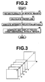
- the center line calculating section 2 calculates a center line that connects the approximate center points of the subject regions represented in the plurality of CT images obtained by the image obtaining section 1, by one of the methods described above (step #2). Then, the pseudo three dimensional medical image generating section 3 executes an intensity projection method radially outward from the center line calculated by the center line calculating section 2 toward the surface of the subject's body, to generate a planar exploded pseudo three dimensional medical image, in which thoracic bones of the subject are not displayed in an overlapping manner (step #3). Note that the RAYSUM method may be employed instead of the MIP method.
- the display section 6 may display the specific region between an inner circle C5 and an outer circle C4 within an image I5 illustrated in Figure 6 as the image M.
- the image M may display the specific region between an inner circle C5 and an outer circle C4 within an image I5 illustrated in Figure 6 as the image M.
Landscapes
- Engineering & Computer Science (AREA)
- Physics & Mathematics (AREA)
- General Physics & Mathematics (AREA)
- Theoretical Computer Science (AREA)
- Geometry (AREA)
- Computer Vision & Pattern Recognition (AREA)
- Computer Graphics (AREA)
- Apparatus For Radiation Diagnosis (AREA)
- Magnetic Resonance Imaging Apparatus (AREA)
Applications Claiming Priority (1)
| Application Number | Priority Date | Filing Date | Title |
|---|---|---|---|
| JP2009003241A JP2010158452A (ja) | 2009-01-09 | 2009-01-09 | 画像処理装置および方法並びにプログラム |
Publications (2)
| Publication Number | Publication Date |
|---|---|
| EP2216751A2 true EP2216751A2 (de) | 2010-08-11 |
| EP2216751A3 EP2216751A3 (de) | 2010-09-15 |
Family
ID=42235127
Family Applications (1)
| Application Number | Title | Priority Date | Filing Date |
|---|---|---|---|
| EP20100150254 Withdrawn EP2216751A3 (de) | 2009-01-09 | 2010-01-07 | Verhindern der Darstellung der Brustraumsknochen in 3D-Bildern |
Country Status (3)
| Country | Link |
|---|---|
| US (1) | US20100177945A1 (de) |
| EP (1) | EP2216751A3 (de) |
| JP (1) | JP2010158452A (de) |
Families Citing this family (13)
| Publication number | Priority date | Publication date | Assignee | Title |
|---|---|---|---|---|
| EP2353147B1 (de) * | 2008-11-28 | 2021-05-19 | Fujifilm Medical Systems U.S.A. Inc. | System und verfahren zur propagation des spine-labeling |
| JP6004875B2 (ja) * | 2012-10-02 | 2016-10-12 | キヤノン株式会社 | 医用画像表示装置、医用画像表示方法及びプログラム |
| JP6616682B2 (ja) * | 2015-12-25 | 2019-12-04 | ジーイー・メディカル・システムズ・グローバル・テクノロジー・カンパニー・エルエルシー | 中心線決定装置、医用装置、およびプログラム |
| JP6957337B2 (ja) * | 2017-12-18 | 2021-11-02 | キヤノン株式会社 | 画像処理装置、画像処理方法及びプログラム |
| CN109124662B (zh) * | 2018-07-13 | 2021-12-03 | 上海皓桦科技股份有限公司 | 肋骨中心线检测装置及方法 |
| US10813612B2 (en) | 2019-01-25 | 2020-10-27 | Cleerly, Inc. | Systems and method of characterizing high risk plaques |
| US20220392065A1 (en) | 2020-01-07 | 2022-12-08 | Cleerly, Inc. | Systems, methods, and devices for medical image analysis, diagnosis, risk stratification, decision making and/or disease tracking |
| US11969280B2 (en) | 2020-01-07 | 2024-04-30 | Cleerly, Inc. | Systems, methods, and devices for medical image analysis, diagnosis, risk stratification, decision making and/or disease tracking |
| JP2023509514A (ja) | 2020-01-07 | 2023-03-08 | クリールリー、 インコーポレーテッド | 医用画像分析、診断、重症度分類、意思決定、および/または疾患追跡のためのシステム、方法、およびデバイス |
| US12406365B2 (en) | 2022-03-10 | 2025-09-02 | Cleerly, Inc. | Systems, devices, and methods for non-invasive image-based plaque analysis and risk determination |
| US12440180B2 (en) | 2022-03-10 | 2025-10-14 | Cleerly, Inc. | Systems, devices, and methods for non-invasive image-based plaque analysis and risk determination |
| US20250143657A1 (en) | 2022-03-10 | 2025-05-08 | Cleerly, Inc. | Systems, devices, and methods for non-invasive image-based plaque analysis and risk determination |
| US20250217981A1 (en) | 2022-03-10 | 2025-07-03 | Cleerly, Inc. | Systems, methods, and devices for image-based plaque analysis and risk determination |
Family Cites Families (9)
| Publication number | Priority date | Publication date | Assignee | Title |
|---|---|---|---|---|
| JP4414078B2 (ja) * | 2000-09-12 | 2010-02-10 | ジーイー・メディカル・システムズ・グローバル・テクノロジー・カンパニー・エルエルシー | 画像表示装置 |
| JP4416736B2 (ja) * | 2003-08-01 | 2010-02-17 | 株式会社日立メディコ | 医用画像診断支援装置及びプログラム |
| US20050110791A1 (en) * | 2003-11-26 | 2005-05-26 | Prabhu Krishnamoorthy | Systems and methods for segmenting and displaying tubular vessels in volumetric imaging data |
| JP4018679B2 (ja) * | 2004-08-24 | 2007-12-05 | ザイオソフト株式会社 | レンダリング処理方法、レンダリング処理プログラム、レンダリング処理装置 |
| EP1859404B1 (de) * | 2005-02-11 | 2018-12-19 | Philips Intellectual Property & Standards GmbH | Indentifizierung von abnormalem gewebe auf computertomographiebildern |
| WO2007122896A1 (ja) * | 2006-03-29 | 2007-11-01 | Hitachi Medical Corporation | 医用画像表示システム及び医用画像表示プログラム |
| JP4922734B2 (ja) * | 2006-11-13 | 2012-04-25 | 株式会社Aze | 医用画像生成装置、方法およびプログラム |
| JP2008259682A (ja) * | 2007-04-12 | 2008-10-30 | Fujifilm Corp | 部位認識結果修正装置、方法、およびプログラム |
| JP4563421B2 (ja) * | 2007-05-28 | 2010-10-13 | ザイオソフト株式会社 | 画像処理方法及び画像処理プログラム |
-
2009
- 2009-01-09 JP JP2009003241A patent/JP2010158452A/ja not_active Abandoned
-
2010
- 2010-01-07 US US12/654,872 patent/US20100177945A1/en not_active Abandoned
- 2010-01-07 EP EP20100150254 patent/EP2216751A3/de not_active Withdrawn
Non-Patent Citations (1)
| Title |
|---|
| KOKUBUN, H.; MIYAZAKI ET AL.: "Radial intensity projection for lumen: application to CT angiographic imaging", MEDICAL IMAGING 2006: PHYSICS OF MEDICAL IMAGING, 2006, pages 6142R - 1,6142R-8 |
Also Published As
| Publication number | Publication date |
|---|---|
| EP2216751A3 (de) | 2010-09-15 |
| JP2010158452A (ja) | 2010-07-22 |
| US20100177945A1 (en) | 2010-07-15 |
Similar Documents
| Publication | Publication Date | Title |
|---|---|---|
| EP2216751A2 (de) | Verhindern der Darstellung der Brustraumsknochen in 3D-Bildern | |
| US10111713B2 (en) | Surgery assistance apparatus, surgery assistance method and non-transitory computer-readable recording medium having stored therein surgery assistance program | |
| EP2896369B1 (de) | Vorrichtung und verfahren zur anzeige dreidimensionaler bilder und programm | |
| JP4931027B2 (ja) | 医用画像診断支援装置および方法、並びにプログラム | |
| US20190021677A1 (en) | Methods and systems for classification and assessment using machine learning | |
| EP2490176B1 (de) | Operationsunterstützungsvorrichtung, verfahren und -programm | |
| JP5011426B2 (ja) | 画像診断支援装置、方法及びプログラム | |
| JP5559642B2 (ja) | 手術支援装置、手術支援方法および手術支援プログラム | |
| EP2196958A2 (de) | Bildverarbeitungsverfahren, Bildverarbeitungsvorrichtung und Bildverarbeitungsprogramm | |
| US8384735B2 (en) | Image display apparatus, image display control method, and computer readable medium having an image display control program recorded therein | |
| US10524823B2 (en) | Surgery assistance apparatus, method and program | |
| US9675317B2 (en) | Interface identification apparatus and method | |
| CA3105430A1 (en) | System and method for linking a segmentation graph to volumetric data | |
| US8907949B2 (en) | Image display apparatus, method and program | |
| EP2484286B1 (de) | Vorrichtung und verfahren zur anzeige medizinischer bilder und programm dafür | |
| EP2199976B1 (de) | Bildverarbeitungsverfahren, Bildverarbeitungsvorrichtung und Bildverarbeitungsprogramm | |
| US12315633B2 (en) | Medical image processing apparatus and medical image processing method |
Legal Events
| Date | Code | Title | Description |
|---|---|---|---|
| PUAI | Public reference made under article 153(3) epc to a published international application that has entered the european phase |
Free format text: ORIGINAL CODE: 0009012 |
|
| AK | Designated contracting states |
Kind code of ref document: A2 Designated state(s): AT BE BG CH CY CZ DE DK EE ES FI FR GB GR HR HU IE IS IT LI LT LU LV MC MK MT NL NO PL PT RO SE SI SK SM TR |
|
| AX | Request for extension of the european patent |
Extension state: AL BA RS |
|
| PUAL | Search report despatched |
Free format text: ORIGINAL CODE: 0009013 |
|
| AK | Designated contracting states |
Kind code of ref document: A3 Designated state(s): AT BE BG CH CY CZ DE DK EE ES FI FR GB GR HR HU IE IS IT LI LT LU LV MC MK MT NL NO PL PT RO SE SI SK SM TR |
|
| AX | Request for extension of the european patent |
Extension state: AL BA RS |
|
| RIC1 | Information provided on ipc code assigned before grant |
Ipc: G06T 7/00 20060101ALI20100806BHEP Ipc: G06T 15/00 20060101AFI20100706BHEP |
|
| 17P | Request for examination filed |
Effective date: 20110315 |
|
| RBV | Designated contracting states (corrected) |
Designated state(s): DE FR GB |
|
| 17Q | First examination report despatched |
Effective date: 20150327 |
|
| STAA | Information on the status of an ep patent application or granted ep patent |
Free format text: STATUS: THE APPLICATION IS DEEMED TO BE WITHDRAWN |
|
| 18D | Application deemed to be withdrawn |
Effective date: 20150807 |