EP2238902A1 - Système de surveillance portable monté sur l'oreille - Google Patents
Système de surveillance portable monté sur l'oreille Download PDFInfo
- Publication number
- EP2238902A1 EP2238902A1 EP10158007A EP10158007A EP2238902A1 EP 2238902 A1 EP2238902 A1 EP 2238902A1 EP 10158007 A EP10158007 A EP 10158007A EP 10158007 A EP10158007 A EP 10158007A EP 2238902 A1 EP2238902 A1 EP 2238902A1
- Authority
- EP
- European Patent Office
- Prior art keywords
- housing
- monitoring system
- sensor
- wearable monitoring
- signal
- Prior art date
- Legal status (The legal status is an assumption and is not a legal conclusion. Google has not performed a legal analysis and makes no representation as to the accuracy of the status listed.)
- Withdrawn
Links
Images

Classifications
-
- A—HUMAN NECESSITIES
- A61—MEDICAL OR VETERINARY SCIENCE; HYGIENE
- A61B—DIAGNOSIS; SURGERY; IDENTIFICATION
- A61B5/00—Measuring for diagnostic purposes; Identification of persons
- A61B5/08—Measuring devices for evaluating the respiratory organs
- A61B5/087—Measuring breath flow
- A61B5/0878—Measuring breath flow using temperature sensing means
-
- A—HUMAN NECESSITIES
- A61—MEDICAL OR VETERINARY SCIENCE; HYGIENE
- A61B—DIAGNOSIS; SURGERY; IDENTIFICATION
- A61B5/00—Measuring for diagnostic purposes; Identification of persons
- A61B5/02—Detecting, measuring or recording for evaluating the cardiovascular system, e.g. pulse, heart rate, blood pressure or blood flow
- A61B5/0205—Simultaneously evaluating both cardiovascular conditions and different types of body conditions, e.g. heart and respiratory condition
- A61B5/02055—Simultaneously evaluating both cardiovascular condition and temperature
-
- A—HUMAN NECESSITIES
- A61—MEDICAL OR VETERINARY SCIENCE; HYGIENE
- A61B—DIAGNOSIS; SURGERY; IDENTIFICATION
- A61B5/00—Measuring for diagnostic purposes; Identification of persons
- A61B5/48—Other medical applications
- A61B5/4806—Sleep evaluation
- A61B5/4818—Sleep apnoea
-
- A—HUMAN NECESSITIES
- A61—MEDICAL OR VETERINARY SCIENCE; HYGIENE
- A61B—DIAGNOSIS; SURGERY; IDENTIFICATION
- A61B5/00—Measuring for diagnostic purposes; Identification of persons
- A61B5/68—Arrangements of detecting, measuring or recording means, e.g. sensors, in relation to patient
- A61B5/6801—Arrangements of detecting, measuring or recording means, e.g. sensors, in relation to patient specially adapted to be attached to or worn on the body surface
- A61B5/6813—Specially adapted to be attached to a specific body part
- A61B5/6814—Head
- A61B5/6815—Ear
-
- A—HUMAN NECESSITIES
- A61—MEDICAL OR VETERINARY SCIENCE; HYGIENE
- A61B—DIAGNOSIS; SURGERY; IDENTIFICATION
- A61B5/00—Measuring for diagnostic purposes; Identification of persons
- A61B5/68—Arrangements of detecting, measuring or recording means, e.g. sensors, in relation to patient
- A61B5/6801—Arrangements of detecting, measuring or recording means, e.g. sensors, in relation to patient specially adapted to be attached to or worn on the body surface
- A61B5/6813—Specially adapted to be attached to a specific body part
- A61B5/6814—Head
- A61B5/6815—Ear
- A61B5/6816—Ear lobe
-
- A—HUMAN NECESSITIES
- A61—MEDICAL OR VETERINARY SCIENCE; HYGIENE
- A61B—DIAGNOSIS; SURGERY; IDENTIFICATION
- A61B5/00—Measuring for diagnostic purposes; Identification of persons
- A61B5/68—Arrangements of detecting, measuring or recording means, e.g. sensors, in relation to patient
- A61B5/6801—Arrangements of detecting, measuring or recording means, e.g. sensors, in relation to patient specially adapted to be attached to or worn on the body surface
- A61B5/6813—Specially adapted to be attached to a specific body part
- A61B5/6814—Head
- A61B5/6815—Ear
- A61B5/6817—Ear canal
-
- A—HUMAN NECESSITIES
- A61—MEDICAL OR VETERINARY SCIENCE; HYGIENE
- A61B—DIAGNOSIS; SURGERY; IDENTIFICATION
- A61B5/00—Measuring for diagnostic purposes; Identification of persons
- A61B5/68—Arrangements of detecting, measuring or recording means, e.g. sensors, in relation to patient
- A61B5/6801—Arrangements of detecting, measuring or recording means, e.g. sensors, in relation to patient specially adapted to be attached to or worn on the body surface
- A61B5/683—Means for maintaining contact with the body
- A61B5/6838—Clamps or clips
-
- A—HUMAN NECESSITIES
- A61—MEDICAL OR VETERINARY SCIENCE; HYGIENE
- A61B—DIAGNOSIS; SURGERY; IDENTIFICATION
- A61B5/00—Measuring for diagnostic purposes; Identification of persons
- A61B5/145—Measuring characteristics of blood in vivo, e.g. gas concentration or pH-value ; Measuring characteristics of body fluids or tissues, e.g. interstitial fluid or cerebral tissue
- A61B5/1455—Measuring characteristics of blood in vivo, e.g. gas concentration or pH-value ; Measuring characteristics of body fluids or tissues, e.g. interstitial fluid or cerebral tissue using optical sensors, e.g. spectral photometrical oximeters
Definitions
- This disclosure relates generally to a wearable monitoring system to collect medical information from a subject.
- apnea there is no movement of muscles of a respiration and a volume of lungs initially remains unchanged. Depending on an openness of airways there may not be a flow of a breathing gas between the lungs and an ambient during apnea.
- Apnea can be drug-induced (e.g., opiate toxicity), mechanically induced (e.g., strangulation or choking), or it can occur as a consequence of neurological disease or trauma as was explained above.
- apnea caused by opiates or other medicine.
- Patients may also have obstructive apnea, which is caused by a blockage in the airways, such as a thong or similar physical obstruction.
- Patients usually don't have any monitoring connected on as it is rather expensive and there are usually many patients in the ward.
- sleep apnea that can be divided into three distinct forms: central, obstructive, and a complex sleep apnea, which is a combination of central and obstructive apnea.
- central sleep apnea breathing is interrupted by the lack of respiratory effort.
- obstructive sleep apnea breathing is interrupted by a physical block to the airflow despite the respiratory effort.
- complex sleep apnea there is a transition from central to obstructive features during the events themselves.
- Central apnea is commonly caused by neurological etc. characteristics, whereas obstructive apnea is caused by for example over weight.
- SIDS sudden infant death syndrome
- Pulse oximetry is a non-invasive method allowing the monitoring of the oxygenation of a patient's hemoglobin.
- a sensor is placed on a thin part of the patient's anatomy , usually a fingertip or earlobe , or in the case of a neonate , across a foot, and a light containing both red and infrared wavelengths is passed from one side to the other. Changing absorbance of each of the two wavelengths is measured, allowing determination of the absorbances due to the pulsing arteria blood alone, excluding venous blood, skin, bone, muscle and fat.
- Pulse oximetry data is necessary whenever a patient's oxygenation is unstable, including intensive care, critical care, and emergency department areas of a hospital. Data can also be obtained for the assessment of any patient's oxygenation in primary care.
- pulse oximetry is used to monitor oxygenation, it cannot determine the metabolism of oxygen, or the amount of oxygen being used by a patient.
- the use of pulse oximetry to detect hypoventilation is impaired with the use of supplemental oxygen, as it is only when patients breathe room air that abnormalities in respiratory function can be detected reliably with its use. Therefore, the routine administration of supplemental oxygen may be unwarranted if the patient is able to maintain adequate oxygenation in room air, since it can result in hypoventilation going undetected.
- devices such as impedance measurement, resistive belts, or piezo-resistive belts etc., do not measure the actual flow of the breathing gas through the nose or the mouth, but they measure for example the respiratory muscle movement or chest movement. These indirect measurements are unreliable since the respiratory muscle or chest movement may occur even when the patient is suffering from apnea. Devices are also sensitive to motion artefacts and other disturbances. Existing devices usually contain cables that connect the measuring devices to a host, which is used to calculate and show respiration and pulse oximetry waveforms and values. Usually the host also delivers electrical power for the devices.
- Disposability is also one of the clinical requirements for the device that is in close contact to patient, especially the patient's airways, since existing cleaning practices are not reliable enough to ensure high enough level of purity for reusable devices.
- Contaminated reusable devices easily cause a risk of cross contamination between different patients, but non-disposable devices also cause a personal hygiene risk, if they are not cleaned frequently enough, for the patients who already have a lowered level of immunity against bacteria and viruses.
- Existing devices are usually complex, also containing cables etc., rather expensive, not easy to clean and thus reusable usually.
- a wearable monitoring system to collect medical information from a subject includes a housing having electronics for managing a signal and which at least one housing is operatively connectable to at least one sensor for transmitting to the housing the signal acquired by the at least one sensor.
- the electronics of the housing is for managing the signal which is indicative of a condition of a respiration function and which housing is installable to the subject's ear or its vicinity.
- a wearable monitoring system to collect medical information from a subject includes a housing having electronics for managing a signal and a cable operatively connectable to at least one sensor for transmitting to the housing the signal acquired by the at least one sensor.
- the electronics of the housing is for managing the signal acquired along the cable from the at least one sensor and which signal is indicative of a condition of a respiration function and which housing is installable behind an ear of the subject.
- a wearable monitoring system to collect medical information from a subject includes at least one sensor for acquiring a signal indicative of a condition of a respiration function and a cable for transmitting the signal acquired by the at least one sensor.
- the wearable monitoring system to collect medical information from a subject also includes a housing having electronics for managing the signal received along the cable from the at least one sensor and which housing is installable behind an ear of the subject.
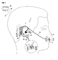
- Fig. 1 shows a schematic side view of a wearable monitoring system 1 that may be wireless and can be used to collect a medical information from a subject.
- the monitoring system 1 can be placed on a side of a subject's head, over an ear 2, extending towards and close to a mouth 3 and a nose 4.
- the monitoring system 1 comprises a housing 5, which is installable to the subject's ear 2 or its vicinity. A suitable place for the housing is behind the subject's ear 2, similarly as commonly known hearing aid devices are placed.
- the housing 5 comprises electonics 6 for managing a signal acquired by at least one sensor 10, 20.
- the electronics 6 may comprise a radio frequency transceiver or similar wireless communication and signal processing.
- the housing 5 may further comprise a power supply 7, such as a small battery to energize the monitoring system 1.
- a power supply 7 such as a small battery to energize the monitoring system 1.
- the housing 5 is operatively connectable to the at least one sensor 10, 20, which may be disposable or reusable, for transmitting the signal from the at least one sensor 10, 20 to the housing 5.
- the signal acquired by the at least one sensor 10, 20 is indicative of a condition of a respiration function.
- the sensor 10, which is placed between the subject's mouth and nose, is preferably disposable and used for acquiring from the respiration flow through the mouth and/or the nose a signal indicative of at least one of a respiratory flow, respiration rate, one or more gas component and a concentration of a gas component.
- One of the sensors operatively connectable to the housing 5 can be an ear sensor 20 placed for example on the subject's ear lope 21 or a place such as an auditory canal, which is used for measuring at least one of the oxygen saturation and the pulse rate from the ear.
- the sensor 10, for acquiring the respiratory flow signal and/or respiration rate signal is substantially insensitive to any motion or mechanical vibrations and can be used to acquire the signal through the nose, the mouth or the both at the same time and can also detect apnea.
- the measurement can be based on measuring a thermal component of the respiration gas flow with at least one respiration detector 11 such as a thermistor. Thermal changes of the respiration gas flowing by the respiration detector 11 change the thermistor's resistance, which is converted into a continuous electrical signal.
- the amplitude of the signal is proportional to the flow rate of the respiration gas and the frequency is proportional to the respiration rate (RR).
- RR respiration rate
- These signals can also be used for detecting apnea.
- the respiration detector 11 can acquire a signal indicative of one or more gas component or their concentration in the respiration gas flow.
- the sensor 10 for acquiring the respiratory flow, respiration rate, one or more gas component or gas concentration signal can be placed on a skin below a nose but above the subject's upper lip or mouth, where it is glued on with a sticker 12 located on the bottom of the sensor 10.
- the sensor 10 may connect to the housing 5 of the monitoring system 1 through a cable 13, which is preferably deformable and retain a shape.
- the cable 13 can have rigid, but flexible mechanical properties, so that it can be shaped by forcing or bending it to fit different type of head geometries of different subjects. It is advantageous if the cable 13 also retain the reshaped shape in the normal use, such as the subject sleeps and the subject's head presses the ear handle, which remains between the head and the base on which the subject lies on. Furthermore, the cable 13 could also imitate the head or bend to make it unnoticeable for the subject.
- the cable 13 can also be a relatively thin flat or round band, preferably comprising plastic or similar non-irritating, clinically approved material.
- the cable 13 also comprising electrical wires preferably inside the band that are used to transfer electrical signals between the sensor 10 and the housing 5 of the monitoring system 1.
- the cable 13 comprises a connector 14, preferably a male type connector at the sensor end of the band (not shown in figures) that mechanically locks-on to the corresponding counterpart connector 15 in the sensor 10, which is preferably a female type connector.
- Connectors 14 and 15 also connect electrically the sensor 10 through the electrical contacts and wires into the housing 5 of the monitoring system 1.
- the sensor 10 preferably disposable but it may also be reusable, is easy to unlock from the cable 13 with one press on the latch pin (not shown in Figures), which may be similar to for example standard Ethernet connector latch pin.
- the electrical operating power of the power supply 7 and possible control signals for the sensor 10 are transferred from the housing 5, whereas electrical signals indicative of the condition of the respiratory function are transferred from the sensor 10 to the housing 5.
- the other end of the cable13 connects in Figure 1 embodiment tightly to the housing 5 of monitoring system 1, but naturally can also be detachable.
- the housing 5 and the cable 13 are preferably reusable, thus they are designed and made of materials that are easy to clean.
- the housing 5 may also be operatively connectable to another sensor 20 for acquiring a signal indicative of a condition of the respiratory function such as an absorbance of a blood which is indicative for example of an oxygen saturation and/or pulse rate.
- a cable 22 connecting the housing and the sensor 20.
- the housing is equipped with a connector 16, which is preferably the same female type connector as the connector 15 in the sensor 10, or similar to that.
- An end of the cable 22 is equipped with a connector 17, which is preferably the same male type connector as the connector 14 in the cable 13, or similar.
- the connector 17 connects to its counterpart connector 16, both mechanically and electrically, similarly as the cable 13 connects to the sensor 10.
- the senor 20 which may be disposable or reusable, is easy to unlock from the housing 5 with one press on the latch pin (not shown in Figures), which may be similar to for example standard Ethernet connector latch pin.
- the electrical operating power and possible control signals for the sensor 20 are transferred from the housing 5, whereas the acquired electrical signals from the sensor 20 are transferred to the housing 5 for further processing.
- the sensor 20 shown in Figure 1 which is a preferably a disposable pulse oximeter sensor placed on the ear lope, allows a non-invasive method of monitoring the oxygenation of the subject's hemoglobin and can be used to acquire a signal indicative of oxygen saturation in blood and pulse rate.
- the sensor 20 is placed on a thin part of the subject's anatomy, usually a fingertip or the ear lope, or in the case of a neonate, across a foot.
- the sensor 20 comprises an emitting unit 25 to emit a radiation through a tissue of the subject and a detector unit 26 to receive the radiation penetrated through a tissue and emitted by the emitting unit 25.
- the detector unit 26 creates a signal indicative of an absorbance of the blood and which signal is provided to the housing 5 along the cable 22.
- the light emitting unit 25 is able to emit at least two different wavelengths of the radiation. Typically the emitting unit 25 comprises one radiation emitter 27 for a red wavelength and one radiation emitter 28 for an infrared wavelength.
- the detector unit 26 may comprise one or more detectors 29, 30 to receive the radiation emitted by the radiation emitters 27, 28. Changing absorbance of each of the two wavelengths is measured, allowing determination of the absorbances due to the pulsing arteria blood alone, excluding venous blood, skin, bone, muscle, fat.
- the monitoring system 1 may also comprise a third sensor 40, which is indicative of a condition other than the respiration function and preferably a disposable temperature measurement used to measure the body temperature of the subject.
- the housing 5 comprises a connector 41, which is preferably a similar female type connector as the connector 15 and 16.
- the sensor 40 is connected by means of a cable 42 and its connector 43 at the end of this cable 42, which is preferably a similar male type connector as the connectors 14 and 17.
- the connector 43 connects to its counterpart connector 41, both mechanically and electrically.
- the sensor 40 is easy to unlock from the housing 5 with one press on the latch pin (not shown in Figures), which may be similar to for example standard Ethernet connector latch pin.
- the electrical operating power and possible control signals for the sensor 40 are transferred from the housing 5, whereas the acquired electrical signals from the sensor 40 are transferred to the housing 5 for further processing.
- the housing 5 is able to communicate with a host unit 50. All the processed or unprocessed data acquired from the sensors 10, 20 and 40 is sent to the host 50 preferably through a communication link 51, which is preferably wireless.
- the host unit can further process the signal managed by the electronics 6 of the housing 5 and also display the processed signal by means of a display unit 52.
- Figure 2 is mainly identical with Figure 1 , but it further shows an ear handle 60 coupling the housing 5 to the subject's ear 2 .
- the embodiment shown in Figure 1 is coupled to the ear by means of the cable 13 or a separate sticker (not shown in Figure 1 ), but in some cases an improved coupling may be necessary.
- the ear handle 60 shown in Figure 2 may be coupled to the cable 13 or it may be coupled to the housing 5 so that it alone or together with the cable 13 encircles at least partly the ear 2.
- the ear handle 60 may comprise an auditory canal element 61 insertable into the auditory canal for better fastening the housing 5.
- the auditory canal element 61 can be an hearing aid insertable into the auditory canal.
- the auditory canal element 61 can be a sensor for measuring the signal indicative of the condition of the respiratory function such as oxygen saturation and pulse rate replacing the subject's ear lope 21 as a place for sensor 20.
Landscapes
- Health & Medical Sciences (AREA)
- Life Sciences & Earth Sciences (AREA)
- Medical Informatics (AREA)
- Molecular Biology (AREA)
- Veterinary Medicine (AREA)
- Biophysics (AREA)
- Pathology (AREA)
- Engineering & Computer Science (AREA)
- Biomedical Technology (AREA)
- Heart & Thoracic Surgery (AREA)
- Public Health (AREA)
- Physics & Mathematics (AREA)
- Surgery (AREA)
- Animal Behavior & Ethology (AREA)
- General Health & Medical Sciences (AREA)
- Otolaryngology (AREA)
- Physiology (AREA)
- Cardiology (AREA)
- Pulmonology (AREA)
- Measurement Of The Respiration, Hearing Ability, Form, And Blood Characteristics Of Living Organisms (AREA)
Applications Claiming Priority (2)
| Application Number | Priority Date | Filing Date | Title |
|---|---|---|---|
| US16637409P | 2009-04-03 | 2009-04-03 | |
| US12/474,690 US20100256460A1 (en) | 2009-04-03 | 2009-05-29 | Wearable Monitoring System |
Publications (1)
| Publication Number | Publication Date |
|---|---|
| EP2238902A1 true EP2238902A1 (fr) | 2010-10-13 |
Family
ID=42244584
Family Applications (1)
| Application Number | Title | Priority Date | Filing Date |
|---|---|---|---|
| EP10158007A Withdrawn EP2238902A1 (fr) | 2009-04-03 | 2010-03-26 | Système de surveillance portable monté sur l'oreille |
Country Status (2)
| Country | Link |
|---|---|
| US (1) | US20100256460A1 (fr) |
| EP (1) | EP2238902A1 (fr) |
Cited By (4)
| Publication number | Priority date | Publication date | Assignee | Title |
|---|---|---|---|---|
| CN108024760A (zh) * | 2015-09-14 | 2018-05-11 | 保健之源股份有限公司 | 可穿戴式呼吸疾病监测设备 |
| US10335083B2 (en) | 2015-07-21 | 2019-07-02 | Courtland Keith Keteyian | Systems and methods for detecting and analyzing biosignals |
| US10955269B2 (en) | 2016-05-20 | 2021-03-23 | Health Care Originals, Inc. | Wearable apparatus |
| US11622716B2 (en) | 2017-02-13 | 2023-04-11 | Health Care Originals, Inc. | Wearable physiological monitoring systems and methods |
Families Citing this family (13)
| Publication number | Priority date | Publication date | Assignee | Title |
|---|---|---|---|---|
| US20120316932A1 (en) * | 2011-06-10 | 2012-12-13 | Aliphcom | Wellness application for data-capable band |
| US9069380B2 (en) | 2011-06-10 | 2015-06-30 | Aliphcom | Media device, application, and content management using sensory input |
| US9591395B2 (en) | 2012-05-11 | 2017-03-07 | Harman International Industries, Incorporated | Earphones and earbuds with physiologic sensors |
| US9078577B2 (en) | 2012-12-06 | 2015-07-14 | Massachusetts Institute Of Technology | Circuit for heartbeat detection and beat timing extraction |
| JP6189705B2 (ja) | 2013-10-10 | 2017-08-30 | 日本光電工業株式会社 | センサ、および形状保持具 |
| US20150150498A1 (en) * | 2013-12-02 | 2015-06-04 | United Sciences, Llc | Sleep study |
| FR3028742B1 (fr) * | 2014-11-24 | 2016-12-30 | Inst Nat Sante Rech Med | Dispositif de stimulation vibrotactile |
| FR3028745B1 (fr) | 2014-11-24 | 2021-07-23 | Inst Nat Sante Rech Med | Dispositif de stimulation vibrotactile |
| GB2551768A (en) * | 2016-06-30 | 2018-01-03 | Gen Electric | Method and apparatus for recording respiratory rate |
| AU2017357747A1 (en) | 2016-11-10 | 2019-05-30 | The Research Foundation For The State University Of New York | System, method and biomarkers for airway obstruction |
| US9905105B1 (en) | 2016-12-01 | 2018-02-27 | General Electric Company | Method of increasing sensing device noticeability upon low battery level |
| CN114018320A (zh) * | 2021-10-25 | 2022-02-08 | 复旦大学 | 一种微光纤可穿戴的呼吸信息监测仪 |
| CN114923598A (zh) * | 2022-05-27 | 2022-08-19 | 杏翔医疗科技(杭州)有限公司 | 一种基于抛弃型测温探头的可穿戴体温计 |
Citations (8)
| Publication number | Priority date | Publication date | Assignee | Title |
|---|---|---|---|---|
| US5195529A (en) * | 1989-10-27 | 1993-03-23 | Instrumentarium Corporation | Sensor for monitoring respiration |
| WO2005065540A1 (fr) * | 2003-12-30 | 2005-07-21 | University Of Florida Research Foundation, Inc. | Nouveau sphygmo-oxymetre nasal special |
| WO2006123774A1 (fr) * | 2005-05-19 | 2006-11-23 | Matsushita Electric Works, Ltd. | Ensemble de capteur pour l’etude du syndrome des apnees du sommeil et instrument pour l’etude du syndrome des apnees du sommeil |
| US20070197881A1 (en) * | 2006-02-22 | 2007-08-23 | Wolf James L | Wireless Health Monitor Device and System with Cognition |
| WO2007100958A1 (fr) * | 2006-02-28 | 2007-09-07 | Koninklijke Philips Electronics, N.V. | Périphérique surveillant en continu le syndrome d'apnée obstructive du sommeil |
| US20080139955A1 (en) * | 2006-12-07 | 2008-06-12 | Drager Medical Ag & Co. Kg | Device and method for determining a respiration rate |
| US20080146892A1 (en) * | 2006-12-19 | 2008-06-19 | Valencell, Inc. | Physiological and environmental monitoring systems and methods |
| WO2008116318A1 (fr) * | 2007-03-28 | 2008-10-02 | Tr Technologies Inc. | Analyse de bruit respiratoire pour l'estimation d'un débit d'air |
Family Cites Families (2)
| Publication number | Priority date | Publication date | Assignee | Title |
|---|---|---|---|---|
| US20010044588A1 (en) * | 1996-02-22 | 2001-11-22 | Mault James R. | Monitoring system |
| DE102006029958A1 (de) * | 2006-06-29 | 2008-01-03 | Siemens Audiologische Technik Gmbh | Modulares Hinter-dem-Ohr-Hörgerät |
-
2009
- 2009-05-29 US US12/474,690 patent/US20100256460A1/en not_active Abandoned
-
2010
- 2010-03-26 EP EP10158007A patent/EP2238902A1/fr not_active Withdrawn
Patent Citations (9)
| Publication number | Priority date | Publication date | Assignee | Title |
|---|---|---|---|---|
| US5195529A (en) * | 1989-10-27 | 1993-03-23 | Instrumentarium Corporation | Sensor for monitoring respiration |
| WO2005065540A1 (fr) * | 2003-12-30 | 2005-07-21 | University Of Florida Research Foundation, Inc. | Nouveau sphygmo-oxymetre nasal special |
| WO2006123774A1 (fr) * | 2005-05-19 | 2006-11-23 | Matsushita Electric Works, Ltd. | Ensemble de capteur pour l’etude du syndrome des apnees du sommeil et instrument pour l’etude du syndrome des apnees du sommeil |
| US20090203970A1 (en) * | 2005-05-19 | 2009-08-13 | Matsushita Electric Works, Ltd. | Sleep apnea test sensor assembly and sleep apnea test equipment using the same |
| US20070197881A1 (en) * | 2006-02-22 | 2007-08-23 | Wolf James L | Wireless Health Monitor Device and System with Cognition |
| WO2007100958A1 (fr) * | 2006-02-28 | 2007-09-07 | Koninklijke Philips Electronics, N.V. | Périphérique surveillant en continu le syndrome d'apnée obstructive du sommeil |
| US20080139955A1 (en) * | 2006-12-07 | 2008-06-12 | Drager Medical Ag & Co. Kg | Device and method for determining a respiration rate |
| US20080146892A1 (en) * | 2006-12-19 | 2008-06-19 | Valencell, Inc. | Physiological and environmental monitoring systems and methods |
| WO2008116318A1 (fr) * | 2007-03-28 | 2008-10-02 | Tr Technologies Inc. | Analyse de bruit respiratoire pour l'estimation d'un débit d'air |
Cited By (6)
| Publication number | Priority date | Publication date | Assignee | Title |
|---|---|---|---|---|
| US10335083B2 (en) | 2015-07-21 | 2019-07-02 | Courtland Keith Keteyian | Systems and methods for detecting and analyzing biosignals |
| CN108024760A (zh) * | 2015-09-14 | 2018-05-11 | 保健之源股份有限公司 | 可穿戴式呼吸疾病监测设备 |
| US11272864B2 (en) | 2015-09-14 | 2022-03-15 | Health Care Originals, Inc. | Respiratory disease monitoring wearable apparatus |
| CN108024760B (zh) * | 2015-09-14 | 2022-06-14 | 保健之源股份有限公司 | 可穿戴式呼吸疾病监测设备 |
| US10955269B2 (en) | 2016-05-20 | 2021-03-23 | Health Care Originals, Inc. | Wearable apparatus |
| US11622716B2 (en) | 2017-02-13 | 2023-04-11 | Health Care Originals, Inc. | Wearable physiological monitoring systems and methods |
Also Published As
| Publication number | Publication date |
|---|---|
| US20100256460A1 (en) | 2010-10-07 |
Similar Documents
| Publication | Publication Date | Title |
|---|---|---|
| US20100256460A1 (en) | Wearable Monitoring System | |
| EP1404221B1 (fr) | Equipement de surveillance de la respiration | |
| JP4751338B2 (ja) | 新規で特殊構成の鼻用パルスオキシメータ | |
| US8413502B2 (en) | Device for measuring infant feeding performance | |
| US7603152B2 (en) | Physiological stress detector device and system | |
| EP2285269B1 (fr) | Capnographie sans fil | |
| EP2236080B1 (fr) | Capteur | |
| EP3250117B1 (fr) | Sonde à insertion buccale et procédé de mesure de paramètres vitaux | |
| CN105828706B (zh) | 抑制或防止误警报的装置和方法 | |
| CN117355251A (zh) | 婴幼儿生理状态确定设备 | |
| EP2361552B1 (fr) | Agencement pour la fixation d'un capteur sur la figure d'un sujet | |
| JP2009136423A (ja) | 睡眠時無呼吸症候群の検査器具 | |
| US20250082884A1 (en) | Systems and methods for monitoring the respiratory status of a user | |
| FI98266C (fi) | Henkilökohtainen elintoimintojen seurantalaite | |
| KR100866545B1 (ko) | 비접촉 방식의 착용형 무선 생체신호 측정 시스템 | |
| CN114903448A (zh) | 一种睡眠呼吸监测装置及睡眠呼吸监测方法 | |
| US20210023317A1 (en) | System for monitoring tracheostomy airflow | |
| WO2006015230A2 (fr) | Catheter oesophagien ou endotracheal intelligent non intrusif, non invasif a mosaique de capteurs | |
| US20240268683A1 (en) | System and method for monitoring infant feeding | |
| CN118892308B (zh) | 一种采用多传感器融合技术的新生儿肺脏实时监测系统 | |
| KR102688458B1 (ko) | 무호흡 상태의 미숙아를 깨우는 기능이 포함된 인큐베이터 | |
| KR102668657B1 (ko) | 미숙아의 자세변경 기능이 포함된 인큐베이터 | |
| CN209826693U (zh) | 一种睡眠中呼吸和心跳的监测装置 | |
| CN121754775A (zh) | 具有监测功能的鼻导管装置 |
Legal Events
| Date | Code | Title | Description |
|---|---|---|---|
| PUAI | Public reference made under article 153(3) epc to a published international application that has entered the european phase |
Free format text: ORIGINAL CODE: 0009012 |
|
| AK | Designated contracting states |
Kind code of ref document: A1 Designated state(s): AT BE BG CH CY CZ DE DK EE ES FI FR GB GR HR HU IE IS IT LI LT LU LV MC MK MT NL NO PL PT RO SE SI SK SM TR |
|
| AX | Request for extension of the european patent |
Extension state: AL BA ME RS |
|
| 17P | Request for examination filed |
Effective date: 20110413 |
|
| 17Q | First examination report despatched |
Effective date: 20120531 |
|
| STAA | Information on the status of an ep patent application or granted ep patent |
Free format text: STATUS: EXAMINATION IS IN PROGRESS |
|
| STAA | Information on the status of an ep patent application or granted ep patent |
Free format text: STATUS: THE APPLICATION IS DEEMED TO BE WITHDRAWN |
|
| 18D | Application deemed to be withdrawn |
Effective date: 20211001 |