EP2243425A2 - Verfahren zur nicht invasiven Messung von Glukose und Vorrichtung zur nicht invasiven Messung von Glukose - Google Patents
Verfahren zur nicht invasiven Messung von Glukose und Vorrichtung zur nicht invasiven Messung von Glukose Download PDFInfo
- Publication number
- EP2243425A2 EP2243425A2 EP10170622A EP10170622A EP2243425A2 EP 2243425 A2 EP2243425 A2 EP 2243425A2 EP 10170622 A EP10170622 A EP 10170622A EP 10170622 A EP10170622 A EP 10170622A EP 2243425 A2 EP2243425 A2 EP 2243425A2
- Authority
- EP
- European Patent Office
- Prior art keywords
- probe
- skin
- measurement
- glucose
- change
- Prior art date
- Legal status (The legal status is an assumption and is not a legal conclusion. Google has not performed a legal analysis and makes no representation as to the accuracy of the status listed.)
- Granted
Links
Images


Classifications
-
- A—HUMAN NECESSITIES
- A61—MEDICAL OR VETERINARY SCIENCE; HYGIENE
- A61B—DIAGNOSIS; SURGERY; IDENTIFICATION
- A61B5/00—Measuring for diagnostic purposes; Identification of persons
- A61B5/145—Measuring characteristics of blood in vivo, e.g. gas concentration or pH-value ; Measuring characteristics of body fluids or tissues, e.g. interstitial fluid or cerebral tissue
- A61B5/1455—Measuring characteristics of blood in vivo, e.g. gas concentration or pH-value ; Measuring characteristics of body fluids or tissues, e.g. interstitial fluid or cerebral tissue using optical sensors, e.g. spectral photometrical oximeters
-
- A—HUMAN NECESSITIES
- A61—MEDICAL OR VETERINARY SCIENCE; HYGIENE
- A61B—DIAGNOSIS; SURGERY; IDENTIFICATION
- A61B5/00—Measuring for diagnostic purposes; Identification of persons
- A61B5/0048—Detecting, measuring or recording by applying mechanical forces or stimuli
- A61B5/0053—Detecting, measuring or recording by applying mechanical forces or stimuli by applying pressure, e.g. compression, indentation, palpation, grasping, gauging
-
- A—HUMAN NECESSITIES
- A61—MEDICAL OR VETERINARY SCIENCE; HYGIENE
- A61B—DIAGNOSIS; SURGERY; IDENTIFICATION
- A61B5/00—Measuring for diagnostic purposes; Identification of persons
- A61B5/145—Measuring characteristics of blood in vivo, e.g. gas concentration or pH-value ; Measuring characteristics of body fluids or tissues, e.g. interstitial fluid or cerebral tissue
- A61B5/14532—Measuring characteristics of blood in vivo, e.g. gas concentration or pH-value ; Measuring characteristics of body fluids or tissues, e.g. interstitial fluid or cerebral tissue for measuring glucose, e.g. by tissue impedance measurement
-
- A—HUMAN NECESSITIES
- A61—MEDICAL OR VETERINARY SCIENCE; HYGIENE
- A61B—DIAGNOSIS; SURGERY; IDENTIFICATION
- A61B5/00—Measuring for diagnostic purposes; Identification of persons
- A61B5/145—Measuring characteristics of blood in vivo, e.g. gas concentration or pH-value ; Measuring characteristics of body fluids or tissues, e.g. interstitial fluid or cerebral tissue
- A61B5/1491—Heated applicators
-
- A—HUMAN NECESSITIES
- A61—MEDICAL OR VETERINARY SCIENCE; HYGIENE
- A61B—DIAGNOSIS; SURGERY; IDENTIFICATION
- A61B5/00—Measuring for diagnostic purposes; Identification of persons
- A61B5/145—Measuring characteristics of blood in vivo, e.g. gas concentration or pH-value ; Measuring characteristics of body fluids or tissues, e.g. interstitial fluid or cerebral tissue
- A61B5/1495—Calibrating or testing of in-vivo probes
-
- A—HUMAN NECESSITIES
- A61—MEDICAL OR VETERINARY SCIENCE; HYGIENE
- A61B—DIAGNOSIS; SURGERY; IDENTIFICATION
- A61B5/00—Measuring for diagnostic purposes; Identification of persons
- A61B5/68—Arrangements of detecting, measuring or recording means, e.g. sensors, in relation to patient
- A61B5/6801—Arrangements of detecting, measuring or recording means, e.g. sensors, in relation to patient specially adapted to be attached to or worn on the body surface
- A61B5/6843—Monitoring or controlling sensor contact pressure
-
- A—HUMAN NECESSITIES
- A61—MEDICAL OR VETERINARY SCIENCE; HYGIENE
- A61B—DIAGNOSIS; SURGERY; IDENTIFICATION
- A61B5/00—Measuring for diagnostic purposes; Identification of persons
- A61B5/68—Arrangements of detecting, measuring or recording means, e.g. sensors, in relation to patient
- A61B5/6801—Arrangements of detecting, measuring or recording means, e.g. sensors, in relation to patient specially adapted to be attached to or worn on the body surface
- A61B5/6813—Specially adapted to be attached to a specific body part
- A61B5/6824—Arm or wrist
Definitions
- the present invention relates to an apparatus and a method for noninvasive measurement, with which glucose in a subject is noninvasively measured optically through a measurement probe.
- noninvasive (NI) measurement of glucose concentration in a subject is described.
- This measurement method generally includes the steps of: bringing an optical probe into contact with a body part; performing a series of optical measurements; and collecting a series of light signals. Subsequently, these light signals or derived optical parameters are mutually associated with blood glucose concentrations for the establishing a calibration relationship. A glucose concentration is determined by a subsequent measurement using the light signals measured at that time and the previously established calibration relationship.
- the method for noninvasive (NI) measurement of glucose is classified into two broad categories: one is a method of tracking molecular properties; and the other is a method of tracking the effect of glucose on tissue properties.
- the method in the first category includes tracking intrinsic properties of glucose such as near-infrared (NIR) absorption coefficients, mid-infrared absorption coefficients, optical rotations, Raman shift band and NIR photoacoustic absorption.
- NIR near-infrared
- Such a method is based on an ability to detect glucose in a tissue or blood independently of other analytes of the body and also physiological conditions of the body.
- the method of the second type is based on the measurement of the effect of glucose on optical properties of tissue such as scattering coefficients of tissue, refractive index of interstitial fluid (ISF) or sound propagation in tissue.
- ISF interstitial fluid
- the method of the first type of tracking the molecular properties of glucose faces a big problem because a signal which can be considered to be specific for glucose is extremely weak. Biological noise, a person-to-person difference, and measurement noise may drown out a small change in signal specific for glucose. In order to extract glucose-related information from a data set with noise, a multivariate analysis has been commonly used.
- the method of the second type of tracking the effect of glucose on tissue properties instead of the intrinsic molecular properties of glucose faces a big problem because of a nonspecific property of the change in parameters to be measured.
- the method of both types of tracking the molecular properties of glucose and the effect of glucose on tissue properties ignores the physiological response of the body to a change in glucose concentration. This response can be seen in the form of a change in blood flow or temperature. Such a change in blood flow or temperature as a result of the physiological response of the body affects NIR light signals.
- the measurement by the method of both types also ignores the effect of the body-probe interaction on signals measured, and further, a specific time window for data collection from the initiation of the body-probe interaction is not defined.
- Patent documents 1, 2, 3, 4, 5, 6, 7 and 8 describe a method in which glucose is measured by bringing an optical probe into contact with a body part and also reflection or transmission signals in near-infrared (NIR) region ranging from 600 to 1100 nm are measured.
- NIR near-infrared
- a blood-containing body part such as a finger
- one or more detectors detect the light passing through the body part.
- a glucose level is derived from the comparison of the reference spectrum of glucose and the background interference.
- Patent documents 12 to 19 disclose a method for NI measurement of glucose using NIR reflectance and transmittance measurement at a wavelength ranging from 1000 to 2000 nm. These patents do not deal with the physiological response of the body to glucose or problems of the probe-tissue interaction, or describe the application of temperature stimulation.
- NIR glucose intrinsic absorption signals An example of the magnitude of NIR glucose intrinsic absorption signals is illustrated by the recently measured values of the molar extinction coefficient ⁇ of glucose in water reported in the article in the journal (Non-patent document 1).
- the absorption ratio of glucose in water was determined to be 0.463 M -1 cm -1 at 1689 nm, 0.129 M -1 cm -1 at 2270 nm and 0.113 M -1 cm -1 at 2293 nm (here, M represents a molar concentration).
- M represents a molar concentration
- a 10 mM glucose solution When a 1 mm pathlength is used, a 10 mM glucose solution has 4.63 ⁇ 10 -4 absorbance units at 1686 nm, and 1.29 ⁇ 10 -4 absorbance units at 2257 nm.
- the 1 mm pathlength is longer than the pathlength encountered in the NIR diffuse reflectance measurement, and has a magnitude comparable to the pathlength in the localized reflectance measurement.
- the intrinsic extinction coefficient of glucose has a magnitude much lower at the higher overtone bands between 800 nm and 1300 nm than that at 2200 nm.
- the quantitative interpretation of data in this spectral range requires an extremely high sensitive detection system with a high signal to noise ratio and tight temperature modulation, and elimination of biological background noise source.
- IR absorption measurement of glucose has reasonable specificity in aqueous solutions, it faces a serious problem when attempted at the body sites of a subject.
- Non-patent document 2 The earliest report of use of measurement of NIR absorption and reflectance was reported in 1992 (Non-patent document 2), however, a commercially available device for noninvasive measurement of glucose by NIR has been unavailable so far.
- Patent document 20 by Simonsen et al. and Patent document 21 by Gratton et al. disclose a method of measuring a scattering coefficient in deep tissue structures such as calf muscle and abdominal area.
- the geometric arrangement of a measurement probe, a distance between a light source and a detecting point, and the use of diffusion approximation in a light transport equation require light sampling at a depth of about several centimeters in a tissue.
- a scattering method for NI measurement of glucose is described in the articles in the literatures of Non-patent documents 3, 4, and 5.
- a change in refractive index of interstitial fluid (ISF) which is resulted from a change in glucose concentration is tracked.
- ISF interstitial fluid
- the effect of a solute concentration on the refractive index of a solution is not specific for a given compound.
- a change in other soluble metabolite and electrolyte concentrations or tissue hydration affects the refractive index in the same manner as a change in glucose concentration.
- the reported clinical results have showed that there is no specificity and it is impossible to predict a glucose concentration (Non-patent document 6).
- Patent document 28 by Buchert and the article of the journal of Non-patent document 15 describe a method for NI measurement of glucose based on a spectral analysis of IR radiation from the tympanic membrane. Buchert and Malchoff et al. did not induce a temperature change for affecting glucose metabolism.
- Patent documents 29, 30, 31, 32, 33 and 34 describe an oximeter probe having a heating element designed such that it is disposed against a body part.
- Patent document 35 a glucose sensor which is brought to a specified temperature and in which a scattering coefficient ⁇ s ' is calculated is described and a glucose concentration is estimated from the effect on the refractive index of interstitial fluid (ISF).
- Patent document 35 does not disclose the calculation of oxygen consumption as a result of the physiological effect of glucose metabolism, or describe the use of temperature-enhanced glucose metabolism, or disclose the use of time window for reducing the tissue-probe adaptation effect on the measurement to the minimum.
- Patent document 36 by Mills describes a method for measuring blood parameters at various temperatures based on the measurement of diffuse reflectance or transmission.
- Patent document 36 does not disclose the calculation of oxygen consumption as a result of the physiological effect of glucose metabolism, or describe the use of temperature-enhanced glucose metabolism, or take the time window for reducing the tissue-probe adaptation effect on the measurement to the minimum into consideration.
- NI quantification of the concentration of an analyte, particularly glucose in a subject generally includes the steps of: bringing a measurement probe into contact with a body part; performing a series of optical measurements; and collecting a set of light signals. These light signals or derived optical parameters calculated from the signals are mutually associated with blood glucose concentrations for establishing a calibration relationship. A glucose concentration is determined by a subsequent measurement using the light signals measured at that time and the previously established calibration relationship.
- the method for noninvasive (NI) determination of glucose is classified into two broad categories: one is a method of tracking molecular properties; and the other is a method of tracking the effect of glucose on tissue properties.
- the method in the first category includes tracking intrinsic properties of glucose such as near-infrared (NIR) absorption coefficients, mid-infrared absorption coefficients, optical rotations, Raman shift band and NIR photoacoustic absorption.
- NIR near-infrared
- Such a method is based on an ability to detect glucose in a tissue or blood independently of other analytes of the body and also physiological conditions of the body.
- the method of the second type depend on the measurement of the effect of glucose on optical properties of tissue such as scattering coefficients of tissue, refractive index of interstitial fluid (ISF) or sound propagation in tissue.
- ISF interstitial fluid
- the method of the first type of tracking the molecular properties of glucose encounters an extremely weak signal which can be considered to be specific for glucose. Biological noise, a person-to-person variation, and measurement noise may drown out a small change in the signal specific for glucose. In order to extract glucose-related information from a data set with noise, a multivariate analysis has been commonly used.
- the method of the second type of tracking the effect of glucose on tissue properties faces a big problem because of a nonspecific property of the change in refractive index calculated from the change in scattering coefficient.
- the intensity of the detected signal is extremely smaller than that of the biological and bodily interface noise.
- the variable probe-skin interaction which is attributable to the variation in the interface between a probe and the body, and a variable contact between a measurement probe and the skin due to the error of repositioning of the probe can have an effect on a measured signal which is larger than an effect on a change in glucose concentration.
- Patent documents 37 to 44 describe a light absorption method for measuring reflection or transmission signals in near-infrared (NIR) region ranging from 600 to 1100 nm for measuring glucose by bringing a measurement probe into contact with a body part.
- NIR near-infrared
- a blood-containing body part such as a finger or a skin region of an arm
- one or more detectors detect the light passing through the body part or reflecting from the body part.
- a glucose level is derived from the comparison of the reference spectrum of glucose and the background interference.
- Patent documents 45, 46 and 47 measurement of glucose NIR signals at a long wavelength ranging from 1000 to 1800 nm has been claimed.
- the method of these patent documents does not deal with a reaction of the skin with a measurement probe when the measurement probe interacts with the skin during measurement.
- Patent documents 48, 49 and 50 transferred to Sensys Medical
- Patent documents 51 and 52 transferred to Matsushita
- Patent documents 53, 54 and 55 transferred to Inlight Solutions disclose a method for NI quantification of glucose using measurement of NIR reflectance and transmission at a wavelength ranging from 1000 to 2000 nm. These patent documents do not deal with problems of the probe-tissue interaction, particularly adaptation of the skin to a probe and a variability of contact between the skin and a measurement probe.
- Patent document 56 by Simonsen et al. and Patent document 57 by Gratton et al. disclose a method for measuring a bulk scattering coefficient in deep tissue structures such as calf muscle and abdominal area. The published clinical results show that it lacks specificity and glucose concentration cannot be predicted (Non-patent document 16). A drift (change) in the signal independent of glucose was observed, and it was larger than a change in scattering coefficient due to a change in glucose concentration in some cases.
- Patent document 56 a glucose sensor that is brought to a specified temperature is described and a bulk scattering coefficient is calculated, and a glucose concentration is estimated from the effect on the refractive index of interstitial fluid (ISF).
- Patent document 56 does not disclose a method for reducing the effect of tissue-probe adaptation on the measurement to the minimum.
- Patent document 58 by Mills describes a method for measuring blood parameters at various temperatures based on the measurement of diffuse reflectance or transmission. Patent document 58 does not disclose a method for reducing the effect of tissue-probe adaptation on the measurement to the minimum.
- An object of the present invention is to track temperature-induced glycolysis by inducing a temperature change in the human skin, measuring localized reflectance signals at several defined light source-detector distances, and correlating functions derived from the reflectance values obtained at a plurality of wavelengths and light source-detector distances with glucose concentrations in a method for noninvasive measurement of glucose and an apparatus for noninvasive measurement of glucose.
- One aspect of the invention is a method for noninvasive measurement of glucose in a subject in which enhancement of the time dependence of glucose metabolism by glycolysis is induced, including the steps of: inducing a change in glucose metabolism in a nutrient capillary in the skin by bringing at least one localized reflectance optical probe whose temperature has been modulated to a temperature substantially different from the normal temperature of the skin such that a change in temperature of a tissue in the very vicinity of the probe and up to a depth surrounded by the skin vascular system is induced, and the temperature change causes a change in the rate of glycolysis, and the temperature-enhanced glycolysis causes a change with respect to light attenuation, oxygen consumption in a tissue and the concentration of a hemoglobin variant into contact with the skin; measuring a change in localized reflectance light signals at a plurality of light source-detector distances and a plurality of wavelengths as a function of a time for which the localized reflectance probe is brought into contact with the skin over a specific time window from
- Another object of the invention is to reduce the influence of mechanical and thermal effects occurring when the measurement probe is brought into contact with the skin of a subject.
- Another aspect of the invention is an apparatus for noninvasive measurement, with which glucose in a subject is noninvasively measured optically through a measurement probe, including a light source and a light detector both of which are connected to the measurement probe, an adaptation device which has a shape similar to the measurement probe, and a computer unit which implements noninvasive measurement by controlling the light source and the light detector, and also ahead of the noninvasive measurement, controls the adaptation device such that the adaptation device is brought into contact with a skin part of a subject for stretching the skin part of the subject under a pressure that is higher than a pressure per unit area applied by the measurement probe during the noninvasive measurement.
- a first embodiment of the present invention relates to a method for noninvasive measurement of glucose concentration in a subject.
- the method utilizes a property that transient glucose metabolism is affected by temperature and a light signal changes according to a change in the metabolism.
- a temperature-modulated localized reflectance optical probe which is similar to a probe described in US Patent No. 6662030 by Khalil et al . and Khalil et al., J. Biomed. Opt. 2003; 8: 191-205 , Yeh et al., J. Biomed. Opt. 2003; 8: 534-44 , and Yeh et al., Clin. Chem. 2003; 49: 924-34 is used.
- a temperature-modulated localized reflectance optical probe which is similar to a probe described in US Patent No. 6662030 by Khalil et al . and Khalil et al., J. Biomed. Opt. 2003; 8: 191-205 , Yeh et al., J. Biomed. Opt. 2003; 8: 534-44 , and Yeh et al., Clin. Chem. 2003; 49: 924-34 is used.
- a temperature-modulated probe described in US Patent No. 66620
- a light signal is generated according to a contact time between the probe at a temperature or a given temperature and a body part.
- the detected signal corresponds to light absorption by tissue chromophores and red blood cells, and light scattering from the center of tissue scattering and red blood cells.
- the visible and NIR light absorption by the blood mainly attributes to the light absorption by a hemoglobin variant in red blood cells (RBC).
- the variant includes oxyhemoglobin (about 95%) and deoxyhemoglobin (about 5%).
- Glucose is metabolized in human tissues in a continuous manner.
- a change in glucose metabolism has been used for detecting a tumor activity by injecting radioactive glucose into the vein of a patient and observing radioactive distribution in tissues by positron emission tomography (PET).
- PET positron emission tomography
- Oxygen consumption in the brain has been calculated from NIR light signals for showing a cognitive activity. Oxygen consumption has been measured also in a tumor for detecting angiogenesis and excess metabolism in the tumor.
- Another method for measuring oxygen consumption is magnetic resonance of iron atoms in oxyhemoglobin and deoxyhemoglobin. This technique is called a functional magnetic resonance imaging method.
- glycolysis In glucose metabolism in a subject, there are three pathways, i.e., glycolysis, conversion into lipids, and glucose storage as glycogen.
- Glycolysis is oxidation of glucose into CO 2 and water, and takes place in all the living cells.
- Glucose conversion into lipids takes place in adipose tissues and muscle and liver cells.
- Glucose storage as glycogen takes place in liver cells.
- red blood cells also known as erythrocytes
- Glycolysis is a major metabolic pathway of glucose in red blood cells.
- An oxygen source in red blood cells is oxygen transport protein hemoglobin. Transport of glucose (G) to red blood cells is instantaneous.
- Glucose is metabolized into carbon dioxide and water in RBC or remains there as a free glucose molecule.
- the free glucose in red blood cells causes glycosylation of hemoglobin to form glycosylated hemoglobin, HbA1c, which is a marker of poor blood glucose regulation.
- An increase in glucose concentration to a level exceeding a normal nondiabetic range leads to an increase in HbA1c concentration.
- a high HbA1c level is a sign of the onset of frequent hyperglycemia.
- a temperature jump effect on a glycolysis process is used.
- glycolysis in red blood cells physically dissolved oxygen (smaller portion) or oxygen transported by hemoglobin, which is an oxygen transport protein, is consumed.
- An oxyhemoglobin molecule dissociates into deoxyhemoglobin and oxygen.
- a change in glucose metabolism leads to an instantaneous change in HbO 2 concentration.
- oxidized hemoglobin is supplied at a measurement site by breathing and blood flow.
- these effects are tracked by measuring the effects of temperature stimulation on localized reflectance signals at a plurality of wavelengths corresponding to light absorption by an oxidized hemoglobin variant.
- the effect of temperature on glucose metabolism with respect to light scattering from the blood and a tissue component is used.
- a scattering coefficient in a tissue is determined by the refractive index mismatch between the center of tissue scattering and ISF.
- the refractive index n ISF of ISF is non-specific for glucose, but determined by a glucose concentration.
- US Patent Nos. 5551422 and 5492118 disclose the use of a scattering coefficient change for NI measurement of glucose.
- the subject experiment shows nonspecificity of the effect and lack of correlation with glucose.
- a change in glycolysis induces a change in refractive index as glucose molecules in the blood are consumed.
- the first embodiment is a method for noninvasive measurement of glucose in a subject.
- the method includes bringing a localized reflectance optical probe whose temperature has been set to a temperature substantially different from the normal temperature of the human skin into contact with the skin for inducing a sudden temperature change in the skin tissue.
- a temperature-enhanced change in glucose metabolism (glycolysis) in red blood cells is caused, and oxygen is consumed.
- the oxygen consumption in RBC can be calculated from light absorption by red blood cells at various wavelengths and various light source-detector distances.
- the localized reflectance probe whose temperature is substantially different from the normal temperature of a tissue is brought into contact with the human skin, whereby a change in glucose concentration associated with the ambient glucose concentration is induced.
- the glucose concentration can be expressed in terms of a physical parameter and a physiological parameter.
- the physical parameter is associated with a rate of change in effective attenuation coefficient according to the probe-skin contact time. Further, a change in blood flow, widening of a blood vessel and scattering by tissue and blood cells will be induced.
- the physiological parameter is associated with a rate of oxygen consumption according to the probe-skin contact time.
- a temperature higher than the normal temperature of the skin can cause enhanced glycolysis in a nutrient capillary before the capillary system reaches an equilibrium with ambient tissues by widening of a blood vessel.
- glycolysis in RBC is dependent on temperature.
- the rate of glycolysis changes as a temperature changes in a range from 30 to 42°C.
- the rate is constant at a body core temperature of 37°C.
- a skin temperature is lower than the body core temperature.
- a sudden increase in temperature in a short time of about 60 sec can cause a change in the consumption of oxygen in a capillary and a rate of glycolysis before oxygen is supplied by blood flow and normal breathing.
- a blood glucose concentration affects a rate of glycolysis in a nutrient capillary and the response to the thermal stimulation.
- the rate of glycolysis is determined by an initial glucose concentration in red blood cells and therefore by a blood glucose concentration.
- the degree of glycolysis over a defined time from the initiation of temperature-induced glycolysis is determined by an initial glucose concentration in red blood cells and therefore by a blood glucose concentration.
- a rate of change in glycolysis is associated with a rate of oxygen consumption in red blood cells, and can be measured at at least two wavelengths, and the at least two wavelengths have values substantially different with respect to the extinction coefficient of deoxyhemoglobin and that of oxyhemoglobin.
- Oxygen consumption in RBC is calculated according to a time from the initiation of contact of the skin with the measurement probe having a temperature substantially different from an ambient skin temperature.
- d G / dt ⁇ - k 2 ⁇ d HbO 2 / dt k 3 ⁇ d oxygen consumption / dt and d G / dt ⁇ k 4 ⁇ d ⁇ ⁇ eff / dt
- a glucose concentration can be represented as follows based on the assumption of one-dimensional kinetics.
- G ⁇ k 4 ⁇ d oxygen consumption / dt and G ⁇ k 5 ⁇ d ⁇ ⁇ eff / dt
- a glucose concentration can be calculated from the sum of two responses as follows.
- G ⁇ i f 1 ⁇ i physical response + ⁇ j f 2 ⁇ j physiological response
- a change in relative reflectance at two wavelengths corresponding to light absorption by a hemoglobin variant is caused as a result of thermal stimulation applied to a nutrient capillary.
- This change in absorption by hemoglobin in the capillary bed is used as an index of the degree of glucose metabolism or the rate of glycolysis in red blood cells.
- Glycolysis is the only metabolic pathway of glucose in red blood cells, therefore, a change in oxidized state of hemoglobin is an index specific for glycolysis and can be used as an index of a blood glucose concentration.
- the rate of oxygen consumption and the degree of oxygen consumption can be used as a physiological parameter and also can have a correlation with a change in glucose concentration.
- US Patent No. 6662030 describes a temperature-modulated localized reflectance probe which can be used in the method of the first embodiment.
- the temperature-modulated probe has a light introduction fiber and several light collection fibers.
- the light collection fibers are disposed at a short distance from the light introduction fiber.
- the maximum light source-detector distance is a center-center distance of less than 2 mm between the center of the light introduction fiber and the light collection fiber.
- One aspect of the first embodiment is the use of a localized reflectance probe whose temperature has been set to a temperature substantially different from the normal temperature of the skin for collecting thermooptical response signals.
- the temperature of the probe is maintained at a temperature substantially different from the normal temperature of the skin.
- a sudden temperature change upon contacting of the skin with the probe induces a change in glucose metabolism in a nutrient capillary in the skin.
- the rate of change in oxygen consumption can be represented as a ratio of ⁇ a at a wavelength at which absorption of deoxyhemoglobin is higher than that of oxyhemoglobin to ⁇ a at a wavelength at which absorption of deoxyhemoglobin is lower than that of oxyhemoglobin.
- Table 1 shows E of oxyhemoglobin and deoxyhemoglobin, and calculated ⁇ a values of two types of hemoglobin, the total hemoglobin at an oxygen saturation value of 95% and a total hemoglobin of 15 g/dL. Selected wavelengths were used in an apparatus for testing the first embodiment, and a large difference in ⁇ between two hemoglobin types is shown.
- ⁇ and ⁇ a of oxyhemoglobin and deoxyhemoglobin and the total hemoglobin are calculated with respect to a total hemoglobin concentration of 15 g/dL and an oxygen saturation value of 95% in Table 1.
- Table 1 Extinction coefficient and absorption coefficient of hemoglobin Wavelength nm Total Hb (15 g/dL) Oxyhemoglobin Deoxyhemoglobin ⁇ a (cm -1 ) ⁇ ⁇ a (cm -1 ) ⁇ ⁇ a (cm -1 ) 592 24.35 10468 21.59 25470 2.76 660 1.01 320 0.66 3227 0.35 880 2.58 1214 2.5 736 0.08 940 2.58 1214 2.5 693 0.08
- a rough estimate of a rate of change in oxygen consumption can be represented as follows.
- d OC / dt ⁇ ⁇ d ⁇ a ⁇ 660 / ⁇ a ⁇ 940 / dt
- d OC / dt ⁇ ⁇ d ⁇ a ⁇ 660 / ⁇ a ⁇ 880 / dt
- the latter functional form is similar to one to be used in the calculation of arterial oxygen saturation in heart beating, which is known as pulse oximetry.
- the plotting of Ln(1/R(r 1 ) against Ln(R(r i )/R(r 1 ) provides tracking of the interaction between an absorption coefficient and a scattering coefficient using a Monte Carlo grid.
- the plotting of Ln(1/R(r 1 ) against Ln(R(r i )/R(r 1 ) at various time points in the probe/skin interaction provides a Monte Carlo-like grid, and its slope can be associated with an effective attenuation coefficient ⁇ eff .
- a blood glucose concentration can be expressed as follows with respect to a localized reflected light intensity ratio at a defined light source-detector distance and wavelength.
- G ⁇ i a i * dIn 1 / R 1 / dLn R i / R 1 ⁇ ⁇ i + ⁇ j b j * dLn R 660 / R 940 / dt ⁇ r j
- ⁇ i is 592, 660 or 880 nm
- rj is a light source-detector distance.
- the first term in the equation (13) describes a change in both absorption and scattering coefficients.
- the scattering is dominant in the shortest light source-detector distance and a low absorption value.
- the absorption is dominant in the longest light source-detector distance and a high absorption value. This shall apply to the measurement at a hemoglobin absorption wavelength.
- ⁇ values of oxyhemoglobin and deoxyhemoglobin are used for deriving coefficients of oxygen consumption equation.
- the derived equation shows an association between a localized reflected light intensity at a defined light source-detector distance and wavelength and a ratio of a hemoglobin ⁇ value to a change in glucose concentration.
- ⁇ C[HbO2] is a change in HbO 2 concentration induced by temperature, which generates oxygen necessary for temperature-induced glycolysis.
- a similar equation is applies to other wavelengths, and the equations (19) to (21) are provided.
- ⁇ C HbO 2 2.928 ⁇ 10 - 5 * 36.75 * Ln R 880 t / R 880 0 - Ln R 592 t / R 592 0
- a change in the molar concentration of glucose by excess glycolysis, ⁇ [G] is associated with ⁇ C[HbO2].
- ⁇ G ⁇ ⁇ ⁇ 2.928 ⁇ 10 - 5 * ⁇ 36.75 * Ln ⁇ R 940 t / R 940 0 - Ln R 592 t / R 592 0
- F(OC) 1 is a function representing oxygen consumption from localization-reflectance optical measurement at 592 nm and 940 nm.
- ⁇ C[HbO2] has a correlation with ⁇ [glucose], ⁇ [G], and a change in the molar concentration of glucose by excess glycolysis caused as a result of thermal stimulation is proportional to ⁇ C[HbO2] in the same manner as the case of the equation (26).
- ⁇ G ⁇ ⁇ ⁇ 1.779 ⁇ 10 - 4 * 4.6566 * Ln R 940 t / R 940 0 - Ln R 660 t / R 660 0
- F(OC) 2 is a function representing oxygen consumption from localization-reflectance optical measurement at 660 nm and 940 nm.
- the oxygen consumption functions, F(OC) 1 and F(OC) 2 are conceptually similar to each other, but can have different values depending on a skin structural effect.
- the calculation method of this embodiment includes the step of calculating a rate of change in localized reflectance values at a plurality of wavelengths and light source-detector distances.
- the calculation method includes the step of calculating a degree of change in at least one parameter associated with the time dependent effect of the temperature stimulation on the localized reflectance values at a plurality of wavelengths and light source-detector distances.
- the degree of change is calculated for at least one time window and is averaged over adjacent time windows. The time window for calculation starts after the skin-probe adaptation.
- temperature-induced glycolysis is tracked by inducing a temperature change in the human skin, measuring localized reflectance signals at several defined light source-detector distances, and correlating functions derived from the reflectance values at a plurality of wavelengths and light source-detector distances with a glucose concentration.
- US Patent Nos. 5795305 and 5924996 US Patent Application Publication No. 2005/0124868 and European Patent Application Publication No. 1537822 did not take the skin-probe adaptation effect on the optical and thermal signals into consideration, and did not induce a temperature change that affects glucose metabolism.
- US Patent No. 5978691 does not disclose the measurement of oxygen consumption as a result of the physiological effect of glucose metabolism or the use of temperature-enhanced glucose metabolism, or does not take the time window for reducing the tissue-probe adaptation effect on the measurement to the minimum into consideration.
- a temperature-induced change in the hemoglobin equilibrium is measured. The method includes measurement of diffuse reflectance or transmission. This does not specify a defined light source-detector distance, and thus does not specify a depth in a tissue.
- a time for adaptation of the skin to the measurement probe is set, and signals to be measured over a specific time window, in which the skin-probe interaction effect on the signals is reduced to the minimum are used.
- US Patent No. 5785305 , US Patent No. 5924996 and US Patent No. 5978691 do not set a time for adaptation of the measurement probe to the skin or do not use signals to be measured over a specific time window, in which the skin-probe adaptation effect on the signals is reduced to the minimum.
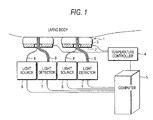
- FIG. 1 shows an apparatus for noninvasive measurement of glucose according to this embodiment.
- FIG. 2 shows a specific structure of a measurement head (also referred to as a probe) of the apparatus for noninvasive measurement of glucose of FIG. 1 .
- a temperature control pack 1 includes a thermoelectric module 2 as a heat generator and a pad 3 which is contacted with the skin of a subject. The heating value of the thermoelectric module 2 is modulated by a temperature controller 4. Further, the temperature modulation operation of the temperature controller 4 is under the control of a computer 5.
- the computer 5 controls the temperature modulation and also plays a role in controlling the operating procedure of the method for noninvasive measurement of glucose according to this embodiment and operating the respective steps.
- a tubular opening is formed and optical fibers 8 and 9 are inserted therein.
- a light source 6 is optically connected, and to the optical fibers 9, a light detector (signal detector) 7 is optically connected.
- a beam splitter 11 is disposed, and a part of light from the light source 6 is introduced into a reference detector 12.
- the optical fiber 8 is disposed in the center, and the optical fibers 9 are arranged around the optical fiber 8 at short intervals.
- the maximum distance between the optical fiber 8 and the optical fibers 9 is less than 2 mm. That is, the optical fibers 8 and 9 are bundled within an area in a substantially circular shape with a diameter of 4 mm.
- Table 2 A relationship between the wavelength of the localized reflectance probe and the distance between the light source 6 and the detector 7 is shown in Table 2.
- one drop of silicone oil is applied to the skin of a subject and then wiped off to leave a very thin layer of oil on the skin, whereby the thermal conductivity is enhanced.
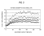
- FIG. 3 shows the response of the skin of a diabetic patient to an optical probe at 40°C pressed against the skin of the forearm of the subject.
- R represents a localized reflectance at each light source-detector distance.
- the signals were normalized to signals at 5 sec intervals.
- the calculated F(OC) increases when the probe at 40°C is brought into contact with the skin, and then, reaches an asymptotic value after about 120 sec.
- the thermal modeling J. Biomedical Optics., 2003; 8, 191-205 ) shows that when the probe came into contact with the skin, the temperature up to a depth of 2 mm reached an equilibrium after 120 sec.
- F(OC) increases at all the light source-detector distances.
- the order of the increase in F(OC) is as follows.
- F(OC) value has a linear relationship with the skin-probe contact time.
- Table 3 shows the results of calculation of functions of oxygen consumption at various light source-detector distances from the data of FIG. 4 .
- a rough estimate of a rate of change in effective attenuation coefficient at each wavelength according to the probe-skin contact can be achieved by plotting the values of Ln(R 4 /R 1 ) at the respective wavelengths against time and calculating the slope.
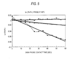
- FIG. 5 shows the plot of Ln(R 4 /R 1 ) against time with respect to a probe heated to 40°C. Lozenges indicate 592 nm, circles indicate 660 nm, Xs indicate 880 nm, and triangles indicate 940 nm. Table 4 shows the results of calculation of approximate ⁇ eff represented by Ln(R 4 /R 1 ) and calculated at various wavelengths from the data of FIG. 5 .
- the approximate ⁇ eff represented by Ln(R 4 /R 1 ) and calculated at various wavelengths changes linearly with the contact time in a time window between 5 and 90 sec except for the wavelength of 600 nm showing the smallest slope and low r 2 .
- the slope approximates to ⁇ eff and changes in the following order.
- Ln ⁇ R 4 / R 1 592 ⁇ nm > Ln ⁇ R 4 / R 1 880 ⁇ nm Ln ⁇ R 4 / R 1 940 ⁇ nm > > Ln ⁇ R 4 / R 1 660 ⁇ nm
- a change in the slope is similar to a change in the extinction coefficient reported at various wavelengths, ⁇ (HbO 2 ) or ⁇ a (HbO 2 ), however, 660 nm which provides the smallest value of ⁇ (HbO 2 ) is excluded. This shows that a change in light absorption is the dominant contributor to be reflected light intensity.
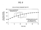
- the degree of change F(OC) is averaged over several adjacent time regions by using a moving average calculation.
- FIG. 6 shows simulation of profile of change in oxygen consumption rate when the skin is contacted with a probe heated such that the temperature thereof is increased by about 10°C.
- the 4-point moving average regions include data between 15 and 30 sec, time regions between 20 and 35 sec, 25 and 40 sec, and 30 and 45 sec. These time regions are used when the data of an embodiment 8 is analyzed.
- B 34e G a 0 + a 1 * d Ln R 4 / R 1 /
- a linear least square regression analysis was used for constructing pentanomial, tetranomial, trinomial, and binomial regression models in combinations of calculated oxygen consumption, calculated attenuation functions, and noninvasively measured glucose concentrations.
- the calibrated standard error and calibrated correlation coefficient were determined with respect to the respective first days of a study.
- the constructed models were used for predicting a glucose concentration from a data point at a later date.
- the data of the respective patients were separately processed.
- the calculated calibration points and prediction points were plotted on a Clarke Error Grid scatter diagram using a modification of the equation 34.
- the Clarke Error Grid is commonly used for showing the distribution of predicted glucose values in various zones of plots, and the respective zones of plots have a special clinical significance in predicted intervention.
- Table 6 Data analysis of 6 diabetic patients using equations 34a to 34n, time interval between 5 and 60 sec, and a probe at 40°C Equation No. Number of terms Type of term: constant + n r c SEC mg/dl r p SEP mg/dl 34a 5 2 ⁇ eff + 2 OC 39 0.81 45.4 0.44 123.0 34b 5 2 ⁇ eff + 2 OC 39 0.81 44.5 0.34 118.4 34c 5 2 ⁇ eff + 2 OC 42 0.81 44.3 0.28 146.4 34d 2 ⁇ eff 43 0.74 50.4 0.73 60.3 34e 2 ⁇ eff 40 0.7 53.7 0.71 64 34f 2 ⁇ eff 43 0.7 49.7 0.72 62.4 34g 3 2 ⁇ eff 40 0.75 49.7 0.72 62.4 34h 3 2 ⁇ eff 43 0.75 49.4 0.72 61.6 34i 2 OC 41 0.74 51.2 0.74 60.4 34j 2 OC 42 0.74 50.5 0.72 61.7 34k 3 2 OC 42 0.76 49.3 0.72
- the five-term equations 34a, 34b and 34c show the evidence of overfitting.
- the equations 34d, 34e and 34f are a two-term equation (constant term + attenuation coefficient term) based on various wavelengths and the inclusion criteria. All have good calibration and prediction parameters, and distribution in the A and B zones of the Clarke Error Grid.
- the attenuation coefficient term at 592 nm (the highest absorbance for hemoglobin) has the best correlation parameters.
- a combination of all two attenuation terms at 592 and 880 nm provides the three-term equations 34g and 34h, which results in improving the two-term equations 34d, 34e and 15f.
- OC term is shown in the equations 34i to 34k.
- the equations 34i and 34j are a two-term equation, and 34k is a three-term equation having two OC terms.
- Table 6 shows the calibration and prediction capability of three OC models.
- the equations 341, 34m and 34n are provided. All these three models result in giving a slight improvement of calibration to 34k.
- the time window in which this calculation is performed extended from 5 sec to 60 sec from the probe-skin interaction.
- the equation 34k was applied to an analysis of 6 patients.
- the equation 34k is a three-term model consisting of a constant term and two OC terms.
- the data on day 1 and day 2 were used in a calibration set.
- the data point on day 3 was used as a prediction set for each patient.
- a cumulative Clarke Error Grid was constructed for data points of all the patients, and overall calibration and prediction parameters were calculated.
- Ten time intervals were selected from 5 sec to 60 sec of the probe-skin interaction windows and as a result, time intervals between 55 sec and 30 sec were provided. The number of time points in the selected intervals is between 12 and 7.
- the calculation results are shown in Table 7.
- Table 7 Effect of changing time window ranging from 5 sec to 60 sec using three terms (constant term and two oxygen consumption terms) Equation Start/finish time (sec) ⁇ t (sec) n r c SEC r p SEP 34k-0 5/60 55 12 0.75 51.0 0.74 62.9 34k-1 10/60 50 11 0.76 49.3 0.72 62.8 34k-2 15/60 45 10 0.78 47.7 0.69 66.3 34k-3 20/60 40 9 0.78 47.2 0.7 63.4 34k-4 25/60 35 8 0.78 47.1 0.75 62.5 34k-5 30/60 30 7 0.80 45.6 0.83 59.0 34k-6 5/55 50 11 0.75 52.2 0.73 69 34k-7 10/55 45 10 0.76 48.8 0.72 61.6 34k-8 15/55 40 9 0.77 46.7 0.70 64.7 34k-9 20/55 35 8 0.8 44.9 0.7 67.5 34k-10 20/55 30 7 0.82 42.5 0.72 69.0
- Equation 341 which is a four-term model including a ⁇ eff term at 592 nm and two OC terms was used.
- the equation 34m is also a four-term model including a ⁇ eff term at 880 nm and two OC terms. It was shown by calculation that the use of a time interval between 30 sec and 60 sec consequently provides a better calibration and prediction model whether a three-term model is used or a four-term model is used. The use of a time interval of 30 sec reduces the contribution of skin-probe adaptation to correlation to the minimum.
- the time intervals were from 30 to 60 sec, however, 7 data points are used in calculation of rates.
- the overall prediction correlation coefficient values are higher than those shown in Table 6, in which a complete time window ranging from 5 to 60 sec was used in calculation.
- Table 8 Body-probe equilibration, Best models after setting equilibration time of 30 sec of body-probe Equation r C SEC r p SEP % in A + B zone % in C + D zone % in E zone 34k-5 0.80 45.6 0.83 59.0 94.7 5.3 0 34l-5 0.86 39.4 0.80 58.6 94.7 5.3 0 34m-5 0.76 48.8 0.72 61.6 94.4 5.6 0
- F(OC)1 and F(OC)2 were defined as follows before.
- F ⁇ OC 1 0.527 * 36.75 * Ln R 940 t / R 940 0 - Ln R 592 t / R 592 0
- F ⁇ OC 2 3.2022 * 4.6566 ⁇ Ln R 940 t / R 940 0 - Ln R 660 t / R 660 0
- a three-term model in which a combination of F(OC)1 and F(OC)2 at different light source-detector distances is used provides the following equation.
- G a 0 + a 1 * F ⁇ OC 1 ⁇ @ ⁇ r 2 - F ⁇ OC 1 ⁇ @ ⁇ r 1 ⁇ a 2 * F ⁇ OC 2 ⁇ @ ⁇ r 2 - F ⁇ OC 2 ⁇ @ ⁇ r 1
- G a 0 + a 1 * F ⁇ OC 1 ⁇ @ ⁇ r 1 - a 2 * F ⁇ OC 1 ⁇ @ ⁇ r 2
- G a 0 + a 1 * F ⁇ OC 2 ⁇ @ ⁇ r 1 - a 2 * F ⁇ OC 2 ⁇ @ ⁇ r 2
- G a 0 + a 1 * F ⁇ OC 1 ⁇ @ ⁇ r 2 - F ⁇ OC 1 ⁇ @ ⁇ r 1 + a 2 * F ⁇ OC 2 ⁇ @ ⁇ r 2 - F ⁇ OC 2 ⁇ @ ⁇ r 1
- G a 0 + a 1 * F ⁇ OC 1 ⁇ @ ⁇ r 2 - F ⁇ OC 1 ⁇ @ ⁇ r 1 + a 2 * F ⁇ OC 2 ⁇ @ ⁇ r 2 - F ⁇ OC 2 ⁇ @ ⁇ r 1
- G a 0 + a
- the OC function is a logarithmic function, therefore, two subtractions as the case of a term of difference between two OC terms in (37) and (38) and F(OC) 1 and F(OC) 2 in the equation 40 represents a ratio term of a localized reflectance ratio at two light source-detector distances.
- the data points of patients were collected in a period of 7 days. 16 out of 30 data points were passed the inclusion criteria. An average of 4 ⁇ 15 sec regions of time response as described above was used. In the calibration, 12 data points from day 1 to day 3 in the clinical study were used, and in the prediction, 4 data points from day 5 to day 7 in the clinical study were used. The results are shown in FIG. 7A to FIG. 7D .
- the equation 37 is a three-term equation including a constant and a difference between two F(OC) 1 terms.
- the data are plotted on FIG. 7A .
- the calibration and prediction parameters are good as shown in Table 9.
- the four predicted glucose values fall in the A zone of the Clarke Error Grid.
- the equation 38 was used for analyzing the data of a subject No. 1001.
- the three-term approximate linear equation 38, the coefficients a 0 , a 1 and a 2 obtained by calibration of two variables are provided in the equation 38a.
- Glucose 167.9 + 7351.3 * F ⁇ OC 2 ⁇ @ ⁇ r 1 - 7985.2 * F ⁇ OC 2 ⁇ @ ⁇ r 2
- the data are plotted on FIG. 7B .
- the calibration and prediction parameters are good as shown in Table 9.
- the four predicted glucose values fall in the A zone of the Clarke Error Grid.
- the use of F(OC) 2 consequently provides a better correlation with respect to this patient.
- the equation 39 was used for analyzing the data of a subject No. 1001.
- the three-term approximate linear equation 39, the coefficients a 0 , a 1 and a 2 obtained by calibration of two variables are provided in the equation 39a.
- G 178.7 + 115.2 * F ⁇ OC 1 ⁇ @ ⁇ r 2 - F ⁇ OC 1 ⁇ @ ⁇ r 1 - 8303.9 * F ⁇ OC 2 ⁇ @ ⁇ r 2 - F ⁇ OC 2 ⁇ @ ⁇ r 1
- the data are plotted on FIG. 7C .
- the calibration and prediction parameters are good as shown in Table 9.
- the four predicted glucose values fall in the A zone of the Clarke Error Grid.
- FIG. 7D shows the use of the equation 40 for analyzing the data of a subject No. 1001.
- the equation 40 is a three-term approximate linear equation including a constant, a difference between two F(OC) 1 terms and a sum of two F(OC) 1 terms.
- the first parenthesis is a difference between two F(OC) 1 values which consequently provide localized reflectance ratios at two light source-detector distances r 1 and r 2 .
- the second term in the parenthesis is a sum of two F(OC) 2 values which consequently provide a multiplication.
- the three- parameter approximate linear equation, the coefficients a 0 , a 1 and a 2 obtained by calibration of two variables are provided in the equation 40a.
- G 154 - 529.8 * F ⁇ OC 1 ⁇ @ ⁇ r 2 - F ⁇ OC 1 ⁇ @ ⁇ r 1 - 1351 * F ⁇ OC 2 ⁇ @ ⁇ r 2 - F ⁇ OC 2 ⁇ @ ⁇ r 1
- equations 38 and 39 provide the best calibration parameters and good prediction parameters.
- the method of adapting the skin includes use of a small amount of silicone oil to be applied to the skin of a subject for enhancing the thermal conductivity.
- An excess amount of oil can make the probe slip on the skin, and an insufficient amount of oil causes incomplete thermal binding.
- other binding jell can be used for the purpose of eliminating a gap between the probe and the skin. However, this reduces a surface reflection loss.
- such a liquid or jell enhances the thermal binding and facilitates the heat transfer between the skin and the probe.
- a temperature-modulated localized reflectance probe can be used.
- the temperature-modulated probe has a light introduction fiber and several light collection fibers.
- the light collection fibers are disposed at a short distance from the light introduction fiber.
- US Patent Nos. 5,795,305 , 5,924,996 , US Patent Application Publication No. 2005/0,124,868 and European Patent Application Publication No. 1537822 did not take the skin-probe adaptation effect on the optical and thermal signals into consideration.
- US Patent No. 5,978,691 did not disclose or take into consideration a time window for reducing the tissue-probe adaptation effect on the measurement to the minimum.
- a temperature-induced change in the hemoglobin equilibrium is measured.
- US Patent No. 5,978,691 does not disclose a method for enhancing the mechanical adaptation of the skin to a measurement probe.
- noninvasive measurement is divided into two major steps. That is, a skin adaptation step and a measurement step.
- the skin adaptation step is performed ahead of the measurement step.
- the measurement probe with a high rigidity is brought into contact with a skin part which is an objective site of a subject and the skin part is adapted to the shape of a surface thereof (the shape of an interface region).
- an adaptation device also referred to as a stretching device which has a shape similar to the measurement probe into contact with a skin part ahead of the contact between the measurement probe and the skin, the skin part is stretched. This contact is carried out under a pressure that is higher than a pressure applied by the measurement probe during the measurement. The contact between the adaptation device and the skin is maintained for a certain period of time.
- the adaptation device may be provided as a separate body from the measurement probe, or may be shared with the measurement probe.
- the measurement probe which also serves as the adaptation device moves up and down and comes into contact with a skin part of an objective site of a subject.
- a part of the subject is irradiated with an optical signal from a light source and light detection unit through an optical fiber unit, and also a reflected wave from the part of the subject is detected at the light source and light detection unit through the optical fiber unit.
- the measurement probe which also serves as the adaptation device comes into contact with the part of the subject, but may not detect the light signal.
- the measurement probe which also serves as the adaptation device detects the light signal at a desired time in which the measurement probe is contacted with the part of the subject.
- the adaptation device constituting the measurement probe and the optical fiber unit are separated.
- the adaptation device moves up and down, but the optical fiber unit does not move.
- the adaptation device and the optical fiber unit move up and down in conjunction with each other.
- the adaptation device is provided as a separate body and functions also as a part of the measurement probe may be applied.
- an optical window is provided, and in the skin adaptation step, the adaptation device moves up and down, and in the measurement step, the optical fibers, the light source and light detection unit move up and down as the measurement probe in conjunction with the adaptation device.
- the measurement probe is brought into contact with the skin which has been consequently adapted, i.e., stretched such that it is conformed to the shape of the surface of the probe for performing measurement.
- a method for adapting the skin of a subject in order to adapt the skin to the shape of a detecting device which is brought into contact with the skin for noninvasive determination of glucose in a tissue of the subject includes the steps of:
- the measurement probe is controlled by a spring.
- a subject sits on a chair, places the left arm on a body interface receiving stand and adjusts the distance between an arm tray and a grip so as to give a comfortable position. Then, the subject pushes in a double-probe head supported by a spring by the right hand and starts countdown. When the count becomes zero, an operator clicks the start icon, and then, the subject relieves the double-head probe for bringing it into contact with the skin.
- the movement of the probe is not controlled, and a force applied by the spring determines the displacement of the skin.
- the contact between the probe and the skin at a fixed temperature for about 120 sec can reduce the effect of adapting the skin to the measurement probe on a signal to the minimum.
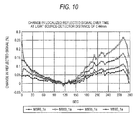
- FIG. 10 shows a time course of a change in signal in this experiment at four different wavelengths: 590 nm, 660 nm, 890 nm and 935 nm.
- FIG. 10 describes a percentage change in localized reflected signal as measured by a probe similar to one described in US Patent No. 6,662,030 at a distance of 0.44 mm from a radiation fiber.
- the data relates to the skin of a forearm of a volunteer (subject 1015, implementation D, September 13, 2003).
- the data is represented as a percentage change in signal normalized to the data point of 120 sec.
- the first data point at 0 sec is the point at 3 sec from the contact, and it was a time required for collecting data and transmitting it from a data board to a computer.
- the subsequent data point starts from the first data point recorded. Because the probe reduced heat transfer between the skin and the measurement probe to the minimum, the temperature was maintained at 30°C very close to the normal temperature of the skin which is generally from 28°C to 32°C. It is apparent that the percentage change in signal greatly fluctuates in the initial 30 sec. The majority of the percentage change in signal occurred in the initial 10 sec. The temperature of the probe was maintained at 30°C very close to the normal temperature of the skin which is generally from 28°C to 32°C, therefore, the majority of this change was caused by the mechanical adaptation of the skin to the measurement probe. The pressure applied to the skin by the probe was 45 g/cm 2 . In order to facilitate thermal and optical binding, silicone oil was applied to the skin.
- the change in signal decreases as the time proceeds and reaches a complete equilibrium state between 90 sec and 120 sec.
- the temperature changed from 120 sec.
- FIG. 10 shows a time course of a localized reflected signal in the skin of a volunteer. The temperature of the measurement probe was maintained constant at 30°C for 120 sec, and then raised at a rate of 4°C per minute.
- Still another aspect of this embodiment is a method for performing noninvasive measurement in a tissue of a subject in order to determine an analyte concentration in the tissue of the subject, and the method includes adapting the skin of a subject for adapting its shape to the shape of a detection device to be brought into contact with the skin for noninvasive quantification of glucose in a tissue liquid of the subject, and includes the steps of:
- the method of this embodiment reduces the effect of the skin-probe interaction on the measured signals by effecting the adaptation of the skin to the measurement probe ahead of the noninvasive measurement.
- US Patent Nos. 5,785,305 , 5,924,996 and 5,978,691 did not disclose a method for adapting the skin to a measurement probe for reducing the skin-probe adaptation effect on the measurement to the minimum.
- the method of this embodiment is independent of the measurement method or the probe to be used.
- the measurement probe may be a light-absorbing probe using NIR wavelength as described by Maruo ( US Patent No. 5,975,841 ), or a light-scattering probe as described by Gratton ( US Patent No. 5,492,118 ) and Simonsen ( US Patent No. 5,551,422 ), or Khalil et al. ( US Patent No. 6,662,030 ).
- the adaptation of the skin by stretching the skin by a device which has a shape similar to the measurement probe can be involved in applying a pressure to the skin for 1 to 10 sec.
- the pressure application time for inducing the adaptation of the skin to the shape of the probe is from 5 sec to 10 sec.
- the pressure applied for conforming the skin to the probe is 2 to 5 times the pressure value applied to the skin by the measurement probe.
- the pressure is defined by a unit of g/cm 2 or a pound per square inch.
- the pressure is 2 to 3 times the maximum value of the pressure applied to the skin by the measurement probe.
- the pressure value depends on the thickness of the skin, the degree of skin aging and a diabetic period and can vary depending on an individual. A high pressure can lead to occlusion of a blood vessel in the skin, and a low pressure can lead to an incomplete contact.
- the pressure which we used before for measurement was generally in the range from 45 to 100 g/cm 2 .
- a pressure lower than 30 g/cm 2 leads to an incomplete contact, and a pressure higher than 130 g/cm 2 applied for several tens of seconds leads to partial occlusion. Therefore, a stretching pressure ranging from 200 to 500 g/cm 2 , preferably from 200 to 300 g/cm 2 stretches the skin thereby to adapt the skin to the measurement probe, and further, because a close contact with an optical element is achieved, the use thereof can be allowed in a limited time such as 10 sec.
- Such a combination can include a high pressure in a short time or the lower limit of the pressure range in a long time.
- the magnitude of the displacement can depend on the properties of the skin, and can vary depending on an individual.
- a binding liquid such as silicone oil, paraffin oil, perfluorohydrocarbon or a water-glycerol mixture was used for enhancing the thermal conductivity and reducing the reflection loss in the measurement of diffusion reflectance to the minimum.
- a jell or a cream such as a mixture of paraffin wax, an oil, water and an emulsifying agent can also be used in the method of this embodiment.
- Such an oil is preferably applied before applying a pressure.
- it is preferred that such a binding liquid or jell is used in a small amount and the skin is uniformly covered therewith.
- a preferred method for applying the binding agent is described in US Patent Application Publication No. 10/823073 , "Apparatus and method for applying binding agent to noninvasive optical sensor" filed on April 13, 2004.
- the use of the clearing solution takes several minutes to affect the stratum corneum, therefore, it may be used first and wiped off, and then, a pressure may be applied.
- the water-glycol-oleic acid mixture can make the stratum corneum smooth, remove several surface flacks, whereby a better probe-skin contact can be achieved.
- the measurement probe is brought into contact with the skin again, and a data stream generated by the measurement is collected over a given period of time.
- a data point in a range of time window is selected to be used in the calculation of the concentration of an analyte.
- this time window starts at several seconds from the initiation of the probe-skin contact.
- the skin adaptation method of this embodiment can be used in several optical and non-optical methods.
- the absorbance and scattering methods, the thermal conductivity method and the method for infrared emission from the skin can be used in the method of this embodiment.
- the method of this embodiment can be used in optical and non-optical measurements. Therefore, in a photoacoustic measurement in which light excites an absorber and an ultrasound measurement probe is brought into contact with the skin, a signal is affected by a sound speed in the tissue affected by compression of the skin, therefore, the improvement of tissue adaptation to the prove ahead of the measurement reduces an error due to compression of tissue.
- the temperature and the adaptation device are maintained at around the normal temperature of the skin, i.e., 28°C to 32°C.
- an arm tray 25 which is caved in a groove shape is formed in a part of a housing of the apparatus 10.
- the surface of a measurement probe 11 is exposed.
- a fixed platform 13 is provided at each of the both ends of the arm tray 25 .
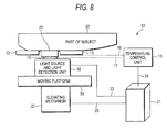
- FIG. 1 shows a single-probe type apparatus for noninvasive measurement 10 according to this embodiment.
- the apparatus has the measurement probe 11 which is installed on the fixed platform 13 and comes into contact with a part of a subject, typically a part of an arm 12 and the skin thereof.
- the measurement probe 11, a temperature modulation element 14, a light source and light detection unit 18 are installed on a moving platform 19.
- a temperature control unit 15 has a feedback input 16 and a control output 17 with respect to the temperature modulation element 14.
- the vertical movement of the moving platform 19 for approaching and retreating from the part of a subject 12 is effected by an elevating mechanism 20 having a stepper motor, an elevating actuator and a spring.
- a computer 21 plays a role in total control for measurement and total control for an adaptation operation of a skin shape ahead of the measurement as well as data analysis by controlling the temperature control unit 15, the light source and light detection unit 18, and the movement of the moving platform 19 via control/signal lines 22, 23 and 24.
- the measurement method may be any of absorbance, reflectance, localized reflectance, photoacoustic spectroscopy and Raman spectroscopy.
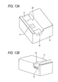
- a patient places an arm or other body part 12 on the fixed plat form 13 (see FIG. 13B ).
- the computer 21 moves the moving platform 19 up toward the skin to press the measurement probe 11 against the skin by a step number n via the elevating mechanism 20 for an adaptation operation, which is characteristic in this embodiment, whereby the skin adaptation step is started. This state is maintained for a period of about 5 to 10 sec, and then relieved.
- This process induces adaptation of the skin to the shape of the measurement probe 11, and in the subsequent noninvasive measurement step, a close contact between the measurement probe 11 and the skin is achieved. Thereafter, the computer 21 separates the moving platform 19 from the skin, and then, the data collection step is started by moving the measurement probe 11 and therefore the moving platform 19 toward the skin again by a step number (n-x) for causing a smaller displacement of the skin.
- x represents an arbitrary step number and this means that the elevating distance of the moving platform 19 in the measurement process is shorter than the elevating distance of the moving platform 19 in the adaptation process by a distance obtained by multiplying the reduced step number x by a step unit distance, in other words, the pressing force of the measurement probe 11 against the skin in the measurement process is reduced according to a distance obtained by multiplying the reduced step number x by a step unit distance from the pressing force of the measurement probe 11 against the skin in the adaptation process.
- the computer 21 causes an operation of data collection from the light source and detection optical element 18 for a specific period of time, and processes the data.
- the use of the temperature modulation element 14, the temperature control unit 15, the feedback input 16 and the feedback control output 17 can reduce the signal drift due to uncontrolled heat transfer between the body and the probe to the minimum, and enables the examination of the effect of an induced temperature change on the signal.
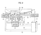
- FIG. 9 shows a double-probe type apparatus for noninvasive measurement according to this embodiment.
- the apparatus has two measurement probes 101 and 102, which are installed on a fixed platform 104 and come into contact with a part of a subject 103. Temperature control units 107 and 108 having temperature modulation elements 105 and 106, feed back inputs 109 to 112 to a temperature modulator are provided for modulating the temperature of the respective measurement probes 101 and 102.
- the measurement probes 101 and 102 have light source and light detection units 113 and 114, respectively.
- the two measurement probes 101 and 102 are installed on a common moving platform 115.
- the vertical movement of the moving platform 115 is effected by an elevating device 116 having a stepper motor or an elevating actuator and a spring.
- a computer 117 which also performs data analysis, plays a role in total control for measurement and total control for an adaptation operation of a skin shape ahead of the measurement as well as data analysis by controlling the temperature control units 107 and 108 of the respective measurement probes 101 and 102, the light source and light detection units 113 and 114 and the movement of the moving platform 115 via control/signal lines 118 to 121.
- a patient places an arm or other body part 103 on the fixed platform 104.
- the computer 117 moves the moving platform 115 up toward the skin to press the measurement probes 101 and 102 against the skin by a step number n via the mechanism 116 for an adaptation operation.
- This state in which the measurement probes 101 and 102 are pressed against the skin is maintained for about 5 to 10 sec.
- This process induces adaptation of the skin to the shape of the measurement probes 101 and 102, and in the subsequent noninvasive measurement step, a close contact between the measurement probes 101 and 102 and the skin is achieved.
- the computer 117 lowers the moving platform 115 for separating the measurement probes 101 and 102 from the skin.
- the data collection cycle is started by moving the measurement probes 101 and 102 and therefore the moving platform 115 toward the skin again by a step number (n-x) for causing a smaller displacement of the skin.
- the computer 117 causes an operation of data collection from the light source and detection optical elements 113 and 114 for a specific period of time, and processes the data.
- the use of the temperature modulation elements 105 and 106, the temperature control units 107 and 108, the feedback inputs and the control outputs 109 to 112 can reduce the signal drift due to uncontrolled heat transfer between the part of a subject 103 and the measurement probes 101 and 102 to the minimum, and enables the examination of the effect of an induced temperature change on the signal.
- the temperature of the respective measurement probes 101 and 102 is modulated separately, and various temperature changing steps can be used for the respective measurement probes 101 and 102.
- US Patent No. 6,662,030 describes a temperature-modulated localized reflectance probe which can be used in the method of this embodiment.
- the probe has a light introduction fiber and several light collection fibers disposed at a short distance from the light introduction fiber.
- the maximum distance between the center of the light collection fiber and the light collection fiber is less than 2 mm.
- a temperature-modulated localized reflectance measurement probe which is similar to a probe described in US Patent No. 6,662,030 by Khalil et al. and Khalil et al., J. Biomed. Opt. 2003; 8: 191-205 , Yeh et al., J. Biomed. Opt. 2003; 8: 534-544 , and Yeh et al., Clin. Chem. 2003; 49: 924-934 is used.
- the measurement of localized reflected signals using a temperature-modulated probe described in US Patent No. 6,662,030 provides a light signal according to a temperature or a contact time between a probe at a given temperature and a part of the body.
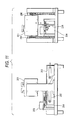
- FIG. 11 shows a structural example of an elevating device of this embodiment.
- An elevating actuator 202 includes a motor 203, a plurality of gears 204, a lever 205 and a spring 206.
- a probe 201 is moved in a vertical direction by moving the lever 206 installed on the bottom with the motor 203 and the gears 204.
- the moving distance of the upper and lower lateral face is generally 10 to 15 mm.
- the probe 201 is supported by the two springs 206.
- the springs 206 keep the contact pressure to the skin.
- the pressing power against the skin can be generally set to about 100 gf/cm 2 .
- FIG. 12 shows a structure of a light source and light detection unit together with two measurement probes 201.
- the light source and light detection unit includes a light source 210, a lens 211, a mirror 212, half mirrors 213-1 and 213-2, an optical fiber for radiation 214, an optical fiber for signal detection 215, a photodiode (PD) for signal detection 216 and a photodiode (PD) for reference 217.
- the light source 210 emits one or more light beams.
- the light beams are collimated and combined by passing through the lens 211, the mirror 212 and the half mirror 213-1.
- the combined light beams pass through the optical fiber for radiation 214, and then illuminate the body.
- a part of the combined light beams is divided by the half mirror 213-2, and detected as a reference signal by the photodiode (PD) for reference 217.
- a part of the reflected signal from the body passes through the optical fiber for signal detection 215 at one or more positions disposed at an appropriate distance from the radiation spot, and then, the reflected signal is detected by a light detector (PD) for signals 216 as a reflected signal.
- PD light detector
- the present invention is not limited to the above embodiments as such, and may be embodied in implementation phase by modifying the components without departing from the scope of the invention.
- various inventions can be created by appropriately combining the plurality of components disclosed in the above embodiments. For example, several components may be eliminated from all the components disclosed in any of the embodiments. Further, the components of different embodiments may be appropriately combined.
- a temperature change is induced in the human skin, localized reflectance signals at several defined light source-detector distances are measured, and functions derived from the reflectance values at a plurality of wavelengths and light source-detector distances are correlated with a glucose concentration, whereby temperature-induced glycolysis can be tracked.
- an influence of mechanical and thermal effects occurring when the measurement probe is brought into contact with the skin of a subject can be reduced.
Landscapes
- Health & Medical Sciences (AREA)
- Life Sciences & Earth Sciences (AREA)
- Physics & Mathematics (AREA)
- Molecular Biology (AREA)
- Animal Behavior & Ethology (AREA)
- Pathology (AREA)
- Engineering & Computer Science (AREA)
- Biomedical Technology (AREA)
- Heart & Thoracic Surgery (AREA)
- Medical Informatics (AREA)
- Veterinary Medicine (AREA)
- Surgery (AREA)
- Biophysics (AREA)
- General Health & Medical Sciences (AREA)
- Public Health (AREA)
- Optics & Photonics (AREA)
- Emergency Medicine (AREA)
- Spectroscopy & Molecular Physics (AREA)
- Measurement Of The Respiration, Hearing Ability, Form, And Blood Characteristics Of Living Organisms (AREA)
- Investigating Or Analysing Biological Materials (AREA)
Priority Applications (3)
| Application Number | Priority Date | Filing Date | Title |
|---|---|---|---|
| EP11180786.3A EP2399517B1 (de) | 2005-11-30 | 2006-11-30 | Verfahren zur nicht invasiven Messung von Glukose und Vorrichtung zur nicht invasiven Messung von Glukose |
| EP11180748A EP2399515A3 (de) | 2005-11-30 | 2006-11-30 | Verfahren zur nicht invasiven Messung von Glukose und Vorrichtung zur nicht invasiven Messung von Glukose |
| EP11180771.5A EP2399516B1 (de) | 2005-11-30 | 2006-11-30 | Verfahren zur nicht invasiven Messung von Glukose und Vorrichtung zur nicht invasiven Messung von Glukose |
Applications Claiming Priority (3)
| Application Number | Priority Date | Filing Date | Title |
|---|---|---|---|
| JP2005347194A JP4880985B2 (ja) | 2005-11-30 | 2005-11-30 | グルコースの非侵襲性測定法及びグルコースの非侵襲性測定装置 |
| JP2006129490A JP5049510B2 (ja) | 2006-05-08 | 2006-05-08 | 非侵襲的測定装置 |
| EP06833798A EP1955653B1 (de) | 2005-11-30 | 2006-11-30 | Verfahren zur nicht-invasiven glucosemessung |
Related Parent Applications (2)
| Application Number | Title | Priority Date | Filing Date |
|---|---|---|---|
| EP06833798.9 Division | 2006-11-30 | ||
| EP06833798A Division EP1955653B1 (de) | 2005-11-30 | 2006-11-30 | Verfahren zur nicht-invasiven glucosemessung |
Related Child Applications (5)
| Application Number | Title | Priority Date | Filing Date |
|---|---|---|---|
| EP11180748A Division-Into EP2399515A3 (de) | 2005-11-30 | 2006-11-30 | Verfahren zur nicht invasiven Messung von Glukose und Vorrichtung zur nicht invasiven Messung von Glukose |
| EP11180771.5A Division-Into EP2399516B1 (de) | 2005-11-30 | 2006-11-30 | Verfahren zur nicht invasiven Messung von Glukose und Vorrichtung zur nicht invasiven Messung von Glukose |
| EP11180771.5A Division EP2399516B1 (de) | 2005-11-30 | 2006-11-30 | Verfahren zur nicht invasiven Messung von Glukose und Vorrichtung zur nicht invasiven Messung von Glukose |
| EP11180786.3A Division-Into EP2399517B1 (de) | 2005-11-30 | 2006-11-30 | Verfahren zur nicht invasiven Messung von Glukose und Vorrichtung zur nicht invasiven Messung von Glukose |
| EP11180786.3A Division EP2399517B1 (de) | 2005-11-30 | 2006-11-30 | Verfahren zur nicht invasiven Messung von Glukose und Vorrichtung zur nicht invasiven Messung von Glukose |
Publications (3)
| Publication Number | Publication Date |
|---|---|
| EP2243425A2 true EP2243425A2 (de) | 2010-10-27 |
| EP2243425A3 EP2243425A3 (de) | 2011-03-30 |
| EP2243425B1 EP2243425B1 (de) | 2014-08-13 |
Family
ID=38092294
Family Applications (5)
| Application Number | Title | Priority Date | Filing Date |
|---|---|---|---|
| EP11180748A Withdrawn EP2399515A3 (de) | 2005-11-30 | 2006-11-30 | Verfahren zur nicht invasiven Messung von Glukose und Vorrichtung zur nicht invasiven Messung von Glukose |
| EP06833798A Active EP1955653B1 (de) | 2005-11-30 | 2006-11-30 | Verfahren zur nicht-invasiven glucosemessung |
| EP10170622.4A Active EP2243425B1 (de) | 2005-11-30 | 2006-11-30 | Verfahren zur nicht invasiven Messung von Glukose und Vorrichtung zur nicht invasiven Messung von Glukose |
| EP11180771.5A Active EP2399516B1 (de) | 2005-11-30 | 2006-11-30 | Verfahren zur nicht invasiven Messung von Glukose und Vorrichtung zur nicht invasiven Messung von Glukose |
| EP11180786.3A Active EP2399517B1 (de) | 2005-11-30 | 2006-11-30 | Verfahren zur nicht invasiven Messung von Glukose und Vorrichtung zur nicht invasiven Messung von Glukose |
Family Applications Before (2)
| Application Number | Title | Priority Date | Filing Date |
|---|---|---|---|
| EP11180748A Withdrawn EP2399515A3 (de) | 2005-11-30 | 2006-11-30 | Verfahren zur nicht invasiven Messung von Glukose und Vorrichtung zur nicht invasiven Messung von Glukose |
| EP06833798A Active EP1955653B1 (de) | 2005-11-30 | 2006-11-30 | Verfahren zur nicht-invasiven glucosemessung |
Family Applications After (2)
| Application Number | Title | Priority Date | Filing Date |
|---|---|---|---|
| EP11180771.5A Active EP2399516B1 (de) | 2005-11-30 | 2006-11-30 | Verfahren zur nicht invasiven Messung von Glukose und Vorrichtung zur nicht invasiven Messung von Glukose |
| EP11180786.3A Active EP2399517B1 (de) | 2005-11-30 | 2006-11-30 | Verfahren zur nicht invasiven Messung von Glukose und Vorrichtung zur nicht invasiven Messung von Glukose |
Country Status (4)
| Country | Link |
|---|---|
| US (1) | US8315681B2 (de) |
| EP (5) | EP2399515A3 (de) |
| DE (1) | DE602006019133D1 (de) |
| WO (1) | WO2007063961A1 (de) |
Cited By (1)
| Publication number | Priority date | Publication date | Assignee | Title |
|---|---|---|---|---|
| WO2019027895A1 (en) * | 2017-07-30 | 2019-02-07 | Dermaflow Llc | METHOD AND DEVICE FOR CALIBRATING CAPILLARY BLOOD FLOWMETER |
Families Citing this family (39)
| Publication number | Priority date | Publication date | Assignee | Title |
|---|---|---|---|---|
| US6640217B1 (en) * | 2000-09-19 | 2003-10-28 | Bocada, Inc, | Method for extracting and storing records of data backup activity from a plurality of backup devices |
| JP5049510B2 (ja) * | 2006-05-08 | 2012-10-17 | 東芝メディカルシステムズ株式会社 | 非侵襲的測定装置 |
| DE602006019133D1 (de) | 2005-11-30 | 2011-02-03 | Toshiba Medical Sys Corp | Verfahren zur nicht-invasiven glucosemessung |
| US7880133B2 (en) * | 2006-06-01 | 2011-02-01 | Weatherford/Lamb, Inc. | Optical multiphase flowmeter |
| US8666466B2 (en) | 2009-06-10 | 2014-03-04 | Medtronic, Inc. | Device and method for monitoring of absolute oxygen saturation and tissue hemoglobin concentration |
| US8463346B2 (en) | 2009-06-10 | 2013-06-11 | Medtronic, Inc. | Absolute calibrated tissue oxygen saturation and total hemoglobin volume fraction |
| US8352008B2 (en) | 2009-06-10 | 2013-01-08 | Medtronic, Inc. | Active noise cancellation in an optical sensor signal |
| WO2010144668A1 (en) | 2009-06-10 | 2010-12-16 | Medtronic, Inc. | Tissue oxygenation monitoring in heart failure |
| US9126049B2 (en) | 2009-06-10 | 2015-09-08 | Medtronic, Inc. | Shock reduction using absolute calibrated tissue oxygen saturation and total hemoglobin volume fraction |
| US8521245B2 (en) | 2009-09-11 | 2013-08-27 | Medtronic, Inc. | Method and apparatus for post-shock evaluation using tissue oxygenation measurements |
| JP5838517B2 (ja) * | 2011-06-21 | 2016-01-06 | セイコーエプソン株式会社 | 濃度定量装置、濃度定量方法 |
| US9078587B2 (en) * | 2011-10-21 | 2015-07-14 | The Regents Of The University Of California | Method and apparatus for photomagnetic imaging |
| US9101707B2 (en) | 2012-04-27 | 2015-08-11 | Gregory Zeltser | Implantable bioartificial perfusion system |
| TW201347727A (zh) * | 2012-05-31 | 2013-12-01 | Ibm | 自動上傳非連接式醫療檢測裝置之量測資料的方法與電腦裝置 |
| US9383476B2 (en) | 2012-07-09 | 2016-07-05 | Weatherford Technology Holdings, Llc | In-well full-bore multiphase flowmeter for horizontal wellbores |
| US9351671B2 (en) | 2012-07-16 | 2016-05-31 | Timothy Ruchti | Multiplexed pathlength resolved noninvasive analyzer apparatus and method of use thereof |
| US9585604B2 (en) | 2012-07-16 | 2017-03-07 | Zyomed Corp. | Multiplexed pathlength resolved noninvasive analyzer apparatus with dynamic optical paths and method of use thereof |
| US20150018642A1 (en) | 2013-07-12 | 2015-01-15 | Sandeep Gulati | Tissue pathlength resolved noninvasive analyzer apparatus and method of use thereof |
| US9351672B2 (en) | 2012-07-16 | 2016-05-31 | Timothy Ruchti | Multiplexed pathlength resolved noninvasive analyzer apparatus with stacked filters and method of use thereof |
| ITMI20130459A1 (it) * | 2013-03-27 | 2014-09-28 | Pignolo Spa | Dispositivo multi-funzionale per la misura non invasiva della glicemia |
| US10588552B2 (en) | 2014-06-19 | 2020-03-17 | Glucovista Inc. | Substance concentration analysis methods and apparatuses |
| EP3157428B1 (de) * | 2014-06-19 | 2019-09-25 | GlucoVista Inc. | Vorrichtungen und verfahren zur stoffkonzentrationsüberwachung |
| WO2016054079A1 (en) | 2014-09-29 | 2016-04-07 | Zyomed Corp. | Systems and methods for blood glucose and other analyte detection and measurement using collision computing |
| US11653862B2 (en) * | 2015-05-22 | 2023-05-23 | Cercacor Laboratories, Inc. | Non-invasive optical physiological differential pathlength sensor |
| US10874333B2 (en) | 2015-09-15 | 2020-12-29 | Massachusetts Institute Of Technology | Systems and methods for diagnosis of middle ear conditions and detection of analytes in the tympanic membrane |
| US11058328B2 (en) * | 2015-12-31 | 2021-07-13 | Banpil Photonics, Inc. | System for screening and diagnosis of diabetes |
| US9554738B1 (en) | 2016-03-30 | 2017-01-31 | Zyomed Corp. | Spectroscopic tomography systems and methods for noninvasive detection and measurement of analytes using collision computing |
| KR102655736B1 (ko) * | 2016-07-19 | 2024-04-05 | 삼성전자주식회사 | 이종 스펙트럼 기반 혈당 추정 장치 및 방법 |
| US11369292B2 (en) * | 2017-02-12 | 2022-06-28 | SeeDevice Inc. | Portable apparatus for noninvasively measuring blood glucose level and operating method thereof |
| US10646145B2 (en) * | 2018-02-09 | 2020-05-12 | General Electric Company | Reflective SpO2 measurement system and method |
| KR102574086B1 (ko) | 2018-02-21 | 2023-09-04 | 삼성전자주식회사 | 라만 프로브 |
| PL3823525T4 (pl) | 2018-07-16 | 2025-06-23 | Bbi Medical Innovations, Llc | Pomiar perfuzji i natleniania |
| US20220142520A1 (en) * | 2019-03-13 | 2022-05-12 | Biofi Medical Healthcare India Pvt Ltd. | A non-invasive glucometer |
| CN111599470B (zh) * | 2020-04-23 | 2022-07-29 | 中国科学院上海技术物理研究所 | 一种用于提高近红外无创血糖检测精度的方法 |
| CN111966857B (zh) * | 2020-08-19 | 2023-09-29 | 南京英德利汽车有限公司 | 一种检测改装车辆的方法及检测系统 |
| WO2023032863A1 (ja) * | 2021-09-01 | 2023-03-09 | 株式会社ジャパンディスプレイ | 検出装置 |
| GB2613032A (en) * | 2021-11-23 | 2023-05-24 | Rsp Systems As | Calibration method and system |
| KR20240038230A (ko) * | 2022-09-16 | 2024-03-25 | 삼성전자주식회사 | 생체정보 추정 장치 및 방법 |
| CN120983033B (zh) * | 2025-10-22 | 2026-02-06 | 成都中医药大学 | 基于近红外光谱检测的无创血糖浓度检测方法 |
Citations (36)
| Publication number | Priority date | Publication date | Assignee | Title |
|---|---|---|---|---|
| US3628525A (en) | 1969-06-19 | 1971-12-21 | American Optical Corp | Blood oxygenation and pulse rate monitoring apparatus |
| US4259963A (en) | 1979-07-03 | 1981-04-07 | Albert Huch | Multi-purpose transducer for transcutaneous blood measurements |
| US4432365A (en) | 1980-05-16 | 1984-02-21 | 501 Hellige GmbH | Physiological sensing unit |
| US4890619A (en) | 1986-04-15 | 1990-01-02 | Hatschek Rudolf A | System for the measurement of the content of a gas in blood, in particular the oxygen saturation of blood |
| US4926867A (en) | 1986-05-27 | 1990-05-22 | Sumitomo Electric Industries, Ltd. | Light-reflecting and heating type oximeter |
| US5028787A (en) | 1989-01-19 | 1991-07-02 | Futrex, Inc. | Non-invasive measurement of blood glucose |
| US5068536A (en) | 1989-01-19 | 1991-11-26 | Futrex, Inc. | Method for providing custom calibration for near infrared instruments for measurement of blood glucose |
| US5077476A (en) | 1990-06-27 | 1991-12-31 | Futrex, Inc. | Instrument for non-invasive measurement of blood glucose |
| US5086229A (en) | 1989-01-19 | 1992-02-04 | Futrex, Inc. | Non-invasive measurement of blood glucose |
| US5131391A (en) | 1989-06-22 | 1992-07-21 | Colin Electronics Co., Ltd. | Pulse oxymeter having probe with warming means |
| US5237178A (en) | 1990-06-27 | 1993-08-17 | Rosenthal Robert D | Non-invasive near-infrared quantitative measurement instrument |
| US5324979A (en) | 1990-09-26 | 1994-06-28 | Futrex, Inc. | Method and means for generating synthetic spectra allowing quantitative measurement in near infrared measuring instruments |
| US5360004A (en) | 1992-12-09 | 1994-11-01 | Diasense, Inc. | Non-invasive determination of analyte concentration using non-continuous radiation |
| US5379764A (en) | 1992-12-09 | 1995-01-10 | Diasense, Inc. | Non-invasive determination of analyte concentration in body of mammals |
| US5460177A (en) | 1993-05-07 | 1995-10-24 | Diasense, Inc. | Method for non-invasive measurement of concentration of analytes in blood using continuous spectrum radiation |
| US5492118A (en) | 1993-12-16 | 1996-02-20 | Board Of Trustees Of The University Of Illinois | Determining material concentrations in tissues |
| US5551422A (en) | 1992-11-09 | 1996-09-03 | Boehringer Mannheim Gmbh | Method and apparatus for analytical determination of glucose in a biological matrix |
| US5576544A (en) | 1989-01-19 | 1996-11-19 | Futrex, Inc. | Method for providing general calibration for near infrared instruments for measurement of blood glucose |
| US5636633A (en) | 1995-08-09 | 1997-06-10 | Rio Grande Medical Technologies, Inc. | Diffuse reflectance monitoring apparatus |
| US5655530A (en) | 1995-08-09 | 1997-08-12 | Rio Grande Medical Technologies, Inc. | Method for non-invasive blood analyte measurement with improved optical interface |
| US5703364A (en) | 1996-02-15 | 1997-12-30 | Futrex, Inc. | Method and apparatus for near-infrared quantitative analysis |
| US5747806A (en) | 1996-02-02 | 1998-05-05 | Instrumentation Metrics, Inc | Method and apparatus for multi-spectral analysis in noninvasive nir spectroscopy |
| US5785305A (en) | 1997-08-14 | 1998-07-28 | Stalker; Kirk T. | Adjustable woodworking clamp |
| US5795305A (en) | 1993-12-12 | 1998-08-18 | Ok-Kyung Cho | Process and device for non-invasive determination of glucose concentration in parts of the human body |
| US5924996A (en) | 1994-07-06 | 1999-07-20 | Ok Kyung Cho | Process and device for detecting the exchange of heat between the human body and the invented device and its correlation to the glucose concentration in human blood |
| US5940182A (en) | 1995-06-07 | 1999-08-17 | Masimo Corporation | Optical filter for spectroscopic measurement and method of producing the optical filter |
| US5957841A (en) | 1997-03-25 | 1999-09-28 | Matsushita Electric Works, Ltd. | Method of determining a glucose concentration in a target by using near-infrared spectroscopy |
| US5975841A (en) | 1997-10-03 | 1999-11-02 | Thermal Corp. | Heat pipe cooling for turbine stators |
| US5978691A (en) | 1996-07-19 | 1999-11-02 | Mills; Alexander Knight | Device and method for noninvasive continuous determination of blood gases, pH, hemoglobin level, and oxygen content |
| US6016435A (en) | 1996-11-26 | 2000-01-18 | Matsushita Electric Works Ltd. | Device for non-invasive determination of a glucose concentration in the blood of a subject |
| US6049081A (en) | 1997-03-12 | 2000-04-11 | Optiscan Biomedical Corporation | Subsurface thermal gradient spectrometry |
| US6280381B1 (en) | 1999-07-22 | 2001-08-28 | Instrumentation Metrics, Inc. | Intelligent system for noninvasive blood analyte prediction |
| US6556850B1 (en) | 1997-03-12 | 2003-04-29 | Optiscan Biomedical Corporation | Method for determining analyte concentration using periodic temperature modulation and phase detection |
| US6654620B2 (en) | 1998-05-18 | 2003-11-25 | Abbott Laboratories | Method for improving non-invasive determination of the concentration of analytes in a biological sample |
| US6662030B2 (en) | 1998-05-18 | 2003-12-09 | Abbott Laboratories | Non-invasive sensor having controllable temperature feature |
| EP1537822A1 (de) | 2003-12-03 | 2005-06-08 | Hitachi, Ltd. | Vorrichtung zur Messung des Blutzuckerspiegels |
Family Cites Families (16)
| Publication number | Priority date | Publication date | Assignee | Title |
|---|---|---|---|---|
| JPH07284490A (ja) * | 1994-02-22 | 1995-10-31 | Minolta Co Ltd | グルコース濃度測定装置 |
| US7016713B2 (en) * | 1995-08-09 | 2006-03-21 | Inlight Solutions, Inc. | Non-invasive determination of direction and rate of change of an analyte |
| US6152876A (en) | 1997-04-18 | 2000-11-28 | Rio Grande Medical Technologies, Inc. | Method for non-invasive blood analyte measurement with improved optical interface |
| JP3758823B2 (ja) | 1997-08-06 | 2006-03-22 | 倉敷紡績株式会社 | 生体の位置決め装置 |
| US6662031B1 (en) * | 1998-05-18 | 2003-12-09 | Abbott Laboratoies | Method and device for the noninvasive determination of hemoglobin and hematocrit |
| US6526298B1 (en) * | 1998-05-18 | 2003-02-25 | Abbott Laboratories | Method for the non-invasive determination of analytes in a selected volume of tissue |
| US7043287B1 (en) * | 1998-05-18 | 2006-05-09 | Abbott Laboratories | Method for modulating light penetration depth in tissue and diagnostic applications using same |
| US6534012B1 (en) * | 2000-08-02 | 2003-03-18 | Sensys Medical, Inc. | Apparatus and method for reproducibly modifying localized absorption and scattering coefficients at a tissue measurement site during optical sampling |
| US6640117B2 (en) * | 2000-09-26 | 2003-10-28 | Sensys Medical, Inc. | Method and apparatus for minimizing spectral effects attributable to tissue state variations during NIR-based non-invasive blood analyte determination |
| US7167734B2 (en) * | 2001-04-13 | 2007-01-23 | Abbott Laboratories | Method for optical measurements of tissue to determine disease state or concentration of an analyte |
| KR100580622B1 (ko) * | 2003-03-19 | 2006-05-16 | 삼성전자주식회사 | 비침습적 혈액성분 측정방법 및 장치 |
| JP3566276B1 (ja) | 2003-05-07 | 2004-09-15 | 株式会社日立製作所 | 血糖値測定装置 |
| JP3566277B1 (ja) * | 2003-06-23 | 2004-09-15 | 株式会社日立製作所 | 血糖値測定装置 |
| JP3868963B2 (ja) * | 2004-05-10 | 2007-01-17 | 株式会社日立製作所 | 血糖値測定装置 |
| JP2006000182A (ja) * | 2004-06-15 | 2006-01-05 | Olympus Corp | 光学式生体情報測定装置 |
| DE602006019133D1 (de) | 2005-11-30 | 2011-02-03 | Toshiba Medical Sys Corp | Verfahren zur nicht-invasiven glucosemessung |
-
2006
- 2006-11-30 DE DE602006019133T patent/DE602006019133D1/de active Active
- 2006-11-30 EP EP11180748A patent/EP2399515A3/de not_active Withdrawn
- 2006-11-30 EP EP06833798A patent/EP1955653B1/de active Active
- 2006-11-30 EP EP10170622.4A patent/EP2243425B1/de active Active
- 2006-11-30 WO PCT/JP2006/323995 patent/WO2007063961A1/ja not_active Ceased
- 2006-11-30 EP EP11180771.5A patent/EP2399516B1/de active Active
- 2006-11-30 EP EP11180786.3A patent/EP2399517B1/de active Active
-
2009
- 2009-04-06 US US12/418,899 patent/US8315681B2/en not_active Expired - Fee Related
Patent Citations (39)
| Publication number | Priority date | Publication date | Assignee | Title |
|---|---|---|---|---|
| US3628525A (en) | 1969-06-19 | 1971-12-21 | American Optical Corp | Blood oxygenation and pulse rate monitoring apparatus |
| US4259963A (en) | 1979-07-03 | 1981-04-07 | Albert Huch | Multi-purpose transducer for transcutaneous blood measurements |
| US4432365A (en) | 1980-05-16 | 1984-02-21 | 501 Hellige GmbH | Physiological sensing unit |
| US4890619A (en) | 1986-04-15 | 1990-01-02 | Hatschek Rudolf A | System for the measurement of the content of a gas in blood, in particular the oxygen saturation of blood |
| US4926867A (en) | 1986-05-27 | 1990-05-22 | Sumitomo Electric Industries, Ltd. | Light-reflecting and heating type oximeter |
| US5576544A (en) | 1989-01-19 | 1996-11-19 | Futrex, Inc. | Method for providing general calibration for near infrared instruments for measurement of blood glucose |
| US5028787A (en) | 1989-01-19 | 1991-07-02 | Futrex, Inc. | Non-invasive measurement of blood glucose |
| US5068536A (en) | 1989-01-19 | 1991-11-26 | Futrex, Inc. | Method for providing custom calibration for near infrared instruments for measurement of blood glucose |
| US5086229A (en) | 1989-01-19 | 1992-02-04 | Futrex, Inc. | Non-invasive measurement of blood glucose |
| US5131391A (en) | 1989-06-22 | 1992-07-21 | Colin Electronics Co., Ltd. | Pulse oxymeter having probe with warming means |
| US5237178A (en) | 1990-06-27 | 1993-08-17 | Rosenthal Robert D | Non-invasive near-infrared quantitative measurement instrument |
| US5077476A (en) | 1990-06-27 | 1991-12-31 | Futrex, Inc. | Instrument for non-invasive measurement of blood glucose |
| US5324979A (en) | 1990-09-26 | 1994-06-28 | Futrex, Inc. | Method and means for generating synthetic spectra allowing quantitative measurement in near infrared measuring instruments |
| US5551422A (en) | 1992-11-09 | 1996-09-03 | Boehringer Mannheim Gmbh | Method and apparatus for analytical determination of glucose in a biological matrix |
| US5360004A (en) | 1992-12-09 | 1994-11-01 | Diasense, Inc. | Non-invasive determination of analyte concentration using non-continuous radiation |
| US5379764A (en) | 1992-12-09 | 1995-01-10 | Diasense, Inc. | Non-invasive determination of analyte concentration in body of mammals |
| US5460177A (en) | 1993-05-07 | 1995-10-24 | Diasense, Inc. | Method for non-invasive measurement of concentration of analytes in blood using continuous spectrum radiation |
| US5795305A (en) | 1993-12-12 | 1998-08-18 | Ok-Kyung Cho | Process and device for non-invasive determination of glucose concentration in parts of the human body |
| US5492118A (en) | 1993-12-16 | 1996-02-20 | Board Of Trustees Of The University Of Illinois | Determining material concentrations in tissues |
| US5924996A (en) | 1994-07-06 | 1999-07-20 | Ok Kyung Cho | Process and device for detecting the exchange of heat between the human body and the invented device and its correlation to the glucose concentration in human blood |
| US5940182A (en) | 1995-06-07 | 1999-08-17 | Masimo Corporation | Optical filter for spectroscopic measurement and method of producing the optical filter |
| US5636633A (en) | 1995-08-09 | 1997-06-10 | Rio Grande Medical Technologies, Inc. | Diffuse reflectance monitoring apparatus |
| US5655530A (en) | 1995-08-09 | 1997-08-12 | Rio Grande Medical Technologies, Inc. | Method for non-invasive blood analyte measurement with improved optical interface |
| US6230034B1 (en) | 1995-08-09 | 2001-05-08 | Rio Grande Medical Technologies, Inc. | Diffuse reflectance monitoring apparatus |
| US5747806A (en) | 1996-02-02 | 1998-05-05 | Instrumentation Metrics, Inc | Method and apparatus for multi-spectral analysis in noninvasive nir spectroscopy |
| US5945676A (en) | 1996-02-02 | 1999-08-31 | Instrumentation Metrics, Inc. | Method and apparatus for multi-spectral analysis in noninvasive NIR spectroscopy |
| US5703364A (en) | 1996-02-15 | 1997-12-30 | Futrex, Inc. | Method and apparatus for near-infrared quantitative analysis |
| US5978691A (en) | 1996-07-19 | 1999-11-02 | Mills; Alexander Knight | Device and method for noninvasive continuous determination of blood gases, pH, hemoglobin level, and oxygen content |
| US6016435A (en) | 1996-11-26 | 2000-01-18 | Matsushita Electric Works Ltd. | Device for non-invasive determination of a glucose concentration in the blood of a subject |
| US6049081A (en) | 1997-03-12 | 2000-04-11 | Optiscan Biomedical Corporation | Subsurface thermal gradient spectrometry |
| US6556850B1 (en) | 1997-03-12 | 2003-04-29 | Optiscan Biomedical Corporation | Method for determining analyte concentration using periodic temperature modulation and phase detection |
| US5957841A (en) | 1997-03-25 | 1999-09-28 | Matsushita Electric Works, Ltd. | Method of determining a glucose concentration in a target by using near-infrared spectroscopy |
| US5785305A (en) | 1997-08-14 | 1998-07-28 | Stalker; Kirk T. | Adjustable woodworking clamp |
| US5975841A (en) | 1997-10-03 | 1999-11-02 | Thermal Corp. | Heat pipe cooling for turbine stators |
| US6654620B2 (en) | 1998-05-18 | 2003-11-25 | Abbott Laboratories | Method for improving non-invasive determination of the concentration of analytes in a biological sample |
| US6662030B2 (en) | 1998-05-18 | 2003-12-09 | Abbott Laboratories | Non-invasive sensor having controllable temperature feature |
| US6280381B1 (en) | 1999-07-22 | 2001-08-28 | Instrumentation Metrics, Inc. | Intelligent system for noninvasive blood analyte prediction |
| EP1537822A1 (de) | 2003-12-03 | 2005-06-08 | Hitachi, Ltd. | Vorrichtung zur Messung des Blutzuckerspiegels |
| US20050124868A1 (en) | 2003-12-03 | 2005-06-09 | Ok-Kyung Cho | Blood sugar level measuring apparatus |
Non-Patent Citations (26)
| Title |
|---|
| AMEROV ET AL.: "Molar absorptivities of glucose and other biological molecules in aqueous solutions over the first overtone and combination regions of the near-infrared spectrum", APPLIED SPECTROSCOPY, vol. 58, 2004, pages 1195 - 1204 |
| BRUULSEMA ET AL.: "Optical Properties of Phantoms and Tissue Measured in vivo from 0.9-1.3 µm using Spatially Resolved Diffuse Reflectance", SPIE PROCEEDINGS, vol. 2979, 1997, pages 325 - 334 |
| CHO ET AL.: "Noninvasive measurement of glucose by metabolic heat conformation method", CLINICAL CHEMISTRY, vol. 50, 2004, pages 1984 - 1988 |
| DIABETES TECHNOLOGY AND THERAPEUTICS, vol. 2, 2000, pages 211 - 220 |
| HEINEMANN ET AL., DIABETES TECHNOLOGY THERAPEUTICS, vol. 2, 2000, pages 211 - 220 |
| HEINMANN ET AL., DIABETES TECHNOLOGY THERAPEUTICS, vol. 2, 2000, pages 211 - 220 |
| J. BIOMEDICAL OPTICS., vol. 8, 2003, pages 191 - 205 |
| KALIL ET AL.: "Temperature modulation of the visible and near infrared absorption and scattering coefficients of intact human skin", J. BIOMEDICAL OPTICS, vol. 8, 2003, pages 191 - 205 |
| KHALIL ET AL., J. BIOMED. OPT., vol. 8, 2003, pages 191 - 205 |
| KHALIL ET AL.: "Response of near IR localized reflectance signals of intact diabetic human skin to thermal stimuli", SPIE PROCEEDINGS, vol. 5086, 2003, pages 142 - 148 |
| KHALIL ET AL.: "Temperature modulation of the visible and near infrared absorption and scattering coefficients of intact human skin", J BIOMEDICAL OPTICS, vol. 8, 2003, pages 191 - 205 |
| KO ET AL.: "Body metabolism provides a foundation for noninvasive blood glucose monitoring", DIABETES CARE, vol. 27, 2004, pages 1211 - 2 |
| KOHL ET AL.: "Influence of glucose concentration on light scattering in tissue stimulating phantoms", OPTICS LETTERS, vol. 19, 1994, pages 2170 - 72 |
| KOHL ET AL.: "The influence of glucose concentration upon the transport of light in tissue-stimulating phantoms", PHYSICS MEDICINE BIOLOGY, vol. 40, 1995, pages 1267 - 87 |
| LAUFER ET AL.: "Effect of temperature on the optical properties of ex vivo human dermis and subdermis", PHYS. MED. BIOL., vol. 43, 1998, pages 2479 - 2489 |
| MAIER ET AL.: "Non-invasive glucose determination by measuring variations of the reduced scattering coefficient of tissues in the near-infrared", OPTICS LETTER, vol. 19, 1994, pages 2062 - 64 |
| MALCHOFF ET AL.: "A novel noninvasive blood glucose monitor", DIABETES CARE, vol. 25, 2002, pages 2268 - 75 |
| ROBINSON ET AL.: "Noninvasive glucose monitoring in diabetic patients: a preliminary evaluation", CLINICAL CHEMISTRY, vol. 38, no. 9, September 1992 (1992-09-01), pages 1618 - 22 |
| VARGAS G; CHAN EK; BARTON JK; RYLANDER HG; 3RD. WELCH AJ: "Use of an agent to reduce scattering in skin", LASERS SURG MED., vol. 24, no. 2, 1999, pages 133 - 41 |
| YEH ET AL., CLIN. CHEM., vol. 49, 2003, pages 924 - 34 |
| YEH ET AL., CLIN. CHEM., vol. 49, 2003, pages 924 - 934 |
| YEH ET AL., J. BIOMED. OPT., vol. 8, 2003, pages 534 - 44 |
| YEH ET AL., J. BIOMED. OPT., vol. 8, 2003, pages 534 - 544 |
| YEH ET AL.: "Near Infrared Thermo-Optical Response of The Localized Reflectance of Intact Diabetic and Non-Diabetic Human Skin", J BIOMEDICAL OPTICS, vol. 8, 2003, pages 534 - 544 |
| YEH ET AL.: "Near infrared Thermo-Optical Response of The Localized Reflectance of Intact Diabetic and Non-Diabetic Human Skin", J. BIOMEDICAL OPTICS, vol. 8, 2003, pages 534 - 544 |
| YEH ET AL.: "Tracking Blood Glucose Changes in Cutaneous Tissue by Temperature-Modulated Localized Reflectance Measurements", CLINICAL CHEMISTRY, vol. 49, 2003, pages 924 - 934 |
Cited By (2)
| Publication number | Priority date | Publication date | Assignee | Title |
|---|---|---|---|---|
| WO2019027895A1 (en) * | 2017-07-30 | 2019-02-07 | Dermaflow Llc | METHOD AND DEVICE FOR CALIBRATING CAPILLARY BLOOD FLOWMETER |
| US11696694B2 (en) | 2017-07-30 | 2023-07-11 | Dermaflow Llc | Method and device for calibration of a capillary blood flow meter |
Also Published As
| Publication number | Publication date |
|---|---|
| EP2399516A2 (de) | 2011-12-28 |
| EP2399517A2 (de) | 2011-12-28 |
| EP2399515A3 (de) | 2012-10-17 |
| EP2243425B1 (de) | 2014-08-13 |
| US8315681B2 (en) | 2012-11-20 |
| US20100016689A1 (en) | 2010-01-21 |
| DE602006019133D1 (de) | 2011-02-03 |
| EP2399515A2 (de) | 2011-12-28 |
| EP2399516B1 (de) | 2017-03-01 |
| EP1955653A1 (de) | 2008-08-13 |
| EP2399517A3 (de) | 2012-10-31 |
| EP1955653A4 (de) | 2009-08-05 |
| EP2243425A3 (de) | 2011-03-30 |
| WO2007063961A1 (ja) | 2007-06-07 |
| EP2399517B1 (de) | 2016-08-10 |
| EP2399516A3 (de) | 2012-10-31 |
| EP1955653B1 (de) | 2010-12-22 |
Similar Documents
| Publication | Publication Date | Title |
|---|---|---|
| EP2399516B1 (de) | Verfahren zur nicht invasiven Messung von Glukose und Vorrichtung zur nicht invasiven Messung von Glukose | |
| US6662030B2 (en) | Non-invasive sensor having controllable temperature feature | |
| US6526298B1 (en) | Method for the non-invasive determination of analytes in a selected volume of tissue | |
| US7016713B2 (en) | Non-invasive determination of direction and rate of change of an analyte | |
| US6240306B1 (en) | Method and apparatus for non-invasive blood analyte measurement with fluid compartment equilibration | |
| JP3566277B1 (ja) | 血糖値測定装置 | |
| US20060015022A1 (en) | Blood sugar level measuring apparatus | |
| JP2003531357A (ja) | 被検体の決定のための選択可能なサンプリング距離を有する光センサ | |
| JP5804822B2 (ja) | グルコースの非侵襲性測定法及びグルコースの非侵襲性測定装置 | |
| JP4880985B2 (ja) | グルコースの非侵襲性測定法及びグルコースの非侵襲性測定装置 | |
| He et al. | Variation of absorption coefficient of glucose water in consideration of water displacement | |
| Yeh et al. | Temperature dependence of optical properties of in-vivo human skin | |
| Olesberg et al. | In vivo near-infrared spectroscopy of rat skin tissue with varying blood glucose levels | |
| JP6046203B2 (ja) | グルコースの非侵襲性測定装置及びグルコースの非侵襲性測定法 | |
| WO2006117526A2 (en) | Determining the age of skin bruises | |
| EP1649808A1 (de) | Blutzuckermessgerät | |
| Khalil | Near-infrared thermo-optical response of the localized reflectance of diabetic and non-diabetic human skin |
Legal Events
| Date | Code | Title | Description |
|---|---|---|---|
| PUAI | Public reference made under article 153(3) epc to a published international application that has entered the european phase |
Free format text: ORIGINAL CODE: 0009012 |
|
| 17P | Request for examination filed |
Effective date: 20100723 |
|
| AC | Divisional application: reference to earlier application |
Ref document number: 1955653 Country of ref document: EP Kind code of ref document: P |
|
| AK | Designated contracting states |
Kind code of ref document: A2 Designated state(s): DE |
|
| PUAL | Search report despatched |
Free format text: ORIGINAL CODE: 0009013 |
|
| AK | Designated contracting states |
Kind code of ref document: A3 Designated state(s): DE |
|
| 17Q | First examination report despatched |
Effective date: 20111108 |
|
| GRAP | Despatch of communication of intention to grant a patent |
Free format text: ORIGINAL CODE: EPIDOSNIGR1 |
|
| INTG | Intention to grant announced |
Effective date: 20140429 |
|
| RAP1 | Party data changed (applicant data changed or rights of an application transferred) |
Owner name: TOSHIBA MEDICAL SYSTEMS CORPORATION |
|
| RIN1 | Information on inventor provided before grant (corrected) |
Inventor name: KHALIL, OMAR, S. Inventor name: KANAYAMA, SHOICHI Inventor name: YEH, SHU-JEN Inventor name: JENG, TZYY-WEN |
|
| GRAS | Grant fee paid |
Free format text: ORIGINAL CODE: EPIDOSNIGR3 |
|
| GRAA | (expected) grant |
Free format text: ORIGINAL CODE: 0009210 |
|
| AC | Divisional application: reference to earlier application |
Ref document number: 1955653 Country of ref document: EP Kind code of ref document: P |
|
| AK | Designated contracting states |
Kind code of ref document: B1 Designated state(s): DE |
|
| REG | Reference to a national code |
Ref country code: DE Ref legal event code: R096 Ref document number: 602006042730 Country of ref document: DE Effective date: 20141002 |
|
| REG | Reference to a national code |
Ref country code: DE Ref legal event code: R097 Ref document number: 602006042730 Country of ref document: DE |
|
| PLBE | No opposition filed within time limit |
Free format text: ORIGINAL CODE: 0009261 |
|
| STAA | Information on the status of an ep patent application or granted ep patent |
Free format text: STATUS: NO OPPOSITION FILED WITHIN TIME LIMIT |
|
| 26N | No opposition filed |
Effective date: 20150515 |
|
| PGFP | Annual fee paid to national office [announced via postgrant information from national office to epo] |
Ref country code: DE Payment date: 20241001 Year of fee payment: 19 |