EP2243443A1 - Ein mit einer Trikuspidalklappe ausgestatteter Stent - Google Patents
Ein mit einer Trikuspidalklappe ausgestatteter Stent Download PDFInfo
- Publication number
- EP2243443A1 EP2243443A1 EP09158497A EP09158497A EP2243443A1 EP 2243443 A1 EP2243443 A1 EP 2243443A1 EP 09158497 A EP09158497 A EP 09158497A EP 09158497 A EP09158497 A EP 09158497A EP 2243443 A1 EP2243443 A1 EP 2243443A1
- Authority
- EP
- European Patent Office
- Prior art keywords
- valve
- heart
- stent
- treatment
- aorta
- Prior art date
- Legal status (The legal status is an assumption and is not a legal conclusion. Google has not performed a legal analysis and makes no representation as to the accuracy of the status listed.)
- Withdrawn
Links
- 210000000591 tricuspid valve Anatomy 0.000 title claims description 3
- 210000000709 aorta Anatomy 0.000 claims description 16
- 238000002513 implantation Methods 0.000 claims description 2
- 238000011282 treatment Methods 0.000 abstract description 12
- 206010019280 Heart failures Diseases 0.000 abstract description 7
- 210000001367 artery Anatomy 0.000 abstract description 7
- 210000002376 aorta thoracic Anatomy 0.000 abstract description 5
- 210000003270 subclavian artery Anatomy 0.000 abstract description 5
- 200000000007 Arterial disease Diseases 0.000 abstract description 2
- 208000029078 coronary artery disease Diseases 0.000 abstract description 2
- 239000000758 substrate Substances 0.000 abstract description 2
- 238000000034 method Methods 0.000 description 7
- 230000007423 decrease Effects 0.000 description 6
- 238000003780 insertion Methods 0.000 description 6
- 230000037431 insertion Effects 0.000 description 6
- 230000000747 cardiac effect Effects 0.000 description 5
- 230000002950 deficient Effects 0.000 description 5
- 210000003709 heart valve Anatomy 0.000 description 5
- 230000001154 acute effect Effects 0.000 description 4
- 210000001765 aortic valve Anatomy 0.000 description 4
- 230000017531 blood circulation Effects 0.000 description 4
- 230000001684 chronic effect Effects 0.000 description 4
- 206010007559 Cardiac failure congestive Diseases 0.000 description 3
- 239000008280 blood Substances 0.000 description 3
- 210000004369 blood Anatomy 0.000 description 3
- 230000008602 contraction Effects 0.000 description 3
- 230000007547 defect Effects 0.000 description 3
- 239000003814 drug Substances 0.000 description 3
- 229940079593 drug Drugs 0.000 description 3
- 230000036316 preload Effects 0.000 description 3
- 230000002792 vascular Effects 0.000 description 3
- 206010007558 Cardiac failure chronic Diseases 0.000 description 2
- 230000003466 anti-cipated effect Effects 0.000 description 2
- 230000009286 beneficial effect Effects 0.000 description 2
- 230000008901 benefit Effects 0.000 description 2
- 230000007850 degeneration Effects 0.000 description 2
- 230000000694 effects Effects 0.000 description 2
- 239000007943 implant Substances 0.000 description 2
- 208000028867 ischemia Diseases 0.000 description 2
- 239000005022 packaging material Substances 0.000 description 2
- 230000010412 perfusion Effects 0.000 description 2
- 230000008092 positive effect Effects 0.000 description 2
- 230000009467 reduction Effects 0.000 description 2
- 230000002784 sclerotic effect Effects 0.000 description 2
- 238000001356 surgical procedure Methods 0.000 description 2
- 238000002560 therapeutic procedure Methods 0.000 description 2
- 206010007522 Cardiac asthma Diseases 0.000 description 1
- 206010007556 Cardiac failure acute Diseases 0.000 description 1
- 201000000057 Coronary Stenosis Diseases 0.000 description 1
- 229920004934 Dacron® Polymers 0.000 description 1
- 208000000059 Dyspnea Diseases 0.000 description 1
- 206010013975 Dyspnoeas Diseases 0.000 description 1
- 208000005189 Embolism Diseases 0.000 description 1
- 102000001554 Hemoglobins Human genes 0.000 description 1
- 108010054147 Hemoglobins Proteins 0.000 description 1
- 206010024119 Left ventricular failure Diseases 0.000 description 1
- 241001465754 Metazoa Species 0.000 description 1
- 208000012868 Overgrowth Diseases 0.000 description 1
- 241000283903 Ovis aries Species 0.000 description 1
- 208000004327 Paroxysmal Dyspnea Diseases 0.000 description 1
- 208000007536 Thrombosis Diseases 0.000 description 1
- 241001408665 Timandra griseata Species 0.000 description 1
- 230000032683 aging Effects 0.000 description 1
- 201000002064 aortic valve insufficiency Diseases 0.000 description 1
- 230000003190 augmentative effect Effects 0.000 description 1
- 210000004763 bicuspid Anatomy 0.000 description 1
- 210000004204 blood vessel Anatomy 0.000 description 1
- 150000003943 catecholamines Chemical class 0.000 description 1
- 230000008859 change Effects 0.000 description 1
- 239000011248 coating agent Substances 0.000 description 1
- 238000000576 coating method Methods 0.000 description 1
- 238000011161 development Methods 0.000 description 1
- 230000018109 developmental process Effects 0.000 description 1
- 230000010339 dilation Effects 0.000 description 1
- 230000003467 diminishing effect Effects 0.000 description 1
- 239000002934 diuretic Substances 0.000 description 1
- 229940030606 diuretics Drugs 0.000 description 1
- 238000002651 drug therapy Methods 0.000 description 1
- 230000003511 endothelial effect Effects 0.000 description 1
- 210000003038 endothelium Anatomy 0.000 description 1
- 230000005713 exacerbation Effects 0.000 description 1
- 238000002474 experimental method Methods 0.000 description 1
- 210000001105 femoral artery Anatomy 0.000 description 1
- 230000000004 hemodynamic effect Effects 0.000 description 1
- 238000011065 in-situ storage Methods 0.000 description 1
- 208000015181 infectious disease Diseases 0.000 description 1
- 238000001990 intravenous administration Methods 0.000 description 1
- 210000005240 left ventricle Anatomy 0.000 description 1
- 239000012528 membrane Substances 0.000 description 1
- 238000013508 migration Methods 0.000 description 1
- 230000005012 migration Effects 0.000 description 1
- 210000004115 mitral valve Anatomy 0.000 description 1
- 238000012544 monitoring process Methods 0.000 description 1
- 230000001453 nonthrombogenic effect Effects 0.000 description 1
- 230000002093 peripheral effect Effects 0.000 description 1
- 239000005020 polyethylene terephthalate Substances 0.000 description 1
- 239000002243 precursor Substances 0.000 description 1
- 238000002360 preparation method Methods 0.000 description 1
- 238000004321 preservation Methods 0.000 description 1
- 210000003102 pulmonary valve Anatomy 0.000 description 1
- 208000013220 shortness of breath Diseases 0.000 description 1
- 210000002027 skeletal muscle Anatomy 0.000 description 1
- 230000002459 sustained effect Effects 0.000 description 1
- 210000003462 vein Anatomy 0.000 description 1
Images
Classifications
-
- A—HUMAN NECESSITIES
- A61—MEDICAL OR VETERINARY SCIENCE; HYGIENE
- A61F—FILTERS IMPLANTABLE INTO BLOOD VESSELS; PROSTHESES; DEVICES PROVIDING PATENCY TO, OR PREVENTING COLLAPSING OF, TUBULAR STRUCTURES OF THE BODY, e.g. STENTS; ORTHOPAEDIC, NURSING OR CONTRACEPTIVE DEVICES; FOMENTATION; TREATMENT OR PROTECTION OF EYES OR EARS; BANDAGES, DRESSINGS OR ABSORBENT PADS; FIRST-AID KITS
- A61F2/00—Filters implantable into blood vessels; Prostheses, i.e. artificial substitutes or replacements for parts of the body; Appliances for connecting them with the body; Devices providing patency to, or preventing collapsing of, tubular structures of the body, e.g. stents
- A61F2/02—Prostheses implantable into the body
- A61F2/24—Heart valves ; Vascular valves, e.g. venous valves; Heart implants, e.g. passive devices for improving the function of the native valve or the heart muscle; Transmyocardial revascularisation [TMR] devices; Valves implantable in the body
- A61F2/2412—Heart valves ; Vascular valves, e.g. venous valves; Heart implants, e.g. passive devices for improving the function of the native valve or the heart muscle; Transmyocardial revascularisation [TMR] devices; Valves implantable in the body with soft flexible valve members, e.g. tissue valves shaped like natural valves
-
- A—HUMAN NECESSITIES
- A61—MEDICAL OR VETERINARY SCIENCE; HYGIENE
- A61F—FILTERS IMPLANTABLE INTO BLOOD VESSELS; PROSTHESES; DEVICES PROVIDING PATENCY TO, OR PREVENTING COLLAPSING OF, TUBULAR STRUCTURES OF THE BODY, e.g. STENTS; ORTHOPAEDIC, NURSING OR CONTRACEPTIVE DEVICES; FOMENTATION; TREATMENT OR PROTECTION OF EYES OR EARS; BANDAGES, DRESSINGS OR ABSORBENT PADS; FIRST-AID KITS
- A61F2/00—Filters implantable into blood vessels; Prostheses, i.e. artificial substitutes or replacements for parts of the body; Appliances for connecting them with the body; Devices providing patency to, or preventing collapsing of, tubular structures of the body, e.g. stents
- A61F2/02—Prostheses implantable into the body
- A61F2/04—Hollow or tubular parts of organs, e.g. bladders, tracheae, bronchi or bile ducts
- A61F2/06—Blood vessels
- A61F2/07—Stent-grafts
-
- A—HUMAN NECESSITIES
- A61—MEDICAL OR VETERINARY SCIENCE; HYGIENE
- A61F—FILTERS IMPLANTABLE INTO BLOOD VESSELS; PROSTHESES; DEVICES PROVIDING PATENCY TO, OR PREVENTING COLLAPSING OF, TUBULAR STRUCTURES OF THE BODY, e.g. STENTS; ORTHOPAEDIC, NURSING OR CONTRACEPTIVE DEVICES; FOMENTATION; TREATMENT OR PROTECTION OF EYES OR EARS; BANDAGES, DRESSINGS OR ABSORBENT PADS; FIRST-AID KITS
- A61F2/00—Filters implantable into blood vessels; Prostheses, i.e. artificial substitutes or replacements for parts of the body; Appliances for connecting them with the body; Devices providing patency to, or preventing collapsing of, tubular structures of the body, e.g. stents
- A61F2/02—Prostheses implantable into the body
- A61F2/24—Heart valves ; Vascular valves, e.g. venous valves; Heart implants, e.g. passive devices for improving the function of the native valve or the heart muscle; Transmyocardial revascularisation [TMR] devices; Valves implantable in the body
- A61F2/2412—Heart valves ; Vascular valves, e.g. venous valves; Heart implants, e.g. passive devices for improving the function of the native valve or the heart muscle; Transmyocardial revascularisation [TMR] devices; Valves implantable in the body with soft flexible valve members, e.g. tissue valves shaped like natural valves
- A61F2/2418—Scaffolds therefor, e.g. support stents
Definitions
- the invention is in the field of devices intended for the treatment of heart failure of any origin with a substrate in the heart, in particular for the treatment of coronary diseases and/or arterial diseases More in particular, the invention provides an artificial valve that is suitable for permanently residing in an artery, more in particular the descending aorta, even more in particular the descending aorta just after branching of the left subclavian artery.
- a device according to the invention may be advantageously employed in the treatment of congestive heart failure or decompensatio cordis.
- a device according to the invention may be employed even if the arterial vessels are stiff.
- vascular valves for placement in smaller veins or arteries have been described.
- Vascular valves for placement in the descending aorta have also been described.
- Such valves have also been described mounted on stents, albeit for temporary placement in the patient's body to overcome a period of acute detoriation of decompensted heart failure. They are all catheter mounted, so they are prone to infections and thrombosis, can only reside a very limited period and make the patient bedridden and to be bound to an intensive cardiac care unit.
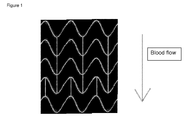
- the device according to the invention comprises a low gradient valve mounted on a stent with an outer surface as depicted in figure 1 .
- a low gradient valve mounted on a stent with an outer surface as depicted in figure 1 .
- Such a device may be delivered percutaneously and transluminally and is non thrombogenic.
- This device provides structures that anchor the stent in the aorta descendens and prevent its delocalization by the blood flow in the aorta.
- This invention relates to a device that provides a mechanical support that may be used to treat congestive heart failure. Placement of the device at its intended location leaves the diseased structure of the existing heart untouched. It is a mechanical support, which does not need the addition of external energy and may be applied both in the chronic and acute setting.
- the device according to the invention may be applied under conditions of heart failure whereas a disfunctioning heart valve is not mandatory.
- the device may be applied to the human or animal body by percutaneous and transluminal insertion. It is also possible to implant the device using a surgical method.
- drugs that may reduce the preload, reduce the afterload or strengthen the contraction force, without negatively influencing the coronary flow.
- Drug therapy with reduction of blood volume ie diuretics, reducing pre-and afterload
- vasodilatantia reducing afterload
- Intravenous drugs such as catecholamines are usually supplied in order to influence the contraction force. Such treatment however is only effective for a limited period of exacerbation, for instance during cardiac asthma.
- the mechanical interventions to change the mechanics of the heart can take place in the heart or in the large blood vessels outside the heart.
- the mechanical functioning of the heart can be changed by electrical add-ons inserting a PM, a PM and ICD, biventricular pacing.
- Mechanical intervention can take place in the structure of the heart so that congenitally defective structures are replaced by structures resembling the original structures as much as possible (eg ASD, VSD) or by removing obstructing structures (eg subaortale membrane, ASH, coronary stenoses) or by replacing defective dysfunctional structures by structures resembling the original structures as much as possible (eg AVR, MVR, PVR). In all these cases it is the aim to restore normal heart mechanics as much as possible by repairing, replacing or removing the defect.
- ASD congenitally defective structures are replaced by structures resembling the original structures as much as possible
- obstructing structures eg subaortale membrane, ASH, coronary stenoses
- replacing defective dysfunctional structures eg AVR, MVR, PVR
- pumps that are relatively easily inserted, such as the Impella pump, the Pulse Cath or the ELS. Usually they are intended to support the heart during a high-risk percutaneous intervention (eg PVR, Main-stem dilation) . There are also pumps that can only be implanted by a major operation eg the well known cardiac assist devices
- the mechanical functioning of the heart may be supported by an intervention outside the heart itself in the large vessels for the purpose to let the heart pump against a smaller resistance.
- the structure of the heart itself remains essentially unchanged.
- Hemoglobin content is high enough, bloodvein is the most simple form, reducing pre-and afterload by decrease in circulating volume but is still effective, even though the method is somewhat obsolete.
- the use of the balloon pump is widespread and is still undergoing improvements.
- the coronary perfusion begin diastole by inflation of the balloon is increased while end-diastolic afterload is reduced by deflation.
- the IABP reduces especially afterload, but is only temporarily applicable, not in a serious sclerotic aorta and also requires energy administration from the outside.
- Precursors of the first surgically replaced aortic valve and the first percutaneously implanted aortic valve were valves both placed in the aorta descendens . These were the valve inserted in 1953 by Hufnagel and the percutaneous inserted valve described in 2000 by Boudjemline and Bonhoeffer.
- Hufnagel has implanted in patients with severe Al events a ball in a cage high in the aorta descendens, he estimated that 75% of the cardiac output flows through the valve. It is estimated that some 4000 Hufnagel valves were implanted but only 55 are described well in the literature. The valve was in some cases 13 years in function. Hufnagel describes the effects of the valve in case of Al are decrease in afterload, an increase of cardiac output (up to 75%), decrease of the PAP, a decline in CTR, a reduction in NYHA class and increase exercise tolerance.
- Boudjemline and Bonhoeffer have implanted a valve in the aorta descendens in lambs by percutaneous insertion after they made an Al artificially. Earlier they had successfully implanted pulmonary valves this way. They argued that minimum back flow is necessary for any valve for sustained performance. Boudjemline and Bonhoeffer describe an increase in CO and coronary flow and decrease in peripheral pressures.
- US2006074483 describes a method of treatment and devices for the treatment of left ventricular failure.
- US3671979 describes a catheter mounted artificial heart valve for implanting in close proximity to a defective natural heart valve.
- US4056854 describes an aortic heart valve catheter. These are all catheter based mechanical techniques for treating the decompensated heart in the acute setting with temporary support, as far as we know these are the only three examples. The primary purpose of these three patented devices in the acute setting was to develop a cheaper solution than the IABP, with less monitoring needed. The entire expandable and impandable valves with mounting catheter remains in situ. The patient is bedridden and the valve must be removed after a short time.
- the current valves most used for PAVR are the Core Valve Revalving (18F) and the Edwards Sapien valve.
- stent-valves in development are the Bonhoefer valve, valve Aortech, Panagua valve, 3-F valve, Palmaz-Baily valve, Direct Flow valve, AorTx cover and Sadra Lotus valve.
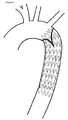
- the invention is primarily aimed at intraluminal and percutaneous insertion of an organic 3-Strut self-expandable or balloon mounted artificial valve, with only flow possible in the blood flow direction, which is inserted in the aorta descendens, preferably positioned a few cm after the left subclavian artery and is called artificial valve in the aorta descendens (AVAD, Van den Berg valve).
- AVAD artificial valve in the aorta descendens
- the valve can be delivered with a 24 18 or less French catheter.
- the AVAD is a biological valve because this presents no resistance to blood flowing in the aorta (gradient over the valve 0 mm Hg).
- the primary function of the valve is in reducing afterload downstream of the valve, where estimated 75% of Cardiac Output flows through.
- the compliance of the vascular system after this valve makes that during diastole the pressures are conducted only to the periphery and that the reflected wave of the first and higher order from the periphery are completely stopped in diastole.
- Bicuspid or tricuspid artificial valves are both suitable for placement in the device. It should be noted however that incomplete closure of the bicuspid valve especially in aging of the valve, as usually some degeneration is seen, occurs less often with a tricuspid valve. The pull forces will in the tricuspidal case be more spread over the whole annulus of artificial valve and will thus be smaller per square mm of the aortic wall and thus beneficial in preventing stent migration.
- the forces on the AVAD will only occur in retrograde direction during diastole, in the target population and is estimated to remain below 100 mm Hg, such as 75 or 50 mm Hg..
- the fixation by means of an expandable stent in the aortic wall must resist these forces, being the preferred that the valve is placed at the upsteam end of the stent and that the stent is at least 5 cm long.
- the downstream side is in particular the fixation side.
- the leaflets may comprise a drug eluting coating or equivalent means.
- the meshes in the stent should be large enough to maintain unrestricted blood flow to branching arteries.
- This invention is part of mechanical support techniques for a decompensated heart and is the only one which may be applied in the chronic setting even if the valves of the heart itself are intact.
- the described invention is a simple outpatient applicable mechanical form of intervention that may be used for chronic heart failure in the mobile patient.
- the AVAD provides the advantage of allowing a reduced risk of the procedure due to the percutaneous transluminal placement. This provides a reduced invasiveness and reduced complexity of insertion. It is a prerequisite for this technique that the aorta descendens after the left subclavian artery is intact over about seven or more centimeters.
- a device according to the invention provides the advantage that the valve is able to sustain a backward pressure gradient of more than 100 mm Hg. Such is true even at an early stage, when the valve is still not covered with endothelium, without dislocation.
- the device according to the invention may be placed in a smooth as well as a sclerotic aorta. If properly placed, i.e. at a few cm from the subclavian left artery and with a max length of 7 cm, the device will not provide any problems with prior arteries. In a device according to the invention, the chances of valve degeneration are minimal.and the chances of endothelial growth on the valve leaflets are also minimal. Moreover, there is no antegrade gradient over the device according to the invention. If the device according to the invention comprises Dacron, endothelialization is facilitated and any micro-embolies may be prevented.
- a device according to the invention may be applied using a conventional balloon.
- the inserted valve mounted on the balloon has a minimum diameter to be able to pass the femoral artery without much risk of damage.
Landscapes
- Health & Medical Sciences (AREA)
- Engineering & Computer Science (AREA)
- Biomedical Technology (AREA)
- Cardiology (AREA)
- Oral & Maxillofacial Surgery (AREA)
- Transplantation (AREA)
- Heart & Thoracic Surgery (AREA)
- Vascular Medicine (AREA)
- Life Sciences & Earth Sciences (AREA)
- Animal Behavior & Ethology (AREA)
- General Health & Medical Sciences (AREA)
- Public Health (AREA)
- Veterinary Medicine (AREA)
- Prostheses (AREA)
Priority Applications (2)
| Application Number | Priority Date | Filing Date | Title |
|---|---|---|---|
| EP09158497A EP2243443A1 (de) | 2009-04-22 | 2009-04-22 | Ein mit einer Trikuspidalklappe ausgestatteter Stent |
| PCT/EP2009/055957 WO2009141286A1 (en) | 2008-05-19 | 2009-05-15 | Method for treating heart diseases |
Applications Claiming Priority (1)
| Application Number | Priority Date | Filing Date | Title |
|---|---|---|---|
| EP09158497A EP2243443A1 (de) | 2009-04-22 | 2009-04-22 | Ein mit einer Trikuspidalklappe ausgestatteter Stent |
Publications (1)
| Publication Number | Publication Date |
|---|---|
| EP2243443A1 true EP2243443A1 (de) | 2010-10-27 |
Family
ID=40934924
Family Applications (1)
| Application Number | Title | Priority Date | Filing Date |
|---|---|---|---|
| EP09158497A Withdrawn EP2243443A1 (de) | 2008-05-19 | 2009-04-22 | Ein mit einer Trikuspidalklappe ausgestatteter Stent |
Country Status (1)
| Country | Link |
|---|---|
| EP (1) | EP2243443A1 (de) |
Citations (8)
| Publication number | Priority date | Publication date | Assignee | Title |
|---|---|---|---|---|
| US3671979A (en) | 1969-09-23 | 1972-06-27 | Univ Utah | Catheter mounted artificial heart valve for implanting in close proximity to a defective natural heart valve |
| US4056854A (en) | 1976-09-28 | 1977-11-08 | The United States Of America As Represented By The Department Of Health, Education And Welfare | Aortic heart valve catheter |
| WO2000047139A1 (en) * | 1999-02-10 | 2000-08-17 | Heartport, Inc. | Methods and devices for implanting cardiac valves |
| US6168614B1 (en) * | 1990-05-18 | 2001-01-02 | Heartport, Inc. | Valve prosthesis for implantation in the body |
| DE10121210A1 (de) * | 2001-04-30 | 2002-11-14 | Universitaetsklinikum Freiburg | Gefässimplantat |
| WO2003092554A1 (en) * | 2002-05-03 | 2003-11-13 | The General Hospital Corporation | Involuted endovascular valve and method of construction |
| US20060074483A1 (en) | 2004-10-01 | 2006-04-06 | Schrayer Howard L | Method of treatment and devices for the treatment of left ventricular failure |
| US20060155366A1 (en) * | 2005-01-10 | 2006-07-13 | Laduca Robert | Apparatus and method for deploying an implantable device within the body |
-
2009
- 2009-04-22 EP EP09158497A patent/EP2243443A1/de not_active Withdrawn
Patent Citations (8)
| Publication number | Priority date | Publication date | Assignee | Title |
|---|---|---|---|---|
| US3671979A (en) | 1969-09-23 | 1972-06-27 | Univ Utah | Catheter mounted artificial heart valve for implanting in close proximity to a defective natural heart valve |
| US4056854A (en) | 1976-09-28 | 1977-11-08 | The United States Of America As Represented By The Department Of Health, Education And Welfare | Aortic heart valve catheter |
| US6168614B1 (en) * | 1990-05-18 | 2001-01-02 | Heartport, Inc. | Valve prosthesis for implantation in the body |
| WO2000047139A1 (en) * | 1999-02-10 | 2000-08-17 | Heartport, Inc. | Methods and devices for implanting cardiac valves |
| DE10121210A1 (de) * | 2001-04-30 | 2002-11-14 | Universitaetsklinikum Freiburg | Gefässimplantat |
| WO2003092554A1 (en) * | 2002-05-03 | 2003-11-13 | The General Hospital Corporation | Involuted endovascular valve and method of construction |
| US20060074483A1 (en) | 2004-10-01 | 2006-04-06 | Schrayer Howard L | Method of treatment and devices for the treatment of left ventricular failure |
| US20060155366A1 (en) * | 2005-01-10 | 2006-07-13 | Laduca Robert | Apparatus and method for deploying an implantable device within the body |
Similar Documents
| Publication | Publication Date | Title |
|---|---|---|
| US7771467B2 (en) | Apparatus for repairing the function of a native aortic valve | |
| US8758430B2 (en) | Medical apparatus for the therapeutic treatment of an insufficient cardiac valve | |
| JP6738392B2 (ja) | 血管内大動脈修復用デバイスおよびその使用方法 | |
| JP4403183B2 (ja) | 置換心臓弁の経カテーテル送達 | |
| CN104470579B (zh) | 人工肾脏瓣膜 | |
| US7004925B2 (en) | Apparatus and method for auto-retroperfusion of a coronary vein | |
| EP1620040B1 (de) | Venenklappenprothese zur verringerung von druckwirkungen bei der kardialen trikuspidalklappen-regurgiation | |
| US7591848B2 (en) | Riveted stent valve for percutaneous use | |
| JP5356499B2 (ja) | 複オリフィス移植可能心臓弁及び移植方法 | |
| KR102563467B1 (ko) | 카테터경유 폐의 볼 판막 조립체 | |
| US7625403B2 (en) | Valved conduit designed for subsequent catheter delivered valve therapy | |
| US7004926B2 (en) | Apparatus and method for auto-retroperfusion of a coronary vein | |
| US7473237B2 (en) | Apparatus for auto-retroperfusion of a coronary vein | |
| US20070156233A1 (en) | Percutaneous atrioventricular valve and method of use | |
| US20060195004A1 (en) | Minimally invasive transvalvular ventricular assist device | |
| US20050049692A1 (en) | Medical device for reduction of pressure effects of cardiac tricuspid valve regurgitation | |
| US20110046726A1 (en) | Apparatus for Implanting an Aortic Valve Prosthesis | |
| US10335272B2 (en) | Apparatus and method for repairing the function of a diseased valve | |
| EP2243443A1 (de) | Ein mit einer Trikuspidalklappe ausgestatteter Stent | |
| WO2009141286A1 (en) | Method for treating heart diseases | |
| RU2845237C1 (ru) | Способ открытой имплантации самораскрывающегося протеза аортального клапана в эксперименте | |
| RU2479287C2 (ru) | Биологический протез аортального клапана сердца |
Legal Events
| Date | Code | Title | Description |
|---|---|---|---|
| PUAI | Public reference made under article 153(3) epc to a published international application that has entered the european phase |
Free format text: ORIGINAL CODE: 0009012 |
|
| AK | Designated contracting states |
Kind code of ref document: A1 Designated state(s): AT BE BG CH CY CZ DE DK EE ES FI FR GB GR HR HU IE IS IT LI LT LU LV MC MK MT NL NO PL PT RO SE SI SK TR |
|
| AX | Request for extension of the european patent |
Extension state: AL BA RS |
|
| STAA | Information on the status of an ep patent application or granted ep patent |
Free format text: STATUS: THE APPLICATION IS DEEMED TO BE WITHDRAWN |
|
| 18D | Application deemed to be withdrawn |
Effective date: 20110428 |