EP2244228A1 - Procédé et appareil pour balayer une préparation ou une cartouche contenant des micro-objets - Google Patents
Procédé et appareil pour balayer une préparation ou une cartouche contenant des micro-objets Download PDFInfo
- Publication number
- EP2244228A1 EP2244228A1 EP09305338A EP09305338A EP2244228A1 EP 2244228 A1 EP2244228 A1 EP 2244228A1 EP 09305338 A EP09305338 A EP 09305338A EP 09305338 A EP09305338 A EP 09305338A EP 2244228 A1 EP2244228 A1 EP 2244228A1
- Authority
- EP
- European Patent Office
- Prior art keywords
- region
- cartridge
- quality value
- quality
- microobjects
- Prior art date
- Legal status (The legal status is an assumption and is not a legal conclusion. Google has not performed a legal analysis and makes no representation as to the accuracy of the status listed.)
- Ceased
Links
Images



Classifications
-
- G—PHYSICS
- G06—COMPUTING OR CALCULATING; COUNTING
- G06T—IMAGE DATA PROCESSING OR GENERATION, IN GENERAL
- G06T7/00—Image analysis
- G06T7/0002—Inspection of images, e.g. flaw detection
- G06T7/0012—Biomedical image inspection
-
- G—PHYSICS
- G06—COMPUTING OR CALCULATING; COUNTING
- G06T—IMAGE DATA PROCESSING OR GENERATION, IN GENERAL
- G06T7/00—Image analysis
- G06T7/10—Segmentation; Edge detection
- G06T7/11—Region-based segmentation
-
- G—PHYSICS
- G06—COMPUTING OR CALCULATING; COUNTING
- G06T—IMAGE DATA PROCESSING OR GENERATION, IN GENERAL
- G06T2207/00—Indexing scheme for image analysis or image enhancement
- G06T2207/10—Image acquisition modality
- G06T2207/10056—Microscopic image
-
- G—PHYSICS
- G06—COMPUTING OR CALCULATING; COUNTING
- G06T—IMAGE DATA PROCESSING OR GENERATION, IN GENERAL
- G06T2207/00—Indexing scheme for image analysis or image enhancement
- G06T2207/30—Subject of image; Context of image processing
- G06T2207/30168—Image quality inspection
Definitions
- the region of interest of the slide or cartridge is determined on the basis of quality values associated with elementary regions of the slide or cartridge. This allows confining the region of interest to a selection of elementary regions having a sufficiently high quality value, to avoid scanning regions of the slide or cartridge of which it is expected that they would give rise to microscopic images of inferior quality.
- the size of each of the elementary regions may match the field of view of the imaging device, or be somewhat smaller.
- Each quality value is understood to indicate a quality of the elementary region.
- the at least one predefined quality value is defined before the fluid is contained in the slide or cartridge.
- This embodiment is based on the insight that a quality distribution of the fluid in the slide or cartridge may depend on intrinsic properties of the slide or cartridge and/or the fluid, such as the geometry of the slide or cartridge and/or the viscosity of the fluid.
- the at least one predefined quality value may be retrieved from a data carrier.
- the data carrier may be arranged on the slide or cartridge or elsewhere.
- the data carrier may be an electronic or optical chip attached to the cartridge.
- the data carrier could be a storage device of an external work station such as a personal computer.
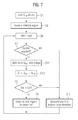
- the region of interest is determined on the basis of the third quality value, the latter being derived from both predefined data and from the image of the elementary region, the image reflecting properties of the fluid actually contained in that region of the slide or cartridge.
- the third quality value For example, when blood is introduced into a passive cartridge, it will flow by capillary forces. This results in a certain distribution of red blood cells that can be taken into account when optically imaging the cartridge.
- This allows for an efficient scanning strategy which may depend mainly on the detected cartridge type but may also adapt to external factors (like temperature, humidity, blood viscosity) by image processing methods. More efficient imaging, and a shorter time to diagnosis may thus be achieved.
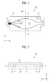
- the staining dye could, for example, be contained in a dissolvable coating on an inner surface of the cartridge 10. Thus the dye would be released to the fluid 22 when the fluid 22 contacts the coating. Once the fluid 22 has filled the inner space 20, it is ready for being imaged by means of an imaging device such as an optical scanning microscope.
- the fluid 22, considered over the entire inner space 20 is usually not homogeneous.
- characteristics of the flow of the fluid 22 velocity, turbulence
- a staining agent is added to the fluid 22, e.g. at the inlet 16, one may expect that the staining intensity will not be homogeneous over the entire volume 20.
- stain intensity could be generally larger near the outlet 18 than near the inlet 16 when the fluid 22 has come to a rest. In this case, an optimum staining intensity might be expected near a central region of the inner space 20.
- the flow pattern of the fluid 22 may be very complex. Therefore, the distribution of microobjects and/or a staining intensity may be equally complex.
- ⁇ RBC can be measured by image segmentation of the RBCs.
- f RBC opt is the optimal ratio for the image processing software.
- ⁇ RBC controls how severely a deviation from the optimal ratio f RBC opt is penalized.
Landscapes
- Engineering & Computer Science (AREA)
- Computer Vision & Pattern Recognition (AREA)
- Physics & Mathematics (AREA)
- General Physics & Mathematics (AREA)
- Theoretical Computer Science (AREA)
- Health & Medical Sciences (AREA)
- General Health & Medical Sciences (AREA)
- Medical Informatics (AREA)
- Nuclear Medicine, Radiotherapy & Molecular Imaging (AREA)
- Radiology & Medical Imaging (AREA)
- Quality & Reliability (AREA)
- Microscoopes, Condenser (AREA)
Priority Applications (2)
| Application Number | Priority Date | Filing Date | Title |
|---|---|---|---|
| EP09305338A EP2244228A1 (fr) | 2009-04-21 | 2009-04-21 | Procédé et appareil pour balayer une préparation ou une cartouche contenant des micro-objets |
| PCT/IB2010/051660 WO2010122462A1 (fr) | 2009-04-21 | 2010-04-16 | Procédé et appareil pour balayer une lame ou une cartouche contenant des micro-objets |
Applications Claiming Priority (1)
| Application Number | Priority Date | Filing Date | Title |
|---|---|---|---|
| EP09305338A EP2244228A1 (fr) | 2009-04-21 | 2009-04-21 | Procédé et appareil pour balayer une préparation ou une cartouche contenant des micro-objets |
Publications (1)
| Publication Number | Publication Date |
|---|---|
| EP2244228A1 true EP2244228A1 (fr) | 2010-10-27 |
Family
ID=40933663
Family Applications (1)
| Application Number | Title | Priority Date | Filing Date |
|---|---|---|---|
| EP09305338A Ceased EP2244228A1 (fr) | 2009-04-21 | 2009-04-21 | Procédé et appareil pour balayer une préparation ou une cartouche contenant des micro-objets |
Country Status (2)
| Country | Link |
|---|---|
| EP (1) | EP2244228A1 (fr) |
| WO (1) | WO2010122462A1 (fr) |
Families Citing this family (1)
| Publication number | Priority date | Publication date | Assignee | Title |
|---|---|---|---|---|
| CN121438311B (zh) * | 2025-12-31 | 2026-04-03 | 奥凸科技有限公司 | 基于多倍率显微图像的稀疏扫描路径规划方法及系统 |
Citations (1)
| Publication number | Priority date | Publication date | Assignee | Title |
|---|---|---|---|---|
| WO1996007919A1 (fr) | 1994-09-02 | 1996-03-14 | Biometric Imaging, Inc. | Cartouche a usage unique et procede pour analyser un echantillon biologique |
-
2009
- 2009-04-21 EP EP09305338A patent/EP2244228A1/fr not_active Ceased
-
2010
- 2010-04-16 WO PCT/IB2010/051660 patent/WO2010122462A1/fr not_active Ceased
Patent Citations (1)
| Publication number | Priority date | Publication date | Assignee | Title |
|---|---|---|---|---|
| WO1996007919A1 (fr) | 1994-09-02 | 1996-03-14 | Biometric Imaging, Inc. | Cartouche a usage unique et procede pour analyser un echantillon biologique |
Non-Patent Citations (6)
| Title |
|---|
| DI RUBERTO C ET AL: "Analysis of infected blood cell images using morphological operators", IMAGE AND VISION COMPUTING ELSEVIER NETHERLANDS, vol. 20, no. 2, 1 February 2002 (2002-02-01), pages 133 - 146, XP002514488, ISSN: 0262-8856 * |
| DIAZ G ET AL: "Infected cell identification in thin blood images based on color pixel classification: comparison and analysis", PROGRESS IN PATTERN RECOGNITION, IMAGE ANALYSIS AND APPLICATIONS. PROCEEDINGS 12TH IBEROAMERICAN CONGRESS ON PATTERN RECOGNITION, CIARP 2007. (LECTURE NOTES IN COMPUTER SCIENCE VOL. 4756) SPRINGER-VERLAG BERLIN, GERMANY, 2007, pages 812 - 821, XP019083029, ISBN: 3-540-76724-X * |
| HALIM S ET AL: "Estimating malaria parasitaemia from blood smear images", 2006 9TH INTERNATIONAL CONFERENCE ON CONTROL, AUTOMATION, ROBOTICS AND VISION (IEEE CAT. NO.06EX1361) IEEE PISCATAWAY, NJ, USA, 2006, pages 6 pp., XP002540927, ISBN: 1-4244-0342-1 * |
| M. T. LE ET AL: "A novel semi-automatic image processing approach to determine Plasmodium falciparum parasitemia in Giemsa -stained thin blood smears", BMC CELL BIOLOGY, vol. 9, 2008, pages 1 - 12, XP002540787, Retrieved from the Internet <URL:http://www.pubmedcentral.nih.gov/picrender.fcgi?artid=2330144&blobtype=pdf> [retrieved on 20090811] * |
| S. W. S. SIO ET AL: "MalariaCount: an image analysis-based program for the accurate determination of parasitemia", J. MICROBIOL. METHODS, vol. 68, no. 1, January 2007 (2007-01-01), pages 11 - 18, XP002540788, Retrieved from the Internet <URL:http://www.ncbi.nlm.nih.gov/pubmed/16837087?ordinalpos=1&itool=EntrezSystem2.PEntrez.Pubmed.Pubmed_ResultsPanel.Pubmed_DiscoveryPanel.Pubmed_Discovery_RA&linkpos=1&log$=relatedarticles&logdbfrom=pubmed> [retrieved on 20090811] * |
| VISHNU V MAKKAPATI ET AL: "Segmentation of malaria parasites in peripheral blood smear images", ACOUSTICS, SPEECH AND SIGNAL PROCESSING, 2009. ICASSP 2009. IEEE INTERNATIONAL CONFERENCE ON, IEEE, PISCATAWAY, NJ, USA, 19 April 2009 (2009-04-19), pages 1361 - 1364, XP031459491, ISBN: 978-1-4244-2353-8 * |
Also Published As
| Publication number | Publication date |
|---|---|
| WO2010122462A1 (fr) | 2010-10-28 |
Similar Documents
| Publication | Publication Date | Title |
|---|---|---|
| US11226280B2 (en) | Automated slide assessments and tracking in digital microscopy | |
| JP7214729B2 (ja) | 光学測定用試料収容器 | |
| Osibote et al. | Automated focusing in bright‐field microscopy for tuberculosis detection | |
| US7177454B2 (en) | Automated detection of objects in a biological sample | |
| Basu | Droplet morphometry and velocimetry (DMV): a video processing software for time-resolved, label-free tracking of droplet parameters | |
| US20180365480A1 (en) | Method and apparatus for automated platelet identification within a whole blood sample from microscopy images | |
| EP3762758B1 (fr) | Appareils, systèmes et procédés d'imagerie | |
| US20190022649A1 (en) | Detection of microscopic objects | |
| EP1203343A1 (fr) | Detection automatique d'objets dans un echantillon biologique | |
| JP7386771B2 (ja) | マルチモーダル極低温適合性guidグリッド | |
| CN105659151A (zh) | 数码显微系统、方法与计算机程序产品 | |
| Horning et al. | A paper microfluidic cartridge for automated staining of malaria parasites with an optically transparent microscopy window | |
| Saeki et al. | Digital cell counting device integrated with a single-cell array | |
| Wilkins Stevens et al. | Imaging and analysis of immobilized particle arrays | |
| Jagannadh et al. | A semi-automated, field-portable microscopy platform for clinical diagnostic applications | |
| EP2244228A1 (fr) | Procédé et appareil pour balayer une préparation ou une cartouche contenant des micro-objets | |
| US20250182274A1 (en) | Quantifying constituents in a sample chamber using images of depth regions | |
| Matula et al. | Acquiarium: free software for the acquisition and analysis of 3D images of cells in fluorescence microscopy | |
| Alidoust Chamandani et al. | Indirect optical geometry measurement based on optical tweezers in transparent microchannels | |
| AU2014202121B2 (en) | Detection of microscopic objects | |
| Shapiro | Scanning laser cytometry | |
| Tan et al. | A novel autofocusing method using the angle of Hilbert space for microscopy | |
| Maia et al. | High-content microscopy | |
| CN118151361A (zh) | 一种样本图像分析设备和样本图像的成像方法 | |
| CA2848951C (fr) | Detection d'objets microscopiques |
Legal Events
| Date | Code | Title | Description |
|---|---|---|---|
| PUAI | Public reference made under article 153(3) epc to a published international application that has entered the european phase |
Free format text: ORIGINAL CODE: 0009012 |
|
| AK | Designated contracting states |
Kind code of ref document: A1 Designated state(s): AT BE BG CH CY CZ DE DK EE ES FI FR GB GR HR HU IE IS IT LI LT LU LV MC MK MT NL NO PL PT RO SE SI SK TR |
|
| STAA | Information on the status of an ep patent application or granted ep patent |
Free format text: STATUS: THE APPLICATION HAS BEEN REFUSED |
|
| 18R | Application refused |
Effective date: 20101203 |