EP2245592B1 - Métrique d'alignement de superposition d'image - Google Patents
Métrique d'alignement de superposition d'image Download PDFInfo
- Publication number
- EP2245592B1 EP2245592B1 EP09701915A EP09701915A EP2245592B1 EP 2245592 B1 EP2245592 B1 EP 2245592B1 EP 09701915 A EP09701915 A EP 09701915A EP 09701915 A EP09701915 A EP 09701915A EP 2245592 B1 EP2245592 B1 EP 2245592B1
- Authority
- EP
- European Patent Office
- Prior art keywords
- voxels
- interest
- tissue
- metric
- data
- Prior art date
- Legal status (The legal status is an assumption and is not a legal conclusion. Google has not performed a legal analysis and makes no representation as to the accuracy of the status listed.)
- Not-in-force
Links
Images



Classifications
-
- G—PHYSICS
- G06—COMPUTING OR CALCULATING; COUNTING
- G06T—IMAGE DATA PROCESSING OR GENERATION, IN GENERAL
- G06T7/00—Image analysis
- G06T7/30—Determination of transform parameters for the alignment of images, i.e. image registration
- G06T7/33—Determination of transform parameters for the alignment of images, i.e. image registration using feature-based methods
-
- G—PHYSICS
- G06—COMPUTING OR CALCULATING; COUNTING
- G06T—IMAGE DATA PROCESSING OR GENERATION, IN GENERAL
- G06T2207/00—Indexing scheme for image analysis or image enhancement
- G06T2207/10—Image acquisition modality
- G06T2207/10072—Tomographic images
- G06T2207/10081—Computed x-ray tomography [CT]
-
- G—PHYSICS
- G06—COMPUTING OR CALCULATING; COUNTING
- G06T—IMAGE DATA PROCESSING OR GENERATION, IN GENERAL
- G06T2207/00—Indexing scheme for image analysis or image enhancement
- G06T2207/10—Image acquisition modality
- G06T2207/10072—Tomographic images
- G06T2207/10108—Single photon emission computed tomography [SPECT]
-
- G—PHYSICS
- G06—COMPUTING OR CALCULATING; COUNTING
- G06T—IMAGE DATA PROCESSING OR GENERATION, IN GENERAL
- G06T2207/00—Indexing scheme for image analysis or image enhancement
- G06T2207/30—Subject of image; Context of image processing
- G06T2207/30004—Biomedical image processing
- G06T2207/30048—Heart; Cardiac
Definitions
- the present application generally relates to registering images generated from data acquired by different imaging modalities. While it is described with particular application to a single photon emission compute tomography / computer tomography (SPECT / CT) system, it also relates to other medical and non-medical imaging applications.
- SPECT / CT computer tomography
- Hybrid multi-modality imaging systems include SPECT/CT, PET/CT and PET/MRI systems.
- a hybrid multi-modality registering system is presented in an article by KESSLER M.L.: "Image registration and data fusion in radiation therapy", THE BRITISH JOURNAL OF RADIOLOGY, vol. 79, 2006, pages S99-S108, XP005050049 ISSN:0167-8140 .
- data is sequentially acquired in that first data is acquired with one of the modalities and then second data is acquired with the other modality.
- the images generated with the first and second data may have some degree of mechanical misalignment. Even if the mechanical misalignment can be corrected, there may be misalignment due to the motion of the patient or the movement of physiological tissue such as an organ from scan to scan.
- the relative position of the heart in the body is a function of the respiratory cycle.
- a CT scan acquires data within seconds or less.
- the CT scan can be considered as a snapshot of the respiratory cycle.
- the SPECT scan acquires data over a relatively longer period of time relative to the CT scan.
- the SPECT scan can be considered as an average over the respiratory cycle.
- a consequence of the different acquisition times is that the resulting CT and SPECT images may not align with respect to the cardiac region.
- the literature has reported misalignment in the cardiac region ranging from zero (0) to twenty-four (24) millimeters (mm) in the head-to-foot or longitudinal direction.
- clinicians have visually inspected the registration of two images before making a clinical diagnostic decision or performing other tasks, such as attenuation correcting (AC) data in the case of SPECT/CT and PET/CT.
- AC attenuation correcting
- the clinician browses through multiple orthogonal views to inspect the misalignment in different slices, and moves the images, if necessary, to correct for misalignment.
- Such manual inspection and correction may be time consuming and tedious.
- the clinician manually aligns the CT heart data with the non-AC SPECT heart data, and then CT-AC is performed on the SPECT data.
- CT-AC is performed on the SPECT data.
- a consequence of such alignment approach is that other tissue may not be appropriately registered, and artifacts may be introduced into the data after the CT-AC.
- a method includes obtaining a combined data set that includes first and second imaging data sets.
- the first and second imaging data sets correspond to different imaging modalities.
- the method further includes determining a metric indicative of an alignment between the first and second imaging data sets in the combined data set.
- the method further includes presenting the metric in a human readable format.
- the method further includes segmenting the first data set, based on tissue of interest, to generate a first segmented region that includes the tissue of interest.
- the method also includes identifying a first set of voxels, in the segmented data set, that have first values that correspond to the tissue of interest.
- the method includes determining a first number of voxels in the first set of voxels, identifying a second set of voxels, in the second data set, that correspond to the first voxels, determining a second number of voxels in the second set of voxels that have second values that correspond to the tissue of interest, and determining the metric based on the first and second number of voxels.
- a system in another aspect, includes a data combiner that combines first and second imaging data sets.
- the first and second imaging data sets correspond to different imaging modalities.
- a metric determining component determines a metric indicative of an alignment between the first and second imaging data sets in the combined data set.
- a registration component registers the first and second imaging data sets when the metric is indicative of an unacceptable alignment between the first and second imaging data sets.
- a segmentation component segments the first data set, based on tissue of interest, to generate a first segmented region that includes the tissue of interest.
- a voxels counter counts a first number of voxels in a first set of voxels in the segmented data set that have first values that correspond to the tissue of interest and that counts a second number of voxels in a second set of voxels in the second data set that both correspond to the first voxels and have second values that correspond to the tissue of interest, wherein the metric is based on the first and second number of voxels.
- a computer readable storage medium containing instructions which, when executed by a computer, cause the computer to perform the steps of: determining a metric indicative of an alignment between first imaging data fused with second imaging data; and registering the first and second imaging data when the metric is indicative of an unacceptable alignment between the first and second imaging data sets, wherein the instructions, when executed by the computer, further cause the computer to perform the steps of: segmenting the first data, based on tissue of interest, to generate a first segmented region that includes the tissue of interest, and determining a number of voxels in a first set of voxels in the segmented data set that have first values that correspond to the tissue of interest; and a second number of voxels in a second set of voxels in the second data set that both correspond to the first set of voxels and have second values that correspond to the tissue of interest, wherein the metric is based on the first and second number of voxels in the first and second sets of voxels.
- the invention may take form in various components and arrangements of components, and in various steps and arrangements of steps.
- the drawings are only for purposes of illustrating the preferred embodiments and are not to be construed as limiting the invention.
- FIGURE 1 illustrates an imaging system 100 that includes a single photon emission computed tomography (SPECT) portion 102 and a computed tomography (CT) portion 104.
- SPECT single photon emission computed tomography
- CT computed tomography
- the portions 102 and 104 are part of a single hybrid scanner.
- the portions 102 and 104 are respectively part of separate scanners, which can be located proximate or remote from each other.
- the system 100 includes a gantry that includes both x-ray detectors and SPECT detectors.
- the medical imaging system may alternatively or additionally include a positron emission tomography (PET) portion and/or a magnetic resonance (MR) portion.
- PET positron emission tomography
- MR magnetic resonance
- the SPECT portion 102 includes at least one gamma radiation sensitive detector 106.
- Two gamma radiation sensitive detectors 106 1 and 106 2 are show in the illustrated example.
- the gamma radiation sensitive detectors 106 are disposed relative to each other at an angle in the range of roughly ninety (90) degrees to one hundred and two (102) degrees. Other angles are also contemplated.
- the gamma radiation sensitive detectors 106 are configured to rotate around a SPECT examination region 108 and about a longitudinal or z-axis 110 to acquire projections for plurality of projection angles or views.
- Each of the illustrated gamma radiation sensitive detectors 106 includes a two-dimensional array of photosensors 112 and a scintillator 114 in optical communication with the two-dimensional array of photosensors 112.
- the scintillator 114 receives gamma radiation from radionuclide decay occurring in the SPECT examination region 108 and produces photons in response thereto.
- the photosensors 112 generate electrical signals indicative of the detected gamma radiation.
- the CT portion 104 includes an x-ray radiation source 116, such as an x-ray tube, that rotates about a CT examination region 118.
- the x-ray radiation source 116 is stationary.
- the radiation source 116 emits x-ray radiation that traverses the CT examination region 118.
- a radiation sensitive detector 120 subtends an angular arc opposite the examination region 118 with respect to the radiation source 116.
- the radiation sensitive detector 120 may rotate in coordination with the x-ray radiation source 116 as in a third generation system, or remain stationary as in a fourth generation system, or any other x-ray (CT) system configuration.
- CT x-ray
- the radiation sensitive detector 120 includes a two-dimensional array of photosensors 124 and a scintillator 122 in optical communication with the two-dimensional array of photosensors 124.
- the scintillator 122 receives x-ray radiation traversing the CT examination region 118 and produces photons in response thereto.
- the photosensors 124 generate electrical signals indicative of the detected x-ray radiation.
- a SPECT reconstructor 128 reconstructs the SPECT projections to generate volumetric image data indicative of the SPECT examination region 108, based on the detected gamma radiation. This may include reconstructing concurrently acquired data for different energy windows, including different primary radiation energy windows and/or a scatter radiation energy window.
- a CT reconstructor 130 reconstructs the CT projections to generate volumetric image data indicative of the CT examination region 118, based on the detected x-ray radiation.
- An object support 132 supports an object, such as a human patient, luggage, etc., to be imaged.
- the object support 132 is configured to longitudinally move in coordination with operation of the system 100 so that an object can be scanned at a plurality of longitudinal locations by either or both of the SPECT and CT portions 102 and 104 according to desired scanning trajectories.
- the SPECT and CT portions 102 and 104 are located in close proximity (as shown) with their respective examination regions 108 and 118 disposed along the common z-axis 110.
- a computer serves an operator console 134.
- the console 134 includes a human readable output device such as a monitor or display and input devices such as a keyboard and mouse.
- Software resident on the console 134 allows the operator to view and otherwise manipulate the volumetric image data, for example, through a graphical user interface (GUI).
- Software resident on the console 134 also allows the operator to control the operation of the system 100 by establishing desired scan protocols, initiating and terminating scans, and otherwise interacting with the scanner 100.
- a data combiner 136 combines the SPECT and CT data. In one instance, this includes re-sampling the CT data into SPECT space. For example, the CT data may be converted into an attenuation map with a substantially same voxel size and resolution as the SPECT data. Other re-sampling is also contemplated. It is to be appreciated that the data in the combined set may be registered via a rigid registration or a non-rigid registration, or not registered.
- a pre-processor 137 suitably processes the combined data. As described in greater detail below, this may include segmenting one or more of the combined data sets, contouring one or more of the combined data sets, and/or otherwise processing one or more the combined data sets.
- An alignment metric determining component 138 determines a first metric or figure of merit indicative of the alignment (or misalignment) of information in the combined data.
- the metric may be a similarity metric indicative of the similarity between information in the combined SPECT data and the CT data.
- a second metric or figure of merit may be generated based on the alignment metric.
- the second metric may be indicative of an estimated distance (e.g., two (2) millimeters) between regions of interest in the combined data.
- One or both metrics may be presented, for example, via the console 134 or otherwise. As a result, an operator may be informed as to the degree of alignment between the information in the data sets in the combined data.
- a decision component 140 determines whether the combined data should be registered or registered again (e.g., when the data sets have already been registered, but the registration for a region or tissue of interest is less than desirable) based on the first and/or second metrics. This may be done automatically and/or based on operator input. As noted above, these metrics are indicative of the degree of alignment between the information in the combined data sets. As such, a human operator and/or executing software can use the metric to determine whether the combined data should be registered. In addition, the operator can selectively browse through and/or otherwise process (e.g., manually or automatically register) the combined data based on the metric.
- a registration component 142 registers or performs a secondary registration (where the data is already registered) when it is determined that the combined data sets should be registered based on the first and/or second metrics. After such a registration, the alignment metric determining component 138 determines the metrics for the newly registered data, and the decision component 140 again determines whether another registration should be performed.
- a processing component 144 further processes the combined SPECT and CT data or the registered combined SPECT and CT data, for example, by attenuation correcting the SPECT data based on the CT data and/or otherwise.
- FIGURE 1 shows the console 134 in connection with the SPECT and CT portions 102 and 104 and the portions 136-144, it is to be appreciated that one or more of the processing portions 136-144 may be part of and/or performed in a different computer, such as a dedicated workstation, a general purpose computer or other computer.
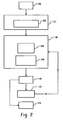
- FIGURE 2 illustrates an example embodiment. The example is discussed in relation to non-cardiac SPECT/CT. However, cardiac SPECT/CT and/or other applications are also contemplated.
- the SPECT and CT data are combined via the data combiner 136, which may include re-sampling the CT data into SPECT space, and the combined data sets may or may not be registered.
- a contour identifying component 202 of the pre-processor 137 identifies a contour of tissue of interest, such as an anatomical organ, in both the SPECT data and the CT data.
- the contour may be identified in the volume of data reconstructed from SPECT primary or scatter data.
- a similarity determining component 204 of the alignment metric determining component 138 determines a similarity metric between the contours.
- the similarity metric is a distance metric such as a Euclidean distance between the contours. Equation 1 below illustrates an example approach for computing the Euclidean distance, normalized by the total number of voxels, between the voxels of the contours.
- D 3 ⁇ i x i ⁇ 1 - x i ⁇ 0 2 + y i ⁇ 1 - y i ⁇ 0 2 + z i ⁇ 1 - z i ⁇ 0 2 , where D 3 represents the Euclidian distance, i represents the total number of voxels, x i 1 , y i 1 and z i 1 represent the coordinates of the voxels of either the SPECT or the CT data, and x i 0 , y i 0 and z i 0 represent the coordinates of the voxels of the other SPECT or the CT data.
- distance metric may alternatively be performed in two-dimensional space based on the pixels of the contours.
- the similarity metric may be based on cross-correlation, a root mean square deviation, mutual information, normalized mutual information, entropy, entropy correlation coefficient, and/or other information. It is also to be appreciated that the similarity metric may be determined based on an exhaustive, stochastic, Gauss-Newton, etc. approach.
- a clinical metric generating component 206 generates a second metric based on the similarity value.
- the second metric may be indicative of an estimated real distance between regions of interest in the combined data, and may also include a relative directional component with respect to one of the data sets or some other defined reference.
- the second metric may indicate that two regions of interest, one from one of the combined data sets and one from the other one of the combined data sets, are misaligned by about X millimeters or other units describing how far apart the regions of interest are with respect to the current registration, wherein X is a real number.
- the second metric is a clinically relevant metric that can be used in an iterative manner to help the clinician provide for a good registration.
- the second metric may be variously determined. For example, a mapping between the similarity value and the second metric may be provided via a look up table. In another example, the second metric may be dynamically determined via a polynomial or other equation. In another example, the second metric is determined without the similarity value, for example, directly or otherwise from the combined data. In one embodiment, the clinical metric generating component 206 is omitted.
- the illustrated decision component 140 determines whether the SPECT and CT data are to be registered or not.
- the decision component 140 may use operator input when rendering the decision. For example, the similarity metric and/or the clinical metric may be presented to the operator, for instance, via the console 134 and/or other output device. An operator or other input (including lack of an input) is then used by the decision component 140 to determine whether the SPECT and CT data are registered.
- the decision component 140 makes the decision without any operator input.
- Other information such as a similarity threshold value, a clinical distance threshold value, a recommendation, etc. that may be useful for making such a determination may also be presented to the operator.
- a suitable threshold may be pre-determined based on manually moving the transmission map with different amounts of misalignments in one or more data sets and verifying the alignment from known data. Machine learning techniques including implicitly and explicitly trained classifiers and the like may also be employed.
- the combined data can be further processed by the processing component 144.
- the registration component 142 performs a volume preserving registration, such as a rigid registration.
- a registration may include translation between data sets in one or more spatial directions including, one (1), two (2) or three (3) spatial directions.
- the combined data may be presented via the console 134 and/or other output device to operator, who manually and interactively translates either the SPECT or the CT data with respect to the other data. While translating the data, the operator can visually assess the overlap of the data, for example, from one or more coronal, sagittal, and/or transaxial views.
- the similarly metric can be dynamically updated and presented to the operator to facilitate registration as the data sets are moved relative to each other.
- the rigid registration may also include a rotational component, and/or an elastic registration is also contemplated.
- an automatic registration is used.
- the general principle may be to find the spatial transformation between the data sets that renders a highest spatial correlation between the data sets. This may be achieved through an optimization algorithm, an exhaustive search throughout all transformation parameters, and/or otherwise.
- the automatic registration may be configured to identify a first maxima, which may be an absolute or a local maxima, or the absolute maxima.
- Various constraints may also be used. For example, it may be assumed that the displacement in each direction could be no larger than a pre-set number of centimeters.
- the result of the automatic registration may be presented to the operator for approval, further registration, or rejection.
- the similarly metric may be determined by first converting the CT images to an attenuation map. Then, the SPECT data can be reconstructed using the generated attenuation map. During the reconstruction, after the forward projection, the calculated projection data is compared with the measured data. A similarity metric can then be computed as discussed herein, for example, via a Euclidian distance, mutual information, normalized mutual information, cross-correlation, entropy correlation coefficient.
- the similarity threshold can be pre-derived from simulation studies or real clinical data sets.
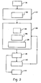
- FIGURE 3 illustrates an example embodiment. The example is discussed in relation to cardiac SPECT/CT. However, non-cardiac SPECT/CT and/or other applications are also contemplated. Again, the combined data sets may or may not be registered.
- a segmentation component 302 of the pre-processor 137 segments tissue of interest from the SPECT data.
- tissue of interest is heart tissue such as the left ventricle of the heart
- various techniques can be used to mitigate segmenting non-heart tissue, which may exhibit similar tracer uptake as the heart. For example, since the heart is generally spatially located nearer the left side of the body, data representative of the right side of the body may be masked or discarded. As such, tissue such as the liver, which may show similar tracer uptake, may be masked or removed prior to performing the segmentation.
- a priori knowledge of the acquisition can be used to facilitate segmentation.
- the position of the object support 132 can be set so that the left ventricle is located generally in a central transaxial slice.
- a filter such as a smoothing with a boxcar average filter or another type of filter, can be applied so that a value such as a maximum value of the segmented data generally represents the spatial center of the left ventricle.
- a window such as a three-dimensional (3D) ellipsoid, sphere, cube, or other shape or other window of other dimension can be used to exclude voxels outside of the window.
- a local histogram thresholding or other technique can be applied within the remaining region to classify the voxels as belonging or not belonging to the left ventricle.
- a voxel counter 304 of the alignment metric determining component 138 determines the number of voxels in the segmented tissue. This value can be stored for example in a storage component such as memory as TotalNumVoxels. For one or more of the voxels classified as belonging to the segmented tissue in the SPECT data, it is determined whether the attenuation coefficient in the CT map corresponds to segmented tissue. This may be based on a threshold which is calculated from an average attenuation factor for the segmented tissue. The voxel counter 304 then determines the number of voxels belonging to the segmented tissue in CT data. This value can be stored in the storage component as TotalTissueOfInterestVoxels.
- a metric determiner 306 of the alignment metric determining component 138 determines a metric based on the TotalNumVoxels and TotalHeartVoxels values. For instance, the metric determiner 306 may compute the metric as a function of Equation 2. 1 - TotalTissueOfInterestVoxels TotalNumVoxels , where (1 - TotalTissueOfInterestVoxels ) represents the total number of voxels corresponding to tissue other than the tissue of interest. In an alternative embodiment, the metric determiner 306 determines the metric based on pixels.
- the clinical metric generating component 206 generates a second metric based on the alignment metric of Equation 2.
- a metric may provide clinically relevant information such an actual or estimated distance between two regions of interest, and as discussed above, may be variously determined, for example, via a look up table, dynamically computed, and/or otherwise.
- the decision component 140 behaves substantially as discussed above.
- the first and/or the second metric be may presented to facilitate determining whether to perform a registration or a secondary registration.
- a threshold may be use to facilitate making such a decision.
- the threshold may be determined using the NURBS-based Cardiac-Torso (NCAT) phantom, by generating different amount of misalignments and verifying the results from known studies.
- NCAT NURBS-based Cardiac-Torso
- the combined data can be further processed by the processing component 144.
- the registration component 142 automatically registers the data.
- the registration includes identifying a voxel deemed as tissue of interest in the SPECT data and deemed as non-tissue of interest in the CT data, and replacing the voxels in the CT data with an average attenuation factor for the tissue of interest. The average may be calculated from other voxels classified as the tissue of interest in the CT data or otherwise. Such voxel replacement can be considered an automatic registration.
- FIGURES 4 and 5 An example of the results of such a registration is shown in FIGURES 4 and 5 .
- A, B and C respectively show a transaxial, a sagittal, and a coronal view depicting a region of misalignment 400 between SPECT and CT data in the myocardium.
- FIGURE 5 A, B and C respectively show a transaxil, a saggital, and a coronal view after registration in which the CT data in the region 400 is replaced as discussed herein.
- FIGURE 6 illustrates a flow chart for determining whether to register two data sets based on a similarity metric.
- a first data set is combined with a second data set.
- the first and second data sets can be data sets from different imaging modalities such as SPECT and CT.
- the resulting data set may include transforming one of the data sets so that it conforms with the domain space of the other data set.
- a similarity metric is determined based on the combined data set, and at 605, a clinically relevant metric is determined based on the similarity metric, as discussed herein. In other embodiments, act 605 may be omitted. As noted above, the metrics indicate a degree of alignment between information with the data sets in the combined data set.
- the combined data set can be further processed. Otherwise, at 610 it is determined whether an automatic registration is to be performed.
- the foregoing can be employed with one or more images or views within a data.
- the above can be used for each image to determine the quality of the alignment between regions of interest in each image.
- the clinician would only have to invoke registration with respect to those images associated with a misalignment deemed unacceptable by the clinician.
- This may improve workflow over an embodiment in which the similarity and/or clinically relevant metric is not used. For instance, when not used, the clinician may have to visually determine on an image-by-image basis whether to invoke registration.
- the above may be implemented by way of computer readable instructions, which when executed by a computer processor(s), cause the processor(s) to carry out the described techniques.
- the instructions are stored in a computer readable storage medium associated with or otherwise accessible to the relevant computer.
- the described techniques need not be performed concurrently with the data acquisition.
- the combined data may include data from the same imaging modality.
Landscapes
- Engineering & Computer Science (AREA)
- Computer Vision & Pattern Recognition (AREA)
- Physics & Mathematics (AREA)
- General Physics & Mathematics (AREA)
- Theoretical Computer Science (AREA)
- Apparatus For Radiation Diagnosis (AREA)
- Nuclear Medicine (AREA)
- Measuring And Recording Apparatus For Diagnosis (AREA)
Claims (15)
- Procédé, comprenant :l'obtention d'un ensemble de données combinées qui comprend des premier et deuxième ensembles de données d'imagerie, dans lequel les premier et deuxième ensembles de données d'imagerie correspondent respectivement à des première et deuxième modalités d'imagerie ;la détermination d'une métrique indicative d'un alignement entre les premier et deuxième ensembles de données d'imagerie dans l'ensemble de données combinées ; etla présentation de la métrique dans un format lisible par l'être humain, dans lequel le procédé comprend en outre :la segmentation du premier ensemble de données, sur la base d'un tissu d'intérêt, pour générer une première région segmentée qui comprend le tissu d'intérêt ;l'identification d'un premier ensemble de voxels, dans l'ensemble de données segmenté, ayant des premières valeurs qui correspondent au tissu d'intérêt ;la détermination d'un premier nombre de voxels dans le premier ensemble de voxels ;l'identification d'un deuxième ensemble de voxels, dans le deuxième ensemble de données, qui correspond aux premiers voxels ;la détermination d'un deuxième nombre de voxels dans le deuxième ensemble de voxels ayant des deuxièmes valeurs qui correspondent au tissu d'intérêt ; etla détermination de la métrique sur la base du premier nombre de voxels et du deuxième nombre de voxels.
- Procédé selon la revendication 1, dans lequel la métrique comprend une description numérique indicative d'une distance pertinente cliniquement entre des première et deuxième régions d'intérêt respectivement des premier et deuxième ensembles de données d'imagerie.
- Procédé selon la revendication 1, comprenant en outre :l'identification d'un premier contour du tissu d'intérêt dans le premier ensemble de données ; etl'identification d'un deuxième contour du tissu d'intérêt dans le deuxième ensemble de données,dans lequel la métrique est indicative d'un alignement entre les premier et deuxième contours.
- Procédé selon la revendication 3, dans lequel la métrique est une distance euclidienne entre les premier et deuxième contours.
- Procédé selon la revendication 1, dans lequel la métrique est déterminée sur la base de ce qui suit :

- Procédé selon la revendication 1, comprenant en outre la superposition des premier et deuxième ensembles de données en remplaçant un voxel qui n'a pas une valeur correspondant au tissu d'intérêt par une moyenne des valeurs des voxels qui ont des valeurs correspondant au tissu d'intérêt.
- Procédé selon la revendication 1, comprenant en outre la superposition des premier et deuxième ensembles de données sur la base d'une entrée d'opérateur.
- Procédé selon la revendication 1, comprenant en outre la comparaison de la métrique à un seuil prédéterminé pour déterminer si l'alignement des premier et deuxième ensembles de données est acceptable.
- Procédé selon la revendication 1, dans lequel le premier ensemble de données est un ensemble de données de tomodensitométrie par émission monophotonique et le deuxième ensemble de données est un ensemble de données de tomodensitométrie.
- Procédé selon la revendication 1, dans lequel le premier ensemble de données d'imagerie est rééchantillonné pour avoir au moins l'une d'une taille ou d'une résolution de voxels sensiblement identique à celle du deuxième ensemble de données d'imagerie.
- Procédé selon la revendication 1, dans lequel les données dans l'ensemble de données combinées ne sont pas superposées.
- Système, comprenant :un combinateur de données (136) qui combine les premier et deuxième ensembles de données d'imagerie, dans lequel les premier et deuxième ensembles de données d'imagerie correspondent respectivement à différentes modalités d'imagerie ;un composant de détermination de métrique (138) qui détermine une métrique indicative d'un alignement entre les premier et deuxième ensembles de données d'imagerie dans l'ensemble de données combinées ; etun composant de superposition (142) qui superpose les premier et deuxième ensembles de données d'imagerie lorsque la métrique est indicative d'un alignement inacceptable entre les premier et deuxième ensembles de données d'imagerie, dans lequel le système comprend en outre :un composant de segmentation (302) qui segmente le premier ensemble de données, sur la base d'un tissu d'intérêt, pour générer une première région segmentée qui comprend le tissu d'intérêt ; etun compteur de voxels (304) qui compte un premier nombre de voxels dans un premier ensemble de voxels dans l'ensemble de données segmenté ayant des premières valeurs correspondant au tissu d'intérêt et qui compte un deuxième nombre de voxels dans un deuxième ensemble de voxels dans le deuxième ensemble de données qui correspondent tous les deux aux premiers voxels et ayant des deuxièmes valeurs correspondant au tissu d'intérêt ;dans lequel la métrique est basée sur le premier nombre de voxels et sur le deuxième nombre de voxels.
- Système selon la revendication 12, dans lequel la superposition comprend le remplacement d'un voxel qui n'a pas une valeur correspondant au tissu d'intérêt par une moyenne des valeurs de l'ensemble correspondant de voxels ayant des valeurs correspondant au tissu d'intérêt.
- Support de stockage lisible par ordinateur contenant des instructions qui, lorsqu'elles sont exécutées par un ordinateur, amènent l'ordinateur à effectuer les étapes de :la détermination d'une métrique indicative d'un alignement entre des premières données d'imagerie combinées à des deuxièmes données d'imagerie ; etla superposition des premières et deuxièmes données d'imagerie lorsque la métrique est indicative d'un alignement inacceptable entre les premier et deuxième ensembles de données d'imagerie ;dans lequel les instructions, lorsqu'elles sont exécutées par l'ordinateur, amènent en outre l'ordinateur à effectuer les étapes de :la segmentation des premières données, sur la base d'un tissu d'intérêt, pour générer une première région segmentée qui comprend le tissu d'intérêt ; etla détermination d'un premier nombre de voxels dans un premier ensemble de voxels dans l'ensemble de données segmenté ayant des premières valeurs correspondant au tissu d'intérêt et d'un deuxième nombre de voxels dans un deuxième ensemble de voxels dans le deuxième ensemble de données qui correspondent tous les deux au premier ensemble de voxels et ayant des deuxièmes valeurs correspondant au tissu d'intérêt, dans lequel la métrique est basée sur le premier nombre de voxels et sur le deuxième nombre de voxels dans les premier et deuxième ensembles de voxels.
- Support de stockage lisible par ordinateur selon la revendication 14, dans lequel les instructions, lorsqu'elles sont exécutées par l'ordinateur, amènent en outre l'ordinateur à effectuer l'étape de :le remplacement d'une valeur correspondant au tissu d'intérêt dans les deuxièmes données par une moyenne des valeurs des deuxièmes voxels ayant des valeurs correspondant au tissu d'intérêt.
Applications Claiming Priority (2)
| Application Number | Priority Date | Filing Date | Title |
|---|---|---|---|
| US2201908P | 2008-01-18 | 2008-01-18 | |
| PCT/IB2009/050041 WO2009090572A2 (fr) | 2008-01-18 | 2009-01-07 | Métrique d'alignement de superposition d'image |
Publications (2)
| Publication Number | Publication Date |
|---|---|
| EP2245592A2 EP2245592A2 (fr) | 2010-11-03 |
| EP2245592B1 true EP2245592B1 (fr) | 2013-03-27 |
Family
ID=40691331
Family Applications (1)
| Application Number | Title | Priority Date | Filing Date |
|---|---|---|---|
| EP09701915A Not-in-force EP2245592B1 (fr) | 2008-01-18 | 2009-01-07 | Métrique d'alignement de superposition d'image |
Country Status (4)
| Country | Link |
|---|---|
| US (1) | US20100284598A1 (fr) |
| EP (1) | EP2245592B1 (fr) |
| CN (1) | CN101911119B (fr) |
| WO (1) | WO2009090572A2 (fr) |
Families Citing this family (13)
| Publication number | Priority date | Publication date | Assignee | Title |
|---|---|---|---|---|
| JP5404277B2 (ja) * | 2009-09-24 | 2014-01-29 | 株式会社Aze | 医用画像データ位置合せ装置、方法およびプログラム |
| RU2568835C2 (ru) * | 2010-02-22 | 2015-11-20 | Конинклейке Филипс Электроникс Н.В. | Реконструкция из разреженных данных для стробированной рентгеновской ст-визуализации |
| EP2609572B1 (fr) * | 2010-08-25 | 2020-03-25 | Koninklijke Philips N.V. | Imagerie bimodale comprenant des métriques de qualité |
| WO2012151579A2 (fr) * | 2011-05-05 | 2012-11-08 | Mayo Foundation For Medical Education And Research | Systèmes et procédés pour analyser in vivo des volumes de tissu en utilisant des données d'imagerie médicale |
| WO2013001410A2 (fr) * | 2011-06-27 | 2013-01-03 | Koninklijke Philips Electronics N.V. | Marquage anatomique d'observations dans des données d'image d'études périodiques |
| CN103649961B (zh) * | 2011-06-27 | 2018-06-26 | 皇家飞利浦有限公司 | 对图像数据中的发现的解剖学标记的方法 |
| US10743773B2 (en) | 2012-05-02 | 2020-08-18 | Koninklijke Philips N.V. | Imaging thermometry |
| US9552533B2 (en) * | 2013-03-05 | 2017-01-24 | Toshiba Medical Systems Corporation | Image registration apparatus and method |
| CN103247046B (zh) * | 2013-04-19 | 2016-07-06 | 深圳先进技术研究院 | 一种放射治疗计划中靶区自动勾画的方法和装置 |
| US11138739B2 (en) * | 2016-11-29 | 2021-10-05 | Koninklijke Philips N.V. | Heart segmentation methodology for cardiac motion correction |
| US10552959B2 (en) * | 2018-02-23 | 2020-02-04 | Cameron Anthony Piron | System and method for using imaging quality metric ranking |
| DE102019217576B4 (de) * | 2019-11-14 | 2021-10-14 | Siemens Healthcare Gmbh | Identifizieren und Bereitstellen von Fehlausrichtungsbildmerkmalen |
| CN112348863B (zh) * | 2020-11-09 | 2022-06-21 | Oppo广东移动通信有限公司 | 图像对齐方法、图像对齐装置及终端设备 |
Family Cites Families (9)
| Publication number | Priority date | Publication date | Assignee | Title |
|---|---|---|---|---|
| JPH03224073A (ja) * | 1990-01-30 | 1991-10-03 | Ezel Inc | 位置合わせ装置 |
| US6754374B1 (en) * | 1998-12-16 | 2004-06-22 | Surgical Navigation Technologies, Inc. | Method and apparatus for processing images with regions representing target objects |
| US20030095696A1 (en) * | 2001-09-14 | 2003-05-22 | Reeves Anthony P. | System, method and apparatus for small pulmonary nodule computer aided diagnosis from computed tomography scans |
| AU2003215755A1 (en) * | 2002-03-22 | 2003-10-08 | British Telecommunications Public Limited Company | Comparing patterns |
| US7223981B1 (en) * | 2002-12-04 | 2007-05-29 | Aguila Technologies Inc. | Gamma ray detector modules |
| US7706633B2 (en) * | 2004-04-21 | 2010-04-27 | Siemens Corporation | GPU-based image manipulation method for registration applications |
| US7561757B2 (en) * | 2004-10-28 | 2009-07-14 | Siemens Medical Solutions Usa, Inc. | Image registration using minimum entropic graphs |
| WO2006054191A1 (fr) * | 2004-11-17 | 2006-05-26 | Koninklijke Philips Electronics N.V. | Fonctionnalite d'enregistrement d'image elastique amelioree |
| US7907761B2 (en) * | 2007-04-20 | 2011-03-15 | General Electric Company | Method for registration and navigation of volumetric scans using energy profiles |
-
2009
- 2009-01-07 WO PCT/IB2009/050041 patent/WO2009090572A2/fr not_active Ceased
- 2009-01-07 EP EP09701915A patent/EP2245592B1/fr not_active Not-in-force
- 2009-01-07 CN CN200980102360.XA patent/CN101911119B/zh not_active Expired - Fee Related
- 2009-01-07 US US12/812,045 patent/US20100284598A1/en not_active Abandoned
Also Published As
| Publication number | Publication date |
|---|---|
| EP2245592A2 (fr) | 2010-11-03 |
| WO2009090572A2 (fr) | 2009-07-23 |
| CN101911119A (zh) | 2010-12-08 |
| US20100284598A1 (en) | 2010-11-11 |
| WO2009090572A3 (fr) | 2009-09-11 |
| CN101911119B (zh) | 2015-02-25 |
Similar Documents
| Publication | Publication Date | Title |
|---|---|---|
| EP2245592B1 (fr) | Métrique d'alignement de superposition d'image | |
| RU2471204C2 (ru) | Локальная позитронная эмиссионная томография | |
| CN109035355B (zh) | 用于pet图像重建的系统和方法 | |
| JP5764147B2 (ja) | 心臓イメージングにおける関心領域定義 | |
| US8218848B2 (en) | System and method for the generation of attenuation correction maps from MR images | |
| US8406495B2 (en) | MR segmentation using transmission data in hybrid nuclear/MR imaging | |
| EP2646979B1 (fr) | Appareil de superposition d'images | |
| WO2020214911A1 (fr) | Procédé et système de génération d'une carte d'atténuation à partir de données d'émission de temp sur la base d'un apprentissage profond | |
| US20090297006A1 (en) | Diagnostic system for multimodality mammography | |
| US8655040B2 (en) | Integrated image registration and motion estimation for medical imaging applications | |
| JP2013522712A5 (fr) | ||
| JP7216722B2 (ja) | 診断撮像における画像特徴のアノテーション | |
| CN102737375A (zh) | 确定坏死组织的方法、图像处理装置和系统以及程序产品 | |
| CN117042695A (zh) | 断层扫描的基于图像的规划 | |
| US20080095414A1 (en) | Correction of functional nuclear imaging data for motion artifacts using anatomical data | |
| CN117530709A (zh) | 用于pet-ct图像伪影评估和校正的系统和方法 | |
| US8391578B2 (en) | Method and apparatus for automatically registering images | |
| Yin et al. | Acquisition, preprocessing, and reconstruction of ultralow dose volumetric CT scout for organ‐based CT scan planning | |
| US20150243026A1 (en) | Multi-structure atlas and/or use thereof | |
| US11495346B2 (en) | External device-enabled imaging support | |
| US7756310B2 (en) | System and method for segmentation | |
| EP4560568B1 (fr) | Procédé d'évaluation de l'exploitabilité de données d'image de tomographie 4d, produit de programme informatique et dispositif de balayage | |
| Sabokrohiyeh et al. | Sketch-based registration of 3d cine mri to 4d flow mri | |
| EP4585155A1 (fr) | Systèmes et procédés d'enregistrement d'images | |
| US20250356544A1 (en) | Attenuation correction factor generation |
Legal Events
| Date | Code | Title | Description |
|---|---|---|---|
| PUAI | Public reference made under article 153(3) epc to a published international application that has entered the european phase |
Free format text: ORIGINAL CODE: 0009012 |
|
| 17P | Request for examination filed |
Effective date: 20100818 |
|
| AK | Designated contracting states |
Kind code of ref document: A2 Designated state(s): AT BE BG CH CY CZ DE DK EE ES FI FR GB GR HR HU IE IS IT LI LT LU LV MC MK MT NL NO PL PT RO SE SI SK TR |
|
| AX | Request for extension of the european patent |
Extension state: AL BA RS |
|
| DAX | Request for extension of the european patent (deleted) | ||
| GRAP | Despatch of communication of intention to grant a patent |
Free format text: ORIGINAL CODE: EPIDOSNIGR1 |
|
| GRAS | Grant fee paid |
Free format text: ORIGINAL CODE: EPIDOSNIGR3 |
|
| GRAA | (expected) grant |
Free format text: ORIGINAL CODE: 0009210 |
|
| AK | Designated contracting states |
Kind code of ref document: B1 Designated state(s): AT BE BG CH CY CZ DE DK EE ES FI FR GB GR HR HU IE IS IT LI LT LU LV MC MK MT NL NO PL PT RO SE SI SK TR |
|
| REG | Reference to a national code |
Ref country code: GB Ref legal event code: FG4D |
|
| REG | Reference to a national code |
Ref country code: CH Ref legal event code: EP |
|
| REG | Reference to a national code |
Ref country code: AT Ref legal event code: REF Ref document number: 603823 Country of ref document: AT Kind code of ref document: T Effective date: 20130415 |
|
| REG | Reference to a national code |
Ref country code: IE Ref legal event code: FG4D |
|
| REG | Reference to a national code |
Ref country code: DE Ref legal event code: R096 Ref document number: 602009014372 Country of ref document: DE Effective date: 20130529 |
|
| RAP2 | Party data changed (patent owner data changed or rights of a patent transferred) |
Owner name: KONINKLIJKE PHILIPS N.V. |
|
| REG | Reference to a national code |
Ref country code: CH Ref legal event code: PFA Owner name: KONINKLIJKE PHILIPS N.V., NL Free format text: FORMER OWNER: KONINKLIJKE PHILIPS ELECTRONICS N.V., NL |
|
| PG25 | Lapsed in a contracting state [announced via postgrant information from national office to epo] |
Ref country code: BG Free format text: LAPSE BECAUSE OF FAILURE TO SUBMIT A TRANSLATION OF THE DESCRIPTION OR TO PAY THE FEE WITHIN THE PRESCRIBED TIME-LIMIT Effective date: 20130627 Ref country code: NO Free format text: LAPSE BECAUSE OF FAILURE TO SUBMIT A TRANSLATION OF THE DESCRIPTION OR TO PAY THE FEE WITHIN THE PRESCRIBED TIME-LIMIT Effective date: 20130627 Ref country code: SE Free format text: LAPSE BECAUSE OF FAILURE TO SUBMIT A TRANSLATION OF THE DESCRIPTION OR TO PAY THE FEE WITHIN THE PRESCRIBED TIME-LIMIT Effective date: 20130327 Ref country code: LT Free format text: LAPSE BECAUSE OF FAILURE TO SUBMIT A TRANSLATION OF THE DESCRIPTION OR TO PAY THE FEE WITHIN THE PRESCRIBED TIME-LIMIT Effective date: 20130327 |
|
| REG | Reference to a national code |
Ref country code: AT Ref legal event code: MK05 Ref document number: 603823 Country of ref document: AT Kind code of ref document: T Effective date: 20130327 |
|
| REG | Reference to a national code |
Ref country code: LT Ref legal event code: MG4D |
|
| PG25 | Lapsed in a contracting state [announced via postgrant information from national office to epo] |
Ref country code: FI Free format text: LAPSE BECAUSE OF FAILURE TO SUBMIT A TRANSLATION OF THE DESCRIPTION OR TO PAY THE FEE WITHIN THE PRESCRIBED TIME-LIMIT Effective date: 20130327 Ref country code: SI Free format text: LAPSE BECAUSE OF FAILURE TO SUBMIT A TRANSLATION OF THE DESCRIPTION OR TO PAY THE FEE WITHIN THE PRESCRIBED TIME-LIMIT Effective date: 20130327 Ref country code: GR Free format text: LAPSE BECAUSE OF FAILURE TO SUBMIT A TRANSLATION OF THE DESCRIPTION OR TO PAY THE FEE WITHIN THE PRESCRIBED TIME-LIMIT Effective date: 20130628 Ref country code: LV Free format text: LAPSE BECAUSE OF FAILURE TO SUBMIT A TRANSLATION OF THE DESCRIPTION OR TO PAY THE FEE WITHIN THE PRESCRIBED TIME-LIMIT Effective date: 20130327 |
|
| REG | Reference to a national code |
Ref country code: NL Ref legal event code: VDEP Effective date: 20130327 |
|
| PG25 | Lapsed in a contracting state [announced via postgrant information from national office to epo] |
Ref country code: BE Free format text: LAPSE BECAUSE OF FAILURE TO SUBMIT A TRANSLATION OF THE DESCRIPTION OR TO PAY THE FEE WITHIN THE PRESCRIBED TIME-LIMIT Effective date: 20130327 Ref country code: HR Free format text: LAPSE BECAUSE OF FAILURE TO SUBMIT A TRANSLATION OF THE DESCRIPTION OR TO PAY THE FEE WITHIN THE PRESCRIBED TIME-LIMIT Effective date: 20130327 |
|
| PG25 | Lapsed in a contracting state [announced via postgrant information from national office to epo] |
Ref country code: SK Free format text: LAPSE BECAUSE OF FAILURE TO SUBMIT A TRANSLATION OF THE DESCRIPTION OR TO PAY THE FEE WITHIN THE PRESCRIBED TIME-LIMIT Effective date: 20130327 Ref country code: PT Free format text: LAPSE BECAUSE OF FAILURE TO SUBMIT A TRANSLATION OF THE DESCRIPTION OR TO PAY THE FEE WITHIN THE PRESCRIBED TIME-LIMIT Effective date: 20130729 Ref country code: EE Free format text: LAPSE BECAUSE OF FAILURE TO SUBMIT A TRANSLATION OF THE DESCRIPTION OR TO PAY THE FEE WITHIN THE PRESCRIBED TIME-LIMIT Effective date: 20130327 Ref country code: RO Free format text: LAPSE BECAUSE OF FAILURE TO SUBMIT A TRANSLATION OF THE DESCRIPTION OR TO PAY THE FEE WITHIN THE PRESCRIBED TIME-LIMIT Effective date: 20130327 Ref country code: CZ Free format text: LAPSE BECAUSE OF FAILURE TO SUBMIT A TRANSLATION OF THE DESCRIPTION OR TO PAY THE FEE WITHIN THE PRESCRIBED TIME-LIMIT Effective date: 20130327 Ref country code: NL Free format text: LAPSE BECAUSE OF FAILURE TO SUBMIT A TRANSLATION OF THE DESCRIPTION OR TO PAY THE FEE WITHIN THE PRESCRIBED TIME-LIMIT Effective date: 20130327 Ref country code: AT Free format text: LAPSE BECAUSE OF FAILURE TO SUBMIT A TRANSLATION OF THE DESCRIPTION OR TO PAY THE FEE WITHIN THE PRESCRIBED TIME-LIMIT Effective date: 20130327 Ref country code: IS Free format text: LAPSE BECAUSE OF FAILURE TO SUBMIT A TRANSLATION OF THE DESCRIPTION OR TO PAY THE FEE WITHIN THE PRESCRIBED TIME-LIMIT Effective date: 20130727 Ref country code: ES Free format text: LAPSE BECAUSE OF FAILURE TO SUBMIT A TRANSLATION OF THE DESCRIPTION OR TO PAY THE FEE WITHIN THE PRESCRIBED TIME-LIMIT Effective date: 20130708 |
|
| PG25 | Lapsed in a contracting state [announced via postgrant information from national office to epo] |
Ref country code: PL Free format text: LAPSE BECAUSE OF FAILURE TO SUBMIT A TRANSLATION OF THE DESCRIPTION OR TO PAY THE FEE WITHIN THE PRESCRIBED TIME-LIMIT Effective date: 20130327 Ref country code: CY Free format text: LAPSE BECAUSE OF FAILURE TO SUBMIT A TRANSLATION OF THE DESCRIPTION OR TO PAY THE FEE WITHIN THE PRESCRIBED TIME-LIMIT Effective date: 20130327 |
|
| PG25 | Lapsed in a contracting state [announced via postgrant information from national office to epo] |
Ref country code: DK Free format text: LAPSE BECAUSE OF FAILURE TO SUBMIT A TRANSLATION OF THE DESCRIPTION OR TO PAY THE FEE WITHIN THE PRESCRIBED TIME-LIMIT Effective date: 20130327 |
|
| PLBE | No opposition filed within time limit |
Free format text: ORIGINAL CODE: 0009261 |
|
| STAA | Information on the status of an ep patent application or granted ep patent |
Free format text: STATUS: NO OPPOSITION FILED WITHIN TIME LIMIT |
|
| PG25 | Lapsed in a contracting state [announced via postgrant information from national office to epo] |
Ref country code: IT Free format text: LAPSE BECAUSE OF FAILURE TO SUBMIT A TRANSLATION OF THE DESCRIPTION OR TO PAY THE FEE WITHIN THE PRESCRIBED TIME-LIMIT Effective date: 20130327 |
|
| 26N | No opposition filed |
Effective date: 20140103 |
|
| REG | Reference to a national code |
Ref country code: DE Ref legal event code: R082 Ref document number: 602009014372 Country of ref document: DE Representative=s name: MEISSNER, BOLTE & PARTNER GBR, DE |
|
| REG | Reference to a national code |
Ref country code: DE Ref legal event code: R097 Ref document number: 602009014372 Country of ref document: DE Effective date: 20140103 |
|
| REG | Reference to a national code |
Ref country code: DE Ref legal event code: R082 Ref document number: 602009014372 Country of ref document: DE Representative=s name: MEISSNER BOLTE PATENTANWAELTE RECHTSANWAELTE P, DE Effective date: 20140331 Ref country code: DE Ref legal event code: R082 Ref document number: 602009014372 Country of ref document: DE Representative=s name: MEISSNER, BOLTE & PARTNER GBR, DE Effective date: 20140331 Ref country code: DE Ref legal event code: R081 Ref document number: 602009014372 Country of ref document: DE Owner name: KONINKLIJKE PHILIPS N.V., NL Free format text: FORMER OWNER: KONINKLIJKE PHILIPS ELECTRONICS N.V., EINDHOVEN, NL Effective date: 20140331 |
|
| PG25 | Lapsed in a contracting state [announced via postgrant information from national office to epo] |
Ref country code: MC Free format text: LAPSE BECAUSE OF FAILURE TO SUBMIT A TRANSLATION OF THE DESCRIPTION OR TO PAY THE FEE WITHIN THE PRESCRIBED TIME-LIMIT Effective date: 20130327 Ref country code: LU Free format text: LAPSE BECAUSE OF FAILURE TO SUBMIT A TRANSLATION OF THE DESCRIPTION OR TO PAY THE FEE WITHIN THE PRESCRIBED TIME-LIMIT Effective date: 20140107 |
|
| REG | Reference to a national code |
Ref country code: CH Ref legal event code: PL |
|
| GBPC | Gb: european patent ceased through non-payment of renewal fee |
Effective date: 20140107 |
|
| PG25 | Lapsed in a contracting state [announced via postgrant information from national office to epo] |
Ref country code: CH Free format text: LAPSE BECAUSE OF NON-PAYMENT OF DUE FEES Effective date: 20140131 Ref country code: LI Free format text: LAPSE BECAUSE OF NON-PAYMENT OF DUE FEES Effective date: 20140131 |
|
| REG | Reference to a national code |
Ref country code: IE Ref legal event code: MM4A |
|
| PG25 | Lapsed in a contracting state [announced via postgrant information from national office to epo] |
Ref country code: GB Free format text: LAPSE BECAUSE OF NON-PAYMENT OF DUE FEES Effective date: 20140107 |
|
| PG25 | Lapsed in a contracting state [announced via postgrant information from national office to epo] |
Ref country code: IE Free format text: LAPSE BECAUSE OF NON-PAYMENT OF DUE FEES Effective date: 20140107 |
|
| REG | Reference to a national code |
Ref country code: FR Ref legal event code: PLFP Year of fee payment: 8 |
|
| PG25 | Lapsed in a contracting state [announced via postgrant information from national office to epo] |
Ref country code: MT Free format text: LAPSE BECAUSE OF FAILURE TO SUBMIT A TRANSLATION OF THE DESCRIPTION OR TO PAY THE FEE WITHIN THE PRESCRIBED TIME-LIMIT Effective date: 20130327 |
|
| PGFP | Annual fee paid to national office [announced via postgrant information from national office to epo] |
Ref country code: DE Payment date: 20160331 Year of fee payment: 8 |
|
| PGFP | Annual fee paid to national office [announced via postgrant information from national office to epo] |
Ref country code: FR Payment date: 20160129 Year of fee payment: 8 |
|
| PG25 | Lapsed in a contracting state [announced via postgrant information from national office to epo] |
Ref country code: TR Free format text: LAPSE BECAUSE OF FAILURE TO SUBMIT A TRANSLATION OF THE DESCRIPTION OR TO PAY THE FEE WITHIN THE PRESCRIBED TIME-LIMIT Effective date: 20130327 Ref country code: HU Free format text: LAPSE BECAUSE OF FAILURE TO SUBMIT A TRANSLATION OF THE DESCRIPTION OR TO PAY THE FEE WITHIN THE PRESCRIBED TIME-LIMIT; INVALID AB INITIO Effective date: 20090107 |
|
| REG | Reference to a national code |
Ref country code: DE Ref legal event code: R119 Ref document number: 602009014372 Country of ref document: DE |
|
| REG | Reference to a national code |
Ref country code: FR Ref legal event code: ST Effective date: 20170929 |
|
| PG25 | Lapsed in a contracting state [announced via postgrant information from national office to epo] |
Ref country code: FR Free format text: LAPSE BECAUSE OF NON-PAYMENT OF DUE FEES Effective date: 20170131 |
|
| PG25 | Lapsed in a contracting state [announced via postgrant information from national office to epo] |
Ref country code: DE Free format text: LAPSE BECAUSE OF NON-PAYMENT OF DUE FEES Effective date: 20170801 |
|
| PG25 | Lapsed in a contracting state [announced via postgrant information from national office to epo] |
Ref country code: MK Free format text: LAPSE BECAUSE OF FAILURE TO SUBMIT A TRANSLATION OF THE DESCRIPTION OR TO PAY THE FEE WITHIN THE PRESCRIBED TIME-LIMIT Effective date: 20130327 |