EP2246426A1 - Zusammensetzung zur neutralisierung von botulinustoxin typ-a und menschlicher antikörper gegen botulinustoxin typ-a - Google Patents
Zusammensetzung zur neutralisierung von botulinustoxin typ-a und menschlicher antikörper gegen botulinustoxin typ-a Download PDFInfo
- Publication number
- EP2246426A1 EP2246426A1 EP09706257A EP09706257A EP2246426A1 EP 2246426 A1 EP2246426 A1 EP 2246426A1 EP 09706257 A EP09706257 A EP 09706257A EP 09706257 A EP09706257 A EP 09706257A EP 2246426 A1 EP2246426 A1 EP 2246426A1
- Authority
- EP
- European Patent Office
- Prior art keywords
- seq
- antibody
- amino acid
- acid sequence
- heavy chain
- Prior art date
- Legal status (The legal status is an assumption and is not a legal conclusion. Google has not performed a legal analysis and makes no representation as to the accuracy of the status listed.)
- Withdrawn
Links
Images














Classifications
-
- C—CHEMISTRY; METALLURGY
- C07—ORGANIC CHEMISTRY
- C07K—PEPTIDES
- C07K16/00—Immunoglobulins [IG], e.g. monoclonal or polyclonal antibodies
- C07K16/12—Immunoglobulins [IG], e.g. monoclonal or polyclonal antibodies against material from bacteria
- C07K16/1267—Gram-positive bacteria
- C07K16/1282—Clostridium (G)
-
- A—HUMAN NECESSITIES
- A61—MEDICAL OR VETERINARY SCIENCE; HYGIENE
- A61P—SPECIFIC THERAPEUTIC ACTIVITY OF CHEMICAL COMPOUNDS OR MEDICINAL PREPARATIONS
- A61P31/00—Antiinfectives, i.e. antibiotics, antiseptics, chemotherapeutics
- A61P31/04—Antibacterial agents
-
- A—HUMAN NECESSITIES
- A61—MEDICAL OR VETERINARY SCIENCE; HYGIENE
- A61K—PREPARATIONS FOR MEDICAL, DENTAL OR TOILETRY PURPOSES
- A61K39/00—Medicinal preparations containing antigens or antibodies
- A61K2039/505—Medicinal preparations containing antigens or antibodies comprising antibodies
-
- A—HUMAN NECESSITIES
- A61—MEDICAL OR VETERINARY SCIENCE; HYGIENE
- A61K—PREPARATIONS FOR MEDICAL, DENTAL OR TOILETRY PURPOSES
- A61K39/00—Medicinal preparations containing antigens or antibodies
- A61K2039/505—Medicinal preparations containing antigens or antibodies comprising antibodies
- A61K2039/507—Comprising a combination of two or more separate antibodies
-
- C—CHEMISTRY; METALLURGY
- C07—ORGANIC CHEMISTRY
- C07K—PEPTIDES
- C07K2317/00—Immunoglobulins specific features
- C07K2317/20—Immunoglobulins specific features characterized by taxonomic origin
- C07K2317/21—Immunoglobulins specific features characterized by taxonomic origin from primates, e.g. man
-
- C—CHEMISTRY; METALLURGY
- C07—ORGANIC CHEMISTRY
- C07K—PEPTIDES
- C07K2317/00—Immunoglobulins specific features
- C07K2317/50—Immunoglobulins specific features characterized by immunoglobulin fragments
- C07K2317/55—Fab or Fab'
-
- C—CHEMISTRY; METALLURGY
- C07—ORGANIC CHEMISTRY
- C07K—PEPTIDES
- C07K2317/00—Immunoglobulins specific features
- C07K2317/50—Immunoglobulins specific features characterized by immunoglobulin fragments
- C07K2317/56—Immunoglobulins specific features characterized by immunoglobulin fragments variable (Fv) region, i.e. VH and/or VL
-
- C—CHEMISTRY; METALLURGY
- C07—ORGANIC CHEMISTRY
- C07K—PEPTIDES
- C07K2317/00—Immunoglobulins specific features
- C07K2317/50—Immunoglobulins specific features characterized by immunoglobulin fragments
- C07K2317/56—Immunoglobulins specific features characterized by immunoglobulin fragments variable (Fv) region, i.e. VH and/or VL
- C07K2317/565—Complementarity determining region [CDR]
-
- C—CHEMISTRY; METALLURGY
- C07—ORGANIC CHEMISTRY
- C07K—PEPTIDES
- C07K2317/00—Immunoglobulins specific features
- C07K2317/70—Immunoglobulins specific features characterized by effect upon binding to a cell or to an antigen
- C07K2317/76—Antagonist effect on antigen, e.g. neutralization or inhibition of binding
-
- C—CHEMISTRY; METALLURGY
- C07—ORGANIC CHEMISTRY
- C07K—PEPTIDES
- C07K2317/00—Immunoglobulins specific features
- C07K2317/90—Immunoglobulins specific features characterized by (pharmaco)kinetic aspects or by stability of the immunoglobulin
- C07K2317/92—Affinity (KD), association rate (Ka), dissociation rate (Kd) or EC50 value
Definitions
- the present invention relates to a genetically recombinant antibody having a binding activity to botulinum toxin type-A and having a fully human variable region; a nucleic acid molecule encoding the antibody; a transformant producing the antibody; a method for producing an antibody using the transformant; a composition for neutralizing botulinum toxin type-A using the antibody; and the like.
- Toxins produced by Clostridium botulinum are classified into types A to G depending on the difference in serotypes.
- the toxin types A, B, E, and F have a serious influence on the human health.
- botulinum diseases in humans such as "foodborne botulism (botulinum food poisoning)", “infant botulism”, “wound botulism” and “infectious botulism”.
- the toxin type-A is involved in 26% of botulinum food poisoning.
- the toxin type-A is involved in 26% of botulinum food poisoning.
- the toxin types B and E are followed by this, and toxin type-F is relatively low in the ratio involved in botulinum food poisoning.
- wound botulism is caused only by toxin type-A or type-B (Non-Patent Document 1).
- Food poisoning botulism in adults is usually developed by oral intake of toxins produced as a result of the growth of bacteria and spores in food, while in the case of infant botulism, symptoms of poisoning are caused by the bacterial growth and proliferation leading to toxin production in the intestinal tract as a result of oral intake of bacteria and spores in food such as honey and the like.
- toxins that cause food poisoning differ somewhat depending on countries, cases caused by toxin types A, B, and E have been reported.
- the majority of the infant botulisms are type-A and type B, but toxin types E and F by C. butiricum also have been reported.
- Botulinum toxin is a molecule having a structure in which an S-S bond (disulfide bond) is formed between the heavy chain and the light chain.
- S-S bond disulfide bond
- the botulinum toxin blocks the neurotransmission to muscles, causing hereby flaccid paralysis.
- botulinum toxin comes into the respiratory tract or respiratory muscle, fatal breathing disorder will be caused.
- the mortality rate by the botulinum toxin is 12%, and it becomes higher in a specific risk group (Non-Patent Document 3).
- botulinum toxin is the most fatal toxin as a biological toxin (the fatal dose of the purified toxin type-A in a human of 70 kg is 0.09-0.15 ⁇ g (i.v. or i.m.), 0.70-0.90 ⁇ g (inhalation), and 70 ⁇ g (even oral) ( JAMA. 2001 Feb 28;285(8):1059-70 )), such a botulinum toxin possesses a particularity such as an applicability to the bioterrorism as an another aspect other than usual poisoning. Therefore, a social attention has been paid to it.
- the neutralizing agent for botulinum toxins is a preparation using an equine serum as a raw material.
- This preparation may have a danger to cause an immunoreaction such as anaphylactic reaction, etc. because it contains, as a main component, globulin derived from equine serum, i.e. a heterologous protein to humans.
- an immunoreaction such as anaphylactic reaction, etc.
- globulin derived from equine serum i.e. a heterologous protein to humans.
- problems of unknown viral infections and serum diseases in the preparation Furthermore, stable supply of this preparation is also difficult. Besides, since one year or more are needed for the production of the preparations, emergency response to bioterrorism and the like is in a difficult situation.
- this preparation is not suitable for a storage purpose because there is a problem in the management of horse rearing.
- infant botulism As for infant botulism, it is a current state that treatment with this equine globulin preparation has not been employed because of avoidance of serious side effects such as anaphylactic reactions and the like.
- a specific treatment for the infant botulism is performed with use of a human globulin preparation prepared from the plasma having a high neutralizing antibody titer obtained by immunizing a healthy person with a botulinum toxin.
- the drug under the brand name called "BabyBIG" for the infant botulism has been granted orphan drug designation for its marketing by FDA.
- this method holds problems of raw material availability and biohazard issues in addition to ethical issues, as well as problems of various virus inspections to be needed from the viewpoint of safe security necessary in the production.
- the preparation has not yet been approved in Japan and cannot be used.
- Patent Document 1 It was reported to have succeeded in producing a recombinant neutralizing chimeric antibody against toxin type-A (Patent Document 1). However, it is necessary to solve the problems such as appearance of HACA (human anti-chimeric antibody) or anaphylactic reactions when the antibody is used. Meanwhile, a research and development for toxin neutralization have been performed from the viewpoint of preparing for particularly bioterrorism threats in the United States of America, and a method of producing a phage antibody library from human B lymphocytes obtained by immunization with a botulinum toxin pentamer and selecting a desired full length human neutralizing antibody from the library was undertaken.
- HACA human anti-chimeric antibody
- the present invention provides a composition useful for neutralizing botulinum toxin type-A in view of the above-mentioned background and is intended to solve various problems (anaphylactic reactions and contaminations with heterologous proteins or viruses) in conventional technologies.
- a technical problem as another aspect of the present invention also resides in showing a neutralizing activity in terms of values adapted to an international standard when the composition for neutralizing botulinum toxin type-A is provided.
- the present inventors at first, constructed a human phage antibody library according to a precedent, and tried to isolate an antibody clone that specifically recognizes botulinum toxin type-A. When a screening had been repeatedly performed, it was found that antibodies having each the common epitope were repeatedly selected though the plurality of antibody clones was obtained by the screening each time. As a result of having examined the cause that this phenomenon has occurred, it was expected that an epitope having an extremely high antigenicity would be present, causing the overlap of the epitope. According to this supposition, blocking of the epitope becomes an effective means for acquiring an antibody having a different epitope effectively.
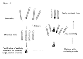
- the present inventors planed a strategy of reacting the botulinum toxin type-A with a phage antibody library after the epitope with a strong antigenicity had been blocked (masked) by adding antibody fragments obtained redundantly through two or more screening tests.
- the present inventors succeeded in acquiring the plurality of antibody clones having each a different epitope by screening again according to the strategy.
- these antibodies were classified into four kinds by an epitope difference. In other words, it was found that specific antibody clones with regards to four epitopes were successfully obtained.
- the present inventors have succeeded in the acquisition of two or more antibody clones that brought a high neutralizing activity against botulinum toxin type-A, and clarified the relation between four epitopes recognized by the antibodies and neutralizing activities.
- This invention is primarily based on these findings and is as follows.
- isolated refers to a state of being taken out from its original environment (for example, natural environment in the case of natural substances), that is, a state that is different from the original existing state by an artificial manipulation.
- isolated antibody does not include an antibody that is natural without any external manipulation (artificial manipulation), that is, the term does not include an antibody in a state of being produced in an individual body and remaining therein. Further, an isolated antibody is typically present in a state in which other kinds of antibodies are not contaminated, that is, it exists alone (as a group of the same kind of antibodies).
- CDR complementarity-determining region
- composition for neutralizing botulinum toxin type-A refers to a pharmaceutical mixture that contains a plurality of kinds of antibodies as an active ingredient and shows a neutralizing activity against botulinum toxin type-A.
- the "antitoxin titer” in the present invention is expressed in terms of an international unit unless otherwise particularly specified.
- the "antitoxin titer” is determined according to The Japanese Pharmacopoeia, clause 3.2.7 "titer test” of "freeze-dried botulism antitoxin, equine” (freeze-dried botulinum antitoxin) described in biological products listed in each monograph on drugs).
- a first aspect of the present invention relates to a composition for neutralizing botulinum toxin type-A (hereinafter also referred to as “composition of the present invention”).
- composition of the present invention At least three kinds of antibodies are used in the composition of the present invention. Any of these three antibodies are each a human antibody that recognizes botulinum toxin type-A. In the present description, the three antibodies are called as "a first human anti-botulinum toxin type-A antibody", "a second human anti-botulinum toxin type-A antibody”, and "a third human anti-botulinum toxin type-A antibody”.
- the first human anti-botulinum toxin type-A antibody is hereinafter abbreviated as “the first antibody”
- the second human anti-botulinum toxin type-A antibody is hereinafter abbreviated as “the second antibody”
- the third human anti-botulinum toxin type-A antibody is hereinafter abbreviated as “the third antibody”.
- the present inventors have succeeded in obtaining a human antibody clone (BT-015, BT-047, BT-058, NT-221, BT-175, NT-320, NT-523, NT-539) effective for neutralization of botulinum toxin type-A (see the description of Examples).
- BT-015, BT-047, BT-058, NT-221, BT-175, NT-320, NT-523, NT-539) effective for neutralization of botulinum toxin type-A (see the description of Examples).
- the first antibody is the one corresponding to an antibody clone BT-015 that was shown to be effective for neutralizing botulinum toxin type-A when used in combination with other antibody clone, and is characterized by recognizing the epitope of the antibody clone BT-015 (i.e.
- the second antibody is the one corresponding to an antibody clone BT-175 that was shown to be effective for neutralizing botulinum toxin type-A when used in combination with other antibody clone, and is characterized by recognizing the epitope of the antibody clone BT-175 (i.e. an antibody having a heavy chain variable region comprising an amino acid sequence of SEQ ID No. 36 and a light chain variable region comprising an amino acid sequence of SEQ ID No. 40). Note that the eligibility as the second antibody can be confirmed in the same manner as in the first antibody clone.
- the third antibody is the one corresponding to an antibody clone (NT-320 or NT-523) that was shown to be effective for neutralizing botulinum toxin type-A when used in combination with an antibody clone BT-015 and an antibody clone BT-175, and is characterized by recognizing the epitope of the antibody clone NT-320 (i.e. an antibody having a heavy chain variable region comprising an amino acid sequence of SEQ ID No. 44 and a light chain variable region comprising an amino acid sequence of SEQ ID No. 48), or the epitope of the antibody clone NT-523 (i.e. an antibody having a heavy chain variable region comprising an amino acid sequence of SEQ ID No. 52 and a light chain variable region comprising an amino acid sequence of SEQ ID No. 56). Note that the eligibility as the third antibody can be confirmed in the same manner as in the first antibody clone.
- the first antibody is any one of antibodies selected from the group consisting of the following (1) to (3); the second antibody is the following antibody (4) or (5); and the third antibody is any one of antibodies selected from the group consisting of the following (6) to (8):
- the above antibody (1) corresponds to an antibody clone BT-015, the above antibody (2) corresponds to an antibody clone BT-047, the above antibody (3) corresponds to a human antibody clone BT-058, the above antibody (4) corresponds to a human antibody clone NT-221, the above antibody (5) corresponds to a human antibody clone BT-175, the above antibody (6) corresponds to a human antibody clone NT-320, the above antibody (7) corresponds to a human antibody clone NT-523, and the above antibody (8) corresponds to a human antibody clone NT-539. Sequence information of each clone is given below.
- the antibody (1) has a heavy chain variable region comprising an amino acid sequence of SEQ ID NO: 4 and a light chain variable region comprising an amino acid sequence of SEQ ID NO: 8.
- the antibody (2) has a heavy chain variable region comprising an amino acid sequence of SEQ ID NO: 12 and a light chain variable region comprising an amino acid sequence of SEQ ID NO: 16.
- the antibody (3) has a heavy chain variable region comprising an amino acid sequence of SEQ ID NO: 20 and a light chain variable region comprising an amino acid sequence of SEQ ID NO: 24.
- the antibody (4) has a heavy chain variable region comprising an amino acid sequence of SEQ ID NO: 28 and a light chain variable region comprising an amino acid sequence of SEQ ID NO: 32.
- the antibody (5) has a heavy chain variable region comprising an amino acid sequence of SEQ ID NO: 36 and a light chain variable region comprising an amino acid sequence of SEQ ID NO: 40.
- the antibody (6) has a heavy chain variable region comprising an amino acid sequence of SEQ ID NO: 44 and a light chain variable region comprising an amino acid sequence of SEQ ID NO: 48.
- the antibody (7) has a heavy chain variable region comprising an amino acid sequence of SEQ ID NO: 52 and a light chain variable region comprising an amino acid sequence of SEQ ID NO: 56.
- the antibody (8) has a heavy chain variable region comprising an amino acid sequence of SEQ ID NO: 60 and a light chain variable region comprising an amino acid sequence of SEQ ID NO: 64.
- each antibody is prepared as a fully human IgG antibody.
- the kind of the constant region in this case is not particularly limited. That is, the heavy chain is not limited to a ⁇ chain, and it may be a ⁇ chain, an ⁇ chain or an ⁇ chain. Similarly, the light chain may be a ⁇ chain or a ⁇ chain.
- Each antibody can also be prepared as an antibody fragment such as Fab, Fab', F(ab') 2 , scFv, or dsFv antibodies. Fab can be obtained by digesting an IgG antibody with papain in the presence of cysteine.
- Fab is a fragment having a molecular weight of about 50,000, and comprising an L-chain region and an H-chain fragment that comprises a part of an H-chain variable region, a CH1 domain, and a hinge region.
- Fab can be obtained by digesting this with papain.
- DNA encoding the L-chain and a part of the H-chain is integrated into an appropriate vector, and Fab can be prepared from the transformant transformed by using this vector.
- Fab' is a fragment having a molecular weight of about 50,000, which can be obtained by cutting the disulfide linkage between H-chains of the F(ab') 2 mentioned below. If a desired IgG antibody is available, Fab' can be obtained by digesting it with pepsin and cutting the disulfide linkage with use of a reducing agent. Furthermore, similar to Fab, Fab' can be prepared by genetic engineering with a DNA that encodes Fab'.
- F(ab') 2 can be obtained by pepsin digestion of an IgG antibody.
- F(ab') 2 is a fragment having a molecular weight of about 100, 000, wherein a fragment (Fab') comprising an L-chain region and an H-chain fragment that comprises a part of the H-chain variable regions, the CH1 doamin, and the hinge region is bound by disulfide linkage.
- Fab' fragment comprising an L-chain region and an H-chain fragment that comprises a part of the H-chain variable regions, the CH1 doamin, and the hinge region is bound by disulfide linkage.
- F(ab') 2 can be obtained by digesting it with pepsin.
- F(ab') 2 can be prepared by genetic engineering with a DNA that encodes F(ab') 2 .
- ScFv is a single stranded antibody fragment in which Fv fragments including an H-chain variable region and an L-chain variable region are linked to each other by linking the C terminal of one chain and the N terminal of the other chain with an appropriate peptide linker.
- the peptide linker for example, (GGGGS) 3 having a high flexibility and the like can be used.
- dsFv is an Fv fragment in which a Cys residue is introduced into appropriate positions of the H-chain variable region and the L-chain variable region so that the H-chain variable region and the L-chain variable region are stabilized by the disulfide linkage.
- the position of each chain into which the Cys residue is introduced can be determined based on the three-dimensional structure predicted by molecule modeling. For example, the three-dimensional structure is predicted from the amino acid sequence of the H-chain variable region and the L-chain variable region to construct a DNA encoding each of the H-chain variable region and the L-chain variable region into which mutation is introduced based on the prediction. This is integrated into an appropriate vector, and dsFv can be prepared from the transformant transformed by using this vector.
- a preferable combination of the antibodies in the composition of the present invention can include the following combinations.
- the combinations correspond to those of antibodies that were observed to have the highest neutralizing activity in the neutralization test shown in the following Examples.
- the antibody (7) is preferable to be used in combination as the fourth human anti-botulinum toxin type-A antibody (the fourth antibody).
- the combination of four kinds of antibodies thus obtained corresponds to that of antibodies that were observed to have the highest neutralizing activity in the neutralization test shown in the following Examples.
- the combination corresponds to that of the antibody that was observed to have the highest neutralizing activity in the neutralization test shown in the following Examples.
- the antibodies as an active ingredient are limited to three kinds of antibodies, it is preferable to use the antibody (8) as the above third antibody because it retains a stronger binding activity than the antibody (7) against toxin subtype-A2 among toxins type-A.
- the antibody (4) is preferable to be used in combination as the fourth human anti-botulinum toxin type-A antibody (as the fourth antibody).
- the combination when four kinds of antibodies are used together is not limited to the above-mentioned two examples. Preferable combinations of four kinds of antibodies are shown below.
- the antibodies (1) to (8) are those used as an active ingredient of the composition showing a high neutralizing activity to botulinum toxin type-A, and they themselves have a high titer.
- an antibody comprising any one of the antibodies (1) to (8) for neutralizing a botulinum toxin type-A is provided.
- an isolated nucleic acid molecule encoding the heavy chain variable region and/or the light chain variable region of the antibody is provided. The nucleotide sequences (the heavy chain variable region and the light chain variable region) of the antibody clone corresponding to each of the antibodies (1) to (8) are shown below.
- each antibody of the above-mentioned (1) to (8) retains a strong binding activity to toxin A2-type that is a subtype of type-A, and can become an active ingredient of the composition showing a strong neutralizing activity.
- the antibodies (antibodies (1) to (8)) of the present invention can be prepared by a genetic engineering method based on the sequence information of each antibody clone. That is, such antibodies of the present invention can be typically prepared by a series of steps comprising preparing a gene expression construct encoding a targeted antibody, expressing using an appropriate expression system, and collecting an expressed product.
- the expression system is not particularly limited. That is, any expression system can be used so long as an objective recombinant antibody is expressed.
- an expression system using animal cells is preferably used.
- the antibody fragment such as Fab, Fab' , F(ab') 2 , scFv, or dsFv derived from the variable regions is made to express, it is possible to utilize an expression system using microorganisms in addition to animal cells, such as E. coli and yeast.
- a suitable expression vector is used depending on the host to be used. If a signal peptide is utilized, a target recombinant antibody can be extracellulary secreted, or it can be transported to a periplasm region, or it can be retained in the cells.
- DNA encoding the heavy chain variable region (VH) of a target antibody is prepared by chemical synthesis, biochemical cleavage /recombination, and the like.
- the resultant DNA encoding the H-chain variable region is ligated with DNA encoding the constant region of the human H-chain to integrate into an expression vector, thereby to obtain an H-chain expression vector.
- an L-chain expression vector is made.
- an FR framework region
- a variable region with some mutations introduced may also be used. In other words, this is because antibodies whose CDR domains are all identical recognize the identical epitope, and modification of the FR is permitted as far as the finally obtained antibody recognizes botulinum toxin type-A and shows a sufficient binding activity
- an amino acid sequence is specified, it is possible to predict a three-dimensional structure (spatial conformation) of the variable region, as well as to predict the change of the three-dimensional structure by having added a specific modification to the FR.
- a computer program Verify-3D for quantitatively evaluating whether or not the environment is suitable for each amino acid residue or a modeling technique for exclusive use of immunoglobulin (WAM-Web Antibody Modeling or AbM manufactured by Oxford Moleclelar) can also be utilized.
- the mutation of the variable region can be optimized by the routine work using the random PCR and the phage display method (see Manuel B. et al., J. Biol. Chem. Vol.272 no. 16 10678 "Antibody humanization using monovalent page display " and WO 2007/094754 , etc.).
- the number of amino acids to be modified is preferably 15% or less of the whole FR, more preferably 10% or less of the whole FR, still more preferably 5% or less of the whole FR, and most preferably 1% or less of the whole FR.
- an "expression vector” refers to a nucleic acid molecule capable of introducing a nucleic acid inserted in a vector to a desired cell (a target cell) and expressing it in the cell.
- the expression vector includes a virus vector and a non-virus vector.
- the gene introduction method using a virus vector utilizes skillfully the phenomenon that the cell is infected with virus, and high gene introduction efficiency is obtained.
- the virus vector includes, but not limited to, an SV40 virus based vector, an EB virus based vector, a BPV (papillomavirus) based vector, and the like.
- a non-virus vector liposomes, positively charged liposomes ( Felgner, P. L. , Gadek, T. R.
- the expression vector may be constructed as such a non-viral vector.
- the promoters included in the expression vector such as an SV40 initial promoter, an SV40 late promoter, a cytomegalovirus promoter, and a chicken ⁇ -actin promoter are used.
- a host cell After having prepared an H-chain expression vector and an L-chain expression vector, a host cell is co-transformed using these vectors.
- a host cell CHO cells (Chinese hamster ovary) ( A. Wright & S. L. Morrison, J. Immunol. 160, 3393-3402 (1998 )), SP2/0 cells (mouse myeloma) ( K. Motmans et al., Eur. J. Cancer Prev. 5, 512-519 (1996 ), R. P. Junghans et al., Cancer Res. 50, 1495-1502 (1990 )) and the like are preferably used.
- the lipofectin method R. W. Malone et al., Proc. Natl.
- the ratio of the H-chain expression vector and the L-chain expression vector, and the timing of the introduction are not particularly limited.
- the ratio of both vectors is, for example, 1:0.5-5, preferably 1:0.8-1.2.
- the H-chain expression vector and the L-chain expression vector may be introduced into the host cell at the same time, or one of both vectors may be firstly introduced.
- transformation may be performed by constructing an expression vector in which both of DNA encoding VH and DNA encoding VL are integrated and transforming a host cell with the vector, instead of by constructing the H-chain expression vector and the L-chain expression vector separately as mentioned above.
- marker genes for example, see A laboratory manual of genetic engineering of animal cells by Michael Kriegler (translated and supervised by Ikunoshin Kato, Takara Shuzo (1994)), including an aminoglycoside 3'-phosphotransferase (neo) gene, a dihydrofolate reductase (dhfr) gene, a puromycin-resistant enzyme gene, a glutamine synthase (GS) gene, etc., can be used.
- neo aminoglycoside 3'-phosphotransferase
- dhfr dihydrofolate reductase
- GS glutamine synthase
- An antibody is collected (separated/purified) from the transformant cells or the culture solution.
- the collection of the antibody may be carried out according to a conventional manner.
- the antibody is collected by using centrifugation, ammonium sulfate fractionation, salting out, ultrafiltration, affinity chromatography (using Protein A resin and Protein G resin), ion exchange chromatography, gel filtration chromatography, or the like.
- the antibody thus obtained is used as an effective ingredient of the composition of the present invention.
- the composition of the present invention may be formulated according to the conventional method.
- other ingredients acceptable for formulation for example, carrier, excipient, disintegrator, buffer, emulsifier, suspending agent, soothing agent, stabilizer, preservative, antiseptic agent, physiological saline, etc.
- the excipient may include water, physiological saline solution, Ringer's solution, dextrose solution, various buffers (phosphate buffer, bicarbonate buffer, Tris-buffer, etc.), lactose, starch, sorbitol, D-mannitol, white soft sugar, and the like.
- disintegrator starch, carboxymethyl cellulose, calcium carbonate, and the like may be used.
- buffer phosphate, citrate, acetate, and the like may be used.
- emulsifier gum Arabic, sodium alginate, tragacanth, and the like may be used.
- suspending agent glyceryl monostearate, aluminum monostearate, methylcellulose, carboxymethyl cellulose, hydroxymethyl cellulose, sodium lauryl sulfate, and the like may be used.
- soothing agent benzyl alcohol, chlorobutanol, sorbitol, and the like may be used.
- propylene glycol, diethylene sulfite, ascorbic acid, and the like may be used.
- preservative phenol, benzalkonium chloride, benzyl alcohol, chlorobutanol, methylparaben, and the like may be used.
- antiseptic agent benzalkonium chloride, paraoxybenzoate, chlorobutanol, and the like may be used.
- dosage form in the formulation pharmaceutical formulations having an immediate action, such as injections, external preparations, or suppositories, are preferable.
- the mixing ratio of each antibody as an active ingredient is not particularly limited, but the amount of each antibody present becomes typically the same amount (mixed in an equimolar ratio).
- a person skilled in the art could optimize the mixing ratio through a similar test as shown later in the neutralization test of Examples.
- the class and morphology of each antibody are arbitrary. Therefore, the composition of the present invention may be comprised of a state where a different class of antibodies are present in a mixture form or a state where antibodies with a different morphology are present in a mixture form. It is preferable to determine the antibody concentration in the composition of the present invention by calculating backwards after taking into consideration of the final dosage form and administration schedule.
- the state of the composition of the present invention is not particularly limited.
- the composition of the present invention is prepared into a liquid state or a dry state (preferably freeze-dried state).
- a dry state preferably freeze-dried state.
- it may be carried out after having adjusted the concentration to 500 units or more of antitoxin titer as an active ingredient in a final 1 mL dissolved with a solvent.
- the content unit of the subdivision container is preferably set to 10000 units or more of the antitoxin titer, according to the clause 5.2 "content unit of subdivision container” of "freeze-dried botulism antitoxin, equine (freeze-dried botulinum antitoxin)" described in biological products listed in each monograph on drugs of The Japanese Pharmacopoeia.
- composition of the present invention is typically used for treatment and prevention of the botulism.
- the composition of the present invention is applied to a subject (patient) by oral administration or parenteral administration (intravenous injection, intraarterial injection, subcutaneous injection, intramuscular injection, intraperitoneal injection, direct introduction to the target cell, and the like) depending upon the dosage form.
- the dose of the composition of the present invention will vary depending on the symptoms, age, sex, body weight, and the like, of the patient, but the person skilled in the art can set an appropriate dose.
- the dose can be set so that a single dose of the antitoxin for an adult (body weight: about 60 kg) is 10,000 units to 100,000 units.
- conditions of a patient, and efficacy duration time of the drug can be considered, taking into consideration of a relative titer of the composition of the present invention and the equine antitoxin, with reference to the dose and administration schedule used in the equine antitoxin ( Nichii Zasshi (Journal of the Japan Medical Association), vol. 128 no. 1, July 1, 2002 ).
- the antitoxin titer of the composition of the present invention is preferably evaluated by an international standard so that the composition becomes suitable for the international use.
- the titer of the antitoxin based on the international standard can be calculated by the detection according to the clause of The Japanese Pharmacopoeia, "freeze-dried botulism antitoxin, equine (freeze-dried botulinum antitoxin)", more particularly, the clause 3.2.7 "titer test" described in biological products listed in each monograph on drugs.
- a toxoid was prepared by culturing Clostridium botulinum types A, B, E and F and using as the material the toxin obtained by purification of the resulting toxin.
- the type-A toxin was prepared by purifying a neurotoxin produced from Clostridium botulinum strain 97 (C. botulinum 97) or 62A (C. botulinum 62A) that has been currently used as a therapeutic toxin, according to the method performed in the B type neurotoxin ( Infect. Immun. 18: 761-766, 1977 ).
- Each type of the purified toxins was inactivated with formalin, mixed with aluminum adjuvant to make a polyvalent mixed toxoid of precipitated ABEF types.
- This polyvalent toxoid was subcutaneously injected in 0.5 mL each as a basal immunity three times every 3-4 weeks and further once in 0.5 ml approximately 12 months later. Furthermore, ten years later and 20 years later, additional injections were made, and the same amount was further injected about three weeks before the following apheresis.
- RNA Extraction Kit (Amersham) to collect 1.89 mg.
- a target antibody gene was amplified using the primer group specific to the H-chain (VH) of the antibody (see below) and the primer group specific to the L-chain (VLCL: kappa chain and lambda chain) (see below).
- the H-chain was integrated into a pscFvCA-E8VHd vector (utilization of SfiI-XhoI), and the L-chain was integrated into a phagemid vector (pFCAH9-E8d: see domestic re-publication of PCT international application WO 01/062907 , utilization of SfiI-Asc), thereby to obtain an H-chain library, a kappa chain library and a lambda chain library.
- the library size it was 7.05 ⁇ 10 9 for the H-chain library, 5.87 ⁇ 10 8 for the kappa chain library, and 5.05 ⁇ 10 8 for the lambda chain library.
- VH1 to VH7 5'-end primer for obtaining a human antibody heavy chain gene
- 3'-end primer four kinds (697 to 700) of human JH primers were mixed in an equal amount
- Primer used for amplification of each VH family Human VH primer (SfiI site are shown with the underline)
- Human JH primer (BstPI, Xhol site are shown with the underline.)
- VLCL kappa chain and lambda chain
- the H-chain gene was cut out from an H-chain library with use of HindIII and XhoI, and integrated into an L-chain library which had been prepared by mixing a kappa chain library and a lambda chain library in an equimolar ratio, thereby to finally obtain an Fab type antibody library of which library size was 1.3 ⁇ 10 10 , for screening of anti-botulinum toxins ( Fig. 1 ).
- the kind of the antibody is brought about by the diversity of CDR3 of VH, and it is thought that the huge antibody library made this time is enough to cover the diversity of the isolated antibody gene.
- a screening strategy which comprises, first, reacting a nontoxic protein component with a human antibody library to absorb the unnecessary antibody in the nontoxic protein component, collecting the reaction supernatant, and then reacting the botulinum toxoid with the human antibody library that had not been absorbed in the nontoxic protein component.
- each 100 ⁇ l of botulimum toxoid and nontoxic protein component which had been prepared to have a concentration of 10 ⁇ g/ml with PBS was subdivided into 6-wells of Microcup (manufactured by Nunc, Maxisorp), and reacted overnight at 4°C for immobilization. After discarding the solution, 1% BSA/PBS was subdivided in 200 ⁇ l each and blocking was performed at 37°C for one hour. The 1.0 ⁇ 10 13 clone equivalent of the human antibody library was first subdivided into the Microcup (130 ⁇ l/well) immobilized with the nontoxic protein component, and allowed to react at 37°C for two hours.
- reaction supernatant was collected, subdivided again into the Microcup immobilized with the nontoxic protein component, and reacted at 37°C for two hours. After completion of the reaction, the reaction solution was discarded and washed five times with PBS.
- 0.1M HCl (prepared to pH 2.2 by dissolving glycine) was subdivided into 100 ⁇ l/well, reacted at room temperature for 15 minutes, and a phage antibody that had been bound to the antigen was collected. This antibody was infected with 20 ml of E.
- coli DH12S having an OD of 0.25 for one hour, added to 60 ml of an SBGA medium (prepared by adding 125 ⁇ g/ml (final concentration) of ampicillin sulfate and 0.5% (final concentration) of glucose to SB medium (prepared by dissolving MOPS 10 g, Tryptone 30 g and Yeast Extract 20 g to prepare the solution to pH 7 with 3N NaOH, and to make it up to a final volume of 1 liter)), cultured at 37°C for two hours, added to 500 ml of an SBA medium, and further cultured at 37°C for two hours.
- SBGA medium prepared by adding 125 ⁇ g/ml (final concentration) of ampicillin sulfate and 0.5% (final concentration) of glucose to SB medium (prepared by dissolving MOPS 10 g, Tryptone 30 g and Yeast Extract 20 g to prepare the solution to pH 7 with 3N NaOH, and to make it up to a final volume of 1 liter)
- helper phage K07 equivalent was added, cultured at 37°C for two hours, and kanamycin was added to have a final concentration of 50 ⁇ g/ml and cultured at 30°C for 18 hours. This was centrifuged at 8000 rpm for 10 minutes to collect the supernatant, to which was added 110 ml of the PEG solution (20% polyethylene glycol 6000, 2.5M NaCl). The mixture was well stirred, centrifuged at 8500 rpm for 15 minutes to precipitate the phage. The phage was suspended in 2.5 ml of PBS, and an aliquot was used to examine the E. coli infection count.
- the phage of the 1st screening (1st phage) was obtained.
- the second screening was carried out under the same conditions as in the 1st screening except that 400 ⁇ l of the 1st phage was used and the washing times with PBS was 20 times.
- the 3rd screening was carried out under the same conditions as in the second screening except that 400 ⁇ l of the phage was used. After the 3rd screening, 46 clones were picked up.
- E. coli obtained by the screening was diluted and seeded on a normal agar medium containing 100 mg/ml of ampicillin. The obtained colonies were picked up (corresponding to 46 clones described above (1)), cultured in 2xYTGA culture medium at 30°C overnight. DNA was extracted by using PI-50 (Kurabo Industries Ltd.) and the base sequence was determined by a dideoxy method.
- E. coli obtained by the screening was diluted and seeded on a normal agar medium containing 100 mg/ml of ampicillin. The obtained colonies were picked up (corresponding to 46 clones described above (1)), cultured in 2xYTGA culture medium at 30°C overnight. An aliquot (20 ⁇ l) was seeded to 2 ml of 0.05% glucose 2xYTA (2xYT, 100 ⁇ g/ml ampicillin sulfate, 0.05% glucose). After culturing at 30°C for three hours, 1M of IPTG 20 ⁇ l was added thereto, and culturing was further continued for 20 hours. Then, the culture solution was treated with a microcentrifuge at 15000 rpm for 5 minutes to collect the supernatant. The supernatant contains cp3 fused type protein of each antibody clone. Using this supernatant, ELISA for toxoids and nontoxic components was carried out.
- Binding ability of the antibody was determined by using ELISA with botulinum toxoids and nontoxic protein components.
- An antibody in response to only botulinum toxoid becomes a target antibody for isolation (i.e., a neutralizing antibody).
- a target antibody for isolation i.e., a neutralizing antibody.
- each 100 ⁇ l of botulimum toxoid and nontoxic protein component which had been prepared to have a concentration of 10 ⁇ g/ml with PBS was subdivided into Microcup (manufactured by Nunc, Maxisorp), and reacted overnight at 4°C for immobilization. After removal of the solution, 1% BSA/PBS was subdivided in 200 ⁇ l each and blocking was performed at 37°C for one hour.
- the reaction was terminated by using 2N sulfuric acid, and the absorbance at 492 nm was measured using SPECTRAmax 340PC (Molecular Devices).
- SPECTRAmax 340PC Molecular Devices
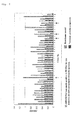
- BT-037 among BT-015, 37, 39, and 44 shown with an arrow, and BT-014 that is reactive with the nontoxic protein component are identical in the amino acid sequence.
- the digested product was converted into a Protein A fused antibody (Fab-pp type antibody) by self-recombining and then DH12S was transformed using this antibody.
- the transformant was cultured in 25 ml of 2xYTGA culture medium at 30°C overnight. This culture solution 20 ml was mixed with 2 liters of 2xYTA culture medium, cultured for three hours, 2 ml of 1M IPTG was added and cultured at 30°C for 20 hours.
- the culture solution was centrifuged at 8000 rpm for 10 minutes at 4°C to collect the supernatant. After adding 1122 g of ammonium sulfate slowly to the collected supernatant, the mixture was stirred at room temperature for one hour, and centrifuged at 8500 rpm for 30 minutes at 4°C. The precipitate was suspended in 100 ml of PBS containing Complete (manufactured by Roche). Subsequently, the suspension was dialyzed against PBS at 4°C overnight, and centrifuged at 10000 rpm for 10 minutes at 4°C.
- the supernatant was collected, added to a column that had been packed with 3 ml of IgG Sepharose (GE Healthcare Bioscience), and passed through the column by natural drop at room temperature. After washing the column with PBS of 300ml, it was eluted with 6 ml of 0.2M glycine (pH 3), 3 ml of 0.2M glycine (pH 2.5) and 10 ml of 0.2M glycine (pH 3). The eluates were each adjusted to pH 7.0 with 2M Tris, placed in a dialysis/concentration tube (manufactured by Millipore, Amicon Ultra-15, 4°C, 5530 rpm rotation), and dialyzed against PBS for concentration. After that, SDS-PAGE was carried out to calculate the protein concentration.
- the neutralizing ability of each antibody was evaluated by a semiquantitative test method that is to observe the symptom such as paralysis and the like in mice after mixing the isolated/prepared Fab-pp type antibody with the toxin and administering it subcutaneously to the mice.
- the test method was carried out according to the references ( M. Takahashi et al., International Journal of Food Microbiology, 11, 271-278, 1990 . Jpn. J. Med. Sci. Biol., 43, 163-170, 1990 ).
- the comparative control was prepared by diluting the botulinum toxin type-A antitoxin 10-fold (Japanese Standard: 10 units/ml) to make a stock solution and further diluting the antitoxin stock solution (1 unit/ml) with PBS by six serial 1:3 dilutions (hereinafter referred to as "standard dilution).
- standard dilution the antibody solution obtained in (5) was diluted 5-fold each with PBS to make an antibody stock solution, and the stock solution was further subjected to four serial 1:5 dilutions to make a specimen (hereinafter referred to as "specimen dilution”.
- the botulinum neurotoxin (3.8 ⁇ 10 5 LD50/ml) was diluted 400-fold to make a stock solution (950LD50/ml), and this was further diluted 500-fold to prepare a test toxin (1.9LD50/ml).
- the test toxin (0.25ml) and standard dilution and specimen dilution (0.25ml each) were accurately taken, mixed well, reacted at room temperature for one hour, and then subcutaneously inoculated into mice (one group: two mice) at a dose of 0.2 ml each, followed by three-day observation.
- the titer of the specimen was judged by clinical symptoms (complete remission, degree of paralysis) in the test group mice.
- BT-015 showed the neutralizing ability ( Fig. 3 ).
- a mixture of the antibody proteins of 19 clones which had been judged to be negative was prepared by the following method.
- DNAs of the 19 clones were mixed, digested with a restriction enzyme SaII, converted into an Fab-pp type antibody by self-recombining, and then DH12S was transformed using this antibody.
- the transformant was cultured in 100 ml of 2xYTGA culture medium at 30°C overnight. This culture solution of 40 ml was mixed with 4 liters of 2xYTA culture medium and cultured for 3 hours, 2 ml of 1M IPTG was added and cultured at 30°C for 20 hours.
- the culture solution was centrifuged at 8000 rpm for 10 minutes at 4°C to collect a supernatant. After adding 2244 g of ammonium sulfate slowly to the collected supernatant, the mixture was stirred at room temperature for one hour, and centrifuged at 8500 rpm for 30 minutes at 4°C. The precipitate was suspended in 200 ml of PBS containing Complete (manufactured by Roche). Subsequently, the suspension was dialyzed against PBS at 4°C overnight and centrifuged at 10000 rpm for 10 minutes at 4°C.
- the supernatant was collected, added to a column that had been packed with 3 ml of IgG Sepharose (GE Healthcare Bioscience), and passed through the column by natural drop at room temperature. After washing the column with 500ml of PBS, it was eluted with 6 ml of 0.2M glycine (pH 3), 3 ml of 0.2M glycine (pH 2.5) and 10 ml of 0.2M glycine (pH 3). The eluates were each adjusted to pH 7.0 with 2M Tris, placed in a dialysis/concentration tube (manufactured by Millipore, Amicon Ultra-15, 4°C, 5530 rpm rotation), and dialyzed against PBS for concentration. After that, SDS-PAGE was carried out to calculate the protein concentration.
- the mixed antibody proteins prepared by the above method were added in 230 ⁇ g/ml to mask the antigen (see Fig. 4 ), and a screening was carried out by a method similar to the screening experiment 1. As a result, 46 clones were collected. As a result of having analyzed each antibody clone by ELISA, it was revealed that 27 new antibody clones that specifically react with the toxoid were included. In this way, by masking the antigen, the antibodies that react with the nontoxic site were largely decreased (2/46) and the screening efficiency was increased (lower panel (4th) of Fig. 5 ).
- 1% BSA/PBS was subdivided in 200 ⁇ l each and blocking was performed at 37°C for one hour. After the blocking solution was discarded, the mixed antibody protein prepared in 2-2 was subdivided in 30 ⁇ g/well, followed by reaction at 37°C for one hour, thereby to mask the antigen. After removal of the antibody protein solution, the 2nd phage (3.6 ⁇ 10 13 equivalents) and the mixed antibody protein prepared in 2-2 (180 ⁇ g) were mixed to be subdivided into Microcup, and allowed to react at 37°C for two hours. The subsequent procedure was carried out in the same manner with the above screening experiment 1.
- the phage (third-2 phage) of 7.2 ⁇ 10 12 equivalents obtained in the screening experiment 3 the mixed antibody protein of 180 ⁇ g prepared in 2-2, and the antibody protein of 180 ⁇ g prepared from the antibody clone BT-015 were mixed to be subdivided into Microcup, and reacted at 37°C for two hours.
- the subsequent procedure was carried out in the same manner with the above screening experiment 1.
- each antibody clone was evaluated by a simple test method in which symptoms such as life and death/paralysis in mice occurred when the antibody was mixed with the toxin and subcutaneously administered. Concrete test method was performed according to the reference (Japanese Minimum Requirements for Biological Products: Ministry of Health, Labour and Welfare). Fab-pp type antibodies prepared from each antibody clone were used in the test.
- the comparative control was prepared by diluting 10-fold the botulinum toxin type-A antitoxin (Japanese Standard: 10 units/ml) to make a stock solution as an antitoxin stock solution of 1 unit/ml, which was then diluted with PBS to 0.1 unit/ml, and is also prepared by further subjecting the stock solution to five serial 1:2 dilutions. (hereinafter referred to as "standard dilution).
- standard dilution The prepared stock solution as it is was used as a specimen.
- the botulinum neurotoxin was diluted 400-fold to make a stock solution (950LD50/ml), and this was further diluted 10-fold to prepare a test toxin (95LD50/ml).
- test toxin (0.25ml), each standard dilution, and a specimen (0.25ml) were accurately taken, mixed well, reacted at room temperature for one hour, and then subcutaneously inoculated into the mice (one group: two mice) at a dose of 0.2 ml each, followed by four-day observation.
- the titer of the specimen was judged by clinical symptoms (complete remission, degree of paralysis) in the test group mice.
- antibody clone BT-175 showed a neutralizing ability ( Fig. 8 ).
- Botulinum toxin was purified from Clostridium botulinum strain 627A from which nontoxic protein components were discarded to yield a purified neurotoxin (active center of the toxin: 150kDa) according to the literature ( INFECTION AND IMMUNITY, Vol. 12, No. 6. Dec. 1975, p. 1262-1270 ). A screening using this neurotoxin as an antigen was carried out to obtain a neutralizing antibody that recognizes an epitope different from those of the antibody clones BT-015 and BT-175.
- a screening experiment was further carried out for the purpose of excluding a lot of antibody clones (clones that are identical or similar to BT-015 or BT-175) obtained even in the screening (2-5) using neurotoxin as an antigen.
- 100 ⁇ l each of neurotoxin which had been prepared to a concentration of 10 ⁇ g/ml with PBS was subdivided into 8-wells of Microcup and reacted overnight at 4°C for immobilization. After discarding the solution, 1% BSA/PBS was subdivided in 200 ⁇ l each and blocking was performed at 37°C for one hour.
- an antibody protein prepared from the antibody clones BT-015 and BT-175 were subdivided (30 ⁇ g/well each) and reacted at 37°C for one hour, thereby to mask the antigen.
- the phage (3rd phage) of 2.0 ⁇ 10 13 obtained in the 3rd screening of the screening experiment 5 and the antibody protein prepared from the antibody clones BT-015 and BT-175 were mixed (180 ⁇ g each), subdivided into Microcup, and allowed to react at 37°C for two hours.
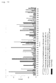
- 188 clones were picked up, and the binding ability thereof was confirmed by ELISA. It revealed that there were 24 kinds of clones that reacted specifically with neurotoxin ( Figs. 12 and 13 ). Further, the above screening process is shown in Fig. 15 .
- Fab-pp type antibodies were prepared from 30 kinds of antibody clones obtained in the screening (screening experiments 5 and 6) using neurotoxin as an antigen.
- the neutralizing ability of each Fab-pp type antibodies was evaluated by a test method in which symptoms such as life and death/paralysis in mice occurred when the antibody was mixed with the toxin and subcutaneously administered were observed.
- the test method was carried out according to the reference (Japanese Minimum Requirements for Biological Products: Ministry of Health, Labour and Welfare, The Japanese Pharmacopoeia), and the titer of the specimen was determined as a relative value to Japanese Standard.
- the botulinum toxin type-A antitoxin (Japanese Standard: 10 units/ml) was diluted 10-fold to make a stock solution that is an antitoxin stock solution (1 unit/ml), which was diluted with PBS to prepare a dilution of 0.1 unit/ml, and further five serial 1:2 dilutions were also prepared (hereinafter referred to as "standard dilution).
- standard dilution The prepared stock solution as it is was used as a specimen.
- the botulinum neurotoxin was diluted 400-fold to make a stock solution (950LD50/ml), and this was further diluted 10-fold to prepare a test toxin (95LD50/ml).
- test toxin (0.25ml), each standard dilutions, and a specimen (0.25ml) were accurately taken, mixed well, reacted at room temperature for one hour, and then subcutaneously inoculated into the mice (one group: two mice) at a dose of 0.2 ml each, followed by 40-day observation.
- the titer of the specimen was judged by clinical symptoms (complete remission, degree of paralysis) in the test group mice.
- four kinds of antibody clones (NT-221, NT-320, NT-523, NT-539) showed a neutralizing ability ( Fig. 14 ).
- IgG antibodies were prepared from antibody clones (BT-015, BT-175, NT-221, NT-320, NT-523) wherein a neutralizing activity was observed in the Fab-pp type antibody.
- a gene fragment encoding VH is ligated to a human ⁇ 1 chain constant region DNA and integrated into an expression vector "BCMGS Neo vector” ( Hajime Karasuyama, "Bovine Papillomavirus Vector", Edited by Masami Muramatsu and Hiroto Okayama, Jikken Igaku (Experimental Medicine), Suppl. Gene Engineering Handbook, Published by Yodosha Co., Ltd., pp.
- the antibody expression vectors were transfected into a Chinese hamster cell strain CHO-K1 using the Lipofectin method, and the cells were cultured at 37°C for a given time, and transplanted in a 96-well plate to select a high expression cell.
- Each cell selected was cultured for two weeks in a large scale culture flask (CELLine 1000 Flask, manufactured by Becton Dickinson) using a serum-free culture medium (CHO-S-SFM II: manufactured by Invitrogen, 1% (v/v) Penicillin- Streptomycin Solution: manufactured by Sigma-Aldrich, 700 ⁇ g/ml G418: manufactured by Sigma-Aldrich).
- the culture supernatant was collected, and supplied to a protein G binding affinity column.
- the column was washed with PBS, eluted with a glycine hydrochloric acid buffer solution (pH 2.7), and neutralized with a Tris-HCl buffer solution (pH 8.9). Then, the eluate was placed in a dialysis/concentration tube (Amicon Ultra-15, manufactured by Millipore), and dialyzed against PBS for concentration.
- the thus obtained IgG type antibody was used in the subsequent experiment.
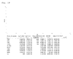
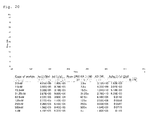
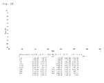
- a biosensor device Biacore3000 was used for analysis. At first neurotoxin 880RU was immobilized to a sensor chip (CM5) and Fab-PP type or IgG type antibody with a sequentially varying concentration (1.9 nM to 1 ⁇ M) was injected (40 ⁇ l, two minutes). The analysis results are shown in Fig. 17 (Fab-PP type antibody of BT-015), Fig. 18 (IgG type antibody of BT-015), Fig. 19 (Fab-PP type antibody of BT-175), Fig. 20 (IgG type antibody of BT-175), Fig. 21 (Fab-PP type antibody of NT-221), and Fig. 22 (Fab-PP type antibody of NT-320). As a result of analysis, all the antibodies were found to have the excellent binding strength.
- Fab-PP type antibodies of six kinds of antibody clones (BT-015, BT-175, NT-221, NT-320, NT-523, NT-539) that showed a neutralizing activity against botulinum toxin type-A were prepared, and a competitive experiment was carried out by Biacore using these antibodies.
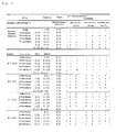
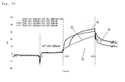
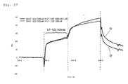
- Neurotoxin 800RU was immobilized to a sensor chip (CM5) and the antibodies were sequentially injected at the concentration shown below, to determine the presence or absence of the competition. The injection was flowed at a rate of 40 ⁇ l (two minutes). All analysis experiments were carried out at 25°C in HBS-EP (Biacore) at an HBS-EP flow rate of 20 ⁇ l/min. The regeneration was performed with 20 ⁇ l (flow rate: 60 ⁇ l/min, 20 seconds) of 100 mM glycine-HCl (pH 2.0). [Table 1]
- a neutralization test was carried out according to Minimum Requirements for Biological Products.
- a botulinum type-A Japanese standard was diluted to prepare five serial dilutions (hereinafter referred to as "Standard Dilution") containing a suitable interval concentration unit in consideration of test accuracy mainly on 0.050 units in 0.25 ml.
- Standard Dilution serial dilutions
- Specimen Dilution the same dilutions
- test toxin type-A was diluted to prepare a solution containing one test toxin amount in 0.25mL (hereinafter referred to as "Toxin Dilution").
- Toxin Dilution a solution containing one test toxin amount in 0.25mL
- Each standard dilution and specimen dilution, and each toxin dilution were taken accurately in an equal amount, and they were mixed well and allowed to stand for one hour.
- Four mice of 23-29 days age were used as one group, and the dose 0.5 ml of the mixed solution per one mouse was intraperitoneally injected, using one group for each mixed solution. After that, observation was continued for three days to calculate the relative value to the standard product.
- the antibodies prepared to 1 mg/ml each were mixed in an equal amount to prepare an antibody solution that is served as a specimen stock solution.
- the specimen stock solution was subjected to a 3-fold serial dilution to 81-fold dilution, thereby to make a specimen diluted solution.
- the mixture was well mixed with a test toxin of 0.63 ml, allowed to react at room temperature for one hour, and inoculated intraperitoneally to the mice (one group:2 mice), followed by 40-day observation.
- Botulinum antitoxin type-A Japanese standard (10 units/ml) was used as a comparative control, and it was diluted with PBS to prepare a five serial dilution containing a suitable interval concentration unit (hereinafter referred to as "Standard Dilution"; Table 4) in consideration of test accuracy mainly on 0.50 unit/ml.
- Botulinum neurotoxin corresponding to each antitoxin was diluted to prepare a test toxin (214ipLD50/ml).
- test toxin (0.63 ml) and 0.63 ml each of the standard dilution and the specimen dilution were taken accurately, mixed well, allowed to react at room temperature for one hour, inoculated to the mice (one group: two mice) intraperitoneally at a dose of 0.5 ml each, and observed for 40 days.
- the titer of the specimens was determined as a relative value to Japanese Standard.
- an antitoxin antibody type-A is contained in 10000 units/vial in a current therapeutic freeze-dried equine botulinum antitoxin type-A (a mixture of four kinds of A, B, E, and F), an equal activity will be obtained by mixing each antibody (about 210 mg each) together in the alternative case with use of four kinds of antibodies.
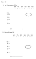
- ELISA was carried out using each antigen shown below.
- NT-539 was superior to NT-523 in the possibility for neutralization of toxin type-A2.
- NT neurotoxin
- Hc E. coli recombinant protein
- SDS-PAGE was performed using acrylamide gel of 10% concentration, transcribed/blocked to a membrane, diluted to two kinds of concentration including 50 ⁇ g/ml (solid line lane) and 5 ⁇ g/ml (dashed line lane), and allowed to react with 200 ⁇ g each of six kinds of IgG type neutralizing antibodies.
- the product was washed with PBS and reacted with an HRP labeled goat anti-human IgG antibody that had been diluted 20000-fold with PBS.
- the product was washed with PBS and detected using DAB as a chromogenic substrate ( Fig. 30 ).
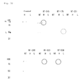
- An epitople analysis was carried out by immunoprecipitation using further three kinds of antigens (neurotoxin, heavy chain, light chain).
- IgG type neutralizing antibodies (5 ⁇ g each) were reacted with protein G Sepharose (100 ⁇ l) to prepare six kinds of antibody beads.
- An antigen (neurotoxin: 0.6 ⁇ g/100 ⁇ l, H-chain: 0.4 ⁇ g/100 ⁇ l, L-chain: 0.4 ⁇ g/100 ⁇ l) was reacted with the antibody beads at 4°C overnight.
- SDS-PAGE was performed using acrylamide gel of 10% concentration, transcribed/blocked to a membrane, and allowed to react with a rabbit anti-botulinum neurotoxin antibody type-A (diluted 1000-fold) that had been purified through affinity purification. After washing the product with PBS, this was reacted with an HRP labeled goat anti-rabbit IgG type-antibody (BIO-RAD) at 37°C. After washing with PBS, the product was detected using a chromogenic substrate ( Fig. 31 ).
- NT shows the case where there is a binding activity to the neurotoxin
- H shows the case where there is a binding activity to the H-chain
- Hc shows the case where there is a binding activity to the neuron-binding domain of the H-chain C-terminal end
- L shows the case where there is a binding activity to the L-chain.
- the symbol (-) indicates "not evaluable”.
- the composition for neutralizing botulinum toxin type-A of the present invention contains a plurality of antibodies whose epitopes are different from each other and exhibits a high neutralizing activity.
- the active ingredient of the present invention is an antibody having a fully human variable region. This feature can solve the problems faced in the conventional preparation (equine antitoxin preparation or neutralizing chimeric antibody), such as much time for the production, difficulty in stable supply, and further contamination of unknown virus, can be solved by this feature.
- the present invention enables the practical use of a stable and economical anti-botulinum toxin type-A preparation and greatly contributes to treatment and prevention of a botulism patient.
- the present invention provides a composition for neutralizing botulinum toxin type-A, which is conformed to the international standard of toxin neutralization test. Therefore, the present invention is extremely useful as internationally usable means for treating and preventing the poisoning in the developing countries where the storage system is not yet in order, particularly sudden and great outbreaking poisoning, and bioterrorism, etc.
- the present invention is not limited by the descriptions of Embodiments and Examples of the above-described invention at all.
- the present invention also includes a variety of modified aspects within the scope where those skilled in the art can easily conceive without departing from the scope of the claims.
- the contents of the theses, publications of patent applications, and patent publications specified in this description are herein incorporated by reference in their entireties.
Landscapes
- Health & Medical Sciences (AREA)
- Chemical & Material Sciences (AREA)
- Organic Chemistry (AREA)
- Life Sciences & Earth Sciences (AREA)
- General Health & Medical Sciences (AREA)
- Medicinal Chemistry (AREA)
- Immunology (AREA)
- Biochemistry (AREA)
- Biophysics (AREA)
- Genetics & Genomics (AREA)
- Molecular Biology (AREA)
- Proteomics, Peptides & Aminoacids (AREA)
- Communicable Diseases (AREA)
- Oncology (AREA)
- Chemical Kinetics & Catalysis (AREA)
- General Chemical & Material Sciences (AREA)
- Nuclear Medicine, Radiotherapy & Molecular Imaging (AREA)
- Pharmacology & Pharmacy (AREA)
- Animal Behavior & Ethology (AREA)
- Public Health (AREA)
- Veterinary Medicine (AREA)
- Peptides Or Proteins (AREA)
- Medicines Containing Antibodies Or Antigens For Use As Internal Diagnostic Agents (AREA)
- Preparation Of Compounds By Using Micro-Organisms (AREA)
- Micro-Organisms Or Cultivation Processes Thereof (AREA)
Applications Claiming Priority (2)
| Application Number | Priority Date | Filing Date | Title |
|---|---|---|---|
| JP2008017152 | 2008-01-29 | ||
| PCT/JP2009/000250 WO2009096162A1 (ja) | 2008-01-29 | 2009-01-23 | A型ボツリヌス毒素中和組成物及びヒト抗a型ボツリヌス毒素抗体 |
Publications (2)
| Publication Number | Publication Date |
|---|---|
| EP2246426A1 true EP2246426A1 (de) | 2010-11-03 |
| EP2246426A4 EP2246426A4 (de) | 2011-08-10 |
Family
ID=40912519
Family Applications (1)
| Application Number | Title | Priority Date | Filing Date |
|---|---|---|---|
| EP09706257A Withdrawn EP2246426A4 (de) | 2008-01-29 | 2009-01-23 | Zusammensetzung zur neutralisierung von botulinustoxin typ-a und menschlicher antikörper gegen botulinustoxin typ-a |
Country Status (5)
| Country | Link |
|---|---|
| US (1) | US8540987B2 (de) |
| EP (1) | EP2246426A4 (de) |
| JP (1) | JP5432727B2 (de) |
| CN (1) | CN101965403B (de) |
| WO (1) | WO2009096162A1 (de) |
Cited By (1)
| Publication number | Priority date | Publication date | Assignee | Title |
|---|---|---|---|---|
| US20110014211A1 (en) * | 2008-01-29 | 2011-01-20 | Institute For Antibodies Co., Ltd. | Composition for neutralizing botulinus toxin type-a, and human anti-botulinus toxin type-a antibody |
Families Citing this family (7)
| Publication number | Priority date | Publication date | Assignee | Title |
|---|---|---|---|---|
| US7563874B2 (en) * | 1998-08-31 | 2009-07-21 | The Regents Of The University Of California | Therapeutic monoclonal antibodies that neutralize botulinum neurotoxins |
| US20110033431A1 (en) * | 2008-03-31 | 2011-02-10 | The Chemo-Sero-Therapeutic Research Institute | Type a2 botulinum toxin preparation |
| US11505599B2 (en) | 2016-01-14 | 2022-11-22 | Memorial Sloan-Kettering Cancer Center | T cell receptor-like antibodies specific for Foxp3-derived peptides |
| EP3534953A4 (de) * | 2016-11-02 | 2020-10-07 | Vanderbilt University | Humane zirka-virus-antikörper und verfahren zur verwendung davon |
| CN119613539B (zh) * | 2024-12-12 | 2025-11-04 | 中国人民解放军军事科学院军事医学研究院 | 抗肉毒毒素l-hn抗原的中和抗体及其相关生物材料与应用 |
| CN121499818B (zh) * | 2026-01-09 | 2026-05-08 | 兰州生物技术开发有限公司 | 一种检测a型肉毒毒素血凝素-70的试剂盒、抗体及方法 |
| CN121522173B (zh) * | 2026-01-09 | 2026-04-14 | 兰州生物技术开发有限公司 | 一种检测a型肉毒毒素ha-70的检测方法及产品 |
Family Cites Families (20)
| Publication number | Priority date | Publication date | Assignee | Title |
|---|---|---|---|---|
| NZ311174A (en) | 1995-06-30 | 1999-04-29 | Mochida Pharm Co Ltd | Anti-fas ligand antibody which suppresses fas ligand-induced apoptosis in cells and method of assaying a fas ligand using the anti-fas ligand antibody |
| US20020155114A1 (en) * | 1998-08-31 | 2002-10-24 | James D. Marks | Therapeutic monoclonal antibodies that neutralize botulinum neurotoxins |
| US7563874B2 (en) * | 1998-08-31 | 2009-07-21 | The Regents Of The University Of California | Therapeutic monoclonal antibodies that neutralize botulinum neurotoxins |
| DE19925739A1 (de) * | 1999-06-07 | 2000-12-21 | Biotecon Ges Fuer Biotechnologische Entwicklung & Consulting Mbh | Therapeutikum mit einem Botulinum-Neurotoxin |
| WO2001062907A1 (fr) | 2000-02-22 | 2001-08-30 | Medical & Biological Laboratories Co., Ltd. | Bibliotheque d'anticorps |
| US20040258699A1 (en) * | 2002-02-11 | 2004-12-23 | Bowdish Katherine S. | Immunotherapeutics for biodefense |
| AU2003213022A1 (en) * | 2002-02-11 | 2003-09-22 | Alexion Pharmaceuticals, Inc. | Immunotherapeutics for biodefense |
| ATE541047T1 (de) | 2002-08-01 | 2012-01-15 | Univ California | Therapeutische monoklonale antikörper zur neutralisierung von botulinum neurotoxinen |
| US20050042775A1 (en) | 2003-08-21 | 2005-02-24 | Nicholas Pomato | Botulinum antitoxin compositions and methods |
| WO2007094754A2 (en) * | 2005-01-27 | 2007-08-23 | The Regents Of The University Of Califordnia | Therapeutic monoclonal antibodies that neutralize botulinum neurotoxins |
| JP5004154B2 (ja) | 2005-04-06 | 2012-08-22 | 一般財団法人化学及血清療法研究所 | 組換え抗ボツリヌス神経毒素抗体 |
| US20100222555A1 (en) * | 2005-06-17 | 2010-09-02 | Thomas Jefferson University | Monoclonal antibodies that neutralize botulinum neurotoxin |
| EP2015065B1 (de) * | 2006-04-28 | 2011-10-05 | Juridical Foundation The Chemo-Sero-Therapeutic Research Institute | Verfahren zur quantifizierung von neurotoxin |
| CA2680741A1 (en) * | 2007-03-22 | 2009-01-15 | The Regents Of The University Of California | Therapeutic monoclonal antibodies that neutralize botulinum neurotoxins |
| AU2008230795B2 (en) * | 2007-03-27 | 2014-05-29 | Bioassets, Llc | Constructs and libraries comprising antibody surrogate light chain sequences |
| EP2246426A4 (de) * | 2008-01-29 | 2011-08-10 | Inst Antibodies Co Ltd | Zusammensetzung zur neutralisierung von botulinustoxin typ-a und menschlicher antikörper gegen botulinustoxin typ-a |
| MY155049A (en) * | 2008-03-14 | 2015-08-28 | Allergan Inc | Immuno-based botolinum toxin serotype a activity assay |
| KR101604515B1 (ko) * | 2008-03-14 | 2016-03-17 | 알러간, 인코포레이티드 | 면역-기반 보툴리눔 독소 세로타입 a 활성 검정 |
| US9000131B2 (en) * | 2008-07-31 | 2015-04-07 | The Regents Of The University Of California | Antibodies that neutralize botulinum neurotoxins |
| US8673890B2 (en) * | 2009-10-29 | 2014-03-18 | Janssen Pharmaceutica Nv | 2,3-dihydro-1H-isoindol-1-imine derivatives useful as thrombin PAR-1 receptor antagonist |
-
2009
- 2009-01-23 EP EP09706257A patent/EP2246426A4/de not_active Withdrawn
- 2009-01-23 JP JP2009551427A patent/JP5432727B2/ja active Active
- 2009-01-23 CN CN2009801071001A patent/CN101965403B/zh not_active Expired - Fee Related
- 2009-01-23 US US12/864,018 patent/US8540987B2/en not_active Expired - Fee Related
- 2009-01-23 WO PCT/JP2009/000250 patent/WO2009096162A1/ja not_active Ceased
Cited By (2)
| Publication number | Priority date | Publication date | Assignee | Title |
|---|---|---|---|---|
| US20110014211A1 (en) * | 2008-01-29 | 2011-01-20 | Institute For Antibodies Co., Ltd. | Composition for neutralizing botulinus toxin type-a, and human anti-botulinus toxin type-a antibody |
| US8540987B2 (en) * | 2008-01-29 | 2013-09-24 | Institute For Antibodies Co., Ltd. | Composition for neutralizing botulinus toxin type-A, and human anti-botulinus toxin type-A antibody |
Also Published As
| Publication number | Publication date |
|---|---|
| WO2009096162A1 (ja) | 2009-08-06 |
| CN101965403A (zh) | 2011-02-02 |
| CN101965403B (zh) | 2013-07-03 |
| US8540987B2 (en) | 2013-09-24 |
| JP5432727B2 (ja) | 2014-03-05 |
| US20110014211A1 (en) | 2011-01-20 |
| JPWO2009096162A1 (ja) | 2011-05-26 |
| EP2246426A4 (de) | 2011-08-10 |
Similar Documents
| Publication | Publication Date | Title |
|---|---|---|
| US8540987B2 (en) | Composition for neutralizing botulinus toxin type-A, and human anti-botulinus toxin type-A antibody | |
| TWI351407B (en) | Method of humanizing immune system molecules | |
| EP2510013B1 (de) | Pcsk9-antagonisten | |
| EP4282880A1 (de) | Vollständig menschlicher breitspektrumneutralisierender antikörper 26e1 gegen coronavirus und verwendung davon | |
| CN113544156A (zh) | 抗Claudin18.2抗体及其应用 | |
| CN105968200A (zh) | 抗人pd-l1人源化单克隆抗体及其应用 | |
| JP2003528052A (ja) | 超高親和性中和抗体 | |
| US20240409620A1 (en) | Design and application of fully human antibody for neutralizing respiratory syncytial virus | |
| US20120269822A1 (en) | Anti-Botulinum Neurotoxin a Single Domain Antibody Antibodies | |
| CA2687681A1 (en) | Antigen-binding proteins targeting s. aureus orf0657n | |
| CN104781277A (zh) | 具有末端修饰的抗原结合分子 | |
| CN112513088A (zh) | 抗ox40抗体、其抗原结合片段及其医药用途 | |
| JP7854469B2 (ja) | 親和性成熟したmica抗体及びその応用 | |
| TW202140547A (zh) | 抗angptl3抗體及其應用 | |
| US9982040B2 (en) | Anti-ricin antibodies and uses thereof | |
| US12269890B2 (en) | Humanized anti-human OX40 monoclonal antibody, preparation method therefor and use thereof | |
| US20250129141A1 (en) | Antibody for tetanus toxin and use thereof | |
| EP4059963A1 (de) | Molekül, das an menschliches 4-1bb binden kann, und anwendung des moleküls | |
| CN113817052A (zh) | 抗SARS-CoV-2核衣壳蛋白单克隆抗体及其制备方法和用途 | |
| KR101964990B1 (ko) | 보툴리눔 독소 b형 또는 e형에 대한 중화능을 가지는 단클론 인간화 항체 | |
| CA3152860A1 (en) | Anti-tfpi monoclonal antibodies | |
| CN116606373A (zh) | 新型冠状病毒中和抗体及其用途 | |
| CN119841941B (zh) | 抗破伤风毒素l-hn片段的纳米抗体及其相关生物材料与应用 | |
| CN104592385A (zh) | 一种抗炭疽芽孢杆菌pa抗原的全人源抗体 | |
| RU2838565C1 (ru) | Антитело mica с созреванием аффинности и его применение |
Legal Events
| Date | Code | Title | Description |
|---|---|---|---|
| PUAI | Public reference made under article 153(3) epc to a published international application that has entered the european phase |
Free format text: ORIGINAL CODE: 0009012 |
|
| 17P | Request for examination filed |
Effective date: 20100818 |
|
| AK | Designated contracting states |
Kind code of ref document: A1 Designated state(s): AT BE BG CH CY CZ DE DK EE ES FI FR GB GR HR HU IE IS IT LI LT LU LV MC MK MT NL NO PL PT RO SE SI SK TR |
|
| AX | Request for extension of the european patent |
Extension state: AL BA RS |
|
| DAX | Request for extension of the european patent (deleted) | ||
| RAP1 | Party data changed (applicant data changed or rights of an application transferred) |
Owner name: OSAKA PREFECTURE UNIVERSITY PUBLIC CORPORATION Owner name: JAPAN AS REPRESENTED BY DIRECTOR GENERAL OF NATION Owner name: INSTITUTE FOR ANTIBODIES CO., LTD. |
|
| A4 | Supplementary search report drawn up and despatched |
Effective date: 20110708 |
|
| RIC1 | Information provided on ipc code assigned before grant |
Ipc: C12P 21/08 20060101ALI20110704BHEP Ipc: C12N 5/10 20060101ALI20110704BHEP Ipc: C12N 1/21 20060101ALI20110704BHEP Ipc: C12N 1/19 20060101ALI20110704BHEP Ipc: C12N 1/15 20060101ALI20110704BHEP Ipc: C07K 16/12 20060101ALI20110704BHEP Ipc: A61K 39/395 20060101ALI20110704BHEP Ipc: C12N 15/09 20060101AFI20110704BHEP |
|
| 17Q | First examination report despatched |
Effective date: 20130304 |
|
| STAA | Information on the status of an ep patent application or granted ep patent |
Free format text: STATUS: THE APPLICATION IS DEEMED TO BE WITHDRAWN |
|
| 18D | Application deemed to be withdrawn |
Effective date: 20150408 |