EP2248897A2 - Verfahren zur Induktion der Differenzierung von undifferenzierten Säugetier-Zellen zu Osteoblasten - Google Patents
Verfahren zur Induktion der Differenzierung von undifferenzierten Säugetier-Zellen zu Osteoblasten Download PDFInfo
- Publication number
- EP2248897A2 EP2248897A2 EP10174536A EP10174536A EP2248897A2 EP 2248897 A2 EP2248897 A2 EP 2248897A2 EP 10174536 A EP10174536 A EP 10174536A EP 10174536 A EP10174536 A EP 10174536A EP 2248897 A2 EP2248897 A2 EP 2248897A2
- Authority
- EP
- European Patent Office
- Prior art keywords
- nucleotide sequence
- protein
- cells
- sirna
- coupled receptor
- Prior art date
- Legal status (The legal status is an assumption and is not a legal conclusion. Google has not performed a legal analysis and makes no representation as to the accuracy of the status listed.)
- Withdrawn
Links
Images




Classifications
-
- C—CHEMISTRY; METALLURGY
- C12—BIOCHEMISTRY; BEER; SPIRITS; WINE; VINEGAR; MICROBIOLOGY; ENZYMOLOGY; MUTATION OR GENETIC ENGINEERING
- C12N—MICROORGANISMS OR ENZYMES; COMPOSITIONS THEREOF; PROPAGATING, PRESERVING, OR MAINTAINING MICROORGANISMS; MUTATION OR GENETIC ENGINEERING; CULTURE MEDIA
- C12N15/00—Mutation or genetic engineering; DNA or RNA concerning genetic engineering, vectors, e.g. plasmids, or their isolation, preparation or purification; Use of hosts therefor
- C12N15/09—Recombinant DNA-technology
- C12N15/11—DNA or RNA fragments; Modified forms thereof; Non-coding nucleic acids having a biological activity
- C12N15/113—Non-coding nucleic acids modulating the expression of genes, e.g. antisense oligonucleotides; Antisense DNA or RNA; Triplex- forming oligonucleotides; Catalytic nucleic acids, e.g. ribozymes; Nucleic acids used in co-suppression or gene silencing
- C12N15/1137—Non-coding nucleic acids modulating the expression of genes, e.g. antisense oligonucleotides; Antisense DNA or RNA; Triplex- forming oligonucleotides; Catalytic nucleic acids, e.g. ribozymes; Nucleic acids used in co-suppression or gene silencing against enzymes
-
- A—HUMAN NECESSITIES
- A61—MEDICAL OR VETERINARY SCIENCE; HYGIENE
- A61P—SPECIFIC THERAPEUTIC ACTIVITY OF CHEMICAL COMPOUNDS OR MEDICINAL PREPARATIONS
- A61P19/00—Drugs for skeletal disorders
-
- A—HUMAN NECESSITIES
- A61—MEDICAL OR VETERINARY SCIENCE; HYGIENE
- A61P—SPECIFIC THERAPEUTIC ACTIVITY OF CHEMICAL COMPOUNDS OR MEDICINAL PREPARATIONS
- A61P19/00—Drugs for skeletal disorders
- A61P19/08—Drugs for skeletal disorders for bone diseases, e.g. rachitism, Paget's disease
-
- A—HUMAN NECESSITIES
- A61—MEDICAL OR VETERINARY SCIENCE; HYGIENE
- A61P—SPECIFIC THERAPEUTIC ACTIVITY OF CHEMICAL COMPOUNDS OR MEDICINAL PREPARATIONS
- A61P19/00—Drugs for skeletal disorders
- A61P19/08—Drugs for skeletal disorders for bone diseases, e.g. rachitism, Paget's disease
- A61P19/10—Drugs for skeletal disorders for bone diseases, e.g. rachitism, Paget's disease for osteoporosis
-
- C—CHEMISTRY; METALLURGY
- C12—BIOCHEMISTRY; BEER; SPIRITS; WINE; VINEGAR; MICROBIOLOGY; ENZYMOLOGY; MUTATION OR GENETIC ENGINEERING
- C12N—MICROORGANISMS OR ENZYMES; COMPOSITIONS THEREOF; PROPAGATING, PRESERVING, OR MAINTAINING MICROORGANISMS; MUTATION OR GENETIC ENGINEERING; CULTURE MEDIA
- C12N5/00—Undifferentiated human, animal or plant cells, e.g. cell lines; Tissues; Cultivation or maintenance thereof; Culture media therefor
- C12N5/06—Animal cells or tissues; Human cells or tissues
- C12N5/0602—Vertebrate cells
- C12N5/0652—Cells of skeletal and connective tissues; Mesenchyme
- C12N5/0654—Osteocytes, Osteoblasts, Odontocytes; Bones, Teeth
-
- C—CHEMISTRY; METALLURGY
- C12—BIOCHEMISTRY; BEER; SPIRITS; WINE; VINEGAR; MICROBIOLOGY; ENZYMOLOGY; MUTATION OR GENETIC ENGINEERING
- C12Y—ENZYMES
- C12Y207/00—Transferases transferring phosphorus-containing groups (2.7)
- C12Y207/11—Protein-serine/threonine kinases (2.7.11)
- C12Y207/11001—Non-specific serine/threonine protein kinase (2.7.11.1), i.e. casein kinase or checkpoint kinase
-
- G—PHYSICS
- G01—MEASURING; TESTING
- G01N—INVESTIGATING OR ANALYSING MATERIALS BY DETERMINING THEIR CHEMICAL OR PHYSICAL PROPERTIES
- G01N33/00—Investigating or analysing materials by specific methods not covered by groups G01N1/00 - G01N31/00
- G01N33/48—Biological material, e.g. blood, urine; Haemocytometers
- G01N33/50—Chemical analysis of biological material, e.g. blood, urine; Testing involving biospecific ligand binding methods; Immunological testing
- G01N33/68—Chemical analysis of biological material, e.g. blood, urine; Testing involving biospecific ligand binding methods; Immunological testing involving proteins, peptides or amino acids
- G01N33/6893—Chemical analysis of biological material, e.g. blood, urine; Testing involving biospecific ligand binding methods; Immunological testing involving proteins, peptides or amino acids related to diseases not provided for elsewhere
-
- C—CHEMISTRY; METALLURGY
- C12—BIOCHEMISTRY; BEER; SPIRITS; WINE; VINEGAR; MICROBIOLOGY; ENZYMOLOGY; MUTATION OR GENETIC ENGINEERING
- C12N—MICROORGANISMS OR ENZYMES; COMPOSITIONS THEREOF; PROPAGATING, PRESERVING, OR MAINTAINING MICROORGANISMS; MUTATION OR GENETIC ENGINEERING; CULTURE MEDIA
- C12N2310/00—Structure or type of the nucleic acid
- C12N2310/10—Type of nucleic acid
- C12N2310/11—Antisense
-
- C—CHEMISTRY; METALLURGY
- C12—BIOCHEMISTRY; BEER; SPIRITS; WINE; VINEGAR; MICROBIOLOGY; ENZYMOLOGY; MUTATION OR GENETIC ENGINEERING
- C12N—MICROORGANISMS OR ENZYMES; COMPOSITIONS THEREOF; PROPAGATING, PRESERVING, OR MAINTAINING MICROORGANISMS; MUTATION OR GENETIC ENGINEERING; CULTURE MEDIA
- C12N2310/00—Structure or type of the nucleic acid
- C12N2310/10—Type of nucleic acid
- C12N2310/11—Antisense
- C12N2310/111—Antisense spanning the whole gene, or a large part of it
-
- C—CHEMISTRY; METALLURGY
- C12—BIOCHEMISTRY; BEER; SPIRITS; WINE; VINEGAR; MICROBIOLOGY; ENZYMOLOGY; MUTATION OR GENETIC ENGINEERING
- C12N—MICROORGANISMS OR ENZYMES; COMPOSITIONS THEREOF; PROPAGATING, PRESERVING, OR MAINTAINING MICROORGANISMS; MUTATION OR GENETIC ENGINEERING; CULTURE MEDIA
- C12N2310/00—Structure or type of the nucleic acid
- C12N2310/10—Type of nucleic acid
- C12N2310/14—Type of nucleic acid interfering nucleic acids [NA]
-
- C—CHEMISTRY; METALLURGY
- C12—BIOCHEMISTRY; BEER; SPIRITS; WINE; VINEGAR; MICROBIOLOGY; ENZYMOLOGY; MUTATION OR GENETIC ENGINEERING
- C12N—MICROORGANISMS OR ENZYMES; COMPOSITIONS THEREOF; PROPAGATING, PRESERVING, OR MAINTAINING MICROORGANISMS; MUTATION OR GENETIC ENGINEERING; CULTURE MEDIA
- C12N2310/00—Structure or type of the nucleic acid
- C12N2310/30—Chemical structure
- C12N2310/31—Chemical structure of the backbone
- C12N2310/315—Phosphorothioates
-
- C—CHEMISTRY; METALLURGY
- C12—BIOCHEMISTRY; BEER; SPIRITS; WINE; VINEGAR; MICROBIOLOGY; ENZYMOLOGY; MUTATION OR GENETIC ENGINEERING
- C12N—MICROORGANISMS OR ENZYMES; COMPOSITIONS THEREOF; PROPAGATING, PRESERVING, OR MAINTAINING MICROORGANISMS; MUTATION OR GENETIC ENGINEERING; CULTURE MEDIA
- C12N2310/00—Structure or type of the nucleic acid
- C12N2310/50—Physical structure
- C12N2310/53—Physical structure partially self-complementary or closed
-
- C—CHEMISTRY; METALLURGY
- C12—BIOCHEMISTRY; BEER; SPIRITS; WINE; VINEGAR; MICROBIOLOGY; ENZYMOLOGY; MUTATION OR GENETIC ENGINEERING
- C12N—MICROORGANISMS OR ENZYMES; COMPOSITIONS THEREOF; PROPAGATING, PRESERVING, OR MAINTAINING MICROORGANISMS; MUTATION OR GENETIC ENGINEERING; CULTURE MEDIA
- C12N2510/00—Genetically modified cells
-
- C—CHEMISTRY; METALLURGY
- C12—BIOCHEMISTRY; BEER; SPIRITS; WINE; VINEGAR; MICROBIOLOGY; ENZYMOLOGY; MUTATION OR GENETIC ENGINEERING
- C12N—MICROORGANISMS OR ENZYMES; COMPOSITIONS THEREOF; PROPAGATING, PRESERVING, OR MAINTAINING MICROORGANISMS; MUTATION OR GENETIC ENGINEERING; CULTURE MEDIA
- C12N2799/00—Uses of viruses
- C12N2799/02—Uses of viruses as vector
- C12N2799/021—Uses of viruses as vector for the expression of a heterologous nucleic acid
Definitions
- the present invention relates to methods for inducing the differentiation of undifferentiated mammalian cells into osteoblasts.
- the invention further relates to methods for identifying one or more compounds that induce said differentiation, as well as to the compounds per se for inducing said differentiation.
- Bone contains two distinct cell lineages, bone-forming cells (e.g. osteoblasts) and bone-resorbing cells (e.g. osteoclasts). It is a dynamic tissue that is continuously being destroyed (resorbed) and rebuilt, by an intricate interplay between these osteoblasts and osteoclasts.
- bone-forming cells e.g. osteoblasts
- bone-resorbing cells e.g. osteoclasts
- Osteoblasts originate from differentiated mesenchymal progenitor cells (MPCs). During differentiation into osteoblasts bone alkaline phosphatase activity (BAP) becomes upregulated. Bone formation in vivo occurs through two distinct pathways during embryonic development: endochondral or intramembranous ossification ( Figure 1 ). As shown in this figure, mesenchymal progenitor or stem cells represent the starting points for both forms of bone formation.
- flat bones such as those of the skull or clavicles, are formed directly from condensations of mesenchymal cells.
- mesenchymal condensations first lead to a cartilage intermediate that is invaded during further development by endothelial cells, osteoclasts and mesenchymal cells that will differentiate into osteoblasts and osteocytes (From Nakashima and de Crombrugghe, 2003).
- a number of diseases are known which are caused by a disturbance of the fine-tuned balance between bone resorption and bone build-up.
- These skeletal diseases represent a large number of patients: hypercalcemia of malignancy, Paget's disease, inflammatory bone diseases like rheumatoid arthritis and periodontal disease, focal osteogenesis occurring during skeletal metastases, Crouzon's syndrome, rickets, opsismodysplasia, pycnodysostosis/Toulouse-Lautrec disease, osteogenesis imperfecta, but the single most important bone disease is osteoporosis.
- Osteoporosis affects 1 in 5 women over 50 and 1 in 20 men over 50. A number of treatments are available for the patient. These mostly tackle the net increase in bone resorption, i.e.:
- BMPs bone morphogenetic proteins
- Receptors for the bone morphogenetic proteins have been identified in many tissues, and the BMPs themselves are expressed in a large variety of tissues in specific temporal and spatial patterns. This suggests that BMPs may have effects on many tissues other than bone, potentially limiting their usefulness as therapeutic agents when administered systemically.
- novel anabolics that circumvent one or more of the shortcomings of the above mentioned anabolics.
- This object is achieved by providing a method for inducing differentiation of undifferentiated mammalian cells into osteoblasts, comprising contacting said undifferentiated cells with an inhibitor of any of the polypeptides listed in table 4, and/or fragments and/or derivatives of said polypeptide.
- inhibitor of a polypeptide relates to any type of molecule that inhibits, e.g. downmodulates the biological activity of said polypeptide, by inhibiting the expression and/or translation of said polypeptide, or by inhibiting the polypeptide per se.
- Undifferentiated cells are pluripotent cells which are in an early stage of specialization, i.e ., which do not yet have their final function and can be induced to form almost any given cell type.
- these are cells which have not yet differentiated to e.g . osteoblasts or osteoclasts.
- Such cells are for example blood cells and cells present in bone marrow, as well as cells derived from adipose tissue.
- cells which still can be differentiated into mesenchymal precursor cells are ontemplated in the present invention, such as, for example, totipotent stem cells such as embryonic stem cells.
- Osteoblast differentiation can be measured by measuring the level of enzymes that are induced during the differentiation process, such as alkaline phosphatase (BAP), type-1 collagen, osteocalcin and osteopontin.
- the alkaline phosphatase activity can be measured by adding methylumbelliferyl heptaphosphate (MUP) solution (Sigma) to the cells.
- MUP methylumbelliferyl heptaphosphate
- the fluorescence generated upon cleavage of the MUP substrate by the AP activity is measured on a fluorescence plate reader (Fluostar, BMG).
- the inhibitor is an expression or translation inhibitor inhibiting the expression or translation of a polyribonucleotide encoding the polypeptide.
- the inhibitor is an nucleic acid expressing the expression or translation inhibitor inhibiting the expression or translation of a polyribonucleotide encoding the polypeptide.
- the nucleic acid is included within a vector.
- the vector is an adenoviral, retroviral, adeno-associated viral, lentiviral or a sendaiviral vector.
- Nucleic acids expressing the inhibitor e.g. antisense RNA, ribozyme, antisense oligodeoxynucleotide (ODN), and/or siRNA, such as vectors and viral vectors, have the advantage that they produce said inhibitors continuously.
- Vectors include plasmids and viruses.
- the nucleic acid sequence may be placed under the control of a appropriate promoter, such as CMV, HSV, TK, SV40, or, the elongation factor among others. A prolonged production of the inhibitor will give a longer inhibitory effect and less transfections are needed.
- the inhibitor is selected from the group consisting of an antisense RNA, a ribozyme that cleaves the polyribonucleotide, an antisense oligodeoxynucleotide (ODN), and a small interfering RNA (siRNA) that is sufficiently homologous to a portion of the polyribonucleotide such that the siRNA is capable of inhibiting the polyribonucleotide that would otherwise cause the production of the polypeptide.
- an antisense RNA a ribozyme that cleaves the polyribonucleotide
- ODN antisense oligodeoxynucleotide
- siRNA small interfering RNA
- inhibitors form also part of the present invention, and relate to inhibitors which are modified in order to enhance their stability.
- the inhibitors thus may contain a variety of modifications that confer resistance to nucleolytic degradation, such as, for example, modified internucleoside linkages, modified nucleic acid bases and/or modified sugars and the like.
- the antisense oligonucleotides of the invention can also be modified by chemically linking the oligonucleotide to one or more moieties or conjugates to enhance the activity, cellular distribution, or cellular uptake of the antisense oligonucleotide.
- Such moieties or conjugates include but are not limited to lipids such as cholesterol, cholic acid, thioether, aliphatic chains, phospholipids, polyamines, polyethylene glycol (PEG), or palmityl moieties.
- lipids such as cholesterol, cholic acid, thioether, aliphatic chains, phospholipids, polyamines, polyethylene glycol (PEG), or palmityl moieties.
- Other forms of chemical modification including but not limited to 2'-O-methyl-, Methyl-phosphonate-, phosphothioate, inverted end- (3'-3'-linkage) modifications, are also possible.
- an antisense nucleic acid e.g. DNA
- an antisense nucleic acid may be introduced into cells in vitro , or administered to a subject in vivo , as gene therapy to inhibit cellular expression of nucleic acids comprising any of the genes listed in Table 4.
- Anti-sense nucleic acid is intended to mean an nucleic acid that has a nucleotide sequence that interacts through base pairing with a specific complementary nucleic acid sequence involved in the expression of the target such, that the expression of the gene is reduced.
- the specific complementary nucleic acid sequence involved in the expression of the gene is a genomic DNA molecule or mRNA molecule that encodes the gene. This genomic DNA molecule can comprise regulatory regions of the gene, or the coding sequence for the mature gene.
- Antisense nucleic acids preferably comprise a sequence containing from about 17 to about 100 nucleotides and more preferably the antisense nucleic acids comprise from about 18 to about 30 nucleotides.
- Antisense nucleic acids of the invention are preferably nucleic acid fragments capable of specifically hybridizing with all or part of a nucleic acid encoded by any of the genes listed in Table 4 or the corresponding messenger RNA, or they may be DNA sequences whose expression in the cell produces RNA complementary to all or part of the mRNA comprisingnucleic acid sequences encoded by any of the genes listed in Table 4.
- Antisense nucleic acids may be prepared by expression of all or part of a sequence selected from the group nucleic acid sequences encoded by any of the genes listed in Table 4, in the opposite orientation.
- the antisense nucleic acid is prepared by expression of a sequence selected from the groups consisting of SEQ ID NO: 1-226 and SEQ ID NO 247-333, in the opposite orientation.
- the antisense sequence is at least about 17 nucleotides in length.
- complementary to a nucleotide sequence in the context of antisense nucleic acids and methods therefore means sufficiently complementary to such a sequence as to allow hybridization to that sequence in a cell, i.e., under physiological conditions.
- ribozymes as used herein relates to catalytic RNA molecules capable of cleaving other RNA molecules at phosphodiester bonds in a manner specific to the sequence.
- the hydrolysis of the target sequence to be cleaved is initiated by the formation of a catalytically active complex consisting of ribozyme and substrate RNA. All ribozymes capable of cleaving phosphodiester bonds in trans, that is to say intermolecularly, are suitable for the purposes of the invention.
- RNAi RNA interference
- dsRNA double stranded RNA
- the inhibitor is a siRNA, comprising a first nucleotide sequence of 17-23 nucleotides homologous to a nucleotide sequence selected from any of the target genes listed in table 4, and a second nucleotide sequence of 17-23 nucleotides complementary to said first nucleotide sequence.
- the inhibitor is a siRNA, comprising a first nucleotide sequence of 17-23 nucleotides selected from the nucleotide sequences identified by SEQ. ID NO. 1-220 and 247-333 in table 4 and 5, and a second nucleotide sequence of 17-23 nucleotides complementary to said first nucleotide sequence.
- the siRNA further comprises a third nucleotide sequence connecting the first and second nucleotide sequence, and capable of forming a stem-loop structure within the siRNA.
- Such self-complementing single stranded siRNA polynucleotide according to the present invention thus at least comprises a first guide sequence, and a second guide sequence, which complements the first guide sequence, and a third sequence capable of forming a stem-loop structure within said second sequence, which is covalently linked to the distal end of the first sequence and the proximal end of the second sequence.
- All nucleotides in the first and second sequences may base pair, or alternatively there may be mismatches between the first and second sequences.
- the nucleotide sequences are preferably between about 17 and 23 nts in length.
- the first or the second sequence is a oligonucleotide sequence between about 17 and 23 nt in length selected from the polynucleotide sequences of any of the genes listed in Table 4.
- siRNA polynucleotides with a loop are selfcomplementary and can form stable hairpins. Hairpins are more stable than ordinary dsRNA. In addition, they are more easily produced from vectors.
- the first or second nucleotide sequence comprises a nucleotide sequence consisting of SEQ ID NO: 1-220 and SEQ ID NO 247-333.
- the nucleotide sequences of SEQ ID NO 1-220 and SEQ ID NO 247-333 are selected according to siRNA designing rules that give an improved reduction of the target sequences compared to nucleotide sequences that do not comply with these siRNA designing rules (see WO 2004/094636 ).
- the third nucleotide sequence is 4-30 nucleotides long, more preferably 5-15 nucleotides long and most preferably 8 nucleotides long.
- the third nucleotide sequence is UUGCUAUA (SEQ ID NO: 334).
- the present invention further relates to a method for identifying a compound that induces differentiation of undifferentiated mammalian cells into osteoblasts, comprising:
- polypeptides or the polynucleotides of the present invention employed in the methods described above may be free in solution, affixed to a solid support, borne on a cell surface, or located intracellularly.
- the polypeptide of the present invention or the compound to facilitate separation of complexes from uncomplexed forms of the polypeptide, as well as to accommodate automation of the assay.
- Interaction (e.g., binding of) of the polypeptide of the present invention with a compound can be accomplished in any vessel suitable for containing the reactants. Examples of such vessels include microtiter plates, test tubes, and microcentrifuge tubes.
- a fusion protein can be provided which adds a domain that allows the polypeptide to be bound to a matrix.
- the polypeptide of the present invention can be "His" tagged, and subsequently adsorbed onto Ni-NTA microtitre plates, or ProtA fusions with the polypeptides of the present invention can be adsorbed to IgG, which are then combined with the cell lysates (e.g ., (35) S -labelled) and the candidate compound, and the mixture incubated under conditions favorable for complex formation (e.g., at physiological conditions for salt and pH). Following incubation, the plates are washed to remove any unbound label, and the matrix is immobilized. The amount of radioactivity can be determined directly, or in the supernatant after dissociation of the complexes. Alternatively, the complexes can be dissociated from the matrix, separated by SDS-PAGE, and the level of the protein binding to the protein of the present invention quantitated from the gel using standard electrophoretic techniques.
- Biotinylated protein molecules of the present invention can be prepared from biotin-NHS (N-hydroxy-succinimide) using techniques well known in the art (e.g., biotinylation kit, Pierce Chemicals, Rockford, III.), and immobilized in the wells of streptavidin-coated 96 well plates (Pierce Chemical).
- antibodies reactive with the polypeptides of the present invention but which do not interfere with binding of the polypeptide to the compound can be derivatized to the wells of the plate, and the polypeptide of the present invention can be trapped in the wells by antibody conjugation.
- preparations of a labeled candidate compound are incubated in the wells of the plate presenting the polypeptide of the present invention, and the amount of complex trapped in the well can be quantitated.
- the binding affinity of the compound with the polypeptide or polynucleotide can be measured by methods known in the art, such as using surface plasma resonance biosensors (Biacore), by saturation binding analysis with a labeled compound (e.g. Scatchard and Lindmo analysis), via displacement reactions, by differential UV spectrophotometer, fluorescence polarisation assay, Fluorometric Imaging Plate Reader (FLIPR ® ) system, Fluorescence resonance energy transfer, and Bioluminescence resonance energy transfer.
- the binding affinity of compounds can also be expressed in a dissociation constant (Kd) or as IC50 or EC50.
- Kd dissociation constant
- IC50 represents the concentration of a compound that is required for 50% inhibition of binding of another ligand to the polypetide.
- the EC50 represents the concentration required for obtaining 50% of the maximum effect in vitro .
- the dissociation constant, Kd is a measure of how well a ligand binds to the polypeptide, it is equivalent to the ligand concentration required to saturate exactly half of the binding-sites on the polypeptide.
- Compounds with a high affinity binding have low Kd, IC50 and EC50 values, i.e. in the range of 100 nM to 1 pM; a moderate to low affinity binding relates to a high Kd, IC50 and EC50 values, i.e. in the micromolar range.
- libraries of compounds can be used such as peptide libraries (e.g. LOPAPTM, Sigma Aldrich), lipid libraries (BioMol), synthetic compound libraries (e.g. LOPACTM, Sigma Aldrich) or natural compound libraries (Specs, TimTec).
- peptide libraries e.g. LOPAPTM, Sigma Aldrich
- lipid libraries BioMol
- synthetic compound libraries e.g. LOPACTM, Sigma Aldrich
- natural compound libraries Specs, TimTec
- the invention also relates to a method for identifying a compound or mixture of compounds that induces differentiation of undifferentiated mammalian cells into osteoblasts, comprising
- the compound or mixture of compounds that are identified according to this method can thus be applied to induce differentiation of undifferentiated mammalian cells into osteoblasts.
- the compounds identified in these methods of the invention are low molecular weight compounds.
- Low molecular weight compounds i.e. with a molecular weight of 500 Dalton or less, are likely to have good absorption and permeation in biological systems and are consequently more likely to be successful drug candidates than compounds with a molecular weight above 500 Dalton (Lipinsky et al, 2001).
- the compounds are peptides.
- Peptides can be excellent drug candidates and there are multiple examples of commercially valuable peptides such as fertility hormones and platelet aggregation inhibitors.
- the compounds are natural compounds.
- Natural compounds are compounds that have been extracted from natural sources, e.g. plants. Using natural compounds in screens has the advantage that more diverse molecules are screened. Natural compounds have an enormous variety of different molecules. Synthetic compounds do not exhibit such variety of different molecules. Other compounds may be selected from carbohydrates, and glycosylated polypeptides.
- the present invention relates to polynucleotide comprising a sequence of 17-23 nucleotides homologous to a nucleotide sequence selected from any of the target genes listed in table 4, and variants and/or the reverse complements thereof.
- said polynucleotides have a nucleotide sequence selected from the nucleotide sequences identified by SEQ. ID NO. 1-220 and 247-333 in table 4 and 5, or said polynucleotides have a reverse complement of a nucleotide sequence selected from the nucleotide sequences identified by SEQ. ID NO. 1-220 and 247-333 in table 4 and 5.
- the polynucleotides of the invention have been shown to increase osteoblast differentiation.
- the invention also relates to these polynucleotides for use as a medicament.
- the invention relates to the use of said polynucleotides in the manufacture of a medicament for the treatment of a disease involving a systemic or local decrease in mean bone density.
- the polypeptides comprising an amino acid sequence selected from the group listed in Table 4 are involved in the differentiation of undifferentiated mammalian cells into osteoblasts
- compounds that are known or will be known to inhibit the activity of any of the polypeptides comprising an amino acid sequence selected from the group listed in Table 4 can now also be used to induce differentiation of undifferentiated mammalian cells into osteoblasts. Therefore, the present invention also relates to the use of compounds that are known in the art to inhibit the activity of any of polypeptides comprising an amino acid sequence selected from the group listed in Table 4, as a medicament.
- the present invention also relates to the use of compounds that are known in the art to inhibit the activity of any of polypeptides comprising an amino acid sequence selected from the group listed in Table 4, as a medicament for the treatment of a disease involving a systemic or local decrease in mean bone density.
- the invention furthermore relates to a vector comprising any of the polynucleotides described above.
- the invention relates to said vectors for use as a medicament.
- the vector is an adenoviral, retroviral, adeno-associated viral, lentiviral or a sendaiviral vector.
- the vector encodes a siRNA comprising a nucleotide sequence selected from the groups consisting of SEQ ID NO: 1-220 and SEQ ID NO 247-333.
- the invention further relates to the use of said vectors for the manufacture of a medicament for the treatment of a disease involving a systemic or local decrease in mean bone density.
- the disease is selected from the group consisting of osteoporosis, hypercalcemia of malignancy, multiple myelomatosis, hyperparathyroidism, and hyperthyroidism.
- Recombinant viruses are commonly used for gene transfer. To date, the three most commonly used viruses for gene transfer are adenovirus, retrovirus and adeno-associated virus. More recently lentiviruses, a subgroup of the retroviruses, and sendaivirus are being used. Adenoviruses are able to transduce both dividing and non-dividing cells and can be produced at high viral titres. Retroviruses can infect dividing cells only and integrate their genome into the host chromosome. Integration achieves long-term gene expression. Lentiviruses, a sub-family of retroviruses, share all the standard properties of retroviruses but in addition they have the capacity to transduce non-dividing cells.
- Adeno-associated virus is a small, non-pathogenic, single-stranded DNA virus. It requires co-infection with a helper virus (adenovirus or herpes virus) in order to undergo productive infection. In the absence of helper virus, wild-type AAV integrates site-specifically, into the host genome. Similarly to retrovirus, integration facilitates longer gene expression. AAV can infect both non-dividing and dividing cells. Sendai virus is a member of the paramyxoviridae, and is a single-stranded RNA virus that is able to transfect both dividing and non-dividing cells. Its method of entering cells involves sialic acid and cholesterol that are common to many cell types. Sendai viral gene expression and replication are localized in the cytoplasm, in contrast to most viruses that need to enter the nucleus.
- helper virus adenovirus or herpes virus
- the present invention relates to a method for in vitro production of bone tissue, comprising
- the invention thus provides a method for producing a substrate with a matrix grown thereon, which can be used for the provision of load-bearing implants, including joint prostheses, such as artificial hip joints, knee joints and finger joints, and maxillofacial implants, such as dental implants. It can also be used for special surgery devices, such as spacers, or bone fillers, and for use in augmentation, obliteration or reconstitution of bone defects and damaged or lost bone. Bone formation can be optimized by variation in mineralization, both by inductive and by conductive processes.
- a combination of the provision of a load-bearing implant (preferably coated with a matrix as described above) with a bone filler comprising a matrix as described, constitutes an advantageous method according to the present invention.
- the method of the invention is also very suitable in relation to revision surgery, i.e., when previous surgical devices have to be replaced.
- Suitable undifferentiated cells are bone marrow cells, including haematopoietic cells and in particular stromal cells.
- the marrow cells, and especially the stromal cells are found to be very effective in the bone producing process when taken from their original environment.
- the undifferentiated cells can be directly applied on the substrate or they can advantageously be multiplied in the absence of the substrate before being applied on the substrate. In the latter mode, the cells are still largely undifferentiated after multiplication and, for the purpose of the invention, they are still referred to as undifferentiated. Subsequently, the cells are allowed to differentiate. Differentiation can be induced or enhanced by the presence of suitable inductors, such as glucocorticoids, and dexamethasone. Especially suitable inductors of differentiation are the expression inhibitory agents of the present invention.
- undifferentiated cells provide several advantages. Firstly, their lower differentiation implies a higher proliferation rate and allows the eventual functionality to be better directed and controlled. Moreover, culturing these cells not only produces the required bone matrix containing organic and inorganic components, but also results in the presence, in the culture medium and in the matrix, of several factors which are essential for growth of the tissue and for adaptation to existing living tissue. Also, the culture medium can be a source of active factors such as growth factors, to be used in connection with the implanting process. Furthermore, such undifferentiated cells are often available in large quantities and more conveniently than e.g ., mature bone cells, and exhibit a lower morbidity during recovery. Moreover, the undifferentiated cells can be obtained from the patient for whom the implant is intended. The bone resulting from these cells is autologous to the patient and thus no immune response will be induced. Matrices as thick as 100 ⁇ m can be produced as a result of the use of undifferentiated cells.
- the substrate on which the undifferentiated cells can be applied and cultured can be a metal, such as titanium, cobalt/chromium alloy or stainless steel, a bioactive surface such as a calcium phosphate, polymer surfaces such as polyethylene, and the like. Although less preferred, siliceous material such as glass ceramics, can also be used as a substrate. Most preferred are metals, such as titanium, and calcium phosphates, even though calcium phosphate is not an indispensable component of the substrate.
- the substrate may be porous or non-porous.
- the cells can be applied at a rate of e.g ., 10 3 -10 6 per cm 2 , in particular 10 4 -2X10 5 cells per cm 2 .
- the culture medium to be used in the method according to the invention can be a commonly known culture medium such as MEM (minimum essential medium).
- the medium can be a conditioned medium.
- a conditioned medium is understood to be a medium wherein similar cells have previously been incubated, causing the medium to contain factors such as polypeptides, secreted by the cells which are important for cell growth and cell differentiation.
- the cells are cultured for a time sufficient to produce a matrix layer, e.g ., a matrix layer having a thickness of at least 0.5 ⁇ m, in particular from 1 up to 100 ⁇ m, more in particular of 10-50 ⁇ m.
- the cells may be contacted with the culture medium for e.g . 2-15 weeks, in particular 4-10 weeks.
- the production of the matrix when applied on a substrate, results in a continuous or quasi-continuous coating covering the substrate for at least 50%, in particular at least 80% of its surface area.
- the present invention provides osteoblast cells obtainable by methods of the present invention.
- Another embodiment of the present invention involves the use of the osteoblast cells obtainable by methods of the present invention for in vitro production of bone tissue.
- the present invention relates further to a method for diagnosing a pathological condition involving a systemic or local decrease in mean bone density or a susceptibility to the condition in a subject, comprising
- the present invention relates to a method for diagnosing a pathological condition involving a systemic or local decrease in mean bone density or a susceptibility to the condition in a subject, comprising
- the term "derivatives of a polypeptide” relates to those proteins, proteinaceous molecules, fractions of proteins, peptides, and oligopeptides that comprise a stretch of contiguous amino acid residues of the polypeptide and that retain the biological activity of the protein, e.g. polypeptides that have amino acid mutations compared to the amino acid sequence of a naturally-occurring form of the polypeptide.
- a derivative may further comprise additional naturally occurring, altered, glycosylated, acylated or non-naturally occurring amino acid residues compared to the amino acid sequence of a naturally occurring form of the polypeptide.
- It may also contain one or more non-amino acid substituents compared to the amino acid sequence of a naturally occurring form of the polypeptide, for example a reporter molecule or other ligand, covalently or non-covalently bound to the amino acid sequence.
- fragment of a polypeptide relates to peptides, oligopeptides, polypeptides, proteins and enzymes that comprise a stretch of contiguous amino acid residues, and exhibit substantially a similar, but not necessarily identical, functional activity as the complete sequence.
- polynucleotide refers to nucleic acids, such as double stranded, or single stranded DNA and (messenger) RNA, and all types of oligonucleotides. It also includes nucleic acids with modified backbones such as peptide nucleic acid (PNA), polysiloxane, and 2'-O-(2-methoxy)ethylphosphorothioate.
- PNA peptide nucleic acid
- polysiloxane polysiloxane
- 2'-O-(2-methoxy)ethylphosphorothioate 2'-O-(2-methoxy)ethylphosphorothioate
- derivatives of a polynucleotide relates to DNA-molecules, RNA- molecules, and oligonucleotides that comprise a stretch or nucleic acid residues of the polynucleotide, e.g. polynucleotides that may have nucleic acid mutations as compared to the nucleic acid sequence of a naturally occurring form of the polynucleotide.
- a derivative may further comprise nucleic acids with modified backbones such as PNA, polysiloxane, and 2'-O-(2-methoxy)ethyl-phosphorothioate, non-naturally occurring nucleic acid residues, or one or more nuclei acid substituents, such as methyl-, thio-, sulphate, benzoyl-, phenyl-, amino-, propyl-, chloro-, and methanocarbanucleosides, or a reporter molecule to facilitate its detection.
- nucleic acids with modified backbones such as PNA, polysiloxane, and 2'-O-(2-methoxy)ethyl-phosphorothioate, non-naturally occurring nucleic acid residues, or one or more nuclei acid substituents, such as methyl-, thio-, sulphate, benzoyl-, phenyl-, amino-, propyl-, chloro-, and methanocarban
- fragment of a polynucleotide relates to oligonucleotides that comprise a stretch of contiguous nucleic acid residues that exhibit substantially a similar, but not necessarily identical, activity as the complete sequence.
- homologous sequence whether a nucleic acid sequence or an amino acid sequence, is limited to a sequence that is identical or substantially identical to the parent sequence and has the same biological property as the parent sequence.
- the term is meant to include naturally occurring mutants and variants of the parent sequence.
- the parent sequence is either provided in table 4-6, or is indicated by the GenBank accession numbers in these tables.
- a homologous sequence is at least 95% identical to the parent sequence. However, up to two mismatches are allowed for the 17-21 bp siRNA sequences, provided that the homologous sequence leads to a similar down-regulation of the corresponding gene as the parent sequence.
- Example 1 Development of a high-throughput screening method for the detection of endogenous alkaline phosphatase
- MPCs Mesenchymal progenitor cells
- BMP2 basalpha-1 phosphatase
- Ad-siRNA siRNA adenoviruses
- SilenceSelectTM collection AdC15-hCAR/ AdC20-hCAR co-infection increases the AdC01-siRNA infection efficiency.
- Cellular AP activity was determined 13 days after the start of the infection (13 dpi).
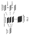
- Figure 2 illustrates the principle of the assay.
- MPCs were isolated from bone marrow of healthy volunteers, obtained after informed consent (Cambrex/Biowhittaker, Verviers, Belgium).
- cell seeding density multiplicities of infection (MOI) of control viruses (Ad-BMP2 or Ad-eGFP)
- MOI of Ad-hCAR MOI of Ad-hCAR
- duration of infection duration of infection
- toxicity toxicity
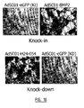
- infection efficiency using Ad-eGFP
- Ad-BMP2 BMP2 overexpression
- Ad-BMP2 Ad-control-virus
- Upregulation of alkaline phosphatase was read at 13 dpi: 15 ⁇ l 4-Methylumbelliferylphosphate (MUP, Sigma) was added to each well, the plates were incubated for 15 min at 37°C and monitored for AP activity using a fluorescence plate reader (Fluostar, BMG).
- MUP 4-Methylumbelliferylphosphate
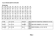
- This "knock-down" control plate contains Ad-H9 (H24-010) and Ad-H10 (H24-011) as positive controls; Ad-eGFP (knock-in virus) as infection control; and Ad-eGFP-siRNA, Ad-M6PR-siRNA and Ad-Luc-siRNA (all 3 are knock-down viruses) as negative controls.
- control viruses were pipetted from 96 well KD control plates into 384 well plates using robotics.
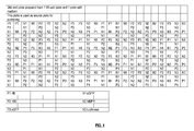
- the final lay-out of the 384 well plate is depicted in Figure 4 .
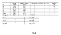
- Figure 5 shows results from the automated screening procedure using the KD control plate.
- the mean and standard deviations of the KD negative controls (N1-N3) were used to calculate a cut-off for hit analysis, which was set at the mean for N1, N2, N3 ('All negatives') plus 3 times the standard deviation for 'All negatives'.
- the positive controls (P1 and P2), scored in more than 95% of the infected wells.
- the negative control viruses scored in less than 5% of the wells.
- the optimized protocol for screening the SilenceSelect library is the following: on day 0, MPC cells are seeded in black 384 well plates with clear bottom (Costar or Nunc) in 60 ⁇ l medium at a density of 500 cells per well.
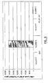
- the KD control plate was run under the same conditions as the aliquot plates from the SilenceSelect collection. All Ad-siRNA viruses were screened in duplicate, with each singular on a different MPC plate. Plates were then incubated at 37°C. Four days post infection the medium containing the adenoviruses was replaced by fresh medium free of virus. Thirteen days post infection, the AP activity readout was performed. A typical result of a 384 well screening plate is depicted in Figure 6 , in which the relative fluorescence units (RFU) are plotted for each of the data points of the 384 well plate on the Y-axis; while the numbers on the X-axis correspond to positions in the 384 well plate.
- REU relative fluorescence units
- the data obtained from measuring the AP activity were analyzed as follows: the background was calculated by taking the mean of the data points from all the plates except the control plate. A cut-off value for hit calling was calculated by adding 3 times the standard deviation of all data points, excluding the control plate. Each data point was analyzed for scoring above or under the cut-off. Only Ad-siRNAs inducing endogenous AP activity levels above the cut-off were of further interest. Hits were prioritized according to their scoring in single or duplo, in one or both of the screens. Data were collected for 7980 Ad-siRNA virus constructs representing 4091 independent genes. An overview of the constructs is given in Table 4.
- genes identified in the screen were targeted by 2 Ad-siRNAs. These genes are AVPR1B, FLJ22955, IL1 F8, PPIA, USP38, C9, LOC254378 and BRS3 (see Table 4).
- the Ad-siRNA hits hereafter referred to as 'targets' were subjected to further analysis to establish their therapeutic application as bone anabolic factor.
- a first step entailed a quality control on the Ad-siRNA selected for further analysis (this example).
- Other validation steps are the validation of targets at the mRNA level (Example 5), screening of the targets in osteogenesis assays such as the mineralization assay (Example 6), and development of additional Ad-siRNAs targeting the identified genes (Example 9).
- Targets that remain of interest after these validation assays are considered for drug discovery: assays are developed allowing the discovery and optimization of compounds that mimick the bone anabolic actions of the target Ad-siRNAs (Example 7).
- the anti-resorptive activities of the identified Ad-siRNAs are validated in osteoclast assays (Example 8).
- Target Ad-siRNAs were propagated using PerC6 cells (Crucell, Leiden, The Netherlands) at a 96 well plate level, followed by rescreening these viruses at several MOls in the primary assay (see Example 1) and by sequencing the siRNAs encoded by the target Ad-siRNA viruses.
- PerC6/E2A cells were seeded in 96 well plates at a density of 40 000 cells per well in 180 ⁇ l PerC6/E2A medium. Cells were then incubated overnight at 39°C in a 10% C0 2 humidified incubator. One day later, cells were infected with 1 ⁇ l of crude cell lysate from SilenceSelect stocks containing target Ad-siRNAs. Cells were incubated further at 34°C, 10% CO 2 until appearance of cytopathic effect (as revealed by the swelling and rounding up of the cells, typically 7 days post infection).
- the supernatant was collected and the virus crude lysate was treated with proteinase K: 12 ⁇ l crude lysate was added to 4 ⁇ l Lysis buffer (1x Expand High Fidelity buffer with MgCl 2 (Roche Molecular Biochemicals, Cat. No 1332465) supplemented with 1 mg/ml proteinase K (Roche Molecular Biochemicals, Cat No 745 723) and 0.45% Tween-20 (Roche Molecular Biochemicals, Cat No 1335465) in sterile PCR tubes. These were incubated at 55°C for 2 h followed by a 15 min inactivation step at 95°C.
- PCR reaction 1 ⁇ l lysate was added to a PCR master mix composed of 5 ⁇ l 10x Expand High Fidelity buffer with MgCl 2 , 0.5 ⁇ l of dNTP mix (10 mM for each dNTP), 1 ⁇ l of 'forward primer' (10 mM stock, sequence: 5' CCG TTT ACG TGG AGA CTC GCC, SEQ ID NO: 245), 1 ⁇ l of 'Reverse Primer' (10 mM stock, sequence: 5' CCC CCA CCT TAT ATA TAT TCT TTC C, SEQ ID NO: 246), 0.2 ⁇ l of Expand High Fidelity DNA polymerase (3.5 U/ ⁇ l, Roche Molecular Biochemicals) and 41.3 ⁇ l of H 2 O.
- Expand High Fidelity DNA polymerase 3.5 U/ ⁇ l, Roche Molecular Biochemicals
- PCR was performed in a PE Biosystems GeneAmp PCR system 9700 as follows: the PCR mixture (50 ⁇ l in total) was incubated at 95°C for 5 min; each of 35 subsequent cycles ran at 95°C for 15 sec, 55°C for 30 sec, 68°C for 4 min.. A final incubation at 68°C was performed for 7 min. 5 ⁇ l of the PCR mixture was mixed with 2 ⁇ l of 6 x gel loading buffer, loaded on a 0.8% agarose gel containing 0.5 ⁇ g/ ⁇ l ethidium bromide to resolve the amplification products. The size of the amplified fragments was estimated from a standard DNA ladder loaded on the same gel. The expected size was ⁇ 500 bp.
- siRNA constructs expressed by the target adenoviruses were amplified by PCR using primers complementary to vector sequences flanking the Sapl site of the pIPspAdapt6-U6 plasmid. The sequence of the PCR fragments was determined and compared with the expected sequence.
- the propagated target Ad-siRNAs were rescreened at several MOls in the AP assay (see Example 1).
- the Ad-siRNAs had to score in duplo at at least one MOI to pass this quality control step.
- Ad-siRNAs An initial validation of the target Ad-siRNAs was carried out on RNA isolated from infected MPCs. First, the expression of the targets was analyzed in several isolates of primary human MPCs and osteoblasts (hOBs). Second, the knock-down of the target gene expression by the Ad-siRNA was verified at the mRNA level. Third, the upregulation of endogenous bone AP mRNA versus that of placental or intestinal AP mRNA was analyzed.
- RNA concentration was measured with the Ribogreen RNA Quantification kit (Molecular Probes, Leiden, The Netherlands, Cat No. R-11490).
- cDNA synthesis was performed using 40 ng total RNA per reaction using the TaqMan Universal PCR Master Mix, No AmpErase UNG, kit (Applied Biosystems, Warrington, UK,Part number 4324018).
- a minus-RT reaction negative control: no enzyme included in the reaction was performed.
- the real-time reverse transcriptase (rtRT)-PCR reaction was performed with gene specific primers on both cDNA and minus-RT samples, using the SYBR Green PCR Master Mix (Applied Biosystems, Warrington, UK, Part number 4309155).
- a RT-PCR reaction was performed on human ⁇ -actin using the Human ⁇ -actin kit (Applied Biosystems, Warrington, UK, Part number 4310881 E).
- the following program was run on a real-time PCR apparatus (ABI PRISM 7000 Sequence Detection System): 10 min at 25°C, 30 min at 48°C, 5 min at 95°C.
- MPCs were seeded at 25 000 cells per well of a 24 well plate. After 24 hours the cells were infected with knock-down hit viruses or negative control viruses Ad-gPPARg and Ad-GL2.2 that knock down the expression of respectively PPAR ⁇ (all four known splice variants) and luciferase, both of which are not related to osteogenesis.
- Ad-siRNAs cells were co-infected with Ad-hCAR and Ad-siRNA crude lysates (MOI Ad-hCAR: 750; 40, 10, 3.3 ⁇ L virus with average titer of 2.5 x 10E9 virus particles/ml). At 5 dpi the medium was refreshed and at 14 dpi the cell lysates were prepared.
- mRNA levels for a specific gene were normalized for ⁇ -actin levels and compared to levels measured for the negative control Ad-siRNAs.
- An example of these kind of analyses is provided in Figure 7B .
- BAP is the physiologically relevant AP involved in bone formation.
- mRNA levels for all AP genes were analysed for infected MPCs. mRNA levels were determined as described in the previous sections. The difference is in the primer set used (see Table 2): one set detects BAP ALPL (human alkaline phosphatase liver/bone/kidney) mRNA expression. Another set detects the expression of the 3 other AP genes (ALPI (human alkaline phosphatase intestinal), ALPP (human alkaline phosphatase placental (PLAP)), and ALPPL2 (human alkaline phosphatase placental-like)). ALPI, ALPP and ALPPL2 are highly similar at the nucleotide level and can therefore be amplified using one primer pair.
- FIG. 9 illustrates the strong upregulation of BAP mRNA by Ad-BMP2 and the absence of upregulation of expression of any of the other AP genes.
- MPCs were infected in 24 well plate format using Ad-eGFP (negative control) or the osteogenic Ad-BMP2.
- Cells were harvested and RNA was prepared and subjected to rtRT-PCR using primer sets amplifying BAP mRNA or mRNA from the other 3 AP genes (PLAP/IAP).
- Ad-BMP2 strongly upregulates BAP mRNA levels but not the mRNA levels of the other 3 AP genes.
- Both primer sets were then used to measure mRNA levels for all AP genes in RNA isolated from Ad-siRNA infected MPCs.
- osteogenesis consists of several successive events. During the initial phases of osteogenesis, bone alkaline phosphatase (BAP) becomes upregulated. It is however equally important to look at specific events occurring in later stages of osteogenesis such as mineralization.
- BAP bone alkaline phosphatase
- osteogenesis consists of several successive events. During the initial phases of osteogenesis, bone alkaline phosphatase (BAP) becomes upregulated. Later, during differentiation, cells deposit (hydroxy)apatite (Ca 2+ -phosphate precipitate) on an extracellular matrix consisting mostly of collagen type I to form mineralized bone.
- BAP bone alkaline phosphatase
- BM assay bone cell mineralizing assay
- primary human MSCs are differentiated in vitro into mineralizing osteoblasts using BMP2 (recombinant or delivered by adenoviral transduction) as an osteogenic agent. Mineralization is then visualized by staining the MSCs with Alizarin Red, a dye with a high affinity for calcium (see Figure 10 ).
- Ad-shRNAs that induced mineralization are presented in Table 6.
- polypeptides of the present invention are incubated with a labeled (radiolabeled, fluorescent- or antibody-labeled, or any other detectable label) ligand that is known to bind to the polypeptide and is further incubated with an unlabeled compound.
- a labeled radiolabeled, fluorescent- or antibody-labeled, or any other detectable label
- the displacement of the labeled ligand from the polypeptide is determined by measuring the amount of labeled ligand that is still associated with the polypeptide.
- the amount of the labeled ligand associated with the peptide is an indication of the affinity for the unlabeled compound.
- the amount of labeled ligand associated with the polypeptide is plotted against the concentration of the unlabeled compound to calculate IC50 values. This value reflects the binding affinity of the unlabeled compound to its target, i.e. the polypeptides of the present invention.
- Compounds are considered strong binders, when having an IC50 in the nanomolar and even picomolar range.
- Compounds that have an IC50 of at least 10 micromol or even better in the nmol to pmol range are applied in either the bone alkaline phosphatase assay (BAP) and/or in assays to determine their effect on the induction of osteoblast markers and osteoblast function.
- BAP bone alkaline phosphatase assay
- Compounds with a lower IC50 are generally considered as of less interest.
- the polypeptides of the present invention can be prepared in a number of ways depending on whether the assay will be run on cells, cell fractions or biochemically, on purified proteins. Such preparations are well known in the art, as are the different assays.
- the skeleton is in a constant state of remodeling. Focal areas of bone are resorbed by osteoclasts and then replaced by bone matrix newly formed by osteoblasts.
- the development of osteoporosis is characterized by severe bone loss due to the deregulation of the balance between osteoclast and osteoblast activity, leading to an increased osteoclast-mediated bone resorption.
- Osteoclasts emanate from cells of the monocyte/macrophage lineage. In vivo , the differentiation of osteoclast precursor cells towards osteoclasts is controlled by two central factors expressed by stromal cells (MPCs): receptor activator of NF ⁇ B ligand (RANKL) and osteoprotegerin (OPG).
- RANKL is a membrane bound ligand expressed on the surface of MPCs which drives osteoclast differentiation.
- OPG is a soluble decoy receptor for RANKL which inhibits osteoclast differentiation by scavenging active RANKL. The balance between RANKL and OPG expression by MPCs determines the level of osteoclast differentiation.
- Target Ad-siRNAs thatdecrease osteoclast differentiation/activity, are very valuable, as these are expected to increase bone apposition by two mechanisms: increase of differentiation / activity of osteoblasts and decrease in osteoclast activity.
- various precedents Thirunavukkarasu et al., (2000) J Biol Chem 275 : 25163-72 ; Yamada et al., (2003) Blood 101 : 2227-34 ) such a pleiotropic effect of osteogenic factors can be expected.
- osteogenic factors The effect of osteogenic factors on osteoclastogenesis is evaluated through two types of assays.
- a co-culture of MPCs with primary human mononuclear cells is performed.
- the effect of the infection of the MPC monolayer with a knock-down virus on its capacity to support osteoclastogenesis is evaluated.
- the desired effect is the following: knock-down of the Ad-siRNA target gene expression in the MPCs should inhibit osteoclast differentiation driven by a physiological trigger as e.g . a mixture of 10nM 1,25(OH) 2 vitD 3 and 50nM M-CSF.
- the monocytes used can be derived from bone marrow or peripheral blood.
- PBMCs peripheral blood derived mononuclear cells
- MPCs obtained from Cambrex/Biowhittaker, Verviers, Belgium are seeded in 96 well plates (1000 cells per well) in ⁇ -MEM medium (GIBCO-Life Technologies) supplemented with 10% FBS and a day later, these are infected with a target Ad-siRNA. At least three days later, 100 000 PBMCs per well are added as well as M-CSF (R&D systems, 50ng/ml final concentration). Half the volume of medium is refreshed twice a week by medium + 50ng/ml M-CSF and 10nM 1,25(OH) 2 vitD 3 . Readout is performed 14 days after addition of the PBMCs to the coculture.
- Spontaneous osteoclast differentiation driven by the physiologically relevant mixture of triggers can be assessed by multiple readouts. Microscopic assessment of the number of 'TRAP positive', multinucleated cells per well is a generally accepted measure for the level of osteoclast differentiation.
- 'TRAP positive' means that the cells possess a tartrate resistant acidic phosphatase (TRAP) activity.
- TRAP tartrate resistant acidic phosphatase
- the coculture is subjected to an in situ TRAP staining performed according to the Acid Phosphatase detection kit (SIGMA, 386-A). Positive cells aquire a purple color upon treatment.
- SIGMA Acid Phosphatase detection kit
- a marker specific for mature osteoclasts is measured e.g.
- TRACP5b thyroid resistant acidic phosphatase type 5b
- CTR calcitonin receptor
- CTSK Cathepsin K
- Measurement of the amounts of osteoclast-derived tartrate resistant acidic phosphatase protein (TRACP5b) in the coculture supernatant is performed by a commercially available ELISA (BoneTRAP assay, Sba sciences, Turku, Finland).
- CTR or CTSK are detected by immunocytochemistry, upon application of following general protocol.
- the mixture is removed, the cells washed (PBS + 0.1% Tween20) and the suited, HRP conjugated secondary antibody, diluted in the same buffer as the primary antibody, is added. After an incubation of at least 4hrs, a washing step is performed (PBS + 0.1 % Tween20) and luminol (a substrate for HRP yielding a luminescent signal: BM Chemiluminescence ELISA Substrate [POD] (luminol), Roche Diagnostics, Cat No 1582950) is added. After 5 min incubation, readout is performed with a luminometer (Luminoskan Ascent, Labsystem).
- the 2 assays described allow to assess the differentiation of the mononuclear cells towards osteoclasts, but do not yield information about the bone resorptive activity of the osteoclasts formed.
- Activity of the osteoclasts is measured in the pit formation assay.
- the co-culture and infection of cells is performed as described for assays described above with the difference that a bone-like substrate is present at the bottom of the well in which the co-culture is performed.
- This bone-like substrate can be a dentin slice (e.g. Kamiya Biomedical Company, Seattle (Cat No KT018)) or equivalent (Calcium carbonate coating, OAASTM, Gentaur; BiocoatTM OsteologicTM, BD Biosciences) that is commercially available.
- the co-culture is performed for at least 14 days on the bone like substrate.
- Cells are then removed by treatment with sodium hypochlorite and the area resorbed by the osteoclasts (the resorption pit) can be assessed microsopically. This can be facilitated by the treatment of the surface of the dentin slice with toluidine blue.
- the effect of the infection of the osteoclast precursor cells (PBMCs or BMMCs) with a hit virus on its ability to differentiate towards an osteoclast is measured in a monoculture assay.
- the monocytes (PBMCs or BMMCs) are seeded in a 384 well plate in ⁇ MEM medium supplemented with 10% serum and 25ng/ml recombinant M-CSF (R&D systems).
- R&D systems recombinant M-CSF
- One day after seeding the cells are infected with target Ad-siRNAs.
- recombinant RANKL is added to the wells (25ng/ml, R&D systems). Medium is refreshed twice a week.
- PBMCs are obtained from peripheral blood (obtained from patients after informed consent) subjected to the following protocol. Blood is aseptically poured into 50 ml Falcon tubes and spun at 3000 g for 10 min at 25°C. The buffy coat is then collected and diluted 1:1 with PBS. The diluted buffy coat is poured on top of 20 ml Lymphoprep (Sigma) contained in a 50ml Falcon tube. Upon centrifugation (35 min at 400 g at 25°C), a white layer of mononuclear cells on top of the Lymphoprep is collected and washed twice with PBS (centrifugation at 200 g, 10 min, 25°C) and rediluted in 7 ml PBS.
- This solution is pipetted onto a layer of 7ml of hyperosmolar Percoll gradient contained in a 15ml Falcon tube and centrifuged 35 min at 400 g at 25°C.
- SiRNAs exert knock-down of gene expression through a recently discovered and partially understood mechanism. It is generally accepted that the specific annealing of the siRNA sequence to mRNA is responsible for a gene-specific 'on-target' knock-down. However, it cannot be excluded yet that limited mismatching between the siRNA and another mRNA can induce 'off-target' down-regulation of gene expression.
- siRNAs/shRNAs were designed for 38 targets that induced mineralization (Example 6) using stringent design criteria. The additional Ad-shRNAs were then tested in the BAP assay.
- siRNA sequences were designed that align perfectly with the mRNA targeted by the original siRNA.
- Preferred siRNA sequences do not align to other mRNAs.
- siRNAs could be designed that showed some overlap to other mRNAs.
- siRNAs that aligned to a minimal number of 'off-target' mRNAs the following rules were applied: the putative 'off-target' mRNAs must be different from the putative 'off-target' mRNAs identified for the original siRNA; and putative 'off-target' mRNAs must be different from the putative 'off-target' mRNAs identified for all original target siRNAs.
- siRNAs were designed and processed to derive recombinant adenoviruses. All siRNAs were sequenced upon cloning, to verify their identity and exclude errors due to the oligonucleotide synthesis.
- Ad-shRNAS were successfully generated and tested in the BAP assay at 3 MOls in 2 independent experiments, in parallel with the original 38 Ad-shRNAs.
- Ad-shRNAs Recombinant adenoviruses encoding the designed shRNAs (Ad-shRNAs) were produced, titered, aliquoted in 96 well plates and stored at -80°C. These plates were processed in the primary BAP assay as follows:
- Ad-shRNAs were screened at 3 multiplicities of infection (MOls): 12,000, 4,000 and 1,333.
- Ad-shRNAs were aliquotted into the inner wells of a 96 well plate at a titer of 1.5 x 10 9 /4 ⁇ l and 1/3 and 1/9 dilutions were derived of this plate ( Figure 11 ).
- the resulting 3 plates were used to infect 3 x 384 well plates seeded with MSCs, in the A1 and B2 positions of each quadrant using robotics.
- the viruses from the control plate were pipetted in the A2 and B1 positions.
- adenovirus expressing the human coxsackie and adenovirus receptor (hCAR) was transferred into these wells (final MOI of 155) from a 96 well V-bottom plate with the aid of the 96-channel dispenser.
- Thresholds were calculated for hit calling using either all negative controls present in one screening round ('Global' analysis) or using the negative controls present on one screening plate ('Local' analysis). Threshold was set at a BAP signal higher than the mean plus 3 times the standard deviation of negative controls. The two individual datapoints for each virus in the batch were analyzed independently. Hits were selected if one Ad-shRNAs scored at least at one MOI in duplicate in at least one of the 2 screens above the threshold (see Table 3).
- a 'Global' analysis of the data identified 61 siRNAs targeting 32 loci and a 'Local' analysis' identified 84 siRNAs targeting 35 loci.
- the identity of the 38 selected genes is presented in Table 1 together with the final number of siRNAs that scored in the BAP assay. In this Table, the numbers indicate all siRNAs that scored in the BAP assay, including the original 38 siRNAs. Based on the 'Global' respectively 'Local' analysis, an average of 2.61 and 3.21 constructs was finally identified for each of the 38 validated targets.
- Ad-shRNAs targeting 38 selected targets were designed and constructed. Negative controls present on the control plates were used per plate ('Local' analysis) or per batch of plates ('Global' analysis) to determine the cutoff for hit calling.
- the 'Global' analysis resulted in 61 viruses that scored positive in the BAP assay, confirming 32 of the 38 validated targets.
- the 'Local' analysis resulted in 84 viruses that scored positive in the BAP assay, confirming 35 of the 38 validated targets. All original 38 Ad-shRNA viruses scored in the BAP assay when using either the 'Global' or the 'Local' analysis.
- the targets LOC160662, PPIA and SLC39A4 were identified only in the 'Local' analysis.
- KD Target Sequence Target Gene Symbol GenBank accession Name KD constructs CGTTGGCAGAATCATTTAC GPR38 NM_001507 G protein-coupled receptor 38 247 TTCGCGGATGATGTACTTC GPR38 NM_001507 G protein-coupled receptor 38 248 CTCTCAGTACTTTAACATC GPR38 NM_001507 G protein-coupled receptor 38 249 CATAACAAAGGCATCGCCC MKNK2 - MNK2 NM_199054 - NM_017572 - SK236 MAP kinase-interacting serine/threonine kinase 2 250 TTAATGATAGCCATCCAGC CSNK1G1 - CK1g1 NM_022048 - SK647 casein kinase 1, gamma 1 251 ATTCCAGGCCATTTATGGC GZMK NM_002104 granzy
- H24-035 GGGCTCAGCCAGGAGATTC CABC1 - ADCK3 NM_020247 - SK609 chaperone, ABC1 activity of bc1 complex like (S. pombe)
- H24-036 CAGGTAGACATGGCGGCAC FRK NM_002031 fyn-related kinase H24-038 GCACGATTTGGAGGTCGCC ULK1 NM_003565 unc-51-like kinase 1 (C.
Landscapes
- Health & Medical Sciences (AREA)
- Life Sciences & Earth Sciences (AREA)
- Engineering & Computer Science (AREA)
- Chemical & Material Sciences (AREA)
- Biomedical Technology (AREA)
- Organic Chemistry (AREA)
- Genetics & Genomics (AREA)
- Bioinformatics & Cheminformatics (AREA)
- General Health & Medical Sciences (AREA)
- Zoology (AREA)
- Molecular Biology (AREA)
- Biotechnology (AREA)
- Wood Science & Technology (AREA)
- General Engineering & Computer Science (AREA)
- Biochemistry (AREA)
- Microbiology (AREA)
- Physical Education & Sports Medicine (AREA)
- Medicinal Chemistry (AREA)
- Orthopedic Medicine & Surgery (AREA)
- Rheumatology (AREA)
- Physics & Mathematics (AREA)
- Immunology (AREA)
- Cell Biology (AREA)
- Urology & Nephrology (AREA)
- Hematology (AREA)
- Chemical Kinetics & Catalysis (AREA)
- General Chemical & Material Sciences (AREA)
- Veterinary Medicine (AREA)
- Public Health (AREA)
- Animal Behavior & Ethology (AREA)
- Pharmacology & Pharmacy (AREA)
- Nuclear Medicine, Radiotherapy & Molecular Imaging (AREA)
- Pathology (AREA)
- Food Science & Technology (AREA)
- Biophysics (AREA)
- Proteomics, Peptides & Aminoacids (AREA)
- General Physics & Mathematics (AREA)
- Analytical Chemistry (AREA)
- Plant Pathology (AREA)
- Virology (AREA)
Priority Applications (1)
| Application Number | Priority Date | Filing Date | Title |
|---|---|---|---|
| EP10174536A EP2248897A3 (de) | 2003-12-29 | 2004-12-29 | Verfahren zur Induktion der Differenzierung von undifferenzierten Säugetier-Zellen zu Osteoblasten |
Applications Claiming Priority (3)
| Application Number | Priority Date | Filing Date | Title |
|---|---|---|---|
| PCT/EP2003/014994 WO2005063983A1 (en) | 2003-12-29 | 2003-12-29 | Modulators of bone homeostasis identified in a high-throughput screen |
| EP10174536A EP2248897A3 (de) | 2003-12-29 | 2004-12-29 | Verfahren zur Induktion der Differenzierung von undifferenzierten Säugetier-Zellen zu Osteoblasten |
| EP04804467A EP1709173A2 (de) | 2003-12-29 | 2004-12-29 | Verfahren zur induzierung der differenzierung undifferenzierter säugerzellen in osteoblasten |
Related Parent Applications (1)
| Application Number | Title | Priority Date | Filing Date |
|---|---|---|---|
| EP04804467.1 Division | 2004-12-29 |
Publications (2)
| Publication Number | Publication Date |
|---|---|
| EP2248897A2 true EP2248897A2 (de) | 2010-11-10 |
| EP2248897A3 EP2248897A3 (de) | 2012-07-25 |
Family
ID=34717148
Family Applications (4)
| Application Number | Title | Priority Date | Filing Date |
|---|---|---|---|
| EP04804467A Withdrawn EP1709173A2 (de) | 2003-12-29 | 2004-12-29 | Verfahren zur induzierung der differenzierung undifferenzierter säugerzellen in osteoblasten |
| EP20100174538 Withdrawn EP2248898A3 (de) | 2003-12-29 | 2004-12-29 | Verfahren zur Induktion der Differenzierung von undifferenzierten Säugertier-Zellen in Osteoblasten |
| EP10174536A Withdrawn EP2248897A3 (de) | 2003-12-29 | 2004-12-29 | Verfahren zur Induktion der Differenzierung von undifferenzierten Säugetier-Zellen zu Osteoblasten |
| EP10174533A Withdrawn EP2248896A3 (de) | 2003-12-29 | 2004-12-29 | Verfahren zur Induktion der Differenzierung von undifferenzierten Säugetier-Zellen zu Osteoblasten |
Family Applications Before (2)
| Application Number | Title | Priority Date | Filing Date |
|---|---|---|---|
| EP04804467A Withdrawn EP1709173A2 (de) | 2003-12-29 | 2004-12-29 | Verfahren zur induzierung der differenzierung undifferenzierter säugerzellen in osteoblasten |
| EP20100174538 Withdrawn EP2248898A3 (de) | 2003-12-29 | 2004-12-29 | Verfahren zur Induktion der Differenzierung von undifferenzierten Säugertier-Zellen in Osteoblasten |
Family Applications After (1)
| Application Number | Title | Priority Date | Filing Date |
|---|---|---|---|
| EP10174533A Withdrawn EP2248896A3 (de) | 2003-12-29 | 2004-12-29 | Verfahren zur Induktion der Differenzierung von undifferenzierten Säugetier-Zellen zu Osteoblasten |
Country Status (6)
| Country | Link |
|---|---|
| US (2) | US7638288B2 (de) |
| EP (4) | EP1709173A2 (de) |
| JP (1) | JP5667333B2 (de) |
| AU (1) | AU2003300239A1 (de) |
| CA (1) | CA2551920A1 (de) |
| WO (2) | WO2005063983A1 (de) |
Families Citing this family (35)
| Publication number | Priority date | Publication date | Assignee | Title |
|---|---|---|---|---|
| WO2008030239A1 (en) * | 2006-09-05 | 2008-03-13 | Sirna Therapeutics, Inc. | RNA INTERFERENCE MEDIATED INHIBITION OF HISTONE DEACETYLASE (HDAC) GENE EXPRESSION USING SHORT INTERFERING NUCLEIC ACID (siNA) |
| WO2005063983A1 (en) | 2003-12-29 | 2005-07-14 | Galapagos Genomics N.V. | Modulators of bone homeostasis identified in a high-throughput screen |
| WO2005124343A2 (en) * | 2004-06-16 | 2005-12-29 | Galapagos N.V. | Methods for modulating bone tissue formation, orthogenic agents and pharmaceutical compositions |
| JP2008503712A (ja) | 2004-06-21 | 2008-02-07 | ガラパゴス・ナムローゼ・フェンノートシャップ | 骨関節炎治療の方法及び手段 |
| EP1734118A1 (de) * | 2005-06-15 | 2006-12-20 | Deutsches Krebsforschungszentrum Stiftung des öffentlichen Rechts | Genom-weites RNAi-Screening zur Identifizierung von Genen die das JAK/STAT Pathway modulieren |
| JP2007191467A (ja) * | 2005-12-20 | 2007-08-02 | Pentax Corp | 肥大化能を有する軟骨細胞の産生する新しい細胞機能調節因子 |
| AR057252A1 (es) * | 2005-12-27 | 2007-11-21 | Alcon Mfg Ltd | Inhibicion de rho quinasa mediada por arni para el tratamiento de trastornos oculares |
| CA2822302A1 (en) * | 2006-02-13 | 2007-08-23 | Alethia Biotherapeutics Inc. | Methods of impairing osteoclast differentiation |
| US9580515B2 (en) | 2006-08-21 | 2017-02-28 | Zensun (Shanghai) Science & Technology, Co., Ltd. | Neukinase, a downstream protein of neuregulin |
| WO2008128161A2 (en) * | 2007-04-13 | 2008-10-23 | University Of Florida Research Foundation Inc. | Identification of cardiac specific myosin light chain kinase |
| WO2008155397A2 (en) | 2007-06-20 | 2008-12-24 | Galapagos N.V. | Molecular targets and compounds, and methods to identify the same, useful in the treatment of bone and joint degenerative diseases |
| EP2944651B1 (de) * | 2007-10-02 | 2018-01-31 | Universität zu Köln | Neuartige markergene für regulatorische t-zellen aus menschlichem blut |
| WO2009064957A1 (en) * | 2007-11-14 | 2009-05-22 | Osteosphere, Llc | Development of a human colloidal bone graft material |
| WO2009064917A2 (en) * | 2007-11-14 | 2009-05-22 | Osteosphere, Llc | Generation of an hla-negative osteogenic precursor cell line |
| CN102282155B (zh) | 2008-12-02 | 2017-06-09 | 日本波涛生命科学公司 | 磷原子修饰的核酸的合成方法 |
| RU2612521C2 (ru) | 2009-07-06 | 2017-03-09 | Онтории, Инк. | Новые пролекарства нуклеиновых кислот и способы их применения |
| WO2011105901A2 (en) * | 2010-02-23 | 2011-09-01 | Academisch Ziekenhuis Bij De Universiteit Van Amsterdam | Antagonists of complement component 9 (c9) and uses thereof |
| EP2595663A4 (de) | 2010-07-19 | 2014-03-05 | Isis Pharmaceuticals Inc | Modulation der dystrophia-myotonica-proteinkinase (dmpk)-expression |
| JP5868324B2 (ja) | 2010-09-24 | 2016-02-24 | 株式会社Wave Life Sciences Japan | 不斉補助基 |
| US9340584B2 (en) | 2011-03-29 | 2016-05-17 | The General Hospital Corporation | Engineered thioredoxin-like fold proteins |
| CN103796657B (zh) | 2011-07-19 | 2017-07-11 | 波涛生命科学有限公司 | 合成官能化核酸的方法 |
| PL2872485T3 (pl) | 2012-07-13 | 2021-05-31 | Wave Life Sciences Ltd. | Asymetryczna grupa pomocnicza |
| TWI460274B (zh) * | 2012-07-24 | 2014-11-11 | Univ Nat Yang Ming | 偵測ROCKs活化之試劑 |
| EP4056686A1 (de) | 2012-09-11 | 2022-09-14 | Corning Incorporated | Verbrauchbare kryokonservierte zellen mit vorübergehender überexpression von gen(en), die für wirkstofftransporterprotein(e) und/oder wirkstoffmetabolisierende enzym(e) codieren |
| TW201536329A (zh) | 2013-08-09 | 2015-10-01 | Isis Pharmaceuticals Inc | 用於調節失養性肌強直蛋白質激酶(dmpk)表現之化合物及方法 |
| WO2015057727A1 (en) * | 2013-10-14 | 2015-04-23 | Isis Pharmaceuticals, Inc. | Compositions for modulating expression of c9orf72 antisense transcript |
| US11162096B2 (en) | 2013-10-14 | 2021-11-02 | Ionis Pharmaceuticals, Inc | Methods for modulating expression of C9ORF72 antisense transcript |
| BR112016016400A2 (pt) | 2014-01-16 | 2017-10-03 | Wave Life Sciences Ltd | Composições de oligonucleotídeos quiralmente controlados, seu uso, sua composição farmacêutica, e métodos |
| US10793855B2 (en) | 2015-01-06 | 2020-10-06 | Ionis Pharmaceuticals, Inc. | Compositions for modulating expression of C9ORF72 antisense transcript |
| ES2876947T3 (es) | 2015-02-13 | 2021-11-15 | Univ Illinois | Inhibición con péptidos de enfermedades o afecciones mediadas por CCR3 |
| US10407678B2 (en) | 2015-04-16 | 2019-09-10 | Ionis Pharmaceuticals, Inc. | Compositions for modulating expression of C9ORF72 antisense transcript |
| CN109456968B (zh) * | 2018-12-29 | 2022-10-04 | 上海奕谱生物科技有限公司 | 基于甲基化修饰的肿瘤标记物 |
| US20220267781A1 (en) * | 2019-07-26 | 2022-08-25 | Ludwig Institute For Cancer Research Ltd | Immune cells with enhanced cytotoxicity and methods of use thereof |
| WO2021202509A1 (en) * | 2020-03-30 | 2021-10-07 | Eternity Bioscience Inc. | Treatment of alzheimer's disease |
| WO2023034870A2 (en) | 2021-09-01 | 2023-03-09 | Ionis Pharmaceuticals, Inc. | Compounds and methods for reducing dmpk expression |
Citations (1)
| Publication number | Priority date | Publication date | Assignee | Title |
|---|---|---|---|---|
| WO2004094636A1 (en) | 2003-04-24 | 2004-11-04 | Galapagos Genomics N.V. | Effective sirna knock-down constructs |
Family Cites Families (16)
| Publication number | Priority date | Publication date | Assignee | Title |
|---|---|---|---|---|
| US6413776B1 (en) | 1998-06-12 | 2002-07-02 | Galapagos Geonomics N.V. | High throughput screening of gene function using adenoviral libraries for functional genomics applications |
| ATE481487T1 (de) * | 1998-11-27 | 2010-10-15 | Ucb Sa | Zusammensetzungen und verfahren zur erhöhung der knochenmineralisierung |
| CN1420929A (zh) * | 2000-04-07 | 2003-05-28 | 阿瑞那制药公司 | 非内源性组成型活化的已知g蛋白偶联受体 |
| US7696168B2 (en) * | 2000-04-21 | 2010-04-13 | Tufts Medical Center, Inc. | G protein coupled receptor agonists and antagonists and methods of activating and inhibiting G protein coupled receptors using the same |
| US7332337B2 (en) | 2000-05-16 | 2008-02-19 | Galapagos Nv | Viral vectors having tissue tropism for T-lymphocytes, B- and mast cells |
| DE60138403D1 (de) | 2000-09-26 | 2009-05-28 | Crucell Holland Bv | Adenovirale vektoren für die übertragung von genen in zellen der skelettmuskulatur oder myoblasten |
| US20030186360A1 (en) * | 2000-09-27 | 2003-10-02 | Feder John N. | Novel human G-protein coupled receptor, HGPRBMY3, expressed highly in immune -and colon-related tissues |
| CA2456199A1 (en) * | 2001-08-03 | 2003-02-13 | Gene Logic, Inc. | Gene associated with bone disorders |
| CA2457879A1 (en) * | 2001-08-22 | 2003-03-06 | Galapagos Genomics N.V. | Modulators of bone homeostasis identified in a high-throughput screen |
| US20030198627A1 (en) | 2001-09-01 | 2003-10-23 | Gert-Jan Arts | siRNA knockout assay method and constructs |
| US20040101818A1 (en) * | 2001-12-18 | 2004-05-27 | Darren Ji | Gene expression profiles associated with osteoblast differentiation |
| EP1364968A1 (de) * | 2002-05-23 | 2003-11-26 | Bayer Aktiengesellschaft | Diagnostika und Therapeutika für Krankheiten, die mit Arginin-Vasopressin-Rezeptor 1 (AVPR1) assoziiert sind |
| US20040209808A1 (en) * | 2003-02-11 | 2004-10-21 | Kornacker Michael G. | Modulators of human G-protein coupled receptors |
| JP2007525953A (ja) * | 2003-06-06 | 2007-09-13 | ワイス | 骨リモデリングを調節する物質を同定するための方法および材料、ならびにそれによって同定された物質 |
| WO2005063983A1 (en) | 2003-12-29 | 2005-07-14 | Galapagos Genomics N.V. | Modulators of bone homeostasis identified in a high-throughput screen |
| WO2005124343A2 (en) * | 2004-06-16 | 2005-12-29 | Galapagos N.V. | Methods for modulating bone tissue formation, orthogenic agents and pharmaceutical compositions |
-
2003
- 2003-12-29 WO PCT/EP2003/014994 patent/WO2005063983A1/en not_active Ceased
- 2003-12-29 AU AU2003300239A patent/AU2003300239A1/en not_active Abandoned
-
2004
- 2004-12-29 CA CA002551920A patent/CA2551920A1/en not_active Abandoned
- 2004-12-29 EP EP04804467A patent/EP1709173A2/de not_active Withdrawn
- 2004-12-29 EP EP20100174538 patent/EP2248898A3/de not_active Withdrawn
- 2004-12-29 EP EP10174536A patent/EP2248897A3/de not_active Withdrawn
- 2004-12-29 WO PCT/EP2004/014885 patent/WO2005063976A2/en not_active Ceased
- 2004-12-29 EP EP10174533A patent/EP2248896A3/de not_active Withdrawn
- 2004-12-29 JP JP2006546129A patent/JP5667333B2/ja not_active Expired - Fee Related
-
2006
- 2006-06-29 US US11/478,193 patent/US7638288B2/en not_active Expired - Fee Related
-
2009
- 2009-11-21 US US12/623,395 patent/US8338124B2/en not_active Expired - Fee Related
Patent Citations (1)
| Publication number | Priority date | Publication date | Assignee | Title |
|---|---|---|---|---|
| WO2004094636A1 (en) | 2003-04-24 | 2004-11-04 | Galapagos Genomics N.V. | Effective sirna knock-down constructs |
Non-Patent Citations (5)
| Title |
|---|
| LIPINSKY, CA ET AL., ADV DRUG DELIV REV, vol. 46, 2001, pages 3 - 26 |
| MARZIA M ET AL., J CELL BIOL, vol. 151, 2000, pages 311 |
| NAKASHIMA K; DE CROMBRUGGHE B., TRENDS GENET., vol. 19, no. 8, 2003, pages 458 - 66 |
| THIRUNAVUKKARASU ET AL., J BIOL CHEM, vol. 275, 2000, pages 25163 - 72 |
| YAMADA ET AL., BLOOD, vol. 101, 2003, pages 2227 - 34 |
Also Published As
| Publication number | Publication date |
|---|---|
| EP2248896A2 (de) | 2010-11-10 |
| WO2005063983A1 (en) | 2005-07-14 |
| EP2248898A3 (de) | 2012-07-25 |
| EP2248897A3 (de) | 2012-07-25 |
| EP1709173A2 (de) | 2006-10-11 |
| AU2003300239A1 (en) | 2005-07-21 |
| EP2248898A2 (de) | 2010-11-10 |
| JP2007516714A (ja) | 2007-06-28 |
| WO2005063976A2 (en) | 2005-07-14 |
| US7638288B2 (en) | 2009-12-29 |
| CA2551920A1 (en) | 2005-07-14 |
| EP2248896A3 (de) | 2012-07-25 |
| JP5667333B2 (ja) | 2015-02-12 |
| US8338124B2 (en) | 2012-12-25 |
| US20070031940A1 (en) | 2007-02-08 |
| US20100093833A1 (en) | 2010-04-15 |
| WO2005063976A3 (en) | 2005-12-15 |
Similar Documents
| Publication | Publication Date | Title |
|---|---|---|
| EP2248897A2 (de) | Verfahren zur Induktion der Differenzierung von undifferenzierten Säugetier-Zellen zu Osteoblasten | |
| Ludlow et al. | NOVA1 regulates hTERT splicing and cell growth in non-small cell lung cancer | |
| Zhao et al. | Vimentin affects the mobility and invasiveness of prostate cancer cells | |
| EP2214018B1 (de) | Verfahren, Agentien und Screening-tests für Verbindungen zur Induktion der Differenzierung undifferenzierter Säugerzellen zu Osteoblasten | |
| Chang et al. | MicroRNA‐193b‐3p regulates matrix metalloproteinase 19 expression in interleukin‐1β‐induced human chondrocytes | |
| Chen et al. | The role of microRNA-181a in myocardial fibrosis following myocardial infarction in a rat model | |
| WO2003102185A2 (en) | Methods and compositions for treating neoplasia relating to hnrnp a1 and a2 nucleic acid molecules | |
| US20110200987A1 (en) | Compositions and methods for modifying cellular properties | |
| CN101987197A (zh) | 抑制癌细胞侵袭性的方法和试剂 | |
| US20070004624A1 (en) | Methods for modulating bone tissue formation, orthogenic agents and pharmaceutical compositions | |
| MXPA06007351A (en) | Methods for inducing differentiation of undifferentiated mammalian cells into osteoblasts | |
| KR102012943B1 (ko) | 마이크로rna의 비정규 표적을 억제하는 rna 간섭 유도 핵산 및 그 용도 | |
| CN120249284B (zh) | 一种lncRNA及在山羊卵巢颗粒细胞中的应用 | |
| KR102193864B1 (ko) | 마이크로rna의 비정규 표적을 억제하는 rna 간섭 유도 핵산 및 그 용도 | |
| KR102193873B1 (ko) | 마이크로rna의 비정규 표적을 억제하는 rna 간섭 유도 핵산 및 그 용도 | |
| KR102193869B1 (ko) | 마이크로rna의 비정규 표적을 억제하는 rna 간섭 유도 핵산 및 그 용도 | |
| Petroski et al. | The Downstream Activation Sequence of the Strict Late Herpes Simplex Virus Type 1 UL38 Promoter Interacts With hTAFII70, a Component of TFIID | |
| JP2017127200A (ja) | 核酸導入促進剤 | |
| Xiong et al. | Recombinant adenovirus-mediated shRNA silencing of midkine gene in BxPC-3 cells | |
| PARK et al. | STEM CELL TECHNOLOGY: EPIGENETICS, GENOMICS, PROTEOMICS AND METABONOMICS | |
| Heath | The effects of the circadian clock on wound healing in skin | |
| mRNA Inhibits | Eimile Oakes1, Ashley Anderson2, Aaron Cohen-Gadol3, Heather A. Hundley2 |
Legal Events
| Date | Code | Title | Description |
|---|---|---|---|
| PUAI | Public reference made under article 153(3) epc to a published international application that has entered the european phase |
Free format text: ORIGINAL CODE: 0009012 |
|
| 17P | Request for examination filed |
Effective date: 20100830 |
|
| AC | Divisional application: reference to earlier application |
Ref document number: 1709173 Country of ref document: EP Kind code of ref document: P |
|
| AK | Designated contracting states |
Kind code of ref document: A2 Designated state(s): AT BE BG CH CY CZ DE DK EE ES FI FR GB GR HU IE IS IT LI LT LU MC NL PL PT RO SE SI SK TR |
|
| RIN1 | Information on inventor provided before grant (corrected) |
Inventor name: BROWN, ROBIN JOHN Inventor name: TOMME, PETER HERWIG MARIA Inventor name: VAN ROMPAEY, LUC JULIAAN CORINA |
|
| PUAL | Search report despatched |
Free format text: ORIGINAL CODE: 0009013 |
|
| AK | Designated contracting states |
Kind code of ref document: A3 Designated state(s): AT BE BG CH CY CZ DE DK EE ES FI FR GB GR HU IE IS IT LI LT LU MC NL PL PT RO SE SI SK TR |
|
| RIC1 | Information provided on ipc code assigned before grant |
Ipc: A61P 19/08 20060101ALN20120620BHEP Ipc: C12Q 1/68 20060101ALI20120620BHEP Ipc: G01N 33/68 20060101ALI20120620BHEP Ipc: A61K 31/713 20060101ALI20120620BHEP Ipc: C12N 15/113 20100101AFI20120620BHEP Ipc: A61P 19/10 20060101ALN20120620BHEP Ipc: C12N 5/10 20060101ALI20120620BHEP |
|
| STAA | Information on the status of an ep patent application or granted ep patent |
Free format text: STATUS: THE APPLICATION IS DEEMED TO BE WITHDRAWN |
|
| 18D | Application deemed to be withdrawn |
Effective date: 20130126 |