EP2260865A1 - Antikörper, die Krebserkrankungen modifizieren - Google Patents
Antikörper, die Krebserkrankungen modifizieren Download PDFInfo
- Publication number
- EP2260865A1 EP2260865A1 EP10007120A EP10007120A EP2260865A1 EP 2260865 A1 EP2260865 A1 EP 2260865A1 EP 10007120 A EP10007120 A EP 10007120A EP 10007120 A EP10007120 A EP 10007120A EP 2260865 A1 EP2260865 A1 EP 2260865A1
- Authority
- EP
- European Patent Office
- Prior art keywords
- antibody
- tumor
- antibodies
- cells
- antigen
- Prior art date
- Legal status (The legal status is an assumption and is not a legal conclusion. Google has not performed a legal analysis and makes no representation as to the accuracy of the status listed.)
- Withdrawn
Links
Images


Classifications
-
- C—CHEMISTRY; METALLURGY
- C07—ORGANIC CHEMISTRY
- C07K—PEPTIDES
- C07K16/00—Immunoglobulins [IG], e.g. monoclonal or polyclonal antibodies
- C07K16/18—Immunoglobulins [IG], e.g. monoclonal or polyclonal antibodies against material from animals or humans
- C07K16/28—Immunoglobulins [IG], e.g. monoclonal or polyclonal antibodies against material from animals or humans against receptors, cell surface antigens or cell surface determinants
- C07K16/2884—Immunoglobulins [IG], e.g. monoclonal or polyclonal antibodies against material from animals or humans against receptors, cell surface antigens or cell surface determinants against CD44
-
- A—HUMAN NECESSITIES
- A61—MEDICAL OR VETERINARY SCIENCE; HYGIENE
- A61K—PREPARATIONS FOR MEDICAL, DENTAL OR TOILETRY PURPOSES
- A61K49/00—Preparations for testing in vivo
- A61K49/001—Preparation for luminescence or biological staining
- A61K49/0013—Luminescence
- A61K49/0017—Fluorescence in vivo
- A61K49/005—Fluorescence in vivo characterised by the carrier molecule carrying the fluorescent agent
- A61K49/0058—Antibodies
-
- A—HUMAN NECESSITIES
- A61—MEDICAL OR VETERINARY SCIENCE; HYGIENE
- A61P—SPECIFIC THERAPEUTIC ACTIVITY OF CHEMICAL COMPOUNDS OR MEDICINAL PREPARATIONS
- A61P35/00—Antineoplastic agents
-
- C—CHEMISTRY; METALLURGY
- C07—ORGANIC CHEMISTRY
- C07K—PEPTIDES
- C07K16/00—Immunoglobulins [IG], e.g. monoclonal or polyclonal antibodies
-
- C—CHEMISTRY; METALLURGY
- C07—ORGANIC CHEMISTRY
- C07K—PEPTIDES
- C07K16/00—Immunoglobulins [IG], e.g. monoclonal or polyclonal antibodies
- C07K16/18—Immunoglobulins [IG], e.g. monoclonal or polyclonal antibodies against material from animals or humans
- C07K16/28—Immunoglobulins [IG], e.g. monoclonal or polyclonal antibodies against material from animals or humans against receptors, cell surface antigens or cell surface determinants
- C07K16/30—Immunoglobulins [IG], e.g. monoclonal or polyclonal antibodies against material from animals or humans against receptors, cell surface antigens or cell surface determinants from tumour cells
- C07K16/3015—Breast
-
- C—CHEMISTRY; METALLURGY
- C07—ORGANIC CHEMISTRY
- C07K—PEPTIDES
- C07K16/00—Immunoglobulins [IG], e.g. monoclonal or polyclonal antibodies
- C07K16/18—Immunoglobulins [IG], e.g. monoclonal or polyclonal antibodies against material from animals or humans
- C07K16/28—Immunoglobulins [IG], e.g. monoclonal or polyclonal antibodies against material from animals or humans against receptors, cell surface antigens or cell surface determinants
- C07K16/30—Immunoglobulins [IG], e.g. monoclonal or polyclonal antibodies against material from animals or humans against receptors, cell surface antigens or cell surface determinants from tumour cells
- C07K16/3046—Stomach, Intestines
-
- G—PHYSICS
- G01—MEASURING; TESTING
- G01N—INVESTIGATING OR ANALYSING MATERIALS BY DETERMINING THEIR CHEMICAL OR PHYSICAL PROPERTIES
- G01N33/00—Investigating or analysing materials by specific methods not covered by groups G01N1/00 - G01N31/00
- G01N33/48—Biological material, e.g. blood, urine; Haemocytometers
- G01N33/50—Chemical analysis of biological material, e.g. blood, urine; Testing involving biospecific ligand binding methods; Immunological testing
- G01N33/53—Immunoassay; Biospecific binding assay; Materials therefor
- G01N33/569—Immunoassay; Biospecific binding assay; Materials therefor for microorganisms, e.g. protozoa, bacteria, viruses
- G01N33/56966—Animal cells
- G01N33/56972—White blood cells
-
- G—PHYSICS
- G01—MEASURING; TESTING
- G01N—INVESTIGATING OR ANALYSING MATERIALS BY DETERMINING THEIR CHEMICAL OR PHYSICAL PROPERTIES
- G01N33/00—Investigating or analysing materials by specific methods not covered by groups G01N1/00 - G01N31/00
- G01N33/48—Biological material, e.g. blood, urine; Haemocytometers
- G01N33/50—Chemical analysis of biological material, e.g. blood, urine; Testing involving biospecific ligand binding methods; Immunological testing
- G01N33/53—Immunoassay; Biospecific binding assay; Materials therefor
- G01N33/575—Immunoassay; Biospecific binding assay; Materials therefor for cancer
- G01N33/57515—Immunoassay; Biospecific binding assay; Materials therefor for cancer of the breast
-
- G—PHYSICS
- G01—MEASURING; TESTING
- G01N—INVESTIGATING OR ANALYSING MATERIALS BY DETERMINING THEIR CHEMICAL OR PHYSICAL PROPERTIES
- G01N33/00—Investigating or analysing materials by specific methods not covered by groups G01N1/00 - G01N31/00
- G01N33/48—Biological material, e.g. blood, urine; Haemocytometers
- G01N33/50—Chemical analysis of biological material, e.g. blood, urine; Testing involving biospecific ligand binding methods; Immunological testing
- G01N33/53—Immunoassay; Biospecific binding assay; Materials therefor
- G01N33/575—Immunoassay; Biospecific binding assay; Materials therefor for cancer
- G01N33/5752—Immunoassay; Biospecific binding assay; Materials therefor for cancer of the lungs
-
- G—PHYSICS
- G01—MEASURING; TESTING
- G01N—INVESTIGATING OR ANALYSING MATERIALS BY DETERMINING THEIR CHEMICAL OR PHYSICAL PROPERTIES
- G01N33/00—Investigating or analysing materials by specific methods not covered by groups G01N1/00 - G01N31/00
- G01N33/48—Biological material, e.g. blood, urine; Haemocytometers
- G01N33/50—Chemical analysis of biological material, e.g. blood, urine; Testing involving biospecific ligand binding methods; Immunological testing
- G01N33/53—Immunoassay; Biospecific binding assay; Materials therefor
- G01N33/575—Immunoassay; Biospecific binding assay; Materials therefor for cancer
- G01N33/57535—Immunoassay; Biospecific binding assay; Materials therefor for cancer of the large intestine, e.g. colon, rectum or anus
-
- G—PHYSICS
- G01—MEASURING; TESTING
- G01N—INVESTIGATING OR ANALYSING MATERIALS BY DETERMINING THEIR CHEMICAL OR PHYSICAL PROPERTIES
- G01N33/00—Investigating or analysing materials by specific methods not covered by groups G01N1/00 - G01N31/00
- G01N33/48—Biological material, e.g. blood, urine; Haemocytometers
- G01N33/50—Chemical analysis of biological material, e.g. blood, urine; Testing involving biospecific ligand binding methods; Immunological testing
- G01N33/53—Immunoassay; Biospecific binding assay; Materials therefor
- G01N33/575—Immunoassay; Biospecific binding assay; Materials therefor for cancer
- G01N33/57545—Immunoassay; Biospecific binding assay; Materials therefor for cancer of the ovaries
-
- G—PHYSICS
- G01—MEASURING; TESTING
- G01N—INVESTIGATING OR ANALYSING MATERIALS BY DETERMINING THEIR CHEMICAL OR PHYSICAL PROPERTIES
- G01N33/00—Investigating or analysing materials by specific methods not covered by groups G01N1/00 - G01N31/00
- G01N33/48—Biological material, e.g. blood, urine; Haemocytometers
- G01N33/50—Chemical analysis of biological material, e.g. blood, urine; Testing involving biospecific ligand binding methods; Immunological testing
- G01N33/53—Immunoassay; Biospecific binding assay; Materials therefor
- G01N33/575—Immunoassay; Biospecific binding assay; Materials therefor for cancer
- G01N33/5758—Immunoassay; Biospecific binding assay; Materials therefor for cancer involving compounds serving as markers for tumours, cancers or neoplasias, e.g. cellular determinants, receptors, heat shock/stress proteins, A-protein, oligosaccharides or metabolites
-
- A—HUMAN NECESSITIES
- A61—MEDICAL OR VETERINARY SCIENCE; HYGIENE
- A61K—PREPARATIONS FOR MEDICAL, DENTAL OR TOILETRY PURPOSES
- A61K39/00—Medicinal preparations containing antigens or antibodies
- A61K2039/505—Medicinal preparations containing antigens or antibodies comprising antibodies
Definitions
- This invention relates to the isolation and production of cancerous disease modifying antibodies (CDMAB) and to the use of these CDMAB in therapeutic and diagnostic processes, optionally in combination with one or more chemotherapeutic agents.
- the invention further relates to binding assays which utilize the CDMAB of the instant invention.
- cancerous cells contain antigens that are specific to transformed cells
- monoclonal antibodies can be designed to specifically target transformed cells by binding specifically to these cancer antigens; thus giving rise to the belief that monoclonal antibodies can serve as "Magic Bullets" to eliminate cancer cells.
- CDMAB cancerous disease modifying antibodies
- the cancer patient usually has few options of treatment
- the regimented approach to cancer therapy has produced improvements in global survival and morbidity rates.
- these improved statistics do not necessarily correlate with an improvement in their personal situation.
- U.S. Patent No. 5,750,102 discloses a process wherein cells from a patient's tumor are transfected with MHC genes which may be cloned from cells or tissue from the patient. These transfected cells are then used to vaccinate the patient.
- U.S. Patent No. 4,861,581 discloses a process comprising the steps of obtaining monoclonal antibodies that are specific to an internal cellular component of neoplastic and normal cells of the mammal but not to external components, labeling the monoclonal antibody, contacting the labeled antibody with tissue of a mammal that has received therapy to kill neoplastic cells, and determining the effectiveness of therapy by measuring the binding of the labeled antibody to the internal cellular component of the degenerating neoplastic cells.
- the patentee recognizes that malignant cells represent a convenient source of such antigens.
- U.S. Patent No. 5,171,665 provides a novel antibody and method for its production. Specifically, the patent teaches formation of a monoclonal antibody which has the property of binding strongly to a protein antigen associated with human tumors, e.g. those of the colon and lung, while binding to normal cells to a much lesser degree.
- U.S. Patent No. 5,484,596 provides a method of cancer therapy comprising surgically removing tumor tissue from a human cancer patient, treating the tumor tissue to obtain tumor cells, irradiating the tumor cells to be viable but non-tumorigenic, and using these cells to prepare a vaccine for the patient capable of inhibiting recurrence of the primary tumor while simultaneously inhibiting metastases.
- the patent teaches the development of monoclonal antibodies which are reactive with surface antigens of tumor cells. As set forth at col. 4, lines 45 et seq., the patentees utilize autochthonous tumor cells in the development of monoclonal antibodies expressing active specific immunotherapy in human neoplasia.
- U.S. Patent No. 5,693,763 teaches a glycoprotein antigen characteristic of human carcinomas is not dependent upon the epithelial tissue of origin.
- U.S. Patent No. 5,783,186 is drawn to anti-Her2 antibodies which induce apoptosis in Her2 expressing cells, hybridoma cell lines producing the antibodies, methods of treating cancer using the antibodies and pharmaceutical compositions including said antibodies.
- U.S. Patent No. 5,849,876 describes new hybridoma cell lines for the production of monoclonal antibodies to mucin antigens purified from tumor and non-tumor tissue sources.
- U.S. Patent No. 5,869,268 is drawn to a method for generating a human lymphocyte producing an antibody specific to a desired antigen, a method for producing a monoclonal antibody, as well as monoclonal antibodies produced by the method.
- the patent is particularly drawn to the production of an anti-HD human monoclonal antibody useful for the diagnosis and treatment of cancers.
- U.S. Patent No. 5,869,045 relates to antibodies, antibody fragments, antibody conjugates and single chain immunotoxins reactive with human carcinoma cells.
- the mechanism by which these antibodies function is two-fold, in that the molecules are reactive with cell membrane antigens present on the surface of human carcinomas, and further in that the antibodies have the ability to internalize within the carcinoma cells, subsequent to binding, making them especially useful for forming antibody-drug and antibody-toxin conjugates.
- the antibodies In their unmodified form the antibodies also manifest cytotoxic properties at specific concentrations.
- U.S. Patent No. 5,780,033 discloses the use of autoantibodies for tumor therapy and prophylaxis.
- this antibody is an anti-nuclear autoantibody from an aged mammal.
- the autoantibody is said to be one type of natural antibody found in the immune system. Because the autoantibody comes from "an aged mammal", there is no requirement that the autoantibody actually comes from the patient being treated.
- the patent discloses natural and monoclonal anti-nuclear autoantibody from an aged mammal, and a hybridoma cell line producing a monoclonal anti-nuclear autoantibody.
- an antibody and “monoclonal antibody” (mAb) may be used interchangeably and refer to intact immunoglobulins produced by hybridomas, immunoconjugates and, as appropriate, immunoglobulin fragments and recombinant proteins derived from immunoglobulins, such as chimeric and humanized immunoglobulins, F(ab') and F(ab') 2 fragments, single-chain antibodies, recombinant immunoglobulin variable regions (Fv)s etc.
- mAb monoclonal antibody
- CDMAB can also be conjugated to toxins, cytotoxic moieties or enzymes e.g. biotin conjugated enzymes.
- This application utilizes the method for producing patient specific anti-cancer antibodies as taught in the '357 patent for isolating hybridoma cell lines which encode for cancerous disease modifying monoclonal antibodies. These antibodies can be made specifically for one tumor and thus make possible the customization of cancer therapy.
- anti-cancer antibodies having either cell-killing (cytotoxic) or cell-growth inhibiting (cytostatic) properties will hereafter be referred to as cytotoxic. These antibodies can be used in aid of staging and diagnosis of a cancer, and can be used to treat tumor metastases.
- a likely clinical scenario is that a tumor sample is obtained at the time of presentation, and banked. From this sample, the tumor can be typed from a panel of pre-existing cancerous disease modifying antibodies.
- the patient will be conventionally staged but the available antibodies can be of use in further staging the patient.
- the patient can be treated immediately with the existing antibodies and/or a panel of antibodies specific to the tumor can be produced either using the methods outlined herein or through the use of phage display libraries in conjunction with the screening methods herein disclosed.
- All the antibodies generated will be added to the library of anti-cancer antibodies since there is a possibility that other tumors can bear some of the same epitopes as the one that is being treated.
- the antibodies produced according to this method may be useful to treat cancerous disease in any number of patients who have cancers that bind to these antibodies.
- the mouse monoclonal antibody H460-16-2 was obtained following immunization of mice with cells from a patient's lung tumor biopsy.
- the H460-16-2 antigen was expressed on the cell surface of a broad range of human cell lines from different tissue origins.
- the breast cancer cell line MDA-MB-231 (MB-231) was only 1 of 2 cancer cell lines tested that was susceptible to the cytotoxic effects of H460-16-2.
- H460-16-2 cytotoxicity against MB-23 cells in culture was further extended by its anti-tumor activity towards these cells when transplanted into mice.
- the human MB-231 cells were implanted underneath the skin at the scruff of the neck of immunodeficient mice, as they are incapable of rejecting the human tumor cells due to a lack of certain immune cells.
- Pre-clinical xenograft tumor models are considered valid predictors of therapeutic efficacy. Xenografts in mice grow as solid tumors developing stroma, central necrosis and neo-vasculature.
- the mammary tumor cell line MB-231 has been evaluated as an in vivo xenograft model in immune-deficient mice.
- the good engraftment or 'take-rate' of the MB-231 tumors and the sensitivity of the tumors to standard chemotherapeutic agents have characterized it as a suitable model.
- the parental cell line and variants of the cell line have been used in xenograft tumor models to evaluate a wide range of therapeutic agents.
- H460-16-2 was given to mice one day prior to implantation of tumor cells followed by weekly injections for a period of 7 weeks.
- H460-16-2 treatment was significantly (p ⁇ 0.0001) more effective in suppressing tumor growth during the treatment period than an isotype control antibody, which was identical to H460-16-2 in structure and size but incapable of binding MB-231 cells.
- mice given H464-16-2 had tumors that grew to only 1.3 percent of the control group.
- the treatment effects of H460-16-2 were sustained and the mean tumor volume in the treated groups continued to be significantly smaller than controls until the end of the measurement phase.
- H40-16-2 treatment conferred a survival benefit compared to the control-treated groups.
- H460-16-2 treatment appeared safe, as it did not induce any signs of toxicity, including reduced body weight and clinical distress.
- H460-16-2 treatment was efficacious as it both delayed tumor growth and enhanced survival compared to the control-treated groups in a well-established model of human breast cancer.
- H460-16-2 demonstrated anti-tumor activity against MB-231 cells in an established in vivo tumor model.
- MB-231 breast cancer cells were transplanted subcutaneously into immunodeficient mice such that the tumor reached a critical size before antibody treatment.
- Treatment with H460-16-2 was compared to the standard chemotherapeutic drug, cisplatin, and it was shown that the cisplatin and H460-16-2 treatment groups had significantly (p ⁇ 0.001) smaller mean tumor volumes compared with groups treated with either antibody dilution buffer or the isotype control antibody.
- H460-16-2 treatment mediated tumor suppression that was approximately two-thirds that of cisplatin chemotherapy but without the significant weight loss (p ⁇ 0.003) and clinical distress observed with cisplatin.
- the anti-tumor activity of H460-16-2 and its minimal toxicity make it an attractive anti-cancer therapeutic agent.
- H460-16-2 showed a significant survival benefit (p ⁇ 0.02) as the risk of dying in the H460-16-2 group was about half of that in the isotype control antibody group at >70 days after treatment. The observed survival benefit continued on at 120 days post-treatment where 100 percent of the isotype control and cisplatin treated mice had died compared to 67 percent of the H460-16-2 treatment group. H460-16-2 maintained tumor suppression by delaying tumor growth by 26 percent compared to the isotype control antibody group. At 31 days post treatment, H460-16-2 limited tumor size by reducing tumor growth by 48 percent compared to the isotype control group, which is comparable to the 49 percent reduction observed at the end of the treatment. In the established tumor model of breast cancer, these results indicate the potential of H460-16-2 to maintain tumor suppression beyond the treatment phase and demonstrates the ability of the antibody to reduce the tumor burden and enhance survival in a mammal.
- H460-16-2 By immunohistochemistry (IHC) staining, sections of mouse tissues from multiple organs were stained with H460-16-2 to localize the H460-16-2 antigen within individual cell types of various tissues. Consistent with the tumor suppressive effects of H460-16-2 against MB-231 cells in vivo , the H460-16-2 antigen was strongly expressed on sections of tumor tissue harvested from untreated mice subcutaneously implanted with MB-231 cells. Expression of the H460-16-2 antigen in normal mouse tissues is required for supporting the mouse as an appropriate model of toxicity for H460-16-2. It was observed that the H460-16-2 antigen had a limited expression pattern in the mouse as it was only expressed in the kidney and ovary. In order to validate the mouse as a suitable model for toxicity, there needs to be similar antigen expression in normal human tissue.
- IHC immunohistochemistry
- H460-16-2 For clinical trials and to validate an appropriate animal model for toxicity, the specificity of H460-16-2 towards normal human tissues was determined.
- IHC staining with H460-16-2 the majority of the tissues failed to express the H460-16-2 antigen, including the vital organs, such as the liver, kidney, heart, and lung.
- H460-16-2 stained the skin, ureter, stomach and prostate, and strongly stained the salivary gland. Results from tissue staining indicated that H460-16-2 showed restricted binding to various cell types but had binding to infiltrating macrophages, lymphocytes, and fibroblasts. Therefore, the data indicate that the mouse is probably not the best model for toxicity since that although both the mouse and human show limited H460-16-2 tissue expression; the tissues positive for staining are not the same between the two species.
- H460-16-2 antigen Localization of the H460-16-2 antigen and its prevalence within breast cancer patients is important in assessing the benefits of H460-16-2 immunotherapy to patients and designing effective clinical trials.
- To address H460-16-2 antigen expression in breast tumors from cancer patients tumor tissue samples from 50 individual breast cancer patients were screened for expression of the H460-16-2 antigen. The results of the study showed that 64 percent of tissue samples stained positive for the H460-16-2 antigen. Expression of H460-16-2 within patient samples appeared specific for cancer cells as staining was restricted towards malignant cells. In contrast, H460-16-2 stained 2 of 9 samples of normal tissue from breast cancer patients. Breast tumor expression of the H460-16-2 antigen appeared to be mainly localized to the cell membrane of malignant cells, making it an attractive target for therapy.
- H460-16-2 expression was further evaluated based on breast tumor expression of the receptors for the hormones estrogen and progesterone, which play an important role in the development, treatment, and prognosis of breast tumors. No correlation was apparent between expression of the H460-16-2 antigen and expression of the receptors for either estrogen or progesterone.
- results suggested a trend towards greater positive expression with higher tumor stage, but the results were limited by the small sample size.
- H460-16-2 the frequency and localization of the antigen within various human cancer tissues was determined.
- this data demonstrates that the H460-16-2 antigen is a cancer associated antigen and is expressed in humans, and is a pathologically relevant cancer target. Further, this data also demonstrates the binding of the H460-16-2 antibody to human cancer tissues, and can be used appropriately for assays that can be diagnostic, predictive of therapy, or prognostic. In addition, the cell membrane localization of this antigen is indicative of the cancer status of the cell due to the lack of expression of the antigen in most non-malignant cells, and this observation permits the use of this antigen, its gene or derivatives, its protein or its variants to be used for assays that can be diagnostic, predictive of therapy, or prognostic.
- H460-16-2 could be a variant of the tumor rejection antigen known as the 96 kDa heat shock protein (gp96).
- gp96 96 kDa heat shock protein
- H460-16-2 may be a variant of gp96.
- this invention teaches the use of the H460-16-2 antigen as a target for a therapeutic agent that when administered can reduce the tumor burden of a cancer expressing the antigen in a mammal, and can also lead to a prolonged survival of the treated mammal.
- This invention also teaches the use of a CDMAB (H460-16-2), and its derivatives, to target its antigen to reduce the tumor burden of a cancer expressing the antigen in a mammal, and to prolong the survival of a mammal bearing tumors that express this antigen.
- this invention also teaches the use of detecting the H460-16-2 antigen in cancerous cells that can be useful for the diagnosis, prediction of therapy, and prognosis of mammals bearing tumors that express this antigen.
- the anti-cancer antibodies can be conjugated to red blood cells obtained from that patient and re-infused for treatment of metastases.
- red blood cells obtained from that patient and re-infused for treatment of metastases.
- metastatic cancers are usually well vascularized and the delivery of anti-cancer antibodies by red blood cells can have the effect of concentrating the antibodies at the site of the tumor.
- the antibodies may be conjugated to other hematogenous cells, e.g. lymphocytes, macrophages, monocytes, natural killer cells, etc.
- ADCC antibody-dependent cell-mediated cytotoxicity
- CDC complement-dependent cytotoxicity
- murine IgM and IgG2a antibodies can activate human complement by binding the C-1 component of the complement system thereby activating the classical pathway of complement activation which can lead to tumor lysis.
- human antibodies the most effective complement activating antibodies are generally IgM and IgG1.
- Murine antibodies of the IgG2a and IgG3 isotype are effective at recruiting cytotoxic cells that have Fc receptors which will lead to cell killing by monocytes, macrophages, granulocytes and certain lymphocytes.
- Human antibodies of both the IgG1 and IgG3 isotype mediate ADCC.
- Another possible mechanism of antibody mediated cancer killing may be through the use of antibodies that function to catalyze the hydrolysis of various chemical bonds in the cell membrane and its associated glycoproteins or glycolipids, so-called catalytic antibodies.
- the first is the use of antibodies as a vaccine to induce the body to produce an immune response against the putative antigen that resides on the cancer cell.
- the second is the use of antibodies to target growth receptors and interfere with their function or to down regulate that receptor so that effectively its function is lost.
- CDMAB cytotoxicity is a function of their ability to catalyze hydrolysis of cellular chemical bonds.
- a still further objective of the instant invention is to produce CDMAB which are useful for in a binding assay for diagnosis, prognosis, and monitoring of cancer.
- the hybridoma cell line H460-16-2 was deposited, in accordance with the Budapest Treaty, with the American Type Culture Collection, 10801 University Boulevard., Manassas, VA 20110-2209 on September 4, 2002, under Accession Number PTA-4621. In accordance with 37 CFR 1.808, the depositors assure that all restrictions imposed on the availability to the public of the deposited materials will be irrevocably removed upon the granting of a patent.
- H460-16-2 monoclonal antibody was produced by culturing the hybridomas in CL-1000 flasks (BD Biosciences, Oakville, ON) with collections and reseeding occurring twice/week and was purified according to standard antibody purification procedures with Protein G Sepharose 4 Fast Flow (Amersham Biosciences, Baie d'Urfé, QC).
- Tumor growth was measured roughly every 7th day with calipers for up to 10 weeks or until individual animals reached the Canadian Council for Animal Care (CCAC) end-points or day 120. Body weights of the animals were recorded for the duration of the study. At the end of the study all animals were euthanised according to CCAC guidelines.
- CCAC Canadian Council for Animal Care
- the data presented in this study is a typical example of a longitudinal data set. Usually, in such data sets there are high correlations among time-points and higher correlations are observed between closer time-points. Because of this, repeated measures analysis of variance (Rep. ANOVA) was used to determine the differences among treatments and the method of analysis of covariance was used to determine the time-points when differences occurred. The latter is a suitable method when the differences among groups at each time-point may not be just due to groups but may be due to the previous time-points.
- Rep. ANOVA repeated measures analysis of variance
- H460-16-2 antibody treatment prevented tumor burden and increased survival in comparison to a control antibody in a well-recognized model of human cancer disease.
- mice Female SCID mice, 5 to 6 weeks old, were implanted with 5 million MB-231 breast cancer cells in 100 microliters saline injected subcutaneously in the scruff of the neck. Tumor growth was measured with calipers every week. When the majority of the cohort reached a tumor volume of 100 mm 3 (range 70-130 mm 3 ) at 34 days post implantation, 12 mice were randomized into each of four treatment groups.
- H460-16-2 or isotype control antibody (known not to bind MB-231 cells) was administered intravenously with 15 mg/kg/dose at a volume of 150 microliters after dilution from the stock concentration with a diluent that contained 2.7 mM KCl, 1 mM KH 2 PO 4 , 137 mM NaCl and 20 mM Na 2 HPO 4 ; cisplatin was administered at 9 mg/kg/dose (diluted in saline) intraperitoneally in 300 microliters. The antibodies were then administered 3 times per week for a total of 10 doses in the same fashion until day 48 post-implantation. Cisplatin was administered every four days for 3 doses.
- Tumor growth was measured around every 7th day with calipers for the duration of the study or until individual animals reached CCAC end-points. Body weights of the animals were recorded for the duration of the study. At the end of the study all animals were euthanised according to CCAC guidelines.
- Cisplatin 300.69* 43.1 0 H460-16-2 Isotype 187.58* 41.09 0 Cisplatin 113.12* 43.1 0.012 Cisplatin Isotype 300.69* 43.1 0 H460-16-2 113.12* 43.1 0.012 *The mean difference is significant at the 0.05 level.
- T/C median tumor volume of treated (T) versus the median tumor volume of isotype control (C) in a percent) ratios which reflect growth inhibition.
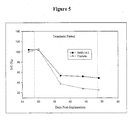
- H460-16-2 antibody achieved an endpoint of median T/C tumor volume equal to 49% ( Figure 5 ).
- Figure 4 further shows that H460-16-2 treatment resulted in marked suppression of tumor growth when compared to the isotype control and that the suppression was 2/3 that of cisplatin given at its maximum tolerated dose (MTD) but without cisplatin's accompanying toxicity or death.
- MTD maximum tolerated dose
- Table 2 Changes In Body Weight And Tumor Growth Suppression (%T/C) At End Of Treatment Therapeutic Agent No./Group Dose % Body Weight Change % Tumor Growth Suppression Isotype Control 12 15 mg/kg/dose* no mean change H460-16-2 12 15 mg/kg/dose* -2.30% 49 Cisplatin 12 (-2) 9 mg/kg/ dose** -19.20% 25 *Dose administered i.v. 3 x per week for 3 weeks. ** Dose administered i.p. 1 x every 4 days for 3 doses.
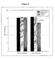
- H460-16-2 showed a survival benefit in comparison to treatment with isotype control ( Figure 7 ). By day 170 (around 120 days post-treatment), 33 percent of the H460-16-2 treatment group was still alive compared to 0 percent for both the cisplatin and isotype control groups.
- H460-16-2 is significantly more effective than the isotype control antibody in suppressing tumor growth in an established tumor xenograft model of breast cancer in SCID mice. Over the 3-week treatment period, H460-16-2 achieved an endpoint of median T/C tumor volumes of less than 50% relative to control. In addition, H460-16-2 resulted in suppression that was two thirds that of cisplatin given at MTD but without the signs of toxicity or death observed with the chemotherapeutic drug.
- H460-16-2 significantly decreased the tumor burden of established tumors in comparison to a control antibody and showed survival benefits in a well-recognized model of human cancer disease suggesting pharmacologic and pharmaceutical benefits of this antibody for therapy in other mammals, including man.
- H460-16-2 antigen was studied in mouse tissues and compared to the gp96 antigen. IHC optimization studies were initially performed in order to determine the conditions for further experiments. H460-16-2 monoclonal antibody was produced and purified as stated above.
- the slides were immersed in 10 mM citrate buffer at pH 6 (Dako, Toronto, ON) then microwaved at high, medium, and low power settings for 5 minutes each and finally immersed in cold PBS. Slides were then immersed in 3% hydrogen peroxide solution for 6 minutes, washed with PBS three times for 5 minutes each, dried, incubated with Universal blocking solution (Dako, Toronto, ON) for 5 minutes at room temperature, and dried.
- H460-16-2, monoclonal mouse anti-vimentin (Dako, Toronto, ON) and anti-grp94, also known as anti-gp96, (Stressgen Biotechnologies, Victoria, BC) were diluted in antibody dilution buffer (Dako, Toronto, ON) to its working concentration (either 2.5 ⁇ g/mL, 5 ⁇ g/mL or 10 ⁇ g/mL for each antibody) and incubated overnight in a humidified chamber at 4° C. The slides were washed with PBS 3 times for 5 minutes each. Immunoreactivity of the primary antibodies was detected/visualized with HRP conjugated secondary antibodies as supplied (Dako Envision System, Toronto, ON) for 30 minutes at room temperature.
- the optimum concentration was the one that produced the expected results for the positive (anti-gp96) and negative control antibodies (anti-vimentin).
- the anti-vimentin antibody has been shown to be negative on mouse tissue but positive on human tissue.
- the anti-gp96 antibody has previously been shown to be positive on both mouse and human tissue. In these studies both the high and low concentrations did not produce the expected results with the control antibodies, but the 5 ⁇ g/mL concentration did.
- Anti-vimentin did not stain mouse kidney.
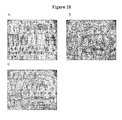
- H460-16-2 ( Figure 10B ) showed apical staining of the proximal and distal convoluted tubules while anti-gp96 produced diffuse staining of the same cells with a cytoplasmic and punctate pattern ( Figure 10C ).
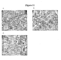
- Anti-vimentin ( Figure 11A ) did not stain mouse ovary.
- H460-16-2 ( Figure 11B ) showed cytoplasmic and nuclear staining of only the ova while anti-gp96 produced diffuse cytoplasmic and nuclear staining of the ova and cytoplasmic and punctate staining of granulosa cells ( Figure 11C ).
- the anti-vimentin negative control antibody gave the expected staining of human tissues and lack of staining of mouse tissues (see Figures 8-11 ).
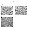
- the anti-gp96 antibody was used as a positive control because of the likelihood that the H460-16-2 antigen is a cancer variant of gp96.
- the anti-gp96 antibody did show staining ofMB-231 cells ( Figure 8 ) which is consistent with the association of gp96 expression with breast cancer.
- Gp96 was also expressed in the cytoplasm of many cell types involved with protein synthesis such as hepatocytes, cells of the Islets of Langerhans in the pancreas, ovarian granulosa cells and the ova, and mucosal epithelium in the fallopian tubes (Table 3). This is entirely consistent with the putative role for gp96 as an endoplasmic reticular chaperone protein.
- H460-16-2 antibody stained MB-231 cells which is consistent with its in vivo effects in the corresponding tumor model. In addition it stained the DCT and PCT in the mouse kidney (Table 3) as well as the mouse ova ( Figure 11 ). From this sampling of mouse tissues it would appear that the H460-16-2 antigen is not restricted to just human cells but is also expressed in the mouse in such a way that the antibody can recognize the antigen. Significantly there are differences in the expression of the H460-16-2 and gp96 antigen as demonstrated with the staining of normal mouse kidney tissue; apical staining was obtained with H460-16-2 while diffuse staining was seen with anti-gp96 ( Figure 10 ). Another example of this is the additional staining of the ova with anti-gp96 ( Figure 11 ). A key difference is that H460-16-2 staining does not occur in the liver, while gp96 staining is quite extensive ( Figure 9 ).
- H460-16-2, anti-gp96 (for comparison to H460-16-2) and anti-vimentin (negative control) was used to stain a normal mouse tissue array (Imgenex, San Diego, CA).
- the staining procedure used was the same as stated above.
- anti-vimentin did not stain any of the tissues tested; H460-16-2 again stained only the ovary and kidney while anti-gp96 continued to stain a much broader range of mouse tissues.
- H460-16-2 stains the same tissues as anti-gp96 but anti-gp96 continued to stain a much broader range of tissues supporting the idea that the H460-16-2 antigen may be a subset of gp96.
- Table 4 IHC on Normal Mouse Tissue Array Tissue Anti-vimentin H460-16-2 Anti-gp96 1 Skin - - - 2 Skin - - - 3 Spleen - - +/-(Lymphocytes) 4 Spleen - +/-(Lymphocytes) +/-(Lymphocytes) 5 Skeletal Muscle - - - 6 Lung - - - 7 Lung - - - 8 Heart - - - 9 Heart - - - 10 Salivary gland - - +/-(Acinar epith.) 11 Liver - - ++(Hepatocytes) 12 Liver - - ++(Heepatocytes) 13 Gall bladder - (NR)
- H460-16-2 antigen may be a cancer variant of gp96 as determined previously by biochemical methods. Binding of antibodies to 60 normal human tissues was performed using a human, normal organ tissue array (Imgenex, San Diego, CA). All primary antibodies (H460-16-2; anti-grp94 (also know as anti-gp96, Stressgen Biotechnologies, Victoria, BC); and mouse IgG 1 negative control (Dako, Toronto, ON)) were diluted in antibody dilution buffer (Dako, Toronto, ON) to a concentration of 5 ⁇ g/ml (found to be the optimal concentration in optimization steps). The negative control antibody has been shown to be negative to all mammalian tissues by the manufacturer. The procedure for IHC from Example 3 was followed.
- Table 5 presents a summary of the results of H460-16-2 staining to an array of normal human tissues. From the table, there are three categories of tissue staining. A group of tissues was completely negative. These tissues included normal heart, kidney, brain, pancreas, breast, testis, ovary and placenta. A second group of tissues comprised tissues that demonstrated positive staining. These included the skin, ureter, stomach and prostate. The salivary gland demonstrated the strongest staining with this antibody. A third group of tissues included tissues in which staining was positive in the tissue section, but was limited to infiltrating macrophages, lymphocytes and fibroblasts.
- tissue that were negative for anti-gp96 included subcutaneous fat, skeletal muscle, lung, heart, stomach smooth muscle, urinary bladder, myometrium, ovary, placental cord, brain (white and gray matter), cerebellum, and spinal cord. With the exception of the myometrium, all of these tissues were also negative for H460-16-2 staining.
- H460-16-2 binds to a smaller subset of the tissues recognized by the anti-gp96 antibody. This is consistent with the mouse tissue study, in which anti-gp96 bound to liver, pancreas, brain and fallopian tubes in addition to the two tissues that were also bound by H460-16-2, kidney and ovary. These results suggest that the antigen for H460-16-2 is not widely expressed on normal tissues, and that the antibody would bind specifically to a limited number of tissues in humans.
- Table 5 IHC On Normal Human Tissue With H460-16-2 Negative Negative except Macrophages, Lymphocytes, Fibroblasts Positive 1. breast subcutaneous fat skin, buttock 2. skeletal muscle spleen salivary gland 3.
- bronchus lymph node mesenteric stomach, antrum 4. heart nasal mucosa prostate 5. pancreas lung seminal vesicle 6. stomach smooth muscle liver endometrium, secretory 7. kidney cortex gallbladder thyroid 8. kidney medulla tonsil ureter 9. testis esophagus myometrium 10. epidydimis stomach, body 11. endometrium, proliferative duodenum 12. ovary ileum 13. placenta, villi appendix 14. placenta, amniochorion colon 15. placenta cord sigmoid colon 16. adrenal cortex urinary bladder 17. adrenal medulla uterine cervix (endocervix) 18. thymus uterine cervix (exocervix) 19. brain, white matter salpinx 20. brain, gray matter 21. cerebellum 22. spinal cord
- the cell types where the antigens are expressed were tabulated in Table 6. From the table, it is clear that the anti-gp96 antibody binds to a wider range of cell types than H460-16-2. Further, the strongest binding of H460-16-2 was to fibroblasts, acinar epithelium, and lymphocytes. There was weak binding to macrophages, keratinocytes, smooth muscle, mucosal epithelium, and thyroid follicular cells. Anti-gp96 bound to an additional 15 cell types, and to each cell type that expressed the H460-16-2 antigen.
- H460-16-2 antigen is a subset of gp96 since there were no cells that expressed H460-16-2 that did not express the gp96 antigen.
- Table 6 Summary Of IHC on Normal Human Tissues Cell Type H460-16-2 Anti-gp96 Fibroblasts +/++ + Acinar epithelium +/++++ + Lymphocytes +/++ +/++ Macrophages + + Keratinocytes + + Smooth muscle + + Mucosal epithelium + + Follicular cells + + Lobular epithelium - + Endothelium - + Mucosal glands - + Ductal epithelium - + Hepatocytes - ++ Acinar cells - ++ Ganslionic cells - + Villous epithelium - + Loops of Henle - + PCT&DCT - +l++ Glandular epithelium - +/++/+++ Germinal cells - ++ Cytotrophoblasts - ++ Syncy
- H460-16-2 antigen has a very limited distribution in normal tissues including the vital organs.
- the experiment also showed that the anti-gp96 antibody bound to a wider range of tissues compared to H460-16-2.
- H460-16-2 binds to a subset of the tissues bound by anti-gp96 and to limited cell types. In the tissues that were H460-16-2 positive but not gp96 positive, H460-16-2 bound to only macrophages and fibroblasts, cell types which generally expressed gp96.
- the difference between the mouse and human tissue surveys also point out that the H460-16-2 antibody recognizes an antigen that is relevant in humans and of limited importance in normal mice since the expression is so limited.
- the H460-16-2 antibody itself is applicable in humans since it does recognize the human form of the antigen.
- Tables 7 and 8 provide binding summaries of H460-16-2 and anti-gp96 antibody to a breast cancer tissue array respectively. Each array contained tumor samples from 50 individual patients. Overall, 64 percent of the 50 patients tested were positive for H460-16-2 antigen compared to 84 percent for gp96. For both the H460-16-2 and gp96 antigen, only 2 out of 9 normal breast tissue samples from breast cancer patients were positive. No clear correlation between estrogen and progesterone receptor status was evident. It also appeared there was a trend to greater positive expression of the H460-16-2 antigen with higher tumor stage. The H460-16-2 staining was quite specific for cancerous cells over normal cells as demonstrated in Figure 12 where stromal cells were clearly negative and sheets of malignant cells were highly positive.
- the cellular localization pattern seen with the H460-16-2 antigen was confined to the cell membrane in the majority of cases.
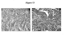
- the anri-gp96 antibody stained more breast cancer samples but consistently showed membrane as well as substantial cytoplasmic localization ( Figure 13 ).
- Anti-gp96 stained the same samples of normal tissue from breast cancer patients as H460-16-2. These results suggest the antigen for H460-16-2 may be expressed by almost two thirds of breast cancer patients.
- the staining pattern showed that in patient samples, the antibody is highly specific for malignant cells and the H460-16-2 antigen is localized to the cell membrane thereby making it an attractive druggable target.
- Table 7 IHC With H460-16-2 On Human Normal Breast And Tumor H460-16-2 Total # - +/- + ++ +++ Total positive % positive Patient Tumor 50 18 13 15 2 2 32 64 Samples Normal 9 7 0 2 0 0 2 22 ER ER + 21 9 5 7 0 0 12 57 Status ER - 28 8 8 2 2 20 71 Unknown 1 1 0 0 0 0 0 0 PR PR + 11 5 2 4 0 0 6 55 Status PR* 38 12 11 11 2 2 26 68 Unknown 1 1 0 0 0 0 0 0 0 AJCC T1 7 3 2 2 0 0 4 57 Tumor T2 26 11 5 6 2 2 15 58 Stage T3 16 4 6 6 0 0 12 75 T4 1 0 0 1 0 0 1 100 Table 8: IHC With Anti-gp96 On Human Breast Normal And Tumor Anti-gp96 Total # - +/- + ++ +++ Total positive % positive Patient Tumor 50 8 9 12 9
- H460-16-2 was used on a multiple human tumor tissue array (Imgenex, San Diego, CA). The following information was provided for each patient: age, sex, organ and diagnosis. The staining procedure used was the same as the one outlined in Example 3. Vimentin was used as a positive control antibody and the same negative control antibody was used as described for the human breast tumor tissue array. All antibodies were used at a working concentration of 5 ⁇ g/mL.
- H460-16-2 stained a number of various human cancers besides breast.
- the following tumor types were always positive for H460-16-2 (albeit to different degrees): lymph node (2/2), bone (2/2), lung (4/4), kidney (3/3), uterus (3/3), and thyroid (2/2).
- the stomach (4/5), liver (2/3) and parotid gland (2/3) also showed up relatively consistently positive for staining.
- H460-16-2 staining was localized predominately on the membrane of cancerous cells.
- H460-16-2 antigen is not solely found on the membranes of breast cancers but also on the membrane of a large variety of tumor types. These results indicate that H460-16-2 has potential as a therapeutic drug in a wide variety of tumor types besides breast.
- Table 9 IHC On Human Multi-Tumor Array Sec. No.
- the present invention relates to the following items:
Landscapes
- Health & Medical Sciences (AREA)
- Life Sciences & Earth Sciences (AREA)
- Immunology (AREA)
- Chemical & Material Sciences (AREA)
- Engineering & Computer Science (AREA)
- Molecular Biology (AREA)
- Biomedical Technology (AREA)
- Hematology (AREA)
- General Health & Medical Sciences (AREA)
- Urology & Nephrology (AREA)
- Medicinal Chemistry (AREA)
- Cell Biology (AREA)
- Biochemistry (AREA)
- Organic Chemistry (AREA)
- Biotechnology (AREA)
- Pathology (AREA)
- General Physics & Mathematics (AREA)
- Analytical Chemistry (AREA)
- Physics & Mathematics (AREA)
- Food Science & Technology (AREA)
- Microbiology (AREA)
- Genetics & Genomics (AREA)
- Biophysics (AREA)
- Proteomics, Peptides & Aminoacids (AREA)
- Animal Behavior & Ethology (AREA)
- Veterinary Medicine (AREA)
- Public Health (AREA)
- Zoology (AREA)
- Virology (AREA)
- Epidemiology (AREA)
- Tropical Medicine & Parasitology (AREA)
- Pharmacology & Pharmacy (AREA)
- Nuclear Medicine, Radiotherapy & Molecular Imaging (AREA)
- General Chemical & Material Sciences (AREA)
- Chemical Kinetics & Catalysis (AREA)
- Medicines Containing Antibodies Or Antigens For Use As Internal Diagnostic Agents (AREA)
- Peptides Or Proteins (AREA)
- Preparation Of Compounds By Using Micro-Organisms (AREA)
- Micro-Organisms Or Cultivation Processes Thereof (AREA)
Priority Applications (1)
| Application Number | Priority Date | Filing Date | Title |
|---|---|---|---|
| EP10182568A EP2322213A1 (de) | 2003-06-23 | 2004-06-08 | Antikörper, die Krebserkrankungen modifizieren |
Applications Claiming Priority (2)
| Application Number | Priority Date | Filing Date | Title |
|---|---|---|---|
| US10/603,000 US7252821B2 (en) | 1999-10-08 | 2003-06-23 | Cancerous disease modifying antibodies |
| EP04737787A EP1635869B1 (de) | 2003-06-23 | 2004-06-08 | Antikörper, die krebserkrankungen modifizieren |
Related Parent Applications (2)
| Application Number | Title | Priority Date | Filing Date |
|---|---|---|---|
| EP04737787.4 Division | 2004-06-08 | ||
| EP04737787A Division-Into EP1635869B1 (de) | 2003-06-23 | 2004-06-08 | Antikörper, die krebserkrankungen modifizieren |
Related Child Applications (1)
| Application Number | Title | Priority Date | Filing Date |
|---|---|---|---|
| EP10182568.5 Division-Into | 2010-09-29 |
Publications (1)
| Publication Number | Publication Date |
|---|---|
| EP2260865A1 true EP2260865A1 (de) | 2010-12-15 |
Family
ID=33539653
Family Applications (3)
| Application Number | Title | Priority Date | Filing Date |
|---|---|---|---|
| EP10182568A Withdrawn EP2322213A1 (de) | 2003-06-23 | 2004-06-08 | Antikörper, die Krebserkrankungen modifizieren |
| EP10007120A Withdrawn EP2260865A1 (de) | 2003-06-23 | 2004-06-08 | Antikörper, die Krebserkrankungen modifizieren |
| EP04737787A Expired - Lifetime EP1635869B1 (de) | 2003-06-23 | 2004-06-08 | Antikörper, die krebserkrankungen modifizieren |
Family Applications Before (1)
| Application Number | Title | Priority Date | Filing Date |
|---|---|---|---|
| EP10182568A Withdrawn EP2322213A1 (de) | 2003-06-23 | 2004-06-08 | Antikörper, die Krebserkrankungen modifizieren |
Family Applications After (1)
| Application Number | Title | Priority Date | Filing Date |
|---|---|---|---|
| EP04737787A Expired - Lifetime EP1635869B1 (de) | 2003-06-23 | 2004-06-08 | Antikörper, die krebserkrankungen modifizieren |
Country Status (9)
| Country | Link |
|---|---|
| US (1) | US7252821B2 (de) |
| EP (3) | EP2322213A1 (de) |
| JP (1) | JP4680186B2 (de) |
| CN (1) | CN1849136B (de) |
| AU (3) | AU2004248865B2 (de) |
| CA (1) | CA2530214A1 (de) |
| ES (1) | ES2394643T3 (de) |
| NZ (1) | NZ544440A (de) |
| WO (1) | WO2004112834A1 (de) |
Families Citing this family (18)
| Publication number | Priority date | Publication date | Assignee | Title |
|---|---|---|---|---|
| US7189397B2 (en) * | 1999-10-08 | 2007-03-13 | Arius Research Inc. | Cytotoxicity mediation of cells evidencing surface expression of CD44 |
| US20050100542A1 (en) * | 1999-10-08 | 2005-05-12 | Young David S. | Cytotoxicity mediation of cells evidencing surface expression of CD44 |
| US7947496B2 (en) * | 1999-10-08 | 2011-05-24 | Hoffmann-La Roche Inc. | Cytotoxicity mediation of cells evidencing surface expression of CD44 |
| US20080124327A1 (en) * | 1999-10-08 | 2008-05-29 | Arius Research, Inc. | Cytotoxicity mediation of cells evidencing surface expression of CD44 |
| US8048416B2 (en) | 1999-10-08 | 2011-11-01 | Hoffmann-La Roche Inc. | Cytotoxicity mediation of cells evidencing surface expression of CD44 |
| US8071072B2 (en) * | 1999-10-08 | 2011-12-06 | Hoffmann-La Roche Inc. | Cytotoxicity mediation of cells evidencing surface expression of CD44 |
| US20090004103A1 (en) * | 1999-10-08 | 2009-01-01 | Young David S F | Cytotoxicity mediation of cells evidencing surface expression of CD44 |
| US7252821B2 (en) * | 1999-10-08 | 2007-08-07 | Arius Research Inc. | Cancerous disease modifying antibodies |
| US7419792B2 (en) * | 1999-10-08 | 2008-09-02 | Arius Research Inc. | Laminin Receptor 1 Precursor Protein (37LRP) epitope delineated by an Hepatocellular carcinoma specific antibody |
| US20080213169A1 (en) * | 2003-04-14 | 2008-09-04 | Arius Research, Inc. | Cytotoxicity mediation of cells evidencing surface expression of CD59 |
| US20060140963A1 (en) * | 2003-04-14 | 2006-06-29 | Arius Research, Inc. | Cytotoxicity mediation of cells evidencing surface expression of CD59 |
| US20080025977A1 (en) * | 2003-04-14 | 2008-01-31 | Arius Research, Inc. | Cytotoxicity mediation of cells evidencing surface expression of CD59 |
| US7195764B2 (en) * | 2003-04-14 | 2007-03-27 | Arius Research Inc. | Cancerous disease modifying antibodies |
| CA2668484A1 (en) | 2006-11-13 | 2008-05-22 | F. Hoffmann-La Roche Ag | Cancerous disease modifying antibodies 180706-02 |
| ES2363669B1 (es) * | 2009-06-10 | 2012-08-09 | Fundacio Privada Institut D'investigacio Biomedica De Bellvitge (Idibell) | Metodo para determinar el riesgo de desarrollar metastasis cerebral y un kit para llevar a cabo dicho procedimiento |
| SI2531527T1 (sl) | 2010-02-04 | 2014-07-31 | F. Hoffmann-La Roche Ag | Monoklonsko protitelo proti CD44 za uporabo pri zdravljenju skvamoznoceličnega karcinoma glave in vratu |
| ES2611479T3 (es) * | 2010-06-16 | 2017-05-09 | University Of Pittsburgh- Of The Commonwealth System Of Higher Education | Anticuerpos contra endoplasmina y su uso |
| IL258574B2 (en) * | 2015-11-11 | 2025-01-01 | Novartis Ag | Uses of myostatin antagonists, combinations containing them and their uses |
Citations (16)
| Publication number | Priority date | Publication date | Assignee | Title |
|---|---|---|---|---|
| US4861581A (en) | 1986-12-05 | 1989-08-29 | Cancer Biologics, Inc. | Detection of necrotic malignant tissue and associated therapy |
| US5171665A (en) | 1989-04-17 | 1992-12-15 | Oncogen | Monoclonal antibody to novel antigen associated with human tumors |
| US5484596A (en) | 1984-01-31 | 1996-01-16 | Akzo N.V. | Active specific immunotherapy |
| US5693763A (en) | 1993-02-05 | 1997-12-02 | Epigen, Inc. | Antibodies to human carcinoma antigen |
| US5750102A (en) | 1992-03-13 | 1998-05-12 | Yeda Research And Development Co., Ltd. | Double transfectants of the MHC genes as cellular vaccines for immuno prevention of tumor metastasis |
| US5780033A (en) | 1994-06-24 | 1998-07-14 | Torchilin; Vladimir P. | Use of autoantibodies for tumor therapy and prophylaxis |
| US5783186A (en) | 1995-12-05 | 1998-07-21 | Amgen Inc. | Antibody-induced apoptosis |
| US5849876A (en) | 1986-11-19 | 1998-12-15 | Sanofi | Hybridomas producing monoclonal antibodies to new mucin epitopes |
| US5869045A (en) | 1989-06-30 | 1999-02-09 | Bristol-Myers Squibb Company | Antibody conjugates reactive with human carcinomas |
| US5869268A (en) | 1991-10-30 | 1999-02-09 | Idemitsu Kosan Company Limited | Methods for producing human lymphocytes and human monoclonal antibodies, and human monoclonal antibodies produced thereby |
| US6180370B1 (en) | 1988-12-28 | 2001-01-30 | Protein Design Labs, Inc. | Humanized immunoglobulins and methods of making the same |
| US6180357B1 (en) | 1999-10-08 | 2001-01-30 | Arius Research, Inc. | Individualized patient-specific anti-cancer antibodies |
| US20020041877A1 (en) * | 1999-10-08 | 2002-04-11 | Young David S. F. | Individualized anti-cancer antibodies |
| WO2002082076A2 (en) * | 2001-04-03 | 2002-10-17 | Merck Patent Gmbh | Renal cell carcinoma tumor markers |
| WO2003055515A1 (en) * | 2001-12-21 | 2003-07-10 | Arius Research, Inc. | Individualized anti-cancer antibodies |
| US20040001789A1 (en) * | 1999-10-08 | 2004-01-01 | Young David S. F. | Cytotoxicity mediation of cells evidencing surface expression of gp96 or precursors thereof |
Family Cites Families (5)
| Publication number | Priority date | Publication date | Assignee | Title |
|---|---|---|---|---|
| US4172124A (en) | 1978-04-28 | 1979-10-23 | The Wistar Institute | Method of producing tumor antibodies |
| PT1231268E (pt) | 1994-01-31 | 2005-11-30 | Univ Boston | Bancos de anticorpos policlonais |
| US7189397B2 (en) * | 1999-10-08 | 2007-03-13 | Arius Research Inc. | Cytotoxicity mediation of cells evidencing surface expression of CD44 |
| US7252821B2 (en) * | 1999-10-08 | 2007-08-07 | Arius Research Inc. | Cancerous disease modifying antibodies |
| US20050100542A1 (en) * | 1999-10-08 | 2005-05-12 | Young David S. | Cytotoxicity mediation of cells evidencing surface expression of CD44 |
-
2003
- 2003-06-23 US US10/603,000 patent/US7252821B2/en not_active Expired - Fee Related
-
2004
- 2004-06-08 CA CA002530214A patent/CA2530214A1/en not_active Abandoned
- 2004-06-08 WO PCT/CA2004/000845 patent/WO2004112834A1/en not_active Ceased
- 2004-06-08 EP EP10182568A patent/EP2322213A1/de not_active Withdrawn
- 2004-06-08 NZ NZ544440A patent/NZ544440A/en not_active IP Right Cessation
- 2004-06-08 EP EP10007120A patent/EP2260865A1/de not_active Withdrawn
- 2004-06-08 ES ES04737787T patent/ES2394643T3/es not_active Expired - Lifetime
- 2004-06-08 AU AU2004248865A patent/AU2004248865B2/en not_active Ceased
- 2004-06-08 EP EP04737787A patent/EP1635869B1/de not_active Expired - Lifetime
- 2004-06-08 JP JP2006515581A patent/JP4680186B2/ja not_active Expired - Fee Related
- 2004-06-08 CN CN2004800239638A patent/CN1849136B/zh not_active Expired - Fee Related
- 2004-08-20 AU AU2004266045A patent/AU2004266045B2/en not_active Ceased
-
2011
- 2011-02-24 AU AU2011200789A patent/AU2011200789B2/en not_active Ceased
Patent Citations (17)
| Publication number | Priority date | Publication date | Assignee | Title |
|---|---|---|---|---|
| US5484596A (en) | 1984-01-31 | 1996-01-16 | Akzo N.V. | Active specific immunotherapy |
| US5849876A (en) | 1986-11-19 | 1998-12-15 | Sanofi | Hybridomas producing monoclonal antibodies to new mucin epitopes |
| US4861581A (en) | 1986-12-05 | 1989-08-29 | Cancer Biologics, Inc. | Detection of necrotic malignant tissue and associated therapy |
| US6180370B1 (en) | 1988-12-28 | 2001-01-30 | Protein Design Labs, Inc. | Humanized immunoglobulins and methods of making the same |
| US5171665A (en) | 1989-04-17 | 1992-12-15 | Oncogen | Monoclonal antibody to novel antigen associated with human tumors |
| US5869045A (en) | 1989-06-30 | 1999-02-09 | Bristol-Myers Squibb Company | Antibody conjugates reactive with human carcinomas |
| US5869268A (en) | 1991-10-30 | 1999-02-09 | Idemitsu Kosan Company Limited | Methods for producing human lymphocytes and human monoclonal antibodies, and human monoclonal antibodies produced thereby |
| US5750102A (en) | 1992-03-13 | 1998-05-12 | Yeda Research And Development Co., Ltd. | Double transfectants of the MHC genes as cellular vaccines for immuno prevention of tumor metastasis |
| US5693763A (en) | 1993-02-05 | 1997-12-02 | Epigen, Inc. | Antibodies to human carcinoma antigen |
| US5780033A (en) | 1994-06-24 | 1998-07-14 | Torchilin; Vladimir P. | Use of autoantibodies for tumor therapy and prophylaxis |
| US5783186A (en) | 1995-12-05 | 1998-07-21 | Amgen Inc. | Antibody-induced apoptosis |
| US6180357B1 (en) | 1999-10-08 | 2001-01-30 | Arius Research, Inc. | Individualized patient-specific anti-cancer antibodies |
| US20020041877A1 (en) * | 1999-10-08 | 2002-04-11 | Young David S. F. | Individualized anti-cancer antibodies |
| US6657048B2 (en) * | 1999-10-08 | 2003-12-02 | Arius Research, Inc. | Individualized anti-cancer antibodies |
| US20040001789A1 (en) * | 1999-10-08 | 2004-01-01 | Young David S. F. | Cytotoxicity mediation of cells evidencing surface expression of gp96 or precursors thereof |
| WO2002082076A2 (en) * | 2001-04-03 | 2002-10-17 | Merck Patent Gmbh | Renal cell carcinoma tumor markers |
| WO2003055515A1 (en) * | 2001-12-21 | 2003-07-10 | Arius Research, Inc. | Individualized anti-cancer antibodies |
Also Published As
| Publication number | Publication date |
|---|---|
| EP2322213A1 (de) | 2011-05-18 |
| ES2394643T3 (es) | 2013-02-04 |
| AU2004266045B2 (en) | 2011-03-17 |
| JP4680186B2 (ja) | 2011-05-11 |
| US20040105815A1 (en) | 2004-06-03 |
| CA2530214A1 (en) | 2004-12-29 |
| NZ544440A (en) | 2009-03-31 |
| US7252821B2 (en) | 2007-08-07 |
| JP2007523851A (ja) | 2007-08-23 |
| EP1635869B1 (de) | 2012-09-19 |
| AU2011200789A1 (en) | 2011-03-17 |
| AU2004266045A1 (en) | 2005-03-03 |
| WO2004112834A1 (en) | 2004-12-29 |
| CN1849136B (zh) | 2010-12-22 |
| AU2011200789B2 (en) | 2014-01-30 |
| AU2004248865B2 (en) | 2011-07-21 |
| EP1635869A1 (de) | 2006-03-22 |
| HK1096866A1 (en) | 2007-06-15 |
| AU2004248865A1 (en) | 2004-12-29 |
| CN1849136A (zh) | 2006-10-18 |
Similar Documents
| Publication | Publication Date | Title |
|---|---|---|
| EP2163564A1 (de) | Eine Krebserkrankung modifizierende Antikörper | |
| EP1635869B1 (de) | Antikörper, die krebserkrankungen modifizieren | |
| US20090074659A1 (en) | Cancerous disease modifying antibodies | |
| US7399835B2 (en) | Cancerous disease modifying antibodies | |
| AU2004205435B2 (en) | Cancerous disease modifying antibodies | |
| US20040105816A1 (en) | Cancerous disease modifying antibodies | |
| US20050027106A1 (en) | Cancerous disease modifying antibodies | |
| US20050255041A1 (en) | Cancerous disease modifying antibodies | |
| HK1096866B (en) | Cancerous disease modifying antibodies |
Legal Events
| Date | Code | Title | Description |
|---|---|---|---|
| PUAI | Public reference made under article 153(3) epc to a published international application that has entered the european phase |
Free format text: ORIGINAL CODE: 0009012 |
|
| AC | Divisional application: reference to earlier application |
Ref document number: 1635869 Country of ref document: EP Kind code of ref document: P |
|
| AK | Designated contracting states |
Kind code of ref document: A1 Designated state(s): AT BE BG CH CY CZ DE DK EE ES FI FR GB GR HU IE IT LI LU MC NL PL PT RO SE SI SK TR |
|
| 17P | Request for examination filed |
Effective date: 20110930 |
|
| 17Q | First examination report despatched |
Effective date: 20130524 |
|
| STAA | Information on the status of an ep patent application or granted ep patent |
Free format text: STATUS: THE APPLICATION IS DEEMED TO BE WITHDRAWN |
|
| 18D | Application deemed to be withdrawn |
Effective date: 20131204 |