EP2264440A2 - Vorrichtung zum Nachweis biologischer Organismen mittels Raman-streuung - Google Patents
Vorrichtung zum Nachweis biologischer Organismen mittels Raman-streuung Download PDFInfo
- Publication number
- EP2264440A2 EP2264440A2 EP10184978A EP10184978A EP2264440A2 EP 2264440 A2 EP2264440 A2 EP 2264440A2 EP 10184978 A EP10184978 A EP 10184978A EP 10184978 A EP10184978 A EP 10184978A EP 2264440 A2 EP2264440 A2 EP 2264440A2
- Authority
- EP
- European Patent Office
- Prior art keywords
- raman
- reactants
- biological organism
- compound
- clause
- Prior art date
- Legal status (The legal status is an assumption and is not a legal conclusion. Google has not performed a legal analysis and makes no representation as to the accuracy of the status listed.)
- Withdrawn
Links
- 238000001069 Raman spectroscopy Methods 0.000 title claims abstract description 85
- 238000001514 detection method Methods 0.000 title claims abstract description 55
- 150000001875 compounds Chemical class 0.000 claims abstract description 22
- 238000000149 argon plasma sintering Methods 0.000 claims abstract description 20
- 239000000376 reactant Substances 0.000 claims description 55
- 239000007787 solid Substances 0.000 claims description 21
- 239000000126 substance Substances 0.000 claims description 19
- 238000006243 chemical reaction Methods 0.000 claims description 15
- 239000011859 microparticle Substances 0.000 claims description 15
- 102000004190 Enzymes Human genes 0.000 claims description 14
- 108090000790 Enzymes Proteins 0.000 claims description 14
- 102000004169 proteins and genes Human genes 0.000 claims description 14
- 108090000623 proteins and genes Proteins 0.000 claims description 14
- 239000000203 mixture Substances 0.000 claims description 13
- 241000894006 Bacteria Species 0.000 claims description 12
- 241000700605 Viruses Species 0.000 claims description 8
- 241001515965 unidentified phage Species 0.000 claims description 8
- CIWBSHSKHKDKBQ-JLAZNSOCSA-N Ascorbic acid Chemical compound OC[C@H](O)[C@H]1OC(=O)C(O)=C1O CIWBSHSKHKDKBQ-JLAZNSOCSA-N 0.000 claims description 6
- 230000003993 interaction Effects 0.000 claims description 6
- CQRYARSYNCAZFO-UHFFFAOYSA-N salicyl alcohol Chemical compound OCC1=CC=CC=C1O CQRYARSYNCAZFO-UHFFFAOYSA-N 0.000 claims description 6
- 239000004816 latex Substances 0.000 claims description 5
- 229920000126 latex Polymers 0.000 claims description 5
- AQLJVWUFPCUVLO-UHFFFAOYSA-N urea hydrogen peroxide Chemical compound OO.NC(N)=O AQLJVWUFPCUVLO-UHFFFAOYSA-N 0.000 claims description 5
- 108010001336 Horseradish Peroxidase Proteins 0.000 claims description 4
- MHAJPDPJQMAIIY-UHFFFAOYSA-N Hydrogen peroxide Chemical compound OO MHAJPDPJQMAIIY-UHFFFAOYSA-N 0.000 claims description 4
- 239000012141 concentrate Substances 0.000 claims description 4
- RLFWWDJHLFCNIJ-UHFFFAOYSA-N 4-aminoantipyrine Chemical compound CN1C(C)=C(N)C(=O)N1C1=CC=CC=C1 RLFWWDJHLFCNIJ-UHFFFAOYSA-N 0.000 claims description 3
- 238000010521 absorption reaction Methods 0.000 claims description 3
- 229960005070 ascorbic acid Drugs 0.000 claims description 3
- 229940088597 hormone Drugs 0.000 claims description 3
- 239000005556 hormone Substances 0.000 claims description 3
- KBOPZPXVLCULAV-UHFFFAOYSA-N mesalamine Chemical compound NC1=CC=C(O)C(C(O)=O)=C1 KBOPZPXVLCULAV-UHFFFAOYSA-N 0.000 claims description 3
- 229960004963 mesalazine Drugs 0.000 claims description 3
- 230000002503 metabolic effect Effects 0.000 claims description 3
- 239000002207 metabolite Substances 0.000 claims description 3
- 239000002211 L-ascorbic acid Substances 0.000 claims description 2
- 235000000069 L-ascorbic acid Nutrition 0.000 claims description 2
- ISWSIDIOOBJBQZ-UHFFFAOYSA-N Phenol Chemical compound OC1=CC=CC=C1 ISWSIDIOOBJBQZ-UHFFFAOYSA-N 0.000 claims description 2
- 239000002253 acid Substances 0.000 claims 1
- 238000000034 method Methods 0.000 abstract description 36
- 230000027455 binding Effects 0.000 abstract description 21
- 230000008859 change Effects 0.000 abstract description 2
- 239000012491 analyte Substances 0.000 abstract 5
- 229920002521 macromolecule Polymers 0.000 abstract 1
- 239000000306 component Substances 0.000 description 51
- 239000000523 sample Substances 0.000 description 39
- 241000186781 Listeria Species 0.000 description 22
- 239000007788 liquid Substances 0.000 description 14
- HEMHJVSKTPXQMS-UHFFFAOYSA-M Sodium hydroxide Chemical compound [OH-].[Na+] HEMHJVSKTPXQMS-UHFFFAOYSA-M 0.000 description 9
- 230000008569 process Effects 0.000 description 9
- 230000035945 sensitivity Effects 0.000 description 9
- 239000000243 solution Substances 0.000 description 9
- FAPWRFPIFSIZLT-UHFFFAOYSA-M Sodium chloride Chemical compound [Na+].[Cl-] FAPWRFPIFSIZLT-UHFFFAOYSA-M 0.000 description 6
- 238000002965 ELISA Methods 0.000 description 5
- 238000001237 Raman spectrum Methods 0.000 description 5
- 239000000872 buffer Substances 0.000 description 5
- 239000003153 chemical reaction reagent Substances 0.000 description 5
- 238000003018 immunoassay Methods 0.000 description 5
- 239000011780 sodium chloride Substances 0.000 description 5
- 238000010586 diagram Methods 0.000 description 4
- 239000001963 growth medium Substances 0.000 description 4
- 239000000463 material Substances 0.000 description 4
- 238000002156 mixing Methods 0.000 description 4
- 238000012360 testing method Methods 0.000 description 4
- -1 argon ion Chemical class 0.000 description 3
- 239000007789 gas Substances 0.000 description 3
- 238000005259 measurement Methods 0.000 description 3
- 239000004033 plastic Substances 0.000 description 3
- 229920003023 plastic Polymers 0.000 description 3
- 238000001228 spectrum Methods 0.000 description 3
- KCXVZYZYPLLWCC-UHFFFAOYSA-N EDTA Chemical compound OC(=O)CN(CC(O)=O)CCN(CC(O)=O)CC(O)=O KCXVZYZYPLLWCC-UHFFFAOYSA-N 0.000 description 2
- 241000709661 Enterovirus Species 0.000 description 2
- 102000003992 Peroxidases Human genes 0.000 description 2
- 229920001213 Polysorbate 20 Polymers 0.000 description 2
- WCUXLLCKKVVCTQ-UHFFFAOYSA-M Potassium chloride Chemical compound [Cl-].[K+] WCUXLLCKKVVCTQ-UHFFFAOYSA-M 0.000 description 2
- 241000607142 Salmonella Species 0.000 description 2
- 241000191940 Staphylococcus Species 0.000 description 2
- 241000607598 Vibrio Species 0.000 description 2
- XKRFYHLGVUSROY-UHFFFAOYSA-N argon Substances [Ar] XKRFYHLGVUSROY-UHFFFAOYSA-N 0.000 description 2
- 229910052786 argon Inorganic materials 0.000 description 2
- 238000010420 art technique Methods 0.000 description 2
- 244000052616 bacterial pathogen Species 0.000 description 2
- 239000011324 bead Substances 0.000 description 2
- 229960000074 biopharmaceutical Drugs 0.000 description 2
- 239000003054 catalyst Substances 0.000 description 2
- 239000000356 contaminant Substances 0.000 description 2
- 238000013461 design Methods 0.000 description 2
- 239000003599 detergent Substances 0.000 description 2
- 208000006454 hepatitis Diseases 0.000 description 2
- 231100000283 hepatitis Toxicity 0.000 description 2
- 229940127121 immunoconjugate Drugs 0.000 description 2
- 239000000543 intermediate Substances 0.000 description 2
- 238000002372 labelling Methods 0.000 description 2
- 238000007834 ligase chain reaction Methods 0.000 description 2
- 239000006193 liquid solution Substances 0.000 description 2
- 230000000813 microbial effect Effects 0.000 description 2
- 108040007629 peroxidase activity proteins Proteins 0.000 description 2
- 238000003752 polymerase chain reaction Methods 0.000 description 2
- 235000010486 polyoxyethylene sorbitan monolaurate Nutrition 0.000 description 2
- 239000000256 polyoxyethylene sorbitan monolaurate Substances 0.000 description 2
- 239000011343 solid material Substances 0.000 description 2
- 239000003053 toxin Substances 0.000 description 2
- 231100000765 toxin Toxicity 0.000 description 2
- 108700012359 toxins Proteins 0.000 description 2
- 241000701161 unidentified adenovirus Species 0.000 description 2
- 238000005406 washing Methods 0.000 description 2
- 239000007987 MES buffer Substances 0.000 description 1
- 239000000020 Nitrocellulose Substances 0.000 description 1
- 239000004677 Nylon Substances 0.000 description 1
- 102000029797 Prion Human genes 0.000 description 1
- 108091000054 Prion Proteins 0.000 description 1
- 238000002835 absorbance Methods 0.000 description 1
- 238000000862 absorption spectrum Methods 0.000 description 1
- 230000006978 adaptation Effects 0.000 description 1
- 230000004075 alteration Effects 0.000 description 1
- 238000004458 analytical method Methods 0.000 description 1
- 235000010323 ascorbic acid Nutrition 0.000 description 1
- 239000011668 ascorbic acid Substances 0.000 description 1
- 238000003556 assay Methods 0.000 description 1
- XVBRCOKDZVQYAY-UHFFFAOYSA-N bronidox Chemical compound [O-][N+](=O)C1(Br)COCOC1 XVBRCOKDZVQYAY-UHFFFAOYSA-N 0.000 description 1
- 239000013043 chemical agent Substances 0.000 description 1
- HLVXFWDLRHCZEI-UHFFFAOYSA-N chromotropic acid Chemical compound OS(=O)(=O)C1=CC(O)=C2C(O)=CC(S(O)(=O)=O)=CC2=C1 HLVXFWDLRHCZEI-UHFFFAOYSA-N 0.000 description 1
- 230000001332 colony forming effect Effects 0.000 description 1
- 201000010099 disease Diseases 0.000 description 1
- 208000037265 diseases, disorders, signs and symptoms Diseases 0.000 description 1
- 230000000694 effects Effects 0.000 description 1
- 238000005516 engineering process Methods 0.000 description 1
- 238000009472 formulation Methods 0.000 description 1
- CPBQJMYROZQQJC-UHFFFAOYSA-N helium neon Chemical compound [He].[Ne] CPBQJMYROZQQJC-UHFFFAOYSA-N 0.000 description 1
- 230000000984 immunochemical effect Effects 0.000 description 1
- 238000011534 incubation Methods 0.000 description 1
- 210000000554 iris Anatomy 0.000 description 1
- 238000004519 manufacturing process Methods 0.000 description 1
- 239000011159 matrix material Substances 0.000 description 1
- 238000012986 modification Methods 0.000 description 1
- 230000004048 modification Effects 0.000 description 1
- 229920001220 nitrocellulos Polymers 0.000 description 1
- 229920001778 nylon Polymers 0.000 description 1
- 230000003287 optical effect Effects 0.000 description 1
- BVJSUAQZOZWCKN-UHFFFAOYSA-N p-hydroxybenzyl alcohol Chemical compound OCC1=CC=C(O)C=C1 BVJSUAQZOZWCKN-UHFFFAOYSA-N 0.000 description 1
- 244000052769 pathogen Species 0.000 description 1
- 230000001717 pathogenic effect Effects 0.000 description 1
- 230000001575 pathological effect Effects 0.000 description 1
- 230000004481 post-translational protein modification Effects 0.000 description 1
- 239000001103 potassium chloride Substances 0.000 description 1
- 235000011164 potassium chloride Nutrition 0.000 description 1
- 102000004196 processed proteins & peptides Human genes 0.000 description 1
- 108090000765 processed proteins & peptides Proteins 0.000 description 1
- 230000002285 radioactive effect Effects 0.000 description 1
- 239000001488 sodium phosphate Substances 0.000 description 1
- 229910000162 sodium phosphate Inorganic materials 0.000 description 1
- 230000009870 specific binding Effects 0.000 description 1
- 238000002798 spectrophotometry method Methods 0.000 description 1
- 238000012546 transfer Methods 0.000 description 1
- 238000002834 transmittance Methods 0.000 description 1
- RYFMWSXOAZQYPI-UHFFFAOYSA-K trisodium phosphate Chemical compound [Na+].[Na+].[Na+].[O-]P([O-])([O-])=O RYFMWSXOAZQYPI-UHFFFAOYSA-K 0.000 description 1
- 239000012224 working solution Substances 0.000 description 1
Images




Classifications
-
- G—PHYSICS
- G01—MEASURING; TESTING
- G01N—INVESTIGATING OR ANALYSING MATERIALS BY DETERMINING THEIR CHEMICAL OR PHYSICAL PROPERTIES
- G01N33/00—Investigating or analysing materials by specific methods not covered by groups G01N1/00 - G01N31/00
- G01N33/48—Biological material, e.g. blood, urine; Haemocytometers
- G01N33/50—Chemical analysis of biological material, e.g. blood, urine; Testing involving biospecific ligand binding methods; Immunological testing
- G01N33/53—Immunoassay; Biospecific binding assay; Materials therefor
- G01N33/543—Immunoassay; Biospecific binding assay; Materials therefor with an insoluble carrier for immobilising immunochemicals
- G01N33/54366—Apparatus specially adapted for solid-phase testing
- G01N33/54373—Apparatus specially adapted for solid-phase testing involving physiochemical end-point determination, e.g. wave-guides, FETS, gratings
-
- G—PHYSICS
- G01—MEASURING; TESTING
- G01N—INVESTIGATING OR ANALYSING MATERIALS BY DETERMINING THEIR CHEMICAL OR PHYSICAL PROPERTIES
- G01N21/00—Investigating or analysing materials by the use of optical means, i.e. using sub-millimetre waves, infrared, visible or ultraviolet light
- G01N21/62—Systems in which the material investigated is excited whereby it emits light or causes a change in wavelength of the incident light
- G01N21/63—Systems in which the material investigated is excited whereby it emits light or causes a change in wavelength of the incident light optically excited
- G01N21/65—Raman scattering
-
- G—PHYSICS
- G01—MEASURING; TESTING
- G01N—INVESTIGATING OR ANALYSING MATERIALS BY DETERMINING THEIR CHEMICAL OR PHYSICAL PROPERTIES
- G01N33/00—Investigating or analysing materials by specific methods not covered by groups G01N1/00 - G01N31/00
- G01N33/48—Biological material, e.g. blood, urine; Haemocytometers
- G01N33/50—Chemical analysis of biological material, e.g. blood, urine; Testing involving biospecific ligand binding methods; Immunological testing
- G01N33/53—Immunoassay; Biospecific binding assay; Materials therefor
- G01N33/543—Immunoassay; Biospecific binding assay; Materials therefor with an insoluble carrier for immobilising immunochemicals
- G01N33/54313—Immunoassay; Biospecific binding assay; Materials therefor with an insoluble carrier for immobilising immunochemicals the carrier being characterised by its particulate form
- G01N33/54326—Magnetic particles
Definitions
- the present disclosure generally relates to the field of biological diagnostic equipment and testing methods.
- antibodies developed to specific compounds are known to have high affinity for these components.
- antibodies provide limited detectability and as such are typically chemically modified with labels or tags that serve to enhance detection of the antibody during a reaction with the target biological component.
- prior art techniques can identify a biologic component.
- the ability to detect the antibody is prone to interference from other things in the sample including the sample matrix, wash components and other chemical agents.
- current detection techniques lack sensitivity at low concentrations or numbers of antibodies (i.e. low concentrations or numbers of targeted biological components).
- Raman light scattering techniques have been used in the past to detect specific chemical components. Raman scattering is a basic property of the interaction of light with molecules. When light hits a molecule it can cause the atoms of the molecule to vibrate. This vibration will then change the energy of additional light hitting the molecule. This additional scattered light has characteristics that are measurable and are unique to the structure of the molecule that was caused to vibrate. Raman spectroscopy by itself lacks specificity and sensitivity for the detection of biological organisms and components.
- the present disclosure is directed to methods and apparatus that uses the combination of Raman spectroscopy and biological labeling techniques to identify and quantify biological organisms and components with higher sensitivity and specificity than prior art techniques.
- the use of present disclosure for detecting antibody/biological component combinations can be performed by use of an immunoassay followed by Raman scattering detection techniques.
- the current invention uses the combination of Raman spectroscopy and biological labeling techniques to identify and quantify biological components, such as proteins or peptides including any post-translational modifications, in specific conformations or conditions associated with disease: for example, prion proteins.
- the immunoassay for certain embodiments of the present invention involves first having an antibody attached to a solid surface bind to the target biologic. Unbound components of the test sample are then washed away leaving only the bound biologic/antibody combinations. At this point the combined biologic/antibody combination can be detected by Raman scattering of ultraviolet light.
- one or more new reactants are then introduced and become bound to the biologic/antibody combination.
- the combination of the new reactant(s) with the biologic/antibody combination can now be detected using Raman scattering of light.
- reactants include, but are not limited to:
- the Raman detection methods can use chemicals that interact with the biologic without the antibody as described in examples 3 and 4 above.
- specific binding partners for the target biologic other than antibodies may be used, for example, a biological receptor (a protein).
- embodiments of the present disclosure can be implemented on a micro-fluidic channel (or well) integrated circuit using micro or nano-fabrication technology in which the binding partner is immobilized in one or more micro-fluidic channels in a custom integrated circuitry which would also include the laser(s) and detectors for Raman spectroscopy.
- Such an implementation could detect single biological components such as pathological bacteria, proteins or genetic material.
- an object of the present disclosure is to have a system for the detection of target biological organisms of components that utilizes a combination of chemical interactions including binding with a final step of Raman light scattering.
- Another object of the present disclosure is to have a system for the detection of target inorganic or organic components that utilizes a combination of chemical interactions including binding with a final step of Raman light scattering.
- Another object of the present disclosure is to combine an immunoassay with detection using Raman light scattering.
- Still another object of the present disclosure is to increase sensitivity of detection by the use of chemical reactants that produce resonant Raman light scattering.
- Yet another object of the present disclosure is to have an integrated circuit design with micro-fluidic channels or wells which can perform the combination of binding and Raman light scattering measurements.
- FIG. 1 is a flow chart of a typical prior art immunoassay technique (ELISA) for the detection of biological organisms or components.
- ELISA immunoassay technique
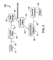
- FIG. 2 is a diagram of an embodiment of the disclosed apparatus.
- FIG. 3 is a flow chart of an embodiment of the disclosed technique for the detection of biological organisms and/or components.
- FIG. 4 is a block diagram of the enzyme system for converting chemical components to a Raman active compound.
- FIG. 5 is a flow chart of a technique for choosing laser light frequencies to excite specific target molecules.
- FIG. 6 is an illustration of a micro-fluidic channel designed to detect Raman active compounds.
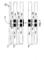
- FIG. 7 is an illustration of an array of micro-fluidic channels such as might be incorporated into a custom integrated circuit.
- FIG. 8 plots Raman spectra from an enzyme-linked immunoassay for a pathogenic bacteria, Listeria, utilizing an antibody linked to peroxidase and with shift numbers (cm-1) plotted on the abscissa and signal magnitudes plotted on the ordinate (arbitrary units) for a sample containing Listeria (a) and a sample not containing Listeria (b).
- FIG. 1 is a flow chart of a typical prior art immunoassay technique (ELISA) 10 for the detection of biological organisms or components.
- the process begins by the step 11 of preparing the liquid sample that includes the target biologic.
- the sample can be prepared by pre-enrichment in a growth medium such as half-Frasier's broth or other suitable microbial growth medium.
- a liquid sample for testing may be obtained from any liquid source. Solid material may be immersed in an appropriate liquid solution and potential target organism or molecules placed in solution and then sampled in the liquid.
- the prepared liquid sample is combined (or mixed) with a binding partner that has been attached to a solid surface.
- Typical binding partners include antibodies, bacteriophage, and bacteriophage proteins.
- step 13 plastic microtiter plates, latex beads or magnetic microparticles may be used.
- Other solid supports such as nitrocellulose, filter paper, nylon and other plastics may also be used.
- the antibody/biologic combination is then incubated in step 13 to allow time for the biologic and antibody to bind together. Once this has occurred the combined binding partner/biologic is decanted (poured off) and washed to remove unbound biologics and other unwanted materials.
- New reactants are added in step 15 to enhance the sensitivity of the mixture to detection of signal molecules by various methods. Examples of such reactants include:
- step 13 The mixture containing the bound binding partner/biologic and new reactants is the incubated in step 13 to allow time for the reaction to occur. At this point in many cases, the reaction part of the process 10 is complete and step 16 of measuring the molecules produced or included in steps 11 through 15 inclusive can be performed. If additional reactants are required, steps 14, 15 and 13 may be repeated one or more times in succession until the appropriate signal molecules are present.
- step 17 The measurement of the signal molecule(s) provides a quantitative result that can then be analyzed and compared in step 17 to a known set of calibrated responses of known concentrations of the target biologic. This comparison results in step 18 which is the quantified result and associated report of the concentration of the target biologic in the sample prepared in step 11.
- process 10 of FIG. 1 has been associated with the detection of a biological organism or component, the process 10 is also applicable to the detection of many types of molecules to which antibodies or other binding partners can react.
- FIG. 2 is a diagram of an embodiment of the present invention detection sub-system 20.
- a laser 21 produces a laser beam 22 which is focused by the focusing optics 23 into a focused laser beam 24 which hits the target sample 25.
- the backscattered light 26 from the sample 25 is focused into the beam 28 by the focusing optics 27.
- the beam 28 is directed into the spectrometer 30 with detector 31.
- the output from the detector 31 is the signal 32 which is received by the personal computer 40 for analysis, storage and/or printing with the printer 42.
- the laser 21 is typically a continuous wavelength (CW) laser with output In the visible range.
- CW continuous wavelength
- Focusing optics 23 and 27 include mirrors, lenses, irises, shutters, diffraction gratings, and/or polarizers.
- the target sample 25 may be liquid, gas or solid and in certain embodiments of the present invention, the target sample would use a liquid or precipitated solid.
- the spectrometer 30 spatially separates the scattered light based on wavelength.
- An example of a usable spectrometer for the present invention is the Lambda Solutions model PS-1.
- the detector 31 measures the amplitude of the light spatially separated by the spectrometer 30 and converts this into an electrical signal (analog or digital). In certain embodiments, the detector would provide the electrical signal using a standardized computer interface such as RS-232, USB, parallel, IEEE 1394.
- An example of a usable detector 30 for the present invention is a Lambda Solutions PS-1.
- the personal computer 40 can be any desktop or laptop PC with an appropriate interface to the detector 31 and software designed to analyze, store and/or print the spectrum of the backscattered light 26 received by the spectrometer 30.
- FIG. 3 is a flow chart of an embodiment of the present invention technique 30 for the detection of biological organisms and/or components.
- the process begins by the step 31 of preparing the liquid sample that includes the target biologic.
- the sample may be prepared by pre-enrichment in a growth medium such as half Frasier's broth or other suitable microbial growth medium.
- a liquid sample for testing may be obtained from any liquid source. Solid material may be immersed in an appropriate liquid solution and potential target organism or molecules placed in solution and then sampled in the liquid.
- the prepared liquid sample is combined (or mixed) with an antibody that has been attached to a solid surface: For example, plastic microtiter plates, latex beads or magnetic microparticles may be used.
- the antibody/biologic combination is then incubated in step 33 to allow time for the biologic and antibody to bind together. Once this has occurred the combined antibody/biologic is decanted (poured off) and washed to remove unbound biologics and other unwanted materials.
- New reactants are added in step 35 to enhance the sensitivity of the mixture to detection by Raman light scattering. Examples of such reactants include:
- step 33 The mixture containing the bound antibody/biologic and new reactants is the incubated in step 33 to allow time for the reaction to occur. At this point in many cases, the reaction part of the process 30 is complete and step 36 of measuring Raman light scattering from Raman active molecules produced by steps 31 through 35 inclusive can be performed. If additional reactants are required, steps 34, 35 and 33 may be repeated one or more times in succession until the appropriate Raman active molecules are present.
- the measurement of Raman light scattering is a spectrum that can then be analyzed and compared in step 37 to a known set of calibrated responses of known concentrations of the target biologic. This comparison results in step 38 which is the quantified result and associated report of the concentration of the target biologic in the sample prepared in step 31.
- Listeria may be measured in an ELISA format. 100 microliters of various concentrations of bacteria; 100,000, 50,000, 25,000, 12,500, 6,250 and 0 colony forming units (cfu) per milliliter are added to microwells coated with anti-Listeria antibodies. After an incubation period between 30 and 60 minutes at 37 C, the wells are decanted and washed with a mild detergent solution three times. 100 microliters of peroxidase-conjugated anti-Listeria antibodies are added to the well and incubated for 1 to 4 hours at 37 C. The wells are decanted and washed with a mild detergent solution three times.
- process 30 of FIG. 3 has been associated with the detection of a biological organism or component, the process 30 is also applicable to the detection of inorganic or organic molecules, contaminants or toxins.
- FIG. 4 is a block diagram for a chemical conversion system 40 which uses an enzyme for converting chemical components to a Raman active compound.
- one or more reactants designated 41, 42 and 43 are mixed with a biological catalyst 44.
- the biological catalyst 44 may be an enzyme specific for metabolizing the reactants provided or RNA structures designed to interact with the one or more reactants 41, 42, and 43.
- a conversion or combination of the reactants occurs in the reaction 45 and a measurable product 46 is formed.
- the reactants and enzyme shown below are mixed together.
- Reactant 1 may also be phenol, 2-hydroxybenzyl alcohol, 4,5 Dihydroxy-naphthalene-2,7-disulfonic acid or other similar compounds.
- Reactant 3 may be other electron donating compounds, most notably hydrogen peroxide.
- the product of the reaction 45 may be used as a quantitative or qualitative reporting molecule for the reaction and as such may be used as a probe for the presence of specific biological targets if conjoined with, for example, specific antibodies or biological or chemical binding partners.
- FIG. 5 is a flow chart of the technique 50, for choosing on or more laser light frequencies to excite specific target molecules for resonance Raman detection.
- A.Raman active product 51 such as the product 46 produced by the reaction 45 of FiG. 4 , is a chemical that possesses a structure which is Raman active.
- the absorbance spectrum of the product 51 is measured in step 52 using a technique such as absorbance or transmittance spectrophotometry.
- one or more wavelengths are identified at which the product 51 absorbs light as seen in the spectrum measured in step 52.
- a laser that emits light at a wavelength corresponding to one of the one or more wavelengths identified in step 53 is then selected.
- Such laser wavelengths can be in the visible range, ultra-violet range or infra-red range. For example, for the Listeria detection reaction 30 described for FIG. 3 , the laser wavelength selected is 532 nm.
- step 55 the laser chosen in step 54 is used to irradiate the Raman active product created in step 51. This will confirm that there is significant Raman scattering of the Raman active product created in step 51 to provide adequate signal for detection.
- FIG. 6 is an illustration of a micro-fluidic channel 60 designed to detect Raman active compounds.
- a source liquid (or gas) sample 61 including the target biological organisms or components flows through the channel 62.
- the target biological organisms or components will react and be bound to the reactant(s) attached to the active surface 64.
- Light 68 from the laser 65 produces Raman scattered light 69 which is detected by the photodetector 66.
- the photodetector is designed to measure one or more specific wavelengths which correspond to the Raman spectrum of the combined reactant(s) and biological organism or component. It is also envisioned that instead of binding the biological organism or component to the surface 64, the reactant(s) may be released from the surface 64 and the Raman scattering laser 65 and detector 66 may be located down stream from the surface 64.
- FIG. 7 is an illustration of an array of micro-fluidic channels 70 designed to detect Raman active compounds.
- One or more source liquid (or gas) samples 71A, 71 B through 71 N which include the target biological organisms or components flow through the channels 72A, 72B through 72N.
- the target biological organisms or components will react and be bound to the reactant(s) attached to the active surfaces 74A, 74B through 74N.
- Light 78A through 78N from the lasers 75 A through 75N produce Raman scattered light 79A through 79N which is detected by the photodetectors 76A through 76N.
- the photodetectors 76A through 76N are designed to measure one or more specific wavelengths which correspond to the Raman spectrum of the combined reactant(s) and biological organisms or components bound to the surfaces 74A through 74N.
- micro-fluidic channels in the array of micro-fluidic channels as limited by the upperbound N, ranges from 2 to 100,000. It is also envisioned that a multiplicity of different reactants and laser wavelengths may be used in different channels. This would allow detection of multiple wavelengths of scattering from the same biological organism or component or it would allow the simultaneous detection of multiple different biological organisms and components. Finally instead of an array of micro-fluidic channels 70, it is envisioned that an array of micro-fluidic wells could be used to produce a 2 dimensional array of Raman scattering detectors.
- FIG. 8 depicts Raman spectra that resulted from an enzyme-linked immunoassay for the pathogenic bacteria Listeria utilizing the two-component BASH-UP chemistry, an enzyme-linked antibody, and Raman detection procedure described below utilizing the following reagents.
- Samples of either heat-killed Listeria or a negative broth (1 ml) were subject to the following procedure. Note, the 1 mL sample may be from culture, control, swab, sponge, etc.
- the Final Chemistry Reagent was a two component BASH-UP chemistry. Signal was generally stable for ⁇ 1 hour or longer.
- the first component in the chemistry (BASH) contained 2-hydroxy benzyl alcohol (0.02 mg/ml), 5-amino salicylic acid (0.1 mg/ml), 0.1% Tween-20, and ascorbic acid (1 ⁇ g/ml) in the Working Saline Buffer (pH 6.0).
- the second component (UP) contained urea peroxide adduct (1 mg/ml) the working Saline Buffer (pH 6.0) including EDTA (1 mM). These formulations maintained activity when refrigerated out of direct light for more than one month. Mixing the two components at a ratio of 1 UP to 10 BASH created a working solution of BASH-UP that was generally stable for one working day.
- Embodiments of the current invention include, a system for the detection of a target biological organism or component from a sample comprising one or more reactants that will bind to the biological organism or compound forming a Raman active product; means to concentrate the Raman active product; and a detection sub-system which utilizes Raman light scattering to detect the Raman active product.
- Biological organisms that can be detected by embodiments of the present invention include bacteria, viruses.
- bacteria that can be detected include Listeria, E-coli, Salmonella, Staphylococcus, Vibrio and Camphelobacter.
- viruses that can be detected by embodiments of the present invention include HIV, Hepatitis, Adenovirus and Rhino virus.
- Alternate embodiments of the invention include a system for the detection of Listeria from a sample comprising: a means for mixing the sample with a Listeria specific antibody bound to a solid surface, the Listeria specific antibody being designed to react with and become bound to any Listeria present in the sample; a means to incubate the mixed sample and antibody to increase concentration of bound Listeria; a means to wash away unwanted material; and a detection sub-system which utilizes Raman light scattering to detect the concentration of bound Listeria.
- Components that can be detected by embodiments of the present invention include proteins, metabolites, hormones genetic material (e.g. DNA and RNA) and metabolic intermediates.
Landscapes
- Health & Medical Sciences (AREA)
- Immunology (AREA)
- Life Sciences & Earth Sciences (AREA)
- Engineering & Computer Science (AREA)
- Chemical & Material Sciences (AREA)
- Molecular Biology (AREA)
- Biomedical Technology (AREA)
- Hematology (AREA)
- Urology & Nephrology (AREA)
- Analytical Chemistry (AREA)
- Physics & Mathematics (AREA)
- Biochemistry (AREA)
- General Health & Medical Sciences (AREA)
- General Physics & Mathematics (AREA)
- Pathology (AREA)
- Microbiology (AREA)
- Cell Biology (AREA)
- Food Science & Technology (AREA)
- Medicinal Chemistry (AREA)
- Biotechnology (AREA)
- Nuclear Medicine, Radiotherapy & Molecular Imaging (AREA)
- Investigating, Analyzing Materials By Fluorescence Or Luminescence (AREA)
Applications Claiming Priority (3)
| Application Number | Priority Date | Filing Date | Title |
|---|---|---|---|
| US72732805P | 2005-10-17 | 2005-10-17 | |
| US83693606P | 2006-08-11 | 2006-08-11 | |
| EP06816934A EP1946083B1 (de) | 2005-10-17 | 2006-10-16 | Verfahren zum nachweis biologischer organismen mittels raman-streuung |
Related Parent Applications (1)
| Application Number | Title | Priority Date | Filing Date |
|---|---|---|---|
| EP06816934.1 Division | 2006-10-16 |
Publications (2)
| Publication Number | Publication Date |
|---|---|
| EP2264440A2 true EP2264440A2 (de) | 2010-12-22 |
| EP2264440A3 EP2264440A3 (de) | 2011-10-19 |
Family
ID=37771029
Family Applications (2)
| Application Number | Title | Priority Date | Filing Date |
|---|---|---|---|
| EP06816934A Active EP1946083B1 (de) | 2005-10-17 | 2006-10-16 | Verfahren zum nachweis biologischer organismen mittels raman-streuung |
| EP10184978A Withdrawn EP2264440A3 (de) | 2005-10-17 | 2006-10-16 | Vorrichtung zum Nachweis biologischer Organismen mittels Raman-streuung |
Family Applications Before (1)
| Application Number | Title | Priority Date | Filing Date |
|---|---|---|---|
| EP06816934A Active EP1946083B1 (de) | 2005-10-17 | 2006-10-16 | Verfahren zum nachweis biologischer organismen mittels raman-streuung |
Country Status (9)
| Country | Link |
|---|---|
| US (2) | US7599057B2 (de) |
| EP (2) | EP1946083B1 (de) |
| AT (1) | ATE511636T1 (de) |
| CA (1) | CA2628968C (de) |
| CY (1) | CY1112090T1 (de) |
| DK (1) | DK1946083T3 (de) |
| PL (1) | PL1946083T3 (de) |
| PT (1) | PT1946083E (de) |
| WO (1) | WO2007047526A1 (de) |
Families Citing this family (10)
| Publication number | Priority date | Publication date | Assignee | Title |
|---|---|---|---|---|
| US7947437B2 (en) | 2005-10-17 | 2011-05-24 | Sword Diagnostics, Inc. | Methods for detecting organisms and enzymatic reactions using raman spectroscopy |
| DK1946083T3 (da) * | 2005-10-17 | 2011-09-19 | Sword Diagnostics Inc | Fremgangsmåde til detektion af biologiske organismer under anvendelse af ramanspredning |
| US20100041016A1 (en) | 2005-10-17 | 2010-02-18 | Sword Diagnostics, Inc. | Methods for detecting organisms and enzymatic reactions using raman spectroscopy and aromatic compounds comprising phosphate |
| JP2011511933A (ja) * | 2008-01-07 | 2011-04-14 | ダイナミック スループット インコーポレイテッド | 一体型マイクロ流体バイオマーカー光学検出アレイデバイスを用いた発見ツールおよび使用方法 |
| EP2414536B1 (de) * | 2009-04-03 | 2014-06-18 | Battelle Memorial Institute | Fluoreszenz unterstuzte raman identifizierung lebender organismen |
| US8279437B2 (en) | 2010-07-14 | 2012-10-02 | Hewlett-Packard Development Company, L.P. | Multi-wavelength Raman light detection for detecting a species |
| US9662047B2 (en) | 2010-08-05 | 2017-05-30 | Massachusetts Institute Of Technology | Portable raman diagnostic system |
| WO2018027213A1 (en) * | 2016-08-05 | 2018-02-08 | The Texas A&M University System | Immobilized substrate enzymatic surface enhanced raman spectroscopy (sers) assays |
| CN120795059A (zh) | 2018-08-27 | 2025-10-17 | 瑞泽恩制药公司 | 拉曼光谱在下游纯化中的应用 |
| CN113295672A (zh) * | 2021-06-02 | 2021-08-24 | 上海海洋大学 | 基于表面增强拉曼光谱技术定量检测海水中碱性磷酸酶的方法 |
Citations (1)
| Publication number | Priority date | Publication date | Assignee | Title |
|---|---|---|---|---|
| WO2000043754A1 (en) * | 1996-06-13 | 2000-07-27 | Grow Ann E | Raman optrode processes and devices for detection of chemicals and microorganisms |
Family Cites Families (13)
| Publication number | Priority date | Publication date | Assignee | Title |
|---|---|---|---|---|
| EP0714025A1 (de) | 1994-11-25 | 1996-05-29 | Kyoto Dai-ichi Kagaku Co., Ltd. | Vorrichtung und Verfahren zur Bestimmung von Wasserstoffperoxid mittels Ramanstreuung |
| US5741660A (en) | 1995-02-01 | 1998-04-21 | Kyoto Dai-Ichi Kagaku Co., Ltd. | Method of measuring enzyme reaction by Raman scattering |
| JP2965502B2 (ja) | 1996-02-20 | 1999-10-18 | 株式会社京都第一科学 | 表面増強ラマン散乱測定による分析方法 |
| US7267948B2 (en) * | 1997-11-26 | 2007-09-11 | Ut-Battelle, Llc | SERS diagnostic platforms, methods and systems microarrays, biosensors and biochips |
| JP2000258346A (ja) | 1999-03-08 | 2000-09-22 | Arkray Inc | ラマン分光法による基質の定量分析方法 |
| WO2003060444A1 (en) * | 2002-01-10 | 2003-07-24 | Chemimage Corporation | Method for detection of pathogenic microorganisms |
| GB0216197D0 (en) | 2002-07-12 | 2002-08-21 | Univ Strathclyde | Serrs active particles |
| US20060199209A1 (en) * | 2005-03-03 | 2006-09-07 | Neal Arthur Siegel | Enhanced detection of biological and bioactive components by resonance Raman spectroscopy |
| US20060216835A1 (en) * | 2005-03-24 | 2006-09-28 | General Electric Company | Method of separating unattached Raman-active tag from bioassay or other reaction mixture |
| WO2007011778A2 (en) | 2005-07-18 | 2007-01-25 | Parallax Biosystems | Use of raman spectroscopy in enzyme activity assays |
| DK1946083T3 (da) | 2005-10-17 | 2011-09-19 | Sword Diagnostics Inc | Fremgangsmåde til detektion af biologiske organismer under anvendelse af ramanspredning |
| US7947437B2 (en) | 2005-10-17 | 2011-05-24 | Sword Diagnostics, Inc. | Methods for detecting organisms and enzymatic reactions using raman spectroscopy |
| US8361932B2 (en) | 2006-05-03 | 2013-01-29 | The Regents Of The University Of California | Detection of protease and protease activity using a single nanoscrescent SERS probe |
-
2006
- 2006-10-16 DK DK06816934.1T patent/DK1946083T3/da active
- 2006-10-16 PT PT06816934T patent/PT1946083E/pt unknown
- 2006-10-16 WO PCT/US2006/040234 patent/WO2007047526A1/en not_active Ceased
- 2006-10-16 PL PL06816934T patent/PL1946083T3/pl unknown
- 2006-10-16 EP EP06816934A patent/EP1946083B1/de active Active
- 2006-10-16 EP EP10184978A patent/EP2264440A3/de not_active Withdrawn
- 2006-10-16 AT AT06816934T patent/ATE511636T1/de active
- 2006-10-16 US US11/580,845 patent/US7599057B2/en active Active
- 2006-10-16 CA CA2628968A patent/CA2628968C/en active Active
-
2009
- 2009-08-27 US US12/461,911 patent/US8243267B2/en active Active
-
2011
- 2011-08-26 CY CY20111100821T patent/CY1112090T1/el unknown
Patent Citations (1)
| Publication number | Priority date | Publication date | Assignee | Title |
|---|---|---|---|---|
| WO2000043754A1 (en) * | 1996-06-13 | 2000-07-27 | Grow Ann E | Raman optrode processes and devices for detection of chemicals and microorganisms |
Also Published As
| Publication number | Publication date |
|---|---|
| DK1946083T3 (da) | 2011-09-19 |
| EP1946083B1 (de) | 2011-06-01 |
| US20100092942A1 (en) | 2010-04-15 |
| ATE511636T1 (de) | 2011-06-15 |
| CA2628968C (en) | 2016-10-11 |
| US20070087430A1 (en) | 2007-04-19 |
| PL1946083T3 (pl) | 2012-04-30 |
| PT1946083E (pt) | 2011-09-13 |
| EP2264440A3 (de) | 2011-10-19 |
| WO2007047526A1 (en) | 2007-04-26 |
| US8243267B2 (en) | 2012-08-14 |
| EP1946083A1 (de) | 2008-07-23 |
| CY1112090T1 (el) | 2015-11-04 |
| US7599057B2 (en) | 2009-10-06 |
| CA2628968A1 (en) | 2007-04-26 |
Similar Documents
| Publication | Publication Date | Title |
|---|---|---|
| US8243267B2 (en) | Method and apparatus for detection of biological organisms using raman scattering | |
| Fu et al. | Fluorescence‐based quantitative platform for ultrasensitive food allergen detection: From immunoassays to DNA sensors | |
| Li et al. | Aptamer-based fluorescent sensor array for multiplexed detection of cyanotoxins on a smartphone | |
| Ligler | Perspective on optical biosensors and integrated sensor systems | |
| Wang et al. | The application of lateral flow immunoassay in point of care testing: a review | |
| Crouch et al. | Kinetic determinations and some kinetic aspects of analytical chemistry | |
| US9260742B2 (en) | Methods for detecting organisms and enzymatic reactions using raman spectroscopy | |
| Yu et al. | A SERS‐active enzymatic product used for the quantification of disease‐related molecules | |
| Zhang et al. | Label-free quantifications of multiplexed mycotoxins by G-quadruplex based on photonic barcodes | |
| CA2751756C (en) | Methods for detecting organisms and enzymatic reactions using raman spectroscopy and aromatic compounds comprising phosphate | |
| Bahavarnia et al. | Recent progress and challenges on the microfluidic assay of pathogenic bacteria using biosensor technology | |
| Li et al. | Nanoparticle-catalyzed reductive bleaching for fabricating turn-off and enzyme-free amplified colorimetric bioassays | |
| Tripathy et al. | Femtomolar detection of SARS-CoV-2 via peptide beacons integrated on a miniaturized TIRF microscope | |
| Hlaváček et al. | Photon-upconversion barcoding with multiple barcode channels: application for droplet microfluidics | |
| Nevolova et al. | Development of fluorescent assay for monitoring of dehalogenase activity | |
| Guliy et al. | Antibiotics and analytical methods used for their determination | |
| Li et al. | A novel immobilization fluorescence capillary analysis method and its applications | |
| Jiang et al. | High-performance suspension bead sensor based on optical tweezers and immuno-rolling circle amplification | |
| Fraser et al. | Next generation ligand binding assays—review of emerging real-time measurement technologies | |
| Kocheril et al. | Amplification-free nucleic acid detection with a fluorescence-based waveguide biosensor | |
| Sun et al. | Development of a chemiluminescence detection technique for malachite green | |
| ES2367045T3 (es) | Procedimiento de detección de organismos biológicos usando dispersión raman. | |
| Zang et al. | Hyperbranched Aggregation-Induced Emission Luminogen-Based Sensor Array for Highly Sensitive Discrimination of Multiple Proteins | |
| Che et al. | Ion Luminescent Micelles: A Surfactant-Mediated Double-End Signal Amplification Strategy for Portable Detection of Phoxim | |
| Reactants | Prepare Sample |
Legal Events
| Date | Code | Title | Description |
|---|---|---|---|
| PUAI | Public reference made under article 153(3) epc to a published international application that has entered the european phase |
Free format text: ORIGINAL CODE: 0009012 |
|
| AC | Divisional application: reference to earlier application |
Ref document number: 1946083 Country of ref document: EP Kind code of ref document: P |
|
| AK | Designated contracting states |
Kind code of ref document: A2 Designated state(s): AT BE BG CH CY CZ DE DK EE ES FI FR GB GR HU IE IS IT LI LT LU LV MC NL PL PT RO SE SI SK TR |
|
| PUAL | Search report despatched |
Free format text: ORIGINAL CODE: 0009013 |
|
| AK | Designated contracting states |
Kind code of ref document: A3 Designated state(s): AT BE BG CH CY CZ DE DK EE ES FI FR GB GR HU IE IS IT LI LT LU LV MC NL PL PT RO SE SI SK TR |
|
| RIC1 | Information provided on ipc code assigned before grant |
Ipc: G01N 33/543 20060101ALI20110913BHEP Ipc: G01N 21/65 20060101AFI20110913BHEP |
|
| RAP1 | Party data changed (applicant data changed or rights of an application transferred) |
Owner name: SWORD DIAGNOSTICS, INC. |
|
| 17P | Request for examination filed |
Effective date: 20120419 |
|
| 17Q | First examination report despatched |
Effective date: 20120820 |
|
| STAA | Information on the status of an ep patent application or granted ep patent |
Free format text: STATUS: THE APPLICATION IS DEEMED TO BE WITHDRAWN |
|
| 18D | Application deemed to be withdrawn |
Effective date: 20130301 |