EP2270033A1 - Mikroröhren, umfassend Bestandteile der äußeren Membran von E. coli Zellen und rekombinant exprimierte S-Layer-Proteine, Verfahren zur Herstellung und Verwendung - Google Patents
Mikroröhren, umfassend Bestandteile der äußeren Membran von E. coli Zellen und rekombinant exprimierte S-Layer-Proteine, Verfahren zur Herstellung und Verwendung Download PDFInfo
- Publication number
- EP2270033A1 EP2270033A1 EP10168382A EP10168382A EP2270033A1 EP 2270033 A1 EP2270033 A1 EP 2270033A1 EP 10168382 A EP10168382 A EP 10168382A EP 10168382 A EP10168382 A EP 10168382A EP 2270033 A1 EP2270033 A1 EP 2270033A1
- Authority
- EP
- European Patent Office
- Prior art keywords
- microtubes
- cells
- coli
- seq
- sequence
- Prior art date
- Legal status (The legal status is an assumption and is not a legal conclusion. Google has not performed a legal analysis and makes no representation as to the accuracy of the status listed.)
- Granted
Links
- 108010082913 S-layer proteins Proteins 0.000 title claims abstract description 33
- 239000012528 membrane Substances 0.000 title claims abstract description 14
- 238000004519 manufacturing process Methods 0.000 title claims abstract description 9
- 241000588724 Escherichia coli Species 0.000 title claims description 82
- 108090000623 proteins and genes Proteins 0.000 claims abstract description 37
- 102000004169 proteins and genes Human genes 0.000 claims abstract description 30
- 101710168515 Cell surface glycoprotein Proteins 0.000 claims abstract description 17
- 101710099182 S-layer protein Proteins 0.000 claims abstract description 17
- 230000014509 gene expression Effects 0.000 claims abstract description 17
- 150000007523 nucleic acids Chemical group 0.000 claims abstract description 15
- LFQSCWFLJHTTHZ-UHFFFAOYSA-N Ethanol Chemical compound CCO LFQSCWFLJHTTHZ-UHFFFAOYSA-N 0.000 claims abstract description 13
- 108091028043 Nucleic acid sequence Proteins 0.000 claims abstract description 9
- 239000007864 aqueous solution Substances 0.000 claims abstract description 6
- 150000002016 disaccharides Chemical class 0.000 claims abstract description 6
- 150000002772 monosaccharides Chemical class 0.000 claims abstract description 6
- 238000000034 method Methods 0.000 claims abstract description 5
- 239000000243 solution Substances 0.000 claims abstract description 5
- 210000004027 cell Anatomy 0.000 claims description 92
- 241000193386 Lysinibacillus sphaericus Species 0.000 claims description 15
- 229910052751 metal Inorganic materials 0.000 claims description 8
- 239000002184 metal Substances 0.000 claims description 8
- 238000011534 incubation Methods 0.000 claims description 4
- 102000004190 Enzymes Human genes 0.000 claims description 3
- 108090000790 Enzymes Proteins 0.000 claims description 3
- 108090000765 processed proteins & peptides Proteins 0.000 claims description 3
- 102000004196 processed proteins & peptides Human genes 0.000 claims description 3
- 239000003054 catalyst Substances 0.000 claims description 2
- 239000003638 chemical reducing agent Substances 0.000 claims description 2
- 239000012266 salt solution Substances 0.000 claims description 2
- 102000029749 Microtubule Human genes 0.000 claims 2
- 108091022875 Microtubule Proteins 0.000 claims 2
- 210000004688 microtubule Anatomy 0.000 claims 2
- 239000004480 active ingredient Substances 0.000 claims 1
- 125000003275 alpha amino acid group Chemical group 0.000 abstract description 8
- 241000588722 Escherichia Species 0.000 abstract 2
- FWMNVWWHGCHHJJ-SKKKGAJSSA-N 4-amino-1-[(2r)-6-amino-2-[[(2r)-2-[[(2r)-2-[[(2r)-2-amino-3-phenylpropanoyl]amino]-3-phenylpropanoyl]amino]-4-methylpentanoyl]amino]hexanoyl]piperidine-4-carboxylic acid Chemical compound C([C@H](C(=O)N[C@H](CC(C)C)C(=O)N[C@H](CCCCN)C(=O)N1CCC(N)(CC1)C(O)=O)NC(=O)[C@H](N)CC=1C=CC=CC=1)C1=CC=CC=C1 FWMNVWWHGCHHJJ-SKKKGAJSSA-N 0.000 abstract 1
- XSQUKJJJFZCRTK-UHFFFAOYSA-N Urea Chemical compound NC(N)=O XSQUKJJJFZCRTK-UHFFFAOYSA-N 0.000 description 12
- 239000010410 layer Substances 0.000 description 12
- 150000001413 amino acids Chemical class 0.000 description 11
- 102000007056 Recombinant Fusion Proteins Human genes 0.000 description 9
- 108010008281 Recombinant Fusion Proteins Proteins 0.000 description 9
- 241000894006 Bacteria Species 0.000 description 8
- 230000005526 G1 to G0 transition Effects 0.000 description 8
- 239000012634 fragment Substances 0.000 description 8
- 102000037865 fusion proteins Human genes 0.000 description 8
- 108020001507 fusion proteins Proteins 0.000 description 8
- 238000003752 polymerase chain reaction Methods 0.000 description 8
- 230000001580 bacterial effect Effects 0.000 description 7
- 230000003287 optical effect Effects 0.000 description 7
- 239000013612 plasmid Substances 0.000 description 7
- 239000000126 substance Substances 0.000 description 7
- 230000015572 biosynthetic process Effects 0.000 description 6
- 239000004202 carbamide Substances 0.000 description 6
- 150000002632 lipids Chemical class 0.000 description 6
- 238000002415 sodium dodecyl sulfate polyacrylamide gel electrophoresis Methods 0.000 description 6
- 239000006228 supernatant Substances 0.000 description 6
- 239000000872 buffer Substances 0.000 description 5
- 210000004899 c-terminal region Anatomy 0.000 description 5
- BPHPUYQFMNQIOC-NXRLNHOXSA-N isopropyl beta-D-thiogalactopyranoside Chemical compound CC(C)S[C@@H]1O[C@H](CO)[C@H](O)[C@H](O)[C@H]1O BPHPUYQFMNQIOC-NXRLNHOXSA-N 0.000 description 5
- 239000002028 Biomass Substances 0.000 description 4
- FAPWRFPIFSIZLT-UHFFFAOYSA-M Sodium chloride Chemical compound [Na+].[Cl-] FAPWRFPIFSIZLT-UHFFFAOYSA-M 0.000 description 4
- 230000004927 fusion Effects 0.000 description 4
- 239000010931 gold Substances 0.000 description 4
- 230000012010 growth Effects 0.000 description 4
- KDLHZDBZIXYQEI-UHFFFAOYSA-N palladium Substances [Pd] KDLHZDBZIXYQEI-UHFFFAOYSA-N 0.000 description 4
- 239000000047 product Substances 0.000 description 4
- 238000004566 IR spectroscopy Methods 0.000 description 3
- OKKJLVBELUTLKV-UHFFFAOYSA-N Methanol Chemical compound OC OKKJLVBELUTLKV-UHFFFAOYSA-N 0.000 description 3
- 108010076504 Protein Sorting Signals Proteins 0.000 description 3
- DBMJMQXJHONAFJ-UHFFFAOYSA-M Sodium laurylsulphate Chemical compound [Na+].CCCCCCCCCCCCOS([O-])(=O)=O DBMJMQXJHONAFJ-UHFFFAOYSA-M 0.000 description 3
- CZMRCDWAGMRECN-UGDNZRGBSA-N Sucrose Chemical compound O[C@H]1[C@H](O)[C@@H](CO)O[C@@]1(CO)O[C@@H]1[C@H](O)[C@@H](O)[C@H](O)[C@@H](CO)O1 CZMRCDWAGMRECN-UGDNZRGBSA-N 0.000 description 3
- 229930006000 Sucrose Natural products 0.000 description 3
- 239000000969 carrier Substances 0.000 description 3
- 238000003119 immunoblot Methods 0.000 description 3
- 230000006698 induction Effects 0.000 description 3
- 238000002955 isolation Methods 0.000 description 3
- 239000000463 material Substances 0.000 description 3
- 244000005700 microbiome Species 0.000 description 3
- 239000000178 monomer Substances 0.000 description 3
- 239000005720 sucrose Substances 0.000 description 3
- 230000009261 transgenic effect Effects 0.000 description 3
- HEDRZPFGACZZDS-UHFFFAOYSA-N Chloroform Chemical compound ClC(Cl)Cl HEDRZPFGACZZDS-UHFFFAOYSA-N 0.000 description 2
- 108020004705 Codon Proteins 0.000 description 2
- 241001646716 Escherichia coli K-12 Species 0.000 description 2
- 230000003698 anagen phase Effects 0.000 description 2
- 238000000089 atomic force micrograph Methods 0.000 description 2
- QVGXLLKOCUKJST-UHFFFAOYSA-N atomic oxygen Chemical compound [O] QVGXLLKOCUKJST-UHFFFAOYSA-N 0.000 description 2
- 230000005540 biological transmission Effects 0.000 description 2
- 238000004113 cell culture Methods 0.000 description 2
- 230000032823 cell division Effects 0.000 description 2
- 210000000170 cell membrane Anatomy 0.000 description 2
- 239000013611 chromosomal DNA Substances 0.000 description 2
- 230000009089 cytolysis Effects 0.000 description 2
- 238000000605 extraction Methods 0.000 description 2
- 150000002739 metals Chemical class 0.000 description 2
- 239000001301 oxygen Substances 0.000 description 2
- 229910052760 oxygen Inorganic materials 0.000 description 2
- 239000008188 pellet Substances 0.000 description 2
- 238000002360 preparation method Methods 0.000 description 2
- 238000001338 self-assembly Methods 0.000 description 2
- 239000011780 sodium chloride Substances 0.000 description 2
- 238000001228 spectrum Methods 0.000 description 2
- XLYOFNOQVPJJNP-UHFFFAOYSA-N water Substances O XLYOFNOQVPJJNP-UHFFFAOYSA-N 0.000 description 2
- NJZHEQOUHLZCOX-ZENOOKHLSA-N (3aR,4R,9bS)-golgicide A Chemical compound C1([C@@H]2NC3=C(F)C=C(C=C3[C@H]3C=CC[C@H]32)F)=CC=CN=C1 NJZHEQOUHLZCOX-ZENOOKHLSA-N 0.000 description 1
- OWEGMIWEEQEYGQ-UHFFFAOYSA-N 100676-05-9 Natural products OC1C(O)C(O)C(CO)OC1OCC1C(O)C(O)C(O)C(OC2C(OC(O)C(O)C2O)CO)O1 OWEGMIWEEQEYGQ-UHFFFAOYSA-N 0.000 description 1
- 229920001817 Agar Polymers 0.000 description 1
- 102000002260 Alkaline Phosphatase Human genes 0.000 description 1
- 108020004774 Alkaline Phosphatase Proteins 0.000 description 1
- GUBGYTABKSRVRQ-XLOQQCSPSA-N Alpha-Lactose Chemical compound O[C@@H]1[C@@H](O)[C@@H](O)[C@@H](CO)O[C@H]1O[C@@H]1[C@@H](CO)O[C@H](O)[C@H](O)[C@H]1O GUBGYTABKSRVRQ-XLOQQCSPSA-N 0.000 description 1
- 238000009010 Bradford assay Methods 0.000 description 1
- 108091026890 Coding region Proteins 0.000 description 1
- 108020004414 DNA Proteins 0.000 description 1
- 101710091045 Envelope protein Proteins 0.000 description 1
- 241001198387 Escherichia coli BL21(DE3) Species 0.000 description 1
- 229930091371 Fructose Natural products 0.000 description 1
- 239000005715 Fructose Substances 0.000 description 1
- RFSUNEUAIZKAJO-ARQDHWQXSA-N Fructose Chemical compound OC[C@H]1O[C@](O)(CO)[C@@H](O)[C@@H]1O RFSUNEUAIZKAJO-ARQDHWQXSA-N 0.000 description 1
- 101150066002 GFP gene Proteins 0.000 description 1
- WQZGKKKJIJFFOK-GASJEMHNSA-N Glucose Natural products OC[C@H]1OC(O)[C@H](O)[C@@H](O)[C@@H]1O WQZGKKKJIJFFOK-GASJEMHNSA-N 0.000 description 1
- GUBGYTABKSRVRQ-QKKXKWKRSA-N Lactose Natural products OC[C@H]1O[C@@H](O[C@H]2[C@H](O)[C@@H](O)C(O)O[C@@H]2CO)[C@H](O)[C@@H](O)[C@H]1O GUBGYTABKSRVRQ-QKKXKWKRSA-N 0.000 description 1
- 241000568397 Lysinibacillus Species 0.000 description 1
- GUBGYTABKSRVRQ-PICCSMPSSA-N Maltose Natural products O[C@@H]1[C@@H](O)[C@H](O)[C@@H](CO)O[C@@H]1O[C@@H]1[C@@H](CO)OC(O)[C@H](O)[C@H]1O GUBGYTABKSRVRQ-PICCSMPSSA-N 0.000 description 1
- 108010052285 Membrane Proteins Proteins 0.000 description 1
- 108700006385 OmpF Proteins 0.000 description 1
- 241000283973 Oryctolagus cuniculus Species 0.000 description 1
- 238000012408 PCR amplification Methods 0.000 description 1
- 241000845082 Panama Species 0.000 description 1
- 241000243198 Peritrichia Species 0.000 description 1
- 241001253201 Pineda Species 0.000 description 1
- 108010013381 Porins Proteins 0.000 description 1
- 102000017033 Porins Human genes 0.000 description 1
- 101710188315 Protein X Proteins 0.000 description 1
- 101100131489 Pseudomonas aeruginosa (strain ATCC 15692 / DSM 22644 / CIP 104116 / JCM 14847 / LMG 12228 / 1C / PRS 101 / PAO1) msuE gene Proteins 0.000 description 1
- 108091006629 SLC13A2 Proteins 0.000 description 1
- XUIMIQQOPSSXEZ-UHFFFAOYSA-N Silicon Chemical compound [Si] XUIMIQQOPSSXEZ-UHFFFAOYSA-N 0.000 description 1
- 102100021696 Syncytin-1 Human genes 0.000 description 1
- 239000007983 Tris buffer Substances 0.000 description 1
- 229910052770 Uranium Inorganic materials 0.000 description 1
- COQLPRJCUIATTQ-UHFFFAOYSA-N Uranyl acetate Chemical compound O.O.O=[U]=O.CC(O)=O.CC(O)=O COQLPRJCUIATTQ-UHFFFAOYSA-N 0.000 description 1
- 239000013543 active substance Substances 0.000 description 1
- 239000008272 agar Substances 0.000 description 1
- WQZGKKKJIJFFOK-PHYPRBDBSA-N alpha-D-galactose Chemical compound OC[C@H]1O[C@H](O)[C@H](O)[C@@H](O)[C@H]1O WQZGKKKJIJFFOK-PHYPRBDBSA-N 0.000 description 1
- 230000003321 amplification Effects 0.000 description 1
- 238000004458 analytical method Methods 0.000 description 1
- 238000003149 assay kit Methods 0.000 description 1
- GUBGYTABKSRVRQ-QUYVBRFLSA-N beta-maltose Chemical compound OC[C@H]1O[C@H](O[C@H]2[C@H](O)[C@@H](O)[C@H](O)O[C@@H]2CO)[C@H](O)[C@@H](O)[C@@H]1O GUBGYTABKSRVRQ-QUYVBRFLSA-N 0.000 description 1
- 238000006555 catalytic reaction Methods 0.000 description 1
- 210000002421 cell wall Anatomy 0.000 description 1
- 238000005119 centrifugation Methods 0.000 description 1
- 230000035605 chemotaxis Effects 0.000 description 1
- 238000004140 cleaning Methods 0.000 description 1
- 239000011248 coating agent Substances 0.000 description 1
- 238000000576 coating method Methods 0.000 description 1
- 239000002131 composite material Substances 0.000 description 1
- 239000000470 constituent Substances 0.000 description 1
- 239000002872 contrast media Substances 0.000 description 1
- 238000007796 conventional method Methods 0.000 description 1
- 230000037029 cross reaction Effects 0.000 description 1
- 230000006378 damage Effects 0.000 description 1
- 230000018044 dehydration Effects 0.000 description 1
- 238000006297 dehydration reaction Methods 0.000 description 1
- 239000012153 distilled water Substances 0.000 description 1
- 239000003814 drug Substances 0.000 description 1
- 238000001493 electron microscopy Methods 0.000 description 1
- 239000002158 endotoxin Substances 0.000 description 1
- 239000003822 epoxy resin Substances 0.000 description 1
- 239000000284 extract Substances 0.000 description 1
- 238000002073 fluorescence micrograph Methods 0.000 description 1
- 238000007306 functionalization reaction Methods 0.000 description 1
- 229930182830 galactose Natural products 0.000 description 1
- 239000000499 gel Substances 0.000 description 1
- 239000008103 glucose Substances 0.000 description 1
- 125000002791 glucosyl group Chemical group C1([C@H](O)[C@@H](O)[C@H](O)[C@H](O1)CO)* 0.000 description 1
- PCHJSUWPFVWCPO-UHFFFAOYSA-N gold Chemical compound [Au] PCHJSUWPFVWCPO-UHFFFAOYSA-N 0.000 description 1
- 229910052737 gold Inorganic materials 0.000 description 1
- 238000003306 harvesting Methods 0.000 description 1
- 238000000265 homogenisation Methods 0.000 description 1
- 238000002329 infrared spectrum Methods 0.000 description 1
- 150000002500 ions Chemical class 0.000 description 1
- 229930027917 kanamycin Natural products 0.000 description 1
- SBUJHOSQTJFQJX-NOAMYHISSA-N kanamycin Chemical compound O[C@@H]1[C@@H](O)[C@H](O)[C@@H](CN)O[C@@H]1O[C@H]1[C@H](O)[C@@H](O[C@@H]2[C@@H]([C@@H](N)[C@H](O)[C@@H](CO)O2)O)[C@H](N)C[C@@H]1N SBUJHOSQTJFQJX-NOAMYHISSA-N 0.000 description 1
- 229960000318 kanamycin Drugs 0.000 description 1
- 229930182823 kanamycin A Natural products 0.000 description 1
- 239000008101 lactose Substances 0.000 description 1
- 229920006008 lipopolysaccharide Polymers 0.000 description 1
- 239000007788 liquid Substances 0.000 description 1
- 238000009630 liquid culture Methods 0.000 description 1
- 239000006166 lysate Substances 0.000 description 1
- 239000011159 matrix material Substances 0.000 description 1
- 239000002207 metabolite Substances 0.000 description 1
- 238000001465 metallisation Methods 0.000 description 1
- 238000001000 micrograph Methods 0.000 description 1
- 238000000386 microscopy Methods 0.000 description 1
- 239000000203 mixture Substances 0.000 description 1
- 230000004048 modification Effects 0.000 description 1
- 238000012986 modification Methods 0.000 description 1
- 239000002105 nanoparticle Substances 0.000 description 1
- 238000003199 nucleic acid amplification method Methods 0.000 description 1
- 235000015097 nutrients Nutrition 0.000 description 1
- 229910000489 osmium tetroxide Inorganic materials 0.000 description 1
- 239000012285 osmium tetroxide Substances 0.000 description 1
- 229910052763 palladium Inorganic materials 0.000 description 1
- 210000001322 periplasm Anatomy 0.000 description 1
- 238000005191 phase separation Methods 0.000 description 1
- 229920000647 polyepoxide Polymers 0.000 description 1
- 239000011148 porous material Substances 0.000 description 1
- 238000002731 protein assay Methods 0.000 description 1
- 239000011546 protein dye Substances 0.000 description 1
- 238000000746 purification Methods 0.000 description 1
- 238000003259 recombinant expression Methods 0.000 description 1
- 102200048773 rs2224391 Human genes 0.000 description 1
- 239000010979 ruby Substances 0.000 description 1
- 229910001750 ruby Inorganic materials 0.000 description 1
- 239000004065 semiconductor Substances 0.000 description 1
- 238000012163 sequencing technique Methods 0.000 description 1
- 229910052710 silicon Inorganic materials 0.000 description 1
- 239000010703 silicon Substances 0.000 description 1
- 230000006641 stabilisation Effects 0.000 description 1
- 238000011105 stabilization Methods 0.000 description 1
- 239000000758 substrate Substances 0.000 description 1
- 239000004753 textile Substances 0.000 description 1
- 238000009210 therapy by ultrasound Methods 0.000 description 1
- 238000004627 transmission electron microscopy Methods 0.000 description 1
- LENZDBCJOHFCAS-UHFFFAOYSA-N tris Chemical compound OCC(N)(CO)CO LENZDBCJOHFCAS-UHFFFAOYSA-N 0.000 description 1
- 238000002604 ultrasonography Methods 0.000 description 1
- JFALSRSLKYAFGM-UHFFFAOYSA-N uranium(0) Chemical compound [U] JFALSRSLKYAFGM-UHFFFAOYSA-N 0.000 description 1
- 239000002699 waste material Substances 0.000 description 1
- 239000003643 water by type Substances 0.000 description 1
Images


Classifications
-
- C—CHEMISTRY; METALLURGY
- C07—ORGANIC CHEMISTRY
- C07K—PEPTIDES
- C07K14/00—Peptides having more than 20 amino acids; Gastrins; Somatostatins; Melanotropins; Derivatives thereof
- C07K14/195—Peptides having more than 20 amino acids; Gastrins; Somatostatins; Melanotropins; Derivatives thereof from bacteria
- C07K14/32—Peptides having more than 20 amino acids; Gastrins; Somatostatins; Melanotropins; Derivatives thereof from bacteria from Bacillus (G)
Definitions
- the present invention relates to tubular biological structures composed of the outer membrane of E. coli and stabilized by heterologously expressed recombinant S-layer proteins, processes for their preparation and their use.
- the novel microtubes are suitable for use in biotechnology, pharmacy, chemical catalysis and nanotechnology.
- S-layer proteins are bacterial or archaeal envelope proteins that can assemble themselves into regular two-dimensional lattice structures (S-layer) (self-assembly). These paracrystalline lattice structures comprise regularly arranged pores of uniform size. This particular arrangement results in regular spacing relationships between molecular groups of the monomer as well as between metal clusters or moieties that have been attached to the monomer by modification. Complex self-assembling structures are thus available with the S-layer proteins, which can not be prepared by technical means or only with great difficulty (Pollmann et al. (2007)).
- Different microorganisms have an S-layer shell. They produce partly very different S-layer proteins.
- the molecular weight of the monomers can vary between 40 and 200 kDa.
- the S-layers based on them usually have a thickness of 5 to 20 nm.
- S-layer proteins unusual chemical and structural properties that make them interesting for a wide variety of applications.
- metallic or semiconductor nanoparticles can be deposited on S-layers, S-layers can be used as metal-binding structures or as carriers for biofunctional molecules, or they can be used for the coating of medicaments.
- E. coli is a Gram-negative, peritrich flagellated bacterium that occurs naturally in the gut and is one of the best-studied organisms in the world. For the laboratory use in the expression of recombinant proteins of a variety of different strains was derived.
- the cells of E. coli by an outer membrane (outer membrane) surrounded.
- This is a peculiarity of gram-negative bacteria.
- the outer membrane surrounds the bacterial cell wall as an additional membrane, is composed of lipopolysaccharides and confines the periplasm. Unlike the cytoplasmic membrane, it is permeable to most low molecular weight substances (water, ions, small metabolites) and contains porins that allow larger molecules to pass up to 700 Da.
- E. coli cells form rods of about 3 .mu.m.times.1 .mu.m. In the growth phase, short chains of single cells are frequently observed.
- filamentous E. coli cells such as. B. in E. coli str. CS1959, a mutant of E. coli K-12 ( Parker, CT et al., 1992, J. Bacteriol. 174, pp. 2525-2538 ), or during growth at 42 ° C ( Koch, AL et al., 1987, J. Bacteriol. 169, pp. 1979-1984 ) or under increased oxygen supply ( Preusser, JH, 1959, Archive for Microbiology 33, pp. 105-123 )(1).
- the object underlying the invention is to provide a new form of biological microstructures.
- microtubes formed by E. coli cells which are capable of forming filaments and which recombinantly express S-layer proteins or fragments of S-layer proteins. These microtubes comprise the outer membrane of these E. coli cells and the S-layer proteins or protein fragments recombinantly expressed by these E. coli cells.
- the microtubes according to the invention are formed from the outer E. coli cell membrane or its constituents.
- the microtubes therefore contain the proteins and lipids of the outer membrane of E. coli cells. They are stabilized by the attachment of S-layer proteins or protein fragments, so that they are stable even in the stationary phase and by chemicals such. B. 1% SDS (sodium dodecyl sulfate), urea or 3 M NaC1 can not be destroyed.
- the filament-forming E. coli cells are preferably cells of the strain E. coli BI21 (DE3), which already exist naturally form long filaments during the exponential phase, but later decay into typical E. coli single cells in the stationary phase.
- strain E. coli BI21 (DE3), which is used in the art for the expression of recombinant proteins, in the exponential phase in liquid medium at room temperature long filaments of e.g. T. over 100 microns, but (if the E. coli cells express no S-layer proteins) in the later stationary phase disintegrate into typical E. coli single cells.
- the inventors have surprisingly found that when expressing S-layer proteins or protein fragments in these filament-forming E. coli cells, the cells form previously unknown microtubes.
- the microtubes form around the cells and surround one or more cells.
- the tube material increases.
- the microtube does not share with the E. coli cells, but prolongs and thus envelops all daughter cells.
- the microtubes are thus formed by the E. coli cells as extracellular structures, with the cells eventually being removed after formation of the microtubes.
- Cell-free microtubes are as follows through lysis of the cells (eg, by chemotaxis) from the microtubes available below.
- the S-layer proteins or protein fragments produced recombinantly by the E. coli cells are preferably silent and / or functional S-layer proteins of the bacterial genus Lysinibacillus, in order to these related proteins and / or protein fragments thereof.
- the S-layer proteins or protein fragments are derived from the species Lysinibacillus sphaericus, more preferably from the strains Lysinibacillus sphaericus JG-A12 (genebank accession number AJ866975) and / or Lysinibacillus sphaericus NCTC 9602 (Genbank accession number AJ866974 ).
- the S-layer proteins produced recombinantly by the E. coli cells preferably have an amino acid sequence which is selected from the amino acid sequences of the S-layer proteins SlfA ( SEQ ID No. 2 ), S11A ( SEQ ID No. 4 ) , SlfB ( SEQ ID No. 6 ) or SIIB ( SEQ ID No. 8 ) or a partial sequence thereof with a length of at least 100 amino acids, preferably at least 150 amino acids, preferably at least 200 amino acids, more preferably at least 250 amino acids, or a to at least one of these sequences homologous sequence with at least 50% sequence identity, preferably 70% sequence identity, preferably 80% sequence identity, particularly preferably 90% sequence identity.
- microtubes according to the invention are obtainable by the expression of a nucleic acid sequence coding for an S-layer protein, preferably an S-layer protein of Lysinibacillus sphaericus JG-A12, or a partial sequence thereof in a filament-forming E. coli host cell.
- the nucleic acid sequences recombinantly expressed by the E. coli cells are very particularly preferably the genes slfA coding for S-layer proteins ( SEQ ID No. 1 , Genbank accession No. AJ849547), SEQ ID No. 1 ( SEQ. ID. 3; Genbank Akzessionsnr AJ849548), slfB (SEQ ID No 5;... Akzessionsnr Genbank AJ849549) and / or sllB (SEQ ID-No. 7; Genbank Akzessionsnr AJ849550) of Lysinibacillus sphaericus JG-A12..
- sequence region responsible for the formation of microtubes is in the range between 650 bp and the stop codon, especially the sequence regions between positions 650 bp and 1800 bp and between positions 2473 bp and the stop codon of one of the sequences selected from SEQ ID-No , 1, 3, 5 or 7 is located.
- the corresponding regions of the S-layer proteins for microtube formation thus lie in the region ranging from 217 amino acids to the C-terminal end, especially the portions of 217 to 600 amino acids and 824 amino acids to the C-terminal end of a the sequences according to SEQ ID No. 2, 4, 6 or 8 corresponds.
- microtubes are generated with partial sequences of the S-layer proteins, these preferably comprise at least one of the regions which have from 217 to 600 amino acids or 824 amino acids to the C-terminal end of one of the sequences according to SEQ ID NO. 2, 4, 6 or 8, or a sequence homologous to at least one of these sequences having at least 50% sequence identity, preferably at least 70% sequence identity, preferably 80% sequence identity, most preferably 90% sequence identity.
- the partial sequences comprise the corresponding sections of the amino acid sequences according to SEQ ID NO. 2 and / or 8 or a sequence homologous to at least one of these sequences with at least 50% sequence identity, preferably at least 70% sequence identity, preferably 80% sequence identity, particularly preferably 90% sequence identity.
- amino acid sequences SEQ ID-No. 9 and 10 corresponding to the sections from 217 to 600 amino acids (SEQ ID No. 9) or from 824 to 1101 amino acids (SEQ ID No. 10) of the S-layer protein SllB from Lysinibacillus sphaericus JG-A12, or one to at least one of these sequences homologous sequence with at least 50% sequence identity, preferably at least 70% sequence identity, preferably 80% sequence identity, more preferably 90% sequence identity.
- filamentous E. coli strain genes that are suitable for S-layer genes z. Lysinibacillus sphaericus (eg, sll A, sll B, sff A , s / f B) and expressing these S-layer proteins, the filamentous structures of the exponential phase are thereby stabilized.
- a component of the invention is therefore also the use of a nucleic acid sequence selected from SEQ. ID. 1, 3, 5 or 7, a partial sequence thereof or a nucleic acid sequence homologous to at least one of these sequences having at least 50% sequence identity, preferably at least 70% sequence identity, preferably 80% sequence identity, more preferably 90% sequence identity, for the preparation of the microtubes of the invention; an E. coli cell comprising one of these sequences.
- E. coli cells reach the stationary phase, long tubular structures can be observed in which single cells of E. coli are contained. These structures are similar in composition to the outer membrane of E. coli.
- the recombinantly expressed S-layer proteins are associated with and stabilize this tubular membrane structure.
- the microtubes of the invention are chemically extremely stable and can be hardly or not at all destroyed by chemicals such as 1% SDS, urea or 3 M NaCl.
- the microtube-producing bacterial cells according to the invention advantageously also have an increased mechanical stability.
- the microtube-forming cells are not or only very difficult to destroy by conventional methods such as ultrasound or high-pressure homogenization. This can be used to advantage if lysis of the cells by mechanical treatment is to be avoided.
- the microtubes according to the invention reach a length of in some cases more than 100 ⁇ m.
- they are between 5 .mu.m and 200 .mu.m, preferably between 10 .mu.m and 100 .mu.m, particularly preferably between 20 .mu.m and 80 .mu.m in length.
- Their cross section is influenced by the cell diameter of the bacteria they surround. It is preferably between 0.4 ⁇ m and 2 ⁇ m, preferably between 0.5 ⁇ m and 1 ⁇ m.
- the microtubes according to the invention may contain the bacteria from which they were formed. Preferably, however, the bacteria are removed by treatment with various chemicals. Whether the bacteria disturb or not depends on the intended use.
- microtubes are very stable against various chemicals, the removal of the bacterial cells contained in them is not readily possible.
- a component of the invention is therefore also a process for isolating the microtubes according to the invention and for removing the E. coli cells from the microtubes.
- the microtubes are incubated with the cells contained therein in an aqueous solution of a mono- and / or disaccharide or an aqueous ethanol solution.
- the mono- and / or disaccharide is selected from glucose, fructose, galactose, sucrose, maltose, and lactose. Particularly preferred is sucrose.
- the proportion of ethanol is preferably from 5% to 20%, particularly preferably from 7.5% to 17.5%, very particularly preferably from 9% to 15%.
- the total proportion of monosaccharides and / or disaccharides is preferably 20% by weight to 60% by weight, particularly preferably 30% by weight to 50% by weight. , most preferably 35 wt .-% to 45 wt .-%.
- both the microtubes and the cells are subsequently treated with a highly concentrated urea solution.
- the concentration of the urea solution is preferably between 4 mol / l and 8 mol / l, preferably between 5 mol / l and 7 mol / l.
- growth conditions are preferably between 50 to 100 ml LB (Luria-Bertani) medium in suitable Erlenmeyer shake flasks (for example, a size of 100 ml to 300 ml), and shaking frequencies between 200 to 300 revolutions / min, preferably from 220 to 250 Turns / min selected.
- the production of the microtubes by the E. coli cells is preferably carried out at temperatures below 37 ° C, preferably at less than 25 ° C, more preferably between 10 and 14 ° C.
- Part of the invention are also microtubes, which are metallized.
- the fabricated tube structures show very good binding properties for different metals, e.g. Au (III) or Pd (II).
- the binding capacity for Pd (II) is 8 mg / g biomass, for Au (III) 14 mg / g biomass and for Pb (II) 8.5 mg / g biomass.
- An application according to the invention is therefore the production of thin metallic microtubes, for example gold or palladium microtubes, for electronics by using the microtubes as a matrix for metallization.
- the metallized microtubes of the invention are obtainable by incubation in a metal salt solution (eg, Au (III)) followed by the addition of a reducing agent (e.g., DMAB).
- a metal salt solution eg, Au (III)
- a reducing agent e.g., DMAB
- the microtubes are also advantageously suitable for inexpensive removal of metals from (waste) waters.
- the invention therefore also includes the use of the micropipe structures as metal-binding filter materials.
- microtubes according to the invention are suitable for functionalization in that the recombinantly expressed S-layer proteins are fused with peptides or proteins and these peptides and proteins are fused by fusion with the S-layer proteins Micro tubes are tied.
- the microtubes can be functionalized in this way, for example, with enzymes and used as catalysts. It is also conceivable to use the functionalized tubes for the production of antibodies.
- novel microtubes are also suitable for use in composites and / or textiles because of their fibrous structure.
- the microtubes are suitable for use as carriers for pharmaceutical active substances.
- Another application concerns the use of the microtube structures as containers, envelopes or as carriers for heterologously coexpressed proteins, e.g. Enzymes.
- heterologously coexpressed proteins e.g. Enzymes.
- S-layer proteins which provide for the stabilization of the microtube structure, other recombinant proteins are expressed which remain in the microtubes even after destruction of the cells and are thus immobilized.
- Lysinibacillus sphaericus JG-A12 is a natural isolate from the uranium spoil heave Haberland near Johanngeorgenstadt, Germany (Selenska-Pobell et al., 1999).
- the bacteria are usually cultured in NB (nutrient broth) medium.
- Escherichia coli NovaBlue GigaSingles (Novagen) and Escherichia coli BI21 (DE3) (Novagen) are strains derived from E. coli K12 and are usually cultured in LB (Luria-Bertani) medium.
- genomic DNA of Lysinibacillus sphaericus JG-A12 is isolated and purified.
- the s-layer protein-encoding gene sllB is isolated from the chromosomal DNA of L. sphaericus JG-A12 with the primer pair Lic93f ( SEQ ID No. 11 ; 5'-GAC GAC GAC AAG ATG / GCA GGA TTC TCA GAT GTA GCA-3 ') and Lic_PlHis ( SEQ ID No. 12 ; 5'-GAG GAG AAG CCC GGT / TGTA TGG AGT TGG CTT TAC TGT AAT A-3').
- Positive PCR products coding for the silent S-layer protein or the N-terminally truncated fragment are cloned into the vector pET-30 Ek / LIC (Novagen) and the construct obtained is cloned into the non-expressing host cell Escherichia coli NovaBlue (Novagen) transformed. Positive clones are isolated and transformed into E. coli BI21 (DE3) as an expressing host cell.
- the GFP gene with the primer pair Lic_GFP_BamHI_F ( SEQ ID NO : 14 , 5'-AAAGGATCCATGAGTAAAGGAGAAGAACTT-3 ') and Lic_GFP_EagI_R ( SEQ ID NO : 15 ; 5'-TTTCGGCCGCTATTTGTATAGTTAATCCA-3') from the pGFP Vector (Clontech) amplified.
- the PCR products of the primer pair Lic_GFP_BamHI_F and Lic_GFP_Eagl_R are C-terminally ligated into the purified and restriction-digested plasmid rSIIB-N / Lic704f_Pl and transformed into Escherichia coli NovaBlue (Novagen) as non-expressing host cell. Positive clones are identified, isolated and transformed into E. coli BL21 (DE3) as an expressing host cell.
- coding plasmids are generated as described in Example 1, expressing the recombinant proteins and the expression then checked by SDS-PAGE.
- LB Luria-Bertani
- IPTG isopropyl thiogalactoside
- the protein profile of the supernatant containing the soluble proteins and the centrifuged cell debris is checked by SDS-PAGE (sodium dodecyl sulfate polyacrylamide gel electrophoresis). SDS-PAGE is carried out as described by Laemmli (1970). Compared to cell extracts from untransformed E.
- SIIB is monitored by immunoblotting using polyclonal primary antibody raised against the S-layer protein of L. sphaericus JG-A12 (Pineda Antibody Service, Berlin) and an immunoblot assay kit (BioRad. Hercules, USA) with a suitable secondary antibody.
- an immunoblot assay kit BioRad. Hercules, USA
- secondary anti-rabbit antibody conjugated to alkaline phosphatase after incubation with secondary anti-rabbit antibody conjugated to alkaline phosphatase, a strong cross-reaction between the high molecular weight protein bands identified in the SDS-PAGE and the polyclonal rabbit antiserum raised against SlfB from Lysinibacillus sphaericus JG-A12 , to observe.
- the recombinant proteins are quantified with commercially available protein stain (SYPRO-Ruby Protein Gel stain, BioRad, Hercules, USA) and a commercially available kit for determining protein content (BioRad Protein Assay, BioRad, Hercules, USA).
- concentration of the fusion proteins is determined by means of the commercially available protein dye and by the Bradford method.
- the 100 ml culture of rSIIB / Lic93f_PlHis transformed E. coli BI21 (DE3) contains ⁇ 14.5 mg fusion protein ( ⁇ 25% of the total protein content), the 100 ml culture of rSIIB-N / Lic704f_PlHis transformed E.
- coli BI21 (DE3 ) contains ⁇ 18 mg of fusion protein ( ⁇ 36% of the total protein content) and the 100 ml culture of rSIIB-N / Lic704f_Pl_GFP transformed E. coli BI21 (DE3) contains ⁇ 12 mg of fusion protein ( ⁇ 18% of the total protein content).
- the silent S-layer gene sll B of the strain Lysinibacillus sphaericus JG-A12 (Genbank accession number AJ849550) is amplified by using gene-specific primers by PCR as described in Example 1.
- the resulting PCR product with a length of 3210 bp is ligated into the Vector pET-30 Ek / Lic (Novagene).
- the vector with the sllB insert is first transformed into E. coli NovaBlue (Novagene). From positive clones plasmids are isolated, which are transformed into the expression strain E. coli BI21 (DE3).
- sll B For the expression of sll B, a preculture is first grown in LB medium. This is inoculated into fresh LB medium and incubated to an OD of about 0.1. Growth occurs at room temperature ( ⁇ 25 ° C). After about 2 h growth, an addition of 0.1 mmol / l IPTG and thus an induction of the expression of the recombinant SllB proteins. In the exponential phase, formation of long filaments of E. coli cells occurs. In the late exponential phase and stationary phase are long microtubes of a length of z. T.> 100 microns, in which single cells of E. coli can be seen.
- a stable microtube formation takes place only in liquid culture with sufficient oxygen supply and temperatures ⁇ 25 ° C. On agar plates, the bacteria form normal single cells.
- the isolation of the microtubes is as follows: After harvesting the cells by centrifugation (10,000 x g , 10 min), the cells are resuspended in 1x PBS buffer and washed twice with distilled H 2 O or 1xPBS buffer. The supernatants are discarded. Subsequently, the cells are mixed with 40% sucrose and shaken for 1 h at RT. The cells leave the microtubes. The cells are centrifuged for 15 min at 3000 x g and 4 ° C and the supernatant with the microtubes is removed. The supernatant is then centrifuged for 30 min at 12,000 x g .
- the pellet with the microtubes is mixed with 6 mol / l urea and incubated for 1 h at room temperature.
- the cells are also resuspended in 6 mol / l urea and shaken for 1 h. Both fractions are again centrifuged at 3000 x g .
- the microtubes are in the supernatant from which they are centrifuged at 12,000 x g .
- the pellet with the microtubes is repeatedly in dist. H 2 O and centrifuged at 12000 x g , and dissolved in distilled water and stored.
- the microtubes contain various outer bacterial membrane proteins such as OmpF.
- the fractions contain the recombinant proteins.
- the microtubes were examined by IR spectroscopy ( Fig. 6 ).
- the membranes of rSllB / Lic93f_PlHis and rSllB-N / Lic704f_Pl_GFP for the purpose of complete lipid extraction with chloroform and methanol are treated (Bligh and Dyer 1959), wherein initially all proteins are precipitated, which can then be investigated as a third fraction.
- both phases, the lower lipid phase and the upper lipid-free phase are analyzed by IR spectroscopy ( Fig. 6 ).
- the results of the IR spectroscopy support the results of the SDS-PAGE that the microtubes largely consist of the recombinant proteins.
- the microtubes to date contain unidentified lipids and other unspecified materials.
- AFM samples are applied to silicon substrates and dried, and analyzed with the Cantilever AC240 (Olympus Optical, Europe, GmbH, Hamburg) in AC mode in the air.
- the samples are treated as described above in a volume of 200 ⁇ l of well-grown cell culture in 1 ⁇ PBS and by adding osmium tetroxide and uranyl acetate as contrast agent (Spurr, 1969). After fixation, contrasting and dehydration, the cells are embedded in epoxy resin (Serva Modified Spurr) according to the manufacturer's instructions.
- osmium tetroxide and uranyl acetate as contrast agent
- the cells are embedded in epoxy resin (Serva Modified Spurr) according to the manufacturer's instructions.
- thin sections with a thickness of 50-300 nm are produced with the microtome Leica Ultracut UCR (Leica Microsystems GmbH, Wetzlar, Germany) and the Diatome Diamont knife (Diatome, Hatfield, Panama). The air-dried cuts are stored dry.
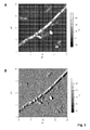
- E. coli BI21 (DE3) cells containing no SllB-encoding plasmid are on average 1-2 ⁇ m long and 0.5-0.7 ⁇ m wide ( Fig. 2 C, D) .
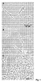
- E. coli BI21 (DE3) expressing SllB form longer single cells, on average 4-5 ⁇ m long and 0.8-1 ⁇ m wide, and the length of a cell complex is between 5 and 200 ⁇ m ( Fig. 1 AC ) .
- These cell clones form the microtubes according to the invention, which grow with the cells and are not shared by cell division.
- the cells of non-transgenic E. coli BI21 (DE3) also form such special microtubes, but disaggregate upon reaching the stationary phase, leaving the individual cells behind.
- the transgenic E. coli BI21 (DE3) form the same long microtubes around each cell, which are not shared by cell division.
- the microtubes of the transgenic E. coli BI21 (DE3) remain stable upon reaching the stationary phase.
Landscapes
- Chemical & Material Sciences (AREA)
- Organic Chemistry (AREA)
- Health & Medical Sciences (AREA)
- General Health & Medical Sciences (AREA)
- Biochemistry (AREA)
- Biophysics (AREA)
- Life Sciences & Earth Sciences (AREA)
- Genetics & Genomics (AREA)
- Medicinal Chemistry (AREA)
- Molecular Biology (AREA)
- Proteomics, Peptides & Aminoacids (AREA)
- Gastroenterology & Hepatology (AREA)
- Micro-Organisms Or Cultivation Processes Thereof (AREA)
- Preparation Of Compounds By Using Micro-Organisms (AREA)
- Peptides Or Proteins (AREA)
Abstract
Description
- Die vorliegende Erfindung betrifft röhrenartige biologische Strukturen, die sich aus der äußeren Membran von E. coli zusammensetzen und durch heterolog exprimierte rekombinante S-Layer-Proteine stabilisiert werden, Verfahren zu deren Herstellung und deren Verwendung. Die neuartigen Mikroröhren eignen sich für die Anwendung in der Biotechnologie, Pharmazie, in der chemischen Katalyse und in der Nanotechnologie.
- S-Layer-Proteine sind bakterielle oder archaeale Hüllproteine, die sich selbst zu regelmäßigen zweidimensionalen Gitterstrukturen (S-Layer) zusammenlagern können (Selbstassemblierung). Diese parakristallinen Gitterstrukturen umfassen regelmäßig angeordnete Poren einheitlicher Größe. Diese spezielle Anordnung führt zu regelmäßigen Abstandsbeziehungen zwischen Molekülgruppen des Monomers wie auch zwischen Metallclustern oder Molekülgruppen, die durch Modifikation an das Monomer gebunden wurden. Damit stehen mit den S-Layer-Proteinen komplexe selbstorganisierende Strukturen zur Verfügung, die mit technischen Mitteln nicht oder nur sehr aufwendig hergestellt werden können (Pollmann u.a. (2007)).
- Verschiedene Mikroorganismen verfügen über eine S-Layer-Hülle. Sie produzieren dazu teils sehr unterschiedliche S-Layer-Proteine. Das Molekulargewicht der Monomere kann dabei zwischen 40 und 200 kDa variieren. Die auf ihnen basierenden S-Layer haben in der Regel eine Dicke von 5 bis 20 nm.
- Die Fähigkeit zur Selbstassemblierung verleiht den S-Layer-Proteinen ungewöhnliche chemische und strukturelle Eigenschaften, die sie für verschiedenste Anwendungen interessant machen. So lassen sich an S-Layern beispielsweise metallische bzw. Halbleiter-Nanopartikel abscheiden, S-Layer können als Metall-bindende Strukturen oder als Träger für biofunktionale Moleküle verwendet werden, oder sie können zur Umhüllung von Medikamenten eingesetzt werden.
- Zur Gewinnung von S-Layer-Proteinen können die Stämme herangezogen werden, die die entsprechenden Proteine natürlicherweise exprimieren (Sleytr, U.B. et al. (2007). Oft sind diese Mikroorganismen jedoch nur schwer kultivierbar und/oder die S-Layer-Isolierung ist sehr aufwendig und damit kostenintensiv. Zur rekombinanten Expression von Proteinen werden daher in der Molekularbiologie häufig im Labor einfach zu kultivierende Mikroorganismen herangezogen, die gentechnisch so modifiziert wurden, dass sie die für das betreffende Protein kodierende DNA-Sequenz enthalten und so das Zielprotein exprimieren. Einer der am häufigsten zu diesem Zweck verwendeten Bakterienstämme ist Escherichia coli (E. coli) (Pollmann u.a (2007)).
- E. coli ist ein gram-negatives, peritrich begeißeltes Bakterium, das natürlicherweise im Darm vorkommt und zu den am besten untersuchten Organismen der Welt gehört. Für den labortechnischen Einsatz bei der Expression von rekombinanten Proteinen wurde davon eine Vielzahl an unterschiedlichen Stämmen abgeleitet.
- Die Zellen von E. coli sind von einer äußeren Membran (outer membrane) umgeben. Dies ist eine Besonderheit von gram-negativen Bakterien. Die äußere Membran umgibt die Zellwand der Bakterien als zusätzliche Membran, ist aus Lipopolysacchariden zusammengesetzt und begrenzt das Periplasma. Anders als die Cytoplasmamembran ist sie permeabel für die meisten niedermolekularen Stoffe (Wasser, Ionen, kleine Metabolite) und enthält Porine, die größere Moleküle bis zu 700 Da passieren lassen.
- Üblicherweise bilden E. coli-Zellen Stäbchen von ca. 3 µm x 1 µm aus. In der Wachstumsphase werden häufig kurze Ketten von Einzelzellen beobachtet. Nur in wenigen Publikationen wird von filamentösen E. coli Zellen berichtet, wie z. B. bei E. coli str. CS1959, einem Mutanten von E. coli K-12 (Parker, C. T. et al., 1992, J. Bacteriol. 174, pp. 2525-2538), oder während Wachstum bei 42°C (Koch, A. L. et al., 1987, J. Bacteriol. 169, pp. 1979-1984) oder unter erhöhter Sauerstoffzufuhr (Preusser, J. H., 1959, Archiv für Mikrobiologie 33, pp. 105-123)(1).
- Die der Erfindung zugrundeliegende Aufgabe besteht darin, eine neue Form von biologischen Mikro-Strukturen zur Verfügung zu stellen.
- Die Aufgabe wird gelöst durch Mikroröhren, die von E. coli Zellen gebildet werden, welche in der Lage sind, Filamente zu bilden, und welche S-Layer-Proteine oder Fragmente von S-Layer-Proteinen rekombinant exprimieren. Diese Mikroröhren umfassen die äußere Membran dieser E. coli Zellen und die von diesen E. coli Zellen rekombinant exprimierten S-Layer-Proteine bzw. -Proteinfragmente.
- Die erfindungsgemäßen Mikroröhren werden aus der äußeren E. coli Zellmembran bzw. deren Bestandteilen gebildet. Die Mikroröhren enthalten daher die Proteine und Lipide der äußeren Membran von E. coli Zellen. Sie werden durch die Anlagerung von S-Layer-Proteinen oder -Proteinfragmenten stabilisiert, so dass sie auch in der stationären Phase stabil sind und durch Chemikalien wie z. B. 1 % SDS (Natriumdodecylsulfat), Harnstoff oder 3 M NaC1 nicht zerstört werden können. Bei den Filamente-bildenden E. coli Zellen handelt es sich bevorzugt um Zellen des Stammes E. coli BI21 (DE3), die natürlicherweise bereits während der exponentiellen Phase lange Filamente ausbilden, die aber später in der stationären Phase wieder in typische E. coli Einzelzellen zerfallen.
- Die Erfinder haben beobachtet, dass der Stamm E. coli BI21 (DE3), der im Stand der Technik für die Expression von rekombinanten Proteinen verwendet wird, in der exponentiellen Phase in Flüssigmedium bei Raumtemperatur lange Filamente von z. T. über 100 µm ausbildet, die aber (wenn die E. coli-Zellen keine S-Layer-Proteine exprimieren) in der späteren stationären Phase in typische E. coli Einzelzellen zerfallen.
- Die Erfinder haben überraschend festgestellt, dass, wenn man S-Layer-Proteine oder -Proteinfragmente in diesen filamentbildenden E. coli-Zellen exprimiert, die Zellen bisher unbekannte Mikroröhren bilden. Die Mikroröhren bilden sich dabei um die Zellen herum und umschließen eine oder mehrere Zellen. Wenn sich die in den Mikroröhren befindlichen E. coli-Zellen teilen, vermehrt sich auch das Röhrenmaterial. Die Mikroröhre teilt sich allerdings nicht zusammen mit den E. coli-Zellen, sondern verlängert sich und hüllt somit sämtliche Tochterzellen ein. Die Mikroröhren werden somit von den E. coli Zellen als extrazelluläre Strukturen gebildet, wobei die Zellen ggf. nach Formierung der Mikroröhren entfernt werden.Zellfreie Mikroröhren sind durch Lyse bzw. Herauslocken der Zellen (z. B. durch Chemotaxis) aus den Mikroröhren wie weiter unten beschrieben erhältlich.
- Bei den von den E. coli Zellen rekombinant produzierten S-Layer-Proteinen oder -Proteinfragmenten handelt es sich vorzugsweise um stumme und/oder funktionale S-Layer-Proteine der Bakteriengattung Lysinibacillus, um zu diesen verwandte Proteine und/oder Proteinfragmente hiervon.
- Bevorzugt stammen die S-Layer-Proteine oder -Proteinfragmente von den Spezies Lysinibacillus sphaericus, besonders bevorzugt von den Stämmen Lysinibacillus sphaericus JG-A12 (Isolat einer Uranabraumhalde) (Genbank-Akzessionsnummer AJ866975) und/oder Lysinibacillus sphaericus NCTC 9602 (Genbank-Akzessionsnummer AJ866974).
- Die von den E. coli Zellen rekombinant produzierten S-Layer-Proteine haben bevorzugt eine Aminosäuresequenz, die ausgewählt ist aus den Aminosäuresequenzen der S-Layer-Proteine SlfA (SEQ ID-No. 2), SllA (SEQ ID-No. 4), SlfB (SEQ ID-No. 6) oder SllB (SEQ ID-No. 8) oder einer Teilsequenz davon mit einer Länge von mindestens 100 Aminosäuren, vorzugsweise mindestens 150 Aminosäuren, bevorzugt mindestens 200 Aminosäuren, besonders bevorzugt mindestens 250 Aminosäuren, oder eine zu mindestens einer dieser Sequenzen homologe Sequenz mit mindestens 50 % Sequenzidentität, vorzugsweise 70 % Sequenzidentität, bevorzugt 80 % Sequenzidentität, besonders bevorzugt 90 % Sequenzidentität.
- Die erfindungsgemäßen Mikroröhren sind erhältlich durch die Expression von einer für ein S-Layer-Protein, bevorzugt ein S-Layer-Protein von Lysinibacillus sphaericus JG-A12, kodierenden Nukleinsäuresequenz oder einer Teilsequenz davon in einer Filamente-bildenden E. coli Wirtszelle.
- Ganz besonders bevorzugt handelt es sich bei den von den E. coli Zellen rekombinant exprimierten Nukleinsäuresequenzen um die für S-Layer-Proteine kodierenden Gene slfA (SEQ ID-No. 1; Genbank-Akzessionsnr. AJ849547), sllA (SEQ ID-No. 3; Genbank-Akzessionsnr. AJ849548), slfB (SEQ ID-No. 5; Genbank-Akzessionsnr. AJ849549) und/oder sllB (SEQ ID-No. 7; Genbank-Akzessionsnr. AJ849550) von Lysinibacillus sphaericus JG-A12.
- Für die Bildung von Mikroröhren ist insbesondere der Sequenzbereich verantwortlich, der dem Bereich zwischen 650 bp und dem Stoppcodon, besonders den Sequenzbereichen zwischen den Positionen 650 bp und 1800 bp und zwischen den Positionen 2473 bp und dem Stoppcodon einer der Sequenzen ausgewählt von SEQ ID-No. 1, 3, 5 oder 7 befindet.
- Die entsprechenden Bereiche der S-Layer-Proteine für die Mikroröhrenbildung liegen also in dem Bereich, der dem Abschnitt von 217 Aminosäuren bis zum C-terminalen Ende, besonders den Abschnitten von 217 bis 600 Aminosäuren und von 824 Aminosäuren bis zum C-terminalen Ende einer der Sequenzen gemäß den SEQ ID-No. 2, 4, 6 oder 8 entspricht.
- Werden also Mikroröhren mit Teilsequenzen der S-Layer-Proteine erzeugt, umfassen diese bevorzugt mindestens einen der Bereiche, die von 217 bis 600 Aminosäuren bzw. von 824 Aminosäuren bis zum C-terminalen Ende einer der Sequenzen gemäß den SEQ ID-No. 2, 4, 6 oder 8 reichen, oder eine zu mindestens einer dieser Sequenzen homologe Sequenz mit mindestens 50 % Sequenzidentität, vorzugsweise mindestens 70 % Sequenzidentität, bevorzugt 80 % Sequenzidentität, besonders bevorzugt 90 % Sequenzidentität.
- Besonders bevorzugt umfassen die Teilsequenzen die entsprechenden Abschnitte der Aminosäuresequenzen gemäß den SEQ ID-No. 2 und/oder 8 oder eine zu mindestens einer dieser Sequenzen homologe Sequenz mit mindestens 50 % Sequenzidentität, vorzugsweise mindestens 70 % Sequenzidentität, bevorzugt 80 % Sequenzidentität, besonders bevorzugt 90 % Sequenzidentität.
- Ganz besonders bevorzugt umfassen sie die folgenden Aminosäuresequenzen SEQ ID-No. 9 und 10, die den Abschnitten von 217 bis 600 Aminosäuren (SEQ ID-No. 9) bzw. von 824 bis 1101 Aminosäuren (SEQ ID-No. 10) des S-Layer-Proteins SllB aus Lysinibacillus sphaericus JG-A12 entsprechen, oder eine zu mindestens einer dieser Sequenzen homologe Sequenz mit mindestens 50 % Sequenzidentität, vorzugsweise mindestens 70 % Sequenzidentität, bevorzugt 80 % Sequenzidentität, besonders bevorzugt 90 % Sequenzidentität.


- Werden in die Zellen des Filamente-bildenden E. coli Stammes Gene kloniert, die für S-Layer-Gene z. B. von Lysinibacillus sphaericus (z.B. sllA, sllB, slfA, s/fB) kodieren und werden diese S-Layer-Proteine exprimiert, werden die filamentösen Strukturen der exponentiellen Phase dadurch stabilisiert.
- Bestandteil der Erfindung ist daher auch die Verwendung einer Nukleinsäuresequenz ausgewählt aus den SEQ-ID No. 1, 3, 5 oder 7, einer Teilsequenz davon oder einer zu mindestens einer dieser Sequenzen homologen Nukleinsäuresequenz mit mindestens 50 % Sequenzidentität, vorzugsweise mindestens 70 % Sequenzidentität, bevorzugt 80 % Sequenzidentität, besonders bevorzugt 90 % Sequenzidentität, zur Herstellung der erfindungsgemäßen Mikroröhren, sowie eine E. coli-Zelle, die eine dieser Sequenzen umfasst.
- Erreichen die E. coli-Zellen die stationäre Phase, so sind lange röhrenartige Strukturen zu beobachten, in denen Einzelzellen von E. coli enthalten sind. Diese Strukturen ähneln in ihrer Zusammensetzung der äußeren Membran von E. coli. Die rekombinant exprimierten S-Layer-Proteine sind mit dieser röhrenförmigen Membranstruktur assoziiert und stabilisieren diese. Die erfindungsgemäßen Mikroröhren sind chemisch äußerst stabil und lassen sich durch Chemikalien wie z.B. 1 % SDS, Harnstoff oder 3 M NaCl kaum oder gar nicht zerstören.
- Durch die hohe Stabilität der Mikroröhren erhalten die erfindungsgemäßen Mikroröhren produzierenden Bakterienzellen vorteilhaft ebenfalls eine erhöhte mechanische Stabilität. Die Mikroröhren-bildenden Zellen sind mit herkömmlichen Verfahren wie Ultraschall oder Hochdruckhomogenisation nicht oder nur sehr schwer zu zerstören. Dies kann vorteilhaft genutzt werden, wenn eine Lyse der Zellen durch mechanische Behandlung vermieden werden soll.
- Die erfindungsgemäßen Mikroröhren erreichen eine Länge von teilweise über 100 µm. Vorzugsweise sind sie zwischen 5 µm und 200 µm, bevorzugt zwischen 10 µm und 100 µm, besonders bevorzugt zwischen 20 µm und 80 µm lang. Ihr Querschnitt ist durch den Zelldurchmesser der von ihnen umgebenen Bakterien beeinflusst. Vorzugsweise liegt er zwischen 0,4 µm bis 2 µm, bevorzugt zwischen 0,5 µm und 1 µm.
- Die erfindungsgemäßen Mikroröhren können die Bakterien, von denen sie gebildet wurden enthalten. Vorzugsweise sind die Bakterien aber durch Behandlung mit verschiedenen Chemikalien entfernt. Ob die Bakterien stören oder nicht ist jedoch abhängig vom Verwendungszweck.
- Da die Mikroröhren sehr stabil gegen verschiedene Chemikalien sind, ist die Entfernung der in ihnen enthaltenen Bakterienzellen nicht ohne weiteres möglich.
- Bestandteil der Erfindung ist daher auch ein Verfahren zur Isolierung der erfindungsgemäßen Mikroröhren und zur Entfernung der E. coli Zellen aus den Mikroröhren. Vorzugsweise werden dazu die Mikroröhren mit den darin enthaltenen Zellen in einer wässrigen Lösung eines Mono- und/oder Disaccharids oder einer wässrigen Ethanollösung inkubiert. Bevorzugt wird das Mono- und/oder Disaccharid ausgewählt aus Glucose, Fructose, Galactose, Saccharose, Maltose, und Lactose. Besonders bevorzugt ist Saccharose.
- Enthält die wässrige Lösung Ethanol, liegt der Anteil an Ethanol bevorzugt bei 5 % bis 20 %, besonders bevorzugt bei 7,5 % bis 17,5 %, ganz besonders bevorzugt bei 9 % bis 15 %.
- Enthält die wässrige Lösung ein oder mehrere Mono- und/oder Disaccharide, ist der Gesamtanteil der Mono- und/oder Disaccharide bevorzugt 20 Gew.-% bis 60 Gew.-%, besonders bevorzugt 30 Gew.-% bis 50 Gew.-%, ganz besonders bevorzugt 35 Gew.-% bis 45 Gew.-%. Durch diese Behandlung verlässt ein Großteil der E. coli Zellen die Mikroröhren.
- Vorzugsweise werden sowohl die Mikroröhren als auch die Zellen anschließend mit einer hochkonzentrierten Harnstofflösung behandelt. Durch diesen zweiten Reinigungsschritt, werden die noch in den Röhren verbliebenen Zellen zum Verlassen der Röhren gezwungen. Vorzugsweise beträgt die Konzentration der Harnstofflösung zwischen 4 mol/l und 8 mol/l, bevorzugt zwischen 5 mol/l und 7 mol/l.
- Vorteilhaft wachsen die die S-Layer-Proteine rekombinant exprimierenden, Mikroröhren-bildenden E. coli-Zellen nach Induktion der S-Layer-Produktion unter Standard-Wachstumsbedingungen, wie sie in Standardwerken wie z. B. Sambrook et al. (1989) beschrieben werden, zu sehr hohen Zelldichten von bis zu A600 = 5,0 heran. Damit eignen sie sich zur effizienten Produktion von rekombinanten Proteinen. Als Wachstumsbedingungen werden dabei bevorzugt zwischen 50 bis 100 ml LB(Luria-Bertani)-Medium in geeigneten Erlenmeyer-Schüttelkolben (beispielsweise einer Größe von 100 ml bis 300 ml), und Schüttelfrequenzen zwischen 200 bis 300 Umdrehungen/min, vorzugsweise von 220 bis 250 Umdrehungen/min gewählt.
- Die Produktion der Mikroröhren durch die E. coli-Zellen erfolgt vorzugsweise bei Temperaturen unter 37 °C, bevorzugt bei weniger als 25 °C, besonders bevorzugt zwischen 10 und 14 °C.
- Bestandteil der Erfindung sind auch Mikroröhren, die metallisiert sind. Die hergestellten Röhrenstrukturen zeigen sehr gute Bindungseigenschaften für unterschiedliche Metalle, wie z.B. Au(III) oder Pd(II). So beträgt die Bindungskapazität für Pd(II) 8 mg/g Biomasse, für Au(III) 14 mg/g Biomasse und für Pb(II) 8,5 mg/g Biomasse. Eine erfindungsgemäße Anwendung ist daher die Herstellung von dünnen metallischen Mikroröhren, beispielsweise Gold- oder Palladium-Mikroröhren, für die Elektronik, indem die Mikroröhren als Matrix für eine Metallisierung verwendet werden.
- Die erfindungsgemäßen metallisierten Mikroröhren sind durch Inkubation in einer Metallsalzlösung (z. B. Au(III)) und anschließende Zugabe eines Reduktionsmittels (z.B. DMAB) erhältlich.
- Da eine preiswerte Biomassegewinnung durch das Erreichen hoher Zelldichten sehr gut möglich ist, sind die Mikroröhren außerdem vorteilhaft für eine preiswerte Entfernung von Metallen aus (Ab-)Wässern geeignet. Bestandteil der Erfindung ist daher auch die Verwendung der Mikroröhrenstrukturen als metallbindende Filtermaterialien.
- Die erfindungsgemäßen Mikroröhren eignen sich außerdem für eine Funktionalisierung, indem die rekombinant exprimierten S-Layer-Proteine mit Peptiden oder Proteinen fusioniert und diese Peptide und Proteine durch die Fusion mit den S-Layer-Proteinen an die Mikroröhren gebunden werden. Die Mikroröhren können auf diese Weise beispielsweise mit Enzymen funktionalisiert und als Katalysatoren verwendet werden. Denkbar ist auch die Verwendung der funktionalisierten Röhren zur Herstellung von Antikörpern.
- Die neuartigen Mikroröhren eignen sich aufgrund ihrer faserartigen Struktur auch zur Verwendung in Verbundmaterialien und/oder Textilien.
- Prinzipiell eignen sich die Mikroröhren zur Verwendung als Carrier (Träger) für pharmazeutische Wirkstoffe. Eine andere Anwendungsmöglichkeit betrifft die Verwendung der Mikroröhrenstrukturen als Container, Umhüllung bzw. als Carrier (Träger) für heterolog koexprimierte Proteine wie z.B. Enzyme. Dabei werden zusätzlich zu den S-Layer-Proteinen, die für die Stabilisierung der Mikroröhrenstruktur sorgen, andere rekombinante Proteine exprimiert, die in den Mikroröhren auch nach Zerstörung der Zellen verbleiben und so immobilisiert sind.
- Die Erfindung wird durch die folgenden Figuren und Ausführungsbeispiele näher erläutert, ohne auf diese beschränkt zu sein.
-
Fig. 1 A bis C enthält verschiedene lichtmikroskopische Aufnahmen von S-Layer-Protein exprimierenden E. coli BI21(DE3), die mit einem Olympus BX61-Mikroskop (Olympus Optical, Europe; GmbH, Hamburg) im Phasenkontrastverfahren aufgenommen wurden, und zeigt die Mikroröhrenbildung nach der Induktion der S-Layer-Protein-Expression durch IPTG. -
Fig. 2 A und B zeigen transmissionselektronenmikroskopische (TEM-)Aufnahmen von SllB-exprimierenden E. coli BI21(DE3)-Zellen. Als Vergleich dienenFig. 2 C und D, die Aufnahmen von E. coli BI21(DE3)-Zellen zeigen, die kein für SllB kodierendes Plasmid enthalten. Die Aufnahmen wurden mit einem EM 912 OMEGA Transmissionselektronenmikroskop (LEO Elektronenmikroskopie. GmbH, Oberkochen, Deutschland) erstellt. -
Fig. 3 A, B zeigt mit einem Rasterkraftmikroskop aufgenommene Bilder einer mit E. coli BI21(DE3)-Zellen gefüllten Mikroröhre. Die AFM-Bilder wurden mit dem Rasterkraftmikroskop MFP3D (Bio Asylum Research, Santa Barbara, Canada) aufgenommen. -
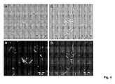
Fig. 4 zeigt lichtmikroskopisch (Phasenkontrast) (A, C) und fluoreszenzmikroskopisch aufgenommene Bilder einer E. coli BI21(DE3)-Zellkultur, die das SIIB-GFP-Fusionsprotein exprimiert (B, D). Die Bilder wurden mit dem Olympus BX61-Mikroskop (Olympus Optical, Europe; GmbH, Hamburg) unter Verwendung des GFP-Filters U-MNIBA2 (Olympus Optical, Europe; GmbH, Hamburg) mit einer Belichtungszeit von 100 ms aufgenommen. -
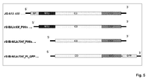
Fig. 5 Schematischer Aufbau der Konstrukte für die Expression von verschiedenen rekombinanten (Fusions)proteinen. Ganz oben ist das gesamte sllB-Gen einschließlich der für das Signalpeptid kodierenden Sequenz dargestellt und zeigt den grundsätzlichen Aufbau des Gens, das als Templat für die PCR(Polymerase Chain Reaction)-Amplifikation verwendet wurde. SP = Signalpeptid; NTD = N-terminale Domäne; CD = Zentrale Domäne; CTD = C-terminale Domäne. -
Fig. 6 zeigt IR-Spektren verschiedener Röhren- oder Membranfraktionen während der Aufreinigung der Mikroröhren bestehend aus den E. coli-Röhrenkomponenten und dem S-Layer Protein SllB, sowie im Vergleich dazu das S-Layer-Spektrum von SlfB, welches ein dem SllB vergleichbares Spektrum zeigt. Von oben nach unten sind dies: reines JG-A12 S-Layer-Protein (Kontrolle), Röhrenisolat von SllB-exprimierenden E. coli BI21 (DE3)-Zellen, bei Lipidextration aus dem Röhrenisolat ausgefällte Proteinfraktion, obere, lipidfreie Phase, untere Lipidphase, und Röhrenisolat von E. coli BI21 (DE3)-Zellen, die kein S-Layer-Protein exprimieren (Kontrolle). - Lysinibacillus sphaericus JG-A12 ist ein natürliches Isolat von der Uranabraumhalde Haberland nähe Johanngeorgenstadt, Deutschland (Selenska-Pobell et al. 1999). Die Bakterien werden üblicherweise in NB-(nutrient broth)-Medium kultiviert. Escherichia coli NovaBlue GigaSingles (Novagen) und Escherichia coli BI21 (DE3) (Novagen) sind von E. coli K12 abgeleitete Stämme und werden üblicherweise in LB(Luria-Bertani)-Medium kultiviert.
- Als Templat für die PCR-Amplifikation wird genomische DNA von Lysinibacillus sphaericus JG-A12 isoliert und aufgereinigt.
- Für die Erzeugung eines N-terminal His-getaggten (d. h. mit einem His-Tag versehenen) rekombinanten Proteins (rSllB/Lic93f_PlHis,
Fig. 5 , zweite von oben) wird das S-layer-Protein kodierende Gen sllB von der chromosomalen DNA von L. sphaericus JG-A12 mit dem Primerpaar Lic93f (SEQ ID-No. 11; 5'-GAC GAC GAC AAG ATG/GCA GGA TTC TCA GAT GTA GCA-3') und Lic_PlHis (SEQ ID-No. 12; 5'-GAG GAG AAG CCC GGT/TTA TGG AGT TGG CTT TAC TGT AAT A-3') amplifiziert. - Für die Erzeugung eines N-terminal verkürzten und His-getaggten rekombinanten Proteins (rSllB-N/Lic704f_PlHis,
Fig. 5 , dritte von oben) wird das S-Layer-Protein kodierende Gen sllB von der chromosomalen DNA von L. sphaericus JG-A12 mit dem Primerpaar Lic704f (SEQ ID-No. 13; 5'-GAC GAC GAC AAG ATG/ATC AAC AAC ACA ACT GTT GAA-3') und Lic_PlHis amplifiziert. - Positive PCR-Produkte, die für das stille S-layer-Protein bzw. das N-terminal verkürzte Fragment kodieren, werden in den Vektor pET-30 Ek/LlC (Novagen) kloniert und das erhaltene Konstrukt in die nicht exprimierende Wirtszelle Escherichia coli NovaBlue (Novagen) transformiert. Positive Klone werden isoliert und in E. coli BI21 (DE3) als exprimierende Wirtszelle transformiert.
- Für die Erzeugung eines N-terminal His-getaggten rekombinanten SllB-GFP-Fusionsproteins (rSllB-N/Lic704f_Pl_GFP,
Fig. 5 , ganz unten) wird das GFP-Gen mit dem Primerpaar Lic_GFP_BamHI_F (SEQ ID-No. 14; 5'-AAAGGATCCATGAGTAAAGGAGAAGAACTT-3') und Lic_GFP_EagI_R (SEQ ID-No. 15; 5'-TTTCGGCCGCTATTTGTATAGTTAATCCA-3') von dem pGFP-Vektor (Clontech) amplifiziert. Anschließend werden die PCR-Produkte des Primerpaars Lic_GFP_BamHI_F und Lic_GFP_Eagl_R C-terminal in das aufgereinigte und restriktionsverdaute Plasmid rSllB-N/Lic704f_Pl ligiert und in Escherichia coli NovaBlue (Novagen) als nichtexprimierende Wirtszelle transformiert. Positive Klone werden identifiziert, isoliert und in E. coli BL21 (DE3) als exprimierende Wirtszelle transformiert. - Für die verschiedenen SllB-Fusionsproteine, nämlich a) SllB ohne Signalpeptid und mit N-terminalem His-tag (rSllB/Lic93f_PlHis) (
Fig. 5 , zweite von oben), b) N-terminal verkürztes SllB mit N-terminalem His-tag (rSllB-N/Lic704f_PlHis) (Fig. 5 , dritte von oben) und c) N-terminal verkürztes SllB mit C-terminaler GFP-Fusion und N-terminalem His-tag (rSllB-N/Lic704f_Pl_GFP) (Fig. 5 , ganz unten), kodierende Plasmide werden wie in Ausführungsbeispiel 1 beschrieben erzeugt, die rekombinanten Proteine exprimiert und die Expression anschließend durch SDS-PAGE überprüft. - Für die Expression werden 100 ml of Luria-Bertani(LB)-Medium mit Kanamycin (25 µg/ml) versetzt und mit 5 ml einer in LB gezogenen Vorkultur des mit dem entsprechenden Plasmid transformierten E. coli BI21 (DE3)-Stammes beimpft. Die Kulturen werden bei Raumtemperatur inkubiert. Nach zwei Stunden wird die rekombinante Genexpression durch die Zugabe von 0.1 mmol/l Isopropylthiogalactosid (IPTG) induziert.
- Die Zellen werden nach der Inkubation über Nacht in der stationären Phase geerntet, indem sie 5 min bei 6000 x g abzentrifugiert und zweimal in Resuspensionspuffer (100 mmol/l NaH2PO4xH2O; 10 mmol/l Tris-Base; 0.5 m NaCl, pH=8.0) gewaschen werden. Nach der Resuspension in 5 ml Puffer werden die Zellen durch jeweils 5 bis 6 halbminütige Ultraschallbehandlung bei 60 % Amplitude aufgeschlossen (Sonifier W250-D, Branson). Das Lysat wird 30 min bei 30,000 x g abzentrifugiert und der Überstand abgenommen und bei 4 °C für die weitere Untersuchung aufbewahrt. Die abzentrifugierten Zelltrümmer werden in Resuspensionspuffer resuspendiert.
- Das Proteinprofil des Überstandes, der die löslichen Proteine enthält, und der abzentrifugierten Zelltrümmer, wird durch SDS-PAGE (sodium dodecyl sulfatepolyacrylamide gel electrophoresis) überprüft. Die SDS-PAGE wird ausgeführt wie von Laemmli (1970) beschrieben. Im Vergleich zu Zellextrakten von nicht-transformierten E. coli BI21 (DE3) können in allen Proben zusätzliche hochmolekulare Proteinbanden beobachtet werden, die in etwa den berechneten Molekulargewichten der Fusionsproteine rSllB/Lic93f_PlHis (∼118 kDa), rSllB-N/Lic704f_PlHis (∼96 kDa) und rSllB-N/Lic704f_Pl_GFP (∼124 kDa) (
Fig. 5 ) entsprechen. - Die Expression von SllB wird durch Immunblot unter Verwendung von polyklonalem primärem Antikörper, der gegen das S-layer-Protein von L. sphaericus JG-A12 (Pineda Antikörper Service, Berlin) erzeugt worden war, und einem Immun-Blot Assay Kit (BioRad, Hercules, USA) mit geeignetem sekundärem Antikörper analysiert. Auf den Immunblots ist nach der Inkubation mit sekundärem Anti-Kaninchen-Antikörper konjugiert mit Alkaliner Phosphatase eine starke Kreuzreaktion zwischen den in der SDS-Page identifizierten hochmolekularen Proteinbanden und dem polyklonalen Kaninchen-Antiserum, das gegen SlfB von Lysinibacillus sphaericus JG-A12 erzeugt worden war, zu beobachten.
- Die rekombinanten Proteine werden mit kommerziell erhältlichem Proteinfarbstoff (SYPRO-Ruby Protein Gel stain; BioRad, Hercules, USA) und einem kommerziell erhältlichen Kit zur Bestimmung des Proteingehalts (BioRad Protein Assay; BioRad, Hercules, USA) quantifiziert. Die Konzentration der Fusionsproteine wird mittels des kommerziell erhältlichen Proteinfarbstoffs sowie über die Bradford-Methode bestimmt. Die 100 ml-Kultur von mit rSllB/Lic93f_PlHis transformierten E. coli BI21 (DE3) enthält ∼14.5 mg Fusionsprotein (∼25 % des Gesamtproteingehalts), die 100 ml-Kultur von mit rSllB-N/Lic704f_PlHis transformierten E. coli BI21 (DE3) enthält ∼18 mg Fusionsprotein (∼36 % des Gesamtproteingehalts) und die 100 ml-Kultur von mit rSllB-N/Lic704f_Pl_GFP transformierten E. coli BI21 (DE3) enthält ∼12 mg Fusionsprotein (∼18 % des Gesamtproteingehalts).
- Zunächst wird das stumme S-Layer Gen sllB des Stammes Lysinibacillus sphaericus JG-A12 (Genbank-Akzessionsnummer AJ849550) unter Verwendung von genspezifischen Primern durch PCR wie in Ausführungsbeispiel 1 beschrieben vervielfältigt. Das erhaltene PCR-Produkt mit einer Länge von 3210 bp wird in den Vector pET-30 Ek/Lic (Novagene) ligiert. Der Vector mit dem sllB-Insert wird zunächst in E. coli NovaBlue (Novagene) transformiert. Von positiven Klonen werden Plasmide isoliert, die in den Expressionsstamm E. coli BI21 (DE3) transformiert werden.
- Für die Expression von sllB wird zunächst eine Vorkultur in LB-Medium angezogen. Diese wird in frisches LB-Medium überimpft und bis zu einer O.D. von ca. 0,1 inkubiert. Das Wachstum erfolgt bei Raumtemperatur (< 25°C). Nach ca. 2 h Wachstum erfolgt eine Zugabe von 0,1 mmol/l IPTG und damit eine Induktion der Expression der rekombinanten SllB Proteine. In der exponentiellen Phase erfolgt eine Bildung von langen Filamenten von E. coli-Zellen. In der späten exponentiellen Phase und stationären Phase sind lange Mikroröhren einer Länge von z. T. > 100 µm zu beobachten, in denen Einzelzellen von E. coli zu erkennen sind.
- Eine stabile Mikroröhrenbildung erfolgt nur in Flüssigkultur bei genügender Sauerstoffzufuhr und Temperaturen <25 °C. Auf Agar-Platten bilden die Bakterien normale Einzelzellen aus.
- Die Isolierung der Mikroröhren erfolgt folgendermaßen: Nach der Ernte der Zellen durch Zentrifugation (10,000 x g, 10 min) werden die Zellen in 1x PBS Puffer resuspendiert und zweimal mit destilliertem H2O oder in 1xPBS-Puffer gewaschen. Die Überstände werden verworfen. Anschließend werden die Zellen mit 40 % Saccharose versetzt und für 1 h bei RT geschüttelt. Dabei verlassen die Zellen die Mikroröhren. Die Zellen werden 15 min bei 3000 x g und 4 °C abzentrifugiert und der Überstand mit den Mikroröhren wird abgenommen. Der Überstand wird anschließend 30 min bei 12000 x g zentrifugiert. Das Pellet mit den Mikroröhren wird mit 6 mol/l Harnstoff versetzt und 1 h bei Raumtemperatur inkubiert. Die Zellen werden ebenfalls in 6 mol/l Harnstoff resuspendiert und für 1 h geschüttelt. Beide Fraktionen werden erneut bei 3000 x g zentrifugiert. Die Mikroröhren befinden sich im Überstand, aus dem sie bei 12000 x g abzentrifugiert werden. Das Pellet mit den Mikroröhren wird mehrfach in dest. H2O gewaschen und bei 12000 x g abzentrifugiert, und in destilliertem Wasser aufgelöst und aufbewahrt.
- Wie durch die Ansequenzierung des N-Terminus festgestellt wurde, enthalten die Mikroröhren verschiedene Proteine der äußeren Bakterienmembran wie beispielsweise OmpF. Zusätzlich enthalten die Fraktionen die rekombinanten Proteine. Die Mikroröhren wurden IR-spektroskopisch untersucht (
Fig. 6 ). Dazu werden die Membranen von rSllB/Lic93f_PlHis und rSllB-N/Lic704f_Pl_GFP zum Zweck der kompletten Lipidextraktion mit Chloroform und Methanol behandelt (Bligh and Dyer 1959), wobei zunächst alle Proteine ausgefällt werden, welche anschließend als dritte Fraktion untersucht werden können. Nach der Phasentrennung werden beide Phasen, die untere Lipidphase und die obere lipidfreie Phase, durch IR-Spektroskopie analysiert (Fig. 6 ). Die Ergebnisse der IR-Spektroskopie stützen die Ergebnisse der SDS-PAGE, dass die Mikroröhren zum Großteil aus den rekombinanten Proteinen bestehen. Außerdem enthalten die Mikroröhren bis dato nicht identifizierte Lipide und andere nicht näher bestimmte Materialien. - AFM-Proben werden auf Siliziumträger aufgebracht und getrocknet, und mit dem Cantilever AC240 (Olympus Optical, Europe; GmbH, Hamburg) im AC-Modus an der Luft analysiert.
- Für die Untersuchung per Transmissionselektronenmikroskopie werden die Proben wie zuvor beschrieben in einem Volumen von 200 µl gut gewachsener Zellkultur in 1 x PBS und durch Zugabe von Osmiumtetroxid und Uranylacetat als Kontrastmittel behandelt (Spurr, 1969). Nach der Fixierung, Kontrastierung und Dehydrierung werden die Zellen in Epoxidharz (Serva Modified Spurr) entsprechend der Vorschriften des Herstellers eingebettet. Von den Proben werden mit dem Mikrotom Leica Ultracut UCR (Leica Microsystems GmbH, Wetzlar, Germany) und dem Diatome Diamont Messer (Diatome, Hatfield, Panama) Dünnschnitte mit einer Dicke von 50-300 nm hergestellt. Die luftgetrockneten Schnitte werden trocken gelagert.
- E. coli BI21(DE3) Zellen, die kein SllB-kodierendes Plasmid enthalten, sind durchschnittlich 1-2 µm lang und 0.5-0.7 µm breit (
Fig. 2 C, D). Im Vergleich bilden, E. coli BI21(DE3), die SllB exprimieren, längere Einzelzellen aus, die im Durchschnitt 4-5 µm lang und 0.8-1 µm breit sind, und die Länge eines Zellkomplexes beträgt zwischen 5 und 200 µm (Fig. 1 A-C ). Diese Zellklone bilden die erfindungsgemäßen Mikroröhren, die mit den Zellen wachsen und nicht durch die Zellteilung geteilt werden. - In der frühen exponentiellen Wachstumsphase formen die Zellen von nicht-transgenen E. coli BI21(DE3) ebenfalls solche speziellen Mikroröhren, die jedoch beim Erreichen der stationären Phase disaggregieren, wobei die einzelnen Zellen zurückbleiben. Im Vergleich dazu bilden die transgenen E. coli BI21(DE3) dieselben langen Mikroröhren um jede Zelle, die nicht durch Zellteilung geteilt werden. Die Mikroröhren der transgenen E. coli BI21(DE3) bleiben jedoch bei Erreichen der stationären Phase stabil.
-
- Battistuzzi G, Esan GJF, Fasuan FA, Modiano G, Luzzatto L (1977) Comparison of GdA und GdB Activities in Nigerians. A Study of the Variation of the G6PD Activity. Am J Hum Genet 29:31-36
- Bligh ED, Dyer WJ (1959) A rapid method of total lipid extraction and purification. Canad. J. Biochem. Physiol. 37, 911-917.
- Laemmli UK (1970) Cleavage of structural proteins during assembly of the head of bacteriophage T4. Nature 227:680-685
- Pollmann, K. u. a. (2005). Novel surface layer protein genes in Bacillus sphaericus associated with unusual isertium elements. Microbiology 151 (Pt9) 2961-2973.
- Pollmann, K. u. a (2007). Construction of S-layer protein exhibiting modified self-assembling properties and enhanced metal binding capacities. Appl. Microbiol. Biotechnology 75 (5) 1079-1085.
- Preusser, H. J. 1959. Form und Größe des Kernäquivalentes von Escherichia coli in Abhängigkeit von den Kulturbedingungen. Archives of Microbiology 33:105-123.
- Sanger F., Nicklen S., Coulson A.R., 1977. DNA Sequenzing with Chain-Terminating Inhibitors. Proceedings of the National Academy of Sciences of the United States of America, Vol 74, No.12, pp. 5463-5467
- Selenska-Pobell S., Panak P., Miteva V., Boudakov 1., Bernhard G., Nitsche H. (1999) Selective accumulation of heavy metals by three indigenous Bacillus strains, B. cereus, B. megaterium and B. sphaericus, from drain waters of uranium waste pile. FEMS Microbiol Ecol 29:59-67
- Sleytr, U.B. et al., (2007). S-layers as a tool kit for nanobiotechnological applications. FEMS Microbiol. Lett. 267(2) 131-144
-
Spurr AR (1969) A low-viscosity epoxy resin embedding medium for electron microscopy. J Ultrastruct.Res. 26: 31-43


Claims (14)
- Mikroröhren mit einem Durchmesser von 0,4 µm bis 2 µm, umfassend Bestandteile der äußeren Membran von E. coli-Zellen und rekombinant exprimierte S-layer-Proteine.
- Mikroröhren gemäß Anspruch 1, erhältlich durch die Expression von einer für ein S-Layer-Protein kodierende Nukleinsäuresequenz oder eine Teilsequenz davon in E. coli-Zellen, wobei die Mikroröhren sich um die Zellen herum bilden und eine oder mehrere Zellen umschließen, wobei die Zellen ggf. anschließend entfernt werden.
- Mikroröhren gemäß Anspruch 1 oder 2 mit einem Durchmesser von 0,5 µm bis 1 µm, und /oder einer Länge 5 µm bis 200 µm, bevorzugt von 10 µm und 100 µm, besonders bevorzugt von 20 µm bis 80 µm.
- Mikroröhren gemäß einem der Ansprüche 1 bis 3, dadurch gekennzeichnet, dass die S-Layer-Proteine S-Layer-Proteine der Gattung Lysinibacillus sphaericus sind.
- Mikroröhren gemäß einem der Ansprüche 1 bis 4, dadurch gekennzeichnet, dass die E. coli-Zellen vom Stamm E. coli BI21 (DE3) und/oder andere filamentbildende E. coli-Zellen sind.
- Mikroröhren gemäß einem der Ansprüche 1 bis 5, dadurch gekennzeichnet, dass die S-Layer-Proteine mit Peptiden oder Proteinen fusioniert sind.
- Mikroröhren gemäß einem der Ansprüche 1 bis 6, dadurch gekennzeichnet, dass die Röhren metallisiert sind.
- Mikroröhren gemäß Anspruch 7, dadurch gekennzeichnet, dass die Röhren durch Inkubation in einer Metallsalzlösung und anschließende Zugabe eines Reduktionsmittels metallisiert sind.
- Verfahren zur Herstellung von Mikroröhren nach einem der Ansprüche 1 bis 8 mit den Schritten:a. Expression von S-Layer-Proteinen in E. coli-Zellen, wobei die Mikroröhren sich um die Zellen herum bilden und eine oder mehrere Zellen umschließen undb. ggf. Entfernung der E. coli-Zellen, bevorzugt durch die Behandlung mit der wässrigen Lösung eines Mono- und/oder Disaccharids oder einer wässrigen Ethanollösung.
- Verwendung einer Nukleinsäuresequenz welche für ein S-Layerprotein codiert und ausgewählt ist aus Nukleinsäuresequenzen, die für ein Protein gemäß SEQ ID No. 2, 4, 6 oder 8 codierenden, Nukleinsäuresequenzen mit der SEQ-ID No. 1, 3, 5 oder 7, einer Teilsequenz davon oder einer zu mindestens einer dieser Sequenzen homologe Sequenz mit mindestens 50 % Sequenzidentität, vorzugsweise 70 % Sequenzidentität, bevorzugt 80 % Sequenzidentität, besonders bevorzugt 90 % Sequenzidentität, zur Herstellung von biologischen Mikroröhren gemäß einem der Ansprüche 1 bis 9.
- Verwendung von Mikroröhren gemäß einem der Ansprüche 1 bis 8 zur Bindung von Metall aus einer wässrigen Lösung, zur Herstellung funktionalisierter Schichten, als Carrier für Enzyme oder als Carrier für pharmazeutische Wirkstoffe.
- Verwendung von Mikroröhren gemäß einem der Ansprüche 6 bis 8 als Katalysator.
- E. coli-Zelle umfassend eine Nukleinsäuresequenz ausgewählt aus Sequenzen, die für ein S-Layerprotein codiert und ausgewählt ist aus Nukleinsäuresequenzen, die für ein Protein gemäß SEQ ID No. 2, 4, 6 oder 8 codiert, Nukleinsäuresequenzen gemäß SEQ-ID No. 1, 3, 5 oder 7, einer Teilsequenz davon oder eine zu mindestens einer dieser Sequenzen homologe Sequenz mit mindestens 50 % Sequenzidentität, vorzugsweise 70 % Sequenzidentität, bevorzugt 80 % Sequenzidentität, besonders bevorzugt 90 % Sequenzidentität
- E. coli-Zelle gemäß Anspruch 13, dadurch gekennzeichnet, dass die E. coli-Zelle vom Stamm E. coli BI21 (DE3) und/oder eine andere filamentbildende E. coli-Zelle ist.
Applications Claiming Priority (1)
| Application Number | Priority Date | Filing Date | Title |
|---|---|---|---|
| DE200910032645 DE102009032645B3 (de) | 2009-07-03 | 2009-07-03 | Mikroröhren, umfassend Bestandteile der äußeren Membran von E. coli Zellen und rekombinant exprimierte S-Layer-Proteine, Verfahren zu ihrer Herstellung und Verwendung |
Publications (2)
| Publication Number | Publication Date |
|---|---|
| EP2270033A1 true EP2270033A1 (de) | 2011-01-05 |
| EP2270033B1 EP2270033B1 (de) | 2016-02-17 |
Family
ID=42734793
Family Applications (1)
| Application Number | Title | Priority Date | Filing Date |
|---|---|---|---|
| EP10168382.9A Not-in-force EP2270033B1 (de) | 2009-07-03 | 2010-07-03 | Mikroröhren, umfassend Bestandteile der äußeren Membran von E. coli Zellen und rekombinant exprimierte S-Layer-Proteine, Verfahren zur Herstellung und Verwendung |
Country Status (2)
| Country | Link |
|---|---|
| EP (1) | EP2270033B1 (de) |
| DE (1) | DE102009032645B3 (de) |
Cited By (1)
| Publication number | Priority date | Publication date | Assignee | Title |
|---|---|---|---|---|
| WO2012175080A3 (de) * | 2011-04-05 | 2013-05-23 | Helmholtz-Zentrum Dresden - Rossendorf E.V. | Verwendung eine biokompositmaterials zur entfernung von arsenverunreinigungen aus wasser und verfahren |
Citations (1)
| Publication number | Priority date | Publication date | Assignee | Title |
|---|---|---|---|---|
| EP2101175A1 (de) * | 2008-03-10 | 2009-09-16 | Forschungszentrum Dresden - Rossendorf e.V. | Hochgeordnete Nanostruktur und Sensor und deren Verwendung |
-
2009
- 2009-07-03 DE DE200910032645 patent/DE102009032645B3/de not_active Expired - Fee Related
-
2010
- 2010-07-03 EP EP10168382.9A patent/EP2270033B1/de not_active Not-in-force
Patent Citations (1)
| Publication number | Priority date | Publication date | Assignee | Title |
|---|---|---|---|---|
| EP2101175A1 (de) * | 2008-03-10 | 2009-09-16 | Forschungszentrum Dresden - Rossendorf e.V. | Hochgeordnete Nanostruktur und Sensor und deren Verwendung |
Non-Patent Citations (16)
| Title |
|---|
| BATTISTUZZI G; ESAN GJF; FASUAN FA; MODIANO G; LUZZATTO L: "Comparison of GdA und GdB Activities in Nigerians. A Study of the Variation of the G6PD Activity", AM J HUM GENET, vol. 29, 1977, pages 31 - 36 |
| BLIGH ED; DYER WJ: "A rapid method of total lipid extraction and purification", CANAD. J. BIOCHEM. PHYSIOL., vol. 37, 1959, pages 911 - 917 |
| KOCH, A. L. ET AL., J. BACTERIOL., vol. 169, 1987, pages 1979 - 1984 |
| LAEMMLI UK: "Cleavage of structural proteins during assembly of the head of bacteriophage T4", NATURE, vol. 227, 1970, pages 680 - 685 |
| PARKER, C. T. ET AL., J. BACTERIOL., vol. 174, 1992, pages 2525 - 2538 |
| POLLMANN KATRIN ET AL: "Construction of an S-layer protein exhibiting modified self-assembling properties and enhanced metal binding capacities.", APPLIED MICROBIOLOGY AND BIOTECHNOLOGY JUL 2007 LNKD- PUBMED:17437097, vol. 75, no. 5, July 2007 (2007-07-01), pages 1079 - 1085, XP019513753, ISSN: 0175-7598 * |
| POLLMANN KATRIN ET AL: "Novel surface layer protein genes in Bacillus sphaericus associated with unusual insertion elements.", MICROBIOLOGY (READING, ENGLAND) SEP 2005 LNKD- PUBMED:16151207, vol. 151, no. Pt 9, September 2005 (2005-09-01), pages 2961 - 2973, XP009138823, ISSN: 1350-0872 * |
| POLLMANN, K. U. A.: "Novel surface layer protein genes in Bacillus sphaericus associated with unusual isertium elements", MICROBIOLOGY, vol. 151, 2005, pages 2961 - 2973 |
| POLLMANN, K. U. A: "Construction of S-layer protein exhibiting modified self-assembling properties and enhanced metal binding capacities", APPL. MICROBIOL. BIOTECHNOLOGY, vol. 75, no. 5, 2007, pages 1079 - 1085 |
| PREUSSER, H. J.: "Form und Größe des Kernäquivalentes von Escherichia coli in Abhängigkeit von den Kulturbedingungen", ARCHIVES OF MICROBIOLOGY, vol. 33, 1959, pages 105 - 123 |
| PREUSSER, J. H., ARCHIV FÜR MIKROBIOLOGIE, vol. 33, 1959, pages 105 - 123 |
| SANGER F.; NICKLEN S.; COULSON A.R.: "DNA Sequenzing with Chain-Terminating Inhibitors", PROCEEDINGS OF THE NATIONAL ACADEMY OF SCIENCES OF THE UNITED STATES OF AMERICA, vol. 74, no. 12, 1977, pages 5463 - 5467 |
| SELENSKA-POBELL S.; PANAK P.; MITEVA V.; BOUDAKOV , BERNHARD G.; NITSCHE H.: "Selective accumulation of heavy metals by three indigenous Bacillus strains, B. cereus, B. megaterium and B. sphaericus, from drain waters of uranium waste pile", FEMS MICROBIOL ECOL, vol. 29, 1999, pages 59 - 67 |
| SLEYTR UWE B ET AL: "S-layers as a tool kit for nanobiotechnological applications", FEMS MICROBIOLOGY LETTERS, BLACKWELL PUBLISHING, AMSTERDAM, NL LNKD- DOI:J.1574-6968.2006.00573.X, vol. 267, no. 2, 1 February 2007 (2007-02-01), pages 131 - 144, XP002529465, ISSN: 0378-1097, [retrieved on 20070104] * |
| SLEYTR, U.B. ET AL.: "S-layers as a tool kit for nanobiotechnological applications", FEMS MICROBIOL. LETT., vol. 267, no. 2, 2007, pages 131 - 144 |
| SPURR AR: "A low-viscosity epoxy resin embedding medium for electron microscopy", J ULTRASTRUCT.RES., vol. 26, 1969, pages 31 - 43 |
Cited By (1)
| Publication number | Priority date | Publication date | Assignee | Title |
|---|---|---|---|---|
| WO2012175080A3 (de) * | 2011-04-05 | 2013-05-23 | Helmholtz-Zentrum Dresden - Rossendorf E.V. | Verwendung eine biokompositmaterials zur entfernung von arsenverunreinigungen aus wasser und verfahren |
Also Published As
| Publication number | Publication date |
|---|---|
| EP2270033B1 (de) | 2016-02-17 |
| DE102009032645B3 (de) | 2011-03-17 |
Similar Documents
| Publication | Publication Date | Title |
|---|---|---|
| DE60207740T2 (de) | Muteine von menschlichem mit neutrophiler Gelatinase-assoziiertem Lipocalin und damit verwandte Proteine | |
| DE69730514T2 (de) | Amidase | |
| EP1819729B1 (de) | Verfahren zur herstellung von carboxy-terminal amidierten peptiden | |
| DE60023860T2 (de) | Verfahren zur herstellung von rekombinanten peptiden | |
| DE60022224T2 (de) | Bacillus thuringiensis toxin aktiv gegen formicidae (ameise) | |
| Klug et al. | Hemolytic and toxic properties of Hydra attenuata nematocysts | |
| EP2270033B1 (de) | Mikroröhren, umfassend Bestandteile der äußeren Membran von E. coli Zellen und rekombinant exprimierte S-Layer-Proteine, Verfahren zur Herstellung und Verwendung | |
| DE10196565B4 (de) | Rekombinantes Human-Parathyroidhormon erzeugender Hefe-Transformant und Verfahren zur Herstellung des Hormons | |
| EP1287139B1 (de) | Synthetische spinnenseidenproteine und deren expression in transgenen pflanzen | |
| DE69619808T2 (de) | Chimäre msp und daf proteine mit einem telloberflächenlokalisierendem domän | |
| DE69524954T2 (de) | Gegen schaben aktive bacillus thuringiensis isolate | |
| DE102005048828B4 (de) | Fluoreszenzmarker und dessen Verwendung | |
| DE69936363T2 (de) | DNAs die für neue fusions-Proteine kodieren und Verfahren zur Herstellung von nützlichen Polypeptiden durch Expression der DNAs | |
| EP1539976B1 (de) | Verfahren zur herstellung von biologisch abbaubaren, funktionalisierten polymerpartikeln und deren verwendung als arzneimittelträger | |
| WO1999040109A2 (de) | Rekombinante mistellektine | |
| DE60031126T2 (de) | Streptomyces avermitilis gen zum regulieren des verhältnisses der b2:b1 avermectinen | |
| JP2609786B2 (ja) | 甲虫目昆虫の幼虫に対する殺虫性タンパク質、及びその殺虫性タンパク質をコードする新規dna | |
| WO2001002587A2 (de) | Neue antimycotika und fungizide, verfahren zu deren herstellung und ihre verwendung | |
| EP1218405A2 (de) | Hämocyanin und dafür kodierende nukleinsäuresequenz | |
| DE102004032888B4 (de) | Rekombinante ScV-Partikel, ihre kodierenden Nukleinsäuren und ihre Verwendungen | |
| EP1233980A2 (de) | Nukleinsäuremolekül, umfassend eine für ein hämocyanin kodierende nukleinsäuresequenz und mindestens eine intronsequenz | |
| WO1997014798A2 (de) | Polynukleotide und die durch diese kodierrte proteine, geeignet zur bekämpfung von melolontha sp.käfern | |
| DE69932351T2 (de) | DNS-kodierende Fusionsproteine, die durch Kupfer(II)-Ionen spezifisch spaltbar sind | |
| DE19957889A1 (de) | Neue Nukleinsäuren aus Tetrahymena kodierend für eine Triterpenoid-Cyclase, ihre Herstellung Verwendung | |
| DE69416879T2 (de) | Identifizierung, kwantifizierung und reinigung von insektiziden proteinen aus bacillus thuringiensis |
Legal Events
| Date | Code | Title | Description |
|---|---|---|---|
| PUAI | Public reference made under article 153(3) epc to a published international application that has entered the european phase |
Free format text: ORIGINAL CODE: 0009012 |
|
| AK | Designated contracting states |
Kind code of ref document: A1 Designated state(s): AL AT BE BG CH CY CZ DE DK EE ES FI FR GB GR HR HU IE IS IT LI LT LU LV MC MK MT NL NO PL PT RO SE SI SK SM TR |
|
| AX | Request for extension of the european patent |
Extension state: BA ME RS |
|
| RAP1 | Party data changed (applicant data changed or rights of an application transferred) |
Owner name: HELMHOLTZ-ZENTRUM DRESDEN - ROSSENDORF E.V. |
|
| 17P | Request for examination filed |
Effective date: 20110701 |
|
| 17Q | First examination report despatched |
Effective date: 20130802 |
|
| GRAP | Despatch of communication of intention to grant a patent |
Free format text: ORIGINAL CODE: EPIDOSNIGR1 |
|
| INTG | Intention to grant announced |
Effective date: 20150925 |
|
| GRAS | Grant fee paid |
Free format text: ORIGINAL CODE: EPIDOSNIGR3 |
|
| GRAA | (expected) grant |
Free format text: ORIGINAL CODE: 0009210 |
|
| AK | Designated contracting states |
Kind code of ref document: B1 Designated state(s): AL AT BE BG CH CY CZ DE DK EE ES FI FR GB GR HR HU IE IS IT LI LT LU LV MC MK MT NL NO PL PT RO SE SI SK SM TR |
|
| REG | Reference to a national code |
Ref country code: GB Ref legal event code: FG4D Free format text: NOT ENGLISH |
|
| REG | Reference to a national code |
Ref country code: CH Ref legal event code: EP |
|
| REG | Reference to a national code |
Ref country code: IE Ref legal event code: FG4D Free format text: LANGUAGE OF EP DOCUMENT: GERMAN |
|
| REG | Reference to a national code |
Ref country code: AT Ref legal event code: REF Ref document number: 775626 Country of ref document: AT Kind code of ref document: T Effective date: 20160315 |
|
| REG | Reference to a national code |
Ref country code: DE Ref legal event code: R096 Ref document number: 502010011062 Country of ref document: DE |
|
| REG | Reference to a national code |
Ref country code: NL Ref legal event code: MP Effective date: 20160217 |
|
| REG | Reference to a national code |
Ref country code: LT Ref legal event code: MG4D |
|
| PG25 | Lapsed in a contracting state [announced via postgrant information from national office to epo] |
Ref country code: FI Free format text: LAPSE BECAUSE OF FAILURE TO SUBMIT A TRANSLATION OF THE DESCRIPTION OR TO PAY THE FEE WITHIN THE PRESCRIBED TIME-LIMIT Effective date: 20160217 Ref country code: NO Free format text: LAPSE BECAUSE OF FAILURE TO SUBMIT A TRANSLATION OF THE DESCRIPTION OR TO PAY THE FEE WITHIN THE PRESCRIBED TIME-LIMIT Effective date: 20160517 Ref country code: IT Free format text: LAPSE BECAUSE OF FAILURE TO SUBMIT A TRANSLATION OF THE DESCRIPTION OR TO PAY THE FEE WITHIN THE PRESCRIBED TIME-LIMIT Effective date: 20160217 Ref country code: ES Free format text: LAPSE BECAUSE OF FAILURE TO SUBMIT A TRANSLATION OF THE DESCRIPTION OR TO PAY THE FEE WITHIN THE PRESCRIBED TIME-LIMIT Effective date: 20160217 Ref country code: GR Free format text: LAPSE BECAUSE OF FAILURE TO SUBMIT A TRANSLATION OF THE DESCRIPTION OR TO PAY THE FEE WITHIN THE PRESCRIBED TIME-LIMIT Effective date: 20160518 |
|
| PG25 | Lapsed in a contracting state [announced via postgrant information from national office to epo] |
Ref country code: LT Free format text: LAPSE BECAUSE OF FAILURE TO SUBMIT A TRANSLATION OF THE DESCRIPTION OR TO PAY THE FEE WITHIN THE PRESCRIBED TIME-LIMIT Effective date: 20160217 Ref country code: LV Free format text: LAPSE BECAUSE OF FAILURE TO SUBMIT A TRANSLATION OF THE DESCRIPTION OR TO PAY THE FEE WITHIN THE PRESCRIBED TIME-LIMIT Effective date: 20160217 Ref country code: PL Free format text: LAPSE BECAUSE OF FAILURE TO SUBMIT A TRANSLATION OF THE DESCRIPTION OR TO PAY THE FEE WITHIN THE PRESCRIBED TIME-LIMIT Effective date: 20160217 Ref country code: SE Free format text: LAPSE BECAUSE OF FAILURE TO SUBMIT A TRANSLATION OF THE DESCRIPTION OR TO PAY THE FEE WITHIN THE PRESCRIBED TIME-LIMIT Effective date: 20160217 Ref country code: NL Free format text: LAPSE BECAUSE OF FAILURE TO SUBMIT A TRANSLATION OF THE DESCRIPTION OR TO PAY THE FEE WITHIN THE PRESCRIBED TIME-LIMIT Effective date: 20160217 Ref country code: PT Free format text: LAPSE BECAUSE OF FAILURE TO SUBMIT A TRANSLATION OF THE DESCRIPTION OR TO PAY THE FEE WITHIN THE PRESCRIBED TIME-LIMIT Effective date: 20160617 |
|
| PG25 | Lapsed in a contracting state [announced via postgrant information from national office to epo] |
Ref country code: DK Free format text: LAPSE BECAUSE OF FAILURE TO SUBMIT A TRANSLATION OF THE DESCRIPTION OR TO PAY THE FEE WITHIN THE PRESCRIBED TIME-LIMIT Effective date: 20160217 Ref country code: EE Free format text: LAPSE BECAUSE OF FAILURE TO SUBMIT A TRANSLATION OF THE DESCRIPTION OR TO PAY THE FEE WITHIN THE PRESCRIBED TIME-LIMIT Effective date: 20160217 |
|
| REG | Reference to a national code |
Ref country code: DE Ref legal event code: R097 Ref document number: 502010011062 Country of ref document: DE |
|
| PG25 | Lapsed in a contracting state [announced via postgrant information from national office to epo] |
Ref country code: SK Free format text: LAPSE BECAUSE OF FAILURE TO SUBMIT A TRANSLATION OF THE DESCRIPTION OR TO PAY THE FEE WITHIN THE PRESCRIBED TIME-LIMIT Effective date: 20160217 Ref country code: CZ Free format text: LAPSE BECAUSE OF FAILURE TO SUBMIT A TRANSLATION OF THE DESCRIPTION OR TO PAY THE FEE WITHIN THE PRESCRIBED TIME-LIMIT Effective date: 20160217 Ref country code: SM Free format text: LAPSE BECAUSE OF FAILURE TO SUBMIT A TRANSLATION OF THE DESCRIPTION OR TO PAY THE FEE WITHIN THE PRESCRIBED TIME-LIMIT Effective date: 20160217 Ref country code: RO Free format text: LAPSE BECAUSE OF FAILURE TO SUBMIT A TRANSLATION OF THE DESCRIPTION OR TO PAY THE FEE WITHIN THE PRESCRIBED TIME-LIMIT Effective date: 20160217 |
|
| PLBE | No opposition filed within time limit |
Free format text: ORIGINAL CODE: 0009261 |
|
| STAA | Information on the status of an ep patent application or granted ep patent |
Free format text: STATUS: NO OPPOSITION FILED WITHIN TIME LIMIT |
|
| PG25 | Lapsed in a contracting state [announced via postgrant information from national office to epo] |
Ref country code: BE Free format text: LAPSE BECAUSE OF NON-PAYMENT OF DUE FEES Effective date: 20160731 |
|
| 26N | No opposition filed |
Effective date: 20161118 |
|
| PG25 | Lapsed in a contracting state [announced via postgrant information from national office to epo] |
Ref country code: BG Free format text: LAPSE BECAUSE OF FAILURE TO SUBMIT A TRANSLATION OF THE DESCRIPTION OR TO PAY THE FEE WITHIN THE PRESCRIBED TIME-LIMIT Effective date: 20160517 Ref country code: SI Free format text: LAPSE BECAUSE OF FAILURE TO SUBMIT A TRANSLATION OF THE DESCRIPTION OR TO PAY THE FEE WITHIN THE PRESCRIBED TIME-LIMIT Effective date: 20160217 |
|
| REG | Reference to a national code |
Ref country code: CH Ref legal event code: PL |
|
| PG25 | Lapsed in a contracting state [announced via postgrant information from national office to epo] |
Ref country code: MC Free format text: LAPSE BECAUSE OF FAILURE TO SUBMIT A TRANSLATION OF THE DESCRIPTION OR TO PAY THE FEE WITHIN THE PRESCRIBED TIME-LIMIT Effective date: 20160217 |
|
| PG25 | Lapsed in a contracting state [announced via postgrant information from national office to epo] |
Ref country code: LI Free format text: LAPSE BECAUSE OF NON-PAYMENT OF DUE FEES Effective date: 20160731 Ref country code: CH Free format text: LAPSE BECAUSE OF NON-PAYMENT OF DUE FEES Effective date: 20160731 Ref country code: FR Free format text: LAPSE BECAUSE OF NON-PAYMENT OF DUE FEES Effective date: 20160801 |
|
| REG | Reference to a national code |
Ref country code: FR Ref legal event code: ST Effective date: 20170331 |
|
| REG | Reference to a national code |
Ref country code: IE Ref legal event code: MM4A |
|
| PG25 | Lapsed in a contracting state [announced via postgrant information from national office to epo] |
Ref country code: IE Free format text: LAPSE BECAUSE OF NON-PAYMENT OF DUE FEES Effective date: 20160703 |
|
| PG25 | Lapsed in a contracting state [announced via postgrant information from national office to epo] |
Ref country code: LU Free format text: LAPSE BECAUSE OF NON-PAYMENT OF DUE FEES Effective date: 20160703 |
|
| PGFP | Annual fee paid to national office [announced via postgrant information from national office to epo] |
Ref country code: GB Payment date: 20170724 Year of fee payment: 8 Ref country code: DE Payment date: 20170731 Year of fee payment: 8 |
|
| PGFP | Annual fee paid to national office [announced via postgrant information from national office to epo] |
Ref country code: AT Payment date: 20170719 Year of fee payment: 8 |
|
| PG25 | Lapsed in a contracting state [announced via postgrant information from national office to epo] |
Ref country code: HU Free format text: LAPSE BECAUSE OF FAILURE TO SUBMIT A TRANSLATION OF THE DESCRIPTION OR TO PAY THE FEE WITHIN THE PRESCRIBED TIME-LIMIT; INVALID AB INITIO Effective date: 20100703 Ref country code: CY Free format text: LAPSE BECAUSE OF FAILURE TO SUBMIT A TRANSLATION OF THE DESCRIPTION OR TO PAY THE FEE WITHIN THE PRESCRIBED TIME-LIMIT Effective date: 20160217 |
|
| PG25 | Lapsed in a contracting state [announced via postgrant information from national office to epo] |
Ref country code: IS Free format text: LAPSE BECAUSE OF FAILURE TO SUBMIT A TRANSLATION OF THE DESCRIPTION OR TO PAY THE FEE WITHIN THE PRESCRIBED TIME-LIMIT Effective date: 20160217 Ref country code: MT Free format text: LAPSE BECAUSE OF FAILURE TO SUBMIT A TRANSLATION OF THE DESCRIPTION OR TO PAY THE FEE WITHIN THE PRESCRIBED TIME-LIMIT Effective date: 20160217 Ref country code: HR Free format text: LAPSE BECAUSE OF FAILURE TO SUBMIT A TRANSLATION OF THE DESCRIPTION OR TO PAY THE FEE WITHIN THE PRESCRIBED TIME-LIMIT Effective date: 20160217 Ref country code: MK Free format text: LAPSE BECAUSE OF FAILURE TO SUBMIT A TRANSLATION OF THE DESCRIPTION OR TO PAY THE FEE WITHIN THE PRESCRIBED TIME-LIMIT Effective date: 20160217 Ref country code: TR Free format text: LAPSE BECAUSE OF FAILURE TO SUBMIT A TRANSLATION OF THE DESCRIPTION OR TO PAY THE FEE WITHIN THE PRESCRIBED TIME-LIMIT Effective date: 20160217 |
|
| PG25 | Lapsed in a contracting state [announced via postgrant information from national office to epo] |
Ref country code: AL Free format text: LAPSE BECAUSE OF FAILURE TO SUBMIT A TRANSLATION OF THE DESCRIPTION OR TO PAY THE FEE WITHIN THE PRESCRIBED TIME-LIMIT Effective date: 20160217 |
|
| REG | Reference to a national code |
Ref country code: DE Ref legal event code: R119 Ref document number: 502010011062 Country of ref document: DE |
|
| REG | Reference to a national code |
Ref country code: AT Ref legal event code: MM01 Ref document number: 775626 Country of ref document: AT Kind code of ref document: T Effective date: 20180703 |
|
| GBPC | Gb: european patent ceased through non-payment of renewal fee |
Effective date: 20180703 |
|
| PG25 | Lapsed in a contracting state [announced via postgrant information from national office to epo] |
Ref country code: DE Free format text: LAPSE BECAUSE OF NON-PAYMENT OF DUE FEES Effective date: 20190201 Ref country code: AT Free format text: LAPSE BECAUSE OF NON-PAYMENT OF DUE FEES Effective date: 20180703 Ref country code: GB Free format text: LAPSE BECAUSE OF NON-PAYMENT OF DUE FEES Effective date: 20180703 |