EP2271937B1 - Verfahren und vorrichtung zum nachweisen und zählen einzelner blutplättchen und von blutplättchen in aggregathaufen - Google Patents
Verfahren und vorrichtung zum nachweisen und zählen einzelner blutplättchen und von blutplättchen in aggregathaufen Download PDFInfo
- Publication number
- EP2271937B1 EP2271937B1 EP09722141.0A EP09722141A EP2271937B1 EP 2271937 B1 EP2271937 B1 EP 2271937B1 EP 09722141 A EP09722141 A EP 09722141A EP 2271937 B1 EP2271937 B1 EP 2271937B1
- Authority
- EP
- European Patent Office
- Prior art keywords
- sample
- platelets
- platelet
- area
- chamber
- Prior art date
- Legal status (The legal status is an assumption and is not a legal conclusion. Google has not performed a legal analysis and makes no representation as to the accuracy of the status listed.)
- Active
Links
Images




Classifications
-
- G—PHYSICS
- G01—MEASURING; TESTING
- G01N—INVESTIGATING OR ANALYSING MATERIALS BY DETERMINING THEIR CHEMICAL OR PHYSICAL PROPERTIES
- G01N33/00—Investigating or analysing materials by specific methods not covered by groups G01N1/00 - G01N31/00
- G01N33/48—Biological material, e.g. blood, urine; Haemocytometers
- G01N33/483—Physical analysis of biological material
- G01N33/487—Physical analysis of biological material of liquid biological material
- G01N33/49—Blood
-
- G—PHYSICS
- G01—MEASURING; TESTING
- G01N—INVESTIGATING OR ANALYSING MATERIALS BY DETERMINING THEIR CHEMICAL OR PHYSICAL PROPERTIES
- G01N15/00—Investigating characteristics of particles; Investigating permeability, pore-volume or surface-area of porous materials
- G01N15/10—Investigating individual particles
- G01N15/14—Optical investigation techniques, e.g. flow cytometry
- G01N15/1429—Signal processing
- G01N15/1433—Signal processing using image recognition
-
- G—PHYSICS
- G01—MEASURING; TESTING
- G01N—INVESTIGATING OR ANALYSING MATERIALS BY DETERMINING THEIR CHEMICAL OR PHYSICAL PROPERTIES
- G01N15/00—Investigating characteristics of particles; Investigating permeability, pore-volume or surface-area of porous materials
- G01N15/10—Investigating individual particles
- G01N15/14—Optical investigation techniques, e.g. flow cytometry
- G01N15/1468—Optical investigation techniques, e.g. flow cytometry with spatial resolution of the texture or inner structure of the particle
- G01N15/147—Optical investigation techniques, e.g. flow cytometry with spatial resolution of the texture or inner structure of the particle the analysis being performed on a sample stream
-
- G—PHYSICS
- G01—MEASURING; TESTING
- G01N—INVESTIGATING OR ANALYSING MATERIALS BY DETERMINING THEIR CHEMICAL OR PHYSICAL PROPERTIES
- G01N15/00—Investigating characteristics of particles; Investigating permeability, pore-volume or surface-area of porous materials
- G01N2015/0092—Monitoring flocculation or agglomeration
-
- G—PHYSICS
- G01—MEASURING; TESTING
- G01N—INVESTIGATING OR ANALYSING MATERIALS BY DETERMINING THEIR CHEMICAL OR PHYSICAL PROPERTIES
- G01N15/00—Investigating characteristics of particles; Investigating permeability, pore-volume or surface-area of porous materials
- G01N15/01—Investigating characteristics of particles; Investigating permeability, pore-volume or surface-area of porous materials specially adapted for biological cells, e.g. blood cells
- G01N2015/012—Red blood cells
- G01N2015/014—Reticulocytes
-
- G—PHYSICS
- G01—MEASURING; TESTING
- G01N—INVESTIGATING OR ANALYSING MATERIALS BY DETERMINING THEIR CHEMICAL OR PHYSICAL PROPERTIES
- G01N15/00—Investigating characteristics of particles; Investigating permeability, pore-volume or surface-area of porous materials
- G01N15/01—Investigating characteristics of particles; Investigating permeability, pore-volume or surface-area of porous materials specially adapted for biological cells, e.g. blood cells
- G01N2015/016—White blood cells
-
- G—PHYSICS
- G01—MEASURING; TESTING
- G01N—INVESTIGATING OR ANALYSING MATERIALS BY DETERMINING THEIR CHEMICAL OR PHYSICAL PROPERTIES
- G01N15/00—Investigating characteristics of particles; Investigating permeability, pore-volume or surface-area of porous materials
- G01N15/01—Investigating characteristics of particles; Investigating permeability, pore-volume or surface-area of porous materials specially adapted for biological cells, e.g. blood cells
- G01N2015/018—Platelets
-
- G—PHYSICS
- G01—MEASURING; TESTING
- G01N—INVESTIGATING OR ANALYSING MATERIALS BY DETERMINING THEIR CHEMICAL OR PHYSICAL PROPERTIES
- G01N15/00—Investigating characteristics of particles; Investigating permeability, pore-volume or surface-area of porous materials
- G01N15/10—Investigating individual particles
- G01N15/14—Optical investigation techniques, e.g. flow cytometry
- G01N2015/1486—Counting the particles
Definitions
- the present invention relates to apparatus and methods for analysis of blood samples in general, and apparatus and methods for detecting and enumerating platelets, and differentiating platelets from giant platelets, and giant platelets from platelet clumps, in particular.
- RBCs red blood cells
- WBCs white blood cells
- platelets platelets
- platelets In mammals, platelets (also referred to as thrombocytes) are small irregularly shaped anuclear cell fragments that are derived from fragmentation of megakaryocytes. Thrombocytes in certain animals (e.g., birds, reptiles and fish) are similar in function to mammalian platelets, but are about ten times larger and nucleated. Platelet analyses can include the number, size, shape, texture, and volume determinations of the platelets within the sample, including the determination of the presence of clumps of platelets or thrombocytes within the sample.
- platelets Under certain naturally occurring conditions, platelets will aggregate into clumps within a subject as a useful response to a trauma (e.g., hemorrhaging, tissue trauma, etc.) experienced by the body. Platelet clumps forming within a blood sample collected for analysis, on the other hand, are typically not useful and can hinder the analysis of the blood sample. Anticoagulants (e.g., EDTA) can be used to prevent platelets from clumping within a sample, but clumps may may still form if there is delay in mixing the anticoagulant with the blood sample. Once clumps form, anticoagulants are typically ineffective in separating them into individual platelets. Platelet clumps are often problematic within a sample being analyzed because they can lead to erroneously low platelet counts, which can lead to misdiagnosis and serious consequences to the patient.
- a trauma e.g., hemorrhaging, tissue trauma, etc.
- Known blood examination techniques generally divide the examination methods into manual, centrifugal, and impedance type methods.
- Manual methods typically involve the creation of an accurately determined volume of a blood or fluid sample that is quantitatively diluted and visually counted in a counting chamber.
- Manual examination methods for cell enumeration include examining a peripheral smear where the relative amounts of the particulate types are determined by visual inspection.
- Centrifugal examination methods involve centrifuging the sample, causing the sample to separate into constituent layers according to the relative densities of the constituents. The component layers can be stained to enhance visibility or detection.
- Impedance methods involve the examination of an accurate volume of blood which is treated according to the particulate being measured; e.g., lysing RBCs for enumeration of the nucleated cells and volumetrically diluting the sample in a conductive fluid.
- the process typically involves monitoring a current or voltage applied to sample passing through a narrow passage to determine the effect particulates have on the current/voltage as the particulates pass through in single file.
- Other techniques involve analyzing the intensity and angle of scatter of light incident to particulates passing single file through a light beam.
- Flow cytometric methods can also be used that involve staining particulates of interest in suspension with fiuorophores attached to antibodies directed against surface epitopes present on cell or particle types, exciting the stained particulates with light of appropriate wavelengths, and analyzing the emission of the individual particulates/cells.
- An example of a flow-type method is WO-98/02727 , describing a method and apparatus for performing automated analysis.
- CBC complete blood count
- a method for enumerating platelets within a substantially undiluted blood sample includes the steps of: 1) depositing the sample into an analysis chamber adapted to quiescently hold the sample for analysis, the chamber defined by a first panel and a second panel, both of which panels are transparent; 2) admixing a colorant with the sample, which colorant is operative to cause the platelets to fluoresce upon exposure to one or more predetermined first wavelengths of light; 3) illuminating at least a portion of the sample containing the platelets at the first wavelengths; 4) imaging the at least a portion of the sample, including producing image signals indicative of fluorescent emissions from the platelets, which fluorescent emissions have an intensity; 5) identifying the platelets by their fluorescent emissions, using the image signals; 6) determining an average fluorescent emission intensity value for the individual platelets identified within the at least a portion of the sample; 7) identifying clumps of platelets within the at least a portion of the sample using one or more of their fluorescent emissions, area
- an apparatus for illuminating platelets within a blood sample as claimed in claim 14.
- An advantage of the present invention is that it provides an accurate platelet count within a blood sample.
- Most prior art hematology analyzers count the number of platelets within the sample by assuming that constituents within the sample of a certain size are in fact platelets. Giant platelets and platelet clumps, both of which are bigger than normal size platelets, may not therefore be considered in the count and may be counted as white blood cells. The resultant lower platelet count can be erroneously interpreted as a thrombocytopenia.
- the present invention identifies giant platelets and platelet clumps and enumerates the platelets within the platelet clumps.
- a platelet count is provided that is more accurate than that provided by most prior art automated hematology analyzers and one which avoids counting giant platelets and platelet clumps as white blood cells resulting in falsely low platelet counts and falsely high white blood cell counts.
- Another advantage of the present invention is that it permits the identification and enumeration of giant platelets within a blood sample.
- Another advantage of the present invention is that it can be used to determine characteristics of a blood sample using an extremely small sample volume that may be obtained directly from the patient by capillary puncture rendering it more useful for point of care applications or from a venous sample if desired.
- Another advantage of the present method is that it operates free of external and internal fluidics, and independent of gravity or orientation, and therefore is adaptable for use in a portable or hand held device and in microgravity conditions.
- the present method generally utilizes an analysis chamber that is operable to quiescently hold a sample of substantially undiluted anticoagulated whole blood for analysis.
- the chamber is typically sized to hold about 0. 2 to 1.0 ⁇ l of sample, but the chamber is not limited to any particular volume capacity, and the capacity can vary to suit the analysis application.
- substantially undiluted as used herein describes a blood sample which is either not diluted at all or has not been diluted purposefully, but has had some reagents added thereto for purposes of the analysis. To the extent the addition of the reagents dilutes the sample, if at all, such dilution has no clinically significant impact on the analysis performed.
- the only reagents that will be used in performing the present method are anticoagulants (e.g., EDTA, heparin) and colorants. These reagents are generally added in dried form and are not intended to dilute the sample. Under certain circumstances (e.g., very rapid analysis - such as may happen when blood is drawn from a patient finger stick or a neonatal heel-stick), it may not be necessary to add the anticoagulating agent, but it is preferable to do so in most cases to ensure the sample is in a form acceptable for analysis.
- the term "quiescent" is used to describe that the sample is deposited within the chamber for analysis, and is not purposefully moved relative to the chamber during the analysis. To the extent that motion is present within the blood sample, it will predominantly be that due to Brownian motion of the blood sample's formed constituents, which motion is not disabling of the use of the device of this invention.
- the colorant e.g., a dye, stain, etc.
- the colorant fluoresces along characteristic wavelengths (e.g., 530 nm, 585 nm, and 660 nm) when excited by light along certain wavelengths (e.g., about 470 nm).
- the specific wavelengths at which a constituent will fluoresce are a characteristic of that constituent and the wavelength(s) of the exciting light.
- the colorant can also absorb light at one or more predetermined wavelengths as a function of the concentration of the colorant within the constituent.
- acceptable colorants include the supravital dyes acridine orange and astrozone orange.
- the invention is not limited to supravital dyes, however. A person of skill in the art would know appropriate concentration ranges of colorants, or could determine the same without undue experimentation.
- the analysis chamber 10 is defined by a first panel 12 having an interior surface 14, and a second panel 16 having an interior surface 18.
- the panels 12, 16 are both sufficiently transparent to allow the transmission of light along predetermined wavelengths there through in an amount sufficient to perform the optical density analysis described below. At least a portion of the panels 12, 16 are parallel with one another, and within that portion the interior surfaces 14, 18 are separated from one another by a height 20, which height may be known or measurable. RBCs 22 are shown disposed within the chamber 10.
- the present method can utilize a variety of different analysis chambers types having the aforesaid characteristics, and is not therefore limited to any particular type of analysis chamber.
- An analysis chamber having parallel panels 12, 16 simplifies the analysis and is therefore preferred, but is not required for the present invention; e.g., a chamber having one panel disposed at a known non-parallel angle relative to the other panel could be used.
- an example of an acceptable chamber 10 that includes a first panel 12, a second panel 16, and at least three separators 26 disposed between the panels 12, 16.
- the separators 26 can be any structure that is disposable between the panels 12, 16, operable to space the panels 12, 16 apart from one another.
- the dimension 28 of a separator 26 that extends between the panels 12, 16 is referred to herein as the height 28 of the separator 26.
- the heights 28 of the separators 26 typically do not equal one another exactly (e.g., manufacturing tolerances), but are within commercially acceptable tolerance for spacing means used in similar analysis apparatus.
- Spherical beads are an example of an acceptable separator 26 and are commercially available from, for example, Bangs Laboratories of Fishers, Indiana, U.S.A.
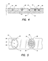
- the separators 26 consist of a material that has greater flexibility than one or both of the first panel 12 and the second panel 16. As can be seen in FIG. 3 , the larger separators 26 are compressed to the point where most separators 26 are touching the interior surfaces of the panels 12, 16, thereby making the chamber height just slightly less than the mean separator 26 diameters. In the chamber embodiment shown in FIG. 4 , the separators 26 consist of a material that has less flexibility than one or both of the first panel 12 and the second panel 16. In FIG. 4 , the first panel 12 is formed from a material more flexible than the spherical separators 26 and the second panel 16, and will overlay the separators 26 in a tent-like fashion.
- the separators 26 and panels 12, 16 can be made from a variety of materials, provided the panels 12, 16 are sufficiently transparent.
- Transparent plastic films consisting of Polyethylene terephthalate is an example of acceptable panels 12, 16, and spherical beads made of polystyrene, polycarbonate, silicone, and the like, are acceptable separators 26.
- an acceptable separator is spheres made of polystyrene that are commercially available, for example, from Thermo Scientific of Fremont, California, U.S.A., catalogue no. 4204A, in four micron (4 ⁇ m) diameter.
- the panel 12 that is to be vertically disposed above the other includes a plurality of ports 30 disposed at regular intervals (e.g., that act as air vents), and the panels 12, 16 are bonded together at points.
- the bonding material 32 forms an outer chamber wall operable to laterally contain the sample 34 within the analysis chamber 10.
- This example of an acceptable analysis chamber is described in greater detail in U.S. Patent Application Publication Nos. 2007/0243117 , 2007/0087442 , and U.S. Provisional Patent Application Numbers 61/041,783, filed April 2, 2008 ; and 61/110,341, filed October 31, 2008 .
- FIGS. 6 and 7 Another example of an acceptable chamber 10 is disposed in a disposable container 36 as shown in FIGS. 6 and 7 .
- the chamber 10 is formed between a first panel 12 and a second panel 16. Both the first panel 12 and the second panel 16 are transparent to allow light to pass through the chamber 10. At least a portion of the first panel 12 and the second panel 16 are parallel with one another, and within that portion the interior surfaces 14, 18 are separated from one another by a height 20.
- This chamber 10 embodiment is described in greater detail in U.S. Patent No. 6,723,290 .
- the analysis chambers shown in FIGS. 2-7 represent chambers that are acceptable for use in the present method. The present method is not, however, limited to these particular embodiments.
- a chamber height of about two to six microns (2-6 ⁇ m) is acceptable for most animal species based on typical cell sizes.
- a chamber height 20 of about three to five microns (3-5 ⁇ m) is particularly well suited for analyzing human blood.
- the present invention is not, however, limited to any particular chamber height provided the methodology described herein can be accomplished with such chamber height.
- the analysis of the sample quiescently disposed within the chamber 10 is performed using an analysis device that is operable to illuminate and image at least a portion of the sample and perform an analysis on the image.
- the image is produced in a manner that permits fluorescent emissions from, and the optical density of, the portion of the sample to be determined on a per unit basis.
- per unit basis or "image unit” means a defined incremental unit of which the image of the sample can be dissected.
- a pixel, which is generally defined as the smallest element of an image that can be individually processed within a particular imaging system, is an example of an image unit, and an image unit may include a small number of pixels in a collective unit..
- the magnification of an imaging device can also be described in linear terms (e.g., microns per pixel at the focal plane), where the linear dimension is along a particular axis of an orthogonal grid applied to the image.
- the actual area of the sample captured by pixels of the sensor at the focal plane is therefore a function of the magnification factor applied by the imaging device.
- the volume associated with that pixel is therefore the area of the image per pixel times the chamber height. For example if the magnification was 0.5 microns per pixel, an image occupying 200 pixels would have an area of 50 square microns, and a volume of 50 square microns times the chamber height.
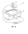
- an example of an analysis device 54 that can be adapted for use with the present method includes a sample illuminator 56, an image dissector 58, and a programmable analyzer 50.
- the sample illuminator 56 includes a light source that selectively produces light along certain desired wavelengths. For example, LEDs that emit the desired wavelengths (e.g., 420 nm, 440 nm, 470 nm, etc.) can be used. Alternatively, a light source that produces a broad wavelength range (e.g., approximately 400 - 670 nm) can be used, although in some instances such a light source may require filtering.
- the analysis device 54 may include optics for manipulating the light.
- the sample illuminator 56 includes a transmittance light source and an epi-illumination light source, each operable to illuminate some, or all, of the sample residing within the chamber 10.
- An example of an acceptable image dissector 58 is a charge couple device (CCD) type image sensor that converts an image of the light passing through the sample into an electronic data format (i.e., a signal).
- CCD charge couple device
- CMOS Complimentary metal oxide semiconductor
- the programmable analyzer 50 includes a central processing unit (CPU) and is connected to the sample illuminator 56 and image dissector 58.
- the CPU is adapted (e.g., programmed) to selectively perform the functions necessary to perform the present method.
- programmable analyzer 50 may be implemented using hardware, software, firmware, or a combination thereof.
- a person skilled in the art would be able to program the processing unit to perform the functionality described herein without undue experimentation.
- the analysis device is adapted to process the image signals created from the illumination of at least a portion of the sample to identify and enumerate constituents within the sample.
- the image signals include fluorescent emissions and optical density values on a per pixel basis. The intensity and color of the emissions and the optical density per pixel collectively establish the image of the illuminated sample portion.
- the analysis device is adapted to identify a profile of selected constituents, using one or more of the fluorescent intensity, color content, and optical density of the fluorescent emissions, and in some instances physical characteristics (e.g., area, edge geometry, etc.) of the constituents.
- the analysis device uses the image profiles to distinguish amongst the selected constituents until the remaining constituents represent the target constituents (e.g., platelets), at which point the target constituents can be enumerated.
- a sample of substantially undiluted whole blood is introucked into a chamber 10, and thereinafter resides quiescently within the chamber 10.
- An anticoagulating agent and a colorant are admixed with the sample either prior to its introduction into the chamber or upon introduction into the chamber.
- the colorant is absorbed by the constituents (e.g., WBCs, platelets, reticulocytes) within the sample.
- an isovolumetric sphering agent is added to the sample to cause some or all of the RBCs within the sample to assume a spherical-like shape.
- An example of an acceptable isovolumetric sphering agent is a zwitterionic detergent.
- a specific example of a sphering agent is Zwittergent(R) 3-16 detergent, which is a zwitterionic detergent produced by Calibriochem, an entity of EMD Chemicals, Inc. of New Jersey, U.S.A.
- the amount of sphering agent added to the sample is an amount adequate to sphere at least a number of RBCs required to perform the present hematocrit analysis. The specific amount will depend on the particular agent and test circumstances, which can be determined by a person of skill in the art without undue experimentation.
- the natural bioconcave shape of RBCs, and the relative size of RBCs relative to platelets, can cause platelets within a sample to be "hidden” amongst the RBCs in the sample; e.g., within the concavities of an RBC.
- Sphering the RBCs decreases the likelihood of platelets being hidden within a sample amongst the RBCs and increases the likelihood that such platelets can be viewed as individuals within the plasma, and thereby increases the accuracy of quantitative platelet analyses performed on the sample.
- At least a portion of the sample quiescently residing within the chamber is illuminated by the analysis device 54, which transmits light through the sample.
- the analysis device 54 transmits light through the sample.
- the sample is illuminated with wavelengths known to excite a fluorescent emission from the constituents relating to the colorant absorbed by the constituents.
- Constituents stained with acridine orange produce a fluorescent emission when illuminated with violet light at a wavelength of about 470 nm.
- 9-13 illustrate the fluorescent emissions of constituents (e.g., platelets, giant platelets, WBCs, reticulocytes, platelet clumps) found within the sample.
- the specific emissions depend upon the colorant used and the intracellular composition of the illuminated cell (e.g., interaction of the colorant with contents of the cell or platelet creates the emissions).
- Some constituents have fluorescent emissions that act as a fluorometric signature (also referred to as a "profile"), which signature represents a particular ratio of fluorescent emissions along wavelengths that produce combinations of light (e.g., a characteristic "red/green” ratio) that are relatively unique to that constituent and can therefore be used to identify that constituent.
- Other constituents have fluorescent emission signatures that cannot easily be distinguished from one another.
- the sample is illuminated with wavelengths of light that are absorbed by hemoglobin in appreciably greater amounts than would be absorbed by the platelets or plasma.
- the amount of absorption can be measured in terms of optical density, and the optical density in turn can be used to distinguish constituents containing hemoglobin from those that do not.
- the fluorescent emission portion of the image is a function of factors such as the type of colorant used and the concentration of the colorant within the sample, it is useful, but not required, to calibrate the sample for intensity.
- the fluorescent emission from WBCs is on average higher than the fluorescent emission from platelets.
- FIGS. 9 and 10 which are images of the same sample.
- the amplification of the fluorescent emission in FIG. 10 is greater than that used in FIG. 9.
- FIG. 9 clearly shows the fluorescent emissions from the WBCs 40, and faintly shows the emissions from the platelets 42.
- FIG. 10 clearly shows both WBCs 40 and platelets 42, and also shows how each can be distinguished by their fluorescent emission intensity.
- the calibration identifies the intensity level associated with the WBCs and the intensity level associated with the platelets to the analysis device, and calibrates the analysis device accordingly.
- the fluorescent emissions and transmitted light produced by illuminating the sample are converted into image signals on a per pixel basis that collectively establish the image of the illuminated sample portion.
- the analysis device is adapted to identify a profile of certain selected constituents, using one or more of the fluorescent intensity, color content, and optical density of the fluorescent emissions.
- the process of identifying the constituents profiles via aforesaid characteristics can be performed using algorithms that compare the various characteristics to identify the constituent Once the constituent is identified, it can be further analyzed. For example, a representative number of platelets can be identified within the sample via their fluorescent emission profile and collectively analyzed to determine an average of the fluorescent emission intensity for the platelets.
- constituent fluorescent profiles are also used to determine the internal areas and edge regions of the constituents using the fluorescent signal profiles.
- the areas of individual constituents can be averaged to determine an average area value.
- the edge profiles can be analyzed for smoothness and/or for geometry; e.g., determine if the edge of a constituent is circular, non-circular, irregular, etc. These characteristics are subsequently used to distinguish constituents within the sample, until the remaining constituents represent the target constituents (e.g., platelets), at which point the target constituents can be enumerated.
- a substantially undiluted sample of blood is admixed with EDTA, acridine orange, and a zwitterionic detergent and is introduced within a chamber having two transparent panels for the purpose of determining a platelet count within the sample.
- Constituents including RBCs, reticulocytes, WBCs, platelets, giant platelets, and platelet clumps, reside quiescently within the sample.
- the sample is illuminated at 470 nm, at least one of 413 nm and 540 nm.
- the 470 nm illumination produces a fluorescent emission from the acridine orange.
- Other colorants may emit light upon illumination at other wavelengths.
- the 413 nm and/or 540 nm illumination is used to indicate the presence of hemoglobin by its optical density, as will be discussed below. Digital images of the illuminated sample are taken.
- the image of the sample is analyzed to identify various constituents disposed within the sample.
- WBCs for example, are individually identified by one or more of their fluorescent signature (e.g., fluorescent emission pattern consisting of a significant red cytoplasmic fluorescence and a green nuclear fluorescence), the relative intensity of their fluorescent emissions, the area they occupy, and their shape (see FIGS. 9-11 ).
- the WBCs are thereby distinguished from the remainder of the sample; e.g., by filtering the image so that WBCs are no longer considered within the image.
- giant platelets 44 can be identified within the sample by one or more of their fluorescent profile, area, and shape. RBCs 45 can be seen faintly in the background.
- the colorimetric ratio of a giant platelet 44 is similar to that of a normal platelet 42, but the emissions are greater in intensity due to the larger mass of the particle.
- Giant platelets 44 are also significantly larger than normal platelets 42 and circular in size. Normal platelets 42 are typically irregularly shaped. Most normal sized platelets are 1.5 to 3 ⁇ m in diameter.
- Giant platelets 44 in contrast, are larger than 7 ⁇ m and usually in the range of 10 ⁇ m to 20 ⁇ m in diameter.
- giant platelets 44 within the sample are identified by comparing one or more of their fluorescent emissions (colorimetric and intensity), area, and perimeter shape against normal platelet fluorescent emission values, area, and perimeter shapes, including the average platelet intensity and area values.
- CML chronic myelogenous leukemia
- polycythemia vera polycythemia vera
- essential (primary) thrombocythemia and agnogenic myeloid metaplasia.
- Giant platelets 44 within the sample are identified by comparing one or more of their fluorescent emissions (colorimetric and intensity), area, and perimeter shape against normal platelet fluorescent emission values, area, and perimeter shapes, including the average platelet intensity and area values.
- the analysis device can be programmed with one or more comparative criteria relative to normal platelets (e.g., a multiple of the average platelet area or intensity based on standard deviations of the average normal platelet area or intensity, or by a predetermined area or intensity value, etc.).
- a multiple of the average platelet area or intensity based on standard deviations of the average normal platelet area or intensity, or by a predetermined area or intensity value, etc. With respect to with average platelet area, the distribution of platelet areas within any given sample is typically described as a log-normal function and can be determined statistically using know techniques.
- the identified giant platelets are then distinguished and, depending upon the specific information desired, are included in the platelet count and/or are considered as an independent constituent population.
- Reticulocytes 46 emit a fluorescent profile that is similar to that of normal platelets because of the nuclear material they contain.
- the photograph in FIG. 13 shows the fluorescent emissions of reticulocytes 46 and platelets 42.
- the circular blacked out portions are portions of the image where WBCs were disposed, but which image was masked out.
- the reticulocytes 46 can be distinguished from platelets 42 to some extent by their fluorescent emission, which appears slightly brighter and has slightly more red.
- the reticulocytes 46 can also be distinguished by illuminating the sample with light at wavelengths of 413 nm and/or 540 nm, which wavelengths are absorbed by the hemoglobin to a substantially greater amount than any other material present within the sample and is, therefore, indicative of the presence of hemoglobin.
- the light absorption of the hemoglobin can be quantified in terms of optical density.
- FIG. 14 illustrates the OD of the reticulocytes. The reticulocytes are distinguished from the remainder of the sample using one or both
- the constituents with a fluorescent emission remaining after the WBCs, giant platelets, and reticulocytes have been distinguished are predominantly, if not entirely, platelets.
- Individual platelets within the sample can be identified and enumerated.
- a portion of the platelets within the sample may be aggregated into one or more clumps, which clumps can be very large in size; e.g., one to four times the size of a WBC.
- the photograph in FIG. 11 shows both platelet clumps 48 and WBCs 40.
- the platelet clumps 48 are identifiable and distinguishable from other constituents (e.g., WBCs 40, giant platelets 44) by their fluorescent emission profile which has a red/green ratio that is distinguishable from that of a WBC 40, by their granularity, and in some instances by their area and/or shape.
- platelet clumps 48 are distinguishable from giant platelets because platelet clumps are irregular shaped and giant platelets are substantially circularly shaped.
- the relative circularity of both platelet clumps and giant platelets can be determined by image analysis software programmed into analysis device.
- Detecting the presence of platelet clumps within the sample is important because of the possibility that the clumps will be counted as WBCs (and thereby falsely elevate the WBC count) and/or lead to a false diagnosis of thrombocytopenia. These potential problems are particularly relevant with existing automated analyzers.
- the presence of platelet clumps can be an indicator of inadequate mixing of the sample with EDTA.
- the integrated fluorescent emission intensity of the clump and the area of the clump can be determined.
- the number of platelets within the clump can then be determined by dividing the integrated fluorescent emission intensity by the average platelet emission intensity.
- the quotient value is an acceptable approximation of the actual number of platelets within the clump.
- An approximation of the number of platelets within a clump can also be determined by dividing the area of the clump by the average platelet area.
Landscapes
- Health & Medical Sciences (AREA)
- Life Sciences & Earth Sciences (AREA)
- Chemical & Material Sciences (AREA)
- Engineering & Computer Science (AREA)
- Physics & Mathematics (AREA)
- Biomedical Technology (AREA)
- Pathology (AREA)
- Analytical Chemistry (AREA)
- General Physics & Mathematics (AREA)
- Immunology (AREA)
- Biochemistry (AREA)
- General Health & Medical Sciences (AREA)
- Hematology (AREA)
- Dispersion Chemistry (AREA)
- Biophysics (AREA)
- Medicinal Chemistry (AREA)
- Food Science & Technology (AREA)
- Ecology (AREA)
- Urology & Nephrology (AREA)
- Molecular Biology (AREA)
- Signal Processing (AREA)
- Investigating, Analyzing Materials By Fluorescence Or Luminescence (AREA)
- Investigating Or Analysing Biological Materials (AREA)
Claims (14)
- Ein Verfahren zum Aufzählen von Blutplättchen in einer Blutprobe mit einer Vielzahl von Blutplättchen (42), umfassend die Schritte:Deponieren der Probe in einer Analysekammer (10) geeignet, die Probe zur Analyse bewegungslos zu halten, wobei die Kammer (10) von einer ersten Platte (12) und einer zweiten Platte (16) begrenzt wird, wobei beide der Platten (12, 16) transparent sind;Vermischen eines Farbstoffes mit der Probe, wobei der Farbstoff dazu dient, die Blutplättchen (42) zu veranlassen, beim Aussetzen an ein oder mehrere vorbestimmte erste Wellenlängen von Licht zu fluoreszieren;Beleuchten von zumindest einem Teil der Probe, die die Blutplättchen (42) aufweist, mit den ersten Wellenlängen;Abbilden des zumindest einen Teils der Probe, umfassend Produzieren von Bildsignalen, die Fluoreszenzemissionen von den Blutplättchen (42) anzeigen, wobei die Fluoreszenzemissionen eine Intensität aufweisen;Identifizieren der Blutplättchen (42) mittels ihrer Fluoreszenzemissionen unter Verwendung der Bildsignale;Bestimmen eines durchschnittlichen Fluoreszenzemissionsintensitätswertes für die in der Probe identifizierten individuellen Blutplättchen (42);Identifizieren von Blutplättchenklumpen (48) in der Probe unter Verwendung von ein oder mehrerer ihrer Fluoreszenzemissionen, Fläche, Form und Körnigkeit;Aufzählen von Blutplättchen in jedem Blutplättchenklumpen (48) unter Verwendung des für die individuellen Blutplättchen (42) in der Probe identifizierten durchschnittlichen Fluoreszenzemissionsintensitätswertes.
- Das Verfahren nach Anspruch 1, wobei die Blutprobe im Wesentlichen unverdünnt ist.
- Das Verfahren nach Anspruch 1 oder 2, wobei die Blutrobe Vollblut aufweist.
- Das Verfahren nach Anspruch 1, 2 oder 3, weiterhin umfassend den Schritt des Aufzählens von Blutplättchen, die einzeln in der Probe anwesend sind, und Bestimmens einer Gesamtzahl von Blutplättchen in der Probe als Summer der Blutplättchen in jedem Blutplättchenklumpen (48) und der in der Probe einzeln anwesenden Blutplättchen (42).
- Das Verfahren nach einem der vorhergehenden Ansprüche, weiterhin umfassend den Schritt des Identifizierens von riesigen Blutplättchen (44) in der Probe.
- Das Verfahren nach Anspruch 5, wobei die riesigen Blutplättchen (44) in der Probe mittels ein oder mehrerer von Fluoreszenzemissionen, Fläche und Form identifiziert werden.
- Das Verfahren nach Anspruch 5 oder 6, weiterhin umfassend den Schritt des Bestimmens der Fläche von zumindest einem der riesigen Blutplättchen (44) in der Probe.
- Das Verfahren nach Anspruch 7, weiterhin umfassend den Schritt des Bestimmens einer durchschnittlichen Fläche für die in der Probe identifizierten einzelnen Blutplättchen (42), und Vergleichen der Fläche des zumindest einen riesigen Blutplättchens (44) mit der durchschnittlichen Fläche der Blutplättchen.
- Das Verfahren nach Anspruch 7, weiterhin umfassend den Schritt des Bestimmens einer durchschnittlichen Fläche für die in der Probe identifizierten einzelnen Blutplättchen (42) und einer statistischen Abweichung von Flächen der einzelnen Blutplättchen (42), und Vergleichen der Fläche des zumindest einen riesigen Blutplättchens (44) mit dem einem Wert basierend auf der statistischen Abweichung der Flächen der einzelnen Blutplättchen, um das zumindest eine riesige Blutplättchen (44) zu identifizieren.
- Das Verfahren nach Anspruch 7, weiterhin umfassend den Schritt des Vergleichens der Fläche des zumindest einen riesigen Blutplättchens (44) mit einem vorbestimmten Wert, um das zumindest eine riesige Blutplättchen (44) zu identifizieren.
- Das Verfahren nach einem der vorhergehenden Ansprüche, weiterhin umfassend den Schritt des Bestimmens einer durchschnittlichen Fläche für die in der Probe identifizierten einzelnen Blutplättchen (42), wobei der Schritt des Aufzählens von Blutplättchen in jedem Blutplättchenklumpen (48) weiterhin das Verwenden der durchschnittlichen Fläche für die einzelnen Blutplättchen (42) in der Probe umfasst.
- Das Verfahren nach einem der vorhergehenden Ansprüche, weiterhin umfassend die Schritte:Beleuchten von zumindest einem Teil der die Blutplättchen (42) aufweisenden Probe mit in oder mehr zweiten Wellenlängen;Abbilden des zumindest einen Teils der Probe, umfassend Produzieren von Bildsignalen, die eine optische Dichte von ein oder mehreren Bestandteilen in der Probe anzeigen;Bestimmen der optischen Dichte von ein oder mehreren Bestandteilen in der Probe unter Verwendung der Bildsignale; undUnterscheiden der Blutplättchen (42) und den ein oder mehreren Bestandteilen voneinander unter Verwendung der optischen Dichte.
- Das Verfahren nach einem der vorhergehenden Ansprüche, weiterhin umfassend den Schritt des Unterscheidens der Blutplättchen (42) von Bestandteilen der Hämoglobin enthaltenden Probe mittels Beleuchten des zumindest einen Teils der Probe mit Licht bei ein oder mehreren zweiten Wellenlängen, die von dem Hämoglobin absorbiert werden.
- Eine Vorrichtung zum Aufzählen von Blutplättchen in einer Blutprobe, umfassend:eine Analysekammer (10) geeignet, die Probe zur Analyse bewegungslos zu halten, wobei die Kammer (10) von einer ersten Platte (12) und einer zweiten Platte (16) begrenzt wird, wobei beide der Platten transparent sind; wobei eine Menge von Farbstoff in der Kammer (10) disponiert ist, wobei ein Farbstoff dazu dient, die Blutplättchen (42) zu veranlassen, beim Aussetzen an ein oder mehrere vorbestimmte erste Wellenlängen von Licht zu fluoreszieren;Beleuchten von zumindest einem Teil der Probe, die die Blutplättchen (42) aufweist, mit den ersten Wellenlängen;eine Abbildungseinheit, die einen Beleuchter (56) und einen Bildsezierer (58) aufweist, wobei die Einheit betreibbar ist, um zumindest einen Teil der Probe abzubilden und Bildsignale zu erzeugen, die Fluoreszenzemissionen von den Blutplättchen (42) anzeigen, wobei die Fluoreszenzemissionen eine Intensität aufweisen; undein programmierbares Analysegerät (50), das geeignet ist, Blutplättchen (42) mittels ihrer Fluoreszenzemissionen unter Verwendung der Bildsignale zu identifizieren, und einen durchschnittlichen Fluoreszenzemissionsintensitätswert für die in der Probe identifizierten individuellen Blutplättchen (42) zu bestimmen, und Blutplättchenklumpen (48) in der Probe unter Verwendung von ein oder mehrerer ihrer Fluoreszenzemissionen, Fläche, Form und Körnigkeit zu identifizieren, und Blutplättchen in jedem Blutplättchenklumpen (48) unter Verwendung des für die individuellen Blutplättchen (42) in der Probe identifizierten durchschnittlichen Fluoreszenzemissionsintensitätswertes aufzuzählen.
Priority Applications (1)
| Application Number | Priority Date | Filing Date | Title |
|---|---|---|---|
| EP16150962.5A EP3026433A1 (de) | 2008-03-21 | 2009-03-20 | Verfahren und vorrichtung zum nachweisen und zählen einzelner blutplättchen und von blutplättchen in aggregathaufen |
Applications Claiming Priority (2)
| Application Number | Priority Date | Filing Date | Title |
|---|---|---|---|
| US3855408P | 2008-03-21 | 2008-03-21 | |
| PCT/US2009/037844 WO2009117682A1 (en) | 2008-03-21 | 2009-03-20 | Method and apparatus for detecting and counting platelets individually and in aggregate clumps |
Related Child Applications (1)
| Application Number | Title | Priority Date | Filing Date |
|---|---|---|---|
| EP16150962.5A Division EP3026433A1 (de) | 2008-03-21 | 2009-03-20 | Verfahren und vorrichtung zum nachweisen und zählen einzelner blutplättchen und von blutplättchen in aggregathaufen |
Publications (2)
| Publication Number | Publication Date |
|---|---|
| EP2271937A1 EP2271937A1 (de) | 2011-01-12 |
| EP2271937B1 true EP2271937B1 (de) | 2016-01-13 |
Family
ID=40775819
Family Applications (2)
| Application Number | Title | Priority Date | Filing Date |
|---|---|---|---|
| EP09722141.0A Active EP2271937B1 (de) | 2008-03-21 | 2009-03-20 | Verfahren und vorrichtung zum nachweisen und zählen einzelner blutplättchen und von blutplättchen in aggregathaufen |
| EP16150962.5A Withdrawn EP3026433A1 (de) | 2008-03-21 | 2009-03-20 | Verfahren und vorrichtung zum nachweisen und zählen einzelner blutplättchen und von blutplättchen in aggregathaufen |
Family Applications After (1)
| Application Number | Title | Priority Date | Filing Date |
|---|---|---|---|
| EP16150962.5A Withdrawn EP3026433A1 (de) | 2008-03-21 | 2009-03-20 | Verfahren und vorrichtung zum nachweisen und zählen einzelner blutplättchen und von blutplättchen in aggregathaufen |
Country Status (7)
| Country | Link |
|---|---|
| US (4) | US7929121B2 (de) |
| EP (2) | EP2271937B1 (de) |
| JP (1) | JP5184697B2 (de) |
| CN (2) | CN102027369B (de) |
| CA (1) | CA2719012C (de) |
| ES (1) | ES2560098T3 (de) |
| WO (1) | WO2009117682A1 (de) |
Families Citing this family (95)
| Publication number | Priority date | Publication date | Assignee | Title |
|---|---|---|---|---|
| EP1733226B1 (de) | 2004-04-07 | 2015-07-29 | Levine, Robert Aaron | Einwegkammer zur analyse biologischer flüssigkeiten |
| US7731901B2 (en) | 2005-10-19 | 2010-06-08 | Abbott Laboratories | Apparatus and method for performing counts within a biologic fluid sample |
| US7951599B2 (en) * | 2008-03-21 | 2011-05-31 | Abbott Point Of Care, Inc. | Method and apparatus for determining the hematocrit of a blood sample utilizing the intrinsic pigmentation of hemoglobin contained within the red blood cells |
| EP2271937B1 (de) * | 2008-03-21 | 2016-01-13 | Abbott Point Of Care, Inc. | Verfahren und vorrichtung zum nachweisen und zählen einzelner blutplättchen und von blutplättchen in aggregathaufen |
| ES2588223T3 (es) * | 2008-03-21 | 2016-10-31 | Abbott Point Of Care, Inc. | Procedimiento para el análisis de células individuales o partículas en la sangre usando fluorescencia y absorción de un colorante |
| US9602777B2 (en) * | 2008-04-25 | 2017-03-21 | Roche Diagnostics Hematology, Inc. | Systems and methods for analyzing body fluids |
| JP2012515931A (ja) * | 2008-04-25 | 2012-07-12 | ウィンケルマン、ジェイムズ | 全血球数及び白血球百分率を決定するシステム及び方法 |
| FR2945126B1 (fr) * | 2009-04-29 | 2019-11-01 | Commissariat A L'energie Atomique | Procede et appareil de comptage des thrombocytes |
| JP5530691B2 (ja) * | 2009-09-25 | 2014-06-25 | シスメックス株式会社 | 血球計数装置、診断支援装置、診断支援方法及びコンピュータプログラム |
| JP5709894B2 (ja) | 2009-12-18 | 2015-04-30 | アボット ポイント オブ ケア インコーポレイテッド | 生物学的液体分析カートリッジ |
| EP2519820B1 (de) * | 2009-12-31 | 2013-11-06 | Abbott Point Of Care, Inc. | Verfahren und vorrichtung zur bestimmung des mittleren zellvolumen roter blutzellen |
| US8472693B2 (en) | 2010-03-18 | 2013-06-25 | Abbott Point Of Care, Inc. | Method for determining at least one hemoglobin related parameter of a whole blood sample |
| US9522396B2 (en) | 2010-12-29 | 2016-12-20 | S.D. Sight Diagnostics Ltd. | Apparatus and method for automatic detection of pathogens |
| CN103282123B (zh) | 2010-12-30 | 2015-05-06 | 艾博特健康公司 | 带有样品处理区和分析室区的生物液体分析卡式盒 |
| JP5997256B2 (ja) | 2011-04-15 | 2016-09-28 | コンスティテューション・メディカル・インコーポレイテッドConstitution Medical, Inc. | 細胞の体積および成分の計測 |
| CN103827658B (zh) * | 2011-07-22 | 2016-04-13 | 罗氏血液诊断股份有限公司 | 识别和测量网状细胞 |
| CN105817276B (zh) | 2011-08-24 | 2018-02-06 | 艾博特健康公司 | 生物流体样品分析盒 |
| WO2013041951A1 (en) * | 2011-09-22 | 2013-03-28 | Foce Technology International Bv | Optical platelet counter method |
| WO2013098821A1 (en) | 2011-12-29 | 2013-07-04 | Parasight Ltd. | Methods and systems for detecting a pathogen in a biological sample |
| CN107064079A (zh) * | 2011-12-30 | 2017-08-18 | 艾博特健康公司 | 用于识别全血样品中的血小板的方法和设备 |
| AU2013295679A1 (en) * | 2012-07-25 | 2015-01-29 | Theranos, Inc. | Image analysis and measurement of biological samples |
| US9513224B2 (en) | 2013-02-18 | 2016-12-06 | Theranos, Inc. | Image analysis and measurement of biological samples |
| EP2939001B1 (de) * | 2012-12-31 | 2019-12-04 | Beckman Coulter, Inc. | Systeme und verfahren zur blutplättchenzählung mit klumpenanpassung |
| CN108663344A (zh) * | 2013-02-19 | 2018-10-16 | 艾博特健康公司 | 分析生物流体样品的方法 |
| WO2014188405A1 (en) | 2013-05-23 | 2014-11-27 | Parasight Ltd. | Method and system for imaging a cell sample |
| IL227276A0 (en) | 2013-07-01 | 2014-03-06 | Parasight Ltd | A method and system for preparing a monolayer of cells, particularly suitable for diagnosis |
| WO2015013605A1 (en) * | 2013-07-26 | 2015-01-29 | Abbott Point Of Care, Inc. | Method and apparatus for producing an image of undiluted whole blood sample having wright stain coloration |
| EP3039477B1 (de) | 2013-08-26 | 2021-10-20 | S.D. Sight Diagnostics Ltd. | Digitale mikroskopiesysteme, verfahren und computerprogrammprodukte |
| US10482595B2 (en) | 2014-08-27 | 2019-11-19 | S.D. Sight Diagnostics Ltd. | System and method for calculating focus variation for a digital microscope |
| US12005441B1 (en) | 2014-11-26 | 2024-06-11 | Medica Corporation | Automated microscopic cell analysis |
| US11478789B2 (en) | 2014-11-26 | 2022-10-25 | Medica Corporation | Automated microscopic cell analysis |
| US10625259B1 (en) | 2014-11-26 | 2020-04-21 | Medica Corporation | Automated microscopic cell analysis |
| US20170328924A1 (en) | 2014-11-26 | 2017-11-16 | Ronald Jones | Automated microscopic cell analysis |
| RU2018107453A (ru) | 2015-08-10 | 2019-09-12 | Эссенликс Корп. | Устройства для анализов и способы в биологии/химии, предусматривающие упрощенные стадии, образцы небольшого объема, повышенную скорость и простоту применения |
| MY194887A (en) | 2015-09-14 | 2022-12-21 | Essenlix Corp | Device and System for Analyzing a Sample, Particularly Blood, as Well as Methods of Using The Same |
| KR101982330B1 (ko) | 2015-09-14 | 2019-05-24 | 에센릭스 코프. | 증기 응축액 특히 입김 응축액을 수집 및 분석하기 위한 장치 및 시스템 그리고 이 장치 및 시스템을 사용하는 방법 |
| EP3859425B1 (de) | 2015-09-17 | 2024-04-17 | S.D. Sight Diagnostics Ltd. | Verfahren und vorrichtung zur detektion einer entität in einer körperprobe |
| JP6518016B2 (ja) * | 2015-12-24 | 2019-05-22 | コーニンクレッカ フィリップス エヌ ヴェKoninklijke Philips N.V. | 細胞懸濁液の決定のための方法及びシステム |
| CA3018536A1 (en) | 2016-03-30 | 2017-10-05 | S.D. Sight Diagnostics Ltd | Distinguishing between blood sample components |
| EP3455626B1 (de) | 2016-05-11 | 2025-08-06 | S.D. Sight Diagnostics Ltd. | Durchführung optischer messungen auf einer probe |
| EP4177593A1 (de) | 2016-05-11 | 2023-05-10 | S.D. Sight Diagnostics Ltd. | Probenträger für optische messungen |
| CA3048002A1 (en) | 2016-12-21 | 2018-06-28 | Essenlix Corporation | Devices and methods for authenticating a sample and use of the same |
| JP7003158B2 (ja) | 2017-02-07 | 2022-01-20 | エッセンリックス コーポレーション | 圧縮開放流アッセイおよび使用 |
| WO2018148764A1 (en) | 2017-02-08 | 2018-08-16 | Essenlix Corporation | Molecular manipulation and assay with controlled temperature |
| CN120685901A (zh) | 2017-02-08 | 2025-09-23 | 上海宜晟生物科技有限公司 | 数字测定 |
| CA3053005A1 (en) | 2017-02-08 | 2018-08-16 | Essenlix Corporation | Sample collection and handling for delayed analysis |
| US11927560B2 (en) | 2017-02-08 | 2024-03-12 | Essenlix Corporation | Bio/chemical material extraction and assay |
| CA3052809A1 (en) | 2017-02-08 | 2018-08-23 | Essenlix Corporation | Qmax assays and applications |
| WO2018148607A1 (en) | 2017-02-09 | 2018-08-16 | Essenlix Corporation | Assay using different spacing heights |
| CN110998325B (zh) | 2017-02-09 | 2024-08-16 | 上海宜晟生物科技有限公司 | 扩增测定 |
| CN110770572B (zh) | 2017-02-09 | 2023-08-18 | Essenlix公司 | 比色测定 |
| CA3053295A1 (en) | 2017-02-15 | 2018-08-23 | Essenlix Corporation | Assay with rapid temperature change |
| JP7107953B2 (ja) | 2017-02-16 | 2022-07-27 | エッセンリックス コーポレーション | テクスチャ表面を用いたアッセイ |
| WO2018231877A1 (en) | 2017-06-12 | 2018-12-20 | Essenlix Corporation | Homogeneous assay |
| US11280706B2 (en) | 2017-08-01 | 2022-03-22 | Essenlix Corporation | Dilution calibration |
| WO2019028133A1 (en) | 2017-08-01 | 2019-02-07 | Essenlix Corporation | DEVICES AND METHODS FOR EXAMINING THE EFFECTS OF MEDICINE ON MICROORGANISMS |
| US11243201B2 (en) | 2017-08-01 | 2022-02-08 | Essenlix Corporation | Sample collection, holding and assaying |
| WO2019035077A1 (en) | 2017-08-17 | 2019-02-21 | Abbott Point Of Care Inc. | DEVICES, SYSTEMS AND METHODS FOR PERFORMING OPTICAL ASSAYS |
| CN110869746B (zh) | 2017-08-17 | 2023-08-11 | 雅培医护站股份有限公司 | 利用通用电路系统执行光学和电化学测定的技术 |
| WO2019035079A1 (en) | 2017-08-17 | 2019-02-21 | Abbott Point Of Care Inc. | DEVICES, SYSTEMS AND METHODS FOR PERFORMING OPTICAL AND ELECTROCHEMICAL ASSAYS |
| US12403465B2 (en) | 2017-10-11 | 2025-09-02 | Essenlix Corporation | Containing a liquid sample |
| US11393561B2 (en) | 2017-10-13 | 2022-07-19 | Essenlix Corporation | Devices and methods for authenticating a medical test and use of the same |
| US11609224B2 (en) | 2017-10-26 | 2023-03-21 | Essenlix Corporation | Devices and methods for white blood cell analyses |
| US10807095B2 (en) | 2017-10-26 | 2020-10-20 | Essenlix Corporation | Making and tracking assay card |
| US11237113B2 (en) | 2017-10-26 | 2022-02-01 | Essenlix Corporation | Rapid pH measurement |
| EP3700419A4 (de) * | 2017-10-26 | 2021-11-10 | Essenlix Corporation | Schnelltest von blutplättchen |
| CN111788471B (zh) | 2017-11-14 | 2023-12-12 | 思迪赛特诊断有限公司 | 用于光学测量的样品载体 |
| US11047845B1 (en) | 2017-11-15 | 2021-06-29 | Medica Corporation | Control material and methods for cell analyzers |
| US11648551B2 (en) | 2017-12-12 | 2023-05-16 | Essenlix Corporation | Sample manipulation and assay with rapid temperature change |
| US11510608B2 (en) | 2017-12-14 | 2022-11-29 | Essenlix Corporation | Devices, systems, and methods for monitoring hair |
| US11156606B2 (en) | 2018-01-11 | 2021-10-26 | Essenlix Corporation | Homogeneous assay (II) |
| CN112424825B (zh) | 2018-05-15 | 2025-01-07 | 文塔纳医疗系统公司 | 染色聚集体中信号的定量 |
| US11885952B2 (en) | 2018-07-30 | 2024-01-30 | Essenlix Corporation | Optics, device, and system for assaying and imaging |
| US11455716B2 (en) * | 2018-11-13 | 2022-09-27 | Rivian Ip Holdings, Llc | Image analysis of applied adhesive with fluorescence enhancement |
| USD898221S1 (en) | 2018-11-15 | 2020-10-06 | Essenlix Corporation | Assay plate |
| USD897555S1 (en) | 2018-11-15 | 2020-09-29 | Essenlix Corporation | Assay card |
| USD898224S1 (en) | 2018-11-15 | 2020-10-06 | Essenlix Corporation | Assay plate with sample landing mark |
| USD898939S1 (en) | 2018-11-20 | 2020-10-13 | Essenlix Corporation | Assay plate with sample landing mark |
| USD910202S1 (en) | 2018-11-21 | 2021-02-09 | Essenlix Corporation | Assay plate with sample landing mark |
| USD893469S1 (en) | 2018-11-21 | 2020-08-18 | Essenlix Corporation | Phone holder |
| USD910203S1 (en) | 2018-11-27 | 2021-02-09 | Essenlix Corporation | Assay plate with sample landing mark |
| USD893470S1 (en) | 2018-11-28 | 2020-08-18 | Essenlix Corporation | Phone holder |
| USD912842S1 (en) | 2018-11-29 | 2021-03-09 | Essenlix Corporation | Assay plate |
| USD898222S1 (en) | 2019-01-18 | 2020-10-06 | Essenlix Corporation | Assay card |
| CN110595959B (zh) * | 2019-04-19 | 2023-03-21 | 齐鲁工业大学 | 一种糖膏颗粒粒度分析系统及分析方法 |
| USD1003453S1 (en) | 2019-05-14 | 2023-10-31 | Essenlix Corporation | Assay card hinge |
| WO2020243741A1 (en) * | 2019-05-28 | 2020-12-03 | Levine Robert A | Apparatus and method for transferring and analyzing suspended particles in a liquid sample |
| MX2022004703A (es) | 2019-10-22 | 2022-05-11 | S D Sight Diagnostics Ltd | Contabilidad de errores en mediciones opticas. |
| EP4071485A4 (de) * | 2019-12-06 | 2023-04-12 | Shenzhen Mindray Bio-Medical Electronics Co., Ltd | Probenanalysesystem und -verfahren, zellenbildanalysator und speichermedium |
| CA3160702A1 (en) * | 2019-12-12 | 2021-06-17 | S.D. Sight Diagnostics Ltd | Artificial generation of a color blood smear image |
| BR112022011233A2 (pt) | 2019-12-12 | 2022-08-30 | S D Sight Diagnostics Ltd | Detecção de plaquetas em uma amostra sanguínea |
| EP4073492A1 (de) | 2019-12-12 | 2022-10-19 | S.D. Sight Diagnostics Ltd. | Analyse eines analyten in einem medium |
| KR102496468B1 (ko) * | 2021-05-27 | 2023-02-06 | (주)유아이엠디 | 이미지 기반의 혈소판 카운팅 방법 및 카운팅 정보 출력방법 |
| CN115791731A (zh) * | 2022-12-01 | 2023-03-14 | 天津智善生物科技有限公司 | 一种成分血检测方法、成分血检测片和成分血检测装置 |
| CN117233149A (zh) * | 2023-08-16 | 2023-12-15 | 武汉血液中心 | 一种基于血小板超微结构成像的特征提取方法及系统 |
Citations (1)
| Publication number | Priority date | Publication date | Assignee | Title |
|---|---|---|---|---|
| WO1999045384A1 (en) * | 1998-03-07 | 1999-09-10 | Wardlaw Partners, Lp | Determination of white blood cell differential and reticulocyte counts |
Family Cites Families (52)
| Publication number | Priority date | Publication date | Assignee | Title |
|---|---|---|---|---|
| US4023716A (en) * | 1976-04-20 | 1977-05-17 | Justin Joel Shapiro | Micro-dispensing liquid pipet |
| DE2732291A1 (de) | 1977-07-16 | 1979-02-01 | Bayer Ag | Verfahren zur herstellung von 1-amino-8-naphthol-3,6-disulfonsaeure (h-saeure) |
| US4197088A (en) * | 1977-09-23 | 1980-04-08 | Akro-Medic Engineering, Inc. | Method for qualitative and quantitative determination of immunological reactions |
| US4615878A (en) * | 1980-03-12 | 1986-10-07 | Lawrence Kass | Metachromatic dye sorption means for differential determination of sub-populations of lymphocytes |
| US4487081A (en) * | 1982-08-27 | 1984-12-11 | Donald H. De Vaughn | Pipetting techniques using replaceable tips |
| JPH0743381B2 (ja) | 1988-10-28 | 1995-05-15 | 株式会社日立製作所 | 光音響免疫分析方法及び装置 |
| US5012818A (en) * | 1989-05-04 | 1991-05-07 | Joishy Suresh K | Two in one bone marrow surgical needle |
| US5068181A (en) * | 1989-12-01 | 1991-11-26 | Akzo N.V. | Method of monitoring reagent delivery in a scanning spectrophotometer |
| GB2254414A (en) | 1991-03-21 | 1992-10-07 | Univ London | Volume measurement of microbial organisms. |
| US5192511A (en) * | 1991-05-31 | 1993-03-09 | Tri-Continent Scientific, Inc. | Pipette tip and piston |
| US5364557A (en) * | 1991-11-27 | 1994-11-15 | Faris Sades M | Aligned cholesteric liquid crystal inks |
| US5284771A (en) * | 1991-12-05 | 1994-02-08 | Miles Inc. | Reagent compositions and their use in sphering cells |
| US5447838A (en) * | 1992-08-05 | 1995-09-05 | Hybritech Incorporated | Protein-dye conjugate for confirmation of correct dilution of calibrators |
| US5594808A (en) * | 1993-06-11 | 1997-01-14 | Ortho Diagnostic Systems Inc. | Method and system for classifying agglutination reactions |
| DE4330562A1 (de) | 1993-09-09 | 1995-03-16 | Behringwerke Ag | Kunststoffpipette |
| US5454268A (en) * | 1993-11-15 | 1995-10-03 | Kim; Young S. | Double-plunger liquid displacement syringe pipet |
| GB9326238D0 (en) * | 1993-12-23 | 1994-02-23 | Sinvent As | Method of assay |
| US5460782A (en) * | 1994-07-18 | 1995-10-24 | Safe-Tec Clinical Products, Inc. | Automatic filling micropipette with dispensing means |
| US5891734A (en) * | 1994-08-01 | 1999-04-06 | Abbott Laboratories | Method for performing automated analysis |
| CA2185292A1 (en) * | 1995-09-15 | 1997-03-16 | James C. Smith | Positive displacement liquid drawing and dispensing apparatus and method |
| AU739824B2 (en) * | 1997-05-05 | 2001-10-18 | Chemometec A/S | A method and a system for determination of particles in a liquid sample |
| US6752965B2 (en) * | 1998-03-06 | 2004-06-22 | Abner Levy | Self resealing elastomeric closure |
| US6723290B1 (en) | 1998-03-07 | 2004-04-20 | Levine Robert A | Container for holding biologic fluid for analysis |
| US5948686A (en) * | 1998-03-07 | 1999-09-07 | Robert A. Leuine | Method for performing blood cell counts |
| US6127184A (en) * | 1998-03-07 | 2000-10-03 | Robert A. Levine | Calibration of a whole blood sample analyzer |
| US6929953B1 (en) * | 1998-03-07 | 2005-08-16 | Robert A. Levine | Apparatus for analyzing biologic fluids |
| US6235536B1 (en) * | 1998-03-07 | 2001-05-22 | Robert A. Levine | Analysis of quiescent anticoagulated whole blood samples |
| WO1999046047A2 (en) * | 1998-03-10 | 1999-09-16 | Large Scale Proteomics Corporation | Detection and characterization of microorganisms |
| GB9903555D0 (en) * | 1999-02-16 | 1999-04-07 | The Technology Partnership Plc | Chemical and biological assay method and apparatus |
| US6589759B1 (en) * | 1999-03-30 | 2003-07-08 | Trustees Of Boston University | Compositions and methods for producing platelets and/or proplatelets from megakaryocytes |
| DE10011235C2 (de) | 2000-03-08 | 2002-08-08 | Max Planck Gesellschaft | Ausstechvorrichtung zur Probenaufnahme und Verfahren zur Probenaufnahme |
| DE10033268C2 (de) * | 2000-07-10 | 2002-08-08 | Innovatis Gmbh | Verfahren zur Untersuchung von Zellen in einer Kulturflüssigkeit |
| AU2001275985A1 (en) | 2000-09-13 | 2002-03-26 | Biometric Imaging, Inc. | Aggregation-based assays |
| EP1239284A1 (de) | 2001-03-08 | 2002-09-11 | The Technology Partnership Public Limited Company | Nicht-trennender Testverfahren und System mit opaken Partikeln |
| US6630990B2 (en) * | 2001-06-05 | 2003-10-07 | Abbott Laboratories | Optical method and apparatus for red blood cell differentiation on a cell-by-cell basis, and simultaneous analysis of white blood cell differentiation |
| EP1365240B1 (de) * | 2002-05-22 | 2006-09-13 | Sysmex Corporation | Immunologische Verfahren, Vorrichtungen und Reagenzien |
| DE10240742A1 (de) | 2002-08-31 | 2004-03-18 | Weber, Jörg | Vorrichtung und Verfahren zur Aufnahme und Abgabe von Untersuchungsmedium |
| US20040165090A1 (en) * | 2003-02-13 | 2004-08-26 | Alex Ning | Auto-focus (AF) lens and process |
| EP1649038B1 (de) * | 2003-06-26 | 2014-07-02 | Litron Laboratories Ltd. | Verfahren zum auszählen von mikronuklei enthaltenden erythrozytenpopulationen mit unterscheidung von blutplättchen und/oder blutplättchenassoziierten aggregaten |
| JP2005024472A (ja) * | 2003-07-04 | 2005-01-27 | Sysmex Corp | 幼若血小板測定装置 |
| US7598093B2 (en) * | 2003-07-23 | 2009-10-06 | Ctl Analyzers, Llc | Nanoparticle and microparticle based detection of cellular products |
| US20060258018A1 (en) * | 2003-09-23 | 2006-11-16 | Curl Claire L | Method and apparatus for determining the area or confluency of a sample |
| EP1733226B1 (de) | 2004-04-07 | 2015-07-29 | Levine, Robert Aaron | Einwegkammer zur analyse biologischer flüssigkeiten |
| CN101650998B (zh) * | 2005-01-20 | 2013-02-06 | 卢米尼克斯股份有限公司 | 微球、包含其的微球群、以及分析试验样品的方法 |
| US7796797B2 (en) * | 2005-09-28 | 2010-09-14 | Sysmex Corporation | Apparatus for obtaining an image of a blood cell and method for obtaining an image of a blood cell |
| US7731901B2 (en) * | 2005-10-19 | 2010-06-08 | Abbott Laboratories | Apparatus and method for performing counts within a biologic fluid sample |
| US7344890B2 (en) * | 2005-11-09 | 2008-03-18 | Beckman Coulter, Inc. | Method for discriminating platelets from red blood cells |
| SE531041C2 (sv) * | 2006-07-17 | 2008-11-25 | Hemocue Ab | Räkning av trombocyter |
| US7998747B2 (en) * | 2006-09-15 | 2011-08-16 | Artel, Inc. | Quantitative dual-dye photometric method for determining dilution impact |
| CN102027368B (zh) * | 2008-03-21 | 2014-02-26 | 艾博特健康公司 | 利用红细胞内含有的血红蛋白的本征色素沉着来确定血样的红细胞指数的方法及设备 |
| EP2271937B1 (de) * | 2008-03-21 | 2016-01-13 | Abbott Point Of Care, Inc. | Verfahren und vorrichtung zum nachweisen und zählen einzelner blutplättchen und von blutplättchen in aggregathaufen |
| US10549494B2 (en) | 2016-04-20 | 2020-02-04 | Himax Technologies Limited | Imprinting apparatus and imprinting method |
-
2009
- 2009-03-20 EP EP09722141.0A patent/EP2271937B1/de active Active
- 2009-03-20 EP EP16150962.5A patent/EP3026433A1/de not_active Withdrawn
- 2009-03-20 JP JP2011500991A patent/JP5184697B2/ja active Active
- 2009-03-20 WO PCT/US2009/037844 patent/WO2009117682A1/en not_active Ceased
- 2009-03-20 US US12/408,500 patent/US7929121B2/en active Active
- 2009-03-20 ES ES09722141.0T patent/ES2560098T3/es active Active
- 2009-03-20 CA CA2719012A patent/CA2719012C/en active Active
- 2009-03-20 CN CN200980116792.6A patent/CN102027369B/zh active Active
- 2009-03-20 CN CN201811100268.7A patent/CN109239321A/zh active Pending
-
2011
- 2011-04-18 US US13/088,853 patent/US8077296B2/en active Active
- 2011-12-12 US US13/323,438 patent/US8310659B2/en active Active
-
2012
- 2012-11-09 US US13/673,616 patent/US8638427B2/en active Active
Patent Citations (1)
| Publication number | Priority date | Publication date | Assignee | Title |
|---|---|---|---|---|
| WO1999045384A1 (en) * | 1998-03-07 | 1999-09-10 | Wardlaw Partners, Lp | Determination of white blood cell differential and reticulocyte counts |
Also Published As
| Publication number | Publication date |
|---|---|
| JP5184697B2 (ja) | 2013-04-17 |
| US20130176551A1 (en) | 2013-07-11 |
| JP2011515683A (ja) | 2011-05-19 |
| US20110193957A1 (en) | 2011-08-11 |
| US7929121B2 (en) | 2011-04-19 |
| EP2271937A1 (de) | 2011-01-12 |
| CN102027369B (zh) | 2018-10-26 |
| CN102027369A (zh) | 2011-04-20 |
| CA2719012A1 (en) | 2009-09-24 |
| US20090238439A1 (en) | 2009-09-24 |
| EP3026433A1 (de) | 2016-06-01 |
| CA2719012C (en) | 2013-07-09 |
| ES2560098T3 (es) | 2016-02-17 |
| US8310659B2 (en) | 2012-11-13 |
| WO2009117682A1 (en) | 2009-09-24 |
| US20120147357A1 (en) | 2012-06-14 |
| US8077296B2 (en) | 2011-12-13 |
| CN109239321A (zh) | 2019-01-18 |
| US8638427B2 (en) | 2014-01-28 |
Similar Documents
| Publication | Publication Date | Title |
|---|---|---|
| CA2719012C (en) | Method and apparatus for detecting and counting platelets individually and in aggregate clumps | |
| US9733233B2 (en) | Method and apparatus for analyzing individual cells or particulates using fluorescent quenching and/or bleaching | |
| CA2718995C (en) | Method and apparatus for determining red blood cell indices of a blood sample utilizing the intrinsic pigmentation of hemoglobin contained within the red blood cells |
Legal Events
| Date | Code | Title | Description |
|---|---|---|---|
| PUAI | Public reference made under article 153(3) epc to a published international application that has entered the european phase |
Free format text: ORIGINAL CODE: 0009012 |
|
| 17P | Request for examination filed |
Effective date: 20101021 |
|
| AK | Designated contracting states |
Kind code of ref document: A1 Designated state(s): AT BE BG CH CY CZ DE DK EE ES FI FR GB GR HR HU IE IS IT LI LT LU LV MC MK MT NL NO PL PT RO SE SI SK TR |
|
| AX | Request for extension of the european patent |
Extension state: AL BA RS |
|
| DAX | Request for extension of the european patent (deleted) | ||
| 17Q | First examination report despatched |
Effective date: 20140228 |
|
| REG | Reference to a national code |
Ref country code: DE Ref legal event code: R079 Ref document number: 602009035817 Country of ref document: DE Free format text: PREVIOUS MAIN CLASS: G01N0033500000 Ipc: G01N0015020000 |
|
| RIC1 | Information provided on ipc code assigned before grant |
Ipc: G01N 33/80 20060101ALI20150615BHEP Ipc: G01N 33/49 20060101ALI20150615BHEP Ipc: G01N 15/14 20060101ALI20150615BHEP Ipc: G01N 15/00 20060101ALI20150615BHEP Ipc: G01N 33/50 20060101ALI20150615BHEP Ipc: G01N 33/58 20060101ALI20150615BHEP Ipc: G01N 15/02 20060101AFI20150615BHEP |
|
| GRAP | Despatch of communication of intention to grant a patent |
Free format text: ORIGINAL CODE: EPIDOSNIGR1 |
|
| INTG | Intention to grant announced |
Effective date: 20150807 |
|
| RIN1 | Information on inventor provided before grant (corrected) |
Inventor name: WARDLAW, STEPHEN C. Inventor name: LALPURIA, NITEN V. Inventor name: LEVINE, ROBERT A. |
|
| GRAS | Grant fee paid |
Free format text: ORIGINAL CODE: EPIDOSNIGR3 |
|
| GRAA | (expected) grant |
Free format text: ORIGINAL CODE: 0009210 |
|
| AK | Designated contracting states |
Kind code of ref document: B1 Designated state(s): AT BE BG CH CY CZ DE DK EE ES FI FR GB GR HR HU IE IS IT LI LT LU LV MC MK MT NL NO PL PT RO SE SI SK TR |
|
| REG | Reference to a national code |
Ref country code: GB Ref legal event code: FG4D |
|
| REG | Reference to a national code |
Ref country code: CH Ref legal event code: EP |
|
| REG | Reference to a national code |
Ref country code: FR Ref legal event code: PLFP Year of fee payment: 8 |
|
| REG | Reference to a national code |
Ref country code: IE Ref legal event code: FG4D |
|
| REG | Reference to a national code |
Ref country code: AT Ref legal event code: REF Ref document number: 770855 Country of ref document: AT Kind code of ref document: T Effective date: 20160215 |
|
| REG | Reference to a national code |
Ref country code: ES Ref legal event code: FG2A Ref document number: 2560098 Country of ref document: ES Kind code of ref document: T3 Effective date: 20160217 |
|
| REG | Reference to a national code |
Ref country code: DE Ref legal event code: R096 Ref document number: 602009035817 Country of ref document: DE |
|
| REG | Reference to a national code |
Ref country code: LT Ref legal event code: MG4D |
|
| REG | Reference to a national code |
Ref country code: NL Ref legal event code: MP Effective date: 20160113 |
|
| REG | Reference to a national code |
Ref country code: AT Ref legal event code: MK05 Ref document number: 770855 Country of ref document: AT Kind code of ref document: T Effective date: 20160113 |
|
| PG25 | Lapsed in a contracting state [announced via postgrant information from national office to epo] |
Ref country code: NL Free format text: LAPSE BECAUSE OF FAILURE TO SUBMIT A TRANSLATION OF THE DESCRIPTION OR TO PAY THE FEE WITHIN THE PRESCRIBED TIME-LIMIT Effective date: 20160113 |
|
| PG25 | Lapsed in a contracting state [announced via postgrant information from national office to epo] |
Ref country code: FI Free format text: LAPSE BECAUSE OF FAILURE TO SUBMIT A TRANSLATION OF THE DESCRIPTION OR TO PAY THE FEE WITHIN THE PRESCRIBED TIME-LIMIT Effective date: 20160113 Ref country code: NO Free format text: LAPSE BECAUSE OF FAILURE TO SUBMIT A TRANSLATION OF THE DESCRIPTION OR TO PAY THE FEE WITHIN THE PRESCRIBED TIME-LIMIT Effective date: 20160413 Ref country code: GR Free format text: LAPSE BECAUSE OF FAILURE TO SUBMIT A TRANSLATION OF THE DESCRIPTION OR TO PAY THE FEE WITHIN THE PRESCRIBED TIME-LIMIT Effective date: 20160414 Ref country code: HR Free format text: LAPSE BECAUSE OF FAILURE TO SUBMIT A TRANSLATION OF THE DESCRIPTION OR TO PAY THE FEE WITHIN THE PRESCRIBED TIME-LIMIT Effective date: 20160113 |
|
| PG25 | Lapsed in a contracting state [announced via postgrant information from national office to epo] |
Ref country code: IS Free format text: LAPSE BECAUSE OF FAILURE TO SUBMIT A TRANSLATION OF THE DESCRIPTION OR TO PAY THE FEE WITHIN THE PRESCRIBED TIME-LIMIT Effective date: 20160513 Ref country code: PL Free format text: LAPSE BECAUSE OF FAILURE TO SUBMIT A TRANSLATION OF THE DESCRIPTION OR TO PAY THE FEE WITHIN THE PRESCRIBED TIME-LIMIT Effective date: 20160113 Ref country code: LT Free format text: LAPSE BECAUSE OF FAILURE TO SUBMIT A TRANSLATION OF THE DESCRIPTION OR TO PAY THE FEE WITHIN THE PRESCRIBED TIME-LIMIT Effective date: 20160113 Ref country code: BE Free format text: LAPSE BECAUSE OF NON-PAYMENT OF DUE FEES Effective date: 20160331 Ref country code: PT Free format text: LAPSE BECAUSE OF FAILURE TO SUBMIT A TRANSLATION OF THE DESCRIPTION OR TO PAY THE FEE WITHIN THE PRESCRIBED TIME-LIMIT Effective date: 20160513 Ref country code: SE Free format text: LAPSE BECAUSE OF FAILURE TO SUBMIT A TRANSLATION OF THE DESCRIPTION OR TO PAY THE FEE WITHIN THE PRESCRIBED TIME-LIMIT Effective date: 20160113 Ref country code: AT Free format text: LAPSE BECAUSE OF FAILURE TO SUBMIT A TRANSLATION OF THE DESCRIPTION OR TO PAY THE FEE WITHIN THE PRESCRIBED TIME-LIMIT Effective date: 20160113 Ref country code: LV Free format text: LAPSE BECAUSE OF FAILURE TO SUBMIT A TRANSLATION OF THE DESCRIPTION OR TO PAY THE FEE WITHIN THE PRESCRIBED TIME-LIMIT Effective date: 20160113 |
|
| REG | Reference to a national code |
Ref country code: DE Ref legal event code: R097 Ref document number: 602009035817 Country of ref document: DE |
|
| PG25 | Lapsed in a contracting state [announced via postgrant information from national office to epo] |
Ref country code: LU Free format text: LAPSE BECAUSE OF FAILURE TO SUBMIT A TRANSLATION OF THE DESCRIPTION OR TO PAY THE FEE WITHIN THE PRESCRIBED TIME-LIMIT Effective date: 20160320 Ref country code: MC Free format text: LAPSE BECAUSE OF FAILURE TO SUBMIT A TRANSLATION OF THE DESCRIPTION OR TO PAY THE FEE WITHIN THE PRESCRIBED TIME-LIMIT Effective date: 20160113 Ref country code: DK Free format text: LAPSE BECAUSE OF FAILURE TO SUBMIT A TRANSLATION OF THE DESCRIPTION OR TO PAY THE FEE WITHIN THE PRESCRIBED TIME-LIMIT Effective date: 20160113 Ref country code: EE Free format text: LAPSE BECAUSE OF FAILURE TO SUBMIT A TRANSLATION OF THE DESCRIPTION OR TO PAY THE FEE WITHIN THE PRESCRIBED TIME-LIMIT Effective date: 20160113 |
|
| REG | Reference to a national code |
Ref country code: CH Ref legal event code: PL |
|
| PLBE | No opposition filed within time limit |
Free format text: ORIGINAL CODE: 0009261 |
|
| STAA | Information on the status of an ep patent application or granted ep patent |
Free format text: STATUS: NO OPPOSITION FILED WITHIN TIME LIMIT |
|
| PG25 | Lapsed in a contracting state [announced via postgrant information from national office to epo] |
Ref country code: SK Free format text: LAPSE BECAUSE OF FAILURE TO SUBMIT A TRANSLATION OF THE DESCRIPTION OR TO PAY THE FEE WITHIN THE PRESCRIBED TIME-LIMIT Effective date: 20160113 Ref country code: RO Free format text: LAPSE BECAUSE OF FAILURE TO SUBMIT A TRANSLATION OF THE DESCRIPTION OR TO PAY THE FEE WITHIN THE PRESCRIBED TIME-LIMIT Effective date: 20160113 Ref country code: CZ Free format text: LAPSE BECAUSE OF FAILURE TO SUBMIT A TRANSLATION OF THE DESCRIPTION OR TO PAY THE FEE WITHIN THE PRESCRIBED TIME-LIMIT Effective date: 20160113 |
|
| 26N | No opposition filed |
Effective date: 20161014 |
|
| REG | Reference to a national code |
Ref country code: IE Ref legal event code: MM4A |
|
| PG25 | Lapsed in a contracting state [announced via postgrant information from national office to epo] |
Ref country code: BE Free format text: LAPSE BECAUSE OF FAILURE TO SUBMIT A TRANSLATION OF THE DESCRIPTION OR TO PAY THE FEE WITHIN THE PRESCRIBED TIME-LIMIT Effective date: 20160113 |
|
| PG25 | Lapsed in a contracting state [announced via postgrant information from national office to epo] |
Ref country code: IE Free format text: LAPSE BECAUSE OF NON-PAYMENT OF DUE FEES Effective date: 20160320 Ref country code: LI Free format text: LAPSE BECAUSE OF NON-PAYMENT OF DUE FEES Effective date: 20160331 Ref country code: CH Free format text: LAPSE BECAUSE OF NON-PAYMENT OF DUE FEES Effective date: 20160331 |
|
| REG | Reference to a national code |
Ref country code: FR Ref legal event code: PLFP Year of fee payment: 9 |
|
| PG25 | Lapsed in a contracting state [announced via postgrant information from national office to epo] |
Ref country code: BG Free format text: LAPSE BECAUSE OF FAILURE TO SUBMIT A TRANSLATION OF THE DESCRIPTION OR TO PAY THE FEE WITHIN THE PRESCRIBED TIME-LIMIT Effective date: 20160413 Ref country code: SI Free format text: LAPSE BECAUSE OF FAILURE TO SUBMIT A TRANSLATION OF THE DESCRIPTION OR TO PAY THE FEE WITHIN THE PRESCRIBED TIME-LIMIT Effective date: 20160113 |
|
| PG25 | Lapsed in a contracting state [announced via postgrant information from national office to epo] |
Ref country code: MT Free format text: LAPSE BECAUSE OF FAILURE TO SUBMIT A TRANSLATION OF THE DESCRIPTION OR TO PAY THE FEE WITHIN THE PRESCRIBED TIME-LIMIT Effective date: 20160113 |
|
| REG | Reference to a national code |
Ref country code: FR Ref legal event code: PLFP Year of fee payment: 10 |
|
| PG25 | Lapsed in a contracting state [announced via postgrant information from national office to epo] |
Ref country code: HU Free format text: LAPSE BECAUSE OF FAILURE TO SUBMIT A TRANSLATION OF THE DESCRIPTION OR TO PAY THE FEE WITHIN THE PRESCRIBED TIME-LIMIT; INVALID AB INITIO Effective date: 20090320 Ref country code: CY Free format text: LAPSE BECAUSE OF FAILURE TO SUBMIT A TRANSLATION OF THE DESCRIPTION OR TO PAY THE FEE WITHIN THE PRESCRIBED TIME-LIMIT Effective date: 20160113 |
|
| PG25 | Lapsed in a contracting state [announced via postgrant information from national office to epo] |
Ref country code: MT Free format text: LAPSE BECAUSE OF FAILURE TO SUBMIT A TRANSLATION OF THE DESCRIPTION OR TO PAY THE FEE WITHIN THE PRESCRIBED TIME-LIMIT Effective date: 20160331 Ref country code: MK Free format text: LAPSE BECAUSE OF FAILURE TO SUBMIT A TRANSLATION OF THE DESCRIPTION OR TO PAY THE FEE WITHIN THE PRESCRIBED TIME-LIMIT Effective date: 20160113 Ref country code: TR Free format text: LAPSE BECAUSE OF FAILURE TO SUBMIT A TRANSLATION OF THE DESCRIPTION OR TO PAY THE FEE WITHIN THE PRESCRIBED TIME-LIMIT Effective date: 20160113 |
|
| REG | Reference to a national code |
Ref country code: DE Ref legal event code: R082 Ref document number: 602009035817 Country of ref document: DE |
|
| P01 | Opt-out of the competence of the unified patent court (upc) registered |
Effective date: 20230530 |
|
| PGFP | Annual fee paid to national office [announced via postgrant information from national office to epo] |
Ref country code: ES Payment date: 20250409 Year of fee payment: 17 |
|
| PGFP | Annual fee paid to national office [announced via postgrant information from national office to epo] |
Ref country code: GB Payment date: 20260211 Year of fee payment: 18 |
|
| PGFP | Annual fee paid to national office [announced via postgrant information from national office to epo] |
Ref country code: DE Payment date: 20260213 Year of fee payment: 18 |
|
| PGFP | Annual fee paid to national office [announced via postgrant information from national office to epo] |
Ref country code: IT Payment date: 20260310 Year of fee payment: 18 |
|
| PGFP | Annual fee paid to national office [announced via postgrant information from national office to epo] |
Ref country code: FR Payment date: 20260209 Year of fee payment: 18 |