EP2272860A1 - Épitope polyvalent dans un complexe avec un marqueur de détection pour le sérodiagnostic précoce d'infections - Google Patents
Épitope polyvalent dans un complexe avec un marqueur de détection pour le sérodiagnostic précoce d'infections Download PDFInfo
- Publication number
- EP2272860A1 EP2272860A1 EP09290560A EP09290560A EP2272860A1 EP 2272860 A1 EP2272860 A1 EP 2272860A1 EP 09290560 A EP09290560 A EP 09290560A EP 09290560 A EP09290560 A EP 09290560A EP 2272860 A1 EP2272860 A1 EP 2272860A1
- Authority
- EP
- European Patent Office
- Prior art keywords
- domain
- detection reagent
- virus
- antigen
- detectable marker
- Prior art date
- Legal status (The legal status is an assumption and is not a legal conclusion. Google has not performed a legal analysis and makes no representation as to the accuracy of the status listed.)
- Withdrawn
Links
- 238000001514 detection method Methods 0.000 title claims abstract description 59
- 239000003550 marker Substances 0.000 title claims abstract description 48
- 208000015181 infectious disease Diseases 0.000 title description 28
- 108090000765 processed proteins & peptides Proteins 0.000 claims abstract description 86
- 239000000427 antigen Substances 0.000 claims abstract description 67
- 102000036639 antigens Human genes 0.000 claims abstract description 67
- 108091007433 antigens Proteins 0.000 claims abstract description 67
- 239000003153 chemical reaction reagent Substances 0.000 claims abstract description 62
- 230000000890 antigenic effect Effects 0.000 claims abstract description 46
- 102000004196 processed proteins & peptides Human genes 0.000 claims abstract description 39
- 238000000034 method Methods 0.000 claims abstract description 35
- 230000001717 pathogenic effect Effects 0.000 claims abstract description 15
- 210000002966 serum Anatomy 0.000 claims description 85
- 108010090804 Streptavidin Proteins 0.000 claims description 45
- 108010001336 Horseradish Peroxidase Proteins 0.000 claims description 39
- 229920001184 polypeptide Polymers 0.000 claims description 31
- 241000700605 Viruses Species 0.000 claims description 22
- 241000710831 Flavivirus Species 0.000 claims description 21
- 241000588724 Escherichia coli Species 0.000 claims description 19
- 244000000010 microbial pathogen Species 0.000 claims description 17
- 238000009739 binding Methods 0.000 claims description 15
- 108091008324 binding proteins Proteins 0.000 claims description 13
- 241000710886 West Nile virus Species 0.000 claims description 11
- 239000007787 solid Substances 0.000 claims description 11
- 241000725619 Dengue virus Species 0.000 claims description 10
- 241000710842 Japanese encephalitis virus Species 0.000 claims description 10
- 241000710772 Yellow fever virus Species 0.000 claims description 10
- 229940051021 yellow-fever virus Drugs 0.000 claims description 10
- 108010093488 His-His-His-His-His-His Proteins 0.000 claims description 9
- 230000003993 interaction Effects 0.000 claims description 9
- 102000004190 Enzymes Human genes 0.000 claims description 8
- 108090000790 Enzymes Proteins 0.000 claims description 8
- 230000006287 biotinylation Effects 0.000 claims description 8
- 238000007413 biotinylation Methods 0.000 claims description 8
- 108060003951 Immunoglobulin Proteins 0.000 claims description 7
- 241001465754 Metazoa Species 0.000 claims description 7
- 230000015572 biosynthetic process Effects 0.000 claims description 7
- 102000018358 immunoglobulin Human genes 0.000 claims description 7
- 238000004519 manufacturing process Methods 0.000 claims description 7
- 238000000338 in vitro Methods 0.000 claims description 6
- 238000002156 mixing Methods 0.000 claims description 6
- 206010014599 encephalitis Diseases 0.000 claims description 5
- 239000000284 extract Substances 0.000 claims description 5
- 108090001008 Avidin Proteins 0.000 claims description 4
- 241000710908 Murray Valley encephalitis virus Species 0.000 claims description 4
- 239000013604 expression vector Substances 0.000 claims description 4
- 230000003441 anti-flavivirus Effects 0.000 claims description 3
- PCHJSUWPFVWCPO-UHFFFAOYSA-N gold Chemical compound [Au] PCHJSUWPFVWCPO-UHFFFAOYSA-N 0.000 claims description 3
- 241000894007 species Species 0.000 claims description 3
- 241000224482 Apicomplexa Species 0.000 claims description 2
- 241000588807 Bordetella Species 0.000 claims description 2
- 241000589968 Borrelia Species 0.000 claims description 2
- 241000589562 Brucella Species 0.000 claims description 2
- 241000589876 Campylobacter Species 0.000 claims description 2
- 241000606161 Chlamydia Species 0.000 claims description 2
- 241000223782 Ciliophora Species 0.000 claims description 2
- 241000193403 Clostridium Species 0.000 claims description 2
- 241000186216 Corynebacterium Species 0.000 claims description 2
- 241000194033 Enterococcus Species 0.000 claims description 2
- 241000588722 Escherichia Species 0.000 claims description 2
- 241000589601 Francisella Species 0.000 claims description 2
- 241000606790 Haemophilus Species 0.000 claims description 2
- 241000589989 Helicobacter Species 0.000 claims description 2
- 102000028555 IgG binding proteins Human genes 0.000 claims description 2
- 108091009325 IgG binding proteins Proteins 0.000 claims description 2
- 102000023891 IgM binding proteins Human genes 0.000 claims description 2
- 108091008418 IgM binding proteins Proteins 0.000 claims description 2
- 241000589248 Legionella Species 0.000 claims description 2
- 208000007764 Legionnaires' Disease Diseases 0.000 claims description 2
- 241000186781 Listeria Species 0.000 claims description 2
- 208000016604 Lyme disease Diseases 0.000 claims description 2
- 241000243190 Microsporidia Species 0.000 claims description 2
- 241000186359 Mycobacterium Species 0.000 claims description 2
- 241000204031 Mycoplasma Species 0.000 claims description 2
- 241000588653 Neisseria Species 0.000 claims description 2
- 241000702244 Orthoreovirus Species 0.000 claims description 2
- 241000709664 Picornaviridae Species 0.000 claims description 2
- 241000125945 Protoparvovirus Species 0.000 claims description 2
- 241000589516 Pseudomonas Species 0.000 claims description 2
- 241000606701 Rickettsia Species 0.000 claims description 2
- 241000607142 Salmonella Species 0.000 claims description 2
- 241000607768 Shigella Species 0.000 claims description 2
- 241000191940 Staphylococcus Species 0.000 claims description 2
- 241000194017 Streptococcus Species 0.000 claims description 2
- 241000589886 Treponema Species 0.000 claims description 2
- 241000607598 Vibrio Species 0.000 claims description 2
- 241000607734 Yersinia <bacteria> Species 0.000 claims description 2
- 238000001261 affinity purification Methods 0.000 claims description 2
- 244000052616 bacterial pathogen Species 0.000 claims description 2
- 230000001413 cellular effect Effects 0.000 claims description 2
- 238000012258 culturing Methods 0.000 claims description 2
- 239000002184 metal Substances 0.000 claims description 2
- 229910052751 metal Inorganic materials 0.000 claims description 2
- 239000013642 negative control Substances 0.000 claims description 2
- 108010087904 neutravidin Proteins 0.000 claims description 2
- 108020004707 nucleic acids Proteins 0.000 claims description 2
- 102000039446 nucleic acids Human genes 0.000 claims description 2
- 150000007523 nucleic acids Chemical class 0.000 claims description 2
- 239000013641 positive control Substances 0.000 claims description 2
- 230000002285 radioactive effect Effects 0.000 claims description 2
- 238000012216 screening Methods 0.000 claims description 2
- 230000001131 transforming effect Effects 0.000 claims description 2
- 241000701161 unidentified adenovirus Species 0.000 claims description 2
- 241001529453 unidentified herpesvirus Species 0.000 claims description 2
- 241001430294 unidentified retrovirus Species 0.000 claims description 2
- 102000023732 binding proteins Human genes 0.000 claims 4
- 244000052769 pathogen Species 0.000 abstract description 15
- 230000000405 serological effect Effects 0.000 abstract description 6
- 108090000623 proteins and genes Proteins 0.000 description 35
- 238000002965 ELISA Methods 0.000 description 34
- 239000000872 buffer Substances 0.000 description 32
- 102000004169 proteins and genes Human genes 0.000 description 28
- 238000003556 assay Methods 0.000 description 25
- 238000010790 dilution Methods 0.000 description 16
- 239000012895 dilution Substances 0.000 description 16
- 239000013612 plasmid Substances 0.000 description 16
- 201000010099 disease Diseases 0.000 description 15
- 208000037265 diseases, disorders, signs and symptoms Diseases 0.000 description 15
- 238000006243 chemical reaction Methods 0.000 description 14
- 238000002360 preparation method Methods 0.000 description 13
- 208000009714 Severe Dengue Diseases 0.000 description 12
- RAXXELZNTBOGNW-UHFFFAOYSA-N imidazole Natural products C1=CNC=N1 RAXXELZNTBOGNW-UHFFFAOYSA-N 0.000 description 12
- 101710126499 Envelope glycoprotein E Proteins 0.000 description 11
- 206010012310 Dengue fever Diseases 0.000 description 10
- 239000000523 sample Substances 0.000 description 10
- 102000014914 Carrier Proteins Human genes 0.000 description 9
- 102000004160 Phosphoric Monoester Hydrolases Human genes 0.000 description 9
- 108090000608 Phosphoric Monoester Hydrolases Proteins 0.000 description 9
- 239000000178 monomer Substances 0.000 description 9
- 208000024891 symptom Diseases 0.000 description 9
- YBJHBAHKTGYVGT-ZKWXMUAHSA-N (+)-Biotin Chemical compound N1C(=O)N[C@@H]2[C@H](CCCCC(=O)O)SC[C@@H]21 YBJHBAHKTGYVGT-ZKWXMUAHSA-N 0.000 description 8
- QAOWNCQODCNURD-UHFFFAOYSA-N Sulfuric acid Chemical compound OS(O)(=O)=O QAOWNCQODCNURD-UHFFFAOYSA-N 0.000 description 8
- LOKCTEFSRHRXRJ-UHFFFAOYSA-I dipotassium trisodium dihydrogen phosphate hydrogen phosphate dichloride Chemical compound P(=O)(O)(O)[O-].[K+].P(=O)(O)([O-])[O-].[Na+].[Na+].[Cl-].[K+].[Cl-].[Na+] LOKCTEFSRHRXRJ-UHFFFAOYSA-I 0.000 description 8
- RWSXRVCMGQZWBV-WDSKDSINSA-N glutathione Chemical compound OC(=O)[C@@H](N)CCC(=O)N[C@@H](CS)C(=O)NCC(O)=O RWSXRVCMGQZWBV-WDSKDSINSA-N 0.000 description 8
- 239000002953 phosphate buffered saline Substances 0.000 description 8
- 238000000746 purification Methods 0.000 description 8
- 208000003322 Coinfection Diseases 0.000 description 7
- 230000000694 effects Effects 0.000 description 7
- 229940088598 enzyme Drugs 0.000 description 7
- 230000014509 gene expression Effects 0.000 description 7
- 102000002260 Alkaline Phosphatase Human genes 0.000 description 6
- 108020004774 Alkaline Phosphatase Proteins 0.000 description 6
- 201000002950 dengue hemorrhagic fever Diseases 0.000 description 6
- 201000009892 dengue shock syndrome Diseases 0.000 description 6
- 238000003745 diagnosis Methods 0.000 description 6
- 239000000126 substance Substances 0.000 description 6
- 230000003612 virological effect Effects 0.000 description 6
- DGVVWUTYPXICAM-UHFFFAOYSA-N β‐Mercaptoethanol Chemical compound OCCS DGVVWUTYPXICAM-UHFFFAOYSA-N 0.000 description 6
- UAIUNKRWKOVEES-UHFFFAOYSA-N 3,3',5,5'-tetramethylbenzidine Chemical compound CC1=C(N)C(C)=CC(C=2C=C(C)C(N)=C(C)C=2)=C1 UAIUNKRWKOVEES-UHFFFAOYSA-N 0.000 description 5
- 102000005720 Glutathione transferase Human genes 0.000 description 5
- 238000010240 RT-PCR analysis Methods 0.000 description 5
- 238000004458 analytical method Methods 0.000 description 5
- 238000001727 in vivo Methods 0.000 description 5
- 239000000463 material Substances 0.000 description 5
- 208000001490 Dengue Diseases 0.000 description 4
- 108010070675 Glutathione transferase Proteins 0.000 description 4
- 102100034349 Integrase Human genes 0.000 description 4
- 201000003176 Severe Acute Respiratory Syndrome Diseases 0.000 description 4
- FAPWRFPIFSIZLT-UHFFFAOYSA-M Sodium chloride Chemical compound [Na+].[Cl-] FAPWRFPIFSIZLT-UHFFFAOYSA-M 0.000 description 4
- XSQUKJJJFZCRTK-UHFFFAOYSA-N Urea Chemical compound NC(N)=O XSQUKJJJFZCRTK-UHFFFAOYSA-N 0.000 description 4
- 229960000723 ampicillin Drugs 0.000 description 4
- AVKUERGKIZMTKX-NJBDSQKTSA-N ampicillin Chemical compound C1([C@@H](N)C(=O)N[C@H]2[C@H]3SC([C@@H](N3C2=O)C(O)=O)(C)C)=CC=CC=C1 AVKUERGKIZMTKX-NJBDSQKTSA-N 0.000 description 4
- 229960002685 biotin Drugs 0.000 description 4
- 235000020958 biotin Nutrition 0.000 description 4
- 239000011616 biotin Substances 0.000 description 4
- 239000012496 blank sample Substances 0.000 description 4
- 239000004202 carbamide Substances 0.000 description 4
- 239000003795 chemical substances by application Substances 0.000 description 4
- 230000037029 cross reaction Effects 0.000 description 4
- 210000000805 cytoplasm Anatomy 0.000 description 4
- 208000025729 dengue disease Diseases 0.000 description 4
- 238000002474 experimental method Methods 0.000 description 4
- 230000002068 genetic effect Effects 0.000 description 4
- 239000003547 immunosorbent Substances 0.000 description 4
- 239000000047 product Substances 0.000 description 4
- 235000011149 sulphuric acid Nutrition 0.000 description 4
- 239000001117 sulphuric acid Substances 0.000 description 4
- QKNYBSVHEMOAJP-UHFFFAOYSA-N 2-amino-2-(hydroxymethyl)propane-1,3-diol;hydron;chloride Chemical compound Cl.OCC(N)(CO)CO QKNYBSVHEMOAJP-UHFFFAOYSA-N 0.000 description 3
- 241000894006 Bacteria Species 0.000 description 3
- 241000283153 Cetacea Species 0.000 description 3
- 108010024636 Glutathione Proteins 0.000 description 3
- 229920001213 Polysorbate 20 Polymers 0.000 description 3
- 241000710888 St. Louis encephalitis virus Species 0.000 description 3
- 208000037386 Typhoid Diseases 0.000 description 3
- 230000008901 benefit Effects 0.000 description 3
- 101150031021 birA gene Proteins 0.000 description 3
- 238000010276 construction Methods 0.000 description 3
- 239000012634 fragment Substances 0.000 description 3
- 230000028993 immune response Effects 0.000 description 3
- 238000011534 incubation Methods 0.000 description 3
- 101150109249 lacI gene Proteins 0.000 description 3
- 235000004213 low-fat Nutrition 0.000 description 3
- 235000010486 polyoxyethylene sorbitan monolaurate Nutrition 0.000 description 3
- 239000000256 polyoxyethylene sorbitan monolaurate Substances 0.000 description 3
- 230000035945 sensitivity Effects 0.000 description 3
- 239000006228 supernatant Substances 0.000 description 3
- 201000008297 typhoid fever Diseases 0.000 description 3
- 108091003079 Bovine Serum Albumin Proteins 0.000 description 2
- 101100184147 Caenorhabditis elegans mix-1 gene Proteins 0.000 description 2
- 108020004705 Codon Proteins 0.000 description 2
- 101710121417 Envelope glycoprotein Proteins 0.000 description 2
- 101710091045 Envelope protein Proteins 0.000 description 2
- 208000015220 Febrile disease Diseases 0.000 description 2
- 206010057199 Flaviviral infections Diseases 0.000 description 2
- 206010054261 Flavivirus infection Diseases 0.000 description 2
- 101710128560 Initiator protein NS1 Proteins 0.000 description 2
- RWSXRVCMGQZWBV-PHDIDXHHSA-N L-Glutathione Natural products OC(=O)[C@H](N)CCC(=O)N[C@H](CS)C(=O)NCC(O)=O RWSXRVCMGQZWBV-PHDIDXHHSA-N 0.000 description 2
- 206010024238 Leptospirosis Diseases 0.000 description 2
- 201000005505 Measles Diseases 0.000 description 2
- 108010052285 Membrane Proteins Proteins 0.000 description 2
- 102000018697 Membrane Proteins Human genes 0.000 description 2
- PXHVJJICTQNCMI-UHFFFAOYSA-N Nickel Chemical compound [Ni] PXHVJJICTQNCMI-UHFFFAOYSA-N 0.000 description 2
- 101710144127 Non-structural protein 1 Proteins 0.000 description 2
- 101710188315 Protein X Proteins 0.000 description 2
- 206010037660 Pyrexia Diseases 0.000 description 2
- 241000186983 Streptomyces avidinii Species 0.000 description 2
- 230000001154 acute effect Effects 0.000 description 2
- 238000001042 affinity chromatography Methods 0.000 description 2
- 150000001413 amino acids Chemical group 0.000 description 2
- 230000001580 bacterial effect Effects 0.000 description 2
- 229940098773 bovine serum albumin Drugs 0.000 description 2
- 210000004556 brain Anatomy 0.000 description 2
- 150000001720 carbohydrates Chemical class 0.000 description 2
- 210000004027 cell Anatomy 0.000 description 2
- 238000004113 cell culture Methods 0.000 description 2
- 125000003636 chemical group Chemical group 0.000 description 2
- 229960005091 chloramphenicol Drugs 0.000 description 2
- WIIZWVCIJKGZOK-RKDXNWHRSA-N chloramphenicol Chemical compound ClC(Cl)C(=O)N[C@H](CO)[C@H](O)C1=CC=C([N+]([O-])=O)C=C1 WIIZWVCIJKGZOK-RKDXNWHRSA-N 0.000 description 2
- 230000001086 cytosolic effect Effects 0.000 description 2
- 230000029087 digestion Effects 0.000 description 2
- 239000000539 dimer Substances 0.000 description 2
- 231100000673 dose–response relationship Toxicity 0.000 description 2
- 230000002255 enzymatic effect Effects 0.000 description 2
- 230000004927 fusion Effects 0.000 description 2
- 229960003180 glutathione Drugs 0.000 description 2
- 208000035474 group of disease Diseases 0.000 description 2
- 230000036541 health Effects 0.000 description 2
- 210000003000 inclusion body Anatomy 0.000 description 2
- 239000012678 infectious agent Substances 0.000 description 2
- 230000001524 infective effect Effects 0.000 description 2
- 238000002955 isolation Methods 0.000 description 2
- 229910001453 nickel ion Inorganic materials 0.000 description 2
- 239000002245 particle Substances 0.000 description 2
- 239000008188 pellet Substances 0.000 description 2
- 210000001322 periplasm Anatomy 0.000 description 2
- 235000008476 powdered milk Nutrition 0.000 description 2
- 230000004044 response Effects 0.000 description 2
- 108091008146 restriction endonucleases Proteins 0.000 description 2
- 238000003118 sandwich ELISA Methods 0.000 description 2
- 239000011780 sodium chloride Substances 0.000 description 2
- 238000000527 sonication Methods 0.000 description 2
- 239000011550 stock solution Substances 0.000 description 2
- 239000000758 substrate Substances 0.000 description 2
- 238000012360 testing method Methods 0.000 description 2
- 230000009466 transformation Effects 0.000 description 2
- HVCOBJNICQPDBP-UHFFFAOYSA-N 3-[3-[3,5-dihydroxy-6-methyl-4-(3,4,5-trihydroxy-6-methyloxan-2-yl)oxyoxan-2-yl]oxydecanoyloxy]decanoic acid;hydrate Chemical compound O.OC1C(OC(CC(=O)OC(CCCCCCC)CC(O)=O)CCCCCCC)OC(C)C(O)C1OC1C(O)C(O)C(O)C(C)O1 HVCOBJNICQPDBP-UHFFFAOYSA-N 0.000 description 1
- FWMNVWWHGCHHJJ-SKKKGAJSSA-N 4-amino-1-[(2r)-6-amino-2-[[(2r)-2-[[(2r)-2-[[(2r)-2-amino-3-phenylpropanoyl]amino]-3-phenylpropanoyl]amino]-4-methylpentanoyl]amino]hexanoyl]piperidine-4-carboxylic acid Chemical compound C([C@H](C(=O)N[C@H](CC(C)C)C(=O)N[C@H](CCCCN)C(=O)N1CCC(N)(CC1)C(O)=O)NC(=O)[C@H](N)CC=1C=CC=CC=1)C1=CC=CC=C1 FWMNVWWHGCHHJJ-SKKKGAJSSA-N 0.000 description 1
- QCVGEOXPDFCNHA-UHFFFAOYSA-N 5,5-dimethyl-2,4-dioxo-1,3-oxazolidine-3-carboxamide Chemical compound CC1(C)OC(=O)N(C(N)=O)C1=O QCVGEOXPDFCNHA-UHFFFAOYSA-N 0.000 description 1
- HRPVXLWXLXDGHG-UHFFFAOYSA-N Acrylamide Chemical compound NC(=O)C=C HRPVXLWXLXDGHG-UHFFFAOYSA-N 0.000 description 1
- 102000007469 Actins Human genes 0.000 description 1
- 108010085238 Actins Proteins 0.000 description 1
- 240000003291 Armoracia rusticana Species 0.000 description 1
- 235000011330 Armoracia rusticana Nutrition 0.000 description 1
- 208000006820 Arthralgia Diseases 0.000 description 1
- 208000023275 Autoimmune disease Diseases 0.000 description 1
- 108050003866 Bifunctional ligase/repressor BirA Proteins 0.000 description 1
- 102100033743 Biotin-[acetyl-CoA-carboxylase] ligase Human genes 0.000 description 1
- 101100393844 Caenorhabditis elegans gst-3 gene Proteins 0.000 description 1
- 101710132601 Capsid protein Proteins 0.000 description 1
- 201000006082 Chickenpox Diseases 0.000 description 1
- 238000001712 DNA sequencing Methods 0.000 description 1
- 108010000912 Egg Proteins Proteins 0.000 description 1
- 102000002322 Egg Proteins Human genes 0.000 description 1
- 101800001632 Envelope protein E Proteins 0.000 description 1
- 208000010201 Exanthema Diseases 0.000 description 1
- 241000710781 Flaviviridae Species 0.000 description 1
- 108010053070 Glutathione Disulfide Proteins 0.000 description 1
- 229930186217 Glycolipid Natural products 0.000 description 1
- 108090000288 Glycoproteins Proteins 0.000 description 1
- 102000003886 Glycoproteins Human genes 0.000 description 1
- 206010061192 Haemorrhagic fever Diseases 0.000 description 1
- 206010019233 Headaches Diseases 0.000 description 1
- 241000282412 Homo Species 0.000 description 1
- 102000018071 Immunoglobulin Fc Fragments Human genes 0.000 description 1
- 108010091135 Immunoglobulin Fc Fragments Proteins 0.000 description 1
- 108060001084 Luciferase Proteins 0.000 description 1
- 101710085938 Matrix protein Proteins 0.000 description 1
- 101710127721 Membrane protein Proteins 0.000 description 1
- 102000016943 Muramidase Human genes 0.000 description 1
- 108010014251 Muramidase Proteins 0.000 description 1
- 241000699666 Mus <mouse, genus> Species 0.000 description 1
- 241000699670 Mus sp. Species 0.000 description 1
- 101100449771 Musca domestica Gst3 gene Proteins 0.000 description 1
- 208000000112 Myalgia Diseases 0.000 description 1
- 108010062010 N-Acetylmuramoyl-L-alanine Amidase Proteins 0.000 description 1
- 101800000512 Non-structural protein 1 Proteins 0.000 description 1
- 238000002944 PCR assay Methods 0.000 description 1
- 102000017975 Protein C Human genes 0.000 description 1
- 208000035415 Reinfection Diseases 0.000 description 1
- 108010017898 Shiga Toxins Proteins 0.000 description 1
- 101710172711 Structural protein Proteins 0.000 description 1
- 239000004098 Tetracycline Substances 0.000 description 1
- 102000004243 Tubulin Human genes 0.000 description 1
- 108090000704 Tubulin Proteins 0.000 description 1
- 206010046980 Varicella Diseases 0.000 description 1
- 108020000999 Viral RNA Proteins 0.000 description 1
- 238000002835 absorbance Methods 0.000 description 1
- 230000004075 alteration Effects 0.000 description 1
- 125000003277 amino group Chemical group 0.000 description 1
- 238000010420 art technique Methods 0.000 description 1
- 230000001363 autoimmune Effects 0.000 description 1
- 230000008033 biological extinction Effects 0.000 description 1
- 230000002051 biphasic effect Effects 0.000 description 1
- 230000000903 blocking effect Effects 0.000 description 1
- 230000003197 catalytic effect Effects 0.000 description 1
- 238000005119 centrifugation Methods 0.000 description 1
- 210000001175 cerebrospinal fluid Anatomy 0.000 description 1
- 238000012512 characterization method Methods 0.000 description 1
- 238000012412 chemical coupling Methods 0.000 description 1
- 208000035850 clinical syndrome Diseases 0.000 description 1
- 238000002425 crystallisation Methods 0.000 description 1
- 238000002405 diagnostic procedure Methods 0.000 description 1
- 230000004069 differentiation Effects 0.000 description 1
- 238000006471 dimerization reaction Methods 0.000 description 1
- 231100000676 disease causative agent Toxicity 0.000 description 1
- 238000013399 early diagnosis Methods 0.000 description 1
- 235000014103 egg white Nutrition 0.000 description 1
- 210000000969 egg white Anatomy 0.000 description 1
- 238000010828 elution Methods 0.000 description 1
- 238000004836 empirical method Methods 0.000 description 1
- 244000309457 enveloped RNA virus Species 0.000 description 1
- 201000005884 exanthem Diseases 0.000 description 1
- 238000002637 fluid replacement therapy Methods 0.000 description 1
- 108091006047 fluorescent proteins Proteins 0.000 description 1
- 102000034287 fluorescent proteins Human genes 0.000 description 1
- YPZRWBKMTBYPTK-BJDJZHNGSA-N glutathione disulfide Chemical compound OC(=O)[C@@H](N)CCC(=O)N[C@H](C(=O)NCC(O)=O)CSSC[C@@H](C(=O)NCC(O)=O)NC(=O)CC[C@H](N)C(O)=O YPZRWBKMTBYPTK-BJDJZHNGSA-N 0.000 description 1
- 239000001963 growth medium Substances 0.000 description 1
- 231100000869 headache Toxicity 0.000 description 1
- 230000002008 hemorrhagic effect Effects 0.000 description 1
- 230000036039 immunity Effects 0.000 description 1
- 230000000984 immunochemical effect Effects 0.000 description 1
- 102000028557 immunoglobulin binding proteins Human genes 0.000 description 1
- 108091009323 immunoglobulin binding proteins Proteins 0.000 description 1
- 229940072221 immunoglobulins Drugs 0.000 description 1
- 238000011065 in-situ storage Methods 0.000 description 1
- 230000006698 induction Effects 0.000 description 1
- 238000011081 inoculation Methods 0.000 description 1
- 239000002198 insoluble material Substances 0.000 description 1
- BPHPUYQFMNQIOC-NXRLNHOXSA-N isopropyl beta-D-thiogalactopyranoside Chemical compound CC(C)S[C@@H]1O[C@H](CO)[C@H](O)[C@H](O)[C@H]1O BPHPUYQFMNQIOC-NXRLNHOXSA-N 0.000 description 1
- 229960000318 kanamycin Drugs 0.000 description 1
- 229930027917 kanamycin Natural products 0.000 description 1
- SBUJHOSQTJFQJX-NOAMYHISSA-N kanamycin Chemical compound O[C@@H]1[C@@H](O)[C@H](O)[C@@H](CN)O[C@@H]1O[C@H]1[C@H](O)[C@@H](O[C@@H]2[C@@H]([C@@H](N)[C@H](O)[C@@H](CO)O2)O)[C@H](N)C[C@@H]1N SBUJHOSQTJFQJX-NOAMYHISSA-N 0.000 description 1
- 229930182823 kanamycin A Natural products 0.000 description 1
- 150000002632 lipids Chemical class 0.000 description 1
- 239000007788 liquid Substances 0.000 description 1
- 230000007774 longterm Effects 0.000 description 1
- 229960000274 lysozyme Drugs 0.000 description 1
- 235000010335 lysozyme Nutrition 0.000 description 1
- 239000004325 lysozyme Substances 0.000 description 1
- 201000004792 malaria Diseases 0.000 description 1
- 125000005439 maleimidyl group Chemical group C1(C=CC(N1*)=O)=O 0.000 description 1
- 230000001404 mediated effect Effects 0.000 description 1
- 239000002609 medium Substances 0.000 description 1
- 239000012528 membrane Substances 0.000 description 1
- 201000011475 meningoencephalitis Diseases 0.000 description 1
- 235000013336 milk Nutrition 0.000 description 1
- 239000008267 milk Substances 0.000 description 1
- 210000004080 milk Anatomy 0.000 description 1
- 239000000203 mixture Substances 0.000 description 1
- 238000001823 molecular biology technique Methods 0.000 description 1
- 238000002703 mutagenesis Methods 0.000 description 1
- 201000009240 nasopharyngitis Diseases 0.000 description 1
- 230000003472 neutralizing effect Effects 0.000 description 1
- 101150093139 ompT gene Proteins 0.000 description 1
- YPZRWBKMTBYPTK-UHFFFAOYSA-N oxidized gamma-L-glutamyl-L-cysteinylglycine Natural products OC(=O)C(N)CCC(=O)NC(C(=O)NCC(O)=O)CSSCC(C(=O)NCC(O)=O)NC(=O)CCC(N)C(O)=O YPZRWBKMTBYPTK-UHFFFAOYSA-N 0.000 description 1
- 102000013415 peroxidase activity proteins Human genes 0.000 description 1
- 108040007629 peroxidase activity proteins Proteins 0.000 description 1
- 239000013600 plasmid vector Substances 0.000 description 1
- 238000003752 polymerase chain reaction Methods 0.000 description 1
- 239000002243 precursor Substances 0.000 description 1
- 229960000856 protein c Drugs 0.000 description 1
- 108020001580 protein domains Proteins 0.000 description 1
- 239000000700 radioactive tracer Substances 0.000 description 1
- 206010037844 rash Diseases 0.000 description 1
- 230000035484 reaction time Effects 0.000 description 1
- 230000009467 reduction Effects 0.000 description 1
- 239000011347 resin Substances 0.000 description 1
- 229920005989 resin Polymers 0.000 description 1
- 238000010839 reverse transcription Methods 0.000 description 1
- 230000000630 rising effect Effects 0.000 description 1
- 210000003296 saliva Anatomy 0.000 description 1
- 238000009738 saturating Methods 0.000 description 1
- 238000011896 sensitive detection Methods 0.000 description 1
- 238000000926 separation method Methods 0.000 description 1
- 238000009589 serological test Methods 0.000 description 1
- AJPJDKMHJJGVTQ-UHFFFAOYSA-M sodium dihydrogen phosphate Chemical compound [Na+].OP(O)([O-])=O AJPJDKMHJJGVTQ-UHFFFAOYSA-M 0.000 description 1
- 238000002415 sodium dodecyl sulfate polyacrylamide gel electrophoresis Methods 0.000 description 1
- 229910000162 sodium phosphate Inorganic materials 0.000 description 1
- 230000000392 somatic effect Effects 0.000 description 1
- 238000001179 sorption measurement Methods 0.000 description 1
- 238000011895 specific detection Methods 0.000 description 1
- 239000000725 suspension Substances 0.000 description 1
- 208000011580 syndromic disease Diseases 0.000 description 1
- 230000009885 systemic effect Effects 0.000 description 1
- 229960002180 tetracycline Drugs 0.000 description 1
- 229930101283 tetracycline Natural products 0.000 description 1
- 235000019364 tetracycline Nutrition 0.000 description 1
- 150000003522 tetracyclines Chemical class 0.000 description 1
- 125000003396 thiol group Chemical group [H]S* 0.000 description 1
- 230000007306 turnover Effects 0.000 description 1
- 229960005486 vaccine Drugs 0.000 description 1
- 238000002424 x-ray crystallography Methods 0.000 description 1
Images

Classifications
-
- G—PHYSICS
- G01—MEASURING; TESTING
- G01N—INVESTIGATING OR ANALYSING MATERIALS BY DETERMINING THEIR CHEMICAL OR PHYSICAL PROPERTIES
- G01N33/00—Investigating or analysing materials by specific methods not covered by groups G01N1/00 - G01N31/00
- G01N33/48—Biological material, e.g. blood, urine; Haemocytometers
- G01N33/50—Chemical analysis of biological material, e.g. blood, urine; Testing involving biospecific ligand binding methods; Immunological testing
- G01N33/53—Immunoassay; Biospecific binding assay; Materials therefor
- G01N33/543—Immunoassay; Biospecific binding assay; Materials therefor with an insoluble carrier for immobilising immunochemicals
- G01N33/54393—Improving reaction conditions or stability, e.g. by coating or irradiation of surface, by reduction of non-specific binding, by promotion of specific binding
-
- G—PHYSICS
- G01—MEASURING; TESTING
- G01N—INVESTIGATING OR ANALYSING MATERIALS BY DETERMINING THEIR CHEMICAL OR PHYSICAL PROPERTIES
- G01N33/00—Investigating or analysing materials by specific methods not covered by groups G01N1/00 - G01N31/00
- G01N33/48—Biological material, e.g. blood, urine; Haemocytometers
- G01N33/50—Chemical analysis of biological material, e.g. blood, urine; Testing involving biospecific ligand binding methods; Immunological testing
- G01N33/68—Chemical analysis of biological material, e.g. blood, urine; Testing involving biospecific ligand binding methods; Immunological testing involving proteins, peptides or amino acids
- G01N33/6854—Immunoglobulins
Definitions
- the present invention relates to a new method to determine the presence of antibodies to a pathogen in a serological sample using a new detection reagent which comprises at least two and preferably at least four copies of an antigen from the pathogen and a detectable marker.
- the present invention also relates to a new detection reagent which consists of at least two and preferably at least four antigenic peptides in a complex with a detectable marker via interacting portions upon both the antigenic peptides and detectable marker.
- Febrile illnesses are characterised by a period of fever which can last a short time, continue throughout the infection or arise intermittently whilst the infection persists.
- 'low' risk illnesses that is illnesses which generally do not have any long term effects upon the patients health, such as the common cold, measles and chicken pox
- 'high' risk diseases that is diseases which often lead to patient mortality or severe symptoms which prevent the patient from recovering to their original level of health.
- 'High' risk febrile diseases include severe acute respiratory syndrome (SARS), typhoid fever as well as the group of diseases caused by flaviviruses such as dengue hemorrhagic fever (DHF) and dengue shock syndrome (DSS).
- Flaviviruses are enveloped RNA viruses. They encode three structural proteins, the core protein C, the premembrane/membrane protein prM/M and the envelope glycoprotein gpE, and seven non-structural (NS) proteins. The structures of the whole virus and of gpE have been solved for several flaviviruses by electron cryomicroscopy and X-ray crystallography. Ninety dimers of gpE cover the surface of the virus. Each monomer comprises three ectodomains, ED1 to ED3, and a transmembrane segment. ED1 and ED2 are interwoven. ED2 contains the dimerization interface and the peptide of fusion with the cellular membrane. ED3 is continuous and comprises residues 296-400 of gpE. Its fold is compact, immunoglobulin-like and stabilized by a disulfide bond (Mukhopadhyay et al. 2005).
- flaviviruses are responsible for widespread emerging or re-emerging diseases, e.g. the four serotypes of dengue virus (DENV1 to DENV4), the West-Nile virus (WNV), the yellow fever virus (YFV), and the Japanese encephalitis virus (JEV).
- DENV1 to DENV4 dengue virus
- WNV West-Nile virus
- YFV yellow fever virus
- JEV Japanese encephalitis virus
- Infections with flaviviruses cause three main clinical syndromes in humans: systemic febrile illness, hemorrhagic fever and meningoencephalitis.
- the illness generally follows a biphasic course with a mild febrile phase, often going unrecognized, followed by defervescence and an apparent relapse about five to seven days after the onset of symptoms, and then by a more severe illness in a few percent of cases (Gould and Solomon 2008).
- the 'high' risk illnesses that are caused by DENV include the dengue hemorrhagic fever DHF and shock syndrome DSS (WHO 1997).
- the high mortality associated with DHF and DSS (40 to 50 % for untreated patients) can be minimized through timely medical care and fluid replacement, which depend on accurate diagnosis (Igarashi 1997).
- Infections by DENV result in the production of neutralizing antibodies and lifelong immunity against the homologous DENV serotype but there is no cross-immunity against the heterologous serotypes.
- secondary infection is suspected to be a risk factor for DHF and DSS (Halstead 1988; Mathew and Rothman 2008).
- Flavivirus and in particular DENV infections can be diagnosed during the acute phase of the disease by virus isolation, RT-PCR of viral RNA, or immunochemical detection of the viral NS1 protein in serum samples (Kuno 2003; Shu and Huang 2004; Dauphin and Zientara 2007).
- these methods can only be used during the viremic phase of infection, i.e. 4-6 days after the onset of symptoms and they no longer work after defervescence (Yamada et al. 1999; de Oliveira Poersch et al. 2005; Dussart et al. 2006).
- these techniques cannot distinguish between primary and secondary infections (Schilling et al. 2004).
- IgM immunoglobulin M
- primary infections immunoglobulin M antibodies that are directed against DENV, appear as early as 4-6 days after the onset of symptoms, peak at about 2 weeks, and wane thereafter.
- IgG antibodies appear shortly afterwards and persist for several years.
- secondary infections IgMs appear earlier than in primary infections, often with lower titers, whereas IgGs appear with high titers either before or along with IgMs (Innis et al. 1989; Kuno 2003; Schilling et al. 2004; Shu and Huang 2004; Teles et al. 2005; Dauphin and Zientara 2007).
- Cross-reactive antibodies are produced during an infection with a flavivirus. They react with the infecting virus but also with other flaviviruses, Flaviviridae or even unrelated infectious agents. These cross-reactive antibodies give rise to false positives in serological tests, e.g. enzyme-linked immunosorbent assays (ELISA) and complicate their interpretation. For example, cross-reactions arise between sera from patients infected with DENV or vaccinated against YFV and JEV, when whole virus antigens are used in the tests. They also arise between sera from patients with flaviviral diseases and those from patients with malaria, typhoid, leptospirosis or autoimmune diseases (Schwartz et al. 2000; Takasaki et al. 2002; Kuno 2003; Mongkolsapaya et al. 2003; AnandaRao et al. 2005; Holmes et al. 2005).
- ELISA enzyme-linked immunosorbent assays
- IgMs are less cross-reactive than IgG but they have a lower affinity for their antigen than IgGs since they do not undergo somatic hyper-mutagenesis (Westaway et al. 1975; Makino et al. 1994; Martin et al. 2002; Kuno 2003; Dauphin and Zientara 2007).
- IgM-specific indirect ELISA see below, the IgM signal can be quenched by the high amount of non-specific IgG present in the serum.
- IgM-antibody specific capture ELISA see below, the IgMs of the serum are first captured by anti-IgM antibodies. MAC-ELISAs are therefore more sensitive and specific than IgM-specific indirect ELISAs and preferred over them (Kuno 2003).
- the immunosorbent assays (ISA) for the detection of viral antibodies in the serum of patients belong to two main types: indirect ISA and antibody-specific capture ISA.
- a solid support is sensitized with the viral antigen (virAg).
- the immobilized antigen is reacted with the human or animal serum under analysis.
- the bound antibodies are revealed with a reporter system, which generally consists of a conjugate between an immunoglobulin binding protein (@Ig) and an enzyme (Enz), typically horseradish peroxidase (HRP) or alkaline phosphatase (PhoA).
- HRP horseradish peroxidase
- PhoA alkaline phosphatase
- ELISA enzyme-linked ISA
- Other types of probes can be used, e.g. a fluorophore or colloidal gold.
- the general scheme for an indirect ISA is the following: Support - virAg : : Serum : : @ ⁇ Ig - Reporter where "-" stands for a covalent bond or immobilization; and "::”, for non-covalent interactions.
- the indirect ISA have been described, in particular the antigen capture ISA, the epitope blocking ISA, and the avidity ISA (Blitvich et al., 2003; Johnson et al., 2000; Matheus et al., 2005).
- the immobilized Ig binding protein is reacted successively with the serum under analysis, the viral antigen and then a reporter system, which generally consists of a conjugate between an antigen binding molecule (@virAg) and an enzyme (Enz).
- a generic IgX specific capture ISA (XAC-ISA) can be schematized as follows: Support - @ ⁇ IgX : : Serum : : virAg : : @ ⁇ virAg - Reporter
- Ig binding protein Depending on the Ig binding protein (@IgX), one can speak of IgM, IgG or IgA antibody capture immunosorbent assays (MAC-ELISA, GAC-ELISA or AAC-ELISA).
- the immunosorbent assays for IgM antibodies are among the most useful serologic procedures for determining recent infections by flaviviruses, since these IgM molecules appear early in infection, rise rapidly in the course of the disease, and are usually less cross-reactive with other viruses than IgG antibodies (Kuno, 2003). IgM molecules can be detected as soon as the 5th day after infection but their affinity for a monomeric antigen is generally lower than that of other immunoglobulin molecule types.
- the MAC-ELISA is preferred over the IgM-specific indirect ELISA because the IgG antibodies from previous infections by related viruses can have a suppressive effect on the sensitivity of the latter assay (Vorndam and Kuno, 1997). It is recommended by WHO for the serological diagnosis of several flaviviral infections and in particular dengue (WHO, 1997).
- the MAC-ELISA has the following advantages: If paired serum samples are available, a rising, stable or falling titer in IgM can indicate the time of infection.
- the ratio of IgM to IgG antibody in parallel MAC- and GAC-ELISA on a single sample can be used to differentiate primary from secondary infections since the IgM/IgG ratio is higher than one in the former case and lower in the latter (Innis et al., 1989). It can detect anti-flaviviral IgM in the cerebrospinal fluid and saliva (Kao et al., 2005; Teles et al., 2005). IgA specific ELISAs have also been developed. The IgA response develops after the IgM response but before the IgG one.
- the IgA/IgM ratio in parallel MAC- and AAC-ELISAs can indicate whether the infection is recent or dates from a few months, for DENV and WNV (Prince and Lape-Nixon, 2005; Talarmin et al., 1998).
- the specificity of the immunosorbent assays comes mainly from the interaction between the serum under analysis and the antigen and thus depends on the nature of the antigen preparation. However, it may also come from the nature of the reporter molecule.
- ISA sandwich ELISA
- GST GST - 3 ⁇ D ⁇ 6 epitope : : scFv ⁇ 3 ⁇ D ⁇ 6 - PhoA
- @GST represents antibodies directed against the Glutathione-S-Transferase (GST); GST-(3D6 epitope), a hybrid protein between GST and an epitope of antibody 3D6; and scFv3D6-PhoA, a hybrid protein between a single-chain variable fragment (scFv) of antibody 3D6 and alkaline phosphatase.
- GST Glutathione-S-Transferase
- GST-(3D6 epitope) a hybrid protein between GST and an epitope of antibody 3D6
- scFv3D6-PhoA a hybrid protein between a single-chain variable fragment (scFv) of antibody 3D6 and alkaline phosphatase.
- Sandwich ELISA assays are used to detect the presence of an antigen in the serum of patients, but not antibodies directed against an infectious agent as in the current invention. Moreover such methods require the isolation and characterization of at least two non-competing antibodies to be used in the assay.
- ISA is a reverse ELISA (D. Ludolfs et al., 2007) whose format is the following: Support - RF : : serum : : rED ⁇ 3 - HRP where the Rhumatoid Factor (RF) is an autoimmune antibody that recognizes the Fc fragment of the IgG immunoglobulins; and rED3-HRP is a chemical conjugate between Horseradish Peroxydase (HRP) and a recombinant domain 3 (rED3) of the envelope protein E from the West-Nile virus.
- HRP is a monomeric protein, whereas alkaline phosphatase is dimeric, and the rED3-HRP hybrid protein was obtained by chemical coupling of the two partners, rED3 and HRP.
- the antigens used for ISA were mainly extracts of suckling mouse brains (SMB) or cell cultures, infected by the virus under consideration. These are being progressively replaced with recombinant prM/gpE virus like particles (VLP), where prM and gpE are the precursor of the membrane protein and envelope glycoprotein of the virus or with a recombinant extracellular domain (sE) of gpE.
- VLP virus like particles
- prM and gpE are the precursor of the membrane protein and envelope glycoprotein of the virus or with a recombinant extracellular domain (sE) of gpE.
- the non-structural protein NS1 has also been used as an antigen in both IgG-specific indirect ELISA and MAC-ELISA. NS1 can differentiate between primary and secondary infections and correctly identify the serotype of the infecting DENV in the sera of patients with primary infection (Shu et al., 2004; Shu et al., 2003; Shu et al.
- H6-ED3-PhoA 2 validated the concept of using an antigenic viral domain, and in particular the ED3 domain from flaviviruses, in a bivalent state, to detect specific low affinity IgMs in the serum of patients by MAC-ELISA.
- PhoA limits these reagents as many immunoenzymatic diagnostic assays are based on horseradish peroxidase (HRP) rather than PhoA because the former has a higher activity and is smaller, more stable, and cheaper to produce than the latter.
- HRP horseradish peroxidase
- the inventors used the PhoA enzyme as a marker as it can be easily expressed as a single peptide in combination with the selected antigen. It is not possible to prepare a HRP/ED3 single antigenic peptide as ED3 requires specific conditions so as to allow the formation of an essential disulfide bond which in turn would render the HRP enzyme inactive.
- a method for the detection of antibodies against at least one pathogenic micro organism in a human or animal host characterized in that it comprises:
- the Inventors have found that using a multivalent detection reagent and in particular a tetravalent detection reagent, the problems of specificity and efficiency of detection are overcome.
- the inventors have used again the ED3 domain from Flavivirus to validate this new method.
- the inventors have also found that using a detection reagent which consists of a multivalent antigen complexed with a marker, difficult to express antigens can be combined with markers that can not be expressed in the same peptide with a marker.
- a complex between the antigenic peptide and a marker is a molecular structure in which the components therein are linked by non-covalent bonds.
- a multivalent detection reagent consists of at least two antigens and preferably at least four antigens.
- an antigen is a substance that prompts the generation of antibodies and can cause an immune response.
- an antigen may be in the form of a peptide this being an antigenic peptide.
- an antigen may be a lipid, saccharide or other molecule and/or a combination of any of these for instance a glycolipid or glycoprotein.
- a copy of an antigen may comprise a number of epitopes each of which may be targeted by one or more antibodies.
- the antigen can each comprise a first multimerization or alternatively groups of for instance two or more antigens may comprise a first multimerization domain.
- the Ig binding protein is selected in the group consisting of anti-IgM, anti-IgG and anti-IgA antibodies.
- the antigen is from a pathogenic micro organism which causes a febrile illness and in particular is from an organism selected from the group consisting of: a pathogenic virus, selected from the family Adenoviruses, Herpesviruses, Poxviruses, Parvoviruses, Reoviruses, Picornaviruses, Togaviruses, Orthomyxoviruses, Rhabdoviruses, Retroviruses, Hepadnaviruses; a pathogenic bacteria, selected from the Genus Bordetella, Borrelia, Brucella, Campylobacter, Chlamydia, Clostridium, Corynebacterium, Enterococcus, Escherichia, Francisella, Haemophilus, Helicobacter, Legionella, Listeria, Mycobacterium, Mycoplasma, Neisseria, Pseudomonas, Rickettsia, Salmonella, Shigella, Staphylococcus, Streptococcus, Trepon
- This new method using multivalent antigen detection reagents can be used to detect a number of pathogenic micro organisms of the various types listed above.
- the detectable marker is selected from the group consisting of: an enzyme which catalyses the production of a detectable product, a fluorescent, radioactive or metal detectable tag, in particular wherein said detectable marker is selected from the group horseradish peroxidase (HRP), alkaline phosphatase (PhoA), a fluorophore or colloidal gold.
- HRP horseradish peroxidase
- PhoA alkaline phosphatase
- fluorophore colloidal gold
- the detection reagent also comprises a polypeptide tag selected in the group consisting of HIS, c-MYC, HA, VSV-G, HSV, V5 and FLAG.
- polypeptide tags can be used to identify the detection reagent in situ, for instance as an antibody mediated detection marker or as a site to attach a fluorophore and/or can be used to affinity purify the detection reagent.
- the method comprises detecting antibodies against a panel of detection reagents each comprising a different antigen from the same pathogenic micro organism or a group of different pathogenic micro organisms, wherein the immunocomplex formed in step a). are incubated with said panel of detection reagents and the presence of said panel of detection reagents in association with said immunocomplex formed in step a). is detected.
- the present method therefore can be used to detect either simultaneously using a panel of reagents comprising antigens from different pathogens upon a panel of bound serum antibodies or alternatively sequentially upon the same bound serum antibody sample the presence or absence of several pathogens and/or antigens from the same pathogen.
- the method may also comprise the formation of two sets of immunocomplexes as formed in step a)., a first immunocomplex consisting of a first solid surface coated in IgG binding proteins bound to IgG molecules in said sample and a second immunocomplex consisting of a second solid surface coated in IgM binding proteins bound to IgM molecules in said sample; following which; said first and said second immunocomplexes are incubated with a detection reagent comprising a at least two and preferably at least four copies of an antigen from said at least one pathogenic micro organism each of which comprises a first multimerization domain and a detectable marker which comprises a second multimerization domain, wherein said complex is formed via the interaction of said first and said second multimerization domains, and the amount of said detection reagent in association with said first and said second immunocomplexes is determined quantitatively and compared.
- a detection reagent comprising a at least two and preferably at least four copies of an antigen from said at least one pathogenic micro organism each of which comprises
- a detection reagent comprising:
- the first and second multimerization domains may be any peptide, saccharide or other chemical moieties which cause a first molecule comprising the first multimerization domain to form a complex with a second molecule comprising the second multimerization domain.
- the first and second multimerization domains are naturally occurring or adapted systems which involve the recognition and binding of one molecule to another such as the recognition and binding of avidin to biotin or any of the various known variants of this system.
- the first and second multimerization domains may be an epitope and an antibody or a fragment thereof which recognises and binds to the epitope.
- the multimerization domains may also be protein domains which in vivo cause protein-protein multimerization such as tubulin dimerisation domains, actin recognition and binding domains or any other protein-protein binding domain.
- the first and second multimerization domains can also consist of reactive chemical groups such as thiol groups, maleimide groups or other suitable bond forming chemical groups. The function of the first and second multimerization domains is therefore to create a stable molecular complex between several copies of an antigenic peptide and a detectable marker or alternatively, several different antigenic peptides and a detectable marker.
- first and second multimerization domains could be a coiled coil domain (Blondel & Bedouelle, 1991; Müller et al., 2000); a four helix-bundle (Pack & Plückthun, 1992); the pentamerization domain of the E. coli verotoxin (Zhang et al., 2004).
- the antigenic peptide may comprise a single polypeptide chain, comprising several copies of the same antigenic peptide, such an arrangement could be constructed at the genetic level using known molecular biology techniques.
- This new class of reagent shows vastly improved performance over existing reagents for use in ELISA and in particular MAC-ELISA assays.
- the antigenic peptide is in complex with a separate detectable marker allows the combination of antigens with markers that were not previously possible.
- certain antigens comprise disulfide bonds or other complex chemical structures which require specific conditions to form.
- ED3 is an example of such an antigen.
- To produce this antigen or an antigenic peptide comprising this antigen or a fragment thereof it is necessary either for the protein to be exported into the periplasmic space or alternatively produced as inclusion bodies in the cytoplasm and refolded in vitro so as to provide the required conditions for the formation of the disulfide bond.
- the inventors have overcome these problems by generating two separate components, an antigenic peptide and a detectable marker each of which comprises a multimerization domain and via this multimerization domain several copies of the antigenic protein form a molecular complex with the detectable marker.
- This separation of the antigenic and detectable portions of the final detection reagent and the defined means by which the two components can polymerise with each other ensures that the important structure of the antigen is maintained in the final detection reagent and also the relative quantities of antigenic peptide:detectable marker can be adjusted by modifying the multimerization domains used.
- molecular complex it is intended to mean a non-covalent molecular assembly between the antigenic peptide and detectable marker.
- Detection reagents consisting of antigenic peptides covalently attached to the detectable marker are also encompassed by the present invention.
- this reagent in addition to the use of this reagent in MAC-ELISA it can also be used as an adsorbed monovalent antigen in an IgG-specific indirect ELISA.
- antigenic peptide and/or detectable marker may comprise an affinity tag.
- the antigenic peptide comprises a hexahistidine affinity tag, a flaviviral ED3 domain, selected from the group consisting of: a yellow fever virus ED3 domain polypeptide, a West Nile virus ED3 domain polypeptide, a Dengue virus ED3 domain polypeptide, a St Louis encephalitis virus ED3 domain polypeptide, a Murray Valley encephalitis virus ED3 domain polypeptide and a Japanese encephalitis virus ED3 domain polypeptide and wherein the first multimerization domain comprises a biotinylation site or avidin binding peptide for instance Strep-tagTM (Schmidt et al 2007); and said detectable marker comprises a Horseradish Peroxidase enzyme and a second multimerization domain which is selected from the group consisting of avidin, strepavidin, neutravidin or a functional derivative thereof.
- a hexahistidine affinity tag selected from the group consisting of: a yellow fever virus ED3 domain
- the inventors have therefore extended the concept of simplified MAC-ELISA to include the use of HRP.
- HRP is a monomeric and cytoplasmic protein whereas ED3 carries an essential disulfide bond that cannot form in the reducing environment of the cytoplasm.
- the Inventors constructed recombinant plasmids that directed the expression of bt-ED3-H6 hybrids between (i) an AviTag peptide, which is biotinylated in vivo and that is labelled "bt" in this Patent Application; (ii) the ED3 domain from the flavivirus under consideration; and (iii) an hexahistidine for its immobilisation on a nickel ion column and purification by affinity chromatography.
- These hybrids were produced in the cytoplasm of E. coli, refolded in vitro, purified and then associated with a (SA) 4 -HRP conjugate between the tetramer of streptavidin (SA) from Streptomyces avidinii and HRP.
- the (bt-ED3-H6:SA) 4 -HRP complexes displayed the ED3 domain in a form that was tetravalent and strongly associated with HRP.
- the complex corresponding to serotype 1 of dengue virus was produced and evaluated with success in a MAC-ELISA of serums from human patients that had been infected by one of the four serotypes of DENV.
- HRP is a preferred marker as many substrates exist for HRP, giving chromogenic, fluorescent, or luminescent products. All of these could be used to generate a measurable signal in a MAC-ELISA performed with the (bt-ED3-H6:SA) 4 -HRP complexes.
- the present invention also relates to multivalent antigen complexes linked via streptavidin or via other means, with other enzymes (e.g. alkaline phosphatases, luciferases), fluorescent proteins, fluorophores, radioactive tracers, etc, and used in all the applications where multivalency is necessary for an assay or brings additional sensitivity, whether in vivo, ex vivo or in vitro.
- enzymes e.g. alkaline phosphatases, luciferases
- fluorescent proteins e.g. alkaline phosphatases, luciferases
- fluorophores e.g., fluorophores, radioactive tracers, etc
- the peptide sequence of the antigenic peptide is selected from the group consisting of: SEQ ID NO: 1; SEQ ID NO: 4; SEQ ID NO: 5; SEQ ID NO: 6; SEQ ID NO: 7; SEQ ID NO: 8; SEQ ID NO: 9; SEQ ID NO: 10.
- nucleic acid encoding the antigenic peptide or detectable marker according the second aspect of the present invention.
- the preparation of recombinant antigens, produced in E. coli offers many advantages over the more traditional and empirical methods, e.g. the preparation of brain extracts from suckling mice or of supernatants from cell cultures infected with the virus.
- the antigenic peptide component of detection reagents according to the present invention can be produced in E. coli and purified with low levels of security and expertise, in contrast to the more traditional preparations of antigens, which must be performed in high level security laboratories.
- the antigenic peptide comprises a biotinylation site
- biotinylation of this site can be performed in vivo and this is considered preferable to enzymatic biotinylation in vitro, which involves additional method steps.
- the inventors also consider in vivo biotinylation preferable to an in vitro chemical biotinylation, which generally occurs at random sites of the target polypeptides, e.g. the amine groups, results in an inhomogeneous population of conjugated molecules and may mask the epitopes of the antigenic peptide.
- kits for diagnosing and/or screening for anti-flavivirus antibodies in a subject comprising:
- the kit comprises an Ig binding protein selected from the group consisting of anti-IgM, anti-IgG and anti-IgA, and said detection reagent comprises a hexahistidine tag, a flavivirus ED3 domain and the Horseradish Peroxidase enzyme.
- a method for the detection of antibodies against at least one pathogenic micro organism in a human or animal host characterized in that it comprises:
- gpE envelope glycoprotein E
- sE extracellular domain of gpE
- ED3, domain 3 of gpE ED3.DEN1, ED3 from serotype DENV1 of dengue virus
- ELISA enzyme-linked immunosorbent assay
- MAC-ELISA IgM antibody specific capture ELISA
- VLP virus like particle
- RT-PCR a combination of reverse transcription and polymerase chain reactions
- bt biotinylated AviTag peptide
- SA streptavidin monomer
- (SA) 4 SA tetramer
- HRP horse radish peroxidase.
- the culture media LB and 2-YT have been described (Sambrook and Russell 2001). Ampicillin was used at 200 ⁇ g/mL, kanamycin at 50 ⁇ g/mL, tetracycline at 15 ⁇ g/mL and chloramphenicol at 10 ⁇ g/mL. LB medium with ampicillin was used for all the genetic constructions.
- TMB 3,3',5,5'-tetramethylbenzidine
- Buffer A contained 20 mM Tris-HCl, pH 8.0, 500 mM NaCl; buffer B, 100 mM NaH 2 PO 4 , 10 mM Tris-HCl, 8 M urea, 10 mM 2-mercaptoethanol, pH 8.0; buffer C, 20 mM Tris-HCl, pH 8.0, 100 mM NaCl; buffer D, 1 mM reduced L-glutathione, 0.5 mM oxidized L-glutathione in buffer C; buffer E, 0.1 % Tween 20 in PBS; buffer F, 1 % low-fat dried milk in buffer E; buffer G, 0.5 % Tween 20 and 5 % low-fat dried milk in PBS.
- E. coli strains XL1-Blue Bactet al. 1987
- NEB-Express F - , lon, ompT ; New England Biolabs
- AVB99 pBirAcm in XL1-Blue; Avidity
- Plasmid pBirAcm carries the birA gene under control of the ptac promoter, and the lacI q gene, both from E. coli.
- pLB11 is a derivative of pET20b+ and codes for a hybrid ED3.DEN1-H6 between the ED3 domain (residues 296-400) of DENV1 (strain FGA/89, GenBank accession N° AF226687) and a hexahistidine.
- pAT224 codes for a hybrid AviTag-MalE-H6 between the AviTag peptide, the MalE protein of E. coli and a hexahistidine, under control of the T5 promoter and lacO operator; it also carries the lacI q gene.
- Strain NZ2 was constructed by introducing the pBirAcm plasmid of AVB99 into NEB-Express by transformation.
- the AviTag-ED3.DEN1-H6 hybrid gene (SEQ ID NO: 1), coding for a hybrid protein between the AviTag, the ED3.DEN1 domain and a hexahistidine, was constructed as follows. Plasmids pLB11 and pAT224 were digested with the restriction enzymes NcoI and BlpI. The products of digestion were purified with a PCR purification kit (Qiagen) to remove the BlpI enzyme, which is heat stable, ligated and then introduced into XL1-Blue by transformation. The recombinant plasmids were identified by digestion with the restriction enzyme NdeI and the sequence of the hybrid gene checked by DNA sequencing. The resulting plasmid, pNZ21, coded for the AviTag-ED3.DEN1-H6 hybrid protein (SEQ ID NO: 2), under control of the T5 promoter and lacO operator.
- the bt-ED3-H6 hybrid was expressed from plasmid pNZ21 in strain NZ2.
- a pre-culture of the producing strain was obtained by inoculation of 2xYT broth, containing ampicillin and chloramphenicol, with an isolated colony and overnight incubation at 30 °C.
- the subsequent steps were performed at 4 °C.
- the culture was cooled on ice and then centrifuged 10 min at 5000 rpm to pellet the bacteria.
- the bacterial pellet was resuspended in 1/20th volume of 1 mg/mL hen egg white lysozyme, 5 mM imidazole in buffer A and the cells were broken open by sonication.
- the insoluble materials were pelleted by centrifugation for 20 min at 10,000g and the supernatant discarded.
- the subsequent steps were performed at room temperature to avoid crystallisation of urea.
- the insolubles were resuspended in 1/20th volume of buffer B.
- the suspension was mildly agitated until it became translucent, and then centrifuged 20 min at 10,000g to remove the cell debris.
- the supernatant contained the solubilized proteins.
- the bt-ED3-H6 hybrid was separated from the other solubilized proteins by affinity chromatography on a column of NiNTA resin (2-3 mL/L of culture, Qiagen), and refolded in the column before elution.
- the column was equilibrated with 6 volumes of buffer B, loaded with the preparation of solubilized proteins, and then washed with 20 volumes of buffer C to remove urea and the unbound proteins.
- the subsequent steps were performed at 4 °C.
- the column was equilibrated with 5 volumes of buffer D, its outlet was capped, 1 volume of buffer D was added and the reaction of refolding was left to run overnight.
- the column was washed with 5 volumes of buffer A to remove glutathione and then with 5 mM imidazole in buffer A to remove the weakly bound proteins.
- bt-ED3-H6 was then eluted by a step gradient of imidazole in buffer A.
- the fractions of purifications were analyzed by SDS-PAGE (12% acrylamide, NuPage Novex system, Invitrogen) in reducing conditions. The purest fractions were pooled, snap-frozen and kept at -80 °C. They were homogeneous at > 98 %.
- the molecular mass MM and extinction coefficient ⁇ 280nm of the proteins were calculated from their amino acid sequences with the subroutine Pepstats of the software suite EMBOSS (Rice et al. 2000).
- the MM value of the biotin moiety is equal to 226 (Beckett et al. 1999).
- the MM value of the bt-ED3-H6 hybrid was equal to 14989 and its ⁇ 280nm value to 15220 M -1 cm -1 .
- the MM value of the strepta-vidine monomer from Streptomyces avidinii was equal to 16622, that of horse radish peroxidase to 44000 (Sigma-Aldrich) and therefore that of the (SA) 4 -HRP conjugate to 110488 (Sigma-Aldrich).
- the (bt-ED3-H6:SA) 4 -HRP complex was prepared by mixing the bt-ED3-H6 hybrid and (SA) 4 -HRP conjugate in a 12:1 molar ratio (i.e. 3 moles of bt-ED3-H6 for 1 mole of SA monomer; see Results).
- a preparation of complex was formed extemporaneously by mixing 13.1 ⁇ L of bt-ED3.DEN1-H6 hybrid (purified preparation at 0.87 mg/mL) and 7 ⁇ L of (SA) 4 -HRP conjugate (stock solution at 1 mg/mL in PBS), followed by incubation for 1 hour at room temperature. The concentration of complex was therefore equal to 3.15 ⁇ M in the resulting preparation.
- Two additional mixes were prepared for control experiments.
- Mix-1 was prepared by mixing 13.1 ⁇ L of the purified preparation of bt-ED3.DEN1-H6 and 7 ⁇ L PBS;
- Mix-2 by mixing 13.1 ⁇ L of PBS and 7 ⁇ L of the stock solution of (SA) 4 -HRP.
- the IgM antibody specific capture ELISA was performed using microtiter plates coated with anti-human IgM and a volume of 100 ⁇ L/well.
- the human serum samples were diluted 100-fold in buffer F, unless otherwise indicated.
- the preparation of (bt-ED3-H6:SA) 4 -HRP complex was diluted 10- to 1280-fold in buffer F.
- Wells from one column of the microplate were loaded with the diluted serums or buffer F for the blank.
- the plate was incubated for 1 h at 37 °C for the reaction of antibody capture.
- the wells were washed three times with buffer E and then loaded with the dilutions of complex, except for the blank well.
- the plate was incubated for 1 h at 37 °C for the binding reaction.
- the wells were washed three times with buffer E and the bound (bt-ED3-H6:SA) 4 -HRP molecules detected by addition of TMB.
- a 620nm was measured after 5, 10, 15 and 20 min at 25 °C.
- the reaction was stopped by addition of 50 ⁇ L of 1 N sulfuric acid and the ratio A 450nm/ A 620nm measured.
- the signal of the serum was considered as significant if its value was at least twice that of the blank controls. This condition was used to define the end-point value of the serum concentration when the latter was varied.
- the indirect ELISAs were performed in microtiter plates (Polysorb, Nunc) with volumes of 100 ⁇ L/well.
- the wells of a plate were sensitized with the bt-ED3.DEN1-H6 hybrid.
- the hybrid (0.3 ⁇ g/mL in PBS) was loaded in the wells and the plate incubated overnight at 4 °C.
- the wells were washed three times with buffer E and then blocked with buffer G for 1 h at 37 °C.
- the wells were washed as above.
- the positive and negative human serums were diluted 400- to 102400-fold in buffer F and then loaded in duplicate in the sample wells while buffer F alone was loaded in the blank wells.
- the plate was incubated for 1 h at 37 °C for the binding reaction to occur and then the wells were washed three times with buffer E.
- a dilution of (anti-human IgG)-HRP conjugate 1000-fold in buffer F) was added in the sample and blank wells.
- the plate was incubated for 1 h at 37 °C for the reaction of capture and then the wells were washed as above.
- the bound molecules of (anti-human Ig)-HRP were detected by addition of TMB.
- a 620nm was measured after 5, 10 and 15 min at 25°C.
- the reaction was stopped by addition of 50 ⁇ L of 1 N sulfuric acid and A 450nm /A 620nm measured.
- the signal of the serum was considered as significant if its value was at least twice that of the blank control. This condition was used to define the end-point value of the serum concentration.
- the inventors constructed plasmid vectors that coded for the cytoplasmic expression of AviTag-ED3-H6 hybrids in E. coli, under control of the T5 promoter and lacO operator (Table 1). Table 1. Recombinant plasmids for the expression of bt-ED3-H6 hybrids in E . coli. Column 2 gives the residues of the envelope protein from the virus in column 3 that are carried by the plasmid in column 1. Some codons may be synonymous but not identical in the plasmids and in the GenBank sequences.
- DENV1-4 serotypes 1 to 4 of dengue virus
- YFV yellow fever virus
- WNV West-Nile virus
- JEV Japanese encephalitis virus
- SLEV Saint-Louis encephalitis virus.
- the codons in the plasmids of column 1 might differ from those in the genes of column 6 but the amino acid sequences, restricted to the segment of column 2, were identical.
- Plasmid Residues Virus Strain Origin GenBank No Antigenic Peptide SEQ ID NO
- pNZ21 296-400 DENV1 FGA/89 French-Guiana AF226687 1 pNZ22 296-400 DENV2 Jamaica/N.1409 Jamaica M20558 4 pNZ23 294-398 DENV3 PaH881/88 Thailand AF349753 5 pNZ24 296-400 DENV4 ThD4-0113-76 Burma/Thailand AY618949 6 pNZ25 299-406
- hybrid proteins comprised three segments from N- to C-terminus: (i) the AviTag peptide, which is specifically biotinylated by biotin-protein ligase BirA, product of the birA gene from E. coli (Barker and Campbell 1981); (ii) the ED3 domain from the envelope protein of a flavivirus, which contains one disulfide bond; (iii) a hexahistidine for the immobilisation and purification of the hybrid on a nickel ion column. Plasmid pNZ21, which corresponded to the ED3 domain from DENV1, was introduced into the E. coli strain NZ2 for expression of the hybrid protein bt-ED3.DEN1-H6.
- Strain NZ2 harboured plasmid pBirA, which carried the birA gene under control of the ptac promoter. Both pNZ21 and pBirA carried the lacI q gene and both expressions of BirA and bt-ED3-H6 were under control of the LacI repressor.
- the producing bacteria were broken open by sonication and the biotinylated hybrid, bt-ED3-H6, was purified from the insoluble fraction. Briefly, the insoluble proteins were solubilized with 8 M urea and 2-mercaptoethanol to reduce the intermolecular disulfide-bonds. The solubilized proteins were loaded on a NiNTA column, the unbound proteins and 2-mercaptoethanol were washed out of the column, and then the bound proteins were refolded in the column in the presence of a mixture of reduced and oxidized glutathione to favour the formation of the correct intramolecular disulfide bond.
- the (SA) 4 -HRP conjugate between streptavidin and horseradish peroxidase contains four binding sites for biotin, one per SA monomer. It was essential that these four sites be occupied by the bt-ED3-H6 hybrid to generate a multivalency of the ED3 antigen and an avidity effect.
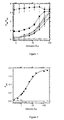
- the MAC-ELISA signal was an increasing and saturating function of the concentration in DENV1-infected serum, and a constant and nil function of the concentration in non-infected serum, when the (bt-ED3.DEN1-H6:SA) 4 -HRP complex was used at a high dilution (1280-fold; Figure 2 ).
- the dilution of the DENV1 serum N° 1 for half-maximal signal was equal to 275-fold and its end point value to 2000-fold.
- the (bt-ED3.DEN1-H6:SA) 4 -HRP complex was pre-formed, it displayed the ED3 domain tetravalently, and its two components were captured in one step by the IgMs of the serum under assay. We also performed the capture of these two components in two steps to test the importance of the multivalency of the ED3 domain.
- the bt-ED3.DEN1-H6 hybrid was presented in a free monovalent state to the IgMs that were bound to the plates. The unbound bt-ED3.DEN1-H6 molecules were subsequently washed off.
- the ED3 domains can be used in an isolated format to detect IgG antibodies that are directed against the cognate flaviviruses in the serum of patients (see Introduction).
- the ED3 domain is immobilized in the wells of a microtiter plate by passive adsorption and captures IgG antibodies in the serum under assay.
- the captured IgGs are then detected with a conjugate between a secondary antibody, directed against human IgGs, and an enzyme, usually HRP.
- HRP enzyme
- bt-ED3-H6 hybrid could be used both as a tetravalent (bt-ED3-H6:SA) 4 -HRP complex in a MAC-ELISA and as an adsorbed monovalent antigen in an IgG-specific indirect ELISA.
- the bt-ED3-H6 domain can be produced in the E. coli cytoplasm as inclusion bodies and then folded within the purification column, according to a simple protocol.
- the yields of production and purification are higher for the isolated bt-ED3-H6 domain (7.1 mg/L of culture; 470 nmoles/L of culture) than for the H6-ED3-PhoA hybrid (1.0 mg monomer/L; 16 nmoles/L), especially when calculated as moles/L of culture because the MM value of the isolated domain, 14989, is much smaller than that of the hybrid, 60631.
- the bt-ED3-H6 domain was used at a monomer concentration of 0.0025 ⁇ M, i. e. ⁇ 5-fold lower for a 10-fold faster generation of the chromogenic signal.
Landscapes
- Health & Medical Sciences (AREA)
- Life Sciences & Earth Sciences (AREA)
- Immunology (AREA)
- Engineering & Computer Science (AREA)
- Chemical & Material Sciences (AREA)
- Molecular Biology (AREA)
- Biomedical Technology (AREA)
- Urology & Nephrology (AREA)
- Hematology (AREA)
- Medicinal Chemistry (AREA)
- Analytical Chemistry (AREA)
- Cell Biology (AREA)
- Pathology (AREA)
- Food Science & Technology (AREA)
- Biotechnology (AREA)
- Physics & Mathematics (AREA)
- Microbiology (AREA)
- Biochemistry (AREA)
- General Health & Medical Sciences (AREA)
- General Physics & Mathematics (AREA)
- Proteomics, Peptides & Aminoacids (AREA)
- Chemical Kinetics & Catalysis (AREA)
- Peptides Or Proteins (AREA)
Priority Applications (4)
| Application Number | Priority Date | Filing Date | Title |
|---|---|---|---|
| EP09290560A EP2272860A1 (fr) | 2009-07-10 | 2009-07-10 | Épitope polyvalent dans un complexe avec un marqueur de détection pour le sérodiagnostic précoce d'infections |
| EP10740338A EP2451828A1 (fr) | 2009-07-10 | 2010-07-06 | Epitope multivalent en complexe avec un marqueur de détection pour le sérodiagnostic précoce d'infections |
| PCT/IB2010/053096 WO2011004324A1 (fr) | 2009-07-10 | 2010-07-06 | Epitope multivalent en complexe avec un marqueur de détection pour le sérodiagnostic précoce d'infections |
| US13/382,479 US20120208176A1 (en) | 2009-07-10 | 2010-07-06 | Multivalent Epitope in Complex with a Detection Marker for the Early Serodiagnosis of Infections |
Applications Claiming Priority (1)
| Application Number | Priority Date | Filing Date | Title |
|---|---|---|---|
| EP09290560A EP2272860A1 (fr) | 2009-07-10 | 2009-07-10 | Épitope polyvalent dans un complexe avec un marqueur de détection pour le sérodiagnostic précoce d'infections |
Publications (1)
| Publication Number | Publication Date |
|---|---|
| EP2272860A1 true EP2272860A1 (fr) | 2011-01-12 |
Family
ID=41277429
Family Applications (2)
| Application Number | Title | Priority Date | Filing Date |
|---|---|---|---|
| EP09290560A Withdrawn EP2272860A1 (fr) | 2009-07-10 | 2009-07-10 | Épitope polyvalent dans un complexe avec un marqueur de détection pour le sérodiagnostic précoce d'infections |
| EP10740338A Withdrawn EP2451828A1 (fr) | 2009-07-10 | 2010-07-06 | Epitope multivalent en complexe avec un marqueur de détection pour le sérodiagnostic précoce d'infections |
Family Applications After (1)
| Application Number | Title | Priority Date | Filing Date |
|---|---|---|---|
| EP10740338A Withdrawn EP2451828A1 (fr) | 2009-07-10 | 2010-07-06 | Epitope multivalent en complexe avec un marqueur de détection pour le sérodiagnostic précoce d'infections |
Country Status (3)
| Country | Link |
|---|---|
| US (1) | US20120208176A1 (fr) |
| EP (2) | EP2272860A1 (fr) |
| WO (1) | WO2011004324A1 (fr) |
Cited By (8)
| Publication number | Priority date | Publication date | Assignee | Title |
|---|---|---|---|---|
| CN103149357A (zh) * | 2013-01-29 | 2013-06-12 | 杭州迪恩科技有限公司 | 一种利用竞争法检测牛布鲁氏菌抗体的检测试纸卡 |
| CN103267844A (zh) * | 2013-05-06 | 2013-08-28 | 南京凯基生物科技发展有限公司 | 一种腺病毒IgM、IgG抗体的胶体金法检测试纸条、试剂盒及其制备方法 |
| EP2492689B1 (fr) * | 2011-02-22 | 2013-10-30 | Bernhard-Nocht-Institut für Tropenmedizin | Détection des anticorps à l'aide d'un complexe immun amélioré ELISA |
| WO2015161835A1 (fr) * | 2014-04-20 | 2015-10-29 | New / Era / Mabs Gmbh | Cellule libérant des biomolécules et sa sélection au moyen d'une protéine de surface |
| EP3229024A1 (fr) * | 2016-04-08 | 2017-10-11 | Viramed Biotech AG | Diagnostic du virus zika |
| WO2020119669A1 (fr) * | 2018-12-10 | 2020-06-18 | Kei International Limited | Feuille de nitrocellulose comprenant des immunoglobulines immobilisées et des antigènes à base de lipides et son utilisation |
| US11243204B2 (en) | 2016-06-08 | 2022-02-08 | Kei International Limited | Method for detecting the presence of mycobacterial material in a sample using at least two antigens |
| US12031980B2 (en) | 2018-08-08 | 2024-07-09 | Kei International Limited | Synthetic antigens for tuberculosis detection |
Families Citing this family (6)
| Publication number | Priority date | Publication date | Assignee | Title |
|---|---|---|---|---|
| EP2753932A4 (fr) * | 2011-09-07 | 2015-07-15 | Alpha Biotech Ab | Détermination d'infections bactériennes du genre rickettsia et éventuellement borrelia, chez des patients présentant des symptômes d'une maladie et étant des donneurs de sang |
| MX2015008117A (es) * | 2012-12-21 | 2016-03-31 | Amplimmune Inc | Anticuerpos anti-h7cr. |
| CN104374914B (zh) * | 2014-11-14 | 2017-06-16 | 宁波大学 | 一种恶臭假单胞菌检测试纸条及其制备方法 |
| MX374731B (es) | 2016-06-06 | 2025-03-06 | Univ Wien Med | Método para la detección de un anticuerpo igm específico para un flavivirus en una muestra. |
| WO2019143712A2 (fr) * | 2018-01-19 | 2019-07-25 | Arizona Board Of Regents On Behalf Of Arizona State University | Réponse d'anticorps commune induite |
| CN116148462B (zh) * | 2021-11-22 | 2025-11-18 | 菲鹏生物股份有限公司 | 一种捕获法检测抗体的方法和应用 |
Citations (3)
| Publication number | Priority date | Publication date | Assignee | Title |
|---|---|---|---|---|
| WO2006078657A2 (fr) * | 2005-01-19 | 2006-07-27 | Vaxinnate Corporation | Compositions a motifs moleculaires associes a des agents pathogenes et methodes d'utilisation |
| US20060276622A1 (en) * | 1992-03-06 | 2006-12-07 | Robert De Leys | Process for the determination of peptides corresponding to immunologically important epitopes and their use in a process for determination of antibodies or biotinylated peptides corresponding to immunologically important epitopes, a process for preparing them and compositions containing them |
| WO2008152528A2 (fr) | 2007-06-15 | 2008-12-18 | Institut Pasteur | Procédé de diagnostic ou de criblage d'une infection par arbovirus, réactifs utiles dans le procédé et leurs applications |
Family Cites Families (2)
| Publication number | Priority date | Publication date | Assignee | Title |
|---|---|---|---|---|
| WO2005014627A1 (fr) * | 2003-08-07 | 2005-02-17 | International Centre For Genetic Engineering And Biotechnology | Proteines a epitopes multiples de dengue recombinantes en tant qu'intermediaires de diagnostic |
| CA2508266A1 (fr) * | 2005-06-20 | 2006-12-20 | Institut Pasteur | Polypeptides chimeriques et leurs applications therapeutiques contre une infection a flaviviridae |
-
2009
- 2009-07-10 EP EP09290560A patent/EP2272860A1/fr not_active Withdrawn
-
2010
- 2010-07-06 WO PCT/IB2010/053096 patent/WO2011004324A1/fr not_active Ceased
- 2010-07-06 US US13/382,479 patent/US20120208176A1/en not_active Abandoned
- 2010-07-06 EP EP10740338A patent/EP2451828A1/fr not_active Withdrawn
Patent Citations (3)
| Publication number | Priority date | Publication date | Assignee | Title |
|---|---|---|---|---|
| US20060276622A1 (en) * | 1992-03-06 | 2006-12-07 | Robert De Leys | Process for the determination of peptides corresponding to immunologically important epitopes and their use in a process for determination of antibodies or biotinylated peptides corresponding to immunologically important epitopes, a process for preparing them and compositions containing them |
| WO2006078657A2 (fr) * | 2005-01-19 | 2006-07-27 | Vaxinnate Corporation | Compositions a motifs moleculaires associes a des agents pathogenes et methodes d'utilisation |
| WO2008152528A2 (fr) | 2007-06-15 | 2008-12-18 | Institut Pasteur | Procédé de diagnostic ou de criblage d'une infection par arbovirus, réactifs utiles dans le procédé et leurs applications |
Non-Patent Citations (71)
| Title |
|---|
| ANANDARAO, R. ET AL.: "A custom-designed recombinant multiepitope protein as a dengue diagnostic reagent", PROTEIN EXPR PURIF, vol. 41, 2005, pages 136 - 147 |
| BARKER, D.F.; CAMPBELL, A.M.: "The birA gene of Escherichia coli encodes a biotin holoenzyme synthetase", JMOL BIOL, vol. 146, 1981, pages 451 - 467 |
| BEASLEY, D.W. ET AL.: "Use of a recombinant envelope protein subunit antigen for specific serological diagnosis of West Nile virus infection", J CLIN MICROBIOL, vol. 42, 2004, pages 2759 - 2765 |
| BECKETT, D.; KOVALEVA, E.; SCHATZ, P.J.: "A minimal peptide substrate in biotin holoenzyme synthetase-catalyzed biotinylation", PROTEIN SCI, vol. 8, 1999, pages 921 - 929 |
| BEDOUELLE, H. ET AL., METHOD FOR THE DIAGNOSIS OR THE SCREENING OF AN ARBOVIRUS INFECTION, REAGENTS USEFUL IN SAID METHOD AND THEIR APPLICATIONS, 2008 |
| BINZ, H.K. ET AL.: "High-affinity binders selected from designed ankyrin repeat protein libraries", NAT BIOTECHNOL, vol. 22, 2004, pages 575 - 582 |
| BLONDEL A; BEDOUELLE H.: "Engineering the quaternary structure of an exported protein with a leucine zipper", PROTEIN ENG., vol. 4, 1991, pages 457 - 461 |
| BULLOCK, W.O.; FERNANDEZ, J.M.; SHORT, J.M.: "XLI-Blue: a high efficiency plasmid transforming recA Escherichia coli strain with beta- galactosidase selection", BIOTECHNIQUES, vol. 5, 1987, pages 376 - 379 |
| CRILL, W.D.; CHANG, G.J.: "Localization and characterization of flavivirus envelope glycoprotein cross-reactive epitopes", J VIROL, vol. 78, 2004, pages 13975 - 13986 |
| CRILL, W.D.; ROEHRIG, J.T.: "Monoclonal antibodies that bind to domain III of dengue virus E glycoprotein are the most efficient blockers of virus adsorption to Vero cells", J VIROL, vol. 75, 2001, pages 7769 - 7773 |
| DATABASE Geneseq [online] 2 April 2009 (2009-04-02), "DEN1-PhoA H6-ED3 coding sequence, SEQ ID NO:1.", XP002557349, retrieved from EBI accession no. GSN:AUM00817 Database accession no. AUM00817 * |
| DATABASE Geneseq [online] 2 April 2009 (2009-04-02), "DEN4-PhoA H6-ED3 hybrid protein, SEQ ID NO:8.", XP002557348, retrieved from EBI accession no. GSP:AUM00824 Database accession no. AUM00824 * |
| DATABASE Geneseq [online] 2 April 2009 (2009-04-02), "YF-PhoA H6-ED3 hybrid protein, SEQ ID NO:10.", XP002557350, retrieved from EBI accession no. GSP:AUM00828 Database accession no. AUM00828 * |
| DATABASE Geneseq [online] 21 September 2006 (2006-09-21), "Dengue 2 virus envelope protein.", XP002557351, retrieved from EBI accession no. GSP:AEJ39112 Database accession no. AEJ39112 * |
| DATABASE Geneseq [online] 21 September 2006 (2006-09-21), "Dengue 3 virus envelope protein.", XP002557355, retrieved from EBI accession no. GSP:AEJ39114 Database accession no. AEJ39114 * |
| DATABASE Geneseq [online] 21 September 2006 (2006-09-21), "Dengue 4 virus envelope protein.", XP002557352, retrieved from EBI accession no. GSP:AEJ39116 Database accession no. AEJ39116 * |
| DATABASE Geneseq [online] 21 September 2006 (2006-09-21), "Japanese encephalitis virus envelope protein region, SEQ ID NO:177.", XP002557354, retrieved from EBI accession no. GSP:AEJ39127 Database accession no. AEJ39127 * |
| DATABASE Geneseq [online] 21 September 2006 (2006-09-21), "West Nile Virus envelope protein fragment, SEQ ID NO:7.", XP002557353, retrieved from EBI accession no. GSP:AEJ38957 Database accession no. AEJ38957 * |
| DAUPHIN, G.; ZIENTARA, S.: "West Nile virus: recent trends in diagnosis and vaccine development", VACCINE, vol. 25, 2007, pages 5563 - 5576 |
| DE OLIVEIRA POERSCH, C. ET AL.: "Dengue virus infections: comparison of methods for diagnosing the acute disease", J CLIN VIROL, vol. 32, 2005, pages 272 - 277 |
| DUSSART, P. ET AL.: "Evaluation of an enzyme immunoassay for detection of dengue virus NS 1 antigen in human serum", CLIN VACCINE IMMUNOL, vol. 13, 2006, pages 1185 - 1189 |
| DUSSART, P. ET AL.: "Evaluation of Two New Commercial Tests for the Diagnosis of Acute Dengue Virus Infection Using NS 1 Antigen Detection in Human Serum", PLOS NEGL TROP DIS, vol. 2, 2008, pages E280 |
| GOULD, E.A.; SOLOMON, T.: "Pathogenic flaviviruses", LANCET, vol. 371, 2008, pages 500 - 509 |
| GRITSUN, T.S.; HOLMES, E.C.; GOULD, E.A.: "Analysis of flavivirus envelope proteins reveals variable domains that reflect their antigenicity and may determine their pathogenesis", VIRUS RES, vol. 35, 1995, pages 307 - 321 |
| HALSTEAD, S.B.: "Pathogenesis of dengue: challenges to molecular biology", SCIENCE, vol. 239, 1988, pages 476 - 481 |
| HOLBROOK, M.R.; SHOPE, R.E.; BARRETT, A.D.: "Use of recombinant E protein domain III-based enzyme-linked immunosorbent assays for differentiation of tick-borne encephalitis serocomplex flaviviruses from mosquito- borne flaviviruses", J CLIN MICROBIOL, vol. 42, 2004, pages 4101 - 4110 |
| HOLMES, D.A. ET AL.: "Comparative analysis of immunoglobulin M (IgM) capture enzyme-linked immunosorbent assay using virus-like particles or virus-infected mouse brain antigens to detect IgM antibody in sera from patients with evident flaviviral infections", J CLIN MICROBIOL, vol. 43, 2005, pages 3227 - 3236 |
| IGARASHI, A.: "Impact of dengue virus infection and its control", FEMS IMMUNOL MED MICROBIOL, vol. 18, 1997, pages 291 - 300 |
| INNIS, B.L. ET AL.: "An enzyme-linked immunosorbent assay to characterize dengue infections where dengue and Japanese encephalitis co-circulate", AM J TROP MED HYG, vol. 40, 1989, pages 418 - 427 |
| JOHNSON, A.J. ET AL.: "Detection of anti-arboviral immunoglobulin G by using a monoclonal antibody-based capture enzyme-linked immunosorbent assay", J CLIN MICROBIOL, vol. 38, 2000, pages 1827 - 1831 |
| KANAI, R. ET AL.: "Crystal structure of west nile virus envelope glycoprotein reveals viral surface epitopes", J VIROL, vol. 80, 2006, pages 11000 - 11008 |
| KAO, C.L. ET AL.: "Laboratory diagnosis of dengue virus infection: current and future perspectives in clinical diagnosis and public health", J MICROBIOL IMMUNOL INFECT, vol. 38, 2005, pages 5 - 16 |
| KUNO, G: "Serodiagnosis of flaviviral infections and vaccinations in humans", ADV VIRUS RES, vol. 61, 2003, pages 3 - 65 |
| LANCIOTTI, R.S. ET AL.: "Rapid detection and typing of dengue viruses from clinical samples by using reverse transcriptase-polymerase chain reaction", J CLIN MICROBIOL, vol. 30, 1992, pages 545 - 551 |
| LISOVA, O. ET AL.: "Mapping to completeness and transplantation of a group-specific, discontinuous, neutralizing epitope in the envelope protein of dengue virus", J GEN VIROL, vol. 88, 2007, pages 2387 - 2397 |
| LUDOLFS DIANA ET AL: "Highly specific detection of antibodies to tick-borne encephalitis (TBE) virus in humans using a domain III antigen and a sensitive immune complex (IC) ELISA.", JOURNAL OF CLINICAL VIROLOGY : THE OFFICIAL PUBLICATION OF THE PAN AMERICAN SOCIETY FOR CLINICAL VIROLOGY JUN 2009, vol. 45, no. 2, June 2009 (2009-06-01), pages 125 - 128, XP002557347, ISSN: 1873-5967 * |
| LUDOLFS, D. ET AL.: "Serological differentiation of infections with dengue virus serotypes 1 to 4 by using recombinant antigens", J CLIN MICROBIOL, vol. 40, 2002, pages 4317 - 4320 |
| MAKINO, Y. ET AL.: "Studies on serological cross-reaction in sequential flavivirus infections", MICROBIOL IMMUNOL, vol. 38, 1994, pages 951 - 955 |
| MARTIN, D.A. ET AL.: "Standardization of immunoglobulin M capture enzyme-linked immunosorbent assays for routine diagnosis of arboviral infections", J CLIN MICROBIOL, vol. 38, 2000, pages 1823 - 1826 |
| MARTIN, D.A. ET AL.: "Use of immunoglobulin m cross-reactions in differential diagnosis of human flaviviral encephalitis infections in the United States", CLIN DIAGN LAB IMMUNOL, vol. 9, 2002, pages 544 - 549 |
| MATHEW, A.; ROTHMAN, A.L.: "Understanding the contribution of cellular immunity to dengue disease pathogenesis", LMMUNOL REV, vol. 25, 2008, pages 300 - 313 |
| MODIS, Y. ET AL.: "A ligand-binding pocket in the dengue virus envelope glycoprotein", PROC NATL ACAD SCI USA, vol. 100, 2003, pages 6986 - 6991 |
| MODIS, Y. ET AL.: "I Variable surface epitopes in the crystal structure of dengue virus type 3 envelope glycoprotein", J VIROL, vol. 79, 2005, pages 1223 - 1231 |
| MODIS, Y. ET AL.: "Structure of the dengue virus envelope protein after membrane fusion", NATURE, vol. 427, 2004, pages 313 - 319 |
| MONGKOLSAPAYA, J. ET AL.: "Original antigenic sin and apoptosis in the pathogenesis of dengue hemorrhagic fever", NAT MED, vol. 9, 2003, pages 921 - 927 |
| MORINET F ET AL: "Development of an IgM antibody capture test using labelled fusion protein as antigen for diagnosis of B19 human parvovirus infections", BEHRING INSTITUTE: MITTEILUNGEN, MARBURG, DE, vol. 85, 1 August 1990 (1990-08-01), pages 28 - 34, XP000196154 * |
| MUKHOPADHYAY, S.; KUHN, R.J.; ROSSMANN, M.G.: "A structural perspective of the flavivirus life cycle", NAT REV MICROBIOL, vol. 3, 2005, pages 13 - 22 |
| MÜLLER KM; ARNDT KM; ALBER T.: "Protein fusions to coiled- coil domains", METHODS ENZYMOL., vol. 328, 2000, pages 261 - 282 |
| PACK P; PLUCKTHUN A.: "Miniantibodies: use of amphipathic helices to produce functional, flexibly linked dimeric FV fragments with high avidity in Escherichia coli", BIOCHEMISTRY, vol. 31, 1992, pages 1579 - 1584 |
| POTTS, J.A.; ROTHMAN, A.L.: "Clinical and laboratory features that distinguish dengue from other febrile illnesses in endemic populations", TROP MED INT HEALTH, vol. 13, 2008, pages 1328 - 1340 |
| REY, F.A. ET AL.: "The envelope glycoprotein from tick-borne encephalitis virus at 2 A resolution", NATURE, vol. 375, 1995, pages 291 - 298 |
| RICE, P.; LONGDEN, I.; BLEASBY, A.: "EMBOSS: the European Molecular Biology Open Software Suite", TRENDS GENET, vol. 16, 2000, pages 276 - 277 |
| RIVETZ B ET AL: "New dengue antibody assay with unique differential detection of IgG and IgM antibodies", CLINICAL BIOCHEMISTRY, ELSEVIER INC, US, CA, vol. 42, no. 3, 1 February 2009 (2009-02-01), pages 180 - 184, XP025884895, ISSN: 0009-9120, [retrieved on 20081106] * |
| SAMBROOK, J.; RUSSELL, D.W.: "Molecular Cloning: A Laboratory Manual", 2001, COLD SPRING HARBOR PRESS, COLD SPRING HARBOR |
| SANCHEZ, M.D. ET AL.: "Characterization of neutralizing antibodies to West Nile virus", VIROLOGY, vol. 336, 2005, pages 70 - 82 |
| SCHILLING, S. ET AL.: "Laboratory diagnosis of primary and secondary dengue infection", J CLIN VIROL, vol. 31, 2004, pages 179 - 184 |
| SCHMIDT TGM; SKERRA A: "The Strep-tag system for one-step purification and high-affinity detection or capturing of proteins", NATURE PROTOCOLS, vol. 2, 2007, pages 1528 - 1535 |
| SCHWARTZ, E. ET AL.: "Evaluation of ELISA-based sero-diagnosis of dengue fever in travelers", J CLIN VIROL, vol. 19, 2000, pages 169 - 173 |
| SHU, P.Y.; HUANG, J.H.: "Current advances in dengue diagnosis", CLIN DIAGN LAB IMMUNOL, vol. 11, 2004, pages 642 - 650 |
| SIGMA-ALDRICH.: "Product information on Streptavidin-Peroxidase from Streptomyces avidinii", CATALOG NUMBER, pages 5512 |
| SIGMA-ALDRICH: "Product information on peroxidase from horseradish", CATALOG NUMBER, pages 8375 |
| SIMMONS, M. ET AL.: "Evaluation of recombinant dengue viral envelope B domain protein antigens for the detection of dengue complex-specific antibodies", AM J TROP MED HYG, vol. 58, 1998, pages 144 - 151 |
| TAKASAKI, T. ET AL.: "Evaluation of dengue IgM detection tests using sera from patients with autoimmune diseases", J VIROL METHODS, vol. 102, 2002, pages 61 - 66 |
| TELES, F.R.; PRAZERES, D.M.; LIMA-FILHO, J.L.: "Trends in dengue diagnosis", REV MED VIROL, vol. 15, 2005, pages 287 - 302 |
| VILJA P ET AL: "Determination of immunoglobulin M antibodies for hepatitis B core antigen with a capture enzyme immunoassay and biotin-labeled core antigen produced in Escherichia coli.", JOURNAL OF CLINICAL MICROBIOLOGY OCT 1985, vol. 22, no. 4, October 1985 (1985-10-01), pages 637 - 640, XP002557346, ISSN: 0095-1137 * |
| WEBER, P.C. ET AL.: "Structural origins of high-affinity biotin binding to streptavidin.", SCIENCE, vol. 243, 1989, pages 85 - 88 |
| WESTAWAY, E.G.; SHEW, M.; DELLA-PORTA, A.J.: "Reactions of purified hemagglutinating antigens of flaviviruses with 19S and 7S antibodies", INFECT IMMUN, vol. 11, 1975, pages 630 - 634 |
| WHO.: "Dengue Haemorrhagic Fever: diagnosis, prevention, treatment and control", 1997, WHO PUBLICATIONS |
| WILCHEK, M.; BAYER, E.A.; LIVNAH, O.: "Essentials of biorecognition: the (strept)avidin-biotin system as a model for protein-protein and protein-ligand interaction", IMMUNOL LETT, vol. 103, 2006, pages 27 - 32 |
| YAMADA, K. ET AL.: "Laboratory diagnosis of dengue virus infection by reverse transcriptase polymerase chain reaction (RT-PCR) and IgM-capture enzyme-linked immunosorbent assay (ELISA).", JPN J INFECT DIS, vol. 52, 1999, pages 150 - 155 |
| ZHANG J ET AL.: "Pentamerization of single-domain antibodies from phage libraries: a novel strategy for the rapid generation of high- avidity antibody reagents", J MOL BIOL., vol. 335, 2004, pages 49 - 56 |
Cited By (11)
| Publication number | Priority date | Publication date | Assignee | Title |
|---|---|---|---|---|
| EP2492689B1 (fr) * | 2011-02-22 | 2013-10-30 | Bernhard-Nocht-Institut für Tropenmedizin | Détection des anticorps à l'aide d'un complexe immun amélioré ELISA |
| CN103149357A (zh) * | 2013-01-29 | 2013-06-12 | 杭州迪恩科技有限公司 | 一种利用竞争法检测牛布鲁氏菌抗体的检测试纸卡 |
| CN103149357B (zh) * | 2013-01-29 | 2016-04-20 | 浙江迪恩生物科技股份有限公司 | 一种利用竞争法检测牛布鲁氏菌抗体的检测试纸卡 |
| CN103267844A (zh) * | 2013-05-06 | 2013-08-28 | 南京凯基生物科技发展有限公司 | 一种腺病毒IgM、IgG抗体的胶体金法检测试纸条、试剂盒及其制备方法 |
| WO2015161835A1 (fr) * | 2014-04-20 | 2015-10-29 | New / Era / Mabs Gmbh | Cellule libérant des biomolécules et sa sélection au moyen d'une protéine de surface |
| US10526394B2 (en) | 2014-04-20 | 2020-01-07 | New / Era / Mabs Gmbh | Biomolecule-releasing cell and selection thereof by means of a surface protein |
| EP3229024A1 (fr) * | 2016-04-08 | 2017-10-11 | Viramed Biotech AG | Diagnostic du virus zika |
| WO2017174193A1 (fr) * | 2016-04-08 | 2017-10-12 | Viramed Biotech Ag | Diagnostic du virus zika |
| US11243204B2 (en) | 2016-06-08 | 2022-02-08 | Kei International Limited | Method for detecting the presence of mycobacterial material in a sample using at least two antigens |
| US12031980B2 (en) | 2018-08-08 | 2024-07-09 | Kei International Limited | Synthetic antigens for tuberculosis detection |
| WO2020119669A1 (fr) * | 2018-12-10 | 2020-06-18 | Kei International Limited | Feuille de nitrocellulose comprenant des immunoglobulines immobilisées et des antigènes à base de lipides et son utilisation |
Also Published As
| Publication number | Publication date |
|---|---|
| US20120208176A1 (en) | 2012-08-16 |
| EP2451828A1 (fr) | 2012-05-16 |
| WO2011004324A1 (fr) | 2011-01-13 |
Similar Documents
| Publication | Publication Date | Title |
|---|---|---|
| EP2272860A1 (fr) | Épitope polyvalent dans un complexe avec un marqueur de détection pour le sérodiagnostic précoce d'infections | |
| Kim et al. | Development of a SARS-CoV-2-specific biosensor for antigen detection using scFv-Fc fusion proteins | |
| JP6351571B2 (ja) | マルチプレックス免疫スクリーニングアッセイ | |
| AU2008263507B2 (en) | Method for the diagnosis or the screening of an arbovirus infection, reagents useful in said method and their applications | |
| WO2021231498A2 (fr) | Dosage de détection d'anticorps anti-sars-cov-2 | |
| JP2023036749A (ja) | 可溶性および免疫反応性のフラビウイルスns1ポリペプチド | |
| US11187706B2 (en) | Method and kit for detection of anti-Zika virus antibodies | |
| Klontz et al. | Laboratory testing for Powassan virus: past, present, and future | |
| JPWO2013018836A1 (ja) | 生物学的試料中の物質を検出する工程において、非特異的結合を抑制する方法、当該方法に使用するための剤 | |
| EP1745291B1 (fr) | Detection du virus du nil occidental | |
| JP2019527837A (ja) | サンプル中のフラビウイルスに特異的なIgM抗体の検出方法 | |
| EP2756850A1 (fr) | Diagnostic d'une infection de Leishmania | |
| JP2017534055A (ja) | シャーガス病の検出のための抗原組成物 | |
| Ludolfs et al. | Reverse ELISA for the detection of anti West Nile virus IgG antibodies in humans | |
| ES2623891T3 (es) | Variantes de la lipoproteína 15 (Lp15) de Vibrio cholerae como aditivo anti-interferencia en inmunoensayos basados en TpN17 para la detección de anticuerpos anti-Treponema | |
| Rizzo et al. | A method for rapid and high-yield production of the tick-borne encephalitis virus E and DIII recombinant proteins in E. coli with preservation of the antigenic properties | |
| US10228370B2 (en) | Recombinant Trypanosoma cruzi JL7 antigen variants and their use for detecting Chagas disease | |
| HK1236542B (zh) | 重组克氏锥虫jl7抗原变体及其用於检测查加斯病的用途 |
Legal Events
| Date | Code | Title | Description |
|---|---|---|---|
| PUAI | Public reference made under article 153(3) epc to a published international application that has entered the european phase |
Free format text: ORIGINAL CODE: 0009012 |
|
| AK | Designated contracting states |
Kind code of ref document: A1 Designated state(s): AT BE BG CH CY CZ DE DK EE ES FI FR GB GR HR HU IE IS IT LI LT LU LV MC MK MT NL NO PL PT RO SE SI SK SM TR |
|
| AX | Request for extension of the european patent |
Extension state: AL BA RS |
|
| STAA | Information on the status of an ep patent application or granted ep patent |
Free format text: STATUS: THE APPLICATION IS DEEMED TO BE WITHDRAWN |
|
| 18D | Application deemed to be withdrawn |
Effective date: 20110713 |