EP2272989B1 - Verfahren und Reagenzien zur frühen Erkennung von Melanomen - Google Patents
Verfahren und Reagenzien zur frühen Erkennung von Melanomen Download PDFInfo
- Publication number
- EP2272989B1 EP2272989B1 EP10251223.3A EP10251223A EP2272989B1 EP 2272989 B1 EP2272989 B1 EP 2272989B1 EP 10251223 A EP10251223 A EP 10251223A EP 2272989 B1 EP2272989 B1 EP 2272989B1
- Authority
- EP
- European Patent Office
- Prior art keywords
- melanoma
- pcr
- silv
- sample
- benign
- Prior art date
- Legal status (The legal status is an assumption and is not a legal conclusion. Google has not performed a legal analysis and makes no representation as to the accuracy of the status listed.)
- Active
Links
Images

Classifications
-
- C—CHEMISTRY; METALLURGY
- C12—BIOCHEMISTRY; BEER; SPIRITS; WINE; VINEGAR; MICROBIOLOGY; ENZYMOLOGY; MUTATION OR GENETIC ENGINEERING
- C12Q—MEASURING OR TESTING PROCESSES INVOLVING ENZYMES, NUCLEIC ACIDS OR MICROORGANISMS; COMPOSITIONS OR TEST PAPERS THEREFOR; PROCESSES OF PREPARING SUCH COMPOSITIONS; CONDITION-RESPONSIVE CONTROL IN MICROBIOLOGICAL OR ENZYMOLOGICAL PROCESSES
- C12Q1/00—Measuring or testing processes involving enzymes, nucleic acids or microorganisms; Compositions therefor; Processes of preparing such compositions
- C12Q1/68—Measuring or testing processes involving enzymes, nucleic acids or microorganisms; Compositions therefor; Processes of preparing such compositions involving nucleic acids
- C12Q1/6876—Nucleic acid products used in the analysis of nucleic acids, e.g. primers or probes
- C12Q1/6883—Nucleic acid products used in the analysis of nucleic acids, e.g. primers or probes for diseases caused by alterations of genetic material
- C12Q1/6886—Nucleic acid products used in the analysis of nucleic acids, e.g. primers or probes for diseases caused by alterations of genetic material for cancer
-
- C—CHEMISTRY; METALLURGY
- C12—BIOCHEMISTRY; BEER; SPIRITS; WINE; VINEGAR; MICROBIOLOGY; ENZYMOLOGY; MUTATION OR GENETIC ENGINEERING
- C12Q—MEASURING OR TESTING PROCESSES INVOLVING ENZYMES, NUCLEIC ACIDS OR MICROORGANISMS; COMPOSITIONS OR TEST PAPERS THEREFOR; PROCESSES OF PREPARING SUCH COMPOSITIONS; CONDITION-RESPONSIVE CONTROL IN MICROBIOLOGICAL OR ENZYMOLOGICAL PROCESSES
- C12Q1/00—Measuring or testing processes involving enzymes, nucleic acids or microorganisms; Compositions therefor; Processes of preparing such compositions
- C12Q1/68—Measuring or testing processes involving enzymes, nucleic acids or microorganisms; Compositions therefor; Processes of preparing such compositions involving nucleic acids
- C12Q1/6813—Hybridisation assays
- C12Q1/6834—Enzymatic or biochemical coupling of nucleic acids to a solid phase
- C12Q1/6837—Enzymatic or biochemical coupling of nucleic acids to a solid phase using probe arrays or probe chips
-
- C—CHEMISTRY; METALLURGY
- C12—BIOCHEMISTRY; BEER; SPIRITS; WINE; VINEGAR; MICROBIOLOGY; ENZYMOLOGY; MUTATION OR GENETIC ENGINEERING
- C12Q—MEASURING OR TESTING PROCESSES INVOLVING ENZYMES, NUCLEIC ACIDS OR MICROORGANISMS; COMPOSITIONS OR TEST PAPERS THEREFOR; PROCESSES OF PREPARING SUCH COMPOSITIONS; CONDITION-RESPONSIVE CONTROL IN MICROBIOLOGICAL OR ENZYMOLOGICAL PROCESSES
- C12Q1/00—Measuring or testing processes involving enzymes, nucleic acids or microorganisms; Compositions therefor; Processes of preparing such compositions
- C12Q1/68—Measuring or testing processes involving enzymes, nucleic acids or microorganisms; Compositions therefor; Processes of preparing such compositions involving nucleic acids
- C12Q1/6844—Nucleic acid amplification reactions
-
- C—CHEMISTRY; METALLURGY
- C12—BIOCHEMISTRY; BEER; SPIRITS; WINE; VINEGAR; MICROBIOLOGY; ENZYMOLOGY; MUTATION OR GENETIC ENGINEERING
- C12Q—MEASURING OR TESTING PROCESSES INVOLVING ENZYMES, NUCLEIC ACIDS OR MICROORGANISMS; COMPOSITIONS OR TEST PAPERS THEREFOR; PROCESSES OF PREPARING SUCH COMPOSITIONS; CONDITION-RESPONSIVE CONTROL IN MICROBIOLOGICAL OR ENZYMOLOGICAL PROCESSES
- C12Q2600/00—Oligonucleotides characterized by their use
- C12Q2600/112—Disease subtyping, staging or classification
-
- C—CHEMISTRY; METALLURGY
- C12—BIOCHEMISTRY; BEER; SPIRITS; WINE; VINEGAR; MICROBIOLOGY; ENZYMOLOGY; MUTATION OR GENETIC ENGINEERING
- C12Q—MEASURING OR TESTING PROCESSES INVOLVING ENZYMES, NUCLEIC ACIDS OR MICROORGANISMS; COMPOSITIONS OR TEST PAPERS THEREFOR; PROCESSES OF PREPARING SUCH COMPOSITIONS; CONDITION-RESPONSIVE CONTROL IN MICROBIOLOGICAL OR ENZYMOLOGICAL PROCESSES
- C12Q2600/00—Oligonucleotides characterized by their use
- C12Q2600/158—Expression markers
Definitions
- Cutaneous malignant melanoma is a serious health are problem with at least 62,000 new, invasive melanoma cases diagnosed in people expected in 2008 in the United States, among which 8,200 will die of this disease.
- the incidence of melanoma continues to increase fatser than that of any othetr malignancy. Also, it is one of the most common cancers in young adults and, thus, is the number two type of cancer in terams o average years of life lost.
- Certain proteins have been shown to be associated with melanoma and its metastases. These proteins or their activities have been used in immunohistochemistry to identify metastases and include L1CAM (Thies et al. (2002); Fogel et al. (2003)); and S-100 (Diego et al. (2003)). High-density microarrays have been applied to simultaneously monitor expression of thousands of genes in biologoical samples. Studies have resulted in the identification of genes differentially expressed in benign and malignant lesions, as well as genes that might be of prognostic value. Luo et al. (2001); and Wang et al. (2004).
- WO-2006-002433 discloses a method of identifying a melanoma by measuring the levels of PLAB, L1CAM andNTRK3.
- Wagner et al. (1997) discusses the analysis of gp100 expression in primary human tissue specimens, and its implications for melanoma immuno- and gene-therapy.
- Alexandrescu et al. (2010) discusses melanoma-spcific marker expression in skin biopsy tissues as a tool to facilitate melanoma diagnosis.
- the invention provides a method of identifying a melanoma in a tissue sample comprising the steps of (a) measuring the expression levels in the sample of genes encoding mRNA corresponding to SILV; (b) measuring the expression level of a gene encoding tyrosinase; and (c) measuring the expression level of SILV relative to tyrosinase; wherein the expression levels of SILV relative to tyrosinase are indicative of the presence of a melanoma.
- the invention also provides the use of a kit in the method of the invention, wherein the kit comprises nucleic acid amplification and detection reagents.
- the present invention provides a method of identifying a melanoma in a tissue sample, comprising assaying and measuring the expression levels in the sample for genes encoding mRNA corresponding to SILV (me20m), and tyrosinase (TYR), as defined in the claims. TYR was used as a normalization control that confirms the presence of melanocytes in the tested sample. Also disclosed herein is a method of identifying a melanoma by obtaining a tissue sample; and assaying and measuring the expression levels in the sample of genes encoding mRNA corresponding to one or both of GDF15 or L1CAM and recognized by the primer/probe sets SEQ. ID NOs.: 5-7 and 8-10, respectively, where gene expression is above a pre-determined cut-off is indicative of the presence of a melanoma.
- Also disclosed herein is a method of distinguishing a malignant melanocyte from a benign melanocyte by obtaining a tissue sample; and assaying and measuring the expression levels in the sample of genes encoding SILV where the gene expression levels above pre-determined cut-off is indicative of the presence of a melanoma in the sample.
- Also disclosed herein is a method of distinguishing a malignant melanocyte from a benign melanocyte by obtaining a tissue sample; and assaying and measuring the expression levels in the sample for genes encoding oen or both of GDF15 and L1CAM and recognized by the primer/probe sets SEQ/ ID NO.: 5-7 and 8-10. Gene expression levels above pre-determined cut-off is indicative of the presence of a melanoma in the sample.
- Also disclosed herein is a method of determining patient treatment protocol by obtaining a tissue sample from the patient; and assaying and measuring the expression levels in the sample of genes encoding SILV where the gene expression levels above pre-determined cut-off levels are indicative of the presence of a melanoma in the sample.
- Also disclosed herein is a method of determining patient treatment protocol by obtaining a tissue sample from the patient; and assaying and measuring the expression levels in the sample of genes encoding one or both of GDF15 and L1CAM recognized by the primer/probe sets SEQ. ID NOS.: 5-7 and 8-10 where the gene expression levels above pre-determined cut-off levels are indicative of the presence of a melanoma in the sample.
- the final Marker is SILV and is defined herein as the gene encoding any variant, allele etc.
- SILV is also described as MELANOCYTE PROTEIN 17; PMEL 17; PREMELANOSOMAL PROTEIN; PMEL; GP 100; ME20; SI; SIL; D12S53E and represented by Accession No. NM_006928.3.
- the invention further provides the use of a kit for conducting an assay to determine the presence of melanoma in a cell sample comprising: nucleic acid amplification and detection reagents.
- the invention further provides primer/probe sets for amplification and detection of PCR products obtained in the inventive methods. These sets include the following:
- primer/probe sets for amplification and detection of PCR products obtained in the disclosed methods include the following:
- amplicons obtained by PCR methods utilized in the inventive methods. These amplicons include the following:
- the present invention provides methods of qualitatively and quantitatively identifying a melanoma, as defined in the claims. Also disclosed herein are methods of distinguishing a malignant melanocyte from a benign melanocyte; diagnosing melanocytic lesions with uncertain pathological features; and determining a melanoma patient treatment protocol. The methods further provide aids in patient prognosis, patient monitoring and drug development. The methods rely on assaying and measuring expression levels of SILV (me20m) as a final Marker for melanoma biopsy assays where a certain level of gene expression, relative to TYR normalization control, is indicative of the presence of a malignant melanocyte in the sample assayed.
- SILV SILV
- the present invention focuses on the utility of identified gene expression markers to diagnose malignant melanoma among various skin lesions, including lesions with uncertain morphological features or suspicious primary melanocytic lesions termed equivocal cases using paraffin-embedded ("FFPE") tissues.
- FFPE paraffin-embedded
- This setting is where discrepancies between the opinions of dermatopathologists occur.
- the utility of this invention is to identify gene expression markers that differentiate benign melanocytic skin lesions from primary melanomas in gene expression assays, based on RT-PCR, for the diagnosis of melanoma in suspicious lesions.
- High-density cDNA and oligonucleotide microarrays allow simultaneous monitoring of the expression of thousands of genes.
- Microarray technology provides a quantitative measurement of mRNA abundance and has gained acceptance as a tool for marker discovery based on gene expression.
- microarray analysis has identified genes differentially expressed in benign and malignant lesions for different cancer types or that have prognostic significance. Luo et al. (2001); Su et al. (2001); Henshall et al. (2003); and Wang et al. (2004).
- the first gene expression profiling of malignant melanoma used a microarray containing probes for 8,150 mDNA trabscripts and identified genes that might be associated with aggressive tumor behavior.
- PCR was demonstrated to have sufficient specificity and sensitivity to detect metastasis of melanoma.
- a method with improved diagnostic performance in differentiating melanoma from non-melanoma lesions from primary skin biopsies is provided.
- the assay of the invention is useful in diagnosing clear-cut, or unequivocal, from suspicious, or equivocal, lesions.
- the instant invention may find particular utility in testing of early stage tissue samples.
- a probability measurement will distinguish tissues having melanoma relative to benign melanocyte or normal tissue.
- the methods of the invention employ a rapid technique for extracting nucleic acids from FFPE tissue samples and a method of amplifying and detecting nucleic acid fragments indicative of melanoma in primary skin lesions.
- the nucleic acid fragments qualitatively and quantitatively measure mRNA encoded by the Marker gene.
- Tissue samples include skin lesions derived from punch, needle, excisional or shave biopsies.
- the methods provided herein allow for melanoma detection in primary skin biopsy samples allowing a physician to determine whether to immediately implement an appropriate treatment protocol for early stage disease.
- RNA is isolated from an FFPE tissue sample block.
- Any suitable commercially available paraffin kit may be used, such as High Pure RNA Paraffin Kit from Roche (Cat. #3270289).
- the FFPE samples are sectioned according to the sixe of the embedded tumor as follows: ⁇ 2 to 5 mm sectioned to 9 x 10 ⁇ m; ⁇ 6 to 8 mm sectioned to 6 x 10 ⁇ m. Sections are de-paraffinized according to the manufacturer's instructions and the isolated RNA may be stored in RNase-free water at -80° C.
- assays can be by any means known in the art and include methods such as PCR, Rolling Circle Amplification (RCA), Ligase Chain Reaction (LCR), Strand Displacement Amplification (SDA), Nucleic Acid Sequence Based Amplification (NASBA), and others.
- RCA Rolling Circle Amplification
- LCR Ligase Chain Reaction
- SDA Strand Displacement Amplification
- NASBA Nucleic Acid Sequence Based Amplification
- the rapid molecular diagnostics involved are most preferably quantitative PCR methods, including QRT-PCR.
- Detection can be by any method known in the art including microarrays, gene chips and fluorescence.
- a typical PCR includes multiple amplification steps, or cycles that selectively amplify target nucleic acid species.
- a typical PCR includes three steps: a denaturing step in which a target nucleic acid is denatured; an annealing step in which a set of PCR primers (forward and backward primers) anneal to complementary DNA strands; and an elongation step in which a thermostable DNA polymerase elongates the primers. By repeating this step multiple times, a DNA fragment is amplified to produce an amplicon, corresponding to the target DNA sequence.
- Typical PCR includes 20 or more cycles of denaturation, annealing and elongation. Often, the annealing and elongation steps can be performed concurrently, in which case the cycle contains only two steps.
- the RT-PCR amplification reaction may be conducted in a time so that the lengths of each of these steps can be in the seconds range, rather than minutes.
- certain new thermal cyclers being capable of generating a thermal ramp rate of at least about 5C° per second, RT-PCR amplifications in 30 minutes or less are used. More preferably, amplifications are conducted in less than 25 minutes.
- the denaturation step may be conducted for times of 10 seconds or less. In fact, some thermal cyclers have settings for "0 seconds" which may be the optimal duration of the denaturation step. That is, it is enough that the thermal cycler reaches the denaturation temperature.
- annealing and elongation steps are most preferably less than 10 seconds each, and when conducted at the same temperature, the combination annealing/elongation step may be less than 10 seconds.
- Some homogeneous probe detection methods may require a separate step for elongation to maximize rapid assay performance.
- annealing temperatures are typically above 50°C. More preferably annealing temperatures are above 55°C.
- a single combined reaction for RT-PCR, with no experimenter intervention, is desirable for several reasons: (1) decreased risk of experimenter error; (2) decreased risk of target or product contamination; and (3) increased assay speed.
- the reaction can consist of either one or two polymerases.
- one of these enzymes is typically an RNA-based DNA polymerase (reverse transcriptase) and one is a thermostable DNA-based DNA polymerase.
- RNA-based DNA polymerase reverse transcriptase
- thermostable DNA-based DNA polymerase RNA-based DNA polymerase
- US Patents 5,411,876 and 5,985,619 provide examples of different "hot start" approaches.
- thermoactivation methods include the use of one or more thermoactivation methods that sequester one or more of the components required for efficient DNA polymerization.
- US Patents 5,550,044 and 5,413,924 describe methods for preparing reagents for use in such methods.
- US Patent 6,403,341 describes a sequestering approach that involves chemical alteration of one of the PCR reagent components. In the most preferred embodiment, both RNA- and DNA-dependent polymerase activities reside in a single enzyme.
- Other components that are required for efficient amplification include nucleoside triphosphates, divalent salts and buffer components. In some instances, non-specific nucleic acid and enzyme stabilizers may be beneficial.
- the amounts of certain reverse transcriptase and the PCR components are atypical in order to take advantage of the faster ramp times of some thermal cyclers. Specifically, the primer concentrations are very high.
- Typical gene-specific primer concentrations for reverse transcriptase reactions are less than about 20 nM.
- the reverse transcriptase primer concentration is raised to greater than 20 nM, preferably at least about 50 nM, and typically about 100 nM.
- Standard PCR primer concentrations range from 100 nM to 300 nM. Higher concentrations may be used in standard PCR to compensate for Tm variations. However, for the purposes herein, the referenced primer concentrations are for circumstances where no Tm compensation is needed. Proportionately higher concentrations of primers may be empirically determined and used if Tm compensation is necessary or desired.
- the PCR primer concentrations typically are greater than 250 nM, preferably greater than about 300 nM and typically about 500 nM.
- RNA quantity a measure of the amount of DNA in a particular amplification reaction in case of a negative result.
- Potential causes of false negative results that must be controlled in an RT-PCR include: inadequate RNA quantity, degradation of RNA, inhibition of RT and/or PCR and experimenter error.
- protein levels can be measured by binding to an antibody or antibody fragment specific for the protein and measuring the amount of antibody-bound protein.
- Antibodies can be labeled by radioactive, fluorescent or other detectable reagents to facilitate detection. Methods of detection include, without limitation, enzyme-linked immunosorbent assay (ELISA) and immunoblot techniques.
- ELISA enzyme-linked immunosorbent assay
- the invention provides specificity and sensitivity sufficient to identify a malignant melanocyte in a tissue sample.
- the methods determine expression of a particular Marker gene, SILV, by measuring mRNA encoded by the Marker.
- SILV is a leading marker demonstrating clear discrimination between melanoma and benign, unequivocal cases as well as between different atypia subgroups in suspicious tissue group samples.
- the Marker SILV is defined herein as the gene encoding any variant, allele etc. including SEQ ID NO: 1-3.
- tyrosinase is used as a control gene to demonstrate the presence of melanocytes in the tissue sample and to normalize for melanocytic content.
- Tyrosinase is described by Mandelcorn-Monson et al. (2003); and US Patent No. 6,153,388 and is represented by Accession No. NM_000372.
- Tyrosinase is also defined as the gene encoding mRNA recognized by the ABI assay on demand (Hs00165976_m1) with PCR amplicon SEQ ID NO: 14.
- the specificity of any given amplification-based molecular diagnostic relies heavily, but not exclusively, on the identity of the primer sets.
- the primer sets are pairs of forward and reverse oligonucleotide primers that anneal to a target DNA sequence to permit amplification of the target sequence, thereby producing a target sequence-specific amplicon.
- the primers must be capable of amplifying Markers of the disease state of interest. In the case of the instant invention, the Marker is directed to melanoma.
- the reaction must also contain some means of detection of a specific signal. This is preferably accomplished through the use of a reagent that detects a region of DNA sequence derived from polymerization of the target sequence of interest. Preferred reagents for detection give a measurable signal differential when bound to a specific nucleic acid sequence of interest. Often, these methods involve nucleic acid probes that give increased fluorescence when bound to the sequence of interest. Typically, the progress of the reactions of the inventive methods are monitored by analyzing the relative rates of amplicon production for each PCR primer set.
- the invention further includes the use of primer/probe sets in the claimed methods, wherein the sequences IDs are: SEQ, ID NOs.: 1-3.
- Monitoring amplicon production may be achieved by a number of detection reagents and methods, including without limitation, fluorescent primers, and fluorogenic probes and fluorescent dyes that bind double-stranded DNA. Molecular beacons, Scorpions, and other detection schemes may also be used.
- a common method of monitoring a PCR employs a fluorescent hydrolysis probe assay. This method exploits the 5' nuclease activity of certain thermostable DNA polymerases (such as Taq or Tfl DNA polymerases) to cleave an oligomeric probe during the PCR process.
- amplicons obtained by PCR methods utilized in the inventive methods include the sequences: SEQ. ID Nos: 11-14.
- the oligomer is selected to anneal to the amplified target sequence under elongation conditions.
- the probe typically has a fluorescent reporter on its 5' end and a fluorescent quencher of the reporter at the 3' end. So long as the oligomer is intact, the fluorescent signal from the reporter is quenched. However, when the oligomer is digested during the elongation process, the fluorescent reporter is no longer in proximity to the quencher.
- the relative accumulation of free fluorescent reporter for a given amplicon may be compared to the accumulation of the same amplicons for a control sample and/or to that of a control gene, such as, without limitation, ⁇ -Actin or PBGD to determine the relative abundance of a given cDNA product of a given RNA in a RNA population.
- a control gene such as, without limitation, ⁇ -Actin or PBGD
- Products and reagents for the fluorescent hydrolysis probe assay are readily available commercially, for instance from Applied Biosystems.
- the most preferred detection reagents are TaqMan® probes (Roche Diagnostics, Branchburg, NJ) and they are described in US Patents 5,210,015 ; 5,487,972 ; and 5,804,375 .
- these probes involve nucleic acid detection by virtue of the separation of a fluor-quencher combination on a probe through the 5'-3' exonuclease activity of the polymerase used in the PCR.
- Any suitable fluorophore can be used for any of the Markers or controls.
- fluorophores include, without limitation, Texas Red, Cal Red, Fam, Cy3 and Cy5.
- the following fluorophores correspond to the noted Markers: PLAB: Fam; L1CAM: Texas Red or Cal Red, tyrosinase: C1; PBGD: Cy5.
- Equipment and software also are readily available for controlling and monitoring amplicon accumulation in PCR and QRT-PCR including the Smart Cycler thermocylcer commercially available from Cepheid of Sunnyvale, California, and the ABI Prism 7900 Sequence Detection System, commercially available from Applied Biosystems.
- kits for detection of specific nucleic acids are particularly useful.
- the kit includes reagents for amplifying and detecting Markers.
- the kit includes sample preparation reagents and or articles (e.g., tubes) to extract nucleic acids from lymph node tissue.
- the kits may also include articles to minimize the risk of sample contamination (e.g., disposable scalpel and surface for lymph node dissection and preparation).
- reagents necessary for the one-tube QRT-PCR process described above are included such as reverse transcriptase, a reverse transcriptase primer, a corresponding PCR primer set (preferably for Markers and controls), a thermostable DNA polymerase, such as Taq polymerase, and a suitable detection reagent(s), such as, without limitation, a scorpion probe, a probe for a fluorescent hydrolysis probe assay, a molecular beacon probe, a single dye primer or a fluorescent dye specific to double-stranded DNA, such as ethidium bromide.
- the primers are preferably in quantities that yield the high concentrations described above.
- Thermostable DNA polymerases are commonly and commercially available from a variety of manufacturers.
- kits may include: suitable reaction tubes or vials, a barrier composition, typically a wax bead, optionally including magnesium; reaction mixtures (typically 10X) for the reverse transcriptase and the PCR stages, including necessary buffers and reagents such as dNTPs; nuclease-or RNase-free water; RNase inhibitor; control nucleic acid(s) and/or any additional buffers, compounds, co-factors, ionic constituents, proteins and enzymes, polymers, and the like that may be used in reverse transcriptase and/or PCR stages of QRT-PCR.
- the kits include nucleic acid extraction reagents and materials. Instructions are also preferably included in the kits.
- FFPE skin biopsy tissue specimens were selected from patients with primary melanocytic skin lesions diagnosed at Georgetown University Hospital.
- the patient series included specimens with 102 unequivocal features of invasive melanoma or benign nevi and 102 specimens with various degrees of cellular atypia. These atypical specimens were initially classified as suspicious/atypical and subsequently resolved by expert dermatopathologists as atypical nevi or malignant melanoma.
- Two patient samples were excluded because of insufficient RNA yield (less than 350 ng) after a sample preparation step. An additional nine RNA samples were excluded due to the failure of the PCR control.
- the final sample set eligible for analysis consisted of 193 biopsy tissues (95% of the original sample set) representing 47 melanomas, 48 benign nevi and 98 atypical/suspicious, including 48 atypical nevi and 50 melanomas as assigned by dermatopathologists.
- a summary of the pathological and clinical characteristics of the melanoma samples is shown in Table 1. Table 1.
- Samples were ordered from most benign to most malignant cases, based on the provided clinical data by pathology. Using a histological diagnosis, five major categories were created: advanced melanoma, melanoma, severe atypia, moderate atypia and benign nevi, with each of the 193 samples fitting into one of these groups. Lentigo maligna melanoma, melanoma in situ, and superficial invasive melanomas were combined into a single melanoma category. Two sets of melanomas with advanced features (superficial spreading with T2 and greater and nodular or metastatic) were added into an advanced melanoma group.
- a binary classification was based on splitting advanced melanomas and melanomas into malignant, and the remaining classes as benign. This stratification contained 97 malignant cases with 47 unequivocal and 50 severely atypical lesions classified as melanomas, and 96 benign cases representing 48 benign and 48 atypical nevi. All further data analysis described is presented for unequivocal cases classified into one of the 3 groups: advanced melanoma, melanoma and benign and for equivocal cases classified into one of the 4 groups: advanced melanoma, melanoma, severe and moderate atypia.
- tissue samples Two hundred four tissue samples were collected from individuals diagnosed with primary melanocytic skin lesions. All samples were collected using excisional, punch, or shave biopsy depending on lesion size, depth, and physician judgment and embedded in FFPE blocks.
- RNA from melanoma, benign nevi, and atypical/suspicious FFPE tissue was carried out with one-step RT-PCR with RNA from melanoma, benign nevi, and atypical/suspicious FFPE tissue.
- the specifmens included two seriesof samples: 1) unequivocal, or clear-cut, menaoma and benign nevi cases and 2.) samples with various degrees of atypia.
- Tyrosinase (“TYR”) was used as a housekeeping gene to control for the input quantity and quality of RNA in the reactions. DNase treatment was not used. Instead, primers or probes were designed to span an intron so they would not report on genomic DNA.
- the gene expression markers GDF-15, SILV, and L1CAM along with the normalization control tyrosinase (“TYR”) were tested in the melanoma biopsy assay using a single reaction RT-PCR format on the ABI7900 platform. Single, one step qRT-PCR assays were run in accordance with the following protocol. 50 ng of total RNA was used for qRT-PCR. The total RNA was reverse transcribed using 40X Multiscribe and RNase inhibitor mix contained in the TAQMAN® One Step PCR Master Mix Reagents Kit (Applied Biosystems, Foster City, CA).
- the cDNA was then subjected to the 2x Master Mix using UNG and PCR amplification was carried out on the ABI 7900 HT Sequence Detection System (Applied Biosystems, Foster City, CA) in the 384-well format using a 10 ⁇ l reaction size.
- Each reaction was composed of 5.0 ⁇ l of 2X One Step RT-PCR Master Mix, 0.5 ⁇ l of primer/probe mix, 0.25 ⁇ l of 40X Multiscribe enxyme, and RNase Inihibitor Mix, 0.25 ⁇ l of dNTP and 4 ⁇ l of 12.5 ng/ ⁇ l total RNA.
- the final primer/probe mix was composed of a final concentration of 900 nM of forward and reverse primers, listed in Table 3, and 250 nM of fluorescent probe.
- the dNTP mix contained a final concentration of 20 mM each of dATP, dGTP, dCTP, and dTTP.
- the reaction mixture was incubated at 48° C for 30 min. for the reverse transcription, followed by an Amplitaq activation step of 95° C for 10 minutes, and finally 40 cycles of 95° C for 15 sec. denaturing and 60° C for 1 minute anneal and extension. Sequences used in the reactions were as follows, each written in the 5' to 3' direction. Table 3 Symbol Primer/Probe Sequence ID. No.
- Ct normalized Ct marker ⁇ CT TYR
- AUC values were calculated based on ROC curve analysis using R software package version 2.5.0. (team RDC www.r-project.org ).
- SILV demonstrated the best performance with a linear response across the three patient groups (advanced melanoma, melanoma, and benign cases), representing continuously changing degrees of disease status as defined by pathology.
- Table 4 Marker Normalized to TYR Advanced Melanoma Melanoma Benign Nevi P-values (benign v malignant) AUC (classification as benign or malignant) L1CAM 6.85 7.5 7.66 0.47 0.49 SILV 1.18 2.09 4.93 ⁇ 0.001 0.94 GDF15 5.02 7.17 8.34 0.003 0.67
- SILV and GDF15 were assessed for differentiation between unequivocal melanomas and benign nevi using a univariate ROC curve analysis. As shown in Figure 2 , AUC values were 0.94 and 0.67, respectively. Based on multivariate analysis with a linear regression model, the combination of SILV and GDF15 did not improve assay performance beyond the AUV of 0.94 in unequivocal cases. Therefore, GDF15 was not pursued further for analysis of suspicious (equivocal cases). Finally, normliazation to TYR improved performance of SILV to 0.94 compared to 0.78 when using raw Ct values.
- SILV was assessed further by compring suspicious cases to unequivocal bening cases.
- the average ⁇ Ct of SILV in the equivocal samples in each by histology, excluding advanced melanoma since n 2, was compared to the average ⁇ Ct of the unequivocal bening group.
- the average ⁇ Ct values and p-values for t-test comparisions to the unequivocal begin samples are listed in Table 5 below.
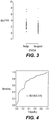
- Figures 1 through 4 confirm that SILV is the leading marker and demonstrated clear discrimination between melanoma and benign equivocal cases as well as between different atypia subgroups in the suspicious group of tissue samples.
- Table 5 Marker Normalized to TYR Normalized ⁇ Ct Values P-Values Melanoma Severe Atypia Moderate Atypia Benign Unequivocal Benign v. Moderate Benign v. Severe Benign v. Melanoma SILV 1.7 2.49 3.15 4.93 0.002 3.35E-09 9.98E-16
- the invention herein presents a melanoma biopsy assay with improved diagnostic performance in differentiating melanoma from melanocytic lesions by identifying and validating a specific genetic signature of melanoma.
- the testing results demonstrate a progressive increase in at least two genes that are differentially expressed in melanoma: SILV and GDF15.
- SILV is designated as the final marker for the melanoma biopsy assay.
Landscapes
- Chemical & Material Sciences (AREA)
- Life Sciences & Earth Sciences (AREA)
- Health & Medical Sciences (AREA)
- Organic Chemistry (AREA)
- Proteomics, Peptides & Aminoacids (AREA)
- Engineering & Computer Science (AREA)
- Zoology (AREA)
- Wood Science & Technology (AREA)
- Immunology (AREA)
- Analytical Chemistry (AREA)
- Genetics & Genomics (AREA)
- Pathology (AREA)
- Physics & Mathematics (AREA)
- Biotechnology (AREA)
- Microbiology (AREA)
- Molecular Biology (AREA)
- Biophysics (AREA)
- Biochemistry (AREA)
- Bioinformatics & Cheminformatics (AREA)
- General Engineering & Computer Science (AREA)
- General Health & Medical Sciences (AREA)
- Hospice & Palliative Care (AREA)
- Oncology (AREA)
- Chemical Kinetics & Catalysis (AREA)
- Measuring Or Testing Involving Enzymes Or Micro-Organisms (AREA)
Claims (7)
- Verfahren zum Identifizieren eines Melanoms in einer Gewebeprobe, umfassend die folgenden Schritte:a) Messen der Expressionsniveaus von Genen, die für mRNA codieren, entsprechend SILV, in der Probe;b) Messen des Expressionsniveaus eines Gens, das für Tyrosinase codiert; undc) Messen des Expressionsniveaus von SILV im Vergleich zu Tyrosinase;wobei die Expressionsniveaus von SILV im Vergleich zu Tyrosinase das Vorliegen eines Melanoms anzeigen.
- Verfahren nach Anspruch 1, wobei es sich bei der Probe um eine primäre Hautbiopsieprobe handelt.
- Verfahren nach Anspruch 1 oder Anspruch 2, wobei die Genexpression auf einem Mikro-Array oder Gen-Chip gemessen wird.
- Verfahren nach Anspruch 1 oder Anspruch 2, wobei die Genexpression mittels Nukleinsäureamplifikation, die durch Polymerasekettenreaktion (PCR) von aus der Probe extrahierter RNA durchgeführt wird.
- Verfahren nach Anspruch 4, wobei es sich bei der Polymerasekettenreaktion um qRT-PCR handelt.
- Verwendung eines Kits in dem Verfahren nach einem der Ansprüche 1 bis 4, wobei das Kit Nukleinsäureamplifikations- und -nachweisreagenzien umfasst.
- Verwendung nach Anspruch 6, wobei die Nukleinsäureamplifikationsreagenzien Primer mit der Sequenz von SEQ ID NO: 1 und 2 und eine Sonde mit der Sequenz von SEQ ID NO: 3 umfassen.
Applications Claiming Priority (1)
| Application Number | Priority Date | Filing Date | Title |
|---|---|---|---|
| US22389409P | 2009-07-08 | 2009-07-08 |
Publications (2)
| Publication Number | Publication Date |
|---|---|
| EP2272989A1 EP2272989A1 (de) | 2011-01-12 |
| EP2272989B1 true EP2272989B1 (de) | 2017-01-04 |
Family
ID=43033202
Family Applications (1)
| Application Number | Title | Priority Date | Filing Date |
|---|---|---|---|
| EP10251223.3A Active EP2272989B1 (de) | 2009-07-08 | 2010-07-08 | Verfahren und Reagenzien zur frühen Erkennung von Melanomen |
Country Status (11)
| Country | Link |
|---|---|
| US (1) | US20110009288A1 (de) |
| EP (1) | EP2272989B1 (de) |
| JP (1) | JP2011015679A (de) |
| KR (1) | KR20110004800A (de) |
| CN (1) | CN101948915B (de) |
| BR (1) | BRPI1006640B8 (de) |
| CA (1) | CA2709106C (de) |
| DK (1) | DK2272989T3 (de) |
| ES (1) | ES2617090T3 (de) |
| IL (1) | IL206810A (de) |
| MX (1) | MX2010007514A (de) |
Families Citing this family (1)
| Publication number | Priority date | Publication date | Assignee | Title |
|---|---|---|---|---|
| CA3077539C (en) * | 2012-12-27 | 2023-09-12 | Quest Diagnostics Investments Incorporated | Ddr2 mutations as targetable features of melanoma or basal cell carcinoma |
Family Cites Families (10)
| Publication number | Priority date | Publication date | Assignee | Title |
|---|---|---|---|---|
| DK0515506T3 (da) * | 1990-02-16 | 2000-05-08 | Hoffmann La Roche | Fremgangsmåde til påvisning af carcinogene humane papillomavirus |
| US5210015A (en) | 1990-08-06 | 1993-05-11 | Hoffman-La Roche Inc. | Homogeneous assay system using the nuclease activity of a nucleic acid polymerase |
| US5413924A (en) | 1992-02-13 | 1995-05-09 | Kosak; Kenneth M. | Preparation of wax beads containing a reagent for release by heating |
| US6153388A (en) * | 1994-10-27 | 2000-11-28 | University Of South Florida | Method of determining melanoma micrometastasis using tyrosinase |
| CA2168712A1 (en) * | 1995-02-07 | 1996-08-08 | John William Henderson Sutherland | Use of exonuclease and/or glycosylase as supplements to anti-polymerase antibody to increase specificity in polymerase chain reaction |
| US6403341B1 (en) | 2001-08-02 | 2002-06-11 | Wayne M. Barnes | Magnesium precipitate hot start method for PCR |
| BRPI0512587A (pt) * | 2004-06-25 | 2007-09-18 | Veridex Llc | processo e reagentes para detecção de melanoma |
| US20070154889A1 (en) | 2004-06-25 | 2007-07-05 | Veridex, Llc | Methods and reagents for the detection of melanoma |
| CN1796410A (zh) * | 2004-12-29 | 2006-07-05 | 北京师范大学 | 黑色素瘤相关抗原MAAT1p15的功能与应用 |
| EP1843773A4 (de) * | 2005-01-17 | 2008-07-30 | Viralytics Ltd | Verfahren und zusammensetzung zur behandlung von neoplasien |
-
2010
- 2010-07-05 IL IL206810A patent/IL206810A/en active IP Right Grant
- 2010-07-07 JP JP2010154736A patent/JP2011015679A/ja active Pending
- 2010-07-07 CA CA2709106A patent/CA2709106C/en active Active
- 2010-07-08 ES ES10251223.3T patent/ES2617090T3/es active Active
- 2010-07-08 US US12/832,462 patent/US20110009288A1/en not_active Abandoned
- 2010-07-08 MX MX2010007514A patent/MX2010007514A/es not_active Application Discontinuation
- 2010-07-08 CN CN201010226228.4A patent/CN101948915B/zh active Active
- 2010-07-08 BR BRPI1006640A patent/BRPI1006640B8/pt active IP Right Grant
- 2010-07-08 EP EP10251223.3A patent/EP2272989B1/de active Active
- 2010-07-08 DK DK10251223.3T patent/DK2272989T3/en active
- 2010-07-08 KR KR1020100065733A patent/KR20110004800A/ko not_active Ceased
Non-Patent Citations (1)
| Title |
|---|
| TALANTOV DMITRI ET AL: "Novel genes associated with malignant melanoma but not benign melanocytic lesions", CLINICAL CANCER RESEARCH, THE AMERICAN ASSOCIATION FOR CANCER RESEARCH, US, vol. 11, no. 20, 15 October 2005 (2005-10-15), pages 7234 - 7242, XP002377436, ISSN: 1078-0432, DOI: 10.1158/1078-0432.CCR-05-0683 * |
Also Published As
| Publication number | Publication date |
|---|---|
| US20110009288A1 (en) | 2011-01-13 |
| ES2617090T3 (es) | 2017-06-15 |
| BRPI1006640B8 (pt) | 2021-07-27 |
| BRPI1006640A2 (pt) | 2012-06-26 |
| IL206810A (en) | 2015-07-30 |
| CN101948915A (zh) | 2011-01-19 |
| EP2272989A1 (de) | 2011-01-12 |
| MX2010007514A (es) | 2011-03-16 |
| DK2272989T3 (en) | 2017-02-27 |
| CA2709106A1 (en) | 2011-01-08 |
| BRPI1006640B1 (pt) | 2020-12-29 |
| JP2011015679A (ja) | 2011-01-27 |
| IL206810A0 (en) | 2010-12-30 |
| CA2709106C (en) | 2021-08-03 |
| KR20110004800A (ko) | 2011-01-14 |
| CN101948915B (zh) | 2018-10-02 |
Similar Documents
| Publication | Publication Date | Title |
|---|---|---|
| CA2860338C (en) | System and method of detecting rnas altered by cancer in peripheral blood | |
| US20110312515A1 (en) | Identification of markers in lung and breast cancer | |
| EP3246332B1 (de) | Oligonukleotide und verfahren zur erkennung von pik3ca-mutationen | |
| JP2009528825A (ja) | デュークスb大腸がんの再発を予測するための分子的解析 | |
| EP2121988B1 (de) | Überleben und rezidiv von prostatakrebs | |
| US11053551B2 (en) | Method and apparatus for determining a probability of colorectal cancer in a subject | |
| JP2008504034A (ja) | 黒色腫を検出するための方法および試薬 | |
| EP3458614A1 (de) | Verfahren zum nachweis eines mutierten gens durch multiplexen von digitalem pcr | |
| US20250092461A1 (en) | Personalized cancer management and monitoring based on dna methylation changes in cell-free dna | |
| EP2272989B1 (de) | Verfahren und Reagenzien zur frühen Erkennung von Melanomen | |
| HK1152974B (en) | Methods and reagents for the early detection of melanoma | |
| HK1152974A (en) | Methods and reagents for the early detection of melanoma | |
| CN116356029B (zh) | Txnrd1基因甲基化水平检测在子宫内膜癌诊断和/或筛查中的应用 | |
| WO2024047182A2 (en) | Detection of variant esr1 sequences | |
| KR20230026734A (ko) | 마이크로rna를 이용한 당뇨병성 신경병증의 예측 및 진단 방법 및 이를 위한 키트 | |
| CN117721207A (zh) | 用于检测宫颈癌和/或宫颈高级别病变的组合物及其用途 |
Legal Events
| Date | Code | Title | Description |
|---|---|---|---|
| PUAI | Public reference made under article 153(3) epc to a published international application that has entered the european phase |
Free format text: ORIGINAL CODE: 0009012 |
|
| AK | Designated contracting states |
Kind code of ref document: A1 Designated state(s): AL AT BE BG CH CY CZ DE DK EE ES FI FR GB GR HR HU IE IS IT LI LT LU LV MC MK MT NL NO PL PT RO SE SI SK SM TR |
|
| AX | Request for extension of the european patent |
Extension state: BA ME RS |
|
| 17P | Request for examination filed |
Effective date: 20110712 |
|
| 17Q | First examination report despatched |
Effective date: 20111122 |
|
| REG | Reference to a national code |
Ref country code: HK Ref legal event code: DE Ref document number: 1152974 Country of ref document: HK |
|
| RAP1 | Party data changed (applicant data changed or rights of an application transferred) |
Owner name: JANSSEN DIAGNOSTICS, LLC |
|
| GRAP | Despatch of communication of intention to grant a patent |
Free format text: ORIGINAL CODE: EPIDOSNIGR1 |
|
| INTG | Intention to grant announced |
Effective date: 20160719 |
|
| GRAS | Grant fee paid |
Free format text: ORIGINAL CODE: EPIDOSNIGR3 |
|
| GRAA | (expected) grant |
Free format text: ORIGINAL CODE: 0009210 |
|
| AK | Designated contracting states |
Kind code of ref document: B1 Designated state(s): AL AT BE BG CH CY CZ DE DK EE ES FI FR GB GR HR HU IE IS IT LI LT LU LV MC MK MT NL NO PL PT RO SE SI SK SM TR |
|
| REG | Reference to a national code |
Ref country code: GB Ref legal event code: FG4D |
|
| REG | Reference to a national code |
Ref country code: CH Ref legal event code: EP Ref country code: CH Ref legal event code: NV Representative=s name: E. BLUM AND CO. AG PATENT- UND MARKENANWAELTE , CH |
|
| REG | Reference to a national code |
Ref country code: AT Ref legal event code: REF Ref document number: 859305 Country of ref document: AT Kind code of ref document: T Effective date: 20170115 |
|
| REG | Reference to a national code |
Ref country code: IE Ref legal event code: FG4D |
|
| REG | Reference to a national code |
Ref country code: DE Ref legal event code: R096 Ref document number: 602010039293 Country of ref document: DE |
|
| REG | Reference to a national code |
Ref country code: DK Ref legal event code: T3 Effective date: 20170220 |
|
| REG | Reference to a national code |
Ref country code: NL Ref legal event code: FP |
|
| REG | Reference to a national code |
Ref country code: LT Ref legal event code: MG4D |
|
| REG | Reference to a national code |
Ref country code: FR Ref legal event code: PLFP Year of fee payment: 8 |
|
| REG | Reference to a national code |
Ref country code: ES Ref legal event code: FG2A Ref document number: 2617090 Country of ref document: ES Kind code of ref document: T3 Effective date: 20170615 Ref country code: AT Ref legal event code: MK05 Ref document number: 859305 Country of ref document: AT Kind code of ref document: T Effective date: 20170104 |
|
| PG25 | Lapsed in a contracting state [announced via postgrant information from national office to epo] |
Ref country code: NO Free format text: LAPSE BECAUSE OF FAILURE TO SUBMIT A TRANSLATION OF THE DESCRIPTION OR TO PAY THE FEE WITHIN THE PRESCRIBED TIME-LIMIT Effective date: 20170404 Ref country code: HR Free format text: LAPSE BECAUSE OF FAILURE TO SUBMIT A TRANSLATION OF THE DESCRIPTION OR TO PAY THE FEE WITHIN THE PRESCRIBED TIME-LIMIT Effective date: 20170104 Ref country code: IS Free format text: LAPSE BECAUSE OF FAILURE TO SUBMIT A TRANSLATION OF THE DESCRIPTION OR TO PAY THE FEE WITHIN THE PRESCRIBED TIME-LIMIT Effective date: 20170504 Ref country code: FI Free format text: LAPSE BECAUSE OF FAILURE TO SUBMIT A TRANSLATION OF THE DESCRIPTION OR TO PAY THE FEE WITHIN THE PRESCRIBED TIME-LIMIT Effective date: 20170104 Ref country code: GR Free format text: LAPSE BECAUSE OF FAILURE TO SUBMIT A TRANSLATION OF THE DESCRIPTION OR TO PAY THE FEE WITHIN THE PRESCRIBED TIME-LIMIT Effective date: 20170405 Ref country code: LT Free format text: LAPSE BECAUSE OF FAILURE TO SUBMIT A TRANSLATION OF THE DESCRIPTION OR TO PAY THE FEE WITHIN THE PRESCRIBED TIME-LIMIT Effective date: 20170104 |
|
| PG25 | Lapsed in a contracting state [announced via postgrant information from national office to epo] |
Ref country code: PL Free format text: LAPSE BECAUSE OF FAILURE TO SUBMIT A TRANSLATION OF THE DESCRIPTION OR TO PAY THE FEE WITHIN THE PRESCRIBED TIME-LIMIT Effective date: 20170104 Ref country code: AT Free format text: LAPSE BECAUSE OF FAILURE TO SUBMIT A TRANSLATION OF THE DESCRIPTION OR TO PAY THE FEE WITHIN THE PRESCRIBED TIME-LIMIT Effective date: 20170104 Ref country code: BG Free format text: LAPSE BECAUSE OF FAILURE TO SUBMIT A TRANSLATION OF THE DESCRIPTION OR TO PAY THE FEE WITHIN THE PRESCRIBED TIME-LIMIT Effective date: 20170404 Ref country code: PT Free format text: LAPSE BECAUSE OF FAILURE TO SUBMIT A TRANSLATION OF THE DESCRIPTION OR TO PAY THE FEE WITHIN THE PRESCRIBED TIME-LIMIT Effective date: 20170504 Ref country code: LV Free format text: LAPSE BECAUSE OF FAILURE TO SUBMIT A TRANSLATION OF THE DESCRIPTION OR TO PAY THE FEE WITHIN THE PRESCRIBED TIME-LIMIT Effective date: 20170104 Ref country code: SE Free format text: LAPSE BECAUSE OF FAILURE TO SUBMIT A TRANSLATION OF THE DESCRIPTION OR TO PAY THE FEE WITHIN THE PRESCRIBED TIME-LIMIT Effective date: 20170104 |
|
| REG | Reference to a national code |
Ref country code: DE Ref legal event code: R097 Ref document number: 602010039293 Country of ref document: DE |
|
| PG25 | Lapsed in a contracting state [announced via postgrant information from national office to epo] |
Ref country code: SK Free format text: LAPSE BECAUSE OF FAILURE TO SUBMIT A TRANSLATION OF THE DESCRIPTION OR TO PAY THE FEE WITHIN THE PRESCRIBED TIME-LIMIT Effective date: 20170104 Ref country code: CZ Free format text: LAPSE BECAUSE OF FAILURE TO SUBMIT A TRANSLATION OF THE DESCRIPTION OR TO PAY THE FEE WITHIN THE PRESCRIBED TIME-LIMIT Effective date: 20170104 Ref country code: EE Free format text: LAPSE BECAUSE OF FAILURE TO SUBMIT A TRANSLATION OF THE DESCRIPTION OR TO PAY THE FEE WITHIN THE PRESCRIBED TIME-LIMIT Effective date: 20170104 Ref country code: RO Free format text: LAPSE BECAUSE OF FAILURE TO SUBMIT A TRANSLATION OF THE DESCRIPTION OR TO PAY THE FEE WITHIN THE PRESCRIBED TIME-LIMIT Effective date: 20170104 |
|
| PLBE | No opposition filed within time limit |
Free format text: ORIGINAL CODE: 0009261 |
|
| STAA | Information on the status of an ep patent application or granted ep patent |
Free format text: STATUS: NO OPPOSITION FILED WITHIN TIME LIMIT |
|
| PG25 | Lapsed in a contracting state [announced via postgrant information from national office to epo] |
Ref country code: SM Free format text: LAPSE BECAUSE OF FAILURE TO SUBMIT A TRANSLATION OF THE DESCRIPTION OR TO PAY THE FEE WITHIN THE PRESCRIBED TIME-LIMIT Effective date: 20170104 |
|
| 26N | No opposition filed |
Effective date: 20171005 |
|
| REG | Reference to a national code |
Ref country code: HK Ref legal event code: GR Ref document number: 1152974 Country of ref document: HK |
|
| PG25 | Lapsed in a contracting state [announced via postgrant information from national office to epo] |
Ref country code: SI Free format text: LAPSE BECAUSE OF FAILURE TO SUBMIT A TRANSLATION OF THE DESCRIPTION OR TO PAY THE FEE WITHIN THE PRESCRIBED TIME-LIMIT Effective date: 20170104 |
|
| PG25 | Lapsed in a contracting state [announced via postgrant information from national office to epo] |
Ref country code: LU Free format text: LAPSE BECAUSE OF NON-PAYMENT OF DUE FEES Effective date: 20170708 |
|
| PG25 | Lapsed in a contracting state [announced via postgrant information from national office to epo] |
Ref country code: MT Free format text: LAPSE BECAUSE OF NON-PAYMENT OF DUE FEES Effective date: 20170708 |
|
| PG25 | Lapsed in a contracting state [announced via postgrant information from national office to epo] |
Ref country code: MC Free format text: LAPSE BECAUSE OF FAILURE TO SUBMIT A TRANSLATION OF THE DESCRIPTION OR TO PAY THE FEE WITHIN THE PRESCRIBED TIME-LIMIT Effective date: 20170104 Ref country code: HU Free format text: LAPSE BECAUSE OF FAILURE TO SUBMIT A TRANSLATION OF THE DESCRIPTION OR TO PAY THE FEE WITHIN THE PRESCRIBED TIME-LIMIT; INVALID AB INITIO Effective date: 20100708 |
|
| PG25 | Lapsed in a contracting state [announced via postgrant information from national office to epo] |
Ref country code: CY Free format text: LAPSE BECAUSE OF NON-PAYMENT OF DUE FEES Effective date: 20170104 |
|
| PG25 | Lapsed in a contracting state [announced via postgrant information from national office to epo] |
Ref country code: MK Free format text: LAPSE BECAUSE OF FAILURE TO SUBMIT A TRANSLATION OF THE DESCRIPTION OR TO PAY THE FEE WITHIN THE PRESCRIBED TIME-LIMIT Effective date: 20170104 |
|
| PG25 | Lapsed in a contracting state [announced via postgrant information from national office to epo] |
Ref country code: TR Free format text: LAPSE BECAUSE OF FAILURE TO SUBMIT A TRANSLATION OF THE DESCRIPTION OR TO PAY THE FEE WITHIN THE PRESCRIBED TIME-LIMIT Effective date: 20170104 |
|
| PG25 | Lapsed in a contracting state [announced via postgrant information from national office to epo] |
Ref country code: AL Free format text: LAPSE BECAUSE OF FAILURE TO SUBMIT A TRANSLATION OF THE DESCRIPTION OR TO PAY THE FEE WITHIN THE PRESCRIBED TIME-LIMIT Effective date: 20170104 |
|
| P01 | Opt-out of the competence of the unified patent court (upc) registered |
Effective date: 20230526 |
|
| PGFP | Annual fee paid to national office [announced via postgrant information from national office to epo] |
Ref country code: GB Payment date: 20250626 Year of fee payment: 16 |
|
| PGFP | Annual fee paid to national office [announced via postgrant information from national office to epo] |
Ref country code: NL Payment date: 20250704 Year of fee payment: 16 |
|
| PGFP | Annual fee paid to national office [announced via postgrant information from national office to epo] |
Ref country code: ES Payment date: 20250806 Year of fee payment: 16 |
|
| PGFP | Annual fee paid to national office [announced via postgrant information from national office to epo] |
Ref country code: DK Payment date: 20250714 Year of fee payment: 16 Ref country code: DE Payment date: 20250702 Year of fee payment: 16 |
|
| PGFP | Annual fee paid to national office [announced via postgrant information from national office to epo] |
Ref country code: IT Payment date: 20250626 Year of fee payment: 16 |
|
| PGFP | Annual fee paid to national office [announced via postgrant information from national office to epo] |
Ref country code: BE Payment date: 20250703 Year of fee payment: 16 |
|
| PGFP | Annual fee paid to national office [announced via postgrant information from national office to epo] |
Ref country code: FR Payment date: 20250703 Year of fee payment: 16 |
|
| PGFP | Annual fee paid to national office [announced via postgrant information from national office to epo] |
Ref country code: CH Payment date: 20250801 Year of fee payment: 16 |
|
| PGFP | Annual fee paid to national office [announced via postgrant information from national office to epo] |
Ref country code: IE Payment date: 20250702 Year of fee payment: 16 |