EP2277553A1 - Präparate zur zeitbegrenzte Markierung von Biopsie-Orten - Google Patents
Präparate zur zeitbegrenzte Markierung von Biopsie-Orten Download PDFInfo
- Publication number
- EP2277553A1 EP2277553A1 EP20100185263 EP10185263A EP2277553A1 EP 2277553 A1 EP2277553 A1 EP 2277553A1 EP 20100185263 EP20100185263 EP 20100185263 EP 10185263 A EP10185263 A EP 10185263A EP 2277553 A1 EP2277553 A1 EP 2277553A1
- Authority
- EP
- European Patent Office
- Prior art keywords
- substance
- site
- marker
- detectable
- biopsy
- Prior art date
- Legal status (The legal status is an assumption and is not a legal conclusion. Google has not performed a legal analysis and makes no representation as to the accuracy of the status listed.)
- Ceased
Links
- 238000001574 biopsy Methods 0.000 title claims abstract description 184
- 238000002360 preparation method Methods 0.000 title description 8
- 238000003384 imaging method Methods 0.000 claims abstract description 59
- 238000001514 detection method Methods 0.000 claims abstract description 14
- 239000003550 marker Substances 0.000 claims description 166
- 239000000463 material Substances 0.000 claims description 91
- 238000000034 method Methods 0.000 claims description 85
- 239000000126 substance Substances 0.000 claims description 63
- 108010035532 Collagen Proteins 0.000 claims description 51
- 102000008186 Collagen Human genes 0.000 claims description 51
- 229920001436 collagen Polymers 0.000 claims description 50
- 108010010803 Gelatin Proteins 0.000 claims description 32
- 239000008273 gelatin Substances 0.000 claims description 32
- 229920000159 gelatin Polymers 0.000 claims description 32
- 235000019322 gelatine Nutrition 0.000 claims description 32
- 235000011852 gelatine desserts Nutrition 0.000 claims description 32
- 238000002559 palpation Methods 0.000 claims description 19
- 238000009607 mammography Methods 0.000 claims description 17
- 229910021645 metal ion Inorganic materials 0.000 claims description 15
- 230000005855 radiation Effects 0.000 claims description 13
- 238000012800 visualization Methods 0.000 claims description 13
- 238000002604 ultrasonography Methods 0.000 claims description 12
- 239000011324 bead Substances 0.000 claims description 11
- OKTJSMMVPCPJKN-UHFFFAOYSA-N Carbon Chemical compound [C] OKTJSMMVPCPJKN-UHFFFAOYSA-N 0.000 claims description 8
- 229910052799 carbon Inorganic materials 0.000 claims description 8
- 230000009969 flowable effect Effects 0.000 claims description 8
- 239000002245 particle Substances 0.000 claims description 8
- 230000008569 process Effects 0.000 claims description 7
- SQGYOTSLMSWVJD-UHFFFAOYSA-N silver(1+) nitrate Chemical compound [Ag+].[O-]N(=O)=O SQGYOTSLMSWVJD-UHFFFAOYSA-N 0.000 claims description 7
- 239000003086 colorant Substances 0.000 claims description 6
- 239000000843 powder Substances 0.000 claims description 6
- 229910021607 Silver chloride Inorganic materials 0.000 claims description 5
- 150000001875 compounds Chemical class 0.000 claims description 5
- 150000002632 lipids Chemical class 0.000 claims description 5
- 229920000747 poly(lactic acid) Polymers 0.000 claims description 5
- 239000004626 polylactic acid Substances 0.000 claims description 5
- HKZLPVFGJNLROG-UHFFFAOYSA-M silver monochloride Chemical compound [Cl-].[Ag+] HKZLPVFGJNLROG-UHFFFAOYSA-M 0.000 claims description 5
- 229920000954 Polyglycolide Polymers 0.000 claims description 4
- 238000002594 fluoroscopy Methods 0.000 claims description 4
- 238000002513 implantation Methods 0.000 claims description 4
- 230000005291 magnetic effect Effects 0.000 claims description 4
- 238000002595 magnetic resonance imaging Methods 0.000 claims description 4
- 239000004005 microsphere Substances 0.000 claims description 4
- 239000004633 polyglycolic acid Substances 0.000 claims description 4
- 239000000725 suspension Substances 0.000 claims description 4
- XLYOFNOQVPJJNP-UHFFFAOYSA-N water Substances O XLYOFNOQVPJJNP-UHFFFAOYSA-N 0.000 claims description 4
- 150000003863 ammonium salts Chemical class 0.000 claims description 3
- 238000002591 computed tomography Methods 0.000 claims description 3
- 238000002224 dissection Methods 0.000 claims description 3
- 229940011957 ethiodized oil Drugs 0.000 claims description 3
- 159000000001 potassium salts Chemical class 0.000 claims description 3
- 239000002002 slurry Substances 0.000 claims description 3
- 159000000000 sodium salts Chemical class 0.000 claims description 3
- 230000001934 delay Effects 0.000 claims description 2
- VTYYLEPIZMXCLO-UHFFFAOYSA-L Calcium carbonate Chemical compound [Ca+2].[O-]C([O-])=O VTYYLEPIZMXCLO-UHFFFAOYSA-L 0.000 claims 2
- AYJRCSIUFZENHW-UHFFFAOYSA-L barium carbonate Chemical compound [Ba+2].[O-]C([O-])=O AYJRCSIUFZENHW-UHFFFAOYSA-L 0.000 claims 2
- TZCXTZWJZNENPQ-UHFFFAOYSA-L barium sulfate Chemical compound [Ba+2].[O-]S([O-])(=O)=O TZCXTZWJZNENPQ-UHFFFAOYSA-L 0.000 claims 2
- PNEYBMLMFCGWSK-UHFFFAOYSA-N aluminium oxide Inorganic materials [O-2].[O-2].[O-2].[Al+3].[Al+3] PNEYBMLMFCGWSK-UHFFFAOYSA-N 0.000 claims 1
- 239000013590 bulk material Substances 0.000 claims 1
- 229910000019 calcium carbonate Inorganic materials 0.000 claims 1
- 229910052593 corundum Inorganic materials 0.000 claims 1
- 229910001845 yogo sapphire Inorganic materials 0.000 claims 1
- 238000001356 surgical procedure Methods 0.000 abstract description 9
- 210000001519 tissue Anatomy 0.000 description 83
- 230000003902 lesion Effects 0.000 description 40
- 210000000481 breast Anatomy 0.000 description 33
- 239000007789 gas Substances 0.000 description 17
- FOIXSVOLVBLSDH-UHFFFAOYSA-N Silver ion Chemical compound [Ag+] FOIXSVOLVBLSDH-UHFFFAOYSA-N 0.000 description 14
- 229910052709 silver Inorganic materials 0.000 description 11
- 239000004332 silver Substances 0.000 description 11
- 239000011159 matrix material Substances 0.000 description 9
- CURLTUGMZLYLDI-UHFFFAOYSA-N Carbon dioxide Chemical compound O=C=O CURLTUGMZLYLDI-UHFFFAOYSA-N 0.000 description 8
- BQCADISMDOOEFD-UHFFFAOYSA-N Silver Chemical compound [Ag] BQCADISMDOOEFD-UHFFFAOYSA-N 0.000 description 7
- 230000001070 adhesive effect Effects 0.000 description 7
- 239000000203 mixture Substances 0.000 description 7
- 239000007787 solid Substances 0.000 description 7
- 238000011282 treatment Methods 0.000 description 7
- 208000026310 Breast neoplasm Diseases 0.000 description 6
- 239000000853 adhesive Substances 0.000 description 6
- 238000003745 diagnosis Methods 0.000 description 6
- 239000000975 dye Substances 0.000 description 6
- 239000008188 pellet Substances 0.000 description 6
- 206010006187 Breast cancer Diseases 0.000 description 5
- WSFSSNUMVMOOMR-UHFFFAOYSA-N Formaldehyde Chemical compound O=C WSFSSNUMVMOOMR-UHFFFAOYSA-N 0.000 description 5
- 206010028980 Neoplasm Diseases 0.000 description 5
- 238000007792 addition Methods 0.000 description 5
- 210000004027 cell Anatomy 0.000 description 5
- -1 silver ions Chemical class 0.000 description 5
- 235000000177 Indigofera tinctoria Nutrition 0.000 description 4
- 230000009286 beneficial effect Effects 0.000 description 4
- 230000015572 biosynthetic process Effects 0.000 description 4
- 210000004369 blood Anatomy 0.000 description 4
- 239000008280 blood Substances 0.000 description 4
- 239000001569 carbon dioxide Substances 0.000 description 4
- 229910002092 carbon dioxide Inorganic materials 0.000 description 4
- 238000009472 formulation Methods 0.000 description 4
- 229940097275 indigo Drugs 0.000 description 4
- COHYTHOBJLSHDF-UHFFFAOYSA-N indigo powder Natural products N1C2=CC=CC=C2C(=O)C1=C1C(=O)C2=CC=CC=C2N1 COHYTHOBJLSHDF-UHFFFAOYSA-N 0.000 description 4
- 150000002500 ions Chemical class 0.000 description 4
- 238000013188 needle biopsy Methods 0.000 description 4
- 239000003921 oil Substances 0.000 description 4
- 101710134784 Agnoprotein Proteins 0.000 description 3
- KWYUFKZDYYNOTN-UHFFFAOYSA-M Potassium hydroxide Chemical compound [OH-].[K+] KWYUFKZDYYNOTN-UHFFFAOYSA-M 0.000 description 3
- 201000011510 cancer Diseases 0.000 description 3
- 239000003431 cross linking reagent Substances 0.000 description 3
- 238000002405 diagnostic procedure Methods 0.000 description 3
- CXKWCBBOMKCUKX-UHFFFAOYSA-M methylene blue Chemical compound [Cl-].C1=CC(N(C)C)=CC2=[S+]C3=CC(N(C)C)=CC=C3N=C21 CXKWCBBOMKCUKX-UHFFFAOYSA-M 0.000 description 3
- 229960000907 methylthioninium chloride Drugs 0.000 description 3
- 238000007254 oxidation reaction Methods 0.000 description 3
- 239000004814 polyurethane Substances 0.000 description 3
- 229920002635 polyurethane Polymers 0.000 description 3
- 241000283690 Bos taurus Species 0.000 description 2
- 206010011732 Cyst Diseases 0.000 description 2
- SXRSQZLOMIGNAQ-UHFFFAOYSA-N Glutaraldehyde Chemical compound O=CCCCC=O SXRSQZLOMIGNAQ-UHFFFAOYSA-N 0.000 description 2
- 230000004075 alteration Effects 0.000 description 2
- 239000004067 bulking agent Substances 0.000 description 2
- 231100000504 carcinogenesis Toxicity 0.000 description 2
- 230000015556 catabolic process Effects 0.000 description 2
- 210000002808 connective tissue Anatomy 0.000 description 2
- 208000031513 cyst Diseases 0.000 description 2
- 238000006731 degradation reaction Methods 0.000 description 2
- 238000012217 deletion Methods 0.000 description 2
- 230000037430 deletion Effects 0.000 description 2
- 238000004090 dissolution Methods 0.000 description 2
- 239000003814 drug Substances 0.000 description 2
- 238000011156 evaluation Methods 0.000 description 2
- 239000000834 fixative Substances 0.000 description 2
- 239000012530 fluid Substances 0.000 description 2
- 230000008014 freezing Effects 0.000 description 2
- 238000007710 freezing Methods 0.000 description 2
- 230000002452 interceptive effect Effects 0.000 description 2
- NTHXOOBQLCIOLC-UHFFFAOYSA-N iohexol Chemical compound OCC(O)CN(C(=O)C)C1=C(I)C(C(=O)NCC(O)CO)=C(I)C(C(=O)NCC(O)CO)=C1I NTHXOOBQLCIOLC-UHFFFAOYSA-N 0.000 description 2
- 239000012528 membrane Substances 0.000 description 2
- 230000004060 metabolic process Effects 0.000 description 2
- 238000002156 mixing Methods 0.000 description 2
- 238000012986 modification Methods 0.000 description 2
- 230000004048 modification Effects 0.000 description 2
- 230000007170 pathology Effects 0.000 description 2
- 230000000737 periodic effect Effects 0.000 description 2
- 238000001959 radiotherapy Methods 0.000 description 2
- 238000004153 renaturation Methods 0.000 description 2
- 229910001961 silver nitrate Inorganic materials 0.000 description 2
- 241000894006 Bacteria Species 0.000 description 1
- 241001631457 Cannula Species 0.000 description 1
- 208000005623 Carcinogenesis Diseases 0.000 description 1
- 206010061619 Deformity Diseases 0.000 description 1
- ZNZYKNKBJPZETN-WELNAUFTSA-N Dialdehyde 11678 Chemical compound N1C2=CC=CC=C2C2=C1[C@H](C[C@H](/C(=C/O)C(=O)OC)[C@@H](C=C)C=O)NCC2 ZNZYKNKBJPZETN-WELNAUFTSA-N 0.000 description 1
- 108010080379 Fibrin Tissue Adhesive Proteins 0.000 description 1
- XEEYBQQBJWHFJM-UHFFFAOYSA-N Iron Chemical compound [Fe] XEEYBQQBJWHFJM-UHFFFAOYSA-N 0.000 description 1
- GRYLNZFGIOXLOG-UHFFFAOYSA-N Nitric acid Chemical compound O[N+]([O-])=O GRYLNZFGIOXLOG-UHFFFAOYSA-N 0.000 description 1
- FAPWRFPIFSIZLT-UHFFFAOYSA-M Sodium chloride Chemical compound [Na+].[Cl-] FAPWRFPIFSIZLT-UHFFFAOYSA-M 0.000 description 1
- 208000002847 Surgical Wound Diseases 0.000 description 1
- 108090000190 Thrombin Proteins 0.000 description 1
- 108010077465 Tropocollagen Proteins 0.000 description 1
- 230000002159 abnormal effect Effects 0.000 description 1
- 230000005856 abnormality Effects 0.000 description 1
- 239000002253 acid Substances 0.000 description 1
- 238000004458 analytical method Methods 0.000 description 1
- 230000000845 anti-microbial effect Effects 0.000 description 1
- 229910052788 barium Inorganic materials 0.000 description 1
- DSAJWYNOEDNPEQ-UHFFFAOYSA-N barium atom Chemical compound [Ba] DSAJWYNOEDNPEQ-UHFFFAOYSA-N 0.000 description 1
- 230000005587 bubbling Effects 0.000 description 1
- 230000036952 cancer formation Effects 0.000 description 1
- 230000008859 change Effects 0.000 description 1
- 230000009920 chelation Effects 0.000 description 1
- 238000006243 chemical reaction Methods 0.000 description 1
- 239000003795 chemical substances by application Substances 0.000 description 1
- 238000002512 chemotherapy Methods 0.000 description 1
- 239000002872 contrast media Substances 0.000 description 1
- 238000004132 cross linking Methods 0.000 description 1
- ZXJXZNDDNMQXFV-UHFFFAOYSA-M crystal violet Chemical compound [Cl-].C1=CC(N(C)C)=CC=C1[C+](C=1C=CC(=CC=1)N(C)C)C1=CC=C(N(C)C)C=C1 ZXJXZNDDNMQXFV-UHFFFAOYSA-M 0.000 description 1
- 238000012258 culturing Methods 0.000 description 1
- 230000002380 cytological effect Effects 0.000 description 1
- 230000034994 death Effects 0.000 description 1
- 231100000517 death Toxicity 0.000 description 1
- 230000003111 delayed effect Effects 0.000 description 1
- 238000011161 development Methods 0.000 description 1
- 238000002059 diagnostic imaging Methods 0.000 description 1
- 150000004985 diamines Chemical class 0.000 description 1
- 150000001991 dicarboxylic acids Chemical class 0.000 description 1
- 201000010099 disease Diseases 0.000 description 1
- 208000037265 diseases, disorders, signs and symptoms Diseases 0.000 description 1
- 238000009826 distribution Methods 0.000 description 1
- 229940079593 drug Drugs 0.000 description 1
- 238000002651 drug therapy Methods 0.000 description 1
- 239000008393 encapsulating agent Substances 0.000 description 1
- 238000005538 encapsulation Methods 0.000 description 1
- 210000003722 extracellular fluid Anatomy 0.000 description 1
- 239000003302 ferromagnetic material Substances 0.000 description 1
- 125000000524 functional group Chemical group 0.000 description 1
- 229960001235 gentian violet Drugs 0.000 description 1
- 230000002070 germicidal effect Effects 0.000 description 1
- 239000011521 glass Substances 0.000 description 1
- 230000002439 hemostatic effect Effects 0.000 description 1
- 230000002962 histologic effect Effects 0.000 description 1
- 230000007062 hydrolysis Effects 0.000 description 1
- 238000006460 hydrolysis reaction Methods 0.000 description 1
- 238000005470 impregnation Methods 0.000 description 1
- 238000011065 in-situ storage Methods 0.000 description 1
- 239000011261 inert gas Substances 0.000 description 1
- 229960001025 iohexol Drugs 0.000 description 1
- 239000007788 liquid Substances 0.000 description 1
- 239000003589 local anesthetic agent Substances 0.000 description 1
- 230000004807 localization Effects 0.000 description 1
- QSHDDOUJBYECFT-UHFFFAOYSA-N mercury Chemical compound [Hg] QSHDDOUJBYECFT-UHFFFAOYSA-N 0.000 description 1
- 229910052753 mercury Inorganic materials 0.000 description 1
- 229910052751 metal Inorganic materials 0.000 description 1
- 239000002184 metal Substances 0.000 description 1
- 230000002906 microbiologic effect Effects 0.000 description 1
- 210000003205 muscle Anatomy 0.000 description 1
- 229910017604 nitric acid Inorganic materials 0.000 description 1
- 238000010899 nucleation Methods 0.000 description 1
- 230000006911 nucleation Effects 0.000 description 1
- 210000000056 organ Anatomy 0.000 description 1
- 230000003647 oxidation Effects 0.000 description 1
- 229920000642 polymer Polymers 0.000 description 1
- 238000012545 processing Methods 0.000 description 1
- 102000004169 proteins and genes Human genes 0.000 description 1
- 108090000623 proteins and genes Proteins 0.000 description 1
- 239000000941 radioactive substance Substances 0.000 description 1
- 150000003839 salts Chemical class 0.000 description 1
- 230000037390 scarring Effects 0.000 description 1
- GGCZERPQGJTIQP-UHFFFAOYSA-N sodium;9,10-dioxoanthracene-2-sulfonic acid Chemical compound [Na+].C1=CC=C2C(=O)C3=CC(S(=O)(=O)O)=CC=C3C(=O)C2=C1 GGCZERPQGJTIQP-UHFFFAOYSA-N 0.000 description 1
- 239000000243 solution Substances 0.000 description 1
- 238000010186 staining Methods 0.000 description 1
- 210000002435 tendon Anatomy 0.000 description 1
- 238000010257 thawing Methods 0.000 description 1
- 230000033912 thigmotaxis Effects 0.000 description 1
- 229960004072 thrombin Drugs 0.000 description 1
- 239000012049 topical pharmaceutical composition Substances 0.000 description 1
- 238000012285 ultrasound imaging Methods 0.000 description 1
- 230000000007 visual effect Effects 0.000 description 1
- 239000011800 void material Substances 0.000 description 1
- 238000005406 washing Methods 0.000 description 1
- 229910052724 xenon Inorganic materials 0.000 description 1
- FHNFHKCVQCLJFQ-UHFFFAOYSA-N xenon atom Chemical compound [Xe] FHNFHKCVQCLJFQ-UHFFFAOYSA-N 0.000 description 1
Images



Classifications
-
- A—HUMAN NECESSITIES
- A61—MEDICAL OR VETERINARY SCIENCE; HYGIENE
- A61B—DIAGNOSIS; SURGERY; IDENTIFICATION
- A61B90/00—Instruments, implements or accessories specially adapted for surgery or diagnosis and not covered by any of the groups A61B1/00 - A61B50/00, e.g. for luxation treatment or for protecting wound edges
- A61B90/39—Markers, e.g. radio-opaque or breast lesions markers
-
- A—HUMAN NECESSITIES
- A61—MEDICAL OR VETERINARY SCIENCE; HYGIENE
- A61K—PREPARATIONS FOR MEDICAL, DENTAL OR TOILETRY PURPOSES
- A61K49/00—Preparations for testing in vivo
- A61K49/001—Preparation for luminescence or biological staining
- A61K49/006—Biological staining of tissues in vivo, e.g. methylene blue or toluidine blue O administered in the buccal area to detect epithelial cancer cells, dyes used for delineating tissues during surgery
-
- A—HUMAN NECESSITIES
- A61—MEDICAL OR VETERINARY SCIENCE; HYGIENE
- A61M—DEVICES FOR INTRODUCING MEDIA INTO, OR ONTO, THE BODY; DEVICES FOR TRANSDUCING BODY MEDIA OR FOR TAKING MEDIA FROM THE BODY; DEVICES FOR PRODUCING OR ENDING SLEEP OR STUPOR
- A61M37/00—Other apparatus for introducing media into the body; Percutany, i.e. introducing medicines into the body by diffusion through the skin
- A61M37/0069—Devices for implanting pellets, e.g. markers or solid medicaments
-
- A—HUMAN NECESSITIES
- A61—MEDICAL OR VETERINARY SCIENCE; HYGIENE
- A61B—DIAGNOSIS; SURGERY; IDENTIFICATION
- A61B10/00—Instruments for taking body samples for diagnostic purposes; Other methods or instruments for diagnosis, e.g. for vaccination diagnosis, sex determination or ovulation-period determination; Throat striking implements
- A61B10/02—Instruments for taking cell samples or for biopsy
-
- A—HUMAN NECESSITIES
- A61—MEDICAL OR VETERINARY SCIENCE; HYGIENE
- A61B—DIAGNOSIS; SURGERY; IDENTIFICATION
- A61B17/00—Surgical instruments, devices or methods
- A61B2017/00004—(bio)absorbable, (bio)resorbable or resorptive
-
- A—HUMAN NECESSITIES
- A61—MEDICAL OR VETERINARY SCIENCE; HYGIENE
- A61B—DIAGNOSIS; SURGERY; IDENTIFICATION
- A61B90/00—Instruments, implements or accessories specially adapted for surgery or diagnosis and not covered by any of the groups A61B1/00 - A61B50/00, e.g. for luxation treatment or for protecting wound edges
- A61B90/36—Image-producing devices or illumination devices not otherwise provided for
- A61B90/37—Surgical systems with images on a monitor during operation
- A61B2090/378—Surgical systems with images on a monitor during operation using ultrasound
- A61B2090/3782—Surgical systems with images on a monitor during operation using ultrasound transmitter or receiver in catheter or minimal invasive instrument
- A61B2090/3788—Surgical systems with images on a monitor during operation using ultrasound transmitter or receiver in catheter or minimal invasive instrument transmitter only
-
- A—HUMAN NECESSITIES
- A61—MEDICAL OR VETERINARY SCIENCE; HYGIENE
- A61B—DIAGNOSIS; SURGERY; IDENTIFICATION
- A61B90/00—Instruments, implements or accessories specially adapted for surgery or diagnosis and not covered by any of the groups A61B1/00 - A61B50/00, e.g. for luxation treatment or for protecting wound edges
- A61B90/39—Markers, e.g. radio-opaque or breast lesions markers
- A61B2090/3904—Markers, e.g. radio-opaque or breast lesions markers specially adapted for marking specified tissue
- A61B2090/3908—Soft tissue, e.g. breast tissue
-
- A—HUMAN NECESSITIES
- A61—MEDICAL OR VETERINARY SCIENCE; HYGIENE
- A61B—DIAGNOSIS; SURGERY; IDENTIFICATION
- A61B90/00—Instruments, implements or accessories specially adapted for surgery or diagnosis and not covered by any of the groups A61B1/00 - A61B50/00, e.g. for luxation treatment or for protecting wound edges
- A61B90/39—Markers, e.g. radio-opaque or breast lesions markers
- A61B2090/3925—Markers, e.g. radio-opaque or breast lesions markers ultrasonic
-
- A—HUMAN NECESSITIES
- A61—MEDICAL OR VETERINARY SCIENCE; HYGIENE
- A61B—DIAGNOSIS; SURGERY; IDENTIFICATION
- A61B90/00—Instruments, implements or accessories specially adapted for surgery or diagnosis and not covered by any of the groups A61B1/00 - A61B50/00, e.g. for luxation treatment or for protecting wound edges
- A61B90/39—Markers, e.g. radio-opaque or breast lesions markers
- A61B2090/3925—Markers, e.g. radio-opaque or breast lesions markers ultrasonic
- A61B2090/3929—Active markers
-
- A—HUMAN NECESSITIES
- A61—MEDICAL OR VETERINARY SCIENCE; HYGIENE
- A61B—DIAGNOSIS; SURGERY; IDENTIFICATION
- A61B90/00—Instruments, implements or accessories specially adapted for surgery or diagnosis and not covered by any of the groups A61B1/00 - A61B50/00, e.g. for luxation treatment or for protecting wound edges
- A61B90/39—Markers, e.g. radio-opaque or breast lesions markers
- A61B2090/3933—Liquid markers
-
- A—HUMAN NECESSITIES
- A61—MEDICAL OR VETERINARY SCIENCE; HYGIENE
- A61B—DIAGNOSIS; SURGERY; IDENTIFICATION
- A61B90/00—Instruments, implements or accessories specially adapted for surgery or diagnosis and not covered by any of the groups A61B1/00 - A61B50/00, e.g. for luxation treatment or for protecting wound edges
- A61B90/39—Markers, e.g. radio-opaque or breast lesions markers
- A61B2090/3937—Visible markers
- A61B2090/395—Visible markers with marking agent for marking skin or other tissue
-
- A—HUMAN NECESSITIES
- A61—MEDICAL OR VETERINARY SCIENCE; HYGIENE
- A61B—DIAGNOSIS; SURGERY; IDENTIFICATION
- A61B90/00—Instruments, implements or accessories specially adapted for surgery or diagnosis and not covered by any of the groups A61B1/00 - A61B50/00, e.g. for luxation treatment or for protecting wound edges
- A61B90/39—Markers, e.g. radio-opaque or breast lesions markers
- A61B2090/397—Markers, e.g. radio-opaque or breast lesions markers electromagnetic other than visible, e.g. microwave
- A61B2090/3975—Markers, e.g. radio-opaque or breast lesions markers electromagnetic other than visible, e.g. microwave active
-
- A—HUMAN NECESSITIES
- A61—MEDICAL OR VETERINARY SCIENCE; HYGIENE
- A61B—DIAGNOSIS; SURGERY; IDENTIFICATION
- A61B90/00—Instruments, implements or accessories specially adapted for surgery or diagnosis and not covered by any of the groups A61B1/00 - A61B50/00, e.g. for luxation treatment or for protecting wound edges
- A61B90/39—Markers, e.g. radio-opaque or breast lesions markers
- A61B2090/3987—Applicators for implanting markers
-
- A—HUMAN NECESSITIES
- A61—MEDICAL OR VETERINARY SCIENCE; HYGIENE
- A61B—DIAGNOSIS; SURGERY; IDENTIFICATION
- A61B90/00—Instruments, implements or accessories specially adapted for surgery or diagnosis and not covered by any of the groups A61B1/00 - A61B50/00, e.g. for luxation treatment or for protecting wound edges
- A61B90/39—Markers, e.g. radio-opaque or breast lesions markers
- A61B2090/3995—Multi-modality markers
Definitions
- the present invention pertains generally to the field of medicine and surgery, and more particularly to methods and compositions of matter which may be used to mark the location from which a tissue sample has been removed so that subsequent treatments (e.g., surgical excision, radio-therapy, drug therapy, etc.) may be precisely performed at such location.
- subsequent treatments e.g., surgical excision, radio-therapy, drug therapy, etc.
- tissue samples are often removed from tumors, lesions, organs, muscles and other tissues of the body.
- removal of tissue samples may be accomplished by open surgical technique (i.e., removal of a small sample of tissue through a small surgical incision using a local anesthetic), or through the use of a specialized biopsy instrument such as a biopsy needle.
- diagnostic tests or examinations such as a) gross and microscopic examination to determine cytology and/or histology, b) biochemical analyses to determine the presence or absence of chemical substances which indicate certain disease states, c) microbiological culturing to determine the presence of bacteria or other microbes, and/or d) other diagnostic procedures.
- diagnostic tests or examinations such as a) gross and microscopic examination to determine cytology and/or histology, b) biochemical analyses to determine the presence or absence of chemical substances which indicate certain disease states, c) microbiological culturing to determine the presence of bacteria or other microbes, and/or d) other diagnostic procedures.
- breast cancer is presently the most common cancer in women and is the second leading cause of cancer deaths in women.
- Periodic physical examination of the breasts is important for early detection of potentially cancerous lesions.
- specialized X-ray studies known as mammography is a proven tool for diagnosing breast cancer in women over 40 years of age. Mammography is also believed by many medical practitioners to be diagnostically beneficial in younger women as well. In mammography, the breast is compressed between two plates while specialized x-ray images are taken.
- ultrasound may be used to determine whether the mass is a solid tumor or a fluid filled cyst.
- Cystic lesions are generally benign and the diagnosis of a cystic lesion is often confirmed by needle aspiration of fluid from the interior of the cyst. (It should be noted, however, that the needle aspiration of cystic lesions is different from "needle biopsy" of solid masses as discussed herebelow). However, solid masses are usually subjected to some type of tissue biopsy to determine if the mass is cancerous.
- a tissue specimen can be removed from the mass by a variety of techniques, including but not limited to open surgical biopsy or a technique known as Fine Needle Aspiration Biopsy (FNAB).
- open surgical biopsy an incision is made and a quantity of tissue is removed from the mass for subsequent histopathological examination.
- FNAB procedure a small sample of cells is aspirated from the mass through a needle and the aspirated cells are then subjected to cytological examination.
- a relatively new biopsy procedure known as "stereotactic needle biopsy” may be used.
- the patient lies on a special biopsy table with her breast compressed between the plates of a mammography apparatus and two separate digital x-rays are taken from two slightly different points of view.
- a computer calculates the exact position of the lesion with X an Y coordinates as well as depth of the lesion within the breast.
- a mechanical stereotactic apparatus is programed with the coordinates and depth information calculated by the computer, and such apparatus is used to precisely advance the biopsy needle into the small lesion.
- this stereotactic technique may be used to obtain cytologic specimens (obtained through FNAB) and/or histologic specimens (obtained through coring needle biopsy). Usually at least five separate biopsy specimens are obtained from locations around the small lesion as well as one from the center of the lesion.
- frozen sections of the biopsy specimen may be prepared by a pathologist and such frozen sections may be used to arrive at a reasonably accurate diagnosis within minutes after removal of the biopsy specimen.
- Such frozen sections are prepared by rapid freezing of the tissue, slicing of the tissue into sections that are approximately greater than 10 microns thick and mounting of the section(s) on glass slides for immediate microscopic examination by a pathologist.
- Frozen sections of this type are not typically used for biopsies of breast lesions because the usual frozen section is too thick for definitive visualization and diagnosis of the cell types found in breast tumors.
- breast biopsy specimens are usually prepared for histopathological examination by a more traditional non-frozen technique wherein the tissue specimen is immersed in a chemical fixative solution (e.g., formalin, glutaraldehyde, etc.) For a period of time sufficient to cause crosslinking of the connective tissue proteins present in the tissue, the fixed tissue is sliced into thin sections approximately 8 microns thick, the tissue sections are mounted on and cell-selective histological stains are applied to stain the tissue prior to microscopic examination.
- a chemical fixative solution e.g., formalin, glutaraldehyde, etc.
- This non-frozen tissue preparation technique typically requires at least 24-48 hours to complete and, as a result, the pathologist's diagnosis of the breast lesion may not be available until 24 to 72 hours after the biopsy specimen was removed from the breast.
- histopathological examination and diagnosis of breast lesions may be much more time consuming than the histopathological examination and diagnosis of other types of lesions.
- the patient may require some time (e.g., several days to more than a week) to consider each of the surgical options available to her, seek second opinion(s) and reach a treatment decision.
- the available surgical options for cancerous lesions of the breast include various degrees of mastectomy or lumpectomy.
- the treating physicians may recommend that some radiation therapy or chemotherapy be performed before proceeding with surgical lumpectomy.
- a biopsy site marker be placed in or on the patient's body to serve as a landmark for subsequent location of the lesion.
- Various types of biopsy site markers have been known in the prior art.
- the prior art has included visually discernible markers that are applied externally to the patient's skin as described in U.S. Pat. No. 2,192,270 (Carswell, Jr. ) and U.S. Pat. No. 5,147,307 (Gluck ).
- radiographically visible markers that may be introduced into the biopsy site such as marker wires that are inserted through the biopsy needle after a tissue sample is removed and are thereafter allowed to remain protruding from the patient's body and radiographically visible tissue markers (e.g., clips or staples) that are attached to tissue adjacent the site from which the biopsy specimen has been removed, as described in International Patent Publication No. WO 98/06346 (Biopsys Medical, Inc.).
- biopsy site markers of the prior art may become attached to adjacent bands of connective tissue that do not remain at the specific location of the biopsy after the breast has been decompressed and removed from the mammography apparatus.
- at least some of the biopsy site markers of the prior art can remain present at the site of implantation for an indefinite period of time if not surgically removed.
- the prior art radiographic biopsy site markers can remain intact indefinitely and may obscure or interfere with follow-up or subsequent mammography or imaging studies.
- WO 98/06346 does mention that the clip-type tissue markers disclosed therein may be "biodegradable” such publication provides no information as to what materials could be used to form such a “biodegradable” tissue marker or the preferred time period in which the tissue marker is to remain present (i.e., non-degraded and visible) at the biopsy site.
- biopsy site markers that a) are deliverable into the cavity created by removal of the biopsy specimen (and not into tissue that is located outside of that biopsy cavity), b) will not migrate from the biopsy cavity even when the breast tissue is moved, manipulated or decompressed, c) will remain detectable at the biopsy site (e.g., within the biopsy cavity or at the site where the biopsy cavity once existed) until at least a predetermined first time point (e.g., 2 weeks after the biopsy) to facilitate subsequent location of the biopsy site by a surgeon or radiation oncologist and d) will clear sufficiently from the biopsy site or otherwise not interfere with imaging of the biopsy site and adjacent tissues at a second predetermined time point (e.g., 6 months after the biopsy) so as not to obscure or interfere with follow-up mammography or imaging procedures.
- a predetermined first time point e.g., 2 weeks after the biopsy
- the present invention provides chemical preparations and methods for marking biopsy sites, whereby a detectable marker (i.e., a substance or article that is detectable by imaging and/or palpation and/or visualization) is introduced into the cavity created by removal of a biopsy specimen (e.g., the "biopsy cavity") such that (i) the marker will remain present and detectable at the biopsy at a first time point (e.g. 2 weeks after introduction), and (ii) the marker will clear sufficiently from the biopsy site, or will otherwise be undetectable by imaging so as not to interfere with follow-up imaging of the biopsy site and adjacent tissues at a second time point (e.g. typically 5-8 months and preferably at about 6 months after introduction).
- a detectable marker i.e., a substance or article that is detectable by imaging and/or palpation and/or visualization
- the marker In embodiments of the invention wherein the marker is detectable by imaging, it will typically be imageable by a suitable imaging means or apparatus.
- the marker may be radiographically visible (e.g., more radiopaque or more radiolucent than the surrounding tissue so as to be imageable by x-ray, CT scan, mammography, fluoroscopy, or other roentgenological means.
- the marker may be imageable by other means such as magnetic resonance imaging (MRI), ultrasound, Doppler, or other presently known or hereafter invented imaging techniques.
- MRI magnetic resonance imaging
- Doppler Doppler
- the marker will comprise a space occupying substance or object(s) that, when introduced into the cavity created by the removal of the biopsy specimen, will form a palpable mass that can be located by closed palpation of the breast and/or by local palpation by a surgeon during dissection of the surrounding breast tissue.
- Space occupying markers that are palpable include balloon(s), beads, microspheres, of flowable bulking materials such as collagen.
- the marker will comprise a substance or object(s) that is of a color that is different from the color of breast tissue and blood such that, when introduced into the cavity created by the removal of the biopsy specimen, the marker will be visually detectable by a surgeon during dissection of the surrounding breast tissue.
- the marker may emit energy that is detectable by a suitable detection apparatus.
- the marker may comprise a radioactive substance that is detectable by way of a gamma detector, scintillation counter or other apparatus for detecting radiation.
- the marker may comprise a signal emitting apparatus (e.g. a transmitter or transponder) that will continuously, or occasionally when interrogated by ultrasound or other type of interrogating energy, emit a signal (e.g., radiofrequency, ultrasound, etc.) that can be detected by an apparatus that is useable to detect that particular type of signal.
- the detectable marker may be detectable by a combination of any two or more of the above-summarized imaging, visual, palpation and/or emission/detection techniques.
- an imageable marker of the present invention may additionally comprise a palpable component as described above (e.g., a space occupying material or article) so as to render the marker both imageable and palpable after implantation at the biopsy site.
- an imageable marker of the present invention may additionally be provided with a visible component as described above (e.g., a colored substance or article) so as to render the marker both imageable and visually discernible after implantation at the biopsy site.
- an imageable marker of the present invention may additionally comprise a palpable component as described above (e.g., a space occupying material or article) and a visible component as described above (e.g., a colored substance or article) so as to render the marker imageable, palpable and visible during surgery.
- a palpable component as described above (e.g., a space occupying material or article)
- a visible component as described above (e.g., a colored substance or article) so as to render the marker imageable, palpable and visible during surgery.
- the detectable marker may comprise a substance (e.g., a gas, lipid, oil, powder, suspension or slurry) that may be delivered into the cavity formed by removal of a biopsy sample (i.e., the "biopsy cavity"), and which has solubility and/or biodistributive properties that allow it to remain present and detectable (e.g., imageable, palpable, energy-emitting and/or visible) at the biopsy site until at least the first predetermined time point (e.g., at least 2 weeks after introduction), but which will allow the substance to be substantially cleared (e.g., dissolved, distributed from or locally metabolized) from the biopsy site at the second predetermined time point (e.g., 6 weeks after introduction).
- a substance e.g., a gas, lipid, oil, powder, suspension or slurry
- the detectable marker may comprise a) a detectable (e.g., imageable, palpable, energy-emitting and/or visible) substance that, if delivered alone into the cavity formed by removal of the biopsy specimen, would clear from such biopsy cavity so as to be no longer detectable at the first predetermined time point (e.g., two (2) weeks after introduction) in combination with b) a clearance limiting element (e.g., a diffusion-limiting polymer matrix, a membrane or liposomal encapsulation, a biodegradable matrix or encapsulant, etc ...) that will limit the dissolution, biodistribution and/or local metabolism of the detectable substance to remain present and detectable at the biopsy site for at least 2 weeks after introduction, but which will allow the detectable substance to be substantially cleared (e.g., dissolved, distributed from or locally metabolized) from the biopsy site at the second predetermined time point (e.g., 5-8 months and preferably at about 6 months after introduction).
- a detectable e.g.,
- the detectable marker may comprise a substance or article that is detectable by a detection method that is different from the imaging method that is intended to be used for follow-up imaging of the biopsy site and adjacent tissues, thus allowing the marker to reside at the biopsy site beyond the second time point (i.e., that time point at which follow-up imaging studies are to be conducted) or even indefinitely, without interfering with such follow-up imaging studies.
- the marker may be detectable by palpation, visualization and/or ultrasound but not visible on x-ray, thereby allowing for follow-up x-ray studies without interference by the marker while remaining locatable by palpation, visualization, specialized detection and/or ultrasound in the event that a surgeon, radiologist or other practitioner may wish to subsequently locate the biopsy site.
- the detectable marker of the present invention may comprise, or may be combined with, an adhesive which will cause the detectable marker to adhere to tissue immediately adjacent the void created by removal of the biopsy sample.
- the method generally comprises the steps of a) visualizing the perimeter of the visually discernible marker and b) excising tissue that lies adjacent to the perimeter of said visually discernible marker.
- This method of surgical excision may be used to accurately excise and remove a quantity of tissue of a specific width (e.g., a region or band that is 2 centimeters wide) that surrounds or lies adjacent to the original biopsy cavity. Because the biopsy site markers of the present invention actually occupy the original biopsy cavity, they serve to accurately mark the perimeter of that biopsy cavity.
- the surgeon is able to accurately visualize the boundary of the biopsy cavity and to then excise and remove tissue that lies within a certain distance (e.g., 2 centimeters) of that cavity boundary.
- Such visualization of the biopsy cavity boundary may be made easier or enhanced when the biopsy site marker comprises, in addition to a visually discernible component such as a dye or carbon particles, a space-occupying bulking agent as described above in reference to palpable embodiments of the invention as the presence of such space occupying or bulking agent may serve to dilate or distend the biopsy cavity, thereby making it easier for the surgeon to visualize the boundaries of that biopsy cavity.
- This surgical excision method may be particularly suitable in cases where the histopathological evaluation of the biopsy specimen suggests that additional cancerous cells may continue to reside in tissue located within a certain distance of the original biopsy cavity boundary.
- FIGS. 1 and 1a shows a human breast B which contains a lesion L, such as a mass suspected to be cancerous.
- An outer cannula 12 has been inserted percutaneously into the lesion L and a biopsy needle (not shown) has been passed through the outer cannula 12 and used to remove a biopsy specimen from the center of the lesion, thereby forming a biopsy cavity BC within the lesion L.
- a marker introduction cannula 15 has been passed through the outer cannula such that its distal end 12d is located within the biopsy cavity BC.
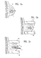
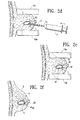
- FIGS. 2a-2g' are schematic, step by step showings of a preferred method for using a detectable marker 10 to mark the site of a lesion L that has been biopsied while the brest B is compressed within a mammography apparatus 16a and 16b.
- the detectable markers 10 of the present invention may be visible on an image created by the particular type or imaging device(s) available during the procedure.
- a form of roentgenographic imaging e.g., mammography, x-ray, fluoroscopy, CT, etc.
- the imageable marker 10 will thus comprise a material that is more or less radio-lucent or more or less radiopaque than the tissue surrounding the biopsy cavity (e.g., air, other gas, lipid, oil, a metal salt, barium powder, etc.) such that the marker 10 can be imaged by such x-ray means.
- ultrasound imaging may be used and the imageable marker will comprise a material or substance that has different ultrasound reflective properties (and possible different radiographic density) than the body tissue surrounding the biopsy cavity BC (e.g., air, carbon dioxide, other gasses, saline solution, other liquids, etc.)
- a magnetic imaging technique such as magnetic resonance imaging (MRI) may be used and the imageable marker 10 will comprise a ferromagnetic material or material (e.g., iron powder) having different magnetic density than the body tissue surrounding the biopsy cavity BC.
- MRI magnetic resonance imaging
- the marker 10 may be a substance or article that emits energy (e.g., radiation) that is detectable by an instrument (e.g., a gamma detector).
- energy e.g., radiation
- an instrument e.g., a gamma detector
- the preferred palpable embodiments of the invention preferable comprise a substance (e.g., a collagen material as described in U.S. Pat. No. 4,066,083 ) or an article (e.g., balloon(s), bead(s), etc.) that are sufficient mass to be palpated and located by tactile means while disposed within the biopsy site.
- a substance e.g., a collagen material as described in U.S. Pat. No. 4,066,083
- an article e.g., balloon(s), bead(s), etc.
- the preferred visible embodiments of the invention may comprise a colored substance such as a dye or colorant (e.g., methylene blue, gentian violet, indigo, dyes used in tattooing, etc.) or colorant particles (e.g., india, indigo, carbon particles or carbon preparations described in Langlois, S. L. P. and Carter, M. L., Carbon Localization of Impalpable Mammographic Abnormalities, Australas Radiol. 35: 237-241 (1991 ) and/or Svane, G. A Stereotaxis Technique for Preoperative Marking of Non-Palpable Breast Lesions, Acta Radiol. 24(2): 145-151 (1983 ).
- a dye or colorant e.g., methylene blue, gentian violet, indigo, dyes used in tattooing, etc.
- colorant particles e.g., india, indigo, carbon particles or carbon preparations described in Langlois, S. L. P. and Carter, M. L., Carbon Localization of
- the markers 10 of this invention may combine the attributes of any of the imageable, palpable and/or visible embodiments to provide for detection of the marker 10 by multiple means, such as a) imaging and palpation, b) imaging and visualization, c) imaging, palpation and visualization, or d) visualization and palpation.
- U.S. Pat. No. 4,847,049 (Yamamoto) describes an ion-impregnation or chelation technique whereby an ion may be impregnated or chelated to collagen for the purpose of imparting antimicrobial properties to the collagen preparation.
- imageable ions such as radiographically visible metal ions, may be bound to a bulky collagenous material to form a marker 10 that may be a) imaged by radiographic means and b) located by palpation of tissue surrounding the biopsy site.
- a silver ion-renatured collagen composition may be prepared by the following process:
- Renatured gelatin or a cross-linked gelatin preparation such as Gelfoam.TM. may be impregnated or combined with a metal ion to provide a gelatin-metal ion marker material.
- the gelatin may be prepared and ion-bound by the same method as set forth hereabove for collagen.
- the above-described collagen/gas or gelatin/gas marker 10 (with or without visible marker component) is introduced into the cavity created by removal of the biopsy specimen.
- the quantity of this marker 10 introduced may be sufficient to distend or stretch the biopsy cavity somewhat, thereby creating a more palpable and obvious mass of marker material at the biopsy site.
- the detectable markers 10 of the present invention are formulated and/or constructed so as not to move or migrate from the biopsy site when the surrounding tissue is flexed or reconfigured (e.g., as occurs when a breast is decompressed and removed from a mammography machine). Additionally, the detectable markers 10 are formulated and/or constructed to (i) remain present at the site in sufficient quantity to permit imaging and location of the site for at least two (2) weeks after introduction and (ii) clear sufficiently from the site to permit imaging of tissue adjacent to the site, without interference from said detectable marker, at six (6) months after introduction.
- the marker 10 is located at, and does not move or migrate from, the biopsy site it serves as a landmark that the surgeon may use to locate and treat or remove the remaining portion of the lesion without having to dissect and explore the surrounding tissue in attempting to locate the lesion.
- This aspect of the invention is particularly beneficial in cases (such as breast lumpectomy procedures) where it is desired to surgically remove the lesion L with minimal disfigurement, scarring or change in architecture of the surrounding tissue.
- the commencement of medical or surgical treatment of the lesion L may be delayed for up to two (2) weeks following the removal of the biopsy specimen and the marker 10 will still be present and useable to assist the treating surgeon or other physician in locating and directing treatment to the remaining portion of the lesion.
- This two (2) week minimum period of residence is especially beneficial in cases where immediate frozen sections can not be read by a pathologist, such as cases of suspected breast cancer wherein a small, non-palpable lesion of the breast has been biopsied by stereotactic biopsy, and the biopsy specimen is sent for routine histopathological evaluation (e.g., fixing, staining and microscopic examination) which takes several days to complete.
- the marker substantially clears from the biopsy site within six (6) months after its introduction, it will not interfere with or obscure subsequent diagnostic imaging of any remaining portion of the lesion L or the surrounding tissue.
- This six (6) month maximum residence time of the marker is especially beneficial in cases where the lesion is determined not to be cancerous at present, but presents a risk for future tumorigenesis that warrants periodic imaging of the site of the lesion L and surrounding tissue.
- the detectable marker 10 may comprise a detectable material that has pharmacokinetic properties (e.g., solubility, dissolution, potential for distribution from the biopsy site, potential for local metabolism or break down at the biopsy site) that cause it to remain present at the biopsy site in sufficient quantity to permit imaging of and location of the site for at least 2 weeks after its introduction, while clearing sufficiently from the site to permit imaging of tissue adjacent to the site without interference from said detectable marker, at 6 months after introduction.
- pharmacokinetic properties e.g., solubility, dissolution, potential for distribution from the biopsy site, potential for local metabolism or break down at the biopsy site
- the particular pharmacokinetic or biodistributive property(ies) that determine the rate at which the marker 10 clears from the biopsy site may include its solubility in the interstitial fluids that are present at the biopsy site.
- detectable material having a solubility coefficient of less than 1.times. 10.sup.-3 grams per 100 cubic centimeters of water
- detectable material will typically have the desired detectable residence time within the biopsy site of at least two (2) weeks but not more than 5 to 7 months, and preferably not more than about 6 months.
- the detectable residence time of the marker 10 at the biopsy site will additionally vary with the amount of marker 10 material that has been introduced in the biopsy cavity.
- a large volume of a material having a relatively high solubility coefficient can be introduced into the biopsy cavity to ensure that, even though the material has a relatively fast clearance rate, an imageable amount of the material will remain present at the biopsy site at the first time point (e.g., two (2) weeks).
- a relatively small volume of material having a low solubility coefficient may be introduced into the biopsy cavity and, due to its slow clearance rate, will remain imageable at the biopsy site at the first time period (e.g., two (2) weeks).
- radiographically visible materials that, if introduced into the biopsy site alone, would exhibit the desired detectable residence time (i.e., at least 2 weeks but not more than 6 weeks) include but are not necessarily limited to; AgCl; Agl; BaCO.sub.3 ; BaSO.sub.4 ; K; CaCO.sub.3 ; ZnO; Al.sub.2 O.sub.3 ; and the possible combinations thereof.
- the detectable marker 10 may comprise a detectable material that, if introduced into biopsy site alone, would clear substantially from the biopsy site in less than two (2) weeks after its introduction, thereby failing to provide the desired minimum detectable residence time at the biopsy site of at least two (2) weeks.
- the detectable material will be combined (e.g., mixed with, encapsulated by, suspended in, etc.) a clearance delaying element that will cause the detectable material to remain present at the biopsy site in sufficient quantity to permit imaging of and location of the site for at least 2 weeks after its introduction, while still allowing the detectable material to clear sufficiently from the biopsy site to permit imaging of tissue adjacent to the site without interference from said detectable marker, at 6 weeks after introduction.
- radiographically visible materials that would clear from most biopsy sites in less than two (2) weeks include but are not necessarily limited to; air, gas, lipid, oil, AgNO.sub.3 ; ammonium salts; sodium salts; potassium salts; ethiodized oil (Ethiodol available commercially from Savage Laboratories, Mellville, N.Y., and certain radiographic contrast agents such as iohexol (Omnipaque, available from Nyegaard-Schering AG, available from Squibb/Bristol Myers.
- clearance delaying elements that may be combined with the detectable material to form a detectable marker of the present invention include but are not necessarily limited to; polylactic acid; polyglycolic acid; polycapriolactone; an encapsulating membrane surrounding the detectable material.
- Formulation 1 Polylactic Acid 30-80 parts by weight Component Amount AgNo.sub.3 20-70 parts by weight Formulation 2 Polyglycolic Acid 50-90 parts by weight Component Amount Ethiodol 10-50 parts by weight Formulation 3 Thrombin 30-90 parts by weight Component Amount Ethiodol 10-70 parts by weight Topical Formulation 3 Air 30-50 parts by weight Component Amount Polylactic Acid 50-70 parts by weight
- FIGS. 3a-3c illustrate examples of the different possible forms or consistencies of detectable markers 10 of the present invention.
- FIG. 3a shows an example of a detectable marker 10a of a flowable consistency that is injectable through the lumen of the introduction cannula 15 into the biopsy cavity BC formed within the lesion L.
- flowable markers 10a will comprise a dry powder, suspension, or solution.
- a quantity of dry AgCl powder of 10-1000 micron particle size may be passed through the introduction cannula 15.
- FIG. 4 shows an example of an injector device 100 that is useable in place of the introduction cannula 15, to introduce a solid (e.g., powdered, particulate or granular) marker 10 of the present invention into a biopsy site.
- this device 100 comprises a non-tapered tubular barrel 110 having a substantially cylindrical inner wall 112 and a plunger 114 that is advanceable within the barrel 110.
- a quantity of a solid marker material of this invention is loaded into the barrel 110 of the device 100, the barrel 100 is inserted into the biopsy site, and the plunger 114 is advanced so as to expel the marker material out of the distal end of the barrel 110 and into the biopsy site.
- FIG. 3b shows an example of an detectable marker 10b that comprises a plurality of beads or pellets of approximately 10-1000 microns in diameter.
- Each bead or pellet may itself be formed of detectable material that is biodegradeable or otherwise clearable from the biopsy site so as to exhibit the desired detectable residence time as described hereabove such as silver chloride or silver nitrate.
- each bead or pellet may contain a detectable material in its interior and the outer surface of the bead or pellet may be a skin or encapsulating material that is biodegradeable such as polyactic acid, so as to provide for the desired detectable residence time within the biopsy site.
- markers 10b of the present invention Beads or pellets filled with air, carbon dioxide, or other suitable gas may be used as markers 10b of the present invention that are detectable by either ultrasound or x-ray.
- gas-filled markers 10b of the present invention are visible on x-ray, they will not obscure or block x-ray imaging of tissue adjacent to the biopsy cavity and, thus, need not biodegrade or clear from the biopsy site by the second time point (e.g., 5-7 months).
- FIG. 3c shows an example of a detectable marker 10c that comprises an inflatable balloon.
- a detectable marker 10c that comprises an inflatable balloon.
- Such inflatable balloon is passed through the introduction cannula and inflated within the biopsy cavity BC formed within the lesion L.
- the material of the balloon itself may be detectable and biodegradable or otherwise clearable from the biopsy site so as to exhibit the desired detectable residence time as described hereabove.
- a material is polyurethane that has been subjected to hydrolysis in situ.
- the balloon may contain a detectable material and the balloon itself may form a skin or encapsulating material (e.g., polyurethane) that is biodegradeable so as to provide for the desired detectable residence time within the biopsy site.
- Balloons filled with air, carbon dioxide, or other suitable gas may be used as markers 10c of the present invention that are detectable by either ultrasound or x-ray.
- gas-filled markers 10c of the present invention are visible on x-ray, they will not obscure or block x-ray imaging of tissue adjacent to the biopsy cavity and, thus, need not biodegrade or clear from the biopsy site by the second time point (e.g., 5-7 months).
- the marker 10 may have inherent adhesive properties, or the marker may further comprise an adhesive such as a polyurethane, polyacrylic compound, polyhydroxymethacrylate, fibrin glue (e.g., Tisseal.TM.), collagen adhesive, or other biological or biocompatable adhesive that will cause the marker to adhere to tissue adjacent the biopsy cavity BC.
- an adhesive such as a polyurethane, polyacrylic compound, polyhydroxymethacrylate, fibrin glue (e.g., Tisseal.TM.), collagen adhesive, or other biological or biocompatable adhesive that will cause the marker to adhere to tissue adjacent the biopsy cavity BC.
- Such optional adhesive will further ensure that the marker 10 does not migrate or move from the biopsy site as tissue surrounding the site is moved, flexed, compressed or decompressed.
- FIGS. 5a-5c show a preferred method for using a visually discernible marker 10 of the present invention to guide the excision and removal of tissues located within a specific band, region or location adjacent the boundaries of the biopsy cavity.
- a visible marker 10 has been introduced into the biopsy cavity BC so as to permit visualization of the boundary BOU of the biopsy cavity BC by the surgeon.
- Such visualization of the boundary BOU of this biopsy cavity BC can enable the surgeon to selectively remove tissue that is located within a general zone Z.sub.1 of potentially cancerous tissue surrounding the entire biopsy cavity BC ( FIG. 5b ) or within a specific zone Z.sub.2 located on only one side of the biopsy cavity BC ( FIG. 5c ).
- the removal of all tissue within a general zone Z.sub.1 surrounding the entire biopsy cavity BC may be desirable in cases where the pathology report has indicated that the previously removed biopsy specimen had no clear margin and, thus, it is desirable to remove all tissue within the zone Z.sub.1 of width X surrounding the biopsy cavity BC on all sides.
- the removal of certain tissue within a specific zone Z.sub.2 located to one side of an axis that has been projected throughout the biopsy cavity BC may be desirable in cases where the pathology report has indicated that the previously removed biopsy specimen had clear margins on all but one side and, thus, it is desirable to remove only tissue that is located within the specific zone Z.sub.1 of width X on one side of the biopsy cavity BC.
Landscapes
- Health & Medical Sciences (AREA)
- Life Sciences & Earth Sciences (AREA)
- Engineering & Computer Science (AREA)
- Biomedical Technology (AREA)
- Veterinary Medicine (AREA)
- Animal Behavior & Ethology (AREA)
- General Health & Medical Sciences (AREA)
- Public Health (AREA)
- Surgery (AREA)
- Heart & Thoracic Surgery (AREA)
- Medical Informatics (AREA)
- Nuclear Medicine, Radiotherapy & Molecular Imaging (AREA)
- Pathology (AREA)
- Oral & Maxillofacial Surgery (AREA)
- Molecular Biology (AREA)
- Oncology (AREA)
- Hematology (AREA)
- Physics & Mathematics (AREA)
- Optics & Photonics (AREA)
- Epidemiology (AREA)
- Dermatology (AREA)
- Anesthesiology (AREA)
- Biodiversity & Conservation Biology (AREA)
- Medicines Containing Antibodies Or Antigens For Use As Internal Diagnostic Agents (AREA)
- Apparatus For Radiation Diagnosis (AREA)
- Investigating Or Analysing Biological Materials (AREA)
- Infusion, Injection, And Reservoir Apparatuses (AREA)
- Dental Preparations (AREA)
- Ultra Sonic Daignosis Equipment (AREA)
Applications Claiming Priority (2)
| Application Number | Priority Date | Filing Date | Title |
|---|---|---|---|
| US09/241,936 US6161034A (en) | 1999-02-02 | 1999-02-02 | Methods and chemical preparations for time-limited marking of biopsy sites |
| EP00914446A EP1146910B1 (de) | 1999-02-02 | 2000-01-26 | Präparate zur zeitbegrenzten markierung von biopsie-orten |
Related Parent Applications (1)
| Application Number | Title | Priority Date | Filing Date |
|---|---|---|---|
| EP00914446.0 Division | 2000-01-26 |
Publications (1)
| Publication Number | Publication Date |
|---|---|
| EP2277553A1 true EP2277553A1 (de) | 2011-01-26 |
Family
ID=22912794
Family Applications (2)
| Application Number | Title | Priority Date | Filing Date |
|---|---|---|---|
| EP00914446A Expired - Lifetime EP1146910B1 (de) | 1999-02-02 | 2000-01-26 | Präparate zur zeitbegrenzten markierung von biopsie-orten |
| EP20100185263 Ceased EP2277553A1 (de) | 1999-02-02 | 2000-01-26 | Präparate zur zeitbegrenzte Markierung von Biopsie-Orten |
Family Applications Before (1)
| Application Number | Title | Priority Date | Filing Date |
|---|---|---|---|
| EP00914446A Expired - Lifetime EP1146910B1 (de) | 1999-02-02 | 2000-01-26 | Präparate zur zeitbegrenzten markierung von biopsie-orten |
Country Status (11)
| Country | Link |
|---|---|
| US (9) | US6161034A (de) |
| EP (2) | EP1146910B1 (de) |
| JP (1) | JP2003517453A (de) |
| AT (1) | ATE484296T1 (de) |
| AU (1) | AU3583500A (de) |
| CA (1) | CA2361530C (de) |
| DE (1) | DE60045094D1 (de) |
| DK (1) | DK1146910T3 (de) |
| ES (1) | ES2353684T3 (de) |
| PT (1) | PT1146910E (de) |
| WO (1) | WO2000045854A2 (de) |
Cited By (3)
| Publication number | Priority date | Publication date | Assignee | Title |
|---|---|---|---|---|
| CN106214269A (zh) * | 2016-08-03 | 2016-12-14 | 杨晓民 | 一种用于乳腺溢液乳管切除术的定位与引导针 |
| CN108969091A (zh) * | 2018-06-29 | 2018-12-11 | 安徽锐捷信息科技有限公司 | 一种肿瘤治疗系统 |
| WO2019055336A1 (en) * | 2017-09-13 | 2019-03-21 | Hologic, Inc. | ACTIVE WIRELESS SURVEILLANCE IMPLANT SYSTEM |
Families Citing this family (294)
| Publication number | Priority date | Publication date | Assignee | Title |
|---|---|---|---|---|
| EP1304085A3 (de) | 1994-09-16 | 2004-01-21 | Ethicon Endo-Surgery, Inc. | Aus biologisch abbaubarem Material hergestellte Gewebemarkierungsvorrichtung |
| US6162192A (en) | 1998-05-01 | 2000-12-19 | Sub Q, Inc. | System and method for facilitating hemostasis of blood vessel punctures with absorbable sponge |
| US6183497B1 (en) | 1998-05-01 | 2001-02-06 | Sub-Q, Inc. | Absorbable sponge with contrasting agent |
| US6071300A (en) | 1995-09-15 | 2000-06-06 | Sub-Q Inc. | Apparatus and method for percutaneous sealing of blood vessel punctures |
| US6071301A (en) | 1998-05-01 | 2000-06-06 | Sub Q., Inc. | Device and method for facilitating hemostasis of a biopsy tract |
| US8288745B2 (en) | 1997-10-10 | 2012-10-16 | Senorx, Inc. | Method of utilizing an implant for targeting external beam radiation |
| US7637948B2 (en) * | 1997-10-10 | 2009-12-29 | Senorx, Inc. | Tissue marking implant |
| US8668737B2 (en) | 1997-10-10 | 2014-03-11 | Senorx, Inc. | Tissue marking implant |
| US20100030256A1 (en) | 1997-11-12 | 2010-02-04 | Genesis Technologies Llc | Medical Devices and Methods |
| US9498604B2 (en) | 1997-11-12 | 2016-11-22 | Genesis Technologies Llc | Medical device and method |
| US6238412B1 (en) * | 1997-11-12 | 2001-05-29 | William Dubrul | Biological passageway occlusion removal |
| US6270464B1 (en) * | 1998-06-22 | 2001-08-07 | Artemis Medical, Inc. | Biopsy localization method and device |
| US6602204B2 (en) | 1998-02-10 | 2003-08-05 | Artemis Medical, Inc | Intraoperative tissue treatment methods |
| US20040010206A1 (en) * | 1998-02-10 | 2004-01-15 | Dubrul William R. | Intraoperative tissue treatment methods |
| US6530923B1 (en) * | 1998-02-10 | 2003-03-11 | Artemis Medical, Inc. | Tissue removal methods and apparatus |
| US6221006B1 (en) | 1998-02-10 | 2001-04-24 | Artemis Medical Inc. | Entrapping apparatus and method for use |
| US6471700B1 (en) | 1998-04-08 | 2002-10-29 | Senorx, Inc. | Apparatus and method for accessing biopsy site |
| US6758848B2 (en) | 1998-03-03 | 2004-07-06 | Senorx, Inc. | Apparatus and method for accessing a body site |
| US6638234B2 (en) | 1998-03-03 | 2003-10-28 | Senorx, Inc. | Sentinel node location and biopsy |
| US6875182B2 (en) | 1998-03-03 | 2005-04-05 | Senorx, Inc. | Electrosurgical specimen-collection system |
| US6497706B1 (en) | 1998-03-03 | 2002-12-24 | Senorx, Inc. | Biopsy device and method of use |
| US6161034A (en) * | 1999-02-02 | 2000-12-12 | Senorx, Inc. | Methods and chemical preparations for time-limited marking of biopsy sites |
| US6997885B2 (en) | 1998-04-08 | 2006-02-14 | Senorx, Inc. | Dilation devices and methods for removing tissue specimens |
| US6540695B1 (en) | 1998-04-08 | 2003-04-01 | Senorx, Inc. | Biopsy anchor device with cutter |
| US6347241B2 (en) * | 1999-02-02 | 2002-02-12 | Senorx, Inc. | Ultrasonic and x-ray detectable biopsy site marker and apparatus for applying it |
| US7625352B1 (en) | 1998-05-01 | 2009-12-01 | Sub-Q, Inc. | Depth and puncture control for system for hemostasis of blood vessel |
| US20010045575A1 (en) | 1998-05-01 | 2001-11-29 | Mark Ashby | Device and method for facilitating hemostasis of a biopsy tract |
| US6315753B1 (en) | 1998-05-01 | 2001-11-13 | Sub-Q, Inc. | System and method for facilitating hemostasis of blood vessel punctures with absorbable sponge |
| US6200328B1 (en) | 1998-05-01 | 2001-03-13 | Sub Q, Incorporated | Device and method for facilitating hemostasis of a biopsy tract |
| US6610026B2 (en) | 1998-05-01 | 2003-08-26 | Sub-Q, Inc. | Method of hydrating a sponge material for delivery to a body |
| US20020058882A1 (en) * | 1998-06-22 | 2002-05-16 | Artemis Medical, Incorporated | Biopsy localization method and device |
| US6179860B1 (en) | 1998-08-19 | 2001-01-30 | Artemis Medical, Inc. | Target tissue localization device and method |
| US6679851B2 (en) | 1998-09-01 | 2004-01-20 | Senorx, Inc. | Tissue accessing and anchoring device and method |
| US6440147B1 (en) | 1998-09-03 | 2002-08-27 | Rubicor Medical, Inc. | Excisional biopsy devices and methods |
| US6936014B2 (en) | 2002-10-16 | 2005-08-30 | Rubicor Medical, Inc. | Devices and methods for performing procedures on a breast |
| US7517348B2 (en) | 1998-09-03 | 2009-04-14 | Rubicor Medical, Inc. | Devices and methods for performing procedures on a breast |
| US7329253B2 (en) | 2003-12-09 | 2008-02-12 | Rubicor Medical, Inc. | Suction sleeve and interventional devices having such a suction sleeve |
| US6022362A (en) | 1998-09-03 | 2000-02-08 | Rubicor Medical, Inc. | Excisional biopsy devices and methods |
| US6036698A (en) | 1998-10-30 | 2000-03-14 | Vivant Medical, Inc. | Expandable ring percutaneous tissue removal device |
| US6356782B1 (en) * | 1998-12-24 | 2002-03-12 | Vivant Medical, Inc. | Subcutaneous cavity marking device and method |
| US9669113B1 (en) | 1998-12-24 | 2017-06-06 | Devicor Medical Products, Inc. | Device and method for safe location and marking of a biopsy cavity |
| US6371904B1 (en) | 1998-12-24 | 2002-04-16 | Vivant Medical, Inc. | Subcutaneous cavity marking device and method |
| US8361082B2 (en) * | 1999-02-02 | 2013-01-29 | Senorx, Inc. | Marker delivery device with releasable plug |
| US9820824B2 (en) | 1999-02-02 | 2017-11-21 | Senorx, Inc. | Deployment of polysaccharide markers for treating a site within a patent |
| US20080039819A1 (en) * | 2006-08-04 | 2008-02-14 | Senorx, Inc. | Marker formed of starch or other suitable polysaccharide |
| US7651505B2 (en) * | 2002-06-17 | 2010-01-26 | Senorx, Inc. | Plugged tip delivery for marker placement |
| US8498693B2 (en) | 1999-02-02 | 2013-07-30 | Senorx, Inc. | Intracorporeal marker and marker delivery device |
| US20090030309A1 (en) | 2007-07-26 | 2009-01-29 | Senorx, Inc. | Deployment of polysaccharide markers |
| US7983734B2 (en) | 2003-05-23 | 2011-07-19 | Senorx, Inc. | Fibrous marker and intracorporeal delivery thereof |
| US6862470B2 (en) | 1999-02-02 | 2005-03-01 | Senorx, Inc. | Cavity-filling biopsy site markers |
| US6725083B1 (en) | 1999-02-02 | 2004-04-20 | Senorx, Inc. | Tissue site markers for in VIVO imaging |
| EP1156741B1 (de) | 1999-02-10 | 2010-12-22 | Sub-Q, Inc. | Gerät zur vereinfachung der hämostase eines biopsietunnels |
| US6575991B1 (en) * | 1999-06-17 | 2003-06-10 | Inrad, Inc. | Apparatus for the percutaneous marking of a lesion |
| US6984219B2 (en) | 1999-09-23 | 2006-01-10 | Mark Ashby | Depth and puncture control for blood vessel hemostasis system |
| US7695492B1 (en) | 1999-09-23 | 2010-04-13 | Boston Scientific Scimed, Inc. | Enhanced bleed back system |
| US6675037B1 (en) * | 1999-09-29 | 2004-01-06 | Regents Of The University Of Minnesota | MRI-guided interventional mammary procedures |
| US6782289B1 (en) * | 1999-10-08 | 2004-08-24 | The Board Of Trustees Of The Leland Stanford Junior University | Methods and apparatus for characterizing lesions in blood vessels and other body lumens |
| US6350244B1 (en) * | 2000-02-21 | 2002-02-26 | Biopsy Sciences, Llc | Bioabsorable markers for use in biopsy procedures |
| US6540735B1 (en) | 2000-05-12 | 2003-04-01 | Sub-Q, Inc. | System and method for facilitating hemostasis of blood vessel punctures with absorbable sponge |
| US6355275B1 (en) | 2000-06-23 | 2002-03-12 | Carbon Medical Technologies, Inc. | Embolization using carbon coated microparticles |
| US6394965B1 (en) | 2000-08-15 | 2002-05-28 | Carbon Medical Technologies, Inc. | Tissue marking using biocompatible microparticles |
| US8909325B2 (en) | 2000-08-21 | 2014-12-09 | Biosensors International Group, Ltd. | Radioactive emission detector equipped with a position tracking system and utilization thereof with medical systems and in medical procedures |
| US8565860B2 (en) | 2000-08-21 | 2013-10-22 | Biosensors International Group, Ltd. | Radioactive emission detector equipped with a position tracking system |
| US8489176B1 (en) | 2000-08-21 | 2013-07-16 | Spectrum Dynamics Llc | Radioactive emission detector equipped with a position tracking system and utilization thereof with medical systems and in medical procedures |
| US7201725B1 (en) | 2000-09-25 | 2007-04-10 | Sub-Q, Inc. | Device and method for determining a depth of an incision |
| AU2002211449A1 (en) * | 2000-10-04 | 2002-04-15 | James Peter Amis | Non-scatterable, radio-opaque marker for medical imaging applications |
| US6544185B2 (en) | 2000-10-23 | 2003-04-08 | Valentino Montegrande | Ultrasound imaging marker and method of use |
| CA2775170C (en) | 2000-11-20 | 2017-09-05 | Senorx, Inc. | An intracorporeal marker delivery system for marking a tissue site |
| US20020072739A1 (en) | 2000-12-07 | 2002-06-13 | Roberta Lee | Methods and devices for radiofrequency electrosurgery |
| WO2002087636A1 (en) | 2001-03-12 | 2002-11-07 | Sub-Q, Inc. | Methods for sterilizing cross-linked gelatin compositions |
| US8187625B2 (en) | 2001-03-12 | 2012-05-29 | Boston Scientific Scimed, Inc. | Cross-linked gelatin composition comprising a wetting agent |
| US20020143248A1 (en) * | 2001-04-03 | 2002-10-03 | Johnson G. Allan | MR microscopy of whole specimens, individual organs or tissue specimens perfused simultaneously with fixative and MR contrast agent |
| US7094205B2 (en) * | 2001-04-06 | 2006-08-22 | Alfred E. Mann Institute For Biomedical Engineering At The University Of Southern California | High-resolution 3D ultrasonic transmission imaging |
| US7029489B1 (en) | 2001-05-18 | 2006-04-18 | Sub-Q, Inc. | System and method for delivering hemostasis promoting material to a blood vessel puncture site |
| US6863680B2 (en) | 2001-11-08 | 2005-03-08 | Sub-Q, Inc. | System and method for delivering hemostasis promoting material to a blood vessel puncture site by fluid pressure |
| US7008440B2 (en) | 2001-11-08 | 2006-03-07 | Sub-Q, Inc. | System and method for delivering hemostasis promoting material to a blood vessel puncture site by fluid pressure |
| WO2003003907A2 (en) * | 2001-07-06 | 2003-01-16 | Wisconsin Alumni Research Foundation | Space-time microwave imaging for cancer detection |
| EP1411851B1 (de) * | 2001-08-03 | 2006-11-02 | Tyco Healthcare Group LP | Marker zur verwendung mit einem gewebemarkierungsgerät |
| GB0120645D0 (en) | 2001-08-24 | 2001-10-17 | Smiths Group Plc | Medico-surgical devices |
| US6605047B2 (en) * | 2001-09-10 | 2003-08-12 | Vivant Medical, Inc. | Biopsy marker delivery system |
| US7037322B1 (en) | 2001-11-08 | 2006-05-02 | Sub-Q, Inc. | System and method for delivering hemostasis promoting material to a blood vessel puncture with a staging tube |
| US7025748B2 (en) | 2001-11-08 | 2006-04-11 | Boston Scientific Scimed, Inc. | Sheath based blood vessel puncture locator and depth indicator |
| US7037323B2 (en) | 2001-11-08 | 2006-05-02 | Sub-Q, Inc. | Pledget-handling system and method for delivering hemostasis promoting material to a blood vessel puncture site by fluid pressure |
| US7192436B2 (en) | 2001-11-08 | 2007-03-20 | Sub-Q, Inc. | Pledget-handling system and method for delivering hemostasis promoting material to a blood vessel puncture site by fluid pressure |
| DE10156178A1 (de) * | 2001-11-15 | 2003-06-05 | Philips Intellectual Property | Mammographie-Zusatz für MR-Elastographie |
| US7505811B2 (en) * | 2001-11-19 | 2009-03-17 | Dune Medical Devices Ltd. | Method and apparatus for examining tissue for predefined target cells, particularly cancerous cells, and a probe useful in such method and apparatus |
| US7769432B2 (en) * | 2001-12-10 | 2010-08-03 | Board Of Trustees Of The University Of Arkansas | Minimally invasive diagnosis and treatment for breast cancer |
| US6654629B2 (en) | 2002-01-23 | 2003-11-25 | Valentino Montegrande | Implantable biomarker and method of use |
| US7769426B2 (en) * | 2002-04-23 | 2010-08-03 | Ethicon Endo-Surgery, Inc. | Method for using an MRI compatible biopsy device with detachable probe |
| US7329414B2 (en) * | 2002-05-03 | 2008-02-12 | Biopsy Sciences, Llc | Biodegradable polymer for marking tissue and sealing tracts |
| AU2003233510A1 (en) * | 2002-05-07 | 2003-11-11 | Polyremedy Llc | Method for treating wound, dressing for use therewith and apparatus and system for fabricating dressing |
| US8407065B2 (en) * | 2002-05-07 | 2013-03-26 | Polyremedy, Inc. | Wound care treatment service using automatic wound dressing fabricator |
| US6780179B2 (en) * | 2002-05-22 | 2004-08-24 | Rubicor Medical, Inc. | Methods and systems for in situ tissue marking and orientation stabilization |
| US7044956B2 (en) | 2002-07-03 | 2006-05-16 | Rubicor Medical, Inc. | Methods and devices for cutting and collecting soft tissue |
| EP1545316B1 (de) * | 2002-08-01 | 2008-01-09 | James E. Selis | Biopsievorrichtungen |
| US8123698B2 (en) * | 2002-10-07 | 2012-02-28 | Suros Surgical Systems, Inc. | System and method for minimally invasive disease therapy |
| US20070260267A1 (en) * | 2002-10-07 | 2007-11-08 | Nicoson Zachary R | Localizing obturator |
| US7347829B2 (en) * | 2002-10-07 | 2008-03-25 | Suros Surgical Systems, Inc. | Introduction system for minimally invasive surgical instruments |
| US20080161720A1 (en) * | 2002-10-07 | 2008-07-03 | Nicoson Zachary R | Registration system |
| US7455680B1 (en) | 2002-11-04 | 2008-11-25 | Boston Scientific Scimed, Inc. | Apparatus and method for inhibiting blood loss |
| US8317821B1 (en) | 2002-11-04 | 2012-11-27 | Boston Scientific Scimed, Inc. | Release mechanism |
| US7955353B1 (en) | 2002-11-04 | 2011-06-07 | Sub-Q, Inc. | Dissolvable closure device |
| US7029451B2 (en) | 2002-11-06 | 2006-04-18 | Rubicor Medical, Inc. | Excisional devices having selective cutting and atraumatic configurations and methods of using same |
| US20060036158A1 (en) | 2003-11-17 | 2006-02-16 | Inrad, Inc. | Self-contained, self-piercing, side-expelling marking apparatus |
| CA2505961C (en) | 2002-11-18 | 2011-10-11 | Inrad, Inc. | Apparatus for implanting a preloaded localization wire |
| WO2004064045A2 (en) * | 2003-01-13 | 2004-07-29 | Disney Enterprises Inc. | Fast play dvd |
| GB0307350D0 (en) | 2003-03-29 | 2003-05-07 | Smiths Group Plc | Catheters |
| US7877133B2 (en) | 2003-05-23 | 2011-01-25 | Senorx, Inc. | Marker or filler forming fluid |
| US20050119562A1 (en) * | 2003-05-23 | 2005-06-02 | Senorx, Inc. | Fibrous marker formed of synthetic polymer strands |
| US7783336B2 (en) | 2003-06-06 | 2010-08-24 | Ethicon Endo-Surgery, Inc. | Subcutaneous biopsy cavity marker device |
| US7122011B2 (en) | 2003-06-18 | 2006-10-17 | Rubicor Medical, Inc. | Methods and devices for cutting and collecting soft tissue |
| US20050020899A1 (en) * | 2003-07-25 | 2005-01-27 | Rubicor Medical, Inc. | Post-biopsy cavity treatmetn implants and methods |
| US7744852B2 (en) | 2003-07-25 | 2010-06-29 | Rubicor Medical, Llc | Methods and systems for marking post biopsy cavity sites |
| US7537788B2 (en) | 2003-07-25 | 2009-05-26 | Rubicor Medical, Inc. | Post-biopsy cavity treatment implants and methods |
| US20050033157A1 (en) * | 2003-07-25 | 2005-02-10 | Klein Dean A. | Multi-modality marking material and method |
| US7001341B2 (en) * | 2003-08-13 | 2006-02-21 | Scimed Life Systems, Inc. | Marking biopsy sites |
| US8172770B2 (en) * | 2005-09-28 | 2012-05-08 | Suros Surgical Systems, Inc. | System and method for minimally invasive disease therapy |
| US20120289859A9 (en) * | 2003-08-27 | 2012-11-15 | Nicoson Zachary R | System and method for minimally invasive disease therapy |
| US8048003B2 (en) | 2003-10-14 | 2011-11-01 | Suros Surgical Systems, Inc. | Vacuum assisted biopsy device |
| US8357103B2 (en) | 2003-10-14 | 2013-01-22 | Suros Surgical Systems, Inc. | Vacuum assisted biopsy needle set |
| US7988642B2 (en) | 2003-10-14 | 2011-08-02 | Suros Surgical Systems, Inc. | Vacuum assisted biopsy device |
| US7390306B2 (en) | 2003-10-14 | 2008-06-24 | Suros Surgical Systems, Inc. | Vacuum assisted biopsy needle set |
| US20050273002A1 (en) | 2004-06-04 | 2005-12-08 | Goosen Ryan L | Multi-mode imaging marker |
| US7875043B1 (en) | 2003-12-09 | 2011-01-25 | Sub-Q, Inc. | Cinching loop |
| US7049652B2 (en) * | 2003-12-10 | 2006-05-23 | Sandisk Corporation | Pillar cell flash memory technology |
| US9408592B2 (en) | 2003-12-23 | 2016-08-09 | Senorx, Inc. | Biopsy device with aperture orientation and improved tip |
| US9470801B2 (en) | 2004-01-13 | 2016-10-18 | Spectrum Dynamics Llc | Gating with anatomically varying durations |
| US9040016B2 (en) | 2004-01-13 | 2015-05-26 | Biosensors International Group, Ltd. | Diagnostic kit and methods for radioimaging myocardial perfusion |
| US10964075B2 (en) | 2004-01-13 | 2021-03-30 | Spectrum Dynamics Llc | Gating with anatomically varying durations |
| EP1709585B1 (de) | 2004-01-13 | 2020-01-08 | Spectrum Dynamics Medical Limited | Mehrdimensionale bildrekonstruktion |
| US8571881B2 (en) | 2004-11-09 | 2013-10-29 | Spectrum Dynamics, Llc | Radiopharmaceutical dispensing, administration, and imaging |
| US8586932B2 (en) | 2004-11-09 | 2013-11-19 | Spectrum Dynamics Llc | System and method for radioactive emission measurement |
| US7637928B2 (en) | 2004-01-26 | 2009-12-29 | Synthes Usa, Llc | Variable angle locked bone fixation system |
| US20050234336A1 (en) * | 2004-03-26 | 2005-10-20 | Beckman Andrew T | Apparatus and method for marking tissue |
| US7465279B2 (en) * | 2004-03-31 | 2008-12-16 | Ethicon Endo-Surgery, Inc. | Marker device and method of deploying a cavity marker using a surgical biopsy device |
| US20050255045A1 (en) * | 2004-05-13 | 2005-11-17 | Woltering Eugene A | Surgical marking composition and method |
| US9638770B2 (en) | 2004-05-21 | 2017-05-02 | Devicor Medical Products, Inc. | MRI biopsy apparatus incorporating an imageable penetrating portion |
| US8932233B2 (en) | 2004-05-21 | 2015-01-13 | Devicor Medical Products, Inc. | MRI biopsy device |
| US7708751B2 (en) | 2004-05-21 | 2010-05-04 | Ethicon Endo-Surgery, Inc. | MRI biopsy device |
| EP1778957A4 (de) | 2004-06-01 | 2015-12-23 | Biosensors Int Group Ltd | Optimierung der messung radioaktiver strahlung an besonderen körperstrukturen |
| US8075568B2 (en) * | 2004-06-11 | 2011-12-13 | Selis James E | Biopsy devices and methods |
| EP1778142A4 (de) * | 2004-07-16 | 2011-02-02 | Polyremedy Inc | Wundverband und gerät zur herstellung |
| WO2006022786A1 (en) * | 2004-08-20 | 2006-03-02 | David Mullen | Tissue marking devices and systems |
| US8050740B2 (en) * | 2004-09-15 | 2011-11-01 | Wisconsin Alumni Research Foundation | Microwave-based examination using hypothesis testing |
| EP1804918A1 (de) * | 2004-10-01 | 2007-07-11 | Calypso Medical Technologies, INC. | Systeme und verfahren zur behandlung eines patienten mit strahlentherapie |
| US8442623B2 (en) * | 2004-10-13 | 2013-05-14 | Suros Surgical Systems, Inc. | Site marker visible under multiple modalities |
| US8280486B2 (en) | 2004-10-13 | 2012-10-02 | Suros Surgical Systems, Inc. | Site marker visable under multiple modalities |
| US8060183B2 (en) | 2004-10-13 | 2011-11-15 | Suros Surgical Systems, Inc. | Site marker visible under multiple modalities |
| US20060079805A1 (en) * | 2004-10-13 | 2006-04-13 | Miller Michael E | Site marker visable under multiple modalities |
| US9316743B2 (en) | 2004-11-09 | 2016-04-19 | Biosensors International Group, Ltd. | System and method for radioactive emission measurement |
| US8615405B2 (en) | 2004-11-09 | 2013-12-24 | Biosensors International Group, Ltd. | Imaging system customization using data from radiopharmaceutical-associated data carrier |
| US8000773B2 (en) | 2004-11-09 | 2011-08-16 | Spectrum Dynamics Llc | Radioimaging |
| US8606349B2 (en) | 2004-11-09 | 2013-12-10 | Biosensors International Group, Ltd. | Radioimaging using low dose isotope |
| US10136865B2 (en) | 2004-11-09 | 2018-11-27 | Spectrum Dynamics Medical Limited | Radioimaging using low dose isotope |
| WO2008059489A2 (en) | 2006-11-13 | 2008-05-22 | Spectrum Dynamics Llc | Radioimaging applications of and novel formulations of teboroxime |
| US8409111B2 (en) | 2004-11-22 | 2013-04-02 | Bard Peripheral Vascular, Inc. | Removable localizing wire |
| US8419656B2 (en) * | 2004-11-22 | 2013-04-16 | Bard Peripheral Vascular, Inc. | Post decompression marker introducer system |
| US8360990B2 (en) | 2004-12-16 | 2013-01-29 | Senorx, Inc. | Biopsy device with aperture orientation and improved tip |
| US7647089B2 (en) * | 2005-01-28 | 2010-01-12 | Wisconsin Alumni Research Foundation | Surface identification using microwave signals for microwave-based detection of cancer |
| US7809427B2 (en) * | 2005-02-11 | 2010-10-05 | Wisconsin Alumni Research Foundation | Time domain inverse scattering techniques for use in microwave imaging |
| US7907991B2 (en) * | 2005-03-02 | 2011-03-15 | C. R. Bard, Inc. | System and method for marking body cavities |
| WO2006102348A1 (en) * | 2005-03-22 | 2006-09-28 | Bayer Healthcare Llc | Packaging container for test sensors |
| US20060224082A1 (en) * | 2005-04-05 | 2006-10-05 | Vetter James W | Methods and devices for removing tissue from a patient and placing a marker in the patient |
| US10357328B2 (en) | 2005-04-20 | 2019-07-23 | Bard Peripheral Vascular, Inc. and Bard Shannon Limited | Marking device with retractable cannula |
| GB2425610A (en) * | 2005-04-29 | 2006-11-01 | Univ London | Magnetic properties sensing system |
| US20060293581A1 (en) * | 2005-05-12 | 2006-12-28 | Sunnybrook And Women's College Health Sciences Centre | Marker device for X-ray, ultrasound and MR imaging |
| US9095325B2 (en) | 2005-05-23 | 2015-08-04 | Senorx, Inc. | Tissue cutting member for a biopsy device |
| US20070010844A1 (en) * | 2005-07-08 | 2007-01-11 | Gorman Gong | Radiopaque expandable body and methods |
| US8837793B2 (en) | 2005-07-19 | 2014-09-16 | Biosensors International Group, Ltd. | Reconstruction stabilizer and active vision |
| EP1909853B1 (de) | 2005-07-19 | 2015-03-18 | Biosensors International Group, Ltd. | Bildgebungsprotokolle |
| US7572236B2 (en) | 2005-08-05 | 2009-08-11 | Senorx, Inc. | Biopsy device with fluid delivery to tissue specimens |
| US8317725B2 (en) | 2005-08-05 | 2012-11-27 | Senorx, Inc. | Biopsy device with fluid delivery to tissue specimens |
| EP1922011B1 (de) * | 2005-08-11 | 2012-05-02 | Navotek Medical Ltd. | Lokalisation einer radioaktiven quelle |
| US20080200834A1 (en) * | 2005-09-28 | 2008-08-21 | Mark Joseph L | Introducer device for improved imaging |
| CA2562580C (en) | 2005-10-07 | 2014-04-29 | Inrad, Inc. | Drug-eluting tissue marker |
| US7702378B2 (en) * | 2005-11-17 | 2010-04-20 | Breast-Med, Inc. | Tissue marker for multimodality radiographic imaging |
| US11241296B2 (en) | 2005-11-17 | 2022-02-08 | Breast-Med, Inc. | Imaging fiducial markers and methods |
| WO2007087447A2 (en) * | 2006-01-25 | 2007-08-02 | Health Beacens, Inc. | Surgical procedure |
| US20080230001A1 (en) * | 2006-02-23 | 2008-09-25 | Meadwestvaco Corporation | Method for treating a substrate |
| US20170066162A9 (en) | 2006-03-28 | 2017-03-09 | Devicor Medical Products, Inc. | Method of Enhancing Ultrasound Visibility of Hyperechoic Materials |
| US11129690B2 (en) | 2006-03-28 | 2021-09-28 | Devicor Medical Products, Inc. | Method for making hydrogel markers |
| US8939910B2 (en) | 2006-03-28 | 2015-01-27 | Devicor Medical Products, Inc. | Method for enhancing ultrasound visibility of hyperechoic materials |
| US8894974B2 (en) | 2006-05-11 | 2014-11-25 | Spectrum Dynamics Llc | Radiopharmaceuticals for diagnosis and therapy |
| WO2007145926A2 (en) | 2006-06-05 | 2007-12-21 | Senorx, Inc. | Biopsy system with integrated ultrasonic imaging |
| US20080294039A1 (en) * | 2006-08-04 | 2008-11-27 | Senorx, Inc. | Assembly with hemostatic and radiographically detectable pellets |
| US7945307B2 (en) | 2006-08-04 | 2011-05-17 | Senorx, Inc. | Marker delivery system with obturator |
| EP2079385B1 (de) | 2006-10-23 | 2013-11-20 | C.R.Bard, Inc. | Brustkennzeichner |
| WO2008088424A1 (en) * | 2006-11-01 | 2008-07-24 | Infection Prevention Systems, Inc. | Hand hygiene verification/tracking system and method |
| US20090237651A1 (en) * | 2006-11-01 | 2009-09-24 | Douglas C Arndt | Verifiable hand cleansing formulation and method |
| US9579077B2 (en) | 2006-12-12 | 2017-02-28 | C.R. Bard, Inc. | Multiple imaging mode tissue marker |
| US8401622B2 (en) | 2006-12-18 | 2013-03-19 | C. R. Bard, Inc. | Biopsy marker with in situ-generated imaging properties |
| WO2008075362A2 (en) | 2006-12-20 | 2008-06-26 | Spectrum Dynamics Llc | A method, a system, and an apparatus for using and processing multidimensional data |
| US8237007B2 (en) * | 2007-01-10 | 2012-08-07 | Polyremedy, Inc. | Wound dressing with controllable permeability |
| CA2714069A1 (en) * | 2007-02-07 | 2008-08-14 | Massachusetts Institute Of Technology | Methods and devices for sensing tissues and tissue compartments |
| US8137320B2 (en) | 2007-05-01 | 2012-03-20 | Suros Surgical Systems, Inc. | Securement for a surgical site marker and deployment device for same |
| US20080287729A1 (en) * | 2007-05-16 | 2008-11-20 | Anthony Biscotti | Interstitial marker and method for creation thereof |
| US20100241160A1 (en) * | 2007-06-11 | 2010-09-23 | Kieran Murphy | Method and kit for cyst aspiration and treatment |
| US20090024225A1 (en) * | 2007-07-16 | 2009-01-22 | Stubbs James B | Implant for Targeting Therapeutic Procedure |
| US20090068116A1 (en) * | 2007-09-12 | 2009-03-12 | Infection Prevention System, Inc. | Chemical system with self-timing indicator |
| US8808200B2 (en) | 2007-10-01 | 2014-08-19 | Suros Surgical Systems, Inc. | Surgical device and method of using same |
| US8202229B2 (en) | 2007-10-01 | 2012-06-19 | Suros Surgical Systems, Inc. | Surgical device |
| US8521253B2 (en) * | 2007-10-29 | 2013-08-27 | Spectrum Dynamics Llc | Prostate imaging |
| US8457757B2 (en) | 2007-11-26 | 2013-06-04 | Micro Transponder, Inc. | Implantable transponder systems and methods |
| US9089707B2 (en) | 2008-07-02 | 2015-07-28 | The Board Of Regents, The University Of Texas System | Systems, methods and devices for paired plasticity |
| US8311610B2 (en) | 2008-01-31 | 2012-11-13 | C. R. Bard, Inc. | Biopsy tissue marker |
| US8079964B2 (en) * | 2008-02-25 | 2011-12-20 | Devicor Medical Products, Inc. | Method and apparatus for inserting biopsy site marker in marker body |
| US8068895B2 (en) * | 2008-02-25 | 2011-11-29 | Devicor Medical Products, Inc. | Biopsy site marker deployment instrument |
| US20090217932A1 (en) * | 2008-03-03 | 2009-09-03 | Ethicon Endo-Surgery, Inc. | Intraluminal tissue markers |
| US20090247900A1 (en) * | 2008-03-25 | 2009-10-01 | Brian Zimmer | Push button adjustable spacer |
| US20090247901A1 (en) * | 2008-03-25 | 2009-10-01 | Brian Zimmer | Latching side removal spacer |
| US8532748B2 (en) * | 2008-04-23 | 2013-09-10 | Devicor Medical Products, Inc. | Devices useful in imaging |
| AU2009201610A1 (en) * | 2008-04-23 | 2009-11-19 | Devicor Medical Products, Inc. | PEM and BSGI biopsy devices and methods |
| US8864681B2 (en) * | 2008-04-23 | 2014-10-21 | Devicor Medical Products, Inc. | Biopsy devices |
| US20100241447A1 (en) * | 2008-04-25 | 2010-09-23 | Polyremedy, Inc. | Customization of wound dressing using rule-based algorithm |
| US8043316B2 (en) * | 2008-05-02 | 2011-10-25 | Suros Surgical Systems, Inc. | Adjustable spacer |
| US8237009B2 (en) * | 2008-06-30 | 2012-08-07 | Polyremedy, Inc. | Custom patterned wound dressings having patterned fluid flow barriers and methods of manufacturing and using same |
| EP2326276B1 (de) * | 2008-08-18 | 2014-04-30 | Naviswiss AG | Medizinisches messsystem und die verwendung dieses medizinischen messsystems |
| US8247634B2 (en) * | 2008-08-22 | 2012-08-21 | Polyremedy, Inc. | Expansion units for attachment to custom patterned wound dressings and custom patterned wound dressings adapted to interface with same |
| US9327061B2 (en) | 2008-09-23 | 2016-05-03 | Senorx, Inc. | Porous bioabsorbable implant |
| US20110190662A1 (en) * | 2008-10-01 | 2011-08-04 | Beacon Endoscopic Corporation | Rapid exchange fna biopsy device with diagnostic and therapeutic capabilities |
| US9782565B2 (en) | 2008-10-01 | 2017-10-10 | Covidien Lp | Endoscopic ultrasound-guided biliary access system |
| US8968210B2 (en) | 2008-10-01 | 2015-03-03 | Covidien LLP | Device for needle biopsy with integrated needle protection |
| US11298113B2 (en) | 2008-10-01 | 2022-04-12 | Covidien Lp | Device for needle biopsy with integrated needle protection |
| US9186128B2 (en) | 2008-10-01 | 2015-11-17 | Covidien Lp | Needle biopsy device |
| US9332973B2 (en) | 2008-10-01 | 2016-05-10 | Covidien Lp | Needle biopsy device with exchangeable needle and integrated needle protection |
| FR2940010B1 (fr) * | 2008-12-19 | 2011-11-11 | Affflex Europ | Dispositif de prelevement d'un echantillon de tissu d'un animal |
| FR2940011B1 (fr) * | 2008-12-19 | 2014-11-28 | Allflex Europe | Dispositif de prelevement de tissu d'un animal et moyens de stockage correspondants |
| CN102256660B (zh) | 2008-12-30 | 2013-07-03 | C·R·巴德公司 | 用于组织标记物放置的标记物输送装置 |
| US8973584B2 (en) | 2009-02-13 | 2015-03-10 | Health Beacons, Inc. | Method and apparatus for locating passive integrated transponder tags |
| US9014787B2 (en) * | 2009-06-01 | 2015-04-21 | Focal Therapeutics, Inc. | Bioabsorbable target for diagnostic or therapeutic procedure |
| US20110077512A1 (en) * | 2009-06-16 | 2011-03-31 | Dept. Of Veterans Affairs | Biopsy marker composition and method of use |
| WO2011009077A2 (en) * | 2009-07-16 | 2011-01-20 | Trustees Of Boston University | Labeled skin lesion biopsy punch and uses thereof |
| US8338788B2 (en) | 2009-07-29 | 2012-12-25 | Spectrum Dynamics Llc | Method and system of optimized volumetric imaging |
| US20110028831A1 (en) * | 2009-07-30 | 2011-02-03 | Kent James P | Permanently visible implantable fiduciary tissue marker |
| US9427186B2 (en) * | 2009-12-04 | 2016-08-30 | Endomagnetics Ltd. | Magnetic probe apparatus |
| US10634741B2 (en) * | 2009-12-04 | 2020-04-28 | Endomagnetics Ltd. | Magnetic probe apparatus |
| US10335577B2 (en) | 2010-05-19 | 2019-07-02 | Nfinium Vascular Technologies, Llc | Augmented delivery catheter and method |
| FR2961087B1 (fr) | 2010-06-09 | 2013-06-28 | Allflex Europ | Outil de prelevement d'un echantillon de tissu animal. |
| US9561094B2 (en) | 2010-07-23 | 2017-02-07 | Nfinium Vascular Technologies, Llc | Devices and methods for treating venous diseases |
| FR2963203B1 (fr) | 2010-07-30 | 2013-11-15 | Allflex Europ | Ensemble de marquage et/ou de prelevement de tissu d'un animal et outil de marquage et/ou de prelevement correspondant. |
| US20190060028A1 (en) * | 2010-12-16 | 2019-02-28 | Devicor Medical Products, Inc. | Method for identifying a site for surgical removal under magnetic guidance |
| WO2012090148A1 (en) * | 2010-12-30 | 2012-07-05 | Mediguide Ltd | System and method for registration of fluoroscopic images in a coordinate system of a medical system |
| US20130289389A1 (en) | 2012-04-26 | 2013-10-31 | Focal Therapeutics | Surgical implant for marking soft tissue |
| US10201655B2 (en) | 2012-08-13 | 2019-02-12 | The Brigham And Women's Hospital, Inc. | Methods and devices for inserting a needle |
| US11883246B2 (en) | 2012-11-21 | 2024-01-30 | Trustees Of Boston University | Tissue markers and uses thereof |
| US10806826B2 (en) | 2013-01-09 | 2020-10-20 | Bacterin International, Inc. | Bone graft substitute containing a temporary contrast agent and a method of generating such and a method of use thereof |
| WO2014113773A1 (en) | 2013-01-18 | 2014-07-24 | The Johns Hopkins University | Ultrasound-detectable markers, ultrasound system, and methods for monitoring vascular flow and patency |
| EP2968560B1 (de) | 2013-03-11 | 2020-04-29 | Endomagnetics Ltd. | Hypoosmotische lösungen zur lymphknotenerkennung |
| US9239314B2 (en) | 2013-03-13 | 2016-01-19 | Endomagnetics Ltd. | Magnetic detector |
| US9234877B2 (en) | 2013-03-13 | 2016-01-12 | Endomagnetics Ltd. | Magnetic detector |
| US8939153B1 (en) | 2013-03-15 | 2015-01-27 | Health Beacons, Inc. | Transponder strings |
| US10874841B2 (en) | 2013-03-15 | 2020-12-29 | Devicor Medical Products, Inc. | Biopsy site marker applier |
| AU2014283217A1 (en) * | 2013-06-20 | 2015-12-24 | Nanobiotix | Compositions and methods for use in medical diagnosis |
| USD716450S1 (en) | 2013-09-24 | 2014-10-28 | C. R. Bard, Inc. | Tissue marker for intracorporeal site identification |
| USD715442S1 (en) | 2013-09-24 | 2014-10-14 | C. R. Bard, Inc. | Tissue marker for intracorporeal site identification |
| USD716451S1 (en) | 2013-09-24 | 2014-10-28 | C. R. Bard, Inc. | Tissue marker for intracorporeal site identification |
| USD715942S1 (en) | 2013-09-24 | 2014-10-21 | C. R. Bard, Inc. | Tissue marker for intracorporeal site identification |
| US10683119B2 (en) | 2014-05-23 | 2020-06-16 | Merit Medical Systems, Inc. | Marker element, device for making a marker element, and method for making a marker element |
| CN105792879A (zh) | 2014-06-04 | 2016-07-20 | 恩菲纽姆血管技术有限公司 | 低径向力血管设备和闭塞方法 |
| US11638640B2 (en) | 2014-06-11 | 2023-05-02 | Bard Shannon Limited | In vivo tissue engineering devices, methods and regenerative and cellular medicine employing scaffolds made of absorbable material |
| US11883275B2 (en) | 2014-06-11 | 2024-01-30 | Bard Shannon Limited | In vivo tissue engineering devices, methods and regenerative and cellular medicine employing scaffolds made of absorbable material |
| WO2016014990A1 (en) | 2014-07-25 | 2016-01-28 | Focal Therapeutics, Inc. | Implantable devices and techniques for oncoplastic surgery |
| US10182797B2 (en) | 2014-08-04 | 2019-01-22 | Aton Holzer | Multi-function dermatological biopsy instrument |
| US10643371B2 (en) | 2014-08-11 | 2020-05-05 | Covidien Lp | Treatment procedure planning system and method |
| US9795455B2 (en) | 2014-08-22 | 2017-10-24 | Breast-Med, Inc. | Tissue marker for multimodality radiographic imaging |
| US10188310B2 (en) | 2014-08-24 | 2019-01-29 | Health Beacons, Inc. | Probe for determining magnetic marker locations |
| EP3597130A1 (de) | 2014-12-17 | 2020-01-22 | Sulzer Mixpac AG | Austragvorrichtung für knochenersatzmaterialien |
| US20160287794A1 (en) * | 2015-04-02 | 2016-10-06 | Xend Medical Systems, Llc | Hypodermic needle system activation mechanism |
| JP2018516256A (ja) | 2015-05-28 | 2018-06-21 | ナノビオティックスNanobiotix | 治療用ワクチンとしての使用のためのナノ粒子 |
| JP6821665B2 (ja) | 2015-06-04 | 2021-01-27 | エンドマグネティクス リミテッド | 磁気マーカ |
| US10548684B2 (en) * | 2015-06-12 | 2020-02-04 | Mindskid Labs, Llc | Corneal marking ink |
| CN108348304A (zh) | 2015-11-11 | 2018-07-31 | Devicor医疗产业收购公司 | 标记物递送装置和部署标记物的方法 |
| CA3013176C (en) * | 2016-02-02 | 2023-09-26 | Aton M. HOLZER | Multi-function biopsy sampling instrument |
| US10610841B1 (en) | 2016-06-30 | 2020-04-07 | Devicor Medical Products, Inc. | Marker having enhanced ultrasound visibility and method of manufacturing the same |
| WO2018144258A1 (en) * | 2017-02-01 | 2018-08-09 | Montefiore Medical Center | Sterilizable fiducial beacon strand for rf target tracking |
| WO2018187594A2 (en) | 2017-04-07 | 2018-10-11 | View Point Medical, Inc. | Multi-mode imaging markers |
| US11219502B2 (en) | 2017-09-11 | 2022-01-11 | Medtronic Advanced Energy, Llc | Transformative shape-memory polymer tissue cavity marker devices, systems and deployment methods |
| KR20200060395A (ko) | 2017-09-26 | 2020-05-29 | 데비코어 메디컬 프로덕츠, 인코포레이티드 | 미소구 코팅이 있는 생검 부위 마커 |
| EP4628023A3 (de) | 2017-12-11 | 2025-12-17 | Hologic, Inc. | Ultraschall-lokalisierungssystem mit fortgeschrittenen biopsiemarkern |
| US11324567B2 (en) | 2018-02-01 | 2022-05-10 | Medtronic Advanced Energy, Llc | Expandable tissue cavity marker devices, systems and deployment methods |
| US11759306B2 (en) | 2018-03-12 | 2023-09-19 | Bard Shannon Limited | In vivo tissue engineering devices, methods and regenerative and cellular medicine employing scaffolds made of absorbable material |
| AU2019290182B2 (en) | 2018-06-22 | 2025-01-30 | Hologic, Inc. | Multi-position ultrasound system |
| KR102232141B1 (ko) * | 2019-03-07 | 2021-03-25 | 고려대학교 산학협력단 | 3차원 영상 정합용 속침을 포함하는 삽입 가능한 의료 디바이스 |
| NL2023395B1 (en) * | 2019-06-27 | 2021-02-01 | Nucletron Operations Bv | Marker for medical imaging |
| WO2021041560A1 (en) | 2019-08-28 | 2021-03-04 | View Point Medical, Inc. | Ultrasound marker detection, markers and associated systems, methods and articles |
| US12478347B2 (en) * | 2019-09-20 | 2025-11-25 | Mayo Foundation For Medical Education And Research | Non-metallic ultrasound-detectable markers |
| US11406489B2 (en) | 2019-10-07 | 2022-08-09 | Cornell University | Implant with fiducial markers |
| US11882992B2 (en) | 2019-11-27 | 2024-01-30 | View Point Medical, Inc. | Composite tissue markers detectable via multiple detection modalities including radiopaque element |
| US11903767B2 (en) | 2019-11-27 | 2024-02-20 | View Point Medical, Inc. | Composite tissue markers detectable via multiple detection modalities |
| BR122023022073A2 (pt) | 2020-03-23 | 2024-02-20 | Bard Shannon Limited | Prótese implantável que compreende corpo de material biocompatível |
| EP4171431B1 (de) * | 2020-06-24 | 2025-07-30 | Bard Peripheral Vascular, Inc. | Brustbiopsiemarkerkatheter |
| US20220008258A1 (en) * | 2020-07-10 | 2022-01-13 | John T. Watabe | Hemostatic material and device for achieving durable hemostasis of a bleeding biopsy tract |
| WO2022251678A1 (en) | 2021-05-27 | 2022-12-01 | Vascular Development Corp, Llc | Improved funnel catheter and novel methods of utilization |
| WO2024178158A2 (en) | 2023-02-21 | 2024-08-29 | Hacker Robert I | Multi-axis tumor margin marker system |
Citations (8)
| Publication number | Priority date | Publication date | Assignee | Title |
|---|---|---|---|---|
| US2192270A (en) | 1938-05-25 | 1940-03-05 | American Brake Co | Brake rigging |
| US3823212A (en) | 1968-11-27 | 1974-07-09 | Freudenberg C Fa | Process for the production of collagen fiber fabrics in the form of felt-like membranes or sponge-like layers |
| US4066083A (en) | 1976-06-03 | 1978-01-03 | Pentapharm A.G. | Sterile surgical collagen product |
| US4294241A (en) | 1977-06-09 | 1981-10-13 | Teruo Miyata | Collagen skin dressing |
| US4847049A (en) | 1985-12-18 | 1989-07-11 | Vitaphore Corporation | Method of forming chelated collagen having bactericidal properties |
| US5147307A (en) | 1991-06-17 | 1992-09-15 | Gluck Seymour M | Anatomical marker device and method |
| WO1998006346A1 (en) | 1996-08-12 | 1998-02-19 | Biopsys Medical, Inc. | Apparatus and method for marking tissue |
| WO2000038579A2 (en) * | 1998-12-24 | 2000-07-06 | Vivant Medical, Inc. | Device and method for safe location and marking of a cavity and sentinel lymph nodes |
Family Cites Families (164)
| Publication number | Priority date | Publication date | Assignee | Title |
|---|---|---|---|---|
| US2899362A (en) * | 1959-08-11 | Hemostatic sponges and method of | ||
| US2481408A (en) * | 1946-08-13 | 1949-09-06 | Olin Mathieson | Regeneration of caustic soda-sodium sulfide cooking liquor |
| US2832888A (en) * | 1956-05-17 | 1958-04-29 | David R Houston | Box car detector |
| US2907327A (en) * | 1957-02-08 | 1959-10-06 | Pfizer & Co C | Pellet implanter |
| US3341417A (en) * | 1965-07-14 | 1967-09-12 | Edwin S Sinaiko | Method of and means for diagnosis of ingested drugs with radio-opaque and other indicators |
| US3516412A (en) * | 1965-08-16 | 1970-06-23 | Electro Catheter Corp | Bipolar electrode having irregularity at inserting end thereof and method of insertion |
| US3402712A (en) * | 1966-07-19 | 1968-09-24 | American Home Prod | Pellet implanter |
| US3593343A (en) * | 1968-07-19 | 1971-07-20 | Robert F Viggers | Prosthetic ball-check heart valve |
| CS151338B1 (de) * | 1971-01-22 | 1973-10-19 | ||
| US3757781A (en) * | 1971-09-17 | 1973-09-11 | R Smart | Tool for administering pills to animals |
| US3921632A (en) * | 1974-08-16 | 1975-11-25 | Frank M Bardani | Implant device |
| US4005699A (en) * | 1974-10-09 | 1977-02-01 | Louis Bucalo | Methods and apparatus for use in magnetic treatment of the body |
| US4197846A (en) * | 1974-10-09 | 1980-04-15 | Louis Bucalo | Method for structure for situating in a living body agents for treating the body |
| US4007732A (en) * | 1975-09-02 | 1977-02-15 | Robert Carl Kvavle | Method for location and removal of soft tissue in human biopsy operations |
| US4041931A (en) * | 1976-05-17 | 1977-08-16 | Elliott Donald P | Radiopaque anastomosis marker |
| US4217889A (en) * | 1976-09-15 | 1980-08-19 | Heyer-Schulte Corporation | Flap development device and method of progressively increasing skin area |
| US4105030A (en) * | 1977-01-03 | 1978-08-08 | Syntex (U.S.A.) Inc. | Implant apparatus |
| US4103690A (en) * | 1977-03-21 | 1978-08-01 | Cordis Corporation | Self-suturing cardiac pacer lead |
| US4588395A (en) * | 1978-03-10 | 1986-05-13 | Lemelson Jerome H | Catheter and method |
| US4172449A (en) * | 1978-05-01 | 1979-10-30 | New Research And Development Laboratories, Inc. | Body fluid pressure monitor |
| US4276885A (en) * | 1979-05-04 | 1981-07-07 | Rasor Associates, Inc | Ultrasonic image enhancement |
| US4331654A (en) * | 1980-06-13 | 1982-05-25 | Eli Lilly And Company | Magnetically-localizable, biodegradable lipid microspheres |
| US4390018A (en) * | 1980-09-15 | 1983-06-28 | Zukowski Henry J | Method for preventing loss of spinal fluid after spinal tap |
| US4442843A (en) * | 1980-11-17 | 1984-04-17 | Schering, Ag | Microbubble precursors and methods for their production and use |
| US4470160A (en) * | 1980-11-21 | 1984-09-11 | Cavon Joseph F | Cast gel implantable prosthesis |
| US4740208A (en) * | 1980-11-21 | 1988-04-26 | Cavon Joseph F | Cast gel implantable prosthesis |
| US4298998A (en) * | 1980-12-08 | 1981-11-10 | Naficy Sadeque S | Breast prosthesis with biologically absorbable outer container |
| US4428082A (en) * | 1980-12-08 | 1984-01-31 | Naficy Sadeque S | Breast prosthesis with filling valve |
| US4487209A (en) * | 1981-03-16 | 1984-12-11 | Creative Research And Manufacturing Inc. | Biopsy needle |
| US4401124A (en) * | 1981-08-13 | 1983-08-30 | Technicare Corporation | Reflection enhancement of a biopsy needle |
| US4400170A (en) * | 1981-09-29 | 1983-08-23 | Syntex (U.S.A.) Inc. | Implanting device and implant magazine |
| US4582061A (en) * | 1981-11-18 | 1986-04-15 | Indianapolis Center For Advanced Research, Inc. | Needle with ultrasonically reflective displacement scale |
| US4582640A (en) * | 1982-03-08 | 1986-04-15 | Collagen Corporation | Injectable cross-linked collagen implant material |
| US4405314A (en) * | 1982-04-19 | 1983-09-20 | Cook Incorporated | Apparatus and method for catheterization permitting use of a smaller gage needle |
| US4545367A (en) * | 1982-07-16 | 1985-10-08 | Cordis Corporation | Detachable balloon catheter and method of use |
| CS235464B1 (en) * | 1982-07-20 | 1985-05-15 | Jiri Jerabek | Pulse magnetotherapeutical apparatus connection |
| US4438253A (en) * | 1982-11-12 | 1984-03-20 | American Cyanamid Company | Poly(glycolic acid)/poly(alkylene glycol) block copolymers and method of manufacturing the same |
| GB2138298B (en) * | 1983-04-21 | 1986-11-05 | Hundon Forge Ltd | Pellet implanter |
| US4647480A (en) * | 1983-07-25 | 1987-03-03 | Amchem Products, Inc. | Use of additive in aqueous cure of autodeposited coatings |
| US4655226A (en) * | 1983-12-16 | 1987-04-07 | Southland Instruments, Inc. | Disposable biopsy needle unit |
| CH661199A5 (de) * | 1983-12-22 | 1987-07-15 | Sulzer Ag | Markierungsimplantat. |
| US4549560A (en) | 1984-03-19 | 1985-10-29 | Andis Company | Hair curling appliance with elastomer material covering heating element |
| IL74715A0 (en) * | 1984-03-27 | 1985-06-30 | Univ New Jersey Med | Biodegradable matrix and methods for producing same |
| US4963150B1 (en) * | 1984-08-30 | 1994-10-04 | Daniel Brauman | Implantable prosthetic device |
| US4820267A (en) * | 1985-02-19 | 1989-04-11 | Endocon, Inc. | Cartridge injector for pellet medicaments |
| US4863470A (en) * | 1985-03-19 | 1989-09-05 | Medical Engineering Corporation | Identification marker for a breast prosthesis |
| US4693237A (en) * | 1986-01-21 | 1987-09-15 | Hoffman Richard B | Radiopaque coded ring markers for use in identifying surgical grafts |
| US4682606A (en) * | 1986-02-03 | 1987-07-28 | Decaprio Vincent H | Localizing biopsy apparatus |
| US4661103A (en) * | 1986-03-03 | 1987-04-28 | Engineering Development Associates, Ltd. | Multiple implant injector |
| US4832686A (en) * | 1986-06-24 | 1989-05-23 | Anderson Mark E | Method for administering interleukin-2 |
| US4832680A (en) * | 1986-07-03 | 1989-05-23 | C.R. Bard, Inc. | Apparatus for hypodermically implanting a genitourinary prosthesis |
| US4813062A (en) * | 1986-08-13 | 1989-03-14 | Milliken Research Corporation | Radio-opaque marker and method |
| US4994028A (en) * | 1987-03-18 | 1991-02-19 | Endocon, Inc. | Injector for inplanting multiple pellet medicaments |
| US4874376A (en) * | 1987-04-13 | 1989-10-17 | Hawkins Jr Irvin F | Needle guide assembly |
| US4989608A (en) * | 1987-07-02 | 1991-02-05 | Ratner Adam V | Device construction and method facilitating magnetic resonance imaging of foreign objects in a body |
| US5120802A (en) * | 1987-12-17 | 1992-06-09 | Allied-Signal Inc. | Polycarbonate-based block copolymers and devices |
| US4889707A (en) * | 1988-01-29 | 1989-12-26 | The Curators Of The University Of Missouri | Composition and method for radiation synovectomy of arthritic joints |
| US4870966A (en) * | 1988-02-01 | 1989-10-03 | American Cyanamid Company | Bioabsorbable surgical device for treating nerve defects |
| US4994013A (en) * | 1988-07-28 | 1991-02-19 | Best Industries, Inc. | Pellet for a radioactive seed |
| US4938763B1 (en) * | 1988-10-03 | 1995-07-04 | Atrix Lab Inc | Biodegradable in-situ forming implants and method of producing the same |
| US4950665A (en) * | 1988-10-28 | 1990-08-21 | Oklahoma Medical Research Foundation | Phototherapy using methylene blue |
| US4909250A (en) * | 1988-11-14 | 1990-03-20 | Smith Joseph R | Implant system for animal identification |
| US5162430A (en) * | 1988-11-21 | 1992-11-10 | Collagen Corporation | Collagen-polymer conjugates |
| US5141748A (en) * | 1989-02-17 | 1992-08-25 | Hoffmann-La Roche, Inc. | Implant drug delivery device |
| US5289831A (en) * | 1989-03-09 | 1994-03-01 | Vance Products Incorporated | Surface-treated stent, catheter, cannula, and the like |
| US5081997A (en) * | 1989-03-09 | 1992-01-21 | Vance Products Incorporated | Echogenic devices, material and method |
| US5025797A (en) * | 1989-03-29 | 1991-06-25 | Baran Gregory W | Automated biopsy instrument |
| GB2230191B (en) * | 1989-04-15 | 1992-04-22 | Robert Graham Urie | Lesion location device |
| US5012818A (en) * | 1989-05-04 | 1991-05-07 | Joishy Suresh K | Two in one bone marrow surgical needle |
| US5284479A (en) * | 1989-08-30 | 1994-02-08 | N.V. Nederlandsche Apparatenfabriek Nedap | Implanter |
| US5271961A (en) * | 1989-11-06 | 1993-12-21 | Alkermes Controlled Therapeutics, Inc. | Method for producing protein microspheres |
| JPH03158171A (ja) * | 1989-11-17 | 1991-07-08 | Masataka Saito | キャップ一体型使い捨て注射針 |
| US5585112A (en) * | 1989-12-22 | 1996-12-17 | Imarx Pharmaceutical Corp. | Method of preparing gas and gaseous precursor-filled microspheres |
| US5334381A (en) * | 1989-12-22 | 1994-08-02 | Unger Evan C | Liposomes as contrast agents for ultrasonic imaging and methods for preparing the same |
| US5197846A (en) * | 1989-12-22 | 1993-03-30 | Hitachi, Ltd. | Six-degree-of-freedom articulated robot mechanism and assembling and working apparatus using same |
| JPH042372U (de) * | 1990-04-23 | 1992-01-09 | ||
| US5137928A (en) * | 1990-04-26 | 1992-08-11 | Hoechst Aktiengesellschaft | Ultrasonic contrast agents, processes for their preparation and the use thereof as diagnostic and therapeutic agents |
| US5236410A (en) * | 1990-08-02 | 1993-08-17 | Ferrotherm International, Inc. | Tumor treatment method |
| US5353804A (en) * | 1990-09-18 | 1994-10-11 | Peb Biopsy Corporation | Method and device for percutaneous exisional breast biopsy |
| US5221269A (en) * | 1990-10-15 | 1993-06-22 | Cook Incorporated | Guide for localizing a nonpalpable breast lesion |
| DE69121043D1 (de) * | 1990-10-18 | 1996-08-29 | Valor Electronics Inc | Nicht-gekoppelte integrierte magnetische Struktur |
| US5282781A (en) * | 1990-10-25 | 1994-02-01 | Omnitron International Inc. | Source wire for localized radiation treatment of tumors |
| US5370901A (en) * | 1991-02-15 | 1994-12-06 | Bracco International B.V. | Compositions for increasing the image contrast in diagnostic investigations of the digestive tract of patients |
| US5280788A (en) * | 1991-02-26 | 1994-01-25 | Massachusetts Institute Of Technology | Devices and methods for optical diagnosis of tissue |
| WO1992015362A1 (de) * | 1991-03-06 | 1992-09-17 | Süddeutsche Feinmechanik GmbH | Kanüle zum ablegen eines elements |
| US5205290A (en) * | 1991-04-05 | 1993-04-27 | Unger Evan C | Low density microspheres and their use as contrast agents for computed tomography |
| US5147631A (en) * | 1991-04-30 | 1992-09-15 | Du Pont Merck Pharmaceutical Company | Porous inorganic ultrasound contrast agents |
| US5354623A (en) * | 1991-05-21 | 1994-10-11 | Cook Incorporated | Joint, a laminate, and a method of preparing a nickel-titanium alloy member surface for bonding to another layer of metal |
| US5242759A (en) * | 1991-05-21 | 1993-09-07 | Cook Incorporated | Joint, a laminate, and a method of preparing a nickel-titanium alloy member surface for bonding to another layer of metal |
| US5195540A (en) * | 1991-08-12 | 1993-03-23 | Samuel Shiber | Lesion marking process |
| US5199441A (en) * | 1991-08-20 | 1993-04-06 | Hogle Hugh H | Fine needle aspiration biopsy apparatus and method |
| NL9101489A (nl) * | 1991-09-03 | 1993-04-01 | Texas Instruments Holland | Injector voor het onderhuids implanteren van een voorwerp in een levend wezen. |
| FR2686499A1 (fr) * | 1992-01-28 | 1993-07-30 | Technomed Int Sa | Appareil de traitement d'une cible, telle qu'une lesion a l'interieur du corps d'un mammifere, en particulier d'un etre humain, utilisant un element marqueur implante dans ou au voisinage de la cible pour piloter la therapie de ladite cible. |
| AU3783893A (en) * | 1992-02-28 | 1993-09-13 | Stanley E. Order | Use of aggregated proteins to prolong retention time of a therapeutic agent adjacent a targeted site such as a tumor |
| US5674468A (en) * | 1992-03-06 | 1997-10-07 | Nycomed Imaging As | Contrast agents comprising gas-containing or gas-generating polymer microparticles or microballoons |
| EP0634927B1 (de) * | 1992-03-06 | 2001-07-04 | Lica Pharmaceuticals A/S | Behandlung und prophylaxe von durch parasiten oder bakterien verursachten erkrankungen |
| US6350274B1 (en) * | 1992-05-11 | 2002-02-26 | Regen Biologics, Inc. | Soft tissue closure systems |
| NL9200844A (nl) * | 1992-05-13 | 1993-12-01 | De Wijdeven Gijsbertus G P Van | Inrichting en werkwijze voor het injecteren met een vaste stof. |
| US5250026A (en) * | 1992-05-27 | 1993-10-05 | Destron/Idi, Inc. | Adjustable precision transponder injector |
| US5629008A (en) * | 1992-06-02 | 1997-05-13 | C.R. Bard, Inc. | Method and device for long-term delivery of drugs |
| US5366756A (en) * | 1992-06-15 | 1994-11-22 | United States Surgical Corporation | Method for treating bioabsorbable implant material |
| US5281197A (en) * | 1992-07-27 | 1994-01-25 | Symbiosis Corporation | Endoscopic hemostatic agent delivery system |
| US5469847A (en) * | 1992-09-09 | 1995-11-28 | Izi Corporation | Radiographic multi-modality skin markers |
| US5368030A (en) * | 1992-09-09 | 1994-11-29 | Izi Corporation | Non-invasive multi-modality radiographic surface markers |
| US5320613A (en) * | 1993-01-06 | 1994-06-14 | Scimed Life Systems, Inc. | Medical lumen flushing and guide wire loading device and method |
| DK12293D0 (da) * | 1993-02-02 | 1993-02-02 | Novo Nordisk As | Heterocykliske forbindelser og deres fremstilling og anvendelse |
| US5417687A (en) | 1993-04-30 | 1995-05-23 | Medical Scientific, Inc. | Bipolar electrosurgical trocar |
| US5494030A (en) * | 1993-08-12 | 1996-02-27 | Trustees Of Dartmouth College | Apparatus and methodology for determining oxygen in biological systems |
| US5394875A (en) * | 1993-10-21 | 1995-03-07 | Lewis; Judith T. | Automatic ultrasonic localization of targets implanted in a portion of the anatomy |
| US5433204A (en) * | 1993-11-16 | 1995-07-18 | Camilla Olson | Method of assessing placentation |
| US5422730A (en) * | 1994-03-25 | 1995-06-06 | Barlow; Clyde H. | Automated optical detection of tissue perfusion by microspheres |
| US5451406A (en) * | 1994-07-14 | 1995-09-19 | Advanced Uroscience, Inc. | Tissue injectable composition and method of use |
| EP1304085A3 (de) * | 1994-09-16 | 2004-01-21 | Ethicon Endo-Surgery, Inc. | Aus biologisch abbaubarem Material hergestellte Gewebemarkierungsvorrichtung |
| US6540981B2 (en) * | 1997-12-04 | 2003-04-01 | Amersham Health As | Light imaging contrast agents |
| US5643246A (en) * | 1995-02-24 | 1997-07-01 | Gel Sciences, Inc. | Electromagnetically triggered, responsive gel based drug delivery device |
| US5762903A (en) * | 1995-03-10 | 1998-06-09 | Korea Atomic Energy Research Institute | Radioactive chitosan complex for radiation therapy |
| US5817022A (en) * | 1995-03-28 | 1998-10-06 | Sonometrics Corporation | System for displaying a 2-D ultrasound image within a 3-D viewing environment |
| US5580568A (en) * | 1995-07-27 | 1996-12-03 | Micro Therapeutics, Inc. | Cellulose diacetate compositions for use in embolizing blood vessels |
| US5667767A (en) * | 1995-07-27 | 1997-09-16 | Micro Therapeutics, Inc. | Compositions for use in embolizing blood vessels |
| US6183497B1 (en) * | 1998-05-01 | 2001-02-06 | Sub-Q, Inc. | Absorbable sponge with contrasting agent |
| US6162192A (en) | 1998-05-01 | 2000-12-19 | Sub Q, Inc. | System and method for facilitating hemostasis of blood vessel punctures with absorbable sponge |
| US6071301A (en) * | 1998-05-01 | 2000-06-06 | Sub Q., Inc. | Device and method for facilitating hemostasis of a biopsy tract |
| US5782775A (en) * | 1995-10-20 | 1998-07-21 | United States Surgical Corporation | Apparatus and method for localizing and removing tissue |
| US6554404B2 (en) * | 1996-02-07 | 2003-04-29 | Hewlett-Packard Development Company, L.P. | Conductor routing for a printhead |
| US5636255A (en) * | 1996-03-05 | 1997-06-03 | Queen's University At Kingston | Method and apparatus for CT image registration |
| AU712953B2 (en) * | 1996-03-11 | 1999-11-18 | Focal, Inc. | Polymeric delivery of radionuclides and radiopharmaceuticals |
| US5853366A (en) * | 1996-07-08 | 1998-12-29 | Kelsey, Inc. | Marker element for interstitial treatment and localizing device and method using same |
| US5820918A (en) * | 1996-07-11 | 1998-10-13 | Hercules Incorporated | Medical devices containing in-situ generated medical compounds |
| US5695480A (en) * | 1996-07-29 | 1997-12-09 | Micro Therapeutics, Inc. | Embolizing compositions |
| US5823198A (en) * | 1996-07-31 | 1998-10-20 | Micro Therapeutics, Inc. | Method and apparatus for intravasculer embolization |
| US5676146B1 (en) * | 1996-09-13 | 2000-04-18 | Osteotech Inc | Surgical implant containing a resorbable radiopaque marker and method of locating such within a body |
| US5820916A (en) * | 1997-02-14 | 1998-10-13 | Sagliano; Frank S. | Method for growing and preserving wheatgrass nutrients and products thereof |
| US6174330B1 (en) * | 1997-08-01 | 2001-01-16 | Schneider (Usa) Inc | Bioabsorbable marker having radiopaque constituents |
| US6316522B1 (en) * | 1997-08-18 | 2001-11-13 | Scimed Life Systems, Inc. | Bioresorbable hydrogel compositions for implantable prostheses |
| US6309420B1 (en) * | 1997-10-14 | 2001-10-30 | Parallax Medical, Inc. | Enhanced visibility materials for implantation in hard tissue |
| AU1114699A (en) * | 1997-10-24 | 1999-05-17 | Children's Medical Center Corporation | Methods for promoting cell transfection (in vivo) |
| US6015541A (en) * | 1997-11-03 | 2000-01-18 | Micro Therapeutics, Inc. | Radioactive embolizing compositions |
| US6270464B1 (en) * | 1998-06-22 | 2001-08-07 | Artemis Medical, Inc. | Biopsy localization method and device |
| US6251418B1 (en) * | 1997-12-18 | 2001-06-26 | C.R. Bard, Inc. | Systems and methods for local delivery of an agent |
| EP1091685B1 (de) * | 1998-04-07 | 2008-06-11 | Cytyc Corporation | Vorrichtungen zur lokalisierung von läsionen in festem gewebe |
| US6347241B2 (en) * | 1999-02-02 | 2002-02-12 | Senorx, Inc. | Ultrasonic and x-ray detectable biopsy site marker and apparatus for applying it |
| US6161034A (en) * | 1999-02-02 | 2000-12-12 | Senorx, Inc. | Methods and chemical preparations for time-limited marking of biopsy sites |
| US6224630B1 (en) * | 1998-05-29 | 2001-05-01 | Advanced Bio Surfaces, Inc. | Implantable tissue repair device |
| US6159143A (en) * | 1998-06-17 | 2000-12-12 | Scimed Life Systems, Inc. | Method and device for delivery of therapeutic agents in conjunction with isotope seed placement |
| US20020058882A1 (en) * | 1998-06-22 | 2002-05-16 | Artemis Medical, Incorporated | Biopsy localization method and device |
| US6261302B1 (en) * | 1998-06-26 | 2001-07-17 | Ethicon Endo-Surgery, Inc. | Applier for implantable surgical marker |
| US6056700A (en) * | 1998-10-13 | 2000-05-02 | Emx, Inc. | Biopsy marker assembly and method of use |
| US6220248B1 (en) * | 1998-10-21 | 2001-04-24 | Ethicon Endo-Surgery, Inc. | Method for implanting a biopsy marker |
| US6356782B1 (en) * | 1998-12-24 | 2002-03-12 | Vivant Medical, Inc. | Subcutaneous cavity marking device and method |
| US6725083B1 (en) * | 1999-02-02 | 2004-04-20 | Senorx, Inc. | Tissue site markers for in VIVO imaging |
| US7651505B2 (en) * | 2002-06-17 | 2010-01-26 | Senorx, Inc. | Plugged tip delivery for marker placement |
| US6862470B2 (en) * | 1999-02-02 | 2005-03-01 | Senorx, Inc. | Cavity-filling biopsy site markers |
| US8282573B2 (en) * | 2003-02-24 | 2012-10-09 | Senorx, Inc. | Biopsy device with selectable tissue receiving aperture orientation and site illumination |
| US7983734B2 (en) * | 2003-05-23 | 2011-07-19 | Senorx, Inc. | Fibrous marker and intracorporeal delivery thereof |
| US6766186B1 (en) * | 1999-06-16 | 2004-07-20 | C. R. Bard, Inc. | Post biospy tissue marker and method of use |
| US6234177B1 (en) * | 1999-08-12 | 2001-05-22 | Thomas Barsch | Apparatus and method for deploying an expandable biopsy marker |
| US6450937B1 (en) * | 1999-12-17 | 2002-09-17 | C. R. Bard, Inc. | Needle for implanting brachytherapy seeds |
| US6350244B1 (en) * | 2000-02-21 | 2002-02-26 | Biopsy Sciences, Llc | Bioabsorable markers for use in biopsy procedures |
| US6356112B1 (en) * | 2000-03-28 | 2002-03-12 | Translogic Technology, Inc. | Exclusive or/nor circuit |
| US6394965B1 (en) * | 2000-08-15 | 2002-05-28 | Carbon Medical Technologies, Inc. | Tissue marking using biocompatible microparticles |
| US6818668B2 (en) * | 2002-04-12 | 2004-11-16 | Biotest Laboratories, Llc | 5-alkyl-7-alkylcarbonate-isoflavone ester and related method |
| US7877133B2 (en) * | 2003-05-23 | 2011-01-25 | Senorx, Inc. | Marker or filler forming fluid |
| US20050119562A1 (en) * | 2003-05-23 | 2005-06-02 | Senorx, Inc. | Fibrous marker formed of synthetic polymer strands |
| US20090171198A1 (en) * | 2006-08-04 | 2009-07-02 | Jones Michael L | Powdered marker |
-
1999
- 1999-02-02 US US09/241,936 patent/US6161034A/en not_active Expired - Lifetime
-
2000
- 2000-01-26 AT AT00914446T patent/ATE484296T1/de active
- 2000-01-26 EP EP00914446A patent/EP1146910B1/de not_active Expired - Lifetime
- 2000-01-26 JP JP2000596973A patent/JP2003517453A/ja active Pending
- 2000-01-26 ES ES00914446T patent/ES2353684T3/es not_active Expired - Lifetime
- 2000-01-26 PT PT00914446T patent/PT1146910E/pt unknown
- 2000-01-26 CA CA2361530A patent/CA2361530C/en not_active Expired - Lifetime
- 2000-01-26 EP EP20100185263 patent/EP2277553A1/de not_active Ceased
- 2000-01-26 DE DE60045094T patent/DE60045094D1/de not_active Expired - Lifetime
- 2000-01-26 AU AU35835/00A patent/AU3583500A/en not_active Abandoned
- 2000-01-26 WO PCT/US2000/002004 patent/WO2000045854A2/en not_active Ceased
- 2000-01-26 DK DK00914446.0T patent/DK1146910T3/da active
- 2000-09-05 US US09/654,920 patent/US6427081B1/en not_active Expired - Lifetime
-
2002
- 2002-02-27 US US10/087,114 patent/US6567689B2/en not_active Expired - Lifetime
-
2005
- 2005-10-25 US US11/258,324 patent/US20060084865A1/en not_active Abandoned
-
2006
- 2006-01-27 US US11/342,011 patent/US20060122503A1/en not_active Abandoned
- 2006-03-02 US US11/366,956 patent/US20060155190A1/en not_active Abandoned
-
2008
- 2008-12-18 US US12/317,100 patent/US20090131825A1/en not_active Abandoned
-
2010
- 2010-04-06 US US12/755,261 patent/US20100198059A1/en not_active Abandoned
-
2015
- 2015-10-28 US US14/925,562 patent/US20160128797A1/en not_active Abandoned
Patent Citations (8)
| Publication number | Priority date | Publication date | Assignee | Title |
|---|---|---|---|---|
| US2192270A (en) | 1938-05-25 | 1940-03-05 | American Brake Co | Brake rigging |
| US3823212A (en) | 1968-11-27 | 1974-07-09 | Freudenberg C Fa | Process for the production of collagen fiber fabrics in the form of felt-like membranes or sponge-like layers |
| US4066083A (en) | 1976-06-03 | 1978-01-03 | Pentapharm A.G. | Sterile surgical collagen product |
| US4294241A (en) | 1977-06-09 | 1981-10-13 | Teruo Miyata | Collagen skin dressing |
| US4847049A (en) | 1985-12-18 | 1989-07-11 | Vitaphore Corporation | Method of forming chelated collagen having bactericidal properties |
| US5147307A (en) | 1991-06-17 | 1992-09-15 | Gluck Seymour M | Anatomical marker device and method |
| WO1998006346A1 (en) | 1996-08-12 | 1998-02-19 | Biopsys Medical, Inc. | Apparatus and method for marking tissue |
| WO2000038579A2 (en) * | 1998-12-24 | 2000-07-06 | Vivant Medical, Inc. | Device and method for safe location and marking of a cavity and sentinel lymph nodes |
Non-Patent Citations (3)
| Title |
|---|
| ARMSTRONG J S ET AL: "Differential marking of excision planes in screened breast lesions by organically coloured gelatins [see comments].", JOURNAL OF CLINICAL PATHOLOGY, (1990 JUL) 43 (7) 604-7., 1 July 1990 (1990-07-01), XP000971447 * |
| LANGLOIS, S. L. P.; CARTER, M. L.: "Carbon Localization of Impalpable Mammographic Abnormalities", AUSTRALAS RADIOL., vol. 35, 1991, pages 237 - 241, XP002111257 |
| SVANE, G.: "A Stereotaxis Technique for Preoperative Marking of Non-Palpable Breast Lesions", ACTA RADIOL., vol. 24, no. 2, 1983, pages 145 - 151 |
Cited By (5)
| Publication number | Priority date | Publication date | Assignee | Title |
|---|---|---|---|---|
| CN106214269A (zh) * | 2016-08-03 | 2016-12-14 | 杨晓民 | 一种用于乳腺溢液乳管切除术的定位与引导针 |
| WO2019055336A1 (en) * | 2017-09-13 | 2019-03-21 | Hologic, Inc. | ACTIVE WIRELESS SURVEILLANCE IMPLANT SYSTEM |
| US11877857B2 (en) | 2017-09-13 | 2024-01-23 | Hologic, Inc. | Wireless active monitoring implant system |
| US12226222B2 (en) | 2017-09-13 | 2025-02-18 | Hologic, Inc. | Wireless active monitoring implant system |
| CN108969091A (zh) * | 2018-06-29 | 2018-12-11 | 安徽锐捷信息科技有限公司 | 一种肿瘤治疗系统 |
Also Published As
| Publication number | Publication date |
|---|---|
| US20060155190A1 (en) | 2006-07-13 |
| CA2361530C (en) | 2010-10-05 |
| ATE484296T1 (de) | 2010-10-15 |
| EP1146910B1 (de) | 2010-10-13 |
| JP2003517453A (ja) | 2003-05-27 |
| WO2000045854A2 (en) | 2000-08-10 |
| CA2361530A1 (en) | 2000-08-10 |
| ES2353684T3 (es) | 2011-03-04 |
| US20020161298A1 (en) | 2002-10-31 |
| US20060122503A1 (en) | 2006-06-08 |
| US20100198059A1 (en) | 2010-08-05 |
| US6427081B1 (en) | 2002-07-30 |
| DE60045094D1 (de) | 2010-11-25 |
| EP1146910A2 (de) | 2001-10-24 |
| AU3583500A (en) | 2000-08-25 |
| PT1146910E (pt) | 2010-12-02 |
| DK1146910T3 (da) | 2011-01-10 |
| US6161034A (en) | 2000-12-12 |
| WO2000045854A3 (en) | 2001-04-26 |
| US20060084865A1 (en) | 2006-04-20 |
| US20160128797A1 (en) | 2016-05-12 |
| US20090131825A1 (en) | 2009-05-21 |
| US6567689B2 (en) | 2003-05-20 |
Similar Documents
| Publication | Publication Date | Title |
|---|---|---|
| US6161034A (en) | Methods and chemical preparations for time-limited marking of biopsy sites | |
| EP1525856B1 (de) | Gerät zur Anbringung eines Biopsiemarkers | |
| US12274588B2 (en) | Multi-mode imaging markers | |
| EP1494721B1 (de) | Hohlraum füllender biopsie marker | |
| US20040236213A1 (en) | Marker delivery device with releasable plug | |
| US20140114186A1 (en) | Marker or filler forming fluid | |
| CN116808244A (zh) | 明胶海绵颗粒在制备肺结节定位用药剂中的应用与药剂 |
Legal Events
| Date | Code | Title | Description |
|---|---|---|---|
| PUAI | Public reference made under article 153(3) epc to a published international application that has entered the european phase |
Free format text: ORIGINAL CODE: 0009012 |
|
| AC | Divisional application: reference to earlier application |
Ref document number: 1146910 Country of ref document: EP Kind code of ref document: P |
|
| AK | Designated contracting states |
Kind code of ref document: A1 Designated state(s): AT BE CH CY DE DK ES FI FR GB GR IE IT LI LU MC NL PT SE |
|
| 17P | Request for examination filed |
Effective date: 20110722 |
|
| RAP1 | Party data changed (applicant data changed or rights of an application transferred) |
Owner name: SENORX, INC. |
|
| 17Q | First examination report despatched |
Effective date: 20150319 |
|
| STAA | Information on the status of an ep patent application or granted ep patent |
Free format text: STATUS: THE APPLICATION HAS BEEN REFUSED |
|
| 18R | Application refused |
Effective date: 20161010 |