EP2279694A2 - Polstervorrichtung für Kompressionsflächen - Google Patents
Polstervorrichtung für Kompressionsflächen Download PDFInfo
- Publication number
- EP2279694A2 EP2279694A2 EP10181093A EP10181093A EP2279694A2 EP 2279694 A2 EP2279694 A2 EP 2279694A2 EP 10181093 A EP10181093 A EP 10181093A EP 10181093 A EP10181093 A EP 10181093A EP 2279694 A2 EP2279694 A2 EP 2279694A2
- Authority
- EP
- European Patent Office
- Prior art keywords
- compression
- cushioning element
- pad
- ray
- present
- Prior art date
- Legal status (The legal status is an assumption and is not a legal conclusion. Google has not performed a legal analysis and makes no representation as to the accuracy of the status listed.)
- Ceased
Links
Images







Classifications
-
- A—HUMAN NECESSITIES
- A61—MEDICAL OR VETERINARY SCIENCE; HYGIENE
- A61B—DIAGNOSIS; SURGERY; IDENTIFICATION
- A61B6/00—Apparatus or devices for radiation diagnosis; Apparatus or devices for radiation diagnosis combined with radiation therapy equipment
- A61B6/04—Positioning of patients; Tiltable beds or the like
- A61B6/0407—Supports, e.g. tables or beds, for the body or parts of the body
- A61B6/0414—Supports, e.g. tables or beds, for the body or parts of the body with compression means
-
- A—HUMAN NECESSITIES
- A61—MEDICAL OR VETERINARY SCIENCE; HYGIENE
- A61B—DIAGNOSIS; SURGERY; IDENTIFICATION
- A61B6/00—Apparatus or devices for radiation diagnosis; Apparatus or devices for radiation diagnosis combined with radiation therapy equipment
- A61B6/04—Positioning of patients; Tiltable beds or the like
-
- A—HUMAN NECESSITIES
- A61—MEDICAL OR VETERINARY SCIENCE; HYGIENE
- A61B—DIAGNOSIS; SURGERY; IDENTIFICATION
- A61B6/00—Apparatus or devices for radiation diagnosis; Apparatus or devices for radiation diagnosis combined with radiation therapy equipment
- A61B6/04—Positioning of patients; Tiltable beds or the like
- A61B6/0407—Supports, e.g. tables or beds, for the body or parts of the body
- A61B6/0435—Supports, e.g. tables or beds, for the body or parts of the body with means for imaging suspended breasts
-
- A—HUMAN NECESSITIES
- A61—MEDICAL OR VETERINARY SCIENCE; HYGIENE
- A61B—DIAGNOSIS; SURGERY; IDENTIFICATION
- A61B6/00—Apparatus or devices for radiation diagnosis; Apparatus or devices for radiation diagnosis combined with radiation therapy equipment
- A61B6/50—Apparatus or devices for radiation diagnosis; Apparatus or devices for radiation diagnosis combined with radiation therapy equipment specially adapted for specific body parts; specially adapted for specific clinical applications
- A61B6/502—Apparatus or devices for radiation diagnosis; Apparatus or devices for radiation diagnosis combined with radiation therapy equipment specially adapted for specific body parts; specially adapted for specific clinical applications for diagnosis of breast, i.e. mammography
-
- A—HUMAN NECESSITIES
- A61—MEDICAL OR VETERINARY SCIENCE; HYGIENE
- A61B—DIAGNOSIS; SURGERY; IDENTIFICATION
- A61B6/00—Apparatus or devices for radiation diagnosis; Apparatus or devices for radiation diagnosis combined with radiation therapy equipment
- A61B6/44—Constructional features of apparatus for radiation diagnosis
- A61B6/4423—Constructional features of apparatus for radiation diagnosis related to hygiene or sterilisation
Definitions
- the present invention relates generally to medical apparatus and methods and more particularly to devices and methods for cushioning or padding the surface of compression plates applied to body parts for purposes of obtaining x-ray films, for example, mammography, or other scans of compressed tissue.
- Such improved devices must be radiolucent and made of a relatively homogeneous material to avoid striations or other variations on the resulting x-ray image, have a low profile to allow for correct positioning of the breast in the mammography machine, be easily cleaned or disposable for sanitary reasons, and provide structural support and tactile comfort to the patient (both soft to touch and providing a less harsh or "cold" surface).
- such improved devices will permit the use of higher compression forces to be applied to the breast during mammograms without the patient reaching her tolerance level for discomfort, resulting in a thinner tissue section, better image quality, and reduced x-ray dose to the patient.
- improved methods and apparatus are provided for cushioning or providing other patient comfort surfaces on devices used for compressing the patient's tissue, such as radiography machines, fluoroscopy units, mammography units and the like.
- a pad element is provided for releasable attachment to at least one surface of a compression device to be used under x-ray, or other imaging modality.
- a pad assembly consisting of a pad element, an adhesive layer and a release paper layer allowing for temporary attachment to the applied surface (either the mammography paddle, x-ray plate or directly to the patient's skin).
- An alternative embodiment of the present invention includes a reusable cushioned paddle configured of a self-skinned foam to allow for easy cleaning between patients.
- This embodiment may be replaceable after many uses or formed integrally wherein the padded surface and the compression paddle are assembled as one unit.
- the present invention may also incorporate a dispensing unit for access to single pads for single use.
- FIG. 1 A typical or standard mammography unit used to image the breast while under compression is shown in Fig. 1 .
- This unit 10 includes a base 12 and a rotating x-ray source 11, comprising an x-ray source 13, a movable compression paddle 14 and an x-ray plate 15 that holds the film cassette (not shown) as well as serving as a compression surface against which the compression paddle 14 can compress tissue, e.g., a breast to be imaged.
- the x-ray plate 15 in certain configurations known as a "bucky,” is stationary and includes an opening 16 into which an x-ray cassette 17 is placed prior to imaging.
- the x-ray plate has two patient contact surfaces, a front face 18, and a functional surface 19.
- the x-ray plate 15, may optionally include radiopaque markers 19A at the perimeter of the functional surface 19 to allow various marking schemes to be utilized during a procedure.
- Fig. 2B illustrates a more detailed configuration of compression paddle 14, including a front patient contact surface 20 and a functional patient contact surface 21.
- Paddle 14 is typically constructed of a clear radiolucent plastic material and is designed to be removably attached by an interchange assembly 22, to the movable working arm of the mammography machine (not shown).
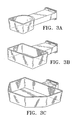
- These paddles are configured in various geometries as depicted in Figs. 3A-3C to accommodate various patient anatomies and specific needs of mammographers, such as coned compression paddles (3A), spot compression paddles (3B) and the axillary paddle shown as Fig. 3C , all configured to attach to the mammography unit through standard interchange assembly 22, as shown earlier.
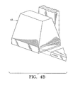
- the x-ray cassette holder may be adapted by various ancillary modules such as the spot compression fitting 41 shown in Fig. 4A , and a magnification fitting 42 shown in Fig. 4B .
- the entire compression system compression paddle and x-ray plate
- compression paddle 14 is modified to include a window 51, allowing the clinician access to the breast, while still under compression, for purposes of placing a device to identify a specific location in the breast, or to perform a biopsy of tissue.
- the pad assembly 60 comprises a padding element 61, an adhesive layer 62, and a release paper 63 to be removed from contact with the adhesive layer just prior to installation on the surface to be padded.
- Pad element 61 may be constructed of various materials having the following characteristics: produce no significant visual artifact on the mammogram (i.e. is radiolucent), be deformable under the forces applied during compression to provide comfort. Furthermore, the material should provide conformance to the tissue and the compression surface so as to reduce the propensity for the material to create air pockets or folds that may be of sufficient size to be visible on the x-ray image. Additionally, it may be desirable for the material to be absorptive to external fluids such as sweat.
- Such materials may be an elastomer or gel, open (reticulated) or closed (non-reticulated) cell foam consisting of polyolefin, or, preferably, a polyurethane open cell foam because of its radiolucent characteristics and soft tactile feel.
- polyurethane foam having a density of 5-6 lbs./cu ft, with approximately 90 pores per inch.
- the padding material 61 may be a thickness of .050" to .500", preferably in the range of .200" and .250".
- adhesive layer 62 may be one of a variety of currently available pressure sensitive adhesives such as acrylic or synthetic rubber based adhesives, to allow sufficient tackiness for secure attachment to the compression surface, while also allowing for easy removal (e.g. leaving no detectable residue of adhesive on the applied surface) and disposal.
- a non-adhesive gel may be used to secure the pad or another layer of material having a greater coefficient of friction against the applied surface. It is also anticipated by the scope of the present invention that the pad element may itself be textured such that it is sufficiently "tacky" to enable its use without an adhesive layer, i.e., by means of friction between the element and the tissue and the unit compression surface.
- the pad element of Fig. 6A can be configured with adhesive on the entire surface of the pad, or at certain regions such as just along the border (see Fig. 6C).
- Fig. 6B depicts a "peel away" packet configuration to house the pad assembly; optionally, the peel away packet can serve as a stiffening element to aid installation of the pad by keeping it in a planar configuration to minimize the possibility of misapplying the pad (leading to inadvertent air pockets or folds in the material, etc.) and to aid in positioning the pad prior to adhering it to the applied surface.
- Fig. 7A illustrates, in stepwise fashion, the installation of the pad assembly 60 of the present invention onto the film holder 15.
- the first step comprises opening the packing material housing the pad assembly 60 (S1), and thereafter removing any release paper 63 therefrom (S2).
- the pad assemblies can be packaged individually or in bulk.
- Installation on the patient contact surfaces of the x-ray plate 15 are shown in steps S3 and S4, S3 showing the placement of the pad element 61 on functional surface 19, and optionally extending to front face surface 18.
- the installed pad is depicted in Fig. 7A .
- the pad of this embodiment includes an adhesive layer which is preferably of a three part construction: a high tack, high peel permanent adhesive securing the adhesive layer to the pad element, a low tack, removable adhesive for securing the adhesive layer to the patient contact surface of the x-ray plate, and a stretchable carrier material between the two adhesives, such as polyethylene.
- a pad according to the present invention can also be formed of a similar construction using, e.g., polyester as a carrier material.
- the pad element may be removed and disposed of and the sequence repeated for the next patient. It may be desirable to score or otherwise provide a fold in the pad element at a fixed point from the edge of the pad to accommodate folding the pad onto the front and/or side face of the applied surface.
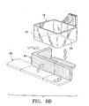
- FIG. 8A A similar sequence of steps (S1 to S4) is illustrated in Fig. 8A showing the installation of pad assembly 60 of the present invention onto compression paddle 14.
- the pad assembly (PA) of Fig. 8B is configured such that portions of the pad extend to cover side surface 24 of the compression paddle, providing added comfort for the patient.
- the pad of the present invention may be installed on the x-ray plate 15 and the compression paddle 14, or one and not the other, and further optionally on the front face and/or side face of either surface depending on the amount of additional cushioning desired.
- increased comfort was noted in all of the various configurations as compared to unpadded compression surfaces.
- An alternative technique for use of the pad is to attach it to the breast of the patient instead of on the mammography machine itself.
- this technique (not shown) the release paper is removed and the adhesive side of the pad is placed directly on the breast in an area of tissue to be compressed prior to placing the breast into the mammography machine.
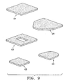
- Typical geometries of the present invention are illustrated in Fig. 9 , including pad elements for x-ray plate 15 (G1), pad elements with windows for stereotactic use (G2), spot compression paddles (G3), coned compression paddles (G4), and axillary paddles (G5).
- the present invention includes pad assemblies, where the pad elements are modular (e.g. using more than one pad to cover a desired surface), or cut to fit the desired surface (oversized with an overlay pattern to guide the operator in cutting the pad to fit).
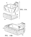
- FIG. 10A shows a modular configuration of the present invention wherein the pad assembly is constructed from a self-skinned foam (PA), i.e., foam having an impermeable membrane covering, such as a vinyl, deployed over a frame (not shown) and fastened to a compression paddle by suction cups, magnets rivets or adhesive (AD) on the non-functional surface of the compression paddle or x-ray plate.
- PA self-skinned foam
- AD adhesive
- Fig. 10B illustrates a pad assembly (PA) attached to the paddle on the nonfunctional surface by snaps or rivets 110.
- Fig. 10C further illustrates an alternative embodiment of the present invention wherein the pad 120 and compression paddle 121 are a single integral unit.
- Fig. 10D illustrates an alternative embodiment of the present invention wherein pad 122 and x-ray plate 123 are a single integral unit.
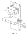
- Fig. 11 illustrates a dispensing unit according to the present invention for housing and dispensing the inventive pad assemblies.
- Dispensing unit 100 includes a housing 101 allowing multiple pad assemblies 103 to be stacked for compact storage, and an access slot 102 for allowing the user to access one pad assembly at a time.
- the pad assemblies as shown are individually packaged. Alternatively, pad assemblies maybe packaged in bulk (e.g., 25 pads per bag/4 bags per case) for ease of storage at the user location.
- a first further preferred embodiment of the present invention is a device for attachment to a compression surface of a mammography unit, said device comprising: a pad element having at least one surface; means for attaching said at least one surface of said pad element to said compression surface.
- said attachment means provides for releasable attachment of said at least one surface to said compression surface.
- This first aspect of the first further preferred embodiment of the present invention forms a second further preferred embodiment of the present invention.
- said means for attachment comprises an adhesive layer on said at least one surface of said pad element.
- Said adhesive layer may be chosen from the following list: acrylic based adhesive, or synthetic rubber based adhesive.
- said means for releasable attachment comprises the interface of the pad element surface and the compression surface.
- said means for releasable attachment is the friction between the pad and the compression surface.
- Said means for releasable attachment may be an elastic layer.
- said means for releasable attachment is attached to the mammography unit on a surface other than the compression surface.
- Said means for releasable attachment may be Velcro.
- said pad element is radiolucent.
- said pad element is formed of a polyurethane foam.
- Said polyurethane form may be an open cell foam.
- said pad element is formed of a fully skinned closed cell foam.
- said pad element is formed of vinyl.
- said pad is formed of a homogeneous material.
- a third further preferred embodiment of the present invention is a device for cushioning a compression surface of a mammograph unit, said device comprising: a pad element having at least one surface for contacting said compression surface, wherein said pad element surface is textured such that said pad remains in contact with said compression surface due to friction between said pad element surface and said compression surface.
- a fourth further preferred embodiment of the present invention is a cushioned mammography paddle comprising: a mammography paddle having a patient contact surface; and a cushioning element adapted for fixed attachment to said paddle patient surface. Said paddle and said cushioning element may be separately formed.
- a fifth further preferred embodiment of the present invention is a cushioned mammography x-ray plate comprising: an x-ray plate having a patient contact surface; and a cushioning element adapted for fixed attachment to said paddle patient surface. Said paddle and said cushioning element may be separately formed.
- a sixth further preferred embodiment of the present invention is a compression device for a radiological apparatus, said compression device comprising: a compression pad wherein said compression pad has at least one cushioned surface and said compression pad and said at least one cushioned surface are substantially transparent to x-ray.
- a seventh further preferred embodiment of the present invention is a device for attachment to the breast of a patient, said device comprising: a pad element having a first and second surface; means for attaching said first surface of said pad element to the breast of said patient.
- An eighth further preferred embodiment of the present invention is a method of performing mammography using the device of one of the first to third further preferred embodiments of the present invention wherein said method allows for the use of increased compressive force.
- a ninth further preferred embodiment of the present invention is a method of performing mammography using the device of one of the first to third further preferred embodiments of the present invention wherein said device produces no significant visual artifact on the mammogram.
- a tenth further preferred embodiment of the present invention is a system for use with a mammography unit comprising the devices of one of the first to third further preferred embodiments of the present invention, and a dispensing unit configured to retain and dispense said devices.
Landscapes
- Health & Medical Sciences (AREA)
- Life Sciences & Earth Sciences (AREA)
- Medical Informatics (AREA)
- Engineering & Computer Science (AREA)
- Radiology & Medical Imaging (AREA)
- Molecular Biology (AREA)
- Biophysics (AREA)
- Nuclear Medicine, Radiotherapy & Molecular Imaging (AREA)
- Optics & Photonics (AREA)
- Pathology (AREA)
- Physics & Mathematics (AREA)
- Biomedical Technology (AREA)
- Heart & Thoracic Surgery (AREA)
- High Energy & Nuclear Physics (AREA)
- Surgery (AREA)
- Animal Behavior & Ethology (AREA)
- General Health & Medical Sciences (AREA)
- Public Health (AREA)
- Veterinary Medicine (AREA)
- Dentistry (AREA)
- Oral & Maxillofacial Surgery (AREA)
- Apparatus For Radiation Diagnosis (AREA)
- Processing And Handling Of Plastics And Other Materials For Molding In General (AREA)
Applications Claiming Priority (3)
| Application Number | Priority Date | Filing Date | Title |
|---|---|---|---|
| US18719800P | 2000-03-06 | 2000-03-06 | |
| US09/620,730 US6577702B1 (en) | 2000-03-06 | 2000-07-20 | Device for cushioning of compression surfaces |
| EP01914717A EP1263321B1 (de) | 2000-03-06 | 2001-03-06 | Mammograph mit Polstereinrichtung für die Kompressionsflächen |
Related Parent Applications (1)
| Application Number | Title | Priority Date | Filing Date |
|---|---|---|---|
| EP01914717.2 Division | 2001-03-06 |
Publications (2)
| Publication Number | Publication Date |
|---|---|
| EP2279694A2 true EP2279694A2 (de) | 2011-02-02 |
| EP2279694A3 EP2279694A3 (de) | 2011-05-04 |
Family
ID=26882819
Family Applications (2)
| Application Number | Title | Priority Date | Filing Date |
|---|---|---|---|
| EP01914717A Expired - Lifetime EP1263321B1 (de) | 2000-03-06 | 2001-03-06 | Mammograph mit Polstereinrichtung für die Kompressionsflächen |
| EP10181093A Ceased EP2279694A3 (de) | 2000-03-06 | 2001-03-06 | Polstervorrichtung für Kompressionsflächen |
Family Applications Before (1)
| Application Number | Title | Priority Date | Filing Date |
|---|---|---|---|
| EP01914717A Expired - Lifetime EP1263321B1 (de) | 2000-03-06 | 2001-03-06 | Mammograph mit Polstereinrichtung für die Kompressionsflächen |
Country Status (8)
| Country | Link |
|---|---|
| US (9) | US6577702B1 (de) |
| EP (2) | EP1263321B1 (de) |
| JP (1) | JP2003525681A (de) |
| AT (1) | ATE556656T1 (de) |
| AU (3) | AU4007301A (de) |
| CA (1) | CA2402429C (de) |
| NZ (1) | NZ520969A (de) |
| WO (1) | WO2001066013A2 (de) |
Cited By (1)
| Publication number | Priority date | Publication date | Assignee | Title |
|---|---|---|---|---|
| NL2009856C2 (en) * | 2012-11-21 | 2014-05-27 | Amc Amsterdam | Mammography apparatus. |
Families Citing this family (64)
| Publication number | Priority date | Publication date | Assignee | Title |
|---|---|---|---|---|
| US6765984B2 (en) * | 2000-03-06 | 2004-07-20 | Biolucent, Inc. | Device for cushioning of compression surfaces |
| US6577702B1 (en) | 2000-03-06 | 2003-06-10 | Biolucent, Inc. | Device for cushioning of compression surfaces |
| FR2832050A1 (fr) * | 2001-11-13 | 2003-05-16 | Ge Med Sys Global Tech Co Llc | Pelote de compression et appareil de mammographie |
| US6850590B2 (en) | 2001-11-23 | 2005-02-01 | Benjamin M. Galkin | Mammography cassette holder for patient comfort and methods of use |
| US6975701B2 (en) * | 2001-11-23 | 2005-12-13 | Galkin Benjamin M | Devices for patient comfort in mammography and methods of use |
| US20040206738A1 (en) * | 2001-12-28 | 2004-10-21 | Ge Medical Systems Global Technology Company, Llc | Mammography patient contact temperature controller |
| SE0201806D0 (sv) * | 2002-06-13 | 2002-06-13 | Siemens Elema Ab | Röntgendiagnostikapparat för mammografi undersökningar |
| SE0203107D0 (sv) | 2002-10-22 | 2002-10-22 | Siemens Elema Ab | Kompressionsplatta för en mammografiapparat |
| US7828744B2 (en) * | 2003-04-23 | 2010-11-09 | Boston Scientific Scimed, Inc. | Method and assembly for breast immobilization |
| US20040218727A1 (en) * | 2003-05-01 | 2004-11-04 | S&S X-Ray Products, Inc. | Mammography compression cushion system |
| US8100839B2 (en) * | 2003-12-30 | 2012-01-24 | Galkin Benjamin M | Acoustic monitoring of a breast and sound databases for improved detection of breast cancer |
| US7142631B2 (en) * | 2003-12-30 | 2006-11-28 | Galkin Benjamin M | Mammography cushioning devices and methods |
| US7248668B2 (en) * | 2006-04-11 | 2007-07-24 | Galkin Benjamin M | Mammography compression devices and methods |
| US7512211B2 (en) * | 2003-12-30 | 2009-03-31 | Galkin Benjamin M | Mammography systems and methods, including methods for improving the sensitivity and specificity of the computer-assisted detection (CAD) process |
| US7505555B2 (en) * | 2004-11-02 | 2009-03-17 | Biolucent, Llc | Pads for mammography and methods for making and using them |
| US20080247508A1 (en) * | 2005-01-18 | 2008-10-09 | Ann Harrington | Bucky warmer with holder |
| US8089030B2 (en) * | 2005-01-18 | 2012-01-03 | Marvel Concepts, Llc | Bucky warmer with holder |
| US7492858B2 (en) * | 2005-05-20 | 2009-02-17 | Varian Medical Systems, Inc. | System and method for imaging and treatment of tumorous tissue in breasts using computed tomography and radiotherapy |
| US7489761B2 (en) * | 2006-03-27 | 2009-02-10 | Hologic, Inc. | Breast compression for digital mammography, tomosynthesis and other modalities |
| JP4950612B2 (ja) * | 2006-09-29 | 2012-06-13 | 富士フイルム株式会社 | 乳房撮影装置、乳房押圧板及び乳房固定方法 |
| US8652022B2 (en) * | 2007-09-10 | 2014-02-18 | Boston Scientific Scimed, Inc. | Stabilizer and method for irradiating tumors |
| JP5042760B2 (ja) * | 2007-09-25 | 2012-10-03 | 富士フイルム株式会社 | 放射線画像撮影装置 |
| FR2924010B1 (fr) | 2007-11-23 | 2010-11-26 | Gen Electric | Perfectionnements aux appareils de mammographie |
| US7559690B2 (en) * | 2007-12-13 | 2009-07-14 | Jared Holthe | X-ray positioning device |
| JP5171289B2 (ja) * | 2008-01-29 | 2013-03-27 | 富士フイルム株式会社 | X線撮像装置及びx線撮像装置使用方法 |
| FR2926973B1 (fr) * | 2008-01-31 | 2011-03-04 | Gen Electric | Pelote de compression et appareil de mammographie comportant une telle pelote |
| JP5042884B2 (ja) | 2008-02-29 | 2012-10-03 | 富士フイルム株式会社 | 放射線画像情報撮影装置 |
| US20090307191A1 (en) * | 2008-06-10 | 2009-12-10 | Li Hong C | Techniques to establish trust of a web page to prevent malware redirects from web searches or hyperlinks |
| US8401145B1 (en) | 2008-10-23 | 2013-03-19 | Beekley Corporation | Imaging sheet and related method |
| DE102008055737A1 (de) * | 2008-11-04 | 2010-05-06 | Siemens Aktiengesellschaft | Tomosynthesegerät |
| US7894573B2 (en) * | 2009-04-24 | 2011-02-22 | Moshe Ein-Gal | Non-recumbent radiotherapy |
| FI126561B (fi) * | 2010-02-23 | 2017-02-15 | Planmeca Oy | Hammaslääketieteellisen röntgenlaitteiston potilastuki |
| JP5583450B2 (ja) * | 2010-03-30 | 2014-09-03 | 富士フイルム株式会社 | 放射線撮影装置、及び圧迫板 |
| JP5661315B2 (ja) * | 2010-03-31 | 2015-01-28 | 富士フイルム株式会社 | 圧迫板、及び、x線撮像装置 |
| FI123379B (fi) | 2010-04-29 | 2013-03-15 | Planmed Oy | Lääketieteellinen tietokonetomografialaitteisto |
| FI125531B (fi) * | 2010-04-29 | 2015-11-13 | Planmed Oy | Lääketieteellinen röntgenkuvauslaitteisto |
| JP6157491B2 (ja) * | 2011-11-18 | 2017-07-05 | ホロジック, インコーポレイテッドHologic, Inc. | 造影と患者の快適性とを改善する膨張性ジャケットを備えた圧迫パドルを用いたx線マンモグラフィーおよび/または乳房トモシンセシス |
| US11259759B2 (en) | 2011-11-18 | 2022-03-01 | Hologic Inc. | X-ray mammography and/or breast tomosynthesis using a compression paddle |
| US9782135B2 (en) | 2011-11-18 | 2017-10-10 | Hologic, Inc. | X-ray mammography and/or breast tomosynthesis using a compression paddle |
| US20130254991A1 (en) * | 2011-12-05 | 2013-10-03 | Charles Y. Shang | Patient support platform system for breast therapy |
| JP2015522337A (ja) | 2012-06-25 | 2015-08-06 | コーニンクレッカ フィリップス エヌ ヴェ | 光学的に適合可能な胸部圧迫素子 |
| JP6043737B2 (ja) * | 2013-01-30 | 2016-12-14 | 富士フイルム株式会社 | 放射線撮影装置 |
| US20140294142A1 (en) * | 2013-03-28 | 2014-10-02 | Samsung Electronics Co., Ltd. | X-ray imaging device and method of controlling the same |
| CN105228526B (zh) * | 2013-03-29 | 2018-06-08 | 通用电气公司 | 乳房造影装置 |
| ES2750678T3 (es) | 2013-04-26 | 2020-03-26 | Hologic Inc | Mamografía de rayos x y/o tomosíntesis de mama con una paleta de compresión |
| JP6222809B2 (ja) * | 2013-05-30 | 2017-11-01 | 東芝メディカルシステムズ株式会社 | マンモグラフィ装置 |
| US9226718B1 (en) * | 2013-08-06 | 2016-01-05 | Alicia Baxley | Image receptor support device |
| US9247915B2 (en) | 2014-03-05 | 2016-02-02 | Brady Worldwide, Inc. | Microfiber radiography comfort device |
| JP6204611B2 (ja) * | 2014-04-02 | 2017-09-27 | コーニンクレッカ フィリップス エヌ ヴェKoninklijke Philips N.V. | 圧迫及び遮蔽装置 |
| JP6598721B2 (ja) * | 2016-04-01 | 2019-10-30 | 富士フイルム株式会社 | 音響マッチング部材、音響マッチング部材群、及び医用撮影装置 |
| JP6827763B2 (ja) | 2016-10-20 | 2021-02-10 | キヤノンメディカルシステムズ株式会社 | 圧迫板、x線診断装置、及び圧迫板の製造方法 |
| EP3868302B1 (de) | 2016-11-08 | 2023-12-06 | Hologic, Inc. | Bildgebung mit gekrümmten kompressionselementen |
| ES3014459T3 (en) * | 2017-08-11 | 2025-04-22 | Hologic Inc | Breast compression paddle |
| EP3664713A4 (de) | 2017-08-11 | 2021-04-28 | Hologic, Inc. | Brustkompressionsplatte mit zugangsecken |
| DE202018006903U1 (de) | 2017-08-16 | 2024-07-29 | Hologic Inc. | Techniken zur Patientenbewegungsartefaktkompensation bei Brustbildgebung |
| WO2019113247A1 (en) | 2017-12-05 | 2019-06-13 | Jarisch Wolfram R | Wide area single or dual guided breast tissue tomography and tomosynthesis imaging systems and methods |
| JP6945491B2 (ja) * | 2018-04-27 | 2021-10-06 | 富士フイルム株式会社 | マンモグラフィ装置 |
| US11596368B2 (en) * | 2018-05-25 | 2023-03-07 | Hologic, Inc. | Breast compression paddles utilizing pivoting foam elements |
| JP7742349B2 (ja) | 2020-01-24 | 2025-09-19 | ホロジック, インコーポレイテッド | 水平に変位可能な発泡体乳房圧迫パドル |
| EP4125603B1 (de) | 2020-03-27 | 2025-12-03 | Hologic, Inc. | Systeme und verfahren zur messung der ablenkung eines schaumstoffbrustkompressionspaddels |
| KR20240000563A (ko) | 2021-04-26 | 2024-01-02 | 홀로직, 인크. | 폼 압축 요소들의 두께를 측정하기 위한 시스템들 및 방법들 |
| WO2023028532A1 (en) * | 2021-08-25 | 2023-03-02 | Hologic, Inc. | Systems and methods for measuring movement of a compressed breast |
| DE102022207880A1 (de) * | 2022-07-29 | 2024-02-01 | Siemens Healthcare Gmbh | Brustfixierungselement |
| US12343185B2 (en) * | 2022-12-28 | 2025-07-01 | GE Precision Healthcare LLC | Device and method for securely holding breast on mammography imaging system |
Citations (10)
| Publication number | Priority date | Publication date | Assignee | Title |
|---|---|---|---|---|
| US3963933A (en) | 1975-08-18 | 1976-06-15 | General Electric Company | Mammography fixture |
| US4691333A (en) | 1985-12-27 | 1987-09-01 | Gabriele Joseph M | Breast compression and needle localization apparatus |
| US4943986A (en) | 1988-10-27 | 1990-07-24 | Leonard Barbarisi | Mammography compression apparatus for prosthetically augmented breast |
| US5081657A (en) | 1990-11-09 | 1992-01-14 | National Imaging Consultants, Inc. | Bucky warmer |
| US5185776A (en) | 1991-08-05 | 1993-02-09 | Townsend Boyd E | Cover for an x-ray cassette |
| US5189686A (en) | 1990-10-02 | 1993-02-23 | American Mammographics, Inc. | Adjustable mounting base for mammographic spot compression and magnification platform |
| US5226070A (en) | 1990-04-03 | 1993-07-06 | Kabushiki Kaisha Toshiba | Mat with low X-ray absorption for X-ray diagnosis |
| US5398272A (en) | 1992-11-10 | 1995-03-14 | Ge Medical Systems | Compression device for radiological apparatus |
| US5541972A (en) | 1993-04-23 | 1996-07-30 | Anthony; Betty J. | Disposable padding device for a mammography X-ray plate |
| US5553111A (en) | 1994-10-26 | 1996-09-03 | The General Hospital Corporation | Apparatus and method for improved tissue imaging |
Family Cites Families (47)
| Publication number | Priority date | Publication date | Assignee | Title |
|---|---|---|---|---|
| FR1302635A (fr) | 1961-07-21 | 1962-08-31 | Radiologie Cie Gle | Système de compression pour appareil d'examen radiologique |
| US3327316A (en) * | 1965-04-19 | 1967-06-27 | Vogt Mfg Corp | Wrestler's headgear |
| US3578971A (en) * | 1968-11-21 | 1971-05-18 | Harold J Lasky | Mammographic x-ray apparatus and technique |
| US3668394A (en) | 1970-08-03 | 1972-06-06 | Norman Panzer | X-ray film identification means |
| DE2335576A1 (de) * | 1973-07-13 | 1975-01-30 | Philips Patentverwaltung | Roentgendiagnostikgeraet fuer die mammographie |
| US4030719A (en) | 1976-08-30 | 1977-06-21 | Contour Fabricators, Inc. | Child immobilizing device for X-rays |
| US4103176A (en) * | 1977-01-06 | 1978-07-25 | Coyle Maurice J | Hand-held compressor for use by radiologist |
| US4121005A (en) * | 1978-02-13 | 1978-10-17 | Minnesota Mining And Manufacturing Company | Conformable foam |
| US4276104A (en) * | 1980-01-11 | 1981-06-30 | The Goodyear Tire & Rubber Company | Transfer drum application of narrow strip components to a tire being built on a tire building drum |
| US4346298A (en) * | 1981-03-02 | 1982-08-24 | Dixit Jagannath K | Automatic air pillow for diagnostic X-ray machine |
| US4433690A (en) | 1981-07-20 | 1984-02-28 | Siemens Ag | Compact ultrasound apparatus for medical examination |
| US5082657A (en) | 1984-04-13 | 1992-01-21 | Akzo N.V. | Leukoregulin, an antitumor lymphokine, and its therapeutic uses |
| US4545517A (en) * | 1984-11-27 | 1985-10-08 | Olson William J | Continuous forms leader |
| US4793635A (en) * | 1987-11-10 | 1988-12-27 | Lovison Douglas I | Radiopaque graphics |
| US4923187A (en) * | 1989-03-10 | 1990-05-08 | Avec Scientific Utility Corporation | Radiolucent iliac crest support frame |
| JPH0349811A (ja) * | 1989-07-13 | 1991-03-04 | Yasuhiro Kida | 水準器付き穴あけ機 |
| JPH0349811U (de) | 1989-09-18 | 1991-05-15 | ||
| JP2627087B2 (ja) * | 1989-10-18 | 1997-07-02 | 富士写真フイルム株式会社 | 乳房用エネルギーサブトラクション装置 |
| US5044008A (en) | 1989-12-12 | 1991-08-27 | Jackson Reginald B | Dental film cartridge cushion |
| JPH0464346A (ja) * | 1990-07-03 | 1992-02-28 | Hitachi Medical Corp | 乳房x線撮影装置の乳房圧迫機構 |
| JP2561584B2 (ja) * | 1991-09-21 | 1996-12-11 | リンテック株式会社 | 両面粘着テープ及びその製造方法 |
| US5166968A (en) * | 1992-02-05 | 1992-11-24 | Morse Arnold W | Portable x-ray cassette holder |
| US5161273A (en) | 1992-05-01 | 1992-11-10 | The United States Of America As Represented By The Secretary Of The Air Force | Neonatal cradle |
| US5311883A (en) | 1992-11-06 | 1994-05-17 | Eleanor Sherman | Sanitary shield for dedicated mammography apparatus |
| FR2702059B1 (fr) | 1993-02-25 | 1995-09-15 | Berneuil Gerard | Procede d'amelioration de la qualite des cliches radiologiques et dispositif permettant la mise en oeuvre du procede. |
| US5297678A (en) * | 1993-05-28 | 1994-03-29 | Eastman Kodak Company | Film cassette |
| US5377254A (en) | 1993-06-21 | 1994-12-27 | Walling; Michael A. | X-ray cassette casing |
| DE4324508A1 (de) | 1993-07-21 | 1995-01-26 | Alois Schnitzler | Vakuumkissen mit Schaumstoffeinlage |
| US6049583A (en) | 1993-08-10 | 2000-04-11 | Galkin; Benjamin M. | Method and apparatus for measuring compression force in mammography |
| US5474072A (en) | 1993-10-29 | 1995-12-12 | Neovision Corporation | Methods and apparatus for performing sonomammography |
| ATE191136T1 (de) * | 1993-10-29 | 2000-04-15 | United States Surgical Corp | Vorrichtung zur kombinierten ultraschall- abbildung und röntgen-abbildung |
| US6038718A (en) * | 1994-08-15 | 2000-03-21 | Midmark Corporation | Surgical table |
| US5416822A (en) | 1994-08-29 | 1995-05-16 | Kunik; Randall L. | Device for registering a dental radiograph having distortion measuring capability and method for using the same |
| WO1996007353A1 (en) | 1994-09-09 | 1996-03-14 | Air-Shields, Inc. | Infant mattress assembly |
| US5632275A (en) * | 1994-09-16 | 1997-05-27 | Scribner-Browne Medical Design Incorporated | Catheter lab table pad and method for using the same |
| WO1996013211A1 (en) * | 1994-10-26 | 1996-05-09 | The General Hospital Corporation | Apparatus and method for improved tissue imaging |
| US5613254A (en) * | 1994-12-02 | 1997-03-25 | Clayman; Ralph V. | Radiolucent table for supporting patients during medical procedures |
| US5719916A (en) | 1995-06-05 | 1998-02-17 | Nelson; Robert S. | Anthropomorphic mammography and lung phantoms |
| US5657367A (en) * | 1996-06-26 | 1997-08-12 | Couch; Denver | Lateral decubitus patient positioning device |
| JPH1033526A (ja) * | 1996-07-23 | 1998-02-10 | Japan Vilene Co Ltd | 医療用透視台及びこれに用いる固定シート |
| US5891074A (en) | 1996-08-22 | 1999-04-06 | Avitar, Inc. | Pressure wound dressing |
| US5832550A (en) * | 1997-08-11 | 1998-11-10 | Biotek | Moldable vacuum cushion |
| US5970119A (en) | 1997-11-18 | 1999-10-19 | Douglas Holtz (Part Interest) | Radiological scaling and alignment device |
| DE19921100A1 (de) | 1998-05-05 | 2000-03-02 | Kari Richter | Medizinische Kompressionsvorrichtung |
| DE19926446A1 (de) | 1998-07-20 | 2000-01-27 | Siemens Ag | Vorrichtung zur Ultraschallankopplung eines Ultraschall-Applikators |
| US6577702B1 (en) * | 2000-03-06 | 2003-06-10 | Biolucent, Inc. | Device for cushioning of compression surfaces |
| US6765984B2 (en) * | 2000-03-06 | 2004-07-20 | Biolucent, Inc. | Device for cushioning of compression surfaces |
-
2000
- 2000-07-20 US US09/620,730 patent/US6577702B1/en not_active Expired - Lifetime
-
2001
- 2001-03-06 CA CA2402429A patent/CA2402429C/en not_active Expired - Fee Related
- 2001-03-06 AU AU4007301A patent/AU4007301A/xx active Pending
- 2001-03-06 EP EP01914717A patent/EP1263321B1/de not_active Expired - Lifetime
- 2001-03-06 NZ NZ520969A patent/NZ520969A/en not_active IP Right Cessation
- 2001-03-06 AU AU2001240073A patent/AU2001240073B2/en not_active Ceased
- 2001-03-06 JP JP2001564671A patent/JP2003525681A/ja active Pending
- 2001-03-06 EP EP10181093A patent/EP2279694A3/de not_active Ceased
- 2001-03-06 WO PCT/US2001/007189 patent/WO2001066013A2/en not_active Ceased
- 2001-03-06 AT AT01914717T patent/ATE556656T1/de active
-
2003
- 2003-03-14 US US10/389,392 patent/US6968033B2/en not_active Expired - Lifetime
-
2004
- 2004-09-21 US US10/946,044 patent/US7616732B2/en not_active Expired - Fee Related
-
2005
- 2005-10-19 AU AU2005225059A patent/AU2005225059A1/en not_active Abandoned
-
2006
- 2006-04-18 US US11/379,193 patent/US7502441B2/en not_active Expired - Lifetime
-
2009
- 2009-10-31 US US12/610,303 patent/US8098793B2/en not_active Expired - Fee Related
-
2012
- 2012-01-16 US US13/351,149 patent/US8705689B2/en not_active Expired - Fee Related
-
2014
- 2014-04-21 US US14/257,856 patent/US9504433B2/en not_active Expired - Fee Related
-
2016
- 2016-11-28 US US15/362,734 patent/US9936925B2/en not_active Expired - Fee Related
-
2018
- 2018-03-21 US US15/927,967 patent/US20180289341A1/en not_active Abandoned
Patent Citations (10)
| Publication number | Priority date | Publication date | Assignee | Title |
|---|---|---|---|---|
| US3963933A (en) | 1975-08-18 | 1976-06-15 | General Electric Company | Mammography fixture |
| US4691333A (en) | 1985-12-27 | 1987-09-01 | Gabriele Joseph M | Breast compression and needle localization apparatus |
| US4943986A (en) | 1988-10-27 | 1990-07-24 | Leonard Barbarisi | Mammography compression apparatus for prosthetically augmented breast |
| US5226070A (en) | 1990-04-03 | 1993-07-06 | Kabushiki Kaisha Toshiba | Mat with low X-ray absorption for X-ray diagnosis |
| US5189686A (en) | 1990-10-02 | 1993-02-23 | American Mammographics, Inc. | Adjustable mounting base for mammographic spot compression and magnification platform |
| US5081657A (en) | 1990-11-09 | 1992-01-14 | National Imaging Consultants, Inc. | Bucky warmer |
| US5185776A (en) | 1991-08-05 | 1993-02-09 | Townsend Boyd E | Cover for an x-ray cassette |
| US5398272A (en) | 1992-11-10 | 1995-03-14 | Ge Medical Systems | Compression device for radiological apparatus |
| US5541972A (en) | 1993-04-23 | 1996-07-30 | Anthony; Betty J. | Disposable padding device for a mammography X-ray plate |
| US5553111A (en) | 1994-10-26 | 1996-09-03 | The General Hospital Corporation | Apparatus and method for improved tissue imaging |
Cited By (1)
| Publication number | Priority date | Publication date | Assignee | Title |
|---|---|---|---|---|
| NL2009856C2 (en) * | 2012-11-21 | 2014-05-27 | Amc Amsterdam | Mammography apparatus. |
Also Published As
| Publication number | Publication date |
|---|---|
| US9504433B2 (en) | 2016-11-29 |
| US20030174807A1 (en) | 2003-09-18 |
| US20060239399A1 (en) | 2006-10-26 |
| EP1263321B1 (de) | 2012-05-09 |
| US20120114096A1 (en) | 2012-05-10 |
| NZ520969A (en) | 2004-03-26 |
| WO2001066013A3 (en) | 2002-01-31 |
| AU2001240073B2 (en) | 2005-07-21 |
| US20170071555A1 (en) | 2017-03-16 |
| US6577702B1 (en) | 2003-06-10 |
| JP2003525681A (ja) | 2003-09-02 |
| CA2402429A1 (en) | 2001-09-13 |
| ATE556656T1 (de) | 2012-05-15 |
| AU4007301A (en) | 2001-09-17 |
| US20050036584A1 (en) | 2005-02-17 |
| US8705689B2 (en) | 2014-04-22 |
| US9936925B2 (en) | 2018-04-10 |
| US7616732B2 (en) | 2009-11-10 |
| US6968033B2 (en) | 2005-11-22 |
| EP2279694A3 (de) | 2011-05-04 |
| US7502441B2 (en) | 2009-03-10 |
| WO2001066013A2 (en) | 2001-09-13 |
| US8098793B2 (en) | 2012-01-17 |
| EP1263321A2 (de) | 2002-12-11 |
| US20140307848A1 (en) | 2014-10-16 |
| AU2005225059A1 (en) | 2005-11-10 |
| US20100046698A1 (en) | 2010-02-25 |
| US20180289341A1 (en) | 2018-10-11 |
| CA2402429C (en) | 2013-05-07 |
Similar Documents
| Publication | Publication Date | Title |
|---|---|---|
| AU2001240073B2 (en) | Device for cushioning of compression surfaces in mammograph | |
| AU2001240073A1 (en) | Device for cushioning of compression surfaces in mammograph | |
| AU2002322630B2 (en) | Device for cushioning of compression surfaces in mammograph | |
| AU2008201638B2 (en) | Device for cushioning of compression surfaces in mammograph | |
| AU2002322630A1 (en) | Device for cushioning of compression surfaces in mammograph | |
| EP1816964B1 (de) | Pads für die mammographie | |
| US20040156472A1 (en) | Devices for patient comfort in mammography and methods of use | |
| US10617367B2 (en) | Imaging sheet and related method | |
| US20040218727A1 (en) | Mammography compression cushion system | |
| WO2015134487A1 (en) | Microfiber radiography comfort device |
Legal Events
| Date | Code | Title | Description |
|---|---|---|---|
| PUAI | Public reference made under article 153(3) epc to a published international application that has entered the european phase |
Free format text: ORIGINAL CODE: 0009012 |
|
| AC | Divisional application: reference to earlier application |
Ref document number: 1263321 Country of ref document: EP Kind code of ref document: P |
|
| AK | Designated contracting states |
Kind code of ref document: A2 Designated state(s): AT BE CH CY DE DK ES FI FR GB GR IE IT LI LU MC NL PT SE TR |
|
| PUAL | Search report despatched |
Free format text: ORIGINAL CODE: 0009013 |
|
| AK | Designated contracting states |
Kind code of ref document: A3 Designated state(s): AT BE CH CY DE DK ES FI FR GB GR IE IT LI LU MC NL PT SE TR |
|
| 17P | Request for examination filed |
Effective date: 20111028 |
|
| 17Q | First examination report despatched |
Effective date: 20111202 |
|
| GRAP | Despatch of communication of intention to grant a patent |
Free format text: ORIGINAL CODE: EPIDOSNIGR1 |
|
| RIC1 | Information provided on ipc code assigned before grant |
Ipc: A61B 6/00 20060101ALI20151020BHEP Ipc: A61B 6/04 20060101AFI20151020BHEP |
|
| INTG | Intention to grant announced |
Effective date: 20151120 |
|
| RAP1 | Party data changed (applicant data changed or rights of an application transferred) |
Owner name: HOLOGIC, INC. |
|
| APBK | Appeal reference recorded |
Free format text: ORIGINAL CODE: EPIDOSNREFNE |
|
| APBN | Date of receipt of notice of appeal recorded |
Free format text: ORIGINAL CODE: EPIDOSNNOA2E |
|
| APBR | Date of receipt of statement of grounds of appeal recorded |
Free format text: ORIGINAL CODE: EPIDOSNNOA3E |
|
| INTC | Intention to grant announced (deleted) | ||
| APAV | Appeal reference deleted |
Free format text: ORIGINAL CODE: EPIDOSDREFNE |
|
| RAP1 | Party data changed (applicant data changed or rights of an application transferred) |
Owner name: HOLOGIC INC. |
|
| RAP1 | Party data changed (applicant data changed or rights of an application transferred) |
Owner name: HOLOGIC, INC. |
|
| APBT | Appeal procedure closed |
Free format text: ORIGINAL CODE: EPIDOSNNOA9E |
|
| STAA | Information on the status of an ep patent application or granted ep patent |
Free format text: STATUS: THE APPLICATION HAS BEEN REFUSED |
|
| 18R | Application refused |
Effective date: 20200928 |