EP2283376B1 - Verbesserungen bei der magnetresonanzbildgebung in bezug auf die korrektur von chemieverlagerungsartefakten und intensitätsungleichmässigkeit - Google Patents
Verbesserungen bei der magnetresonanzbildgebung in bezug auf die korrektur von chemieverlagerungsartefakten und intensitätsungleichmässigkeit Download PDFInfo
- Publication number
- EP2283376B1 EP2283376B1 EP09732976.7A EP09732976A EP2283376B1 EP 2283376 B1 EP2283376 B1 EP 2283376B1 EP 09732976 A EP09732976 A EP 09732976A EP 2283376 B1 EP2283376 B1 EP 2283376B1
- Authority
- EP
- European Patent Office
- Prior art keywords
- image
- fat
- phase
- water
- intensity
- Prior art date
- Legal status (The legal status is an assumption and is not a legal conclusion. Google has not performed a legal analysis and makes no representation as to the accuracy of the status listed.)
- Active
Links
Images

Classifications
-
- G—PHYSICS
- G01—MEASURING; TESTING
- G01R—MEASURING ELECTRIC VARIABLES; MEASURING MAGNETIC VARIABLES
- G01R33/00—Arrangements or instruments for measuring magnetic variables
- G01R33/20—Arrangements or instruments for measuring magnetic variables involving magnetic resonance
- G01R33/44—Arrangements or instruments for measuring magnetic variables involving magnetic resonance using nuclear magnetic resonance [NMR]
- G01R33/48—NMR imaging systems
- G01R33/4828—Resolving the MR signals of different chemical species, e.g. water-fat imaging
-
- G—PHYSICS
- G01—MEASURING; TESTING
- G01R—MEASURING ELECTRIC VARIABLES; MEASURING MAGNETIC VARIABLES
- G01R33/00—Arrangements or instruments for measuring magnetic variables
- G01R33/20—Arrangements or instruments for measuring magnetic variables involving magnetic resonance
- G01R33/44—Arrangements or instruments for measuring magnetic variables involving magnetic resonance using nuclear magnetic resonance [NMR]
- G01R33/48—NMR imaging systems
- G01R33/54—Signal processing systems, e.g. using pulse sequences ; Generation or control of pulse sequences; Operator console
- G01R33/56—Image enhancement or correction, e.g. subtraction or averaging techniques, e.g. improvement of signal-to-noise ratio and resolution
- G01R33/565—Correction of image distortions, e.g. due to magnetic field inhomogeneities
- G01R33/56527—Correction of image distortions, e.g. due to magnetic field inhomogeneities due to chemical shift effects
-
- G—PHYSICS
- G01—MEASURING; TESTING
- G01R—MEASURING ELECTRIC VARIABLES; MEASURING MAGNETIC VARIABLES
- G01R33/00—Arrangements or instruments for measuring magnetic variables
- G01R33/20—Arrangements or instruments for measuring magnetic variables involving magnetic resonance
- G01R33/44—Arrangements or instruments for measuring magnetic variables involving magnetic resonance using nuclear magnetic resonance [NMR]
- G01R33/48—NMR imaging systems
- G01R33/54—Signal processing systems, e.g. using pulse sequences ; Generation or control of pulse sequences; Operator console
- G01R33/56—Image enhancement or correction, e.g. subtraction or averaging techniques, e.g. improvement of signal-to-noise ratio and resolution
- G01R33/5608—Data processing and visualization specially adapted for MR, e.g. for feature analysis and pattern recognition on the basis of measured MR data, segmentation of measured MR data, edge contour detection on the basis of measured MR data, for enhancing measured MR data in terms of signal-to-noise ratio by means of noise filtering or apodization, for enhancing measured MR data in terms of resolution by means for deblurring, windowing, zero filling, or generation of gray-scaled images, colour-coded images or images displaying vectors instead of pixels
-
- G—PHYSICS
- G01—MEASURING; TESTING
- G01R—MEASURING ELECTRIC VARIABLES; MEASURING MAGNETIC VARIABLES
- G01R33/00—Arrangements or instruments for measuring magnetic variables
- G01R33/20—Arrangements or instruments for measuring magnetic variables involving magnetic resonance
- G01R33/44—Arrangements or instruments for measuring magnetic variables involving magnetic resonance using nuclear magnetic resonance [NMR]
- G01R33/48—NMR imaging systems
- G01R33/54—Signal processing systems, e.g. using pulse sequences ; Generation or control of pulse sequences; Operator console
- G01R33/56—Image enhancement or correction, e.g. subtraction or averaging techniques, e.g. improvement of signal-to-noise ratio and resolution
- G01R33/565—Correction of image distortions, e.g. due to magnetic field inhomogeneities
- G01R33/5659—Correction of image distortions, e.g. due to magnetic field inhomogeneities caused by a distortion of the RF magnetic field, e.g. spatial inhomogeneities of the RF magnetic field
Definitions
- the present disclosure relates to improving images acquired from magnetic resonance imaging systems. More specifically it relates to correction of an intensity inhomogeneity in such images.
- Phase sensitive acquisition and reconstruction such as an "in-phase sand out-of-phase"-method of which the Dixon method may be best known, see e.g. W. T. Dixon, "Simple proton spectroscopic imaging,”Radiology,vol. 153(1), pp. 189-194, 1984 , has many advantages.
- Dixon imaging provides two images, one showing the fat content in each voxel and one showing the water content in each voxel. Hence Dixon images are not adversely affected by partial volume effects. Separate water and fat images are also useful in the segmentation process.
- IP W + F
- a chemical shift artifact manifested as a water fat shift artifact causes distortion in water and fat images obtained using phase sensitive acquisition and reconstruction, as in the Dixon method.
- the resonance frequency of protons in human methylene lipid [CH2]n and water differ by 3,5 ppm corresponding to 224 Hz at a field strength of 1.5 T. This intrinsic difference can be utilized for effective fat and water separation using the phase sensitive acquisition and reconstruction.
- a consequence from utilizing resonance frequency shift in such methods is spatial misregistration in the frequency encoding direction known as the chemical shift artifact, in the case of fat and water images, the water-fat shift artifact.
- the frequency is used to encode the spatial position of the signal.
- fat will have a relative frequency shift that cannot be distinguished from the phase difference introduced by the frequency encoding.
- the water-fat shift artifact typically appears close to fat structures and is caused by the bi polar gradient being used.
- a flyback protocol has been proposed, see e.g. Cunningham. Magnetic Resonance in Medicine,2005. p.1286-1289 , to eliminate the problem as the misregistration is constant between acquisition times, but has the effect of decreasing the signal to noise ratio (SNR).
- SNR signal to noise ratio
- Intensity inhomogeneity prevalence in MRI is due to factors such as static field inhomogeneity, RF excitation field non-uniformity, in homogeneity in reception coil sensitivity and patient movement.
- the effect of the non-uniformity is usually a slow varying non anatomic intensity variation over the image.
- it sometimes can be difficult to see the intensity non-uniformity by visual inspection there are implications that significantly can decrease segmentation and registration results as many medical imaging techniques is based on the assumptions that the same tissue has the same intensity throughout a volume. More importantly it affects the linear quantification of the MR signal.
- a voxel containing a certain amount of fat should have the same signal strength, independent of where it is located in space. This is not true in case of intensity inhomogeneity occurrence.
- EP0851236 discloses a method of determining water content in a magnetic resonance image by identifying fat tissue image points. The image is corrected for non-uniformity of spatial intensity by means of a uniform phantom.
- a general object of this disclosure is to present a solution overcoming or at least alleviating problems in the prior art.
- One more specific object is to present a solution that enables correction of, or at least alleviation of effects from, chemical shift artifact in an image acquired from a magnetic resonance imaging system.
- Another more specific object is to present a solution that enables correction of, or at least alleviation of effects from, intensity inhomogeneity in an image acquired from a magnetic resonance imaging system.
- phase sensitive reconstruction such as the two point Dixon method can provide pure fat and water volumes. Therefore there is typically no problem in distinguishing different tissues in the same volume. It has been found that one efficient way to correct for intensity inhomogeneity in image volumes acquired with using e.g. a two point Dixon technique is to locate voxels corresponding to pure adipose tissue and estimate a correction field from these points. A method has been developed addressed to this idea.
- a method of producing an intensity correction field with a magnetic resonance imaging system according to claim 1 and according to a second aspect, a magnetic resonance imaging system according to claim 11.
- the first aspect method has several applications. It can be in accurate fat content estimation enabling integration of fat in the image avoiding partial volume effects. Application of the correction field to the reconstructed water image provides an intensity reference which is stable also after contrast agent injection as the contrast influence on the fat signal is negligible.
- the intensity correction field can preferably be used to correct any image acquired using the MRI system that provided data from which the intensity correction field was generated, i.e. any image from the same image data set and/or from any other image data set.
- the method according to the first aspect can be extended according to what is disclosed in claim 4 and thereto dependent claims.
- This enables to quantitatively measure the abdominal fat composition from MR images.
- Three different types of fat subcutaneous, visceral and non-visceral internal are advantageously measured.
- the method can be fully automated. This distinguishes it from most previously published methods.
- the method allows to quantitatively measure the fat content in each individual voxel in order to avoid partial volume effects.
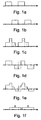
- Fig.1a-f schematically show different exemplary signals in 2-point Dixon imaging, where Fig.1 a shows a water signal, Fig. 1b shows a fat signal, Fig. 1c shows an in-phase signal, Fig. 1d shows an out-of-phase signal, Fig. 1e shows a reconstructed water signal and Fig. 1f shows a reconstructed fat signal.
- the consequence of the artifact is a low pass filtering effect in the extracted fat volume and a derivative contamination effect of the true fat signal in the extracted water volume.
- IP ⁇ x + OP ⁇ x W x * ⁇ x + WFS + W x * ⁇ x + WFS + F x
- Fig. 2 schematically shows steps of a method of producing an image with a MRI system.
- a first step 120 an image data set is acquired with the MRI system and in step 130 in-phase and out of phase image components IP, OP are formed from the data set.
- steps 140-180 corresponding to the above-described steps a)-d).
- a second signal for example a fat signal F n
- a second signal is set to initial starting values in order to have something to start from.
- a first signal is generated using the second signal (set to the initial values or calculated in a previous iteration), for example a water signal W n+1 is calculated using the fat signal F n as in step b).
- step 160 a second signal is generated using the first signal (calculated in step 150), for example a fat signal F n+1 is calculated using water signal W n+1 as in step c).
- step 170 it is checked if a criterion is fulfilled, typically whether a predetermined number of iterations have been reached or not, which can be accomplished by checking the value of an integer parameter n. If the criterion has been met, a resulting image is produced in step 180, which resulting image has a corrected, or at least reduced, chemical shift artifact, the resulting image being for example a fat image, a water image, or a joint image thereof. If the criterion has not been met, the integer parameter n can be increased by one and a next iteration is started from step 150.
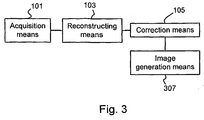
- Fig. 3 schematically illustrates one embodiment of a MRI system configured so that it can carry out the method discussed above.
- the system comprises acquisition means 101, for example computer controlled data acquisition hardware connectable to a magnet assembly, configured to acquire the image data set, i.e so that it can carry out step 120, and reconstruction means 103, e.g. a computer, configured, preferably by software, to form an in-phase image component and out-of-phase image component from the acquired data set, i.e. so that it can carry out step 130.
- correction means 105 also typically a computer, which is configured, preferably by software, to compensate for a chemical shift artifact iteratively and generate compensated first and second signals, i.e.
- An image generation means 107 is configured to produce a resulting image based on the compensated first and/or second signal, i.e. configured so that it can carry out step 180, and is also typically a computer configured by software.
- the computers referred to as examples can be the same or separate computers, and may be general or specific purpose computers in terms of software and/or hardware.
- a simulated, one dimensional object containing fat and water has been used for evaluation of the method based on the correction scheme according to expressions 11-13.
- the limited resolution compared to the WFS of the image was introduced by convolution of the simulated object with a sinc function multiplied with a hamming filter.
- a WFS of 0.415 was used, this since the WFS in most applications, due to physical constraints, is approximately 0.4-0.5 pixels at 1.5 T if the lowest possible bandwidth and the shortest possible out of phase and in phase echo times are used for the desired resolution.
- the obtained IP and OP images were calculated. Water and fat signals obtained without WFS correction were compared to correspondingly corrected water and fat signals, and residuals were studied using 15 iterations and K values of 0.80.and 0.95.
- the corrected water and fat signal of the simulated object showed clear improvements compared with the originally reconstructed signals.
- the low pass filtering effect in the fat signal and ringing artifacts around fat objects in the water signal was removed when high values of the constant K was used.
- K was kept in the range 0 to 1.
- Gaussian noise where added to the IP and OP providing a signal to noise ratio in the IP images of 0, 0.5, 1, and 5,where SNR was defined as the SD of the noise amplitude in the water and fat images divided by the SD of the noise added to the IP and OP images.
- SNR was defined as the SD of the noise amplitude in the water and fat images divided by the SD of the noise added to the IP and OP images.
- SD corrected -SD uncorrected were plotted using 15, 30 and 60 iterations. The convergence rate decreased for higher values of K. High SNR signals appeared to be most effectively corrected using K values close to 1 as the noise amplification effect becomes insignificant. At lower SNR the optimal values of K appeared to become lower. 15 iterations were sufficient to reach convergence also for signal with higher SNR. For high SNR signals usage of higher K values can be used to obtain optimal correction.
- corrected IP and OP images were calculated using a range of K values from 0.5 to 1.05 and 15 iterations.
- the iterative solver was implemented in Matlab (The MathWorks, Inc, Massachusetts, USA).
- FFT fast Fourier transform
- the images were interpolated to a pixel spacing exactly matching the water fat shift using a fast Fourier transform (FFT) based interpolation method. After this operation all convolutions in the iterative method were performed using one pixel shifts of the signals, thus without need of any further interpolation. It is realized that such interpolation before application of the iterative part of the method in general is beneficial since it e.g. allows for simpler and faster implementation.
- FFT fast Fourier transform
- the corrected water images showed visible improvements compared to the original images. Most evident were effects in an outer skin layer but were also apparent in the region around the spine. The effects in the fat images were harder to distinguish by visual inspection but by looking at a difference image, the sharpening effect of the iterative reconstruction algorithm becomes evident. Most apparent were the effects of the correction in a corrected in-phase image where the fat/water displacement artifact and the edge artifact around fat objects were removed.
- the SD of a difference image between a "fly-back" water signal and a corrected water signal was 27% lower compared to the SD of the difference image between the "fly-back" water signal and an uncorrected water signal when measured in all slices excluding voxels outside the body.
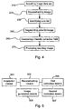
- Fig. 4 schematically shows steps of one embodiment of a method of producing an intensity correction field with a magnetic resonance imaging (MRI) system.
- a first step 210 an image data set is acquired using the MRI system.
- an image is reconstructed based on the acquired image data set, the image containing water and fat phase information represented by an in-phase component IP and an out-of-phase component OP.
- the acquisition and reconstruction is preferably made by two-point Dixon imaging.
- Dixon imaging which, as will be realized by the skilled person, are compatible with the solutions presented herein, e.g. three-point Dixon imaging, or any other technique resulting in-phase and out-of phase components.
- step 230 pure-fat image points, here pure adipose tissue voxels, are identified, thus producing a pure-fat image.
- the intensity non-uniformity in the in-phase volume, IP, and extracted fat volume, F are typically close to equal.
- F r a relative fat content
- an estimate of fat content relative to water content can be reached without the impact of intensity non-uniformity, i.e. the relative fat content can be seen as insensitive to intensity variations.
- a thresholding which can be made simple, of the relative fat content can be used to identify and collect pure adipose tissue voxels. (In the absence of intensity non-uniformities, all such voxels would have the same intensity in e.g. the in-phase image IP.) Voxels entirely located outside the body are preferably removed. Thus the intensity variations can be estimated by examining voxels which are located inside the body and have high relative fat content F r values.
- test criterions than thresholding includes e.g. morphological operators, such as a requirement that a voxel to be accepted must belong to a fat structure of a certain size and/or shape.
- Other test criterion might be based on the combination of Dixon encoding of MR-images with other MR-pulse sequences providing multiple images with different contrast properties.
- An additional masking may be used to only account for values in human tissue. This can be made by an initial thresholding operation with an empirically determined threshold for background values. That is, an additional criterion to separate true fat voxels from false positives caused by low signal intensity in the both the fat and the in phase image may be used to only account for values in human tissue.
- step 250 where the pure-fat image is segmented, preferably by bimodal segmentation.
- Bimodal segmentation means a process to determine an appropriate thresholding value based on a bimodal model of the signal distribution.
- an intensity correction field is generated by interpol a ting a fat image F, which is based on the in-phase component IP and the out-of-phase component OP, using the segmented image, i.e. a field is interpolated from the identified voxels.
- a field is interpolated from the identified voxels.
- the correction field is preferably created with normalized convolution, see e.g. H. Knutsson and C-F. Westin. In Proc. of IEEE Computer Society Conference on Computer Vision and PatfernRecognition, June 1993, pp. 515-523 , where pure fat voxels are weighted as 1 and remaining voxels as 0.
- a resulting, and thus intensity corrected, image is produced, which may involve applying the intensity correction field to the in-phase and out-of phase image components, IP and OP.
- a volume can be reconstructed.
- an extracted fat volume can be divided by the calculated intensity non - uniformity field, i.e. the intensity correction field, to acquire an intensity normalized volume.
- Water W and fat F images can be corrected by multiplication with the inverse of an intensity non-uniformity map corresponding to the intensity correction field.
- _Application of the correction field to the reconstructed water image provides an intensity reference which is stable also after contrast agent injection as the contrast influence on the fat signal is negligible.
- the generated intensity correction field can be used as a reference and can be used to correct not only the fat image, but also the water image, and also images originating from other image data sets acquired using the MRI system.
- Fig. 5 schematically illustrates an embodiment of a magnetic resonance imaging (MRI) system configured so that it can carry out the method discussed above.
- the system comprises acquisition means 301, for example computer controlled data acquisition hardware connectable to a magnet assembly, configured to acquire the image data set, i.e. so that it can carry out step 210, and reconstruction means 303, e.g. a computer, configured, preferably by software, to reconstruct the image based on the acquired image data set, i.e. so that it can carry out step 220.
- reconstruction means 303 e.g. a computer, configured, preferably by software, to reconstruct the image based on the acquired image data set, i.e. so that it can carry out step 220.
- the system comprises a first correction means 305, also typically a computer, which is configured, preferably by software, to compensate for the intensity inhomogeneity, i.e. configured so that it can carry out steps 230-260.
- a second correction means 307 is configured to generate the intensity correction field, i.e. so that can carry out step 260, and which also typically is a computer that has been programmed by software.
- An image generation means 309 is configured to apply the intensity correction field to produce an resulting image, i.e. that can carry out step 270 and is also typically a computer configured by software.
- the intensity correction field can be saved on storing means, e.g. as a file on a disk drive, for later use.
- the correction field is applied by another image generation means, which may be separate.
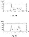
- RESULTS The original and corrected fat and water images were normalized with a fat peak value corresponding to a hundred percent adipose tissue. Peak values were obtained by histogram analysis of voxels in the fat images in the subcutaneous region and the corresponding subcutaneous voxel histograms before and after intensity normalization are presented in Figs. 2.3a-b . An evident improvement could be seen in the corrected fat image. Similar improvement was seen inter slices and was most apparent close to the edges of the reception coil.
- a simple and efficient solution that can be used to account for intensity inhomogenity in two point Dixon imaging is to identify pure adipose tissue values and estimate a correction field by normalized convolution.
- the method has shown good stability in the evaluation of 50 datasets. After application of the correction field there are noticeable signs that imply that the intensity distribution over the volume has improved. Intensity variations seen inter and intra slices was efficiently eliminated. Looking at the subcutaneous fat histogram (see Fig. 6b ) for all voxels after intensity non-uniformity correction and comparing to a corresponding histogram without the correction (see Fig. 6a ), a distinctive peak for pure adipose tissue can be seen without dispersing effects caused by intensity variations.
- the above described method may advantageously be adapted to or used in a fat quantification method that can be used for automatic quantification of subcutaneous, visceral and non-visceral internal fat from MR-images acquired using the two point Dixon technique in the abdominal region.
- the fat quantification method includes (1) a three dimensional phase unwrapping to provide water and fat images, (2) an image intensity inhomogenity correction (as in the method described above), and (3) a non-rigid registration and segmentation of the tissue. This is followed by an integration of the corrected fat images within the different fat compartments that avoids the partial volume effects associated with traditional fat segmentation methods.
- the fat quantification method comprises a number of different steps: Phase correction of two point Dixon images, intensity inhomogenenity compensation and segmentation of the different types of fat.
- the inhomogeneous sensitivity of the RF-coils cause variations in the image magnitude over the field of view.
- the variation in sensitivity affects the signal strength within the 3D volume and furthermore it makes comparison of signal intensities between different examinations difficult. This non-uniformity degrades the quality of the fat content estimation, and therefore needs to be avoided, which is one reason why the method of intensity correction described in the foregoing is advantageous to use with the fat quantification method.
- non-rigid registration is used for atlas based segmentation.
- One such non-rigid registration method is the Morphon, see e.g. H. Knutsson and M. Andersson. In IEEE International Conference on Image Processing (ICIP'05), Genova, Italy, September 2005 .
- a prototype image is registered to a target image by iteratively deforming the prototype.
- the deformation estimate is calculated by maximization of the similarity between the prototype and the target.
- the similarity measure can be based on e.g. image intensity or local phase information. The latter has the main advantage of being invariant to intensity variations between images.
- a phase based approach is preferably used.
- the morphon was used to segment the abdominal adipose tissue into three different types: subcutaneous, visceral and internal non-visceral. This was done by registering a manually segmented prototype to the target image of interest. Registration of an abdominal prototype to a fat image in the abdominal region can be difficult due to the high variability in fat accumulation between different subjects.
- a significant advantage of the phase sensitive Dixon reconstruction is that separate fat and water images can be obtained. Since the water image contains structures such as muscles, bone and spine, with less variability than adipose tissue structures, it was a preferable choice over the fat image for registration intents. However, the water image also contained troublesome areas with high variability. This may cause significant degradation of the registration results.
- the registration is divided into two steps.
- the first step registers a binary prototype to a roughly calculated binary mask of the internal region in the target image. This registration is applied on a coarse scale to provide a good initial guess of the global deformation.
- the resulting deformation field is applied on the second prototype, created from the water image, before proceeding to the finer resolution scales.
- An additional certainty mask covering the muscle and bone structures in the abdominal region, is created to reduce the effects of uncertain regions on the registration result.
- masks of the visceral and nonvisceral regions are defined for the prototype image.
- the target image is subjected to initial pre-processing steps to create binary masks of the body and of the internal region for the initial registration step.
- the arms and background of the target image can efficiently be removed from the rest of the body using local thresholding.
- a local minimum can be expected between the body and arms, and this is used for segmentation of the body. Removing everything except the largest connected component leaves a body mask that is further processed by morphological operations to produce a dense mask.
- the calculated deformation field is applied to the labels associated with the prototype image. Hence, the fat in the target image is labeled as visceral and internal nonvisceral parts.
- Any adipose tissue not associated with any of these labels belongs to the subcutaneous fat. Since Dixon imaging provides separate water and fat images, no segmentation is needed to distinguish between these types of tissue. The purpose of the segmentation process is to separate the adipose tissue into different compartments.
- the fat quantification method presented herein provides good segmentation results also when the target image differs substantially from the prototype.
- One aspect of the method is that the registration of the prototype to the target volume is performed exclusively on the water images eliminating the registration errors from the subcutaneous fat.
- a validation has shown a very strong reduction of the signal intensity variations within and between different subjects. This means that method provides very accurate integration of the fat content within the different compartments avoiding the partial volume effects associated with methods based on summation of voxels segmented in strongly T1 weighted images.
Landscapes
- Physics & Mathematics (AREA)
- High Energy & Nuclear Physics (AREA)
- Condensed Matter Physics & Semiconductors (AREA)
- General Physics & Mathematics (AREA)
- Health & Medical Sciences (AREA)
- General Health & Medical Sciences (AREA)
- Nuclear Medicine, Radiotherapy & Molecular Imaging (AREA)
- Radiology & Medical Imaging (AREA)
- Engineering & Computer Science (AREA)
- Signal Processing (AREA)
- Magnetic Resonance Imaging Apparatus (AREA)
Claims (13)
- Verfahren zum Erzeugen eines Intensitätskorrekturfeldes, das die Rekonstruktion eines Volumens mit normalisierten Intensitätswerten mit einem Magnetresonanz-Bildgebungssystem ermöglicht, wobei das Verfahren umfasst:Erfassen (210) eines Bilddatensatzes mit dem Magnetresonanz-Bildgebungssystem;Rekonstruieren (220) eines Bildes auf der Basis des Bilddatensatzes, wobei das rekonstruierte Bild Wasser- und Fettphasen-Informationen enthält, und wobei die Rekonstruktion phasenempfindlich ist, so dass sie in einer phasengleichen Bildkomponente (IP), die ein Signal "Wasser plus Fett" umfasst, und in einer phasenverschobenen Bildkomponente (OP), die ein Signal "Wasser minus Fett" umfasst, resultiert;Identifizieren (230) von Bildpunkten in dem rekonstruierten Bild, wobei diese Bildpunkte gemäß einem Prüfkriterium reines Fettgewebe repräsentieren und somit ein reines Fettbild erzeugen, wobei das Identifizieren (230) einen Schritt des Schätzens des Fettgehalts relativ zum Wassergehalt durch Berechnen eines relativen Fettgehalts, welcher ein Verhältnis zwischen einer Fett-Bildkomponente und der phasengleichen Bildkomponente ist, umfasst;Segmentieren (250) des reinen Fettbildes, um ein segmentiertes Bild zu erzeugen, das frei von Nicht-Gewebe-Bilddaten ist; undErzeugen (260) des Intensitätskorrekturfeldes durch Interpolieren eines Fettbildes (F) unter Verwendung der Bildpunkte, die in dem segmentierten Bild angegeben sind, wobei das Fettbild auf der Basis der phasengleichen Bildkomponente (IP) und der phasenverschobenen Bildkomponente (OP) berechnet wird.
- Verfahren nach Anspruch 1, wobei das Verfahren ferner umfasst:Ausgleichen des Einflusses einer Intensitätsinhomogenität in dem rekonstruierten Bild durch Erzeugen (270) wenigstens eines resultierenden Bildes durch Anwenden des Intensitätskorrekturfeldes auf die phasengleiche und die phasenverschobene Bildkomponente des rekonstruierten Bildes, wobei das wenigstens eine resultierende Bild wenigstens eines von Folgendem repräsentiert:ein Wasserbild,ein Fettbild, undein gemeinsames Bild, das sowohl Wasser als auch Fett repräsentiert.
- Verfahren nach Anspruch 1, wobei das Verfahren ferner umfasst:Erfassen (210) eines zweiten Bilddatensatzes mit dem Magnetresonanz-Bildgebungssystem; undRekonstruieren (220) eines zweiten Bildes auf der Basis des zweiten Bilddatensatzes; undAusgleichen des Einflusses einer Intensitätsinhomogenität in dem rekonstruierten zweiten Bild durch Anwenden des Intensitätskorrekturfeldes auf eine Bildkomponente des zweiten Bildes.
- Verfahren nach einem der Ansprüche 1-3, wobei der Schritt des Rekonstruierens (220) eines Bildes auf der Basis des Bilddatensatzes eine Phasenkorrektur der phasengleichen Bildkomponente und der phasenverschobenen Bildkomponente umfasst.
- Verfahren nach Anspruch 1, wobei das Prüfkriterium im Schritt des Identifizierens (230) von Bildpunkten in dem rekonstruierten Bild das Festlegen eines Schwellenwertes, insbesondere eines Schwellenwertes für den relativen Fettgehalt, umfasst.
- Verfahren nach einem der Ansprüche 1-5, wobei der Schritt des Segmentierens (250) des reinen Fettbildes eine bimodale Segmentierung umfasst.
- Verfahren nach einem der Ansprüche 1-6, wobei das Interpolieren des Fettbildes (F) im Schritt des Erzeugens (260) eines Intensitätskorrekturfeldes eine normalisierte Faltung der Bildpunkte in dem segmentierten Bild umfasst.
- Verfahren nach Anspruch 7, wobei die normalisierte Faltung das Falten mit einem Gaußschen Glättungskern umfasst.
- Computerprogramm, das in den internen Speicher eines Computers geladen werden kann und Software umfasst, die dazu eingerichtet ist, ein MRI- (Magnetic Resonance Imaging) System (Magnetresonanz-Bildgebungssystem) zu Erfassung von MR- (Magnetresonanz-) Daten zu steuern und die Schritte eines der Ansprüche 1-8 auszuführen, wenn dieses Programm auf dem Computer ausgeführt wird.
- Computerlesbares Medium, auf dem ein Computerprogramm nach Anspruch 9 aufgezeichnet ist.
- Magnetresonanz-Bildgebungssystem zum Erzeugen eines Intensitätskorrekturfeldes, das die Rekonstruktion eines Volumens mit normalisierten Intensitätswerten ermöglicht, umfassend:Erfassungsmittel (301), die dafür ausgelegt sind, einen Bilddatensatz zu erfassen,Rekonstruktionsmittel (303), die dafür ausgelegt sind, ein Bild auf der Basis des erfassten Bilddatensatzes zu rekonstruieren, wobei das rekonstruierte Bild Wasser- und Fettphasen-Informationen enthält, die durch eine phasengleiche Bildkomponente, die ein Signal "Wasser plus Fett" umfasst, und eine phasenverschobene Bildkomponente, die ein Signal "Wasser minus Fett" umfasst, repräsentiert werden,erste Korrekturmittel (304), die dafür ausgelegt sind:eine Identifizierung von Bildpunkten in dem rekonstruierten Bild zu bewirken, wobei diese Bildpunkte gemäß einem Prüfkriterium reines Fettgewebe repräsentieren und somit ein reines Fettbild erzeugen, wobei die Identifizierung eine Schätzung des Fettgehalts relativ zum Wassergehalt durch Berechnung eines relativen Fettgehalts, welcher ein Verhältnis zwischen einer Fett-Bildkomponente und der phasengleichen Bildkomponente ist, umfasst,zu bewirken, dass ein segmentiertes Bild auf der Basis des reinen Fettbildes erzeugt wird, wobei das segmentierte Bild frei von Nicht-Gewebe-Bilddaten ist, undzweite Korrekturmittel (307), die dafür ausgelegt sind, ein Intensitätskorrekturfeld durch Interpolieren eines Fettbildes (F) unter Verwendung von Bildpunkten, die in dem segmentierten Bild angegeben sind, zu erzeugen, wobei das Fettbild auf der Basis der phasengleichen Bildkomponente (IP) und der phasenverschobenen Bildkomponente (OP) berechnet wird.
- Magnetresonanz-Bildgebungssystem nach Anspruch 11, welches ferner umfasst:Bilderzeugungsmittel (309), die dafür ausgelegt sind, das Intensitätskorrekturfeld auf die phasengleiche und die phasenverschobene Bildkomponente des rekonstruierten Bildes anzuwenden, wobei das wenigstens eine resultierende Bild wenigstens eines von Folgendem repräsentiert:ein Wasserbild,ein Fettbild, undein gemeinsames Bild, das sowohl Wasser als auch Fett repräsentiert.
- Magnetresonanz-Bildgebungssystem nach Anspruch 11, welches ferner umfasst:Erfassungsmittel (301), die dafür ausgelegt sind, einen zweiten Bilddatensatz zu erfassen;Rekonstruktionsmittel (303), die dafür ausgelegt sind, ein zweites Bild auf der Basis des zweiten Bilddatensatzes zu rekonstruieren; undBilderzeugungsmittel (309), die dafür ausgelegt sind, das Intensitätskorrekturfeld auf eine Bildkomponente des zweiten Bildes anzuwenden.
Priority Applications (1)
| Application Number | Priority Date | Filing Date | Title |
|---|---|---|---|
| EP16162570.2A EP3112890A1 (de) | 2008-04-17 | 2009-04-17 | Verbesserte magnetresonanzbilder |
Applications Claiming Priority (2)
| Application Number | Priority Date | Filing Date | Title |
|---|---|---|---|
| US4588808P | 2008-04-17 | 2008-04-17 | |
| PCT/SE2009/000195 WO2009128764A1 (en) | 2008-04-17 | 2009-04-17 | Improvement in magnetic resonance imaging relating to correction of chemical shift artifact and intensity inhomogeneity |
Related Child Applications (1)
| Application Number | Title | Priority Date | Filing Date |
|---|---|---|---|
| EP16162570.2A Division EP3112890A1 (de) | 2008-04-17 | 2009-04-17 | Verbesserte magnetresonanzbilder |
Publications (3)
| Publication Number | Publication Date |
|---|---|
| EP2283376A1 EP2283376A1 (de) | 2011-02-16 |
| EP2283376A4 EP2283376A4 (de) | 2011-12-21 |
| EP2283376B1 true EP2283376B1 (de) | 2016-03-30 |
Family
ID=41199325
Family Applications (2)
| Application Number | Title | Priority Date | Filing Date |
|---|---|---|---|
| EP16162570.2A Ceased EP3112890A1 (de) | 2008-04-17 | 2009-04-17 | Verbesserte magnetresonanzbilder |
| EP09732976.7A Active EP2283376B1 (de) | 2008-04-17 | 2009-04-17 | Verbesserungen bei der magnetresonanzbildgebung in bezug auf die korrektur von chemieverlagerungsartefakten und intensitätsungleichmässigkeit |
Family Applications Before (1)
| Application Number | Title | Priority Date | Filing Date |
|---|---|---|---|
| EP16162570.2A Ceased EP3112890A1 (de) | 2008-04-17 | 2009-04-17 | Verbesserte magnetresonanzbilder |
Country Status (4)
| Country | Link |
|---|---|
| US (1) | US8605967B2 (de) |
| EP (2) | EP3112890A1 (de) |
| ES (1) | ES2578706T3 (de) |
| WO (1) | WO2009128764A1 (de) |
Cited By (1)
| Publication number | Priority date | Publication date | Assignee | Title |
|---|---|---|---|---|
| EP3693976A1 (de) | 2019-02-08 | 2020-08-12 | AMRA Medical AB | Verfahren zur bewertung eines muskelassoziierten leidens |
Families Citing this family (23)
| Publication number | Priority date | Publication date | Assignee | Title |
|---|---|---|---|---|
| EP2365354A1 (de) | 2010-02-22 | 2011-09-14 | Koninklijke Philips Electronics N.V. | Kernspintomographie von chemischen Spezies mit einem spektralen Modell |
| WO2011139232A1 (en) * | 2010-05-03 | 2011-11-10 | Nanyang Technological University | Automated identification of adipose tissue, and segmentation of subcutaneous and visceral abdominal adipose tissue |
| CN103140167B (zh) | 2010-09-20 | 2016-01-20 | 皇家飞利浦电子股份有限公司 | 化学物类的磁共振成像 |
| DE102010041125B4 (de) * | 2010-09-21 | 2015-07-30 | Siemens Aktiengesellschaft | Räumliche Korrektur von Bilddaten einer Serie von Magnetresonanzaufnahmen |
| US8824766B2 (en) * | 2012-02-10 | 2014-09-02 | Duke University | Systems and methods for automated magnetic resonance imaging |
| CN104106096B (zh) | 2012-02-17 | 2017-09-22 | 先进穆阿分析公司 | 层析图像中的器官的分类方法 |
| US8868153B2 (en) | 2012-04-19 | 2014-10-21 | General Electric Company | Image correction using multichannel blind deconvolution with homomorphic filtering |
| CN105829906B (zh) * | 2013-12-19 | 2020-11-03 | 皇家飞利浦有限公司 | 具有水/脂肪分离的相位敏感的反转恢复mri |
| JP6434030B2 (ja) * | 2013-12-19 | 2018-12-05 | コーニンクレッカ フィリップス エヌ ヴェKoninklijke Philips N.V. | Dixonタイプ水/脂肪分離する磁気共鳴イメージング |
| ES2914613T3 (es) * | 2014-04-25 | 2022-06-14 | Amra Medical Ab | Cuantificación de la concentración de agua en tejido magro |
| DE102014225299A1 (de) * | 2014-12-09 | 2016-03-03 | Siemens Aktiengesellschaft | Verfahren zur Rekonstruktion von Magnetresonanz-Bilddaten |
| US9508127B1 (en) * | 2015-06-30 | 2016-11-29 | Konica Minolta Laboratory U.S.A., Inc. | Processing for creating a transmission image without artificial noises |
| CN106780407B (zh) * | 2017-03-01 | 2024-03-26 | 清远先导科臻医疗科技有限公司 | 一种针对超声图像斑点噪声的去噪系统及去噪方法 |
| US10762603B2 (en) * | 2017-05-19 | 2020-09-01 | Shanghai United Imaging Healthcare Co., Ltd. | System and method for image denoising |
| US10551463B2 (en) * | 2017-09-21 | 2020-02-04 | Siemens Healthcare Gmbh | Method and magnetic resonance apparatus for reconstructing an image from data acquired from a frequency-modulated balanced steady-state free precession sequence |
| EP3462204A1 (de) | 2017-09-28 | 2019-04-03 | Koninklijke Philips N.V. | Magnetresonanzbildgebung mit wasser-fett-trennung nach dixon-verfahren mit verbesserter fettverschiebungskorrektur |
| DE102018201314A1 (de) | 2018-01-29 | 2019-08-01 | Siemens Healthcare Gmbh | Verfahren zur Normalisierung von Magnetresonanzbildern |
| DE102018203507B4 (de) * | 2018-03-08 | 2020-10-22 | Siemens Healthcare Gmbh | Verfahren zur Aufnahme und Rekonstruktion eines vierdimensionalen Dynamikbilddatensatzes mit einer Magnetresonanzeinrichtung, Magnetresonanzeinrichtung, Computerprogramm und elektronisch lesbarer Datenträger |
| DE102019204151A1 (de) * | 2019-03-26 | 2020-10-01 | Siemens Healthcare Gmbh | Automatisiert optimierte MR-Bildgebung mit ultrakurzen Echozeiten |
| EP3726240A1 (de) * | 2019-04-19 | 2020-10-21 | Koninklijke Philips N.V. | Automatisierte detektion von wasser-fett-vertauschungen in der dixon-magnetresonanzbildgebung |
| IT202000023257A1 (it) * | 2020-10-02 | 2022-04-02 | Esaote Spa | Sistema e metodo per l’imaging diagnostico di acqua e grasso |
| EP4198545A1 (de) | 2021-12-14 | 2023-06-21 | Siemens Healthcare GmbH | Korrektur der chemischen verschiebung für mehrpunkt-magnetresonanzmessungen |
| US20260102075A1 (en) * | 2024-10-16 | 2026-04-16 | Aclarion, Inc. | Multi-voxel prescription for lumbar disc evaluation |
Family Cites Families (13)
| Publication number | Priority date | Publication date | Assignee | Title |
|---|---|---|---|---|
| DE19540837B4 (de) * | 1995-10-30 | 2004-09-23 | Siemens Ag | Verfahren zur Verzeichnungskorrektur für Gradienten-Nichtlinearitäten bei Kernspintomographiegeräten |
| IT1289809B1 (it) | 1996-12-27 | 1998-10-16 | Ist Trentino Di Cultura | Procedimento e sistema automatico per ottenere mappe di contenuto d'acqua e/o di permettivita'elettrica da immagini di risonanza |
| US6466014B1 (en) * | 2000-08-29 | 2002-10-15 | Ge Medical Systems Global Technology Company, Llc | Suppression of fat signals in MR water images produced in Dixon imaging |
| US6906515B2 (en) * | 2000-12-28 | 2005-06-14 | Hitachi Medical Corporation | Magnetic resonance imaging device and method |
| DE10132274B4 (de) * | 2001-07-04 | 2004-01-15 | Siemens Ag | Trennung von Fett- und Wasserbildern durch das Zwei-Punkt-Dixon-Verfahren unter Berücksichtigung einer integrierten 3D-Feld-Messung zur Aufnahme einer Karte der Grundfeldinhomogenität |
| US7099499B2 (en) * | 2002-08-15 | 2006-08-29 | General Electric Company | Fat/water separation and fat minimization magnetic resonance imaging systems and methods |
| US7349729B2 (en) * | 2003-10-20 | 2008-03-25 | The Board Of Trustees Of The Leland Stanford Junior University | Magnetic resonance imaging of different chemical species in a system having magnetic field heterogeneities |
| DE102004061507B4 (de) * | 2004-12-21 | 2007-04-12 | Siemens Ag | Verfahren zur Korrektur von Inhomogenitäten in einem Bild sowie bildgebende Vorrichtung dazu |
| US7176683B2 (en) * | 2005-05-06 | 2007-02-13 | The Board Of Trustees Of The Leland Stanford Junior University | Iterative decomposition of water and fat with echo asymmetry and least square estimation |
| US7592810B2 (en) * | 2006-04-25 | 2009-09-22 | The Board Of Trustees Of The Leland Stanford Junior University | MRI methods for combining separate species and quantifying a species |
| US7486074B2 (en) * | 2006-04-25 | 2009-02-03 | The Board Of Trustees Of The Leland Stanford Junior University | Self-calibration methods for parallel imaging and multipoint water-fat separation methods |
| US7432707B1 (en) * | 2006-06-14 | 2008-10-07 | Fonar Corporation | Magnetic resonance imaging with corrected intensity inhomogeneity |
| US7375522B2 (en) * | 2006-08-28 | 2008-05-20 | Wisconsin Alumni Research Foundation | Method for aligning multiple MR images acquired with alternating readout gradient |
-
2009
- 2009-04-17 EP EP16162570.2A patent/EP3112890A1/de not_active Ceased
- 2009-04-17 WO PCT/SE2009/000195 patent/WO2009128764A1/en not_active Ceased
- 2009-04-17 EP EP09732976.7A patent/EP2283376B1/de active Active
- 2009-04-17 US US12/988,066 patent/US8605967B2/en active Active
- 2009-04-17 ES ES09732976.7T patent/ES2578706T3/es active Active
Cited By (2)
| Publication number | Priority date | Publication date | Assignee | Title |
|---|---|---|---|---|
| EP3693976A1 (de) | 2019-02-08 | 2020-08-12 | AMRA Medical AB | Verfahren zur bewertung eines muskelassoziierten leidens |
| WO2020161274A1 (en) | 2019-02-08 | 2020-08-13 | Amra Medical Ab | Method of evaluating a muscle related condition |
Also Published As
| Publication number | Publication date |
|---|---|
| EP3112890A1 (de) | 2017-01-04 |
| ES2578706T3 (es) | 2016-07-29 |
| EP2283376A4 (de) | 2011-12-21 |
| WO2009128764A9 (en) | 2010-02-11 |
| WO2009128764A1 (en) | 2009-10-22 |
| US8605967B2 (en) | 2013-12-10 |
| US20110091090A1 (en) | 2011-04-21 |
| EP2283376A1 (de) | 2011-02-16 |
Similar Documents
| Publication | Publication Date | Title |
|---|---|---|
| EP2283376B1 (de) | Verbesserungen bei der magnetresonanzbildgebung in bezug auf die korrektur von chemieverlagerungsartefakten und intensitätsungleichmässigkeit | |
| Leinhard et al. | Quantitative abdominal fat estimation using MRI | |
| US7227359B2 (en) | Method and apparatus for phase-sensitive magnetic resonance imaging | |
| US9612300B2 (en) | System and method for object-based initialization of magnetic field inhomogeneity in magnetic resonance imaging | |
| Kim et al. | Denoising of hyperpolarized 13C MR images of the human brain using patch‐based higher‐order singular value decomposition | |
| Zhang et al. | MRI Gibbs‐ringing artifact reduction by means of machine learning using convolutional neural networks | |
| CN102565738B (zh) | 对拍摄的磁共振数据的补充 | |
| US20130310678A1 (en) | Biomedical image reconstruction method | |
| In et al. | Distortion correction in EPI using an extended PSF method with a reversed phase gradient approach | |
| Sharma et al. | Improving chemical shift encoded water–fat separation using object‐based information of the magnetic field inhomogeneity | |
| CN1820208A (zh) | 涉及脂肪抑制和/或黑血预备的mri扫描仪的磁场调整 | |
| Delbany et al. | One‐millimeter isotropic breast diffusion‐weighted imaging: Evaluation of a superresolution strategy in terms of signal‐to‐noise ratio, sharpness and apparent diffusion coefficient | |
| US10725133B2 (en) | Field-mapping and artifact correction in multispectral imaging | |
| Lugauer et al. | Robust spectral denoising for water-fat separation in magnetic resonance imaging | |
| EP3132277B1 (de) | Rekonstruktionsverfahren für die elektrische leitfähigkeitstomographie mit verbesserter stabilität und geschwindigkeit | |
| Sauer et al. | Deep learning k‐space‐to‐image reconstruction facilitates high spatial resolution and scan time reduction in diffusion‐weighted imaging breast MRI | |
| Shaw et al. | Non-linear realignment improves hippocampus subfield segmentation reliability | |
| Ota et al. | Motion robust coronary MR angiography using zigzag centric ky–kz trajectory and high-resolution deep learning reconstruction | |
| Schmidt et al. | High-resolution quantitative T2 mapping of the human brain at 7 T using a multi-echo spin-echo sequence and dictionary-based modeling | |
| Bao et al. | Improved reconstruction for MR spectroscopic imaging | |
| EP1972957A1 (de) | Verfahren zur Bestimmung phasenkorrigierter Amplituden in der relaxometrischen NMR-Bildgebung | |
| Lajous et al. | T2 mapping from super-resolution-reconstructed clinical fast spin echo magnetic resonance acquisitions | |
| US20210270918A1 (en) | Method for generating a magnetic resonance image | |
| Sigmund et al. | Diffusion-weighted imaging of the brain at 7 T with echo-planar and turbo spin echo sequences: preliminary results | |
| US20230243908A1 (en) | Improved whole-blade acquisition and phase correction in magnetic resonance imaging |
Legal Events
| Date | Code | Title | Description |
|---|---|---|---|
| PUAI | Public reference made under article 153(3) epc to a published international application that has entered the european phase |
Free format text: ORIGINAL CODE: 0009012 |
|
| 17P | Request for examination filed |
Effective date: 20101117 |
|
| AK | Designated contracting states |
Kind code of ref document: A1 Designated state(s): AT BE BG CH CY CZ DE DK EE ES FI FR GB GR HR HU IE IS IT LI LT LU LV MC MK MT NL NO PL PT RO SE SI SK TR |
|
| AX | Request for extension of the european patent |
Extension state: AL BA RS |
|
| DAX | Request for extension of the european patent (deleted) | ||
| REG | Reference to a national code |
Ref country code: DE Ref legal event code: R079 Ref document number: 602009037249 Country of ref document: DE Free format text: PREVIOUS MAIN CLASS: G01R0033540000 Ipc: G01R0033480000 |
|
| RIC1 | Information provided on ipc code assigned before grant |
Ipc: G01R 33/56 20060101ALI20111026BHEP Ipc: G01R 33/54 20060101ALI20111026BHEP Ipc: G01R 33/565 20060101ALI20111026BHEP Ipc: G01R 33/48 20060101AFI20111026BHEP |
|
| A4 | Supplementary search report drawn up and despatched |
Effective date: 20111117 |
|
| RAP1 | Party data changed (applicant data changed or rights of an application transferred) |
Owner name: ADVANCED MR ANALYTICS AB |
|
| RIN1 | Information on inventor provided before grant (corrected) |
Inventor name: LUNDBERG, PETER Inventor name: DAHLQVIST LEINHARD, OLAF Inventor name: BORGA, MAGNUS |
|
| RAP1 | Party data changed (applicant data changed or rights of an application transferred) |
Owner name: ADVANCED MR ANALYTICS AB |
|
| 17Q | First examination report despatched |
Effective date: 20150330 |
|
| GRAP | Despatch of communication of intention to grant a patent |
Free format text: ORIGINAL CODE: EPIDOSNIGR1 |
|
| INTG | Intention to grant announced |
Effective date: 20151203 |
|
| RIN1 | Information on inventor provided before grant (corrected) |
Inventor name: BORGA, MAGNUS Inventor name: DAHLQVIST LEINHARD, OLOF Inventor name: LUNDBERG, PETER |
|
| INTG | Intention to grant announced |
Effective date: 20151215 |
|
| GRAS | Grant fee paid |
Free format text: ORIGINAL CODE: EPIDOSNIGR3 |
|
| GRAA | (expected) grant |
Free format text: ORIGINAL CODE: 0009210 |
|
| AK | Designated contracting states |
Kind code of ref document: B1 Designated state(s): AT BE BG CH CY CZ DE DK EE ES FI FR GB GR HR HU IE IS IT LI LT LU LV MC MK MT NL NO PL PT RO SE SI SK TR |
|
| REG | Reference to a national code |
Ref country code: GB Ref legal event code: FG4D |
|
| REG | Reference to a national code |
Ref country code: CH Ref legal event code: EP |
|
| REG | Reference to a national code |
Ref country code: AT Ref legal event code: REF Ref document number: 785956 Country of ref document: AT Kind code of ref document: T Effective date: 20160415 |
|
| REG | Reference to a national code |
Ref country code: IE Ref legal event code: FG4D |
|
| REG | Reference to a national code |
Ref country code: DE Ref legal event code: R096 Ref document number: 602009037249 Country of ref document: DE |
|
| REG | Reference to a national code |
Ref country code: CH Ref legal event code: NV Representative=s name: WEINMANN ZIMMERLI, CH |
|
| REG | Reference to a national code |
Ref country code: SE Ref legal event code: TRGR |
|
| REG | Reference to a national code |
Ref country code: NL Ref legal event code: FP |
|
| REG | Reference to a national code |
Ref country code: LT Ref legal event code: MG4D |
|
| PG25 | Lapsed in a contracting state [announced via postgrant information from national office to epo] |
Ref country code: HR Free format text: LAPSE BECAUSE OF FAILURE TO SUBMIT A TRANSLATION OF THE DESCRIPTION OR TO PAY THE FEE WITHIN THE PRESCRIBED TIME-LIMIT Effective date: 20160330 Ref country code: NO Free format text: LAPSE BECAUSE OF FAILURE TO SUBMIT A TRANSLATION OF THE DESCRIPTION OR TO PAY THE FEE WITHIN THE PRESCRIBED TIME-LIMIT Effective date: 20160630 Ref country code: FI Free format text: LAPSE BECAUSE OF FAILURE TO SUBMIT A TRANSLATION OF THE DESCRIPTION OR TO PAY THE FEE WITHIN THE PRESCRIBED TIME-LIMIT Effective date: 20160330 Ref country code: GR Free format text: LAPSE BECAUSE OF FAILURE TO SUBMIT A TRANSLATION OF THE DESCRIPTION OR TO PAY THE FEE WITHIN THE PRESCRIBED TIME-LIMIT Effective date: 20160701 |
|
| REG | Reference to a national code |
Ref country code: ES Ref legal event code: FG2A Ref document number: 2578706 Country of ref document: ES Kind code of ref document: T3 Effective date: 20160729 |
|
| REG | Reference to a national code |
Ref country code: AT Ref legal event code: MK05 Ref document number: 785956 Country of ref document: AT Kind code of ref document: T Effective date: 20160330 |
|
| PG25 | Lapsed in a contracting state [announced via postgrant information from national office to epo] |
Ref country code: LT Free format text: LAPSE BECAUSE OF FAILURE TO SUBMIT A TRANSLATION OF THE DESCRIPTION OR TO PAY THE FEE WITHIN THE PRESCRIBED TIME-LIMIT Effective date: 20160330 Ref country code: LV Free format text: LAPSE BECAUSE OF FAILURE TO SUBMIT A TRANSLATION OF THE DESCRIPTION OR TO PAY THE FEE WITHIN THE PRESCRIBED TIME-LIMIT Effective date: 20160330 Ref country code: BE Free format text: LAPSE BECAUSE OF NON-PAYMENT OF DUE FEES Effective date: 20160430 |
|
| REG | Reference to a national code |
Ref country code: FR Ref legal event code: PLFP Year of fee payment: 8 |
|
| PG25 | Lapsed in a contracting state [announced via postgrant information from national office to epo] |
Ref country code: EE Free format text: LAPSE BECAUSE OF FAILURE TO SUBMIT A TRANSLATION OF THE DESCRIPTION OR TO PAY THE FEE WITHIN THE PRESCRIBED TIME-LIMIT Effective date: 20160330 Ref country code: IS Free format text: LAPSE BECAUSE OF FAILURE TO SUBMIT A TRANSLATION OF THE DESCRIPTION OR TO PAY THE FEE WITHIN THE PRESCRIBED TIME-LIMIT Effective date: 20160730 Ref country code: PL Free format text: LAPSE BECAUSE OF FAILURE TO SUBMIT A TRANSLATION OF THE DESCRIPTION OR TO PAY THE FEE WITHIN THE PRESCRIBED TIME-LIMIT Effective date: 20160330 |
|
| PG25 | Lapsed in a contracting state [announced via postgrant information from national office to epo] |
Ref country code: PT Free format text: LAPSE BECAUSE OF FAILURE TO SUBMIT A TRANSLATION OF THE DESCRIPTION OR TO PAY THE FEE WITHIN THE PRESCRIBED TIME-LIMIT Effective date: 20160801 Ref country code: SK Free format text: LAPSE BECAUSE OF FAILURE TO SUBMIT A TRANSLATION OF THE DESCRIPTION OR TO PAY THE FEE WITHIN THE PRESCRIBED TIME-LIMIT Effective date: 20160330 Ref country code: AT Free format text: LAPSE BECAUSE OF FAILURE TO SUBMIT A TRANSLATION OF THE DESCRIPTION OR TO PAY THE FEE WITHIN THE PRESCRIBED TIME-LIMIT Effective date: 20160330 Ref country code: RO Free format text: LAPSE BECAUSE OF FAILURE TO SUBMIT A TRANSLATION OF THE DESCRIPTION OR TO PAY THE FEE WITHIN THE PRESCRIBED TIME-LIMIT Effective date: 20160330 Ref country code: CZ Free format text: LAPSE BECAUSE OF FAILURE TO SUBMIT A TRANSLATION OF THE DESCRIPTION OR TO PAY THE FEE WITHIN THE PRESCRIBED TIME-LIMIT Effective date: 20160330 |
|
| REG | Reference to a national code |
Ref country code: DE Ref legal event code: R097 Ref document number: 602009037249 Country of ref document: DE |
|
| REG | Reference to a national code |
Ref country code: IE Ref legal event code: MM4A |
|
| PG25 | Lapsed in a contracting state [announced via postgrant information from national office to epo] |
Ref country code: DK Free format text: LAPSE BECAUSE OF FAILURE TO SUBMIT A TRANSLATION OF THE DESCRIPTION OR TO PAY THE FEE WITHIN THE PRESCRIBED TIME-LIMIT Effective date: 20160330 |
|
| PLBE | No opposition filed within time limit |
Free format text: ORIGINAL CODE: 0009261 |
|
| STAA | Information on the status of an ep patent application or granted ep patent |
Free format text: STATUS: NO OPPOSITION FILED WITHIN TIME LIMIT |
|
| PG25 | Lapsed in a contracting state [announced via postgrant information from national office to epo] |
Ref country code: BE Free format text: LAPSE BECAUSE OF NON-PAYMENT OF DUE FEES Effective date: 20160430 |
|
| PGRI | Patent reinstated in contracting state [announced from national office to epo] |
Ref country code: BE Effective date: 20161005 |
|
| 26N | No opposition filed |
Effective date: 20170103 |
|
| REG | Reference to a national code |
Ref country code: FR Ref legal event code: PLFP Year of fee payment: 9 |
|
| PG25 | Lapsed in a contracting state [announced via postgrant information from national office to epo] |
Ref country code: IE Free format text: LAPSE BECAUSE OF NON-PAYMENT OF DUE FEES Effective date: 20160417 Ref country code: SI Free format text: LAPSE BECAUSE OF FAILURE TO SUBMIT A TRANSLATION OF THE DESCRIPTION OR TO PAY THE FEE WITHIN THE PRESCRIBED TIME-LIMIT Effective date: 20160330 |
|
| REG | Reference to a national code |
Ref country code: FR Ref legal event code: PLFP Year of fee payment: 10 |
|
| PG25 | Lapsed in a contracting state [announced via postgrant information from national office to epo] |
Ref country code: HU Free format text: LAPSE BECAUSE OF FAILURE TO SUBMIT A TRANSLATION OF THE DESCRIPTION OR TO PAY THE FEE WITHIN THE PRESCRIBED TIME-LIMIT; INVALID AB INITIO Effective date: 20090417 Ref country code: CY Free format text: LAPSE BECAUSE OF FAILURE TO SUBMIT A TRANSLATION OF THE DESCRIPTION OR TO PAY THE FEE WITHIN THE PRESCRIBED TIME-LIMIT Effective date: 20160330 |
|
| PG25 | Lapsed in a contracting state [announced via postgrant information from national office to epo] |
Ref country code: MK Free format text: LAPSE BECAUSE OF FAILURE TO SUBMIT A TRANSLATION OF THE DESCRIPTION OR TO PAY THE FEE WITHIN THE PRESCRIBED TIME-LIMIT Effective date: 20160330 Ref country code: TR Free format text: LAPSE BECAUSE OF FAILURE TO SUBMIT A TRANSLATION OF THE DESCRIPTION OR TO PAY THE FEE WITHIN THE PRESCRIBED TIME-LIMIT Effective date: 20160330 Ref country code: MC Free format text: LAPSE BECAUSE OF FAILURE TO SUBMIT A TRANSLATION OF THE DESCRIPTION OR TO PAY THE FEE WITHIN THE PRESCRIBED TIME-LIMIT Effective date: 20160330 Ref country code: LU Free format text: LAPSE BECAUSE OF NON-PAYMENT OF DUE FEES Effective date: 20160417 Ref country code: MT Free format text: LAPSE BECAUSE OF NON-PAYMENT OF DUE FEES Effective date: 20160430 |
|
| PG25 | Lapsed in a contracting state [announced via postgrant information from national office to epo] |
Ref country code: BG Free format text: LAPSE BECAUSE OF FAILURE TO SUBMIT A TRANSLATION OF THE DESCRIPTION OR TO PAY THE FEE WITHIN THE PRESCRIBED TIME-LIMIT Effective date: 20160330 |
|
| REG | Reference to a national code |
Ref country code: DE Ref legal event code: R081 Ref document number: 602009037249 Country of ref document: DE Owner name: AMRA MEDICAL AB, SE Free format text: FORMER OWNER: ADVANCED MR ANALYTICS AB, LINKOEPING, SE |
|
| REG | Reference to a national code |
Ref country code: NL Ref legal event code: HC Owner name: AMRA MEDICAL AB; SE Free format text: DETAILS ASSIGNMENT: CHANGE OF OWNER(S), CHANGE OF OWNER(S) NAME; FORMER OWNER NAME: ADVANCED MR ANALYTICS AB Effective date: 20211118 |
|
| REG | Reference to a national code |
Ref country code: BE Ref legal event code: HC Owner name: AMRA MEDICAL AB; SE Free format text: DETAILS ASSIGNMENT: CHANGE OF OWNER(S), CHANGE OF OWNER(S) NAME; FORMER OWNER NAME: ADVANCED MR ANALYTICS AB Effective date: 20211118 |
|
| REG | Reference to a national code |
Ref country code: ES Ref legal event code: PC2A Owner name: AMRA MEDICAL AB Effective date: 20230216 |
|
| PGFP | Annual fee paid to national office [announced via postgrant information from national office to epo] |
Ref country code: SE Payment date: 20250314 Year of fee payment: 17 |
|
| PGFP | Annual fee paid to national office [announced via postgrant information from national office to epo] |
Ref country code: NL Payment date: 20250320 Year of fee payment: 17 |
|
| PGFP | Annual fee paid to national office [announced via postgrant information from national office to epo] |
Ref country code: BE Payment date: 20250320 Year of fee payment: 17 |
|
| PGFP | Annual fee paid to national office [announced via postgrant information from national office to epo] |
Ref country code: FR Payment date: 20250318 Year of fee payment: 17 |
|
| PGFP | Annual fee paid to national office [announced via postgrant information from national office to epo] |
Ref country code: IT Payment date: 20250317 Year of fee payment: 17 Ref country code: GB Payment date: 20250318 Year of fee payment: 17 |
|
| PGFP | Annual fee paid to national office [announced via postgrant information from national office to epo] |
Ref country code: DE Payment date: 20250318 Year of fee payment: 17 |
|
| PGFP | Annual fee paid to national office [announced via postgrant information from national office to epo] |
Ref country code: ES Payment date: 20250505 Year of fee payment: 17 |
|
| PGFP | Annual fee paid to national office [announced via postgrant information from national office to epo] |
Ref country code: CH Payment date: 20250501 Year of fee payment: 17 |