EP2292631A1 - Régéneration de tissu myocardique endogène par induction de néovascularisation - Google Patents
Régéneration de tissu myocardique endogène par induction de néovascularisation Download PDFInfo
- Publication number
- EP2292631A1 EP2292631A1 EP10164397A EP10164397A EP2292631A1 EP 2292631 A1 EP2292631 A1 EP 2292631A1 EP 10164397 A EP10164397 A EP 10164397A EP 10164397 A EP10164397 A EP 10164397A EP 2292631 A1 EP2292631 A1 EP 2292631A1
- Authority
- EP
- European Patent Office
- Prior art keywords
- agent
- cells
- human
- csf
- cardiomyocyte
- Prior art date
- Legal status (The legal status is an assumption and is not a legal conclusion. Google has not performed a legal analysis and makes no representation as to the accuracy of the status listed.)
- Granted
Links
Images















Classifications
-
- A—HUMAN NECESSITIES
- A61—MEDICAL OR VETERINARY SCIENCE; HYGIENE
- A61K—PREPARATIONS FOR MEDICAL, DENTAL OR TOILETRY PURPOSES
- A61K38/00—Medicinal preparations containing peptides
- A61K38/16—Peptides having more than 20 amino acids; Gastrins; Somatostatins; Melanotropins; Derivatives thereof
- A61K38/17—Peptides having more than 20 amino acids; Gastrins; Somatostatins; Melanotropins; Derivatives thereof from animals; from humans
- A61K38/19—Cytokines; Lymphokines; Interferons
- A61K38/193—Colony stimulating factors [CSF]
-
- A—HUMAN NECESSITIES
- A61—MEDICAL OR VETERINARY SCIENCE; HYGIENE
- A61K—PREPARATIONS FOR MEDICAL, DENTAL OR TOILETRY PURPOSES
- A61K38/00—Medicinal preparations containing peptides
- A61K38/16—Peptides having more than 20 amino acids; Gastrins; Somatostatins; Melanotropins; Derivatives thereof
- A61K38/17—Peptides having more than 20 amino acids; Gastrins; Somatostatins; Melanotropins; Derivatives thereof from animals; from humans
- A61K38/19—Cytokines; Lymphokines; Interferons
- A61K38/20—Interleukins [IL]
- A61K38/2053—IL-8
-
- A—HUMAN NECESSITIES
- A61—MEDICAL OR VETERINARY SCIENCE; HYGIENE
- A61K—PREPARATIONS FOR MEDICAL, DENTAL OR TOILETRY PURPOSES
- A61K31/00—Medicinal preparations containing organic active ingredients
- A61K31/075—Ethers or acetals
- A61K31/085—Ethers or acetals having an ether linkage to aromatic ring nuclear carbon
-
- A—HUMAN NECESSITIES
- A61—MEDICAL OR VETERINARY SCIENCE; HYGIENE
- A61K—PREPARATIONS FOR MEDICAL, DENTAL OR TOILETRY PURPOSES
- A61K31/00—Medicinal preparations containing organic active ingredients
- A61K31/33—Heterocyclic compounds
- A61K31/395—Heterocyclic compounds having nitrogen as a ring hetero atom, e.g. guanethidine or rifamycins
- A61K31/495—Heterocyclic compounds having nitrogen as a ring hetero atom, e.g. guanethidine or rifamycins having six-membered rings with two or more nitrogen atoms as the only ring heteroatoms, e.g. piperazine or tetrazines
- A61K31/505—Pyrimidines; Hydrogenated pyrimidines, e.g. trimethoprim
- A61K31/506—Pyrimidines; Hydrogenated pyrimidines, e.g. trimethoprim not condensed and containing further heterocyclic rings
-
- A—HUMAN NECESSITIES
- A61—MEDICAL OR VETERINARY SCIENCE; HYGIENE
- A61K—PREPARATIONS FOR MEDICAL, DENTAL OR TOILETRY PURPOSES
- A61K31/00—Medicinal preparations containing organic active ingredients
- A61K31/70—Carbohydrates; Sugars; Derivatives thereof
- A61K31/7088—Compounds having three or more nucleosides or nucleotides
- A61K31/7105—Natural ribonucleic acids, i.e. containing only riboses attached to adenine, guanine, cytosine or uracil and having 3'-5' phosphodiester links
-
- A—HUMAN NECESSITIES
- A61—MEDICAL OR VETERINARY SCIENCE; HYGIENE
- A61K—PREPARATIONS FOR MEDICAL, DENTAL OR TOILETRY PURPOSES
- A61K31/00—Medicinal preparations containing organic active ingredients
- A61K31/70—Carbohydrates; Sugars; Derivatives thereof
- A61K31/7088—Compounds having three or more nucleosides or nucleotides
- A61K31/711—Natural deoxyribonucleic acids, i.e. containing only 2'-deoxyriboses attached to adenine, guanine, cytosine or thymine and having 3'-5' phosphodiester links
-
- A—HUMAN NECESSITIES
- A61—MEDICAL OR VETERINARY SCIENCE; HYGIENE
- A61K—PREPARATIONS FOR MEDICAL, DENTAL OR TOILETRY PURPOSES
- A61K35/00—Medicinal preparations containing materials or reaction products thereof with undetermined constitution
- A61K35/12—Materials from mammals; Compositions comprising non-specified tissues or cells; Compositions comprising non-embryonic stem cells; Genetically modified cells
- A61K35/34—Muscles; Smooth muscle cells; Heart; Cardiac stem cells; Myoblasts; Myocytes; Cardiomyocytes
-
- A—HUMAN NECESSITIES
- A61—MEDICAL OR VETERINARY SCIENCE; HYGIENE
- A61K—PREPARATIONS FOR MEDICAL, DENTAL OR TOILETRY PURPOSES
- A61K35/00—Medicinal preparations containing materials or reaction products thereof with undetermined constitution
- A61K35/12—Materials from mammals; Compositions comprising non-specified tissues or cells; Compositions comprising non-embryonic stem cells; Genetically modified cells
- A61K35/44—Vessels; Vascular smooth muscle cells; Endothelial cells; Endothelial progenitor cells
-
- A—HUMAN NECESSITIES
- A61—MEDICAL OR VETERINARY SCIENCE; HYGIENE
- A61K—PREPARATIONS FOR MEDICAL, DENTAL OR TOILETRY PURPOSES
- A61K38/00—Medicinal preparations containing peptides
- A61K38/16—Peptides having more than 20 amino acids; Gastrins; Somatostatins; Melanotropins; Derivatives thereof
- A61K38/17—Peptides having more than 20 amino acids; Gastrins; Somatostatins; Melanotropins; Derivatives thereof from animals; from humans
- A61K38/1703—Peptides having more than 20 amino acids; Gastrins; Somatostatins; Melanotropins; Derivatives thereof from animals; from humans from vertebrates
- A61K38/1709—Peptides having more than 20 amino acids; Gastrins; Somatostatins; Melanotropins; Derivatives thereof from animals; from humans from vertebrates from mammals
-
- A—HUMAN NECESSITIES
- A61—MEDICAL OR VETERINARY SCIENCE; HYGIENE
- A61K—PREPARATIONS FOR MEDICAL, DENTAL OR TOILETRY PURPOSES
- A61K38/00—Medicinal preparations containing peptides
- A61K38/16—Peptides having more than 20 amino acids; Gastrins; Somatostatins; Melanotropins; Derivatives thereof
- A61K38/17—Peptides having more than 20 amino acids; Gastrins; Somatostatins; Melanotropins; Derivatives thereof from animals; from humans
- A61K38/18—Growth factors; Growth regulators
- A61K38/1825—Fibroblast growth factor [FGF]
-
- A—HUMAN NECESSITIES
- A61—MEDICAL OR VETERINARY SCIENCE; HYGIENE
- A61K—PREPARATIONS FOR MEDICAL, DENTAL OR TOILETRY PURPOSES
- A61K38/00—Medicinal preparations containing peptides
- A61K38/16—Peptides having more than 20 amino acids; Gastrins; Somatostatins; Melanotropins; Derivatives thereof
- A61K38/17—Peptides having more than 20 amino acids; Gastrins; Somatostatins; Melanotropins; Derivatives thereof from animals; from humans
- A61K38/18—Growth factors; Growth regulators
- A61K38/1858—Platelet-derived growth factor [PDGF]
- A61K38/1866—Vascular endothelial growth factor [VEGF]
-
- A—HUMAN NECESSITIES
- A61—MEDICAL OR VETERINARY SCIENCE; HYGIENE
- A61K—PREPARATIONS FOR MEDICAL, DENTAL OR TOILETRY PURPOSES
- A61K38/00—Medicinal preparations containing peptides
- A61K38/16—Peptides having more than 20 amino acids; Gastrins; Somatostatins; Melanotropins; Derivatives thereof
- A61K38/17—Peptides having more than 20 amino acids; Gastrins; Somatostatins; Melanotropins; Derivatives thereof from animals; from humans
- A61K38/18—Growth factors; Growth regulators
- A61K38/1891—Angiogenesic factors; Angiogenin
-
- A—HUMAN NECESSITIES
- A61—MEDICAL OR VETERINARY SCIENCE; HYGIENE
- A61K—PREPARATIONS FOR MEDICAL, DENTAL OR TOILETRY PURPOSES
- A61K38/00—Medicinal preparations containing peptides
- A61K38/16—Peptides having more than 20 amino acids; Gastrins; Somatostatins; Melanotropins; Derivatives thereof
- A61K38/17—Peptides having more than 20 amino acids; Gastrins; Somatostatins; Melanotropins; Derivatives thereof from animals; from humans
- A61K38/19—Cytokines; Lymphokines; Interferons
- A61K38/195—Chemokines, e.g. RANTES
-
- A—HUMAN NECESSITIES
- A61—MEDICAL OR VETERINARY SCIENCE; HYGIENE
- A61K—PREPARATIONS FOR MEDICAL, DENTAL OR TOILETRY PURPOSES
- A61K45/00—Medicinal preparations containing active ingredients not provided for in groups A61K31/00 - A61K41/00
- A61K45/06—Mixtures of active ingredients without chemical characterisation, e.g. antiphlogistics and cardiaca
-
- A—HUMAN NECESSITIES
- A61—MEDICAL OR VETERINARY SCIENCE; HYGIENE
- A61P—SPECIFIC THERAPEUTIC ACTIVITY OF CHEMICAL COMPOUNDS OR MEDICINAL PREPARATIONS
- A61P1/00—Drugs for disorders of the alimentary tract or the digestive system
- A61P1/04—Drugs for disorders of the alimentary tract or the digestive system for ulcers, gastritis or reflux esophagitis, e.g. antacids, inhibitors of acid secretion, mucosal protectants
-
- A—HUMAN NECESSITIES
- A61—MEDICAL OR VETERINARY SCIENCE; HYGIENE
- A61P—SPECIFIC THERAPEUTIC ACTIVITY OF CHEMICAL COMPOUNDS OR MEDICINAL PREPARATIONS
- A61P1/00—Drugs for disorders of the alimentary tract or the digestive system
- A61P1/16—Drugs for disorders of the alimentary tract or the digestive system for liver or gallbladder disorders, e.g. hepatoprotective agents, cholagogues, litholytics
-
- A—HUMAN NECESSITIES
- A61—MEDICAL OR VETERINARY SCIENCE; HYGIENE
- A61P—SPECIFIC THERAPEUTIC ACTIVITY OF CHEMICAL COMPOUNDS OR MEDICINAL PREPARATIONS
- A61P11/00—Drugs for disorders of the respiratory system
-
- A—HUMAN NECESSITIES
- A61—MEDICAL OR VETERINARY SCIENCE; HYGIENE
- A61P—SPECIFIC THERAPEUTIC ACTIVITY OF CHEMICAL COMPOUNDS OR MEDICINAL PREPARATIONS
- A61P13/00—Drugs for disorders of the urinary system
- A61P13/10—Drugs for disorders of the urinary system of the bladder
-
- A—HUMAN NECESSITIES
- A61—MEDICAL OR VETERINARY SCIENCE; HYGIENE
- A61P—SPECIFIC THERAPEUTIC ACTIVITY OF CHEMICAL COMPOUNDS OR MEDICINAL PREPARATIONS
- A61P21/00—Drugs for disorders of the muscular or neuromuscular system
-
- A—HUMAN NECESSITIES
- A61—MEDICAL OR VETERINARY SCIENCE; HYGIENE
- A61P—SPECIFIC THERAPEUTIC ACTIVITY OF CHEMICAL COMPOUNDS OR MEDICINAL PREPARATIONS
- A61P25/00—Drugs for disorders of the nervous system
-
- A—HUMAN NECESSITIES
- A61—MEDICAL OR VETERINARY SCIENCE; HYGIENE
- A61P—SPECIFIC THERAPEUTIC ACTIVITY OF CHEMICAL COMPOUNDS OR MEDICINAL PREPARATIONS
- A61P29/00—Non-central analgesic, antipyretic or antiinflammatory agents, e.g. antirheumatic agents; Non-steroidal antiinflammatory drugs [NSAID]
-
- A—HUMAN NECESSITIES
- A61—MEDICAL OR VETERINARY SCIENCE; HYGIENE
- A61P—SPECIFIC THERAPEUTIC ACTIVITY OF CHEMICAL COMPOUNDS OR MEDICINAL PREPARATIONS
- A61P31/00—Antiinfectives, i.e. antibiotics, antiseptics, chemotherapeutics
- A61P31/04—Antibacterial agents
-
- A—HUMAN NECESSITIES
- A61—MEDICAL OR VETERINARY SCIENCE; HYGIENE
- A61P—SPECIFIC THERAPEUTIC ACTIVITY OF CHEMICAL COMPOUNDS OR MEDICINAL PREPARATIONS
- A61P35/00—Antineoplastic agents
-
- A—HUMAN NECESSITIES
- A61—MEDICAL OR VETERINARY SCIENCE; HYGIENE
- A61P—SPECIFIC THERAPEUTIC ACTIVITY OF CHEMICAL COMPOUNDS OR MEDICINAL PREPARATIONS
- A61P43/00—Drugs for specific purposes, not provided for in groups A61P1/00-A61P41/00
-
- A—HUMAN NECESSITIES
- A61—MEDICAL OR VETERINARY SCIENCE; HYGIENE
- A61P—SPECIFIC THERAPEUTIC ACTIVITY OF CHEMICAL COMPOUNDS OR MEDICINAL PREPARATIONS
- A61P7/00—Drugs for disorders of the blood or the extracellular fluid
- A61P7/02—Antithrombotic agents; Anticoagulants; Platelet aggregation inhibitors
-
- A—HUMAN NECESSITIES
- A61—MEDICAL OR VETERINARY SCIENCE; HYGIENE
- A61P—SPECIFIC THERAPEUTIC ACTIVITY OF CHEMICAL COMPOUNDS OR MEDICINAL PREPARATIONS
- A61P9/00—Drugs for disorders of the cardiovascular system
-
- A—HUMAN NECESSITIES
- A61—MEDICAL OR VETERINARY SCIENCE; HYGIENE
- A61P—SPECIFIC THERAPEUTIC ACTIVITY OF CHEMICAL COMPOUNDS OR MEDICINAL PREPARATIONS
- A61P9/00—Drugs for disorders of the cardiovascular system
- A61P9/04—Inotropic agents, i.e. stimulants of cardiac contraction; Drugs for heart failure
-
- A—HUMAN NECESSITIES
- A61—MEDICAL OR VETERINARY SCIENCE; HYGIENE
- A61P—SPECIFIC THERAPEUTIC ACTIVITY OF CHEMICAL COMPOUNDS OR MEDICINAL PREPARATIONS
- A61P9/00—Drugs for disorders of the cardiovascular system
- A61P9/10—Drugs for disorders of the cardiovascular system for treating ischaemic or atherosclerotic diseases, e.g. antianginal drugs, coronary vasodilators, drugs for myocardial infarction, retinopathy, cerebrovascula insufficiency, renal arteriosclerosis
-
- C—CHEMISTRY; METALLURGY
- C07—ORGANIC CHEMISTRY
- C07K—PEPTIDES
- C07K16/00—Immunoglobulins [IG], e.g. monoclonal or polyclonal antibodies
- C07K16/18—Immunoglobulins [IG], e.g. monoclonal or polyclonal antibodies against material from animals or humans
- C07K16/28—Immunoglobulins [IG], e.g. monoclonal or polyclonal antibodies against material from animals or humans against receptors, cell surface antigens or cell surface determinants
- C07K16/2866—Immunoglobulins [IG], e.g. monoclonal or polyclonal antibodies against material from animals or humans against receptors, cell surface antigens or cell surface determinants against receptors for cytokines, lymphokines, interferons
-
- C—CHEMISTRY; METALLURGY
- C12—BIOCHEMISTRY; BEER; SPIRITS; WINE; VINEGAR; MICROBIOLOGY; ENZYMOLOGY; MUTATION OR GENETIC ENGINEERING
- C12N—MICROORGANISMS OR ENZYMES; COMPOSITIONS THEREOF; PROPAGATING, PRESERVING, OR MAINTAINING MICROORGANISMS; MUTATION OR GENETIC ENGINEERING; CULTURE MEDIA
- C12N15/00—Mutation or genetic engineering; DNA or RNA concerning genetic engineering, vectors, e.g. plasmids, or their isolation, preparation or purification; Use of hosts therefor
- C12N15/09—Recombinant DNA-technology
- C12N15/11—DNA or RNA fragments; Modified forms thereof; Non-coding nucleic acids having a biological activity
- C12N15/113—Non-coding nucleic acids modulating the expression of genes, e.g. antisense oligonucleotides; Antisense DNA or RNA; Triplex- forming oligonucleotides; Catalytic nucleic acids, e.g. ribozymes; Nucleic acids used in co-suppression or gene silencing
-
- C—CHEMISTRY; METALLURGY
- C12—BIOCHEMISTRY; BEER; SPIRITS; WINE; VINEGAR; MICROBIOLOGY; ENZYMOLOGY; MUTATION OR GENETIC ENGINEERING
- C12N—MICROORGANISMS OR ENZYMES; COMPOSITIONS THEREOF; PROPAGATING, PRESERVING, OR MAINTAINING MICROORGANISMS; MUTATION OR GENETIC ENGINEERING; CULTURE MEDIA
- C12N15/00—Mutation or genetic engineering; DNA or RNA concerning genetic engineering, vectors, e.g. plasmids, or their isolation, preparation or purification; Use of hosts therefor
- C12N15/09—Recombinant DNA-technology
- C12N15/11—DNA or RNA fragments; Modified forms thereof; Non-coding nucleic acids having a biological activity
- C12N15/113—Non-coding nucleic acids modulating the expression of genes, e.g. antisense oligonucleotides; Antisense DNA or RNA; Triplex- forming oligonucleotides; Catalytic nucleic acids, e.g. ribozymes; Nucleic acids used in co-suppression or gene silencing
- C12N15/1137—Non-coding nucleic acids modulating the expression of genes, e.g. antisense oligonucleotides; Antisense DNA or RNA; Triplex- forming oligonucleotides; Catalytic nucleic acids, e.g. ribozymes; Nucleic acids used in co-suppression or gene silencing against enzymes
-
- C—CHEMISTRY; METALLURGY
- C12—BIOCHEMISTRY; BEER; SPIRITS; WINE; VINEGAR; MICROBIOLOGY; ENZYMOLOGY; MUTATION OR GENETIC ENGINEERING
- C12N—MICROORGANISMS OR ENZYMES; COMPOSITIONS THEREOF; PROPAGATING, PRESERVING, OR MAINTAINING MICROORGANISMS; MUTATION OR GENETIC ENGINEERING; CULTURE MEDIA
- C12N5/00—Undifferentiated human, animal or plant cells, e.g. cell lines; Tissues; Cultivation or maintenance thereof; Culture media therefor
- C12N5/06—Animal cells or tissues; Human cells or tissues
- C12N5/0602—Vertebrate cells
- C12N5/069—Vascular Endothelial cells
-
- A—HUMAN NECESSITIES
- A61—MEDICAL OR VETERINARY SCIENCE; HYGIENE
- A61K—PREPARATIONS FOR MEDICAL, DENTAL OR TOILETRY PURPOSES
- A61K39/00—Medicinal preparations containing antigens or antibodies
- A61K2039/505—Medicinal preparations containing antigens or antibodies comprising antibodies
-
- C—CHEMISTRY; METALLURGY
- C07—ORGANIC CHEMISTRY
- C07K—PEPTIDES
- C07K2317/00—Immunoglobulins specific features
- C07K2317/70—Immunoglobulins specific features characterized by effect upon binding to a cell or to an antigen
- C07K2317/76—Antagonist effect on antigen, e.g. neutralization or inhibition of binding
-
- G—PHYSICS
- G01—MEASURING; TESTING
- G01N—INVESTIGATING OR ANALYSING MATERIALS BY DETERMINING THEIR CHEMICAL OR PHYSICAL PROPERTIES
- G01N2510/00—Detection of programmed cell death, i.e. apoptosis
Definitions
- Vascular network formation is the end result of a complex process that begins in the pre-natal period with induction of vasculogenesis by hemangioblasts - cells derived from the human ventral aorta which give rise to both endothelial and hematopoietic elements (8-11). Cells which can differentiate into endothelial elements also exist in adult bone marrow (12-14) and can induce vasculogenesis in ischemic tissues (15-17). In the adult, new blood vessel formation can occur either through angiogenesis from pre-existing mature endothelium or vasculogenesis mediated by bone marrow-derived endothelial precursors.
- angioblasts endothelial progenitor cells derived from human adult bone marrow which has phenotypic and functional characteristics of embryonic angioblasts (7).
- intravenous administration of these cells resulted in selective homing to ischemic myocardium, induction of infarct bed vasculogenesis, prevention of peri-infarct myocyte apoptosis, and significant improvement in myocardial function (7).
- CXC chemokines containing the ELR motif regulate migration of human bone marrow-derived endothelial progenitor cells to sites of tissue ischemia.
- selective bone marrow homing and engraftment of hematopoietic progenitors depends on CXCR4 binding to SDF-1 expressed constitutively in the bone marrow (28-30)
- interruption of CXCR4/SDF-1 interactions could redirect trafficking of human bone marrow-derived endothelial progenitor cells to sites of tissue ischemia, thereby augmenting therapeutic vasculogenesis.
- CXC chemokines, including IL-8, Gro-alpha, and SOF-1 play a central role in regulating human adult bone marrow-dependent vasculogenesis.
- This invention provides a method of treating a disorder of a subject's heart involving loss of cardiomyocytes which comprises administering to the subject an amount of an agent effective to cause cardiomyocyte proliferation within the subject's heart so as to thereby treat the disorder.
- This invention further provides the instant method wherein the agent is human endothelial progenitor cells.
- This invention further provides the instant method comprising administering an effective amount of a second agent that increases the cardiomyocyte proliferation caused by the human endothelial progenitor cells.
- This invention also provides a method of determining the susceptibility of a cardiomyocyte in a subject to apoptosis comprising:
- a method of treating a disorder of a subject's heart involving loss of cardiomyocytes which comprises administering to the subject an amount of an agent effective to cause cardiomyocyte proliferation within the subject's heart so as to thereby treat the disorder.
- the agent may be human endothelial progenitor cells.
- the method may further comprise administering an effective amount of a second agent that increases the cardiomyocyte proliferation caused by the human endothelial progenitor cells.
- the agent may be an antisense oligonucleotide which specifically inhibits translation of Vitamin D3 Up-Regulated Protein-1 (VDUP-1) mRNA.
- the agent may be a catalytic nucleic acid which specifically inhibits translation of Vitamin D3 Up-Regulated Protein-1 mRNA.
- the catalytic nucleic acid may comprise deoxyribonucleotides.
- the catalytic nucleic acid may comprise ribonucleotides.
- the second agent may be a pro-angiogenic agent.
- the pro-angiogenic agent may be vascular endothelial growth factor, fibroblast growth factor or angiopoietin.
- the second agent may induce expression of a pro-angiogenic factor.
- the second agent may be Hypoxia Inducible Factor-1.
- the second agent may promote trafficking of the endothelial progenitor cells to the subject's heart.
- the second agent that promotes trafficking may be an antibody directed against an epitope of CXCR4.
- the second agent that promotes trafficking may be a CC chemokine.
- the CC chemokine may be RANTES, EOTAXIN, monocyte chemoattractant protein-1 (MCP-1), MCP-2, MCP-3, or MCP.
- the second agent may be a CXC chemokine.
- the CXC chemokine may be Interleukin-8, Gro-Alpha, or Stromal-Derived Factor-1.
- the Stromal-Derived Factor-1 may be Stromal-Derived Factor-1 alpha or Stromal-Derived Factor-1 beta.
- the second agent may promote mobilization of angioblasts into the bloodstream of the subject.
- the agent may be G-CSF, GM-CSF, SDF-1, or VEGF.
- the second agent may be cardiomyocyte progenitor cells.
- the second agent may be skeletal muscle progenitor cells.
- the effective amount of human endothelial progenitor cells may be between 2.5x10 5 and 7.5x10 5 endothelial progenitor cells per kg of the subject's body mass.
- the effective amount may be 5x10 5 endothelial progenitor cells per kg of the subject's body mass.
- the endothelial progenitor cells may be allogeneic with respect to the subject.
- the subject may be an adult.
- the subject may be an embryo or a fetus.
- the administering may comprise injecting directly into the subject's peripheral circulation, heart muscle, left ventricle, right ventricle, coronary artery, cerebro-spinal fluid, neural tissue, ischemic tissue, or post-ischemic tissue.
- the human endothelial progenitor cells may express CD117, CD34 or AC133.
- the endothelial progenitor cells may express a high level of intracellular GATA-2 activity.
- the agent may induce expression of a mRNA encoding a peroxiredoxin.
- the agent may be 2 (3)-t-butyl-4-hydroxyanisole.
- the agent may induce expression of a mRNA encoding NF-E2-related factor 2 (Nrf2).
- the agent may induce dissociation of a Nrf2 protein from a Keap-1.
- the agent may inhibit association of a Nrf2 protein with a Kelp-1.
- the agent may inhibit association of a thiol reductase thioredoxin with a VDUP-1 protein.
- the agent may inhibit c-Ab1 tyrosine kinase activation.
- the agent may be STI-571.
- the agent may be a CXC chemokine.
- the chemokine may be Stromal-Derived Factor-1, 11-8 or Gro-Alpha.
- the agent may be Stromal-Derived Factor-1 and is administered intramyocardially.
- the agent may be Stromal-Derived Factor-1 and is administered intracoronary.
- the agent may be Stromal-Derived Factor-1 and is administered via a stent, a scaffold, or as a slow-release formulation.
- the method may further comprise administering a second agent.
- the second agent may be GM-CSF, G-CSF, IL-8 or a Gro family chemokine.
- the second agent may be an inhibitor of CXCR4 or an inhibitor of SDF-1.
- the agent may be an inhibitor of plasminogen activator inhibitor-1.
- the agent may be an antibody directed against an epitope of CXCR4.
- the agent may be G-CSF, GM-CSF, an inhibitor of VDUP1 expression, or a Gro family chemokine.
- the subject may have a cardiovascular disease.
- the subject may have congestive heart failure.
- the subject may have suffered a myocardial infarct.
- the subject may have suffered myocardial ischemia.
- the subject may have angina.
- the subject may have a cardiomyopathy.
- the subject may be a mammal.
- the mammal may be a human being.
- a method of determining the susceptibility of a cardiomyocyte in a subject to apoptosis comprising: (a) quantitating the amount of mRNA encoding peroxiredoxin in the cardiomyocyte; (b) quantitating the amount of mRNA encoding Vitamin D3 Up-Regulated Protein-1 in the cardiomyocyte; and (c) determining the ratio of the amount of mRNA encoding peroxiredoxin: amount of mRNA encoding Vitamin D3 Up-Regulated Protein-1, wherein a low ratio indicates a high susceptibility of the cardiomyocyte to apoptosis and a high ratio indicates a low susceptibility of the cardiomyocyte to apoptosis in the subject.
- a method of determining the susceptibility of a cardiomyocyte in a subject to apoptosis comprising: (a) quantitating the expression of a peroxiredoxin protein in the cardiomyocyte; (b) quantitating the expression of Vitamin D3 Up-Regulated Protein-1 in the cardiomyocyte; and (c) determining the ratio of the peroxiredoxin protein expression: Vitamin D3 Up-Regulated Protein-1 expression, wherein a low ratio indicates a high susceptibility of the cardiomyocyte to apoptosis and a high ratio indicates a low susceptibility of the cardiomyocyte to apoptosis in the subject.
- a cardiac progenitor cell there is provided a cardiac progenitor cell.
- a method of inducing cell cycling in a cardiac progenitor cell in a heart tissue of a subject comprising administering to the subject an amount of an agent effective to induce neovascularization of the heart tissue in the subject so as to thereby induce cardiac cell cycling within the heart tissue of the subject.
- a method of improving cardiac function in a subject comprising administering to the subject an amount of an agent effective to induce neovascularization of a heart tissue in the subject so as to thereby improve cardiac function in the subject.
- a method of improving cardiac function in a subject comprising administering to the subject an amount of an agent effective to induce proliferation of cardiomyocytes in the heart tissue in the subject so as to thereby improve cardiac function in the subject.
- the subject may have suffered myocardial ischemia or myocardial infarct.
- the agent may be G-CSF, GM-CSF, IL-8, a Gro family chemokine, an inhibitor of CXCR4, or an inhibitor of SDF-1.
- the inhibitor of CXCR4 may be a small molecule or a monoclonal antibody.
- the inhibitor of CXCR4 may be a small molecule and may be AMD 3100.
- the inhibitor of CXCR4 may be AMD 070.
- the inhibitor of CXCR4 may be a catalytic nucleic acid, an oligonucleotide, a monoclonal antibody, RNAi, or a small molecule.
- the inhibitor of SDF-1 may be a small molecule or a monoclonal antibody.
- the Gro family chemokine may be Gro alpha.
- a method of inducing apoptosis in a cell comprising inhibiting expression of a peroxiredoxin in the cell.
- the cell may express peroxiredoxin.
- the cell may be supplied by a vasculature, and the vasculature induces expression of the peroxiredoxin.
- the peroxiredoxin may be peroxiredoxin I, peroxiredoxin II, peroxiredoxin III, peroxiredoxin IV, or peroxiredoxin V.
- the inhibition of expression of peroxiredoxin may be effected by contacting the cell with a catalytic nucleic acid which binds mRNA encoding peroxiredoxin, thereby inhibiting the expression thereof.
- the cell may be a tumor cell.
- a method of treating a disorder of tissue cells of a subject involving loss of tissue cells which comprises administering to the subject an amount of an agent effective to cause tissue cell proliferation within the tissue of the subject so as to thereby treat the disorder of tissue cells of a subject.
- the agent may be G-CSF, SDF-1, GM-CSF, IL-8, or VEGF.
- the agent may be an inhibitor of CXCR4/SDF-1 interaction.
- the inhibitor may be a catalytic nucleic acid, a monoclonal antibody, a antisense oligonucleotide, a small molecule, or RNAi.
- the agent may be an inducer of peroxiredoxin expression.
- the tissue may be cardiac tissue, brain tissue, peripheral vascular tissue, hepatic tissue, renal tissue, gastrointestinal tissue, lung tissue, smooth muscle tissue, or striated muscle tissue.
- the agent may be G-CSF, and the G-CSF is a covalent conjugate of is a covalent conjugate of recombinant methionyl human G-CSF and monomethoxypolyethylene glycol.
- a method of treating a disorder of a tissue of a subject involving apoptosis of cells in the tissue which comprises administering to the subject an amount of an agent effective to inhibit apoptosis of cells in the tissue within the subject so as to thereby treat the disorder.
- the agent may be an inhibitor of VDUP-1 expression.
- the inhibitor of VDUP-1 expression may be catalytic nucleic acid, a monoclonal antibody, an antisense oligonucleotide, a small molecule, or an RNAi.
- the tissue may be cardiac tissue, cerebrovascular tissue, or cerebral tissue.
- a method of inhibiting proliferation of fibroblasts or inflammatory cells in a tissue and thereby inhibiting collagen formation comprising contacting the tissue with an amount of an inhibitor of VDUP-1 expression effective to inhibit proliferation of fibroblasts or inflammatory cells in the tissue.
- the inhibitor of VDUP-1 expression may be a catalytic nucleic acid, a monoclonal antibody, an antisense oligonucleotide, a small molecule, or an RNAi.
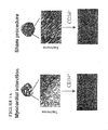
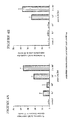
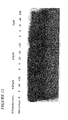
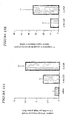
- FIGS 1A-1D IL- 8/Gro-Alpha CXC Chemokines Regulate Migration Of Human Endothelial progenitor cells (angioblasts) To Myocardial Tissue In Vivo And Subsequent Development Of Vasculogenesis.
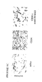
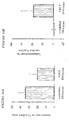
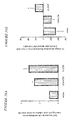
- FIGS. 2A-2C Blocking CXCR4/SDF-1 Interactions Redirects Intravenously Injected Human Endothelial progenitor cells From Bone Marrow To Ischemic Myocardium.
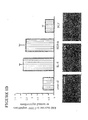
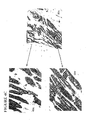
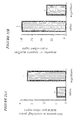
- FIGS 3A-3F Redirected Trafficking Of Human Endothelial progenitor cells (angioblasts) To The Site Of Infarction Induces Vasculogenesis And Protects Cardiomyocytes against Apoptosis.
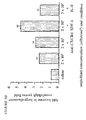
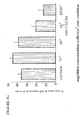
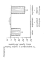
- FIGS 4A-4H Infarct Bed Vasculogenesis Improves Long-Term Myocardial Function Through Mechanisms Involving Both Cardiomyocyte Protection and Proliferation/Regeneration.
- FIG. 5 This figure shows mRNA expression of three genes in the ischemic rat hearts at various time points. You will see that at 48 hours and 2 weeks after LAD ligation and ischemia, HBP23 (the rat homologue of human PAG/NKEF-A,B,C, all of which are part of the family of peroxiredoxins (Prx)) is decreased, and vitamin D3 upregulated protein VDUP-1 is increased. The early (48 hour) reduction in PRX and increase in VDUP-1 results in a compensatory increase in thiol reductase thioredoxin (TRX). Note that endothelial progenitor cell therapy reverses this pattern of mRNA expression.
- HBP23 the rat homologue of human PAG/NKEF-A,B,C, all of which are part of the family of peroxiredoxins (Prx)
- Prx peroxiredoxins
- VEGF vascular endothelial growth factor
- VEGF-R vascular endothelial growth factor receptor
- FGF fibroblast growth factor
- IGF Insulin-like growth factor
- SCF stem cell factor
- G-CSF granulocyte colony stimulating factor
- M-CSF macrophage colony stimulating factor
- GM-CSF granulocyte-macrophage colony stimulating factor
- MCP monocyte chemoattractant protein.
- angioblasts are synonymous with the term “endothelial progenitor cells”.
- CXC CXC chemokine refers to the structure of the chemokine. Each “C” represents a cysteine and “X” represents any amino acid. “CXCR4" refers to CXC receptor 4.
- CC chemokine refers to the structure of the chemokine.
- Each “C” represents a cysteine.
- a “cardiac progenitor cell” refers to a cell that is resident in the heart, or that comes into the heart from elsewhere after acute ischemia, is smaller than mature cardiomyocytes, expresses alpha sarcomeric actin but is negative for troponin, is normally quiescent but can be induced to go into cell cycle as defined by positive Ki61 staining.
- Cardiac progenitor cell may be used synonymously with “cardiomyocyte progenitor cell”.
- Catalytic shall mean the functioning of an agent as a catalyst, i.e. an agent that increases the rate of a chemical reaction without itself undergoing a permanent structural change.
- Nucleic acid shall include without limitation any nuclei acid, including, without limitation, DNA, RNA, oligonucleotides, or polynucleotides, and analogs or derivatives thereof.
- the nucleotides that form the nucleic acid may be nucleotide analogs or derivatives thereof.
- the nucleic acid may incorporate non nucleotides.
- Nucleotides shall include without limitation nucleotides and analogs or derivatives thereof.
- nucleotides may comprise the bases A, C, G, T and U, as well as derivatives thereof. Derivatives of these bases are well known in the art, and are exemplified in PCR Systems, Reagents and Consumables (Perkin Elmer Catalogue 1996-1997, Roche Molecular Systems, Inc., Branchburg, New Jersey, USA).
- Proliferation and “regeneration” are used synonymously when referring to cardiomyocytes in this application. “Proliferation” in respect to cardiomyocytes, shall mean a fold increase in proportion of cardiomyocytes entering the cell cycle relative to untreated rat heart.
- Trafficking means the blood-borne migration of cells, in particular angioblast/endothelial progenitor cells.
- This invention provides a method of treating a disorder of a subject's heart involving loss of cardiomyocytes which comprises administering to the subject an amount of an agent effective to cause cardiomyocyte proliferation within the subject's heart so as to thereby treat the disorder.
- the agent is human endothelial progenitor cells.
- the endothelial progenitor cells are bone marrow-derived. In another they are derived from cord blood, or embryonic or fetal sources. Effective amounts of endothelial progenitor cells sufficient to cause cardiomyocyte proliferation can be done based on animal data using routine computational methods. In one embodiment the effective amount is about 1.5x10 5 endothelial progenitor cells per kg body mass to about 3x10 5 per kg body mass. In another embodiment the effective amount is about 3x10 5 per kg body mass to about 4.5x10 5 endothelial progenitor cells per kg body mass.
- the effective amount is about 4.5x10 5 per kg body mass to about 5.5x10 5 endothelial progenitor cells per kg body mass. In another embodiment the effective amount is about 5.5x10 5 per kg body mass to about 7x10 5 endothelial progenitor cells per kg body mass. In another embodiment the effective amount is about 7x10 5 per kg body mass to about 1x10 6 endothelial progenitor cells per kg body mass. In another embodiment the effective amount is about 1x10 6 per kg body mass to about 1.5x10 6 endothelial progenitor cells per kg body mass.
- the effective amount of human endothelial progenitor cells is between about 1.5x10 6 and 4.5x10 6 endothelial progenitor cells per kg of the subject's body mass and In a preferred embodiment the effective amount is about 5x10 5 endothelial progenitor cells per kg of the subject's body mass.
- the endothelial progenitor cells are allogeneic with respect to the subject.
- the subject is an adult or an embryo or a fetus.
- the endothelial progenitor cells are derived from cloned autologous embryonic stem cells.
- the agent induces expression of a mRNA encoding a peroxiredoxin.
- the expression of peroxiredoxin mRNA may be increased, for example, by administration of 2(3)-t-butyl-4-hydroxyanisole (BHA) (see 106) which has been shown to increase expression of Peroxiredoxin-1 when administered by diet.
- BHA 2(3)-t-butyl-4-hydroxyanisole
- Peroxiredoxin mRNA expression such as that of thiol peroxidases, may also be induced by heme, cadmium or cobalt (see 108).
- Peroxiredoxins include, but are not limited to, PAG, HBP23, MSP23, NKEF.
- the agent induces expression of a mRNA encoding NF-E2-related factor 2 (Nrf2).
- Cytomplasmic NF-E2-related factor 2 (Nrf2) expression can be indirectly increased by raising free Nrf2 levels. Since Nrf2 is tightly bound to keap1 in the cytoplasm then reducing expression of keap1 mRNA is a suitable target e.g. by keap1 antisense oligonucleotides or catalytic nucleic acids (see 109 for keap1). Also, blocking the interaction between Nrf2 and Keap1 by inhibiting the interaction of the Neh2 domain of Nrf2 and the DGR domain of keap1, e.g.
- the agent induces dissociation of a Nrf2 protein from a Keap-1.
- the agent inhibits association of a Nrf2 protein with a Keap-1.
- the agent inhibits association of a thiol reductase thioredoxin with a VDUP-1 protein.
- the agent inhibits c-Ab1 tyrosine kinase activation.
- the agent is STI-571.
- the agent is a CXC chemokine. In further embodiments the agent is Stromal-Derived Actor-1, Il-8 or Gro-Alpha. In one embodiment the amount of CXC chemokine administered is between 0.2 and 5 ⁇ g/ml at a max volume of 10ml for a 70kg human subject. In a preferred embodiment the amount is about 1 ⁇ g/ml. In one embodiment the agent is an inhibitor of plasminogen activator inhibitor-1. In another embodiment the agent is an antibody directed against an epitope of CXCR4. In one embodiment the amount of antibody directed against an epitope of CXCR4 is between 25 and 75 ⁇ g/ml, at a max volume of 10ml for a 70kg human subject.
- the amount is about 50 ⁇ g/ml.
- the agent is Stromal-Derived Factor-1 alpha or Stromal-Derived Factor-1 beta.
- the chemokine can be administered intramyocardially, intracoronary, and/or via a stent, a scaffold, or as a slow-release formulation.
- the agent is G-CSF, CM-CSF, or a Gro family chemokine. In a further embodiment the agent is Gro alpha.
- the instant method further comprises administering an effective amount of a second agent that increases the cardiomyocyte proliferation caused by the human endothelial progenitor cells.
- Effective amounts of the second agent are amounts sufficient to enhance or accelerate cardiomyocyte proliferation in the presence of administered endothelial progenitor cells.
- the endothelial progenitor cells express CD117, CD34, AC133 or a high level of intracellular GATA-2 activity.
- the administering comprises injecting directly into the subject's peripheral circulation, heart muscle, left ventricle, right ventricle, coronary artery, cerebro-spinal fluid, neural tissue, ischemic tissue, or past-ischemic tissue.
- the second agent is an antisense oligonucleotide which specifically inhibits translation of Vitamin D3 Up-Regulated Protein-1 (VDUP-1) mRNA.
- VDUP-1 Vitamin D3 Up-Regulated Protein-1
- VDUP-1 protein (SEQ ID NO:2) expression
- Antisense oligonucleotides are small fragments of DNA and derivatives thereof complementary to a defined sequence on a specified mRNA.
- a VDUP-1 antisense oligonucleotide specifically binds to targets on the VDUP-1 mRNA (SEQ ID NO:1) molecule and in doing so inhibits the translation thereof into VDUP-1 protein (SEQ ID NO:2).
- Antisense oligonucleotide molecules synthesized with a phosphorothioate backbone have proven particularly resistant to exonuclease damage compared to standard deoxyribonucleic acids, and so they are used in preference.
- a phosphorothioate antisense oligonucleotide for VDUP-1 mRNA can be synthesized on an Applied Biosystems (Foster City, CA) model 380B DNA synthesizer by standard methods. E.g. Sulfurization can be performed using tetraethylthiuram disulfide/acetonitrile.
- oligodeoxynucleotides can be base deblocked in ammonium hydroxide at 60°C for 8 h and purified by reversed-phase HPLC [0.1M triethylammonium bicarbonate /acetonitrile; Prop-1 support]. Oligomers can be detritylated in 3% acetic acid and precipitated with 2% lithiumperchlorate/acetone, dissolved in sterile water and reprecipitated as the sodium salt from 1 M NaCl/ethanol. Concentrations of the full length species can be determined by UV spectroscopy.
- mRNA interferes with one or more of the normal functions of VDUP-1 1 mRNA.
- the functions of mRNA to be interfered with include all vital functions such as, for example, translocation of the RNA to the site of protein translation, translation of protein from the RNA, splicing of the RNA to yield one or more mRNA species, and catalytic activity which may be engaged in by the RNA.
- Preferred modified oligonucleotide backbones include, for example, phosphorothioates, chiral phosphorothioates, phosphorodithioates, phosphotriesters, aminoalkylphosphotriesters, methyl and other alkyl phosphonates including 3'-alkylene phosphonates, 5'-alkylene phosphonates and chiral phosphonates, phosphinates, phosphoramidates including 3'-amino phosphoramidate and aminoalkylphosphoramidates, thionophosphoramidates, thionoalkylphosphonates, thionoalkylphosphotriesters, selenophosphates and borano-phosphates having normal 3'-5' linkages, 2'-5' linked analogs of these, and those having inverted polarity wherein one or more internucleotide linkages is a 3' to 3', 5' to 5' or 2' to 2' linkage.
- oligonucleotides having inverted polarity comprise a single 3' to 3' linkage at the 3'-most internucleotide linkage i.e. a single inverted nucleoside residue which may be abasic (the nucleobase is missing or has a hydroxyl group in place thereof) are included.
- Various salts, mixed salts and free acid forms are also included.
- messenger RNA includes not only the information to encode a protein using the three letter genetic code, but also associated ribonucleotides which form a region known to such persons as the 5'-untranslated region, the 3'-untranslated region, the 5' cap region and intron/exon junction ribonucleotides.
- antisense oligonucleotides or the catalytic nucleic acids described below may be formulated in accordance with this invention which are targeted wholly or in part to these associated ribonucleotides as well as to the informational ribonucleotides.
- the antisense oligonucleotides may therefore be specifically hybridizable with a transcription initiation site region, a translation initiation codon region, a 5' cap region, an intron/exon junction, coding sequences, a translation termination codon region or sequences in the 5'- or 3'-untranslated region.
- the catalytic nucleic acids may specifically cleave a transcription initiation site region, a translation initiation codon region, a 5' cap region, an intron/exon junction, coding sequences, a translation termination codon region or sequences in the 5'- or 3'-untranslated region.
- the translation initiation codon is typically 5'-AVG (in transcribed mRNA molecules; 5'-ATG in the corresponding DNA molecule).
- 5'-AVG in transcribed mRNA molecules
- 5'-ATG in the corresponding DNA molecule
- a minority of genes have a translation initiation codon having the RNA sequence 5'-GUG, 5'UUG or 5'-CUG, and 5'-AUA, 5'-11GG and 5'-CUG have been shown to function in vivo.
- the term translation initiation codon can encompass many codon sequences, even though the initiator amino acid in each instance is typically methionine in eukaryotes.
- translation initiation codon refers to the codon or codons that are used in vivo to initiate translation of an mRNA molecule transcribed from a gene encoding VDUP-1, regardless of the sequence(s) of such codons.
- a translation termination codon of a gene may have one of three sequences, i.e., 5'-UAA, 5'-UAG and 5'-UGA (the corresponding DNA sequences are 5'-TAA, 5'-TAG and 5'-TGA, respectively).
- the term "translation initiation codon region” refers to a portion of such an RNA or gene that encompasses from about 25 to about 50 contiguous nucleotides in either direction (i.e., 5' or 3') from a translation initiation codon. This region is one preferred target region.
- translation termination codon region refers to a portion of such an mRNT1 or gene that encompasses from about 25 to about 50 contiguous nucleotides in either direction (i.e., 5' or 3') from a translation termination codon. This region is also one preferred target region.
- Other preferred target regions include the 5' untranslated region (5'UTR), known in the art to refer to the portion of an mRNA in the 5' direction from the translation initiation codon, and thus including nucleotides between the 5' cap site and the translation initiation codon of an mRNA or corresponding nucleotides on the gene, and the 3' untranslated region (3'UTR), known in the art to refer to the portion of an mRNA in the 3' direction from the translation termination codon, and thus including nucleotides between the translation termination codon and 3' end of an mRNA or corresponding nucleotides on the gene.
- 5'UTR 5' untranslated region
- 3'UTR 3' untranslated region
- mRNA splice sites may also be preferred target regions, and are particularly useful in situations where aberrant splicing is implicated in disease, or where an overproduction of a particular mRNA splice product is implicated in disease. Aberrant fusion junctions due to rearrangements or deletions may also be preferred targets.
- antisense oligonucleotides can be chosen which are sufficiently complementary to the target, i.e., hybridize sufficiently well and with sufficient specificity, to give the desired disruption of the function of the molecule.
- "Hybridization” in the context of this invention means hydrogen bonding, also known as watson-Crick base pairing, between complementary bases, usually on opposite nucleic acid strands or two regions of a nucleic acid strand. Guanine and cytosine are examples of complementary bases which are known to form three hydrogen bonds between them. Adenine and thymine are examples of complementary bases which form two hydrogen bonds between them.
- Specifically hybridizable and “complementary” are terms which are used to indicate a sufficient degree of complementarity such that stable and specific binding occurs between the DNA or RNA target and the catalytic nucleic acids.
- catalytic nucleic acids are synthesized once cleavage target sites on the VDUP-1 mRNA molecule.
- antisense oligonucleotides or catalytic nucleic acids or analogs thereof or other agents to mammals suffering from cardiovascular disease, in either native form or suspended in a carrier medium in amounts and upon treatment schedules which are effective to therapeutically treat the mammals to reduce the detrimental effects of cardiovascular disease.
- One or more different catalytic nucleic acids or antisense oligonucleotides or analogs thereof targeting different sections of the nucleic acid sequence of VDUP-1 mRNA may be administered together in a single dose or in different doses and at different amounts and times depending upon the desired therapy.
- the catalytic nucleic acids or antisense oligonucleotides can be administered to mammals in a manner capable of getting the oligonucleotides initially into the blood stream and subsequently into cells, or alternatively in a manner so as to directly introduce the catalytic nucleic acids or antisense oligonucleotides into the cells or groups of cells, for example cardiomyocytes, by such means by electroporation or by direct injection into the heart.
- Antisense oligonucleotides whose presence in cells can inhibit transcription or protein synthesis can be administered by intravenous injection, intravenous drip, subcutaneous, intraperitoneal or intramuscular injection, orally or rectally.
- agents employed in this invention can be administered intracoronary or intramyocardially, for example by injection.
- the agents can be delivered in various means as otherwise discussed, including by stents, such as drug-eluting stents, and in time-release formats such as slow-release.
- the agents are proteins, including but not limited to SDF-1, G-CSF', GM-CSF, and VEGF
- the agents can be administered directly or indirectly, or may be administered via induction of gene expression, through gene therapy, adenoviral delivery and other such methods familiar to those skilled in the art.
- Doses of the oligonucleotides or analogs thereof of the present invention in a pharmaceutical dosage unit will be an efficacious, nontoxic quantity administered to a human patient in need of cardiomyocyte regeneration (or inhibition of VDUP-1 expression) from 1-6 or more times daily or every other day. Dosage is dependent on severity and responsiveness of the effects of abnormal cardiovascular disease to be treated, with course of treatment lasting from several days to months or until a cure is effected or a reduction of the effects is achieved. Oral dosage units for human administration generally use lower doses. The actual dosage administered may take into account the size and weight of the patient, whether the nature of the treatment is prophylactic or therapeutic in nature, the age, weight, health and sex of the patient, the route of administration, and other factors.
- the second agent is a pro-angiogenic agent.
- the pro-angiogenic agent is vascular endothelial growth factor, fibroblast growth factor or anglopoietin.
- the second agent induces expression of a pro-angiogenic factor.
- the second agent is Hypoxia Inducible Factor-1.
- the second agent is a catalytic nucleic acid which specifically inhibits translation of Vitamin D3 Up-Regulated Protein-1 RNA.
- the catalytic nucleic acid comprises deoxyribonucleotides.
- the catalytic nuclei acid comprises ribonucleotides.
- Catalytic nucleic acid molecules can cleave Vitamin D3 Up-Regulated Protein-1 (VDUP-1) mRNA (corresponding DNA shown in SEQ ID NO:1, Figure 5 ) at each and any of the consensus sequences therein. Since catalytic ribo- and deoxyribo- nucleic acid consensus sequences are known, and the VDUP-1. Protein mRNA sequence is known, one of ordinary skill could readily construct a catalytic ribo- or deoxyribo nucleic acid molecule directed to any of the VDUP-1 protein mRNA consensus sequences based on the instant specification. In preferred embodiments of this invention the catalytic deoxyribonucleic acids include the 10-23 structure.
- catalytic ribonucleic acids examples include hairpin and hammerhead ribozyme.
- the catalytic ribonucleic acid molecule is formed in a hammerhead (50) or hairpin motif (51,52,53), but may also be formed in the motif of a hepatitis delta virus (54), group I intron (60), RNaseP RNA (in association with an RNA guide sequence) (55,56) or Neurospora VS RNA (57,58,59).
- catalytic nucleic acids can be designed based on the consensus cleavage sites 5'-purine:pyrimidine-3' in the VDUP-1 mRNA sequence (104) (see figures 6-10 ) for cleavage sites on DNA corresponding to the RNA encoding VDUP-1 (SEQ ID NO:2). Those potential cleavage sites located on an open loop of the mRNA according to RNA folding software e.g. RNADRaw 2.1 are particularly preferred as targets (61).
- the DNA based catalytic nuclei acids can utilize the structure where two sequence-specific arms are attached to a catalytic core based on the VDUP-1 mRNA sequence.
- catalytic DNA structure is detailed in (62) and (63).
- Commercially available mouse brain polyA-RNA (Ambion) can serve as a template in the in vitro cleavage reaction to test the efficiency of the catalytic deoxyribonucleic acids.
- Catalytic RNA is described above, is designed similarly.
- Hammerhead ribozymes can cleave any 5'-NUH-3' triplets of a mRNA, where U is conserved and N is any nucleotide and H can be C,U,A, but not G.
- the sites which can be cleaved by a hammerhead ribozyme in human VDUP-1 mRNA coding region are shown in figure 10 .
- VDUP-1 mRNA with catalytic nucleic acids interferes with one or more of the normal functions of VDUP-1 mRNA.
- the functions of mRNA to be interfered with include all vital functions such as, for example, translocation of the RNA to the site of protein translation, translation of protein from the RNA, splicing of the RNA to yield one or more mRNA species, and catalytic activity which may be engaged in by the RNA.
- the nucleotides may comprise other bases such as inosine, deoxyinosine, hypoxanthine may be used.
- isoteric purine 2'deoxy-furanoside analogs, 2'-deoxynebularine or 2'deoxyxanthosine, or other purine or pyrimidine analogs may also be used.
- inosine may be used to reduce hybridization specificity
- diaminopurines may be used to increase hybridization specificity.
- Adenine and guanine may be modified at positions N3, N7, N9, C2, C4, C5, C6, or C8 and still maintain their hydrogen bonding abilities.
- Cytosine, thymine and uracil may be modified at positions N1, C2, C4, C5, or C6 and still maintain their hydrogen bonding abilities.
- Some base analogs have different hydrogen bonding attributes than the naturally occurring bases. For example, 2-amino-2'-dA forms three (3), instead of the usual two (2), hydrogen bonds to thymine (T).
- Examples of base analogs that have been shown to increase duplex stability include, but are not limited to, 5-fluoro-2'-dU, 5-bromo-2'-dU, 5-methyl-2'-dC, 5-propynyl-2'-dC, 5-propynyl-2'-dU, 2-amino-2'-dA, 7-deazaguanosine, 7-deazadenosine, and N2-Imidazoylpropyl-2'-dG.
- Nucleotide analogs may be created by modifying and/or replacing a sugar moiety.
- the sugar moieties of the nucleotides may also be modified by the addition of one or more substituents.
- the nucleotide may have one or more of its sugars modified and/or replaced so as to be a ribose or hexose (i.e. glucose, galactose) or have one or more anomeric sugars.
- the nucleotide may also have one or more L-sugars.
- the sugar may be modified to contain one or more linkers for attachment to other chemicals such as fluorescent labels.
- the sugar is linked to one or more aminoalkyloxy linkers.
- the sugar contains one or more alkylamino linkers. Aminoalkyloxy and alkylamino linkers may be attached to biotin, cholic acid, fluorescein, or other chemical moieties through their amino group.
- Nucleotide analogs or derivatives may have pendant groups attached.
- Pendant groups serve a variety of purposes which include, but are not limited to, increasing cellular uptake of the oligonucleotide, enhancing degradation of the target nucleic acid, and increasing hybridization affinity.
- Pendant groups can be linked to any portion of the oligonucleotide but are commonly linked to the end(s) of the oligonucleotide chain. Examples of pendant groups include, but are not limited to: acridine derivatives (i.e.
- cross-linkers such as psoralen derivatives, azidophenacyl, proflavin, and azidoproflavin; artificial endonucleases; metal complexes such as EDTA-Fe(II), o-phenanthroline-Cu(I), and porphyrin-Fe(II); alkylating moieties; nucleases such as amino-1-hexanolstaphylococcal nuclease and alkaline phosphatase; terminal transferases; abzymes; cholesteryl moieties; lipophilic carriers; peptide conjugates; long chain alcohols; phosphate esters; amino; mercapto groups; radioactive markers; nonradioactive markers such as dyes; and polylysine or other polyamines.
- cross-linkers such as psoralen derivatives, azidophenacyl, proflavin, and azidoproflavin
- artificial endonucleases such as EDTA-Fe(II), o
- the nucleic acid comprises an oligonucleotide conjugated to a carbohydrate, sulfated carbohydrate, or gylcan.
- Conjugates may be regarded as a way as to introduce a specificity into otherwise unspecific DNA binding molecules by covalently linking them to a selectively hybridizing oligonucleotide.
- the catalytic nucleic acid binding domains i.e. the non-catalytic domains
- antisense oligonucleotide may comprise modified bonds.
- internucleosides bonds of the oligonucleotide may comprise phosphorothioate linkages.
- the nucleic acid may comprise nucleotides having moiety may be modified by replacing one or both of the two bridging oxygen atoms of the linkage with analogues such as -NH, -CH 2 , or -S. Other oxygen analogues known in the art may also be used.
- the phosphorothioate bonds may be stereo regular or stereo random.
- the oligonucleotide moiety may have one or more of its sugars modified or replaced so as to be ribose, glucose, sucrose, or galactose, or any other sugar.
- the phosphorothioate oligonucleotide may have one or more of its sugars substituted or modified in its 2' position, i.e. 2'allyl or 2'-O-allyl.
- An example of a 2'-O-allyl sugar is a 2'-O-methylribonucleotide.
- the phosphorothioate oligonucleotide may have one or more of its sugars substituted or modified to form an ⁇ -anomeric sugar.
- a catalytic nucleic acid may include non-nucleotide substitution.
- the non-nucleotide substitution includes either abasic nucleotide, polyether, polyamine, polyamide, peptide, carbohydrate, lipid or polyhydrocarbon compounds.
- abasic or “abasic nucleotide” as used herein encompasses sugar moieties lacking a base or having other chemical groups in place of base at the 1' position.
- Determining the effective amount of the instant nucleic acid molecules can be done based on animal data using routine computational methods.
- the effective amount contains between about 10 ng and about 100 ⁇ g of the instant nucleic acid molecules per kg body mass. In another embodiment, the effective amount contains between about 100 ng and about 10 ⁇ g of the nucleic acid molecules per kg body mass. In a further embodiment, the effective amount contains between about 1 ⁇ g and about 5 ⁇ g, and in a further embodiment about 2 ⁇ g, of the nucleic acid molecules per kg body mass.
- the second agent is cardiomyocyte progenitor cells.
- the second agent is skeletal muscle progenitor cells. Either of theses cell types can be derived from embryonic, fetal or adult subjects.
- the second agent promotes trafficking of the endothelial progenitor cells to the subject's heart.
- the second agent that promotes trafficking is an antibody directed against an epitope of CXCR4.
- the second agent that promotes trafficking is a CC chemokine.
- the CC chemokine is RANTES, EOTAXIN, monocyte chemoattractant protein-1 (MCP-1), MCP-2, MCP-8, or MCP.
- the second agent that promotes trafficking is a CXC chemokine.
- the CXC chemokine is Interleukin-8, Gro-Alpha, or Stromal-Derived Factor-1.
- the second agent promotes mobilization of angioblasts into the subject's bloodstream.
- the agent is G-CSF, GM-CSF, SDF-1, or VEGF.
- This invention also provides a method of determining the susceptibility of a cardiomyocyte in a subject to apoptosis comprising:
- This invention also provides a method of determining the susceptibility of a cardiomyocyte in a subject to apoptosis comprising:
- a ratio that is at least two standard deviations above that found in control patients with normal hearts is considered a high ratio.
- Quantitation of expression of protein in cardiomyocytes is performed using routine methods known to those of skill in the art, for example, northern and western blots.
- This invention further provides a cardiac progenitor cell.
- the cardiac progenitor cell is resident in the heart, or that comes into the heart from elsewhere after acute ischemia, is smaller than mature cardiomyocytes, expresses alpha sarcomeric actin but is negative for troponin, is normally quiescent but can .be induced to go into cell cycle as defined by positive Ki67 staining.
- This invention further provides a method of inducing cell cycling in a cardiac progenitor cell in a heart tissue of a subject comprising administering to the subject an amount of an agent effective to induce neovascularization of the heart tissue in the subject so as to thereby induce cardiac cell cycling within the heart tissue of the subject.
- This invention further provides a method of improving cardiac function in a subject comprising administering to the subject an amount of an agent effective to induce neovascularization of a heart tissue in the subject so as to thereby improve cardiac function in the subject.
- This invention further provides a method of improving cardiac function in a subject comprising administering to the subject an amount of an agent effective to induce proliferation of cardiomyocytes in the heart tissue in the subject so as to thereby improve cardiac function in the subject.
- Cardiac function, or myocardial function can be determined by any combination of improvement in ejection fraction, perfusion, reduction in anginal symptoms, improvement in quality of life, increased walking ability, longevity of life, reduction in heart medication, and/or prevention of heart failure, as this terms are normally understood in the art.
- An improvement in cardiac function in a subject is measured by determining an any of theses factors in a subject before and after treatment by any of the instant methods.
- the subject has suffered myocardial ischemia or myocardial infarct.
- the agent is G-CSF, GM-CSF, IL-8, a Gro family chemokine, an inhibitor of CXCR4, or an inhibitor of SDF-1.
- the inhibitor of CXCR4 is a small molecule or a monoclonal antibody
- the inhibitor of CXCR4 is a small molecule and is AMD 3100
- the inhibitor of SDF-1 is a small molecule or a monoclonal antibody
- the Gro family chemokine is Gro alpha.
- the inhibitor is AMD 070.
- the inhibitor of CXCR4 is a catalytic nucleic acid, an oligonucleotide, RNAi, (RNA interference) or a small molecule.
- This invention further provides a method of inducing apoptosis in a cell comprising inhibiting expression of a peroxiredoxin in the cell.
- the cell is a tumor cell.
- the peroxiredoxin is peroxiredoxin I, II, II, IV, or V.
- the inhibition of expression of peroxiredoxin is effected by contacting the cell with a catalytic nucleic acid which binds mRNA encoding peroxiredoxin, thereby inhibiting the expression thereof.
- the inhibition of expression of peroxiredoxin is effected by contacting the cell with an antisense oligonucleotide, a monoclonal antibody, RNA interference or a small molecule.
- This invention further provides a method of treating a disorder of a tissue cells of a subject involving loss of tissue cells which comprises administering to the subject an amount of an agent effective to cause tissue cell proliferation within the tissue of the subject so as to thereby treat the disorder.
- the tissue is cardiac tissue, brain tissue, peripheral vascular tissue, hepatic tissue, renal tissue, gastrointestinal tissue, lung tissue, smooth muscle tissue, or striated muscle tissue.
- the agent is G-CSF, SDF-1, GM-CSF, IL-8, VEGF.
- the agent is an inhibitor of CXCR4/SDF-1 interaction.
- the inhibitor is a catalytic nucleic acid, a monoclonal antibody, a antisense oligonucleotide, a small molecule, or RNAi.
- the agent is an inducer of peroxiredoxin expression.
- the peroxiredoxin is peroxiredoxin I, II, III, IV, or V.
- the G-CSF may be a covalent conjugate of recombinant methionyl human G-CSF (Filgrastim) and monomethoxypolyethylene glycol, such as Neulasta.
- Filgrastim is a water-soluble 175 amino acid protein with a molecular weight of approximately 19 kilodaltons (kd). Filgrastim is obtained from the bacterial fermentation of a strain of Escherichia coli transformed with a genetically engineered plasmid containing the human G-CSF gene.
- pegfilgrastim a 20 kd monomethoxypolyethylene glycol molecule is covalently bound to the N-terminal methionyl residue of Filgrastim.
- the average molecular weight of pegfilgrastim is approximately 39 kd.
- Neulasta is supplied in 0.6 mL prefilled syringes for subcutaneous (SC) injection. Each syringe contains 6 mg pegfilgrastim (based on protein weight), in a sterile, clear, colorless, preservative-free solution (pH 4.0) containing acetate (0.35 mg), sorbitol (30. mg), polysorbate 2.0 (0.02 mg), and sodium (0.02 mg) in water for injection, USP.
- This invention provides a method of treating a disorder of a tissue of a subject involving apoptosis of cells in the tissue which comprises administering to the subject an amount of an agent effective to inhibit apoptosis of cells in the tissue within the subject so as to thereby treat the disorder.
- the agent is an inhibitor of VDUP-1 expression.
- the inhibitor of VDUP-1 expression is catalytic nucleic acid, a monoclonal antibody, an antisense oligonucleotide, a small molecule, or an RNAi.
- the tissue is cardiac tissue, cerebrovascular tissue, or cerebral tissue.
- This invention also provides a method of inhibiting proliferation of fibroblasts or inflammatory cells in a tissue and thereby inhibiting collagen formation comprising contacting the tissue with an inhibitor of VDUP-1 expression.
- the inhibitor of VDUP-1 expression is a catalytic nucleic acid, a monoclonal antibody, an antisense oligonucleotide, a small molecule, or an RNAi.
- the subject of any of the above methods is a mammal and in a preferred further embodiment the mammal is a human being.
- the subject has a cardiovascular disease.
- the subject has congestive heart failure, has suffered a myocardial infarct, has suffered myocardial ischemia, has angina, or has a cardiomyopathy.
- CXC Chemokines Regulate Endothelial Progenitor Cell Migration To The Heart.
- LAD coronary artery ligation resulted in trafficking of intravenously injected human endothelial progenitor cells to the site of ischemic myocardium, it was also accompanied by increased distribution of human cells to rat bone marrow.
- Figure 2a at 2-14 days after intravenous injection of 2 x 10 6 human CD34+ cells bone marrow from LAD-ligated rats contained 5-8 fold higher levels of human CD117 bright endothelial progenitor cells compared with bone marrow from normal rats, p ⁇ 0.001.
- Capillary Lumen Size Is Dependent On Absolute Angioblast Numbers.
- Induction of neovascularization at two weeks was measured by performing quantitative analysis of medium- and large-sized capillaries, defined, respectively, as having 3-6 or >6 contiguous endothelial lining cells.
- Medium-sized capillaries had mean lumen diameter of 0.020mm + 0.002, while large-sized capillaries had mean lumen diameter of 0.053mm + 0.004 (p ⁇ 0.001).
- large-lumen capillaries overlapped in size with arterioles which could be distinguished by a thin layer containing 2-3 smooth muscle cells of rat origin, as determined by positive staining with desmin and rat MHC class I mAbs.
- both the group receiving 2x10 5 endothelial progenitor cells and the one receiving 10 5 endothelial progenitor cells plus anti-CXCR4 mAb demonstrated 1.7-fold higher numbers of medium-sized capillaries compared with the other two groups (p ⁇ 0.01).
- the group receiving 2x10 5 endothelial progenitor cells additionally demonstrated 3.3-fold higher numbers of large-lumen capillaries compared with the groups receiving 10 3 or 10 5 endothelial progenitor cells (p ⁇ 0.01), and 2-fold higher numbers of large-lumen capillaries compared with the group receiving 10 5 endothelial progenitor cells plus anti-CXCR4 mAb (p ⁇ 0.01).
- rats receiving 10 5 endothelial progenitor cells plus anti-CXCR4 mAb demonstrated significant improvement in these parameters, 22 + 2% mean recovery in LVEF and 24 + 4% mean reduction in LVAs (both p ⁇ 0.001). Even more strikingly, the group receiving 2x10 5 endothelial progenitor cells had a mean recovery in LVEF of 34 + 4% and a mean reduction in LVAs of 37 + 6% (both p ⁇ 0.001), or 50% further improvement in both parameters.
- the number of cardiomyocytes progressing through celt cycle at the peri-infarct region of rats receiving 2x10 5 human endothelial progenitor cells was 40-fold higher than that at sites distal to the infarct, where myocyte DNA activity was no different than in sham-operated rats.
- animals receiving 2x10 5 human endothelial progenitor cells had a 20-fold higher number of cell-cycling cardiomyocytes at the peri-infarct rim than that found in non-infarcted hearts (1.19 + 0.2% vs 0.06 + 0.03%, p ⁇ 0.01) and 3.5-fold higher than in the same region in LAD-ligated controls receiving saline (1.19 + 0.2% vs 0.344 + 0.1%, p ⁇ 0.01).
- intracardiac injection of SDF-1 in combination with intravenous injection of 2x10 5 human endothelial progenitor cells resulted in approximately an 8-fold cumulative increase in cell-cycling cardiomyocytes at two weeks compared with LAD-ligated controls receiving saline, and translated into over 4-fold greater LVEF improvement, determined by echocardiography, compared with intravenous injection of 2x10 5 endothelial progenitor cells alone ( Figure 4f , p ⁇ 0.01).
- Co-administration of anti-CXCR4 mAb augmented LVEF improvement by 2.8-fold (p ⁇ 0.01) while intracardiac injection of IL-8 conferred no additive benefit.
- PAI-1-inhibiting catalytic nucleic acid augments human angioblast-dependent cardiomyocyte regeneration.
- E2 catalytic nucleic acid
- cdNA subtractive hybridization we observed a striking reciprocal change in expression of a group of genes whose function is linked through their regulation of cellular apoptosis and cell cycle progression following oxidative stress and other inducers of DNA damage.
- certain antioxidant genes such as superoxide dismutase was upregulated in ischemic tissue
- the antioxidant stress responsive genes induced by hemin notably heme binding protein 23 (HBP23) and glutathione-S-transferase
- Vitamin D3 Up-Regulated Proteins VDUP1
- H 2 O 2 hydrogen peroxide
- the cell cycle inhibitory activity of p21Cip1/WAF1 is intimately correlated with its nuclear localization and participation in quaternary complexes of cell cycle regulators by binding to G1 cyclin-CDK through its N-terminal domain and to proliferating cell nuclear antigen (PCNA) through its C-terminal domain (68-71).
- PCNA cell nuclear antigen
- the latter interaction blocks the ability of PCNA to activate DNA polymerase, the principal replicative DNA polymerase (72).
- For a growth-arrested cell to subsequently enter an apoptotic pathway requires signals provided by specific apoptotic stimuli in concert with cell-cycle regulators.
- caspase-nediated cleavage of p21 together with upregulation of cyclin A-associated cdk2 activity, have been shown to be critical steps for induction of cellular apoptosis by either deprivation of growth factors (73) or hypoxia of cardiomyocytes (74).
- the apoptosis signal-regulating kinase 1 (ASK1) is a pivotal component in the mechanism of cytokine- and stress-induced apoptosis (75,76). Under basal conditions, resistance to ASK1-mediated apoptosis appears to be the result of complex formation between ASK1, cytoplasmic p21Cip1/WAFl (77), and the thiol reductase thioredoxin (TRX) (78). Intact cytoplasmic expression of p21Cip1/WAF1 appears to be important for both prevention of apoptosis in response to ASKl (75) and in maintaining a state of terminal differentiation (77).
- the reduced form of TRX binds to the N-terminal portion of ASK1 and is a physiologic inhibitor of ASK1-mediated cellular apoptosis (78).
- the recently-identified protein VDUP1 has been shown to compete with ASKl for binding of the reduced form of TRX (78,79), resulting in augmention of ASKl-mediated apoptosis (80). This indicates that ASKl-mediated cellular apoptosis is increased by processes that result in a net dissociation of TRX from ASK1, such as either generation of TRX-VDUP1 complexes or generation of oxidised TRX by changes in cellular redox status accompanying oxidative stress.
- TRX and glutathione constitute the major cellular reducing systems that maintain the thiol-disulfide status of the cytosol (81).
- the redox-active/dithiol active site of TRX is highly conserved across all species, Trp-Cys-Gly-Pro-Cys-Lys.
- the two cysteine residues at the active site, Cys-32 and Cys-35 undergo reversible oxidation-reduction reactions catalyzed by a NADPH-dependent enzyme TRX reductase. These reactions involve electron transfer via disulfide bridges formed with members of a family of antioxidant enzymes known as peroxiredoxins (Prxs), which show peroxidase activity (82,83).
- Prxs peroxiredoxins
- Prxs are distinct from other peroxidases in that they have no cofactors, such as metals or prosthetic groups. Prxs generally have two conserved cysteines at the N- and C-terminal regions (84), and their antioxidant effects are coupled with the physiological electron donor activity of the TRX system (82,85, 86). Prxs with 95-97% sequence homology have been identified in rats (Heme-binding protein 23, HBP23) (87), mice (mouse macrophage stress protein 23, MSP23) (88) and humans (proliferation-associated gene product, PAG (89) and human natural killer cell-enhancing factor A (90)).
- Prxs are members of a repertoire of oxidative stress responsive genes whose expression is regulated by NF-E2-related factor 2 (Nrf2) which binds to an anti-oxidant responsive element (ARE) present in the promoter of each (91). These include glutathione-S-transferase, heme oxygenase-1, and TRX. Under basal conditions, Nrf2 is bound to a specific protein, Keapl, in the cytosol (92). However, under conditions of oxidative stress Nrf2 dissociates from Keapl and translocates to the nucleus where it induces transcriptional activation of the anti-oxidant genes containing ARE motifs.
- Nrf2 NF-E2-related factor 2
- ARE anti-oxidant responsive element
- Nrf2 nuclear translocation of Nrf2 and subsequent ARE activation appear to be dependent on pathways activated by phosphatidylinositol 3-kinase (P13 kinase) (93).
- P13 kinase phosphatidylinositol 3-kinase
- hemin is a potent inducer of Nrf2 dissociation from Keapl, resulting in TRX gene transcription through the ARE (94).
- Prxs During periods of rapid changes in cellular redox Prxs presumably serve to maintain the cytosolic levels of reduced TRX by accepting electrons from the oxidized form of TRX. This homeostatic mechanism likely enables maintenance of sufficient levels of reduced TRX to ensure adequate binding to ASK1 and prevention of cellular apoptosis. If the endogenous Prx system is overloaded, as might occur during changes in cellular redox when excess oxidized TRX is generated, cellular apoptosis will occur through the unopposed effects of ASK1. To counteract this, transcriptional activation of Prxs must occur following oxidative stress via nuclear translocation of Nrf2. This can be achieved either by Nrf2 dissociation from Keap1 via hemin- and PI3 kinase-dependent mechanisms (93,94), or by increasing Nrf2 mRNA and protein expression as occurs following increase in oxygen tension (95,96).
- the Prx gene products specifically bind the SH3 domain of c-Abl, a non-receptor tyrosine kinase, inhibiting its activation by various stimuli, including agents that damage DNA (97).
- c-Abl activation through the SH3 domain induces either arrest of the cell cycle in phase G1 or cellular apoptosis (98).
- Cell cycle arrest is dependent on the kinase activity of c-Abl (96) and is mediated by the ability of c-Abl to downregulate the activity of the cyclin-dependent kinase Cdk2 and induce the expression of p21 (99).
- c-Abl The apoptotic effects of c-Abl are dependent on the ability of nuclear c-Abl to phosphorylate p73, a member of the p53 family of tumor-suppressor proteins which can induce apoptosis (100,101). Recently, it has been shown that cytoplasmic, rather than nuclear, forms of c-Abl are activated by H 2 O 2 and that this results in mitochondrial localization of c-Abl, c-Abl dependent cytochrome c release, and cellular apoptosis following oxidative stress (102,103).
- the PAG gene product By associating with c-Abl in vivo, the PAG gene product (and presumably the other Prxs) can inhibit tyrosine phosphorylation induced by c-Abl overexpression and rescue cells from both the cytostatic and pro-apoptotic effects of the activated c-Abl gene product (97).
- Nrf2-dependent oxidative stress responsive genes are downregulated following myocardial ischemia likely reflects direct effects of hemin and oxygen deprivation.
- the end result of Prx downregulation in the ischemic heart would be augmentation in ASK1-dependent cellular apoptosis as well as Abl-dependent apoptosis and cell cycle arrest.
- the observed parallel increase in VDUP1 expression would further augment ASK1-dependent cellular apoptosis.
- the ratio in expression of PAG or other Prx mRNA or protein to VDUP1 mRNA or protein can form the basis of a diagnostic assay to predict the degree of risk for cardiomyocyte apoptosis and cell cycle arrest after ischemia, as well as enable monitoring of the response to specific therapy after myocardial ischemia that protects cardiomyocytes against apoptotic death and enhances myocardial proliferation/regeneration.
- Reversing the reduced expression of the Prxs following myocardial ischemia would increase Prxs in the heart in order to protect the ischemic myocardium against apoptosis through both c-Abl inhibition and reduction of oxidised TRX, and to enable cardiomyocyte proliferation/regeneration by inhibiting the effects of c-Abl on cell cycle progression from G1 to S phase.
- Nrf2 mRNA or causing dissociation of Nrf2 protein from Keap1, or preferably cause both to occur simultaneously in the setting of myocardial ischemia in order to increase transcription and activity of members of a repertoire of oxidative stress responsive genes whose expression is regulated by binding of Nrf2 to an anti-oxidant responsive element (ARE) in their promoters, including the Prxs, TRX and glutathione-S-transferase would result in both protection of cardiomyocytes against apoptosis as well as induce cardiomyocyte cell cycle progression following oxidative stress.
- ARE anti-oxidant responsive element
- VDUP1 Reducing the expression of VDUP1 following myocardial ischemia would protect the ischemic myocardium against apoptosis by reducing binding of TRX to VDUP1, and consequently increasing TRX-ASK1 interactions.
- Neovascularization of the myocardium by either bone marrow-derived endothelial progenitors or any other process, is an example of one method which causes induction of Prx expression and reduction in VDUP1 expression after myocardial ischemia, and results in both protection against redox-mediated apoptosis and induction of myocardial proliferation/regeneration.
- Small molecules which specifically inhibit binding of Nrf2 to Keap1 would be expected to have similar protective effects against cardiomyocyte apoptosis and to induce myocardial proliferation/regeneration after ischemia.
- small molecules that specifically inhibit binding of TRX to VDUP1 would be expected to have similar protective effects against cardiomyocyte apoptosis after ischemia.
- small molecules to specifically inhibit c-Abl tyrosine kinase activation after myocardial ischemia would be expected to have similar protective effects against cardiomyocyte apoptosis and to induce myocardial proliferation/regeneration after ischemia.
- a specific example of a small molecule to inhibit c-Abl tyrosine kinase activation is STI-571. Use of this or related molecules after myocardial infarction would protect against cardiomyocyte apoptosis and induce myocardial proliferation/regeneration.
- VDUP1 By subtractive hybridization, we found that mRNA expression of the protein VDUP1 is significantly increased in the heart after acute ischemia.
- VDUP1 has been shown to bind the cytosolic protein thioredoxin, TRX, which functions to maintain the thiol-disulfide status of the cytosol.
- TRX cytosolic protein thioredoxin
- VDUP1 competes for TRX binding with another cytosolic protein that is normally bound to reduced TRX, the apoptosis signal-regulating kinase 1 (ASK1).
- ASK-1 is a pivotal component in the mechanism of cytokine- and stress-induced apoptosis. Its activation results in excessive phosphorylation and activation of p38 MAP kinase, a principal mediator of cellular apoptosis.
- the reduced form of TRX but not the oxidised form, binds to the N-terminal portion of ASK1 and is a physiologic inhibitor of ASK1-mediated cellular apoptosis. Binding of VDUP1 to TRX results in a net dissociation of ASK1 from TRX, potentially resulting in augmentation of ASK1-mediated apoptosis via p38 MAP kinase-dependent pathways.
- VDUP1 With respect to cardiac overexpression of VDUP1, the anticipated effects would be those due to excessive p38 MAP kinase activation and oxidative redox damage, including cardiomyocyte apoptosis, fibroblast proliferation, collagen secretion and scar formation. Similar effects would be expected to be the consequence of VDUP1 overexpression in other tissues undergoing acute or chronic ischemic injury, for example the brain following cerebrovascular ischemia/stroke.
- the DNA enzyme once delivered to the ischemic myocardium (or other ischemic tissues such as the brain) can inhibit local VDUP1 mRNA and protein expression, thus reducing p38 MAP kinase activation and oxidative damage.
- VDUP-1 DNAzyme Reduces Fibroblast Proliferation And Protects Against Cardiomyocyte Apoptosis.
- intramyocardial injection of the rat sequence-specific VDUP1 DNA enzyme at 48 hours after LAD ligation resulted in a 75% mean inhibition of proliferating cardiac fibroblasts in the infarct zone two weeks later in comparison to injection of scrambled DNA enzyme control (p ⁇ 0.01).
- injection of VDUP1 DNA enzyme resulted in 20% mean reduction in apoptotic cardiomyocytes at the peri-infarct region relative to injection with the scrambled DNA enzyme control (p ⁇ 0.05).
- VDUP-1 DNAzyme Reduces Myocardial Scar And Improves Cardiac Function After Acute Infarction. Inhibition of fibroblast proliferation and cardiomyocyte apoptosis resulted in significant reduction of mature scar deposition in the infarct zone, from a mean of 35% for animals receiving control scrambled DNA enzyme to a mean of 20% for those receiving VDUP1 DNA enzyme, figure 14(a) (p ⁇ 0.01). Most dramatic was the effect on cardiac function. As shown in figure 14(b) , animals receiving VDUP1 DNA enzyme demonstrated a 50% mean recovery in cardiac function, as determined by ejection fraction, whereas no improvement was seen in animals receiving scrambled control DNA enzyme (p ⁇ 0.01).
- VDUP1 DNA enzyme prevents cardiomyocyte apoptosis, cardiac fibroblast proliferation and scar formation, resulting in significant improvement in cardiac function after acute ischemia. These effects are presumably due to prevention of p38 MAP kinase activation and protection against redox damage. Similar results might be obtained by administering the VDUP1 DNA enzyme to other tissue sites of reduced blood flow such as the brain after cerebrovascular ischemia.
- G-CSF is a more potent inducer of neovascularization after myocardial infarction than GM-CSF.
- rats injected subcutaneously with human G-CSF at 10 ug/kg for four days starting at two days after myocardial infarction induced by left anterior descending (LAD) coronary artery ligation demonstrated approximately 7.5-fold greater numbers of large-diameter blood vessels at the peri-infarct region two weeks later relative to saline-treated controls (p ⁇ 0.01).
- Rat GM-CSF administered at the same dosage regimen was less effective, though still inducing 4-fold greater numbers of large-lumen vessels than in control animals.
- G-CSF is a more potent inhibitor of cardiomyocyte apoptosis after myocardial infarction than GM-CSF.
- G-CSF injection was a more potent agent for protecting against cardiomyocyte apoptosis than GM-CSF used at the same dosage regimen, figure 15(b) .
- G-CSF administration resulted in 36 + 16% reduction in the numbers of apoptotic cardiomyocytes at the peri-infarct region by two weeks relative to saline-treated controls (p ⁇ 0.01), whereas GM-CSF only reduced apoptotic cardiomyocyte numbers by 12 + 9%.
- G-CSF is a more potent inducer of cardiomyocyte regeneration and functional cardiac recovery after myocardial infarction than GM-CSF.
- rats injected subcutaneously with human G-CSF at 10 ug/kg for four days starting at two days after myocardial infarction demonstrated 3.2-fold greater numbers of cycling cardiomyocytes two weeks later at the peri-infarct region relative to saline-treated controls (p ⁇ 0.05).
- Rat GM-CSF administered at the same dosage regimen was less effective, resulting in 2.6-fold greater numbers of cycling cardiomyocytes.
- Intravenous administration of anti-CXCR4 antibody increases myocardial neovascularization and improves cardiac function after acute infarction.
- the major mechanism by which G-CSF causes mobilization of bone marrow elements is through interruption of interactions between the chemokine receptor CXCR4 on bone marrow resident stem cells and its ligand SDF-1.
- G-CSF induces both cleavage of the N-terminus of CXCR4 and accumulation of serine proteases which directly cleave and inactivate SDF-1.
- anti-CXCR4 treated animals demonstrated a mean recovery in ejection fraction of 10% at two weeks whereas those receiving anti-CXCR2 mAb had a mean loss in cardiac function of 8% (p ⁇ 0.05).
- SDF-1 mRNA expression is not induced early in acutely ischemic myocardium, and its late induction is inhibited by GM-CSF.
- GM-CSF GM-CSF
- SDF-1 is a potent chemotactic factor for endothelial progenitor cells
- systemic GM-CSF administration may result in suboptimal myocardial homing of endothelial progenitors due to decreased myocardial expression of chemotactic ligands such as SDF-1.
- Intramyocardial injection of SDF-1 after acute myocardial infarction results in neovascularization and protection against cardiomyocyte apoptosis.
- SDF-1 expression in the acutely ischemic heart affects endothelial progenitor cell chemotaxis and cardiomyocyte function.
- intramyocardial injection of 4ug/kg human recombinant SDF-1 in a total volume of 0.2 ml at 5 peri-infarct sites two days post-LAD ligation resulted by two weeks in a 5-fold increase in neovascularization and a 44 + 9% reduction in apoptotic cardiomyocytes at the peri-infarct region relative to control animals receiving intramyocardial saline injections (both p ⁇ 0.05).
- the effects of intramyocardial SDF-1 injection on neovascularization and on extent of protection against cardiomyocyte apoptosis were in strikingly close parallel to the results obtained with systemic administration of G-CSF.
- Intramyocardial injection of SDF-1 induces cardiomyocyte regeneration.
- intramyocardial injection of 4ug/kg human recombinant SDF-1 in a total volume of 0.2 ml at 5 peri-infarct sites two days post-LAD ligation resulted in a 4.5-fold increase in the number of cycling cardiomyocyte at the peri-infarct region relative to saline-injected controls (p ⁇ 0.01).
- Addition of systemic CM-CSF administration resulted in synergistic effects on cardiomyocyte regeneration.
- Intramyocardial injection of SDF-1 improves cardiac function and is synergistic with bone marrow mobilization. Intramyocardial injection of SDF-1 resulted in a similar degree of functional myocardial recovery as was seen with systemic G-CSF administration (10% mean improvement in ejection fraction between days 2 and 14 post-LAD ligation compared with 10% mean improvement for G-CSF treated animals and 17% mean loss for saline-treated controls, p ⁇ 0.01 for both treatment arms). Most strikingly, combining SDF-1 injection with bone marrow mobilization by GM-CSF resulted in significant augmentation in functional recovery (21% mean improvement in ejection fraction, p ⁇ 0.01), figure 20(b) .
- cytokine-mobilized human CD34+ cells Single-donor leukopheresis products were obtained from humans treated with recombinant G-CSF 10 mg/kg (Amgen, CA) sc daily for four days. Donors were healthy individuals undergoing standard institutional procedures of bone marrow mobilization, harvesting and isolation for allogeneic stem cell transplants. Mononuclear cells were separated by Ficoll-Hypaque, and highly-purified CD34+ cells (>98% positive) were obtained using magnetic beads coated with anti-CD34 monoclonal antibody (mAb) (Miltenyi Biotech, CA).
- mAb anti-CD34 monoclonal antibody
- CD34 cells were stained with fluorescein-conjugated mAbs against CD34 and CD117 (Becton Dickinson, CA), AC133 (Miltenyi Biotech, CA), CD54 (Immunotech, CA), CD62E (BioSource, MA), VEGFR-2, Tie-2, vWF, eNOS, CXCR1, CXCR2, and CXCR4 (all Santa Cruz Biotech, CA), and analyzed by four-parameter fluorescence using FACScan (Becton Dickinson, CA).
- Cells positively selected for CD34 expression were also stained with phycoerythrin (PE)-conjugated anti-CD117 mAb (Becton Dickinson, CA), and sorted for bright and dim fluorescence using a Facstar Plus (Becton Dickinson) and a PE filter.
- Intracellular staining for GATA-2 was performed by permeabilizing one million cells from each of the brightly and dimly fluorescing cell populations using a Pharmingen Cytofix/Cytoperm TM kit, incubating for 30 minutes on ice with 10 ⁇ l of fluorochrome-conjugated mAbs against both CD117 and CD34 surface antigens (Becton Dickinson, CA).
- Chemotaxis of human bone-marrow derived endothelial progenitors were plated in 48-well chemotaxis chambers fitted with membranes (8 mm pores) (Neuro Probe, MD). After incubation for 2 hours at 37°C, chambers were inverted and cells were cultured for 3 hours in medium containing IL-8, SDF-1 alpha/beta, and SCF at 0.2, 1.0 and 5.0 ⁇ g/ml. The membranes were fixed with methanol and stained with Leukostat"' (Fischer Scientific, I11). Chemotaxis was calculated by counting migrating cells in 10 high-power fields.
- Rowett (rnu/rnu) athymic nude rats (Harlan Sprague Dawley, Indianapolis, Indiana) were used in studies approved by the "Columbia University Institute for Animal Care and Use Committee". After anesthesia, a left thoracotomy was performed, the pericardium was opened, and the left anterior descending (LAD) coronary artery was ligated. Sham-operated rats had a similar surgical procedure without having a suture placed around the coronary artery.
- CD34+ cells obtained from a single donor after G-CSF mobilization were injected into the tail vein 48 hours after LAD ligation either alone or together with 50 ⁇ g/ml monoclonal antibody (mAb) with known functional inhibitory activity against either human CXCR1, human CXCR2, human CXCR4, rat SDF-1 (all R & D Systems, MN), human CD34 (Pharmingen, CA), or rat IL-8 (ImmunoLaboratories, Japan). Controls received either isotype control antibodies at the same concentration or saline after LAD ligation.
- mAb monoclonal antibody
- Quantitation of myocardial infiltration after injection of human cells was performed by assessment of DiI fluorescence in hearts from rats sacrificed 2 days after injection (expressed as number of DiI-positive cells per high power field, minimum 5 fields examined per sample). Quantitation of rat bone marrow infiltration by human cells was performed in 12 rats at baseline, days 2, 7, and 14 by flow cytometric and RT-PCR analysis of the proportion of HLA class I-positive cells relative to the total rat bone marrow population.
- rat CXC chemokine mRNA and protein expression Poly(A)+ mRNA was extracted by standard methods from the hearts of 3 normal and 12 LAD-ligated rats. RT-PCR was used to quantify myocardial expression of rat IL-8 and Gro-alpha mRNA at baseline and at 6, 12, 24 and 48 hours after LAD ligation after normalizing for total rat RNA as measured by GAPDH expression.
- cDNA was amplified in the polymerase chain reaction (PCR) using Tag polymerase (Invitrogen, Carlsbad, CA, USA), radiolabeled dideoxy-nucleotide ([a32P]-ddATP: 3,000 Ci/mmol, Amersham, Arlington Heights, IL), and primers for rat Cinc (rat homologue of human IL-8/Gro-alpha and GAPDH (Fisher Genosys, CA).
- Tag polymerase Invitrogen, Carlsbad, CA, USA
- radiolabeled dideoxy-nucleotide [a32P]-ddATP: 3,000 Ci/mmol, Amersham, Arlington Heights, IL
- primers for rat Cinc rat homologue of human IL-8/Gro-alpha and GAPDH (Fisher Genosys, CA).
- Primer pairs (sense/antisense) for rat Cinc and GAPDH were, gaagatagattgcaccgatg (SEQ ID NO:4)/catagcctctcacatttc SEQ ID NO:5),gcgcccgtccgccaatgagctgcgc SEQ ID NO:6) /cttggggacacccttcagcatcttttgg (SEQ ID NO:7), and ctctacccacggcaagttcaa (SEQ ID NO:8)/gggatgaccttgcccacagc SEQ ID NO:9), respectively.
- rat IL-8/Gro-alpha Serum levels of rat IL-8/Gro-alpha were measured at baseline and at 6, 12, 24 and 48 hours after LAD ligation in four rats by a commercial ELISA using polyclonal antibodies against the rat IL-8/Gro homologue Cinc (ImmunoLaboratories, Japan). The amount of protein in each serum sample was calculated according to a standard curve of optical density (OD) values constructed for known levels of rat IL-8/Gro-alpha protein.
- OD optical density
- Anti-Cinc antibodies were also used according to the manufacturer's instructions at 1:200 dilution in immunohistochemical studies to identify the cellular source of Cinc production in rat myocardium after LAD ligation. Positively-staining cells were visualized as brown through the Avidin/Biotin system described below.
- Histology and measurement of infarct size Following excision at 2 and 15 weeks, left ventricles from each experimental animal were sliced at 10-15 transverse sections from apex to base. Representative sections were fixed in formalin and stained for routine histology (H&E) to determine cellularity of the myocardium, expressed as cell number per high power field (HPF) (600x). A Masson trichrome stain was performed, which labels collagen blue and myocardium red, to evaluate collagen content on a semiquantitative scale (0-3+), with 1+ light blue, 2+ light blue and patches of dark blue, and 3+ dark blue staining. This enabled measurement of the size of the myocardial scar using a digital image analyzer.
- Staining was performed by immunoperoxidase technique using an Avidin/Biotin Blocking Kit, a rat-absorbed biotinylated anti-mouse IgG, and a peroxidase-conjugate (all Vector Laboratories Burlingame, CA).
- Capillary density was determined at 2 weeks post infarction from sections labeled with anti-CD31 mAb, and confirmed with anti-factor VIII mAb, and compared to the capillary density of the unimpaired myocardium. Values are expressed as the number of CD31-positive capillaries per HPF (400x) .
- Cardiomyocyte DNA synthesis and cell cycling was determined by dual staining of rat myocardial tissue sections obtained from LAD-ligated rats at two weeks after injection of either saline or CD34+ human cells, and from healthy rats as negative controls, for cardiomyocyte-specific troponin I and human- or rat-specific Ki-67. Briefly, paraffin embedded sections were microwaved in a 0.1M EDTA buffer, and stained with either a primary monoclonal antibody against rat Ki-67 at 1:3000 dilution (gift of Giorgio Catoretti, Columbia University) or human Ki-67 at 1:300 dilution (Dako, CA) and incubated overnight at 4 degrees C.
- tissue sections were then digested with Proteinase K (10 ⁇ g/ml in Tris/HCL) for 30 minutes at 37°C.
- the slides were then washed 3 times in PBS and incubated with 50 ⁇ l of the TUNEL reaction mixture (TdT and fluorescein-labeled dUTP) and incubated in a humid atmosphere for 60 minutes at 37°C.
- TdT was eliminated from the reaction mixture.
- the sections were then incubated for 30 minutes with an antibody specific for fluorescein-conjugated alkaline phosphatase (AP) (Boehringer Mannheim, Mannheim, Germany).
- AP fluorescein-conjugated alkaline phosphatase
- the TUNEL stain was visualized with a substrate system in which nuclei with DNA fragmentation stained blue, (BCIP/NBT substrate system, Dako, Carpinteria, CA). The reaction was terminated following three minutes of exposure with PBS. To determine the proportion of blue-staining apoptotic nuclei within myocytes, tissue was counterstained with a monoclonal antibody specific for desmin. Endogenous peroxidase was blocked by using a 3% hydrogen perioxidase solution in PBS for 15 minutes, followed by washing with 20% goat serum solution. An anti-troponin I antibody (Accurate Chemicals, CT) was incubated overnight (1:200) at 40 degrees C.
- BCIP/NBT substrate system Dako, Carpinteria, CA
- Tissue sections were examined microscopically at 200x magnification. Within each 200x field 4 regions were examined, containing at least 250 cells per region and cumulatively approximating 1 mm 2 of tissue, at both the peri-infarct site and distally to this site. Stained cells at the edges of the tissue were not counted. Results were expressed as the mean number of apoptotic myocytes per mm 2 at each site examined.
- Echocardiographic studies were performed using a high frequency liner array transducer (SONOS 5500, Hewlett Packard, Andover, MA). 2D images were obtained at mid-papillary and apical levels. End-diastolic (EDV) and end-systolic (ESV) left ventricular volumes were obtained by bi-plane area-length method, and % left ventricular ejection fraction was calculated as [(EDV-ESV)/EDV] x 100.
- SONOS 5500 high frequency liner array transducer
- ESV End-diastolic
- ESV end-systolic
- CDNA Subtractive Hybridization Briefly, messenger RNA was isolated from each heart, and 1 ⁇ g was used for first-strand cDNA synthesis with random primers. The subtractive hybridization was performed with the PCR-select cDNA subtraction kit (CLONTECH), following the manufacturer's recommendations. After second-strand synthesis, the two cDNA libraries were digested with RsaI. Digestion products of the "tester” library were ligated to a specific adapter (T7 promoter), then hybridized with a 30-fold excess of the "driver” library for subtraction. After hybridization, the remaining products were further amplified by PCR.
- the ischemic tissue In the forward subtraction, which determines the genes that are overexpressed in the ischemic sample, the ischemic tissue is the "tester” and the normal tissue is the “driver.” In the reverse subtraction, the "tester” and the “driver” are switched to determine the genes that are down-regulated in the ischemic sample.
- cardiomyocyte-specific troponin I a marker of cardiomyocyte differentiation, but stained positively for alpha-sarcomeric actin, indicating they were immature cells of cardiomyocyte lineage. Similar clusters of cycling cardiomyocyte progenitors were not seen in control animals injected with saline.
- Tissues obtained from animals sacrificed at two weeks after human CD34+ administration no longer demonstrated clusters of small, cycling cardiomyocyte progenitors, but instead a high frequency of large, mature rat cardiomyocytes at the peri-infarct region with detectable DNA activity, as determined by dual staining with mAbs reactive against cardiomyocyte-specific troponin I and rat Ki67, figures 22(b) and (c) .
- cycling, mature cardiomyocytes at the peri-infarct region was 4-fold higher in animals receiving human Endothelial progenitor cells than in LAD-ligated controls receiving saline (p ⁇ 0.01) in whom there was a high frequency of cells with fibroblast morphology and reactivity with rat Ki67, but not troponin I.
- the cycling, mature cardiomyocytes seen at day 14 post-infarction in animals who received human endothelial progenitor cells are the differentiated progeny of the small, cycling immature cardiomyocyte progenitors seen in the same anatomical location at day 5 and accompanying the onset of neovascularization. Whether the cycling cardiomyocyte progenitors represent in situ cardiac stem cells normally residing in the heart in a quiescent state, or whether these cells have migrated to the heart from elsewhere in the body, such as the bone marrow, remains to be determined.
- peroxiredoxins A family of antioxidant enzymes known as peroxiredoxins, which demonstrate peroxidase activity (16) and are induced by oxygen (17), play a critical role in regulating cell survival during periods of oxidative stress, such as ischemia. They serve to maintain the thiol-disulfide status of the cytosol by undergoing reversible oxidation-reduction reactions involving electron transfer via disulfide bridges formed with thioredoxin (TRX), which constitutes one of the principal cellular reducing systems in mammals (18).
- TRX thioredoxin
- peroxiredoxins can inhibit c-Abl tyrosine kinase activity induced by oxidative stress (19,20) and can rescue cells from both a pro-apoptotic state and cell cycle arrest induced by the activate c-Abl gene product (21).
- cDNA subtractive hybridization we found that expression of the rat peroxiredoxin HBP23 mRNA was reduced in rat hearts 48 hours after LAD ligation compared with normal rat hearts.
- HBP23 mRNA levels in rat hearts decreased at two weeks post-LAD ligation by a mean of 34% compared with normal rat hearts, figure 23 (p ⁇ 0.0i) .
- HBP23 mRNA levels in LAD-ligated rat hearts two weeks after receiving human angioblasts returned to levels only 14% lower than in non-ischemic controls. Since mRNA expression of peroxiredoxins is induced by oxygen, these data suggested that HBP23 mRNA expression was inhibited by the acute ischemic event and induced following angioblast-dependent neovascularization.
- HBP23 a catalytic RNA enzyme targeting specific sequences within the rat HBP23 gene (22,23).
- a catalytic RNA enzyme targeting specific sequences within the rat HBP23 gene 22,23.
- PAG proliferation-associated gene
- the nucleotide sequence in the two flanking arms of the HBP23 DNA enzyme was scrambled without altering the catalytic domain.
- the 3' terminus of each molecule was capped with an inverted 3'-3'-linked thymidine for resistance to 3'-'to-5' exonuclease digestion.
- the DNA enzyme against HBP23 cleaved the 23-base oligonucleotide synthesized from the sequence of rat HBP23 mRNA, in a dose- and time-dependent manner, figure 24 (a) .
- the DNA enzyme did not cleave a 23-base oligonucleotide form the human homologue PAG which differs from the rat HBP23 oligonucleotide by only one base, demonstrating its extraordinar target specificity.
- a DNA enzyme with specificity for the same translational start site in the human PAG gene efficiently cleaved the PAG oligonucleotide, but not the one derived from HBP23 (data not shown).
- the scrambled control DNA enzyme did not cleave either oligonucleotide.
- cardiomyocyte monolayers obtained from fetal rat hearts were grown to confluence and transfected with species-specific DNA enzymes or scrambled control. Densitometric analysis of RT-PCR products following reverse transcription of cellular mRNA showed that the HBP23 DNA enzyme inhibited steady-state mRNA levels in cultured rat cells by over 80%, figure 24 (b) , relative to the scrambled DNA.
- HBP23 DNA enzyme had no effect on induction of neovascularization by human bone marrow angioblasts.
- rats who received human endothelial progenitors irrespective of whether HBP23 DNA enzyme or scrambled control was co-injected, demonstrated increased myocardial capillary density in comparison to saline controls.
- HBP23 DNA enzyme In contrast, injection of the HBP23 DNA enzyme, but not the scrambled control, abrogated the anti-apoptotic effects of neovascularization, figure 25 (b) , and the improvement in cardiac function, figure 25 (c) .
- treatment with HBP23 DNA enzyme resulted in 1.7-fold higher levels of cardiomyocyte apoptosis (p ⁇ 0.01) and 38% mean deterioration in cardiac function as assessed by echocardiography (p ⁇ 0.01).
- the HBP23 DNA enzyme exerted striking effects on cardiomyocyte progenitor proliferation/regeneration.
- neovascularization of ischemic myocardium by human bone marrow derived angioblasts results in both protection of mature cardiomyocytes at the peri-infarct region against apoptosis and stimulation of cardiomyocyte progenitors at the same site to enter cell cycle, proliferate and regenerate.
- the mechanism by which angioblast-dependent neovascularization regulates cardiomyocyte survival and self-renewal appears to involve at least one family of genes involved in anti-apoptotic and pro-proliferative pathways accompanying oxidative stress, the peroxiredoxin.
- peroxiredoxin gene expression is induced by protein kinase C delta (25) it is possible that bone marrow angioblasts may be a source of extracellular signals regulating protein kinase C delta activation and consequently cell survival, such as FGF-1 (26).
- Activation of c-Abl through the SH3 domain by stimuli such as agents that damage DNA induces either arrest of the cell cycle in phase G1 or cellular apoptosis (30).
- Cell cycle arrest is dependent on the kinase activity of c-Abl which downregulates the activity of the cyclin-dependent kinase Cdk2 and induces the expression of p21 (31).
- peroxiredoxins can inhibit tyrosine phosphorylation induced by c-Abl overexpression and rescue cells from both the cytostatic and pro-apoptotic effects of the activated c-Abl gene product (32).
- Our in vivo demonstration that co-administration of a DNA enzyme directed against the rat peroxiredoxin HBP23 abrogated the anti-apoptotic and pro-proliferative effects of human angioblast-dependent neovascularization in the infarcted rat heart argues strongly that this family of genes is directly implicated in the mechanism of action by which neovascularization results in improvement in cardiac function and prevention of heart failure.
- cycling cardiomyocyte precursors differentiated in situ to become new, mature, functional cardiomyocytes. Whether these, precursors are derived from a resident pool of cardiomyocyte stem cells or from a renewable source of circulating bone marrow-derived stem cells that home to the damaged myocardium remains to be determined. Moreover, while the signals required for in situ expansion of cardiomyocyte precursors appear to involve, at least in part, pathways regulated by members of the peroxiredoxin gene family, the signals required for cardiomyocyte differentiation are, at present, unknown. Gaining an understanding of these issues may open the possibility of manipulating the biology of endogenous cardiomyocytes in order to augment the healing process after myocardial ischemia.
- cytokine-mobilized human CD34+ cells Single-donor leukopheresis products were obtained from humans treated with recombinant G-CSF 10 mg/kg (Amgen, CA) sc daily for four days. Donors were healthy individuals undergoing standard institutional procedures of bone marrow mobilization, harvesting and isolation for allogeneic stem cell transplants. Mononuclear cells were separated by Ficoll-Hypaque, and highly-purified CD34+ cells (>98% positive) were obtained using magnetic beads coated with anti-CD34 monoclonal antibody (mAb) (Miltenyi Biotech, CA).
- mAb anti-CD34 monoclonal antibody
- CD34 cells were stained with fluorescein-conjugated mAbs against CD34 and CD117 (Becton Dickinson, CA), AC133 (Miltenyi Biotech, CA), CD54 (Immunotech, CA), CD62E (BioSource, MA), VEGFR-2, Tie-2, vWF, eNOS, CXCR1, CXCR2, and CXCR4 (all Santa Cruz Biotech, CA), and analysed by four-parameter fluorescence using FACScan (Becton Dickinson, CA).
- Cells positively selected for CD34 expression were also stained with phycoerythrin (PE)-conjugated anti-CD117 mAb (Becton Dickinson, CA), and sorted for bright and dim fluorescence using a Facstar Plus (Becton Dickinson) and a PE filter.
- Intracellular staining for GATA-2 was performed by permeabilizing one million cells from each of the brightly and dimly fluorescing cell populations using a Pharmingen Cytofix/Cytoperm kit, incubating for 30 minutes on ice with 10ul of fluorochrome-conjugated mAbs against both CD117 and CD34 surface antigens (Becton Dickinson, CA).
- Rowett (rnu/rnu) athymic nude rats (Harlan Sprague Dawley, Indianapolis, Indiana) were used in studies approved by the "Columbia University Institute for Animal Care and Use Committee". After anesthesia, a left thoracotomy was performed, the pericardium was opened, and the left anterior descending (LAD) coronary artery was ligated. Sham-operated rats had a similar surgical procedure without having a suture placed around the coronary artery.
- Staining was performed by immunoperoxidase technique using an Avidin/Biotin Blocking Kit, a rat-absorbed biotinylated anti-mouse IgG, and a peroxidase-conjugate (all Vector Laboratories Burlingame, CA).
- Capillary density was determined at 5 days and 2 weeks post infarction from sections labeled with anti-CD31 mAb, and confirmed with anti-factor VIII mAb, and compared to the capillary density of the unimpaired myocardium. Values are expressed as the number of CD31-positive capillaries per HPF (400x).
- Cardiomyocyte DNA synthesis and cell cycling was determined by dual staining of rat myocardial tissue sections obtained from LAD-ligated rats at two weeks after injection of either saline or CD34+ human cells, and from healthy rats as negative controls, for cardiomyocyte-specific troponin I and human- or rat-specific Ki67. Briefly, paraffin embedded sections were microwaved in a 0.1M EDTA buffer, and stained with either a primary monoclonal antibody against rat Ki67 at 1:3000 dilution (gift of Giorgio Catoretti, Columbia University) or human Ki67 at 1:300 dilution (Dako, CA) and incubated overnight at 4 degrees C.
- Cardiomyocytes progressing through cell cycle in the infarct zone, peri-infarct region, and area distal to the infarct were calculated as the proportion of troponin I-positive cells per high power field co-expressing Ki67.
- fluorescein isothiocyanate (FITC)-conjugated rabbit anti-mouse IgG was used as secondary antibody to detect Ki67 in nuclei.
- FITC fluorescein isothiocyanate
- a Cy5-conjugated mouse mAb against alpha-sarcomeric actin (clone 5C5; Sigma) was used to detect cardiomyocytes, and propidium iodide was used to identify all nuclei.
- tissue sections were then digested with Proteinase K (10ug/ml in Tris/HCL) for 30 minutes at 370 C.
- the slides were then washed 3 times in PBS and incubated with 50ul of the TUNEL reaction mixture (TdT and fluorescein-labeled dUTP) and incubated in a humid atmosphere for 60 minutes at 370 C.
- TdT was eliminated from the reaction mixture.
- the sections were then incubated for 30 minutes with an antibody specific for fluorescein-conjugated alkaline phosphatase (AP) (Boehringer Mannheim, Mannheim, Germany).
- AP fluorescein-conjugated alkaline phosphatase
- the TUNEL stain was visualized with a substrate system in which nuclei with DNA fragmentation stained blue, (BCIP/NBT substrate system, DAKO, Carpinteria, CA). The reaction was terminated following three minutes of exposure with PBS. To determine the proportion of blue-staining apoptotic nuclei within myocytes, tissue was counterstained with a monoclonal antibody specific for desmin. Endogenous peroxidase was blocked by using a 3% hydrogen perioxidase solution in PBS for 15 minutes, followed by washing with 20% goat serum solution. An anti-troponin I antibody (Accurate Chemicals, CT) was incubated overnight (1:200) at 40 degrees C.
- BCIP/NBT substrate system DAKO, Carpinteria, CA
- Tissue sections were examined microscopically at 20X magnification. Within each 20X field 4 regions were examined, containing at least 250 cells per region and cumulatively approximating 1 mm2 of tissue, at both the peri-infarct site and distally to this site. Stained cells at the edges of the tissue were not counted. Results were expressed as the mean number of apoptotic myocytes per mm2 at each site examined.
- cDNA Subtractive Hybridization This technique enabled comparison of the pattern of gene expression between hearts from normal rats and rats who underwent left anterior descending (LAD) coronary artery ligation 48 hours earlier. Briefly, messenger RNA was isolated from each heart, and 1 ⁇ g was used for first-strand cDNA synthesis with random primers. The subtractive hybridization was performed with the PCR-select cDNA subtraction kit (CLONTECH), following the manufacturer's recommendations. After second-strand synthesis, the two cDNA libraries were digested with RsaI. Digestion products of the "tester” library were ligated to a specific adapter (T7 promoter), then hybridized with a 30-fold excess of the "driver” library for subtraction.
- the remaining products were further amplified by PCR.
- the ischemic tissue is the "tester” and the normal tissue is the “driver.”
- the "tester” and the “driver” are switched to determine the genes that are down-regulated in the ischemic sample.
- Primers for HBP23 were 5'-GCTGATGAAGGTATCTCTTTCAGGGGCCTC (SEQ ID NO:10) and 5'-GATGGTCTGCCCCTTACCAATAGTGGAAG (SEQ ID NO:11).
- Rat GAPDH was used as internal control (forward primer 5'TGAAGGTCGGAGTCAACGGATTTG3' (SEQ ID NO:12), reverse primer 5'CATGTGGGCCA TGAGG TCCA CCAC3' ((SEQ ID NO:13)). Ethidium bromide stained bands of amplified fragments were quantified by densitometric.
- DNA enzymes and RNA substrates DNA enzymes with 3'-3' inverted thymidine were synthesized by Integrated DNA technologies (Coralville,IA) and purified by RNase-free IE-HPLC or RP-HPLC. The short RNA substrates corresponding to target DNA enzyme sequences were chemically synthesized followed by RNAse-free PAGE purification and also made by in vitro transcription from a DNA template.
- Rat HBP23 cDNA and human PAG cDNA were amplified by RT-PCR from total RNA of cultured rat fetal cardiomyocytes and HUVEC, respectively, using the following primer pair: 5'TTTACCCTCTTGACTTTACTTTTGTGTGTCCCAC3' (forward primer) and 5'CCAGCTGGGCACACTTCACCATG3' (reverse primer).
- HBP23 and PAG cDNA were cloned into pGEM-T vectors (Promega) to obtain plasmid constructs pGEM-ratHBP23 and pGEM-humanPAG. cDNA sequences were verified using an automatic sequencing machine.
- 32P-labeled-nucleotide rat HBP23 and human PAG RNA transcripts were prepared by in vitro transcription (SP6 polymerase, Promega) in a volume of 20ml for 1 hour at 32°C. Unincorporated label and short nucleotides ( ⁇ 350base) were separated from radiolabeled species by centrigugation on Chromaspin-200 columns (Clontech, Palo Alto,CA). Synthetic RNA substrates were end-labeled with 32P using T4 polynucletiode kinase and incubated with 0.053 ⁇ 45uM HBP23 or scrambled DNA enzyme.
- Subconfluent (70-80%) rat fetal cardiomyocytes were transfected using 0.5ml of serum-free medium containing 0.05%5uM HBP23 or scrambled DNA enzyme and 20 ug/ml cationic lipids (DQTAP). After incubation for eight hours cells were lysed using Trizol reagent (LifeSciences, CA) to isolate RNA for RT-PCR of HBP23 expression, as above.
Landscapes
- Health & Medical Sciences (AREA)
- Life Sciences & Earth Sciences (AREA)
- Chemical & Material Sciences (AREA)
- Engineering & Computer Science (AREA)
- General Health & Medical Sciences (AREA)
- Medicinal Chemistry (AREA)
- Public Health (AREA)
- Veterinary Medicine (AREA)
- Pharmacology & Pharmacy (AREA)
- Animal Behavior & Ethology (AREA)
- Bioinformatics & Cheminformatics (AREA)
- Organic Chemistry (AREA)
- Zoology (AREA)
- Epidemiology (AREA)
- Biomedical Technology (AREA)
- Genetics & Genomics (AREA)
- Immunology (AREA)
- Biotechnology (AREA)
- Proteomics, Peptides & Aminoacids (AREA)
- Gastroenterology & Hepatology (AREA)
- Molecular Biology (AREA)
- Chemical Kinetics & Catalysis (AREA)
- General Chemical & Material Sciences (AREA)
- Nuclear Medicine, Radiotherapy & Molecular Imaging (AREA)
- Biochemistry (AREA)
- Wood Science & Technology (AREA)
- Vascular Medicine (AREA)
- General Engineering & Computer Science (AREA)
- Cell Biology (AREA)
- Developmental Biology & Embryology (AREA)
- Microbiology (AREA)
- Biophysics (AREA)
- Cardiology (AREA)
- Virology (AREA)
- Physics & Mathematics (AREA)
- Plant Pathology (AREA)
- Heart & Thoracic Surgery (AREA)
- Neurology (AREA)
- Urology & Nephrology (AREA)
- Marine Sciences & Fisheries (AREA)
Priority Applications (1)
| Application Number | Priority Date | Filing Date | Title |
|---|---|---|---|
| EP11157749A EP2366706A1 (fr) | 2002-04-23 | 2003-04-23 | Régénération de tissu du myocarde endogène par induction de néovascularisation |
Applications Claiming Priority (2)
| Application Number | Priority Date | Filing Date | Title |
|---|---|---|---|
| US10/128,738 US20030199464A1 (en) | 2002-04-23 | 2002-04-23 | Regeneration of endogenous myocardial tissue by induction of neovascularization |
| EP03724217A EP1501852A4 (fr) | 2002-04-23 | 2003-04-23 | Regeneration de tissu myocardique endogene par induction de neovascularisation |
Related Parent Applications (2)
| Application Number | Title | Priority Date | Filing Date |
|---|---|---|---|
| EP03724217A Division EP1501852A4 (fr) | 2002-04-23 | 2003-04-23 | Regeneration de tissu myocardique endogene par induction de neovascularisation |
| EP03724217.9 Division | 2003-04-23 |
Related Child Applications (1)
| Application Number | Title | Priority Date | Filing Date |
|---|---|---|---|
| EP11157749A Division-Into EP2366706A1 (fr) | 2002-04-23 | 2003-04-23 | Régénération de tissu du myocarde endogène par induction de néovascularisation |
Publications (2)
| Publication Number | Publication Date |
|---|---|
| EP2292631A1 true EP2292631A1 (fr) | 2011-03-09 |
| EP2292631B1 EP2292631B1 (fr) | 2017-02-15 |
Family
ID=29215504
Family Applications (3)
| Application Number | Title | Priority Date | Filing Date |
|---|---|---|---|
| EP03724217A Withdrawn EP1501852A4 (fr) | 2002-04-23 | 2003-04-23 | Regeneration de tissu myocardique endogene par induction de neovascularisation |
| EP10164397.1A Expired - Lifetime EP2292631B1 (fr) | 2002-04-23 | 2003-04-23 | Régénération de tissu myocardique endogène par induction de néovascularisation |
| EP11157749A Ceased EP2366706A1 (fr) | 2002-04-23 | 2003-04-23 | Régénération de tissu du myocarde endogène par induction de néovascularisation |
Family Applications Before (1)
| Application Number | Title | Priority Date | Filing Date |
|---|---|---|---|
| EP03724217A Withdrawn EP1501852A4 (fr) | 2002-04-23 | 2003-04-23 | Regeneration de tissu myocardique endogene par induction de neovascularisation |
Family Applications After (1)
| Application Number | Title | Priority Date | Filing Date |
|---|---|---|---|
| EP11157749A Ceased EP2366706A1 (fr) | 2002-04-23 | 2003-04-23 | Régénération de tissu du myocarde endogène par induction de néovascularisation |
Country Status (9)
| Country | Link |
|---|---|
| US (5) | US20030199464A1 (fr) |
| EP (3) | EP1501852A4 (fr) |
| JP (4) | JP4897199B2 (fr) |
| CN (1) | CN100379751C (fr) |
| AU (2) | AU2003231090B2 (fr) |
| CA (1) | CA2482996C (fr) |
| IL (2) | IL164671A (fr) |
| WO (1) | WO2003090512A2 (fr) |
| ZA (1) | ZA200408777B (fr) |
Cited By (2)
| Publication number | Priority date | Publication date | Assignee | Title |
|---|---|---|---|---|
| US8153113B2 (en) | 2000-06-05 | 2012-04-10 | The Trustees Of Columbia University In The City Of New York | Method of increasing trafficking of endothelial progenitor cells to ischemia-damaged tissue |
| US8242091B2 (en) | 2002-04-23 | 2012-08-14 | The Trustees Of Columbia University In The City Of New York | Treatment of tumor with dnazyme directed to peroxiredoxin |
Families Citing this family (79)
| Publication number | Priority date | Publication date | Assignee | Title |
|---|---|---|---|---|
| JPH0663725B2 (ja) | 1985-12-27 | 1994-08-22 | ダイキン工業株式会社 | 圧電センサ |
| US7351421B2 (en) * | 1996-11-05 | 2008-04-01 | Hsing-Wen Sung | Drug-eluting stent having collagen drug carrier chemically treated with genipin |
| US7288521B2 (en) | 2000-04-06 | 2007-10-30 | Franco Wayne P | Growth factor therapy mobilization of stem cells into the peripheral blood |
| US7166280B2 (en) * | 2000-04-06 | 2007-01-23 | Franco Wayne P | Combination growth factor therapy and cell therapy for treatment of acute and chronic heart disease |
| US7291597B2 (en) * | 2000-04-06 | 2007-11-06 | Franco Wayne P | Growth factor therapy mobilization of stem cells into the peripheral blood |
| US20100303769A1 (en) * | 2000-04-06 | 2010-12-02 | Franco Wayne P | Combination growth factor therapy and cell therapy for treatment of acute and chronic heart disease |
| US20050271639A1 (en) * | 2002-08-22 | 2005-12-08 | Penn Marc S | Genetically engineered cells for therapeutic applications |
| GB2395270B (en) * | 2002-11-14 | 2006-08-16 | Univ Nottingham | Tumour marker proteins and uses thereof |
| CA2547899C (fr) * | 2002-12-05 | 2014-04-15 | Case Western Reserve University | Therapies a base de cellules pour ischemie. |
| US7470538B2 (en) * | 2002-12-05 | 2008-12-30 | Case Western Reserve University | Cell-based therapies for ischemia |
| ATE532863T1 (de) * | 2003-11-21 | 2011-11-15 | Daiichi Sankyo Co Ltd | Verfahren zur anzucht von myokardzellen |
| US20050232905A1 (en) * | 2004-03-26 | 2005-10-20 | Yeh Edward T | Use of peripheral blood cells for cardiac regeneration |
| WO2005113749A2 (fr) * | 2004-05-14 | 2005-12-01 | Becton, Dickinson And Company | Populations de cellules souches et méthodes d'utilisation |
| WO2006002152A1 (fr) * | 2004-06-21 | 2006-01-05 | The Cleveland Clinic Foundation | Ligands ccr pour domiciliation de cellules souches |
| US20060035290A1 (en) * | 2004-08-13 | 2006-02-16 | Medtronic, Inc. | Isolation of endothelial progenitor cell subsets and methods for their use |
| CA2577201C (fr) | 2004-08-27 | 2016-02-09 | Daiichi Asubio Pharma Co., Ltd. | Methode de selection d'un cardiomyocyte en utilisant les mitochondries intracellulaires comme indicateur |
| EP2360242A1 (fr) * | 2004-09-24 | 2011-08-24 | Angioblast Systems, Inc. | Procédé d'amélioration de la prolifération et/ou survie des cellules de précurseurs mesenchymataux (MPC) |
| US20060165667A1 (en) * | 2004-12-03 | 2006-07-27 | Case Western Reserve University | Novel methods, compositions and devices for inducing neovascularization |
| US8999944B2 (en) * | 2005-01-20 | 2015-04-07 | University Of Rochester | Thioredoxin interacting protein (TXNIP) as regulator of vascular function |
| WO2007035451A2 (fr) * | 2005-09-16 | 2007-03-29 | The Board Of Trustees Of The Leland Stanford Junior University | Procedes de modulation de l'angiogenese et de criblage de composes aux fins de decouverte d'activite dans la modulation de l'angiogenese |
| US20110076255A1 (en) | 2005-11-07 | 2011-03-31 | Pecora Andrew L | Compositions and methods for treating progressive myocardial injury due to a vascular insufficiency |
| US8637005B2 (en) | 2005-11-07 | 2014-01-28 | Amorcyte, Inc. | Compositions and methods of vascular injury repair |
| SI1951864T1 (sl) * | 2005-11-07 | 2014-09-30 | Amorcyte, Inc. | Sestavki in postopki za reparacijo vaskularne poškodbe |
| TW200734462A (en) | 2006-03-08 | 2007-09-16 | In Motion Invest Ltd | Regulating stem cells |
| US8574848B2 (en) * | 2006-09-13 | 2013-11-05 | Oncimmune Ltd. | Immunoassay methods |
| US20100034794A1 (en) * | 2006-10-03 | 2010-02-11 | Van Der Strate Barry W A | Endothelial progenitor cell compositions and neovascularization |
| US7696309B2 (en) | 2006-10-23 | 2010-04-13 | The Brigham And Women's Hospital, Inc. | Protease resistant mutants of stromal cell derived factor-1 in the repair of tissue damage |
| WO2008085229A2 (fr) * | 2006-11-15 | 2008-07-17 | Arteriocyte Inc. | Thérapies cellulaires destinées au traitement d'une maladie hépatique |
| ES2520044T3 (es) * | 2007-03-30 | 2014-11-11 | The Cleveland Clinic Foundation | SDF-1 para su uso en el tratamiento de trastornos vasculares periféricos isquémicos |
| CA2709398C (fr) * | 2007-12-14 | 2017-11-07 | The Cleveland Clinic Foundation | Utilisation du facteur 1 derive de cellules stromales afin de favoriser la guerison des plaies |
| CA2720614A1 (fr) * | 2008-04-07 | 2009-10-15 | Kalobios Pharmaceuticals, Inc. | Neutralisation de gm-csf pour le traitement d'une insuffisance cardiaque |
| AU2009244790B2 (en) * | 2008-05-09 | 2013-09-12 | Duke University | Treatment for diseases relying on discovery that thioredoxin mediates nitric oxide release in cells |
| US20120058105A1 (en) * | 2008-06-27 | 2012-03-08 | Martin Kean Chong Ng | Method of treatment of vascular complications |
| US9572817B2 (en) | 2008-07-04 | 2017-02-21 | The Heart Research Institute Ltd. | Use of androgens for vascular regeneration and endothelial repair |
| GB0814302D0 (en) * | 2008-08-05 | 2008-10-01 | Coretherapix Slu | Compounds and methods |
| EP2367432B1 (fr) * | 2008-12-03 | 2017-08-30 | Amorcyte, Inc. | Composition améliorant la perfusion d'une zone d'infarctus |
| WO2010138180A2 (fr) * | 2009-05-26 | 2010-12-02 | The University Of Vermont And State Agriculture College | Compositions et méthodes utilisables dans le cadre de la réparation du tissu cardiaque |
| JP5856059B2 (ja) | 2009-08-28 | 2016-02-09 | ザ クリーブランド クリニック ファウンデーション | 虚血組織を治療するためのsdf−1送達 |
| IN2012DN04875A (fr) * | 2009-12-08 | 2015-09-25 | Health Research Inc | |
| US20110177091A1 (en) * | 2009-12-08 | 2011-07-21 | Gollnick Sandra O | Inhibition of Tumor Angiogenesis by Inhibition of Peroxiredoxin 1 (PRX1) |
| WO2011081229A1 (fr) * | 2009-12-29 | 2011-07-07 | 이화여자대학교 산학협력단 | Composition pharmaceutique pour l'inhibition de l'angiogenèse |
| CA2791331A1 (fr) * | 2010-02-25 | 2011-09-01 | Abt Holding Company | Modulation de l'angiogenese |
| US9308277B2 (en) | 2010-02-25 | 2016-04-12 | Mesoblast International Sàrl | Protease-resistant mutants of stromal cell derived factor-1 in the repair of tissue damage |
| US20150030561A1 (en) | 2011-05-16 | 2015-01-29 | Genzyme Corp. | Use of CXCR4 Antagonists |
| CN103747795A (zh) | 2011-06-07 | 2014-04-23 | 美索布拉斯特国际有限公司 | 使用基质细胞衍生因子-1的蛋白酶抵抗突变体修复组织损伤的方法 |
| AR087363A1 (es) | 2011-07-29 | 2014-03-19 | Pf Medicament | Uso del anticuerpo i-3859 para la deteccion y diagnostico de los canceres |
| AR087364A1 (es) | 2011-07-29 | 2014-03-19 | Pf Medicament | Anticuerpo anti-cxcr4 y su uso para la deteccion y dianostico de canceres |
| CN102382127A (zh) * | 2011-09-23 | 2012-03-21 | 复旦大学 | 特异性促进心肌细胞发生增殖的心肌小分子化合物及其应用 |
| HK1202251A1 (en) * | 2011-11-07 | 2015-09-25 | Hina W. Chaudhry | Methods of cardiac repair |
| US10072247B2 (en) * | 2012-10-05 | 2018-09-11 | Samsung Life Public Welfare Foundation | Composition comprising ischemic serum for promoting activation of stem cell and method for promoting activation of stem cell |
| US9585915B2 (en) * | 2014-01-23 | 2017-03-07 | Emory University | Polypeptide hydrogels and uses related thereto |
| EP3154694B1 (fr) | 2014-06-13 | 2025-01-29 | Children's Medical Center Corporation | Produits et procédés pour isoler des mitochondries |
| US9375406B2 (en) * | 2014-09-30 | 2016-06-28 | Taigen Biotechnology Co., Ltd. | Substituted pyrimidines for mobilizing cells expressing a type 4 CXC chemokine receptor |
| CN108601810B (zh) * | 2015-04-09 | 2022-07-05 | 雷根科公司 | 用于修复心脏组织的心外膜衍生的旁分泌因子 |
| WO2017106630A1 (fr) | 2015-12-18 | 2017-06-22 | The General Hospital Corporation | Polymères polyacétal, conjugués, particules et utilisations associées |
| CN105602888A (zh) * | 2016-01-12 | 2016-05-25 | 温州医科大学附属第一医院 | 一种内皮祖细胞快速扩增体系 |
| ES3057779T3 (en) * | 2016-01-15 | 2026-03-04 | Childrens Medical Ct Corp | Therapeutic use of mitochondria and combined mitochondrial agents |
| WO2018106738A1 (fr) | 2016-12-05 | 2018-06-14 | Massachusetts Institute Of Technology | Polymères en étoile à bras en brosse, conjugués et particules, et leurs utilisations |
| TW201829448A (zh) | 2016-12-15 | 2018-08-16 | 大陸商深圳瑞健生命科學硏究院有限公司 | 纖溶酶原在製備預防或治療肥胖症及疾病倂發的肥胖的藥物中的用途 |
| EP3556385A4 (fr) * | 2016-12-15 | 2020-12-30 | Talengen International Limited | Procédé pour atténuer une maladie cardiaque |
| TW201822805A (zh) | 2016-12-15 | 2018-07-01 | 深圳瑞健生命科學硏究院有限公司 | 一種預防和治療脂肪異常沉積導致的組織損傷的方法 |
| AU2018253115B2 (en) | 2017-04-12 | 2022-08-11 | Edigene Biotechnology, Inc. | Aryl hydrocarbon receptor antagonists and uses thereof |
| EP3661599A4 (fr) * | 2017-08-01 | 2021-07-28 | Temple University - Of The Commonwealth System of Higher Education | Exosomes dérivés de cellules souches d'os cortical pouvant augmenter la fonction cardiaque après une lésion cardiaque |
| US20190343885A1 (en) | 2017-10-31 | 2019-11-14 | Magenta Therapeutics Inc. | Compositions and methods for hematopoietic stem and progenitor cell transplant therapy |
| JP7412341B2 (ja) | 2017-10-31 | 2024-01-12 | エディジーン バイオテクノロジー インコーポレイテッド | 造血幹細胞および前駆細胞の増幅のための組成物および方法 |
| CN111712262A (zh) * | 2017-12-06 | 2020-09-25 | 美真达治疗公司 | 用于动员造血干细胞和祖细胞的给药方案 |
| US11260079B2 (en) | 2017-12-06 | 2022-03-01 | Magenta Therapeutics, Inc. | Dosing regimens for the mobilization of hematopoietic stem and progenitor cells |
| US10058573B1 (en) | 2017-12-06 | 2018-08-28 | Magenta Therapeutics, Inc. | Dosing regimens for the mobilization of hematopoietic stem cells |
| AU2019205262A1 (en) | 2018-01-03 | 2020-07-23 | Magenta Therapeutics Inc. | Compositions and methods for the expansion of hematopoietic stem and progenitor cells and treatment of inherited metabolic disorders |
| JP7387613B2 (ja) * | 2018-02-09 | 2023-11-28 | ザ トラスティーズ オブ ダートマス カレッジ | 神経変性疾患及び障害の治療のためのキメラ抗原受容体 |
| WO2019168685A1 (fr) * | 2018-02-27 | 2019-09-06 | Cytori Therapeutics, Inc. | Amélioration de la fonction des cellules endothéliales |
| CN109589339A (zh) * | 2018-12-26 | 2019-04-09 | 山西医科大学 | 人子宫内膜内皮祖细胞在改善损伤心肌细胞上的应用 |
| EP4003515B1 (fr) * | 2019-07-30 | 2025-10-22 | Victor Chang Cardiac Research Institute | Cardiomyogenèse induite par klf |
| WO2021087406A1 (fr) | 2019-11-01 | 2021-05-06 | Magenta Therapeutics, Inc. | Schémas posologiques pour la mobilisation de cellules souches et progénitrices hématopoïétiques |
| MX2022013493A (es) | 2020-04-27 | 2023-02-22 | Magenta Therapeutics Inc | Metodos y composiciones para transducir celulas madre y progenitoras hematopoyeticas in vivo. |
| EP4308694A1 (fr) | 2021-03-16 | 2024-01-24 | Magenta Therapeutics, Inc. | Schémas posologiques pour la mobilisation des cellules souches hématopoïétiques en vue d'une greffe de cellules souches chez les patients atteints de myélome multiple |
| CN115837081B (zh) * | 2022-12-20 | 2025-03-11 | 佛山大学 | 一种环状rna分子在制备治疗肌肉损伤的药物中的应用 |
| CN117919400B (zh) * | 2024-03-22 | 2024-05-28 | 再少年(北京)生物科技有限公司 | iPS诱导定向分化的成内皮祖细胞和抗体联合治疗心脑血管疾病 |
| WO2025224720A1 (fr) | 2024-04-24 | 2025-10-30 | Biolinerx Ltd. | Procédés de sélection de schéma thérapeutique contre des tumeurs solides |
Citations (29)
| Publication number | Priority date | Publication date | Assignee | Title |
|---|---|---|---|---|
| US3687808A (en) | 1969-08-14 | 1972-08-29 | Univ Leland Stanford Junior | Synthetic polynucleotides |
| US4469863A (en) | 1980-11-12 | 1984-09-04 | Ts O Paul O P | Nonionic nucleic acid alkyl and aryl phosphonates and processes for manufacture and use thereof |
| US4476301A (en) | 1982-04-29 | 1984-10-09 | Centre National De La Recherche Scientifique | Oligonucleotides, a process for preparing the same and their application as mediators of the action of interferon |
| EP0360257A2 (fr) | 1988-09-20 | 1990-03-28 | The Board Of Regents For Northern Illinois University | ARN-catalyseur pour le clivage des séquences spécifiques d'ARN |
| US4987071A (en) | 1986-12-03 | 1991-01-22 | University Patents, Inc. | RNA ribozyme polymerases, dephosphorylases, restriction endoribonucleases and methods |
| US5023243A (en) | 1981-10-23 | 1991-06-11 | Molecular Biosystems, Inc. | Oligonucleotide therapeutic agent and method of making same |
| US5177196A (en) | 1990-08-16 | 1993-01-05 | Microprobe Corporation | Oligo (α-arabinofuranosyl nucleotides) and α-arabinofuranosyl precursors thereof |
| US5188897A (en) | 1987-10-22 | 1993-02-23 | Temple University Of The Commonwealth System Of Higher Education | Encapsulated 2',5'-phosphorothioate oligoadenylates |
| US5194599A (en) | 1988-09-23 | 1993-03-16 | Gilead Sciences, Inc. | Hydrogen phosphonodithioate compositions |
| US5264423A (en) | 1987-03-25 | 1993-11-23 | The United States Of America As Represented By The Department Of Health And Human Services | Inhibitors for replication of retroviruses and for the expression of oncogene products |
| US5276019A (en) | 1987-03-25 | 1994-01-04 | The United States Of America As Represented By The Department Of Health And Human Services | Inhibitors for replication of retroviruses and for the expression of oncogene products |
| US5278302A (en) | 1988-05-26 | 1994-01-11 | University Patents, Inc. | Polynucleotide phosphorodithioates |
| US5321131A (en) | 1990-03-08 | 1994-06-14 | Hybridon, Inc. | Site-specific functionalization of oligodeoxynucleotides for non-radioactive labelling |
| US5399676A (en) | 1989-10-23 | 1995-03-21 | Gilead Sciences | Oligonucleotides with inverted polarity |
| US5405939A (en) | 1987-10-22 | 1995-04-11 | Temple University Of The Commonwealth System Of Higher Education | 2',5'-phosphorothioate oligoadenylates and their covalent conjugates with polylysine |
| US5455233A (en) | 1989-11-30 | 1995-10-03 | University Of North Carolina | Oligoribonucleoside and oligodeoxyribonucleoside boranophosphates |
| US5466677A (en) | 1993-03-06 | 1995-11-14 | Ciba-Geigy Corporation | Dinucleoside phosphinates and their pharmaceutical compositions |
| US5476925A (en) | 1993-02-01 | 1995-12-19 | Northwestern University | Oligodeoxyribonucleotides including 3'-aminonucleoside-phosphoramidate linkages and terminal 3'-amino groups |
| US5519126A (en) | 1988-03-25 | 1996-05-21 | University Of Virginia Alumni Patents Foundation | Oligonucleotide N-alkylphosphoramidates |
| US5550111A (en) | 1984-07-11 | 1996-08-27 | Temple University-Of The Commonwealth System Of Higher Education | Dual action 2',5'-oligoadenylate antiviral derivatives and uses thereof |
| US5571799A (en) | 1991-08-12 | 1996-11-05 | Basco, Ltd. | (2'-5') oligoadenylate analogues useful as inhibitors of host-v5.-graft response |
| US5587361A (en) | 1991-10-15 | 1996-12-24 | Isis Pharmaceuticals, Inc. | Oligonucleotides having phosphorothioate linkages of high chiral purity |
| US5625050A (en) | 1994-03-31 | 1997-04-29 | Amgen Inc. | Modified oligonucleotides and intermediates useful in nucleic acid therapeutics |
| US5672697A (en) | 1991-02-08 | 1997-09-30 | Gilead Sciences, Inc. | Nucleoside 5'-methylene phosphonates |
| US5721218A (en) | 1989-10-23 | 1998-02-24 | Gilead Sciences, Inc. | Oligonucleotides with inverted polarity |
| WO1999065507A1 (fr) * | 1998-06-19 | 1999-12-23 | The General Hospital Corporation | Modulation de la fonction plaquettaire |
| US6248878B1 (en) | 1996-12-24 | 2001-06-19 | Ribozyme Pharmaceuticals, Inc. | Nucleoside analogs |
| US6251666B1 (en) | 1997-03-31 | 2001-06-26 | Ribozyme Pharmaceuticals, Inc. | Nucleic acid catalysts comprising L-nucleotide analogs |
| WO2001094420A1 (fr) * | 2000-06-05 | 2001-12-13 | The Trustees Of Columbia University In The City Of New York | Identification et utilisation des cellules progenitrices endotheliales derivees de la moelle osseuse, destinees a ameliorer la fonction du myocarde apres un accident ischemique |
Family Cites Families (43)
| Publication number | Priority date | Publication date | Assignee | Title |
|---|---|---|---|---|
| US5008284A (en) * | 1989-02-15 | 1991-04-16 | E. R. Squibb & Sons, Inc. | Method of reducing pre- and post-ischemic myocardial arrhythmias and fibrillation |
| JP2750220B2 (ja) * | 1989-07-21 | 1998-05-13 | ワシントン ユニバーシティ | 組換え産生したヒト細胞膜補助因子タンパク(mcp) |
| WO1991008231A1 (fr) | 1989-11-29 | 1991-06-13 | Brigham And Women's Hospital | FACTEUR POLYPEPTIDIQUE [Ala IL-8]77 UTILISE COMME INIHIBITEUR D'ADHESION DES LEUCOCYTES |
| US5061620A (en) * | 1990-03-30 | 1991-10-29 | Systemix, Inc. | Human hematopoietic stem cell |
| IE912365A1 (en) * | 1990-07-23 | 1992-01-29 | Zeneca Ltd | Continuous release pharmaceutical compositions |
| JP2697495B2 (ja) | 1991-06-19 | 1998-01-14 | 富士レビオ株式会社 | アルデヒド誘導体 |
| US5599703A (en) * | 1993-10-28 | 1997-02-04 | The United States Of America As Represented By The Secretary Of The Navy | In vitro amplification/expansion of CD34+ stem and progenitor cells |
| JPH07166906A (ja) * | 1993-12-14 | 1995-06-27 | Nissan Motor Co Ltd | 燃料カットと点火時期変更による加速スリップ制御装置 |
| US5824784A (en) * | 1994-10-12 | 1998-10-20 | Amgen Inc. | N-terminally chemically modified protein compositions and methods |
| DE4442665A1 (de) | 1994-11-30 | 1996-06-05 | Gruenenthal Gmbh | Chimäre Proteine mit fibrinolytischen und thrombinhemmenden Eigenschaften |
| US6506770B1 (en) * | 1996-06-06 | 2003-01-14 | Anormed, Inc. | Antiviral compounds |
| US5980884A (en) * | 1996-02-05 | 1999-11-09 | Amgen, Inc. | Methods for retreatment of patients afflicted with Hepatitis C using consensus interferon |
| US6110889A (en) | 1996-06-14 | 2000-08-29 | Board Of Regents, The University Of Texas System | Peptide tumor cell growth inhibitors |
| JP3866800B2 (ja) * | 1996-08-29 | 2007-01-10 | 東菱薬品工業株式会社 | アポトーシス関連疾患の予防及び/又は治療薬 |
| US5980887A (en) * | 1996-11-08 | 1999-11-09 | St. Elizabeth's Medical Center Of Boston | Methods for enhancing angiogenesis with endothelial progenitor cells |
| AU741350B2 (en) * | 1996-12-20 | 2001-11-29 | Curis, Inc. | Treatment of mammalian myocardium with morphogen locally, or with morphogenically-treated myogenic precursor cells |
| CN1068517C (zh) * | 1997-04-04 | 2001-07-18 | 北京万业经贸发展公司 | 2(3)叔丁基-4-羟基茴香醚在制备防治组织器官损伤性疾病药物中的应用 |
| AU8018498A (en) | 1997-05-27 | 1998-12-30 | Unichema Chemie Bv | Oligomers of fatty acids |
| GB9716656D0 (en) * | 1997-08-07 | 1997-10-15 | Zeneca Ltd | Chemical compounds |
| EP0897980A3 (fr) | 1997-08-20 | 2002-04-17 | Smithkline Beecham Corporation | CXCR4B: Un variant d'épissage humain du récepteur de chemokine CXCR4 |
| US5880090A (en) * | 1997-09-19 | 1999-03-09 | The Hope Heart Institute | Treatment of vascular graft implants with G-CSF |
| WO1999017798A1 (fr) | 1997-10-02 | 1999-04-15 | MAX-PLANCK-Gesellschaft zur Förderung der Wissenschaften e.V. | Procedes de modulation de la neovascularisation et/ou du developpement d'arteres collaterales et/ou d'autres arteres a partir de connexions arteriolaires existantes |
| DE69839610D1 (de) | 1997-10-06 | 2008-07-24 | Acculase Inc | Vorrichtung zur gewebeablation |
| WO1999037779A1 (fr) | 1998-01-22 | 1999-07-29 | Genentech, Inc. | Conjugues fragments d'anticorps-polymeres et anticorps monoclonaux humanises anti-il-8, et leurs utilisations |
| CA2311729A1 (fr) | 1998-01-23 | 1999-07-29 | Imclone Systems Incorporated | Populations purifiees de cellules souches |
| CA2792479A1 (fr) | 1998-02-27 | 1999-09-02 | The Trustees Of The University Of Pennsylvania | Vaccins, agents d'immunotherapie et procedes pour leur utilisation |
| WO1999045775A1 (fr) | 1998-03-09 | 1999-09-16 | St. Elizabeth's Medical Center | Compositions et methodes modulant la vascularisation |
| US20020107196A1 (en) * | 1998-07-21 | 2002-08-08 | Smithkline Beecham Corporation | Method for inducing chemotaxis in endothelial cells by administering stromal cell derived factor-1alpha |
| US6124131A (en) * | 1998-08-25 | 2000-09-26 | The Johns Hopkins University School Of Medicine | Mutant hypoxia inducible factor-1 HIF-1 |
| US20060057722A1 (en) * | 1999-03-30 | 2006-03-16 | Myocardial Therapeutics, Inc. | Conditioned medium of autologous or allogenic progenitor cells for angiogenesis treatment |
| DE60035035T2 (de) | 1999-03-30 | 2008-01-31 | Kornowski, Ran | Injektion von autologem knochenmark in den herzmuskel |
| US20060051334A1 (en) * | 1999-03-30 | 2006-03-09 | Myocardial Therapeutics, Inc. | Injection of bone marrow-derived conditioned medium for angiogenesis |
| AU6634400A (en) * | 1999-08-13 | 2001-03-13 | Chiron Corporation | Dose of an angiogenic factor and method of administering to improve myocardial blood flow |
| AU2001284695A1 (en) * | 2000-07-31 | 2002-02-13 | New York Medical College | Methods and compositions for the repair and/or regeneration of damaged myocardium |
| US7547674B2 (en) * | 2001-06-06 | 2009-06-16 | New York Medical College | Methods and compositions for the repair and/or regeneration of damaged myocardium |
| IL149251A0 (en) * | 2000-08-22 | 2002-11-10 | Brigham & Womens Hospital | Diagnosis and treatment of cardiovascular conditions |
| WO2002020848A2 (fr) * | 2000-09-08 | 2002-03-14 | The Regents Of The University Of California | Gene et variation de sequence associes au cancer |
| KR20030034177A (ko) | 2000-09-13 | 2003-05-01 | 쥬가이 세이야쿠 가부시키가이샤 | 허혈성 질환 치료제 |
| US20020094327A1 (en) * | 2000-11-05 | 2002-07-18 | Petersen Bryon E. | Targeting pluripotent stem cells to tissues |
| IL146970A0 (en) | 2001-12-06 | 2002-08-14 | Yeda Res & Dev | Migration of haematopoietic stem cells and progenitor cells to the liver |
| US20030199464A1 (en) | 2002-04-23 | 2003-10-23 | Silviu Itescu | Regeneration of endogenous myocardial tissue by induction of neovascularization |
| US7178755B2 (en) * | 2003-07-30 | 2007-02-20 | Lincoln Global, Inc | Retainer ring for wire package |
| US7220407B2 (en) | 2003-10-27 | 2007-05-22 | Amgen Inc. | G-CSF therapy as an adjunct to reperfusion therapy in the treatment of acute myocardial infarction |
-
2002
- 2002-04-23 US US10/128,738 patent/US20030199464A1/en not_active Abandoned
-
2003
- 2003-04-23 CN CNB038147157A patent/CN100379751C/zh not_active Expired - Fee Related
- 2003-04-23 EP EP03724217A patent/EP1501852A4/fr not_active Withdrawn
- 2003-04-23 WO PCT/US2003/012768 patent/WO2003090512A2/fr not_active Ceased
- 2003-04-23 CA CA2482996A patent/CA2482996C/fr not_active Expired - Lifetime
- 2003-04-23 EP EP10164397.1A patent/EP2292631B1/fr not_active Expired - Lifetime
- 2003-04-23 JP JP2003587162A patent/JP4897199B2/ja not_active Expired - Fee Related
- 2003-04-23 EP EP11157749A patent/EP2366706A1/fr not_active Ceased
- 2003-04-23 US US10/512,518 patent/US7887796B2/en not_active Expired - Fee Related
- 2003-04-23 ZA ZA200408777A patent/ZA200408777B/en unknown
- 2003-04-23 AU AU2003231090A patent/AU2003231090B2/en not_active Ceased
- 2003-10-23 US US10/693,480 patent/US8663652B2/en not_active Expired - Fee Related
-
2004
- 2004-10-18 IL IL164671A patent/IL164671A/en active IP Right Grant
-
2006
- 2006-12-29 US US11/648,769 patent/US8242091B2/en not_active Expired - Fee Related
-
2009
- 2009-09-10 AU AU2009213032A patent/AU2009213032B2/en not_active Ceased
-
2011
- 2011-02-03 JP JP2011021983A patent/JP5619644B2/ja not_active Expired - Fee Related
-
2013
- 2013-12-02 JP JP2013249643A patent/JP5911841B2/ja not_active Expired - Fee Related
-
2014
- 2014-03-04 US US14/196,711 patent/US20150056161A1/en not_active Abandoned
- 2014-10-19 IL IL235149A patent/IL235149B/en active IP Right Grant
-
2016
- 2016-01-28 JP JP2016014321A patent/JP2016155804A/ja active Pending
Patent Citations (36)
| Publication number | Priority date | Publication date | Assignee | Title |
|---|---|---|---|---|
| US3687808A (en) | 1969-08-14 | 1972-08-29 | Univ Leland Stanford Junior | Synthetic polynucleotides |
| US4469863A (en) | 1980-11-12 | 1984-09-04 | Ts O Paul O P | Nonionic nucleic acid alkyl and aryl phosphonates and processes for manufacture and use thereof |
| US5023243A (en) | 1981-10-23 | 1991-06-11 | Molecular Biosystems, Inc. | Oligonucleotide therapeutic agent and method of making same |
| US4476301A (en) | 1982-04-29 | 1984-10-09 | Centre National De La Recherche Scientifique | Oligonucleotides, a process for preparing the same and their application as mediators of the action of interferon |
| US5550111A (en) | 1984-07-11 | 1996-08-27 | Temple University-Of The Commonwealth System Of Higher Education | Dual action 2',5'-oligoadenylate antiviral derivatives and uses thereof |
| US4987071A (en) | 1986-12-03 | 1991-01-22 | University Patents, Inc. | RNA ribozyme polymerases, dephosphorylases, restriction endoribonucleases and methods |
| US5286717A (en) | 1987-03-25 | 1994-02-15 | The United States Of America As Represented By The Department Of Health And Human Services | Inhibitors for replication of retroviruses and for the expression of oncogene products |
| US5264423A (en) | 1987-03-25 | 1993-11-23 | The United States Of America As Represented By The Department Of Health And Human Services | Inhibitors for replication of retroviruses and for the expression of oncogene products |
| US5276019A (en) | 1987-03-25 | 1994-01-04 | The United States Of America As Represented By The Department Of Health And Human Services | Inhibitors for replication of retroviruses and for the expression of oncogene products |
| US5188897A (en) | 1987-10-22 | 1993-02-23 | Temple University Of The Commonwealth System Of Higher Education | Encapsulated 2',5'-phosphorothioate oligoadenylates |
| US5405939A (en) | 1987-10-22 | 1995-04-11 | Temple University Of The Commonwealth System Of Higher Education | 2',5'-phosphorothioate oligoadenylates and their covalent conjugates with polylysine |
| US5519126A (en) | 1988-03-25 | 1996-05-21 | University Of Virginia Alumni Patents Foundation | Oligonucleotide N-alkylphosphoramidates |
| US5278302A (en) | 1988-05-26 | 1994-01-11 | University Patents, Inc. | Polynucleotide phosphorodithioates |
| US5453496A (en) | 1988-05-26 | 1995-09-26 | University Patents, Inc. | Polynucleotide phosphorodithioate |
| EP0360257A2 (fr) | 1988-09-20 | 1990-03-28 | The Board Of Regents For Northern Illinois University | ARN-catalyseur pour le clivage des séquences spécifiques d'ARN |
| US5194599A (en) | 1988-09-23 | 1993-03-16 | Gilead Sciences, Inc. | Hydrogen phosphonodithioate compositions |
| US5565555A (en) | 1988-09-23 | 1996-10-15 | Gilead Sciences, Inc. | Nucleoside hydrogen phosphonodithioate diesters and activated phosphonodithioate analogues |
| US5721218A (en) | 1989-10-23 | 1998-02-24 | Gilead Sciences, Inc. | Oligonucleotides with inverted polarity |
| US5399676A (en) | 1989-10-23 | 1995-03-21 | Gilead Sciences | Oligonucleotides with inverted polarity |
| US5527899A (en) | 1989-10-23 | 1996-06-18 | Gilead Sciences, Inc. | Oligonucleotides with inverted polarity |
| US5455233A (en) | 1989-11-30 | 1995-10-03 | University Of North Carolina | Oligoribonucleoside and oligodeoxyribonucleoside boranophosphates |
| US5536821A (en) | 1990-03-08 | 1996-07-16 | Worcester Foundation For Biomedical Research | Aminoalkylphosphorothioamidate oligonucleotide deratives |
| US5541306A (en) | 1990-03-08 | 1996-07-30 | Worcester Foundation For Biomedical Research | Aminoalkylphosphotriester oligonucleotide derivatives |
| US5321131A (en) | 1990-03-08 | 1994-06-14 | Hybridon, Inc. | Site-specific functionalization of oligodeoxynucleotides for non-radioactive labelling |
| US5563253A (en) | 1990-03-08 | 1996-10-08 | Worcester Foundation For Biomedical Research | Linear aminoalkylphosphoramidate oligonucleotide derivatives |
| US5177196A (en) | 1990-08-16 | 1993-01-05 | Microprobe Corporation | Oligo (α-arabinofuranosyl nucleotides) and α-arabinofuranosyl precursors thereof |
| US5672697A (en) | 1991-02-08 | 1997-09-30 | Gilead Sciences, Inc. | Nucleoside 5'-methylene phosphonates |
| US5571799A (en) | 1991-08-12 | 1996-11-05 | Basco, Ltd. | (2'-5') oligoadenylate analogues useful as inhibitors of host-v5.-graft response |
| US5587361A (en) | 1991-10-15 | 1996-12-24 | Isis Pharmaceuticals, Inc. | Oligonucleotides having phosphorothioate linkages of high chiral purity |
| US5476925A (en) | 1993-02-01 | 1995-12-19 | Northwestern University | Oligodeoxyribonucleotides including 3'-aminonucleoside-phosphoramidate linkages and terminal 3'-amino groups |
| US5466677A (en) | 1993-03-06 | 1995-11-14 | Ciba-Geigy Corporation | Dinucleoside phosphinates and their pharmaceutical compositions |
| US5625050A (en) | 1994-03-31 | 1997-04-29 | Amgen Inc. | Modified oligonucleotides and intermediates useful in nucleic acid therapeutics |
| US6248878B1 (en) | 1996-12-24 | 2001-06-19 | Ribozyme Pharmaceuticals, Inc. | Nucleoside analogs |
| US6251666B1 (en) | 1997-03-31 | 2001-06-26 | Ribozyme Pharmaceuticals, Inc. | Nucleic acid catalysts comprising L-nucleotide analogs |
| WO1999065507A1 (fr) * | 1998-06-19 | 1999-12-23 | The General Hospital Corporation | Modulation de la fonction plaquettaire |
| WO2001094420A1 (fr) * | 2000-06-05 | 2001-12-13 | The Trustees Of Columbia University In The City Of New York | Identification et utilisation des cellules progenitrices endotheliales derivees de la moelle osseuse, destinees a ameliorer la fonction du myocarde apres un accident ischemique |
Non-Patent Citations (128)
| Title |
|---|
| ADACHI S ET AL.: "Cyclin A/cdk2 activation is involved in hypoxia-induced apoptosis in cardiomyocytes", CIRC RES., vol. 88, 2001, pages 408 |
| AGAMI, R; BLANDINO G; OREN M; SHAUL Y.: "Interaction of c-Abl and p73[alpha] and their collaboration to induce apoptosis", NATURE, vol. 399, 1999, pages 809 - 813, XP002993558, DOI: doi:10.1038/21697 |
| AGOCHA, A.; LEE, H. W.; EGHALI-WEBB; M. HYPOXIA: "regulates basal and induced DNA synthesis and collagen type I production in human cardiac fibroblasts: effects of TGF-beta, thyroid hormone, angiotensis II and basic fibroblast growth factor", J. MOL. CELL. CARDIOL., vol. 29, 1997, pages 2233 - 2244 |
| ANDRES, V.; WALSH, K.: "Myogenin expression, cell cycle withdrawal and phenotypic differentiation are temporally separable events that precedes cell fusion upon myogenesis", J. CELL BIOL., vol. 132, 1996, pages 657 - 666 |
| ANGIOLILLO, AL ET AL.: "Human interferon-inducible protein 10 is a potent inhibitor of angiogenesis in vivo", J EXP MED, vol. 182, 1995, pages 155 - 62, XP002916077, DOI: doi:10.1084/jem.182.1.155 |
| ANVERSA P; NADAL-GINARD B.: "Myocyte renewal and ventricular remodelling", NATURE, vol. 415, 2002, pages 240 - 243 |
| ASADA M; YAMADA T; ICHIJO H; DELIA D, MIYAZONO K; FUKUMURO K; MIZUTANI S: "Apoptosis inhibitory activity of cytoplasmic p21Cip1/WAF1 in monocytic differentiation", EMBO J, vol. 18, 1999, pages 1223 - 1234 |
| ASAHARA, T. ET AL.: "Isolation of putative progenitor cells for endothelial angiogenesis", SCIENCE, vol. 275, 1997, pages 964 - 967, XP002460916, DOI: doi:10.1126/science.275.5302.964 |
| BELTRAMI AP ET AL.: "Evidence that human cardiac myocytes divide after myocardial infarction", N ENGL J MED, vol. 344, 2001, pages 1750 - 7 |
| BOYLE ET AL: "Inhibition Of Interleukin-8 Blocks Myocardial Ischemia-Reperfusion Injury", JOURNAL OF THORACIC AND CARDIOVASCULAR SURGERY, MOSBY-YEAR BOOK, INC., ST. LOUIS, MO, US, vol. 116, no. 1, 1 July 1998 (1998-07-01), pages 114 - 121, XP005694324, ISSN: 0022-5223, DOI: 10.1016/S0022-5223(98)70249-1 * |
| BREAKER, R.R.: "Making catalytic DNAs", SCIENCE, vol. 290, 2000, pages 2095 - 2096 |
| CESSELLI D ET AL.: "Oxidative stress-mediated cardiac cell death is a major determinant of ventricular dysfunction and failure in dog dilated cardiomyopathy", CIRC RES, vol. 89, 2001, pages 279 - 86 |
| CHAE, H. Z.; CHUNG, S. J.; RHEE, S. G., J. BIOL. CHEM., vol. 269, 1994, pages 27670 - 27678 |
| CHAE, H. Z.; CHUNG, S. J.; RHEE, S. G.: "Thioredoxin-dependent peroxide reductase from yeast", J. BIOL. CHEM., vol. 269, 1994, pages 27670 - 27678 |
| CHAE, H. Z.; UHM, T. B.; RHEE, S. G., PROC. NATL. ACAD. SCI USA, vol. 91, 1994, pages 7022 - 7026 |
| CHEN, K.S.; DELUCA,H.F.: "Isolation and characterization of a novel cDNA from HL-60 cells treated with 1,25-dihydroxyvi.tamin D-3", JOURNAL BIOCHIM. BIOPHYS. ACTA, vol. 1219, no. 1, 1994, pages 26 - 32 |
| CHO HY; JEDLICKA AE; REDDY SP; KENSLER TW; YAMAMOTO M; ZHANG LY; KLEEBERGER SR: "Role of NRF2 in Protection Against Hyperoxic Lung Injury in Mice", AM J RESPIR CELL MOL BIOL, vol. 26, 2002, pages 175 - 182 |
| CHOI, K.; KENNEDY, M.; KAZAROV, A.; PAPADIMITRIOU; KELLER, G. A: "common precursor for hematopoietic and endothelial cells", DEVELOPMENT, vol. 125, 1998, pages 725 - 732, XP002125755 |
| COLUCCI, W. S.: "Molecular and cellular mechanisms of myocardial failure", AM. J. CARDIOL., vol. 80, no. 11A, 1997, pages 15L - 25L |
| DAMÅS J K ET AL: "CXC-chemokines, a new group of cytokines in congestive heart failure--possible role of platelets and monocytes.", CARDIOVASCULAR RESEARCH 14 JAN 2000 LNKD- PUBMED:10728363, vol. 45, no. 2, 14 January 2000 (2000-01-14), pages 428 - 436, XP002618592, ISSN: 0008-6363 * |
| DAS KC; PAHL PM; GUO XL; WHITE CW, AM. J. RESPIR. CELL MOL. BIOL., vol. 25, no. 2, 2001, pages 226 - 32 |
| DAS KC; PAHL PM; GUO X-L; WHITE CW.: "Induction of peroxiredoxin gene expression by oxygen in lungs of newborn primates", AM. J. RESPIR. CELL MOL. BIOL., vol. 25, 2001, pages 226 - 232 |
| DE 1A RUBIA, J.; DIAZ, M.A.; VERDEGUER, A. ET AL.: "Donor age-related differences in PBPC mobilization with rHuG-CSF", TRANSFUSION, vol. 41, 2001, pages 201 - 205 |
| ELEFANTY, A. G.; ROBB, L.; BIRNER, R.; BEGLEY, C. G.: "Hematopoietic-specific genes are not induced during in vitro differentiation of scl-null embryonic stem cells", BLOOD, vol. 90, 1997, pages 1435 - 1447, XP002125751 |
| FEIL C; AUGUSTIN HG.: "Endothelial cells differentially express functional CXC-chemokine receptor-4 (CXCR-4/fusin) under the control of autocrine activity and exogenous cytokines", BIOCHEM BIOPHYS RES COMMUN, vol. 247, 1998, pages 38 - 45, XP002922245, DOI: doi:10.1006/bbrc.1998.8499 |
| FOLKMAN J.: "Angiogenesis in cancer, vascular, rheumatoid and other disease", NAT. MED., vol. 1, 1995, pages 27, XP000605147, DOI: doi:10.1038/nm0195-27 |
| FORSTER; ALTMAN, SCIENCE, vol. 249, 1990, pages 783 |
| GLOBERSON, A.: "Hematopoietic stem cells and aging", EXP. GERONTOL., vol. 34, 1999, pages 137 - 146 |
| GUERRIER-TAKADA ET AL., CELL, vol. 35, 1983, pages 849 |
| GUO; COLLINS, EMBO J., vol. 14, 1995, pages 368 |
| HALEVY, O.; NOVITCH, B.G.; SPICER, D.B.; SKAPEK, S.X.; RHEE, J.; HANNON, G.J.; BEACH, D.; LASSAR, A.B.: "Correlation of terminal cell cycle arrest of skeletal muscle with induction of p21 by MyoD", SCIENCE, vol. 267, 1995, pages 1018 - 1021 |
| HAMPEL ET AL., NUCLEIC ACIDS RES., vol. 18, 1990, pages 299 |
| HAMPEL; TRITZ, BIOCHEMISTRY, vol. 28, 1989, pages 4929 |
| HART, P. H. ET AL.: "Activation of human monocytes by granulocyte-macrophage colony-stimulating factor: increased urokinase-type plasminogen activator activity", BLOOD, vol. 77, 1991, pages 841 - 848 |
| HEYMANS, S. ET AL.: "Inhibition of plasminogen activators or matrix metalloproteinases prevents cardiac rupture but impairs therapeutic angiogenesis and causes cardiac failure", NAT. MED., vol. 5, 1999, pages 1135 - 1142, XP002215807, DOI: doi:10.1038/13459 |
| HILL MF; SINGAL PK: "Antioxidant and oxidative stress changes during heart failure subsequent to myocardial infarction in rats", AM J PATHOL., vol. 148, 1996, pages 291 - 300 |
| HILL MF; SINGAL PK: "Right and left myocardial antioxidant responses during heart failure subsequent to myocardial infarction", CIRCULATION, vol. 96, 1997, pages 2414 - 20 |
| HIROTSU, S ET AL.: "Crystal structure of a multifunctional 2-Cys peroxiredoxin heme-binding protein 23 kDa/proliferation-associated gene product", PROC. NATL ACAD SCI, USA, vol. 96, 1999, pages 12333 - 12338, XP002175305, DOI: doi:10.1073/pnas.96.22.12333 |
| HOLMGREN, A., ANNU. REV. BIOCHEM., vol. 54, 1985, pages 237 - 271 |
| HOLMGREN, A., THIOREDOXIN. ANNU. REV. BIOCHEM., vol. 54, 1985, pages 237 - 271 |
| ICHIJO H ET AL.: "Induction of apoptosis by ASKl, a mammalian MAPKKK that activates SAPK/JNK and p38 signaling pathways", SCIENCE, vol. 275, 1997, pages 90 - 94 |
| IMAI, K. ET AL.: "Selective secretion of chemoattractants for haemopoietic progenitor cells by bone marrow endothelial cells: a possible role in homing of haemopoietic progenior cells to bone marrow", BR J HAEMATOL, vol. 106, 1999, pages 905 - 11 |
| IMMENSCHUH S; IWAHARA S; SATOH H; NELL C; KATZ N; MULLER-EBERHARD U., BIOCHEMISTRY, vol. 34, no. 41, 1995, pages 13407 - 11 |
| ISHII T ET AL.: "Transcription factor Nrf2 coordinately regulates a group of oxidative stress-inducible genes in macrophages", J BIOLOGICAL CHEM, vol. 275, 2000, pages 16023 - 16029 |
| ITOH K; WAKABAYASHI N; KATOH Y; ISHII T; IGARASHI K; ENGEL JD; YAMAMOTO M.: "Keap1 represses nuclear activation of antioxidant responsive elements by Nrf2 through binding to the amino-terminal Neh2 domain", GENES DEV., vol. 13, no. 1, 1 January 1999 (1999-01-01), pages 76 - 86 |
| JANOWSKA-WIECZOREK, A. ET AL.: "Growth factors and cytokines upregulate gelatinase expression in bone marrow CD34+ cells and their transmigration through reconstituted basement membrane", BLOOD, vol. 93, 1999, pages 3379 - 3390, XP002315163 |
| JOST, C. A.; MARIN, M. C.; KAELIN, W. J.: "p73 is a human p53-related protein that can induce apoptosis", NATURE, vol. 389, 1997, pages 191 - 194, XP002934936, DOI: doi:10.1038/38298 |
| JUNN E ET AL.: "Vitamin D3 Up-Regulated Proteinl Mediates Oxidative Stress Via Suppressing the Thioredoxin Function", J IMMUNOL., vol. 164, 2000, pages 6287 - 6295, XP002226293 |
| KAJSTURA J; LERI A; FINATO N; DI LORETO N; BELTRAMO CA; ANVERSA P.: "Myocyte proliferation in end-stage cardiac failure in humans", PROC NATL ACAD SCI USA, vol. 95, 1998, pages 8801 - 8805 |
| KALKA, C. ET AL.: "Transplantation of ex vivo expanded endothelial progenitor cells for therapeutic neovascularization", PROC. NATL. ACAD. SCI. USA, vol. 97, 2000, pages 3422 - 3427, XP002204951, DOI: doi:10.1073/pnas.070046397 |
| KANG, S. W.; BAINES, I. C.; RHEE, S. G., J. BIOL. CHEM., vol. 273, 1998, pages 6303 - 6311 |
| KEHAT I ET AL.: "Human embryonic stem cells can differentiate into myocytes with structural and functional properties of cardiomyocytes", J CLIN INVEST., vol. 108, 2001, pages 407 - 14, XP002282071, DOI: doi:10.1172/JCI200112131 |
| KELLERMAN S; MOORE JA; ZIERHUT W; ZIMMER HG; CAMPBELL J; GERDES AM: "Nuclear DNA content and nucleation patterns in rat cardiac myocytes from different models of cardiac hypertrophy", J MOL CELL CARDIOL, vol. 24, 1992, pages 497 - 505, XP022999681, DOI: doi:10.1016/0022-2828(92)91839-W |
| KEN ITOH; NOBUNAO WAKABAYASHI; YASUTAKE KAT.OH; TETSURO ISHII; KAZUHIKO IGARASHI; JAMES DOUGLAS ENGEL; MASAYUKI YAMAMOTO, GENES AND DEVELOPMENT, vol. 13, no. 1, 1999, pages 76 - 86 |
| KENNEDY, M. ET AL.: "A common precursor for primitive erythropoiesis and definitive haematopoiesis", NATURE, vol. 386, 1997, pages 488 - 493, XP009080456, DOI: doi:10.1038/386488a0 |
| KHACHIGIAN, L.M.: "Catalytic DNAs as potential therapeutic agents and sequence-specific molecular tools to dissect biological function", J CLIN INVEST, vol. 106, 2000, pages 1189 - 1195 |
| KHARBANDA, S.; YUAN, Z. M.; WEICHSELBAUM, R.; KUFE, D.: "Determination of cell fate by c-Abl activation in the response to DNA damage", ONCOGENE, vol. 1.7, 1998, pages 3309 - 3318 |
| KHARBANDA, S.; YUAN, Z. M.; WEICHSELBAUM, R.; KUFE, D.: "Determination of cell fate by c-Abl activation in the response to DNA damage", ONCOGENE, vol. 17, 1998, pages 3309 - 3318 |
| KIM YC; MASUTANI H; YAMAGUCHI Y; ITOH K; YAMAMOTO M; YODOI J.: "Hemin-induced activation of the thioredoxin gene by Nrf2. A differential regulation of the antioxidant responsive element by a switch of its binding factors", J BIOL CHEM., vol. 276, 2001, pages 18399 - 406 |
| KOCH, AE. ET AL.: "Interleukin-8 (IL-8) as a macrophage-derived mediator of angiogenesis", SCIENCE, vol. 258, 1992, pages 1798 - 1801, XP002063155, DOI: doi:10.1126/science.1281554 |
| KOCHER, AA ET AL.: "Neovascularization of ischemic myocardium by human bone-marrow-derived angioblasts prevents cardiomyocyte apoptosis, reduces remodeling and improves cardiac function", NAT. MED., vol. 7, 2001, pages 430 - 6, XP002963458, DOI: doi:10.1038/86498 |
| KOCHER, AA ET AL.: "Neovascularization of ischemic myocardium by human bone-marrow-derived endothelial progenitor cells prevents cardiomyocyte apoptosis, reduces remodeling and improves cardiac function", NAT MED, vol. 7, 2001, pages 430 - 6 |
| KUMAR S ET AL.: "Targeting of the c-Abl tyrosine kinase to mitochondria in the necrotic cell death response to oxidative stress", J. BIOL. CHEM., vol. 276, 2001, pages 17281 - 17285 |
| KUMAR S: "Targeting of the c-Abl Tyrosine Kinase to Mitochondria in the Necrotic Cell Death Response to Oxidative Stress", J. BIOL. CHEM., vol. 276, 2001, pages 17281 - 17285 |
| KUMUDA C. DAS; PAULA M. B. PAHL; XIAO-LING GUO; CARL W. WHITE: "Induction, of Peroxiredoxin Gene Expression by Oxygen in Lungs of Newborn Primates", AM. J. RESPIR. CELL MOL. BIOL., vol. 25, 2001, pages 226 - 232 |
| KWON, S. J.; PARK, J.; CHOI, W.; KIM, I. H.; KIM, K., BIOCHEM. BIOPHYS. RES. COMM., vol. 201, 1994, pages 8 - 15 |
| LABASTIE, M. C.; CORTES, F.; ROMEO, P. H.; DULAC, C.; PEAULT, B.: "Molecular identity of hematopoietic precursor cells emerging in the human embryo", BLOOD, vol. 92, 1998, pages 3624 - 3635, XP002195470 |
| LEE, J-M ET AL.: "Phosphatidylinositol 3-Kinase, Not Extracellular Signal-regulated Kinase, Regulates Activation of the Antaoxidant-Responsive Element in IMR-32 Human Neuroblastoma Cells", J. BIOL. CHEM., vol. 276, 2001, pages 20011 - 20016 |
| LEFEROVICH JM ET AL.: "Heart regeneration in adult MRL mice", PROC NATL ACAD SCI USA, vol. 98, no. 98, 2001, pages 30 - 9835 |
| LEVKAU B; KOYAMA H; RAINES EW; CLURMAN BE; HERREN B; ORTH K; ROBERTS JM; ROSS R.: "Cleavage of p21cipl/wafl and p27 kipl mediates apoptosis in endothelial cells through activation of cdk2: role of a caspase cascade", MOL CELL, vol. 1, 1998, pages 553 - 563 |
| LI B; ISHII T; CHOON PING TAN CP; SOH JW; GOFF SP.: "Pathways of induction of Peroxiredoxin I expression in osteoblasts. Roles of p38 mitogen-activated protein kinase and protein kinase c", J. BIOL. CHEM., vol. 277, 2002, pages 12418 - 12422 |
| LI, Y.; JENKINS, C.W.; NICHOLS, M.A.; XIONG, Y.: "Cell cycle expression and p53 regulation of the cyclin-dependent kinase inhibitor p21", ONCOGENE, vol. 9, 1994, pages 2261 - 2268 |
| LIN, Y.; WEISDORF, D. J.; SOLOVEY, A.; HEBBEL, R. P.: "Origins of circulating endothelial cells and endothelial outgrowth from blood", J. CLIN. INVEST., vol. 105, 2000, pages 71 - 77, XP001180169, DOI: doi:10.1172/JCI8071 |
| LUCA M; HUANG S; CERSHENWALD JE; SINGH RK; REICH R; BAR-ELI M.: "Expression of interleukin-8 by human melanoma cells up-regulates MMP-2 activity and increases tumor growth and metastasis", AM J PATHOL, vol. 151, 1997, pages 1105 - 1113 |
| MACLELLAN WR; SCHNEIDER MD: "Genetic dissection of cardiac growth control pathways", ANNU. REV. PHYSIOL., vol. 62, 2000, pages 289 - 320 |
| MASURE, S.; PROOST, P.; VAN DAMME, J.; OPDENAKKER, MD.: "Purification and identification of 91-kDa neutrophil gelatinase. Release by the activating peptide interleukin-8", EUR. J. BIOCHEM., vol. 198, 1991, pages 391 - 398, XP002006640, DOI: doi:10.1111/j.1432-1033.1991.tb16027.x |
| MATZURA O; WENNBORG A: "RNAdraw: an integrated program for RNA secondary structure calculation and analysis under 32-bit Microsoft Windows", COMPUT APPL BIOSCI, vol. 12, 1996, pages 247 - 9, XP009044489 |
| MOHL.E R; BAUTZ F; RAFII S; MOORE MA; BRUGGER W; KANZ L.: "The chemokine receptor CXCR-4 is expressed on CD34+ hematopoietic progenitors and leukemic cells and mediates transendothelial migration induced by stromal cell-derived factor-1", BLOOD, vol. 91, 1998, pages 4523 - 30, XP002127667 |
| MURDOCH C; MONK PN; FINN A.: "Cxc chemokine receptor expression on human endothelial cells", CYTOKINE, vol. 11, 1999, pages 704 - 12, XP002326838, DOI: doi:10.1006/cyto.1998.0465 |
| NAGASAWA, T ET AL.: "Defects of B-cell. lymphopoiesis and bone-marrow myelopoiesis in mice lacking the CXC chemokine PBSF/SDF-1", NATURE, vol. 382, 1996, pages 635 - 638, XP002115695, DOI: doi:10.1038/382635a0 |
| NETTO, L. E. S.; CHAE, H. Z.; KANG, S.; RHEE, S. G.; STADTMAN, E. R., J. BIOL. CHEM., vol. 271, 1996, pages 15315 - 15321 |
| NISHIYAMA A ET AL.: "Identification of Thioredoxin-binding Protein-2/Vitamin D3 Up-regulated Protein 1 as a Negative Regulator of Thioredoxin Function and Expression", J BIOL CHEM, vol. 31, 1999, pages 21645 - 21650, XP002226294, DOI: doi:10.1074/jbc.274.31.21645 |
| ORLIC D ET AL.: "Bone marrow cells regenerate infarcted myocardium", NATURE, vol. 410, 2001, pages 701 - 705, XP002965971, DOI: doi:10.1038/35070587 |
| PEI, X. H. ET AL.: "G-CSF increases secretion of urokinase-type plasminogen activator by human lung cancer cells", CLIN. EXP. METASTASIS, vol. 16, 1998, pages 551 - 558 |
| PELED, A. ET AL.: "Dependence of human stem cell engraftment and repopulation of NOD/SCID mice on CXCR4", SCIENCE, vol. 283, 1999, pages 845 - 88 |
| PERROTTA; BEEN, BIOCHEMISTRY, vol. 31, 1992, pages 16 |
| PFEFFER, J. M.; PFEFFER, M. A; FLETCHER, P. J.; BRAUNWALD, E.: "Progressive ventricular remodeling in rat with myocardial infarction", AM. J. PHYSIOL., vol. 260, 1991, pages H1406 - 414 |
| PROSPERI, M.; FERBUS, D.; KARCZINSKI, I.; GOUBIN, G., J. BIOL. CHEM., vol. 268, 1993, pages 11050 - 11056 |
| RAFII S ET AL.: "Isolation and characterization of human bone marrow microvascular endothelial cells: hematopoietic progenitor cell adhesion", BLOOD, vol. 84, 1994, pages 10 - 19, XP001179804 |
| RAVICHANDRAN, L. V.; PUVANAKRISHNAN, R.: "In vivo labeling studies on the biosynthesis and degradation of collagen in experimental myocardial myocardial infarction", BIOCHEM. INTL., vol. 24, 1991, pages 405 - 414, XP008061159 |
| REMPEL SA; DUDAS S; GE S; GUTIERREZ JA.: "Identification and localization of the cytokine SDF1 and its receptor, CXC chemokine receptor 4, to regions of necrosis and angiogenesis in human glioblastoma", CLIN CANCER RES, vol. 6, 2000, pages 102 - 11, XP002326839 |
| ROSSI ET AL., AIDS RESEARCH AND HUMAN RETROVIRUSES, vol. 8, 1992, pages 183 |
| ROSSIG L; JADIDI AS; URBICH C; BADORFF C; ZEIHER AM; DIMMELER S.: "Akt-dependent phosphorylation of p21Cip1 regulates PCNA binding and proliferation of endothelial cells", MOL CELL BIOL, vol. 21, 2001, pages 5644 - 5657 |
| RÖSSIG L; JADIDI AS; URBICH C; BADORFF C; ZEIHER AM; DIMMELER S.: "Akt-dependent phosphorylation of p21Cipr1. regulates PCNA binding and proliferation of endothelial cells", MOL CELL BIOL, vol. 21, 2001, pages 5644 - 5657 |
| SAITOH H ET AL.: "Mammalian thioredoxin is a direct inhibitor of apoptosis signal-regulating kinase (ASK)1", EMBO J., vol. 17, 1998, pages 2596 - 2606, XP002106020, DOI: doi:10.1093/emboj/17.9.2596 |
| SANTORO SW; JOYCE GF: "A general purpose RNA-cleaving DNA enzyme", PROC NATL ACAD SCI USA, vol. 94, 1997, pages 4262 - 6, XP002745512, DOI: doi:10.1073/pnas.94.9.4262 |
| SANTORO SW; JOYCE GF: "Mechanism and utility of an RNA-cleaving DNA enzyme", BIOCHEMISTRY, vol. 37, 1998, pages 13330 - 42, XP001009842, DOI: doi:10.1021/bi9812221 |
| SAURI, H.; BUTTERFIELD, L.; KIM, A.; SHAU, H., BIOCHEM. BIOPHYS. RES. COMMUN., vol. 208, 1995, pages 964 - 969 |
| SAVILLE; COLLINS, CELL, vol. 61, 1990, pages 685 - 696 |
| SAVILLE; COLLINS, PROC. NATL. ACAD. SCI. USA, vol. 88, 1991, pages 8826 - 8830 |
| SCHUSTER MICHAEL DAVID ET AL: "Stromal derived growth factor (SDF)-1 augments myocardial neovascularization and cardiomyocyte regeneration induced by human bone marrow angioblasts.", CIRCULATION, vol. 106, no. 19 Supplement, 5 November 2002 (2002-11-05), ABSTRACTS FROM SCIENTIFIC SESSIONS; CHICAGO, IL, USA; NOVEMBER 17-20, 2002, pages II - 65, XP009143447, ISSN: 0009-7322 * |
| SEMERAD, CL ET AL.: "A role for G-CSF receptor signalling in the regulation of hematopoietic cell function but not lineage commitment or differentiation", IMMUNITY, vol. 11, 1999, pages 153 - 161 |
| SHERR CJ; ROBERTS JM.: "CDK inhibitors: positive and negative regulators of G(1) -phase progression", GENES DEV., vol. 13, 1999, pages 1501 - 1512, XP002948290 |
| SHI, Q. ET AL.: "Evidence for circulating bone marrow-derived endothelial cells", BLOOD, vol. 92, 1998, pages 362 - 367 |
| SIOW, R. C. M. ET AL., FEBS LETT., vol. 368, 1995, pages 239 - 242 |
| SOONPAA MH; FIELD LJ.: "Assessment of cardiomyocyte DNA synthesis in normal and injured adult mouse hearts", AM J PHYSIOL, vol. 272, 1997, pages H220 - 6 |
| STACEY, K. J.; FOWLES, L. F.; COLMAN, M. S.; OSTROWSKI, M. C.; HUME, D. A.: "Regulation of urokinase-type plasminogen activator gene transcription by macrophage colony-stimulating factor", MOL. CELL. BIOL., vol. 15, 1995, pages 3430 - 3441 |
| STEINMAN, R.A.; HOFFMAN, B.; IRO, A.; GUILLOUF, C.; LIEBERMANN, D.A.; EL-HOUSEINI, M.E.: "Induction of p21 (WAF1/CIP1) during differentiation", ONCOGENE, vol. 9, 1994, pages 3389 - 3396 |
| STRICTER, RM ET AL.: "The functional role of the ELR motif in CXC chemokine-mediated angiogenesis", J. BIOL. CHEM., vol. 270, 1995, pages 27348 - 27357 |
| STRIETER, RM. ET AL.: "Interleukin-8: a corneal factor that induces neovascularization", AM. J. PATHOL., vol. 141, 1992, pages 1279 - 1284 |
| SUN X ET AL.: "Activation of the cytoplasmic c-Abl tyrosine kinase by reactive oxygen species", J BIOL. CHEM., vol. 275, 2000, pages 17237 - 17240 |
| TACHIBANA, K. ET AL.: "The chemokine receptor CXCR4 is essential for vascularization of the gastrointestinal tract", NATURE, vol. 393, 1998, pages 591 - 594, XP002922246, DOI: doi:10.1038/31261 |
| TAKAHASHI, T. ET AL.: "Ischemia-. and cytokine-induced mobilization of bone marrow-derived endothelial progenitor cells for neovascularization", NAT. MED., vol. 5, 1999, pages 434 - 438, XP002918947, DOI: doi:10.1038/8462 |
| TETSURO ISHII; KEN ITOH; JUNETSU AKASAKA; TORU YANAGAWA; SATORU TAKAHASHIL; HIROSHI YOSHIDA; SHIRO BANNAI; MASAYUKI YAMAMOTO, CARCINOGENESIS, vol. 21, no. 5, 2000, pages 1013 - 1016 |
| TOBIUME, K.; INAGE, T.; TAKEDA, K.; ENOMOTO, S.; MIYAZONO, K.; ICHIJO, H: "Molecular cloning and characterization of the mouse apoptosis signal-regulating kinase 1", BIOCHEM. BIOPHYS. RES. COMMUN., vol. 239, 1997, pages 905 - 910 |
| TOMITA S ET AL.: "Autologous transplantation of bone marrow cells improves damaged heart function", CIRCULATION, vol. 100, 1999, pages 11 - 247 |
| TSURIMOTO, T.: "PCNA Binding Proteins", FRONTIERS IN BIOSCIENCE, vol. 4, 1999, pages 849 - 858 |
| VANDER HEIDEN MG; PLAS DR; RATHMELL JC; FOX CJ; HARRIS MH; THOMPSON CB.: "Growth factors can influence cell survival through effects on glucose metabolism", MOL CELL BIOL, vol. 21, 2001, pages 5899 - 5912 |
| VANDER HEIDEN MG; PLAS DR; RATHMELL JC; FOX CJ; HARRIS MH; THOMPSON CB.: "Growth factors can influence cell survival through effects on glucose metabolism", MOL CELL BIOL, vol. 2L, 2001, pages 5899 - 5912 |
| VOERMANS C; GERRITSEN WR; VON DEM BORNE AE; VAN DER SCHOOT CE.: "Increased migration of cord blood-derived CD34+ cells, as compared to bone marrow and mobilized peripheral blood CD34+ cells across uncoated or fibronectin-coated filters", EXP. HEMATOL., vol. 27, 2000, pages 1806 - 14, XP003009556, DOI: doi:10.1016/S0301-472X(99)00113-7 |
| WEN S-T; VAN ETTEN, RA.: "The PAG gene product, a stress-induced protein with antioxidant properties, is an Abl SH3-binding protein and a physiological inhibitor of c-Abl tyrosine kinase activity", GENES AND DEVELOPMENT, vol. 19, 1997, pages 2456 - 2467, XP002182087 |
| WEN S-T; VAN ETTEN: "RA The PAG gene product, a stress-induced protein with antioxidant properties, is an Abl SH3-binding protein and a physiological inhibitor of c-Abl tyrosine kinase activity", GENES AND DEVELOPMENT, vol. 19, 1997, pages 2456 - 2467, XP002182087 |
| WEN S-T; VAN ETTEN: "RA. The PAG gene product, a stress-induced protein with antioxidant properties, is an Abl SH3-binding protein and a physiological inhibitor of c-Abl tyrosine kinase activity", GENES AND DEVELOPMENT, vol. 19, 1997, pages 2456 - 2467, XP002182087 |
| WERT MM; PALFREY HC.: "Divergence in the anti-apoptotic signalling pathways used awareness by nerve growth factor and basic fibroblast growth factor (bFGF) in PC12 cells: rescue by bFGF involves protein kinase Cd", BIOCHEM. J., vol. 352, 2000, pages 175 - 182 |
| YUAN ZM ET AL., NATURE, vol. 382, no. 6588, 1996, pages 272 - 4 |
| YUAN ZM; HUANG Y; WHANG Y; SAWYERS C; WEICHSELBAUM R; KHIARBANDA S; KUFE D.: "Role for c-Abl tyrosine kinase in growth arrest response to DNA damage", NATURE, vol. 382, 1996, pages 272 - 4, XP002046169, DOI: doi:10.1038/382272a0 |
| ZHANG ET AL., CLINICAL PHARMACOLOGY & THERAPEUTICS, vol. 58, no. 1, 1995, pages 44 - 53 |
| ZUKER, M.: "On finding all suboptimal foldings of an RNA molecule", SCIENCE, vol. 244, 1989, pages 48 - 52 |
Cited By (4)
| Publication number | Priority date | Publication date | Assignee | Title |
|---|---|---|---|---|
| US8153113B2 (en) | 2000-06-05 | 2012-04-10 | The Trustees Of Columbia University In The City Of New York | Method of increasing trafficking of endothelial progenitor cells to ischemia-damaged tissue |
| US8486416B2 (en) | 2000-06-05 | 2013-07-16 | The Trustees Of Columbia University In The City Of New York | Use of SDF-1 to improve ischemic myocardial function |
| US8242091B2 (en) | 2002-04-23 | 2012-08-14 | The Trustees Of Columbia University In The City Of New York | Treatment of tumor with dnazyme directed to peroxiredoxin |
| US8663652B2 (en) | 2002-04-23 | 2014-03-04 | The Trustees Of Columbia University In The City Of New York | Regeneration of endogenous myocardial tissue |
Also Published As
Similar Documents
| Publication | Publication Date | Title |
|---|---|---|
| AU2003231090B2 (en) | Regeneration of endogenous myocardial tissue by induction of neovascularization | |
| Grote et al. | The angiogenic factor CCN1 promotes adhesion and migration of circulating CD34+ progenitor cells: potential role in angiogenesis and endothelial regeneration | |
| EP2324839B1 (fr) | Filgrastim pour usage de traitement de l' infarctus du myocard | |
| KR101798664B1 (ko) | 필라델피아 염색체 양성 백혈병의 치료를 위한 피엘지에프의 억제 | |
| KR20220095112A (ko) | Mdsc 활성 저해 약물의 선별 방법 | |
| KR20160029984A (ko) | 오스테오폰틴 단백질의 발현 또는 활성 억제제를 유효성분으로 함유하는 암 치료용 약학적 조성물 | |
| US20100324116A1 (en) | Fas/fasl or other death receptor targeted methods and compositions for killing tumor cells | |
| JP7675438B2 (ja) | 線維化疾患の予防または治療 | |
| KR20150000419A (ko) | Tpl2의 발현억제제 또는 활성억제제를 유효성분으로 포함하는 신세포암의 예방 또는 치료용 약학적 조성물 |
Legal Events
| Date | Code | Title | Description |
|---|---|---|---|
| PUAI | Public reference made under article 153(3) epc to a published international application that has entered the european phase |
Free format text: ORIGINAL CODE: 0009012 |
|
| 17P | Request for examination filed |
Effective date: 20100623 |
|
| AC | Divisional application: reference to earlier application |
Ref document number: 1501852 Country of ref document: EP Kind code of ref document: P |
|
| AK | Designated contracting states |
Kind code of ref document: A1 Designated state(s): AT BE BG CH CY CZ DE DK EE ES FI FR GB GR HU IE IT LI LU MC NL PT RO SE SI SK TR |
|
| 17Q | First examination report despatched |
Effective date: 20140820 |
|
| REG | Reference to a national code |
Ref country code: DE Ref legal event code: R079 Ref document number: 60349903 Country of ref document: DE Free format text: PREVIOUS MAIN CLASS: C07H0021000000 Ipc: A61K0038170000 |
|
| RIC1 | Information provided on ipc code assigned before grant |
Ipc: A61K 35/34 20060101ALI20160607BHEP Ipc: A61K 31/085 20060101ALI20160607BHEP Ipc: A61K 38/18 20060101ALI20160607BHEP Ipc: A61K 35/44 20060101ALI20160607BHEP Ipc: A61K 39/00 20060101ALI20160607BHEP Ipc: A61K 38/20 20060101ALI20160607BHEP Ipc: A61K 31/506 20060101ALI20160607BHEP Ipc: A61K 31/711 20060101ALI20160607BHEP Ipc: A61K 38/19 20060101ALI20160607BHEP Ipc: A61K 38/17 20060101AFI20160607BHEP Ipc: A61K 31/7105 20060101ALI20160607BHEP |
|
| GRAP | Despatch of communication of intention to grant a patent |
Free format text: ORIGINAL CODE: EPIDOSNIGR1 |
|
| INTG | Intention to grant announced |
Effective date: 20161004 |
|
| GRAS | Grant fee paid |
Free format text: ORIGINAL CODE: EPIDOSNIGR3 |
|
| GRAA | (expected) grant |
Free format text: ORIGINAL CODE: 0009210 |
|
| AC | Divisional application: reference to earlier application |
Ref document number: 1501852 Country of ref document: EP Kind code of ref document: P |
|
| AK | Designated contracting states |
Kind code of ref document: B1 Designated state(s): AT BE BG CH CY CZ DE DK EE ES FI FR GB GR HU IE IT LI LU MC NL PT RO SE SI SK TR |
|
| REG | Reference to a national code |
Ref country code: CH Ref legal event code: EP Ref country code: GB Ref legal event code: FG4D |
|
| REG | Reference to a national code |
Ref country code: IE Ref legal event code: FG4D |
|
| REG | Reference to a national code |
Ref country code: AT Ref legal event code: REF Ref document number: 867522 Country of ref document: AT Kind code of ref document: T Effective date: 20170315 |
|
| REG | Reference to a national code |
Ref country code: DE Ref legal event code: R096 Ref document number: 60349903 Country of ref document: DE |
|
| REG | Reference to a national code |
Ref country code: NL Ref legal event code: MP Effective date: 20170215 |
|
| REG | Reference to a national code |
Ref country code: FR Ref legal event code: PLFP Year of fee payment: 15 |
|
| REG | Reference to a national code |
Ref country code: AT Ref legal event code: MK05 Ref document number: 867522 Country of ref document: AT Kind code of ref document: T Effective date: 20170215 |
|
| PG25 | Lapsed in a contracting state [announced via postgrant information from national office to epo] |
Ref country code: GR Free format text: LAPSE BECAUSE OF FAILURE TO SUBMIT A TRANSLATION OF THE DESCRIPTION OR TO PAY THE FEE WITHIN THE PRESCRIBED TIME-LIMIT Effective date: 20170516 Ref country code: FI Free format text: LAPSE BECAUSE OF FAILURE TO SUBMIT A TRANSLATION OF THE DESCRIPTION OR TO PAY THE FEE WITHIN THE PRESCRIBED TIME-LIMIT Effective date: 20170215 |
|
| PG25 | Lapsed in a contracting state [announced via postgrant information from national office to epo] |
Ref country code: PT Free format text: LAPSE BECAUSE OF FAILURE TO SUBMIT A TRANSLATION OF THE DESCRIPTION OR TO PAY THE FEE WITHIN THE PRESCRIBED TIME-LIMIT Effective date: 20170615 Ref country code: ES Free format text: LAPSE BECAUSE OF FAILURE TO SUBMIT A TRANSLATION OF THE DESCRIPTION OR TO PAY THE FEE WITHIN THE PRESCRIBED TIME-LIMIT Effective date: 20170215 Ref country code: SE Free format text: LAPSE BECAUSE OF FAILURE TO SUBMIT A TRANSLATION OF THE DESCRIPTION OR TO PAY THE FEE WITHIN THE PRESCRIBED TIME-LIMIT Effective date: 20170215 Ref country code: BG Free format text: LAPSE BECAUSE OF FAILURE TO SUBMIT A TRANSLATION OF THE DESCRIPTION OR TO PAY THE FEE WITHIN THE PRESCRIBED TIME-LIMIT Effective date: 20170515 Ref country code: NL Free format text: LAPSE BECAUSE OF FAILURE TO SUBMIT A TRANSLATION OF THE DESCRIPTION OR TO PAY THE FEE WITHIN THE PRESCRIBED TIME-LIMIT Effective date: 20170215 Ref country code: AT Free format text: LAPSE BECAUSE OF FAILURE TO SUBMIT A TRANSLATION OF THE DESCRIPTION OR TO PAY THE FEE WITHIN THE PRESCRIBED TIME-LIMIT Effective date: 20170215 |
|
| PG25 | Lapsed in a contracting state [announced via postgrant information from national office to epo] |
Ref country code: SK Free format text: LAPSE BECAUSE OF FAILURE TO SUBMIT A TRANSLATION OF THE DESCRIPTION OR TO PAY THE FEE WITHIN THE PRESCRIBED TIME-LIMIT Effective date: 20170215 Ref country code: CZ Free format text: LAPSE BECAUSE OF FAILURE TO SUBMIT A TRANSLATION OF THE DESCRIPTION OR TO PAY THE FEE WITHIN THE PRESCRIBED TIME-LIMIT Effective date: 20170215 Ref country code: EE Free format text: LAPSE BECAUSE OF FAILURE TO SUBMIT A TRANSLATION OF THE DESCRIPTION OR TO PAY THE FEE WITHIN THE PRESCRIBED TIME-LIMIT Effective date: 20170215 Ref country code: RO Free format text: LAPSE BECAUSE OF FAILURE TO SUBMIT A TRANSLATION OF THE DESCRIPTION OR TO PAY THE FEE WITHIN THE PRESCRIBED TIME-LIMIT Effective date: 20170215 |
|
| REG | Reference to a national code |
Ref country code: DE Ref legal event code: R097 Ref document number: 60349903 Country of ref document: DE |
|
| PG25 | Lapsed in a contracting state [announced via postgrant information from national office to epo] |
Ref country code: DK Free format text: LAPSE BECAUSE OF FAILURE TO SUBMIT A TRANSLATION OF THE DESCRIPTION OR TO PAY THE FEE WITHIN THE PRESCRIBED TIME-LIMIT Effective date: 20170215 |
|
| REG | Reference to a national code |
Ref country code: CH Ref legal event code: PL |
|
| PLBE | No opposition filed within time limit |
Free format text: ORIGINAL CODE: 0009261 |
|
| STAA | Information on the status of an ep patent application or granted ep patent |
Free format text: STATUS: NO OPPOSITION FILED WITHIN TIME LIMIT |
|
| 26N | No opposition filed |
Effective date: 20171116 |
|
| REG | Reference to a national code |
Ref country code: IE Ref legal event code: MM4A |
|
| PG25 | Lapsed in a contracting state [announced via postgrant information from national office to epo] |
Ref country code: MC Free format text: LAPSE BECAUSE OF FAILURE TO SUBMIT A TRANSLATION OF THE DESCRIPTION OR TO PAY THE FEE WITHIN THE PRESCRIBED TIME-LIMIT Effective date: 20170215 |
|
| PG25 | Lapsed in a contracting state [announced via postgrant information from national office to epo] |
Ref country code: CH Free format text: LAPSE BECAUSE OF NON-PAYMENT OF DUE FEES Effective date: 20170430 Ref country code: LI Free format text: LAPSE BECAUSE OF NON-PAYMENT OF DUE FEES Effective date: 20170430 Ref country code: SI Free format text: LAPSE BECAUSE OF FAILURE TO SUBMIT A TRANSLATION OF THE DESCRIPTION OR TO PAY THE FEE WITHIN THE PRESCRIBED TIME-LIMIT Effective date: 20170215 Ref country code: LU Free format text: LAPSE BECAUSE OF NON-PAYMENT OF DUE FEES Effective date: 20170423 |
|
| REG | Reference to a national code |
Ref country code: BE Ref legal event code: MM Effective date: 20170430 |
|
| REG | Reference to a national code |
Ref country code: FR Ref legal event code: PLFP Year of fee payment: 16 |
|
| PG25 | Lapsed in a contracting state [announced via postgrant information from national office to epo] |
Ref country code: IE Free format text: LAPSE BECAUSE OF NON-PAYMENT OF DUE FEES Effective date: 20170423 |
|
| PG25 | Lapsed in a contracting state [announced via postgrant information from national office to epo] |
Ref country code: BE Free format text: LAPSE BECAUSE OF NON-PAYMENT OF DUE FEES Effective date: 20170430 |
|
| PG25 | Lapsed in a contracting state [announced via postgrant information from national office to epo] |
Ref country code: HU Free format text: LAPSE BECAUSE OF FAILURE TO SUBMIT A TRANSLATION OF THE DESCRIPTION OR TO PAY THE FEE WITHIN THE PRESCRIBED TIME-LIMIT; INVALID AB INITIO Effective date: 20030423 |
|
| PG25 | Lapsed in a contracting state [announced via postgrant information from national office to epo] |
Ref country code: CY Free format text: LAPSE BECAUSE OF NON-PAYMENT OF DUE FEES Effective date: 20170215 |
|
| REG | Reference to a national code |
Ref country code: DE Ref legal event code: R082 Ref document number: 60349903 Country of ref document: DE Representative=s name: WITTHOFF JAEKEL STEINECKE PATENTANWAELTE PARTG, DE |
|
| PG25 | Lapsed in a contracting state [announced via postgrant information from national office to epo] |
Ref country code: TR Free format text: LAPSE BECAUSE OF FAILURE TO SUBMIT A TRANSLATION OF THE DESCRIPTION OR TO PAY THE FEE WITHIN THE PRESCRIBED TIME-LIMIT Effective date: 20170215 |
|
| PGFP | Annual fee paid to national office [announced via postgrant information from national office to epo] |
Ref country code: DE Payment date: 20210420 Year of fee payment: 19 Ref country code: IT Payment date: 20210422 Year of fee payment: 19 Ref country code: FR Payment date: 20210423 Year of fee payment: 19 |
|
| PGFP | Annual fee paid to national office [announced via postgrant information from national office to epo] |
Ref country code: GB Payment date: 20210422 Year of fee payment: 19 |
|
| REG | Reference to a national code |
Ref country code: DE Ref legal event code: R119 Ref document number: 60349903 Country of ref document: DE |
|
| GBPC | Gb: european patent ceased through non-payment of renewal fee |
Effective date: 20220423 |
|
| PG25 | Lapsed in a contracting state [announced via postgrant information from national office to epo] |
Ref country code: GB Free format text: LAPSE BECAUSE OF NON-PAYMENT OF DUE FEES Effective date: 20220423 Ref country code: FR Free format text: LAPSE BECAUSE OF NON-PAYMENT OF DUE FEES Effective date: 20220430 Ref country code: DE Free format text: LAPSE BECAUSE OF NON-PAYMENT OF DUE FEES Effective date: 20221103 |
|
| PG25 | Lapsed in a contracting state [announced via postgrant information from national office to epo] |
Ref country code: IT Free format text: LAPSE BECAUSE OF NON-PAYMENT OF DUE FEES Effective date: 20220423 |
|
| P01 | Opt-out of the competence of the unified patent court (upc) registered |
Effective date: 20230314 |