EP2293062B1 - Dispositif d'analyse de sang, procédé d'analyse de sang, agent hémolytique et agent colorant - Google Patents
Dispositif d'analyse de sang, procédé d'analyse de sang, agent hémolytique et agent colorant Download PDFInfo
- Publication number
- EP2293062B1 EP2293062B1 EP09742697.7A EP09742697A EP2293062B1 EP 2293062 B1 EP2293062 B1 EP 2293062B1 EP 09742697 A EP09742697 A EP 09742697A EP 2293062 B1 EP2293062 B1 EP 2293062B1
- Authority
- EP
- European Patent Office
- Prior art keywords
- sample
- measurement sample
- blood
- hemolytic agent
- measurement
- Prior art date
- Legal status (The legal status is an assumption and is not a legal conclusion. Google has not performed a legal analysis and makes no representation as to the accuracy of the status listed.)
- Not-in-force
Links
- 210000004369 blood Anatomy 0.000 title claims description 121
- 239000008280 blood Substances 0.000 title claims description 121
- 239000003219 hemolytic agent Substances 0.000 title claims description 115
- 238000000034 method Methods 0.000 title claims description 21
- 238000010186 staining Methods 0.000 title claims description 20
- 239000003795 chemical substances by application Substances 0.000 title claims description 11
- 238000004159 blood analysis Methods 0.000 title claims description 10
- 238000005259 measurement Methods 0.000 claims description 217
- 210000000265 leukocyte Anatomy 0.000 claims description 133
- 210000003743 erythrocyte Anatomy 0.000 claims description 86
- 201000004792 malaria Diseases 0.000 claims description 63
- 108010054147 Hemoglobins Proteins 0.000 claims description 47
- 102000001554 Hemoglobins Human genes 0.000 claims description 47
- 238000007865 diluting Methods 0.000 claims description 37
- 239000000243 solution Substances 0.000 claims description 33
- 210000003979 eosinophil Anatomy 0.000 claims description 28
- 239000003153 chemical reaction reagent Substances 0.000 claims description 23
- 239000003093 cationic surfactant Substances 0.000 claims description 21
- 210000004698 lymphocyte Anatomy 0.000 claims description 21
- 210000000440 neutrophil Anatomy 0.000 claims description 21
- 210000001616 monocyte Anatomy 0.000 claims description 19
- 210000003651 basophil Anatomy 0.000 claims description 16
- 239000007850 fluorescent dye Substances 0.000 claims description 15
- 210000000601 blood cell Anatomy 0.000 claims description 11
- 239000012895 dilution Substances 0.000 claims description 11
- 238000010790 dilution Methods 0.000 claims description 11
- 208000015181 infectious disease Diseases 0.000 claims description 10
- 238000002156 mixing Methods 0.000 claims description 9
- 210000001772 blood platelet Anatomy 0.000 claims description 8
- 238000002360 preparation method Methods 0.000 claims description 8
- 239000002736 nonionic surfactant Substances 0.000 claims description 2
- 238000001514 detection method Methods 0.000 description 38
- 238000012545 processing Methods 0.000 description 27
- 238000004820 blood count Methods 0.000 description 22
- 239000012530 fluid Substances 0.000 description 20
- 210000004027 cell Anatomy 0.000 description 19
- 239000004065 semiconductor Substances 0.000 description 18
- 238000010586 diagram Methods 0.000 description 11
- 230000003287 optical effect Effects 0.000 description 11
- 239000000203 mixture Substances 0.000 description 9
- 238000004458 analytical method Methods 0.000 description 8
- 238000005534 hematocrit Methods 0.000 description 7
- 235000005811 Viola adunca Nutrition 0.000 description 6
- 240000009038 Viola odorata Species 0.000 description 6
- 235000013487 Viola odorata Nutrition 0.000 description 6
- 235000002254 Viola papilionacea Nutrition 0.000 description 6
- 238000004891 communication Methods 0.000 description 6
- 230000000007 visual effect Effects 0.000 description 6
- 238000006243 chemical reaction Methods 0.000 description 5
- 210000003714 granulocyte Anatomy 0.000 description 5
- 244000045947 parasite Species 0.000 description 5
- 239000002245 particle Substances 0.000 description 5
- 238000011161 development Methods 0.000 description 4
- 239000000126 substance Substances 0.000 description 4
- 108010061951 Methemoglobin Proteins 0.000 description 3
- VBIIFPGSPJYLRR-UHFFFAOYSA-M Stearyltrimethylammonium chloride Chemical compound [Cl-].CCCCCCCCCCCCCCCCCC[N+](C)(C)C VBIIFPGSPJYLRR-UHFFFAOYSA-M 0.000 description 3
- 230000005540 biological transmission Effects 0.000 description 3
- DDXLVDQZPFLQMZ-UHFFFAOYSA-M dodecyl(trimethyl)azanium;chloride Chemical compound [Cl-].CCCCCCCCCCCC[N+](C)(C)C DDXLVDQZPFLQMZ-UHFFFAOYSA-M 0.000 description 3
- 238000005286 illumination Methods 0.000 description 3
- 238000002372 labelling Methods 0.000 description 3
- PRDFBSVERLRRMY-UHFFFAOYSA-N 2'-(4-ethoxyphenyl)-5-(4-methylpiperazin-1-yl)-2,5'-bibenzimidazole Chemical compound C1=CC(OCC)=CC=C1C1=NC2=CC=C(C=3NC4=CC(=CC=C4N=3)N3CCN(C)CC3)C=C2N1 PRDFBSVERLRRMY-UHFFFAOYSA-N 0.000 description 2
- XKRFYHLGVUSROY-UHFFFAOYSA-N Argon Chemical compound [Ar] XKRFYHLGVUSROY-UHFFFAOYSA-N 0.000 description 2
- 238000002835 absorbance Methods 0.000 description 2
- 125000000217 alkyl group Chemical group 0.000 description 2
- 239000012528 membrane Substances 0.000 description 2
- 125000005211 alkyl trimethyl ammonium group Chemical group 0.000 description 1
- 229910052786 argon Inorganic materials 0.000 description 1
- 210000000170 cell membrane Anatomy 0.000 description 1
- 210000003855 cell nucleus Anatomy 0.000 description 1
- 238000007405 data analysis Methods 0.000 description 1
- 239000000975 dye Substances 0.000 description 1
- 238000002474 experimental method Methods 0.000 description 1
- 238000000684 flow cytometry Methods 0.000 description 1
- 239000011521 glass Substances 0.000 description 1
- 239000007788 liquid Substances 0.000 description 1
- 230000002934 lysing effect Effects 0.000 description 1
- 239000000463 material Substances 0.000 description 1
- 238000012986 modification Methods 0.000 description 1
- 230000004048 modification Effects 0.000 description 1
- VMCOQLKKSNQANE-UHFFFAOYSA-N n,n-dimethyl-4-[6-[6-(4-methylpiperazin-1-yl)-1h-benzimidazol-2-yl]-1h-benzimidazol-2-yl]aniline Chemical compound C1=CC(N(C)C)=CC=C1C1=NC2=CC=C(C=3NC4=CC(=CC=C4N=3)N3CCN(C)CC3)C=C2N1 VMCOQLKKSNQANE-UHFFFAOYSA-N 0.000 description 1
- 230000007170 pathology Effects 0.000 description 1
- 239000008055 phosphate buffer solution Substances 0.000 description 1
- 108091008695 photoreceptors Proteins 0.000 description 1
- 229920003217 poly(methylsilsesquioxane) Polymers 0.000 description 1
- 238000003825 pressing Methods 0.000 description 1
- 239000010453 quartz Substances 0.000 description 1
- 238000007789 sealing Methods 0.000 description 1
- VYPSYNLAJGMNEJ-UHFFFAOYSA-N silicon dioxide Inorganic materials O=[Si]=O VYPSYNLAJGMNEJ-UHFFFAOYSA-N 0.000 description 1
- 229920003002 synthetic resin Polymers 0.000 description 1
- 239000000057 synthetic resin Substances 0.000 description 1
- 238000012360 testing method Methods 0.000 description 1
Images






Classifications
-
- C—CHEMISTRY; METALLURGY
- C12—BIOCHEMISTRY; BEER; SPIRITS; WINE; VINEGAR; MICROBIOLOGY; ENZYMOLOGY; MUTATION OR GENETIC ENGINEERING
- C12Q—MEASURING OR TESTING PROCESSES INVOLVING ENZYMES, NUCLEIC ACIDS OR MICROORGANISMS; COMPOSITIONS OR TEST PAPERS THEREFOR; PROCESSES OF PREPARING SUCH COMPOSITIONS; CONDITION-RESPONSIVE CONTROL IN MICROBIOLOGICAL OR ENZYMOLOGICAL PROCESSES
- C12Q1/00—Measuring or testing processes involving enzymes, nucleic acids or microorganisms; Compositions therefor; Processes of preparing such compositions
- C12Q1/68—Measuring or testing processes involving enzymes, nucleic acids or microorganisms; Compositions therefor; Processes of preparing such compositions involving nucleic acids
-
- G—PHYSICS
- G01—MEASURING; TESTING
- G01N—INVESTIGATING OR ANALYSING MATERIALS BY DETERMINING THEIR CHEMICAL OR PHYSICAL PROPERTIES
- G01N33/00—Investigating or analysing materials by specific methods not covered by groups G01N1/00 - G01N31/00
- G01N33/48—Biological material, e.g. blood, urine; Haemocytometers
- G01N33/50—Chemical analysis of biological material, e.g. blood, urine; Testing involving biospecific ligand binding methods; Immunological testing
- G01N33/5005—Chemical analysis of biological material, e.g. blood, urine; Testing involving biospecific ligand binding methods; Immunological testing involving human or animal cells
- G01N33/5094—Chemical analysis of biological material, e.g. blood, urine; Testing involving biospecific ligand binding methods; Immunological testing involving human or animal cells for blood cell populations
-
- G—PHYSICS
- G01—MEASURING; TESTING
- G01N—INVESTIGATING OR ANALYSING MATERIALS BY DETERMINING THEIR CHEMICAL OR PHYSICAL PROPERTIES
- G01N33/00—Investigating or analysing materials by specific methods not covered by groups G01N1/00 - G01N31/00
- G01N33/48—Biological material, e.g. blood, urine; Haemocytometers
- G01N33/50—Chemical analysis of biological material, e.g. blood, urine; Testing involving biospecific ligand binding methods; Immunological testing
- G01N33/53—Immunoassay; Biospecific binding assay; Materials therefor
- G01N33/569—Immunoassay; Biospecific binding assay; Materials therefor for microorganisms, e.g. protozoa, bacteria, viruses
- G01N33/56905—Protozoa
-
- G—PHYSICS
- G01—MEASURING; TESTING
- G01N—INVESTIGATING OR ANALYSING MATERIALS BY DETERMINING THEIR CHEMICAL OR PHYSICAL PROPERTIES
- G01N2333/00—Assays involving biological materials from specific organisms or of a specific nature
- G01N2333/435—Assays involving biological materials from specific organisms or of a specific nature from animals; from humans
- G01N2333/44—Assays involving biological materials from specific organisms or of a specific nature from animals; from humans from protozoa
-
- G—PHYSICS
- G01—MEASURING; TESTING
- G01N—INVESTIGATING OR ANALYSING MATERIALS BY DETERMINING THEIR CHEMICAL OR PHYSICAL PROPERTIES
- G01N2333/00—Assays involving biological materials from specific organisms or of a specific nature
- G01N2333/435—Assays involving biological materials from specific organisms or of a specific nature from animals; from humans
- G01N2333/44—Assays involving biological materials from specific organisms or of a specific nature from animals; from humans from protozoa
- G01N2333/445—Plasmodium
-
- Y—GENERAL TAGGING OF NEW TECHNOLOGICAL DEVELOPMENTS; GENERAL TAGGING OF CROSS-SECTIONAL TECHNOLOGIES SPANNING OVER SEVERAL SECTIONS OF THE IPC; TECHNICAL SUBJECTS COVERED BY FORMER USPC CROSS-REFERENCE ART COLLECTIONS [XRACs] AND DIGESTS
- Y02—TECHNOLOGIES OR APPLICATIONS FOR MITIGATION OR ADAPTATION AGAINST CLIMATE CHANGE
- Y02A—TECHNOLOGIES FOR ADAPTATION TO CLIMATE CHANGE
- Y02A50/00—TECHNOLOGIES FOR ADAPTATION TO CLIMATE CHANGE in human health protection, e.g. against extreme weather
- Y02A50/30—Against vector-borne diseases, e.g. mosquito-borne, fly-borne, tick-borne or waterborne diseases whose impact is exacerbated by climate change
Definitions
- the present invention relates to a blood analyzer and a blood analysis method, and more particularly, it relates to a blood analyzer and a blood analysis method for classifying white blood cells and detecting malaria-infected red blood cells.
- a blood analyzer classifying white blood cells is known in general. Such a blood analyzer is disclosed in Japanese Patent Laying-Open No. 2006-292738 , for example. Also, a blood analyzer detecting malaria-infected red blood cells is known in general. Such a blood analyzer is disclosed in Japanese Patent Laying-Open No. 2006-304774 , for example.
- a blood analyzer described in the aforementioned Japanese Patent Laying-Open No. 2006-292738 is configured to measure scattered light and fluorescent light and classify white blood cells in a measurement sample into four groups by a flow cytometer (light information generation portion) employing a dedicated reagent for classifying white blood cells.
- a blood analyzer described in the aforementioned Japanese Patent Laying-Open No. 2006-304774 is configured to measure scattered light and fluorescent light and detect malaria-infected red blood cells in a measurement sample by a flow cytometer (light information generation portion) employing a dedicated reagent for detecting malaria-infected red blood cells.
- the blood analyzer according to the aforementioned Japanese Patent Laying-Open No. 2006-292738 classifies white blood cells employing the dedicated reagent for classifying white blood cells
- the blood analyzer according to the aforementioned Japanese Patent Laying-Open No. 2006-304774 detects malaria-infected red blood cells employing the dedicated reagent for detecting malaria-infected red blood cells having a different composition from the reagent for classifying white blood cells, and hence two types of reagents having different compositions from each other must be developed separately to classify white blood cells and detect malaria-infected red blood cells also in a case where a blood analyzer capable of both classification of white blood cells and detection of malaria-infected red blood cells is obtained by combining the blood analyzer according to the aforementioned Japanese Patent Laying-Open No. 2006-292738 and the blood analyzer according to the aforementioned Japanese Patent Laying-Open No. 2006-304774 .
- EP 1746407 A2 recites a blood analyzer including a sample preparation section containing a hemolytic agent, a particle data acquirement section, a data analysis section, an output section and also recites a fluorescent dye.
- EP 1746407 A2 also relates to a blood analysis method.
- EP 1406088 A2 relates to an "apparatus for automatically detecting a malaria parasite" comprising a chamber configured to receive a sample, a flow cytometer and a control unit, and to a "method for detecting a malarial parasite".
- EP 1406088 A2 also recites a hemolytic agent and a fluorescent dye.
- the present invention has been proposed in order to solve the aforementioned problems, and an object of the present invention is to provide a blood analyzer and a blood analysis method enabling classification of white blood cells in a measurement sample into four groups and detection of malaria-infected red blood cells.
- a blood analyzer according to a first aspect of the present invention is as defined in claims 1 to 11.
- a blood analysis method is as defined in claim 12.
- a blood analyzer 1 The structure of a blood analyzer 1 according to the embodiment of the present invention is now described with reference to Figs. 1 to 11 .
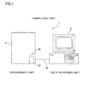
- the blood analyzer 1 is an apparatus employed in blood testings and mainly constituted by a measurement unit 2 and a data processing unit 3, as shown in Fig. 1 .
- the blood analyzer 1 is set in medical facilities such as hospitals or pathology laboratories, for example.
- the measurement unit 2 performs predetermined measurements of components contained in blood samples, and this measurement data are subjected to an analysis process when received by the data processing unit 3.
- the measurement unit 2 and the data processing unit 3 are so connected to each other through a data transmission cable 3a as to be capable of mutual data communication.
- the measurement unit 2 and the data processing unit 3 may be configured to be directly connected to each other through the data transmission cable 3a or may be connected to each other through a communication network such as a dedicated line employing a telephone line, a LAN or the Internet, for example.
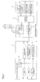
- the measurement unit 2 includes a sample supply portion 4, a WBC classification measurement portion 5, a DC measurement portion 6, an HGB measurement portion 7, a control portion 8 and a communication portion 9, as shown in Fig. 2 .
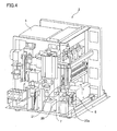
- a blood collection tube set portion 2a so configured that a blood collection tube 20 storing a blood sample can be set thereon is provided at the lower right of the front of the measurement unit 2, as shown in Fig. 3 .
- This blood collection tube set portion 2a is configured to be pushed out in a forward direction by a pressing operation of a button switch 2b provided nearby by a user. The user can set the blood collection tube 20 in a state where the blood collection tube set portion 2a is pushed out. After the blood collection tube 20 has been set, the user again presses the button switch 2b, whereby the blood collection tube set portion 2a is configured to be returned to the inside of the measurement unit 2.
- a pipette 21 for suctioning measurement samples, chambers 22 and 23 (see Fig. 5 ) for preparation by mixing the blood sample and a reagent, and the like are provided within the measurement unit 2, as shown in Figs. 4 and 5 .
- the pipette 21 is formed in a shape of a tube extending vertically, and a tip thereof is sharply tapered.
- the pipette 21 is coupled to a syringe pump not shown in the drawings, and a predetermined quantity of liquid can be suctioned or discharged by an operation of this syringe pump.
- the pipette 21 is also connected to a moving mechanism and configured to be movable in vertical directions and forward and backward directions.
- the pipette 21 is configured to suction the blood sample stored in the blood collection tube 20 by puncturing the sharp tip into a rubber cap 20a sealing the blood collection tube 20.
- the pipette 21 is configured to be moved to a predetermined position by the moving mechanism and supply the blood sample to the insides of the chambers 22 and 23 after suctioning the blood sample.
- the sample supply portion 4 is a flow unit having the chambers 22 and 23, a plurality of electromagnetic valves, diaphragm pumps and the like.
- the chamber 22 is provided for preparing a measurement sample employed in the measurement of red blood cells and platelets and the measurement of a hemoglobin concentration.
- the chamber 23 is provided for preparing a measurement sample employed in the measurement of white blood cells.
- Reagent containers are connected to the flow unit constituted by the sample supply portion 4. More specifically, a diluting solution container 24 for storing a diluting solution, a hemolytic agent container 25 for storing a hemolytic agent 100 and a staining fluid container 26 for storing a staining fluid employed in a measurement sample for detecting malaria are connected to the flow unit.
- the diluting solution and the hemolytic agent 100 can be supplied to the chamber 22, and the diluting solution, the hemolytic agent 100 and the staining fluid can be supplied to the chamber 23.
- the WBC classification measurement portion 5 is an optical flow cytometer and provided for classifying and detecting white blood cells and detecting malaria-infected red blood cells (hereinafter referred to as detecting malaria) by a flow cytometry technique employing a semiconductor laser beam.
- the WBC classification measurement portion 5 has a flow cell 51 (see Fig. 6 ) forming a fluid flow of the measurement sample.
- the flow cell 51 is made of a material such as quartz having a light transmission property, glass, or synthetic resin, in a shape of a tube, and is a flow path through the interior of which a sheath fluid (diluting solution) flows.
- This flow cell 51 is provided with an orifice 51a, the internal cavity of which has an aperture that is narrower than the other parts.
- the vicinity of an inlet of the orifice 51a has a double-tube structure, and an internal side of this tube part becomes a sample nozzle 51b.
- a cavity on an outer side of the sample nozzle 51b is a flow path 51c through which the sheath fluid (diluting solution) flows, and the sheath fluid (diluting solution) flows through the flow path 51c and is introduced into the orifice 51a.
- the sheath fluid (diluting solution) supplied to the flow cell 51 in this manner flows so as to surround the measurement sample discharged from the sample nozzle 51b.
- the measurement sample flow is constricted by the orifice 51a such that particles such as white blood cells and red blood cells contained in the measurement sample are surrounded by the sheath fluid (diluting solution) and pass through the orifice 51a one by one.
- a semiconductor laser light source 52 is arranged in the WBC classification measurement portion 5 so as to emit laser beam toward the orifice 51a of the flow cell 51.
- This semiconductor laser light source 52 has a blue-violet semiconductor laser element 52a and is configured to be capable of emitting a blue-violet laser beam having a wavelength of about 405 nm.
- An illumination lens system 53 constituted by a plurality of lenses is arranged between the semiconductor laser light source 52 and the flow cell 51. Parallel beams emitted from the semiconductor laser light source 52 are collected at a beam spot by the illumination lens system 53.
- a beam stopper 54a is provided on an optical axis extending linearly from the semiconductor laser light source 52 so as to be opposed to the illumination lens system 53 and with the flow cell 51 interposed therebetween, and the beam stopper 54a is configured to block direct light from the semiconductor laser light source 52.
- a photodiode 54 is arranged on an optical axis on a further downstream side of the beam stopper 54a.
- the photodiode 54 is configured to receive scattered light of a laser beam generated by the measurement sample flowing through the flow cell 51. More specifically, among light advancing along the optical axis extending linearly from the semiconductor laser light source 52, the direct light of the semiconductor laser light source 52 is blocked by the beam stopper 54a, and hence the photodiode 54 is configured to basically receive only scattered light (hereinafter referred to as forward scattered light) advancing along the optical axis direction.
- the forward scattered light emitted from the flow cell 51 is subjected to photoelectric conversion by the photodiode 54, and electrical signals (hereinafter referred to as forward scattered light signals) generated by this conversion are transmitted to an amplifier 54b.
- the amplifier 54b is configured to amplify the transmitted forward scattered light signals and output the amplified forward scattered light signals to a control portion 8.
- a side collective lens 55 is arranged at a side of the flow cell 51, in a direction perpendicular to the optical axis extending linearly from the semiconductor laser light source 52 to the photodiode 54, and this side collective lens 55 is configured to collect lateral light (light emitted in a direction intersecting with the aforementioned optical axis) generated when emitting a laser beam to blood cells passing through the flow cell 51.
- a dichroic mirror 56 is provided on a downstream side of the side collective lens 55, and the dichroic mirror 56 is configured to divide signal light transmitted from the side collective lens 55 into a scattered light component and a fluorescent light component.
- a side scattered light photoreceptor photodiode 57 is provided at a side (a direction intersecting with a direction of an optical axis connecting the side collective lens 55 and the dichroic mirror 56) of the dichroic mirror 56, and an optical filter 58a and avalanche photodiode 58 are provided on an optical axis on a downstream side of the dichroic mirror 56.
- the side scattered light component separated by the dichroic mirror 56 is subjected to photoelectric conversion by the photodiode 57, and electrical signals (hereinafter referred to as side scattered light signals) generated by this conversion are transmitted to an amplifier 57a.
- the amplifier 57a is configured to amplify the transmitted side scattered light signals and output the amplified side scattered light signals to the control portion 8.
- the side fluorescent light component is subjected to wavelength selection by the optical filter 58a, and subsequent photoelectric conversion by the avalanche photodiode 58, and electrical signals (side fluorescent light signals) generated by this are transmitted to an amplifier 58b.
- the amplifier 58b is configured to amplify the transmitted side fluorescent light signals and output the amplified side fluorescent light signals to the control portion 8.
- the DC measurement portion 6 is configured to be capable of measuring a red blood cell count (RBC) and a platelet count (PLT) by a sheath flow DC detection method.
- the DC measurement portion 6 is configured to be capable of acquiring measurement data for calculating a hematocrit value (HCT) by a red-blood-cell pulse height detection method. Further, the DC measurement portion 6 is employed in detection of a white blood cell count (WBC) for calculating a lymphocyte ratio.
- WBC white blood cell count
- the DC measurement portion 6 has a flow cell, and the measurement sample is transferred from the chamber 22 to the flow cell.
- a measurement sample prepared by mixing the blood sample and the diluting solution in the chamber 22, along with the sheath fluid (diluting solution), is transferred from the sample supply portion 4 to the flow cell, as shown in Fig 8 .
- a fluid flow in a state where the measurement sample is surrounded by the sheath fluid (diluting solution) is formed in the flow cell.
- the HGB measurement portion 7 is configured to measure the amount of hemoglobin (HGB) by a methemoglobin method.
- the HGB measurement portion 7 has a cell storing a dilute sample, as shown in Fig. 9 , and the measurement sample is transferred from the chamber 22 to this cell.
- the HGB measurement portion 7 has a light-emitting diode emitting light having a wavelength of about 555 nm and is configured to measure absorbance of the measurement sample by emitting the light from the light-emitting diode to the measurement sample in the aforementioned cell.
- a measurement sample is prepared by mixing the blood sample, the diluting solution and the hemolytic agent 100 in the chamber 22.
- the control portion 8 is constituted by a CPU, a ROM, a RAM, etc. and configured to control an operation of each part of the measurement unit 2.
- the communication portion 9 is an RS-232C interface, a USB interface or an Ethernet (registered trademark) interface, for example and is configured to be capable of sending/receiving data to/from the data processing unit 3.
- the data processing unit 3 is constituted by a computer comprising a CPU 31, a ROM 32, a RAM 33, a hard disk 34, a communication interface 35, an input portion 36 such as a keyboard and a mouse, and a display device 37, as shown in Fig. 2 .
- An operating system, and an application program for analyzing the measurement data received from the measurement unit 2 are installed on the hard disk 34 of the data processing unit 3.
- the CPU 31 of the data processing unit 3 is configured to analyze the measurement data and calculate a white blood cell count (WBC), a red blood cell count (RBC), a hemoglobin amount (HGB), a hematocrit value (HCT), a mean red blood cell volume (MCV), a mean red blood cell hemoglobin amount (MCH), a mean red blood cell hemoglobin concentration (MCHC), a platelet count (PLT) by executing this application program.
- WBC white blood cell count
- RBC red blood cell count
- HGB hemoglobin amount
- HCT hematocrit value
- MCV mean red blood cell volume
- MH mean red blood cell hemoglobin amount
- MCHC mean red blood cell hemoglobin concentration
- PHT platelet count
- the CPU 31 is configured to prepare a scattergram employing the forward scattered light signals, the side scattered light signals and the side fluorescent light signals and classify the white blood cells into five groups of neutrophils (Neut), lymphocytes, monocytes (Mono), eosinophils (EO), and basophils (BASO).
- Neut neutrophils
- Mono monocytes
- EO eosinophils
- BASO basophils
- the communication interface 35 is an RS-232C interface, a USB interface or an Ethernet (registered trademark) interface, for example and is configured to be capable of sending/receiving data to/from the measurement unit 2.
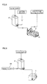
- the hemolytic agent 100 used in this embodiment includes two types of cationic surfactants (lauryl trimethyl ammonium chloride; 34.1 mM, stearyl trimethyl ammonium chloride; 1.7 mM) but is free from a labeling substance, as shown in Fig. 10 .
- This hemolytic agent 100 has a property in that hemoglobin in the blood is inverted to methemoglobin. Further, the hemolytic agent 100 contains a phosphate buffer solution to keep the pH about 5 to about 7.
- red blood cell membranes can be partially lysed so that a fluorescent dye described later can pass through the cell membranes in a state of holding a malarial parasite inside the red blood cells.
- each measurement sample employed in each measurement has a different dilution magnification of the hemolytic agent 100 and a different dilution magnification of the blood sample.
- the two types of cationic surfactants are employed as described above, whereby white blood cells in the measurement sample can be classified into four groups and malaria-infected red blood cells can be detected by simply varying the dilution magnification without employing more than one type of a hemolytic agent.
- the staining fluid contains a fluorescent dye (Hoechst 34580 of Invitrogen, for example) having a structure of a chemical formula shown in Fig. 11 and one of a nonionic surfactant group substantially lysing a red blood cell membrane.
- this fluorescent dye is a DNA-selective fluorescent dye, preferably a DNA-selective bisbenzimide type fluorescent dye.
- the DNA-selective fluorescent dye is a fluorescent dye staining preferentially DNA over RNA, and the DNA-selective bisbenzimide type fluorescent dye is a dye having a bisimide-based skeleton.
- red blood cells without a cell nucleus are not stained but DNA in malarial parasites is stained by employing the DNA-selective fluorescent dye, and hence malaria-infected red blood cells having a malarial parasite inside thereof and a group of the others can be easily classified from a scattergram (see Fig. 17 ) obtained by the aforementioned flow cytometer.
- This fluorescent dye can be excited by the blue-violet laser beam (the wavelength is about 405 nm) emitted from the semiconductor laser light source 52.
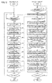
- the application program or the like is initialized, and thereafter the CPU 31 of the data processing unit 3 determines whether or not a measurement starting instruction from a user has been received at a step S1, and this determination is repeated until the instruction has been received.
- a measurement starting instruction signal is transmitted from the data processing unit 3 to the measurement unit 2 at a step S2.
- the control portion 8 of the measurement unit 2 determines whether or not the measurement starting instruction signal has been received at a step S21, and this determination is repeated until the signal has been received.
- the measurement unit 2 has received the measurement starting instruction signal
- the blood sample is suctioned from the blood collection tube 20 set on the blood collection tube set portion 2a by the pipette 21 at a step S22.
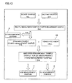
- an RBC/PLT measurement sample (hereinafter referred to as the fourth measurement sample) is prepared by the sample supply portion 4. More specifically, a predetermined quantity (2.0 mL, for example) of the diluting solution from the diluting solution container 24 and a predetermined quantity (6 ⁇ L, for example) of the blood sample suctioned from the blood collection tube 20 by the pipette 21 are supplied to the chamber 22 and stirred, as shown in Fig. 13 . Thus, a predetermined quantity (2.0 mL, for example) of the fourth measurement sample is prepared.
- a WBC/HGB measurement sample (partly for DC detection) (hereinafter referred to as the third measurement sample) is prepared by the sample supply portion 4. More specifically, a predetermined quantity (0.5 mL, for example) of the hemolytic agent 100 is supplied from the hemolytic agent container 25 to the chamber 22 in which a predetermined quantity (1 mL, for example) of the fourth measurement sample remains and stirred, as shown in Fig. 13 . In other words, after the blood sample and the diluting solution are mixed in the chamber 22, the hemolytic agent 100 is mixed to prepare the third measurement sample.
- the red blood cells are hemolyzed, and the hemoglobin is inverted to methemoglobin.
- the third measurement sample in the chamber 22 is transferred to the DC measurement portion 6, and the WBC in the third measurement sample is measured.
- the third measurement sample is transferred to the HGB measurement portion 7, and the HGB of the third measurement sample is detected.
- a predetermined quantity (1 mL, for example) of a diluted hemolytic agent obtained by diluting the same hemolytic agent 100 as that contained in the aforementioned first measurement sample by 9 times (hemolytic agent/diluting solution 1/8)
- the blood sample and the staining fluid are mixed with each other in a state where the hemolytic agent 100 and the diluting solution are mixed with each other in the chamber 23.
- the hemolytic agent 100 is mixed with the blood sample in a state of being diluted with the diluting solution, and hence the blood sample can be inhibited from being mixed with the hemolytic agent having a concentration higher than a desired concentration.
- the second measurement sample in which the blood sample is diluted by 100 times is prepared.
- both the first measurement sample and the second measurement sample can be prepared employing the common hemolytic agent 100 stored in the hemolytic agent container 25.
- the second measurement sample in the chamber 23, along with the sheath fluid (diluting solution) is transferred to the WBC classification measurement portion 5 and the WBC classification measurement portion 5 detects malaria in the second measurement sample at a step S31.
- measurement data obtained by measurement in each detection portion is transmitted from the measurement unit 2 to the data processing unit 3.
- the data processing unit 3 determines whether or not the measurement data transmitted from the measurement unit 2 has been received at a step S3, and this determination is repeated until the measurement data has been received.
- the CPU 31 calculates the white blood cell count (WBC) on the basis of the measurement data obtained by the WBC detection, measured at the step S26.
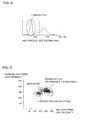
- the CPU 31 prepares a particle size distribution chart of the white blood cells on the basis of the measurement data obtained by the WBC detection, as shown in Fig. 14 and calculates the ratio of the lymphocytes to the white blood cell count (WBC).
- the lymphocyte appears as a first peak (group) from the left in the particle size distribution chart.
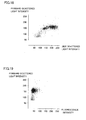
- the CPU 31 classifies the white blood cells into three groups of a group of lymphocytes and basophils, monocytes and granulocytes (a group of neutrophils and eosinophils) on the basis of the measurement data obtained by the WBC classification and detection, measured at the step S29. More specifically, the CPU 31 prepares a scattergram, employing the forward scattered light signals and the side scattered light signals, as shown in Fig. 15 and calculates ratios of a group of lymphocytes and basophils, monocytes and granulocytes (a group of neutrophils and eosinophils) to the white blood cell count (WBC) from this scattergram.
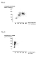
- FIG. 22 shows a scattergram by a forward scattered light signal and a side scattered light signal obtained by measuring a blood sample actually collected from a subject employing the hemolytic agent (see Fig. 10 ) in this embodiment.
- the white blood cells can be classified into three groups of a group of lymphocytes and basophils, monocytes and granulocytes (a group of neutrophils and eosinophils) on the scattergram.
- the CPU 31 classifies the white blood cells into two groups of eosinophils and the others on the basis of the measurement data obtained by the WBC classification and detection. More specifically, the CPU 31 prepares a scattergram, employing the forward scattered light signals and the side fluorescent light signals, as shown in Fig. 16 and calculates a ratio of the eosinophils to the white blood cell count (WBC) from this scattergram.
- These side fluorescent light signals are based on intrinsic fluorescence of the white blood cells excited by the blue-violet semiconductor laser beam (the wavelength is about 405 nm) emitted from the semiconductor laser light source 52, and the eosinophils have a stronger fluorescence intensity than a group of the others in the white blood cells.
- the CPU 31 can also calculate the ratio of the eosinophils to the white blood cell count (WBC) from a scattergram obtained by employing the side scattered light signals and the side fluorescent light signals.
- Fig. 23 shows a scattergram by the forward scattered light signal and a side fluorescent light signal obtained by measuring the blood sample actually collected from the subject employing the hemolytic agent (see Fig. 10 ) in this embodiment. As shown in Fig. 23 , it is also understood from an actual measurement result that the white blood cells can be classified into the eosinophils and the group of the others on the scattergram.
- the CPU 31 calculates a ratio of the neutrophils to the white blood cell count (WBC) by subtracting the ratio of the eosinophils calculated at the step S7 from the ratio of the granulocytes (a group of neutrophils and eosinophils) calculated at the step S6.
- the white blood cells are classified into four groups of the group of lymphocytes and basophils, the monocytes, the neutrophils and the eosinophils.
- the CPU 31 calculates a ratio of the basophils to the white blood cell count (WBC) by subtracting the ratio of the lymphocytes calculated at the step S5 from the ratio of the group of lymphocytes and basophils.
- the white blood cells are classified into five groups of the lymphocytes, the basophils, the monocytes, the neutrophils and the eosinophils.
- the CPU 31 separates the malaria-infected red blood cells from a group of the others on the basis of the measurement data obtained by the malaria detection, measured at the step S31. Specifically, the CPU 31 prepares a scattergram, employing the forward scattered light signals and the side fluorescent light signals, as shown in Fig. 17 and separates the malaria-infected red blood cells from the group of the others from this scattergram. More specifically, malaria-negative red blood cells appear in an area where a fluorescence intensity is small whereas the malaria-infected red blood cells appear in an area where the fluorescence intensity is relatively large on the scattergram in Fig. 17 . Further, the white blood cells appear in an area where both the fluorescence intensity and a scattered light intensity are large due to the size and the amount of DNA thereof. Thus, the presence of malaria infection can be determined.
- the CPU 31 calculates the red blood cell count (RBC), the platelet count (PLT) and the hematocrit value (HCT) on the basis of the measurement data obtained by the RBC/PLT detection, measured at the step S24.
- RBC red blood cell count
- PHT platelet count
- HCT hematocrit value
- the CPU 31 calculates the amount of the hemoglobin (HGB) on the basis of the measurement data obtained by the HGB detection, measured at the step S27.
- a hemoglobin concentration is calculated on the basis of the absorbance obtained by HGB detection employing an SLS hemoglobin method.
- the hemoglobin concentration can be acquired employing the third measurement sample identical to the measurement sample employed to classify the white blood cells into five groups (neutrophils, lymphocytes, monocytes, eosinophils and basophils).
- the CPU 31 calculates the mean red blood cell volume (MCV), the mean red blood cell hemoglobin amount (MCH) and the mean red blood cell hemoglobin concentration (MCHC) from the red blood cell count (RBC), the hematocrit value (HCT) and the amount of the hemoglobin (HGB).
- MCV mean red blood cell volume
- MCH mean red blood cell hemoglobin amount
- MCHC mean red blood cell hemoglobin concentration
- MCV represents a mean red blood cell volume (fL)
- HCT represents a hematocrit value (%)
- RBC represents a red blood cell count ( ⁇ 10 4 / ⁇ L).
- MCH represents a mean red blood cell hemoglobin amount (pg)
- HGB represents the amount of hemoglobin (g/dL)
- RBC represents a red blood cell count ( ⁇ 10 4 / ⁇ L).
- MCHC HGB / HCT ⁇ 100
- MCHC represents a mean red blood cell hemoglobin concentration (g/dL)
- HGB represents the amount of hemoglobin (g/dL)
- HCT represents a hematocrit value (%).
- step S14 calculation results of the white blood cell count (WBC), the red blood cell count (RBC), the amount of the hemoglobin (HGB), the hematocrit value (HCT), the mean red blood cell volume (MCV), the mean red blood cell hemoglobin amount (MCH), the mean red blood cell hemoglobin concentration (MCHC) and the platelet count (PLT) calculated as described above are output to the display device 37. Further, the ratios of the neutrophils, the lymphocytes, the monocytes, the eosinophils and the basophils to the white blood cell count (WBC) are output to the display device 37, and the result of detection of malaria is also output.
- the white blood cell count (WBC) In addition to the ratio of each blood cell to the white blood cell count (WBC), the white blood cell count (WBC) and the neutrophil count, the lymphocyte count, the monocyte count, the eosinophil count and the basophil count calculated on the basis of the ratio of each blood cell are output.
- a step S15 the presence of a shutdown instruction from the user is determined, and when the shutdown instruction has not been received, the CPU 31 moves to the step S1.
- the shutdown instruction has been received, an operation of the data processing unit 3 of the sample analysis processing in the blood analyzer 1 is terminated.
- the control portion 8 moves to the step S21.
- the shutdown instruction has been received, an operation of the measurement unit 2 of the sample analysis processing in the blood analyzer 1 is terminated.
- the CPU 31 classifying the white blood cells in the first measurement sample into at least four groups of the monocytes, the neutrophils, the eosinophils and a group of the others on the basis of the side fluorescent light signals, the forward scattered light signals and the side scattered light signals generated from the first measurement sample containing the blood sample and the hemolytic agent 100 by the WBC classification measurement portion 5 and classifying the blood cells in the second measurement sample into the malaria-infected red blood cells and the group of the others on the basis of the side fluorescent light signals and the forward scattered light signals generated from the second measurement sample containing the blood sample, the same hemolytic agent 100 as the aforementioned hemolytic agent 100 and the staining agent by the WBC classification measurement portion 5 is provided, whereby the hemolytic agent for classifying the white blood cells into four groups and the hemolytic agent for detecting the malaria-infected red blood cells can be rendered common, and hence it is not necessary to develop two types of reagents (hemolytic agents) having different compositions to classify
- the CPU 31 is configured to classify the white blood cells in the third measurement sample into the lymphocytes and the group of the others on the basis of the measurement data obtained by the DC measurement portion 6 and classify the white blood cells in the measurement sample into at least five groups of the lymphocytes, the basophils, the monocytes, the neutrophils and the eosinophils on the basis of this classification result and the aforementioned classification result of the four groups of the white blood cells, whereby the white blood cells can be classified into the lymphocytes and the group of the others employing the same hemolytic agent 100 as the hemolytic agent for classifying the white blood cells and detecting the malaria-infected red blood cells, and hence the white blood cells can be classified into five groups without developing a hemolytic agent having a different composition separately.
- the hemolytic agent used in this embodiment is employed in the blood analysis method comprising the step of classifying the white blood cells in the first measurement sample into at least four groups of the monocytes, the neutrophils, the eosinophils, the group of the others on the basis of the side fluorescent light signals, the forward scattered light signals and the side scattered light signals generated from the first measurement sample containing the blood sample and the hemolytic agent 100 by the WBC classification measurement portion 5 and the step of classifying the blood cells in the second measurement sample into the malaria-infected red blood cells and the group of the others on the basis of the side fluorescent light signals and the forward scattered light signals generated from the second measurement sample containing the blood sample, the same hemolytic agent 100 as the aforementioned hemolytic agent 100 and the staining agent by the WBC classification measurement portion 5, as hereinabove described, whereby the hemolytic agent for classifying the white blood cells into four groups and the hemolytic agent for detecting the malaria-infected red blood cells can be rendered common, and hence it is not necessary to develop two types of reagents (
- the staining agent used in this embodiment is employed in the blood analysis method comprising the step of classifying the white blood cells in the first measurement sample into at least four groups of the monocytes, the neutrophils, the eosinophils, the group of the others on the basis of the side fluorescent light signals, the forward scattered light signals and the side scattered light signals generated from the first measurement sample containing the blood sample and the hemolytic agent 100 by the WBC classification measurement portion 5 and the step of classifying the blood cells in the second measurement sample into the malaria-infected red blood cells and the group of the others on the basis of the side fluorescent light signals and the forward scattered light signals generated from the second measurement sample containing the blood sample, the same hemolytic agent 100 as the aforementioned hemolytic agent 100 and the staining agent by the WBC classification measurement portion 5, as hereinabove described, whereby the hemolytic agent for classifying the white blood cells into four groups and the hemolytic agent for detecting the malaria-infected red blood cells can be rendered common, and hence it is not necessary to develop two types of reagents
- the malaria infection rates obtained by the visual observation and the malaria infection rates obtained on the basis of the method described in the aforementioned embodiment are substantially identical to each other, and according to the method described in the aforementioned embodiment, it could be confirmed that malaria-infected red blood cells could be accurately detected.
- Malaria infection rate % X / Y ⁇ 100
- Y represents the aforementioned predetermined number.
- Y is about 10,000 with respect to Samples 1 and 2, about 20,000 with respect to Sample 3 and about 30,000 with respect to Sample 4.
- Malaria infection rate % 6 / 7 ⁇ 100
- (6) represents A ⁇ B/C
- (7) represents the number of red blood cells obtained by measuring the same blood sample by a multiparameter automated hematology analyzer Model XE-2100 (SYSMEX CORPORATION).
- a of the aforementioned (6) is the number of blood cells in a malaria area in Fig. 17
- B of the aforementioned (6) is the number of white blood cells obtained by measuring the same blood sample by the multiparameter automated hematology analyzer Model XE-2100
- C of the aforementioned (6) is the number of blood cells in a white blood cell area in Fig. 17 .
- the present invention is not restricted to this, but four hemolytic agent containers may be connected to the sample supply portion so as to store the hemolytic agents employed in the respective detections separately, or a hemolytic agent employed in any of the aforementioned four detections may be stored in a common hemolytic agent container and two or three hemolytic agent containers may be connected to the sample supply portion. Alternatively, more than four hemolytic agent containers may be connected to the sample supply portion.
- the hemolytic agents stored in the respective hemolytic agent containers are diluted by respective predetermined dilution magnifications, no steps to dilute the hemolytic agents by desired dilution magnifications may be provided separately when preparing the measurement samples employed in the respective detections.
- hemolytic agent free from a labeling substance has been shown as an example of a hemolytic agent in the aforementioned embodiment, the present disclosure is not restricted to this, but the hemolytic agent may contain a labeling substance.
- the present invention is not restricted to this, but different dedicated hemolytic agents may be employed in the respective WBC detection, HGB detection and WBC classification detection.
- each detection processing in the order of the RBC/PLT detection, the WBC detection, the HGB detection, the WBC classification detection and the malaria detection from the earliest in the sample analysis processing has been shown in the aforementioned embodiment, the present invention is not restricted to this, but each detection processing may be performed in another order other than the aforementioned order in the sample analysis processing. Also, the order of white blood cell classification processing, malaria classification processing, red blood cell count/platelet count calculation processing and hemoglobin amount calculation processing in the sample analysis processing can be properly varied.
- the present invention is not restricted to this, but a light source having another laser element other than the blue-violet semiconductor laser element, such as a blue semiconductor laser element or an argon laser element may be provided.
- the hemolytic agent in the second measurement sample is preferably diluted by at least 9 times and not more than 12 times.
- hemolytic agent including the cationic surfactant (lauryl trimethyl ammonium chloride; 34.1 mM, stearyl trimethyl ammonium chloride; 1.7 mM), which is an alkyl trimethyl ammonium salt and the number of carbons of the alkyl group of which is at least twelve and not more than eighteen, has been shown as an example of a hemolytic agent in the aforementioned embodiment, the present disclosure is not restricted to this, but a hemolytic agent including a cationic surfactant having a concentration other than the aforementioned concentration may be employed so far as a concentration of the cationic surfactant (the total of lauryl trimethyl ammonium chloride and stearyl trimethyl ammonium chloride in the aforementioned embodiment) in the WBC measurement sample (for classification) is at least 0.62 mM and not more than 2.15 mM.
- a concentration of the cationic surfactant the total of lauryl trimethyl ammonium chloride and stearyl trimethyl ammonium chlor
- the measurement sample is prepared by diluting the hemolytic agent by 25 times, and hence a concentration of the cationic surfactant in the hemolytic agent is 15.5 nM when the concentration of the cationic surfactant in the WBC measurement sample (for classification) is 0.62 mM, and the concentration of the cationic surfactant in the hemolytic agent is 53.75 nM when the concentration of the cationic surfactant in the WBC measurement sample (for classification) is 2.15 mM.
- the measurement sample can be measured even when a concentration of the cationic surfactant in the WBC measurement sample (for classification) is 2.15 mM or more.
- the white blood cells can be classified into three groups of the group of lymphocytes and basophils, the monocytes and the granulocytes (the group of neutrophils and eosinophils) on the scattergram. As shown in Figs. 19 and 21 , the white blood cells can be classified into the eosinophils and the group of the others on the scattergram. Further, the white blood cells can be classified into four groups of the group of lymphocytes and basophils, the monocytes, the neutrophils and the eosinophils from these classification results. Therefore, the white blood cells can be conceivably classified into four groups in a case where the concentration of the cationic surfactant in the WBC measurement sample (for classification) is in the range of at least 0.62 mM and not more than 2.15 mM.
Landscapes
- Health & Medical Sciences (AREA)
- Life Sciences & Earth Sciences (AREA)
- Engineering & Computer Science (AREA)
- Immunology (AREA)
- Chemical & Material Sciences (AREA)
- Molecular Biology (AREA)
- Biomedical Technology (AREA)
- Urology & Nephrology (AREA)
- Hematology (AREA)
- Cell Biology (AREA)
- Physics & Mathematics (AREA)
- Biotechnology (AREA)
- Microbiology (AREA)
- Analytical Chemistry (AREA)
- Biochemistry (AREA)
- General Health & Medical Sciences (AREA)
- Tropical Medicine & Parasitology (AREA)
- Food Science & Technology (AREA)
- Medicinal Chemistry (AREA)
- General Physics & Mathematics (AREA)
- Pathology (AREA)
- Organic Chemistry (AREA)
- Ecology (AREA)
- Virology (AREA)
- Proteomics, Peptides & Aminoacids (AREA)
- Wood Science & Technology (AREA)
- Zoology (AREA)
- Biophysics (AREA)
- Bioinformatics & Cheminformatics (AREA)
- General Engineering & Computer Science (AREA)
- Genetics & Genomics (AREA)
- Investigating Or Analysing Biological Materials (AREA)
Claims (12)
- Analyseur de sang comprenant :une partie de préparation d'échantillons conçue pour préparer un premier échantillon de mesure contenant un échantillon de sang, un agent hémolytique et une solution de dilution, et un deuxième échantillon de mesure contenant ledit échantillon de sang, le même agent hémolytique que ledit agent hémolytique, un agent colorant, et la même solution de dilution que ladite solution de dilution ;une partie de génération d'informations de lumière conçue pour générer une première information de fluorescence et au moins deux types de première information de lumière diffusée à partir dudit premier échantillon de mesure et pour générer une deuxième information de fluorescence et une deuxième information de lumière diffusée à partir dudit deuxième échantillon de mesure ; etune partie de commande conçue pour effectuer une première classification des globules blancs dans ledit premier échantillon de mesure en au moins quatre groupes de monocytes, neutrophiles, éosinophiles et un groupe des autres, sur la base de ladite première information de fluorescence et desdits deux types de première information de lumière diffusée générées par ladite partie de génération d'informations de lumière et pour classer les cellules sanguines dans ledit deuxième échantillon de mesure en globules rouges infectés par le paludisme et un groupe des autres, sur la base de ladite deuxième information de fluorescence et ladite deuxième information de lumière diffusée générées par ladite partie de génération d'informations de lumière ;dans lequel ladite partie de préparation d'échantillons est conçue pour préparer ledit premier échantillon de mesure en mélangeant ledit échantillon de sang, ledit agent hémolytique stocké dans un récipient de réactif prédéterminé et une quantité prédéterminée de solution de dilution et préparer ledit deuxième échantillon de mesure en mélangeant ledit échantillon de sang, ledit agent hémolytique stocké dans ledit récipient de réactif prédéterminé et une quantité de ladite solution de dilution plus petite que ladite quantité prédéterminée de solution de dilution pour le premier échantillon de mesure, de sorte que ladite amplification de dilution dudit agent hémolytique dans ledit deuxième échantillon de mesure est plus petite que ladite amplification de dilution dudit agent hémolytique dans ledit premier échantillon de mesure, etdans lequel l'agent colorant comprend un colorant fluorescent sélectif vis-à-vis de l'ADN.
- Analyseur de sang selon la revendication 1, dans lequel la partie de commande est conçue pour dénombrer les globules rouges infectés par le paludisme et calculer le taux d'infection par la malaria, sur la base du résultat du dénombrement.
- Analyseur de sang selon la revendication 1 ou 2, dans lequel
ladite partie de préparation d'échantillons est conçue pour préparer en outre un troisième échantillon de mesure contenant ledit échantillon de sang et ledit agent hémolytique,
ledit analyseur de sang comprenant en outre une partie de génération d'informations électriques conçue pour générer une information électrique d'un échantillon à partir dudit troisième échantillon de mesure, dans lequel
ladite partie de commande est conçue pour effectuer une deuxième classification des globules blancs dans ledit troisième échantillon de mesure en au moins les lymphocytes et un groupe des autres sur la base de ladite information électrique générée par ladite partie de génération d'informations électriques et pour classer les globules blancs dans lesdits échantillon de mesure en au moins cinq groupes de lymphocytes, basophiles, monocytes, neutrophiles et éosinophiles, sur la base des résultats de classification de ladite première classification et ladite deuxième classification. - Analyseur de sang selon l'une quelconque des revendications 1 à 3, comprenant en outre une deuxième partie de génération d'informations de lumière conçue pour générer au moins soit une information de lumière transmise soit une information de lumière diffusée d'un échantillon à partir dudit troisième échantillon de mesure, dans lequel
ladite partie de commande est conçue pour acquérir une concentration en hémoglobine dans ledit troisième échantillon de mesure sur la base d'au moins soit ladite information de lumière transmise soit ladite information de lumière diffusée générée par ladite deuxième partie de génération d'informations de lumière. - Analyseur de sang selon l'une quelconque des revendications 1 à 4, dans lequel ladite partie de préparation d'échantillons est conçue pour préparer en outre un quatrième échantillon de mesure en mélangeant ledit échantillon de sang et la même solution de dilution que ladite solution de dilution, et
ladite partie de commande est conçue pour dénombrer les globules rouges dans ledit quatrième échantillon de mesure. - Analyseur de sang selon la revendication 5, dans lequel ladite partie de commande est conçue pour dénombrer les plaquettes dans ledit quatrième échantillon de mesure.
- Analyseur de sang selon la revendication 1, dans lequel
ladite partie de préparation d'échantillons est conçue pour préparer ledit deuxième échantillon de mesure en mélangeant ledit échantillon de sang dans un état de mélange dudit agent hémolytique et de ladite solution de dilution. - Analyseur de sang selon la revendication 1, dans lequel
ladite partie de préparation d'échantillons est conçue pour préparer ledit deuxième échantillon de mesure en mélangeant au moins ledit échantillon de sang et ledit agent hémolytique stocké dans un deuxième récipient de réactif différent d'un premier récipient de réactif stockant ledit agent hémolytique utilisé dans ledit premier échantillon de mesure. - Analyseur de sang selon la revendication 8, dans lequel
ledit agent hémolytique stocké dans ledit deuxième récipient de réactif est dilué au moins 9 fois et pas plus de 12 fois. - Analyseur de sang selon la revendication 1, dans lequel
ledit agent hémolytique comprend deux types de tensioactifs cationiques. - Analyseur de sang selon la revendication 1, dans lequel
ledit agent colorant comprend un colorant fluorescent présenté dans la formule suivante et un tensio-actif non ionique.
- Méthode d'analyse du sang comprenant les étapes de :préparation d'un premier échantillon de mesure contenant un échantillon de sang et un agent hémolytique et d'un deuxième échantillon de mesure contenant ledit échantillon de sang, le même agent hémolytique que ledit agent hémolytique et un agent colorant ;génération d'une première information de fluorescence et d'au moins deux types de première information de lumière diffusée à partir dudit premier échantillon de mesure et génération d'une deuxième information de fluorescence et d'une deuxième information de lumière diffusée à partir dudit deuxième échantillon de mesure ;classification des globules blancs dans ledit premier échantillon de mesure en au moins quatre groupes de monocytes, neutrophiles, éosinophiles et un groupe des autres sur la base de ladite première information de fluorescence et desdits deux types de première information de lumière diffusée générées à partir dudit premier échantillon de mesure ; etclassification des cellules sanguines dans ledit deuxième échantillon de mesure en globules rouges infectés par le paludisme et un groupe des autres sur la base de ladite deuxième information de fluorescence et de ladite deuxième information de lumière diffusée générées à partir dudit deuxième échantillon de mesure ;dans lequel une amplification de dilution dudit agent hémolytique dans ledit deuxième échantillon de mesure est plus petite que ladite amplification de dilution dudit agent hémolytique dans ledit premier échantillon de mesure ; etdans lequel l'agent colorant comprend un colorant fluorescent sélectif vis-à-vis de l'ADN.
Priority Applications (1)
| Application Number | Priority Date | Filing Date | Title |
|---|---|---|---|
| EP14198316.3A EP2889620B1 (fr) | 2008-05-09 | 2009-04-28 | Analyseur de sang, procédé d'analyse de sang, agent hémolytique et agent colorant |
Applications Claiming Priority (2)
| Application Number | Priority Date | Filing Date | Title |
|---|---|---|---|
| JP2008123403 | 2008-05-09 | ||
| PCT/JP2009/058327 WO2009136573A1 (fr) | 2008-05-09 | 2009-04-28 | Dispositif d'analyse de sang, procédé d'analyse de sang, agent hémolytique et agent colorant |
Related Child Applications (2)
| Application Number | Title | Priority Date | Filing Date |
|---|---|---|---|
| EP14198316.3A Division EP2889620B1 (fr) | 2008-05-09 | 2009-04-28 | Analyseur de sang, procédé d'analyse de sang, agent hémolytique et agent colorant |
| EP14198316.3A Division-Into EP2889620B1 (fr) | 2008-05-09 | 2009-04-28 | Analyseur de sang, procédé d'analyse de sang, agent hémolytique et agent colorant |
Publications (3)
| Publication Number | Publication Date |
|---|---|
| EP2293062A1 EP2293062A1 (fr) | 2011-03-09 |
| EP2293062A4 EP2293062A4 (fr) | 2011-05-25 |
| EP2293062B1 true EP2293062B1 (fr) | 2015-01-28 |
Family
ID=41264628
Family Applications (2)
| Application Number | Title | Priority Date | Filing Date |
|---|---|---|---|
| EP14198316.3A Not-in-force EP2889620B1 (fr) | 2008-05-09 | 2009-04-28 | Analyseur de sang, procédé d'analyse de sang, agent hémolytique et agent colorant |
| EP09742697.7A Not-in-force EP2293062B1 (fr) | 2008-05-09 | 2009-04-28 | Dispositif d'analyse de sang, procédé d'analyse de sang, agent hémolytique et agent colorant |
Family Applications Before (1)
| Application Number | Title | Priority Date | Filing Date |
|---|---|---|---|
| EP14198316.3A Not-in-force EP2889620B1 (fr) | 2008-05-09 | 2009-04-28 | Analyseur de sang, procédé d'analyse de sang, agent hémolytique et agent colorant |
Country Status (5)
| Country | Link |
|---|---|
| US (2) | US8920726B2 (fr) |
| EP (2) | EP2889620B1 (fr) |
| JP (1) | JP5619604B2 (fr) |
| CN (2) | CN104297463A (fr) |
| WO (1) | WO2009136573A1 (fr) |
Cited By (1)
| Publication number | Priority date | Publication date | Assignee | Title |
|---|---|---|---|---|
| EP3896426A1 (fr) * | 2020-04-13 | 2021-10-20 | Sysmex Corporation | Analyseur de sang |
Families Citing this family (35)
| Publication number | Priority date | Publication date | Assignee | Title |
|---|---|---|---|---|
| GB2494653A (en) * | 2011-09-13 | 2013-03-20 | Orreco Ltd | Apparatus for blood analysis |
| CN103091286B (zh) | 2011-10-31 | 2016-08-17 | 深圳迈瑞生物医疗电子股份有限公司 | 疟原虫感染的红细胞的识别方法及装置 |
| SE537208C2 (sv) | 2012-12-03 | 2015-03-03 | Tommy Forsell | Blodanalysapparat för malariaanalys |
| JP6364426B2 (ja) | 2012-12-31 | 2018-07-25 | ベックマン コールター, インコーポレイテッド | 未成熟血小板計算システム及び方法 |
| JP2015021938A (ja) | 2013-07-23 | 2015-02-02 | シスメックス株式会社 | 検体分析装置、疾患監視システムおよび検体分析装置のデータ管理方法 |
| US10831013B2 (en) | 2013-08-26 | 2020-11-10 | S.D. Sight Diagnostics Ltd. | Digital microscopy systems, methods and computer program products |
| JP6383216B2 (ja) | 2014-08-08 | 2018-08-29 | シスメックス株式会社 | 血液分析方法、血液分析装置およびプログラム |
| WO2016043683A1 (fr) * | 2014-09-18 | 2016-03-24 | Ciftci Ihsan Hakki | Dispositif de numération globulaire du sang |
| JP6352750B2 (ja) * | 2014-09-26 | 2018-07-04 | シスメックス株式会社 | 血液分析装置および血液分析方法 |
| US9835640B2 (en) | 2015-02-13 | 2017-12-05 | Abbott Laboratories | Automated storage modules for diagnostic analyzer liquids and related systems and methods |
| JP6661278B2 (ja) * | 2015-03-27 | 2020-03-11 | シスメックス株式会社 | 血液分析装置および血液分析方法 |
| WO2016158139A1 (fr) * | 2015-03-31 | 2016-10-06 | ソニー株式会社 | Dispositif et procédé de mesure des caractéristiques électriques, système d'analyse de l'état du sang, et programme de mesure des caractéristiques électriques pour informatiser ledit procédé |
| JP6621620B2 (ja) * | 2015-08-31 | 2019-12-18 | シスメックス株式会社 | 血液分析方法ならびにそれに用いる染色液および血液分析装置 |
| JP6635720B2 (ja) | 2015-08-31 | 2020-01-29 | シスメックス株式会社 | 血液分析装置及び血液分析方法 |
| AU2016322966B2 (en) * | 2015-09-17 | 2021-10-14 | S.D. Sight Diagnostics Ltd | Methods and apparatus for detecting an entity in a bodily sample |
| JP6871729B2 (ja) * | 2016-12-06 | 2021-05-12 | シスメックス株式会社 | マラリア原虫に感染した赤血球の検出方法及び血液分析装置 |
| EP3837543A4 (fr) * | 2018-08-16 | 2022-11-16 | Essenlix Corporation | Analyse cellulaire dans des fluides corporels, notamment le sang |
| CN114174826A (zh) * | 2019-09-19 | 2022-03-11 | 深圳迈瑞动物医疗科技有限公司 | 一种动物血液细胞分析方法、分析仪及存储介质 |
| AU2020372024B2 (en) | 2019-10-22 | 2026-03-12 | S.D. Sight Diagnostics Ltd | Accounting for errors in optical measurements |
| CN110579613A (zh) * | 2019-10-28 | 2019-12-17 | 深圳开立生物医疗科技股份有限公司 | 一种血液分析仪 |
| AU2020400400B2 (en) | 2019-12-12 | 2026-03-19 | S.D. Sight Diagnostics Ltd | Detecting platelets in a blood sample |
| BR112022011312A2 (pt) | 2019-12-12 | 2022-08-23 | S D Sight Diagnostics Ltd | Análise de um analito disposto dentro de um meio |
| JP7425597B2 (ja) * | 2019-12-25 | 2024-01-31 | シスメックス株式会社 | 血液分析方法 |
| JP7660123B2 (ja) | 2019-12-27 | 2025-04-10 | ベックマン コールター, インコーポレイテッド | サンプル調製器具 |
| WO2021179177A1 (fr) * | 2020-03-10 | 2021-09-16 | 深圳迈瑞生物医疗电子股份有限公司 | Analyseur de sang, méthode d'analyse sanguine et support d'enregistrement lisible par ordinateur |
| CN111521834B (zh) * | 2020-03-20 | 2023-03-24 | 佛山市顺德区德维医疗科技有限公司 | Xn系列全自动模块式血液体液分析仪用试剂 |
| CN113702267B (zh) * | 2020-05-22 | 2025-08-12 | 深圳迈瑞动物医疗科技股份有限公司 | 血红蛋白浓度的检测方法及血液细胞分析仪 |
| CN113959911B (zh) * | 2020-07-20 | 2024-06-18 | 深圳迈瑞生物医疗电子股份有限公司 | 抗血小板聚集干扰的检测方法、试剂及其应用 |
| CN119354855A (zh) * | 2020-09-23 | 2025-01-24 | 深圳迈瑞生物医疗电子股份有限公司 | 样本检测方法和样本分析仪 |
| EP4222479A1 (fr) | 2020-09-29 | 2023-08-09 | Universidade do Minho | Dispositif automatique pour le diagnostic non invasif du paludisme par des techniques de réflectance optique, procédés et utilisations associés |
| CN116569041B (zh) | 2020-12-01 | 2026-01-02 | 深圳迈瑞生物医疗电子股份有限公司 | 样本分析方法、样本分析仪及计算机可读存储介质 |
| CN115201154A (zh) * | 2021-04-08 | 2022-10-18 | 深圳迈瑞生物医疗电子股份有限公司 | 血液样本的检测方法和样本分析仪 |
| CN115219392A (zh) * | 2021-04-14 | 2022-10-21 | 深圳迈瑞生物医疗电子股份有限公司 | 样本分析仪和样本分析方法 |
| CN118185751B (zh) * | 2024-04-07 | 2024-12-10 | 重庆大学 | 一种白细胞分类检测系统及方法 |
| CN121898960A (zh) * | 2024-10-21 | 2026-04-21 | 希森美康株式会社 | 被检体测定装置 |
Family Cites Families (18)
| Publication number | Priority date | Publication date | Assignee | Title |
|---|---|---|---|---|
| JPH05322882A (ja) * | 1992-05-21 | 1993-12-07 | Hitachi Ltd | 血液分析装置 |
| JP3830613B2 (ja) | 1997-04-18 | 2006-10-04 | シスメックス株式会社 | 血液中の白血球及びヘモグロビン濃度測定試薬 |
| US6210969B1 (en) * | 1999-04-28 | 2001-04-03 | Coulter International Corp. | Composition and method for differentiation of basophil and eosinophil subpopulations of leukocytes in blood |
| EP1406088A3 (fr) * | 2002-10-04 | 2004-04-14 | Sysmex Corporation | Trousse de réactifs et procédé pour la détection des parasites de la Malaria par hémolyse in deux étapes à l'aide de surfactants |
| JP4388779B2 (ja) * | 2002-10-04 | 2009-12-24 | シスメックス株式会社 | マラリア原虫の検出方法、検出装置、およびその試薬キット |
| WO2005088301A1 (fr) * | 2004-02-27 | 2005-09-22 | Beckman Coulter, Inc. | Procede de detection du paludisme et autres infections parasitaires |
| JP2005333868A (ja) * | 2004-05-26 | 2005-12-08 | Sysmex Corp | マラリア原虫測定方法及び測定装置 |
| JP4873969B2 (ja) | 2005-03-17 | 2012-02-08 | シスメックス株式会社 | 試料分析装置及び試料分析方法 |
| CN1834659A (zh) * | 2005-03-17 | 2006-09-20 | 希森美康株式会社 | 试料分析装置和试料分析方法以及血液分析装置 |
| US9243993B2 (en) | 2005-03-17 | 2016-01-26 | Sysmex Corporation | Sample analyzer and sample analyzing method |
| ES2439728T3 (es) * | 2005-03-29 | 2014-01-24 | Sysmex Corporation | Un reactivo para lisar parcialmente una membrana celular de un glóbulo rojo, un reactivo para detectar glóbulos rojos infectados de malaria, y un método para analizar muestras para detectar glóbulos rojos infectados de malaria |
| JP4969115B2 (ja) | 2005-03-29 | 2012-07-04 | シスメックス株式会社 | マラリア感染赤血球の検出方法並びにこれに使用する検出用試薬及び赤血球膜部分溶解試薬 |
| US7580120B2 (en) * | 2005-04-07 | 2009-08-25 | Sysmex Corporation | Blood analyzer, sample analyzer, and flow cytometer |
| JP2007024844A (ja) * | 2005-07-21 | 2007-02-01 | Sysmex Corp | 血液分析方法及び血液分析装置 |
| JP4964446B2 (ja) * | 2005-09-14 | 2012-06-27 | シスメックス株式会社 | 分析装置及び検体情報処理プログラム |
| JP4745030B2 (ja) * | 2005-11-15 | 2011-08-10 | シスメックス株式会社 | 血液分析装置 |
| JP5431732B2 (ja) * | 2005-12-29 | 2014-03-05 | ハネウェル・インターナショナル・インコーポレーテッド | マイクロ流体フォーマットにおけるアッセイ実装 |
| US8597935B2 (en) * | 2006-04-21 | 2013-12-03 | Nihon Kohden Corporation | Classification of blood cells and apparatus therefore |
-
2009
- 2009-04-28 EP EP14198316.3A patent/EP2889620B1/fr not_active Not-in-force
- 2009-04-28 EP EP09742697.7A patent/EP2293062B1/fr not_active Not-in-force
- 2009-04-28 CN CN201410546643.6A patent/CN104297463A/zh active Pending
- 2009-04-28 WO PCT/JP2009/058327 patent/WO2009136573A1/fr not_active Ceased
- 2009-04-28 JP JP2010511054A patent/JP5619604B2/ja not_active Expired - Fee Related
- 2009-04-28 CN CN200980116617.7A patent/CN102016573B/zh not_active Expired - Fee Related
-
2010
- 2010-11-09 US US12/942,752 patent/US8920726B2/en active Active
-
2014
- 2014-11-03 US US14/530,973 patent/US9328375B2/en active Active
Cited By (2)
| Publication number | Priority date | Publication date | Assignee | Title |
|---|---|---|---|---|
| EP3896426A1 (fr) * | 2020-04-13 | 2021-10-20 | Sysmex Corporation | Analyseur de sang |
| US12174118B2 (en) | 2020-04-13 | 2024-12-24 | Sysmex Corporation | Blood analyzer |
Also Published As
| Publication number | Publication date |
|---|---|
| US20110053212A1 (en) | 2011-03-03 |
| JP5619604B2 (ja) | 2014-11-05 |
| EP2293062A1 (fr) | 2011-03-09 |
| EP2889620A1 (fr) | 2015-07-01 |
| WO2009136573A1 (fr) | 2009-11-12 |
| CN102016573B (zh) | 2014-11-12 |
| CN104297463A (zh) | 2015-01-21 |
| JPWO2009136573A1 (ja) | 2011-09-08 |
| CN102016573A (zh) | 2011-04-13 |
| US8920726B2 (en) | 2014-12-30 |
| EP2293062A4 (fr) | 2011-05-25 |
| US9328375B2 (en) | 2016-05-03 |
| EP2889620B1 (fr) | 2017-10-11 |
| US20150050643A1 (en) | 2015-02-19 |
Similar Documents
| Publication | Publication Date | Title |
|---|---|---|
| EP2293062B1 (fr) | Dispositif d'analyse de sang, procédé d'analyse de sang, agent hémolytique et agent colorant | |
| US9891210B2 (en) | Blood analyzer, blood analysis method and hemolytic agent | |
| EP1710558A2 (fr) | Analyseur de sang, analyseur de l'échantillon et cytomètre de flux | |
| EP1746407A2 (fr) | Procédé d'analyse de sang et analyseur de sang | |
| JP2003510557A (ja) | 全血液サンプルにおけるセル分析方法および装置 | |
| CN103941026A (zh) | 标本分析仪 | |
| EP2233909B1 (fr) | Appareil de mesure des cellules sanguines animales | |
| US10295553B2 (en) | Blood analyzing method as well as stain solution and blood analyzer used for the same | |
| US20260110618A1 (en) | Sample measurement apparatus | |
| EP3842783B1 (fr) | Procédé d'analyse de sang | |
| JP2026074826A (ja) | 検体測定装置 | |
| JP2025010969A (ja) | 血小板凝集の検出方法 |
Legal Events
| Date | Code | Title | Description |
|---|---|---|---|
| PUAI | Public reference made under article 153(3) epc to a published international application that has entered the european phase |
Free format text: ORIGINAL CODE: 0009012 |
|
| 17P | Request for examination filed |
Effective date: 20101123 |
|
| AK | Designated contracting states |
Kind code of ref document: A1 Designated state(s): AT BE BG CH CY CZ DE DK EE ES FI FR GB GR HR HU IE IS IT LI LT LU LV MC MK MT NL NO PL PT RO SE SI SK TR |
|
| AX | Request for extension of the european patent |
Extension state: AL BA RS |
|
| A4 | Supplementary search report drawn up and despatched |
Effective date: 20110429 |
|
| DAX | Request for extension of the european patent (deleted) | ||
| 17Q | First examination report despatched |
Effective date: 20120615 |
|
| REG | Reference to a national code |
Ref country code: DE Ref legal event code: R079 Ref document number: 602009029235 Country of ref document: DE Free format text: PREVIOUS MAIN CLASS: G01N0033480000 Ipc: G01N0033500000 |
|
| RIC1 | Information provided on ipc code assigned before grant |
Ipc: G01N 33/569 20060101ALI20130626BHEP Ipc: G01N 33/50 20060101AFI20130626BHEP |
|
| GRAP | Despatch of communication of intention to grant a patent |
Free format text: ORIGINAL CODE: EPIDOSNIGR1 |
|
| INTG | Intention to grant announced |
Effective date: 20140806 |
|
| GRAS | Grant fee paid |
Free format text: ORIGINAL CODE: EPIDOSNIGR3 |
|
| GRAA | (expected) grant |
Free format text: ORIGINAL CODE: 0009210 |
|
| AK | Designated contracting states |
Kind code of ref document: B1 Designated state(s): AT BE BG CH CY CZ DE DK EE ES FI FR GB GR HR HU IE IS IT LI LT LU LV MC MK MT NL NO PL PT RO SE SI SK TR |
|
| REG | Reference to a national code |
Ref country code: GB Ref legal event code: FG4D |
|
| REG | Reference to a national code |
Ref country code: CH Ref legal event code: EP |
|
| REG | Reference to a national code |
Ref country code: IE Ref legal event code: FG4D |
|
| REG | Reference to a national code |
Ref country code: DE Ref legal event code: R096 Ref document number: 602009029235 Country of ref document: DE Effective date: 20150312 |
|
| REG | Reference to a national code |
Ref country code: AT Ref legal event code: REF Ref document number: 708445 Country of ref document: AT Kind code of ref document: T Effective date: 20150315 |
|
| REG | Reference to a national code |
Ref country code: AT Ref legal event code: MK05 Ref document number: 708445 Country of ref document: AT Kind code of ref document: T Effective date: 20150128 |
|
| REG | Reference to a national code |
Ref country code: NL Ref legal event code: VDEP Effective date: 20150128 |
|
| REG | Reference to a national code |
Ref country code: LT Ref legal event code: MG4D |
|
| PG25 | Lapsed in a contracting state [announced via postgrant information from national office to epo] |
Ref country code: BG Free format text: LAPSE BECAUSE OF FAILURE TO SUBMIT A TRANSLATION OF THE DESCRIPTION OR TO PAY THE FEE WITHIN THE PRESCRIBED TIME-LIMIT Effective date: 20150428 Ref country code: ES Free format text: LAPSE BECAUSE OF FAILURE TO SUBMIT A TRANSLATION OF THE DESCRIPTION OR TO PAY THE FEE WITHIN THE PRESCRIBED TIME-LIMIT Effective date: 20150128 Ref country code: HR Free format text: LAPSE BECAUSE OF FAILURE TO SUBMIT A TRANSLATION OF THE DESCRIPTION OR TO PAY THE FEE WITHIN THE PRESCRIBED TIME-LIMIT Effective date: 20150128 Ref country code: NO Free format text: LAPSE BECAUSE OF FAILURE TO SUBMIT A TRANSLATION OF THE DESCRIPTION OR TO PAY THE FEE WITHIN THE PRESCRIBED TIME-LIMIT Effective date: 20150428 Ref country code: FI Free format text: LAPSE BECAUSE OF FAILURE TO SUBMIT A TRANSLATION OF THE DESCRIPTION OR TO PAY THE FEE WITHIN THE PRESCRIBED TIME-LIMIT Effective date: 20150128 Ref country code: LT Free format text: LAPSE BECAUSE OF FAILURE TO SUBMIT A TRANSLATION OF THE DESCRIPTION OR TO PAY THE FEE WITHIN THE PRESCRIBED TIME-LIMIT Effective date: 20150128 Ref country code: SE Free format text: LAPSE BECAUSE OF FAILURE TO SUBMIT A TRANSLATION OF THE DESCRIPTION OR TO PAY THE FEE WITHIN THE PRESCRIBED TIME-LIMIT Effective date: 20150128 |
|
| PG25 | Lapsed in a contracting state [announced via postgrant information from national office to epo] |
Ref country code: LV Free format text: LAPSE BECAUSE OF FAILURE TO SUBMIT A TRANSLATION OF THE DESCRIPTION OR TO PAY THE FEE WITHIN THE PRESCRIBED TIME-LIMIT Effective date: 20150128 Ref country code: NL Free format text: LAPSE BECAUSE OF FAILURE TO SUBMIT A TRANSLATION OF THE DESCRIPTION OR TO PAY THE FEE WITHIN THE PRESCRIBED TIME-LIMIT Effective date: 20150128 Ref country code: GR Free format text: LAPSE BECAUSE OF FAILURE TO SUBMIT A TRANSLATION OF THE DESCRIPTION OR TO PAY THE FEE WITHIN THE PRESCRIBED TIME-LIMIT Effective date: 20150429 Ref country code: IS Free format text: LAPSE BECAUSE OF FAILURE TO SUBMIT A TRANSLATION OF THE DESCRIPTION OR TO PAY THE FEE WITHIN THE PRESCRIBED TIME-LIMIT Effective date: 20150528 Ref country code: AT Free format text: LAPSE BECAUSE OF FAILURE TO SUBMIT A TRANSLATION OF THE DESCRIPTION OR TO PAY THE FEE WITHIN THE PRESCRIBED TIME-LIMIT Effective date: 20150128 Ref country code: PL Free format text: LAPSE BECAUSE OF FAILURE TO SUBMIT A TRANSLATION OF THE DESCRIPTION OR TO PAY THE FEE WITHIN THE PRESCRIBED TIME-LIMIT Effective date: 20150128 |
|
| REG | Reference to a national code |
Ref country code: DE Ref legal event code: R097 Ref document number: 602009029235 Country of ref document: DE |
|
| PG25 | Lapsed in a contracting state [announced via postgrant information from national office to epo] |
Ref country code: RO Free format text: LAPSE BECAUSE OF FAILURE TO SUBMIT A TRANSLATION OF THE DESCRIPTION OR TO PAY THE FEE WITHIN THE PRESCRIBED TIME-LIMIT Effective date: 20150128 Ref country code: EE Free format text: LAPSE BECAUSE OF FAILURE TO SUBMIT A TRANSLATION OF THE DESCRIPTION OR TO PAY THE FEE WITHIN THE PRESCRIBED TIME-LIMIT Effective date: 20150128 Ref country code: CZ Free format text: LAPSE BECAUSE OF FAILURE TO SUBMIT A TRANSLATION OF THE DESCRIPTION OR TO PAY THE FEE WITHIN THE PRESCRIBED TIME-LIMIT Effective date: 20150128 Ref country code: DK Free format text: LAPSE BECAUSE OF FAILURE TO SUBMIT A TRANSLATION OF THE DESCRIPTION OR TO PAY THE FEE WITHIN THE PRESCRIBED TIME-LIMIT Effective date: 20150128 Ref country code: SK Free format text: LAPSE BECAUSE OF FAILURE TO SUBMIT A TRANSLATION OF THE DESCRIPTION OR TO PAY THE FEE WITHIN THE PRESCRIBED TIME-LIMIT Effective date: 20150128 |
|
| PG25 | Lapsed in a contracting state [announced via postgrant information from national office to epo] |
Ref country code: LU Free format text: LAPSE BECAUSE OF FAILURE TO SUBMIT A TRANSLATION OF THE DESCRIPTION OR TO PAY THE FEE WITHIN THE PRESCRIBED TIME-LIMIT Effective date: 20150428 Ref country code: MC Free format text: LAPSE BECAUSE OF FAILURE TO SUBMIT A TRANSLATION OF THE DESCRIPTION OR TO PAY THE FEE WITHIN THE PRESCRIBED TIME-LIMIT Effective date: 20150128 |
|
| REG | Reference to a national code |
Ref country code: CH Ref legal event code: PL |
|
| PLBE | No opposition filed within time limit |
Free format text: ORIGINAL CODE: 0009261 |
|
| STAA | Information on the status of an ep patent application or granted ep patent |
Free format text: STATUS: NO OPPOSITION FILED WITHIN TIME LIMIT |
|
| PG25 | Lapsed in a contracting state [announced via postgrant information from national office to epo] |
Ref country code: IT Free format text: LAPSE BECAUSE OF FAILURE TO SUBMIT A TRANSLATION OF THE DESCRIPTION OR TO PAY THE FEE WITHIN THE PRESCRIBED TIME-LIMIT Effective date: 20150128 |
|
| 26N | No opposition filed |
Effective date: 20151029 |
|
| REG | Reference to a national code |
Ref country code: IE Ref legal event code: MM4A |
|
| PG25 | Lapsed in a contracting state [announced via postgrant information from national office to epo] |
Ref country code: CH Free format text: LAPSE BECAUSE OF NON-PAYMENT OF DUE FEES Effective date: 20150430 Ref country code: LI Free format text: LAPSE BECAUSE OF NON-PAYMENT OF DUE FEES Effective date: 20150430 |
|
| PG25 | Lapsed in a contracting state [announced via postgrant information from national office to epo] |
Ref country code: SI Free format text: LAPSE BECAUSE OF FAILURE TO SUBMIT A TRANSLATION OF THE DESCRIPTION OR TO PAY THE FEE WITHIN THE PRESCRIBED TIME-LIMIT Effective date: 20150128 |
|
| REG | Reference to a national code |
Ref country code: FR Ref legal event code: PLFP Year of fee payment: 8 |
|
| PG25 | Lapsed in a contracting state [announced via postgrant information from national office to epo] |
Ref country code: IE Free format text: LAPSE BECAUSE OF NON-PAYMENT OF DUE FEES Effective date: 20150428 |
|
| PG25 | Lapsed in a contracting state [announced via postgrant information from national office to epo] |
Ref country code: BE Free format text: LAPSE BECAUSE OF FAILURE TO SUBMIT A TRANSLATION OF THE DESCRIPTION OR TO PAY THE FEE WITHIN THE PRESCRIBED TIME-LIMIT Effective date: 20150128 |
|
| PG25 | Lapsed in a contracting state [announced via postgrant information from national office to epo] |
Ref country code: MT Free format text: LAPSE BECAUSE OF FAILURE TO SUBMIT A TRANSLATION OF THE DESCRIPTION OR TO PAY THE FEE WITHIN THE PRESCRIBED TIME-LIMIT Effective date: 20150128 |
|
| REG | Reference to a national code |
Ref country code: FR Ref legal event code: PLFP Year of fee payment: 9 |
|
| PG25 | Lapsed in a contracting state [announced via postgrant information from national office to epo] |
Ref country code: HU Free format text: LAPSE BECAUSE OF FAILURE TO SUBMIT A TRANSLATION OF THE DESCRIPTION OR TO PAY THE FEE WITHIN THE PRESCRIBED TIME-LIMIT; INVALID AB INITIO Effective date: 20090428 |
|
| PG25 | Lapsed in a contracting state [announced via postgrant information from national office to epo] |
Ref country code: CY Free format text: LAPSE BECAUSE OF FAILURE TO SUBMIT A TRANSLATION OF THE DESCRIPTION OR TO PAY THE FEE WITHIN THE PRESCRIBED TIME-LIMIT Effective date: 20150128 |
|
| PG25 | Lapsed in a contracting state [announced via postgrant information from national office to epo] |
Ref country code: PT Free format text: LAPSE BECAUSE OF FAILURE TO SUBMIT A TRANSLATION OF THE DESCRIPTION OR TO PAY THE FEE WITHIN THE PRESCRIBED TIME-LIMIT Effective date: 20150528 |
|
| PG25 | Lapsed in a contracting state [announced via postgrant information from national office to epo] |
Ref country code: TR Free format text: LAPSE BECAUSE OF FAILURE TO SUBMIT A TRANSLATION OF THE DESCRIPTION OR TO PAY THE FEE WITHIN THE PRESCRIBED TIME-LIMIT Effective date: 20150128 |
|
| REG | Reference to a national code |
Ref country code: FR Ref legal event code: PLFP Year of fee payment: 10 |
|
| PGFP | Annual fee paid to national office [announced via postgrant information from national office to epo] |
Ref country code: GB Payment date: 20180329 Year of fee payment: 10 |
|
| PGFP | Annual fee paid to national office [announced via postgrant information from national office to epo] |
Ref country code: FR Payment date: 20180315 Year of fee payment: 10 |
|
| PG25 | Lapsed in a contracting state [announced via postgrant information from national office to epo] |
Ref country code: MK Free format text: LAPSE BECAUSE OF FAILURE TO SUBMIT A TRANSLATION OF THE DESCRIPTION OR TO PAY THE FEE WITHIN THE PRESCRIBED TIME-LIMIT Effective date: 20150128 |
|
| PGFP | Annual fee paid to national office [announced via postgrant information from national office to epo] |
Ref country code: DE Payment date: 20180417 Year of fee payment: 10 |
|
| REG | Reference to a national code |
Ref country code: DE Ref legal event code: R119 Ref document number: 602009029235 Country of ref document: DE |
|
| GBPC | Gb: european patent ceased through non-payment of renewal fee |
Effective date: 20190428 |
|
| PG25 | Lapsed in a contracting state [announced via postgrant information from national office to epo] |
Ref country code: GB Free format text: LAPSE BECAUSE OF NON-PAYMENT OF DUE FEES Effective date: 20190428 Ref country code: DE Free format text: LAPSE BECAUSE OF NON-PAYMENT OF DUE FEES Effective date: 20191101 |
|
| PG25 | Lapsed in a contracting state [announced via postgrant information from national office to epo] |
Ref country code: FR Free format text: LAPSE BECAUSE OF NON-PAYMENT OF DUE FEES Effective date: 20190430 |