EP2293070A1 - Moyens et procédés pour le pronostic du cancer ovarien - Google Patents
Moyens et procédés pour le pronostic du cancer ovarien Download PDFInfo
- Publication number
- EP2293070A1 EP2293070A1 EP09167847A EP09167847A EP2293070A1 EP 2293070 A1 EP2293070 A1 EP 2293070A1 EP 09167847 A EP09167847 A EP 09167847A EP 09167847 A EP09167847 A EP 09167847A EP 2293070 A1 EP2293070 A1 EP 2293070A1
- Authority
- EP
- European Patent Office
- Prior art keywords
- sample
- rbm3
- ovarian cancer
- subject
- protein
- Prior art date
- Legal status (The legal status is an assumption and is not a legal conclusion. Google has not performed a legal analysis and makes no representation as to the accuracy of the status listed.)
- Withdrawn
Links
Images






Classifications
-
- G—PHYSICS
- G01—MEASURING; TESTING
- G01N—INVESTIGATING OR ANALYSING MATERIALS BY DETERMINING THEIR CHEMICAL OR PHYSICAL PROPERTIES
- G01N33/00—Investigating or analysing materials by specific methods not covered by groups G01N1/00 - G01N31/00
- G01N33/48—Biological material, e.g. blood, urine; Haemocytometers
- G01N33/50—Chemical analysis of biological material, e.g. blood, urine; Testing involving biospecific ligand binding methods; Immunological testing
- G01N33/53—Immunoassay; Biospecific binding assay; Materials therefor
- G01N33/575—Immunoassay; Biospecific binding assay; Materials therefor for cancer
- G01N33/57545—Immunoassay; Biospecific binding assay; Materials therefor for cancer of the ovaries
Definitions
- Cancer is one of the most common causes of disease and death in the western world. In general, incidence rates increase with age for most forms of cancer. As human populations continue to live longer, due to an increase of the general health status, cancer may affect an increasing number of individuals. The cause of most common cancer types is still largely unknown, although there is an increasing body of knowledge providing a link between environmental factors (dietary, tobacco smoke, UV radiation etc) as well as genetic factors (germ line mutations in "cancer genes" such as p53, APC, BRCA1, XP etc) and the risk for development of cancer.
- cancer is essentially a cellular disease and defined as a transformed cell population with net cell growth and anti-social behavior.
- Malignant transformation represents the transition to a malignant phenotype based on irreversible genetic alterations. Although this has not been formally proven, malignant transformation is believed to take place in one cell, from which a subsequently developed tumor originates (the "clonality of cancer" dogma).
- Carcinogenesis is the process by which cancer is generated and is generally accepted to include multiple events that ultimately lead to growth of a malignant tumor. This multi-step process includes several rate-limiting steps, such as addition of mutations and possibly also epigenetic events, leading to formation of cancer following stages of precancerous proliferation.
- the stepwise changes involve accumulation of errors (mutations) in vital regulatory pathways that determine cell division, asocial behavior and cell death.
- Each of these changes may provide a selective Darwinian growth advantage compared to surrounding cells, resulting in a net growth of the tumor cell population.
- a malignant tumor does not only necessarily consist of the transformed tumor cells themselves but also surrounding normal cells which act as a supportive stroma.
- This recruited cancer stroma consists of connective tissue, blood vessels and various other normal cells, e.g., inflammatory cells, which act in concert to supply the transformed tumor cells with signals necessary for continued tumor growth.
- cancers arise in somatic cells and are predominantly of epithelial origin, e.g., prostate, breast, colon, urothelium and skin, followed by cancers originating from the hematopoetic lineage, e.g., leukemia and lymphoma, neuroectoderm, e.g., malignant gliomas, and soft tissue tumors, e.g., sarcomas.
- epithelial origin e.g., prostate, breast, colon, urothelium and skin
- cancers originating from the hematopoetic lineage e.g., leukemia and lymphoma
- neuroectoderm e.g., malignant gliomas
- soft tissue tumors e.g., sarcomas.
- Microscopic evaluation of a tissue section taken from a tumor remains the golden standard for determining a diagnosis of cancer.
- biopsy material from suspected tumors is collected and examined under the microscope.
- the tumor tissue is fixated in formalin, histo-processed and paraffin embedded.
- tissue sections can be produced and stained using both histochemical, i.e., hematoxylin-eosin staining, and immunohistochemical (IHC) methods.
- the surgical specimen is then evaluated with pathology techniques, including gross and microscopic analysis. This analysis often forms the basis for assigning a specific diagnosis, i.e., classifying the tumor type and grading the degree of malignancy, of a tumor.
- TMM tumor-node-metastasis
- NMM tumor-node-metastasis
- N stage describes the local extent of the primary tumor, i.e., how far the tumor has invaded and imposed growth into surrounding tissues
- N stage and M stage describe how the tumor has developed metastases, with the N stage describing spread of tumor to lymph nodes and the M stage describing growth of tumor in other distant organs.
- Early stages include: T0-1, N0, M0, representing localized tumors with negative lymph nodes.
- More advanced stages include: T2-4, N0, M0, localized tumors with more widespread growth and T1-4, N1-3, M0, tumors that have metastasized to lymph nodes and T1-4, N1-3, M1, tumors with a metastasis detected in a distant organ.
- Staging of tumors is often based on several forms of examination, including surgical, radiological and histopathological analyses.
- a classification system to grade the level of malignancy.
- the grading systems rely on morphological assessment of a tumor tissue sample and are based on the microscopic features found in a given tumor. These grading systems may be based on the degree of differentiation, proliferation and atypical appearance of the tumor cells. Examples of generally employed grading systems include Gleason grading for prostatic carcinomas and the Nottingham Histological Grade (NHG) grading for breast carcinomas.
- IHC immunohistochemical staining
- IHC allows for the detection of protein expression patterns in tissues and cells using specific antibodies.
- the use of IHC in clinical diagnostics allows for the detection of immunoreactivity in different cell populations, in addition to the information regarding tissue architecture and cellular morphology that is assessed from the histochemically stained tumor tissue section.
- IHC can be involved in supporting the accurate diagnosis, including staging and grading, of a primary tumor as well as in the diagnostics of metastases of unknown origin.
- the most commonly used antibodies in clinical practice today include antibodies against cell type "specific" proteins, e.g., PSA (prostate), MelanA (melanocytes) and Thyroglobulin (thyroid gland), and antibodies recognizing intermediate filaments (epithelial, mesenchymal, glial), cluster of differentiation (CD) antigens (hematopoetic, sub-classification of lympoid cells) and markers of malignant potential, e.g., Ki67 (proliferation), p53 (commonly mutated tumor suppressor gene) and HER-2 (growth factor receptor).
- EOC Epithelial ovarian cancer
- Ovarian cancer including epithelial ovarian cancer (EOC) is one of the most common forms of human cancer worldwide and a leading cause of death from gynecological malignancy in women.
- EOC epithelial ovarian cancer
- Data from the GLOBOCAN 2002 database presented by Parkin et al shows that around 0.2 million new cases of ovarian cancer are identified yearly, with the highest incidence rates in developed countries ( Parkin DM et al (2005) CA Cancer J Clin 55, 74-108 ). Further, the incidence of ovarian cancer in the world is approximately 4.0 % of all cancers, and ovarian cancer constitutes the seventh most common cause of death from cancer in women.
- Ovarian cancer has been named the "silent killer” as symptoms are few and non-specific. Symptoms include fatigue, indigestion, back pain, pain with intercourse, constipation and menstrual irregularities. As these symptoms also commonly are found in women in the general population who do not have ovarian cancer, they are not so useful in identifying ovarian cancer. As a consequence, ovarian cancer is difficult to diagnose until it spreads and advances to later stages.
- a conclusive diagnosis requires a surgical procedure, i.e. an open procedure or keyhole surgery where suspicious areas are removed and sent for microscopic analysis. Also fluid from the abdominal cavity may be analyzed for cancerous cells.
- Ovarian cancer tumors are categorized into stage I-IV (1-4) according to the Federation of Gynecology and Obstetrics (FIGO) classification, a system similar to the TNM staging system. Briefly, at stage I, the tumor is limited to one (Ia) or both (Ib) of the ovaries and the capsule is intact. At stage Ic the tumor is limited to the ovaries but the capsule is interrupted and/or tumor tissue is present on the ovarian surface. At stage II, malignancy is present on one (IIa) or both (IIb) of the ovaries, with pelvic extension and ascites. At stage III, microscopic peritoneal implants are present on the outside of the pelvis or are limited to the pelvis with extension to the small bowel or omentum. Stage IV includes distant metastases to the liver or outside the peritoneal cavity.
- FIGO Federation of Gynecology and Obstetrics
- Stage I Early stage tumors (Stage I) are generally associated with a relatively favorable outcome, with an overall five-year survival as high as 70-90%. However, the overall five-year survival rate of ovarian cancer is low, approximately 45 % (1996-2003) in the US ( Jemdal A et al (2008) CA Cancer J Clin 58, 71-96 ). A major reason for this is that approximately 70% of the patients receive their diagnosis at an advanced stage (Stage II-IV), thus distant metastases are common.
- Epithelial ovarian cancer comprise a heterogeneous group of neoplasms including serous-, mucinous- and endometiroid adenocarcinoma as well as clear cell-, transitional-, squamous-, undifferentiated- and mixed carcinoma.
- the by far most common type of EOC is serous adenocarcinoma. This subgroup can be divided into low-grade serous carcinoma and high-grade serous carcinoma with two distinct tumor types with a different underlying pathogenesis. Clear cell and endometroid are the second and third most common types. Endometrioid carcinoma is sometimes considered as a less aggressive type of EOC. One explanation for this may be that this type of EOC presents symptoms at an earlier stage and thus is diagnosed earlier than other types of EOC.
- stage II-IV EOC Today most patients diagnosed with stage II-IV EOC are recommended postoperative chemotherapy. In addition, patients with high-grade tumors, aneuploid tumors, clear cell carcinoma and all patients at stage Ic may be offered adjuvant chemotherapy e.g. carboplatin. Thus, stage is the only parameter taken into consideration when deciding on post-operative treatment for patients diagnosed with stage II-IV EOC.
- OS Overall survival
- OCSS ovarian cancer-specific survival
- ovarian cancer may be considered a collection of complex interrelated diseases represented by an immense natural heterogeneity in tumor phenotypes, disease outcomes and response to treatment. Further, the inventors have realized that a major challenge is to identify and validate diagnostic and prognostic biomarkers that can shed light on the heterogeneity ascribed to ovarian cancer. In addition, prognostic and/or predictive biomarkers may guide current treatment protocols and the development and application of new targeted therapies.
- a method for determining whether a mammalian subject having an ovarian cancer belongs to a first or a second group, wherein the prognosis of subjects of the first group is better than the prognosis of subjects of the second group comprising the steps of:
- RNA-binding motif 3 (RBM3) protein level as an ovarian cancer status indicator has a number of benefits.
- a prognosis may be important for various reasons. Frequently, the prognosis for an ovarian cancer subject reflects the aggressiveness of the cancer. In general, identification of the aggressiveness of an ovarian cancer is of vital importance as it helps a physician selecting an appropriate treatment strategy. For example, if a particularly aggressive form of a cancer is identified, a painful or in any other sense unpleasant treatment which normally is avoided may anyway be considered. Further, if less aggressive forms can be identified, over-treatment may be avoided.
- the RBM3 protein as a marker for which a certain level of expression is correlated with a certain pattern of disease progression, has a great potential for example in a panel for making predictions or prognoses or for the selection of a treatment regimen.
- an ovarian cancer subject belongs to a first or a second group, wherein subjects of the first group generally have a better prognosis than subjects of the second group.
- the division of ovarian cancer subjects into the two groups is determined by comparing samples values from the subjects with a reference value.
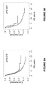
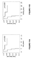
- various reference values may be employed to discriminate between subjects that generally survived for a comparatively long period (represented by the upper curve) and subjects that generally survived for a comparatively short period (represented by the lower curve).
- the reference value is thus the determinant for the size of the respective groups; the higher the reference value, the fewer the subjects in the first group and the lower the likelihood that a tested subject belongs to the first group.
- a high reference value may in some instances be selected to identify subjects with a particularly good prognosis.
- the person skilled in the art may select relevant reference values without undue burden. This is further discussed below.
- the first and the second group may consist exclusively of subjects having ovarian cancers of the same or similar stage (see figures 9 , 11 and 12 ), differentiation grade (see figures 7 , 8 and 14 ) and/or type (see figures 10- 14 ) as the tested subject. Further, the groups may consist only of subjects having the same or similar age, race, menopausal status, genetic characteristics or medical status or history.
- a physician may use the method according to the first aspect to obtain additional information regarding the prognosis of an ovarian cancer subject, which in turn may help him to select the most appropriate treatment regimen. For example, a subject shown to belong to the first group using the method of the first aspect may be given a less aggressive treatment than what would otherwise have been considered, and vice versa.
- the prognosis of the tested subject may also be determined relative to a reference prognosis. Accordingly, as a first configuration of the first aspect, there is provided a method for determining a prognosis for a mammalian subject having an ovarian cancer, comprising the steps of:
- a method for determining whether a prognosis for a mammalian subject having an ovarian cancer is better than a reference prognosis comprising the steps of:
- a method for determining whether a prognosis for a mammalian subject having an ovarian cancer is worse than or equal to a reference prognosis comprising the steps of:
- inventive concept of the present disclosure may also form the basis for a decision to refrain from a certain treatment regimen.
- the prognoses for subjects showing high RBM3 protein levels are generally better than those for subjects showing low RBM3 protein levels.
- a physician may consider the prognosis of an RBM3 protein high subject as being so favorable that certain adjuvant treatment regimens are avoided and a less aggressive adjuvant treatment regimen is selected instead.
- monotherapy may be selected instead of a combination therapy or a therapeutic agent may be given in a lower dose. The decision may thus be to apply a platin-based drug monotherapy and refrain from any additional adjuvant therapy.
- Refraining from any adjuvant therapy in addition to a platinum-based drug may be particularly relevant when said ovarian cancer is an endometroid ovarian cancer, especially an endometroid cancer in stage 1 or 2, as the prognoses for RBM3 high subjects having such a cancer appears to be particularly good (see figures 10-12 ).
- Refraining from any adjuvant therapy in addition to a platinum-based drug may also be particularly relevant when said ovarian cancer is moderately or well differentiated (see figures 7 and 8 ).
- the present disclosure may relieve subjects from over-treatment.
- a method for determining whether a subject having an ovarian cancer is not in need of a treatment with an ovarian cancer treatment regimen comprising the steps of:
- a non-treatment strategy method for a subject having an ovarian cancer comprising:
- the refraining of step c) of the fifth configuration may be a refraining from treatment during at least one week from the completion of steps a) - b), such as at least one month from the completion of steps a) - b), such as at least three months from the completion of steps a) - b), such as at least six months from the completion of steps a) - b), such as at least one year from the completion of steps a) - b), such as at least two years from the completion of steps a) - b).
- the refraining of step c) may be a refraining from treatment until the next time the method is performed or until recurrence of an ovarian cancer tumor.
- a method for establishing a prognosis for a mammalian subject having an ovarian cancer comprising the steps of:
- establishing a prognosis refers to establishing a specific prognosis or a prognosis interval.
- the sample may be an earlier obtained sample.
- step b) refers to any way of associating survival data to the obtained sample value so as to establish a prognosis for the subject.
- sample values corresponding to various prognoses are presented.
- a low sample value is associated with a poorer prognosis than a high sample value.
- the sample value is compared to a reference value, and if the sample value is equal to or lower than the reference value, it is concluded that the prognosis for the subject is equal to, or worse than, a reference prognosis associated with the reference value.
- the method may be adapted to a reference value.
- a reference value which is equal to the sample value may be selected.
- a reference prognosis associated with that reference value may be established.
- the person skilled in the art understands how to establish a reference prognosis which corresponds to a given reference value. For example, the relation between sample values and survival data in a relevant group of cancer patients may be examined in line with what is described in Examples, Section 4, below. The procedure described therein may be adapted to a given reference value. Then, a prognosis corresponding to the given reference value may be selected as the reference prognosis.
- the above method may be adapted to a given reference prognosis.
- a corresponding reference value may be established.
- the person skilled in the art understands how to establish a reference value which corresponds to a given reference prognosis.
- the relation between sample values and survival data in a group of cancer patients may be examined as in Examples, Section 4, below, but the procedure described therein may be adapted to establish reference values corresponding to a given reference prognosis. For example, different reference values may be tested until one which correlates with the given reference prognosis is found.
- the reference prognosis of the first aspect may be based on a previously established prognosis, e.g., obtained by an examination of a relevant population of subjects. Such reference population may be selected to match the tested subject's age, sex, race, ovarian cancer stage and/or medical status and history. Further, a prognosis may be adapted to a background risk in the general population, a statistical prognosis/risk or an assumption based on an examination of the subject. Such examination may also comprise the subject's age, sex, race, ovarian cancer stage, menopausal status and/or medical status and history.
- a physician may for example adapt the reference prognosis to the subject's ovarian cancer history, the stage of the tumor, the morphology of the tumor, the location of the tumor, the menopausal status, the presence and spread of metastases and/or further cancer characteristics.
- the physician responsible for the treatment may take several parameters into account, such as the result of an immunohistochemical evaluation, patient age, tumor type, stage and grade, hormone receptor status, general condition and medical history, such as ovarian cancer history.
- the physician may perform a RBM3 protein test, or order a RBM3 protein test performed, according to the first aspect. Further, the physician may assign to someone else, such as a lab worker, to perform step a), and optionally step b), while performing step c), and optionally b), himself.
- inventive concept of the present disclosure may also form the basis for applying various treatment regimes.
- the prognoses for subjects showing low RBM3 protein levels are generally worse than those for subjects showing high RBM3 protein levels.
- a physician may thus consider the prognosis of an RBM3 protein low subject as being so poor that a certain adjuvant treatment regimen is appropriate.
- the present disclosure may thus provide for accurate treatment of a previously undertreated group.
- a method of treatment of a subject having an ovarian cancer comprising:
- the method may comprise the additional step:
- the reference value of step b) may be associated with a reference prognosis and said treatment regimen of step c) may be adapted to a prognosis which is worse than or equal to the reference prognosis.
- the method may comprise the additional step: d) and if said sample value is higher than said reference value, treating said subject with a treatment regimen adapted to a prognosis which is better than the reference prognosis, for which the appropriate treatment regimen may be no treatment.
- the physician responsible for the treatment according to the second aspect may assign to someone else, such as a lab worker, to perform step a), and optionally step b), while performing step c), and optionally b), himself.
- the method of treatment may be limited to the decision-making and treatment.
- a method of treatment of a subject having an ovarian cancer comprising:
- an increase in the amount of RBM3 protein typically results in an increase in the sample value, and not the other way around.
- the evaluated amount may correspond to any of a predetermined number of discrete sample values.
- a first amount and a second, increased, amount may correspond to the same sample value.
- an increase in the amount of RBM3 protein will not result in a decrease in the sample value in the context of the present disclosure.
- the evaluated amounts may be inversely related to sample values if the qualification between step b) and c) is inverted.
- the qualification between step b) and c) is inverted if the phrase "if the sample value is lower than or equal to the reference value" is replaced with "if the sample value is higher than or equal to the reference value".
- prognosis refers to the prediction of the course or outcome of a disease and its treatment.
- prognosis may also refer to a determination of chance of survival or recovery from a disease, as well as to a prediction of the expected survival time of a subject.
- a prognosis may specifically involve establishing the likelihood for survival of a subject during a period of time into the future, such as three years, five years, ten years or any other period of time.
- a prognosis may further be represented by a single value or a range of values.
- “earlier obtained” refers to obtained before the method is performed. Consequently, if a sample earlier obtained from a subject used in a method, the method does not involve obtaining the sample from the subject, i.e., the sample was previously obtained from the subject in a step separate from the method.
- a mammalian subject having an ovarian cancer refers to a mammalian subject having a primary or secondary ovarian tumor or a mammalian subject which has had an ovarian tumor removed, wherein the removal of the tumor refers to killing or removing the tumor by any appropriate type of surgery or therapy.
- a mammalian subject having an ovarian cancer also includes the cases wherein the mammalian subject is suspected of having an ovarian cancer at the time of the performance of the use or method and the ovarian cancer diagnosis is established later.
- the "reference value” refers to a predetermined value found to be relevant for making decisions or drawing conclusions regarding the prognosis or a suitable treatment strategy for the subject.
- a reference value being "associated" with a reference prognosis refers to the reference value being assigned a corresponding reference prognosis, based on empirical data and/or clinically relevant assumptions.
- the reference value may be the average RBM3 protein value in a relevant group of subjects and the reference prognosis may be an average survival in the same group.
- the reference value does not have to be assigned to a reference prognosis directly derived from prognosis data of a group of subjects exhibiting the reference value.
- the reference prognosis may for example correspond to the prognosis for subjects exhibiting the reference value or lower.
- the reference prognosis may be the prognosis of the subjects exhibiting the values 0 or 1. Consequently, the reference prognosis may also be adapted to the nature of the available data. As further discussed above, the reference prognosis may be further adapted to other parameters as well.
- Step a) of the methods of the above aspects involve evaluating an amount of RBM3 protein present in at least part of the sample, and determining a sample value corresponding to the amount.
- the "at least part of the sample” refers to a relevant part or relevant parts of the sample for establishing the prognosis or drawing conclusions regarding suitable treatments.
- the person skilled in the art understands which part or parts that are relevant under the circumstances present when performing the method. For example, if evaluating a sample comprising cells, the skilled person may only consider the tumor cells, or only the cytoplasms of tumor cells, of the sample.
- step a) an amount is evaluated and a sample value corresponding to the amount is determined. Consequently, an exact measurement of the amount of RBM3 protein is not required for obtaining the sample value.
- the amount of RBM3 protein may be evaluated by visual inspection of a stained tissue sample and the sample value may then be categorized as for example high or low based on the evaluated amount.
- the treatment regimen of the present disclosure may be an adjuvant and/or a neo-adjuvant therapy.
- the general strategy is that a more comprehensive treatment is applied if a subject is found to be RBM3 protein low than if the subject is found to be RBM3 protein high.
- Neo-adjuvant therapy may for example be chemotherapy.
- Neo-adjuvant chemotherapy may be particularly relevant for a subject having a widespread tumors/a heavy tumor load.
- Appropriate adjuvant therapies are primarily chemotherapies. Radiation therapy may be a suitable complement to chemotherapy in some cases, such as after tumor recurrence. The adjuvant treatment may thus be chemotherapy in combination with radiation therapy.
- the adjuvant treatment regimen may be chemotherapy. That means that according to some of the above methods, a chemotherapeutic agent is administered if the subject is RBM3 protein low. If, however, the subject is RBM3 protein high, the treatment with the chemotherapeutic agent may be considered unnecessary, and therefore, not administered to the subject. In the latter case, surgery may be considered sufficient.
- the treatment regimen may be a combination of two or more chemotherapeutic agents. That means that according to some of the above methods, the combination is administered if the subject is RBM3 protein low. If, however, the subject is RBM3 protein high, the combination may be considered unnecessary, and therefore, not applied to the subject. In the latter case, treatment with one therapeutic agent only may be considered sufficient.
- a combination is a platinum based drug, such as carboplatin, and a taxan, such as paclitaxel.
- the combination is applied in both cases, however in a relatively high dose if the subject is RBM3 protein low and in a relatively low dose if the subject is RBM3 protein high.
- the "relatively high dose” is high relative the “relatively low dose”.
- a dose considered to be normal within the art may be decreased if a sample value from the subject is higher than the reference value or increased if the sample value from the subject is lower than or equal to the reference value.
- some of the methods of the present disclosure may yield information which forms the basis of a personalized treatment regimen.
- the adjuvant or neo-adjuvant treatment regimen of the present disclosure may comprise treatment with one or more of the platinum-based drugs, such as carboplatin, paraplatin and cisplatin.
- the adjuvant or neo-adjuvant treatment regimen of the present disclosure may comprise treatment with one or more drugs selected from taxanes (such as paclitaxel and docetaxel), gemcitabine, topoisomerase inhibitors (such as topotecan) and anthracyclins (such as doxorubicin).
- taxanes such as paclitaxel and docetaxel
- gemcitabine gemcitabine
- topoisomerase inhibitors such as topotecan
- anthracyclins such as doxorubicin
- the adjuvant chemotherapy of the present disclosure may for example be administered intraperitoneally.
- the adjuvant or neo-adjuvant treatment regimen of the present disclosure may also comprise immunotherapy or radio-immunotherapy.
- Such therapies may for example be combined with one or more chemotherapies mentioned above.
- Examples of monoclonal antibodies that may be used in the immunotherapy or radio-immunotherapy are listed in tables I-V of the review article by Oei and co-workers ( Oei, A.M. et al (2008) Int J Onc 32:1145-1157 ).
- the ovarian cancer may thus be epithelial ovarian cancer.
- the subjects behind the data in Examples, Section 4 had all received adjuvant treatment with a platinum-based drug, i.e. cisplatin or carboplatin.
- the subject may thus be receiving platinum-based drugs or be scheduled for treatment with platinum-based drugs.
- the level of RBM3 protein expression is prognostically relevant in groups of patients having cancers in all differentiation grades (see figures 2-6 , 9-14 ), the prognostic indication is shown herein to be particularly accentuated in subjects having moderately differentiated (grade 2) ovarian cancers (see figure 7 ) or well or moderately differentiated (grade 1 or 2) ovarian cancers (see figures 8 and 15).
- the ovarian cancer may be selected from moderately and well differentiated ovarian cancers.
- the three-step differentiation grade scale of well (1)/moderately (2)/poorly (3) differentiated is used, which is well known to the skilled person.
- the level of RBM3 protein expression is prognostically relevant in groups of patients having cancers in all stages (see figures 2-8 , 10 , 12-14 ), it is shown herein that the prognostic indication may be particularly accentuated in subjects having cancers in stage 1 or 2 (see figures 9 and 11 ).
- stage refers to stage according to the Federation of Gynecology and Obstetrics (FIGO) classification in the present disclosure.
- the stage of the ovarian cancer may be selected from 1 and 2.
- the level of RBM3 protein expression is prognostically relevant in groups of patients having various types of ovarian cancers (see figures 2-9 ), the prognostic indication is shown herein to particularly accentuated in subjects having endometroid (see figures 10-12 ) or serous (see fig 13-15 ) ovarian cancer.
- the ovarian cancer is selected from serous and endometroid ovarian cancers.
- the first and the second group may consist of subjects having cancers of the same stage, grade and/or type as the subject of the method.
- the prognosis may be a probability of survival.
- the survival of the present disclosure may for example be overall survival or ovarian cancer specific survival.
- the "survival” may be measured over different periods, such as five, ten or 15 years. Accordingly, the survival may be a five-year, ten-year or 15-year survival. Where a reference prognosis is employed, it is of the same type as the prognosis of the subject.
- the sample may be a body fluid sample.
- the body fluid sample may be selected from the group consisting of blood, plasma, serum, cerebral fluid, urine, lymph and exudate.
- the sample may be a cytology sample or a stool sample.
- the level of RBM3 protein expression may preferably be measured intracellularly.
- the body fluid, cytology or stool sample may for example comprise cells, such as tumor cells.
- the sample may be a tissue sample, such as an ovarian tissue sample, such as an ovarian tumor tissue sample, e.g, from a surgical removal of an ovarian tumor.
- tissue sample such as an ovarian tissue sample, such as an ovarian tumor tissue sample, e.g, from a surgical removal of an ovarian tumor.
- step a) may be limited to the nuclei and/or cytoplasms of cells, such as tumor cells or stromal cells, of said sample. Consequently, when a tissue sample is examined, only the nuclei and/or cytoplasms of tumor cells may be taken into consideration. Such examination may for example be aided by immunohistochemical staining.
- sample value and “reference value” are to be interpreted broadly.
- the quantification of RBM3 protein to obtain these values may be done via automatic means, via a scoring system based on visual or microscopic inspection of samples, or via combinations thereof.
- a skilled person such as a person skilled in the art of histopathology, to determine the sample and reference values merely by inspection, e.g., of tissue slides that have been stained for RBM3 protein expression.
- the determination of the sample value being higher than the reference value may thus correspond to the determination, upon visual or microscopic inspection, that a sample tissue slide is more densely stained and/or exhibit a larger fraction of stained cells than is the case for a reference tissue slide.
- sample value may also be compared to a reference value given by a literal reference, such as a reference value described in wording or by a reference picture. Consequently, the sample and/or reference values may in some cases be mental values that the skilled person determines upon inspection and comparison.
- step a) and optionally step b) may be performed in an automatic analysis apparatus, and such apparatus may be based on a platform adapted for immunohistochemically analysis.
- one or more tumor tissue sample(s) from the subject in question may be prepared for imunohistochemical analysis manually and then loaded into the automatic analysis apparatus, which gives the sample value of step a) and optionally also performs the comparison with the reference value of step b).
- the operator performing the analysis, the physician ordering the analysis or the apparatus itself may then draw the conclusion of step c). Consequently, software adapted for drawing the conclusion of step c) may be implemented on the apparatus.
- a reference value found to be relevant for establishing prognosis or making treatment decisions regarding ovarian cancer subjects, for use as comparison with the sample value from the subject, may be provided in various ways. With the knowledge of the teachings of the present disclosure, the skilled artisan can, without undue burden, provide relevant reference values for performing the methods of the present disclosure.
- the person performing the methods of the above aspects may, for example, adapt the reference value to desired information.
- the reference value may be adapted to yield the most significant prognostic information, e.g., the largest separation between the RBM3 protein high survival curve and the RBM3 protein low survival curve (see the figures), which corresponds to the largest difference in survival between the first and the second group of the first aspect.
- the reference value may be selected such that a group of subjects having particularly good prognoses or particularly poor prognoses is singled out.
- the reference value may correspond to the amount of RBM3 protein expression in a healthy tissue, such as healthy ovarian tissue, or stroma tissue of the subject of the method.
- the reference value may be provided by the amount of RBM3 protein expression measured in a standard sample of normal tissue from another, comparable subject.
- the reference value may be provided by the amount of RBM3 protein expression measured in a reference sample comprising tumor cells, such as a reference sample of tumor tissue, e.g., ovarian cancer tissue.
- the amount of protein expression of the reference sample may preferably be previously established. Consequently, the reference value may be provided by the amount of RBM3 protein measured in a reference sample comprising cells expressing a predetermined amount of RBM3 protein.
- the reference value may for example be provided by the amount of RBM3 protein expression measured in a reference sample comprising cell lines, such as cancer cell lines, expressing a predetermined, or controlled, amount of RBM3 protein.
- a reference sample comprising cell lines, such as cancer cell lines, expressing a predetermined, or controlled, amount of RBM3 protein.
- the person skilled in the art understands how to provide such cell lines, for example guided by the disclosure of Rhodes et al. (2006) The biomedical scientist, p 515-520 .
- the reference value may be a predetermined value corresponding to the amount of RBM3 protein expression in a reference sample.
- the amount of RBM3 protein in the reference sample does not have to directly correspond to the reference value.
- the reference sample may also provide an amount of RBM3 protein that helps a person performing the method to assess various reference values.
- the reference sample(s) may help in creating a mental image of the reference value by providing a "positive" reference value and/or a "negative" reference value.
- the fraction may for example be: a "cellular fraction", wherein the RBM3 protein expression of the whole cells is taken into account; a "cytoplasmic fraction”, wherein the RBM3 protein expression of only the cytoplasms of the cells is taken into account; or a "nuclear fraction”, wherein the RBM3 protein expression of only the nuclei of the cells is taken into account.
- the nuclear or cytoplasmic fraction may for example be classified as ⁇ 2 %, 2 - 25 %, > 25 - 75 % or > 75 % immunoreactive cells of the relevant cell population.
- the "cytoplasmic fraction” corresponds to the percentage of relevant cells in a sample that exhibits a positive staining in the cytoplasm, wherein a medium or distinct and strong immunoreactivity in the cytoplasm is considered positive and no or faint immunoreactivity in the cytoplasm is considered negative.
- the "nuclear fraction” corresponds to the percentage of relevant cells in a sample that exhibits a positive staining in the nucleus, wherein a medium or distinct and strong immunoreactivity in the nucleus is considered positive and no or faint immunoreactivity in the nucleus is considered negative.
- the person skilled in the art of pathology understands which cells that are relevant under the conditions present when performing the method and may determine a cytoplasmic or nuclear fraction based on his general knowledge and the teachings of the present disclosure.
- the relevant cells may for example be tumor cells.
- the skilled artisan understands how to perform corresponding measurements employing the "cellular fraction".
- the intensity may for example be: a "cellular intensity”, wherein the RBM3 protein expression of the whole cells is taken into account; a "cytoplasmic intensity”, wherein the RBM3 protein expression of only the cytoplasms of the cells is taken into account, or a "nuclear intensity”, wherein the RBM3 protein expression of only the nuclei of the cells is taken into account. Cytoplasmic and nuclear intensity is subjectively evaluated in accordance with standards used in clinical histopathological diagnostics.
- the weak and moderate values may be combined into a weak/moderate value (see also Examples, section 4).
- the person skilled in the art understands which cells that are relevant under the conditions present when performing the method and may determine a nuclear or cytoplasmic intensity based on his general knowledge and the teachings of the present disclosure.
- the relevant cells may for example be tumor cells. Further, the skilled artisan understands how to perform corresponding measurements employing the "cellular intensity".
- the inventors have found that both cytoplasmic and nuclear expression of RBM3 protein is relevant for establishing prognoses.
- the reference value may be a cytoplasmic fraction, a cytoplasmic intensity or a combination thereof.
- the sample value may be a cytoplasmic fraction, a cytoplasmic intensity or a combination thereof.
- the reference value may be a nuclear fraction, a nuclear intensity or a combination thereof.
- the sample value may be a nuclear fraction, a nuclear intensity or a combination thereof.
- the criterion for the conclusion in step c) is that the sample value is higher than a nuclear or cytoplasmic fraction of 0 %, 1 %, 2 %, 5 %, 10 %, 15 %, 20 %, 25 %, 30 %, 35 %, 40 %, 50 %, 60 %, 70 %, 75 %, 80 %, 90 % or 95 %.
- the reference value of step b) is a nuclear or cytoplasmic fraction of 95 % or lower, such as 90 % or lower, such as 85 % or lower, such as 80 % or lower, such as 75 % or lower, such as 70 % or lower, such as 65 % or lower, such as 60 % or lower, such as 55 % or lower, such as 50 % or lower, such as 45 % or lower, such as 40 % or lower, such as 35 % or lower, such as 30 % or lower, such as 25 % or lower, such as 20 % or lower, such as 15 % or lower, such as 10 % or lower, such as 5 % or lower, such as 2 % or lower, such as 1 % or lower, such as 0 %.
- the criterion for the conclusion in step c) may be a sample value, which is higher than absent cytoplasmic or nuclear intensity, such as higher than weak cytoplasmic or nuclear intensity, such as higher than moderate cytoplasmic or nuclear intensity.
- the reference value of step b) may be a moderate cytoplasmic or nuclear intensity of RBM3 protein expression or lower, such as a weak cytoplasmic or nuclear intensity of RBM3 protein expression or lower, such as an absent cytoplasmic or nuclear intensity.
- the reference value may be any type of intensity or fraction.
- the inventors have found that if the reference value is selected from a nuclear fraction, a nuclear intensity and a cytoplasmic intensity, particularly relevant for conclusions regarding prognoses may be drawn (see the figures).
- the nuclear fraction used as reference value in figures 2-4 and 7-12 is 75 %. Consequently, in embodiments of the present disclosure, the reference value may be a nuclear fraction in the range 40 - 90 %, such as 50 - 85%.
- the reference value is a moderate nuclear intensity. Consequently, in embodiments of the present disclosure, the reference value may be a moderate or weak/moderate nuclear intensity.
- the reference value is a weak/moderate nuclear intensity. Consequently, in embodiments of the present disclosure, the reference value may be a weak, weak/moderate or moderate nuclear intensity.
- the reference value may be a combination of a fraction value and an intensity value.
- the reference value may be a function of a fraction value and an intensity value.
- a function may be a staining score.
- the "staining score" is calculated as described in Examples, Section 3 and table 1 below.
- the reference value may be a staining score of 2 or lower, such as 1 or lower, such as 0.
- an intensity value and/or a fraction value as the reference value may depend on the staining procedure, e.g., on the employed anti-RBM3 antibody and on the staining reagents.
- a person skilled in the art e.g., a pathologist understands how to perform the evaluation yielding a fraction, such as a cellular, cytoplasmic or nuclear fraction, or an intensity, such as a cellular, cytoplasmic or nuclear intensity.
- a fraction such as a cellular, cytoplasmic or nuclear fraction
- an intensity such as a cellular, cytoplasmic or nuclear intensity.
- the skilled artisan may use a reference sample comprising a predetermined amount of RBM3 protein for establishing the appearance of a certain fraction or intensity.
- a reference sample may not only be used for the provision of the actual reference value, but also for the provision of an example of a sample with an amount of RBM3 protein that is higher than the amount corresponding to the reference value.
- histochemical staining such as in immunohistochemical staining
- the skilled artisan may use a reference sample for establishing the appearance of a stained sample having a high amount of RBM3 protein, e.g., a positive reference. Subsequently, the skilled artisan may assess the appearances of samples having lower amounts of RBM3 protein, such as the appearance of a sample with an amount of RBM3 protein corresponding to the reference value.
- the skilled artisan may use a reference sample to create a mental image of a reference value corresponding to an amount of RBM3 protein which is lower than that of the reference sample.
- the skilled artisan may use another reference sample having a low amount of RBM3 protein, or lacking detectable RBM3 protein, for establishing the appearance of such sample, e.g., as a "negative reference”.
- two reference samples may be employed: a first reference sample having no detectable RBM3 protein, and thus corresponding to a absent nuclear intensity, which is lower than the reference value; and a second reference sample having an amount of RBM3 protein corresponding to a strong nuclear intensity, which is higher than the reference value.
- reference sample for establishing the appearance of a sample with a high amount of RBM3 protein.
- Such reference sample may be a sample comprising tissue expressing a high amount of RBM3 protein, such as a sample comprising ovarian tumor tissue having a pre-established high expression of RBM3 protein.
- the reference sample may provide an example of a strong cytoplasmic intensity (CI).
- CI cytoplasmic intensity
- the skilled artisan may then divide samples into the CI categories absent, weak, moderate and strong. This division may be further assisted by a reference sample lacking detectable RBM3 protein (negative reference), i.e., a reference sample providing an absent cytoplasmic intensity.
- the reference sample may provide an example of a sample with a nuclear fraction (NF) higher than 75 %. With the knowledge of the appearance of a sample with more than 75 % positive cells, the skilled artisan may then evaluate the NF of other samples having e.g., a lower percentage of positive cells. This division may be further assisted by a reference sample essentially lacking RBM3 protein (negative reference), i.e., a reference sample providing a low NF (e.g., ⁇ 5%, such as ⁇ 2%), or a NF of 0.
- a reference sample essentially lacking RBM3 protein negative reference
- cell lines expressing a controlled amount of RBM3 protein may be used as the reference, in particular as a positive reference.
- One or more pictures may also be provided as the "reference sample”.
- a picture may show an example of a tumor tissue slide stained with a certain antibody during certain conditions and exhibiting a certain cytoplasmic intensity and/or fraction.
- the above discussion about the "reference sample” applies mutatis mutandis to pictures.
- the RBM3 protein may comprise a sequence selected from:
- sequence ii) above is at least 90 % identical, at least 91 % identical, at least 92 % identical, at least 93 % identical, at least 94 % identical, at least 95 % identical, at least 96 % identical, at least 97 % identical, at least 98 % identical or at least 99 % identical to SEQ ID NO:1.
- the RBM3 protein may comprise, or consists of, a sequence selected from:
- sequence ii) above is at least 90 % identical, at least 91 % identical, at least 92 % identical, at least 93 % identical, at least 94 % identical, at least 95 % identical, at least 96 % identical, at least 97 % identical, at least 98 % identical or at least 99 % identical to SEQ ID NO:2.
- % identical is calculated as follows.
- the query sequence is aligned to the target sequence using the CLUSTAL W algorithm ( Thompson, J.D., Higgins, D.G. and Gibson, T.J., Nucleic Acids Research, 22: 4673-4680 (1994 )).
- the amino acid residues at each position are compared, and the percentage of positions in the query sequence that have identical correspondences in the target sequence is reported as % identical.
- the target sequence determines the number of positions that are compared. Consequently, in the context of the present disclosure, a query sequence that is shorter than the target sequence can never be 100 % identical to the target sequence. For example, a query sequence of 85 amino acids may at the most be 85 % identical to a target sequence of 100 amino acids.
- the methods of the above aspects may comprise a further step preceding step a), which further step comprises:
- the RBM3 protein may be detected and/or quantified through the application to the sample of a detectable and/or quantifiable affinity ligand, which is capable of selective interaction with the RBM3 protein.
- the application of the affinity ligand is performed under conditions that enable binding of the affinity ligand to any RBM3 protein in the sample.
- step a) may comprise:
- affinity ligand remaining in association with the sample refers to affinity ligand which was not removed in step a2), e.g., the affinity ligand bound to the sample.
- the binding may for example be the interaction between antibody and antigen.
- step a) may comprise:
- affinity ligands that may prove useful, as well as examples of formats and conditions for detection and/or quantification, are given below for the sake of illustration.
- the affinity ligand may be selected from the group consisting of antibodies, fragments thereof and derivatives thereof, i.e., affinity ligands based on an immunoglobulin scaffold.
- the antibodies and the fragments or derivatives thereof may be isolated and/or mono-specific.
- Antibodies comprise monoclonal and polyclonal antibodies of any origin, including murine, rabbit, human and other antibodies, as well as chimeric antibodies comprising sequences from different species, such as partly humanized antibodies, e.g., partly humanized mouse antibodies. Polyclonal antibodies are produced by immunization of animals with the antigen of choice.
- Monoclonal antibodies of defined specificity can be produced using the hybridoma technology developed by Köhler and Milstein ( Köhler G and Milstein C (1976) Eur. J. Immunol. 6:511-519 ).
- the antibody fragments and derivatives of the present disclosure are capable of selective interaction with the same antigen (e.g. RBM3 protein) as the antibody they are fragments or derivatives of.
- Antibody fragments and derivatives comprise Fab fragments, consisting of the first constant domain of the heavy chain (CH1), the constant domain of the light chain (CL), the variable domain of the heavy chain (VH) and the variable domain of the light chain (VL) of an intact immunoglobulin protein; Fv fragments, consisting of the two variable antibody domains VH and VL ( Skerra A and Plückthun A (1988) Science 240:1038-1041 ); single chain Fv fragments (scFv), consisting of the two VH and VL domains linked together by a flexible peptide linker ( Bird RE and Walker BW (1991) Trends Biotechnol. 9:132-137 ); Bence Jones dimers ( Stevens FJ et al.
- SEQ ID NO:1 was designed for immunizations, e.g., designed to lack transmembrane regions to ensure efficient expression in E . coli, and to lack any signal peptide, since those are cleaved off in the mature protein. Consequently, an antibody or fragment or derivative thereof according to the present disclosure may for example be one that is obtainable by a process comprising a step of immunizing an animal, such as a rabbit, with a protein whose amino acid sequence comprises, preferably consists of, the sequence SEQ ID NO:1.
- the immunization process may comprise primary immunization with the protein in Freund's complete adjuvant.
- the immunization process may further comprise boosting at least two times, in intervals of 2-6 weeks, with the protein in Freund's incomplete adjuvant.
- Processes for the production of antibodies or fragments or derivatives thereof against a given target are known in the art.
- an antibody or fragment or derivative thereof according to the present disclosure may be obtainable by a process comprising a step of immunizing an animal with a peptide whose amino acid sequence consists of SEQ ID NO:4. Also, the antibody or fragment may be obtainable by a process comprising a step of immunizing an animal with an RBM3 fragment which consists of 15 amino acids or less and comprises a sequence selected from SEQ ID NO:5-8.
- a "mono-specific antibody” is one of a population of polyclonal antibodies which has been affinity purified on its own antigen, thereby separating such mono-specific antibodies from other antiserum proteins and non-specific antibodies. This affinity purification results in antibodies that bind selectively to its antigen.
- the polyclonal antisera are purified by a two-step immunoaffinity based protocol to obtain mono-specific antibodies selective for the target protein. Antibodies directed against generic affinity tags of antigen fragments are removed in a primary depletion step, using the immobilized tag protein as the capturing agent.
- the serum is loaded on a second affinity column with the antigen as capturing agent, in order to enrich for antibodies specific for the antigen (see also Nilsson P et al. (2005) Proteomics 5:4327-4337 ).
- the biomolecular diversity needed for selection of affinity ligands may be generated by combinatorial engineering of one of a plurality of possible scaffold molecules, and specific and/or selective affinity ligands are then selected using a suitable selection platform.
- the scaffold molecule may be of immunoglobulin protein origin ( Bradbury AR and Marks JD (2004) J. Immunol. Meths. 290:29-49 ), of non-immunoglobulin protein origin ( Nygren P ⁇ and Skerra A (2004) J. Immunol. Meths. 290:3-28 ), or of an oligonucleotide origin ( Gold L et al. (1995) Annu. Rev. Biochem. 64:763-797 ).
- Non-limiting examples of such structures useful for generating affinity ligands against RBM3 protein for use according to the present disclosure, are staphylococcal protein A and domains thereof and derivatives of these domains, such as protein Z ( Nord K et al. (1997) Nat. Biotechnol. 15:772-777 ); lipocalins ( Beste G et al. (1999) Proc. Natl. Acad. Sci. U.S.A. 96:1898-1903 ); ankyrin repeat domains ( Binz HK et al. (2003) J. Mol. Biol.
- CBD cellulose binding domains
- GFP green fluorescent protein
- CTL-4 Hufton SE et al. (2000) FEBS Lett. 475:225-231 ; Irving RA et al. (2001) J. Immunol. Meth.
- protease inhibitors such as Knottin proteins ( Wentzel A et al. (2001) J. Bacteriol. 183:7273-7284 ; Baggio R et al. (2002) J. Mol. Recognit. 15:126-134 ) and Kunitz domains ( Roberts BL et al. (1992) Gene 121:9-15 ; Dennis MS and Lazarus RA (1994) J. Biol. Chem. 269:22137-22144 ); PDZ domains ( Schneider S et al. (1999) Nat. Biotechnol. 17:170-175 ); peptide aptamers, such as thioredoxin ( Lu Z et al.
- non-immunoglobulin protein scaffolds include scaffold proteins presenting a single randomized loop used for the generation of novel binding specificities, protein scaffolds with a rigid secondary structure where side chains protruding from the protein surface are randomized for the generation of novel binding specificities, and scaffolds exhibiting a non-contiguous hyper-variable loop region used for the generation of novel binding specificities.
- oligonucleotides may also be used as affinity ligands.
- Single stranded nucleic acids called aptamers or decoys, fold into well-defined three-dimensional structures and bind to their target with high affinity and specificity.
- aptamers or decoys Single stranded nucleic acids
- the oligonucleotide ligands can be either RNA or DNA and can bind to a wide range of target molecule classes.
- Selection platforms include, but are not limited to, phage display ( Smith GP (1985) Science 228:1315-1317 ), ribosome display ( Hanes J and Plückthun A (1997) Proc. Natl. Acad. Sci. U.S.A.
- yeast two-hybrid system Fields S and Song O (1989) Nature 340:245-246
- yeast display Gai SA and Wittrup KD (2007) Curr Opin Struct Biol 17:467-473
- mRNA display Roberts RW and Szostak JW (1997) Proc. Natl. Acad. Sci. U.S.A. 94:12297-12302
- bacterial display Daugherty PS (2007) Curr Opin Struct Biol 17:474-480 , Kronqvist N et al. (2008) Protein Eng Des Sel 1-9 , Harvey BR et al.
- the affinity ligand may be a non-immunoglobulin affinity ligand derived from any of the protein scaffolds listed above, or an oligonucleotide molecule.
- the RBM3 protein fragment SEQ ID NO:1 was designed to consist of a unique sequence with low homology with other human proteins and to minimize cross reactivity of generated affinity reagents. Consequently, in embodiments of the present disclosure, the affinity ligand may be capable of selective interaction with a polypeptide consisting of the sequence SEQ ID NO:1.
- the epitope region SEQ ID NO:4 has been identified within SEQ ID NO:1.
- the results indicate that the epitope region SEQ ID NO:4 provides for particularly strong and selective binding to the affinity ligand.
- the affinity ligand of the present disclosure is capable of selective interaction with a peptide consisting of an amino acid sequence selected from SEQ ID NO:4.
- the affinity ligand of the present disclosure is capable of selective interaction with an RBM3 fragment which consists of 15 amino acids or less and comprises a sequence selected from SEQ ID NO:5-8.
- Antibodies having selectivity for a single epitope region may provide for increased reproducibility in detection analyses as compared to antibodies generated against a longer peptide sequence (such as a PrEST or a full-length protein).
- the antibodies having a selectivity for a single epitope region may also provide for distinct and strong staining in immunohistochemical analyses.
- antibodies capable of selective interaction with any one of SEQ ID NO:4-8 may be obtained by immunizing an animal with an antigen consisting of the amino acid sequence SEQ ID NO:1 followed by affinity purification of the antisera using peptides consisting of the amino acid sequences SEQ ID NO:4-8, respectively.
- the detection and/or quantification of the affinity ligand capable of selective interaction with the RBM3 protein may be accomplished in any way known to the skilled person for detection and/or quantification of binding reagents in assays based on biological interactions. Accordingly, any affinity ligand described above may be used to quantitatively and/or qualitatively detect the presence of the RBM3 protein.
- affinity ligands may be labeled themselves with various markers or may in turn be detected by secondary, labeled affinity ligands to allow detection, visualization and/or quantification.
- Non-limiting examples of labels that can be conjugated to primary and/or secondary affinity ligands include fluorescent dyes or metals (e.g., fluorescein, rhodamine, phycoerythrin, fluorescamine), chromophoric dyes (e.g., rhodopsin), chemiluminescent compounds (e.g., luminal, imidazole) and bioluminescent proteins (e.g., luciferin, luciferase), haptens (e.g., biotin).
- fluorescent dyes or metals e.g., fluorescein, rhodamine, phycoerythrin, fluorescamine
- chromophoric dyes e.g., rhodopsin
- chemiluminescent compounds e.g., luminal, imidazole
- bioluminescent proteins e.g., luciferin, luciferase
- haptens
- Affinity ligands can also be labeled with enzymes (e.g., horseradish peroxidase, alkaline phosphatase, beta-lactamase), radioisotopes (e.g., 3 H, 14 C, 32 P, 35 S or 125 I) and particles (e.g., gold).
- enzymes e.g., horseradish peroxidase, alkaline phosphatase, beta-lactamase
- radioisotopes e.g., 3 H, 14 C, 32 P, 35 S or 125 I
- particles e.g., gold
- particles e.g., gold
- particles e.g., gold
- particles e.g., gold
- particles e.g., gold
- particles e.g., gold
- particles e.g., gold
- particles e.g., gold
- particles e.g., gold
- particles e.g., gold
- particles e.
- the different types of labels can be conjugated to an affinity ligand using various chemistries, e.g., the amine reaction or the thiol reaction.
- chemistries e.g., the amine reaction or the thiol reaction.
- other reactive groups than amines and thiols can be used, e.g., aldehydes, carboxylic acids and glutamine.
- the detection, localization and/or quantification of a labeled affinity ligand bound to its RBM3 protein target may involve visualizing techniques, such as light microscopy or immunofluoresence microscopy. Other methods may involve the detection via flow cytometry or luminometry.
- a biological sample such as a tumor tissue sample (biopsy), which has been removed from the subject may be used for detection and/or quantification of RBM3 protein.
- the biological sample such as the biopsy, may be an earlier obtained sample. If using an earlier obtained sample in a method, no steps of the method are practiced on the human or animal body.
- the affinity ligand may be applied to the biological sample for detection and/or quantification of the RBM3 protein. This procedure enables not only detection of RBM3 protein, but may in addition show the distribution and relative level of expression thereof.
- the method of visualization of labels on the affinity ligand may include, but is not restricted to, fluorometric, luminometric and/or enzymatic techniques. Fluorescence is detected and/or quantified by exposing fluorescent labels to light of a specific wavelength and thereafter detecting and/or quantifying the emitted light in a specific wavelength region. The presence of a luminescently tagged affinity ligand may be detected and/or quantified by luminescence developed during a chemical reaction. Detection of an enzymatic reaction is due to a color shift in the sample arising from chemical reaction. Those of skill in the art are aware that a variety of different protocols can be modified in order for proper detection and/or quantification.
- a biological sample may be immobilized onto a solid phase support or carrier, such as nitrocellulose or any other solid support matrix capable of immobilizing RBM3 protein present in the biological sample applied to it.
- solid state support materials useful in the present invention include glass, carbohydrate (e.g., Sepharose), nylon, plastic, wool, polystyrene, polyethene, polypropylene, dextran, amylase, films, resins, cellulose, polyacrylamide, agarose, alumina, gabbros and magnetite.
- primary affinity ligand specific to RBM3 protein may be applied, e.g., as described in Examples, Sections 4, of the present disclosure.
- the supporting matrix may be washed with one or more appropriate buffers known in the art, followed by exposure to a secondary labeled affinity ligand and washed once again with buffers to remove unbound affinity ligands. Thereafter, selective affinity ligands may be detected and/or quantified with conventional methods.
- the binding properties for an affinity ligand may vary from one solid state support to the other, but those skilled in the art should be able to determine operative and optimal assay conditions for each determination by routine experimentation.
- the quantifiable affinity ligand of a1) or aI) may be detected using a secondary affinity ligand capable of recognizing the quantifiable affinity ligand.
- the quantification of a3) or aII) may thus be carried out by means of a secondary affinity ligand with affinity for the quantifiable affinity ligand.
- the secondary affinity ligand may be an antibody or a fragment or a derivative thereof.
- one available method for detection and/or quantification of the RBM3 protein is by linking the affinity ligand to an enzyme that can then later be detected and/or quantified in an enzyme immunoassay (such as an EIA or ELISA).
- an enzyme immunoassay such as an EIA or ELISA.
- the biological sample is brought into contact with a solid material or with a solid material conjugated to an affinity ligand against the RBM3 protein, which is then detected and/or quantified with an enzymatically labeled secondary affinity ligand.
- an appropriate substrate is brought to react in appropriate buffers with the enzymatic label to produce a chemical moiety, which for example is detected and/or quantified using a spectrophotometer, fluorometer, luminometer or by visual means.
- primary and any secondary affinity ligands can be labeled with radioisotopes to enable detection and/or quantification.
- appropriate radiolabels in the present disclosure are 3 H, 14 C, 32 P, 35 S or 125 I.
- the specific activity of the labeled affinity ligand is dependent upon the half-life of the radiolabel, isotopic purity, and how the label has been incorporated into the affinity ligand.
- Affinity ligands are preferably labeled using well-known techniques ( Wensel TG and Meares CF (1983) in: Radioimmunoimaging and Radioimmunotherapy (Burchiel SW and Rhodes BA eds.) Elsevier, New York, pp 185-196 ).
- a thus radiolabeled affinity ligand can be used to visualize RBM3 protein by detection of radioactivity in vivo or in vitro.
- Radionuclear scanning with e.g., gamma camera, magnetic resonance spectroscopy or emission tomography function for detection in vivo and in vitro, while gamma/beta counters, scintillation counters and radiographies are also used in vitro.
- kit for carrying out a method according to the above aspects which comprises:
- kit according to the third aspect may be selected and specified as described above in connection with the method aspects of the present disclosure.
- the kit according to the present disclosure comprises an affinity ligand against an RBM3 protein, as well as other means that help to quantify the specific and/or selective affinity ligand after it has bound specifically and/or selectively to the RBM3 protein.
- the kit may contain a secondary affinity ligand for detecting and/or quantifying a complex formed by the RBM3 protein and the affinity ligand capable of selective interaction with the RBM3 protein.
- the kit may also contain various auxiliary substances other than affinity ligands, to enable the kit to be used easily and efficiently.
- auxiliary substances include solvents for dissolving or reconstituting lyophilized protein components of the kit, wash buffers, substrates for measuring enzyme activity in cases where an enzyme is used as a label, target retrieval solution to enhance the accessibility to antigens in cases where paraffin or formalin-fixed tissue samples are used, and substances such as reaction arresters, e.g., endogenous enzyme block solution to decrease the background staining and/or counterstaining solution to increase staining contrast, that are commonly used in immunoassay reagent kits.
- reaction arresters e.g., endogenous enzyme block solution to decrease the background staining and/or counterstaining solution to increase staining contrast, that are commonly used in immunoassay reagent kits.
- the affinity ligand may be selected as described above in connection with the method aspects.
- the detectable affinity ligand may in embodiments of the kit aspect comprise a label selected from the group consisting of fluorescent dyes and metals, chromophoric dyes, chemiluminescent compounds and bioluminescent proteins, enzymes, radioisotopes, particles and quantum dots.
- the reagents necessary for quantifying the amount of the affinity ligand comprise a secondary affinity ligand capable of recognizing the quantifiable affinity ligand.
- the secondary affinity ligand capable of recognizing the quantifiable affinity ligand comprises a label selected from the group consisting of fluorescent dyes or metals, chromophoric dyes, chemiluminescent compounds and bioluminescent proteins, enzymes, radioisotopes, particles and quantum dots.
- the kit according to the kit aspect may also advantageously comprise a reference sample for provision of, or yielding, the reference value to be used for comparison with the sample value.
- the reference sample may comprise a predetermined amount of RBM3 protein.
- a reference sample may for example be constituted by a tissue sample containing the predetermined amount of RBM3 protein.
- the tissue reference sample may then be used by the person of skill in the art in the determination of the RBM3 expression status in the sample being studied, by manual, such as ocular, or automated comparison of expression levels in the reference tissue sample and the subject sample.
- the reference sample may comprise cell lines, such as cancer cell lines, expressing a predetermined, or controlled, amount of RBM3 protein.
- the cell lines may be formalin fixed.
- such formalin fixed cell lines may be paraffin embedded.
- the wording "reference sample for provision of the reference value” is to be interpreted broadly in the context of the present disclosure.
- the reference sample may comprise an amount of RBM3 protein actually corresponding to the reference value, but it may also comprise an amount of RBM3 protein corresponding to a value being higher than the reference value.
- the "high" value may be used by a person performing the method as an upper reference (positive reference) for assessing, e.g., the appearance of, a reference value which is lower than the "high” value.
- the person skilled in the art of immunohistochemistry understands how to do such an assessment.
- the skilled person may use another reference sample comprising a low amount of RBM3 protein for provision of a "low" value in such an assessment, e.g., as a negative reference. This is further discussed above in connection with the method aspects.
- the reference sample may comprise an amount of RBM3 protein corresponding to the reference value.
- the reference sample may comprise an amount of RBM3 protein corresponding to a nuclear or cytoplasmic fraction of 95 % or lower, such as 90 % or lower, such as 85 % or lower, such as 80 % or lower, such as 75 % or lower, such as 70 % or lower, such as 65 % or lower, such as 60 % or lower, such as 55 % or lower, such as 50 % or lower, such as 45 % or lower, such as 40 % or lower, such as 35 % or lower, such as 30 % or lower, such as 25 % or lower, such as 20 % or lower, such as 15 % or lower, such as 10 % or lower, such as 5 % or lower, such as 2 % or lower, such as 1 % or lower, such as 0 %.
- the reference sample may comprise an amount of RBM3 protein corresponding to a moderate nuclear or cytoplasmic intensity expression or lower, such as a weak nuclear or cytoplasmic intensity of RBM3 protein expression or lower, such as an absent nuclear or cytoplasmic intensity.
- the reference sample may comprise an amount of RBM3 protein corresponding to a nuclear fraction in the range 40 - 90 %, such as 50-85%.
- the reference value is a moderate nuclear intensity. Consequently, in embodiments of the present disclosure, the reference sample may comprise an amount of RBM3 protein corresponding to a moderate or weak/moderate nuclear intensity.
- the reference value is a weak/moderate nuclear intensity. Consequently, in embodiments of the present disclosure, the reference sample may comprise an amount of RBM3 protein corresponding to a weak, weak/moderate or moderate nuclear intensity.
- the kit may comprise a reference sample comprising an amount of RBM3 protein corresponding to a value being higher than the reference value.
- the reference sample may for example comprise an amount of RBM3 protein corresponding to a nuclear or cytoplasmic fraction of 75 % or higher and/or a strong nuclear or cytoplasmic intensity.
- the kit may comprise a reference sample comprising an amount of RBM3 protein corresponding to a value being lower than or equal to the reference value, e.g., an absent nuclear or cytoplasmic intensity and/or a nuclear or cytoplasmic fraction of ⁇ 2 % RBM3 protein positive cells, such as 0 % RBM3 protein positive cells.
- a reference sample comprising an amount of RBM3 protein corresponding to a value being lower than or equal to the reference value, e.g., an absent nuclear or cytoplasmic intensity and/or a nuclear or cytoplasmic fraction of ⁇ 2 % RBM3 protein positive cells, such as 0 % RBM3 protein positive cells.
- the kit may thus comprise: a reference sample comprising an amount of RBM3 protein corresponding to a predetermined reference value; a reference sample comprising an amount of RBM3 protein corresponding to a value being higher than a predetermined reference value; and/or a reference sample comprising an amount of RBM3 protein corresponding to a value being lower than or equal to a predetermined reference value.
- kits may comprise: a first reference sample comprising an amount of RBM3 protein being higher than a predetermined reference value; and a second reference sample comprising an amount of RBM3 protein being lower than or equal to the predetermined reference value.
- the reference sample may be a tissue sample, such as a tissue sample adapted to ocular or microscopic evaluation.
- the tissue reference sample may be fixated in paraffin or buffered formalin and/or histo-processed to sections (e.g., ⁇ m-thin sections) that are mounted on microscopic glass-slides.
- the tissue reference sample may be further adapted to staining with affinity ligands, such as antibodies, against an RBM3 protein.
- the reference sample may be adapted to directly, or indirectly, provide any relevant reference value, such as any one of the reference values discussed above.
- the inventors have realized several uses for the RBM3 protein or a fragment thereof.
- RBM3 protein such as RBM3 protein in a sample, as a prognostic marker for a mammalian subject having an ovarian cancer.
- RBM3 protein such as RBM3 protein in a sample as a marker of a relatively good prognosis for a mammalian subject having an ovarian cancer.
- prognostic marker refers to something material which presence indicates a prognosis.
- the marker may thus be a biomarker, such as a human protein.
- a RBM3 protein or an antigenically active fragment thereof, for the production, selection or purification of a prognostic agent for a mammalian subject having an ovarian cancer.
- prognostic agent refers to an agent having at least one property being valuable in an establishment of a prognosis, e.g., a prognosis for a mammalian subject having an ovarian cancer.
- the prognostic agent may be capable of selective interaction with the prognostic marker.
- the prognostic agent may be an affinity ligand capable of selective interaction with the RBM3 protein or the antigenically active fragment thereof. Examples of such affinity ligands are discussed above in connection with the method aspects.
- the person skilled in the art understands how to use the RBM3 protein or fragment in the production, selection or purification of the prognostic agent.
- the use may comprise affinity purification on a solid support onto which the RBM3 protein has been immobilized.
- the solid support may for example be arranged in a column.
- the use may comprise selection of affinity ligands having specificity for the RBM3 protein using a solid support onto which the polypeptide has been immobilized.
- Such solid support may be well plates (such as 96 well plates), magnetic beads, agarose beads or sepharose beads.
- the use may comprise analysis of affinity ligands on a soluble matrix, for example using a dextran matrix, or use in a surface plasmon resonance instrument, such as a BiacoreTM instrument, wherein the analysis may for example comprise monitoring the affinity for the immobilized RBM3 protein of a number of potential affinity ligands.
- the RBM3 protein or an antigenically active fragment thereof may be used in an immunization of an animal.
- the steps following the first step may be:
- amino acid sequence of the RBM3 protein may comprise or consist of a sequence selected from:
- sequence ii) is at least 90 % identical, at least 91 % identical, at least 92 % identical, at least 93 % identical, at least 94 % identical, at least 95 % identical, at least 96 % identical, at least 97 % identical, at least 98 % identical or at least 99 % identical to SEQ ID NO:1.
- amino acid sequence of the RBM3 protein may comprise or consist of a sequence selected from:
- sequence ii) is at least 90 % identical, at least 91 % identical, at least 92 % identical, at least 93 % identical, at least 94 % identical, at least 95 % identical, at least 96 % identical, at least 97 % identical, at least 98 % identical or at least 99 % identical to SEQ ID NO:2.
- the an RBM3 fragment which consists of 50 amino acids or less, such as 29 amino acids or less, and comprises a sequence selected from SEQ ID NO:4-8 is used. In some embodiments, such a fragment is consisting of 20 amino acids or less, such as 15 amino acids or less and comprising a sequence selected from SEQ ID NO:5-8 is used.
- an RBM3 fragment may find various applications. Consequently, as a fifth aspect of the present disclosure, there is provided an RBM3 protein fragment which consists of 50 amino acids or less and comprises a sequence selected from SEQ ID NO:4-8.
- the fragment consists of 29 amino acids or less.
- the fragment consists of 20 amino acids or less, such as 15 amino acids or less, and comprises a sequence selected from SEQ ID NO:5-8.
- an affinity ligand capable of selective interaction with a RBM3 protein.
- the affinity ligand may be used for in vivo diagnosis, such as in vivo imaging.
- an affinity ligand capable of selective interaction with an RBM3 protein, for in vivo use as a prognostic agent in a mammalian subject having an ovarian cancer.
- the affinity ligand may be for use in an in vivo method for establishing a prognosis for a mammalian subject having an ovarian cancer or determining whether such subject should undergo a certain treatment regimen.
- the affinity ligand may for example be labeled for enabling imaging, i.e. labeled with a detectable label.
- Appropriate labels for labeling affinity ligands such as antibodies are well known to the skilled person.
- the in vivo method for establishing a prognosis may for example reveal RBM3 protein expression in a tumor in vivo, which in turn may form the basis of a treatment decision.
- Various in vivo methods, labels and detection techniques that may be used in the context of this embodiment are further discussed above.
- an affinity ligand capable of selective interaction with an RBM3 protein, for in vivo evaluation an amount of RBM3 protein in a subject having an ovarian cancer. For example, the level of RBM3 expression in the ovarian tumor may be evaluated.
- an affinity ligand according to the sixth aspect as prognostic agent for a mammalian subject having an ovarian cancer. Consequently, the affinity ligand may be used for establishing a prognosis for an ovarian cancer subject. Such use may for example be performed in vitro, e.g., involving the determination of the amount of RBM3 in at least part of a sample earlier obtained from the subject.
- a suitable fragment of the target protein encoded by the EnsEMBL Gene ID ENSG00000102317 was selected using bioinformatic tools with the human genome sequence as template ( Lindskog M et al (2005) Biotechniques 38:723-727 , EnsEMBL, www.ensembl.org). The fragment was used as template for the production of a 134 amino acid long fragment corresponding to amino acids 18-151 (SEQ ID NO:1) of the RBM3 protein (SEQ ID NO:2; EnsEMBL entry no. ENSP00000365946).
- a fragment of the RBM3 gene transcript containing nucleotides 281-682 of EnsEMBL entry number ENST00000376755 was isolated by a SuperscriptTM One-Step RT-PCR amplification kit with Platinum® Taq (Invitrogen) and a human total RNA pool panel as template (Human Total RNA, BD Biosciences Clontech). Flanking restriction sites NotI and AscI were introduced into the fragment through the PCR amplification primers, to allow in-frame cloning into the expression vector (forward primer: GACGAGCAGGCACTGGAAG (SEQ ID NO:9), reverse primer: GTAATTTCCTCCTGAGTAGC (SEQ ID NO:10).
- downstream primer was biotinylated to allow solid-phase cloning as previously described, and the resulting biotinylated PCR product was immobilized onto Dynabeads M280 Streptavidin (Dynal Biotech) ( Larsson M et al (2000) J. Biotechnol. 80:143-157 ).
- the fragment was released from the solid support by NotI-AscI digestion (New England Biolabs), ligated into the pAff8c vector (Larsson M et al, supra ) in frame with a dual affinity tag consisting of a hexahistidyl tag for immobilized metal ion chromatography (IMAC) purification and an immunopotentiating albumin binding protein (ABP) from streptococcal protein G ( Sjölander A et al (1997) J. Immunol.
- NotI-AscI digestion New England Biolabs
- IMAC immobilized metal ion chromatography
- ABSP immunopotentiating albumin binding protein
- BL21 (DE3) cells harboring the expression vector were inoculated in 100 ml 30 g/l tryptic soy broth (Merck KGaA) supplemented with 5 g/l yeast extract (Merck KGaA) and 50 mg/l kanamycin (Sigma-Aldrich) by addition of 1 ml of an overnight culture in the same culture medium.
- the cell culture was incubated in a 1 liter shake flask at 37 °C and 150 rpm until the optical density at 600 nm reached 0.5-1.5.
- Protein expression was then induced by addition of isopropyl- ⁇ -D-thiogalactopyranoside (Apollo Scientific) to a final concentration of 1 mM, and the incubation was continued overnight at 25 °C and 150 rpm.
- the His 6 -tagged fusion protein was purified by immobilized metal ion affinity chromatography (IMAC) on columns with 1 ml Talon® metal (Co 2+ ) affinity resin (BD Biosciences Clontech) using an automated protein purification procedure ( Steen J et al (2006) Protein Expr. Purif. 46:173-178 ) on an ASPEC XL4TM (Gilson).
- the resin was equilibrated with 20 ml denaturing washing buffer (6 M guanidine hydrochloride, 46.6 mM Na 2 HPO 4 , 3.4 mM NaH 2 PO 4 , 300 mM NaCl, pH 8.0-8.2). Clarified cell lysates were then added to the column.
- the resin was washed with a minimum of 31.5 ml washing buffer prior to elution in 2.5 ml elution buffer (6 M urea, 50 mM NaH 2 PO 4 , 100 mM NaCl, 30 mM acetic acid, 70 mM Na-acetate, pH 5.0).
- the eluted material was fractioned in three pools of 500, 700 and 1300 ⁇ l.
- the 700 ⁇ l fraction, containing the antigen, and the pooled 500 and 1300 ⁇ l fractions were stored for further use.
- the antigen fraction was diluted to a final concentration of 1 M urea with phosphate buffered saline (PBS; 1.9 mM NaH 2 PO 4 , 8.1 mM Na 2 HPO 4 , 154 mM NaCl) followed by a concentration step to increase the protein concentration using Vivapore 10/20 ml concentrator with molecular weight cut off at 7500 Da (Vivascience AG).
- the protein concentration was determined using a bicinchoninic acid (BCA) micro assay protocol (Pierce) with a bovine serum albumin standard according to the manufacturer's recommendations.
- BCA bicinchoninic acid
- the protein quality was analyzed on a Bioanalyzer instrument using the Protein 50 or 200 assay (Agilent Technologies).
- a gene fragment corresponding to nucleotides 281-682 of the full-lengths transcript of RBM3 was successfully isolated by RT-PCR from a human RNA pool using primers specific.
- the fragment codes for amino acids 18 to 151 of the target protein RBM3 (SEQ ID NO:2).
- the 134 amino acid fragment (SEQ ID NO:1) of the target protein (SEQ ID NO:2) was designed to lack transmembrane regions to ensure efficient expression in E. coli, and to lack any signal peptide, since those are cleaved off in the mature protein.
- the protein fragment was designed to consist of a unique sequence with low homology with other human proteins, to minimize cross reactivity of generated affinity reagents, and to be of a suitable size to allow the formation of conformational epitopes and still allow efficient cloning and expression in bacterial systems.
- a clone encoding the correct amino acid sequence was identified, and, upon expression in E. coli, a single protein of the correct size was produced and subsequently purified using immobilized metal ion chromatography. After dilution of the eluted sample to a final concentration of 1 M urea and concentration of the sample to 1 ml, the concentration of the protein fragment was determined to be 10.4 mg/ml and was 96.0 % pure according to purity analysis.
- the purified RBM3 fragment as obtained above was used as antigen to immunize a rabbit in accordance with the national guidelines (Swedish permit no. A 84-02).
- the rabbit was immunized intramuscularly with 200 ⁇ g of antigen in Freund's complete adjuvant as the primary immunization, and boosted three times in four week intervals with 100 ⁇ g antigen in Freund's incomplete adjuvant.
- Antiserum from the immunized animal was purified by a three-step immunoaffinity based protocol ( Agaton C et al (2004) J. Chromatogr. A 1043:33-40 ; Nilsson P et al (2005) Proteomics 5:4327-4337 ).
- the first step 7 ml of total antiserum was buffered with 10x PBS to a final concentration of 1x PBS (1.9 mM NaH 2 PO 4 , 8.1 mM Na 2 HPO 4 , 154 mM NaCl), filtered using a 0.45 ⁇ m pore-size filter (Acrodisc®, Life Science) and applied to an affinity column containing 5 ml N-hydroxysuccinimide-activated SepharoseTM 4 Fast Flow (GE Healthcare) coupled to the dual affinity tag protein His 6 -ABP (a hexahistidyl tag and an albumin binding protein tag) expressed from the pAff8c vector and purified in the same way as described above for the antigen protein fragment.
- 1x PBS 1.9 mM NaH 2 PO 4 , 8.1 mM Na 2 HPO 4 , 154 mM NaCl
- the flow-through depleted of antibodies against the dual affinity tag His 6 -ABP
- the His 6 -ABP protein and the protein fragment antigen were coupled to the NHS activated matrix as recommended by the manufacturer. Unbound material was washed away with 1x PBST (1x PBS, 0.1 % Tween20, pH 7.25), and captured antibodies were eluted using a low pH glycine buffer (0.2 M glycine, 1 mM EGTA, pH 2.5).
- the eluted antibody fraction was collected automatically, and loaded onto two 5 ml HiTrapTM desalting columns (GE Healthcare) connected in series for efficient buffer exchange in the third step.
- the second and third purification steps were run on the ⁇ KTAxpressTM platform (GE Healthcare).
- the antigen selective (mono-specific) antibodies (msAbs) were eluted with PBS buffer, supplemented with glycerol and NaN 3 to final concentrations of 40 % and 0.02 %, respectively, for long term storage at -20 °C ( Nilsson P et al (2005) Proteomics 5:4327-4337 ).
- the specificity and selectivity of the affinity purified antibody fraction were analyzed by binding analysis against the antigen itself and against 94 other human protein fragments in a protein array set-up ( Nilsson P et al (2005) Proteomics 5:4327-4337 ).
- the protein fragments were diluted to 40 ⁇ g/ml in 0.1 M urea and 1x PBS (pH 7.4) and 50 ⁇ l of each were transferred to the wells of a 96-well spotting plate.
- the protein fragments were spotted in duplicate and immobilized onto epoxy slides (SuperEpoxy, TeleChem) using a pin-and-ring arrayer (Affymetrix 427).
- the slide was washed in 1x PBS (5 min) and the surface was then blocked (SuperBlock®, Pierce) for 30 minutes.
- An adhesive 16-well silicone mask (Schleicher & Schuell) was applied to the glass before the mono-specific antibodies were added (diluted 1:2000 in 1x PBST to appr. 50 ng/ml) and incubated on a shaker for 60 min.
- Affinity tag-specific IgY antibodies were co-incubated with the mono-specific antibodies in order to quantify the amount of protein in each spot.
- the slide was washed with 1x PBST and 1x PBS twice for 10 min each.
- the specificity and selectivity of the affinity-purified antibody were analyzed by Western blot.
- Western blot was performed by separation of total protein extracts from selected human cell lines on pre-cast 10-20 % SDS-PAGE gradient gels (Bio-Rad Laboratories) under reducing conditions, followed by electro-transfer to PVDF membranes (Bio-Rad Laboratories) according to the manufacturer's recommendations.
- the membranes were blocked (5 % dry milk, 1x TBST; 0.1 M Tris-HCl, 0.5 M NaCl, 0.1 % Tween20) for 1 h at room temperature, incubated with the primary affinity purified antibody (diluted 1:500 in blocking buffer) and washed in TBST.
- the secondary HRP-conjugated antibody (swine anti-rabbit immunoglobulin/HRP, DakoCytomation) was diluted 1:3000 in blocking buffer and chemiluminescence detection was carried out using a ChemidocTM CCD camera (Bio-Rad Laboratories) and SuperSignal® West Dura Extended Duration substrate (Pierce), according to the manufacturer's protocol.
- a two-color dye labeling system was used, with a combination of primary and secondary antibodies.
- Tag-specific IgY antibodies generated in hen were detected with a secondary goat anti-hen antibody labeled with Alexa 555 fluorescent dye.
- the specific binding of the rabbit msAb to its antigen on the array was detected with a fluorescently Alexa 647 labeled goat anti-rabbit antibody.
- Each protein fragment was spotted in duplicates.
- the protein array analysis shows that the affinity purified mono-specific antibody against RBM3 is highly selective to the correct protein fragment and has a very low background to all other protein fragments analyzed on the array.
- the result of the Western blot analysis shows that the antibody specifically detects a single band of approximately 16 kDa in two breast tumor cell lines, T47D and MCF-7.
- the theoretical molecular weight of RBM3 is 16 kDa (as calculated from the RBM3 amino acid sequence SEQ ID NO:2), corresponding well to the result obtained.
- tissue microarray TMA
- All tissues used as donor blocks for tissue microarray (TMA) production were selected from the archives at the Department of Pathology, University Hospital, Uppsala, in agreement with approval from the local ethical committee. All tissue sections used for TMA analysis were examined to determine diagnosis and to select representative areas in donor blocks.
- Normal tissue was defined as microscopically normal (non-neoplastic) and was most often selected from specimens collected from the vicinity of surgically removed tumors. Cancer tissue was reviewed for diagnosis and classification. All tissues were formalin fixated, paraffin embedded, and sectioned for diagnostic purposes.
- TMA production was performed essentially as previously described ( Kononen J et al (1998) Nature Med. 4:844-847 ; Kallioniemi OP et al (2001) Hum. Mol. Genet. 10:657-662 ). Briefly, a hole was made in the recipient TMA block and a cylindrical core tissue sample from the donor block was acquired and deposited in the recipient TMA block. This was repeated in an automated tissue arrayer from Beecher Instrument (ATA-27, Beecher Instruments, Sun Prairie, CA, USA) until a complete TMA design was produced. TMA recipient blocks were baked at 42 °C for 2 h prior to sectioning.
- TMA The design of TMA:s was focused on obtaining samples from a large range of representative normal tissues, and on including representative cancer tissues. This has previously been described in detail in Kampf C et al (2004) Clin. Proteomics 1:285-300 . In brief, samples from 48 normal tissues and from 20 of the most common cancer types affecting humans were selected. In total, eight different designs of TMA blocks, each containing 72 cores of tissue with 1 mm diameter, were produced. Two of the TMA:s represented normal tissues, corresponding to 48 different normal tissues in triplicates from different individuals. The remaining 6 TMA:s represented cancer tissue from 20 different types of cancer.
- TMA blocks were sectioned with 4 ⁇ m thickness using a waterfall microtome (Leica), and placed onto SuperFrost® (Roche Applied Science) glass slides for IHC analysis.
- the slides were incubated for 30 min at room temperature with the primary antibody obtained as in Examples, Section 2, followed by incubation for 30 min at room temperature with goat anti-rabbit peroxidase conjugated Envision®. Between all steps, slides were rinsed in wash buffer (DakoCytomation). Finally, diaminobenzidine (DakoCytomation) was used as chromogen and Harris hematoxylin (Sigma-Aldrich) was used for counterstaining. The slides were mounted with Pertex® (Histolab).
- This procedure is similar to a calculation of an Allred score, see e.g. Allred et al (1998) Mod Pathol 11 (2), 155 .
- Staining score Staining score Staining intensity Fraction of stained cells 0 absent ⁇ 2% 0 absent 2 - 25% 0 absent > 25 - 75% 0 absent > 75% 0 weak ⁇ 2% 0 weak 2 - 25% 1 weak > 25 - 75% 1 weak > 75% 1 moderate ⁇ 2% 1 moderate 2 - 25% 2 moderate > 25 - 75% 2 moderate > 75% 2 strong ⁇ 2% 2 strong 2 - 25% 3 strong > 25 - 75% 3 strong > 75%
- Section 2 showed a particular immunoreactivity in several normal tissues (Table 2) and cancer tissues. Immunoreactivity was observed in cytoplasm with the exception of spermatogonia cells of testis (Table 2), which displayed a distinct nuclear staining.
- Table 2 shows the RBM3 protein expression pattern in normal human tissues. Using IHC and TMA technology, 144 spots (1 mm in diameter) representing 48 different types of normal tissue were screened for expression of RBM3. A weak immunoreactivity (staining score 1) was observed in glandular cells of breast epididymis and parathyroid gland. A weak expression was also observed in lung, placenta, skeletal muscle, skin and thyroid. No other cells and tissues showed positive immunoreactivity (staining score 0). In a few cases no representative tissue (N.R.) were observed.
- ovarian stromal cells 0 Pancreas exocrine pancreas 0 islet cells 0 Parathyroid gland glandular cells 1 Placenta decidual cells 0 trophoblastic cells 1 Prostate glandular cells 0 Rectum glandular cells 0 Salivary gland glandular cells 0 Seminal vesicle glandular cells 0 Skeletal muscle myocytes 1 Skin adnexal cells N.R.
- RBM3 protein expression was further evaluated in tissue samples from various cancer types. Table 3 shows the level of RBM3 expression in 12 different ovarian cancer tissues samples. All of these samples showed representative tissue but none of these showed positivity. Further, a monoclonal antibody (Examples, Section 5) was tested on the same material and one of these tissue cores showed positivity, i.e. weak immunoreactivity, was observed in both nuclei and cytoplasms.
- the median age of patients was 63 (47-83) years. Stage (according to the FIGO scale) was recorded for all specimens: 26 tumors were stage I, 18 tumors were stage II, 75 stage III and 22 stage IV. Also grade was recorded: 8 tumors were well differentiated (grade 1), 39 were moderately differentiated (grade 2) and 107 were poorly differentiated (grade 3).
- the cohort included 90 cases of serous carcinoma, 35 endometrioid carcinomas, 12 mucinous carcinomas, 9 clear cell carcinomas, 1 Brenner tumor and 7 of indeterminate histological type. Median follow-up time was 2.67 years.
- TMA histopathologically re-evaluated on slides stained with hematoxylin and eosin.
- TMA samples were then constructed by sampling 2 x 1.0 mm cores per case from areas representative of epithelial ovarian cancer.
- the slides were incubated for 30 min at room temperature with the primary RBM3 antibody obtained as in Examples, Section 2, followed by incubation for 30 min at room temperature with goat anti-rabbit peroxidase conjugated Envision®. Between all steps, slides were rinsed in wash buffer (Dako). Finally, diaminobenzidine (Dako) was used as chromogen and Harris hematoxylin (Sigma-Aldrich) was used for counterstaining. The slides were mounted with Pertex® (Histolab).
- Basic annotation parameters included an evaluation of i) subcellular localization (nuclear and/or cytoplasmic/membranous), ii) staining intensity (SI) and iii) fraction of stained cells (FSC).
- fraction of stained cells was subjectively evaluated in accordance to standards used in clinical histo-pathological diagnostics and outcome was classified as: ⁇ 2 %, 2 - 25 %, > 25 - 75 % or > 75 % immunoreactive cells of the relevant cell population.
- tissue annotation was essentially done as described in Examples Section 3 above, with the exception that staining intensity and fraction of stained cells were not combined to yield a "staining score".
- NF nuclear fraction
- NI nuclear intensity
- CI cytoplasmic intensity
- Immunohistochemical analysis of RBM3 expression could be performed on all the 154 tumor samples.
- a positive nuclear fraction (NF > 0) was observed in 147 of the 154 tissue cores (95 %).
- Tumor cells with a nucleic staining (NI > 0) were observed in 147 of the 154 subjects (95 %) and tumor cells with a cytoplasmic staining (CI > 0) were observed in 84 of the 154 subjects (55 %).
- RBM3 protein level was also significantly correlated with OS and OCSS in patients diagnosed with stage I or II EOC. ( Figure 9a and 9B ). Consequently, patients diagnosed with stage I or II EOC may be particularly suitable for analysis of RBM3 protein level.
- RBM3 protein level was also significantly correlated with OS and OCSS in the subgroup of patients having endometroid tumors ( Figure 10A and 10B ).
- the prognostic value of RBM3 was even more accentuated If only patients with endometroid EOC at stage I or II were analyzed ( Figure 11A and 12B ). Consequently, patients diagnosed with endometroid EOC may be particularly suitable for analysis of RBM3 protein level.
- the purified fragment (SEQ ID NO:1) as obtained in Examples, section 1 was used as antigen for production of monoclonal antibodies.
- Antigen was sent to AbSea Biotechnology Ldt (Beijing, China) and briefly, the antigen was injected subcutaneously into BALB/c mice (4-6 weeks old, female) at three week intervals. The antigen was mixed with complete Freund's adjuvant for the first injection and incomplete Freund's adjuvant for the following injections. Three days before fusion, the mouse was last challenged with antigen intravenously. Hybridomas were generated by fusion of mouse splenocytes with the Sp2/0 myeloma cell line.
- Cell-lines were screened by ELISA (at AbSea) to identify lines that produce monoclonal antibodies (mAbs) that recognize the antigen (SEQ ID NO:1), but not the affinity tag His-ABP. Eight cell-lines showed specific binding to the antigen SEQ ID NO:1 in ELISA and were selected for further testing. For each of the selected thirteen clones 150 - 300 ⁇ l supernatant was collected, azide was added, and the supernatants were delivered to Atlas Antibodies AB on wet ice. The supernatants were stored at +4°C upon arrival according to the instructions from AbSea.
- clones 1B5, 6F11 and 7G3 that give positive results in both Western blot and IHC analysis. These clones were selected for subcloning and expansion, performed by AbSea Biotechnology Ldt.
- RBM3 DNA corresponding to SEQ ID NO:1 (i.e. aa 18-151 of ENSP00000365946 or bp 261-682 ENST00000376755) was amplified by PCR using vector pAff8c as template.
- the amplified DNA was fragmentized to various lengths (approximately 50-150 bp) by sonication, followed by ligation into the staphylococcal display vector (pSCEM2) and transformed into S. Carnosus yielding around 100000 transformants. In-frame DNA fragments were displayed as peptides on the staphylococcal surface.
- SEQ ID NO:4 (RGFGFITFTNPEHASVAMRAMNGESLDGR), within SEQ ID NO:1 was confirmed.
- a PEPscreen library consisting of 25 biotinylated peptides corresponding to the PrEST HPRR232631 (SEQ ID NO:1) on RBM3 was synthesized by Sigma-Genosys (Sigma-Aldrich).
- the peptides were 15 amino acids long with a 10 amino acid overlap, together covering the entire PrEST-sequence.
- the peptides were resolved in 80% DMSO to a final concentration of 10 mg/ml.
- Neutravidin (Pierce, Rockford, IL) was immobilized on carboxylated beads (COOH Microspheres, Luminex-Corp., Austin, TX) in accordance to the manufacturer's protocol. Coupling of 10 6 beads was performed using a filter membrane bottomed microtiter plate (MultiScreen-HTS, Millipore, Billerica, MA) as previously described ( Larsson et al (2009) J Immunol Methods 15;34(1-2):20-32 , Schwenk et al (2007) Mol Cell Proteomics 6(1) 125:32 ).
- biotinylated peptides were diluted in BRE to a concentration of 20 ⁇ M, and 100 ⁇ l of each peptide was used in the coupling reaction, which was conducted for 60 min with shaking at RT. Finally, the beads were washed with 3 ⁇ 100 ⁇ l BRE buffer and stored at 4 °C until further use.
- a bead mixture containing all 25 bead IDs was prepared and 45 ⁇ l of each antibody diluted to 50 ng/ml in PBS was mixed with 5 ⁇ l of the bead mix and incubated for 60 min at RT.
- a filter bottomed microtiter plate (Millipore) was utilized for washing and following each incubation all wells were washed with 3 ⁇ 100 ⁇ l PBST.
- 50 ⁇ l of R-Phycoerythrine labeled anti-rabbit IgG antibody 0.5 ⁇ g/ml, Jackson ImmunoResearch
- 50 ⁇ l of Alexa Fluor 555 goat anti-mouse IgG were added (0.4 ug/ml) for a final incubation of 60 min at RT.
- Measurements were performed using the Luminex LX200 instrumentation with Luminex xPONENT software. For each experiment 50 events per bead ID were counted and the median fluorescence intensity (MFI) was used as a measurement of antibody binding to individual bead populations.
- MFI median fluorescence intensity
- the specificities of the monospecific polyclonal antibody (anti-RBM3, HPA003624) and the monoclonal antibody 6F11 were tested in an assay using beads coupled with synthetic biotinylated peptides.
- HPA003624 showed strong binding to 8 of the peptides, namely 6, 7, 8, 14, 15, 16, 24 and 25, corresponding to three distinct regions on the PrEST sequence, consensus sequences SEQ ID NO: 6, 7 and 8.
- peptide 24 and 25, corresponding to SEQ ID NO:8 generated a strong signal.
- the monoclonal antibody 6F11 reacted with two peptides: 15 and 16, corresponding to one distinct region on the PrEST sequence, consensus sequence SEQ ID NO: 7.
- both HPA003624 and 6F11 bound to peptides 15 and 16 this indicates that these antibodies share one or more epitope(s) within this region.
- a tumor tissue sample from the patient is obtained.
- the tumor tissue sample may be obtained from a biopsy performed earlier during the diagnosis of the cancer or from a specimen from an earlier surgical removal of the tumor.
- a sample is taken from archival material comprising tissue having low, or essentially lacking, RBM3 protein expression.
- Such archival tissue may for example be ovarian cancer tissue having a pre-established low RBM3 protein expression level.
- a sample is taken from archival material comprising tissue having high RBM3 protein expression, such as ovarian cancer tissue having a pre-established high RBM3 protein expression level.
- the sample material is fixated in buffered formalin and histo-processed in order to obtain thin sections (4 ⁇ m) of the of the sample material.
- Immunohistochemistry is performed as described in Examples, Section 1.
- One or more sample sections from each sample is/are mounted on glass slides that are incubated for 45 min in 60 °C, de-paraffinized (if the sample in question was paraffinized) in xylene (2 x 15 min) and hydrated in graded alcohols.
- slides are immersed in TRS (Target Retrieval Solution, pH 6.0, DakoCytomation) and boiled for 4 min at 125 °C in a Decloaking chamber® (Biocare Medical). Slides are placed in the Autostainer® (DakoCytomation) and endogenous peroxidase is initially blocked with H 2 O 2 (DakoCytomation). The reason for mounting multiple sample sections is to increase the accuracy of the results.
- a primary RBM3 protein specific antibody e.g. a monospecific polyclonal antibody anti-RBM3 (HPA003624) Examples, Section 1 or the monoclonal antibody 6F11, Examples, Section 5) is added to the slides and incubated for 30 min in room temperature, followed by 30 min of incubation in room temperature with a labeled secondary antibody; e.g. goat-anti-rabbit peroxidase conjugated Envision®.
- a labeled secondary antibody e.g. goat-anti-rabbit peroxidase conjugated Envision®.
- diaminobenzidine DakoCytomation
- Harris hematoxylin Sigma-Aldrich
- two control cell-lines may be used; e.g. one slide with cells expressing RBM3 protein (positive cell line) and one slide having cells with indistinct weak or no RBM3 protein expression (negative cell line).
- RBM3 protein positive cell line
- negative cell line negative cell line
- the skilled artisan understands how to provide such cell lines, for example guided by the disclosure of Rhodes et al. (2006) The biomedical scientist, p 515-520 .
- the control-line slides may be simultaneously stained in the same procedure as the other slides, i.e. incubated with the same primary and secondary antibodies.
- the tumor tissue slides from the subject, the staining reference slides, and optionally, the slides with control cell-lines may be scanned in a light microscope using a ScanScope T2 automated slide scanning system (Aperio Technologies) at x20 magnification.
- ScanScope T2 automated slide scanning system Aperio Technologies
- this scanning step is not necessary, but may make the procedure easier if, for example, the preparation and staining of the slides and the evaluation of the stained slides (see below) are performed at different locations or by different persons.
- control cell-lines are used, these are inspected to validate the staining procedure. If the cell-lines display staining results outside acceptable criteria, e.g. staining artifacts recognized by the skilled artisan, the staining of the tissue samples is considered invalid and the whole staining procedure is repeated with new slides. If the positive and negative cell-lines display strong staining intensity and indistinct weak or no staining intensity, respectively, the staining is considered as valid.
- the stained sample slide(s) from the tumor tissue sample from the patient is/are evaluated manually by visual inspection, and a sample value (cytoplasmic intensity (CI), cytoplasmic fraction (NI) and/or nuclear intensity (NF)) of each slide is determined according to in Examples, Section 4.
- CI cytoplasmic intensity
- NI cytoplasmic fraction
- NF nuclear intensity
- the sample value(s) from the tumor tissue sample from the patient is/are then compared to a reference value. If more than one sample slide are evaluated and thereby more than one sample value are obtained, the sample value that is compared to the reference value may be a mean or median value of the obtained sample values.
- the reference value may be a NF of 75 %.
- the tested patient belongs to a group of patients having a relatively good prognosis if the NF of the sample is higher than 75 % and a group of patients having a relatively poor prognosis if the NF of the sample is 75 % or lower.
- the prognoses of the respective groups may be read from dichotomized data as those presented in figure 2B , 3B or 4 , wherein the upper curve represents the group of patients having the relatively good prognosis and the lower curve represents the group of patients having the relatively poor prognosis.
- the relatively good prognosis may be an average five-year overall survival of about 44 % and the relatively poor prognosis may be an average five-year overall survival of about 25 % ( fig 4 ).
- the relatively good prognosis may be an average five-year recurrence free survival of about 39 % and the poor prognosis may be an average five-year recurrence free survival of about 12 % ( fig 2B ).
- the respective prognoses may be read from dichotomized data exclusively based on subjects having such a cancer, such as in figure 11 .
Landscapes
- Health & Medical Sciences (AREA)
- Life Sciences & Earth Sciences (AREA)
- Immunology (AREA)
- Engineering & Computer Science (AREA)
- Molecular Biology (AREA)
- Biomedical Technology (AREA)
- Chemical & Material Sciences (AREA)
- Hematology (AREA)
- Urology & Nephrology (AREA)
- Biotechnology (AREA)
- Biochemistry (AREA)
- Cell Biology (AREA)
- Food Science & Technology (AREA)
- Medicinal Chemistry (AREA)
- Physics & Mathematics (AREA)
- Analytical Chemistry (AREA)
- Microbiology (AREA)
- General Health & Medical Sciences (AREA)
- General Physics & Mathematics (AREA)
- Pathology (AREA)
- Investigating Or Analysing Biological Materials (AREA)
- Peptides Or Proteins (AREA)
- Measuring Or Testing Involving Enzymes Or Micro-Organisms (AREA)
Priority Applications (30)
| Application Number | Priority Date | Filing Date | Title |
|---|---|---|---|
| EP09167847A EP2293070A1 (fr) | 2009-08-13 | 2009-08-13 | Moyens et procédés pour le pronostic du cancer ovarien |
| PCT/EP2009/067419 WO2010091763A1 (fr) | 2009-02-16 | 2009-12-17 | Rbm3 comme marqueur pour le pronostic du mélanome malin |
| AU2009339802A AU2009339802B2 (en) | 2009-02-16 | 2009-12-17 | RBM3 as a marker for malignant melanoma prognosis |
| US13/201,742 US8632984B2 (en) | 2009-02-16 | 2009-12-17 | RBM3 as a marker for malignant melanoma prognosis |
| EP09803750.0A EP2396657B1 (fr) | 2009-02-16 | 2009-12-17 | Rbm3 comme marqueur pour le pronostic du mélanome malin |
| AU2010212764A AU2010212764B2 (en) | 2009-02-16 | 2010-02-16 | RBM3 protein in colorectal cancer prognostics |
| CA2749350A CA2749350A1 (fr) | 2009-02-16 | 2010-02-16 | Prevision de la reponse a une therapie a base de platine |
| PCT/EP2010/051935 WO2010092187A1 (fr) | 2009-02-16 | 2010-02-16 | Protéine rbm3 utilisée dans le diagnostic et le pronostic du cancer des testicules |
| AU2010212769A AU2010212769C1 (en) | 2009-02-16 | 2010-02-16 | RBM3 protein in testicular cancer diagnostics and prognostics |
| CN201080007960.0A CN102334032B (zh) | 2009-02-16 | 2010-02-16 | 预测对基于铂的治疗的反应性 |
| CA2749392A CA2749392A1 (fr) | 2009-02-16 | 2010-02-16 | Proteine rbm3 dans le diagnostic d'un cancer colorectal |
| DK10704927.2T DK2396659T3 (da) | 2009-02-16 | 2010-02-16 | RBM3-protein i testikelkræftdiagnostik og -prognostik |
| HK12107047.4A HK1166371B (en) | 2009-02-16 | 2010-02-16 | Prediction of response to platinum-based therapy |
| JP2011549604A JP5767116B2 (ja) | 2009-02-16 | 2010-02-16 | 白金に基づく治療に対する応答の予測 |
| JP2011549603A JP5669757B2 (ja) | 2009-02-16 | 2010-02-16 | 精巣癌診断および予後診断におけるrbm3タンパク質 |
| DK10704928.0T DK2396660T3 (da) | 2009-02-16 | 2010-02-16 | Forudsigelse af reaktion på platinbaseret terapi |
| CA2749349A CA2749349A1 (fr) | 2009-02-16 | 2010-02-16 | Proteine rbm3 utilisee dans le diagnostic et le pronostic du cancer des testicules |
| JP2011549601A JP5611986B2 (ja) | 2009-02-16 | 2010-02-16 | 結腸直腸癌の予後診断におけるrbm3タンパク質 |
| EP10704927A EP2396659B1 (fr) | 2009-02-16 | 2010-02-16 | Protéine rbm3 utilisée dans le diagnostic et le pronostic du cancer des testicules |
| PCT/EP2010/051917 WO2010092182A1 (fr) | 2009-02-16 | 2010-02-16 | Protéine rbm3 dans le diagnostic d'un cancer colorectal |
| EP10705141A EP2396346B1 (fr) | 2009-02-16 | 2010-02-16 | Protéine rbm3 dans le diagnostic d'un cancer colorectal |
| PCT/EP2010/051920 WO2010092184A1 (fr) | 2009-02-16 | 2010-02-16 | Moyens et procédé de pronostic du cancer de l'ovaire |
| PCT/EP2010/051941 WO2010092190A2 (fr) | 2009-02-16 | 2010-02-16 | Prévision de la réponse à une thérapie à base de platine |
| EP10704928.0A EP2396660B1 (fr) | 2009-02-16 | 2010-02-16 | Prevision d'une reponse a un traitement a base de platine |
| AU2010212772A AU2010212772B2 (en) | 2009-02-16 | 2010-02-16 | Prediction of response to platinum-based therapy |
| US13/201,689 US8747910B2 (en) | 2009-02-16 | 2010-02-16 | Prediction of response to platinum-based therapy |
| US13/201,740 US20120058202A1 (en) | 2009-02-16 | 2010-02-16 | Means and Method for Ovarian Cancer Prognosis |
| US13/210,816 US8728739B2 (en) | 2009-02-16 | 2011-08-16 | RBM3 in colorectal cancer prognostics |
| US13/210,809 US20120040338A1 (en) | 2009-02-16 | 2011-08-16 | RBM3 in Testicular Cancer Diagnostics and Prognostics |
| US14/248,398 US9416176B2 (en) | 2009-02-16 | 2014-04-09 | RBM3 protein in colorectal cancer prognostics |
Applications Claiming Priority (1)
| Application Number | Priority Date | Filing Date | Title |
|---|---|---|---|
| EP09167847A EP2293070A1 (fr) | 2009-08-13 | 2009-08-13 | Moyens et procédés pour le pronostic du cancer ovarien |
Publications (1)
| Publication Number | Publication Date |
|---|---|
| EP2293070A1 true EP2293070A1 (fr) | 2011-03-09 |
Family
ID=41168498
Family Applications (1)
| Application Number | Title | Priority Date | Filing Date |
|---|---|---|---|
| EP09167847A Withdrawn EP2293070A1 (fr) | 2009-02-16 | 2009-08-13 | Moyens et procédés pour le pronostic du cancer ovarien |
Country Status (1)
| Country | Link |
|---|---|
| EP (1) | EP2293070A1 (fr) |
Cited By (1)
| Publication number | Priority date | Publication date | Assignee | Title |
|---|---|---|---|---|
| CN110835623A (zh) * | 2018-08-15 | 2020-02-25 | 复旦大学附属肿瘤医院 | 一种原发性铂耐药的人卵巢癌细胞系fdovl、其制备方法和应用 |
Citations (4)
| Publication number | Priority date | Publication date | Assignee | Title |
|---|---|---|---|---|
| WO2001004144A2 (fr) | 1999-07-13 | 2001-01-18 | Scil Proteins Gmbh | Fabrication de proteines a feuillet plisse beta et a proprietes de liaison specifiques |
| WO2001005808A2 (fr) | 1999-07-20 | 2001-01-25 | Affibody Technology Sweden Ab | Selection in vitro et identification facultative de polypeptides a l'aide de porteurs a support solide |
| WO2007084485A2 (fr) * | 2006-01-13 | 2007-07-26 | Battelle Memorial Institute | Marqueurs utilisés pour évaluer des maladies liées à une bronchopneumopathie chronique obstructive (bpco) |
| EP2090890A1 (fr) * | 2008-02-15 | 2009-08-19 | Atlas Antibodies AB | RBM3 constituant un marqueur pour le pronostic du cancer du sein |
-
2009
- 2009-08-13 EP EP09167847A patent/EP2293070A1/fr not_active Withdrawn
Patent Citations (4)
| Publication number | Priority date | Publication date | Assignee | Title |
|---|---|---|---|---|
| WO2001004144A2 (fr) | 1999-07-13 | 2001-01-18 | Scil Proteins Gmbh | Fabrication de proteines a feuillet plisse beta et a proprietes de liaison specifiques |
| WO2001005808A2 (fr) | 1999-07-20 | 2001-01-25 | Affibody Technology Sweden Ab | Selection in vitro et identification facultative de polypeptides a l'aide de porteurs a support solide |
| WO2007084485A2 (fr) * | 2006-01-13 | 2007-07-26 | Battelle Memorial Institute | Marqueurs utilisés pour évaluer des maladies liées à une bronchopneumopathie chronique obstructive (bpco) |
| EP2090890A1 (fr) * | 2008-02-15 | 2009-08-19 | Atlas Antibodies AB | RBM3 constituant un marqueur pour le pronostic du cancer du sein |
Non-Patent Citations (75)
| Title |
|---|
| AGATON C ET AL., J. CHROMATOGR. A, vol. 1043, 2004, pages 33 - 40 |
| ALLRED ET AL., MOD PATHOL, vol. 11, no. 2, 1998, pages 155 |
| BAGGIO R ET AL., J. MOL. RECOGNIT., vol. 15, 2002, pages 126 - 134 |
| BESTE G ET AL., PROC. NATL. ACAD. SCI. U.S.A., vol. 96, 1999, pages 1898 - 1903 |
| BIANCHI E ET AL., J. MOL. BIOL., vol. 247, 1995, pages 154 - 160 |
| BINZ HK ET AL., J. MOL. BIOI., vol. 332, 2003, pages 489 - 503 |
| BIRD RE; WALKER BW, TRENDS BIOTECHNOL., vol. 9, 1991, pages 132 - 137 |
| BRADBURY AR; MARKS JD, J. IMMUNOL. METHS, vol. 290, 2004, pages 29 - 49 |
| BRAND L; GOHLKE JR, ANNU. REV. BIOCHEM., vol. 41, 1972, pages 843 - 868 |
| BRODY EN; GOLD L, J. BIOTECHNOL., vol. 74, 2000, pages 5 - 13 |
| CAI X; GAREN A, PROC. NATL. ACAD. SCI. U.S.A., vol. 93, 1996, pages 6280 - 6285 |
| CHAN ET AL., CURR OPI BIOTECH., vol. 13, 2002, pages 40 - 46 |
| DANNO S ET AL: "Decreased expression of mouse Rbm3, a cold-shock protein, in Sertoli cells of cryptorchid testis", AMERICAN JOURNAL OF PATHOLOGY, AMERICAN SOCIETY FOR INVESTIGATIVE PATHOLOGY, US, vol. 156, no. 5, 1 May 2000 (2000-05-01), pages 1685 - 1692, XP002486927, ISSN: 0002-9440 * |
| DAUGHERTY PS, CURR OPIN STRUCT BIOL, vol. 17, 2007, pages 474 - 480 |
| DENNIS MS; LAZARUS RA, J. BIOL. CHEM., vol. 269, 1994, pages 22137 - 22144 |
| DOOLEY H ET AL., MOL. IMMUNOL., vol. 40, 2003, pages 25 - 33 |
| DRESIOS J ET AL: "Cold stress-induced protein Rbm3 binds 60S ribosomal subunits, alters microRNA levels, and enhances global protein synthesis", PROCEEDINGS OF THE NATIONAL ACADEMY OF SCIENCES OF USA, NATIONAL ACADEMY OF SCIENCE, WASHINGTON, DC, US, vol. 102, no. 6, 8 February 2005 (2005-02-08), pages 1865 - 1870, XP002486928, ISSN: 0027-8424 * |
| ELLINGTON AD; SZOSTAK JW, NATURE, vol. 346, 1990, pages 818 - 822 |
| FIELDS S; SONG 0, NATURE, vol. 340, 1989, pages 245 - 246 |
| GAI SA; WITTRUP KD, CURR OPIN STRUCT BIOL, vol. 17, 2007, pages 467 - 473 |
| GOLD L ET AL., ANNU. REV. BIOCHEM., vol. 64, 1995, pages 763 - 797 |
| HAMERS-CASTERMAN C ET AL., NATURE, vol. 363, 1993, pages 446 - 448 |
| HANES J; P UCKTHUN A, PROC. NATL. ACAD. SCI. U.S.A., vol. 94, 1997, pages 4937 - 4942 |
| HARVEY BR ET AL., PNAS, vol. 101, no. 25, 2004, pages 913 - 9198 |
| HUFTON SE ET AL., FEBS LETT., vol. 475, 2000, pages 225 - 231 |
| IRVING RA ET AL., J. IMMUNOL. METH., vol. 248, 2001, pages 31 - 45 |
| JEMDAL A ET AL., CA CANCER J CLIN, vol. 58, 2008, pages 71 - 96 |
| K6HLER G; MILSTEIN C, EUR. J. IMMUNOL., vol. 6, 1976, pages 511 - 519 |
| KALLIONIEMI OP ET AL., HUM. MOL. GENET., vol. 10, 2001, pages 657 - 662 |
| KAMPF C ET AL., CLIN. PROTEOMICS, vol. 1, 2004, pages 285 - 300 |
| KLEVENZ B ET AL., CELL. MOL. LIFE SCI., vol. 59, 2002, pages 1993 - 1998 |
| KLUG A, J. MOL. BIOL., vol. 293, 1999, pages 215 - 218 |
| KOIDE A ET AL., J. MOL. BIOL., vol. 284, 1998, pages 1141 - 1151 |
| KONONEN J ET AL., NATURE MED., vol. 4, 1998, pages 844 - 847 |
| KRONQVIST N ET AL., PROTEIN ENG DES SEL, 2008, pages 1 - 9 |
| L6FBLOM, J.; WERNÉRUS, H.; STBHL, S.: "Fine affinity discrimination by normalized fluorescence activated cell sorting in staphylococcal surface display", FEMS MICROBIOL LETT, vol. 248, 2005, pages 189 - 198 |
| LARSSON ET AL., J IMMUNOL METHODS 15, vol. 34, no. 1-2, 2009, pages 20 - 32 |
| LARSSON M ET AL., J. BIOTECHNOL., vol. 80, 2000, pages 143 - 157 |
| LEHTI6 J ET AL., PROTEINS, vol. 41, 2000, pages 316 - 322 |
| LI R ET AL., PROTEIN ENG., vol. 16, 2003, pages 65 - 72 |
| LINDSKOG M ET AL., BIOTECHNIQUES, vol. 38, 2005, pages 723 - 727 |
| LU Z ET AL., BIOTECHNOLOGY, vol. 13, 1995, pages 366 - 372 |
| MASAT L ET AL., PROC. NATL. ACAD. SCI. U.S.A., vol. 91, 1994, pages 893 - 896 |
| MAYER G; JENNE A, BIODRUGS, vol. 18, 2004, pages 351 - 359 |
| MCCONELL SJ; HOESS RH, J. MOL. BIOL., vol. 250, 1995, pages 460 - 479 |
| NILSSON P ET AL., PROTEOMICS, vol. 5, 2005, pages 4327 - 4337 |
| NORD 0 ET AL., J BIOTECHNOL, vol. 106, 2003, pages 1 - 13 |
| NORD K ET AL., NAT. BIOTECHNOL., vol. 15, 1997, pages 772 - 777 |
| NORMAN TC ET AL., SCIENCE, vol. 285, 1999, pages 591 - 595 |
| NYGREN PA; SKERRA A, J. IMMUNOL. METHS., vol. 290, 2004, pages 3 - 28 |
| OEI, A.M. ET AL., INT J ONC, vol. 32, 2008, pages 1145 - 1157 |
| PARKIN DM ET AL., CA CANCER J CLIN, vol. 55, 2005, pages 74 - 108 |
| PEELLE B ET AL., CHEM. BIOL., vol. 8, 2001, pages 521 - 534 |
| RAMALINGAM SATISH ET AL: "RNA translation enhancing protein RBM3 is a novel protooncogene that induces anchorage independent growth", GASTROENTEROLOGY, vol. 134, no. 4, Suppl. 1, April 2008 (2008-04-01), & DIGESTIVE DISEASE WEEK MEETING/109TH ANNUAL MEETING OF THE AMERICAN-GASTROENTEROLOGICAL-ASSOCIATION; SAN DIEGO, CA, USA; MAY 17 22, 2008, pages A382, XP002551241, ISSN: 0016-5085 * |
| REMY I; MICHNICK SW, PROC. NATL. ACAD. SCI. U.S.A., vol. 96, 1999, pages 5394 - 5399 |
| RHODES ET AL., THE BIOMEDICAL SCIENTIST, 2006, pages 515 - 520 |
| ROBERTS BL ET AL., GENE, vol. 121, 1992, pages 9 - 15 |
| ROBERTS RW; SZOSTAK JW, PROC. NATL. ACAD. SCI. U.S.A., vol. 94, 1997, pages 12297 - 12302 |
| SCHNEIDER S ET AL., NAT. BIOTECHNOL., vol. 17, 1999, pages 170 - 175 |
| SCHWENK ET AL., MOL CELL PROTEOMICS 6(1), no. 32, 2007, pages 125 |
| SEGAL DJ ET AL., BIOCHEMISTRY, vol. 42, 2003, pages 2137 - 2148 |
| SJOIANDER A ET AL., J. IMMUNOL. METHODS, vol. 201, 1997, pages 115 - 123 |
| SKERRA A; P UCKTHUN A, SCIENCE, vol. 240, 1988, pages 1038 - 1041 |
| SMITH GP ET AL., J. MOL. BIOI., vol. 277, 1998, pages 317 - 332 |
| SMITH GP, SCIENCE, vol. 228, 1985, pages 1315 - 1317 |
| STAH S ET AL.: "Encyclopedia of Bioprocess Technology: Fermentation, Biocatalysis and Bioseparation", 1999, JOHN WILEY AND SONS INC., pages: 49 - 63 |
| STEEN J ET AL., PROTEIN EXPR. PURIF., vol. 46, 2006, pages 173 - 178 |
| STEVENS FJ ET AL., BIOCHEMISTRY, vol. 30, 1991, pages 6803 - 6805 |
| STRYER L, SCIENCE, vol. 162, 1968, pages 526 - 533 |
| THOMPSON, J.D.; HIGGINS, D.G.; GIBSON, T.J., NUCLEIC ACIDS RESEARCH, vol. 22, 1994, pages 4673 - 4680 |
| TUERK C; GOLD L, SCIENCE, vol. 249, 1990, pages 505 - 510 |
| WENSEL TG; MEARES CF: "Radioimmunoimaging and Radioimmunotherapy", 1983, ELSEVIER, pages: 185 - 196 |
| WENTZEL A ET AL., J. BACTERIOL., vol. 183, 2001, pages 7273 - 7284 |
| XU L ET AL., CHEM. BIOI., vol. 9, 2002, pages 933 - 942 |
| ZENG YU ET AL: "Down-Regulating Cold Shock Protein Genes Impairs Cancer Cell Survival and Enhances Chemosensitivity", JOURNAL OF CELLULAR BIOCHEMISTRY, WILEY-LISS INC, US, vol. 107, no. 1, 10 March 2009 (2009-03-10), pages 179 - 188, XP002540669, ISSN: 0730-2312 * |
Cited By (2)
| Publication number | Priority date | Publication date | Assignee | Title |
|---|---|---|---|---|
| CN110835623A (zh) * | 2018-08-15 | 2020-02-25 | 复旦大学附属肿瘤医院 | 一种原发性铂耐药的人卵巢癌细胞系fdovl、其制备方法和应用 |
| CN110835623B (zh) * | 2018-08-15 | 2022-08-30 | 复旦大学附属肿瘤医院 | 一种原发性铂耐药的人卵巢癌细胞系fdovl、其制备方法和应用 |
Similar Documents
| Publication | Publication Date | Title |
|---|---|---|
| EP2494359B1 (fr) | Protéine podxl dans le cancer colorectal | |
| EP2396346B1 (fr) | Protéine rbm3 dans le diagnostic d'un cancer colorectal | |
| US9416176B2 (en) | RBM3 protein in colorectal cancer prognostics | |
| EP2936150B1 (fr) | Podxl dans le cancer de la vessie | |
| EP2107375A1 (fr) | Utilisations de la protéine WARS dans les pronostics de cancer | |
| EP2293070A1 (fr) | Moyens et procédés pour le pronostic du cancer ovarien | |
| EP2241889A1 (fr) | Protéine RBM3 dans les pronostics de cancer colorectal | |
| HK1166371B (en) | Prediction of response to platinum-based therapy |
Legal Events
| Date | Code | Title | Description |
|---|---|---|---|
| PUAI | Public reference made under article 153(3) epc to a published international application that has entered the european phase |
Free format text: ORIGINAL CODE: 0009012 |
|
| AK | Designated contracting states |
Kind code of ref document: A1 Designated state(s): AT BE BG CH CY CZ DE DK EE ES FI FR GB GR HR HU IE IS IT LI LT LU LV MC MK MT NL NO PL PT RO SE SI SK SM TR |
|
| AX | Request for extension of the european patent |
Extension state: AL BA RS |
|
| STAA | Information on the status of an ep patent application or granted ep patent |
Free format text: STATUS: THE APPLICATION IS DEEMED TO BE WITHDRAWN |
|
| 18D | Application deemed to be withdrawn |
Effective date: 20110919 |