EP2295541A1 - Aus Herzgewebe abgeleitete pluripotente Stammzellen - Google Patents
Aus Herzgewebe abgeleitete pluripotente Stammzellen Download PDFInfo
- Publication number
- EP2295541A1 EP2295541A1 EP10179375A EP10179375A EP2295541A1 EP 2295541 A1 EP2295541 A1 EP 2295541A1 EP 10179375 A EP10179375 A EP 10179375A EP 10179375 A EP10179375 A EP 10179375A EP 2295541 A1 EP2295541 A1 EP 2295541A1
- Authority
- EP
- European Patent Office
- Prior art keywords
- cells
- cell
- cardiac
- derived
- pluripotent stem
- Prior art date
- Legal status (The legal status is an assumption and is not a legal conclusion. Google has not performed a legal analysis and makes no representation as to the accuracy of the status listed.)
- Granted
Links
- 210000001778 pluripotent stem cell Anatomy 0.000 title claims abstract description 87
- 210000005003 heart tissue Anatomy 0.000 title claims abstract description 80
- 210000004027 cell Anatomy 0.000 claims abstract description 225
- 238000000034 method Methods 0.000 claims abstract description 65
- 210000000130 stem cell Anatomy 0.000 claims abstract description 60
- 239000001963 growth medium Substances 0.000 claims abstract description 46
- 239000006285 cell suspension Substances 0.000 claims abstract description 29
- 238000002560 therapeutic procedure Methods 0.000 claims abstract description 26
- 108050007372 Fibroblast Growth Factor Proteins 0.000 claims abstract description 18
- 102000018233 Fibroblast Growth Factor Human genes 0.000 claims abstract description 18
- 229940126864 fibroblast growth factor Drugs 0.000 claims abstract description 18
- 239000012634 fragment Substances 0.000 claims abstract description 18
- VBEQCZHXXJYVRD-GACYYNSASA-N uroanthelone Chemical compound C([C@@H](C(=O)N[C@H](C(=O)N[C@@H](CS)C(=O)N[C@@H](CC(N)=O)C(=O)N[C@@H](CS)C(=O)N[C@H](C(=O)N[C@@H]([C@@H](C)CC)C(=O)NCC(=O)N[C@@H](CC=1C=CC(O)=CC=1)C(=O)N[C@@H](CO)C(=O)NCC(=O)N[C@@H](CC(O)=O)C(=O)N[C@@H](CCCNC(N)=N)C(=O)N[C@@H](CS)C(=O)N[C@@H](CCC(N)=O)C(=O)N[C@@H]([C@@H](C)O)C(=O)N[C@@H](CCCNC(N)=N)C(=O)N[C@@H](CC(O)=O)C(=O)N[C@@H](CC(C)C)C(=O)N[C@@H](CCCNC(N)=N)C(=O)N[C@@H](CC=1C2=CC=CC=C2NC=1)C(=O)N[C@@H](CC=1C2=CC=CC=C2NC=1)C(=O)N[C@@H](CCC(O)=O)C(=O)N[C@@H](CC(C)C)C(=O)N[C@@H](CCCNC(N)=N)C(O)=O)C(C)C)[C@@H](C)O)NC(=O)[C@H](CO)NC(=O)[C@H](CC(O)=O)NC(=O)[C@H](CC(C)C)NC(=O)[C@H](CO)NC(=O)[C@H](CCC(O)=O)NC(=O)[C@@H](NC(=O)[C@H](CC=1NC=NC=1)NC(=O)[C@H](CCSC)NC(=O)[C@H](CS)NC(=O)[C@@H](NC(=O)CNC(=O)CNC(=O)[C@H](CC(N)=O)NC(=O)[C@H](CC(C)C)NC(=O)[C@H](CS)NC(=O)[C@H](CC=1C=CC(O)=CC=1)NC(=O)CNC(=O)[C@H](CC(O)=O)NC(=O)[C@H](CC=1C=CC(O)=CC=1)NC(=O)[C@H](CO)NC(=O)[C@H](CO)NC(=O)[C@H]1N(CCC1)C(=O)[C@H](CS)NC(=O)CNC(=O)[C@H]1N(CCC1)C(=O)[C@H](CC=1C=CC(O)=CC=1)NC(=O)[C@H](CO)NC(=O)[C@@H](N)CC(N)=O)C(C)C)[C@@H](C)CC)C1=CC=C(O)C=C1 VBEQCZHXXJYVRD-GACYYNSASA-N 0.000 claims abstract description 13
- 102000009024 Epidermal Growth Factor Human genes 0.000 claims abstract description 12
- 101800003838 Epidermal growth factor Proteins 0.000 claims abstract description 12
- 229940116977 epidermal growth factor Drugs 0.000 claims abstract description 12
- 241000282414 Homo sapiens Species 0.000 claims description 53
- 210000004413 cardiac myocyte Anatomy 0.000 claims description 53
- 238000011282 treatment Methods 0.000 claims description 41
- 210000001519 tissue Anatomy 0.000 claims description 25
- 208000019622 heart disease Diseases 0.000 claims description 22
- 210000000056 organ Anatomy 0.000 claims description 21
- 210000003556 vascular endothelial cell Anatomy 0.000 claims description 20
- 210000002216 heart Anatomy 0.000 claims description 19
- 239000000203 mixture Substances 0.000 claims description 19
- 208000020446 Cardiac disease Diseases 0.000 claims description 17
- 210000000107 myocyte Anatomy 0.000 claims description 17
- 238000012258 culturing Methods 0.000 claims description 15
- 201000010099 disease Diseases 0.000 claims description 15
- 208000037265 diseases, disorders, signs and symptoms Diseases 0.000 claims description 15
- 102100037241 Endoglin Human genes 0.000 claims description 11
- 101000881679 Homo sapiens Endoglin Proteins 0.000 claims description 11
- 210000001789 adipocyte Anatomy 0.000 claims description 9
- 210000002919 epithelial cell Anatomy 0.000 claims description 9
- 210000004498 neuroglial cell Anatomy 0.000 claims description 9
- 241000124008 Mammalia Species 0.000 claims description 8
- 238000002360 preparation method Methods 0.000 claims description 7
- 239000003937 drug carrier Substances 0.000 claims description 6
- 230000002062 proliferating effect Effects 0.000 claims description 4
- 241000894007 species Species 0.000 claims description 4
- 208000026062 Tissue disease Diseases 0.000 claims description 2
- 238000000926 separation method Methods 0.000 abstract description 22
- 238000004114 suspension culture Methods 0.000 abstract description 12
- 230000001172 regenerating effect Effects 0.000 abstract description 9
- 230000004069 differentiation Effects 0.000 description 53
- 241000699666 Mus <mouse, genus> Species 0.000 description 49
- 239000000243 solution Substances 0.000 description 27
- 210000004165 myocardium Anatomy 0.000 description 21
- 238000004458 analytical method Methods 0.000 description 20
- 230000014509 gene expression Effects 0.000 description 20
- 230000006698 induction Effects 0.000 description 18
- 108010043121 Green Fluorescent Proteins Proteins 0.000 description 17
- 102000004144 Green Fluorescent Proteins Human genes 0.000 description 17
- 239000005090 green fluorescent protein Substances 0.000 description 17
- 230000002255 enzymatic effect Effects 0.000 description 15
- 102100024616 Platelet endothelial cell adhesion molecule Human genes 0.000 description 13
- 238000002054 transplantation Methods 0.000 description 13
- UREBDLICKHMUKA-CXSFZGCWSA-N dexamethasone Chemical compound C1CC2=CC(=O)C=C[C@]2(C)[C@]2(F)[C@@H]1[C@@H]1C[C@@H](C)[C@@](C(=O)CO)(O)[C@@]1(C)C[C@@H]2O UREBDLICKHMUKA-CXSFZGCWSA-N 0.000 description 12
- 229960003957 dexamethasone Drugs 0.000 description 12
- 239000006144 Dulbecco’s modified Eagle's medium Substances 0.000 description 11
- 238000010186 staining Methods 0.000 description 11
- 230000002107 myocardial effect Effects 0.000 description 10
- 239000000427 antigen Substances 0.000 description 9
- 102000036639 antigens Human genes 0.000 description 9
- 108091007433 antigens Proteins 0.000 description 9
- 238000000432 density-gradient centrifugation Methods 0.000 description 9
- 229960005322 streptomycin Drugs 0.000 description 9
- 108010051583 Ventricular Myosins Proteins 0.000 description 8
- LOKCTEFSRHRXRJ-UHFFFAOYSA-I dipotassium trisodium dihydrogen phosphate hydrogen phosphate dichloride Chemical compound P(=O)(O)(O)[O-].[K+].P(=O)(O)([O-])[O-].[Na+].[Na+].[Cl-].[K+].[Cl-].[Na+] LOKCTEFSRHRXRJ-UHFFFAOYSA-I 0.000 description 8
- 239000007758 minimum essential medium Substances 0.000 description 8
- 239000002953 phosphate buffered saline Substances 0.000 description 8
- 102000029816 Collagenase Human genes 0.000 description 7
- 108060005980 Collagenase Proteins 0.000 description 7
- 102000004190 Enzymes Human genes 0.000 description 7
- 108090000790 Enzymes Proteins 0.000 description 7
- 101000800116 Homo sapiens Thy-1 membrane glycoprotein Proteins 0.000 description 7
- 102100033523 Thy-1 membrane glycoprotein Human genes 0.000 description 7
- 229960002424 collagenase Drugs 0.000 description 7
- 229940088598 enzyme Drugs 0.000 description 7
- 230000000007 visual effect Effects 0.000 description 7
- 102000035195 Peptidases Human genes 0.000 description 6
- 108091005804 Peptidases Proteins 0.000 description 6
- 239000004365 Protease Substances 0.000 description 6
- 102000013394 Troponin I Human genes 0.000 description 6
- 108010065729 Troponin I Proteins 0.000 description 6
- 230000001939 inductive effect Effects 0.000 description 6
- 239000002609 medium Substances 0.000 description 6
- WOVKYSAHUYNSMH-RRKCRQDMSA-N 5-bromodeoxyuridine Chemical compound C1[C@H](O)[C@@H](CO)O[C@H]1N1C(=O)NC(=O)C(Br)=C1 WOVKYSAHUYNSMH-RRKCRQDMSA-N 0.000 description 5
- 102000007469 Actins Human genes 0.000 description 5
- 108010085238 Actins Proteins 0.000 description 5
- 108091003079 Bovine Serum Albumin Proteins 0.000 description 5
- 101100257359 Caenorhabditis elegans sox-2 gene Proteins 0.000 description 5
- 102100031573 Hematopoietic progenitor cell antigen CD34 Human genes 0.000 description 5
- 101000777663 Homo sapiens Hematopoietic progenitor cell antigen CD34 Proteins 0.000 description 5
- 101100257363 Mus musculus Sox2 gene Proteins 0.000 description 5
- 102000016971 Proto-Oncogene Proteins c-kit Human genes 0.000 description 5
- 108010014608 Proto-Oncogene Proteins c-kit Proteins 0.000 description 5
- 101100537532 Rattus norvegicus Tnni3 gene Proteins 0.000 description 5
- 108090000631 Trypsin Proteins 0.000 description 5
- 102000004142 Trypsin Human genes 0.000 description 5
- 239000012588 trypsin Substances 0.000 description 5
- FWBHETKCLVMNFS-UHFFFAOYSA-N 4',6-Diamino-2-phenylindol Chemical compound C1=CC(C(=N)N)=CC=C1C1=CC2=CC=C(C(N)=N)C=C2N1 FWBHETKCLVMNFS-UHFFFAOYSA-N 0.000 description 4
- 102100031585 ADP-ribosyl cyclase/cyclic ADP-ribose hydrolase 1 Human genes 0.000 description 4
- 101800001288 Atrial natriuretic factor Proteins 0.000 description 4
- 101800001890 Atrial natriuretic peptide Proteins 0.000 description 4
- 239000012583 B-27 Supplement Substances 0.000 description 4
- 101000777636 Homo sapiens ADP-ribosyl cyclase/cyclic ADP-ribose hydrolase 1 Proteins 0.000 description 4
- 102100026057 Myosin regulatory light chain 2, atrial isoform Human genes 0.000 description 4
- 101710098224 Myosin regulatory light chain 2, atrial isoform Proteins 0.000 description 4
- 101150114527 Nkx2-5 gene Proteins 0.000 description 4
- 101100460507 Xenopus laevis nkx-2.5 gene Proteins 0.000 description 4
- 238000004113 cell culture Methods 0.000 description 4
- 239000012091 fetal bovine serum Substances 0.000 description 4
- 230000035755 proliferation Effects 0.000 description 4
- UCSJYZPVAKXKNQ-HZYVHMACSA-N streptomycin Chemical compound CN[C@H]1[C@H](O)[C@@H](O)[C@H](CO)O[C@H]1O[C@@H]1[C@](C=O)(O)[C@H](C)O[C@H]1O[C@@H]1[C@@H](NC(N)=N)[C@H](O)[C@@H](NC(N)=N)[C@H](O)[C@H]1O UCSJYZPVAKXKNQ-HZYVHMACSA-N 0.000 description 4
- 239000006228 supernatant Substances 0.000 description 4
- WOVKYSAHUYNSMH-UHFFFAOYSA-N BROMODEOXYURIDINE Natural products C1C(O)C(CO)OC1N1C(=O)NC(=O)C(Br)=C1 WOVKYSAHUYNSMH-UHFFFAOYSA-N 0.000 description 3
- RTZKZFJDLAIYFH-UHFFFAOYSA-N Diethyl ether Chemical compound CCOCC RTZKZFJDLAIYFH-UHFFFAOYSA-N 0.000 description 3
- KCXVZYZYPLLWCC-UHFFFAOYSA-N EDTA Chemical compound OC(=O)CN(CC(O)=O)CCN(CC(O)=O)CC(O)=O KCXVZYZYPLLWCC-UHFFFAOYSA-N 0.000 description 3
- 238000012413 Fluorescence activated cell sorting analysis Methods 0.000 description 3
- 206010061216 Infarction Diseases 0.000 description 3
- 101100058550 Mus musculus Bmi1 gene Proteins 0.000 description 3
- 101000851196 Mus musculus Pro-epidermal growth factor Proteins 0.000 description 3
- 102000008730 Nestin Human genes 0.000 description 3
- 108010088225 Nestin Proteins 0.000 description 3
- 229930182555 Penicillin Natural products 0.000 description 3
- JGSARLDLIJGVTE-MBNYWOFBSA-N Penicillin G Chemical compound N([C@H]1[C@H]2SC([C@@H](N2C1=O)C(O)=O)(C)C)C(=O)CC1=CC=CC=C1 JGSARLDLIJGVTE-MBNYWOFBSA-N 0.000 description 3
- 108010017842 Telomerase Proteins 0.000 description 3
- 108010073929 Vascular Endothelial Growth Factor A Proteins 0.000 description 3
- 102000005789 Vascular Endothelial Growth Factors Human genes 0.000 description 3
- 108010019530 Vascular Endothelial Growth Factors Proteins 0.000 description 3
- 238000010009 beating Methods 0.000 description 3
- 229950004398 broxuridine Drugs 0.000 description 3
- 230000000747 cardiac effect Effects 0.000 description 3
- 210000002889 endothelial cell Anatomy 0.000 description 3
- 238000005194 fractionation Methods 0.000 description 3
- 230000007574 infarction Effects 0.000 description 3
- 210000003205 muscle Anatomy 0.000 description 3
- 210000005055 nestin Anatomy 0.000 description 3
- 229940049954 penicillin Drugs 0.000 description 3
- JKMHFZQWWAIEOD-UHFFFAOYSA-N 2-[4-(2-hydroxyethyl)piperazin-1-yl]ethanesulfonic acid Chemical compound OCC[NH+]1CCN(CCS([O-])(=O)=O)CC1 JKMHFZQWWAIEOD-UHFFFAOYSA-N 0.000 description 2
- -1 CD31 Proteins 0.000 description 2
- LFQSCWFLJHTTHZ-UHFFFAOYSA-N Ethanol Chemical compound CCO LFQSCWFLJHTTHZ-UHFFFAOYSA-N 0.000 description 2
- 102100031181 Glyceraldehyde-3-phosphate dehydrogenase Human genes 0.000 description 2
- 239000007995 HEPES buffer Substances 0.000 description 2
- 206010019280 Heart failures Diseases 0.000 description 2
- 101000738771 Homo sapiens Receptor-type tyrosine-protein phosphatase C Proteins 0.000 description 2
- 101000819074 Homo sapiens Transcription factor GATA-4 Proteins 0.000 description 2
- 102100024392 Insulin gene enhancer protein ISL-1 Human genes 0.000 description 2
- ROHFNLRQFUQHCH-YFKPBYRVSA-N L-leucine Chemical compound CC(C)C[C@H](N)C(O)=O ROHFNLRQFUQHCH-YFKPBYRVSA-N 0.000 description 2
- 108090000143 Mouse Proteins Proteins 0.000 description 2
- 101100013973 Mus musculus Gata4 gene Proteins 0.000 description 2
- 108010038512 Platelet-Derived Growth Factor Proteins 0.000 description 2
- 102000010780 Platelet-Derived Growth Factor Human genes 0.000 description 2
- 229940124158 Protease/peptidase inhibitor Drugs 0.000 description 2
- 101710150336 Protein Rex Proteins 0.000 description 2
- 241000700159 Rattus Species 0.000 description 2
- 102100037422 Receptor-type tyrosine-protein phosphatase C Human genes 0.000 description 2
- 102100021380 Transcription factor GATA-4 Human genes 0.000 description 2
- 102000004987 Troponin T Human genes 0.000 description 2
- 108090001108 Troponin T Proteins 0.000 description 2
- 102100026893 Troponin T, cardiac muscle Human genes 0.000 description 2
- 101710165323 Troponin T, cardiac muscle Proteins 0.000 description 2
- 229940122618 Trypsin inhibitor Drugs 0.000 description 2
- 101710162629 Trypsin inhibitor Proteins 0.000 description 2
- 239000003242 anti bacterial agent Substances 0.000 description 2
- 229940088710 antibiotic agent Drugs 0.000 description 2
- 230000008901 benefit Effects 0.000 description 2
- 238000004820 blood count Methods 0.000 description 2
- 102000006783 calponin Human genes 0.000 description 2
- 108010086826 calponin Proteins 0.000 description 2
- 230000032823 cell division Effects 0.000 description 2
- 238000005119 centrifugation Methods 0.000 description 2
- 230000000694 effects Effects 0.000 description 2
- 210000001671 embryonic stem cell Anatomy 0.000 description 2
- 108020004445 glyceraldehyde-3-phosphate dehydrogenase Proteins 0.000 description 2
- 238000010438 heat treatment Methods 0.000 description 2
- 108010090448 insulin gene enhancer binding protein Isl-1 Proteins 0.000 description 2
- 230000003834 intracellular effect Effects 0.000 description 2
- 230000000302 ischemic effect Effects 0.000 description 2
- 238000002955 isolation Methods 0.000 description 2
- 229930027917 kanamycin Natural products 0.000 description 2
- 229960000318 kanamycin Drugs 0.000 description 2
- SBUJHOSQTJFQJX-NOAMYHISSA-N kanamycin Chemical compound O[C@@H]1[C@@H](O)[C@H](O)[C@@H](CN)O[C@@H]1O[C@H]1[C@H](O)[C@@H](O[C@@H]2[C@@H]([C@@H](N)[C@H](O)[C@@H](CO)O2)O)[C@H](N)C[C@@H]1N SBUJHOSQTJFQJX-NOAMYHISSA-N 0.000 description 2
- 229930182823 kanamycin A Natural products 0.000 description 2
- 238000002372 labelling Methods 0.000 description 2
- 239000003550 marker Substances 0.000 description 2
- 238000000386 microscopy Methods 0.000 description 2
- 230000000877 morphologic effect Effects 0.000 description 2
- 208000010125 myocardial infarction Diseases 0.000 description 2
- 239000000137 peptide hydrolase inhibitor Substances 0.000 description 2
- 230000008929 regeneration Effects 0.000 description 2
- 238000011069 regeneration method Methods 0.000 description 2
- 230000008439 repair process Effects 0.000 description 2
- 239000002753 trypsin inhibitor Substances 0.000 description 2
- WEEMDRWIKYCTQM-UHFFFAOYSA-N 2,6-dimethoxybenzenecarbothioamide Chemical compound COC1=CC=CC(OC)=C1C(N)=S WEEMDRWIKYCTQM-UHFFFAOYSA-N 0.000 description 1
- 206010002091 Anaesthesia Diseases 0.000 description 1
- 108010081589 Becaplermin Proteins 0.000 description 1
- 241000283690 Bos taurus Species 0.000 description 1
- 238000011746 C57BL/6J (JAX™ mouse strain) Methods 0.000 description 1
- 241000283707 Capra Species 0.000 description 1
- 206010007559 Cardiac failure congestive Diseases 0.000 description 1
- 241000700199 Cavia porcellus Species 0.000 description 1
- 241000282693 Cercopithecidae Species 0.000 description 1
- 108090000317 Chymotrypsin Proteins 0.000 description 1
- 102000008186 Collagen Human genes 0.000 description 1
- 108010035532 Collagen Proteins 0.000 description 1
- 206010056370 Congestive cardiomyopathy Diseases 0.000 description 1
- 241000699800 Cricetinae Species 0.000 description 1
- 201000010046 Dilated cardiomyopathy Diseases 0.000 description 1
- 241000282326 Felis catus Species 0.000 description 1
- 102100037362 Fibronectin Human genes 0.000 description 1
- 108010067306 Fibronectins Proteins 0.000 description 1
- 101000851176 Homo sapiens Pro-epidermal growth factor Proteins 0.000 description 1
- 108091006905 Human Serum Albumin Proteins 0.000 description 1
- 235000019454 L-leucine Nutrition 0.000 description 1
- 239000004395 L-leucine Substances 0.000 description 1
- 241001529936 Murinae Species 0.000 description 1
- 241000283973 Oryctolagus cuniculus Species 0.000 description 1
- 101710160107 Outer membrane protein A Proteins 0.000 description 1
- 241001494479 Pecora Species 0.000 description 1
- 108090000284 Pepsin A Proteins 0.000 description 1
- 102000057297 Pepsin A Human genes 0.000 description 1
- 241000009328 Perro Species 0.000 description 1
- BELBBZDIHDAJOR-UHFFFAOYSA-N Phenolsulfonephthalein Chemical compound C1=CC(O)=CC=C1C1(C=2C=CC(O)=CC=2)C2=CC=CC=C2S(=O)(=O)O1 BELBBZDIHDAJOR-UHFFFAOYSA-N 0.000 description 1
- 238000011579 SCID mouse model Methods 0.000 description 1
- VYPSYNLAJGMNEJ-UHFFFAOYSA-N Silicium dioxide Chemical compound O=[Si]=O VYPSYNLAJGMNEJ-UHFFFAOYSA-N 0.000 description 1
- 241000282898 Sus scrofa Species 0.000 description 1
- 230000001133 acceleration Effects 0.000 description 1
- 230000009471 action Effects 0.000 description 1
- 230000004520 agglutination Effects 0.000 description 1
- 230000037005 anaesthesia Effects 0.000 description 1
- 210000001765 aortic valve Anatomy 0.000 description 1
- 239000007864 aqueous solution Substances 0.000 description 1
- 210000004369 blood Anatomy 0.000 description 1
- 239000008280 blood Substances 0.000 description 1
- 210000000601 blood cell Anatomy 0.000 description 1
- 210000004204 blood vessel Anatomy 0.000 description 1
- 210000001185 bone marrow Anatomy 0.000 description 1
- 229940098773 bovine serum albumin Drugs 0.000 description 1
- 239000007853 buffer solution Substances 0.000 description 1
- 229960002376 chymotrypsin Drugs 0.000 description 1
- 239000011248 coating agent Substances 0.000 description 1
- 238000000576 coating method Methods 0.000 description 1
- 229920001436 collagen Polymers 0.000 description 1
- 210000004351 coronary vessel Anatomy 0.000 description 1
- 210000004748 cultured cell Anatomy 0.000 description 1
- 230000003247 decreasing effect Effects 0.000 description 1
- 230000018109 developmental process Effects 0.000 description 1
- 239000003814 drug Substances 0.000 description 1
- 210000001174 endocardium Anatomy 0.000 description 1
- 238000005516 engineering process Methods 0.000 description 1
- 235000019441 ethanol Nutrition 0.000 description 1
- 238000001914 filtration Methods 0.000 description 1
- 230000004927 fusion Effects 0.000 description 1
- 210000003958 hematopoietic stem cell Anatomy 0.000 description 1
- 229960003136 leucine Drugs 0.000 description 1
- 210000002901 mesenchymal stem cell Anatomy 0.000 description 1
- 239000013642 negative control Substances 0.000 description 1
- GVUGOAYIVIDWIO-UFWWTJHBSA-N nepidermin Chemical compound C([C@@H](C(=O)N[C@@H]([C@@H](C)CC)C(=O)NCC(=O)N[C@@H](CCC(O)=O)C(=O)N[C@@H](CCCNC(N)=N)C(=O)N[C@@H](CS)C(=O)N[C@@H](CCC(N)=O)C(=O)N[C@@H](CC=1C=CC(O)=CC=1)C(=O)N[C@@H](CCCNC(N)=N)C(=O)N[C@@H](CC(O)=O)C(=O)N[C@@H](CC(C)C)C(=O)N[C@@H](CCCCN)C(=O)N[C@@H](CC=1C2=CC=CC=C2NC=1)C(=O)N[C@@H](CC=1C2=CC=CC=C2NC=1)C(=O)N[C@@H](CCC(O)=O)C(=O)N[C@@H](CC(C)C)C(=O)N[C@@H](CCCNC(N)=N)C(O)=O)NC(=O)CNC(=O)[C@@H](NC(=O)[C@@H](NC(=O)[C@H](CS)NC(=O)[C@H](CC(N)=O)NC(=O)[C@H](CS)NC(=O)[C@H](C)NC(=O)[C@H](CC=1C=CC(O)=CC=1)NC(=O)[C@H](CCCCN)NC(=O)[C@H](CC(O)=O)NC(=O)[C@H](CC(C)C)NC(=O)[C@H](C)NC(=O)[C@H](CCC(O)=O)NC(=O)[C@@H](NC(=O)[C@H](CC=1C=CC(O)=CC=1)NC(=O)[C@H](CCSC)NC(=O)[C@H](CS)NC(=O)[C@@H](NC(=O)CNC(=O)[C@H](CC(O)=O)NC(=O)[C@H](CC=1NC=NC=1)NC(=O)[C@H](CC(C)C)NC(=O)[C@H](CS)NC(=O)[C@H](CC=1C=CC(O)=CC=1)NC(=O)CNC(=O)[C@H](CC(O)=O)NC(=O)[C@H](CC=1NC=NC=1)NC(=O)[C@H](CO)NC(=O)[C@H](CC(C)C)NC(=O)[C@H]1N(CCC1)C(=O)[C@H](CS)NC(=O)[C@H](CCC(O)=O)NC(=O)[C@H](CO)NC(=O)[C@H](CC(O)=O)NC(=O)[C@H](CO)NC(=O)[C@@H](N)CC(N)=O)C(C)C)[C@@H](C)CC)C(C)C)C(C)C)C1=CC=C(O)C=C1 GVUGOAYIVIDWIO-UFWWTJHBSA-N 0.000 description 1
- 210000000944 nerve tissue Anatomy 0.000 description 1
- 229940111202 pepsin Drugs 0.000 description 1
- 229960003531 phenolsulfonphthalein Drugs 0.000 description 1
- 239000002504 physiological saline solution Substances 0.000 description 1
- 239000013641 positive control Substances 0.000 description 1
- 230000008569 process Effects 0.000 description 1
- 239000000741 silica gel Substances 0.000 description 1
- 229910002027 silica gel Inorganic materials 0.000 description 1
- 230000001954 sterilising effect Effects 0.000 description 1
- 238000004659 sterilization and disinfection Methods 0.000 description 1
- 239000011550 stock solution Substances 0.000 description 1
- 229960002385 streptomycin sulfate Drugs 0.000 description 1
- 208000024891 symptom Diseases 0.000 description 1
- 230000001225 therapeutic effect Effects 0.000 description 1
- 229960001322 trypsin Drugs 0.000 description 1
- 230000002792 vascular Effects 0.000 description 1
- 238000005406 washing Methods 0.000 description 1
Images


Classifications
-
- A—HUMAN NECESSITIES
- A61—MEDICAL OR VETERINARY SCIENCE; HYGIENE
- A61L—METHODS OR APPARATUS FOR STERILISING MATERIALS OR OBJECTS IN GENERAL; DISINFECTION, STERILISATION OR DEODORISATION OF AIR; CHEMICAL ASPECTS OF BANDAGES, DRESSINGS, ABSORBENT PADS OR SURGICAL ARTICLES; MATERIALS FOR BANDAGES, DRESSINGS, ABSORBENT PADS OR SURGICAL ARTICLES
- A61L27/00—Materials for grafts or prostheses or for coating grafts or prostheses
- A61L27/36—Materials for grafts or prostheses or for coating grafts or prostheses containing ingredients of undetermined constitution or reaction products thereof, e.g. transplant tissue, natural bone, extracellular matrix
- A61L27/38—Materials for grafts or prostheses or for coating grafts or prostheses containing ingredients of undetermined constitution or reaction products thereof, e.g. transplant tissue, natural bone, extracellular matrix containing added animal cells
- A61L27/3804—Materials for grafts or prostheses or for coating grafts or prostheses containing ingredients of undetermined constitution or reaction products thereof, e.g. transplant tissue, natural bone, extracellular matrix containing added animal cells characterised by specific cells or progenitors thereof, e.g. fibroblasts, connective tissue cells, kidney cells
- A61L27/3834—Cells able to produce different cell types, e.g. hematopoietic stem cells, mesenchymal stem cells, marrow stromal cells, embryonic stem cells
-
- A—HUMAN NECESSITIES
- A61—MEDICAL OR VETERINARY SCIENCE; HYGIENE
- A61K—PREPARATIONS FOR MEDICAL, DENTAL OR TOILETRY PURPOSES
- A61K35/00—Medicinal preparations containing materials or reaction products thereof with undetermined constitution
- A61K35/12—Materials from mammals; Compositions comprising non-specified tissues or cells; Compositions comprising non-embryonic stem cells; Genetically modified cells
- A61K35/34—Muscles; Smooth muscle cells; Heart; Cardiac stem cells; Myoblasts; Myocytes; Cardiomyocytes
-
- A—HUMAN NECESSITIES
- A61—MEDICAL OR VETERINARY SCIENCE; HYGIENE
- A61L—METHODS OR APPARATUS FOR STERILISING MATERIALS OR OBJECTS IN GENERAL; DISINFECTION, STERILISATION OR DEODORISATION OF AIR; CHEMICAL ASPECTS OF BANDAGES, DRESSINGS, ABSORBENT PADS OR SURGICAL ARTICLES; MATERIALS FOR BANDAGES, DRESSINGS, ABSORBENT PADS OR SURGICAL ARTICLES
- A61L27/00—Materials for grafts or prostheses or for coating grafts or prostheses
- A61L27/36—Materials for grafts or prostheses or for coating grafts or prostheses containing ingredients of undetermined constitution or reaction products thereof, e.g. transplant tissue, natural bone, extracellular matrix
- A61L27/38—Materials for grafts or prostheses or for coating grafts or prostheses containing ingredients of undetermined constitution or reaction products thereof, e.g. transplant tissue, natural bone, extracellular matrix containing added animal cells
- A61L27/3839—Materials for grafts or prostheses or for coating grafts or prostheses containing ingredients of undetermined constitution or reaction products thereof, e.g. transplant tissue, natural bone, extracellular matrix containing added animal cells characterised by the site of application in the body
- A61L27/3873—Muscle tissue, e.g. sphincter
-
- A—HUMAN NECESSITIES
- A61—MEDICAL OR VETERINARY SCIENCE; HYGIENE
- A61L—METHODS OR APPARATUS FOR STERILISING MATERIALS OR OBJECTS IN GENERAL; DISINFECTION, STERILISATION OR DEODORISATION OF AIR; CHEMICAL ASPECTS OF BANDAGES, DRESSINGS, ABSORBENT PADS OR SURGICAL ARTICLES; MATERIALS FOR BANDAGES, DRESSINGS, ABSORBENT PADS OR SURGICAL ARTICLES
- A61L27/00—Materials for grafts or prostheses or for coating grafts or prostheses
- A61L27/36—Materials for grafts or prostheses or for coating grafts or prostheses containing ingredients of undetermined constitution or reaction products thereof, e.g. transplant tissue, natural bone, extracellular matrix
- A61L27/38—Materials for grafts or prostheses or for coating grafts or prostheses containing ingredients of undetermined constitution or reaction products thereof, e.g. transplant tissue, natural bone, extracellular matrix containing added animal cells
- A61L27/3895—Materials for grafts or prostheses or for coating grafts or prostheses containing ingredients of undetermined constitution or reaction products thereof, e.g. transplant tissue, natural bone, extracellular matrix containing added animal cells using specific culture conditions, e.g. stimulating differentiation of stem cells, pulsatile flow conditions
-
- A—HUMAN NECESSITIES
- A61—MEDICAL OR VETERINARY SCIENCE; HYGIENE
- A61P—SPECIFIC THERAPEUTIC ACTIVITY OF CHEMICAL COMPOUNDS OR MEDICINAL PREPARATIONS
- A61P41/00—Drugs used in surgical methods, e.g. surgery adjuvants for preventing adhesion or for vitreum substitution
-
- A—HUMAN NECESSITIES
- A61—MEDICAL OR VETERINARY SCIENCE; HYGIENE
- A61P—SPECIFIC THERAPEUTIC ACTIVITY OF CHEMICAL COMPOUNDS OR MEDICINAL PREPARATIONS
- A61P43/00—Drugs for specific purposes, not provided for in groups A61P1/00-A61P41/00
-
- A—HUMAN NECESSITIES
- A61—MEDICAL OR VETERINARY SCIENCE; HYGIENE
- A61P—SPECIFIC THERAPEUTIC ACTIVITY OF CHEMICAL COMPOUNDS OR MEDICINAL PREPARATIONS
- A61P9/00—Drugs for disorders of the cardiovascular system
-
- C—CHEMISTRY; METALLURGY
- C12—BIOCHEMISTRY; BEER; SPIRITS; WINE; VINEGAR; MICROBIOLOGY; ENZYMOLOGY; MUTATION OR GENETIC ENGINEERING
- C12N—MICROORGANISMS OR ENZYMES; COMPOSITIONS THEREOF; PROPAGATING, PRESERVING, OR MAINTAINING MICROORGANISMS; MUTATION OR GENETIC ENGINEERING; CULTURE MEDIA
- C12N5/00—Undifferentiated human, animal or plant cells, e.g. cell lines; Tissues; Cultivation or maintenance thereof; Culture media therefor
- C12N5/06—Animal cells or tissues; Human cells or tissues
- C12N5/0602—Vertebrate cells
- C12N5/0607—Non-embryonic pluripotent stem cells, e.g. MASC
-
- C—CHEMISTRY; METALLURGY
- C12—BIOCHEMISTRY; BEER; SPIRITS; WINE; VINEGAR; MICROBIOLOGY; ENZYMOLOGY; MUTATION OR GENETIC ENGINEERING
- C12N—MICROORGANISMS OR ENZYMES; COMPOSITIONS THEREOF; PROPAGATING, PRESERVING, OR MAINTAINING MICROORGANISMS; MUTATION OR GENETIC ENGINEERING; CULTURE MEDIA
- C12N5/00—Undifferentiated human, animal or plant cells, e.g. cell lines; Tissues; Cultivation or maintenance thereof; Culture media therefor
- C12N5/06—Animal cells or tissues; Human cells or tissues
- C12N5/0602—Vertebrate cells
- C12N5/0652—Cells of skeletal and connective tissues; Mesenchyme
- C12N5/0661—Smooth muscle cells
-
- C—CHEMISTRY; METALLURGY
- C12—BIOCHEMISTRY; BEER; SPIRITS; WINE; VINEGAR; MICROBIOLOGY; ENZYMOLOGY; MUTATION OR GENETIC ENGINEERING
- C12N—MICROORGANISMS OR ENZYMES; COMPOSITIONS THEREOF; PROPAGATING, PRESERVING, OR MAINTAINING MICROORGANISMS; MUTATION OR GENETIC ENGINEERING; CULTURE MEDIA
- C12N5/00—Undifferentiated human, animal or plant cells, e.g. cell lines; Tissues; Cultivation or maintenance thereof; Culture media therefor
- C12N5/06—Animal cells or tissues; Human cells or tissues
- C12N5/0602—Vertebrate cells
- C12N5/0652—Cells of skeletal and connective tissues; Mesenchyme
- C12N5/0662—Stem cells
- C12N5/0668—Mesenchymal stem cells from other natural sources
-
- C—CHEMISTRY; METALLURGY
- C12—BIOCHEMISTRY; BEER; SPIRITS; WINE; VINEGAR; MICROBIOLOGY; ENZYMOLOGY; MUTATION OR GENETIC ENGINEERING
- C12N—MICROORGANISMS OR ENZYMES; COMPOSITIONS THEREOF; PROPAGATING, PRESERVING, OR MAINTAINING MICROORGANISMS; MUTATION OR GENETIC ENGINEERING; CULTURE MEDIA
- C12N5/00—Undifferentiated human, animal or plant cells, e.g. cell lines; Tissues; Cultivation or maintenance thereof; Culture media therefor
- C12N5/06—Animal cells or tissues; Human cells or tissues
- C12N5/0602—Vertebrate cells
- C12N5/069—Vascular Endothelial cells
- C12N5/0691—Vascular smooth muscle cells; 3D culture thereof, e.g. models of blood vessels
-
- A—HUMAN NECESSITIES
- A61—MEDICAL OR VETERINARY SCIENCE; HYGIENE
- A61K—PREPARATIONS FOR MEDICAL, DENTAL OR TOILETRY PURPOSES
- A61K35/00—Medicinal preparations containing materials or reaction products thereof with undetermined constitution
- A61K35/12—Materials from mammals; Compositions comprising non-specified tissues or cells; Compositions comprising non-embryonic stem cells; Genetically modified cells
-
- A—HUMAN NECESSITIES
- A61—MEDICAL OR VETERINARY SCIENCE; HYGIENE
- A61L—METHODS OR APPARATUS FOR STERILISING MATERIALS OR OBJECTS IN GENERAL; DISINFECTION, STERILISATION OR DEODORISATION OF AIR; CHEMICAL ASPECTS OF BANDAGES, DRESSINGS, ABSORBENT PADS OR SURGICAL ARTICLES; MATERIALS FOR BANDAGES, DRESSINGS, ABSORBENT PADS OR SURGICAL ARTICLES
- A61L2430/00—Materials or treatment for tissue regeneration
- A61L2430/20—Materials or treatment for tissue regeneration for reconstruction of the heart, e.g. heart valves
-
- C—CHEMISTRY; METALLURGY
- C12—BIOCHEMISTRY; BEER; SPIRITS; WINE; VINEGAR; MICROBIOLOGY; ENZYMOLOGY; MUTATION OR GENETIC ENGINEERING
- C12N—MICROORGANISMS OR ENZYMES; COMPOSITIONS THEREOF; PROPAGATING, PRESERVING, OR MAINTAINING MICROORGANISMS; MUTATION OR GENETIC ENGINEERING; CULTURE MEDIA
- C12N2501/00—Active agents used in cell culture processes, e.g. differentation
- C12N2501/10—Growth factors
- C12N2501/11—Epidermal growth factor [EGF]
-
- C—CHEMISTRY; METALLURGY
- C12—BIOCHEMISTRY; BEER; SPIRITS; WINE; VINEGAR; MICROBIOLOGY; ENZYMOLOGY; MUTATION OR GENETIC ENGINEERING
- C12N—MICROORGANISMS OR ENZYMES; COMPOSITIONS THEREOF; PROPAGATING, PRESERVING, OR MAINTAINING MICROORGANISMS; MUTATION OR GENETIC ENGINEERING; CULTURE MEDIA
- C12N2501/00—Active agents used in cell culture processes, e.g. differentation
- C12N2501/10—Growth factors
- C12N2501/115—Basic fibroblast growth factor (bFGF, FGF-2)
-
- C—CHEMISTRY; METALLURGY
- C12—BIOCHEMISTRY; BEER; SPIRITS; WINE; VINEGAR; MICROBIOLOGY; ENZYMOLOGY; MUTATION OR GENETIC ENGINEERING
- C12N—MICROORGANISMS OR ENZYMES; COMPOSITIONS THEREOF; PROPAGATING, PRESERVING, OR MAINTAINING MICROORGANISMS; MUTATION OR GENETIC ENGINEERING; CULTURE MEDIA
- C12N2501/00—Active agents used in cell culture processes, e.g. differentation
- C12N2501/70—Enzymes
-
- C—CHEMISTRY; METALLURGY
- C12—BIOCHEMISTRY; BEER; SPIRITS; WINE; VINEGAR; MICROBIOLOGY; ENZYMOLOGY; MUTATION OR GENETIC ENGINEERING
- C12N—MICROORGANISMS OR ENZYMES; COMPOSITIONS THEREOF; PROPAGATING, PRESERVING, OR MAINTAINING MICROORGANISMS; MUTATION OR GENETIC ENGINEERING; CULTURE MEDIA
- C12N2506/00—Differentiation of animal cells from one lineage to another; Differentiation of pluripotent cells
- C12N2506/03—Differentiation of animal cells from one lineage to another; Differentiation of pluripotent cells from non-embryonic pluripotent stem cells
-
- C—CHEMISTRY; METALLURGY
- C12—BIOCHEMISTRY; BEER; SPIRITS; WINE; VINEGAR; MICROBIOLOGY; ENZYMOLOGY; MUTATION OR GENETIC ENGINEERING
- C12N—MICROORGANISMS OR ENZYMES; COMPOSITIONS THEREOF; PROPAGATING, PRESERVING, OR MAINTAINING MICROORGANISMS; MUTATION OR GENETIC ENGINEERING; CULTURE MEDIA
- C12N2506/00—Differentiation of animal cells from one lineage to another; Differentiation of pluripotent cells
- C12N2506/13—Differentiation of animal cells from one lineage to another; Differentiation of pluripotent cells from connective tissue cells, from mesenchymal cells
- C12N2506/1315—Differentiation of animal cells from one lineage to another; Differentiation of pluripotent cells from connective tissue cells, from mesenchymal cells from cardiomyocytes
Definitions
- the present invention relates to a cardiac tissue-derived pluripotent stem cell, and in particular to a pluripotent stem cell having excellent differentiation capability into cardiac myocyte. Furthermore, the present invention relates to a preparation method for the stem cell, and to a therapeutic method for cardiac disease using the stem cell.
- c-kit-negative/CD31-positive/CD34-negative/Sca-1-positive mouse stem cell (refer to Oh H., et. Al., "Cardiac progenitor cells from adult myocardium: Homing, differentiation, and fusion after infarction", Proc Natl Acad Sci USA, Vol. 100, 12313-12318, October 14, 2003 ) and c-kit-positive/CD31-positive/CD34-positive rat stem cell (refer to Messina E., et. Al., "Isolation and expansion of adult cardiac stem cells from human and murine heart", Circ Res., Vol. 95, 911-921, 2004 and Beltrami AP., et.
- myocardial stem cells search for stem cells differentiating into cardiac myocyte is under way around bone marrow-derived hematopoietic cells and mesenchymal stem cells, in addition to cardiac tissue-derived myocardial stem cells; however, cells reported in prior art are not clinically applicable as the degree of differentiation into cardiac myocyte is extremely low.
- c-kit-negative/c-met-negative/CD34-negative/Sca-1-negative/Pax (3/7)-negative cardiac myocyte progenitor cells of muscle origin have been reported to be capable of differentiating into spontaneously beating cardiac myocyte (refer to WO2003/0358 ).
- the stem cell described in this WO2003/035838 can be isolated taking the muscle as the origin, and is known to be non-isolable from cardiac tissue (refer to Example 11 in WO2003/035838 ).
- the present inventors conducted earnest studies to solve the above problems, and found that a pluripotent stem cell in particular with excellent differentiation capability into cardiac myocyte could be obtained by treating enzymatically a collected cardiac tissue fragment to prepare a cell suspension, and use the cell suspension to perform (1) cell separation by a density gradient method, (2) suspension culture in a culture medium containing fibroblast growth factor and epidermal growth factor, and (3) selection and separation of cell mass forming a floating sphere.
- the stem cell is excellent not only in the above-mentioned differentiation capability but also from the point of self-renewal capability, confirming applicability in regenerative therapy by cell transplantation.
- the present invention was completed by further studies based on these observations.
- the present invention provides the preparation methods for pluripotent stem cell mentioned in the following:
- the present invention provides the pluripotent stem cells mentioned in the following:
- the present invention provides the therapeutic methods mentioned in the following:
- the present invention provides the use of a stem cell in the modes mentioned in the following:
- the present invention provides a stem cell derived from cardiac tissue, capable of differentiating into cardiac myocyte, vascular smooth myocyte, vascular endothelial cell or the like, and regenerating various tissues and organs such as heart.
- a stem cell derived from cardiac tissue capable of differentiating into cardiac myocyte, vascular smooth myocyte, vascular endothelial cell or the like, and regenerating various tissues and organs such as heart.
- treatment of various tissue and organ diseases becomes possible, by a new methodology i.e. cell transplantation.
- the pluripotent stem cell of the present invention has the advantage of being available by a simple method of suspension-culturing a group of cardiac tissue-derived cells under specific conditions to obtain a floating sphere, and is clinically highly useful.
- stem cells grown from a single cell are selected and separated, giving also the advantage of high homogeneity of the stem cells per se, which is clinically highly useful.
- the pluripotent stem cell of the present invention has excellent differentiation capability in particular into cardiac myocyte, allowing a patient of severe heart failure, who has no choice but to depend on heart transplantation, to be provided with a novel therapeutic method by cell transplantation, and is useful for a therapeutic method for cardiac disease that is an alternative to heart transplantation.
- a cell suspension is prepared by treating enzymatically a cardiac tissue fragment taken from a mammal (Step (i)).
- the cardiac tissue serving as the source for the collection of pluripotent stem cells is not limited in particular, as long as it is mammal-derived.
- mouse, rat, guinea pig, hamster, rabbit, cat, dog, sheep, pig, cow, goat, monkey, human and the like may be cited as mammals.
- the tissue is human-derived.
- the site of cardiac tissue used in the present step is also not limited in particular.
- the collection of a cardiac tissue fragment from a mammal is carried out by excising a cardiac tissue fragment by a conventional surgical method.
- tissues other than cardiac tissue for instance, blood vessel, nerve tissue and the like
- the collected cardiac tissue fragment is chopped into fragments of approximately 1mm 3 or less prior to being subjected to enzymatic treatment.
- the enzymatic treatment is carried out using enzymes generally used when preparing a cell suspension from a biological tissue fragment.
- enzymes include proteases such as collagenase, trypsin, chymotrypsin, pepsin, etc.
- collagenase is preferable.
- Specific example of the collagenase includes collagenase type 2 (manufactured by Worthington; 205U/mg). Note that in the present specification, collagenase 1U represents the amount of enzyme that allows 1 ⁇ mol of L-leucine to be liberated from collagen at pH7.5, 37°C and in 5 hours.
- enzymatic treatment conditions there are also no particular limitations regarding the enzymatic treatment conditions, and as one example, the following enzymatic treatment conditions are illustrative:
- the cell suspension obtained in this manner is treated by centrifugal separation to remove the supernatant and adding culture medium appropriate for the growth of the cells.
- culture medium appropriate for the growth of the cells include Dulbecco's Modified Eagle Medium (DMEM) culture medium containing 10 vol.% fetal bovine serum (FBS) and 1 vol.% penicillin-streptomycin (mixture of 5000U/ml penicillin and 5000 ⁇ g/ml streptomycin sulfate).
- DMEM Dulbecco's Modified Eagle Medium
- FBS fetal bovine serum
- penicillin-streptomycin mixture of 5000U/ml penicillin and 5000 ⁇ g/ml streptomycin sulfate
- Step (ii) a group of cardiac tissue-derived cells is separated from the above cell suspension by the density gradient method.
- the separation of the group of cardiac tissue-derived cells can be performed by the density gradient method, which is typically adopted for the separation of cells.
- Example of preferred mode of separation of group of cardiac tissue-derived cells include the method of separating a group of cardiac tissue-derived cells by percoll density gradient centrifugation.
- Percoll density gradient centrifugation is a well-known method using percoll, which is one type of silica gel, to carry out centrifugal separation, and as percoll is used in layers, separation is possible without destroying the cells due to centrifugal force.
- a centrifugal fractionation in a discontinuous density gradient comprising 30 vol.% percoll solution and 70 vol.% percoll solution at room temperature and 1000G, for 20 minutes, of the above-mentioned cell suspension, is adequate, whereby a group of cardiac tissue-derived cells containing the target stem cells is obtained at the interface of the 30 vol.% percoll solution and the 70 vol.% percoll solution.
- Step (iii) After suspension-culturing the group of cardiac tissue-derived cells obtained in the above Step (ii) in a culture medium containing epidermal growth factor (EGF) and fibroblast growth factor (FGF), cells forming a floating sphere (cell mass) are selected and separated (Step (iii)).
- EGF epidermal growth factor
- FGF fibroblast growth factor
- Step (ii) Prior to the suspension culture, it is desirable to subject the group of cardiac tissue-derived cells obtained in the above Step (ii) to a further enzymatic treatment to eliminate cell-to-cell bonds and attachment.
- enzymatic treatment has no particular limitation on the specific methods therefor, and can be carried out via well-known methods using a protease or the like.
- the enzymatic treatment include the method whereby the group of cardiac tissue-derived cells are treated in a solution containing 0.05 wt.% trypsin and 0.53mM EDTA, at 37°C for about 10 minutes.
- a protease inhibitor is added to inactivate the protease activity before subjection to the present Step (iii).
- a culture medium used in a conventional cell culture (suspension culture) to which epidermal growth factor and fibroblast growth factor have been added is sufficient for the culture medium used in the present step.
- preferred culture medium include a culture medium comprising a DMEM/F12HAM medium containing human serum or bovine serum albumin to which the above epidermal growth factor and fibroblast growth factor have been added.
- the culture medium used in the present step may contain, if necessary, antibiotics such as streptomycin, kanamycin and penicillin, B27 supplement (manufactured by GIBCO), HEPES (5mM), and the like.
- the proportion of epidermal growth factor and fibroblast growth factor added to culture medium in the present step is 10 to 20ng/ml, and preferably about 20ng/ml of epidermal growth factor; and 10 to 40ng/ml, and preferably about 40ng/ml of fibroblast growth factor.
- the cell concentration at culture start time is set to 1 ⁇ 10 4 to 2 ⁇ 10 4 cells/ml, and preferably 2 ⁇ 10 4 cells/ml, to carry out the culture.
- the suspension culture in the present step is carried out typically at 37°C, under 5% CO 2 , typically for 14 to 21 days, preferably for 14 days.
- pluripotent stem cells repeat cell divisions to form a sphere (cell mass), which floats in the culture solution. Consequently, by recovering this sphere, the target pluripotent stem cells can be obtained.
- the mammalian cardiac tissue-derived pluripotent stem cells obtained in this way have the capability to differentiate into various mature cells such as cardiac myocyte as well as self-renewal capability.
- Examples of cells that the pluripotent stem cells can differentiate into include cardiac myocyte, smooth myocyte, vascular endothelial cell, adipocyte, glial cell and epithelial cell.
- the pluripotent stem cells have excellent differentiation capability into cardiac myocytes, which can be cited as one characteristic.
- the pluripotent stem cells obtained by the above preparation method are exemplified by c-kit-negative, CD31-negative and CD34-negative. Furthermore, the pluripotent stem cells are exemplified by those showing CD105-positive as a property of the cell surface antigens. In addition, the pluripotent stem cells are exemplified by those showing Sca-1-positive, CD45-negative, CD38-positive and CD90-positive as property of the cell surface antigens. Such properties of cell surface antigens can be determined by a well-known methods.
- pluripotent stem cells having such properties of cell surface antigens can also be obtained through fractionation of cells having the above-mentioned cell surface antigen characteristics from the group of cardiac tissue-derived cells obtained in the above Step (ii) by well-known methods.
- methods for fractionating cells in this way include methods using a flow cytometer provided with a sorting function.
- Step (iv) Culturing the above pluripotent stem cells in a culture medium containing epidermal growth factor and fibroblast growth factor allow the pluripotent stem cells to be proliferated.
- the present step it is desirable to break down the sphere prior to the culture by treating the sphere obtained in the above Step (iii) with a protease, and suspend the pluripotent stem cells.
- the method for suspending pluripotent stem cells in this way can be exemplified by the method of treating with trypsin at a concentration of 0.05 wt.%, for about 20 minutes at 37°C.
- culture mediums used in the present step is the same as those used in the previous Step (iii).
- the above pluripotent stem cells can be proliferated to the desired quantity, for example, by carrying out culture with a cell concentration at culture start time of 20 cells/ ⁇ l, at 37°C, under 5%CO 2 , and typically for 14 to 21 days.
- Method for inducing differentiation of the above pluripotent stem cells into various cells such as cardiac myocyte can be exemplified by the method of culturing the proliferated above pluripotent stem cells in a medium containing dexamethasone.
- dexamethasone in the culture medium used for the induction of differentiation, there is no particular limitation as long as induction of differentiation into cardiac myocyte is possible, and typically, it is adequate that dexamethasone is contained at a proportion of about 1 ⁇ 10 -8 mol/l in the culture medium.
- culture medium used in the induction of differentiation.
- Preferred culture medium can be exemplified by an MEM culture medium (minimum essential medium, manufactured by GIBCO) into which dexamethasone was added.
- MEM culture medium minimum essential medium, manufactured by GIBCO
- the culture medium may contain, if necessary, antibiotics such as streptomycin, kanamycin and penicillin, HEPES (5mM) and the like.
- Culturing the above pluripotent stem cells using the above culture medium typically at 37°C, under 5% CO 2 , typically for 7 to 21 days, and preferably for on the order of 14 days, allows the above pluripotent stem cells to be induced to differentiate into various cells such as cardiac myocytes at a given proportion.
- the above-mentioned method of culturing in a culture medium containing dexamethasone is preferably adopted to induce the above pluripotent stem cells to differentiate into cardiac myocytes.
- the method of culturing the grown above pluripotent stem cells in a culture medium containing platelet-derived growth factor (PDGF-BB) may be cited as a method for inducing differentiation into smooth myocyte.
- the platelet-derived growth factor concentration in the culture medium is typically on the order of 10ng/ml, and the culture conditions are similar to the above-mentioned case of induction of differentiation into cardiac myocyte.
- the method of culturing the proliferated above pluripotent stem cells in a culture medium containing vascular endothelial growth factor (VEGF) is exemplified as a method for inducing differentiation into vascular endothelial cell.
- VEGF vascular endothelial growth factor
- the vascular endothelial growth factor concentration in the culture medium is typically about 10ng/ml, and the culture conditions are similar to the above-mentioned case of induction of differentiation into cardiac myocyte.
- the above pluripotent stem cells can be used in the regeneration or repair of various tissues or organs. Specifically, in a patient having a diseased tissue or organ, transplanting a therapeutic effective amount of the above pluripotent stem cells to the diseased site of the tissue or organ allows the disease to be treated.
- diseases to be targeted in the treatment using the above pluripotent stem cells can be exemplified by cardiac diseases.
- the above pluripotent stem cells are cardiac tissue-derived, the capability of differentiation into cardiac myocyte is particularly excellent, such that it is used preferably for treatments against cardiac diseases, among the above-mentioned diseases.
- Targeted cardiac diseases can be exemplified by cardiac diseases such as those damaging a cardiac muscle or the coronary artery and decreasing contractile force, and specifically can be exemplified by myocardial infarction, dilated cardiomyopathy, ischemic cardiac disease, congestive heart failure, and the like.
- Methods for transplanting a pluripotent stem cell can be exemplified by the method of using a catheter to inject the above pluripotent stem cells to the diseased site of the tissue or the organ targeted for treatment, or the method of practicing an incision to inject the above pluripotent stem cells directly to the diseased site of the tissue or the organ targeted for treatment, and the like.
- the administration amount of the pluripotent stem cells to be transplanted to the affected area it is suitably set according to the type of the disease, the extent of the symptoms, the age and the sex of the patient, and the like, and for example, 1.0 ⁇ 10 6 to 1.0 ⁇ 10 8 pluripotent stem cells can be administered in one transplantation.
- the use of the patient's own cardiac tissue-derived pluripotent stem cella are desirable from the point of view of suppressing rejection.
- therapeutic method of the present invention includes as therapeutic method for cardiac diseases, method with the following modes:
- the present invention further provides a composition for the treatment of tissue or organ disease containing the above pluripotent stem cells and a pharmaceutically acceptable carrier.
- the composition is used by being administered to the diseased site, in the treatment of a tissue or organ disease.
- physiological saline, buffer solution, or the like is used as a pharmaceutically acceptable carrier.
- amount of above pluripotent stem cells mixed in the composition for the treatment it is suitably set based on the amount of pluripotent stem cells to be transplanted to the affected area.
- the composition is excellent as a composition for the treatment of cardiac disease, because the above pluripotent stem cells are cardiac tissue-derived and their capability of differentiation into cardiac myocyte is excellent.
- a 6 to 8 weeks-old female C57Bl/6J mouse (manufactured by Shimizu Laboratory Supplies Co., Ltd) (hereinafter, may be noted wild-type mouse) or a mouse obtained by conferring green fluorescent protein (GFP) expression capability to the same mouse (hereinafter, may be noted GFP-expressing mouse) was euthanized under diethyl ether anesthesia by manual cervical dislocation, and immersed in an aqueous solution of 70 vol.% ethyl alcohol to disinfect the entire body. Using pointed tweezers and scissors that have undergone high pressure steam sterilization beforehand, median sternotomy was performed and the heart was extracted.
- GFP green fluorescent protein
- the extracted heart was placed inside a petri dish containing cold PBS (Phosphate buffered saline) on ice, and using a syringe fitted with a 23 gauge needle, 2ml of cold PBS was injected three times from the aortic valve ring to eliminate intracardiac blood.
- cold PBS Phosphate buffered saline
- an incision was practiced in the midsection of the heart, and the heart cavities were washed in a new petri dish containing cold PBS. Furthermore, this washing of heart cavities was repeated twice, and PBS was eliminated at the end. Thereafter, cardiac tissue fragments that were fragmented using sterilized scissors were shredded so they were approximately 1mm 3 or less.
- the shredded cardiac tissue fragments (approximately 100mg) were transferred to a 100ml-capacity Erlenmeyer flask, 20ml of a solution containing 0.2 wt.% collagenase type2 (manufactured by Worthington) was further added, and enzymatic treatment was carried out by shaking for 20 minutes inside a 37°C constant temperature chamber. Next, further using a 10ml electric pipetor, pipetting was performed at a speed of 3ml/sec and the content was stirred well, then 2.2ml of a solution containing 0.1 wt.% DNAse I (manufactured by Worthington) was further added, and [the solution] was shaken for 3 minutes inside a 37°C constant temperature chamber.
- the enzyme was neutralized by the addition of 20ml of DMEM (manufactured by GIBCO) culture medium containing 10 vol.% FBS (fetal bovine serum) (manufactured by Hyclone) and 1 vol.% penicillin-streptomycin (hereinafter, noted "Culture Medium 1") to prepare a cell-containing solution, then, the solution was filtered with a 70 ⁇ m cell strainer (manufactured by FALCON) and a 40 ⁇ m cell strainer (manufactured by FALCON).
- DMEM manufactured by GIBCO
- FBS fetal bovine serum
- penicillin-streptomycin hereinafter, noted "Culture Medium 1"
- the cell-containing solution after filtration was subjected to centrifugal separation for 5 minutes at 1500rpm, the supernatant thereof was eliminated, then, 10ml of Culture Medium 1 was added to prepare a cell suspension (hereinafter, noted Cell Suspension 1), and this was conserved in ice.
- Cell Suspension 1 a cell suspension
- the same treatment was performed again on the cardiac tissue fragments remaining in the 100ml-capacity Erlenmeyer flask, and a cell suspension was prepared similarly (hereinafter, noted Cell Suspension 2).
- the Cell Suspensions 1 and 2 obtained in this way were mixed and subjected to the steps described below.
- the percoll stock was diluted with 1 ⁇ PBS (-) (manufactured by GIBCO) to prepare solutions with percoll stock concentrations of 30 vol.% and 70 vol.%.
- the 30 vol.% percoll solution was colored by the addition of 0.1 vol.% phenol red (manufactured by SIGMA).
- trypsin-EDTA containing 0.05 wt.% trypsin and 0.53mM EDTA ⁇ 4Na
- trypsin inhibitor manufactured by Roche
- DMEM/F12Ham culture medium manufactured by GIBCO
- Suspension culture of the group of cardiac tissue-derived cells derived from wild-type mouse or GFP-expressing mouse obtained in (2) above was carried out using mouse expansion medium [containing DMEM/F12Ham (manufactured by GIBCO), 2 wt.% B27 supplement (manufactured by GIBCO), 1 vol.% penicillin-streptomycin, 40ng/ml recombinant human basic FGF (manufactured by Promega), and 20ng/ml mouse EGF (manufactured by SIGMA)], on a cell culture dish (noncoat cell culture dish) (manufactured by Becton Dickinson), at 37°C, under 5% CO 2 and for 14 days. Note that the cell concentration at culture start time was set to be 2.0 ⁇ 10 4 cells/ml.
- the group of wild-type mouse cardiac tissue-derived cells obtained in (2) above and the group of GFP-expressing mouse cardiac tissue-derived cells obtained in (2) above were mixed at a proportion of 1:1, and a suspension culture was carried out with similar conditions as in (3) above.
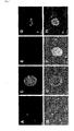
- FIG. 1 Results of observation of sphere floating in the culture solution after the culture are shown in Fig. 1 .
- A, C, E and G show photographs taken under a fluorescence microscope
- B, D, F and H show photographs under from a phase contrast microscope. Note that with the fluorescence microscope, only GFP-expressing mouse-derived spheres are observed, and with the phase contrast microscope, spheres from both the wild-type mouse and the GFP-expressing mouse are observed.
- a and B, C and D, E and F, and G and H are the same visual fields photographed respectively. From the photographs A and B, wild-type mouse-derived spheres and GFP-expressing mouse-derived spheres were shown to co-exist in the culture solution.
- the recovered sphere was placed into a 2ml of DMEM/F12Ham (manufactured by GIBCO) culture medium, which was mixed well, then, this was subjected to centrifugal separation (4°C, 1500rpm, 5 minutes), and supernatant was eliminated sufficiently. Then, 1ml of a solution of trypsin-EDTA (containing 0.05 wt.% trypsin and 0.53mM EDTA ⁇ 4Na) (manufactured by GIBCO) was added, and sphere was broken down by shaking for 20 minutes inside a 37°C constant temperature chamber to float cells forming the sphere (hereinafter, noted sphere-forming cells). Next, 500 ⁇ l of trypsin inhibitor (manufactured by Roche) was added to suspend sufficiently, and then the cell number was counted with a blood cell counting plate.
- trypsin inhibitor manufactured by Roche
- mouse expansion medium containing DMEM/F12Ham (manufactured by GIBCO), 2 wt.% B27 supplement (manufactured by GIBCO), 1 vol.% penicillin-streptomycin, 40ng/ml recombinant human basic FGF (manufactured by Promega), and 20ng/ml mouse EGF (manufactured by SIGMA)]
- mouse expansion medium containing DMEM/F12Ham (manufactured by GIBCO), 2 wt.% B27 supplement (manufactured by GIBCO), 1 vol.% penicillin-streptomycin, 40ng/ml recombinant human basic FGF (manufactured by Promega), and 20ng/ml mouse EGF (manufactured by SIGMA)
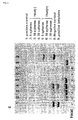
- FACS analysis was performed on the sphere-forming cells proliferated in (5) above for various cell surface antigens (Sca-1, c-kit, CD34, CD45, CD31, CD38, CD90 and CD105).
- the result obtained is shown in Fig. 2 .
- the obtained sphere-forming cells were determined to be c-kit-negative, CD31-negative and CD34-negative, and be further CD105-positive.
- the cells were also determined to be Sca-1-positive, CD45-negative, CD38-positive and CD90-positive.
- the sphere-forming cells proliferated in (5) above were also analyzed by PCR for the expression of various markers (Bmi 1, TERT, Bcrp 1, Oct 4, UTF 1, Nanog, Brachyury, Sox 2, Nestin, and Islet 1).
- the result obtained is shown in Fig. 3 .
- Oct 4 and UTF 1 which are markers of embryonic stem cell
- Brachyury which is a marker of mesoblastic stem cells
- Sox 2 and Nestin which are markers of ectodermal stem cells
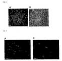
- telomerase was analyzed in the sphere obtained in (3) above.
- 5 spheres served as samples, using these with heat treatment (85°C, 15 minutes) (heat (+)) and without heat treatment (heat (-)), and further, telomer-positive cells (positive control), culture medium only (negative control) and telomer template (positive template) were also analyzed as control samples.
- the result obtained is shown in Fig. 5 .
- telomerase was strongly expressed in the sphere obtained in (3) above.
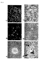
- the sphere-forming cells proliferated in (5) above were recovered by centrifugal separation, and the cells were cultured in a MEM culture medium (manufactured by GIBCO) containing 1 ⁇ 10 -8 mol/l dexamethasone and 1 vol.% penicillin-streptomycin, at 37°C, under 5% CO 2 and for 21 days. It was determined that the above-mentioned sphere-forming cells differentiate into beating cardiac myocytes by this culture. In addition, photographs used to observe the morphology of the cell in the culture is shown in Fig. 6 . As shown in Fig. 6 , the sphere-forming cells were found to proliferate and differentiate in a concentric circular shape, in the differentiation process into cardiac myocyte. Note that differentiation into cardiac myocyte was also determined from the following analytical results.
- the sphere-forming cells proliferated in (5) above were recovered by centrifugal separation, and the cells were cultured in a MEM culture medium (manufactured by GIBCO) containing 1 ⁇ 10 -8 M
- dexamethasone at 37°C, under 5% CO 2 and for 14 days.
- ⁇ -SMA smooth muscle actin
- the sphere-forming cells proliferated in (5) above were recovered by centrifugal separation, and the cells were cultured in a MEM culture medium (manufactured by GIBCO) containing 1 ⁇ 10 -8 M dexamethasone, at 37°C, under 5% CO 2 and for 14 days.
- a MEM culture medium manufactured by GIBCO
- 1 ⁇ 10 -8 M dexamethasone 1 ⁇ 10 -8 M dexamethasone
- Example 1 From the results of Example 1 shown above, the obtained sphere-forming cells were found to have self-renewal capability together with the property of differentiating into various cells, and to be pluripotent stem cells.
- mice-derived myocardial stem cells The GFP-expressing mouse-derived sphere-forming cells (pluripotent stem cells) obtained in the above Example 1 were cultured and proliferated in mouse expansion medium [containing DMEM/F12Ham (manufactured by GIBCO), 2 wt.% B27 supplement (manufactured by GIBCO), 1 vol.% penicillin-streptomycin, 40ng/ml recombinant human basic FGF (manufactured by Promega) and 20ng/ml mouse EGF (manufactured by SIGMA)].
- mouse expansion medium containing DMEM/F12Ham (manufactured by GIBCO), 2 wt.% B27 supplement (manufactured by GIBCO), 1 vol.% penicillin-streptomycin, 40ng/ml recombinant human basic FGF (manufactured by Promega) and 20ng/ml mouse EGF (manufactured by SIGMA
- the proliferated stem cells (approximately 1 ⁇ 10 6 cells) were suspended in 15 ⁇ l of PBS (-) (manufactured by GIBCO), this was transplanted using BD Ultra Fine II lancet (manufactured by Becton Dickinson) into an infarcted cardiac muscle created in a 10 to 12 weeks-old NOD/SCID mouse (purchased from Jackson Laboratory).
- the heart was extracted from the mouse 21 days after transplantation of the stem cells.
- the cardiac muscle of the extracted heart was checked for the grafting to the host cardiac muscle of the stem cells showing green fluorescence (GFP) (refer to the A in Fig. 10 ).
- GFP green fluorescence
- cTnT staining (identified in red) was performed in an identical visual field to the A in Fig. 10 (refer to the B in Fig. 10 ).
- the presence of stem cells green
- the presence of cTnI expression red
- the C and the D in Fig. 10 From the facts, it is determined that the transplanted cardiac tissue-derived stem cells differentiated into cardiac myocytes, contributing to repairing the heart.
- a culture was carried out according to the methods described in "(3) Sphere formation-1" of the above Example 1, to form a sphere.
- Photomicrographs of a sphere floating in the culture solution taken one day after and seven days after the culture are shown in Fig. 11 .
- human cardiac tissue-derived sphere-forming cells were obtained by recovering the sphere.
- the recovered sphere-forming cells were proliferated by carrying out a culture according to the methods of "(5) Proliferation of sphere-forming cell" described in the above Example 1.
- the sphere-forming cells after culture were analyzed by PCR for the expression of various markers (Rex 1, TERT, Oct 4, Nanog, Brachyury and Sox 2). The result is shown in Fig. 12 . From this result, human cardiac tissue-derived sphere-forming cells were determined to have similar differentiation properties to ectodermal stem cells and embryonic stem cells.
- the proliferated sphere-forming cells were analyzed for various cell surface antigens (c-kit, CD34, CD90 and CD105).
- the analytical result is shown in Fig. 13 . From this result, the human-derived sphere-forming cells were determined to be c-kit-negative, CD34-negative, CD90-positive and CD105-positive.
- the proliferated sphere-forming cells were induced to differentiate into cardiac myocyte was performed according to the methods of "(7) Determination of differentiation into cardiac myocyte" described in the above Example 1. This determined that the human cardiac tissue-derived sphere-forming cells differentiate into beating cardiac myocytes. Note that differentiation into cardiac myocyte was also determined from the following analytical results.
- Fig. 15 Cells at 21 days after the start of induction of differentiation were analyzed by PCR for the expression of various markers (Nkx-2.5, GATA4, ANP, ⁇ -ca-actin, TnT, MLC2v, MLC2a, ⁇ -MHC ( ⁇ -myosin heavy chain), ⁇ -MHC ( ⁇ -myosin heavy chain) and ⁇ actin).
- markers Nkx-2.5, GATA4, ANP, ⁇ -ca-actin, TnT, MLC2v, MLC2a, ⁇ -MHC ( ⁇ -myosin heavy chain), ⁇ -MHC ( ⁇ -myosin heavy chain) and ⁇ actin).
- Fig. 15 it was determined that the above various markers were expressed and the above human cardiac tissue-derived sphere-forming cells differentiated into cardiac myocytes by culturing in the presence of dexamethasone.
- the proliferated sphere-forming cells were induced to differentiate according to the methods of "(8-2) Differentiation into vascular endothelial cell" described in the above-mentioned Example 1. This determined that the human cardiac tissue-derived sphere-forming cells differentiate into smooth myocytes. Note that the differentiation into cardiac myocyte was also determined from the following analytical results.
- Fig. 17 Cells at 21 days after the start of induction of differentiation were analyzed by PCR for the expression of various markers (SM-22 ⁇ and calponin). The obtained result is shown in Fig. 17 . As is clear from Fig. 17 , it was determined that, the above markers were expressed and the above human cardiac tissue-derived sphere-forming cells differentiated into smooth myocytes, after the induction of differentiation.
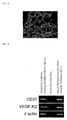
- the proliferated sphere-forming cells were induced to differentiate into endothelial cell according to the methods of "(4) Determination of differentiation into other cells" described in the above Example 1. This determined that the human cardiac tissue-derived sphere-forming cells differentiated into vascular endothelial cells. Note that the differentiation into vascular endothelial cell was also determined from the following analytical results.
- Fig. 19 Cell after induction of differentiation were analyzed by PCR for the expression of various markers (CD31 and VEGF-R2). The obtained result is shown in Fig. 19 . As is clear from Fig. 19 , the above markers were expressed and the above human cardiac tissue-derived sphere-forming cells differentiated into vascular endothelial cells, after induction of differentiation.
- Example 3 From the results of Example 3 shown above, the obtained human-derived sphere-forming cells were found to have self-renewal capability and at the same time the property of differentiating into various cells, and to be pluripotent stem cells.
- the human cardiac tissue-derived sphere-forming cells obtained in the above Example 3, were cultured and proliferated in human expansion medium [containing DMEM/F12Ham (manufactured by GIBCO), 1 vol.% penicillin-streptomycin, 40ng/ml recombinant human basic FGF (manufactured by Promega), and 20ng/ml human EGF (manufactured by SIGMA)]. Thereafter, the proliferated human cardiac tissue-derived pluripotent stem cells (approximately 1 ⁇ 10 6 cells) were transplanted into an ischemic cardiac muscle mouse by the same method of above Example 2. The heart was extracted from the mouse 21 days after transplantation of myocardial stem cells.
- human expansion medium containing DMEM/F12Ham (manufactured by GIBCO), 1 vol.% penicillin-streptomycin, 40ng/ml recombinant human basic FGF (manufactured by Promega), and 20ng/ml human EGF (
- Nuclei in the cells of the cardiac muscle of the extracted heart were stained in blue using DAPI (4'6-diamino-2-phenylindole). Furthermore, cardiac myocytes differentiated from sphere-forming cells were stained in red using human cardiac muscle-specific toroponin-T. As a result of this, it was determined that the human cardiac tissue-derived cells transplanted into the thinned infarct migrated and grafted, and mainly the endocardium side was regenerated by new cardiac myocytes (refer to A to E in Fig. 20 ). In addition, when CD31 staining was performed concomitantly on the extracted heart, it was determined that human cardiac tissue-derived cells also differentiated into vascular endothelial cells and grafted (refer to F in Fig. 20 ).
Landscapes
- Health & Medical Sciences (AREA)
- Life Sciences & Earth Sciences (AREA)
- Engineering & Computer Science (AREA)
- Biomedical Technology (AREA)
- Chemical & Material Sciences (AREA)
- Zoology (AREA)
- Organic Chemistry (AREA)
- Bioinformatics & Cheminformatics (AREA)
- Biotechnology (AREA)
- Genetics & Genomics (AREA)
- Wood Science & Technology (AREA)
- General Health & Medical Sciences (AREA)
- Cell Biology (AREA)
- Developmental Biology & Embryology (AREA)
- Microbiology (AREA)
- General Engineering & Computer Science (AREA)
- Biochemistry (AREA)
- Animal Behavior & Ethology (AREA)
- Public Health (AREA)
- Veterinary Medicine (AREA)
- Medicinal Chemistry (AREA)
- Chemical Kinetics & Catalysis (AREA)
- Epidemiology (AREA)
- Vascular Medicine (AREA)
- Rheumatology (AREA)
- Transplantation (AREA)
- Botany (AREA)
- Dermatology (AREA)
- Oral & Maxillofacial Surgery (AREA)
- Pharmacology & Pharmacy (AREA)
- Nuclear Medicine, Radiotherapy & Molecular Imaging (AREA)
- General Chemical & Material Sciences (AREA)
- Cardiology (AREA)
- Immunology (AREA)
- Virology (AREA)
- Heart & Thoracic Surgery (AREA)
- Surgery (AREA)
- Hematology (AREA)
- Urology & Nephrology (AREA)
- Micro-Organisms Or Cultivation Processes Thereof (AREA)
Applications Claiming Priority (2)
| Application Number | Priority Date | Filing Date | Title |
|---|---|---|---|
| JP2005060831 | 2005-03-04 | ||
| EP06715194A EP1857544B1 (de) | 2005-03-04 | 2006-03-03 | Aus herzgewebe stammende pluripotente stammzelle |
Related Parent Applications (2)
| Application Number | Title | Priority Date | Filing Date |
|---|---|---|---|
| EP06715194.4 Division | 2006-03-03 | ||
| EP06715194A Division EP1857544B1 (de) | 2005-03-04 | 2006-03-03 | Aus herzgewebe stammende pluripotente stammzelle |
Publications (2)
| Publication Number | Publication Date |
|---|---|
| EP2295541A1 true EP2295541A1 (de) | 2011-03-16 |
| EP2295541B1 EP2295541B1 (de) | 2016-04-27 |
Family
ID=36941306
Family Applications (2)
| Application Number | Title | Priority Date | Filing Date |
|---|---|---|---|
| EP06715194A Expired - Lifetime EP1857544B1 (de) | 2005-03-04 | 2006-03-03 | Aus herzgewebe stammende pluripotente stammzelle |
| EP10179375.0A Revoked EP2295541B1 (de) | 2005-03-04 | 2006-03-03 | Aus Herzgewebe abgeleitete pluripotente Stammzellen |
Family Applications Before (1)
| Application Number | Title | Priority Date | Filing Date |
|---|---|---|---|
| EP06715194A Expired - Lifetime EP1857544B1 (de) | 2005-03-04 | 2006-03-03 | Aus herzgewebe stammende pluripotente stammzelle |
Country Status (5)
| Country | Link |
|---|---|
| US (1) | US9867854B2 (de) |
| EP (2) | EP1857544B1 (de) |
| JP (1) | JP4783909B2 (de) |
| CA (1) | CA2600653C (de) |
| WO (1) | WO2006093276A1 (de) |
Families Citing this family (18)
| Publication number | Priority date | Publication date | Assignee | Title |
|---|---|---|---|---|
| US11660317B2 (en) | 2004-11-08 | 2023-05-30 | The Johns Hopkins University | Compositions comprising cardiosphere-derived cells for use in cell therapy |
| EP2210622B1 (de) | 2007-10-10 | 2015-07-08 | Kyoto University | Therapeutisches mittel gegen herzerkrankungen zur verwendung in der zelltransplantationstherapie |
| JP2009215191A (ja) * | 2008-03-07 | 2009-09-24 | Keio Gijuku | 神経損傷治療剤及び神経損傷治療方法 |
| US9845457B2 (en) | 2010-04-30 | 2017-12-19 | Cedars-Sinai Medical Center | Maintenance of genomic stability in cultured stem cells |
| US9884076B2 (en) | 2012-06-05 | 2018-02-06 | Capricor, Inc. | Optimized methods for generation of cardiac stem cells from cardiac tissue and their use in cardiac therapy |
| EP3563859B1 (de) | 2012-08-13 | 2021-10-13 | Cedars-Sinai Medical Center | Von kardiosphären abgeleitete exosomen zur geweberegeneration |
| EP2948543A1 (de) | 2013-01-24 | 2015-12-02 | Bernardo Nadal-Ginard | Modulation von kardialen stamm- und vorläuferzelldifferenzierung, tests und verwendungen davon |
| EP3048169B1 (de) * | 2013-09-04 | 2019-07-10 | Otsuka Pharmaceutical Factory, Inc. | Verfahren zur herstellung pluripotenter stammzellen |
| JP5924750B2 (ja) | 2014-05-01 | 2016-05-25 | iHeart Japan株式会社 | Cd82陽性心筋前駆細胞 |
| EP4494699A3 (de) | 2014-10-03 | 2025-04-30 | Cedars-Sinai Medical Center | Kardiosphären-zellen und durch diese zellen sekretierte exosomen bei der behandlung von muskeldystrophie |
| WO2017123662A1 (en) | 2016-01-11 | 2017-07-20 | Cedars-Sinai Medical Center | Cardiosphere-derived cells and exosomes secreted by such cells in the treatment of heart failure with preserved ejection fraction |
| WO2017210652A1 (en) | 2016-06-03 | 2017-12-07 | Cedars-Sinai Medical Center | Cdc-derived exosomes for treatment of ventricular tachyarrythmias |
| US11541078B2 (en) | 2016-09-20 | 2023-01-03 | Cedars-Sinai Medical Center | Cardiosphere-derived cells and their extracellular vesicles to retard or reverse aging and age-related disorders |
| JP7336769B2 (ja) | 2017-04-19 | 2023-09-01 | シーダーズ―シナイ メディカル センター | 骨格筋ジストロフィーを治療する方法及び組成物 |
| WO2019078278A1 (ja) * | 2017-10-18 | 2019-04-25 | 国立大学法人京都大学 | 心筋細胞に分化させるための多能性幹細胞の製造方法 |
| WO2019126068A1 (en) | 2017-12-20 | 2019-06-27 | Cedars-Sinai Medical Center | Engineered extracellular vesicles for enhanced tissue delivery |
| EP3749344A4 (de) | 2018-02-05 | 2022-01-26 | Cedars-Sinai Medical Center | Verfahren zur therapeutischen verwendung von exosomen und y-rnas |
| JP7373246B1 (ja) * | 2023-02-24 | 2023-11-02 | 株式会社メトセラ | 心臓内幹細胞を含む細胞集団 |
Citations (3)
| Publication number | Priority date | Publication date | Assignee | Title |
|---|---|---|---|---|
| WO2003035838A2 (en) | 2001-10-22 | 2003-05-01 | The Government Of The United States Of America, Represented By The Secretary Of The Department Of Health And Human Services | Stem cells that transform to beating cardiomyocytes |
| WO2004019767A2 (en) * | 2002-08-29 | 2004-03-11 | Baylor College Of Medicine | Heart derived cells for cardiac repair |
| EP1437406A2 (de) * | 2001-09-20 | 2004-07-14 | Kyowa Hakko Kogyo Co., Ltd. | Aus skelettmuskel-intestinalgewebe stammende pluripotente stammzellen |
Family Cites Families (2)
| Publication number | Priority date | Publication date | Assignee | Title |
|---|---|---|---|---|
| US7862810B2 (en) | 2000-07-31 | 2011-01-04 | New York Medical College | Methods and compositions for the repair and/or regeneration of damaged myocardium |
| WO2005001080A2 (en) * | 2003-06-27 | 2005-01-06 | Ethicon, Incorporated | Postpartum-derived cells for use in treatment of disease of the heart and circulatory system |
-
2006
- 2006-03-03 JP JP2007506027A patent/JP4783909B2/ja not_active Expired - Lifetime
- 2006-03-03 EP EP06715194A patent/EP1857544B1/de not_active Expired - Lifetime
- 2006-03-03 CA CA2600653A patent/CA2600653C/en not_active Expired - Lifetime
- 2006-03-03 WO PCT/JP2006/304111 patent/WO2006093276A1/ja not_active Ceased
- 2006-03-03 EP EP10179375.0A patent/EP2295541B1/de not_active Revoked
-
2015
- 2015-10-29 US US14/926,583 patent/US9867854B2/en active Active
Patent Citations (3)
| Publication number | Priority date | Publication date | Assignee | Title |
|---|---|---|---|---|
| EP1437406A2 (de) * | 2001-09-20 | 2004-07-14 | Kyowa Hakko Kogyo Co., Ltd. | Aus skelettmuskel-intestinalgewebe stammende pluripotente stammzellen |
| WO2003035838A2 (en) | 2001-10-22 | 2003-05-01 | The Government Of The United States Of America, Represented By The Secretary Of The Department Of Health And Human Services | Stem cells that transform to beating cardiomyocytes |
| WO2004019767A2 (en) * | 2002-08-29 | 2004-03-11 | Baylor College Of Medicine | Heart derived cells for cardiac repair |
Non-Patent Citations (7)
| Title |
|---|
| BELTRAMI ANTONIO P ET AL: "Adult cardiac stem cells are multipotent and support myocardial regeneration", CELL, CELL PRESS, CAMBRIDGE, NA, US, vol. 114, no. 6, 19 September 2003 (2003-09-19), pages 763 - 776, XP002304004, ISSN: 0092-8674 * |
| BELTRAMI AP.: "Adult cardiac stem cells are multipotent and support myocardial regeneration", CELL, vol. 114, 19 September 2003 (2003-09-19), pages 763 - 776 |
| HIERLIHY A M ET AL: "The post-natal heart contains a myocardial stem cell population", FEBS LETTERS, ELSEVIER, AMSTERDAM, NL, vol. 530, no. 1-3, 23 October 2002 (2002-10-23), pages 239 - 243, XP004388124, ISSN: 0014-5793 * |
| MESSINA E.: "Isolation and expansion of adult cardiac stem cells from human and murine heart", CIRC RES., vol. 95, 2004, pages 911 - 921 |
| MESSINA ELISA ET AL: "Isolation and expansion of adult cardiac stem cells from human and murine heart", CIRCULATION RESEARCH, AMERICAN HEART ASSOCIATION, INC, vol. 95, no. 9, 29 October 2004 (2004-10-29), pages 911 - 921, XP002477755, ISSN: 1524-4571 * |
| OH H.: "Cardiac progenitor cells from adult myocardium: Homing, differentiation, and fusion after infarction", PROC NATL ACAD SCI USA, vol. 100, 14 October 2003 (2003-10-14), pages 12313 - 12318 |
| TATEISHI ET AL: "Clonal Isolation and Characterization of Multipotent Adult Progenitor Cells From Mammalian Heart:Therapeutic Potential for Severe Heart Failure", JOURNAL OF CARDIAL FAILURE, CHURCHILL LIVINGSTONE, NAPERVILLE, IL, US, vol. 11, no. 9, 1 December 2005 (2005-12-01), pages S271, XP005177419, ISSN: 1071-9164 * |
Also Published As
| Publication number | Publication date |
|---|---|
| US20160228472A1 (en) | 2016-08-11 |
| WO2006093276A8 (ja) | 2007-08-23 |
| EP1857544A1 (de) | 2007-11-21 |
| US9867854B2 (en) | 2018-01-16 |
| CA2600653A1 (en) | 2006-09-08 |
| EP1857544A4 (de) | 2008-08-13 |
| EP2295541B1 (de) | 2016-04-27 |
| CA2600653C (en) | 2014-09-09 |
| JP4783909B2 (ja) | 2011-09-28 |
| WO2006093276A1 (ja) | 2006-09-08 |
| EP1857544B1 (de) | 2012-06-20 |
| JPWO2006093276A1 (ja) | 2008-08-07 |
Similar Documents
| Publication | Publication Date | Title |
|---|---|---|
| US9867854B2 (en) | Therapeutic method using cardiac tissue-derived pluripotent stem cells | |
| KR100907248B1 (ko) | 분화된 어린 지방 세포와 생분해성 중합체의 이식에 의한신체의 부피 대체 방법 | |
| JP4336821B2 (ja) | 哺乳動物の骨髄細胞または臍帯血由来細胞と脂肪組織を利用した心筋細胞の誘導 | |
| US20120034595A1 (en) | Enriched stem cell and progenitor cell populations, and methods of producing and using such populations | |
| US20080020459A1 (en) | Progenitor cell populations, expansion thereof, and growth of non-hematopoietic cell types and tissues therefrom | |
| KR20040094910A (ko) | 개선된 지방세포 분화된 지방 유래 성체 줄기세포 및 이의용도 | |
| MX2011010367A (es) | Aislamiento de celulas germinales mesenquimales derivadas de la sangre de cordon umbilical humano. | |
| JP6890549B2 (ja) | 間葉系幹細胞の製造方法 | |
| US20100310527A1 (en) | Liposome mediated delivery of lineage determining factors | |
| CN101384708B (zh) | 从成人牙周滤泡组织中收集和分离胚胎样干细胞群的方法 | |
| JP3762975B2 (ja) | 単球由来多能性細胞momc | |
| WO2007010858A1 (ja) | 骨格筋組織由来の単一細胞よりクローン化した多能性幹細胞 | |
| KR20110112164A (ko) | 인간 줄기세포의 활성을 증가시키는 방법 | |
| US20080241111A1 (en) | Pluripotent Stem Cell Derived from Cardiac Tissue | |
| KR100677054B1 (ko) | 제대혈로부터 다분화능 전구세포를 분리하여 배양하는 방법 및 이의 분화 유도방법 | |
| KR20180092523A (ko) | 중간엽 줄기세포의 연골세포로의 분화용 무혈청 배지 첨가제 조성물 및 분화 방법 | |
| WO2011126264A2 (ko) | 인간 줄기세포의 활성을 증가시키는 방법 | |
| JP2009153514A (ja) | 心筋細胞分化誘導促進剤及びその使用方法 | |
| JP2006115771A (ja) | 骨格筋由来の心筋幹細胞 | |
| KR20080094431A (ko) | 말초 혈액 유래 단핵 세포로부터 신경 전구 세포를 분리,배양 및 분화하는 방법 | |
| KR100939361B1 (ko) | 여성의 소음순 유래 성체세포로부터 리프로그래밍의 과정을통해 혈관 세포로 분화할 수 있는 혈관 줄기세포를제조하는 방법 및 상기 분화된 세포의 세포 치료제로서의용도 | |
| RU2542964C1 (ru) | Способ получения прогениторных клеток миокарда | |
| JP3928881B2 (ja) | 単球由来多能性細胞momc | |
| Graziano et al. | HUMAN CORD STROMA CELLS AFTER CO-CULTURE WITH HUMAN DENTAL PULP CELLS UNDERGO TO OSTEOGENIC DIFFERENTIATION THROUGH A BMP 2-MEDIATED PATHWAY | |
| KR20090126227A (ko) | 여성의 소음순 유래 성체세포로부터 리프로그래밍의 과정을 통해 혈관 세포로 분화할 수 있는 혈관 줄기세포를 제조하는 방법 및 상기 분화된 세포의 세포 치료제로서의 용도 |
Legal Events
| Date | Code | Title | Description |
|---|---|---|---|
| PUAI | Public reference made under article 153(3) epc to a published international application that has entered the european phase |
Free format text: ORIGINAL CODE: 0009012 |
|
| 17P | Request for examination filed |
Effective date: 20100924 |
|
| AC | Divisional application: reference to earlier application |
Ref document number: 1857544 Country of ref document: EP Kind code of ref document: P |
|
| AK | Designated contracting states |
Kind code of ref document: A1 Designated state(s): DE GB |
|
| 17Q | First examination report despatched |
Effective date: 20111026 |
|
| GRAP | Despatch of communication of intention to grant a patent |
Free format text: ORIGINAL CODE: EPIDOSNIGR1 |
|
| INTG | Intention to grant announced |
Effective date: 20151022 |
|
| GRAS | Grant fee paid |
Free format text: ORIGINAL CODE: EPIDOSNIGR3 |
|
| GRAA | (expected) grant |
Free format text: ORIGINAL CODE: 0009210 |
|
| RIN1 | Information on inventor provided before grant (corrected) |
Inventor name: MATSUBARA, HIROAKI Inventor name: TATEISHI, KENTO Inventor name: OH, HIDEMASA |
|
| AC | Divisional application: reference to earlier application |
Ref document number: 1857544 Country of ref document: EP Kind code of ref document: P |
|
| AK | Designated contracting states |
Kind code of ref document: B1 Designated state(s): DE GB |
|
| REG | Reference to a national code |
Ref country code: GB Ref legal event code: FG4D |
|
| REG | Reference to a national code |
Ref country code: DE Ref legal event code: R096 Ref document number: 602006048911 Country of ref document: DE |
|
| REG | Reference to a national code |
Ref country code: DE Ref legal event code: R026 Ref document number: 602006048911 Country of ref document: DE |
|
| PLBI | Opposition filed |
Free format text: ORIGINAL CODE: 0009260 |
|
| PLAX | Notice of opposition and request to file observation + time limit sent |
Free format text: ORIGINAL CODE: EPIDOSNOBS2 |
|
| 26 | Opposition filed |
Opponent name: STRAWMAN LIMITED Effective date: 20170127 |
|
| PLBB | Reply of patent proprietor to notice(s) of opposition received |
Free format text: ORIGINAL CODE: EPIDOSNOBS3 |
|
| PLAB | Opposition data, opponent's data or that of the opponent's representative modified |
Free format text: ORIGINAL CODE: 0009299OPPO |
|
| R26 | Opposition filed (corrected) |
Opponent name: STRAWMAN LIMITED Effective date: 20170127 |
|
| PLAB | Opposition data, opponent's data or that of the opponent's representative modified |
Free format text: ORIGINAL CODE: 0009299OPPO |
|
| R26 | Opposition filed (corrected) |
Opponent name: STRAWMAN LIMITED Effective date: 20170127 |
|
| RDAF | Communication despatched that patent is revoked |
Free format text: ORIGINAL CODE: EPIDOSNREV1 |
|
| REG | Reference to a national code |
Ref country code: DE Ref legal event code: R064 Ref document number: 602006048911 Country of ref document: DE Ref country code: DE Ref legal event code: R103 Ref document number: 602006048911 Country of ref document: DE |
|
| PGFP | Annual fee paid to national office [announced via postgrant information from national office to epo] |
Ref country code: GB Payment date: 20180228 Year of fee payment: 13 Ref country code: DE Payment date: 20180220 Year of fee payment: 13 |
|
| RDAG | Patent revoked |
Free format text: ORIGINAL CODE: 0009271 |
|
| STAA | Information on the status of an ep patent application or granted ep patent |
Free format text: STATUS: PATENT REVOKED |
|
| 27W | Patent revoked |
Effective date: 20180407 |
|
| GBPR | Gb: patent revoked under art. 102 of the ep convention designating the uk as contracting state |
Effective date: 20180407 |