EP2295975B1 - Bestimmung des Expressionsstatus des humanen epidermischen Wachstumsfaktorrezeptors 2 (HER2) in biologischen Proben - Google Patents
Bestimmung des Expressionsstatus des humanen epidermischen Wachstumsfaktorrezeptors 2 (HER2) in biologischen Proben Download PDFInfo
- Publication number
- EP2295975B1 EP2295975B1 EP10175852.2A EP10175852A EP2295975B1 EP 2295975 B1 EP2295975 B1 EP 2295975B1 EP 10175852 A EP10175852 A EP 10175852A EP 2295975 B1 EP2295975 B1 EP 2295975B1
- Authority
- EP
- European Patent Office
- Prior art keywords
- her2
- growth factor
- factor receptor
- epidermal growth
- cancer
- Prior art date
- Legal status (The legal status is an assumption and is not a legal conclusion. Google has not performed a legal analysis and makes no representation as to the accuracy of the status listed.)
- Active
Links
Images





Classifications
-
- G—PHYSICS
- G01—MEASURING; TESTING
- G01N—INVESTIGATING OR ANALYSING MATERIALS BY DETERMINING THEIR CHEMICAL OR PHYSICAL PROPERTIES
- G01N33/00—Investigating or analysing materials by specific methods not covered by groups G01N1/00 - G01N31/00
- G01N33/48—Biological material, e.g. blood, urine; Haemocytometers
- G01N33/50—Chemical analysis of biological material, e.g. blood, urine; Testing involving biospecific ligand binding methods; Immunological testing
- G01N33/53—Immunoassay; Biospecific binding assay; Materials therefor
- G01N33/575—Immunoassay; Biospecific binding assay; Materials therefor for cancer
- G01N33/5758—Immunoassay; Biospecific binding assay; Materials therefor for cancer involving compounds serving as markers for tumours, cancers or neoplasias, e.g. cellular determinants, receptors, heat shock/stress proteins, A-protein, oligosaccharides or metabolites
-
- G—PHYSICS
- G01—MEASURING; TESTING
- G01N—INVESTIGATING OR ANALYSING MATERIALS BY DETERMINING THEIR CHEMICAL OR PHYSICAL PROPERTIES
- G01N2333/00—Assays involving biological materials from specific organisms or of a specific nature
- G01N2333/435—Assays involving biological materials from specific organisms or of a specific nature from animals; from humans
- G01N2333/705—Assays involving receptors, cell surface antigens or cell surface determinants
- G01N2333/71—Assays involving receptors, cell surface antigens or cell surface determinants for growth factors; for growth regulators
-
- G—PHYSICS
- G01—MEASURING; TESTING
- G01N—INVESTIGATING OR ANALYSING MATERIALS BY DETERMINING THEIR CHEMICAL OR PHYSICAL PROPERTIES
- G01N2800/00—Detection or diagnosis of diseases
- G01N2800/52—Predicting or monitoring the response to treatment, e.g. for selection of therapy based on assay results in personalised medicine; Prognosis
-
- G—PHYSICS
- G01—MEASURING; TESTING
- G01N—INVESTIGATING OR ANALYSING MATERIALS BY DETERMINING THEIR CHEMICAL OR PHYSICAL PROPERTIES
- G01N33/00—Investigating or analysing materials by specific methods not covered by groups G01N1/00 - G01N31/00
- G01N33/48—Biological material, e.g. blood, urine; Haemocytometers
- G01N33/50—Chemical analysis of biological material, e.g. blood, urine; Testing involving biospecific ligand binding methods; Immunological testing
- G01N33/68—Chemical analysis of biological material, e.g. blood, urine; Testing involving biospecific ligand binding methods; Immunological testing involving proteins, peptides or amino acids
- G01N33/6803—General methods of protein analysis not limited to specific proteins or families of proteins
- G01N33/6848—Methods of protein analysis involving mass spectrometry
-
- G—PHYSICS
- G01—MEASURING; TESTING
- G01N—INVESTIGATING OR ANALYSING MATERIALS BY DETERMINING THEIR CHEMICAL OR PHYSICAL PROPERTIES
- G01N33/00—Investigating or analysing materials by specific methods not covered by groups G01N1/00 - G01N31/00
- G01N33/48—Biological material, e.g. blood, urine; Haemocytometers
- G01N33/50—Chemical analysis of biological material, e.g. blood, urine; Testing involving biospecific ligand binding methods; Immunological testing
- G01N33/68—Chemical analysis of biological material, e.g. blood, urine; Testing involving biospecific ligand binding methods; Immunological testing involving proteins, peptides or amino acids
- G01N33/6803—General methods of protein analysis not limited to specific proteins or families of proteins
- G01N33/6848—Methods of protein analysis involving mass spectrometry
- G01N33/6851—Methods of protein analysis involving laser desorption ionisation mass spectrometry
Definitions
- Adenocarcinoma of the breast is a leading cause of cancer morbidity and mortality among women worldwide.
- a major challenge faced by clinicians treating patients with breast cancer is how to best assess patient outcome and predict the clinical course of the disease so that the most appropriate treatment regimen can be identified.
- Determining the expression status of the human epidermal growth factor receptor 2 (HER2) in newly diagnosed breast cancer tissues is critically important for therapeutic decision making. Current guidelines recommend that the HER2 expression status should be evaluated for every patient with newly diagnosed, primary breast cancer.
- HER2 human epidermal growth factor receptor 2
- HER2 testing stand alone in determining which patients afflicted with primary breast cancer are likely to respond to trastuzumab (Herceptin ® ), a monoclonal antibody directed to HER2.
- the HER2 expression status can be determined at the DNA-, the mRNA-, or the protein level.
- Various techniques are available for assessing these different target molecules, each with benefits and drawbacks.
- trastuzumab was initially approved by the USA Food and Drug Administration (FDA)
- FDA USA Food and Drug Administration
- CA IX carbonic anhydrase IX
- HER-2 HER-2/neu/c-erbB-2
- diagnostic/prognostic and therapeutic methods in parallel with and/or alternative to those targeting HER-2.
- Particularly preferred were assays to detect both the HER-2 ectodomain ("p100") and the CA IX extracellular domain (50/54 kilodaltons) in the same body fluid sample from a cancer patient.
- CRIP1 belongs to the LIM/double zinc finger protein family, which includes cysteine- and glycine-rich protein-1, rhombotin-1, rhombotin-2, and rhombotin-3.
- Human CRIP1 primarily a cytosolic protein, was cloned in 1997 using RT-PCR of human small intestine RNA and oligonucleotides whose sequence was derived from the human heart homologue of this protein, CRHP.
- Mass spectrometry has extensively been used for investigating protein patterns in biological samples, like body fluids or homogenized tissue, to find biomarkers. Interrogation of the resulting complex mass spectrometry (MS) data sets using modem computational tools has resulted in identification of both disease-state and patient-prognosis specific protein patterns.
- biomarkers are used or at least predicted to be used for diagnosing cancer, qualifying different types of cancer, predicting the response to a cancer drug or predicting the prognosis of a cancer patient. Biomarkers can further be measured and optionally be identified with high accuracy by tandem mass spectrometry, e.g. by multiple reaction monitoring (MRM).
- MRM multiple reaction monitoring
- MALDI imaging mass spectrometry is a powerful tool for investigating protein patterns through the direct (in situ) analysis of tissue sections.
- MALDI IMS has advantage over other assay methods (i.e. those requiring homogenization), because it is morphologically driven. This allows the direct evaluation of tumor cells, correlation with other morphologic features and the ability to assay smaller patient tumor tissue specimen such as needle core biopsy specimens. This makes it a seemingly ideal tool for rapid tissue diagnostics and molecular histology.
- MALDI IMS can determine the distribution of hundreds of compounds in a single measurement without any need of labeling.
- MALDI IMS Previously published work using MALDI IMS for tumor classification suggests that this technology enables accurate classification (Yanagisawa et al., 2003; Schwartz et al., 2005). To date, MALDI IMS has been applied to multiple types of diseased tissues, including human non-small cell lung tumors, gliomas, as well as ovarian and breast cancers. From recent publications it has become clear that the integration of MALDI IMS into clinical management regarding disease diagnosis and outcome prediction will take place soon (Franck et al., 2009; Cornett et al., 2007). As MALDI IMS protein expression data will become available for various tumor tissue types, this approach will provide a common disease-wide methodology that can be applied to a variety of clinical questions. While many of these MALDI IMS studies rather focus on the identification of new biomarkers, so far only few studies have evaluated the potential of MALDI IMS for molecular classification of tissues based on protein patterns.
- the present invention provides a method as defined in claim 1.
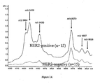
- the amount of at least one further protein is measured and compared to a control amount, the at least one further protein having a molecular weight of 4740, 8455, 8570, 8607 or 8626 atomic mass units and being related to fragments of the human epidermal growth factor receptor 2.
- the amount of at least one further protein is measured and compared to a control amount, the at least one further protein having a molecular weight of 8435, 8450 or 8465 atomic mass units and being related to variants of the human cystein-rich intestinal protein 1.
- three or more of the marker proteins are measured in step (b). Most preferably, all seven marker proteins are measured in step (a).
- the proteins of molecular weights of 4740, 8455, 8570, 8607 and 8626 atomic mass units are presumably related to fragments of higher molecular weight proteins correlated with HER2, whereas the marker protein of molecular weight of 8404 and 8419 atomic mass units are related within normal mass accuracy to the human cystein-rich intestinal protein 1 (CRIP1) and a variant thereof, respectively.
- the underivatized CRIP1_HUMAN des-Met 1 has an average molecular weight of 8402 u calculated based on the gene sequence.
- the modified molecular weight of the variant can be a result of a modification, a post-translational modification, a sequence variation, a point mutation and/or a sequence length variation.
- the variants of the human cystein-rich intestinal protein 1 have preferably one of following molecular weights 8419, 8435, 8450 or 8465 atomic mass units. These and other variations can result from one or more structural variations, wherein the human cystein-rich intestinal protein 1 is modified, e.g., by disulfide bond formation and/or one or more methyl groups and/or oxygen atoms and/or by at least one mutation. At least one of modifications and mutations can be located in the core region.
- the expression of the human epidermal growth factor receptor 2 is determined as up-regulated if amounts of CRIP1 and/or CRIP1 variants are increased compared to the control amounts, especially in the case of breast tissue.
- the HER2 oncogene encodes a 185 ku epidermal growth factor receptor-related transmembrane protein. Due to this high molecular weight, it is important to note that mass spectrometry and especially MALDI IMS are not suited to directly detect HER2 as the typical observation mass range of this technology is 2.4 to 25 ku.
- the HER2 can not be found in body fluids due to the activity of proteases.
- protein profiles of HER2 "surrogate markers" are detected. These marker proteins are either co-expressed proteins with low molecular weights, fragments of HER2 or other high molecular weight proteins, CRIP1 variants and other proteins from the HER2 signaling pathway. In contrast to gene transcription experiments, it is preferred to measure the amounts of CRIP1 and at least one CRIP1 variant or the amounts of more than one CRIP1 variant to achieve results with adequate confidence levels.
- the sample of the subject can be a body fluid, like whole blood, plasma, serum, nipple aspirate fluid, cerebrospinal fluid or urine, a homogenized or extracted tissue, a lysate of a homogenized sample or a tissue section.
- the amounts of the marker proteins can be directly measured by imaging mass spectrometry, preferably utilizing matrix assisted laser desorption/ionization.
- the amounts of marker proteins, CRIP1 and/or one or more of CRIP1 variants in a sample can be measured by mass spectrometric means and/or by tandem mass spectrometry, e.g. by multiple reaction monitoring (MRM) following state of the art procedures.
- MRM multiple reaction monitoring
- the tandem mass spectrometry further enables the identification of the measured proteins resulting in high accuracies of the methods.
- the amounts of CRIP1 and/or one or more of CRIP1 variants can be measured by an immunoassay (immunohisto-chemical analysis).
- the CRIP1 and/or one or more of CRIP1 variants can be extracted from the sample utilizing an antibody or other affinity reagents, like aptamers or affibodies, specific for CRIP1 or its variants. Hao et al.
- HER2 human epidermal growth factor receptor 2
- the preferred embodiment describes the MALDI IMS technology for determining the HER2 expression status in breast cancer tissues, Clinical laboratory testing for HER2 expression status in newly diagnosed, primary breast cancer tissues is critically important for therapeutic decision making.
- Matrix-assisted laser desorption/iomzation (MALDI) imaging mass spectrometry (IMS) is a powerful tool for investigating proteins through the direct analysis of tissue sections. Unlike immunobistochemistry (IHC), MALDI-IMS enables the acquisition of complex protein expression profiles without any labeling.
- HER2 expression status All cases were carefully reviewed before study inclusion for HER2 expression status. The discovery set was evaluated by imnunohistochemistry (IHC) and fluorescence in situ hybridization (FISH), while the validation set was evaluated by IHC only. Scoring criteria were applied according to the American Society of Clinical Oncology/College of American Pathologists guideline recommendations for HER2 testing in breast cancer (Wolff et al., 2007).
- a positive HER2 result is IHC staining of 3+ (uniform, intense membrane staining of> 30% of invasive tumor cells), a FISH result of more than six HER2 gene copies per nucleus or a FISH ratio (HER2 gene signals to chromosome 17 signals) of more than 2.2; a negative result is an IHC staining of 0 or 1+, a FISH result of less than 4.0 HER2 gene copies per nucleus or FISH ratio of less than 1.8.
- 48 frozen tissue samples of invasive ductal breast cancer patients were used and the pre-characterized HER2 expression status was confirmed again by IHC on a consecutive cryosection.
- MALDI Imaging Mass Spectrometry (MALDI IMS) experiments Samples were cryosectioned on conductive Indium-Tin-Oxide (ITO) coated glass slides (Bruker Daltonik GmbH, Bremen, Germany), briefly washed in 70% and 100% ethanol, dried under vacuum, and directly covered with matrix.
- ITO Indium-Tin-Oxide
- the MALI matrix was applied by use of the ImagePrep station (broker Daltonik GmbH, Bremen, Germany) and the standard protocol provided with the instrument, whereas sinapinic acid (Sigma-Aldrich, Taufkirchen, Germany) at 10 mg/ml in water/acetonitrile 40:60 (v/v) with 0.2% trifluoroacetic acid (TFA, Applied Biosystems, Darmstadt, Germany) was used as matrix for the MALDI measurements.
- the MALDI measurement was performed on an Ultraflex III MALDI-TOF/TOF in linear mode with a mass range of 2400-25000 u with a sampling rate of 0.1 GS/s using FlexControl 3.0 and FlexImaging 2.1 software packages (all Bruker Daltonik GmbH, Bremen, German).
- the lateral resolution for MALDI IMS was set to 200 ⁇ m and a total of 200 laser shots were accumulated per pixel at constant laser power.
- the cryosections were incubated in 70% ethanol to remove the matrix and then washed in distilled water. Subsequently, the very same section, which was measured by MALDI IMS, was stained with hematoxylin and eosin (H&E), scanned with the MIRAX ® DESK system (Carl Zeiss Micro Imaging GmbH, Göttingen, Germany), and co-registered with the MALDI IMS results to correlate mass spectrometric data with the histological features of the section.
- H&E hematoxylin and eosin
- Tumor associated spectra were selected using the FlexImaging 2.1 software (Bruker). For this purpose, we defined in every tissue section regions of interest (ROIs) that contained invasive ductal breast cancer tissue specific for HER2-positive and HER2-negative cell populations, respectively. These extracted mass spectra comprising about 300 peaks per spectrum underwent recalibration on common "background” peaks (also known as spectral alignment) and normalization based on their total ion count in the observation mass range utilizing ClinPro Tools 2.2 software (Bruker Daltonik GmbH, Bremen, Germany). An average spectrum across all spectra of an ROI was used for peak defining integration ranges. These integration ranges were used to obtain the average peak intensities of each respective ROI. Significant differences in peak intensities between both histological groups (HER2-positive and HER2-negative) were evaluated by the wilco on rank-sum test with a significance cutoff point of p ⁇ 0.05.
- R statistical software R Foundation for Statistical Computing
- SVM support vector machine
- ANN artificial neuronal network
- Both classifiers were tested internally and externally using a 10-fold cross validation and the validation set, respectively.
- the features were selected that best segregated the samples within the training set into the related groups (i.e. HER2-positive or HER2-negitive), where an MS signal was included once its area under curve (AUC) exceeded 0.8 as calculated by receiver operating characteristics (ROC) analysis.
- AUC area under curve
- ROC receiver operating characteristics
- CLUTO is a software package for clustering low- and high-dimensional datasets and for analyzing the characteristics of the various clusters.
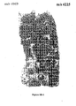
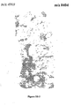
- Results In the initial discovery experiment, we analyzed 30 tumor tissue samples (15 HER2-negative samples, 15 HER2-positive samples). 1129 individual mass spectra, representing specific HER2-positive or HER2-negative tumor cell populations defined by ROIs, were used for comparative analyses. Representative example of images derived from mapping discriminating peaks are shown in Figures 1 and 3 . Another peak at m / z 4969 was expressed in tumor stroma with little or no expression seen in cancer cells ( Figures 3A-2 , 3A-3 , 3B-2 and 3B-3 ).
- the sample was measured with Maximum Scan Mode at 4600/sec, polarity positive, Scan Range from m / z 100-3000, 10 spectra averaged and rolling average of 4.
- Chosen precursor was m / z 602.4 (charge state 14 + ) with analyte ICC of 125,000 and isolation width of m / z 4.0
- MS 2 stage was ETD with reaction time of 45 ms and reactant ICC of 115,000
- MS 3 stage was PTR with reaction time of 80 ms and reactant ICC of 175,000.
- ETD/PTR spectra were processed using DataAnalysis 4.0 with the SNAPII algorithm, deconvoluted spectra were further analyzed with BioTools 3.2 (all Bruker Daltonik GmbH, Bremen, Germany) and submitted to a Mascot (Matrix Science, London, UK) database search (Mascot 2.2.04, SwissProt 56.1, taxonomy: mammals, Peptide mass tolerance +/- 10 u, fragment mass tolerance +/- 0.8 u, Instrument type ETD-Trap). The identified protein sequence was manually validated in BioTools on a residue-by residue basis using the raw data.
- Cystein-rich protein 1 (CRIP1_HUMAN) was identified by database searching with a mascot score of 126. A theoretical average molecular weight of 8402 u was calculated from the underivatized CRIP1_HUMAN des-Met 1 gene sequence in good agreement with the MALDI and ESI data. However, a cluster of CRIP1 variants at 8419, 8435, 8450 and 8464 u were determined experimentally after charge deconvolution of the multiple charge states in the ion trap ESI spectrum.
- the varying molecular weights result from one or more structural variations, wherein the human cystein-rich intestinal protein 1 is modified, e.g., by disulfide bond formation and/or one or more methyl groups and/or oxygen atoms and/or by at least one mutation.
- the MALDI IMS proteomic approach is a label-free and morphology-driven alternative for HER2 testing in breast cancer tissues and will have great implications for clinical management of breast cancer patients, and may be applicable to other cancer diseases as well. Only small amounts of fresh-frozen and unprocessed tumor tissue, easily available in a clinical setting, are needed to obtain the profiles of the marker proteins that can be used to accurately classify tumors associated with molecular features such as HER2.
- the tissue sample can be gained from core needle biopsies or can be available as a tissue microarray set.
Landscapes
- Health & Medical Sciences (AREA)
- Life Sciences & Earth Sciences (AREA)
- Immunology (AREA)
- Engineering & Computer Science (AREA)
- Urology & Nephrology (AREA)
- Biomedical Technology (AREA)
- Hematology (AREA)
- Chemical & Material Sciences (AREA)
- Molecular Biology (AREA)
- Cell Biology (AREA)
- Biotechnology (AREA)
- Microbiology (AREA)
- Food Science & Technology (AREA)
- Medicinal Chemistry (AREA)
- Physics & Mathematics (AREA)
- Analytical Chemistry (AREA)
- Biochemistry (AREA)
- General Health & Medical Sciences (AREA)
- General Physics & Mathematics (AREA)
- Pathology (AREA)
- Investigating Or Analysing Biological Materials (AREA)
- Other Investigation Or Analysis Of Materials By Electrical Means (AREA)
- Measuring Or Testing Involving Enzymes Or Micro-Organisms (AREA)
Claims (14)
- Verfahren zum Bestimmen der Expression des menschlichen epidermalen Wachstumsfaktor-Rezeptors 2 (HER2) eines Subjektes zum Bestimmen der am Besten geeigneten Brustkrebsbehandlung, umfassend:(a) Messen der Mengen von zwei Proteinen in einer Probe des Subjekts, wobei die beiden Proteine ein Molekulargewicht von 8404 atomaren Masseneinheiten und 8419 atomaren Masseneinheiten haben, wobei die genannten Molekulargewichte dem menschlichen Cystein-reichen Darmprotein 1 (CRIP1) bzw. einer strukturell modifizierten Variante des menschlichen Cystein-reichen Darmproteins 1 zugeordnet sind und(b) Vergleichen der Mengen des Proteins mit Kontrollmengen, die für die Expression des menschlichen epidermalen Wachstumsfaktor-Rezeptors 2 entscheidend sind, wobei die Expression des menschlichen epidermalen Wachstumsfaktor-Rezeptors 2 als hochreguliert bestimmt wird, falls die Mengen der zwei Proteine im Vergleich zu den Kontrollmengen erhöht sind.
- Verfahren nach Anspruch 1, wobei die Menge mindestens eines weiteren Proteins gemessen und mit einer Kontrollmenge verglichen wird, wobei das mindestens eine weitere Protein ein Molekulargewicht von 4740, 8455, 8570, 8607 oder 8626 atomaren Masseneinheiten hat und sich auf Fragmente des menschlichen epidermalen Wachstumsfaktor-Rezeptors 2 bezieht.
- Verfahren nach Anspruch 1, wobei die Menge mindestens eines weiteren Proteins gemessen und mit einer Kontrollmenge verglichen wird, wobei das mindestens eine weitere Protein ein Molekulargewicht von 8435, 8450 oder 8465 atomaren Masseneinheiten hat und sich auf Varianten des menschlichen Cystein-reichen Darmproteins 1 bezieht.
- Verfahren nach Anspruch 1, wobei die strukturelle Variation mindestens eine der Folgenden ist: in vivo oder in vitro Modifikation, posttranslationale Modifikation, Sequenz-Variation oder Punktmutation.
- Verfahren nach Anspruch 1, wobei die Mengen der zwei Proteine entweder durch ein Immunoassay oder durch Massenspektrometrie gemessen werden oder gemessen und optional mittels Tandem-Massenspektrometrie identifiziert werden.
- Verfahren nach Anspruch 1, wobei das menschliche Cystein-reiche Darmprotein 1 entweder durch eine oder mehrere strukturelle Variationen modifiziert wird, wie beispielsweise mindestens eine der Folgenden: Disulfidbindungs-Bildung, eine oder mehrere Methylgruppen, und Sauerstoffatome, oder durch mindestens eine Mutation modifiziert wird.
- Verfahren nach Anspruch 6, wobei mindestens eine der Modifikationen und Mutationen in dem Kernbereich liegt.
- Verfahren nach Anspruch 1, wobei die Probe des Subjekts entweder eine Körperflüssigkeit wie Vollblut, Plasma, Serum, Nippelaspiratflüssigkeit, Cerebrospinalflüssigkeit oder Urin, oder homogenisiertes oder extrahiertes Gewebe, wie Lysat eines homogenisierten Gewebes, oder ein Gewebeschnitt ist.
- Verfahren nach Anspruch 8, wobei der Gewebeschnitt durch Bildgebende Massenspektrometrie, wie beispielsweise Bildgebende Massenspektrometrie unter Verwendung von Matrix-unterstützter Laserdesorption/Laserionisation analysiert wird.
- Verfahren nach Anspruch 1, wobei Brustkrebs, Eierstockkrebs, Magenkrebs, Gebärmutterkrebs, Endometriumkarzinome, Prostatakrebs, Gebärmutterhalskrebs, oder Bauchspeicheldrüsenkrebs entweder festgestellt wird im Falle einer hochregulierten Expression des menschlichen epidermalen Wachstumsfaktor-Rezeptors 2 im Vergleich mit gesunden Subjekten oder durch den bestimmten Expressionsstatus des menschlichen epidermalen Wachstumsfaktor-Rezeptors 2 in Frage kommt.
- Verfahren nach Anspruch 1, wobei eine positive Auswirkung eines Medikaments im Falle einer hoch- oder herunterregulierten Expression des menschlichen epidermalen Wachstumsfaktor-Rezeptors 2 vorausgesagt wird.
- Verfahren nach Anspruch 11, wobei eine positive Auswirkung von Trastuzumab im Falle einer hochregulierten Expression des menschlichen epidermalen Wachstumsfaktor-Rezeptors 2 vorausgesagt wird.
- Verfahren nach Anspruch 1, wobei die Proteine aus der Probe extrahiert werden unter Verwendung eines Antikörpers oder anderer Affinitätsreagenzien, wie Aptamere oder Antikörper, die für CRIP1 oder seine Varianten spezifisch sind, und wobei die Mengen der zwei Proteine durch Massenspektrometrie oder Tandem-Massenspektrometrie gemessen werden.
- Verfahren nach Anspruch 9, wobei der Gewebeschnitt ein Brustkrebs-Gewebeschnitt ist.
Priority Applications (1)
| Application Number | Priority Date | Filing Date | Title |
|---|---|---|---|
| EP13182442.7A EP2674756B1 (de) | 2009-09-10 | 2010-09-08 | Bestimmung des Expressionsstatus des humanen epidermischen Wachstumsfaktorrezeptors 2 (HER2) in biologischen Proben |
Applications Claiming Priority (1)
| Application Number | Priority Date | Filing Date | Title |
|---|---|---|---|
| US24125309P | 2009-09-10 | 2009-09-10 |
Related Child Applications (2)
| Application Number | Title | Priority Date | Filing Date |
|---|---|---|---|
| EP13182442.7A Division EP2674756B1 (de) | 2009-09-10 | 2010-09-08 | Bestimmung des Expressionsstatus des humanen epidermischen Wachstumsfaktorrezeptors 2 (HER2) in biologischen Proben |
| EP13182442.7A Division-Into EP2674756B1 (de) | 2009-09-10 | 2010-09-08 | Bestimmung des Expressionsstatus des humanen epidermischen Wachstumsfaktorrezeptors 2 (HER2) in biologischen Proben |
Publications (2)
| Publication Number | Publication Date |
|---|---|
| EP2295975A1 EP2295975A1 (de) | 2011-03-16 |
| EP2295975B1 true EP2295975B1 (de) | 2014-06-18 |
Family
ID=42740308
Family Applications (2)
| Application Number | Title | Priority Date | Filing Date |
|---|---|---|---|
| EP10175852.2A Active EP2295975B1 (de) | 2009-09-10 | 2010-09-08 | Bestimmung des Expressionsstatus des humanen epidermischen Wachstumsfaktorrezeptors 2 (HER2) in biologischen Proben |
| EP13182442.7A Active EP2674756B1 (de) | 2009-09-10 | 2010-09-08 | Bestimmung des Expressionsstatus des humanen epidermischen Wachstumsfaktorrezeptors 2 (HER2) in biologischen Proben |
Family Applications After (1)
| Application Number | Title | Priority Date | Filing Date |
|---|---|---|---|
| EP13182442.7A Active EP2674756B1 (de) | 2009-09-10 | 2010-09-08 | Bestimmung des Expressionsstatus des humanen epidermischen Wachstumsfaktorrezeptors 2 (HER2) in biologischen Proben |
Country Status (2)
| Country | Link |
|---|---|
| US (1) | US9164098B2 (de) |
| EP (2) | EP2295975B1 (de) |
Families Citing this family (21)
| Publication number | Priority date | Publication date | Assignee | Title |
|---|---|---|---|---|
| JP6800875B2 (ja) | 2015-03-06 | 2020-12-16 | マイクロマス ユーケー リミテッド | 急速蒸発イオン化質量分析(「reims」)装置に連結されたイオンアナライザのための流入器具 |
| EP3264989B1 (de) | 2015-03-06 | 2023-12-20 | Micromass UK Limited | Spektrometrische analyse |
| EP3265822B1 (de) | 2015-03-06 | 2021-04-28 | Micromass UK Limited | Gewebeanalyse mittels massenspektrometrie oder ionenmobilitätsspektrometrie |
| CA2977900A1 (en) | 2015-03-06 | 2016-09-15 | Micromass Uk Limited | Collision surface for improved ionisation |
| CN107533032A (zh) | 2015-03-06 | 2018-01-02 | 英国质谱公司 | 用于从块状组织直接映射的原位电离质谱测定成像平台 |
| CA2978165A1 (en) | 2015-03-06 | 2016-09-15 | Micromass Uk Limited | Improved ionisation of gaseous samples |
| US11139156B2 (en) | 2015-03-06 | 2021-10-05 | Micromass Uk Limited | In vivo endoscopic tissue identification tool |
| US11037774B2 (en) | 2015-03-06 | 2021-06-15 | Micromass Uk Limited | Physically guided rapid evaporative ionisation mass spectrometry (“REIMS”) |
| US11031222B2 (en) | 2015-03-06 | 2021-06-08 | Micromass Uk Limited | Chemically guided ambient ionisation mass spectrometry |
| GB2554206B (en) | 2015-03-06 | 2021-03-24 | Micromass Ltd | Spectrometric analysis of microbes |
| GB2556436B (en) | 2015-03-06 | 2022-01-26 | Micromass Ltd | Cell population analysis |
| US10978284B2 (en) | 2015-03-06 | 2021-04-13 | Micromass Uk Limited | Imaging guided ambient ionisation mass spectrometry |
| WO2016142691A1 (en) * | 2015-03-06 | 2016-09-15 | Micromass Uk Limited | Rapid evaporative ionisation mass spectrometry ("reims") and desorption electrospray ionisation mass spectrometry ("desi-ms") analysis of swabs and biopsy samples |
| GB2551294B (en) | 2015-03-06 | 2021-03-17 | Micromass Ltd | Liquid trap or separator for electrosurgical applications |
| GB201517195D0 (en) | 2015-09-29 | 2015-11-11 | Micromass Ltd | Capacitively coupled reims technique and optically transparent counter electrode |
| CN106018813A (zh) * | 2015-11-15 | 2016-10-12 | 陈博 | 一种唇癌特异性检测的试剂盒 |
| WO2017178833A1 (en) | 2016-04-14 | 2017-10-19 | Micromass Uk Limited | Spectrometric analysis of plants |
| US10950424B2 (en) | 2017-09-25 | 2021-03-16 | Bruker Daltonik, Gmbh | Method for monitoring the quality of mass spectrometric imaging preparation workflows |
| EP3894865A1 (de) * | 2018-12-11 | 2021-10-20 | Helmholtz Zentrum München Deutsches Forschungszentrum für Gesundheit und Umwelt GmbH | In-vitro-verfahren zur diagnose und prognose einer krankheit |
| CA3163492A1 (en) * | 2019-12-12 | 2021-06-17 | Tempus Labs, Inc. | Real-world evidence of diagnostic testing and treatment patterns in u.s. breast cancer patients |
| CN120629574B (zh) * | 2025-08-01 | 2025-10-14 | 吉林大学 | 一种快速免疫组化双染试剂盒的制备方法及其应用 |
Family Cites Families (3)
| Publication number | Priority date | Publication date | Assignee | Title |
|---|---|---|---|---|
| DE10063112A1 (de) * | 2000-12-18 | 2002-06-20 | Bayer Ag | Verfahren zur Erhöhung der klinischen Spezifität bei der Detektion von Tumoren und ihren Vorstufen durch simultane Messung von mindestens zwei verschiedenen molekularen Markern |
| HUE026218T2 (en) * | 2002-02-21 | 2016-05-30 | Inst Virology | MN / CA IX-Specific Monoclonal Antibodies and Methods for MN / CA IX Deficient Mice |
| US7811778B2 (en) * | 2006-09-06 | 2010-10-12 | Vanderbilt University | Methods of screening for gastrointestinal cancer |
-
2010
- 2010-09-08 EP EP10175852.2A patent/EP2295975B1/de active Active
- 2010-09-08 EP EP13182442.7A patent/EP2674756B1/de active Active
- 2010-09-10 US US12/879,619 patent/US9164098B2/en active Active
Also Published As
| Publication number | Publication date |
|---|---|
| US20110059554A1 (en) | 2011-03-10 |
| EP2674756B1 (de) | 2016-08-03 |
| EP2295975A1 (de) | 2011-03-16 |
| US9164098B2 (en) | 2015-10-20 |
| EP2674756A1 (de) | 2013-12-18 |
Similar Documents
| Publication | Publication Date | Title |
|---|---|---|
| EP2295975B1 (de) | Bestimmung des Expressionsstatus des humanen epidermischen Wachstumsfaktorrezeptors 2 (HER2) in biologischen Proben | |
| Frantzi et al. | Clinical proteomic biomarkers: relevant issues on study design & technical considerations in biomarker development | |
| Maes et al. | Proteomics in cancer research: Are we ready for clinical practice? | |
| Daily et al. | Using tears as a non-invasive source for early detection of breast cancer | |
| US20150079078A1 (en) | Biomarkers for triple negative breast cancer | |
| KR101760464B1 (ko) | 교모세포종의 진단 또는 예후 예측용 바이오 마커 및 그 용도 | |
| WO2008064336A9 (en) | Autoimmune disease biomarkers | |
| CA2827115C (en) | COMPOSITIONS AND DIAGNOSTIC METHODS FOR OVARIAN CANCER | |
| KR20230080442A (ko) | 폐암의 검출 및 치료를 위한 방법 | |
| CN106461647A (zh) | 用于检测结肠直肠肿瘤的蛋白质生物标志物谱 | |
| CN116735889B (zh) | 一种用于结直肠癌早期筛查的蛋白质标志物、试剂盒及应用 | |
| US20110207156A1 (en) | Biomarkers for Diagnosis of Breast Cancer | |
| US20050100967A1 (en) | Detection of endometrial pathology | |
| WO2025112150A1 (zh) | 基于血清蛋白的乳腺肿瘤筛查的标志物及应用 | |
| Piga et al. | Paving the path toward multi-omics approaches in the diagnostic challenges faced in thyroid pathology | |
| Gromov et al. | Proteomic analysis of tissue samples in translational breast cancer research | |
| Yang et al. | Proteomic profiling of invasive ductal carcinoma (IDC) using magnetic beads‐based serum fractionation and MALDI‐TOF MS | |
| US20150338412A1 (en) | Composition for diagnosis of lung cancer and diagnosis kit for lung cancer | |
| EP4357782A1 (de) | Protein-biomarker-panel für die diagnose von darmkrebs | |
| US20070249000A1 (en) | Method for diagnosing a person having b-cell pathologies | |
| BR112021002774A2 (pt) | composições para avaliação do câncer de ovário | |
| CN119968213A (zh) | 用于检测和治疗卵巢癌的方法 | |
| Rizk et al. | Detection of epithelial ovarian cancer using C8magnetic bead separation and MALDI-TOF plasma proteome profiling in Egyptian females | |
| US20070184511A1 (en) | Method for Diagnosing a Person Having Sjogren's Syndrome | |
| Thomsen et al. | Mass spectrometry and machine learning for classification and molecular phenotyping of renal cell carcinoma and benign tumors |
Legal Events
| Date | Code | Title | Description |
|---|---|---|---|
| PUAI | Public reference made under article 153(3) epc to a published international application that has entered the european phase |
Free format text: ORIGINAL CODE: 0009012 |
|
| AK | Designated contracting states |
Kind code of ref document: A1 Designated state(s): AL AT BE BG CH CY CZ DE DK EE ES FI FR GB GR HR HU IE IS IT LI LT LU LV MC MK MT NL NO PL PT RO SE SI SK SM TR |
|
| AX | Request for extension of the european patent |
Extension state: BA ME RS |
|
| 17P | Request for examination filed |
Effective date: 20110914 |
|
| 17Q | First examination report despatched |
Effective date: 20111104 |
|
| GRAP | Despatch of communication of intention to grant a patent |
Free format text: ORIGINAL CODE: EPIDOSNIGR1 |
|
| INTG | Intention to grant announced |
Effective date: 20131218 |
|
| RIN1 | Information on inventor provided before grant (corrected) |
Inventor name: DEININGER, SOEREN-OLIVER Inventor name: BELAU, ECKHARD Inventor name: RAUSER, SANDRA Inventor name: ALBERS, CHRISTIAN Inventor name: WALCH, AXEL Inventor name: SUCKAU, DETLEF |
|
| GRAS | Grant fee paid |
Free format text: ORIGINAL CODE: EPIDOSNIGR3 |
|
| GRAA | (expected) grant |
Free format text: ORIGINAL CODE: 0009210 |
|
| AK | Designated contracting states |
Kind code of ref document: B1 Designated state(s): AL AT BE BG CH CY CZ DE DK EE ES FI FR GB GR HR HU IE IS IT LI LT LU LV MC MK MT NL NO PL PT RO SE SI SK SM TR |
|
| REG | Reference to a national code |
Ref country code: GB Ref legal event code: FG4D |
|
| REG | Reference to a national code |
Ref country code: CH Ref legal event code: EP |
|
| REG | Reference to a national code |
Ref country code: AT Ref legal event code: REF Ref document number: 673634 Country of ref document: AT Kind code of ref document: T Effective date: 20140715 |
|
| REG | Reference to a national code |
Ref country code: IE Ref legal event code: FG4D |
|
| REG | Reference to a national code |
Ref country code: DE Ref legal event code: R096 Ref document number: 602010016745 Country of ref document: DE Effective date: 20140731 |
|
| PG25 | Lapsed in a contracting state [announced via postgrant information from national office to epo] |
Ref country code: FI Free format text: LAPSE BECAUSE OF FAILURE TO SUBMIT A TRANSLATION OF THE DESCRIPTION OR TO PAY THE FEE WITHIN THE PRESCRIBED TIME-LIMIT Effective date: 20140618 Ref country code: NO Free format text: LAPSE BECAUSE OF FAILURE TO SUBMIT A TRANSLATION OF THE DESCRIPTION OR TO PAY THE FEE WITHIN THE PRESCRIBED TIME-LIMIT Effective date: 20140918 Ref country code: GR Free format text: LAPSE BECAUSE OF FAILURE TO SUBMIT A TRANSLATION OF THE DESCRIPTION OR TO PAY THE FEE WITHIN THE PRESCRIBED TIME-LIMIT Effective date: 20140919 Ref country code: CY Free format text: LAPSE BECAUSE OF FAILURE TO SUBMIT A TRANSLATION OF THE DESCRIPTION OR TO PAY THE FEE WITHIN THE PRESCRIBED TIME-LIMIT Effective date: 20140618 Ref country code: LT Free format text: LAPSE BECAUSE OF FAILURE TO SUBMIT A TRANSLATION OF THE DESCRIPTION OR TO PAY THE FEE WITHIN THE PRESCRIBED TIME-LIMIT Effective date: 20140618 |
|
| REG | Reference to a national code |
Ref country code: NL Ref legal event code: VDEP Effective date: 20140618 |
|
| REG | Reference to a national code |
Ref country code: AT Ref legal event code: MK05 Ref document number: 673634 Country of ref document: AT Kind code of ref document: T Effective date: 20140618 |
|
| REG | Reference to a national code |
Ref country code: LT Ref legal event code: MG4D |
|
| PG25 | Lapsed in a contracting state [announced via postgrant information from national office to epo] |
Ref country code: HR Free format text: LAPSE BECAUSE OF FAILURE TO SUBMIT A TRANSLATION OF THE DESCRIPTION OR TO PAY THE FEE WITHIN THE PRESCRIBED TIME-LIMIT Effective date: 20140618 Ref country code: SE Free format text: LAPSE BECAUSE OF FAILURE TO SUBMIT A TRANSLATION OF THE DESCRIPTION OR TO PAY THE FEE WITHIN THE PRESCRIBED TIME-LIMIT Effective date: 20140618 Ref country code: LV Free format text: LAPSE BECAUSE OF FAILURE TO SUBMIT A TRANSLATION OF THE DESCRIPTION OR TO PAY THE FEE WITHIN THE PRESCRIBED TIME-LIMIT Effective date: 20140618 |
|
| PG25 | Lapsed in a contracting state [announced via postgrant information from national office to epo] |
Ref country code: EE Free format text: LAPSE BECAUSE OF FAILURE TO SUBMIT A TRANSLATION OF THE DESCRIPTION OR TO PAY THE FEE WITHIN THE PRESCRIBED TIME-LIMIT Effective date: 20140618 Ref country code: RO Free format text: LAPSE BECAUSE OF FAILURE TO SUBMIT A TRANSLATION OF THE DESCRIPTION OR TO PAY THE FEE WITHIN THE PRESCRIBED TIME-LIMIT Effective date: 20140618 Ref country code: ES Free format text: LAPSE BECAUSE OF FAILURE TO SUBMIT A TRANSLATION OF THE DESCRIPTION OR TO PAY THE FEE WITHIN THE PRESCRIBED TIME-LIMIT Effective date: 20140618 Ref country code: SK Free format text: LAPSE BECAUSE OF FAILURE TO SUBMIT A TRANSLATION OF THE DESCRIPTION OR TO PAY THE FEE WITHIN THE PRESCRIBED TIME-LIMIT Effective date: 20140618 Ref country code: PT Free format text: LAPSE BECAUSE OF FAILURE TO SUBMIT A TRANSLATION OF THE DESCRIPTION OR TO PAY THE FEE WITHIN THE PRESCRIBED TIME-LIMIT Effective date: 20141020 Ref country code: CZ Free format text: LAPSE BECAUSE OF FAILURE TO SUBMIT A TRANSLATION OF THE DESCRIPTION OR TO PAY THE FEE WITHIN THE PRESCRIBED TIME-LIMIT Effective date: 20140618 |
|
| PG25 | Lapsed in a contracting state [announced via postgrant information from national office to epo] |
Ref country code: IS Free format text: LAPSE BECAUSE OF FAILURE TO SUBMIT A TRANSLATION OF THE DESCRIPTION OR TO PAY THE FEE WITHIN THE PRESCRIBED TIME-LIMIT Effective date: 20141018 Ref country code: AT Free format text: LAPSE BECAUSE OF FAILURE TO SUBMIT A TRANSLATION OF THE DESCRIPTION OR TO PAY THE FEE WITHIN THE PRESCRIBED TIME-LIMIT Effective date: 20140618 Ref country code: NL Free format text: LAPSE BECAUSE OF FAILURE TO SUBMIT A TRANSLATION OF THE DESCRIPTION OR TO PAY THE FEE WITHIN THE PRESCRIBED TIME-LIMIT Effective date: 20140618 Ref country code: PL Free format text: LAPSE BECAUSE OF FAILURE TO SUBMIT A TRANSLATION OF THE DESCRIPTION OR TO PAY THE FEE WITHIN THE PRESCRIBED TIME-LIMIT Effective date: 20140618 |
|
| REG | Reference to a national code |
Ref country code: DE Ref legal event code: R097 Ref document number: 602010016745 Country of ref document: DE |
|
| PLBE | No opposition filed within time limit |
Free format text: ORIGINAL CODE: 0009261 |
|
| STAA | Information on the status of an ep patent application or granted ep patent |
Free format text: STATUS: NO OPPOSITION FILED WITHIN TIME LIMIT |
|
| PG25 | Lapsed in a contracting state [announced via postgrant information from national office to epo] |
Ref country code: DK Free format text: LAPSE BECAUSE OF FAILURE TO SUBMIT A TRANSLATION OF THE DESCRIPTION OR TO PAY THE FEE WITHIN THE PRESCRIBED TIME-LIMIT Effective date: 20140618 Ref country code: LU Free format text: LAPSE BECAUSE OF FAILURE TO SUBMIT A TRANSLATION OF THE DESCRIPTION OR TO PAY THE FEE WITHIN THE PRESCRIBED TIME-LIMIT Effective date: 20140908 Ref country code: IT Free format text: LAPSE BECAUSE OF FAILURE TO SUBMIT A TRANSLATION OF THE DESCRIPTION OR TO PAY THE FEE WITHIN THE PRESCRIBED TIME-LIMIT Effective date: 20140618 |
|
| REG | Reference to a national code |
Ref country code: CH Ref legal event code: PL |
|
| 26N | No opposition filed |
Effective date: 20150319 |
|
| REG | Reference to a national code |
Ref country code: IE Ref legal event code: MM4A |
|
| PG25 | Lapsed in a contracting state [announced via postgrant information from national office to epo] |
Ref country code: SI Free format text: LAPSE BECAUSE OF FAILURE TO SUBMIT A TRANSLATION OF THE DESCRIPTION OR TO PAY THE FEE WITHIN THE PRESCRIBED TIME-LIMIT Effective date: 20140618 Ref country code: CH Free format text: LAPSE BECAUSE OF NON-PAYMENT OF DUE FEES Effective date: 20140930 Ref country code: LI Free format text: LAPSE BECAUSE OF NON-PAYMENT OF DUE FEES Effective date: 20140930 |
|
| PG25 | Lapsed in a contracting state [announced via postgrant information from national office to epo] |
Ref country code: IE Free format text: LAPSE BECAUSE OF NON-PAYMENT OF DUE FEES Effective date: 20140908 |
|
| PG25 | Lapsed in a contracting state [announced via postgrant information from national office to epo] |
Ref country code: SM Free format text: LAPSE BECAUSE OF FAILURE TO SUBMIT A TRANSLATION OF THE DESCRIPTION OR TO PAY THE FEE WITHIN THE PRESCRIBED TIME-LIMIT Effective date: 20140618 |
|
| PG25 | Lapsed in a contracting state [announced via postgrant information from national office to epo] |
Ref country code: BG Free format text: LAPSE BECAUSE OF FAILURE TO SUBMIT A TRANSLATION OF THE DESCRIPTION OR TO PAY THE FEE WITHIN THE PRESCRIBED TIME-LIMIT Effective date: 20140618 Ref country code: MT Free format text: LAPSE BECAUSE OF FAILURE TO SUBMIT A TRANSLATION OF THE DESCRIPTION OR TO PAY THE FEE WITHIN THE PRESCRIBED TIME-LIMIT Effective date: 20140618 Ref country code: MC Free format text: LAPSE BECAUSE OF NON-PAYMENT OF DUE FEES Effective date: 20140618 |
|
| PG25 | Lapsed in a contracting state [announced via postgrant information from national office to epo] |
Ref country code: BE Free format text: LAPSE BECAUSE OF FAILURE TO SUBMIT A TRANSLATION OF THE DESCRIPTION OR TO PAY THE FEE WITHIN THE PRESCRIBED TIME-LIMIT Effective date: 20140618 Ref country code: TR Free format text: LAPSE BECAUSE OF FAILURE TO SUBMIT A TRANSLATION OF THE DESCRIPTION OR TO PAY THE FEE WITHIN THE PRESCRIBED TIME-LIMIT Effective date: 20140618 Ref country code: HU Free format text: LAPSE BECAUSE OF FAILURE TO SUBMIT A TRANSLATION OF THE DESCRIPTION OR TO PAY THE FEE WITHIN THE PRESCRIBED TIME-LIMIT; INVALID AB INITIO Effective date: 20100908 |
|
| REG | Reference to a national code |
Ref country code: FR Ref legal event code: PLFP Year of fee payment: 7 |
|
| REG | Reference to a national code |
Ref country code: FR Ref legal event code: PLFP Year of fee payment: 8 |
|
| PG25 | Lapsed in a contracting state [announced via postgrant information from national office to epo] |
Ref country code: MK Free format text: LAPSE BECAUSE OF FAILURE TO SUBMIT A TRANSLATION OF THE DESCRIPTION OR TO PAY THE FEE WITHIN THE PRESCRIBED TIME-LIMIT Effective date: 20140618 |
|
| REG | Reference to a national code |
Ref country code: FR Ref legal event code: PLFP Year of fee payment: 9 |
|
| PG25 | Lapsed in a contracting state [announced via postgrant information from national office to epo] |
Ref country code: AL Free format text: LAPSE BECAUSE OF FAILURE TO SUBMIT A TRANSLATION OF THE DESCRIPTION OR TO PAY THE FEE WITHIN THE PRESCRIBED TIME-LIMIT Effective date: 20140618 |
|
| REG | Reference to a national code |
Ref country code: GB Ref legal event code: 732E Free format text: REGISTERED BETWEEN 20210902 AND 20210908 |
|
| REG | Reference to a national code |
Ref country code: DE Ref legal event code: R081 Ref document number: 602010016745 Country of ref document: DE Owner name: BRUKER DALTONICS GMBH & CO. KG, DE Free format text: FORMER OWNER: BRUKER DALTONIK GMBH, 28359 BREMEN, DE |
|
| P01 | Opt-out of the competence of the unified patent court (upc) registered |
Effective date: 20231221 |
|
| PGFP | Annual fee paid to national office [announced via postgrant information from national office to epo] |
Ref country code: DE Payment date: 20250919 Year of fee payment: 16 |
|
| PGFP | Annual fee paid to national office [announced via postgrant information from national office to epo] |
Ref country code: GB Payment date: 20250918 Year of fee payment: 16 |
|
| PGFP | Annual fee paid to national office [announced via postgrant information from national office to epo] |
Ref country code: FR Payment date: 20250919 Year of fee payment: 16 |
|
| REG | Reference to a national code |
Ref country code: DE Ref legal event code: R079 Ref document number: 602010016745 Country of ref document: DE Free format text: PREVIOUS MAIN CLASS: G01N0033574000 Ipc: G01N0033575000 |