EP2310491B1 - Modules de tissus auto-assemblés - Google Patents
Modules de tissus auto-assemblés Download PDFInfo
- Publication number
- EP2310491B1 EP2310491B1 EP09766884.2A EP09766884A EP2310491B1 EP 2310491 B1 EP2310491 B1 EP 2310491B1 EP 09766884 A EP09766884 A EP 09766884A EP 2310491 B1 EP2310491 B1 EP 2310491B1
- Authority
- EP
- European Patent Office
- Prior art keywords
- cells
- aggregates
- supracellular
- tissue
- microwells
- Prior art date
- Legal status (The legal status is an assumption and is not a legal conclusion. Google has not performed a legal analysis and makes no representation as to the accuracy of the status listed.)
- Active
Links
Images


Classifications
-
- C—CHEMISTRY; METALLURGY
- C12—BIOCHEMISTRY; BEER; SPIRITS; WINE; VINEGAR; MICROBIOLOGY; ENZYMOLOGY; MUTATION OR GENETIC ENGINEERING
- C12N—MICROORGANISMS OR ENZYMES; COMPOSITIONS THEREOF; PROPAGATING, PRESERVING, OR MAINTAINING MICROORGANISMS; MUTATION OR GENETIC ENGINEERING; CULTURE MEDIA
- C12N5/00—Undifferentiated human, animal or plant cells, e.g. cell lines; Tissues; Cultivation or maintenance thereof; Culture media therefor
- C12N5/0062—General methods for three-dimensional culture
-
- C—CHEMISTRY; METALLURGY
- C12—BIOCHEMISTRY; BEER; SPIRITS; WINE; VINEGAR; MICROBIOLOGY; ENZYMOLOGY; MUTATION OR GENETIC ENGINEERING
- C12N—MICROORGANISMS OR ENZYMES; COMPOSITIONS THEREOF; PROPAGATING, PRESERVING, OR MAINTAINING MICROORGANISMS; MUTATION OR GENETIC ENGINEERING; CULTURE MEDIA
- C12N5/00—Undifferentiated human, animal or plant cells, e.g. cell lines; Tissues; Cultivation or maintenance thereof; Culture media therefor
- C12N5/06—Animal cells or tissues; Human cells or tissues
- C12N5/0602—Vertebrate cells
- C12N5/0652—Cells of skeletal and connective tissues; Mesenchyme
- C12N5/0662—Stem cells
- C12N5/0663—Bone marrow mesenchymal stem cells (BM-MSC)
-
- C—CHEMISTRY; METALLURGY
- C12—BIOCHEMISTRY; BEER; SPIRITS; WINE; VINEGAR; MICROBIOLOGY; ENZYMOLOGY; MUTATION OR GENETIC ENGINEERING
- C12N—MICROORGANISMS OR ENZYMES; COMPOSITIONS THEREOF; PROPAGATING, PRESERVING, OR MAINTAINING MICROORGANISMS; MUTATION OR GENETIC ENGINEERING; CULTURE MEDIA
- C12N5/00—Undifferentiated human, animal or plant cells, e.g. cell lines; Tissues; Cultivation or maintenance thereof; Culture media therefor
- C12N5/06—Animal cells or tissues; Human cells or tissues
- C12N5/0602—Vertebrate cells
- C12N5/069—Vascular Endothelial cells
-
- C—CHEMISTRY; METALLURGY
- C12—BIOCHEMISTRY; BEER; SPIRITS; WINE; VINEGAR; MICROBIOLOGY; ENZYMOLOGY; MUTATION OR GENETIC ENGINEERING
- C12N—MICROORGANISMS OR ENZYMES; COMPOSITIONS THEREOF; PROPAGATING, PRESERVING, OR MAINTAINING MICROORGANISMS; MUTATION OR GENETIC ENGINEERING; CULTURE MEDIA
- C12N5/00—Undifferentiated human, animal or plant cells, e.g. cell lines; Tissues; Cultivation or maintenance thereof; Culture media therefor
- C12N5/06—Animal cells or tissues; Human cells or tissues
- C12N5/0602—Vertebrate cells
- C12N5/069—Vascular Endothelial cells
- C12N5/0691—Vascular smooth muscle cells; 3D culture thereof, e.g. models of blood vessels
-
- C—CHEMISTRY; METALLURGY
- C12—BIOCHEMISTRY; BEER; SPIRITS; WINE; VINEGAR; MICROBIOLOGY; ENZYMOLOGY; MUTATION OR GENETIC ENGINEERING
- C12N—MICROORGANISMS OR ENZYMES; COMPOSITIONS THEREOF; PROPAGATING, PRESERVING, OR MAINTAINING MICROORGANISMS; MUTATION OR GENETIC ENGINEERING; CULTURE MEDIA
- C12N2513/00—3D culture
-
- C—CHEMISTRY; METALLURGY
- C12—BIOCHEMISTRY; BEER; SPIRITS; WINE; VINEGAR; MICROBIOLOGY; ENZYMOLOGY; MUTATION OR GENETIC ENGINEERING
- C12N—MICROORGANISMS OR ENZYMES; COMPOSITIONS THEREOF; PROPAGATING, PRESERVING, OR MAINTAINING MICROORGANISMS; MUTATION OR GENETIC ENGINEERING; CULTURE MEDIA
- C12N2533/00—Supports or coatings for cell culture, characterised by material
- C12N2533/50—Proteins
- C12N2533/52—Fibronectin; Laminin
-
- C—CHEMISTRY; METALLURGY
- C12—BIOCHEMISTRY; BEER; SPIRITS; WINE; VINEGAR; MICROBIOLOGY; ENZYMOLOGY; MUTATION OR GENETIC ENGINEERING
- C12N—MICROORGANISMS OR ENZYMES; COMPOSITIONS THEREOF; PROPAGATING, PRESERVING, OR MAINTAINING MICROORGANISMS; MUTATION OR GENETIC ENGINEERING; CULTURE MEDIA
- C12N2533/00—Supports or coatings for cell culture, characterised by material
- C12N2533/50—Proteins
- C12N2533/54—Collagen; Gelatin
Definitions
- the disclosure provides a new approach to constructing cellular aggregates of defined sizes and shapes in vitro and their use in methods for producing 3D-tissue constructs in a modular way.
- tissues consist of multiple cell types organized with specific microscale heterogeneity. Typically, one cubic centimeter can hold up to 300 million cells. These cells form different structures within the tissue, including blood capillaries and a neural network, which are crucial for nutrition, innervation and homeostasis of the tissue. Cells organize and interact in a multitude of architectures and synthesize a variety of biologically active molecules for mechanical support and cellular instruction. Therefore, living tissues are highly complex.
- Tissue engineering is a term used for attempts to produce living tissue in vitro from individual or groups of cells. It aims at repairing or replacing portions of or whole tissues and provides solutions to shortage of organ donation or to the use of experimental animals for testing new therapies.
- tissue engineering 2007, 13(8), 2087-2095 describe a method to form cellular aggregates by self-assembly on micromolded non-adhesive hydrogels. This document does not describe the formation of pre-condensed cellular aggregates in a first step and subsequent self-assembly of the cellular aggregates as building blocks in a second step. This method thus induces intense and non-predictable remodeling (e.g . shrinking) of the tissue construct.
- the invention aims at overcoming one or more of these problems by producing supracellular aggregates of cells of any cell type using spatial confinement. These aggregates are used as building blocks and combined using boundary conditions promoting their self-assembly and self-organization to create complex multicellular architectures.
- the disclosure provides a method for in vitro producing a tissue construct comprising:
- the disclosure also provides various advantages over prior art methods, including the use of simple tools that can be handled in most biology labs, the ability to generate a very large amount of aggregates in quick and simple procedure (e.g . 220 000 aggregates per conventional 12 well plate), and the absence of a hydrogel as supporting material.
- the spatial confinement can be achieved in various manners.
- a well-known and often applied way is by using arrays of microwells.
- Other ways of imposing spatial confinement include using air-liquid interfaces like the Hanging drop method or microfluidic channels.
- Any biocompatible, processable material can be used for the spatial confinement applied for assembling the cells into supracellular aggregates.
- microwell as used in this application is meant to refer to an array of numerous cup-like structures formed in a substantially uniform layer of material by photolithographic patterning, molding, embossing or other manufacturing processes.
- Each microwell thus includes a lower wall (which may be formed by a substrate on which the microwell material is deposited) and one or more peripheral side walls (e.g. a single circular wall, or three or more contiguous substantially straight walls) that extend upward from the bottom wall and surround a predefined lower wall area, with upper edges of the peripheral side walls defining an open end of the microwell.
- peripheral side walls e.g. a single circular wall, or three or more contiguous substantially straight walls
- microwells having an enveloping diameter of 50-500 ⁇ m can be used.
- the depth of the microwells is normally in the range of 100-1000 ⁇ m.
- the maximum distance between two individual neighboring microwells on the array can for example be 300 ⁇ m or less, preferably 200 ⁇ m or less, more preferably 100 ⁇ m or less, such as about 50 ⁇ m.
- the number of microwells in the array can vary depending on the size of the microwells and the distance between individual microwells.
- One array can suitably have 50-20 000 wells, such as 50-5000, or 100-2000 wells.
- tissue units or small building units
- the individual cells or cellular aggregates adhere together in specific arrangements to give one-dimensional, two-dimensional or three-dimensional superstructures.
- the aggregation may be spontaneous without human intervention, or may be as a result of changing local environmental conditions, e.g . temperature, concentration of cells, physical boundaries (such as specific shape or dimension of the microwells and/or mold), etc.
- a method as disclosed herein comprises producing a supracellular aggregate of cells. These cells may be of the same (“homocellular”) or different (“heterocellular”) type within one aggregate. It is preferred, however, that one aggregate is formed of cells of one cell type. Diversification of the tissue construct to be produced may be achieved by combining aggregates of different cell types.
- the choice of cell type will vary depending on the type of three dimensional construct to be built.
- the cell aggregates will advantageously comprise a cell type or types typically found in vascular tissue (e.g . endothelial cells, smooth muscle cells, etc. ) .
- the composition of the cell aggregates may vary if a different type of construct is to be produced (e.g. intestine, liver, kidney, etc. ) .
- suitable cell types include contractile or muscle cells (e.g .
- striated muscle cells and smooth muscle cells include neural cells, connective tissue (including bone, cartilage, osteoblasts, osteoclasts, cells differentiating into bone forming cells and chondrocytes, and lymph tissues), hepatocytes, cardiomyocytes, myocytes, Schwann cells, urothelial cells, parenchymal cells, epithelial cells (including endothelial cells that form linings in cavities and vessels or channels, exocrine secretory epithelial cells, epithelial absorptive cells, keratinizing epithelial cells, and extracellular matrix secretion cells), and undifferentiated cells (such as embryonic cells, progenitor cells, (mesenchymal) stem cells, bone marrow cells, satellite cells, fibroblasts, and other precursor cells), among others. Also plant cells and algae may suitably be used.
- the aggregates may vary in both size and shape. They may for example be in the form of a sphere, a cylinder (preferably with equal height and diameter), a rod, a cube, or the like. Although other shaped aggregates may be used, it is generally preferable that the cell aggregates are spherical, cylindrical (with equal height and diameter), or cuboidal ( i.e . cubes), as aggregates of these shapes may be easier to manipulate.
- the shape of the cellular aggregates can play an important role in promoting self-assembly. Different shapes of aggregates can generate different arrangements by stacking. The shapes of the cellular aggregates can for instance promote close proximity between cellular aggregates ( e.g . key-lock system), or create free space at their interfaces. Aggregates are substantially uniform in size and substantially uniform in shape when they are combined but different shapes and sizes can be assembled to generate different heterogeneous structures.
- Cell aggregates may comprise a minimal number of cells (e.g . two or three cells) per aggregate, or may comprise many hundreds or thousands of cells per aggregate. Typically, cell aggregates comprise hundreds to hundreds of thousands of cells per aggregate.
- the number of cells in one aggregate can be controlled by the applied spatial confinement. For instance, the number of cells in one aggregate can be dependent on the number of cells that are seeded in a microwell and the size of the well. There is, however, no 1:1 ratio, because cell death and proliferation may occur during formation of the aggregate.
- the number of cells that is provided per microwell is 2-500 000, such as 100-100 000, or 100-50 000.
- the number of cells applied, e.g . per microwell also depends on the desired aggregate size.
- the cellular aggregates are typically from about 100 microns to about 600 microns in size, such as from about 200 to about 400 microns, although the size may be greater or less than this range, depending on cell type. As described herein, the cell aggregates are from about 250 microns to about 400 microns in size. As also described herein, the cell aggregates are about 250 microns in size.
- spherical cell aggregates are preferably from about 20 microns to about 600 microns in diameter (such as from about 100 microns to about 600 microns), cylindrical cell aggregates are preferably from about 100 microns to about 600 microns in diameter and height, and the sides of cuboidal cell aggregates are preferably from about 100 microns to about 600 microns in length. Aggregates of other shapes will typically be of similar size. The size of the aggregates can be measured using standard light microscopy techniques.
- the size of the cellular aggregates can be controlled by the spatial confinement, such as by size of the microwells, as well as by the number of cells that is used, such as the number of cells seeded to the microwells.
- the size of the aggregates depends more on the number of cells than on the enveloping diameter of the spatial confinement.
- the size and/or the shape of the spatial confinement can be roughly adjusted to facilitate proper aggregate formation. If the spatial confinement is too large, the cells will not find each other and will not aggregate. If the spatial confinement is too small, then not all cells will fit in the well.
- aggregates having a size between 0 and 90 ⁇ m circular microwells with a diameter of 100 microns are suitable; for aggregates having a size between 90 and 150 ⁇ m circular microwells with a diameter of 200 ⁇ m are suitable; for aggregates between 150 and 350 ⁇ m circular microwells with a diameter of 400 microns are suitable.
- aggregates of cells are produced using arrays of microwells that can be produced with technologies that include, but are not limited to: microchip technology, hot embossing, selective laser sintering, solid free-form fabrication, and phase separation micromolding.
- arrays of microwells can be produced in sheets of different materials including, but not limited to: PDMS (polydimethylsiloxane), collagen, gelatin, hydrogels, and the like.
- PDMS polydimethylsiloxane
- collagen collagen
- gelatin gelatin
- hydrogels hydrogels
- cell aggregates are formed by applying a cell suspension on top of the microwell array.
- the cell concentration in the cell seed suspension is in the range of 500 000 cells per ml to 5 000 000 cells per ml. Cells either settle in the microwells spontaneously due to gravitational forces, or are forced in the microwells using for instance centrifugal, capillary forces or microfluidic devices.
- the cells Upon spatial confinement the cells will aggregate spontaneously by adhesion between the cells.
- the adhesion between the same cell types is not necessarily better than between different cell types, although this may be the case for some specific cell types.
- the adhesion between the different cells differs from cell type to cell type. For instance, human mesenchymal stem cells will form spheroids that condense a lot due to strong adhesion between the cells, HUVEC will form spheroids that hardly condense due to moderate adhesion between the cells, and Chinese hamster ovary cells will form plates in stead of spheroids due to low adhesion between the cells.
- Assembly of the cells into supracellular aggregates may be assisted by various tools known in the art, such as microfluidic tools, moving liquids, confining chambers with modular properties (adherent/non-adherent surfaces), using surfaces with topographies, or using surfaces with coatings.
- tools known in the art such as microfluidic tools, moving liquids, confining chambers with modular properties (adherent/non-adherent surfaces), using surfaces with topographies, or using surfaces with coatings.
- the adhesion between cells and the surrounding material is preferably lower than the adhesion between the cells themselves.
- This can, for example, be achieved by using microwells of materials that display low cellular adhesion, such as PEG (polyethyleneglycol), PDMS or the like, or by coating the microwell surface with molecules that prevent cellular adhesion, such as PEG and BSA (bovine serum albumin).
- PEG polyethyleneglycol
- BSA bovine serum albumin
- the shape of the aggregates can be manipulated by altering the spatial confinement shape.
- the size of the aggregates can be manipulated by altering the size of the spatial confinement, the cell concentration of the cell suspension used, and/or the composition of the culture medium that is used during cellular aggregation.
- microwells when compared to classical methods to produce cellular aggregates, like the hanging-drop method, is that in a single 'step' one can make thousands of aggregates at the same time, instead of merely one aggregate. This enables the fabrication of the vast quantities of aggregates that are needed for this bottom-up approach. For instance, in comparison with the spontaneous aggregation in a cell suspension, the microwells allow a precise control and reproducibility of the shape, size, and surface properties of the aggregates.
- the disclosure is further illustrated in Figure 1 .
- the top scheme describes the technical steps to assemble cells into tissues with geometric steps in a bottom up approach.
- Cells are assembled into spheroids that are used as building blocks to build tissues. These tissues are shaped, e.g . to promote self-remodeling and can be influenced to self-organize. For example, sharp tips of a triangular tissue promote compaction of the construct inducing further developmental mechanisms.
- the bottom picture shows some of the tools that can be used to bring the disclosed method into practice.
- Polymeric stamps can be used to replicate structures into agarose.
- Agarose chips can be inserted into a conventional microwell plate and used for cell and tissue culture.
- the cell suspension can suitably be added to a container (for instance 12-well plate) in which an array of microwells has been placed on the bottom.
- the cells can then sink into the wells by gravitational or centrifugational forces.
- the values of temperature and pH do not have to vary from the values that are used during standard culture of the respective cell types. However, there are applications foreseeable where for instance a change in temperature can be used to initiate cell aggregation.
- the basis for the cell suspension is normally a culture medium supplemented with standard nutrients (not different from normal cell culture). Aggregation usually takes place in a standard incubator (humidified, 37 °C, 5 % CO 2 ).
- different cell types are used, they can be mixed in one cell suspension, or they can be applied separately, depending on the initial situation one wants to create. If both cells are mixed in one cell suspension, the different cell types will typically be regularly mixed in the resulting aggregate. If the different cell types are applied in different cell suspensions one after the other, the resulting aggregate will typically consist of two (or more) distinct regions containing the two (or more) different cell types.
- aggregates may be formed, that do not contain anything but living cells.
- the use of a gel is not necessary. This way, aggregates of particular high cell density may be formed. In some cases this can lead to a better contact between the different cells for exchange of compounds, since some cells rely on direct cell contact for cellular communication.
- the absence of a gel allows for the cells to better produce their own extracellular matrix in a physiological way.
- the addition of a gel from xenogenous origin may impose a complication for clinical applications. By only using autologous cells the product is completely patient-own.
- Step a) allows the condensation of cells into building blocks (supracellular aggregates). This condensation process that is occurring over time is essential, since shaped microtissues cannot be produced on a large (mm) scale by seeding the cells in large wells. Condensation of the small aggregates minimizes the condensation of the bigger shapes in a later stage. If the step of forming the supracellular aggregates through condensation would be skipped, then the shape of a subsequent seeding surface (such as macro-wells) will not be translated to the desired construct. After seeding, the tissue will condense toward a spheroid, regardless of the shape of the seeding surface. Apart from that, the inventors found that pre-condensation (i.e .
- supracellular aggregates allows to seed a larger number of cells as aggregates (e.g . spheroids), compared to seeding a cell suspension. It is therefore necessary to first condense cells into dense building blocks (supracellular aggregates) that thereafter can be used and assembled into bigger constructs.
- tissue constructs Thereafter, the cellular aggregates are combined to obtain larger tissue constructs. This can be described as a two-stage process.
- the first stage is the self-assembly of cellular aggregates into a bigger tissue construct.
- the aggregates are removed from a microwell array by flushing (culture) medium over the surface of the microwell.
- Another possibility is to invert a chip with microwells onto the surface to be seeded. Aggregates are then released by gravitational or centrifugational forces and transferred to the seeding surface (e.g . biomaterial, scaffold, macro-well).
- the aggregates can for instance be transferred into wells having an enveloping diameter of at least 500 ⁇ m. Any biocompatible, processable material can be used for the spatial confinement applied for assembling the supracellular aggregates into tissue constructs.
- Self-assembly of the cellular aggregates will be governed by imposed "boundary conditions" of the cellular aggregates such as supracellular aggregate size, supracellular aggregate shape, supracellular aggregate surface properties (for instance hydrophilicity/hydrophobicity or a coating with bioactive molecules that results in specific interactions between the cellular aggregates), supracellular aggregate electrical charge, supracellular aggregate magnetic charge and of "boundary conditions" of the chamber used to assemble the cellular aggregates such as adherent or non-adherent surfaces of the chamber, topographies of the surface of the chamber, protein deposition and patterning at the surface of the chamber and the use of microfluidic to promote the arrangement and assembly of the cellular aggregates.
- boundary conditions such as supracellular aggregate size, supracellular aggregate shape, supracellular aggregate surface properties (for instance hydrophilicity/hydrophobicity or a coating with bioactive molecules that results in specific interactions between the cellular aggregates), supracellular aggregate electrical charge, supracellular aggregate magnetic charge and of "boundary conditions" of the chamber used to assemble the cellular aggregates
- the "boundary conditions" are imposed on the aggregates before they are released from the initial spatial confinement. Depending on the type of boundary condition, this may or may not require an extra active step.
- the boundary condition “supracellular aggregate size” is already imposed by the spatial confinement and the seeding density.
- the boundary condition “supracellular aggregate surface properties” can be adjusted, for instance, by coating the aggregates before releasing them from the spatial confinement.
- the boundary condition “supracellular aggregate magnetic charge” can be imposed during seeding ( e.g . by including magnetic particles) or by coating the aggregates before releasing them from the spatial confinement.
- self-assembly of the cellular aggregates can be guided e.g . in a chamber or in a moving liquid by applying for instance mechanical constraints, shear stress using a liquid, compression, shaking, electrical fields, magnetic fields, or gradients of morphogens and/or growth factors.
- the shape, size and cell type(s) of the supracellular aggregates is important in the early stage of the assembly to promote mesoscale organization and create the heterogeneous structure of interest.
- Self-assembly of the aggregates normally takes several hours. Typically, it takes at most one day.
- the structure of interest can include the simple assembly of spherical aggregates into the shape of a cylinder or the more complex assembly of spherical aggregates into blocks (cubes, triangles, etc. ) that can then be assembled into bigger constructs.
- spherical aggregates into the shape of a cylinder
- blocks cubes, triangles, etc.
- chambers with compensated shapes can be designed, which result in the desired tissue construct shapes.
- the design and structures of those constructs should promote the creation of local environment leading to further remodeling and tissue development.
- Some illustrative examples of conditions that can be used to promote self-assembly of the cellular aggregates into a tissue construct include the cell type, the medium used to culture the tissue, and the time of incubation on e.g. the microwell array before the transfer to the final chamber.
- the second stage involves the remodeling and/or reorganization of the cells and/or tissue in the construct.
- conditions are applied that induce tissue morphogenesis in the tissue construct.
- morphogenesis in this application is meant to refer to a coordinated series of molecular and cellular events that shape the structure of the tissue construct. Tissue morphogenesis and can be governed by migration of cells, physical traction of cells, differentiation of cells, local production of soluble or insoluble (extra-cellular matrix) biological factors, or combinations thereof. Remodeling and/or reorganization can further involve compaction of the cells and/or tissue in the tissue construct.
- This second stage can be characterized as further development of the tissue construct and can again be guided by applying artificial parameters such as mechanical constraints, compression, shaking, electrical fields, magnetic fields, the action of objects embedded into the construct and subjected or not to external forces, or gradients of morphogens and/or growth factors.
- artificial parameters such as mechanical constraints, compression, shaking, electrical fields, magnetic fields, the action of objects embedded into the construct and subjected or not to external forces, or gradients of morphogens and/or growth factors.
- the combination of cellular aggregates of different sizes in a stirred liquid promotes the formation of patterned arrangements.
- the combination of cellular aggregates of complementary shapes promotes the formation of tissues with repetitive units.
- Remodeling and/or reorganization can for instance involve applying geometrical constraints (such as using a chamber with specific geometry) to the tissue construct. This can induce self-organization into tissues (such as local compaction, local growth of capillaries).
- the geometrical shape of the tissue in itself can induce local remodeling and/or reorganization of the cells, including compaction of the cells, local stress, local sprouting of endothelial cells into blood capillaries.
- the cells can be assembled on chips made of biocompatible materials including agarose, PDMS or PLLA casted on etched silicone wafer by conventional lithography process or replicated by hot-embossing, polymers can be further functionalize to modify their interaction with cells using coated with polymers (e.g. PEG) or proteins (e.g. BSA), patterns of adhesive proteins promoting local adhesion of the tissue construct or nanometer and micrometer topographies.
- Chips are in the order of centimeter scale and fit in classical cell-culture well-plates. Wells in the order of 100 to 1500 ⁇ m (such as in the order of 500 to 1500 ⁇ m) are generated in which the aggregates can self-assemble.
- constructs shapes can be designed and prepared using the method as disclosed herein. For instance, when using wells, in which the aggregates are combined to constructs, the shape of the wells will be translated to the shape of the construct.
- construct size may vary widely. However, the maximal size may be limited by the diffusion distance of oxygen and nutrients. A way to overcome this is for instance by using perfusion or superfusion systems.
- the constructs will normally have a size of at least 500 ⁇ m, or at least 1 mm. The upper limit of the size can for instance be 4 mm or 1.5 cm.
- self-assembly may be assisted using a biomaterial, e.g . to form a scaffold and provide mechanical support or to assist in achieving a particular desired shape.
- a biomaterial e.g . to form a scaffold and provide mechanical support or to assist in achieving a particular desired shape.
- biomaterials or bio-active factors may be included that guide the development or organization of the tissue construct.
- Types of biomaterials that can be incorporated include, but are not limited to: ceramics, bioglasses, polymeric materials (biodegradable or non-biodegradable), metals, gels.
- bio-active factors include, but are not limited to: enzymes, receptors, neurotransmitters, hormones, cytokines, cell response modifiers such as growth factors and chemotactic factors, antibodies, vaccines, haptens, toxins, interferons, ribozymes, anti-sense agents, plasmids, DNA, and RNA.
- Biodegradable object and/or metallic objects are preferred. It is possible to combine the object with living cells, to combine the object with supracellular aggregates, and/or to combine the object with tissue constructs. The object can thus be introduced in steps, a) or b) and/or in steps c) or d).
- Metallic objects can be used to modify the cellular aggregate or tissue by using an electrical or magnetic field.
- aggregates after having been combined, will self-assemble into biological tissues, which may vary in complexity.
- aggregates of different cell types are preferably combined.
- Aggregates of the cell types that make up a tissue may be combined to replicate said tissue.
- Features to incorporate in tissues may include, but are not limited to, a vascular network (endothelial cells and smooth muscle cells/pericytes), a neural network (neural cells), a lymphatic network (lymphatic endothelial cells).
- vascular network endothelial cells and smooth muscle cells/pericytes
- neural network neural network
- lymphatic network lymphatic endothelial cells
- tissue morphogenesis the self-assembly of the aggregates into tissue structures
- tissue morphogenesis can be caused by migration of cells, physical traction or compaction of cells, local production of soluble or insoluble (extra-cellular matrix) biological factors, differentiation of cells, or combinations thereof.
- tissue constructs can be used for various applications. They can for instance serve as a platform for creating constructs for tissue repair, or as a platform for studying tissue development (as a scientific tool), as an in vitro test model for compound testing in pharmacology or cosmetics, etc.
- the disclosure will now be further elucidated by way of the following examples.
- microdevice shown in Figure 2 consisting of an array of microwells of various dimensions (well diameter, spacing and depth) fabricated in PDMS, was used for the spontaneous and simultaneous formation of a number of microscale spheroids, in a fast, controlled and reproducible way (see Figure 3 ). Aggregate formation is straightforward and requires reduced amounts of cells and biological factors.
- the size of the microtissues is tunable ( ⁇ 25 to 100 000 cells) and more suitable for imaging purposes. We have firstly studied the optimal properties of the material i.e .
- spheroids based on hMSCs (human Mesenchymal Stem Cells) or HUVECs (Human Umbilical Vein Endothelial Cells), and we have notably investigated the PDMS composition (curing agent:base ratio) in combination with various coatings. Both parameters greatly influence cellular adherence and aggregation. The results range from strong to no adherence on the surface, and cellular assembly from isolate cell "suspension" to extensive cell aggregation. Best efficiency in the formation of spheroids is observed with a coating of 35 000 MW PEG and a 10:0.5 PDMS composition.
- PDMS 10:0.5 gives the smallest cellular adherence and 35 000 MW PEG at a concentration of 50 mg/ml promotes cellular aggregation (see Table 1).
- the resulting spheroids exhibit a size in the hundreds of micron range depending on the size of the microwells and the cell seeding density, see Figure 4 .
- Cellular aggregates can then be harvested and assembled into different shapes, and different cell types can be combined.
- the mold is made by replication of agarose on a stainless steel master (1 ⁇ 1 ⁇ 10 mm), see Figure 5 . 5 000 cellular aggregates of each cell type were combined into this mold. They self assembled into a stratified tube with a layer of HUVEC surrounding a core of hMSC ( Figure 7 ). This self-assembly process is due to the differential surface tension of the two types of aggregates promoting segregation. Over time, the construct will remodel according to biological processes of angiogenesis and lead to a vascularized cylinder of dense tissue.
- human mesenchymal stem cell from bone marrow and human umbilical vein endothelial cells were separately cultured and aggregated onto chips.
- the chips are made of PDMS coated with 50 mg/ml BSA.
- the microwells on the chip are 200 microns diameter and 300 microns deep. Cells were allowed to aggregate into spheroids during 24 hours.
- hMSC are cultured in DMEM + glutamax, 100 nM dexamethasone (Sigma), 1 % Pen/Strep (100 U / 100 ⁇ g/ml, GIBCO), 50 mg/ml ITS-plus Premix (BD), 50 ⁇ g/ml ascorbic acid (Sigma), 40 ⁇ g/ml proline (Sigma), 100 ⁇ g/ml sodium pyruvate (Sigma).
- HUVEC are grown and aggregated in EGM2 medium (Lonza).
- the 10 000 spheroids quickly aggregated and formed a cylindrical tissue construct. This construct was cultured for 6 days and sectioned and immunostained at day 3 and 6 for CD31 and Dapi.
- spheroids of hMSC were produced as described above. 15 000 spheroids of 100 microns diameter were transferred onto an agarose chip with wells of different shapes (i.a . squares, triangles and circles). The agarose chip (4 % agarose) is molded on a PDMS mold. The wells have a total surface area of 0.64 mm 2 and a depth of 1 mm. The spheroids were seeded onto the chip and formed mesoscale tissue of defined size and shape. Those mesoscale tissues were harvested after 24 hours and can be combined and used to built tissue models or tissue implants.
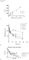
- Figure 9A shows that the size of the building blocks depends on the number of cells seeded and on the size of the microwells. Using human mesenchymal stem cells and two different sizes of microwells (200 and 400 ⁇ m), building blocks from 30 to 150 ⁇ m were assembled. In Figure 9B it is shown that also different culturing media can induce different levels of compaction. Furthermore, as shown in Figure 9C , different cell types show different plasticity and maintenance of the shape over time. This plasticity decreased with longer incubation on the microwells array. For each cell type and each culturing medium, a time of incubation on the microwells array has to be adjusted.
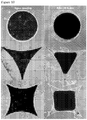
- Figure 10 shows that compaction of the tissue construct is not uniform for all shapes. Corners are regions of greater compaction. Compensated shapes can be designed to promote remodeling of the tissue into a desired geometry
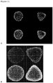
- Figure 11 it is shown that both the local compaction and the local stress of the tissue depends on the geometrical shape.
- Figure 11A shows a nuclear staining of 7 ⁇ m cuts. A local compaction of the cells (nuclei are closer to each other) was observed on the outside of tissues compared to the inside in discs (left pictures) and tip effects with local cell compaction in the tips of triangular tissues (right pictures).
- Figure 11B shows a cytoskeleton staining of 7 ⁇ m cuts. Regions of more intense F-actin populations of cells were observed. The shape of the tissues created local microenvironments of stress.
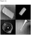
- tissue of clinically relevant size (such as centimeter-scale) is possible. Tissues spontaneously fuse and can be manipulated thus achieving clinical relevance.
Landscapes
- Health & Medical Sciences (AREA)
- Engineering & Computer Science (AREA)
- Life Sciences & Earth Sciences (AREA)
- Biomedical Technology (AREA)
- Genetics & Genomics (AREA)
- Organic Chemistry (AREA)
- Chemical & Material Sciences (AREA)
- Zoology (AREA)
- Wood Science & Technology (AREA)
- Biotechnology (AREA)
- Bioinformatics & Cheminformatics (AREA)
- Microbiology (AREA)
- Cell Biology (AREA)
- Biochemistry (AREA)
- General Engineering & Computer Science (AREA)
- General Health & Medical Sciences (AREA)
- Developmental Biology & Embryology (AREA)
- Vascular Medicine (AREA)
- Rheumatology (AREA)
- Immunology (AREA)
- Hematology (AREA)
- Micro-Organisms Or Cultivation Processes Thereof (AREA)
- Apparatus Associated With Microorganisms And Enzymes (AREA)
- Materials For Medical Uses (AREA)
Claims (18)
- Procédé de production in vitro d'une construction tissulaire, comprenant :a) la combinaison de cellules vivantes pour former des agrégats supracellulaires en utilisant un confinement spatial, le confinement spatial dans un ensemble de micropuits permettant la condensation/l'agrégation desdites cellules en agrégats supracellulaires et ledit confinement spatial comprenant un ensemble de micropuits ayant un diamètre enveloppant de 50 à 500 µm et une profondeur de 100 à 1 000 µm ;b) la combinaison de deux ou plusieurs des agrégats supracellulaires dans un moule ou sur un biomatériau comprenant des macropuits ;c) l'application de conditions qui induisent un auto-assemblage dans les agrégats supracellulaires combinés pour obtenir la construction tissulaire ; etd) l'application de conditions qui induisent une morphogenèse tissulaire dans la construction tissulaire,dans lequel lesdites conditions appliquées à l'étape c) et lesdites conditions appliquées à l'étape d) comprennent l'application de contraintes géométriques à la construction tissulaire.
- Procédé selon la revendication 1, dans lequel ladite morphogenèse tissulaire comprend une migration et/ou une différenciation de cellules.
- Procédé selon la revendication 1 ou 2, dans lequel la forme d'au moins certains des micropuits est telle que les agrégats ainsi obtenus peuvent s'auto-assembler selon le principe « clé-serrure ».
- Procédé selon la revendication 1, dans lequel 2 à 500 000 cellules par micropuits sont combinées pour former un agrégat supracellulaire.
- Procédé selon la revendication 4, dans lequel 10 à 10 000 cellules par micropuits sont combinées pour former un agrégat supracellulaire.
- Procédé selon la revendication 4 ou 5, dans lequel l'ensemble de micropuits est préparé par la technologie des micropuces, par gaufrage à chaud, par frittage sélectif au laser, par fabrication solide en forme libre, ou par micromoulage par séparation de phases.
- Procédé selon l'une quelconque des revendications précédentes, dans lequel les cellules vivantes sont du même type ou de différents types de cellules et sont combinées pour former les agrégats supracellulaires, les cellules étant sélectionnées dans le groupe constitué de cellules endothéliales, de cellules musculaires lisses, de cellules musculaires striées, de cellules neurales, de cellules de tissu conjonctif, d'ostéoblastes, d'ostéoclastes, de chondrocytes, d'hépatocytes, de cardiomyocytes, de myocytes, de cellules de Schwann, de cellules urothéliales, de cellules parenchymateuses, de cellules épithéliales, de cellules épithéliales sécrétoires exocrines, de cellules épithéliales d'absorption, de cellules épithéliales kératinisantes, de cellules sécrétoires de matrice extracellulaire, ou de cellules indifférenciées, de cellules embryonnaires, de cellules progénitrices, de cellules souches (mésenchymateuses), de cellules de moelle osseuse, de cellules satellites, de fibroblastes, et d'autres cellules précurseurs.
- Procédé selon la revendication 7, dans lequel les agrégats supracellulaires ont une taille de particule moyenne de 20 à 400 µm, mesurée par microscopie optique.
- Procédé selon l'une quelconque des revendications précédentes, dans lequel le biomatériau est sélectionné dans le groupe constitué de céramiques, de bioverres, de verres, de matériaux polymères biodégradables, de matériaux polymères non biodégradables et de métaux.
- Procédé selon l'une quelconque des revendications 1 à 9, dans lequel l'ensemble de micropuits comprend au moins deux micropuits ayant différentes tailles et/ou formes.
- Procédé selon l'une quelconque des revendications 1 à 10, dans lequel les micropuits se composent d'agarose, de PEG (polyéthylène glycol) ou de PDMS et/ou dans lequel la surface des micropuits est revêtue d'un ou plusieurs composés capables de réduire et/ou de prévenir l'adhérence cellulaire.
- Procédé selon la revendication 11, dans lequel le composé capable de réduire et/ou de prévenir l'adhérence cellulaire est le PEG, la sérum-albumine bovine, le collagène et/ou la fibronectine.
- Procédé selon l'une quelconque des revendications précédentes, dans lequel les cellules vivantes sont combinées en présence de fibronectine et/ou de collagène.
- Procédé selon l'une quelconque des revendications précédentes, dans lequel les propriétés de surface, la charge magnétique et/ou la charge électrique des agrégats supracellulaires sont modifiées avant la combinaison de deux ou plusieurs des agrégats supracellulaires.
- Procédé selon l'une quelconque des revendications précédentes, dans lequel les agrégats supracellulaires sont combinés dans un liquide en mouvement et/ou dans lequel les agrégats supracellulaires sont combinés dans un puits ayant un diamètre enveloppant d'au moins 500 µm.
- Procédé selon l'une quelconque des revendications précédentes, dans lequel les conditions à l'étape c) comprennent une ou plusieurs conditions sélectionnées dans le groupe constitué de contraintes mécaniques, d'une compression, de secousses, de champs électriques, de champs magnétiques et de gradients de morphogènes ou de facteurs de croissance.
- Procédé selon l'une quelconque des revendications précédentes, dans lequel à l'étape a) ou b), les cellules vivantes sont combinées ou l'agrégat supracellulaire est combiné avec un objet et/ou dans lequel à l'étape c) ou d), la construction tissulaire est combinée avec un objet, pour induire une réponse locale.
- Procédé selon la revendication 1, dans lequel l'étape a) comprend un ajustement du temps d'incubation sur l'ensemble de micropuits pour obtenir une plasticité souhaitée des agrégats supracellulaires.
Priority Applications (2)
| Application Number | Priority Date | Filing Date | Title |
|---|---|---|---|
| SI200931851T SI2310491T1 (en) | 2008-06-20 | 2009-06-22 | Only composite tissue modules |
| EP09766884.2A EP2310491B1 (fr) | 2008-06-20 | 2009-06-22 | Modules de tissus auto-assemblés |
Applications Claiming Priority (3)
| Application Number | Priority Date | Filing Date | Title |
|---|---|---|---|
| EP08158652 | 2008-06-20 | ||
| EP09766884.2A EP2310491B1 (fr) | 2008-06-20 | 2009-06-22 | Modules de tissus auto-assemblés |
| PCT/NL2009/050368 WO2009154466A1 (fr) | 2008-06-20 | 2009-06-22 | Modules de tissu auto-assemblables |
Publications (2)
| Publication Number | Publication Date |
|---|---|
| EP2310491A1 EP2310491A1 (fr) | 2011-04-20 |
| EP2310491B1 true EP2310491B1 (fr) | 2018-03-07 |
Family
ID=39926404
Family Applications (1)
| Application Number | Title | Priority Date | Filing Date |
|---|---|---|---|
| EP09766884.2A Active EP2310491B1 (fr) | 2008-06-20 | 2009-06-22 | Modules de tissus auto-assemblés |
Country Status (6)
| Country | Link |
|---|---|
| US (2) | US9303245B2 (fr) |
| EP (1) | EP2310491B1 (fr) |
| ES (1) | ES2672525T3 (fr) |
| HU (1) | HUE037881T2 (fr) |
| SI (1) | SI2310491T1 (fr) |
| WO (1) | WO2009154466A1 (fr) |
Families Citing this family (40)
| Publication number | Priority date | Publication date | Assignee | Title |
|---|---|---|---|---|
| US8241905B2 (en) | 2004-02-24 | 2012-08-14 | The Curators Of The University Of Missouri | Self-assembling cell aggregates and methods of making engineered tissue using the same |
| EP2310491B1 (fr) | 2008-06-20 | 2018-03-07 | Universiteit Maastricht | Modules de tissus auto-assemblés |
| JP5624981B2 (ja) | 2008-06-24 | 2014-11-12 | ザ・キュレーターズ・オブ・ザ・ユニバーシティ・オブ・ミズーリThe Curators Ofthe University Of Missouri | 自己集合性多細胞体、および前記多細胞体を用いて3次元の生物構造体を作製する方法 |
| JP5791176B2 (ja) | 2010-08-06 | 2015-10-07 | コバレントマテリアル株式会社 | 細胞培養担体及び細胞培養方法 |
| AU2011318437B2 (en) | 2010-10-21 | 2015-02-26 | Organovo, Inc. | Devices, systems, and methods for the fabrication of tissue |
| HK1200483A1 (en) * | 2011-09-12 | 2015-08-07 | 奥加诺沃公司 | Engineered tissues for in vitro research uses, arrays thereof, and methods of making the same |
| EP2762558B1 (fr) * | 2011-09-27 | 2026-01-21 | Public University Corporation Yokohama City University | Procédé de production de tissu et d'organe |
| US20130203069A1 (en) * | 2012-02-07 | 2013-08-08 | The Regents Of The University Of California | Dynamic Assay for Maintenance and Disruption of Tissue Level Organization and Architecture |
| US9499779B2 (en) | 2012-04-20 | 2016-11-22 | Organovo, Inc. | Devices, systems, and methods for the fabrication of tissue utilizing UV cross-linking |
| GB201219707D0 (en) * | 2012-11-02 | 2012-12-12 | Univ Singapore | In situ cardiac tissue engineering |
| GB2507744A (en) * | 2012-11-07 | 2014-05-14 | Universitaetsklinikum Freiburg | Matrix for generating and cultivating uniform cell aggregations |
| JP2016504022A (ja) | 2012-12-07 | 2016-02-12 | ザ ガバニング カウンシル オブ ザ ユニバーシティ オブ トロント | 心臓組織コンストラクト及びその製造方法 |
| US9442105B2 (en) | 2013-03-15 | 2016-09-13 | Organovo, Inc. | Engineered liver tissues, arrays thereof, and methods of making the same |
| SG11201600770RA (en) | 2013-07-31 | 2016-02-26 | Organovo Inc | Automated devices, systems, and methods for the fabrication of tissue |
| EP3808838B1 (fr) | 2014-04-04 | 2024-04-10 | Organovo, Inc. | Tissu mammaire tridimensionnel artificiel, tissu adipeux, et modèle de maladies tumorales |
| KR20170064547A (ko) | 2014-10-06 | 2017-06-09 | 오가노보, 인크. | 조작된 신장 조직, 이의 어레이, 및 이를 제조하는 방법 |
| US11529436B2 (en) | 2014-11-05 | 2022-12-20 | Organovo, Inc. | Engineered three-dimensional skin tissues, arrays thereof, and methods of making the same |
| AU2016209378A1 (en) | 2015-01-23 | 2017-07-13 | The Trustees Of Columbia University In The City Of New York | Engineering mechanically functional human cartilage and method of making same |
| WO2016140716A1 (fr) | 2015-03-02 | 2016-09-09 | The Trustees Of Columbia University In The City Of New York | Systèmes, dispositifs et procédés de micro-tissus injectables |
| EP3359641B1 (fr) | 2015-10-09 | 2020-01-08 | DEKA Products Limited Partnership | Système de bioréacteur et de pompage de fluide |
| WO2017083402A1 (fr) | 2015-11-09 | 2017-05-18 | Organovo, Inc. | Procédés améliorés de fabrication de tissus |
| NL2016404B1 (en) * | 2016-03-09 | 2017-09-26 | Mimetas B V | Double tubular structures. |
| GB201615517D0 (en) | 2016-09-13 | 2016-10-26 | Micromatrices Ass L | Spheroid tissue miroarray and method of manufacture |
| US10345208B2 (en) | 2016-07-12 | 2019-07-09 | Deka Products Limited Partnership | System and method for applying force to a device |
| US11254901B2 (en) | 2016-07-12 | 2022-02-22 | Deka Products Limited Partnership | System and method for printing tissue |
| EP3296018A1 (fr) | 2016-09-19 | 2018-03-21 | Ecole Polytechnique Fédérale de Lausanne (EPFL) | Réseaux d'organoïdes |
| US11299705B2 (en) | 2016-11-07 | 2022-04-12 | Deka Products Limited Partnership | System and method for creating tissue |
| EP3323882A1 (fr) | 2016-11-18 | 2018-05-23 | Ecole Polytechnique Fédérale de Lausanne | Génie tissulaire organoïde |
| US10570362B2 (en) | 2017-07-12 | 2020-02-25 | Deka Products Limited Partnership | System and method for transferring tissue |
| US12297416B2 (en) | 2017-07-12 | 2025-05-13 | Deka Products Limited Partneship | System and method for transferring tissue |
| US11970560B2 (en) | 2017-09-03 | 2024-04-30 | The University Of North Carolina At Chapel Hill | Self-assembled elastomers with molecularly encoded tissue-like softness, strain-adaptive stiffening and coloration |
| EP3743080A4 (fr) * | 2018-01-26 | 2021-10-27 | Polarityte, Inc. | Matériaux d'auto-assemblage coordinés à une interface vivante complexe (clicsam) |
| CN110468049A (zh) * | 2018-05-10 | 2019-11-19 | 澳门大学 | 用于制备干细胞球的装置、制备干细胞球的方法以及保存干细胞的方法 |
| WO2020232436A1 (fr) | 2019-05-16 | 2020-11-19 | Brown University | Modèle de cardiotoxicité in vitro humain |
| TWI724528B (zh) * | 2019-09-04 | 2021-04-11 | 三顧股份有限公司 | 高增生活性的3d結構細胞球體、其製造方法與用途 |
| US20210189327A1 (en) * | 2019-12-24 | 2021-06-24 | Mcmaster University | 3d stimulated tissue constructs and methods of making thereof |
| US12427229B2 (en) | 2020-08-06 | 2025-09-30 | PhosPrint P.C. | Laser ablation/removal and laser induced forward transfer of biological material |
| US20240081377A1 (en) * | 2020-10-20 | 2024-03-14 | Orbillion Bio, Inc. | Using organoids and/or spheroids to cultivate meat |
| US12076437B2 (en) | 2021-07-12 | 2024-09-03 | Brown University | Proangiogenic protein cocktails delivered in custom biomaterials to revascularize ischemic tissue |
| US12435312B2 (en) | 2022-02-28 | 2025-10-07 | Brown University | Quantifying cell-derived changes in collagen synthesis, alignment, and mechanics in a 3D connective tissue model |
Family Cites Families (5)
| Publication number | Priority date | Publication date | Assignee | Title |
|---|---|---|---|---|
| DE4132379A1 (de) * | 1991-09-28 | 1993-04-08 | Kernforschungsz Karlsruhe | Substrat fuer zellkulturen und kultur von zellen oder zellaggregaten |
| US6667176B1 (en) * | 2000-01-11 | 2003-12-23 | Geron Corporation | cDNA libraries reflecting gene expression during growth and differentiation of human pluripotent stem cells |
| CA2637663C (fr) * | 2006-01-24 | 2015-06-02 | Brown University | Dispositif et procede d'agregation et d'encapsulation cellulaires |
| CA2648327A1 (fr) * | 2006-04-05 | 2007-10-11 | William Marsh Rice University | Genie tissulaire a l'aide de cellules souches embryonnaires humaines |
| EP2310491B1 (fr) | 2008-06-20 | 2018-03-07 | Universiteit Maastricht | Modules de tissus auto-assemblés |
-
2009
- 2009-06-22 EP EP09766884.2A patent/EP2310491B1/fr active Active
- 2009-06-22 HU HUE09766884A patent/HUE037881T2/hu unknown
- 2009-06-22 US US12/999,841 patent/US9303245B2/en active Active
- 2009-06-22 ES ES09766884.2T patent/ES2672525T3/es active Active
- 2009-06-22 SI SI200931851T patent/SI2310491T1/en unknown
- 2009-06-22 WO PCT/NL2009/050368 patent/WO2009154466A1/fr not_active Ceased
-
2016
- 2016-03-08 US US15/064,360 patent/US20160257926A1/en not_active Abandoned
Non-Patent Citations (1)
| Title |
|---|
| CHUNG YOUNG ET AL: "Human embryonic stem cell lines generated without embryo destruction", CELL STEM CELL, ELSEVIER, CELL PRESS, AMSTERDAM, NL, vol. 2, no. 2, 7 February 2008 (2008-02-07), pages 113 - 117, XP002604696, ISSN: 1934-5909, DOI: 10.1016/J.STEM.2007.12.013 * |
Also Published As
| Publication number | Publication date |
|---|---|
| SI2310491T1 (en) | 2018-07-31 |
| US9303245B2 (en) | 2016-04-05 |
| US20160257926A1 (en) | 2016-09-08 |
| HUE037881T2 (hu) | 2018-09-28 |
| EP2310491A1 (fr) | 2011-04-20 |
| WO2009154466A1 (fr) | 2009-12-23 |
| ES2672525T3 (es) | 2018-06-14 |
| US20110171712A1 (en) | 2011-07-14 |
Similar Documents
| Publication | Publication Date | Title |
|---|---|---|
| EP2310491B1 (fr) | Modules de tissus auto-assemblés | |
| Khoshnood et al. | A comprehensive review on scaffold-free bioinks for bioprinting | |
| JP5607535B2 (ja) | 細胞培養キット、スクリーニング方法、及び細胞培養キットの製造方法 | |
| Yeh et al. | Micromolding of shape-controlled, harvestable cell-laden hydrogels | |
| US8501476B2 (en) | Assays and methods for fusing cell aggregates to form proto-tissues | |
| JP5507244B2 (ja) | 細胞培養容器及び細胞培養方法 | |
| JP5985570B2 (ja) | 自己集合性多細胞体、および前記多細胞体を用いて3次元の生物構造体を作製する方法 | |
| US20070281353A1 (en) | Microfabricated Compositions and Processes for Engineering Tissues Containing Multiple Cell Types | |
| JP5177774B2 (ja) | 細胞の3次元階層的共培養法 | |
| Peng et al. | Challenges in bio-fabrication of organoid cultures | |
| CN112852706A (zh) | 一种基于双水相液滴微流控的3d类器官工程化方法 | |
| WO2010047133A1 (fr) | Procédé de stockage de cellules et procédé de transport de cellules | |
| CN111051492B (zh) | 细胞片形成构件、细胞片形成构件的制造方法、及细胞片的制造方法 | |
| US20060228386A1 (en) | Polymeric microstructures | |
| Wang et al. | Engineering biological tissues from the bottom-up: Recent advances and future prospects | |
| CN104630148A (zh) | 一种基于水凝胶微孔板的细胞球原位制备方法 | |
| JP5940758B2 (ja) | 細胞培養方法 | |
| Ou et al. | Biomaterials with droplet microfluidics | |
| KR20260008783A (ko) | 3d 배양용 dna 오리가미 및 이의 제조 방법과 응용 | |
| Coutinho et al. | Micro-and nanotechnology in tissue engineering | |
| Altmann et al. | The famous versus the inconvenient-or the dawn and the rise of 3D-culture systems | |
| Zhang et al. | Techniques for biofabrication of vascular and vascularized tissue | |
| Blakely et al. | Formation of multicellular microtissues and applications in biofabrication | |
| Zhan et al. | Droplet-based Microfluidic Platform for High-throughput Formation of Multicellular Spheroids | |
| Blakely et al. | Biofabrication: Chapter 8. Formation of Multicellular Microtissues and Applications in Biofabrication |
Legal Events
| Date | Code | Title | Description |
|---|---|---|---|
| PUAI | Public reference made under article 153(3) epc to a published international application that has entered the european phase |
Free format text: ORIGINAL CODE: 0009012 |
|
| 17P | Request for examination filed |
Effective date: 20110118 |
|
| AK | Designated contracting states |
Kind code of ref document: A1 Designated state(s): AT BE BG CH CY CZ DE DK EE ES FI FR GB GR HR HU IE IS IT LI LT LU LV MC MK MT NL NO PL PT RO SE SI SK TR |
|
| AX | Request for extension of the european patent |
Extension state: AL BA RS |
|
| 17Q | First examination report despatched |
Effective date: 20121018 |
|
| R17C | First examination report despatched (corrected) |
Effective date: 20121018 |
|
| RAP1 | Party data changed (applicant data changed or rights of an application transferred) |
Owner name: UNIVERSITEIT MAASTRICHT |
|
| REG | Reference to a national code |
Ref country code: DE Ref legal event code: R079 Ref document number: 602009051124 Country of ref document: DE Free format text: PREVIOUS MAIN CLASS: C12N0005000000 Ipc: C12N0005071000 |
|
| RIC1 | Information provided on ipc code assigned before grant |
Ipc: C12N 5/00 20060101ALI20170822BHEP Ipc: C12N 5/071 20100101AFI20170822BHEP Ipc: C12N 5/0775 20100101ALI20170822BHEP |
|
| GRAP | Despatch of communication of intention to grant a patent |
Free format text: ORIGINAL CODE: EPIDOSNIGR1 |
|
| INTG | Intention to grant announced |
Effective date: 20171004 |
|
| RIN1 | Information on inventor provided before grant (corrected) |
Inventor name: TRUCKENMULLER, ROMAN Inventor name: RIVRON, NICOLAS CLEMENT Inventor name: VAN BLITTERSWIJK, CLEMENS ANTONI Inventor name: ROUWKEMA, JEROEN Inventor name: VRIJ, ERIK JACOB Inventor name: LE GAC, SEVERINE |
|
| RIN1 | Information on inventor provided before grant (corrected) |
Inventor name: LE GAC, SEVERINE Inventor name: RIVRON, NICOLAS CLEMENT Inventor name: ROUWKEMA, JEROEN Inventor name: VAN BLITTERSWIJK, CLEMENS ANTONI Inventor name: VRIJ, ERIK JACOB Inventor name: TRUCKENMULLER, ROMAN |
|
| GRAS | Grant fee paid |
Free format text: ORIGINAL CODE: EPIDOSNIGR3 |
|
| GRAA | (expected) grant |
Free format text: ORIGINAL CODE: 0009210 |
|
| AK | Designated contracting states |
Kind code of ref document: B1 Designated state(s): AT BE BG CH CY CZ DE DK EE ES FI FR GB GR HR HU IE IS IT LI LT LU LV MC MK MT NL NO PL PT RO SE SI SK TR |
|
| AX | Request for extension of the european patent |
Extension state: AL BA RS |
|
| REG | Reference to a national code |
Ref country code: GB Ref legal event code: FG4D |
|
| REG | Reference to a national code |
Ref country code: CH Ref legal event code: EP Ref country code: AT Ref legal event code: REF Ref document number: 976582 Country of ref document: AT Kind code of ref document: T Effective date: 20180315 |
|
| REG | Reference to a national code |
Ref country code: DE Ref legal event code: R096 Ref document number: 602009051124 Country of ref document: DE |
|
| REG | Reference to a national code |
Ref country code: IE Ref legal event code: FG4D |
|
| REG | Reference to a national code |
Ref country code: ES Ref legal event code: FG2A Ref document number: 2672525 Country of ref document: ES Kind code of ref document: T3 Effective date: 20180614 |
|
| REG | Reference to a national code |
Ref country code: SE Ref legal event code: TRGR Ref country code: FR Ref legal event code: PLFP Year of fee payment: 10 |
|
| REG | Reference to a national code |
Ref country code: NL Ref legal event code: FP |
|
| REG | Reference to a national code |
Ref country code: LT Ref legal event code: MG4D |
|
| PG25 | Lapsed in a contracting state [announced via postgrant information from national office to epo] |
Ref country code: FI Free format text: LAPSE BECAUSE OF FAILURE TO SUBMIT A TRANSLATION OF THE DESCRIPTION OR TO PAY THE FEE WITHIN THE PRESCRIBED TIME-LIMIT Effective date: 20180307 Ref country code: NO Free format text: LAPSE BECAUSE OF FAILURE TO SUBMIT A TRANSLATION OF THE DESCRIPTION OR TO PAY THE FEE WITHIN THE PRESCRIBED TIME-LIMIT Effective date: 20180607 Ref country code: LT Free format text: LAPSE BECAUSE OF FAILURE TO SUBMIT A TRANSLATION OF THE DESCRIPTION OR TO PAY THE FEE WITHIN THE PRESCRIBED TIME-LIMIT Effective date: 20180307 Ref country code: HR Free format text: LAPSE BECAUSE OF FAILURE TO SUBMIT A TRANSLATION OF THE DESCRIPTION OR TO PAY THE FEE WITHIN THE PRESCRIBED TIME-LIMIT Effective date: 20180307 Ref country code: CY Free format text: LAPSE BECAUSE OF FAILURE TO SUBMIT A TRANSLATION OF THE DESCRIPTION OR TO PAY THE FEE WITHIN THE PRESCRIBED TIME-LIMIT Effective date: 20180307 |
|
| REG | Reference to a national code |
Ref country code: AT Ref legal event code: MK05 Ref document number: 976582 Country of ref document: AT Kind code of ref document: T Effective date: 20180307 |
|
| PG25 | Lapsed in a contracting state [announced via postgrant information from national office to epo] |
Ref country code: GR Free format text: LAPSE BECAUSE OF FAILURE TO SUBMIT A TRANSLATION OF THE DESCRIPTION OR TO PAY THE FEE WITHIN THE PRESCRIBED TIME-LIMIT Effective date: 20180608 Ref country code: LV Free format text: LAPSE BECAUSE OF FAILURE TO SUBMIT A TRANSLATION OF THE DESCRIPTION OR TO PAY THE FEE WITHIN THE PRESCRIBED TIME-LIMIT Effective date: 20180307 Ref country code: BG Free format text: LAPSE BECAUSE OF FAILURE TO SUBMIT A TRANSLATION OF THE DESCRIPTION OR TO PAY THE FEE WITHIN THE PRESCRIBED TIME-LIMIT Effective date: 20180607 |
|
| REG | Reference to a national code |
Ref country code: HU Ref legal event code: AG4A Ref document number: E037881 Country of ref document: HU |
|
| PG25 | Lapsed in a contracting state [announced via postgrant information from national office to epo] |
Ref country code: PL Free format text: LAPSE BECAUSE OF FAILURE TO SUBMIT A TRANSLATION OF THE DESCRIPTION OR TO PAY THE FEE WITHIN THE PRESCRIBED TIME-LIMIT Effective date: 20180307 Ref country code: EE Free format text: LAPSE BECAUSE OF FAILURE TO SUBMIT A TRANSLATION OF THE DESCRIPTION OR TO PAY THE FEE WITHIN THE PRESCRIBED TIME-LIMIT Effective date: 20180307 Ref country code: RO Free format text: LAPSE BECAUSE OF FAILURE TO SUBMIT A TRANSLATION OF THE DESCRIPTION OR TO PAY THE FEE WITHIN THE PRESCRIBED TIME-LIMIT Effective date: 20180307 |
|
| PG25 | Lapsed in a contracting state [announced via postgrant information from national office to epo] |
Ref country code: AT Free format text: LAPSE BECAUSE OF FAILURE TO SUBMIT A TRANSLATION OF THE DESCRIPTION OR TO PAY THE FEE WITHIN THE PRESCRIBED TIME-LIMIT Effective date: 20180307 Ref country code: SK Free format text: LAPSE BECAUSE OF FAILURE TO SUBMIT A TRANSLATION OF THE DESCRIPTION OR TO PAY THE FEE WITHIN THE PRESCRIBED TIME-LIMIT Effective date: 20180307 |
|
| REG | Reference to a national code |
Ref country code: DE Ref legal event code: R097 Ref document number: 602009051124 Country of ref document: DE |
|
| PG25 | Lapsed in a contracting state [announced via postgrant information from national office to epo] |
Ref country code: PT Free format text: LAPSE BECAUSE OF FAILURE TO SUBMIT A TRANSLATION OF THE DESCRIPTION OR TO PAY THE FEE WITHIN THE PRESCRIBED TIME-LIMIT Effective date: 20180709 |
|
| PLBE | No opposition filed within time limit |
Free format text: ORIGINAL CODE: 0009261 |
|
| STAA | Information on the status of an ep patent application or granted ep patent |
Free format text: STATUS: NO OPPOSITION FILED WITHIN TIME LIMIT |
|
| PG25 | Lapsed in a contracting state [announced via postgrant information from national office to epo] |
Ref country code: DK Free format text: LAPSE BECAUSE OF FAILURE TO SUBMIT A TRANSLATION OF THE DESCRIPTION OR TO PAY THE FEE WITHIN THE PRESCRIBED TIME-LIMIT Effective date: 20180307 |
|
| 26N | No opposition filed |
Effective date: 20181210 |
|
| REG | Reference to a national code |
Ref country code: BE Ref legal event code: MM Effective date: 20180630 |
|
| REG | Reference to a national code |
Ref country code: IE Ref legal event code: MM4A |
|
| PG25 | Lapsed in a contracting state [announced via postgrant information from national office to epo] |
Ref country code: LU Free format text: LAPSE BECAUSE OF NON-PAYMENT OF DUE FEES Effective date: 20180622 Ref country code: MC Free format text: LAPSE BECAUSE OF FAILURE TO SUBMIT A TRANSLATION OF THE DESCRIPTION OR TO PAY THE FEE WITHIN THE PRESCRIBED TIME-LIMIT Effective date: 20180307 |
|
| PG25 | Lapsed in a contracting state [announced via postgrant information from national office to epo] |
Ref country code: IE Free format text: LAPSE BECAUSE OF NON-PAYMENT OF DUE FEES Effective date: 20180622 |
|
| PG25 | Lapsed in a contracting state [announced via postgrant information from national office to epo] |
Ref country code: BE Free format text: LAPSE BECAUSE OF NON-PAYMENT OF DUE FEES Effective date: 20180630 |
|
| PG25 | Lapsed in a contracting state [announced via postgrant information from national office to epo] |
Ref country code: MT Free format text: LAPSE BECAUSE OF NON-PAYMENT OF DUE FEES Effective date: 20180622 |
|
| PG25 | Lapsed in a contracting state [announced via postgrant information from national office to epo] |
Ref country code: TR Free format text: LAPSE BECAUSE OF FAILURE TO SUBMIT A TRANSLATION OF THE DESCRIPTION OR TO PAY THE FEE WITHIN THE PRESCRIBED TIME-LIMIT Effective date: 20180307 |
|
| PG25 | Lapsed in a contracting state [announced via postgrant information from national office to epo] |
Ref country code: MK Free format text: LAPSE BECAUSE OF NON-PAYMENT OF DUE FEES Effective date: 20180307 |
|
| PG25 | Lapsed in a contracting state [announced via postgrant information from national office to epo] |
Ref country code: IS Free format text: LAPSE BECAUSE OF FAILURE TO SUBMIT A TRANSLATION OF THE DESCRIPTION OR TO PAY THE FEE WITHIN THE PRESCRIBED TIME-LIMIT Effective date: 20180707 |
|
| P01 | Opt-out of the competence of the unified patent court (upc) registered |
Effective date: 20230525 |
|
| REG | Reference to a national code |
Ref country code: CH Ref legal event code: U11 Free format text: ST27 STATUS EVENT CODE: U-0-0-U10-U11 (AS PROVIDED BY THE NATIONAL OFFICE) Effective date: 20251222 |
|
| PGFP | Annual fee paid to national office [announced via postgrant information from national office to epo] |
Ref country code: DE Payment date: 20251211 Year of fee payment: 17 |
|
| PGFP | Annual fee paid to national office [announced via postgrant information from national office to epo] |
Ref country code: GB Payment date: 20251219 Year of fee payment: 17 |
|
| PGFP | Annual fee paid to national office [announced via postgrant information from national office to epo] |
Ref country code: IT Payment date: 20251223 Year of fee payment: 17 |
|
| PGFP | Annual fee paid to national office [announced via postgrant information from national office to epo] |
Ref country code: HU Payment date: 20251218 Year of fee payment: 17 Ref country code: NL Payment date: 20251219 Year of fee payment: 17 Ref country code: FR Payment date: 20251219 Year of fee payment: 17 |
|
| PGFP | Annual fee paid to national office [announced via postgrant information from national office to epo] |
Ref country code: SE Payment date: 20251219 Year of fee payment: 17 Ref country code: CH Payment date: 20251222 Year of fee payment: 17 |
|
| PGFP | Annual fee paid to national office [announced via postgrant information from national office to epo] |
Ref country code: CZ Payment date: 20251204 Year of fee payment: 17 |
|
| PGFP | Annual fee paid to national office [announced via postgrant information from national office to epo] |
Ref country code: ES Payment date: 20251229 Year of fee payment: 17 Ref country code: SI Payment date: 20251204 Year of fee payment: 17 |