EP2311436A1 - Stabilisierte Protein-Kristalle, diese enthaltende Formulierungen und ihre Herstellungsverfahren - Google Patents
Stabilisierte Protein-Kristalle, diese enthaltende Formulierungen und ihre Herstellungsverfahren Download PDFInfo
- Publication number
- EP2311436A1 EP2311436A1 EP10011893A EP10011893A EP2311436A1 EP 2311436 A1 EP2311436 A1 EP 2311436A1 EP 10011893 A EP10011893 A EP 10011893A EP 10011893 A EP10011893 A EP 10011893A EP 2311436 A1 EP2311436 A1 EP 2311436A1
- Authority
- EP
- European Patent Office
- Prior art keywords
- protein
- crystals
- formulation
- crystal
- proteins
- Prior art date
- Legal status (The legal status is an assumption and is not a legal conclusion. Google has not performed a legal analysis and makes no representation as to the accuracy of the status listed.)
- Withdrawn
Links
- 239000013078 crystal Substances 0.000 title claims abstract description 521
- 108090000623 proteins and genes Proteins 0.000 title claims abstract description 372
- 102000004169 proteins and genes Human genes 0.000 title claims abstract description 366
- 239000000203 mixture Substances 0.000 title claims abstract description 324
- 238000009472 formulation Methods 0.000 title claims abstract description 203
- 238000000034 method Methods 0.000 title abstract description 109
- 238000003860 storage Methods 0.000 claims abstract description 31
- 108090001060 Lipase Proteins 0.000 claims description 94
- 102000004882 Lipase Human genes 0.000 claims description 94
- 239000004367 Lipase Substances 0.000 claims description 94
- 235000019421 lipase Nutrition 0.000 claims description 94
- 239000000546 pharmaceutical excipient Substances 0.000 claims description 87
- 229920000642 polymer Polymers 0.000 claims description 82
- 239000004366 Glucose oxidase Substances 0.000 claims description 81
- 108010015776 Glucose oxidase Proteins 0.000 claims description 81
- 235000019420 glucose oxidase Nutrition 0.000 claims description 81
- 229940116332 glucose oxidase Drugs 0.000 claims description 81
- 230000000694 effects Effects 0.000 claims description 67
- CZMRCDWAGMRECN-UGDNZRGBSA-N Sucrose Chemical compound O[C@H]1[C@H](O)[C@@H](CO)O[C@@]1(CO)O[C@@H]1[C@H](O)[C@@H](O)[C@H](O)[C@@H](CO)O1 CZMRCDWAGMRECN-UGDNZRGBSA-N 0.000 claims description 40
- 229930006000 Sucrose Natural products 0.000 claims description 38
- 239000005720 sucrose Substances 0.000 claims description 38
- HDTRYLNUVZCQOY-UHFFFAOYSA-N α-D-glucopyranosyl-α-D-glucopyranoside Natural products OC1C(O)C(O)C(CO)OC1OC1C(O)C(O)C(O)C(CO)O1 HDTRYLNUVZCQOY-UHFFFAOYSA-N 0.000 claims description 35
- HDTRYLNUVZCQOY-WSWWMNSNSA-N Trehalose Natural products O[C@@H]1[C@@H](O)[C@@H](O)[C@@H](CO)O[C@@H]1O[C@@H]1[C@H](O)[C@@H](O)[C@@H](O)[C@@H](CO)O1 HDTRYLNUVZCQOY-WSWWMNSNSA-N 0.000 claims description 35
- HDTRYLNUVZCQOY-LIZSDCNHSA-N alpha,alpha-trehalose Chemical compound O[C@@H]1[C@@H](O)[C@H](O)[C@@H](CO)O[C@@H]1O[C@@H]1[C@H](O)[C@@H](O)[C@H](O)[C@@H](CO)O1 HDTRYLNUVZCQOY-LIZSDCNHSA-N 0.000 claims description 33
- 108091006905 Human Serum Albumin Proteins 0.000 claims description 29
- 102000008100 Human Serum Albumin Human genes 0.000 claims description 29
- 239000004005 microsphere Substances 0.000 claims description 27
- 239000008273 gelatin Substances 0.000 claims description 26
- 229920000159 gelatin Polymers 0.000 claims description 26
- 229920001223 polyethylene glycol Polymers 0.000 claims description 26
- 108010010803 Gelatin Proteins 0.000 claims description 23
- 235000019322 gelatine Nutrition 0.000 claims description 23
- 235000011852 gelatine desserts Nutrition 0.000 claims description 23
- ODLHGICHYURWBS-LKONHMLTSA-N trappsol cyclo Chemical compound CC(O)COC[C@H]([C@H]([C@@H]([C@H]1O)O)O[C@H]2O[C@@H]([C@@H](O[C@H]3O[C@H](COCC(C)O)[C@H]([C@@H]([C@H]3O)O)O[C@H]3O[C@H](COCC(C)O)[C@H]([C@@H]([C@H]3O)O)O[C@H]3O[C@H](COCC(C)O)[C@H]([C@@H]([C@H]3O)O)O[C@H]3O[C@H](COCC(C)O)[C@H]([C@@H]([C@H]3O)O)O3)[C@H](O)[C@H]2O)COCC(O)C)O[C@@H]1O[C@H]1[C@H](O)[C@@H](O)[C@@H]3O[C@@H]1COCC(C)O ODLHGICHYURWBS-LKONHMLTSA-N 0.000 claims description 23
- 239000002202 Polyethylene glycol Substances 0.000 claims description 20
- 238000005033 Fourier transform infrared spectroscopy Methods 0.000 claims description 19
- LYCAIKOWRPUZTN-UHFFFAOYSA-N Ethylene glycol Chemical compound OCCO LYCAIKOWRPUZTN-UHFFFAOYSA-N 0.000 claims description 18
- 229910052799 carbon Inorganic materials 0.000 claims description 18
- 239000000832 lactitol Substances 0.000 claims description 17
- 229960003451 lactitol Drugs 0.000 claims description 17
- VQHSOMBJVWLPSR-JVCRWLNRSA-N lactitol Chemical compound OC[C@H](O)[C@@H](O)[C@@H]([C@H](O)CO)O[C@@H]1O[C@H](CO)[C@H](O)[C@H](O)[C@H]1O VQHSOMBJVWLPSR-JVCRWLNRSA-N 0.000 claims description 16
- 235000010448 lactitol Nutrition 0.000 claims description 16
- 230000007423 decrease Effects 0.000 claims description 12
- 239000011159 matrix material Substances 0.000 claims description 9
- FBPFZTCFMRRESA-FSIIMWSLSA-N D-Glucitol Natural products OC[C@H](O)[C@H](O)[C@@H](O)[C@H](O)CO FBPFZTCFMRRESA-FSIIMWSLSA-N 0.000 claims description 8
- FBPFZTCFMRRESA-JGWLITMVSA-N D-glucitol Chemical compound OC[C@H](O)[C@@H](O)[C@H](O)[C@H](O)CO FBPFZTCFMRRESA-JGWLITMVSA-N 0.000 claims description 8
- 235000010356 sorbitol Nutrition 0.000 claims description 8
- 239000000600 sorbitol Substances 0.000 claims description 8
- 150000001720 carbohydrates Chemical class 0.000 claims description 6
- FBPFZTCFMRRESA-KVTDHHQDSA-N D-Mannitol Chemical compound OC[C@@H](O)[C@@H](O)[C@H](O)[C@H](O)CO FBPFZTCFMRRESA-KVTDHHQDSA-N 0.000 claims description 5
- 229930195725 Mannitol Natural products 0.000 claims description 5
- 108090000854 Oxidoreductases Proteins 0.000 claims description 5
- 102000004316 Oxidoreductases Human genes 0.000 claims description 5
- 150000001413 amino acids Chemical class 0.000 claims description 5
- 239000000594 mannitol Substances 0.000 claims description 5
- 235000010355 mannitol Nutrition 0.000 claims description 5
- 229920001282 polysaccharide Polymers 0.000 claims description 5
- KIUKXJAPPMFGSW-DNGZLQJQSA-N (2S,3S,4S,5R,6R)-6-[(2S,3R,4R,5S,6R)-3-Acetamido-2-[(2S,3S,4R,5R,6R)-6-[(2R,3R,4R,5S,6R)-3-acetamido-2,5-dihydroxy-6-(hydroxymethyl)oxan-4-yl]oxy-2-carboxy-4,5-dihydroxyoxan-3-yl]oxy-5-hydroxy-6-(hydroxymethyl)oxan-4-yl]oxy-3,4,5-trihydroxyoxane-2-carboxylic acid Chemical compound CC(=O)N[C@H]1[C@H](O)O[C@H](CO)[C@@H](O)[C@@H]1O[C@H]1[C@H](O)[C@@H](O)[C@H](O[C@H]2[C@@H]([C@@H](O[C@H]3[C@@H]([C@@H](O)[C@H](O)[C@H](O3)C(O)=O)O)[C@H](O)[C@@H](CO)O2)NC(C)=O)[C@@H](C(O)=O)O1 KIUKXJAPPMFGSW-DNGZLQJQSA-N 0.000 claims description 4
- IAJILQKETJEXLJ-UHFFFAOYSA-N Galacturonsaeure Natural products O=CC(O)C(O)C(O)C(O)C(O)=O IAJILQKETJEXLJ-UHFFFAOYSA-N 0.000 claims description 4
- 229920002674 hyaluronan Polymers 0.000 claims description 4
- 229960003160 hyaluronic acid Drugs 0.000 claims description 4
- 239000005017 polysaccharide Substances 0.000 claims description 4
- 239000004475 Arginine Substances 0.000 claims description 3
- 229920000858 Cyclodextrin Polymers 0.000 claims description 3
- 102000004157 Hydrolases Human genes 0.000 claims description 3
- 108090000604 Hydrolases Proteins 0.000 claims description 3
- ODKSFYDXXFIFQN-BYPYZUCNSA-P L-argininium(2+) Chemical compound NC(=[NH2+])NCCC[C@H]([NH3+])C(O)=O ODKSFYDXXFIFQN-BYPYZUCNSA-P 0.000 claims description 3
- KDXKERNSBIXSRK-UHFFFAOYSA-N Lysine Natural products NCCCCC(N)C(O)=O KDXKERNSBIXSRK-UHFFFAOYSA-N 0.000 claims description 3
- 239000004472 Lysine Substances 0.000 claims description 3
- ODKSFYDXXFIFQN-UHFFFAOYSA-N arginine Natural products OC(=O)C(N)CCCNC(N)=N ODKSFYDXXFIFQN-UHFFFAOYSA-N 0.000 claims description 3
- WGCNASOHLSPBMP-UHFFFAOYSA-N hydroxyacetaldehyde Natural products OCC=O WGCNASOHLSPBMP-UHFFFAOYSA-N 0.000 claims description 3
- 150000005846 sugar alcohols Chemical class 0.000 claims description 3
- 108700023418 Amidases Proteins 0.000 claims description 2
- 229920002307 Dextran Polymers 0.000 claims description 2
- 229920002527 Glycogen Polymers 0.000 claims description 2
- 102000005922 amidase Human genes 0.000 claims description 2
- 229940097362 cyclodextrins Drugs 0.000 claims description 2
- 150000002016 disaccharides Chemical class 0.000 claims description 2
- 229940097043 glucuronic acid Drugs 0.000 claims description 2
- 229940096919 glycogen Drugs 0.000 claims description 2
- 150000002772 monosaccharides Chemical class 0.000 claims description 2
- AEMOLEFTQBMNLQ-YMDCURPLSA-N D-galactopyranuronic acid Chemical compound OC1O[C@H](C(O)=O)[C@H](O)[C@H](O)[C@H]1O AEMOLEFTQBMNLQ-YMDCURPLSA-N 0.000 claims 1
- AEMOLEFTQBMNLQ-AQKNRBDQSA-N D-glucopyranuronic acid Chemical compound OC1O[C@H](C(O)=O)[C@@H](O)[C@H](O)[C@H]1O AEMOLEFTQBMNLQ-AQKNRBDQSA-N 0.000 claims 1
- 229920002683 Glycosaminoglycan Polymers 0.000 claims 1
- 102000002265 Human Growth Hormone Human genes 0.000 claims 1
- 108010000521 Human Growth Hormone Proteins 0.000 claims 1
- 239000000854 Human Growth Hormone Substances 0.000 claims 1
- 108010039918 Polylysine Proteins 0.000 claims 1
- 150000004676 glycans Chemical class 0.000 claims 1
- 125000003588 lysine group Chemical group [H]N([H])C([H])([H])C([H])([H])C([H])([H])C([H])([H])C([H])(N([H])[H])C(*)=O 0.000 claims 1
- 229920000724 poly(L-arginine) polymer Polymers 0.000 claims 1
- 108010011110 polyarginine Proteins 0.000 claims 1
- 229920000656 polylysine Polymers 0.000 claims 1
- 102000004190 Enzymes Human genes 0.000 abstract description 58
- 108090000790 Enzymes Proteins 0.000 abstract description 58
- 238000002425 crystallisation Methods 0.000 abstract description 51
- 150000007523 nucleic acids Chemical class 0.000 abstract description 51
- 230000008025 crystallization Effects 0.000 abstract description 50
- 102000039446 nucleic acids Human genes 0.000 abstract description 50
- 108020004707 nucleic acids Proteins 0.000 abstract description 50
- 229920002521 macromolecule Polymers 0.000 abstract description 30
- 238000012384 transportation and delivery Methods 0.000 abstract description 30
- 238000002360 preparation method Methods 0.000 abstract description 28
- 108090000765 processed proteins & peptides Proteins 0.000 abstract description 20
- 102000003886 Glycoproteins Human genes 0.000 abstract description 19
- 108090000288 Glycoproteins Proteins 0.000 abstract description 19
- 239000002002 slurry Substances 0.000 abstract description 17
- 102000004196 processed proteins & peptides Human genes 0.000 abstract description 11
- 229940088597 hormone Drugs 0.000 abstract description 10
- 239000005556 hormone Substances 0.000 abstract description 10
- 230000006641 stabilisation Effects 0.000 abstract description 9
- 238000011105 stabilization Methods 0.000 abstract description 9
- 241001465754 Metazoa Species 0.000 abstract description 3
- 235000018102 proteins Nutrition 0.000 description 347
- 239000000243 solution Substances 0.000 description 98
- 229940088598 enzyme Drugs 0.000 description 56
- 239000012452 mother liquor Substances 0.000 description 50
- -1 diagnostics Substances 0.000 description 49
- XLYOFNOQVPJJNP-UHFFFAOYSA-N water Substances O XLYOFNOQVPJJNP-UHFFFAOYSA-N 0.000 description 48
- 102000053602 DNA Human genes 0.000 description 47
- 108020004414 DNA Proteins 0.000 description 47
- 239000003960 organic solvent Substances 0.000 description 37
- 229960004793 sucrose Drugs 0.000 description 36
- SVTBMSDMJJWYQN-UHFFFAOYSA-N 2-methylpentane-2,4-diol Chemical compound CC(O)CC(C)(C)O SVTBMSDMJJWYQN-UHFFFAOYSA-N 0.000 description 34
- 229940074410 trehalose Drugs 0.000 description 33
- 229910001868 water Inorganic materials 0.000 description 31
- KFZMGEQAYNKOFK-UHFFFAOYSA-N Isopropanol Chemical compound CC(C)O KFZMGEQAYNKOFK-UHFFFAOYSA-N 0.000 description 29
- 229920003171 Poly (ethylene oxide) Polymers 0.000 description 26
- 229940014259 gelatin Drugs 0.000 description 25
- 239000007787 solid Substances 0.000 description 25
- HEMHJVSKTPXQMS-UHFFFAOYSA-M Sodium hydroxide Chemical compound [OH-].[Na+] HEMHJVSKTPXQMS-UHFFFAOYSA-M 0.000 description 24
- 239000000523 sample Substances 0.000 description 24
- LFQSCWFLJHTTHZ-UHFFFAOYSA-N Ethanol Chemical compound CCO LFQSCWFLJHTTHZ-UHFFFAOYSA-N 0.000 description 23
- 108010073038 Penicillin Amidase Proteins 0.000 description 23
- 238000001035 drying Methods 0.000 description 23
- 229920002477 rna polymer Polymers 0.000 description 23
- QTBSBXVTEAMEQO-UHFFFAOYSA-N Acetic acid Chemical compound CC(O)=O QTBSBXVTEAMEQO-UHFFFAOYSA-N 0.000 description 22
- 108091007433 antigens Proteins 0.000 description 21
- 102000036639 antigens Human genes 0.000 description 21
- 239000000872 buffer Substances 0.000 description 21
- 238000005119 centrifugation Methods 0.000 description 21
- 239000002904 solvent Substances 0.000 description 21
- IJGRMHOSHXDMSA-UHFFFAOYSA-N Atomic nitrogen Chemical compound N#N IJGRMHOSHXDMSA-UHFFFAOYSA-N 0.000 description 20
- 239000000427 antigen Substances 0.000 description 20
- 238000004132 cross linking Methods 0.000 description 20
- NOESYZHRGYRDHS-UHFFFAOYSA-N insulin Chemical compound N1C(=O)C(NC(=O)C(CCC(N)=O)NC(=O)C(CCC(O)=O)NC(=O)C(C(C)C)NC(=O)C(NC(=O)CN)C(C)CC)CSSCC(C(NC(CO)C(=O)NC(CC(C)C)C(=O)NC(CC=2C=CC(O)=CC=2)C(=O)NC(CCC(N)=O)C(=O)NC(CC(C)C)C(=O)NC(CCC(O)=O)C(=O)NC(CC(N)=O)C(=O)NC(CC=2C=CC(O)=CC=2)C(=O)NC(CSSCC(NC(=O)C(C(C)C)NC(=O)C(CC(C)C)NC(=O)C(CC=2C=CC(O)=CC=2)NC(=O)C(CC(C)C)NC(=O)C(C)NC(=O)C(CCC(O)=O)NC(=O)C(C(C)C)NC(=O)C(CC(C)C)NC(=O)C(CC=2NC=NC=2)NC(=O)C(CO)NC(=O)CNC2=O)C(=O)NCC(=O)NC(CCC(O)=O)C(=O)NC(CCCNC(N)=N)C(=O)NCC(=O)NC(CC=3C=CC=CC=3)C(=O)NC(CC=3C=CC=CC=3)C(=O)NC(CC=3C=CC(O)=CC=3)C(=O)NC(C(C)O)C(=O)N3C(CCC3)C(=O)NC(CCCCN)C(=O)NC(C)C(O)=O)C(=O)NC(CC(N)=O)C(O)=O)=O)NC(=O)C(C(C)CC)NC(=O)C(CO)NC(=O)C(C(C)O)NC(=O)C1CSSCC2NC(=O)C(CC(C)C)NC(=O)C(NC(=O)C(CCC(N)=O)NC(=O)C(CC(N)=O)NC(=O)C(NC(=O)C(N)CC=1C=CC=CC=1)C(C)C)CC1=CN=CN1 NOESYZHRGYRDHS-UHFFFAOYSA-N 0.000 description 20
- 239000000725 suspension Substances 0.000 description 20
- 238000003556 assay Methods 0.000 description 19
- 238000004090 dissolution Methods 0.000 description 19
- 239000004615 ingredient Substances 0.000 description 19
- 235000002639 sodium chloride Nutrition 0.000 description 19
- 239000000126 substance Substances 0.000 description 19
- 229960005486 vaccine Drugs 0.000 description 19
- XEKOWRVHYACXOJ-UHFFFAOYSA-N Ethyl acetate Chemical compound CCOC(C)=O XEKOWRVHYACXOJ-UHFFFAOYSA-N 0.000 description 18
- 239000013612 plasmid Substances 0.000 description 18
- 239000000463 material Substances 0.000 description 17
- 230000008569 process Effects 0.000 description 17
- 230000001225 therapeutic effect Effects 0.000 description 17
- 239000000969 carrier Substances 0.000 description 16
- 238000004108 freeze drying Methods 0.000 description 16
- 239000004971 Cross linker Substances 0.000 description 15
- 239000007788 liquid Substances 0.000 description 15
- CSCPPACGZOOCGX-UHFFFAOYSA-N Acetone Chemical compound CC(C)=O CSCPPACGZOOCGX-UHFFFAOYSA-N 0.000 description 14
- HEDRZPFGACZZDS-UHFFFAOYSA-N Chloroform Chemical compound ClC(Cl)Cl HEDRZPFGACZZDS-UHFFFAOYSA-N 0.000 description 14
- 241000222175 Diutina rugosa Species 0.000 description 14
- JGSARLDLIJGVTE-MBNYWOFBSA-N Penicillin G Chemical compound N([C@H]1[C@H]2SC([C@@H](N2C1=O)C(O)=O)(C)C)C(=O)CC1=CC=CC=C1 JGSARLDLIJGVTE-MBNYWOFBSA-N 0.000 description 14
- VYPSYNLAJGMNEJ-UHFFFAOYSA-N Silicium dioxide Chemical compound O=[Si]=O VYPSYNLAJGMNEJ-UHFFFAOYSA-N 0.000 description 14
- 239000002671 adjuvant Substances 0.000 description 14
- 239000001913 cellulose Chemical class 0.000 description 13
- 150000003839 salts Chemical class 0.000 description 13
- YMWUJEATGCHHMB-UHFFFAOYSA-N Dichloromethane Chemical compound ClCCl YMWUJEATGCHHMB-UHFFFAOYSA-N 0.000 description 12
- FAPWRFPIFSIZLT-UHFFFAOYSA-M Sodium chloride Chemical compound [Na+].[Cl-] FAPWRFPIFSIZLT-UHFFFAOYSA-M 0.000 description 12
- 239000011548 crystallization buffer Substances 0.000 description 12
- SXRSQZLOMIGNAQ-UHFFFAOYSA-N Glutaraldehyde Chemical compound O=CCCCC=O SXRSQZLOMIGNAQ-UHFFFAOYSA-N 0.000 description 11
- PEDCQBHIVMGVHV-UHFFFAOYSA-N Glycerine Chemical compound OCC(O)CO PEDCQBHIVMGVHV-UHFFFAOYSA-N 0.000 description 11
- 239000003814 drug Substances 0.000 description 11
- 238000005538 encapsulation Methods 0.000 description 11
- 239000000843 powder Substances 0.000 description 11
- 239000002244 precipitate Substances 0.000 description 11
- 102000004877 Insulin Human genes 0.000 description 10
- 108090001061 Insulin Proteins 0.000 description 10
- LRHPLDYGYMQRHN-UHFFFAOYSA-N N-Butanol Chemical compound CCCCO LRHPLDYGYMQRHN-UHFFFAOYSA-N 0.000 description 10
- WCUXLLCKKVVCTQ-UHFFFAOYSA-M Potassium chloride Chemical compound [Cl-].[K+] WCUXLLCKKVVCTQ-UHFFFAOYSA-M 0.000 description 10
- 229920002678 cellulose Chemical class 0.000 description 10
- 235000010980 cellulose Nutrition 0.000 description 10
- 238000006243 chemical reaction Methods 0.000 description 10
- 239000011549 crystallization solution Substances 0.000 description 10
- 239000008367 deionised water Substances 0.000 description 10
- 229910021641 deionized water Inorganic materials 0.000 description 10
- 235000019441 ethanol Nutrition 0.000 description 10
- 229940125396 insulin Drugs 0.000 description 10
- 238000004519 manufacturing process Methods 0.000 description 10
- 108020004999 messenger RNA Proteins 0.000 description 10
- 239000002245 particle Substances 0.000 description 10
- 241000725303 Human immunodeficiency virus Species 0.000 description 9
- OKKJLVBELUTLKV-UHFFFAOYSA-N Methanol Chemical compound OC OKKJLVBELUTLKV-UHFFFAOYSA-N 0.000 description 9
- DNIAPMSPPWPWGF-UHFFFAOYSA-N Propylene glycol Chemical compound CC(O)CO DNIAPMSPPWPWGF-UHFFFAOYSA-N 0.000 description 9
- PFNFFQXMRSDOHW-UHFFFAOYSA-N Spermine Natural products NCCCNCCCCNCCCN PFNFFQXMRSDOHW-UHFFFAOYSA-N 0.000 description 9
- 239000007864 aqueous solution Substances 0.000 description 9
- 229920002988 biodegradable polymer Polymers 0.000 description 9
- 239000011575 calcium Substances 0.000 description 9
- 239000003795 chemical substances by application Substances 0.000 description 9
- 229920001577 copolymer Polymers 0.000 description 9
- 229910052757 nitrogen Inorganic materials 0.000 description 9
- 239000004006 olive oil Substances 0.000 description 9
- 235000008390 olive oil Nutrition 0.000 description 9
- 239000000758 substrate Substances 0.000 description 9
- 239000004094 surface-active agent Substances 0.000 description 9
- LENZDBCJOHFCAS-UHFFFAOYSA-N tris Chemical compound OCC(N)(CO)CO LENZDBCJOHFCAS-UHFFFAOYSA-N 0.000 description 9
- 108010088751 Albumins Chemical class 0.000 description 8
- 102000009027 Albumins Human genes 0.000 description 8
- WQZGKKKJIJFFOK-GASJEMHNSA-N Glucose Natural products OC[C@H]1OC(O)[C@H](O)[C@@H](O)[C@@H]1O WQZGKKKJIJFFOK-GASJEMHNSA-N 0.000 description 8
- DHMQDGOQFOQNFH-UHFFFAOYSA-N Glycine Chemical compound NCC(O)=O DHMQDGOQFOQNFH-UHFFFAOYSA-N 0.000 description 8
- 239000007983 Tris buffer Substances 0.000 description 8
- 239000003125 aqueous solvent Substances 0.000 description 8
- 239000004621 biodegradable polymer Substances 0.000 description 8
- 239000003153 chemical reaction reagent Substances 0.000 description 8
- 239000003995 emulsifying agent Substances 0.000 description 8
- 239000002674 ointment Substances 0.000 description 8
- 235000011121 sodium hydroxide Nutrition 0.000 description 8
- 239000003981 vehicle Substances 0.000 description 8
- 241000589513 Burkholderia cepacia Species 0.000 description 7
- 102100038132 Endogenous retrovirus group K member 6 Pro protein Human genes 0.000 description 7
- 229910019142 PO4 Inorganic materials 0.000 description 7
- 239000008351 acetate buffer Substances 0.000 description 7
- DPXJVFZANSGRMM-UHFFFAOYSA-N acetic acid;2,3,4,5,6-pentahydroxyhexanal;sodium Chemical compound [Na].CC(O)=O.OCC(O)C(O)C(O)C(O)C=O DPXJVFZANSGRMM-UHFFFAOYSA-N 0.000 description 7
- 235000010443 alginic acid Nutrition 0.000 description 7
- 229920000615 alginic acid Chemical class 0.000 description 7
- WQZGKKKJIJFFOK-VFUOTHLCSA-N beta-D-glucose Chemical compound OC[C@H]1O[C@@H](O)[C@H](O)[C@@H](O)[C@@H]1O WQZGKKKJIJFFOK-VFUOTHLCSA-N 0.000 description 7
- 229920000249 biocompatible polymer Polymers 0.000 description 7
- 210000004027 cell Anatomy 0.000 description 7
- 230000008859 change Effects 0.000 description 7
- 239000002178 crystalline material Substances 0.000 description 7
- 229960001031 glucose Drugs 0.000 description 7
- 230000007062 hydrolysis Effects 0.000 description 7
- 238000006460 hydrolysis reaction Methods 0.000 description 7
- 230000028993 immune response Effects 0.000 description 7
- 238000011534 incubation Methods 0.000 description 7
- 229940056360 penicillin g Drugs 0.000 description 7
- 235000021317 phosphate Nutrition 0.000 description 7
- 239000002953 phosphate buffered saline Substances 0.000 description 7
- 229920000747 poly(lactic acid) Polymers 0.000 description 7
- 229920002451 polyvinyl alcohol Polymers 0.000 description 7
- 239000000047 product Substances 0.000 description 7
- AJPJDKMHJJGVTQ-UHFFFAOYSA-M sodium dihydrogen phosphate Chemical compound [Na+].OP(O)([O-])=O AJPJDKMHJJGVTQ-UHFFFAOYSA-M 0.000 description 7
- 229910000162 sodium phosphate Inorganic materials 0.000 description 7
- 229960002920 sorbitol Drugs 0.000 description 7
- 239000003826 tablet Substances 0.000 description 7
- 238000011282 treatment Methods 0.000 description 7
- 238000005406 washing Methods 0.000 description 7
- CURLTUGMZLYLDI-UHFFFAOYSA-N Carbon dioxide Chemical compound O=C=O CURLTUGMZLYLDI-UHFFFAOYSA-N 0.000 description 6
- KCXVZYZYPLLWCC-UHFFFAOYSA-N EDTA Chemical compound OC(=O)CN(CC(O)=O)CCN(CC(O)=O)CC(O)=O KCXVZYZYPLLWCC-UHFFFAOYSA-N 0.000 description 6
- 101710121417 Envelope glycoprotein Proteins 0.000 description 6
- 241000588724 Escherichia coli Species 0.000 description 6
- ISWSIDIOOBJBQZ-UHFFFAOYSA-N Phenol Chemical compound OC1=CC=CC=C1 ISWSIDIOOBJBQZ-UHFFFAOYSA-N 0.000 description 6
- NBIIXXVUZAFLBC-UHFFFAOYSA-N Phosphoric acid Chemical compound OP(O)(O)=O NBIIXXVUZAFLBC-UHFFFAOYSA-N 0.000 description 6
- 229920002675 Polyoxyl Polymers 0.000 description 6
- 125000000217 alkyl group Chemical group 0.000 description 6
- BFNBIHQBYMNNAN-UHFFFAOYSA-N ammonium sulfate Chemical class N.N.OS(O)(=O)=O BFNBIHQBYMNNAN-UHFFFAOYSA-N 0.000 description 6
- 238000004458 analytical method Methods 0.000 description 6
- 239000007979 citrate buffer Substances 0.000 description 6
- KRKNYBCHXYNGOX-UHFFFAOYSA-N citric acid Chemical compound OC(=O)CC(O)(C(O)=O)CC(O)=O KRKNYBCHXYNGOX-UHFFFAOYSA-N 0.000 description 6
- 239000000470 constituent Substances 0.000 description 6
- 230000000875 corresponding effect Effects 0.000 description 6
- 239000002537 cosmetic Substances 0.000 description 6
- JVTAAEKCZFNVCJ-UHFFFAOYSA-N lactic acid Chemical compound CC(O)C(O)=O JVTAAEKCZFNVCJ-UHFFFAOYSA-N 0.000 description 6
- 229920001983 poloxamer Polymers 0.000 description 6
- 230000001681 protective effect Effects 0.000 description 6
- 230000004952 protein activity Effects 0.000 description 6
- 230000002441 reversible effect Effects 0.000 description 6
- 239000011780 sodium chloride Substances 0.000 description 6
- 229940063675 spermine Drugs 0.000 description 6
- 239000006228 supernatant Substances 0.000 description 6
- 239000001993 wax Substances 0.000 description 6
- 241000894006 Bacteria Species 0.000 description 5
- WHNWPMSKXPGLAX-UHFFFAOYSA-N N-Vinyl-2-pyrrolidone Chemical compound C=CN1CCCC1=O WHNWPMSKXPGLAX-UHFFFAOYSA-N 0.000 description 5
- 239000004743 Polypropylene Substances 0.000 description 5
- 230000002776 aggregation Effects 0.000 description 5
- 238000004220 aggregation Methods 0.000 description 5
- 238000013459 approach Methods 0.000 description 5
- 239000002775 capsule Substances 0.000 description 5
- 238000000576 coating method Methods 0.000 description 5
- 239000012153 distilled water Substances 0.000 description 5
- 239000006196 drop Substances 0.000 description 5
- 150000002148 esters Chemical class 0.000 description 5
- 230000006870 function Effects 0.000 description 5
- 239000000499 gel Substances 0.000 description 5
- 230000014509 gene expression Effects 0.000 description 5
- 235000011187 glycerol Nutrition 0.000 description 5
- 229960005150 glycerol Drugs 0.000 description 5
- 230000036039 immunity Effects 0.000 description 5
- 230000001024 immunotherapeutic effect Effects 0.000 description 5
- 230000006698 induction Effects 0.000 description 5
- 239000003446 ligand Substances 0.000 description 5
- 239000012071 phase Substances 0.000 description 5
- 229920001155 polypropylene Polymers 0.000 description 5
- 239000001103 potassium chloride Substances 0.000 description 5
- 235000011164 potassium chloride Nutrition 0.000 description 5
- 230000002685 pulmonary effect Effects 0.000 description 5
- 238000011160 research Methods 0.000 description 5
- 238000003756 stirring Methods 0.000 description 5
- 238000007920 subcutaneous administration Methods 0.000 description 5
- 230000003612 virological effect Effects 0.000 description 5
- VBICKXHEKHSIBG-UHFFFAOYSA-N 1-monostearoylglycerol Chemical compound CCCCCCCCCCCCCCCCCC(=O)OCC(O)CO VBICKXHEKHSIBG-UHFFFAOYSA-N 0.000 description 4
- IIZPXYDJLKNOIY-JXPKJXOSSA-N 1-palmitoyl-2-arachidonoyl-sn-glycero-3-phosphocholine Chemical compound CCCCCCCCCCCCCCCC(=O)OC[C@H](COP([O-])(=O)OCC[N+](C)(C)C)OC(=O)CCC\C=C/C\C=C/C\C=C/C\C=C/CCCCC IIZPXYDJLKNOIY-JXPKJXOSSA-N 0.000 description 4
- HSJKGGMUJITCBW-UHFFFAOYSA-N 3-hydroxybutanal Chemical compound CC(O)CC=O HSJKGGMUJITCBW-UHFFFAOYSA-N 0.000 description 4
- CIWBSHSKHKDKBQ-JLAZNSOCSA-N Ascorbic acid Chemical compound OC[C@H](O)[C@H]1OC(=O)C(O)=C1O CIWBSHSKHKDKBQ-JLAZNSOCSA-N 0.000 description 4
- 241000228245 Aspergillus niger Species 0.000 description 4
- UXVMQQNJUSDDNG-UHFFFAOYSA-L Calcium chloride Chemical compound [Cl-].[Cl-].[Ca+2] UXVMQQNJUSDDNG-UHFFFAOYSA-L 0.000 description 4
- 102000009123 Fibrin Human genes 0.000 description 4
- 108010073385 Fibrin Proteins 0.000 description 4
- BWGVNKXGVNDBDI-UHFFFAOYSA-N Fibrin monomer Chemical compound CNC(=O)CNC(=O)CN BWGVNKXGVNDBDI-UHFFFAOYSA-N 0.000 description 4
- 239000004471 Glycine Substances 0.000 description 4
- AEMRFAOFKBGASW-UHFFFAOYSA-N Glycolic acid Chemical compound OCC(O)=O AEMRFAOFKBGASW-UHFFFAOYSA-N 0.000 description 4
- VEXZGXHMUGYJMC-UHFFFAOYSA-N Hydrochloric acid Chemical compound Cl VEXZGXHMUGYJMC-UHFFFAOYSA-N 0.000 description 4
- 235000010643 Leucaena leucocephala Nutrition 0.000 description 4
- 240000007472 Leucaena leucocephala Species 0.000 description 4
- TWRXJAOTZQYOKJ-UHFFFAOYSA-L Magnesium chloride Chemical compound [Mg+2].[Cl-].[Cl-] TWRXJAOTZQYOKJ-UHFFFAOYSA-L 0.000 description 4
- CSNNHWWHGAXBCP-UHFFFAOYSA-L Magnesium sulfate Chemical compound [Mg+2].[O-][S+2]([O-])([O-])[O-] CSNNHWWHGAXBCP-UHFFFAOYSA-L 0.000 description 4
- 229920000168 Microcrystalline cellulose Polymers 0.000 description 4
- 229920001710 Polyorthoester Polymers 0.000 description 4
- 229920002125 Sokalan® Polymers 0.000 description 4
- 241000700605 Viruses Species 0.000 description 4
- 229960000583 acetic acid Drugs 0.000 description 4
- 239000002253 acid Substances 0.000 description 4
- 238000007605 air drying Methods 0.000 description 4
- 239000000783 alginic acid Substances 0.000 description 4
- 229960001126 alginic acid Drugs 0.000 description 4
- 150000004781 alginic acids Chemical class 0.000 description 4
- 150000001408 amides Chemical class 0.000 description 4
- 125000003118 aryl group Chemical group 0.000 description 4
- HUMNYLRZRPPJDN-UHFFFAOYSA-N benzaldehyde Chemical compound O=CC1=CC=CC=C1 HUMNYLRZRPPJDN-UHFFFAOYSA-N 0.000 description 4
- 230000015572 biosynthetic process Effects 0.000 description 4
- 239000001110 calcium chloride Substances 0.000 description 4
- 229910001628 calcium chloride Inorganic materials 0.000 description 4
- OSGAYBCDTDRGGQ-UHFFFAOYSA-L calcium sulfate Chemical compound [Ca+2].[O-]S([O-])(=O)=O OSGAYBCDTDRGGQ-UHFFFAOYSA-L 0.000 description 4
- 235000014633 carbohydrates Nutrition 0.000 description 4
- 229910002092 carbon dioxide Inorganic materials 0.000 description 4
- 239000001768 carboxy methyl cellulose Substances 0.000 description 4
- 230000015556 catabolic process Effects 0.000 description 4
- 238000012512 characterization method Methods 0.000 description 4
- 239000011248 coating agent Substances 0.000 description 4
- 230000000295 complement effect Effects 0.000 description 4
- 239000003431 cross linking reagent Substances 0.000 description 4
- 238000005202 decontamination Methods 0.000 description 4
- 238000006731 degradation reaction Methods 0.000 description 4
- 230000000593 degrading effect Effects 0.000 description 4
- 238000011161 development Methods 0.000 description 4
- 239000008121 dextrose Substances 0.000 description 4
- 238000009792 diffusion process Methods 0.000 description 4
- BNIILDVGGAEEIG-UHFFFAOYSA-L disodium hydrogen phosphate Chemical compound [Na+].[Na+].OP([O-])([O-])=O BNIILDVGGAEEIG-UHFFFAOYSA-L 0.000 description 4
- 229960001484 edetic acid Drugs 0.000 description 4
- 239000000839 emulsion Substances 0.000 description 4
- 239000003623 enhancer Substances 0.000 description 4
- 238000001704 evaporation Methods 0.000 description 4
- 230000008020 evaporation Effects 0.000 description 4
- 229950003499 fibrin Drugs 0.000 description 4
- 238000001914 filtration Methods 0.000 description 4
- 235000013305 food Nutrition 0.000 description 4
- 229940051250 hexylene glycol Drugs 0.000 description 4
- 239000012510 hollow fiber Substances 0.000 description 4
- 230000002163 immunogen Effects 0.000 description 4
- 238000001727 in vivo Methods 0.000 description 4
- 238000002347 injection Methods 0.000 description 4
- 239000007924 injection Substances 0.000 description 4
- 235000010445 lecithin Nutrition 0.000 description 4
- 239000000787 lecithin Substances 0.000 description 4
- 229940067606 lecithin Drugs 0.000 description 4
- 238000012423 maintenance Methods 0.000 description 4
- 229960001855 mannitol Drugs 0.000 description 4
- 239000002609 medium Substances 0.000 description 4
- 229940016286 microcrystalline cellulose Drugs 0.000 description 4
- 235000019813 microcrystalline cellulose Nutrition 0.000 description 4
- 239000008108 microcrystalline cellulose Substances 0.000 description 4
- 239000000178 monomer Substances 0.000 description 4
- 231100000252 nontoxic Toxicity 0.000 description 4
- 230000003000 nontoxic effect Effects 0.000 description 4
- QIQXTHQIDYTFRH-UHFFFAOYSA-N octadecanoic acid Chemical compound CCCCCCCCCCCCCCCCCC(O)=O QIQXTHQIDYTFRH-UHFFFAOYSA-N 0.000 description 4
- 239000003921 oil Substances 0.000 description 4
- 235000019198 oils Nutrition 0.000 description 4
- 244000045947 parasite Species 0.000 description 4
- 239000008363 phosphate buffer Substances 0.000 description 4
- 150000003013 phosphoric acid derivatives Chemical class 0.000 description 4
- 229920000728 polyester Polymers 0.000 description 4
- 230000000069 prophylactic effect Effects 0.000 description 4
- 230000002829 reductive effect Effects 0.000 description 4
- 238000012552 review Methods 0.000 description 4
- 235000019812 sodium carboxymethyl cellulose Nutrition 0.000 description 4
- 229920001027 sodium carboxymethylcellulose Polymers 0.000 description 4
- 239000002195 soluble material Substances 0.000 description 4
- 238000001228 spectrum Methods 0.000 description 4
- PRAKJMSDJKAYCZ-UHFFFAOYSA-N squalane Chemical compound CC(C)CCCC(C)CCCC(C)CCCCC(C)CCCC(C)CCCC(C)C PRAKJMSDJKAYCZ-UHFFFAOYSA-N 0.000 description 4
- 235000000346 sugar Nutrition 0.000 description 4
- 230000009469 supplementation Effects 0.000 description 4
- 239000006188 syrup Substances 0.000 description 4
- 235000020357 syrup Nutrition 0.000 description 4
- VZGDMQKNWNREIO-UHFFFAOYSA-N tetrachloromethane Chemical compound ClC(Cl)(Cl)Cl VZGDMQKNWNREIO-UHFFFAOYSA-N 0.000 description 4
- MGSRCZKZVOBKFT-UHFFFAOYSA-N thymol Chemical compound CC(C)C1=CC=C(C)C=C1O MGSRCZKZVOBKFT-UHFFFAOYSA-N 0.000 description 4
- 238000013518 transcription Methods 0.000 description 4
- 230000035897 transcription Effects 0.000 description 4
- 210000004881 tumor cell Anatomy 0.000 description 4
- 229910052721 tungsten Inorganic materials 0.000 description 4
- 239000006200 vaporizer Substances 0.000 description 4
- CHHHXKFHOYLYRE-UHFFFAOYSA-M 2,4-Hexadienoic acid, potassium salt (1:1), (2E,4E)- Chemical compound [K+].CC=CC=CC([O-])=O CHHHXKFHOYLYRE-UHFFFAOYSA-M 0.000 description 3
- QKNYBSVHEMOAJP-UHFFFAOYSA-N 2-amino-2-(hydroxymethyl)propane-1,3-diol;hydron;chloride Chemical compound Cl.OCC(N)(CO)CO QKNYBSVHEMOAJP-UHFFFAOYSA-N 0.000 description 3
- YSFGBPCBPNVLOK-UHFFFAOYSA-N 6-hydroxy-2-methylhex-2-enamide Chemical compound NC(=O)C(C)=CCCCO YSFGBPCBPNVLOK-UHFFFAOYSA-N 0.000 description 3
- FHVDTGUDJYJELY-UHFFFAOYSA-N 6-{[2-carboxy-4,5-dihydroxy-6-(phosphanyloxy)oxan-3-yl]oxy}-4,5-dihydroxy-3-phosphanyloxane-2-carboxylic acid Chemical class O1C(C(O)=O)C(P)C(O)C(O)C1OC1C(C(O)=O)OC(OP)C(O)C1O FHVDTGUDJYJELY-UHFFFAOYSA-N 0.000 description 3
- QTBSBXVTEAMEQO-UHFFFAOYSA-M Acetate Chemical compound CC([O-])=O QTBSBXVTEAMEQO-UHFFFAOYSA-M 0.000 description 3
- WEVYAHXRMPXWCK-UHFFFAOYSA-N Acetonitrile Chemical compound CC#N WEVYAHXRMPXWCK-UHFFFAOYSA-N 0.000 description 3
- GUBGYTABKSRVRQ-XLOQQCSPSA-N Alpha-Lactose Chemical compound O[C@@H]1[C@@H](O)[C@@H](O)[C@@H](CO)O[C@H]1O[C@@H]1[C@@H](CO)O[C@H](O)[C@H](O)[C@H]1O GUBGYTABKSRVRQ-XLOQQCSPSA-N 0.000 description 3
- 108010022366 Carcinoembryonic Antigen Proteins 0.000 description 3
- 102100025475 Carcinoembryonic antigen-related cell adhesion molecule 5 Human genes 0.000 description 3
- 102000008186 Collagen Human genes 0.000 description 3
- 108010035532 Collagen Proteins 0.000 description 3
- 208000035473 Communicable disease Diseases 0.000 description 3
- 229920001651 Cyanoacrylate Polymers 0.000 description 3
- 102000004127 Cytokines Human genes 0.000 description 3
- 108090000695 Cytokines Proteins 0.000 description 3
- 108010002156 Depsipeptides Proteins 0.000 description 3
- 229920001353 Dextrin Polymers 0.000 description 3
- 239000004375 Dextrin Substances 0.000 description 3
- 229930091371 Fructose Natural products 0.000 description 3
- 239000005715 Fructose Substances 0.000 description 3
- RFSUNEUAIZKAJO-ARQDHWQXSA-N Fructose Chemical compound OC[C@H]1O[C@](O)(CO)[C@@H](O)[C@@H]1O RFSUNEUAIZKAJO-ARQDHWQXSA-N 0.000 description 3
- 229920002907 Guar gum Polymers 0.000 description 3
- UFHFLCQGNIYNRP-UHFFFAOYSA-N Hydrogen Chemical compound [H][H] UFHFLCQGNIYNRP-UHFFFAOYSA-N 0.000 description 3
- DGAQECJNVWCQMB-PUAWFVPOSA-M Ilexoside XXIX Chemical compound C[C@@H]1CC[C@@]2(CC[C@@]3(C(=CC[C@H]4[C@]3(CC[C@@H]5[C@@]4(CC[C@@H](C5(C)C)OS(=O)(=O)[O-])C)C)[C@@H]2[C@]1(C)O)C)C(=O)O[C@H]6[C@@H]([C@H]([C@@H]([C@H](O6)CO)O)O)O.[Na+] DGAQECJNVWCQMB-PUAWFVPOSA-M 0.000 description 3
- 102100025390 Integrin beta-2 Human genes 0.000 description 3
- 102000000588 Interleukin-2 Human genes 0.000 description 3
- 108010002350 Interleukin-2 Proteins 0.000 description 3
- 102000004890 Interleukin-8 Human genes 0.000 description 3
- 108090001007 Interleukin-8 Proteins 0.000 description 3
- RRHGJUQNOFWUDK-UHFFFAOYSA-N Isoprene Chemical compound CC(=C)C=C RRHGJUQNOFWUDK-UHFFFAOYSA-N 0.000 description 3
- GUBGYTABKSRVRQ-QKKXKWKRSA-N Lactose Natural products OC[C@H]1O[C@@H](O[C@H]2[C@H](O)[C@@H](O)C(O)O[C@@H]2CO)[C@H](O)[C@@H](O)[C@H]1O GUBGYTABKSRVRQ-QKKXKWKRSA-N 0.000 description 3
- 244000062730 Melissa officinalis Species 0.000 description 3
- 235000010654 Melissa officinalis Nutrition 0.000 description 3
- 244000246386 Mentha pulegium Species 0.000 description 3
- 235000016257 Mentha pulegium Nutrition 0.000 description 3
- 235000004357 Mentha x piperita Nutrition 0.000 description 3
- NTIZESTWPVYFNL-UHFFFAOYSA-N Methyl isobutyl ketone Chemical compound CC(C)CC(C)=O NTIZESTWPVYFNL-UHFFFAOYSA-N 0.000 description 3
- UIHCLUNTQKBZGK-UHFFFAOYSA-N Methyl isobutyl ketone Natural products CCC(C)C(C)=O UIHCLUNTQKBZGK-UHFFFAOYSA-N 0.000 description 3
- 229920000881 Modified starch Polymers 0.000 description 3
- ZMXDDKWLCZADIW-UHFFFAOYSA-N N,N-Dimethylformamide Chemical compound CN(C)C=O ZMXDDKWLCZADIW-UHFFFAOYSA-N 0.000 description 3
- 239000007832 Na2SO4 Substances 0.000 description 3
- 108091005804 Peptidases Proteins 0.000 description 3
- RVGRUAULSDPKGF-UHFFFAOYSA-N Poloxamer Chemical compound C1CO1.CC1CO1 RVGRUAULSDPKGF-UHFFFAOYSA-N 0.000 description 3
- 229920002732 Polyanhydride Polymers 0.000 description 3
- 239000004372 Polyvinyl alcohol Substances 0.000 description 3
- KWYUFKZDYYNOTN-UHFFFAOYSA-M Potassium hydroxide Chemical compound [OH-].[K+] KWYUFKZDYYNOTN-UHFFFAOYSA-M 0.000 description 3
- 239000004365 Protease Substances 0.000 description 3
- 108010065868 RNA polymerase SP6 Proteins 0.000 description 3
- PMZURENOXWZQFD-UHFFFAOYSA-L Sodium Sulfate Chemical compound [Na+].[Na+].[O-]S([O-])(=O)=O PMZURENOXWZQFD-UHFFFAOYSA-L 0.000 description 3
- VMHLLURERBWHNL-UHFFFAOYSA-M Sodium acetate Chemical compound [Na+].CC([O-])=O VMHLLURERBWHNL-UHFFFAOYSA-M 0.000 description 3
- DBMJMQXJHONAFJ-UHFFFAOYSA-M Sodium laurylsulphate Chemical compound [Na+].CCCCCCCCCCCCOS([O-])(=O)=O DBMJMQXJHONAFJ-UHFFFAOYSA-M 0.000 description 3
- HVUMOYIDDBPOLL-XWVZOOPGSA-N Sorbitan monostearate Chemical compound CCCCCCCCCCCCCCCCCC(=O)OC[C@@H](O)[C@H]1OC[C@H](O)[C@H]1O HVUMOYIDDBPOLL-XWVZOOPGSA-N 0.000 description 3
- 229920002472 Starch Polymers 0.000 description 3
- 235000021355 Stearic acid Nutrition 0.000 description 3
- 208000002463 Sveinsson chorioretinal atrophy Diseases 0.000 description 3
- YXFVVABEGXRONW-UHFFFAOYSA-N Toluene Chemical compound CC1=CC=CC=C1 YXFVVABEGXRONW-UHFFFAOYSA-N 0.000 description 3
- 235000011054 acetic acid Nutrition 0.000 description 3
- 229940072056 alginate Drugs 0.000 description 3
- 229910000147 aluminium phosphate Inorganic materials 0.000 description 3
- 235000001014 amino acid Nutrition 0.000 description 3
- 229940024606 amino acid Drugs 0.000 description 3
- 229910052921 ammonium sulfate Inorganic materials 0.000 description 3
- 235000011130 ammonium sulphate Nutrition 0.000 description 3
- 239000003708 ampul Substances 0.000 description 3
- 239000003963 antioxidant agent Substances 0.000 description 3
- 235000006708 antioxidants Nutrition 0.000 description 3
- 239000007900 aqueous suspension Substances 0.000 description 3
- 230000001580 bacterial effect Effects 0.000 description 3
- 230000008901 benefit Effects 0.000 description 3
- 239000000440 bentonite Substances 0.000 description 3
- 229910000278 bentonite Inorganic materials 0.000 description 3
- SVPXDRXYRYOSEX-UHFFFAOYSA-N bentoquatam Chemical compound O.O=[Si]=O.O=[Al]O[Al]=O SVPXDRXYRYOSEX-UHFFFAOYSA-N 0.000 description 3
- 229960000686 benzalkonium chloride Drugs 0.000 description 3
- CADWTSSKOVRVJC-UHFFFAOYSA-N benzyl(dimethyl)azanium;chloride Chemical compound [Cl-].C[NH+](C)CC1=CC=CC=C1 CADWTSSKOVRVJC-UHFFFAOYSA-N 0.000 description 3
- BTANRVKWQNVYAZ-UHFFFAOYSA-N butan-2-ol Chemical compound CCC(C)O BTANRVKWQNVYAZ-UHFFFAOYSA-N 0.000 description 3
- 239000001569 carbon dioxide Substances 0.000 description 3
- 150000004649 carbonic acid derivatives Chemical class 0.000 description 3
- 239000004359 castor oil Substances 0.000 description 3
- 235000019438 castor oil Nutrition 0.000 description 3
- 239000003054 catalyst Substances 0.000 description 3
- 229920001436 collagen Polymers 0.000 description 3
- 229960005188 collagen Drugs 0.000 description 3
- 150000001875 compounds Chemical class 0.000 description 3
- 239000006071 cream Substances 0.000 description 3
- NLCKLZIHJQEMCU-UHFFFAOYSA-N cyano prop-2-enoate Chemical class C=CC(=O)OC#N NLCKLZIHJQEMCU-UHFFFAOYSA-N 0.000 description 3
- 230000003588 decontaminative effect Effects 0.000 description 3
- 235000019425 dextrin Nutrition 0.000 description 3
- 235000014113 dietary fatty acids Nutrition 0.000 description 3
- ZBCBWPMODOFKDW-UHFFFAOYSA-N diethanolamine Chemical compound OCCNCCO ZBCBWPMODOFKDW-UHFFFAOYSA-N 0.000 description 3
- LOKCTEFSRHRXRJ-UHFFFAOYSA-I dipotassium trisodium dihydrogen phosphate hydrogen phosphate dichloride Chemical compound P(=O)(O)(O)[O-].[K+].P(=O)(O)([O-])[O-].[Na+].[Na+].[Cl-].[K+].[Cl-].[Na+] LOKCTEFSRHRXRJ-UHFFFAOYSA-I 0.000 description 3
- 229940079593 drug Drugs 0.000 description 3
- 239000002158 endotoxin Substances 0.000 description 3
- 238000005516 engineering process Methods 0.000 description 3
- BEFDCLMNVWHSGT-UHFFFAOYSA-N ethenylcyclopentane Chemical compound C=CC1CCCC1 BEFDCLMNVWHSGT-UHFFFAOYSA-N 0.000 description 3
- 229930195729 fatty acid Natural products 0.000 description 3
- 239000000194 fatty acid Substances 0.000 description 3
- 150000004665 fatty acids Chemical class 0.000 description 3
- 238000007710 freezing Methods 0.000 description 3
- 230000008014 freezing Effects 0.000 description 3
- 239000003517 fume Substances 0.000 description 3
- 239000007789 gas Substances 0.000 description 3
- 238000001415 gene therapy Methods 0.000 description 3
- 239000011521 glass Substances 0.000 description 3
- 239000008103 glucose Substances 0.000 description 3
- ZEMPKEQAKRGZGQ-XOQCFJPHSA-N glycerol triricinoleate Natural products CCCCCC[C@@H](O)CC=CCCCCCCCC(=O)OC[C@@H](COC(=O)CCCCCCCC=CC[C@@H](O)CCCCCC)OC(=O)CCCCCCCC=CC[C@H](O)CCCCCC ZEMPKEQAKRGZGQ-XOQCFJPHSA-N 0.000 description 3
- 239000000122 growth hormone Substances 0.000 description 3
- 235000010417 guar gum Nutrition 0.000 description 3
- 239000000665 guar gum Substances 0.000 description 3
- 229960002154 guar gum Drugs 0.000 description 3
- 235000001050 hortel pimenta Nutrition 0.000 description 3
- 229910052739 hydrogen Inorganic materials 0.000 description 3
- 235000010979 hydroxypropyl methyl cellulose Nutrition 0.000 description 3
- 229920003088 hydroxypropyl methyl cellulose Polymers 0.000 description 3
- 239000001866 hydroxypropyl methyl cellulose Substances 0.000 description 3
- UFVKGYZPFZQRLF-UHFFFAOYSA-N hydroxypropyl methyl cellulose Chemical compound OC1C(O)C(OC)OC(CO)C1OC1C(O)C(O)C(OC2C(C(O)C(OC3C(C(O)C(O)C(CO)O3)O)C(CO)O2)O)C(CO)O1 UFVKGYZPFZQRLF-UHFFFAOYSA-N 0.000 description 3
- 210000000987 immune system Anatomy 0.000 description 3
- 230000001965 increasing effect Effects 0.000 description 3
- 235000014655 lactic acid Nutrition 0.000 description 3
- 239000004310 lactic acid Substances 0.000 description 3
- 239000008101 lactose Substances 0.000 description 3
- 229960001375 lactose Drugs 0.000 description 3
- 238000012417 linear regression Methods 0.000 description 3
- 239000000865 liniment Substances 0.000 description 3
- 238000011068 loading method Methods 0.000 description 3
- 239000006210 lotion Substances 0.000 description 3
- 239000000314 lubricant Substances 0.000 description 3
- 229910052751 metal Inorganic materials 0.000 description 3
- 239000002184 metal Substances 0.000 description 3
- 238000002156 mixing Methods 0.000 description 3
- VLKZOEOYAKHREP-UHFFFAOYSA-N n-Hexane Chemical compound CCCCCC VLKZOEOYAKHREP-UHFFFAOYSA-N 0.000 description 3
- GKTNLYAAZKKMTQ-UHFFFAOYSA-N n-[bis(dimethylamino)phosphinimyl]-n-methylmethanamine Chemical compound CN(C)P(=N)(N(C)C)N(C)C GKTNLYAAZKKMTQ-UHFFFAOYSA-N 0.000 description 3
- GLDOVTGHNKAZLK-UHFFFAOYSA-N octadecan-1-ol Chemical compound CCCCCCCCCCCCCCCCCCO GLDOVTGHNKAZLK-UHFFFAOYSA-N 0.000 description 3
- OQCDKBAXFALNLD-UHFFFAOYSA-N octadecanoic acid Natural products CCCCCCCC(C)CCCCCCCCC(O)=O OQCDKBAXFALNLD-UHFFFAOYSA-N 0.000 description 3
- 229920001542 oligosaccharide Polymers 0.000 description 3
- 150000002482 oligosaccharides Chemical class 0.000 description 3
- 125000000962 organic group Chemical group 0.000 description 3
- 239000006072 paste Substances 0.000 description 3
- 235000019271 petrolatum Nutrition 0.000 description 3
- 239000008194 pharmaceutical composition Substances 0.000 description 3
- 229960003742 phenol Drugs 0.000 description 3
- 239000010452 phosphate Substances 0.000 description 3
- NBIIXXVUZAFLBC-UHFFFAOYSA-K phosphate Chemical compound [O-]P([O-])([O-])=O NBIIXXVUZAFLBC-UHFFFAOYSA-K 0.000 description 3
- 229960000502 poloxamer Drugs 0.000 description 3
- 229920001308 poly(aminoacid) Polymers 0.000 description 3
- 229920000117 poly(dioxanone) Polymers 0.000 description 3
- 229920001610 polycaprolactone Polymers 0.000 description 3
- 102000040430 polynucleotide Human genes 0.000 description 3
- 108091033319 polynucleotide Proteins 0.000 description 3
- 239000002157 polynucleotide Substances 0.000 description 3
- 229920005862 polyol Polymers 0.000 description 3
- 150000003077 polyols Chemical class 0.000 description 3
- 150000004804 polysaccharides Chemical class 0.000 description 3
- 229940068984 polyvinyl alcohol Drugs 0.000 description 3
- 235000019422 polyvinyl alcohol Nutrition 0.000 description 3
- 235000010241 potassium sorbate Nutrition 0.000 description 3
- 239000004302 potassium sorbate Substances 0.000 description 3
- 229940069338 potassium sorbate Drugs 0.000 description 3
- 229920003124 powdered cellulose Polymers 0.000 description 3
- 235000019814 powdered cellulose Nutrition 0.000 description 3
- BDERNNFJNOPAEC-UHFFFAOYSA-N propan-1-ol Chemical compound CCCO BDERNNFJNOPAEC-UHFFFAOYSA-N 0.000 description 3
- 229960004063 propylene glycol Drugs 0.000 description 3
- 235000013772 propylene glycol Nutrition 0.000 description 3
- 238000005086 pumping Methods 0.000 description 3
- 150000003254 radicals Chemical class 0.000 description 3
- 239000011541 reaction mixture Substances 0.000 description 3
- 230000009467 reduction Effects 0.000 description 3
- OHRURASPPZQGQM-GCCNXGTGSA-N romidepsin Chemical compound O1C(=O)[C@H](C(C)C)NC(=O)C(=C/C)/NC(=O)[C@H]2CSSCC\C=C\[C@@H]1CC(=O)N[C@H](C(C)C)C(=O)N2 OHRURASPPZQGQM-GCCNXGTGSA-N 0.000 description 3
- 150000003384 small molecules Chemical class 0.000 description 3
- 239000011734 sodium Substances 0.000 description 3
- 229910052708 sodium Inorganic materials 0.000 description 3
- 235000019333 sodium laurylsulphate Nutrition 0.000 description 3
- 229910052938 sodium sulfate Inorganic materials 0.000 description 3
- 239000008279 sol Substances 0.000 description 3
- 235000010199 sorbic acid Nutrition 0.000 description 3
- 239000004334 sorbic acid Substances 0.000 description 3
- 229940075582 sorbic acid Drugs 0.000 description 3
- 235000011076 sorbitan monostearate Nutrition 0.000 description 3
- 239000001587 sorbitan monostearate Substances 0.000 description 3
- 229940035048 sorbitan monostearate Drugs 0.000 description 3
- 241000894007 species Species 0.000 description 3
- 230000000087 stabilizing effect Effects 0.000 description 3
- 239000008107 starch Substances 0.000 description 3
- 235000019698 starch Nutrition 0.000 description 3
- 229940032147 starch Drugs 0.000 description 3
- 239000008117 stearic acid Substances 0.000 description 3
- 150000008163 sugars Chemical class 0.000 description 3
- 238000002560 therapeutic procedure Methods 0.000 description 3
- 210000001519 tissue Anatomy 0.000 description 3
- 230000000699 topical effect Effects 0.000 description 3
- 238000000101 transmission high energy electron diffraction Methods 0.000 description 3
- 150000003673 urethanes Chemical class 0.000 description 3
- 239000012224 working solution Substances 0.000 description 3
- QYEAAMBIUQLHFQ-UHFFFAOYSA-N (2,5-dioxopyrrolidin-1-yl) 6-[3-(pyridin-2-yldisulfanyl)propanoylamino]hexanoate Chemical compound O=C1CCC(=O)N1OC(=O)CCCCCNC(=O)CCSSC1=CC=CC=N1 QYEAAMBIUQLHFQ-UHFFFAOYSA-N 0.000 description 2
- JNYAEWCLZODPBN-JGWLITMVSA-N (2r,3r,4s)-2-[(1r)-1,2-dihydroxyethyl]oxolane-3,4-diol Chemical class OC[C@@H](O)[C@H]1OC[C@H](O)[C@H]1O JNYAEWCLZODPBN-JGWLITMVSA-N 0.000 description 2
- ALSTYHKOOCGGFT-KTKRTIGZSA-N (9Z)-octadecen-1-ol Chemical compound CCCCCCCC\C=C/CCCCCCCCO ALSTYHKOOCGGFT-KTKRTIGZSA-N 0.000 description 2
- WRIDQFICGBMAFQ-UHFFFAOYSA-N (E)-8-Octadecenoic acid Natural products CCCCCCCCCC=CCCCCCCC(O)=O WRIDQFICGBMAFQ-UHFFFAOYSA-N 0.000 description 2
- PFNQVRZLDWYSCW-UHFFFAOYSA-N (fluoren-9-ylideneamino) n-naphthalen-1-ylcarbamate Chemical compound C12=CC=CC=C2C2=CC=CC=C2C1=NOC(=O)NC1=CC=CC2=CC=CC=C12 PFNQVRZLDWYSCW-UHFFFAOYSA-N 0.000 description 2
- FFJCNSLCJOQHKM-CLFAGFIQSA-N (z)-1-[(z)-octadec-9-enoxy]octadec-9-ene Chemical compound CCCCCCCC\C=C/CCCCCCCCOCCCCCCCC\C=C/CCCCCCCC FFJCNSLCJOQHKM-CLFAGFIQSA-N 0.000 description 2
- WXTMDXOMEHJXQO-UHFFFAOYSA-N 2,5-dihydroxybenzoic acid Chemical compound OC(=O)C1=CC(O)=CC=C1O WXTMDXOMEHJXQO-UHFFFAOYSA-N 0.000 description 2
- HZAXFHJVJLSVMW-UHFFFAOYSA-N 2-Aminoethan-1-ol Chemical compound NCCO HZAXFHJVJLSVMW-UHFFFAOYSA-N 0.000 description 2
- OIALAIQRYISUEV-UHFFFAOYSA-N 2-[2-[2-[2-[2-[2-[2-[2-[2-[2-[2-[2-[2-[2-[2-[2-[2-[2-[2-[2-[2-[2-[2-[2-[2-[2-[2-[2-[2-[2-[2-[2-[2-[2-[2-[2-[2-[2-[2-[2-[2-[2-[2-[2-[2-[2-[2-[2-[2-(2-hydroxyethoxy)ethoxy]ethoxy]ethoxy]ethoxy]ethoxy]ethoxy]ethoxy]ethoxy]ethoxy]ethoxy]ethoxy]ethoxy]ethoxy]e Polymers CCCCCCCCCCCCCCCCCC(=O)OCCOCCOCCOCCOCCOCCOCCOCCOCCOCCOCCOCCOCCOCCOCCOCCOCCOCCOCCOCCOCCOCCOCCOCCOCCOCCOCCOCCOCCOCCOCCOCCOCCOCCOCCOCCOCCOCCOCCOCCOCCOCCOCCOCCOCCOCCOCCOCCOCCOCCO OIALAIQRYISUEV-UHFFFAOYSA-N 0.000 description 2
- WRMNZCZEMHIOCP-UHFFFAOYSA-N 2-phenylethanol Chemical compound OCCC1=CC=CC=C1 WRMNZCZEMHIOCP-UHFFFAOYSA-N 0.000 description 2
- LQJBNNIYVWPHFW-UHFFFAOYSA-N 20:1omega9c fatty acid Natural products CCCCCCCCCCC=CCCCCCCCC(O)=O LQJBNNIYVWPHFW-UHFFFAOYSA-N 0.000 description 2
- JRBJSXQPQWSCCF-UHFFFAOYSA-N 3,3'-Dimethoxybenzidine Chemical compound C1=C(N)C(OC)=CC(C=2C=C(OC)C(N)=CC=2)=C1 JRBJSXQPQWSCCF-UHFFFAOYSA-N 0.000 description 2
- CFKMVGJGLGKFKI-UHFFFAOYSA-N 4-chloro-m-cresol Chemical compound CC1=CC(O)=CC=C1Cl CFKMVGJGLGKFKI-UHFFFAOYSA-N 0.000 description 2
- XZIIFPSPUDAGJM-UHFFFAOYSA-N 6-chloro-2-n,2-n-diethylpyrimidine-2,4-diamine Chemical compound CCN(CC)C1=NC(N)=CC(Cl)=N1 XZIIFPSPUDAGJM-UHFFFAOYSA-N 0.000 description 2
- QSBYPNXLFMSGKH-UHFFFAOYSA-N 9-Heptadecensaeure Natural products CCCCCCCC=CCCCCCCCC(O)=O QSBYPNXLFMSGKH-UHFFFAOYSA-N 0.000 description 2
- 229920001817 Agar Polymers 0.000 description 2
- 102100022749 Aminopeptidase N Human genes 0.000 description 2
- ATRRKUHOCOJYRX-UHFFFAOYSA-N Ammonium bicarbonate Chemical compound [NH4+].OC([O-])=O ATRRKUHOCOJYRX-UHFFFAOYSA-N 0.000 description 2
- 239000004254 Ammonium phosphate Substances 0.000 description 2
- XKRFYHLGVUSROY-UHFFFAOYSA-N Argon Chemical compound [Ar] XKRFYHLGVUSROY-UHFFFAOYSA-N 0.000 description 2
- 108010077805 Bacterial Proteins Proteins 0.000 description 2
- BTBUEUYNUDRHOZ-UHFFFAOYSA-N Borate Chemical compound [O-]B([O-])[O-] BTBUEUYNUDRHOZ-UHFFFAOYSA-N 0.000 description 2
- QFOHBWFCKVYLES-UHFFFAOYSA-N Butylparaben Chemical compound CCCCOC(=O)C1=CC=C(O)C=C1 QFOHBWFCKVYLES-UHFFFAOYSA-N 0.000 description 2
- VTYYLEPIZMXCLO-UHFFFAOYSA-L Calcium carbonate Chemical compound [Ca+2].[O-]C([O-])=O VTYYLEPIZMXCLO-UHFFFAOYSA-L 0.000 description 2
- 102000007644 Colony-Stimulating Factors Human genes 0.000 description 2
- 108010071942 Colony-Stimulating Factors Proteins 0.000 description 2
- SRBFZHDQGSBBOR-IOVATXLUSA-N D-xylopyranose Chemical compound O[C@@H]1COC(O)[C@H](O)[C@H]1O SRBFZHDQGSBBOR-IOVATXLUSA-N 0.000 description 2
- RTZKZFJDLAIYFH-UHFFFAOYSA-N Diethyl ether Chemical compound CCOCC RTZKZFJDLAIYFH-UHFFFAOYSA-N 0.000 description 2
- 238000002965 ELISA Methods 0.000 description 2
- 239000001692 EU approved anti-caking agent Substances 0.000 description 2
- 108090000371 Esterases Proteins 0.000 description 2
- 239000001856 Ethyl cellulose Substances 0.000 description 2
- ZZSNKZQZMQGXPY-UHFFFAOYSA-N Ethyl cellulose Chemical compound CCOCC1OC(OC)C(OCC)C(OCC)C1OC1C(O)C(O)C(OC)C(CO)O1 ZZSNKZQZMQGXPY-UHFFFAOYSA-N 0.000 description 2
- 238000001157 Fourier transform infrared spectrum Methods 0.000 description 2
- VZCYOOQTPOCHFL-OWOJBTEDSA-N Fumaric acid Chemical compound OC(=O)\C=C\C(O)=O VZCYOOQTPOCHFL-OWOJBTEDSA-N 0.000 description 2
- 102000004862 Gastrin releasing peptide Human genes 0.000 description 2
- 108090001053 Gastrin releasing peptide Proteins 0.000 description 2
- 241000282412 Homo Species 0.000 description 2
- 241000713772 Human immunodeficiency virus 1 Species 0.000 description 2
- 229920002153 Hydroxypropyl cellulose Polymers 0.000 description 2
- 206010020751 Hypersensitivity Diseases 0.000 description 2
- 108010002386 Interleukin-3 Proteins 0.000 description 2
- 102000000646 Interleukin-3 Human genes 0.000 description 2
- 102000004388 Interleukin-4 Human genes 0.000 description 2
- 108090000978 Interleukin-4 Proteins 0.000 description 2
- 108010002616 Interleukin-5 Proteins 0.000 description 2
- 102000000743 Interleukin-5 Human genes 0.000 description 2
- 102000004889 Interleukin-6 Human genes 0.000 description 2
- 108090001005 Interleukin-6 Proteins 0.000 description 2
- 108010002586 Interleukin-7 Proteins 0.000 description 2
- 102000000704 Interleukin-7 Human genes 0.000 description 2
- 102000004317 Lyases Human genes 0.000 description 2
- 108090000856 Lyases Proteins 0.000 description 2
- 108010064548 Lymphocyte Function-Associated Antigen-1 Proteins 0.000 description 2
- FYYHWMGAXLPEAU-UHFFFAOYSA-N Magnesium Chemical compound [Mg] FYYHWMGAXLPEAU-UHFFFAOYSA-N 0.000 description 2
- 102000015728 Mucins Human genes 0.000 description 2
- 108010063954 Mucins Proteins 0.000 description 2
- 241000699666 Mus <mouse, genus> Species 0.000 description 2
- 101001026869 Mus musculus F-box/LRR-repeat protein 3 Proteins 0.000 description 2
- 241000699670 Mus sp. Species 0.000 description 2
- 206010028980 Neoplasm Diseases 0.000 description 2
- 239000005642 Oleic acid Substances 0.000 description 2
- ZQPPMHVWECSIRJ-UHFFFAOYSA-N Oleic acid Natural products CCCCCCCCC=CCCCCCCCC(O)=O ZQPPMHVWECSIRJ-UHFFFAOYSA-N 0.000 description 2
- 102000004264 Osteopontin Human genes 0.000 description 2
- 108010081689 Osteopontin Proteins 0.000 description 2
- 239000008118 PEG 6000 Substances 0.000 description 2
- 208000035467 Pancreatic insufficiency Diseases 0.000 description 2
- 235000019483 Peanut oil Nutrition 0.000 description 2
- 102000035195 Peptidases Human genes 0.000 description 2
- 102000003992 Peroxidases Human genes 0.000 description 2
- 239000004264 Petrolatum Substances 0.000 description 2
- 229920000604 Polyethylene Glycol 200 Polymers 0.000 description 2
- 229920002584 Polyethylene Glycol 6000 Polymers 0.000 description 2
- 229920002701 Polyoxyl 40 Stearate Polymers 0.000 description 2
- 229920001213 Polysorbate 20 Polymers 0.000 description 2
- 229920001219 Polysorbate 40 Polymers 0.000 description 2
- 229920001214 Polysorbate 60 Polymers 0.000 description 2
- ATUOYWHBWRKTHZ-UHFFFAOYSA-N Propane Chemical compound CCC ATUOYWHBWRKTHZ-UHFFFAOYSA-N 0.000 description 2
- XBDQKXXYIPTUBI-UHFFFAOYSA-M Propionate Chemical compound CCC([O-])=O XBDQKXXYIPTUBI-UHFFFAOYSA-M 0.000 description 2
- ZTHYODDOHIVTJV-UHFFFAOYSA-N Propyl gallate Chemical compound CCCOC(=O)C1=CC(O)=C(O)C(O)=C1 ZTHYODDOHIVTJV-UHFFFAOYSA-N 0.000 description 2
- 108010072866 Prostate-Specific Antigen Proteins 0.000 description 2
- 102100038358 Prostate-specific antigen Human genes 0.000 description 2
- 102000007327 Protamines Human genes 0.000 description 2
- 108010007568 Protamines Proteins 0.000 description 2
- CDBYLPFSWZWCQE-UHFFFAOYSA-L Sodium Carbonate Chemical compound [Na+].[Na+].[O-]C([O-])=O CDBYLPFSWZWCQE-UHFFFAOYSA-L 0.000 description 2
- IYFATESGLOUGBX-YVNJGZBMSA-N Sorbitan monopalmitate Chemical compound CCCCCCCCCCCCCCCC(=O)OC[C@@H](O)[C@H]1OC[C@H](O)[C@H]1O IYFATESGLOUGBX-YVNJGZBMSA-N 0.000 description 2
- NINIDFKCEFEMDL-UHFFFAOYSA-N Sulfur Chemical compound [S] NINIDFKCEFEMDL-UHFFFAOYSA-N 0.000 description 2
- QAOWNCQODCNURD-UHFFFAOYSA-N Sulfuric acid Chemical compound OS(O)(=O)=O QAOWNCQODCNURD-UHFFFAOYSA-N 0.000 description 2
- RAHZWNYVWXNFOC-UHFFFAOYSA-N Sulphur dioxide Chemical compound O=S=O RAHZWNYVWXNFOC-UHFFFAOYSA-N 0.000 description 2
- 239000005844 Thymol Substances 0.000 description 2
- GWEVSGVZZGPLCZ-UHFFFAOYSA-N Titan oxide Chemical compound O=[Ti]=O GWEVSGVZZGPLCZ-UHFFFAOYSA-N 0.000 description 2
- 244000007731 Tolu balsam tree Species 0.000 description 2
- 235000007423 Tolu balsam tree Nutrition 0.000 description 2
- 102000004887 Transforming Growth Factor beta Human genes 0.000 description 2
- 108090001012 Transforming Growth Factor beta Proteins 0.000 description 2
- GSEJCLTVZPLZKY-UHFFFAOYSA-N Triethanolamine Chemical compound OCCN(CCO)CCO GSEJCLTVZPLZKY-UHFFFAOYSA-N 0.000 description 2
- 108010046334 Urease Proteins 0.000 description 2
- 235000009499 Vanilla fragrans Nutrition 0.000 description 2
- 244000263375 Vanilla tahitensis Species 0.000 description 2
- 235000012036 Vanilla tahitensis Nutrition 0.000 description 2
- 108010067390 Viral Proteins Proteins 0.000 description 2
- 108010087302 Viral Structural Proteins Proteins 0.000 description 2
- 150000001241 acetals Chemical class 0.000 description 2
- 229940081735 acetylcellulose Drugs 0.000 description 2
- 125000002252 acyl group Chemical group 0.000 description 2
- 239000008272 agar Substances 0.000 description 2
- 235000010419 agar Nutrition 0.000 description 2
- 238000013019 agitation Methods 0.000 description 2
- 239000003570 air Substances 0.000 description 2
- 150000001298 alcohols Chemical class 0.000 description 2
- 150000001299 aldehydes Chemical class 0.000 description 2
- 208000026935 allergic disease Diseases 0.000 description 2
- 230000007815 allergy Effects 0.000 description 2
- PNEYBMLMFCGWSK-UHFFFAOYSA-N aluminium oxide Inorganic materials [O-2].[O-2].[O-2].[Al+3].[Al+3] PNEYBMLMFCGWSK-UHFFFAOYSA-N 0.000 description 2
- CEGOLXSVJUTHNZ-UHFFFAOYSA-K aluminium tristearate Chemical compound [Al+3].CCCCCCCCCCCCCCCCCC([O-])=O.CCCCCCCCCCCCCCCCCC([O-])=O.CCCCCCCCCCCCCCCCCC([O-])=O CEGOLXSVJUTHNZ-UHFFFAOYSA-K 0.000 description 2
- 229940063655 aluminum stearate Drugs 0.000 description 2
- 150000001412 amines Chemical class 0.000 description 2
- 125000000539 amino acid group Chemical group 0.000 description 2
- 239000001099 ammonium carbonate Substances 0.000 description 2
- 235000012501 ammonium carbonate Nutrition 0.000 description 2
- 229910000148 ammonium phosphate Inorganic materials 0.000 description 2
- 235000019289 ammonium phosphates Nutrition 0.000 description 2
- 230000000692 anti-sense effect Effects 0.000 description 2
- PYMYPHUHKUWMLA-UHFFFAOYSA-N arabinose Natural products OCC(O)C(O)C(O)C=O PYMYPHUHKUWMLA-UHFFFAOYSA-N 0.000 description 2
- 235000009697 arginine Nutrition 0.000 description 2
- 235000010323 ascorbic acid Nutrition 0.000 description 2
- 239000011668 ascorbic acid Substances 0.000 description 2
- 125000004429 atom Chemical group 0.000 description 2
- QVGXLLKOCUKJST-UHFFFAOYSA-N atomic oxygen Chemical compound [O] QVGXLLKOCUKJST-UHFFFAOYSA-N 0.000 description 2
- 230000004888 barrier function Effects 0.000 description 2
- 229940095076 benzaldehyde Drugs 0.000 description 2
- WPYMKLBDIGXBTP-UHFFFAOYSA-N benzoic acid Chemical compound OC(=O)C1=CC=CC=C1 WPYMKLBDIGXBTP-UHFFFAOYSA-N 0.000 description 2
- SESFRYSPDFLNCH-UHFFFAOYSA-N benzyl benzoate Chemical compound C=1C=CC=CC=1C(=O)OCC1=CC=CC=C1 SESFRYSPDFLNCH-UHFFFAOYSA-N 0.000 description 2
- SRBFZHDQGSBBOR-UHFFFAOYSA-N beta-D-Pyranose-Lyxose Natural products OC1COC(O)C(O)C1O SRBFZHDQGSBBOR-UHFFFAOYSA-N 0.000 description 2
- 229960000074 biopharmaceutical Drugs 0.000 description 2
- 239000003114 blood coagulation factor Substances 0.000 description 2
- 210000000988 bone and bone Anatomy 0.000 description 2
- 229910021538 borax Inorganic materials 0.000 description 2
- 239000001273 butane Substances 0.000 description 2
- AIYUHDOJVYHVIT-UHFFFAOYSA-M caesium chloride Chemical compound [Cl-].[Cs+] AIYUHDOJVYHVIT-UHFFFAOYSA-M 0.000 description 2
- 235000011132 calcium sulphate Nutrition 0.000 description 2
- 125000002915 carbonyl group Chemical group [*:2]C([*:1])=O 0.000 description 2
- 229920003123 carboxymethyl cellulose sodium Polymers 0.000 description 2
- 229940063834 carboxymethylcellulose sodium Drugs 0.000 description 2
- 229920002301 cellulose acetate Polymers 0.000 description 2
- 229960001927 cetylpyridinium chloride Drugs 0.000 description 2
- YMKDRGPMQRFJGP-UHFFFAOYSA-M cetylpyridinium chloride Chemical compound [Cl-].CCCCCCCCCCCCCCCC[N+]1=CC=CC=C1 YMKDRGPMQRFJGP-UHFFFAOYSA-M 0.000 description 2
- OSASVXMJTNOKOY-UHFFFAOYSA-N chlorobutanol Chemical compound CC(C)(O)C(Cl)(Cl)Cl OSASVXMJTNOKOY-UHFFFAOYSA-N 0.000 description 2
- HVYWMOMLDIMFJA-DPAQBDIFSA-N cholesterol Chemical compound C1C=C2C[C@@H](O)CC[C@]2(C)[C@@H]2[C@@H]1[C@@H]1CC[C@H]([C@H](C)CCCC(C)C)[C@@]1(C)CC2 HVYWMOMLDIMFJA-DPAQBDIFSA-N 0.000 description 2
- 238000004587 chromatography analysis Methods 0.000 description 2
- 235000015165 citric acid Nutrition 0.000 description 2
- 239000008119 colloidal silica Substances 0.000 description 2
- 229940075614 colloidal silicon dioxide Drugs 0.000 description 2
- 229940047120 colony stimulating factors Drugs 0.000 description 2
- 229940001442 combination vaccine Drugs 0.000 description 2
- 238000013270 controlled release Methods 0.000 description 2
- 239000012050 conventional carrier Substances 0.000 description 2
- 238000007796 conventional method Methods 0.000 description 2
- 235000005687 corn oil Nutrition 0.000 description 2
- 239000002285 corn oil Substances 0.000 description 2
- 235000012343 cottonseed oil Nutrition 0.000 description 2
- 239000002385 cottonseed oil Substances 0.000 description 2
- 239000002577 cryoprotective agent Substances 0.000 description 2
- 230000018044 dehydration Effects 0.000 description 2
- 238000006297 dehydration reaction Methods 0.000 description 2
- 238000004925 denaturation Methods 0.000 description 2
- 230000036425 denaturation Effects 0.000 description 2
- 239000003599 detergent Substances 0.000 description 2
- 229940096516 dextrates Drugs 0.000 description 2
- 229940099371 diacetylated monoglycerides Drugs 0.000 description 2
- MNNHAPBLZZVQHP-UHFFFAOYSA-N diammonium hydrogen phosphate Chemical compound [NH4+].[NH4+].OP([O-])([O-])=O MNNHAPBLZZVQHP-UHFFFAOYSA-N 0.000 description 2
- FLKPEMZONWLCSK-UHFFFAOYSA-N diethyl phthalate Chemical compound CCOC(=O)C1=CC=CC=C1C(=O)OCC FLKPEMZONWLCSK-UHFFFAOYSA-N 0.000 description 2
- 230000029087 digestion Effects 0.000 description 2
- 239000003085 diluting agent Substances 0.000 description 2
- 229940008099 dimethicone Drugs 0.000 description 2
- 239000004205 dimethyl polysiloxane Substances 0.000 description 2
- 235000013870 dimethyl polysiloxane Nutrition 0.000 description 2
- 229910001873 dinitrogen Inorganic materials 0.000 description 2
- 235000019329 dioctyl sodium sulphosuccinate Nutrition 0.000 description 2
- AMTWCFIAVKBGOD-UHFFFAOYSA-N dioxosilane;methoxy-dimethyl-trimethylsilyloxysilane Chemical compound O=[Si]=O.CO[Si](C)(C)O[Si](C)(C)C AMTWCFIAVKBGOD-UHFFFAOYSA-N 0.000 description 2
- 201000010099 disease Diseases 0.000 description 2
- 208000037265 diseases, disorders, signs and symptoms Diseases 0.000 description 2
- 238000006073 displacement reaction Methods 0.000 description 2
- 238000012377 drug delivery Methods 0.000 description 2
- 239000003792 electrolyte Substances 0.000 description 2
- 230000001804 emulsifying effect Effects 0.000 description 2
- 239000008387 emulsifying waxe Substances 0.000 description 2
- 235000019325 ethyl cellulose Nutrition 0.000 description 2
- 229920001249 ethyl cellulose Polymers 0.000 description 2
- CBOQJANXLMLOSS-UHFFFAOYSA-N ethyl vanillin Chemical compound CCOC1=CC(C=O)=CC=C1O CBOQJANXLMLOSS-UHFFFAOYSA-N 0.000 description 2
- 238000000605 extraction Methods 0.000 description 2
- 239000000706 filtrate Substances 0.000 description 2
- 239000006260 foam Substances 0.000 description 2
- 229960002737 fructose Drugs 0.000 description 2
- 125000000524 functional group Chemical group 0.000 description 2
- PUBCCFNQJQKCNC-XKNFJVFFSA-N gastrin-releasingpeptide Chemical compound C([C@@H](C(=O)N[C@@H](CC(C)C)C(=O)N[C@@H](CCSC)C(N)=O)NC(=O)CNC(=O)[C@@H](NC(=O)[C@H](C)NC(=O)[C@H](CC=1C2=CC=CC=C2NC=1)NC(=O)[C@H](CC=1N=CNC=1)NC(=O)[C@H](CC(N)=O)NC(=O)CNC(=O)[C@H](CCCN=C(N)N)NC(=O)[C@H]1N(CCC1)C(=O)[C@H](CC=1C=CC(O)=CC=1)NC(=O)[C@H](CCSC)NC(=O)[C@H](CCCCN)NC(=O)[C@@H](NC(=O)[C@H](CC(C)C)NC(=O)[C@@H](NC(=O)[C@@H](NC(=O)CNC(=O)CNC(=O)CNC(=O)[C@H](C)NC(=O)[C@H]1N(CCC1)C(=O)[C@H](CC(C)C)NC(=O)[C@H]1N(CCC1)C(=O)[C@@H](N)C(C)C)[C@@H](C)O)C(C)C)[C@@H](C)O)C(C)C)C1=CNC=N1 PUBCCFNQJQKCNC-XKNFJVFFSA-N 0.000 description 2
- 210000001035 gastrointestinal tract Anatomy 0.000 description 2
- 125000005456 glyceride group Chemical group 0.000 description 2
- 229940075507 glyceryl monostearate Drugs 0.000 description 2
- 229960002449 glycine Drugs 0.000 description 2
- PCHJSUWPFVWCPO-UHFFFAOYSA-N gold Chemical compound [Au] PCHJSUWPFVWCPO-UHFFFAOYSA-N 0.000 description 2
- 229910052737 gold Inorganic materials 0.000 description 2
- 239000010931 gold Substances 0.000 description 2
- 239000003102 growth factor Substances 0.000 description 2
- 150000004820 halides Chemical class 0.000 description 2
- 229940116364 hard fat Drugs 0.000 description 2
- BXWNKGSJHAJOGX-UHFFFAOYSA-N hexadecan-1-ol Chemical compound CCCCCCCCCCCCCCCCO BXWNKGSJHAJOGX-UHFFFAOYSA-N 0.000 description 2
- FUZZWVXGSFPDMH-UHFFFAOYSA-M hexanoate Chemical compound CCCCCC([O-])=O FUZZWVXGSFPDMH-UHFFFAOYSA-M 0.000 description 2
- 239000003906 humectant Substances 0.000 description 2
- 230000036571 hydration Effects 0.000 description 2
- 238000006703 hydration reaction Methods 0.000 description 2
- 239000001257 hydrogen Substances 0.000 description 2
- 235000010977 hydroxypropyl cellulose Nutrition 0.000 description 2
- 239000001863 hydroxypropyl cellulose Substances 0.000 description 2
- 238000002649 immunization Methods 0.000 description 2
- 230000003053 immunization Effects 0.000 description 2
- 239000007943 implant Substances 0.000 description 2
- 230000001976 improved effect Effects 0.000 description 2
- 239000011261 inert gas Substances 0.000 description 2
- 230000005764 inhibitory process Effects 0.000 description 2
- 238000001990 intravenous administration Methods 0.000 description 2
- 150000002500 ions Chemical class 0.000 description 2
- NNPPMTNAJDCUHE-UHFFFAOYSA-N isobutane Chemical compound CC(C)C NNPPMTNAJDCUHE-UHFFFAOYSA-N 0.000 description 2
- QXJSBBXBKPUZAA-UHFFFAOYSA-N isooleic acid Natural products CCCCCCCC=CCCCCCCCCC(O)=O QXJSBBXBKPUZAA-UHFFFAOYSA-N 0.000 description 2
- JMMWKPVZQRWMSS-UHFFFAOYSA-N isopropanol acetate Natural products CC(C)OC(C)=O JMMWKPVZQRWMSS-UHFFFAOYSA-N 0.000 description 2
- 229940059904 light mineral oil Drugs 0.000 description 2
- 230000000670 limiting effect Effects 0.000 description 2
- 235000019626 lipase activity Nutrition 0.000 description 2
- 150000002632 lipids Chemical class 0.000 description 2
- 238000000464 low-speed centrifugation Methods 0.000 description 2
- 210000003141 lower extremity Anatomy 0.000 description 2
- 235000018977 lysine Nutrition 0.000 description 2
- 239000011777 magnesium Substances 0.000 description 2
- 229910052749 magnesium Inorganic materials 0.000 description 2
- 229910001629 magnesium chloride Inorganic materials 0.000 description 2
- HQKMJHAJHXVSDF-UHFFFAOYSA-L magnesium stearate Chemical compound [Mg+2].CCCCCCCCCCCCCCCCCC([O-])=O.CCCCCCCCCCCCCCCCCC([O-])=O HQKMJHAJHXVSDF-UHFFFAOYSA-L 0.000 description 2
- 229910052943 magnesium sulfate Inorganic materials 0.000 description 2
- 239000012528 membrane Substances 0.000 description 2
- 229920000609 methyl cellulose Polymers 0.000 description 2
- OSWPMRLSEDHDFF-UHFFFAOYSA-N methyl salicylate Chemical compound COC(=O)C1=CC=CC=C1O OSWPMRLSEDHDFF-UHFFFAOYSA-N 0.000 description 2
- 239000001923 methylcellulose Substances 0.000 description 2
- 229960002900 methylcellulose Drugs 0.000 description 2
- LXCFILQKKLGQFO-UHFFFAOYSA-N methylparaben Chemical compound COC(=O)C1=CC=C(O)C=C1 LXCFILQKKLGQFO-UHFFFAOYSA-N 0.000 description 2
- 239000011859 microparticle Substances 0.000 description 2
- 239000002480 mineral oil Substances 0.000 description 2
- 235000010446 mineral oil Nutrition 0.000 description 2
- 230000004048 modification Effects 0.000 description 2
- 238000012986 modification Methods 0.000 description 2
- 239000001788 mono and diglycerides of fatty acids Substances 0.000 description 2
- 230000000921 morphogenic effect Effects 0.000 description 2
- 239000001673 myroxylon balsanum l. absolute Substances 0.000 description 2
- JXTPJDDICSTXJX-UHFFFAOYSA-N n-Triacontane Natural products CCCCCCCCCCCCCCCCCCCCCCCCCCCCCC JXTPJDDICSTXJX-UHFFFAOYSA-N 0.000 description 2
- IJDNQMDRQITEOD-UHFFFAOYSA-N n-butane Chemical compound CCCC IJDNQMDRQITEOD-UHFFFAOYSA-N 0.000 description 2
- OFBQJSOFQDEBGM-UHFFFAOYSA-N n-pentane Natural products CCCCC OFBQJSOFQDEBGM-UHFFFAOYSA-N 0.000 description 2
- 210000005036 nerve Anatomy 0.000 description 2
- 230000003472 neutralizing effect Effects 0.000 description 2
- ZQPPMHVWECSIRJ-KTKRTIGZSA-N oleic acid Chemical compound CCCCCCCC\C=C/CCCCCCCC(O)=O ZQPPMHVWECSIRJ-KTKRTIGZSA-N 0.000 description 2
- 229940055577 oleyl alcohol Drugs 0.000 description 2
- XMLQWXUVTXCDDL-UHFFFAOYSA-N oleyl alcohol Natural products CCCCCCC=CCCCCCCCCCCO XMLQWXUVTXCDDL-UHFFFAOYSA-N 0.000 description 2
- 230000003287 optical effect Effects 0.000 description 2
- 238000000643 oven drying Methods 0.000 description 2
- 239000007800 oxidant agent Substances 0.000 description 2
- 150000002924 oxiranes Chemical class 0.000 description 2
- 239000001301 oxygen Substances 0.000 description 2
- 229910052760 oxygen Inorganic materials 0.000 description 2
- QNGNSVIICDLXHT-UHFFFAOYSA-N para-ethylbenzaldehyde Natural products CCC1=CC=C(C=O)C=C1 QNGNSVIICDLXHT-UHFFFAOYSA-N 0.000 description 2
- 238000007911 parenteral administration Methods 0.000 description 2
- 230000036961 partial effect Effects 0.000 description 2
- 230000001717 pathogenic effect Effects 0.000 description 2
- 239000000312 peanut oil Substances 0.000 description 2
- 239000001814 pectin Substances 0.000 description 2
- 229920001277 pectin Polymers 0.000 description 2
- 235000010987 pectin Nutrition 0.000 description 2
- 239000008188 pellet Substances 0.000 description 2
- 108040007629 peroxidase activity proteins Proteins 0.000 description 2
- 150000002978 peroxides Chemical class 0.000 description 2
- 229940066842 petrolatum Drugs 0.000 description 2
- 229920000435 poly(dimethylsiloxane) Polymers 0.000 description 2
- 229920001606 poly(lactic acid-co-glycolic acid) Polymers 0.000 description 2
- 229920000058 polyacrylate Polymers 0.000 description 2
- 229920000515 polycarbonate Polymers 0.000 description 2
- 239000004417 polycarbonate Substances 0.000 description 2
- 235000010486 polyoxyethylene sorbitan monolaurate Nutrition 0.000 description 2
- 239000000256 polyoxyethylene sorbitan monolaurate Substances 0.000 description 2
- 235000010482 polyoxyethylene sorbitan monooleate Nutrition 0.000 description 2
- 239000000244 polyoxyethylene sorbitan monooleate Substances 0.000 description 2
- 235000010483 polyoxyethylene sorbitan monopalmitate Nutrition 0.000 description 2
- 239000000249 polyoxyethylene sorbitan monopalmitate Substances 0.000 description 2
- 235000010989 polyoxyethylene sorbitan monostearate Nutrition 0.000 description 2
- 239000001818 polyoxyethylene sorbitan monostearate Substances 0.000 description 2
- 229940099429 polyoxyl 40 stearate Drugs 0.000 description 2
- 229920001184 polypeptide Polymers 0.000 description 2
- 229940068977 polysorbate 20 Drugs 0.000 description 2
- 229940101027 polysorbate 40 Drugs 0.000 description 2
- 229940113124 polysorbate 60 Drugs 0.000 description 2
- 229940068968 polysorbate 80 Drugs 0.000 description 2
- 229920000053 polysorbate 80 Polymers 0.000 description 2
- 229920000036 polyvinylpyrrolidone Polymers 0.000 description 2
- 235000013855 polyvinylpyrrolidone Nutrition 0.000 description 2
- XAEFZNCEHLXOMS-UHFFFAOYSA-M potassium benzoate Chemical compound [K+].[O-]C(=O)C1=CC=CC=C1 XAEFZNCEHLXOMS-UHFFFAOYSA-M 0.000 description 2
- 229940069328 povidone Drugs 0.000 description 2
- 238000001556 precipitation Methods 0.000 description 2
- 238000012545 processing Methods 0.000 description 2
- RUOJZAUFBMNUDX-UHFFFAOYSA-N propylene carbonate Chemical compound CC1COC(=O)O1 RUOJZAUFBMNUDX-UHFFFAOYSA-N 0.000 description 2
- QELSKZZBTMNZEB-UHFFFAOYSA-N propylparaben Chemical compound CCCOC(=O)C1=CC=C(O)C=C1 QELSKZZBTMNZEB-UHFFFAOYSA-N 0.000 description 2
- 229950008679 protamine sulfate Drugs 0.000 description 2
- 125000006239 protecting group Chemical group 0.000 description 2
- 239000012460 protein solution Substances 0.000 description 2
- 238000000746 purification Methods 0.000 description 2
- 102000005962 receptors Human genes 0.000 description 2
- 108020003175 receptors Proteins 0.000 description 2
- 238000009877 rendering Methods 0.000 description 2
- 230000004044 response Effects 0.000 description 2
- 239000012465 retentate Substances 0.000 description 2
- 239000008132 rose water Substances 0.000 description 2
- 229920006395 saturated elastomer Polymers 0.000 description 2
- 230000035945 sensitivity Effects 0.000 description 2
- 210000002966 serum Anatomy 0.000 description 2
- 239000000377 silicon dioxide Substances 0.000 description 2
- 235000012239 silicon dioxide Nutrition 0.000 description 2
- 229960001866 silicon dioxide Drugs 0.000 description 2
- 229940083037 simethicone Drugs 0.000 description 2
- 239000007974 sodium acetate buffer Substances 0.000 description 2
- 235000010339 sodium tetraborate Nutrition 0.000 description 2
- 238000005063 solubilization Methods 0.000 description 2
- 230000007928 solubilization Effects 0.000 description 2
- 239000002594 sorbent Substances 0.000 description 2
- 229940035044 sorbitan monolaurate Drugs 0.000 description 2
- 235000011071 sorbitan monopalmitate Nutrition 0.000 description 2
- 239000001570 sorbitan monopalmitate Substances 0.000 description 2
- 229940031953 sorbitan monopalmitate Drugs 0.000 description 2
- ATHGHQPFGPMSJY-UHFFFAOYSA-N spermidine Chemical compound NCCCCNCCCN ATHGHQPFGPMSJY-UHFFFAOYSA-N 0.000 description 2
- 239000007921 spray Substances 0.000 description 2
- 229940032094 squalane Drugs 0.000 description 2
- 239000003381 stabilizer Substances 0.000 description 2
- 229960004274 stearic acid Drugs 0.000 description 2
- 229910052717 sulfur Inorganic materials 0.000 description 2
- 239000011593 sulfur Substances 0.000 description 2
- 239000010414 supernatant solution Substances 0.000 description 2
- 238000013268 sustained release Methods 0.000 description 2
- 239000012730 sustained-release form Substances 0.000 description 2
- 208000024891 symptom Diseases 0.000 description 2
- 239000000454 talc Substances 0.000 description 2
- 229910052623 talc Inorganic materials 0.000 description 2
- HLZKNKRTKFSKGZ-UHFFFAOYSA-N tetradecan-1-ol Chemical compound CCCCCCCCCCCCCCO HLZKNKRTKFSKGZ-UHFFFAOYSA-N 0.000 description 2
- ZRKFYGHZFMAOKI-QMGMOQQFSA-N tgfbeta Chemical compound C([C@H](NC(=O)[C@H](C(C)C)NC(=O)CNC(=O)[C@H](CCC(O)=O)NC(=O)[C@H](CCCNC(N)=N)NC(=O)[C@H](CC(N)=O)NC(=O)[C@H](CC(C)C)NC(=O)[C@H]([C@@H](C)O)NC(=O)[C@H](CCC(O)=O)NC(=O)[C@H]([C@@H](C)O)NC(=O)[C@H](CC(C)C)NC(=O)CNC(=O)[C@H](C)NC(=O)[C@H](CO)NC(=O)[C@H](CCC(N)=O)NC(=O)[C@@H](NC(=O)[C@H](C)NC(=O)[C@H](C)NC(=O)[C@@H](NC(=O)[C@H](CC(C)C)NC(=O)[C@@H](N)CCSC)C(C)C)[C@@H](C)CC)C(=O)N[C@@H]([C@@H](C)O)C(=O)N[C@@H](C(C)C)C(=O)N[C@@H](CC=1C=CC=CC=1)C(=O)N[C@@H](C)C(=O)N1[C@@H](CCC1)C(=O)N[C@@H]([C@@H](C)O)C(=O)N[C@@H](CC(N)=O)C(=O)N[C@@H](CCC(O)=O)C(=O)N[C@@H](C)C(=O)N[C@@H](CC=1C=CC=CC=1)C(=O)N[C@@H](CCCNC(N)=N)C(=O)N[C@@H](C)C(=O)N[C@@H](CC(C)C)C(=O)N1[C@@H](CCC1)C(=O)N1[C@@H](CCC1)C(=O)N[C@@H](CCCNC(N)=N)C(=O)N[C@@H](CCC(O)=O)C(=O)N[C@@H](CCCNC(N)=N)C(=O)N[C@@H](CO)C(=O)N[C@@H](CCCNC(N)=N)C(=O)N[C@@H](CC(C)C)C(=O)N[C@@H](CC(C)C)C(O)=O)C1=CC=C(O)C=C1 ZRKFYGHZFMAOKI-QMGMOQQFSA-N 0.000 description 2
- 229960000790 thymol Drugs 0.000 description 2
- 229940098465 tincture Drugs 0.000 description 2
- 229930003799 tocopherol Natural products 0.000 description 2
- 239000011732 tocopherol Substances 0.000 description 2
- 229940088660 tolu balsam Drugs 0.000 description 2
- RUVINXPYWBROJD-ONEGZZNKSA-N trans-anethole Chemical compound COC1=CC=C(\C=C\C)C=C1 RUVINXPYWBROJD-ONEGZZNKSA-N 0.000 description 2
- URAYPUMNDPQOKB-UHFFFAOYSA-N triacetin Chemical compound CC(=O)OCC(OC(C)=O)COC(C)=O URAYPUMNDPQOKB-UHFFFAOYSA-N 0.000 description 2
- QORWJWZARLRLPR-UHFFFAOYSA-H tricalcium bis(phosphate) Chemical compound [Ca+2].[Ca+2].[Ca+2].[O-]P([O-])([O-])=O.[O-]P([O-])([O-])=O QORWJWZARLRLPR-UHFFFAOYSA-H 0.000 description 2
- BSVBQGMMJUBVOD-UHFFFAOYSA-N trisodium borate Chemical compound [Na+].[Na+].[Na+].[O-]B([O-])[O-] BSVBQGMMJUBVOD-UHFFFAOYSA-N 0.000 description 2
- 229960004418 trolamine Drugs 0.000 description 2
- MDYZKJNTKZIUSK-UHFFFAOYSA-N tyloxapol Chemical compound O=C.C1CO1.CC(C)(C)CC(C)(C)C1=CC=C(O)C=C1 MDYZKJNTKZIUSK-UHFFFAOYSA-N 0.000 description 2
- 229920001664 tyloxapol Polymers 0.000 description 2
- 229960004224 tyloxapol Drugs 0.000 description 2
- 235000013311 vegetables Nutrition 0.000 description 2
- 238000009736 wetting Methods 0.000 description 2
- 239000000080 wetting agent Substances 0.000 description 2
- 239000002023 wood Substances 0.000 description 2
- 229920001285 xanthan gum Polymers 0.000 description 2
- 150000003751 zinc Chemical class 0.000 description 2
- 229910052725 zinc Inorganic materials 0.000 description 2
- 239000011701 zinc Substances 0.000 description 2
- NOOLISFMXDJSKH-UTLUCORTSA-N (+)-Neomenthol Chemical compound CC(C)[C@@H]1CC[C@@H](C)C[C@@H]1O NOOLISFMXDJSKH-UTLUCORTSA-N 0.000 description 1
- JWDFQMWEFLOOED-UHFFFAOYSA-N (2,5-dioxopyrrolidin-1-yl) 3-(pyridin-2-yldisulfanyl)propanoate Chemical compound O=C1CCC(=O)N1OC(=O)CCSSC1=CC=CC=N1 JWDFQMWEFLOOED-UHFFFAOYSA-N 0.000 description 1
- FXYPGCIGRDZWNR-UHFFFAOYSA-N (2,5-dioxopyrrolidin-1-yl) 3-[[3-(2,5-dioxopyrrolidin-1-yl)oxy-3-oxopropyl]disulfanyl]propanoate Chemical compound O=C1CCC(=O)N1OC(=O)CCSSCCC(=O)ON1C(=O)CCC1=O FXYPGCIGRDZWNR-UHFFFAOYSA-N 0.000 description 1
- GKSPIZSKQWTXQG-UHFFFAOYSA-N (2,5-dioxopyrrolidin-1-yl) 4-[1-(pyridin-2-yldisulfanyl)ethyl]benzoate Chemical compound C=1C=C(C(=O)ON2C(CCC2=O)=O)C=CC=1C(C)SSC1=CC=CC=N1 GKSPIZSKQWTXQG-UHFFFAOYSA-N 0.000 description 1
- PMJWDPGOWBRILU-UHFFFAOYSA-N (2,5-dioxopyrrolidin-1-yl) 4-[4-(2,5-dioxopyrrol-1-yl)phenyl]butanoate Chemical compound O=C1CCC(=O)N1OC(=O)CCCC(C=C1)=CC=C1N1C(=O)C=CC1=O PMJWDPGOWBRILU-UHFFFAOYSA-N 0.000 description 1
- LAQPKDLYOBZWBT-NYLDSJSYSA-N (2s,4s,5r,6r)-5-acetamido-2-{[(2s,3r,4s,5s,6r)-2-{[(2r,3r,4r,5r)-5-acetamido-1,2-dihydroxy-6-oxo-4-{[(2s,3s,4r,5s,6s)-3,4,5-trihydroxy-6-methyloxan-2-yl]oxy}hexan-3-yl]oxy}-3,5-dihydroxy-6-(hydroxymethyl)oxan-4-yl]oxy}-4-hydroxy-6-[(1r,2r)-1,2,3-trihydrox Chemical compound O[C@H]1[C@H](O)[C@H](O)[C@H](C)O[C@H]1O[C@H]([C@@H](NC(C)=O)C=O)[C@@H]([C@H](O)CO)O[C@H]1[C@H](O)[C@@H](O[C@]2(O[C@H]([C@H](NC(C)=O)[C@@H](O)C2)[C@H](O)[C@H](O)CO)C(O)=O)[C@@H](O)[C@@H](CO)O1 LAQPKDLYOBZWBT-NYLDSJSYSA-N 0.000 description 1
- BJEPYKJPYRNKOW-REOHCLBHSA-N (S)-malic acid Chemical compound OC(=O)[C@@H](O)CC(O)=O BJEPYKJPYRNKOW-REOHCLBHSA-N 0.000 description 1
- 108091032973 (ribonucleotides)n+m Proteins 0.000 description 1
- 102000040650 (ribonucleotides)n+m Human genes 0.000 description 1
- SCYULBFZEHDVBN-UHFFFAOYSA-N 1,1-Dichloroethane Chemical compound CC(Cl)Cl SCYULBFZEHDVBN-UHFFFAOYSA-N 0.000 description 1
- ZORQXIQZAOLNGE-UHFFFAOYSA-N 1,1-difluorocyclohexane Chemical compound FC1(F)CCCCC1 ZORQXIQZAOLNGE-UHFFFAOYSA-N 0.000 description 1
- JCIIKRHCWVHVFF-UHFFFAOYSA-N 1,2,4-thiadiazol-5-amine;hydrochloride Chemical compound Cl.NC1=NC=NS1 JCIIKRHCWVHVFF-UHFFFAOYSA-N 0.000 description 1
- DDMOUSALMHHKOS-UHFFFAOYSA-N 1,2-dichloro-1,1,2,2-tetrafluoroethane Chemical compound FC(F)(Cl)C(F)(F)Cl DDMOUSALMHHKOS-UHFFFAOYSA-N 0.000 description 1
- RMTXUPIIESNLPW-UHFFFAOYSA-N 1,2-dihydroxy-3-(pentadeca-8,11-dienyl)benzene Natural products CCCC=CCC=CCCCCCCCC1=CC=CC(O)=C1O RMTXUPIIESNLPW-UHFFFAOYSA-N 0.000 description 1
- RYHBNJHYFVUHQT-UHFFFAOYSA-N 1,4-Dioxane Chemical compound C1COCCO1 RYHBNJHYFVUHQT-UHFFFAOYSA-N 0.000 description 1
- PWGJDPKCLMLPJW-UHFFFAOYSA-N 1,8-diaminooctane Chemical compound NCCCCCCCCN PWGJDPKCLMLPJW-UHFFFAOYSA-N 0.000 description 1
- XKSOTQXTPALQMY-UHFFFAOYSA-N 1-[3-[(4-azidophenyl)disulfanyl]propanoyloxy]-2,5-dioxopyrrolidine-3-sulfonic acid Chemical compound O=C1C(S(=O)(=O)O)CC(=O)N1OC(=O)CCSSC1=CC=C(N=[N+]=[N-])C=C1 XKSOTQXTPALQMY-UHFFFAOYSA-N 0.000 description 1
- XKQYCEFPFNDDSJ-UHFFFAOYSA-N 1-[3-[2-[(4-azido-2-hydroxybenzoyl)amino]ethyldisulfanyl]propanoyloxy]-2,5-dioxopyrrolidine-3-sulfonic acid Chemical compound OC1=CC(N=[N+]=[N-])=CC=C1C(=O)NCCSSCCC(=O)ON1C(=O)C(S(O)(=O)=O)CC1=O XKQYCEFPFNDDSJ-UHFFFAOYSA-N 0.000 description 1
- VOTJUWBJENROFB-UHFFFAOYSA-N 1-[3-[[3-(2,5-dioxo-3-sulfopyrrolidin-1-yl)oxy-3-oxopropyl]disulfanyl]propanoyloxy]-2,5-dioxopyrrolidine-3-sulfonic acid Chemical compound O=C1C(S(=O)(=O)O)CC(=O)N1OC(=O)CCSSCCC(=O)ON1C(=O)C(S(O)(=O)=O)CC1=O VOTJUWBJENROFB-UHFFFAOYSA-N 0.000 description 1
- OKMWKBLSFKFYGZ-UHFFFAOYSA-N 1-behenoylglycerol Chemical compound CCCCCCCCCCCCCCCCCCCCCC(=O)OCC(O)CO OKMWKBLSFKFYGZ-UHFFFAOYSA-N 0.000 description 1
- NQTSTBMCCAVWOS-UHFFFAOYSA-N 1-dimethoxyphosphoryl-3-phenoxypropan-2-one Chemical compound COP(=O)(OC)CC(=O)COC1=CC=CC=C1 NQTSTBMCCAVWOS-UHFFFAOYSA-N 0.000 description 1
- IXPNQXFRVYWDDI-UHFFFAOYSA-N 1-methyl-2,4-dioxo-1,3-diazinane-5-carboximidamide Chemical compound CN1CC(C(N)=N)C(=O)NC1=O IXPNQXFRVYWDDI-UHFFFAOYSA-N 0.000 description 1
- OWEGMIWEEQEYGQ-UHFFFAOYSA-N 100676-05-9 Natural products OC1C(O)C(O)C(CO)OC1OCC1C(O)C(O)C(O)C(OC2C(OC(O)C(O)C2O)CO)O1 OWEGMIWEEQEYGQ-UHFFFAOYSA-N 0.000 description 1
- ASNTZYQMIUCEBV-UHFFFAOYSA-N 2,5-dioxo-1-[6-[3-(pyridin-2-yldisulfanyl)propanoylamino]hexanoyloxy]pyrrolidine-3-sulfonic acid Chemical compound O=C1C(S(=O)(=O)O)CC(=O)N1OC(=O)CCCCCNC(=O)CCSSC1=CC=CC=N1 ASNTZYQMIUCEBV-UHFFFAOYSA-N 0.000 description 1
- YYDMSFVTLYEPOH-UHFFFAOYSA-N 2,5-dioxo-1-propanoyloxypyrrolidine-3-sulfonic acid Chemical compound CCC(=O)ON1C(=O)CC(S(O)(=O)=O)C1=O YYDMSFVTLYEPOH-UHFFFAOYSA-N 0.000 description 1
- KUXGUCNZFCVULO-UHFFFAOYSA-N 2-(4-nonylphenoxy)ethanol Chemical compound CCCCCCCCCC1=CC=C(OCCO)C=C1 KUXGUCNZFCVULO-UHFFFAOYSA-N 0.000 description 1
- FKOKUHFZNIUSLW-UHFFFAOYSA-N 2-Hydroxypropyl stearate Chemical compound CCCCCCCCCCCCCCCCCC(=O)OCC(C)O FKOKUHFZNIUSLW-UHFFFAOYSA-N 0.000 description 1
- HNLXNOZHXNSSPN-UHFFFAOYSA-N 2-[2-[2-[2-[2-[2-[2-[4-(2,4,4-trimethylpentan-2-yl)phenoxy]ethoxy]ethoxy]ethoxy]ethoxy]ethoxy]ethoxy]ethanol Chemical compound CC(C)(C)CC(C)(C)C1=CC=C(OCCOCCOCCOCCOCCOCCOCCO)C=C1 HNLXNOZHXNSSPN-UHFFFAOYSA-N 0.000 description 1
- LJCNDNBULVLKSG-UHFFFAOYSA-N 2-aminoacetic acid;butane Chemical compound CCCC.CCCC.NCC(O)=O LJCNDNBULVLKSG-UHFFFAOYSA-N 0.000 description 1
- MSXVEPNJUHWQHW-UHFFFAOYSA-N 2-methylbutan-2-ol Chemical compound CCC(C)(C)O MSXVEPNJUHWQHW-UHFFFAOYSA-N 0.000 description 1
- QTWJRLJHJPIABL-UHFFFAOYSA-N 2-methylphenol;3-methylphenol;4-methylphenol Chemical compound CC1=CC=C(O)C=C1.CC1=CC=CC(O)=C1.CC1=CC=CC=C1O QTWJRLJHJPIABL-UHFFFAOYSA-N 0.000 description 1
- LEACJMVNYZDSKR-UHFFFAOYSA-N 2-octyldodecan-1-ol Chemical compound CCCCCCCCCCC(CO)CCCCCCCC LEACJMVNYZDSKR-UHFFFAOYSA-N 0.000 description 1
- MIDXCONKKJTLDX-UHFFFAOYSA-N 3,5-dimethylcyclopentane-1,2-dione Chemical compound CC1CC(C)C(=O)C1=O MIDXCONKKJTLDX-UHFFFAOYSA-N 0.000 description 1
- JMUAKWNHKQBPGJ-UHFFFAOYSA-N 3-(pyridin-2-yldisulfanyl)-n-[4-[3-(pyridin-2-yldisulfanyl)propanoylamino]butyl]propanamide Chemical compound C=1C=CC=NC=1SSCCC(=O)NCCCCNC(=O)CCSSC1=CC=CC=N1 JMUAKWNHKQBPGJ-UHFFFAOYSA-N 0.000 description 1
- TVZRAEYQIKYCPH-UHFFFAOYSA-N 3-(trimethylsilyl)propane-1-sulfonic acid Chemical compound C[Si](C)(C)CCCS(O)(=O)=O TVZRAEYQIKYCPH-UHFFFAOYSA-N 0.000 description 1
- QARRXYBJLBIVAK-UEMSJJPVSA-N 3-[(8e,11e)-pentadeca-8,11-dienyl]benzene-1,2-diol;3-[(8e,11e)-pentadeca-8,11,14-trienyl]benzene-1,2-diol;3-[(8e,11e,13e)-pentadeca-8,11,13-trienyl]benzene-1,2-diol;3-[(e)-pentadec-8-enyl]benzene-1,2-diol;3-pentadecylbenzene-1,2-diol Chemical compound CCCCCCCCCCCCCCCC1=CC=CC(O)=C1O.CCCCCC\C=C\CCCCCCCC1=CC=CC(O)=C1O.CCC\C=C\C\C=C\CCCCCCCC1=CC=CC(O)=C1O.C\C=C\C=C\C\C=C\CCCCCCCC1=CC=CC(O)=C1O.OC1=CC=CC(CCCCCCC\C=C\C\C=C\CC=C)=C1O QARRXYBJLBIVAK-UEMSJJPVSA-N 0.000 description 1
- IYROWZYPEIMDDN-UHFFFAOYSA-N 3-n-pentadec-8,11,13-trienyl catechol Natural products CC=CC=CCC=CCCCCCCCC1=CC=CC(O)=C1O IYROWZYPEIMDDN-UHFFFAOYSA-N 0.000 description 1
- INZOTETZQBPBCE-NYLDSJSYSA-N 3-sialyl lewis Chemical compound O[C@H]1[C@H](O)[C@H](O)[C@H](C)O[C@H]1O[C@H]([C@H](O)CO)[C@@H]([C@@H](NC(C)=O)C=O)O[C@H]1[C@H](O)[C@@H](O[C@]2(O[C@H]([C@H](NC(C)=O)[C@@H](O)C2)[C@H](O)[C@H](O)CO)C(O)=O)[C@@H](O)[C@@H](CO)O1 INZOTETZQBPBCE-NYLDSJSYSA-N 0.000 description 1
- UXTIAFYTYOEQHV-UHFFFAOYSA-N 4-(4-amino-3-methoxyphenyl)-2-methoxyaniline;hydron;dichloride Chemical compound [Cl-].[Cl-].C1=C([NH3+])C(OC)=CC(C=2C=C(OC)C([NH3+])=CC=2)=C1 UXTIAFYTYOEQHV-UHFFFAOYSA-N 0.000 description 1
- HIQIXEFWDLTDED-UHFFFAOYSA-N 4-hydroxy-1-piperidin-4-ylpyrrolidin-2-one Chemical compound O=C1CC(O)CN1C1CCNCC1 HIQIXEFWDLTDED-UHFFFAOYSA-N 0.000 description 1
- 244000215068 Acacia senegal Species 0.000 description 1
- NIXOWILDQLNWCW-UHFFFAOYSA-N Acrylic acid Chemical compound OC(=O)C=C NIXOWILDQLNWCW-UHFFFAOYSA-N 0.000 description 1
- 102000007698 Alcohol dehydrogenase Human genes 0.000 description 1
- 108010021809 Alcohol dehydrogenase Proteins 0.000 description 1
- 108090000072 Aldehyde-Lyases Proteins 0.000 description 1
- 102000003677 Aldehyde-Lyases Human genes 0.000 description 1
- 235000019489 Almond oil Nutrition 0.000 description 1
- 239000005995 Aluminium silicate Substances 0.000 description 1
- 102000004092 Amidohydrolases Human genes 0.000 description 1
- 108090000531 Amidohydrolases Proteins 0.000 description 1
- QGZKDVFQNNGYKY-UHFFFAOYSA-N Ammonia Chemical compound N QGZKDVFQNNGYKY-UHFFFAOYSA-N 0.000 description 1
- 239000004382 Amylase Substances 0.000 description 1
- 108010065511 Amylases Proteins 0.000 description 1
- 102000013142 Amylases Human genes 0.000 description 1
- 108020004491 Antisense DNA Proteins 0.000 description 1
- 108020005544 Antisense RNA Proteins 0.000 description 1
- 108090000935 Antithrombin III Proteins 0.000 description 1
- 102100022977 Antithrombin-III Human genes 0.000 description 1
- 108010024976 Asparaginase Proteins 0.000 description 1
- 102000015790 Asparaginase Human genes 0.000 description 1
- DCXYFEDJOCDNAF-UHFFFAOYSA-N Asparagine Natural products OC(=O)C(N)CC(N)=O DCXYFEDJOCDNAF-UHFFFAOYSA-N 0.000 description 1
- 108010011485 Aspartame Proteins 0.000 description 1
- 241000416162 Astragalus gummifer Species 0.000 description 1
- 102100025218 B-cell differentiation antigen CD72 Human genes 0.000 description 1
- 102100038080 B-cell receptor CD22 Human genes 0.000 description 1
- 102100024222 B-lymphocyte antigen CD19 Human genes 0.000 description 1
- 239000005711 Benzoic acid Substances 0.000 description 1
- 102000015081 Blood Coagulation Factors Human genes 0.000 description 1
- 108010039209 Blood Coagulation Factors Proteins 0.000 description 1
- 241000195940 Bryophyta Species 0.000 description 1
- 239000004255 Butylated hydroxyanisole Substances 0.000 description 1
- 239000004322 Butylated hydroxytoluene Substances 0.000 description 1
- NLZUEZXRPGMBCV-UHFFFAOYSA-N Butylhydroxytoluene Chemical compound CC1=CC(C(C)(C)C)=C(O)C(C(C)(C)C)=C1 NLZUEZXRPGMBCV-UHFFFAOYSA-N 0.000 description 1
- FNSDXAANNVJEAX-UHFFFAOYSA-N C([O-])([O-])=O.[NH4+].B(O)(O)O.[NH4+] Chemical compound C([O-])([O-])=O.[NH4+].B(O)(O)O.[NH4+] FNSDXAANNVJEAX-UHFFFAOYSA-N 0.000 description 1
- 102100021943 C-C motif chemokine 2 Human genes 0.000 description 1
- 102100032367 C-C motif chemokine 5 Human genes 0.000 description 1
- 102100039398 C-X-C motif chemokine 2 Human genes 0.000 description 1
- OBMZMSLWNNWEJA-XNCRXQDQSA-N C1=CC=2C(C[C@@H]3NC(=O)[C@@H](NC(=O)[C@H](NC(=O)N(CC#CCN(CCCC[C@H](NC(=O)[C@@H](CC4=CC=CC=C4)NC3=O)C(=O)N)CC=C)NC(=O)[C@@H](N)C)CC3=CNC4=C3C=CC=C4)C)=CNC=2C=C1 Chemical compound C1=CC=2C(C[C@@H]3NC(=O)[C@@H](NC(=O)[C@H](NC(=O)N(CC#CCN(CCCC[C@H](NC(=O)[C@@H](CC4=CC=CC=C4)NC3=O)C(=O)N)CC=C)NC(=O)[C@@H](N)C)CC3=CNC4=C3C=CC=C4)C)=CNC=2C=C1 OBMZMSLWNNWEJA-XNCRXQDQSA-N 0.000 description 1
- 108700012434 CCL3 Proteins 0.000 description 1
- 102100027207 CD27 antigen Human genes 0.000 description 1
- 101150013553 CD40 gene Proteins 0.000 description 1
- 102100032937 CD40 ligand Human genes 0.000 description 1
- 102100032912 CD44 antigen Human genes 0.000 description 1
- 108010084313 CD58 Antigens Proteins 0.000 description 1
- 108010021064 CTLA-4 Antigen Proteins 0.000 description 1
- 229940045513 CTLA4 antagonist Drugs 0.000 description 1
- 101100289995 Caenorhabditis elegans mac-1 gene Proteins 0.000 description 1
- 101100314454 Caenorhabditis elegans tra-1 gene Proteins 0.000 description 1
- 244000025254 Cannabis sativa Species 0.000 description 1
- OKTJSMMVPCPJKN-UHFFFAOYSA-N Carbon Chemical compound [C] OKTJSMMVPCPJKN-UHFFFAOYSA-N 0.000 description 1
- 101150043916 Cd52 gene Proteins 0.000 description 1
- 102000005575 Cellulases Human genes 0.000 description 1
- 108010084185 Cellulases Proteins 0.000 description 1
- 229920000623 Cellulose acetate phthalate Polymers 0.000 description 1
- 102000000013 Chemokine CCL3 Human genes 0.000 description 1
- 108010055166 Chemokine CCL5 Proteins 0.000 description 1
- 102000019034 Chemokines Human genes 0.000 description 1
- 108010012236 Chemokines Proteins 0.000 description 1
- VEXZGXHMUGYJMC-UHFFFAOYSA-M Chloride anion Chemical compound [Cl-] VEXZGXHMUGYJMC-UHFFFAOYSA-M 0.000 description 1
- KRKNYBCHXYNGOX-UHFFFAOYSA-K Citrate Chemical compound [O-]C(=O)CC(O)(CC([O-])=O)C([O-])=O KRKNYBCHXYNGOX-UHFFFAOYSA-K 0.000 description 1
- 108010078015 Complement C3b Proteins 0.000 description 1
- 108010078546 Complement C5a Proteins 0.000 description 1
- 229920002785 Croscarmellose sodium Polymers 0.000 description 1
- 101710151559 Crystal protein Proteins 0.000 description 1
- XDTMQSROBMDMFD-UHFFFAOYSA-N Cyclohexane Chemical compound C1CCCCC1 XDTMQSROBMDMFD-UHFFFAOYSA-N 0.000 description 1
- 102100039498 Cytotoxic T-lymphocyte protein 4 Human genes 0.000 description 1
- WQZGKKKJIJFFOK-QTVWNMPRSA-N D-mannopyranose Chemical compound OC[C@H]1OC(O)[C@@H](O)[C@@H](O)[C@@H]1O WQZGKKKJIJFFOK-QTVWNMPRSA-N 0.000 description 1
- HMFHBZSHGGEWLO-SOOFDHNKSA-N D-ribofuranose Chemical compound OC[C@H]1OC(O)[C@H](O)[C@@H]1O HMFHBZSHGGEWLO-SOOFDHNKSA-N 0.000 description 1
- NOOLISFMXDJSKH-UHFFFAOYSA-N DL-menthol Natural products CC(C)C1CCC(C)CC1O NOOLISFMXDJSKH-UHFFFAOYSA-N 0.000 description 1
- 108010041986 DNA Vaccines Proteins 0.000 description 1
- XMSXQFUHVRWGNA-UHFFFAOYSA-N Decamethylcyclopentasiloxane Chemical compound C[Si]1(C)O[Si](C)(C)O[Si](C)(C)O[Si](C)(C)O[Si](C)(C)O1 XMSXQFUHVRWGNA-UHFFFAOYSA-N 0.000 description 1
- 239000004287 Dehydroacetic acid Substances 0.000 description 1
- 108020005199 Dehydrogenases Proteins 0.000 description 1
- FEWJPZIEWOKRBE-JCYAYHJZSA-N Dextrotartaric acid Chemical compound OC(=O)[C@H](O)[C@@H](O)C(O)=O FEWJPZIEWOKRBE-JCYAYHJZSA-N 0.000 description 1
- 239000004338 Dichlorodifluoromethane Substances 0.000 description 1
- BWGNESOTFCXPMA-UHFFFAOYSA-N Dihydrogen disulfide Chemical compound SS BWGNESOTFCXPMA-UHFFFAOYSA-N 0.000 description 1
- RWSOTUBLDIXVET-UHFFFAOYSA-N Dihydrogen sulfide Chemical compound S RWSOTUBLDIXVET-UHFFFAOYSA-N 0.000 description 1
- 102100036238 Dihydropyrimidinase Human genes 0.000 description 1
- 108010024212 E-Selectin Proteins 0.000 description 1
- 102100023471 E-selectin Human genes 0.000 description 1
- 102100025137 Early activation antigen CD69 Human genes 0.000 description 1
- LVGKNOAMLMIIKO-UHFFFAOYSA-N Elaidinsaeure-aethylester Natural products CCCCCCCCC=CCCCCCCCC(=O)OCC LVGKNOAMLMIIKO-UHFFFAOYSA-N 0.000 description 1
- 241000196324 Embryophyta Species 0.000 description 1
- 102100038083 Endosialin Human genes 0.000 description 1
- 101710144543 Endosialin Proteins 0.000 description 1
- 102000002045 Endothelin Human genes 0.000 description 1
- 108050009340 Endothelin Proteins 0.000 description 1
- 101710088235 Envelope glycoprotein C homolog Proteins 0.000 description 1
- 102000003951 Erythropoietin Human genes 0.000 description 1
- 108090000394 Erythropoietin Proteins 0.000 description 1
- 102100036509 Erythropoietin receptor Human genes 0.000 description 1
- 101710102442 Erythropoietin receptor Proteins 0.000 description 1
- 101000759376 Escherichia phage Mu Tail sheath protein Proteins 0.000 description 1
- 108091008794 FGF receptors Proteins 0.000 description 1
- 102000001690 Factor VIII Human genes 0.000 description 1
- 108010054218 Factor VIII Proteins 0.000 description 1
- 102000008946 Fibrinogen Human genes 0.000 description 1
- 108010049003 Fibrinogen Proteins 0.000 description 1
- KRHYYFGTRYWZRS-UHFFFAOYSA-M Fluoride anion Chemical compound [F-] KRHYYFGTRYWZRS-UHFFFAOYSA-M 0.000 description 1
- 102000029813 Gastric triacylglycerol lipase Human genes 0.000 description 1
- WHUUTDBJXJRKMK-UHFFFAOYSA-N Glutamic acid Natural products OC(=O)C(N)CCC(O)=O WHUUTDBJXJRKMK-UHFFFAOYSA-N 0.000 description 1
- 102100039620 Granulocyte-macrophage colony-stimulating factor Human genes 0.000 description 1
- 108010009202 Growth Factor Receptors Proteins 0.000 description 1
- 102000009465 Growth Factor Receptors Human genes 0.000 description 1
- 229920000084 Gum arabic Polymers 0.000 description 1
- 208000031886 HIV Infections Diseases 0.000 description 1
- SQUHHTBVTRBESD-UHFFFAOYSA-N Hexa-Ac-myo-Inositol Natural products CC(=O)OC1C(OC(C)=O)C(OC(C)=O)C(OC(C)=O)C(OC(C)=O)C1OC(C)=O SQUHHTBVTRBESD-UHFFFAOYSA-N 0.000 description 1
- 101000757160 Homo sapiens Aminopeptidase N Proteins 0.000 description 1
- 101000934359 Homo sapiens B-cell differentiation antigen CD72 Proteins 0.000 description 1
- 101000884305 Homo sapiens B-cell receptor CD22 Proteins 0.000 description 1
- 101000980825 Homo sapiens B-lymphocyte antigen CD19 Proteins 0.000 description 1
- 101000889128 Homo sapiens C-X-C motif chemokine 2 Proteins 0.000 description 1
- 101000914511 Homo sapiens CD27 antigen Proteins 0.000 description 1
- 101000868215 Homo sapiens CD40 ligand Proteins 0.000 description 1
- 101000868273 Homo sapiens CD44 antigen Proteins 0.000 description 1
- 101000934374 Homo sapiens Early activation antigen CD69 Proteins 0.000 description 1
- 101000746373 Homo sapiens Granulocyte-macrophage colony-stimulating factor Proteins 0.000 description 1
- 101001046686 Homo sapiens Integrin alpha-M Proteins 0.000 description 1
- 101000935043 Homo sapiens Integrin beta-1 Proteins 0.000 description 1
- 101000935040 Homo sapiens Integrin beta-2 Proteins 0.000 description 1
- 101000878605 Homo sapiens Low affinity immunoglobulin epsilon Fc receptor Proteins 0.000 description 1
- 101001063392 Homo sapiens Lymphocyte function-associated antigen 3 Proteins 0.000 description 1
- 101000946889 Homo sapiens Monocyte differentiation antigen CD14 Proteins 0.000 description 1
- 101000581981 Homo sapiens Neural cell adhesion molecule 1 Proteins 0.000 description 1
- 101000973997 Homo sapiens Nucleosome assembly protein 1-like 4 Proteins 0.000 description 1
- 101000947178 Homo sapiens Platelet basic protein Proteins 0.000 description 1
- 101000582950 Homo sapiens Platelet factor 4 Proteins 0.000 description 1
- 101000692455 Homo sapiens Platelet-derived growth factor receptor beta Proteins 0.000 description 1
- 101000687448 Homo sapiens REST corepressor 1 Proteins 0.000 description 1
- 101000738771 Homo sapiens Receptor-type tyrosine-protein phosphatase C Proteins 0.000 description 1
- 101000914514 Homo sapiens T-cell-specific surface glycoprotein CD28 Proteins 0.000 description 1
- 101000914484 Homo sapiens T-lymphocyte activation antigen CD80 Proteins 0.000 description 1
- 101000772267 Homo sapiens Thyrotropin receptor Proteins 0.000 description 1
- 101000611183 Homo sapiens Tumor necrosis factor Proteins 0.000 description 1
- 101000851376 Homo sapiens Tumor necrosis factor receptor superfamily member 8 Proteins 0.000 description 1
- 241000713340 Human immunodeficiency virus 2 Species 0.000 description 1
- 239000004354 Hydroxyethyl cellulose Substances 0.000 description 1
- 229920000663 Hydroxyethyl cellulose Polymers 0.000 description 1
- 102100025323 Integrin alpha-1 Human genes 0.000 description 1
- 102100032817 Integrin alpha-5 Human genes 0.000 description 1
- 102100032816 Integrin alpha-6 Human genes 0.000 description 1
- 102100022338 Integrin alpha-M Human genes 0.000 description 1
- 102100022297 Integrin alpha-X Human genes 0.000 description 1
- 108010041341 Integrin alpha1 Proteins 0.000 description 1
- 108010055795 Integrin alpha1beta1 Proteins 0.000 description 1
- 108010017642 Integrin alpha2beta1 Proteins 0.000 description 1
- 108010072255 Integrin alpha3beta1 Proteins 0.000 description 1
- 108010008212 Integrin alpha4beta1 Proteins 0.000 description 1
- 108010041014 Integrin alpha5 Proteins 0.000 description 1
- 108010042918 Integrin alpha5beta1 Proteins 0.000 description 1
- 108010041100 Integrin alpha6 Proteins 0.000 description 1
- 108010030465 Integrin alpha6beta1 Proteins 0.000 description 1
- 102100025304 Integrin beta-1 Human genes 0.000 description 1
- 108010064593 Intercellular Adhesion Molecule-1 Proteins 0.000 description 1
- 102100037877 Intercellular adhesion molecule 1 Human genes 0.000 description 1
- 102100037872 Intercellular adhesion molecule 2 Human genes 0.000 description 1
- 101710148794 Intercellular adhesion molecule 2 Proteins 0.000 description 1
- 102000001617 Interferon Receptors Human genes 0.000 description 1
- 108010054267 Interferon Receptors Proteins 0.000 description 1
- 102000000589 Interleukin-1 Human genes 0.000 description 1
- 108010002352 Interleukin-1 Proteins 0.000 description 1
- 102000003814 Interleukin-10 Human genes 0.000 description 1
- 108090000174 Interleukin-10 Proteins 0.000 description 1
- 102000003815 Interleukin-11 Human genes 0.000 description 1
- 108090000177 Interleukin-11 Proteins 0.000 description 1
- 102000013462 Interleukin-12 Human genes 0.000 description 1
- 108010065805 Interleukin-12 Proteins 0.000 description 1
- 102000003816 Interleukin-13 Human genes 0.000 description 1
- 108090000176 Interleukin-13 Proteins 0.000 description 1
- 102100020793 Interleukin-13 receptor subunit alpha-2 Human genes 0.000 description 1
- 102000003812 Interleukin-15 Human genes 0.000 description 1
- 102100037792 Interleukin-6 receptor subunit alpha Human genes 0.000 description 1
- 102000015696 Interleukins Human genes 0.000 description 1
- 108010063738 Interleukins Proteins 0.000 description 1
- 108010041739 Iodoproteins Proteins 0.000 description 1
- 102000000576 Iodoproteins Human genes 0.000 description 1
- 102000036770 Islet Amyloid Polypeptide Human genes 0.000 description 1
- 108010041872 Islet Amyloid Polypeptide Proteins 0.000 description 1
- 102000004195 Isomerases Human genes 0.000 description 1
- 108090000769 Isomerases Proteins 0.000 description 1
- NHTMVDHEPJAVLT-UHFFFAOYSA-N Isooctane Chemical compound CC(C)CC(C)(C)C NHTMVDHEPJAVLT-UHFFFAOYSA-N 0.000 description 1
- ONIBWKKTOPOVIA-BYPYZUCNSA-N L-Proline Chemical compound OC(=O)[C@@H]1CCCN1 ONIBWKKTOPOVIA-BYPYZUCNSA-N 0.000 description 1
- 108010092694 L-Selectin Proteins 0.000 description 1
- DCXYFEDJOCDNAF-REOHCLBHSA-N L-asparagine Chemical compound OC(=O)[C@@H](N)CC(N)=O DCXYFEDJOCDNAF-REOHCLBHSA-N 0.000 description 1
- CKLJMWTZIZZHCS-REOHCLBHSA-N L-aspartic acid Chemical compound OC(=O)[C@@H](N)CC(O)=O CKLJMWTZIZZHCS-REOHCLBHSA-N 0.000 description 1
- WHUUTDBJXJRKMK-VKHMYHEASA-N L-glutamic acid Chemical compound OC(=O)[C@@H](N)CCC(O)=O WHUUTDBJXJRKMK-VKHMYHEASA-N 0.000 description 1
- ZDXPYRJPNDTMRX-VKHMYHEASA-N L-glutamine Chemical compound OC(=O)[C@@H](N)CCC(N)=O ZDXPYRJPNDTMRX-VKHMYHEASA-N 0.000 description 1
- KDXKERNSBIXSRK-YFKPBYRVSA-N L-lysine Chemical compound NCCCC[C@H](N)C(O)=O KDXKERNSBIXSRK-YFKPBYRVSA-N 0.000 description 1
- 102000016551 L-selectin Human genes 0.000 description 1
- 108010029541 Laccase Proteins 0.000 description 1
- JVTAAEKCZFNVCJ-UHFFFAOYSA-M Lactate Chemical compound CC(O)C([O-])=O JVTAAEKCZFNVCJ-UHFFFAOYSA-M 0.000 description 1
- 239000004166 Lanolin Substances 0.000 description 1
- 102000004856 Lectins Human genes 0.000 description 1
- 108090001090 Lectins Proteins 0.000 description 1
- 102000003960 Ligases Human genes 0.000 description 1
- 108090000364 Ligases Proteins 0.000 description 1
- 102100038007 Low affinity immunoglobulin epsilon Fc receptor Human genes 0.000 description 1
- 102100030984 Lymphocyte function-associated antigen 3 Human genes 0.000 description 1
- 102000008072 Lymphokines Human genes 0.000 description 1
- 108010074338 Lymphokines Proteins 0.000 description 1
- 108090000542 Lymphotoxin-alpha Proteins 0.000 description 1
- 102000004083 Lymphotoxin-alpha Human genes 0.000 description 1
- 102000043129 MHC class I family Human genes 0.000 description 1
- 108091054437 MHC class I family Proteins 0.000 description 1
- 102000043131 MHC class II family Human genes 0.000 description 1
- 108091054438 MHC class II family Proteins 0.000 description 1
- 101710091439 Major capsid protein 1 Proteins 0.000 description 1
- 229920002774 Maltodextrin Polymers 0.000 description 1
- GUBGYTABKSRVRQ-PICCSMPSSA-N Maltose Natural products O[C@@H]1[C@@H](O)[C@H](O)[C@@H](CO)O[C@@H]1O[C@@H]1[C@@H](CO)OC(O)[C@H](O)[C@H]1O GUBGYTABKSRVRQ-PICCSMPSSA-N 0.000 description 1
- 201000005505 Measles Diseases 0.000 description 1
- 102000018697 Membrane Proteins Human genes 0.000 description 1
- 108010052285 Membrane Proteins Proteins 0.000 description 1
- 102100035877 Monocyte differentiation antigen CD14 Human genes 0.000 description 1
- 102000014842 Multidrug resistance proteins Human genes 0.000 description 1
- 208000005647 Mumps Diseases 0.000 description 1
- 108010014251 Muramidase Proteins 0.000 description 1
- 102000016943 Muramidase Human genes 0.000 description 1
- 108010083674 Myelin Proteins Proteins 0.000 description 1
- 102000006386 Myelin Proteins Human genes 0.000 description 1
- 108010062010 N-Acetylmuramoyl-L-alanine Amidase Proteins 0.000 description 1
- 229910019093 NaOCl Inorganic materials 0.000 description 1
- 108091061960 Naked DNA Proteins 0.000 description 1
- 102100027347 Neural cell adhesion molecule 1 Human genes 0.000 description 1
- GRYLNZFGIOXLOG-UHFFFAOYSA-N Nitric acid Chemical compound O[N+]([O-])=O GRYLNZFGIOXLOG-UHFFFAOYSA-N 0.000 description 1
- 108010033272 Nitrilase Proteins 0.000 description 1
- 108091028043 Nucleic acid sequence Proteins 0.000 description 1
- 150000007930 O-acyl isoureas Chemical class 0.000 description 1
- YYVFXSYQSOZCOQ-UHFFFAOYSA-N Oxyquinoline sulfate Chemical compound [O-]S([O-])(=O)=O.C1=C[NH+]=C2C(O)=CC=CC2=C1.C1=C[NH+]=C2C(O)=CC=CC2=C1 YYVFXSYQSOZCOQ-UHFFFAOYSA-N 0.000 description 1
- 108010035766 P-Selectin Proteins 0.000 description 1
- 102100023472 P-selectin Human genes 0.000 description 1
- 108010067372 Pancreatic elastase Proteins 0.000 description 1
- 102000016387 Pancreatic elastase Human genes 0.000 description 1
- 108700020797 Parathyroid Hormone-Related Proteins 0.000 description 1
- 102000003982 Parathyroid hormone Human genes 0.000 description 1
- 108090000445 Parathyroid hormone Proteins 0.000 description 1
- 102000043299 Parathyroid hormone-related Human genes 0.000 description 1
- 101710176384 Peptide 1 Proteins 0.000 description 1
- 108010089430 Phosphoproteins Proteins 0.000 description 1
- 102000007982 Phosphoproteins Human genes 0.000 description 1
- 102100038124 Plasminogen Human genes 0.000 description 1
- 108010051456 Plasminogen Proteins 0.000 description 1
- 102100036154 Platelet basic protein Human genes 0.000 description 1
- 102100030304 Platelet factor 4 Human genes 0.000 description 1
- 102100026547 Platelet-derived growth factor receptor beta Human genes 0.000 description 1
- 208000000474 Poliomyelitis Diseases 0.000 description 1
- 239000004952 Polyamide Substances 0.000 description 1
- 239000004698 Polyethylene Substances 0.000 description 1
- 229920001030 Polyethylene Glycol 4000 Polymers 0.000 description 1
- 108010059820 Polygalacturonase Proteins 0.000 description 1
- 229920002669 Polyoxyl 20 Cetostearyl Ether Polymers 0.000 description 1
- 229920002685 Polyoxyl 35CastorOil Polymers 0.000 description 1
- 229920002690 Polyoxyl 40 HydrogenatedCastorOil Polymers 0.000 description 1
- ZLMJMSJWJFRBEC-UHFFFAOYSA-N Potassium Chemical compound [K] ZLMJMSJWJFRBEC-UHFFFAOYSA-N 0.000 description 1
- ONIBWKKTOPOVIA-UHFFFAOYSA-N Proline Natural products OC(=O)C1CCCN1 ONIBWKKTOPOVIA-UHFFFAOYSA-N 0.000 description 1
- HDSBZMRLPLPFLQ-UHFFFAOYSA-N Propylene glycol alginate Chemical compound OC1C(O)C(OC)OC(C(O)=O)C1OC1C(O)C(O)C(C)C(C(=O)OCC(C)O)O1 HDSBZMRLPLPFLQ-UHFFFAOYSA-N 0.000 description 1
- 108010029485 Protein Isoforms Proteins 0.000 description 1
- 102000001708 Protein Isoforms Human genes 0.000 description 1
- 241000589516 Pseudomonas Species 0.000 description 1
- 102100024864 REST corepressor 1 Human genes 0.000 description 1
- 102100037422 Receptor-type tyrosine-protein phosphatase C Human genes 0.000 description 1
- 108010008281 Recombinant Fusion Proteins Proteins 0.000 description 1
- 102000007056 Recombinant Fusion Proteins Human genes 0.000 description 1
- PYMYPHUHKUWMLA-LMVFSUKVSA-N Ribose Natural products OC[C@@H](O)[C@@H](O)[C@@H](O)C=O PYMYPHUHKUWMLA-LMVFSUKVSA-N 0.000 description 1
- 239000008156 Ringer's lactate solution Substances 0.000 description 1
- WINXNKPZLFISPD-UHFFFAOYSA-M Saccharin sodium Chemical compound [Na+].C1=CC=C2C(=O)[N-]S(=O)(=O)C2=C1 WINXNKPZLFISPD-UHFFFAOYSA-M 0.000 description 1
- 102000003800 Selectins Human genes 0.000 description 1
- 108090000184 Selectins Proteins 0.000 description 1
- 108091081021 Sense strand Proteins 0.000 description 1
- 229920005654 Sephadex Polymers 0.000 description 1
- 239000012507 Sephadex™ Substances 0.000 description 1
- 229920001800 Shellac Polymers 0.000 description 1
- XUIMIQQOPSSXEZ-UHFFFAOYSA-N Silicon Chemical compound [Si] XUIMIQQOPSSXEZ-UHFFFAOYSA-N 0.000 description 1
- UIIMBOGNXHQVGW-DEQYMQKBSA-M Sodium bicarbonate-14C Chemical compound [Na+].O[14C]([O-])=O UIIMBOGNXHQVGW-DEQYMQKBSA-M 0.000 description 1
- 239000004288 Sodium dehydroacetate Substances 0.000 description 1
- 206010041969 Steatorrhoea Diseases 0.000 description 1
- 108010091105 Subfamily B ATP Binding Cassette Transporter Proteins 0.000 description 1
- 108090000787 Subtilisin Proteins 0.000 description 1
- QAOWNCQODCNURD-UHFFFAOYSA-L Sulfate Chemical compound [O-]S([O-])(=O)=O QAOWNCQODCNURD-UHFFFAOYSA-L 0.000 description 1
- 102100027213 T-cell-specific surface glycoprotein CD28 Human genes 0.000 description 1
- 102100027222 T-lymphocyte activation antigen CD80 Human genes 0.000 description 1
- FEWJPZIEWOKRBE-UHFFFAOYSA-N Tartaric acid Natural products [H+].[H+].[O-]C(=O)C(O)C(O)C([O-])=O FEWJPZIEWOKRBE-UHFFFAOYSA-N 0.000 description 1
- 108090001109 Thermolysin Proteins 0.000 description 1
- 102100029337 Thyrotropin receptor Human genes 0.000 description 1
- 229920001615 Tragacanth Polymers 0.000 description 1
- 102000004357 Transferases Human genes 0.000 description 1
- 108090000992 Transferases Proteins 0.000 description 1
- 108700019146 Transgenes Proteins 0.000 description 1
- DOOTYTYQINUNNV-UHFFFAOYSA-N Triethyl citrate Chemical compound CCOC(=O)CC(O)(C(=O)OCC)CC(=O)OCC DOOTYTYQINUNNV-UHFFFAOYSA-N 0.000 description 1
- 239000007984 Tris EDTA buffer Substances 0.000 description 1
- 108060008682 Tumor Necrosis Factor Proteins 0.000 description 1
- 102000000852 Tumor Necrosis Factor-alpha Human genes 0.000 description 1
- 102100040247 Tumor necrosis factor Human genes 0.000 description 1
- 102100040245 Tumor necrosis factor receptor superfamily member 5 Human genes 0.000 description 1
- 102100036857 Tumor necrosis factor receptor superfamily member 8 Human genes 0.000 description 1
- 101710098624 Tyrosine-protein kinase ABL1 Proteins 0.000 description 1
- 108091008605 VEGF receptors Proteins 0.000 description 1
- 108010000134 Vascular Cell Adhesion Molecule-1 Proteins 0.000 description 1
- 102000005789 Vascular Endothelial Growth Factors Human genes 0.000 description 1
- 108010019530 Vascular Endothelial Growth Factors Proteins 0.000 description 1
- 102100023543 Vascular cell adhesion protein 1 Human genes 0.000 description 1
- 102100033177 Vascular endothelial growth factor receptor 2 Human genes 0.000 description 1
- TVXBFESIOXBWNM-UHFFFAOYSA-N Xylitol Natural products OCCC(O)C(O)C(O)CCO TVXBFESIOXBWNM-UHFFFAOYSA-N 0.000 description 1
- 229920002494 Zein Polymers 0.000 description 1
- HCHKCACWOHOZIP-UHFFFAOYSA-N Zinc Chemical compound [Zn] HCHKCACWOHOZIP-UHFFFAOYSA-N 0.000 description 1
- 238000002835 absorbance Methods 0.000 description 1
- 235000010489 acacia gum Nutrition 0.000 description 1
- 239000000205 acacia gum Substances 0.000 description 1
- 239000000370 acceptor Substances 0.000 description 1
- DHKHKXVYLBGOIT-UHFFFAOYSA-N acetaldehyde Diethyl Acetal Natural products CCOC(C)OCC DHKHKXVYLBGOIT-UHFFFAOYSA-N 0.000 description 1
- 150000001242 acetic acid derivatives Chemical class 0.000 description 1
- 230000002378 acidificating effect Effects 0.000 description 1
- 239000002535 acidifier Substances 0.000 description 1
- 230000003213 activating effect Effects 0.000 description 1
- 239000013543 active substance Substances 0.000 description 1
- 239000000654 additive Substances 0.000 description 1
- 230000000996 additive effect Effects 0.000 description 1
- 108060000200 adenylate cyclase Proteins 0.000 description 1
- 102000030621 adenylate cyclase Human genes 0.000 description 1
- 230000000240 adjuvant effect Effects 0.000 description 1
- 238000000246 agarose gel electrophoresis Methods 0.000 description 1
- IAJILQKETJEXLJ-RSJOWCBRSA-N aldehydo-D-galacturonic acid Chemical compound O=C[C@H](O)[C@@H](O)[C@@H](O)[C@H](O)C(O)=O IAJILQKETJEXLJ-RSJOWCBRSA-N 0.000 description 1
- IAJILQKETJEXLJ-QTBDOELSSA-N aldehydo-D-glucuronic acid Chemical compound O=C[C@H](O)[C@@H](O)[C@H](O)[C@H](O)C(O)=O IAJILQKETJEXLJ-QTBDOELSSA-N 0.000 description 1
- 230000003113 alkalizing effect Effects 0.000 description 1
- 150000001336 alkenes Chemical class 0.000 description 1
- 150000001345 alkine derivatives Chemical class 0.000 description 1
- 239000013566 allergen Substances 0.000 description 1
- 239000008168 almond oil Substances 0.000 description 1
- HMFHBZSHGGEWLO-UHFFFAOYSA-N alpha-D-Furanose-Ribose Natural products OCC1OC(O)C(O)C1O HMFHBZSHGGEWLO-UHFFFAOYSA-N 0.000 description 1
- WQZGKKKJIJFFOK-PHYPRBDBSA-N alpha-D-galactose Chemical compound OC[C@H]1O[C@H](O)[C@H](O)[C@@H](O)[C@H]1O WQZGKKKJIJFFOK-PHYPRBDBSA-N 0.000 description 1
- BJEPYKJPYRNKOW-UHFFFAOYSA-N alpha-hydroxysuccinic acid Natural products OC(=O)C(O)CC(O)=O BJEPYKJPYRNKOW-UHFFFAOYSA-N 0.000 description 1
- 235000012211 aluminium silicate Nutrition 0.000 description 1
- SNAAJJQQZSMGQD-UHFFFAOYSA-N aluminum magnesium Chemical compound [Mg].[Al] SNAAJJQQZSMGQD-UHFFFAOYSA-N 0.000 description 1
- 229960000723 ampicillin Drugs 0.000 description 1
- AVKUERGKIZMTKX-NJBDSQKTSA-N ampicillin Chemical compound C1([C@@H](N)C(=O)N[C@H]2[C@H]3SC([C@@H](N3C2=O)C(O)=O)(C)C)=CC=CC=C1 AVKUERGKIZMTKX-NJBDSQKTSA-N 0.000 description 1
- 235000019418 amylase Nutrition 0.000 description 1
- 229940011037 anethole Drugs 0.000 description 1
- 150000008064 anhydrides Chemical class 0.000 description 1
- 229940031955 anhydrous lanolin Drugs 0.000 description 1
- 238000005571 anion exchange chromatography Methods 0.000 description 1
- 230000000845 anti-microbial effect Effects 0.000 description 1
- 239000002518 antifoaming agent Substances 0.000 description 1
- 230000000890 antigenic effect Effects 0.000 description 1
- 239000004599 antimicrobial Substances 0.000 description 1
- 239000003816 antisense DNA Substances 0.000 description 1
- 229960005348 antithrombin iii Drugs 0.000 description 1
- 239000012736 aqueous medium Substances 0.000 description 1
- 239000008346 aqueous phase Substances 0.000 description 1
- PYMYPHUHKUWMLA-WDCZJNDASA-N arabinose Chemical compound OC[C@@H](O)[C@@H](O)[C@H](O)C=O PYMYPHUHKUWMLA-WDCZJNDASA-N 0.000 description 1
- BTFJIXJJCSYFAL-UHFFFAOYSA-N arachidyl alcohol Natural products CCCCCCCCCCCCCCCCCCCCO BTFJIXJJCSYFAL-UHFFFAOYSA-N 0.000 description 1
- 229910052786 argon Inorganic materials 0.000 description 1
- 150000001501 aryl fluorides Chemical class 0.000 description 1
- 150000001502 aryl halides Chemical class 0.000 description 1
- 229940072107 ascorbate Drugs 0.000 description 1
- 229960005070 ascorbic acid Drugs 0.000 description 1
- 229960003272 asparaginase Drugs 0.000 description 1
- DCXYFEDJOCDNAF-UHFFFAOYSA-M asparaginate Chemical compound [O-]C(=O)C(N)CC(N)=O DCXYFEDJOCDNAF-UHFFFAOYSA-M 0.000 description 1
- 235000009582 asparagine Nutrition 0.000 description 1
- 229960001230 asparagine Drugs 0.000 description 1
- 239000000605 aspartame Substances 0.000 description 1
- 235000010357 aspartame Nutrition 0.000 description 1
- IAOZJIPTCAWIRG-QWRGUYRKSA-N aspartame Chemical compound OC(=O)C[C@H](N)C(=O)N[C@H](C(=O)OC)CC1=CC=CC=C1 IAOZJIPTCAWIRG-QWRGUYRKSA-N 0.000 description 1
- 229960003438 aspartame Drugs 0.000 description 1
- 235000003704 aspartic acid Nutrition 0.000 description 1
- 150000001540 azides Chemical class 0.000 description 1
- 230000003385 bacteriostatic effect Effects 0.000 description 1
- 239000008228 bacteriostatic water for injection Substances 0.000 description 1
- RQPZNWPYLFFXCP-UHFFFAOYSA-L barium dihydroxide Chemical compound [OH-].[OH-].[Ba+2] RQPZNWPYLFFXCP-UHFFFAOYSA-L 0.000 description 1
- 238000010923 batch production Methods 0.000 description 1
- 239000011324 bead Substances 0.000 description 1
- 239000003659 bee venom Substances 0.000 description 1
- 229960001950 benzethonium chloride Drugs 0.000 description 1
- UREZNYTWGJKWBI-UHFFFAOYSA-M benzethonium chloride Chemical compound [Cl-].C1=CC(C(C)(C)CC(C)(C)C)=CC=C1OCCOCC[N+](C)(C)CC1=CC=CC=C1 UREZNYTWGJKWBI-UHFFFAOYSA-M 0.000 description 1
- 235000010233 benzoic acid Nutrition 0.000 description 1
- 229960004365 benzoic acid Drugs 0.000 description 1
- 229960002903 benzyl benzoate Drugs 0.000 description 1
- OQFSQFPPLPISGP-UHFFFAOYSA-N beta-carboxyaspartic acid Natural products OC(=O)C(N)C(C(O)=O)C(O)=O OQFSQFPPLPISGP-UHFFFAOYSA-N 0.000 description 1
- GUBGYTABKSRVRQ-QUYVBRFLSA-N beta-maltose Chemical compound OC[C@H]1O[C@H](O[C@H]2[C@H](O)[C@@H](O)[C@H](O)O[C@@H]2CO)[C@H](O)[C@@H](O)[C@@H]1O GUBGYTABKSRVRQ-QUYVBRFLSA-N 0.000 description 1
- 239000003833 bile salt Substances 0.000 description 1
- 230000027455 binding Effects 0.000 description 1
- 239000011230 binding agent Substances 0.000 description 1
- 230000004071 biological effect Effects 0.000 description 1
- 239000012620 biological material Substances 0.000 description 1
- 230000002051 biphasic effect Effects 0.000 description 1
- 238000004061 bleaching Methods 0.000 description 1
- 229960000182 blood factors Drugs 0.000 description 1
- 230000036765 blood level Effects 0.000 description 1
- KGBXLFKZBHKPEV-UHFFFAOYSA-N boric acid Chemical compound OB(O)O KGBXLFKZBHKPEV-UHFFFAOYSA-N 0.000 description 1
- 239000004327 boric acid Substances 0.000 description 1
- 235000010338 boric acid Nutrition 0.000 description 1
- 239000000337 buffer salt Substances 0.000 description 1
- 239000006172 buffering agent Substances 0.000 description 1
- 235000019282 butylated hydroxyanisole Nutrition 0.000 description 1
- 229940043253 butylated hydroxyanisole Drugs 0.000 description 1
- CZBZUDVBLSSABA-UHFFFAOYSA-N butylated hydroxyanisole Chemical compound COC1=CC=C(O)C(C(C)(C)C)=C1.COC1=CC=C(O)C=C1C(C)(C)C CZBZUDVBLSSABA-UHFFFAOYSA-N 0.000 description 1
- 235000010354 butylated hydroxytoluene Nutrition 0.000 description 1
- 229940095259 butylated hydroxytoluene Drugs 0.000 description 1
- 229940067596 butylparaben Drugs 0.000 description 1
- 229910052791 calcium Inorganic materials 0.000 description 1
- VSGNNIFQASZAOI-UHFFFAOYSA-L calcium acetate Chemical compound [Ca+2].CC([O-])=O.CC([O-])=O VSGNNIFQASZAOI-UHFFFAOYSA-L 0.000 description 1
- 235000011092 calcium acetate Nutrition 0.000 description 1
- 239000001639 calcium acetate Substances 0.000 description 1
- 229960005147 calcium acetate Drugs 0.000 description 1
- 229910000019 calcium carbonate Inorganic materials 0.000 description 1
- 235000010216 calcium carbonate Nutrition 0.000 description 1
- FUFJGUQYACFECW-UHFFFAOYSA-L calcium hydrogenphosphate Chemical compound [Ca+2].OP([O-])([O-])=O FUFJGUQYACFECW-UHFFFAOYSA-L 0.000 description 1
- 239000001506 calcium phosphate Substances 0.000 description 1
- 229910000389 calcium phosphate Inorganic materials 0.000 description 1
- 235000011010 calcium phosphates Nutrition 0.000 description 1
- 239000000378 calcium silicate Substances 0.000 description 1
- 229910052918 calcium silicate Inorganic materials 0.000 description 1
- CJZGTCYPCWQAJB-UHFFFAOYSA-L calcium stearate Chemical compound [Ca+2].CCCCCCCCCCCCCCCCCC([O-])=O.CCCCCCCCCCCCCCCCCC([O-])=O CJZGTCYPCWQAJB-UHFFFAOYSA-L 0.000 description 1
- 235000013539 calcium stearate Nutrition 0.000 description 1
- 239000008116 calcium stearate Substances 0.000 description 1
- 229940078456 calcium stearate Drugs 0.000 description 1
- LNMAXKMUGYXKPJ-UHFFFAOYSA-L calcium;1,1-dioxo-1,2-benzothiazol-2-id-3-one Chemical compound [Ca+2].C1=CC=C2C([O-])=NS(=O)(=O)C2=C1.C1=CC=C2C([O-])=NS(=O)(=O)C2=C1 LNMAXKMUGYXKPJ-UHFFFAOYSA-L 0.000 description 1
- OYACROKNLOSFPA-UHFFFAOYSA-N calcium;dioxido(oxo)silane Chemical compound [Ca+2].[O-][Si]([O-])=O OYACROKNLOSFPA-UHFFFAOYSA-N 0.000 description 1
- 229940112129 campath Drugs 0.000 description 1
- 235000013736 caramel Nutrition 0.000 description 1
- 150000004657 carbamic acid derivatives Chemical class 0.000 description 1
- 150000001718 carbodiimides Chemical class 0.000 description 1
- 229960001631 carbomer Drugs 0.000 description 1
- 150000007942 carboxylates Chemical class 0.000 description 1
- 229940084030 carboxymethylcellulose calcium Drugs 0.000 description 1
- 239000004203 carnauba wax Substances 0.000 description 1
- 235000013869 carnauba wax Nutrition 0.000 description 1
- 229940082483 carnauba wax Drugs 0.000 description 1
- 235000010418 carrageenan Nutrition 0.000 description 1
- 229920001525 carrageenan Polymers 0.000 description 1
- 239000000679 carrageenan Substances 0.000 description 1
- 229940113118 carrageenan Drugs 0.000 description 1
- 229960001777 castor oil Drugs 0.000 description 1
- 238000006555 catalytic reaction Methods 0.000 description 1
- 150000001768 cations Chemical class 0.000 description 1
- 230000001413 cellular effect Effects 0.000 description 1
- 230000004700 cellular uptake Effects 0.000 description 1
- 229940081734 cellulose acetate phthalate Drugs 0.000 description 1
- 229940082500 cetostearyl alcohol Drugs 0.000 description 1
- 229960000541 cetyl alcohol Drugs 0.000 description 1
- 239000003610 charcoal Substances 0.000 description 1
- 239000002738 chelating agent Substances 0.000 description 1
- 230000009920 chelation Effects 0.000 description 1
- 238000002144 chemical decomposition reaction Methods 0.000 description 1
- 150000005829 chemical entities Chemical class 0.000 description 1
- 239000003638 chemical reducing agent Substances 0.000 description 1
- 239000002894 chemical waste Substances 0.000 description 1
- 229960004926 chlorobutanol Drugs 0.000 description 1
- 229960002242 chlorocresol Drugs 0.000 description 1
- HGAZMNJKRQFZKS-UHFFFAOYSA-N chloroethene;ethenyl acetate Chemical compound ClC=C.CC(=O)OC=C HGAZMNJKRQFZKS-UHFFFAOYSA-N 0.000 description 1
- 235000012000 cholesterol Nutrition 0.000 description 1
- 229940001468 citrate Drugs 0.000 description 1
- 150000001860 citric acid derivatives Chemical class 0.000 description 1
- 238000004140 cleaning Methods 0.000 description 1
- 239000012459 cleaning agent Substances 0.000 description 1
- 238000003776 cleavage reaction Methods 0.000 description 1
- 238000010367 cloning Methods 0.000 description 1
- 229920001688 coating polymer Polymers 0.000 description 1
- 229940110456 cocoa butter Drugs 0.000 description 1
- 235000019868 cocoa butter Nutrition 0.000 description 1
- 239000003086 colorant Substances 0.000 description 1
- 230000000052 comparative effect Effects 0.000 description 1
- 239000002299 complementary DNA Substances 0.000 description 1
- 239000008139 complexing agent Substances 0.000 description 1
- 239000012141 concentrate Substances 0.000 description 1
- 235000008504 concentrate Nutrition 0.000 description 1
- 230000021615 conjugation Effects 0.000 description 1
- 238000013267 controlled drug release Methods 0.000 description 1
- 239000000599 controlled substance Substances 0.000 description 1
- 238000001816 cooling Methods 0.000 description 1
- 229910052802 copper Inorganic materials 0.000 description 1
- 239000010949 copper Substances 0.000 description 1
- 230000002596 correlated effect Effects 0.000 description 1
- 229930003836 cresol Natural products 0.000 description 1
- 229940013361 cresol Drugs 0.000 description 1
- 229960001681 croscarmellose sodium Drugs 0.000 description 1
- 235000010947 crosslinked sodium carboxy methyl cellulose Nutrition 0.000 description 1
- 229940086555 cyclomethicone Drugs 0.000 description 1
- 235000018417 cysteine Nutrition 0.000 description 1
- XUJNEKJLAYXESH-UHFFFAOYSA-N cysteine Natural products SCC(N)C(O)=O XUJNEKJLAYXESH-UHFFFAOYSA-N 0.000 description 1
- GVJHHUAWPYXKBD-UHFFFAOYSA-N d-alpha-tocopherol Natural products OC1=C(C)C(C)=C2OC(CCCC(C)CCCC(C)CCCC(C)C)(C)CCC2=C1C GVJHHUAWPYXKBD-UHFFFAOYSA-N 0.000 description 1
- 230000007812 deficiency Effects 0.000 description 1
- 230000002950 deficient Effects 0.000 description 1
- 235000019258 dehydroacetic acid Nutrition 0.000 description 1
- 229940061632 dehydroacetic acid Drugs 0.000 description 1
- JEQRBTDTEKWZBW-UHFFFAOYSA-N dehydroacetic acid Chemical compound CC(=O)C1=C(O)OC(C)=CC1=O JEQRBTDTEKWZBW-UHFFFAOYSA-N 0.000 description 1
- PGRHXDWITVMQBC-UHFFFAOYSA-N dehydroacetic acid Natural products CC(=O)C1C(=O)OC(C)=CC1=O PGRHXDWITVMQBC-UHFFFAOYSA-N 0.000 description 1
- VWTINHYPRWEBQY-UHFFFAOYSA-N denatonium Chemical compound [O-]C(=O)C1=CC=CC=C1.C=1C=CC=CC=1C[N+](CC)(CC)CC(=O)NC1=C(C)C=CC=C1C VWTINHYPRWEBQY-UHFFFAOYSA-N 0.000 description 1
- 229960001610 denatonium benzoate Drugs 0.000 description 1
- 239000003398 denaturant Substances 0.000 description 1
- 230000003544 deproteinization Effects 0.000 description 1
- 239000002274 desiccant Substances 0.000 description 1
- 230000001066 destructive effect Effects 0.000 description 1
- 238000001784 detoxification Methods 0.000 description 1
- 230000001627 detrimental effect Effects 0.000 description 1
- LSXWFXONGKSEMY-UHFFFAOYSA-N di-tert-butyl peroxide Chemical compound CC(C)(C)OOC(C)(C)C LSXWFXONGKSEMY-UHFFFAOYSA-N 0.000 description 1
- 239000012969 di-tertiary-butyl peroxide Substances 0.000 description 1
- 238000000502 dialysis Methods 0.000 description 1
- 229940061607 dibasic sodium phosphate Drugs 0.000 description 1
- 235000019700 dicalcium phosphate Nutrition 0.000 description 1
- 229940095079 dicalcium phosphate anhydrous Drugs 0.000 description 1
- PXBRQCKWGAHEHS-UHFFFAOYSA-N dichlorodifluoromethane Chemical compound FC(F)(Cl)Cl PXBRQCKWGAHEHS-UHFFFAOYSA-N 0.000 description 1
- 235000019404 dichlorodifluoromethane Nutrition 0.000 description 1
- 229940087091 dichlorotetrafluoroethane Drugs 0.000 description 1
- 229940043237 diethanolamine Drugs 0.000 description 1
- 108091022884 dihydropyrimidinase Proteins 0.000 description 1
- UGMCXQCYOVCMTB-UHFFFAOYSA-K dihydroxy(stearato)aluminium Chemical compound CCCCCCCCCCCCCCCCCC(=O)O[Al](O)O UGMCXQCYOVCMTB-UHFFFAOYSA-K 0.000 description 1
- UYAAVKFHBMJOJZ-UHFFFAOYSA-N diimidazo[1,3-b:1',3'-e]pyrazine-5,10-dione Chemical compound O=C1C2=CN=CN2C(=O)C2=CN=CN12 UYAAVKFHBMJOJZ-UHFFFAOYSA-N 0.000 description 1
- LVTYICIALWPMFW-UHFFFAOYSA-N diisopropanolamine Chemical compound CC(O)CNCC(C)O LVTYICIALWPMFW-UHFFFAOYSA-N 0.000 description 1
- 229940043276 diisopropanolamine Drugs 0.000 description 1
- 238000010790 dilution Methods 0.000 description 1
- 239000012895 dilution Substances 0.000 description 1
- JVSWJIKNEAIKJW-UHFFFAOYSA-N dimethyl-hexane Natural products CCCCCC(C)C JVSWJIKNEAIKJW-UHFFFAOYSA-N 0.000 description 1
- 239000007884 disintegrant Substances 0.000 description 1
- 125000002228 disulfide group Chemical group 0.000 description 1
- 150000002019 disulfides Chemical class 0.000 description 1
- 150000004887 dithianes Chemical class 0.000 description 1
- 229960000878 docusate sodium Drugs 0.000 description 1
- 108010067396 dornase alfa Proteins 0.000 description 1
- 239000002552 dosage form Substances 0.000 description 1
- 238000009509 drug development Methods 0.000 description 1
- 238000002651 drug therapy Methods 0.000 description 1
- 239000000428 dust Substances 0.000 description 1
- 229940124274 edetate disodium Drugs 0.000 description 1
- 230000002500 effect on skin Effects 0.000 description 1
- 238000004945 emulsification Methods 0.000 description 1
- ZUBDGKVDJUIMQQ-UBFCDGJISA-N endothelin-1 Chemical compound C([C@@H](C(=O)N[C@@H](CC(C)C)C(=O)N[C@@H](CC(O)=O)C(=O)N[C@@H]([C@@H](C)CC)C(=O)N[C@@H]([C@@H](C)CC)C(=O)N[C@@H](CC=1C2=CC=CC=C2NC=1)C(O)=O)NC(=O)[C@H]1NC(=O)[C@H](CC=2C=CC=CC=2)NC(=O)[C@@H](CC=2C=CC(O)=CC=2)NC(=O)[C@H](C(C)C)NC(=O)[C@H]2CSSC[C@@H](C(N[C@H](CO)C(=O)N[C@@H](CO)C(=O)N[C@H](CC(C)C)C(=O)N[C@@H](CCSC)C(=O)N[C@H](CC(O)=O)C(=O)N[C@@H](CCCCN)C(=O)N[C@@H](CCC(O)=O)C(=O)N2)=O)NC(=O)[C@@H](CO)NC(=O)[C@H](N)CSSC1)C1=CNC=N1 ZUBDGKVDJUIMQQ-UBFCDGJISA-N 0.000 description 1
- 239000002702 enteric coating Substances 0.000 description 1
- 238000009505 enteric coating Methods 0.000 description 1
- 230000007613 environmental effect Effects 0.000 description 1
- 230000002255 enzymatic effect Effects 0.000 description 1
- 230000009088 enzymatic function Effects 0.000 description 1
- 230000007071 enzymatic hydrolysis Effects 0.000 description 1
- 238000006047 enzymatic hydrolysis reaction Methods 0.000 description 1
- 238000003028 enzyme activity measurement method Methods 0.000 description 1
- 102000052116 epidermal growth factor receptor activity proteins Human genes 0.000 description 1
- 108700015053 epidermal growth factor receptor activity proteins Proteins 0.000 description 1
- 229940105423 erythropoietin Drugs 0.000 description 1
- MVPICKVDHDWCJQ-UHFFFAOYSA-N ethyl 3-pyrrolidin-1-ylpropanoate Chemical compound CCOC(=O)CCN1CCCC1 MVPICKVDHDWCJQ-UHFFFAOYSA-N 0.000 description 1
- 235000019439 ethyl acetate Nutrition 0.000 description 1
- 229960004667 ethyl cellulose Drugs 0.000 description 1
- 229960001617 ethyl hydroxybenzoate Drugs 0.000 description 1
- DNJIEGIFACGWOD-UHFFFAOYSA-N ethyl mercaptane Natural products CCS DNJIEGIFACGWOD-UHFFFAOYSA-N 0.000 description 1
- LVGKNOAMLMIIKO-QXMHVHEDSA-N ethyl oleate Chemical compound CCCCCCCC\C=C/CCCCCCCC(=O)OCC LVGKNOAMLMIIKO-QXMHVHEDSA-N 0.000 description 1
- 229940093471 ethyl oleate Drugs 0.000 description 1
- 235000010228 ethyl p-hydroxybenzoate Nutrition 0.000 description 1
- 239000004403 ethyl p-hydroxybenzoate Substances 0.000 description 1
- 229940073505 ethyl vanillin Drugs 0.000 description 1
- NUVBSKCKDOMJSU-UHFFFAOYSA-N ethylparaben Chemical compound CCOC(=O)C1=CC=C(O)C=C1 NUVBSKCKDOMJSU-UHFFFAOYSA-N 0.000 description 1
- 108010093305 exopolygalacturonase Proteins 0.000 description 1
- 229960000301 factor viii Drugs 0.000 description 1
- 230000035558 fertility Effects 0.000 description 1
- 229940012952 fibrinogen Drugs 0.000 description 1
- 102000052178 fibroblast growth factor receptor activity proteins Human genes 0.000 description 1
- 239000012467 final product Substances 0.000 description 1
- 239000000796 flavoring agent Substances 0.000 description 1
- 235000019634 flavors Nutrition 0.000 description 1
- 235000003599 food sweetener Nutrition 0.000 description 1
- 239000012634 fragment Substances 0.000 description 1
- 239000001530 fumaric acid Substances 0.000 description 1
- 229930182830 galactose Natural products 0.000 description 1
- 108010091264 gastric triacylglycerol lipase Proteins 0.000 description 1
- 230000002068 genetic effect Effects 0.000 description 1
- 229960005219 gentisic acid Drugs 0.000 description 1
- 239000012362 glacial acetic acid Substances 0.000 description 1
- 239000003365 glass fiber Substances 0.000 description 1
- 235000013922 glutamic acid Nutrition 0.000 description 1
- 239000004220 glutamic acid Substances 0.000 description 1
- ZDXPYRJPNDTMRX-UHFFFAOYSA-N glutamine Natural products OC(=O)C(N)CCC(N)=O ZDXPYRJPNDTMRX-UHFFFAOYSA-N 0.000 description 1
- 235000004554 glutamine Nutrition 0.000 description 1
- 229940049654 glyceryl behenate Drugs 0.000 description 1
- 239000001087 glyceryl triacetate Substances 0.000 description 1
- 235000013773 glyceryl triacetate Nutrition 0.000 description 1
- 150000002334 glycols Chemical class 0.000 description 1
- 230000013595 glycosylation Effects 0.000 description 1
- 238000006206 glycosylation reaction Methods 0.000 description 1
- 239000008187 granular material Substances 0.000 description 1
- 210000003714 granulocyte Anatomy 0.000 description 1
- 239000004519 grease Substances 0.000 description 1
- 238000000227 grinding Methods 0.000 description 1
- 239000003966 growth inhibitor Substances 0.000 description 1
- 230000003394 haemopoietic effect Effects 0.000 description 1
- 229910052736 halogen Inorganic materials 0.000 description 1
- 150000002367 halogens Chemical class 0.000 description 1
- 239000000383 hazardous chemical Substances 0.000 description 1
- 230000003862 health status Effects 0.000 description 1
- 238000010438 heat treatment Methods 0.000 description 1
- 229910001385 heavy metal Inorganic materials 0.000 description 1
- 239000001307 helium Substances 0.000 description 1
- 229910052734 helium Inorganic materials 0.000 description 1
- SWQJXJOGLNCZEY-UHFFFAOYSA-N helium atom Chemical compound [He] SWQJXJOGLNCZEY-UHFFFAOYSA-N 0.000 description 1
- 208000006454 hepatitis Diseases 0.000 description 1
- 231100000283 hepatitis Toxicity 0.000 description 1
- 208000002672 hepatitis B Diseases 0.000 description 1
- 239000004009 herbicide Substances 0.000 description 1
- IPCSVZSSVZVIGE-UHFFFAOYSA-M hexadecanoate Chemical compound CCCCCCCCCCCCCCCC([O-])=O IPCSVZSSVZVIGE-UHFFFAOYSA-M 0.000 description 1
- 229920001519 homopolymer Polymers 0.000 description 1
- 229940042795 hydrazides for tuberculosis treatment Drugs 0.000 description 1
- 150000004678 hydrides Chemical class 0.000 description 1
- 150000002431 hydrogen Chemical class 0.000 description 1
- 125000004435 hydrogen atom Chemical group [H]* 0.000 description 1
- 239000008172 hydrogenated vegetable oil Substances 0.000 description 1
- 238000007327 hydrogenolysis reaction Methods 0.000 description 1
- 239000008311 hydrophilic ointment Substances 0.000 description 1
- 230000002209 hydrophobic effect Effects 0.000 description 1
- 235000019447 hydroxyethyl cellulose Nutrition 0.000 description 1
- 229920003132 hydroxypropyl methylcellulose phthalate Polymers 0.000 description 1
- 229940031704 hydroxypropyl methylcellulose phthalate Drugs 0.000 description 1
- 150000002463 imidates Chemical class 0.000 description 1
- 210000002865 immune cell Anatomy 0.000 description 1
- 230000002434 immunopotentiative effect Effects 0.000 description 1
- 238000009169 immunotherapy Methods 0.000 description 1
- 230000006872 improvement Effects 0.000 description 1
- 230000002779 inactivation Effects 0.000 description 1
- 230000001939 inductive effect Effects 0.000 description 1
- 239000012678 infectious agent Substances 0.000 description 1
- 239000003112 inhibitor Substances 0.000 description 1
- 239000003999 initiator Substances 0.000 description 1
- 238000011081 inoculation Methods 0.000 description 1
- CDAISMWEOUEBRE-GPIVLXJGSA-N inositol Chemical compound O[C@H]1[C@H](O)[C@@H](O)[C@H](O)[C@H](O)[C@@H]1O CDAISMWEOUEBRE-GPIVLXJGSA-N 0.000 description 1
- 229960000367 inositol Drugs 0.000 description 1
- 239000002917 insecticide Substances 0.000 description 1
- 102000006495 integrins Human genes 0.000 description 1
- 108010044426 integrins Proteins 0.000 description 1
- 230000003993 interaction Effects 0.000 description 1
- 102000002467 interleukin receptors Human genes 0.000 description 1
- 108010093036 interleukin receptors Proteins 0.000 description 1
- 108040006732 interleukin-1 receptor activity proteins Proteins 0.000 description 1
- 102000014909 interleukin-1 receptor activity proteins Human genes 0.000 description 1
- 108040006870 interleukin-10 receptor activity proteins Proteins 0.000 description 1
- 108040006873 interleukin-11 receptor activity proteins Proteins 0.000 description 1
- 108040003610 interleukin-12 receptor activity proteins Proteins 0.000 description 1
- 108040003607 interleukin-13 receptor activity proteins Proteins 0.000 description 1
- 108040002039 interleukin-15 receptor activity proteins Proteins 0.000 description 1
- 102000008616 interleukin-15 receptor activity proteins Human genes 0.000 description 1
- 108040006849 interleukin-2 receptor activity proteins Proteins 0.000 description 1
- 108040006852 interleukin-4 receptor activity proteins Proteins 0.000 description 1
- 108040006859 interleukin-5 receptor activity proteins Proteins 0.000 description 1
- 108040006858 interleukin-6 receptor activity proteins Proteins 0.000 description 1
- 108040006861 interleukin-7 receptor activity proteins Proteins 0.000 description 1
- 102000010681 interleukin-8 receptors Human genes 0.000 description 1
- 108010038415 interleukin-8 receptors Proteins 0.000 description 1
- 229940047122 interleukins Drugs 0.000 description 1
- 230000000968 intestinal effect Effects 0.000 description 1
- 238000001361 intraarterial administration Methods 0.000 description 1
- 238000007917 intracranial administration Methods 0.000 description 1
- 238000007918 intramuscular administration Methods 0.000 description 1
- 238000007912 intraperitoneal administration Methods 0.000 description 1
- 229910052741 iridium Inorganic materials 0.000 description 1
- UQSXHKLRYXJYBZ-UHFFFAOYSA-N iron oxide Inorganic materials [Fe]=O UQSXHKLRYXJYBZ-UHFFFAOYSA-N 0.000 description 1
- CCXRHNNYGGGXTI-UHFFFAOYSA-N iron(3+) 1,10-phenanthroline Chemical compound N1=CC=CC2=CC=C3C=CC=NC3=C12.N1=CC=CC2=CC=C3C=CC=NC3=C12.[Fe+3] CCXRHNNYGGGXTI-UHFFFAOYSA-N 0.000 description 1
- 230000002427 irreversible effect Effects 0.000 description 1
- 239000001282 iso-butane Substances 0.000 description 1
- 150000002527 isonitriles Chemical class 0.000 description 1
- 229940074928 isopropyl myristate Drugs 0.000 description 1
- XUGNVMKQXJXZCD-UHFFFAOYSA-N isopropyl palmitate Chemical compound CCCCCCCCCCCCCCCC(=O)OC(C)C XUGNVMKQXJXZCD-UHFFFAOYSA-N 0.000 description 1
- 229940075495 isopropyl palmitate Drugs 0.000 description 1
- NLYAJNPCOHFWQQ-UHFFFAOYSA-N kaolin Chemical compound O.O.O=[Al]O[Si](=O)O[Si](=O)O[Al]=O NLYAJNPCOHFWQQ-UHFFFAOYSA-N 0.000 description 1
- 229960000829 kaolin Drugs 0.000 description 1
- 150000002576 ketones Chemical class 0.000 description 1
- 229940001447 lactate Drugs 0.000 description 1
- 229940039717 lanolin Drugs 0.000 description 1
- 235000019388 lanolin Nutrition 0.000 description 1
- 229940099367 lanolin alcohols Drugs 0.000 description 1
- 150000002605 large molecules Chemical class 0.000 description 1
- 238000011031 large-scale manufacturing process Methods 0.000 description 1
- 230000002045 lasting effect Effects 0.000 description 1
- 239000002523 lectin Substances 0.000 description 1
- 210000000265 leukocyte Anatomy 0.000 description 1
- 239000002502 liposome Substances 0.000 description 1
- 239000008297 liquid dosage form Substances 0.000 description 1
- 239000012669 liquid formulation Substances 0.000 description 1
- 239000007791 liquid phase Substances 0.000 description 1
- 239000006193 liquid solution Substances 0.000 description 1
- 239000006194 liquid suspension Substances 0.000 description 1
- 230000007774 longterm Effects 0.000 description 1
- 230000002934 lysing effect Effects 0.000 description 1
- 239000004325 lysozyme Substances 0.000 description 1
- 235000010335 lysozyme Nutrition 0.000 description 1
- 229960000274 lysozyme Drugs 0.000 description 1
- UEGPKNKPLBYCNK-UHFFFAOYSA-L magnesium acetate Chemical compound [Mg+2].CC([O-])=O.CC([O-])=O UEGPKNKPLBYCNK-UHFFFAOYSA-L 0.000 description 1
- 235000011147 magnesium chloride Nutrition 0.000 description 1
- HCWCAKKEBCNQJP-UHFFFAOYSA-N magnesium orthosilicate Chemical compound [Mg+2].[Mg+2].[O-][Si]([O-])([O-])[O-] HCWCAKKEBCNQJP-UHFFFAOYSA-N 0.000 description 1
- 239000000391 magnesium silicate Substances 0.000 description 1
- 229910052919 magnesium silicate Inorganic materials 0.000 description 1
- 235000019792 magnesium silicate Nutrition 0.000 description 1
- 235000019359 magnesium stearate Nutrition 0.000 description 1
- 229940057948 magnesium stearate Drugs 0.000 description 1
- 235000019341 magnesium sulphate Nutrition 0.000 description 1
- 239000001630 malic acid Substances 0.000 description 1
- 235000011090 malic acid Nutrition 0.000 description 1
- 108010031620 mandelonitrile lyase Proteins 0.000 description 1
- 230000007246 mechanism Effects 0.000 description 1
- 239000001525 mentha piperita l. herb oil Substances 0.000 description 1
- 229940041616 menthol Drugs 0.000 description 1
- HEBKCHPVOIAQTA-UHFFFAOYSA-N meso ribitol Natural products OCC(O)C(O)C(O)CO HEBKCHPVOIAQTA-UHFFFAOYSA-N 0.000 description 1
- 229910021645 metal ion Inorganic materials 0.000 description 1
- 150000002739 metals Chemical class 0.000 description 1
- 229940117841 methacrylic acid copolymer Drugs 0.000 description 1
- 229920003145 methacrylic acid copolymer Polymers 0.000 description 1
- 125000000956 methoxy group Chemical group [H]C([H])([H])O* 0.000 description 1
- 235000010270 methyl p-hydroxybenzoate Nutrition 0.000 description 1
- 239000004292 methyl p-hydroxybenzoate Substances 0.000 description 1
- 229960001047 methyl salicylate Drugs 0.000 description 1
- 108091005592 methylated proteins Proteins 0.000 description 1
- 229960002216 methylparaben Drugs 0.000 description 1
- 229960003476 methylparaben sodium Drugs 0.000 description 1
- 125000004372 methylthioethyl group Chemical group [H]C([H])([H])SC([H])([H])C([H])([H])* 0.000 description 1
- 230000005012 migration Effects 0.000 description 1
- 238000013508 migration Methods 0.000 description 1
- 238000003801 milling Methods 0.000 description 1
- 230000003020 moisturizing effect Effects 0.000 description 1
- 238000010369 molecular cloning Methods 0.000 description 1
- 229940045641 monobasic sodium phosphate Drugs 0.000 description 1
- 229910000402 monopotassium phosphate Inorganic materials 0.000 description 1
- 235000019796 monopotassium phosphate Nutrition 0.000 description 1
- LPUQAYUQRXPFSQ-DFWYDOINSA-M monosodium L-glutamate Chemical compound [Na+].[O-]C(=O)[C@@H](N)CCC(O)=O LPUQAYUQRXPFSQ-DFWYDOINSA-M 0.000 description 1
- 235000013923 monosodium glutamate Nutrition 0.000 description 1
- 239000004223 monosodium glutamate Substances 0.000 description 1
- 235000019799 monosodium phosphate Nutrition 0.000 description 1
- 229910000403 monosodium phosphate Inorganic materials 0.000 description 1
- PJUIMOJAAPLTRJ-UHFFFAOYSA-N monothioglycerol Chemical compound OCC(O)CS PJUIMOJAAPLTRJ-UHFFFAOYSA-N 0.000 description 1
- 235000011929 mousse Nutrition 0.000 description 1
- 229940051875 mucins Drugs 0.000 description 1
- 208000010805 mumps infectious disease Diseases 0.000 description 1
- 210000003205 muscle Anatomy 0.000 description 1
- 210000005012 myelin Anatomy 0.000 description 1
- 229940043348 myristyl alcohol Drugs 0.000 description 1
- YOHYSYJDKVYCJI-UHFFFAOYSA-N n-[3-[[6-[3-(trifluoromethyl)anilino]pyrimidin-4-yl]amino]phenyl]cyclopropanecarboxamide Chemical compound FC(F)(F)C1=CC=CC(NC=2N=CN=C(NC=3C=C(NC(=O)C4CC4)C=CC=3)C=2)=C1 YOHYSYJDKVYCJI-UHFFFAOYSA-N 0.000 description 1
- GOQYKNQRPGWPLP-UHFFFAOYSA-N n-heptadecyl alcohol Natural products CCCCCCCCCCCCCCCCCO GOQYKNQRPGWPLP-UHFFFAOYSA-N 0.000 description 1
- 229940022007 naked DNA vaccine Drugs 0.000 description 1
- 239000002105 nanoparticle Substances 0.000 description 1
- 150000002823 nitrates Chemical class 0.000 description 1
- 229910017604 nitric acid Inorganic materials 0.000 description 1
- 231100001223 noncarcinogenic Toxicity 0.000 description 1
- 229940073555 nonoxynol-10 Drugs 0.000 description 1
- 229940087419 nonoxynol-9 Drugs 0.000 description 1
- 229920004918 nonoxynol-9 Polymers 0.000 description 1
- 230000000269 nucleophilic effect Effects 0.000 description 1
- 229940098514 octoxynol-9 Drugs 0.000 description 1
- 229920002114 octoxynol-9 Polymers 0.000 description 1
- 239000003883 ointment base Substances 0.000 description 1
- 229960002969 oleic acid Drugs 0.000 description 1
- 239000003791 organic solvent mixture Substances 0.000 description 1
- 230000003647 oxidation Effects 0.000 description 1
- 238000007254 oxidation reaction Methods 0.000 description 1
- NDLPOXTZKUMGOV-UHFFFAOYSA-N oxo(oxoferriooxy)iron hydrate Chemical compound O.O=[Fe]O[Fe]=O NDLPOXTZKUMGOV-UHFFFAOYSA-N 0.000 description 1
- 229960001257 oxyquinoline sulfate Drugs 0.000 description 1
- 229910052763 palladium Inorganic materials 0.000 description 1
- 201000010433 pancreatic steatorrhea Diseases 0.000 description 1
- RUVINXPYWBROJD-UHFFFAOYSA-N para-methoxyphenyl Natural products COC1=CC=C(C=CC)C=C1 RUVINXPYWBROJD-UHFFFAOYSA-N 0.000 description 1
- 239000012188 paraffin wax Substances 0.000 description 1
- 239000000199 parathyroid hormone Substances 0.000 description 1
- 229960001319 parathyroid hormone Drugs 0.000 description 1
- 229960000292 pectin Drugs 0.000 description 1
- 229940049954 penicillin Drugs 0.000 description 1
- 235000019371 penicillin G benzathine Nutrition 0.000 description 1
- 235000019477 peppermint oil Nutrition 0.000 description 1
- 239000002304 perfume Substances 0.000 description 1
- 230000002572 peristaltic effect Effects 0.000 description 1
- 239000000575 pesticide Substances 0.000 description 1
- 229940124531 pharmaceutical excipient Drugs 0.000 description 1
- 239000000825 pharmaceutical preparation Substances 0.000 description 1
- 229940127557 pharmaceutical product Drugs 0.000 description 1
- WVDDGKGOMKODPV-ZQBYOMGUSA-N phenyl(114C)methanol Chemical compound O[14CH2]C1=CC=CC=C1 WVDDGKGOMKODPV-ZQBYOMGUSA-N 0.000 description 1
- 229940067107 phenylethyl alcohol Drugs 0.000 description 1
- 229940096826 phenylmercuric acetate Drugs 0.000 description 1
- PDTFCHSETJBPTR-UHFFFAOYSA-N phenylmercuric nitrate Chemical compound [O-][N+](=O)O[Hg]C1=CC=CC=C1 PDTFCHSETJBPTR-UHFFFAOYSA-N 0.000 description 1
- ACVYVLVWPXVTIT-UHFFFAOYSA-N phosphinic acid Chemical compound O[PH2]=O ACVYVLVWPXVTIT-UHFFFAOYSA-N 0.000 description 1
- 235000011007 phosphoric acid Nutrition 0.000 description 1
- 150000003014 phosphoric acid esters Chemical class 0.000 description 1
- 239000004014 plasticizer Substances 0.000 description 1
- 229960000540 polacrilin potassium Drugs 0.000 description 1
- 229920002647 polyamide Polymers 0.000 description 1
- 238000006068 polycondensation reaction Methods 0.000 description 1
- 229920000573 polyethylene Polymers 0.000 description 1
- 229920005597 polymer membrane Polymers 0.000 description 1
- 238000010094 polymer processing Methods 0.000 description 1
- 229920001296 polysiloxane Polymers 0.000 description 1
- 229920000136 polysorbate Chemical class 0.000 description 1
- 229940068965 polysorbates Drugs 0.000 description 1
- 229920002635 polyurethane Polymers 0.000 description 1
- 239000004814 polyurethane Substances 0.000 description 1
- 229940100467 polyvinyl acetate phthalate Drugs 0.000 description 1
- 239000011591 potassium Substances 0.000 description 1
- 229960003975 potassium Drugs 0.000 description 1
- 229910052700 potassium Inorganic materials 0.000 description 1
- 235000010235 potassium benzoate Nutrition 0.000 description 1
- 239000004300 potassium benzoate Substances 0.000 description 1
- 229940103091 potassium benzoate Drugs 0.000 description 1
- 239000001508 potassium citrate Substances 0.000 description 1
- 229960002635 potassium citrate Drugs 0.000 description 1
- QEEAPRPFLLJWCF-UHFFFAOYSA-K potassium citrate (anhydrous) Chemical compound [K+].[K+].[K+].[O-]C(=O)CC(O)(CC([O-])=O)C([O-])=O QEEAPRPFLLJWCF-UHFFFAOYSA-K 0.000 description 1
- 235000011082 potassium citrates Nutrition 0.000 description 1
- GNSKLFRGEWLPPA-UHFFFAOYSA-M potassium dihydrogen phosphate Chemical compound [K+].OP(O)([O-])=O GNSKLFRGEWLPPA-UHFFFAOYSA-M 0.000 description 1
- 235000011118 potassium hydroxide Nutrition 0.000 description 1
- OQZCJRJRGMMSGK-UHFFFAOYSA-M potassium metaphosphate Chemical compound [K+].[O-]P(=O)=O OQZCJRJRGMMSGK-UHFFFAOYSA-M 0.000 description 1
- 229940099402 potassium metaphosphate Drugs 0.000 description 1
- WVWZXTJUCNEUAE-UHFFFAOYSA-M potassium;1,2-bis(ethenyl)benzene;2-methylprop-2-enoate Chemical compound [K+].CC(=C)C([O-])=O.C=CC1=CC=CC=C1C=C WVWZXTJUCNEUAE-UHFFFAOYSA-M 0.000 description 1
- OXCMYAYHXIHQOA-UHFFFAOYSA-N potassium;[2-butyl-5-chloro-3-[[4-[2-(1,2,4-triaza-3-azanidacyclopenta-1,4-dien-5-yl)phenyl]phenyl]methyl]imidazol-4-yl]methanol Chemical compound [K+].CCCCC1=NC(Cl)=C(CO)N1CC1=CC=C(C=2C(=CC=CC=2)C2=N[N-]N=N2)C=C1 OXCMYAYHXIHQOA-UHFFFAOYSA-N 0.000 description 1
- 230000001376 precipitating effect Effects 0.000 description 1
- 238000004321 preservation Methods 0.000 description 1
- 239000003755 preservative agent Substances 0.000 description 1
- 235000013930 proline Nutrition 0.000 description 1
- 230000002035 prolonged effect Effects 0.000 description 1
- 239000001294 propane Substances 0.000 description 1
- 235000010409 propane-1,2-diol alginate Nutrition 0.000 description 1
- 239000000770 propane-1,2-diol alginate Substances 0.000 description 1
- 239000003380 propellant Substances 0.000 description 1
- QLNJFJADRCOGBJ-UHFFFAOYSA-N propionamide Chemical compound CCC(N)=O QLNJFJADRCOGBJ-UHFFFAOYSA-N 0.000 description 1
- 229940080818 propionamide Drugs 0.000 description 1
- 239000000473 propyl gallate Substances 0.000 description 1
- 235000010388 propyl gallate Nutrition 0.000 description 1
- 229940075579 propyl gallate Drugs 0.000 description 1
- 235000010232 propyl p-hydroxybenzoate Nutrition 0.000 description 1
- 239000004405 propyl p-hydroxybenzoate Substances 0.000 description 1
- 229940116423 propylene glycol diacetate Drugs 0.000 description 1
- 229940093625 propylene glycol monostearate Drugs 0.000 description 1
- 229960003415 propylparaben Drugs 0.000 description 1
- 229960005359 propylparaben sodium Drugs 0.000 description 1
- 235000004252 protein component Nutrition 0.000 description 1
- 230000029983 protein stabilization Effects 0.000 description 1
- 230000004850 protein–protein interaction Effects 0.000 description 1
- 230000017854 proteolysis Effects 0.000 description 1
- 239000008213 purified water Substances 0.000 description 1
- XKMLYUALXHKNFT-UHFFFAOYSA-N rGTP Natural products C1=2NC(N)=NC(=O)C=2N=CN1C1OC(COP(O)(=O)OP(O)(=O)OP(O)(O)=O)C(O)C1O XKMLYUALXHKNFT-UHFFFAOYSA-N 0.000 description 1
- 239000002994 raw material Substances 0.000 description 1
- 238000006894 reductive elimination reaction Methods 0.000 description 1
- 238000005057 refrigeration Methods 0.000 description 1
- 230000008439 repair process Effects 0.000 description 1
- 230000001846 repelling effect Effects 0.000 description 1
- 230000010076 replication Effects 0.000 description 1
- 210000002345 respiratory system Anatomy 0.000 description 1
- 108091008146 restriction endonucleases Proteins 0.000 description 1
- 230000000717 retained effect Effects 0.000 description 1
- 229910052703 rhodium Inorganic materials 0.000 description 1
- XWGJFPHUCFXLBL-UHFFFAOYSA-M rongalite Chemical compound [Na+].OCS([O-])=O XWGJFPHUCFXLBL-UHFFFAOYSA-M 0.000 description 1
- 235000019719 rose oil Nutrition 0.000 description 1
- 239000010666 rose oil Substances 0.000 description 1
- 201000005404 rubella Diseases 0.000 description 1
- 235000019204 saccharin Nutrition 0.000 description 1
- 229940081974 saccharin Drugs 0.000 description 1
- 239000000901 saccharin and its Na,K and Ca salt Substances 0.000 description 1
- 239000012488 sample solution Substances 0.000 description 1
- 239000004576 sand Substances 0.000 description 1
- 230000007017 scission Effects 0.000 description 1
- CDAISMWEOUEBRE-UHFFFAOYSA-N scyllo-inosotol Natural products OC1C(O)C(O)C(O)C(O)C1O CDAISMWEOUEBRE-UHFFFAOYSA-N 0.000 description 1
- 239000008299 semisolid dosage form Substances 0.000 description 1
- 239000008159 sesame oil Substances 0.000 description 1
- 235000011803 sesame oil Nutrition 0.000 description 1
- 239000002453 shampoo Substances 0.000 description 1
- ZLGIYFNHBLSMPS-ATJNOEHPSA-N shellac Chemical compound OCCCCCC(O)C(O)CCCCCCCC(O)=O.C1C23[C@H](C(O)=O)CCC2[C@](C)(CO)[C@@H]1C(C(O)=O)=C[C@@H]3O ZLGIYFNHBLSMPS-ATJNOEHPSA-N 0.000 description 1
- 229940113147 shellac Drugs 0.000 description 1
- 239000004208 shellac Substances 0.000 description 1
- 235000013874 shellac Nutrition 0.000 description 1
- 230000011664 signaling Effects 0.000 description 1
- 229910052710 silicon Inorganic materials 0.000 description 1
- 239000010703 silicon Substances 0.000 description 1
- 239000003998 snake venom Substances 0.000 description 1
- 239000001632 sodium acetate Substances 0.000 description 1
- 235000017281 sodium acetate Nutrition 0.000 description 1
- 229940005550 sodium alginate Drugs 0.000 description 1
- 235000010413 sodium alginate Nutrition 0.000 description 1
- 239000000661 sodium alginate Substances 0.000 description 1
- WXMKPNITSTVMEF-UHFFFAOYSA-M sodium benzoate Chemical compound [Na+].[O-]C(=O)C1=CC=CC=C1 WXMKPNITSTVMEF-UHFFFAOYSA-M 0.000 description 1
- 235000010234 sodium benzoate Nutrition 0.000 description 1
- 239000004299 sodium benzoate Substances 0.000 description 1
- 229960003885 sodium benzoate Drugs 0.000 description 1
- 229910000029 sodium carbonate Inorganic materials 0.000 description 1
- 235000017550 sodium carbonate Nutrition 0.000 description 1
- 239000008354 sodium chloride injection Substances 0.000 description 1
- 239000001509 sodium citrate Substances 0.000 description 1
- NLJMYIDDQXHKNR-UHFFFAOYSA-K sodium citrate Chemical compound O.O.[Na+].[Na+].[Na+].[O-]C(=O)CC(O)(CC([O-])=O)C([O-])=O NLJMYIDDQXHKNR-UHFFFAOYSA-K 0.000 description 1
- 235000011083 sodium citrates Nutrition 0.000 description 1
- 235000019259 sodium dehydroacetate Nutrition 0.000 description 1
- 229940079839 sodium dehydroacetate Drugs 0.000 description 1
- HRZFUMHJMZEROT-UHFFFAOYSA-L sodium disulfite Chemical compound [Na+].[Na+].[O-]S(=O)S([O-])(=O)=O HRZFUMHJMZEROT-UHFFFAOYSA-L 0.000 description 1
- APSBXTVYXVQYAB-UHFFFAOYSA-M sodium docusate Chemical compound [Na+].CCCCC(CC)COC(=O)CC(S([O-])(=O)=O)C(=O)OCC(CC)CCCC APSBXTVYXVQYAB-UHFFFAOYSA-M 0.000 description 1
- SUKJFIGYRHOWBL-UHFFFAOYSA-N sodium hypochlorite Chemical compound [Na+].Cl[O-] SUKJFIGYRHOWBL-UHFFFAOYSA-N 0.000 description 1
- 229940001584 sodium metabisulfite Drugs 0.000 description 1
- 235000010262 sodium metabisulphite Nutrition 0.000 description 1
- RYYKJJJTJZKILX-UHFFFAOYSA-M sodium octadecanoate Chemical compound [Na+].CCCCCCCCCCCCCCCCCC([O-])=O RYYKJJJTJZKILX-UHFFFAOYSA-M 0.000 description 1
- 239000001488 sodium phosphate Substances 0.000 description 1
- 235000011008 sodium phosphates Nutrition 0.000 description 1
- JXKPEJDQGNYQSM-UHFFFAOYSA-M sodium propionate Chemical compound [Na+].CCC([O-])=O JXKPEJDQGNYQSM-UHFFFAOYSA-M 0.000 description 1
- 235000010334 sodium propionate Nutrition 0.000 description 1
- 239000004324 sodium propionate Substances 0.000 description 1
- 229960003212 sodium propionate Drugs 0.000 description 1
- 229920003109 sodium starch glycolate Polymers 0.000 description 1
- 239000008109 sodium starch glycolate Substances 0.000 description 1
- 229940079832 sodium starch glycolate Drugs 0.000 description 1
- 229940080350 sodium stearate Drugs 0.000 description 1
- 229940045902 sodium stearyl fumarate Drugs 0.000 description 1
- AKHNMLFCWUSKQB-UHFFFAOYSA-L sodium thiosulfate Chemical compound [Na+].[Na+].[O-]S([O-])(=O)=S AKHNMLFCWUSKQB-UHFFFAOYSA-L 0.000 description 1
- 229940001474 sodium thiosulfate Drugs 0.000 description 1
- 235000019345 sodium thiosulphate Nutrition 0.000 description 1
- DSOWAKKSGYUMTF-GZOLSCHFSA-M sodium;(1e)-1-(6-methyl-2,4-dioxopyran-3-ylidene)ethanolate Chemical compound [Na+].C\C([O-])=C1/C(=O)OC(C)=CC1=O DSOWAKKSGYUMTF-GZOLSCHFSA-M 0.000 description 1
- PESXGULMKCKJCC-UHFFFAOYSA-M sodium;4-methoxycarbonylphenolate Chemical compound [Na+].COC(=O)C1=CC=C([O-])C=C1 PESXGULMKCKJCC-UHFFFAOYSA-M 0.000 description 1
- IXMINYBUNCWGER-UHFFFAOYSA-M sodium;4-propoxycarbonylphenolate Chemical compound [Na+].CCCOC(=O)C1=CC=C([O-])C=C1 IXMINYBUNCWGER-UHFFFAOYSA-M 0.000 description 1
- HUAUNKAZQWMVFY-UHFFFAOYSA-M sodium;oxocalcium;hydroxide Chemical compound [OH-].[Na+].[Ca]=O HUAUNKAZQWMVFY-UHFFFAOYSA-M 0.000 description 1
- 239000007909 solid dosage form Substances 0.000 description 1
- 238000000935 solvent evaporation Methods 0.000 description 1
- 235000011067 sorbitan monolaureate Nutrition 0.000 description 1
- 239000001590 sorbitan monolaureate Substances 0.000 description 1
- 229940035049 sorbitan monooleate Drugs 0.000 description 1
- 235000011069 sorbitan monooleate Nutrition 0.000 description 1
- 239000001593 sorbitan monooleate Substances 0.000 description 1
- 239000003549 soybean oil Substances 0.000 description 1
- 235000012424 soybean oil Nutrition 0.000 description 1
- 229940063673 spermidine Drugs 0.000 description 1
- 238000009987 spinning Methods 0.000 description 1
- 239000007858 starting material Substances 0.000 description 1
- 230000003068 static effect Effects 0.000 description 1
- 239000008227 sterile water for injection Substances 0.000 description 1
- 239000008229 sterile water for irrigation Substances 0.000 description 1
- 239000003351 stiffener Substances 0.000 description 1
- 239000011550 stock solution Substances 0.000 description 1
- RWSOTUBLDIXVET-UHFFFAOYSA-O sulfonium Chemical compound [SH3+] RWSOTUBLDIXVET-UHFFFAOYSA-O 0.000 description 1
- 229940044609 sulfur dioxide Drugs 0.000 description 1
- 150000003467 sulfuric acid derivatives Chemical class 0.000 description 1
- 235000010269 sulphur dioxide Nutrition 0.000 description 1
- 230000000475 sunscreen effect Effects 0.000 description 1
- 239000000516 sunscreening agent Substances 0.000 description 1
- 239000013589 supplement Substances 0.000 description 1
- 239000002511 suppository base Substances 0.000 description 1
- 239000000375 suspending agent Substances 0.000 description 1
- 230000002459 sustained effect Effects 0.000 description 1
- 239000003765 sweetening agent Substances 0.000 description 1
- 238000003786 synthesis reaction Methods 0.000 description 1
- 229920001059 synthetic polymer Polymers 0.000 description 1
- 239000011975 tartaric acid Substances 0.000 description 1
- 235000002906 tartaric acid Nutrition 0.000 description 1
- FBWNMEQMRUMQSO-UHFFFAOYSA-N tergitol NP-9 Chemical compound CCCCCCCCCC1=CC=C(OCCOCCOCCOCCOCCOCCOCCOCCOCCO)C=C1 FBWNMEQMRUMQSO-UHFFFAOYSA-N 0.000 description 1
- 125000000999 tert-butyl group Chemical group [H]C([H])([H])C(*)(C([H])([H])[H])C([H])([H])[H] 0.000 description 1
- 229960000814 tetanus toxoid Drugs 0.000 description 1
- MHXBHWLGRWOABW-UHFFFAOYSA-N tetradecyl octadecanoate Chemical compound CCCCCCCCCCCCCCCCCC(=O)OCCCCCCCCCCCCCC MHXBHWLGRWOABW-UHFFFAOYSA-N 0.000 description 1
- OULAJFUGPPVRBK-UHFFFAOYSA-N tetratriacontyl alcohol Natural products CCCCCCCCCCCCCCCCCCCCCCCCCCCCCCCCCCO OULAJFUGPPVRBK-UHFFFAOYSA-N 0.000 description 1
- 238000011285 therapeutic regimen Methods 0.000 description 1
- RTKIYNMVFMVABJ-UHFFFAOYSA-L thimerosal Chemical compound [Na+].CC[Hg]SC1=CC=CC=C1C([O-])=O RTKIYNMVFMVABJ-UHFFFAOYSA-L 0.000 description 1
- 229940033663 thimerosal Drugs 0.000 description 1
- 125000003396 thiol group Chemical group [H]S* 0.000 description 1
- 150000003573 thiols Chemical class 0.000 description 1
- 239000004408 titanium dioxide Substances 0.000 description 1
- 229960005196 titanium dioxide Drugs 0.000 description 1
- 238000004448 titration Methods 0.000 description 1
- 235000010384 tocopherol Nutrition 0.000 description 1
- 229960001295 tocopherol Drugs 0.000 description 1
- 125000002640 tocopherol group Chemical class 0.000 description 1
- 235000019149 tocopherols Nutrition 0.000 description 1
- 239000012929 tonicity agent Substances 0.000 description 1
- 238000011200 topical administration Methods 0.000 description 1
- 239000003053 toxin Substances 0.000 description 1
- 231100000765 toxin Toxicity 0.000 description 1
- 108700012359 toxins Proteins 0.000 description 1
- 235000010487 tragacanth Nutrition 0.000 description 1
- 239000000196 tragacanth Substances 0.000 description 1
- 229940116362 tragacanth Drugs 0.000 description 1
- VZCYOOQTPOCHFL-UHFFFAOYSA-N trans-butenedioic acid Natural products OC(=O)C=CC(O)=O VZCYOOQTPOCHFL-UHFFFAOYSA-N 0.000 description 1
- 238000001890 transfection Methods 0.000 description 1
- 230000001131 transforming effect Effects 0.000 description 1
- 229910052723 transition metal Inorganic materials 0.000 description 1
- 229960002622 triacetin Drugs 0.000 description 1
- 235000019731 tricalcium phosphate Nutrition 0.000 description 1
- CYRMSUTZVYGINF-UHFFFAOYSA-N trichlorofluoromethane Chemical compound FC(Cl)(Cl)Cl CYRMSUTZVYGINF-UHFFFAOYSA-N 0.000 description 1
- 229940029284 trichlorofluoromethane Drugs 0.000 description 1
- 239000001069 triethyl citrate Substances 0.000 description 1
- VMYFZRTXGLUXMZ-UHFFFAOYSA-N triethyl citrate Natural products CCOC(=O)C(O)(C(=O)OCC)C(=O)OCC VMYFZRTXGLUXMZ-UHFFFAOYSA-N 0.000 description 1
- 235000013769 triethyl citrate Nutrition 0.000 description 1
- RYFMWSXOAZQYPI-UHFFFAOYSA-K trisodium phosphate Chemical compound [Na+].[Na+].[Na+].[O-]P([O-])([O-])=O RYFMWSXOAZQYPI-UHFFFAOYSA-K 0.000 description 1
- 230000001228 trophic effect Effects 0.000 description 1
- 241000712461 unidentified influenza virus Species 0.000 description 1
- DQTMTQZSOJMZSF-UHFFFAOYSA-N urushiol Natural products CCCCCCCCCCCCCCCC1=CC=CC(O)=C1O DQTMTQZSOJMZSF-UHFFFAOYSA-N 0.000 description 1
- 238000002255 vaccination Methods 0.000 description 1
- 238000001291 vacuum drying Methods 0.000 description 1
- MWOOGOJBHIARFG-UHFFFAOYSA-N vanillin Chemical compound COC1=CC(C=O)=CC=C1O MWOOGOJBHIARFG-UHFFFAOYSA-N 0.000 description 1
- FGQOOHJZONJGDT-UHFFFAOYSA-N vanillin Natural products COC1=CC(O)=CC(C=O)=C1 FGQOOHJZONJGDT-UHFFFAOYSA-N 0.000 description 1
- 235000012141 vanillin Nutrition 0.000 description 1
- 230000003442 weekly effect Effects 0.000 description 1
- 239000003871 white petrolatum Substances 0.000 description 1
- 229940045860 white wax Drugs 0.000 description 1
- 238000002424 x-ray crystallography Methods 0.000 description 1
- 235000010493 xanthan gum Nutrition 0.000 description 1
- 239000000230 xanthan gum Substances 0.000 description 1
- 229940082509 xanthan gum Drugs 0.000 description 1
- 239000000811 xylitol Substances 0.000 description 1
- 235000010447 xylitol Nutrition 0.000 description 1
- HEBKCHPVOIAQTA-SCDXWVJYSA-N xylitol Chemical compound OC[C@H](O)[C@@H](O)[C@H](O)CO HEBKCHPVOIAQTA-SCDXWVJYSA-N 0.000 description 1
- 229960002675 xylitol Drugs 0.000 description 1
- 239000005019 zein Substances 0.000 description 1
- 229940093612 zein Drugs 0.000 description 1
- XOOUIPVCVHRTMJ-UHFFFAOYSA-L zinc stearate Chemical compound [Zn+2].CCCCCCCCCCCCCCCCCC([O-])=O.CCCCCCCCCCCCCCCCCC([O-])=O XOOUIPVCVHRTMJ-UHFFFAOYSA-L 0.000 description 1
- UHVMMEOXYDMDKI-JKYCWFKZSA-L zinc;1-(5-cyanopyridin-2-yl)-3-[(1s,2s)-2-(6-fluoro-2-hydroxy-3-propanoylphenyl)cyclopropyl]urea;diacetate Chemical compound [Zn+2].CC([O-])=O.CC([O-])=O.CCC(=O)C1=CC=C(F)C([C@H]2[C@H](C2)NC(=O)NC=2N=CC(=CC=2)C#N)=C1O UHVMMEOXYDMDKI-JKYCWFKZSA-L 0.000 description 1
- GVJHHUAWPYXKBD-IEOSBIPESA-N α-tocopherol Chemical compound OC1=C(C)C(C)=C2O[C@@](CCC[C@H](C)CCC[C@H](C)CCCC(C)C)(C)CCC2=C1C GVJHHUAWPYXKBD-IEOSBIPESA-N 0.000 description 1
- DGVVWUTYPXICAM-UHFFFAOYSA-N β‐Mercaptoethanol Chemical compound OCCS DGVVWUTYPXICAM-UHFFFAOYSA-N 0.000 description 1
Images


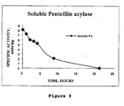
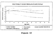


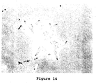


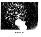


Classifications
-
- C—CHEMISTRY; METALLURGY
- C12—BIOCHEMISTRY; BEER; SPIRITS; WINE; VINEGAR; MICROBIOLOGY; ENZYMOLOGY; MUTATION OR GENETIC ENGINEERING
- C12N—MICROORGANISMS OR ENZYMES; COMPOSITIONS THEREOF; PROPAGATING, PRESERVING, OR MAINTAINING MICROORGANISMS; MUTATION OR GENETIC ENGINEERING; CULTURE MEDIA
- C12N9/00—Enzymes; Proenzymes; Compositions thereof; Processes for preparing, activating, inhibiting, separating or purifying enzymes
- C12N9/0004—Oxidoreductases (1.)
-
- A—HUMAN NECESSITIES
- A61—MEDICAL OR VETERINARY SCIENCE; HYGIENE
- A61K—PREPARATIONS FOR MEDICAL, DENTAL OR TOILETRY PURPOSES
- A61K47/00—Medicinal preparations characterised by the non-active ingredients used, e.g. carriers or inert additives; Targeting or modifying agents chemically bound to the active ingredient
- A61K47/06—Organic compounds, e.g. natural or synthetic hydrocarbons, polyolefins, mineral oil, petrolatum or ozokerite
- A61K47/08—Organic compounds, e.g. natural or synthetic hydrocarbons, polyolefins, mineral oil, petrolatum or ozokerite containing oxygen, e.g. ethers, acetals, ketones, quinones, aldehydes, peroxides
- A61K47/10—Alcohols; Phenols; Salts thereof, e.g. glycerol; Polyethylene glycols [PEG]; Poloxamers; PEG/POE alkyl ethers
-
- A—HUMAN NECESSITIES
- A61—MEDICAL OR VETERINARY SCIENCE; HYGIENE
- A61K—PREPARATIONS FOR MEDICAL, DENTAL OR TOILETRY PURPOSES
- A61K47/00—Medicinal preparations characterised by the non-active ingredients used, e.g. carriers or inert additives; Targeting or modifying agents chemically bound to the active ingredient
- A61K47/06—Organic compounds, e.g. natural or synthetic hydrocarbons, polyolefins, mineral oil, petrolatum or ozokerite
- A61K47/26—Carbohydrates, e.g. sugar alcohols, amino sugars, nucleic acids, mono-, di- or oligo-saccharides; Derivatives thereof, e.g. polysorbates, sorbitan fatty acid esters or glycyrrhizin
-
- A—HUMAN NECESSITIES
- A61—MEDICAL OR VETERINARY SCIENCE; HYGIENE
- A61K—PREPARATIONS FOR MEDICAL, DENTAL OR TOILETRY PURPOSES
- A61K47/00—Medicinal preparations characterised by the non-active ingredients used, e.g. carriers or inert additives; Targeting or modifying agents chemically bound to the active ingredient
- A61K47/30—Macromolecular organic or inorganic compounds, e.g. inorganic polyphosphates
- A61K47/36—Polysaccharides; Derivatives thereof, e.g. gums, starch, alginate, dextrin, hyaluronic acid, chitosan, inulin, agar or pectin
- A61K47/40—Cyclodextrins; Derivatives thereof
-
- A—HUMAN NECESSITIES
- A61—MEDICAL OR VETERINARY SCIENCE; HYGIENE
- A61K—PREPARATIONS FOR MEDICAL, DENTAL OR TOILETRY PURPOSES
- A61K47/00—Medicinal preparations characterised by the non-active ingredients used, e.g. carriers or inert additives; Targeting or modifying agents chemically bound to the active ingredient
- A61K47/30—Macromolecular organic or inorganic compounds, e.g. inorganic polyphosphates
- A61K47/42—Proteins; Polypeptides; Degradation products thereof; Derivatives thereof, e.g. albumin, gelatin or zein
-
- A—HUMAN NECESSITIES
- A61—MEDICAL OR VETERINARY SCIENCE; HYGIENE
- A61K—PREPARATIONS FOR MEDICAL, DENTAL OR TOILETRY PURPOSES
- A61K9/00—Medicinal preparations characterised by special physical form
- A61K9/14—Particulate form, e.g. powders, Processes for size reducing of pure drugs or the resulting products, Pure drug nanoparticles
- A61K9/16—Agglomerates; Granulates; Microbeadlets ; Microspheres; Pellets; Solid products obtained by spray drying, spray freeze drying, spray congealing,(multiple) emulsion solvent evaporation or extraction
- A61K9/1605—Excipients; Inactive ingredients
- A61K9/1629—Organic macromolecular compounds
- A61K9/1641—Organic macromolecular compounds obtained otherwise than by reactions only involving carbon-to-carbon unsaturated bonds, e.g. polyethylene glycol, poloxamers
- A61K9/1647—Polyesters, e.g. poly(lactide-co-glycolide)
-
- C—CHEMISTRY; METALLURGY
- C07—ORGANIC CHEMISTRY
- C07K—PEPTIDES
- C07K14/00—Peptides having more than 20 amino acids; Gastrins; Somatostatins; Melanotropins; Derivatives thereof
- C07K14/435—Peptides having more than 20 amino acids; Gastrins; Somatostatins; Melanotropins; Derivatives thereof from animals; from humans
- C07K14/76—Albumins
- C07K14/765—Serum albumin, e.g. HSA
-
- C—CHEMISTRY; METALLURGY
- C12—BIOCHEMISTRY; BEER; SPIRITS; WINE; VINEGAR; MICROBIOLOGY; ENZYMOLOGY; MUTATION OR GENETIC ENGINEERING
- C12N—MICROORGANISMS OR ENZYMES; COMPOSITIONS THEREOF; PROPAGATING, PRESERVING, OR MAINTAINING MICROORGANISMS; MUTATION OR GENETIC ENGINEERING; CULTURE MEDIA
- C12N9/00—Enzymes; Proenzymes; Compositions thereof; Processes for preparing, activating, inhibiting, separating or purifying enzymes
- C12N9/14—Hydrolases (3)
- C12N9/16—Hydrolases (3) acting on ester bonds (3.1)
- C12N9/18—Carboxylic ester hydrolases (3.1.1)
-
- C—CHEMISTRY; METALLURGY
- C12—BIOCHEMISTRY; BEER; SPIRITS; WINE; VINEGAR; MICROBIOLOGY; ENZYMOLOGY; MUTATION OR GENETIC ENGINEERING
- C12N—MICROORGANISMS OR ENZYMES; COMPOSITIONS THEREOF; PROPAGATING, PRESERVING, OR MAINTAINING MICROORGANISMS; MUTATION OR GENETIC ENGINEERING; CULTURE MEDIA
- C12N9/00—Enzymes; Proenzymes; Compositions thereof; Processes for preparing, activating, inhibiting, separating or purifying enzymes
- C12N9/14—Hydrolases (3)
- C12N9/78—Hydrolases (3) acting on carbon to nitrogen bonds other than peptide bonds (3.5)
- C12N9/80—Hydrolases (3) acting on carbon to nitrogen bonds other than peptide bonds (3.5) acting on amide bonds in linear amides (3.5.1)
- C12N9/84—Penicillin amidase (3.5.1.11)
-
- C—CHEMISTRY; METALLURGY
- C12—BIOCHEMISTRY; BEER; SPIRITS; WINE; VINEGAR; MICROBIOLOGY; ENZYMOLOGY; MUTATION OR GENETIC ENGINEERING
- C12Y—ENZYMES
- C12Y101/00—Oxidoreductases acting on the CH-OH group of donors (1.1)
- C12Y101/03—Oxidoreductases acting on the CH-OH group of donors (1.1) with a oxygen as acceptor (1.1.3)
- C12Y101/03004—Glucose oxidase (1.1.3.4)
-
- C—CHEMISTRY; METALLURGY
- C12—BIOCHEMISTRY; BEER; SPIRITS; WINE; VINEGAR; MICROBIOLOGY; ENZYMOLOGY; MUTATION OR GENETIC ENGINEERING
- C12Y—ENZYMES
- C12Y301/00—Hydrolases acting on ester bonds (3.1)
- C12Y301/01—Carboxylic ester hydrolases (3.1.1)
- C12Y301/01003—Triacylglycerol lipase (3.1.1.3)
-
- C—CHEMISTRY; METALLURGY
- C12—BIOCHEMISTRY; BEER; SPIRITS; WINE; VINEGAR; MICROBIOLOGY; ENZYMOLOGY; MUTATION OR GENETIC ENGINEERING
- C12Y—ENZYMES
- C12Y305/00—Hydrolases acting on carbon-nitrogen bonds, other than peptide bonds (3.5)
- C12Y305/01—Hydrolases acting on carbon-nitrogen bonds, other than peptide bonds (3.5) in linear amides (3.5.1)
- C12Y305/01001—Asparaginase (3.5.1.1)
Definitions
- This invention relates to methods for the stabilization, storage and delivery of biologically active macromolecules, such as proteins, peptides and nucleic acids.
- this invention relates to protein or nucleic acid crystals, formulations and compositions comprising them. Methods are provided for the crystallization of proteins and nucleic acids and for the preparation of stabilized protein or nucleic acid crystals for use in dry or slurry formulations.
- the crystals, crystal formulations and compositions of this invention can be reconstituted with a diluent for the parenteral administration of biologically active macromolecular components.
- the methods of this invention are useful for preparing crystals of "naked" DNA and RNA sequences that code for therapeutic or immunogenic proteins and can be administered parenterally.
- the dissolving DNA and RNA molecules, subsequently taken up by the cells and used to express the protein with the proper glycosylation pattern, can be either therapeutic or immunogenic.
- the present invention is useful for preparing crystals, crystal formulations and compositions of sense and antisense polynucleotides of RNA or DNA.
- the present invention is further directed to encapsulating proteins, glycoproteins, enzymes, antibodies, hormones and peptide crystals or crystal formulations into compositions for biological delivery to humans'and animals.
- protein crystals or crystal formulations are encapsulated within a matrix comprising a polymeric carrier to form a composition.
- the formulations and compositions enhance preservation of the native biologically active tertiary structure of the proteins and create a reservoir which can slowly release active protein where and when it is needed.
- Such polymeric carriers include biocompatible and biodegradable polymers.
- the biologically active protein is subsequently released in a controlled manner over a period of time, as determined by the particular encapsulation technique, polymer formulation, crystal geometry, crystal solubility, crystal crosslinking and formulation conditions used.
- Methods are provided for crystallizing proteins, preparing stabilized formulations using pharmaceutical ingredients or excipients and optionally encapsulating them in a polymeric carrier to produce compositions and using such protein crystal formulations and compositions for biomedical applications, including delivery of therapeutic proteins and vaccines. Additional uses for the protein crystal formulations and compositions of this invention involve protein delivery in human food, agricultural feeds, veterinary compositions, diagnostics, cosmetics and personal care compositions.
- Proteins are used in a wide range of applications in the fields of pharmaceuticals, veterinary products, cosmetics and other consumer products, foods, feeds, diagnostics, industrial chemistry and decontamination. At times, such uses have been limited by constraints inherent in proteins themselves or imposed by the environment or media in which they are used. Such constraints may result in poor stability of the proteins, variability of performance or high cost.
- CLECTM crosslinked enzyme crystal
- Crosslinked enzyme crystals retain their activity in environments that are normally incompatible with enzyme function. Such environments include prolonged exposure to proteases, organic solvents, high temperature or extremes of pH. In such environments, crosslinked enzyme crystals remain insoluble, stable' and active.
- the first problem relates to molecular stability and sensitivity of higher order tertiary structures to chemical and physical denaturation during manufacturing and storage.
- the field of biological delivery of therapeutic proteins requires that vehicles be provided which release native proteins, such as proteins, glycoproteins, enzymes, antibodies, hormones, nucleic acids and peptides at a rate that is consistant with the needs of the particular patient or the disease process.
- Macromolecules are not only susceptible to chemical, but also physical degradation. They are sensitive to a variety of environmental factors, such as temperature, oxidizing agents, pH, freezing, shaking and shear stress [ Cholewinski, M., Luckel, B. and Horn, H., Acta Helv., 71, 405 (1996 )]. In considering a macromolecule for drug development, stability factors must be considered when choosing a production process.
- Proteins, peptides and nucleic acids are increasingly employed in the pharmaceutical, diagnostic, food, cosmetic, detergent and research industries. There is a great need for alternative stabilization procedures, which are fast, inexpensive and applicable to a broad range of biological macromolecules. In particular, stabilization procedures are needed that do not rely on the excessive use of excipients, which can interfere with the functions of those biological macromolecules.
- the stability of small molecule crystalline drugs is such that they can withstand extreme forces during the formulation process (see United States patent 5, 510, 118 ). Forces associated with milling nanoparticles of crystalline material of relatively insoluble drugs include: shear stress, turbulent flow, high impact collisions, cavitation and grinding. Small molecular crystalline compounds have been recognized as being much more stable toward chemical degradation than the corresponding amorphous solid [ Pical, M.J., Lukes, A.L., Lang, J.E. and Gaines, J. Pharm. Sci., 67, 767 (1978 )]. Unfortunately, crystals of macromolecules, such as proteins and nucleic acids, present additional problems and difficulties not associated with small molecules.
- Jensen et al. co-precipitated insulin with an absorbtion enhancer for use in respiratory tract delivery of insulin (See PCT patent application WO 98/42368 ).
- the absorbtion enhancer was desribed as a surfactant, such as a salt of a fatty acid or a bile salt.
- Insulin crystals of less than 10 micrometers in diameter and lacking zinc were produced by S. Havelund (See PCT patent application WO 98/42749 ). Similarly, crystals were also produced in the presence of surfactants to enhance pulmonary administration.
- aqueous suspensions of crystalline insulin are only slightly more stable (to the degree of a factor of two) than corresponding suspensions of amorphous phase [ Brange, J., Langkjaer, L., Havelund, S. and Volund, A., Pharm. Res., 9:715 (1992 )].
- lyophilized amorphous insulin is far more stable than lyophilized crystalline insulin under all conditions investigated [ Pical, M.J. and Rigsbee, D.R., Pharm. Res., 14:1379 (1997 )].
- crystallinity of stored materials is very important, since large scale crystallization can be introduced as a final purification step and/or concentration step in clinical manufacturing processes, such as those for manufacturing therapeutics and vaccines. Moreover, large scale crystallization can replace some of the purification steps in the manufacturing process. For example, protein crystallization can streamline the production of protein formulations making it more affordable.
- solid crystalline preparations can be easily reconstituted to generate ready to use parenteral formulations having very high protein concentration.
- protein concentrations are considered to be particularly useful where the formulation is intended for subcutaneous administration.
- injection volumes of 1.5 ml or less are well tolerated.
- a protein concentration of at least 50 mg/ml is required and 100-200 mg/ml is preferred.
- protein crystals also constitute a particularly advantageous form for pharmaceutical dosage preparation.
- the crystals may be used as a basis for slow release formulations in vivo.
- particle size is of importance for the dissolution of crystals and release of activity. It is also known that the rate of release is more predictable if the crystals have substantially uniform particle size and do not contain amorphous precipitate (see European patent 0 265 214 ).
- protein crystals may be advantageously used (see PCT patent application WO 96/40049 ), on implantable devices. Implant reservoirs are generally on the order of 25-250 ⁇ l. With this volume restriction, a formulation of high concentration (greater than 10%) and a minimum amount of suspension vehicle is preferred. Protein crystals of this invention may be easily formulated in non-aqueous suspensions in such high concentrations.
- crystals are manipulated to modulate the release of macromolecules over time. For example, crystal size, shape, formulation with excipients that effect dissolution, crosslinking, level of crosslinking and encapsulation into a polymer matrix can all be manipulated to produce delivery vehicles for biological molecules.
- the present invention overcomes the above-described obstacles by employing the most stable form of an active protein, the crystalline form and either (1) adding ingredients or excipients where necessary to stabilize dried crystals or (2) encapsulating the protein crystals or crystal formulations within a polymeric carrier to produce a composition that contains each crystal and subsequently allows the release of active protein molecules.
- Any form of protein including glycoproteins, antibodies, enzymes, hormones or peptides, may be crystallized and stabilized or encapsulated into compositions according to the methods of this invention.
- the nucleic acids coding for such proteins may be similarly treated.
- the crystal(s) may be encapsulated using a variety of polymeric carriers having unique properties suitable for delivery to different and specific environments or for effecting specific functions.
- the rate of dissolution of the compositions and, therefore, delivery of the active protein can be modulated by varying crystal size, polymer composition, polymer crosslinking, crystal crosslinking, polymer thickness, polymer hydrophobicity, polymer crystallinity or polymer solubility.
- Preferred ingredients or excipients include: Salts of 1) amino acids such as glycine, arginine, aspartic acid, glutamic acid, lysine, asparagine, glutamine, proline, 2) carbohydrates, e.g. monosaccharides such as glucose, fructose, galactose, Mannose, arabinose, xylose, ribose and 3) disaccharides, such as lactose, trehalose, maltose, sucrose and 4) polysaccharides, such as maltodextrins, dextrans, starch, glycogen and 5) alditols, such as mannitol, xylitol, lactitol, sorbitol 6) glucuronic acid, galacturonic acid, 7) cyclodextrins, such as methyl cyclodextrin, hydroxypropyl- ⁇ -cyclodextrin and alike 8) inorganic salts, such as sodium
- a further preferred group of excipients or ingredients includes sucrose, trehalose, lactose, sorbitol, lactitol, inositol, salts of sodium and potssium such as acetate, phosphates, citrates, borate, glycine, arginine, polyethylene oxide, polyvinyl alcohol, polyethylene glycol, hexylene glycol, methoxy polyethylene glycol, gelatin, hydroxypropyl- ⁇ -cyclodextrin.
- Polymer a large molecule built up by the repetition of small, simple chemical units.
- the repeating units may be linear or branched to form interconnected networks.
- the repeat unit is usually equivalent or nearly equivalent to the monomer.
- Polymeric carriers polymers used for encapsulation of protein crystals for delivery of proteins, including biological delivery. Such polymers include biocompatible and biodegradable polymers.
- the polymeric carrier may be a single polymer type or it may be composed of a mixture of polymer types.
- Polymers useful as the polymeric carrier include for example, poly (acrylic acid), poly (cyanoacrylates), poly (amino acids), poly (anhydrides), poly (depsipeptide), poly (esters) such as poly (lactic acid) or PLA, poly (lactic-co-glycolic acid) or PLGA, poly (B-hydroxybutryate), poly (caprolactone) and poly (dioxanone); poly (ethylene glycol), poly ((hydroxypropyl)methacrylamide, poly [(organo)phosphazene], poly (ortho esters), poly (vinyl alcohol), poly (vinylpyrrolidone), maleic anhydridealkyl vinyl ether copolymers, pluronic polyols, albumin, natural and synthetic polypeptides, alginate, cellulose and cellulose derivatives, collagen, fibrin, gelatin, hyaluronic acid, oligosaccharides, glycaminoglycans, sulfated polysaccharides
- Protein a complex high polymer containing carbon, hydrogen, oxygen, nitrogen and usually sulfur and composed of chains of amino acids connected by peptide linkages. Proteins in this application refer to glycoproteins, antibodies, non-enzyme proteins, enzymes, hormones and peptides. The molecular weight range for proteins includes peptides of 1000 Daltons to glycoproteins of 600 to 1000 kiloDaltons. Small proteins, less than 10,000 Daltons, may be too small to be characterized by a highly organized tertiary structure, wherein said tertiary structure is organized around a hydrophobic core.
- such proteins have a molecular weight of greater than or equal to 10,000 Daltons. According to an alternate embodiment, that molecular weight is greater than or equal to 20,000 Daltons. According to another alternate embodiment, that molecular weight is greater than or equal to 30,000 Daltons. According to a further alternate embodiment, that molecular weight is greater than or equal to 40,000 Daltons. According to another alternate embodiment, that molecular weight is greater than or equal to 50,000 Daltons.
- Protein activity an activity selected from the group consisting of binding, catalysis, signaling, transport, or other activities which induce a functional response within the environment in which the protein is used, such as induction of immune response, enzymatic activity, or combinations thereof.
- Protein activity release rate the quantity of protein dissolved per unit time.
- Protein crystal protein molecules arranged in a crystal lattice. Protein crystals contain a pattern of specific protein-protein interactions that are repeated periodically in three dimensions.
- the protein crystals of this invention do not include amorphous solid forms or precipitates of proteins, such as those obtained by lyophilizing a protein solution.
- Protein crystal formulation a combination of protein crystals encapsulated within a polymeric carrier to form coated particles.
- the coated particles of the protein crystal formulation may have a spherical morphology and be microspheres of up to 500 micro meters in diameter or they may have some other morphology and be microparticulates.
- “protein crystal formulations” are included in the term “compositions”.
- Protein delivery system one or more of a protein crystal formulation or composition, a process for making the formulation or a method of administering the formulation to biological entities or means .therefor.
- Protein loading the protein content of microspheres, as calculated as a percentage by weight of protein relative to the weight of the dry formulation.
- a typical range of protein loading is from 1-80%.
- Protein release the release of active protein from a polymeric carrier, as controlled by one or more of the following factors: (1) degradation of the polymer matrix; (2) rate of crystal dissolution within the polymer matrix; (3) diffusion of dissolved protein through the polymer matrix; (4) protein loading; and (5) diffusion of biological medium into the protein crystal/polymer matrix.
- Prophylactically effective amount an amount of a protein or nucleic acid crystal or crystal formulation or composition which is effective to prevent a condition in a living organism to whom it is administered over some period of time.
- shelf stability the loss of specific activity and/or changes in secondary structure from the native protein over time incubated under specified conditions.
- Stability the loss of specific activity and/or changes in secondary structure from the native protein over time while in solution under specified conditions.
- Stabilization the process of preventing the loss of specific activity and/or changes in secondary structure from the native proteins, by preparing formulations of protein crystals or DNA crystals or RNA crystals with excipients or ingredients.
- Therapeutic protein a protein as described above, which is administered to a living organism in a formulation or composition or a pharmaceutical formulation or composition.
- Therapeutic proteins include all of the protein types described herein.
- Vaccine antigen a protein derived from a pathogenic agent such as a virus, parasite, bacteria or tumor cell.
- the protein activity of such vaccine antigens is the induction of protective immune responses specific for a pathogenic agent or tumor.
- Crystallinity of macromolecules is of great value for their storage and delivery in vivo.
- Crystals of proteins and nucleic acids must be handled with considerable care, since they are extremely fragile and contain a high proportion of solvent.
- It is well known in x-ray crystallography that the diffraction patterns from macromolecular crystals quickly degenerate upon dehydration in air. Normally, a crystal is carefully separated from its mother liquor and inserted into a capillary tube. The tube is sealed from the air using dental wax or silicone grease, along with a small amount of mother liquor inside to maintain hydration [ McPherson, A., Preparation and Analysis of Protein Crystals, Robert E.
- Another technique is to collect data from macromolecular crystals at cryogenic temperatures.
- the crystals are prepared and then rapidly cooled to prevent ice lattice formation in the aqueous medium. Instead of ice, a rigid glass forms, encasing the crystal with little damage. Crystals are then maintained at 100 °K to prevent crystal disintegrations [ Rodgers, D.W., in Methods in Enzymology (Eds., Carter, C.W. and Sweet, R.M.) Academic Press, v.276, p. 183 (1997 )]. While this technique allows one to maintain crystals outside of their mother liquor, it cannot be used at temperatures higher than 100 °K.
- dried crystals can be prepared by lyophilization.
- this technique involves rapid cooling of the material and can be applied only to freeze stable products.
- the aqueous solution is first frozen to between -40 and -50°C. Then, the ice is removed under vacuum. Ice formation is usually destructive to the protein crystal lattice, yielding a mixture of crystals and amorphous precipitate.
- the present invention provides formulations and compositions for storage of crystalline macromolecules as either solid particles or dispersed in a non-aqueous solvent. Furthermore, the invention may be applied to the storage of a single biological macromolecule or a mixture of macromolecules that do not interact with each other.
- this invention provides a method for rendering biological macromolecules suitable for storage in suspensions comprising replacing the mother liquor with a non-aqueous solvent.
- the crystalline slurry can be' rendered solid by spinning out the first solvent and washing the remaining crystalline solid using a second organic solvent to remove water, followed by evaporation of the non-aqueous solvent.
- Non-aqueous slurries of crystalline therapeutic proteins are especially useful for subcutaneous delivery, while solid formulations are ideally suited for pulmonary administration.
- Pulmonary delivery is particularly useful for biological macromolecules which are difficult to deliver by other routes of administration. (See, for example, PCT patent applications WO 96/32152 , WO 95/24183 and WO 97/41833 ).
- the proteins referred to below include protein crystals themselves, or nucleic acid crystals comprising DNA or RNA which encode those proteins upon cellular uptake.
- This invention advantageously provides compositions and formulations of crystals of proteins or nucleic acids.
- protein stability is one of the most important obstacles to successful formulation of polymer microparticulate delivery systems that control the release of proteins.
- the stability of proteins encapsulated in polymeric carriers may be challenged at three separate stages: manufacture of the protein crystal composition, protein release from the resulting composition and in vivo stability after the protein release.
- manufacture of the protein crystal composition protein release from the resulting composition
- in vivo stability after the protein release During preparation of microparticles or microspheres containing soluble or amorphous proteins, the use of organic solvents and lyophilization are especially detrimental to protein stability. Subsequently, released proteins are susceptible to moisture-induced aggregation, thus resulting in permanent inactivation.
- solid state may be divided into two categories: amorphous and crystalline.
- the three-dimensional long-range order that normally exists in a crystalline material does not exist in the amorphous state.
- the position of molecules relative to one another is more random in the amorphous or liquid states, relative to the highly ordered crystalline state..
- amorphous proteins may be less stable than their crystalline counterparts.
- Figure 1 depicts the relative stability of the following molecular states of Candida rugosa lipase ("CRL"): crosslinked amorphous, liquid, crystalline in 20% organic solvent, crosslinked crystalline in 20% organic solvent, crosslinked crystalline without organic solvent.
- Figure 1 shows that crystalline CRL retains 80% activity for more than 175 hours in 20% organic solvent. In contrast, both amorphous and soluble forms of the enzyme are completely inactivated within hours.
- the present invention advantageously utilizes the crystalline forms of proteins because of their superior stability characteristics.
- the protein constituents of the formulations and compositions of this invention may be those which are naturally or synthetically modified. They may be glycoproteins, phosphoproteins, sulphoproteins, iodoproteins, methylated proteins, unmodified proteins or contain other modifications. Such protein constituents may be any protein, including, for example, therapeutic proteins, prophylactic proteins, including antibodies, cleaning agent proteins, including detergent proteins, personal care proteins, including cosmetic proteins, veterinary proteins, food' proteins, feed proteins, diagnostic proteins and decontamination proteins.
- such proteins have a molecular weight of greater than or equal to 10,000 Daltons. According to an alternate embodiment, that molecular weight is greater than or equal to 20,000 Daltons. According to another alternate embodiment, that molecular weight is greater than or equal to 30,000 Daltons. According to a further alternate embodiment, that molecular weight is greater than or equal to 40,000 Daltons. According to another alternate embodiment, that molecular weight is greater than or equal to 50,000 Daltons.
- hydrolases include elastase, esterase, lipase, nitrilase, amylase, pectinase, hydantoinase, asparaginase, urease, subtilisin, thermolysin and other proteases and lysozyme.
- lyases include aldolases and hydroxynitrile lyase.
- oxidoreductases include peroxidase, laccase, glucose oxidase, alcohol dehydrogenase and other dehydrogenases.
- Other enzymes include cellulases and oxidases.
- therapeutic or prophylactic proteins include hormones such as insulin, glucogonlike peptide 1 and parathyroid hormone, antibodies, inhibitors, growth factors, postridical hormones, nerve growth hormones, blood clotting factors, adhesion molecules, bone morphogenic proteins and lectins trophic factors, cytokines such as TGF- ⁇ , IL-2, IL-4, ⁇ -IFN, ⁇ -IFN, ⁇ -IFN, TNF, IL-6, IL-8, lymphotoxin, IL-5, Migration inhibition factor, GMCSF, IL-7, IL-3, monocyte-macrophage colony stimulating factors, granulocyte colony stimulating factors, multidrug resistance proteins, other lymphokines, toxoids, erythropoietin, Factor VIII, amylin, TPA, dornase- ⁇ , ⁇ -1-antitripsin, human growth hormones, nerve growth hormones, bone morphogenic proteins, urease, toxoids, fertility hormones
- Therapeutic proteins such as the following, are also included:
- the protein constituent of the formulations and compositions of this invention may be any natural, synthetic or recombinant protein antigen including, for example, tetanus toxoid, diptheria toxoid, viral surface proteins, such as CMV glycoproteins B, H and gCIII, HIV-1 envelope glycoproteins, RSV envelope glycoproteins, HSV envelope glycoproteins, EBV envelope glycoproteins, VZV envelope glycoproteins, HPV envelope glycoproteins, Influenza virus glycoproteins, Hepatitis family surface antigens; viral structural proteins, viral enzymes, parasite,proteins, parasite glycoproteins, parasite enzymes and bacterial'proteins.
- viral surface proteins such as CMV glycoproteins B, H and gCIII, HIV-1 envelope glycoproteins, RSV envelope glycoproteins, HSV envelope glycoproteins, EBV envelope glycoproteins, VZV envelope glycoproteins, HPV envelope glycoproteins, Influenza virus glycoproteins, Hepatitis family surface antigens
- viral structural proteins viral enzymes,
- tumor antigens such as her2-neu, mucin, CEA and endosialin. Allergens, such as house dust mite antigen, lol p1 (grass) antigens and urushiol are included.
- Toxins such as pseudomonas endotoxin and osteopontin/uropontin, snake venom and bee venom are included.
- glycoprotein tumorassociated antigens for example, carcinoembryonic antigen (CEA), human mucins, her-2/neu and prostatespecific antigen (PSA) [ R.A. Henderson and O.J. Finn, Advances in Immunology, 62, pp. 217-56 (1996 )].
- CEA carcinoembryonic antigen
- PSA prostatespecific antigen
- the protein crystal formulations and compositions of the present invention which include microparticulate-based sustained release systems for protein drugs, advantageously permit improved patient compliance and convenience, more stable blood levels and potential dose reduction.
- the slow and constant release capabilities of the present invention advantageously permit reduced dosages, due to more efficient delivery of active protein. Significant cost savings may be achieved by using the protein formulations and compositions described herein.
- Formulations and compositions comprising protein crystals in polymeric delivery carriers according to this invention may also comprise any conventional carrier or adjuvant used in vaccines, pharmaceuticals, personal care formulations and compositions, veterinary formulations, or oral enzyme supplementation.
- carriers and adjuvants include, for example, Freund's adjuvant, ion exchangers, alumina, aluminum stearate, lecithin, buffer substances, such as phosphates, glycine, sorbic acid, potassium sorbate, partial glyceride mixtures of saturated vegetable fatty acids, water, salts or electrolytes, such as protamine sulfate, disodium hydrogen phosphate, sodium chloride, zinc salts, colloidal silica, magnesium, trisilicate, cellulose-based substances and polyethylene glycol.
- Adjuvants for topical or gel base forms may include, for example, sodium carboxymethylcellulose, polyacrylates, polyoxyethylene-polyoxypropylene-block polymers, polyethylene glycol and wood wax alcohols.
- protein crystals may be combined with any conventional materials used for controlled release administration, including pharmaceutical controlled release administration.
- materials include, for example, coatings, shells and films, such as enteric coatings and polymer coatings and films.
- Protein formulations in polymeric delivery carriers and compositions (compositions) according to this invention which may be devices, such as implantable devices and may be microparticulate protein delivery systems.
- the macromolecule crystals have a longest dimension between about 0.01 ⁇ m and about 500 ⁇ m, alternatively between about 0.1 ⁇ m and about 100 ⁇ m.
- the most preferred embodiment is that the protein crystal of protein crystal formulation components are between about 50 ⁇ m and about 100 ⁇ m in their longest dimension.
- Such crystals may have a shape selected from the group consisting of: spheres, needles, rods, plates, such as hexagons and squares, rhomboids, cubes, bipyramids and prisms.
- encapsulation of protein crystals or protein crystal formulations in polymeric carriers to make compositions may be carried out on protein crystals which are crosslinked or uncrosslinked.
- protein crystals may be obtained commercially or produced as illustrated herein.
- Protein or nucleic acid crystals or crystal formulations and compositions according to this invention may be used as ingredients in personal care compositions, including cosmetics, such as creams, lotions, emulsions, foams, washes, compacts, gels, mousses, slurries, powders, sprays, pastes, ointments, salves, balms, drops, shampoos and sunscreens.
- cosmetics such as creams, lotions, emulsions, foams, washes, compacts, gels, mousses, slurries, powders, sprays, pastes, ointments, salves, balms, drops, shampoos and sunscreens.
- topical creams and lotions for example, they may be used as humectants or for skin protection, softening, bleaching, cleaning, deproteinization, lipid removal, moisturizing, decoloration, coloration or detoxification. They may also be used as anti-oxidants in cosmetics.
- any individual including humans, animals and plants, may be treated in a pharmaceutically acceptable manner with a pharmaceutically effective amount of protein or nucleic acid crystals or a crystal formulation or composition for a period of time sufficient to treat a condition in the individual to whom they are administered over some period of time.
- individuals may receive a prophylactically effective amount of protein or nucleic acid crystals or crystal formulation or composition of this invention which is effective to prevent a condition in the individual to whom they are administered over some period of time.
- Protein or nucleic acid crystals or crystal formulations or compositions may be administered alone, as part of a pharmaceutical, personal care or veterinary preparation, or as part of a prophylactic preparation, such as a vaccine, with or without adjuvant. They may be administered by parenteral or oral routes. For example, they may be administered by oral, pulmonary, nasal, aural, anal, dermal, ocular, intravenous, intramuscular, intraarterial, intraperitoneal, mucosal, sublingual, subcutaneous, or intracranial route. In either pharmaceutical, personal care or veterinary applications, protein or nucleic acid crystal or crystal formulations or compositions may be topically administered to any epithelial surface. Such epithelial surfaces include oral, ocular, aural, anal and nasal surfaces, which may be treated, protected, repaired or detoxified by application of protein or nucleic acid crystals or crystal formulations or compositions.
- compositions comprising protein or nucleic acid crystal or crystal formulations or compositions according to this invention may also be selected from the group consisting of tablets, liposomes, granules, spheres, microparticles, microspheres and capsules.
- protein or nucleic acid crystals or crystal formulations and compositions may be formulated into tablets.
- Such tablets constitute a liquid-free, dust-free form for storage of protein or nucleic acid crystal or crystal formulations or compositions which are easily handled and retain acceptable levels of activity or potency.
- protein or nucleic acid crystals or crystal formulations or compositions may be in a variety of conventional depot forms employed for administration to provide reactive compositions. These include, for example, solid, semi-solid and liquid dosage forms, such as liquid solutions or suspensions, slurries, gels, creams, balms, emulsions, lotions, powders, sprays, foams, pastes, ointments, salves, balms and drops.
- solid, semi-solid and liquid dosage forms such as liquid solutions or suspensions, slurries, gels, creams, balms, emulsions, lotions, powders, sprays, foams, pastes, ointments, salves, balms and drops.
- Protein or nucleic acid crystals on formulations or compositions according to this invention may also comprise any conventional carrier or adjuvant used in pharmaceuticals, personal care compositions or veterinary formulations.
- carriers and adjuvants include, for example, Freund's adjuvant, ion exchangers, alumina, aluminum stearate, lecithin, buffer substances, such as phosphates, glycine, sorbic acid, potassium sorbate, partial glyceride mixtures of saturated vegetable fatty acids, water, salts or electrolytes, such as protamine sulfate, disodium hydrogen phosphate, sodium chloride, zinc salts, colloidal silica, magnesium, trisilicate, cellulose-based substances and polyethylene glycol.
- Adjuvants for topical or gel base forms may include, for example, sodium carboxymethylcellulose, polyacrylates, polyoxyethylene-polyoxypropylene-block polymers, polyethylene glycol and wood wax alcohols.
- the most effective mode of administration and dosage regimen of the protein or nucleic acid crystals or crystal formulations or compositions of this invention will depend on the effect desired, previous therapy, if any, the individual's health status or status of the condition itself and response to the protein or nucleic acid crystals or crystal formulations or compositions and the judgment of the treating physician or clinician.
- the protein or nucleic acid crystals or crystal formulations or compositions may be administered in any dosage form acceptable for pharmaceuticals, vaccinations, gene therapy, immunotherapy, personal care compositions or veterinary formulations, at one time or over a series of treatments.
- the amount of the protein or nucleic acid crystals or crystal formulations or compositions which provides a single dosage will vary depending upon the particular mode of administration, formulation, dose level or dose frequency.
- a typical preparation will contain between about 0.01% and about 99%, preferably between about 1% and about 50%, protein or nucleic acid crystals (w/w).
- a preparation will contain between about 0.01% and about 80% protein crystals, preferably between about 1% and about 50%, protein crystals (w/w).
- a preparation will contain between about 0.01% and about 80% protein crystal formulation, preferably between about 1% and about 50%, protein crystal formulation(w/w).
- a maintenance dose of protein or nucleic acid crystals or crystal formulations or compositions may be administered, if necessary. Subsequently, the dosage or frequency of administration, or both, may be reduced as a function of the symptoms, to a level at which the improved condition is retained. When the condition has been alleviated to the desired level, treatment should cease. Individuals may, however, require intermittent treatment on a long-term basis upon any recurrence of the condition or symptoms thereof.
- crystals, crystal formulations and compositions are prepared by the following process:
- enhancers such as surfactants are not added during crystallization.
- Excipients or ingredients are added to the mother liquor after crystallization, at a concentration of between about 1-10% W/W, alternatively at a concentration of between about 0.1-25% W/W, alternatively at a concentration of between about 0.1-50% W/W.
- the excipient or ingredient is incubated with the crystals in the mother liquor for about 0.1-3 hrs, alternatively the incubation is carried out for 0.1-12 hrs, alternatively the incubation is carried out for 0.1-24 hrs.
- the ingredient or excipient is dissolved in a solution other than the mother liquor, and the protein crystals are removed from the mother liquor and suspended in the excipient or ingredient solution.
- the ingredient or excipient concentrations and the incubation times are the same as those described above.
- encapsulation of lipases in polymeric carriers provide compositions useful to treat patients suffering from intestinal lipase deficiency.
- Such patients include those with pancreatic steatorrhea, due to advanced pancreatic insufficiency require oral lipase supplementation.
- current therapeutic methods may not be flexible enough to protect the active lipase during transit through the gastrointestinal tract and to release the enzyme activity where it is critically needed in the small bowel (See L. Guarner et al., "Fate of oral enzymes in pancreatic insufficiency," Gut, vol. 34, pp. 708-712, (1993 )).
- the combination of encapsulated lipase crystals (compositions) and unencapsulated crosslinked lipase crystals or formulations provides a drug therapy regime in which enzyme activity is available early on from the unencapsulated crosslinked lipase. As this material undergoes proteolytic degradation, the encapsulated enzyme (composition) begins to release enzyme activity into the more distal bowel.
- a similar strategy may be used to solve other enzyme or therapeutic protein supplementation problems.
- the present invention may also utilize other slow release methodologies, such as silicon based rings or rods which have been preloaded with encapsulated protein crystals containing hormones, antibodies or enzymes or compositions containing them.
- the purpose of this technique is to provide a constant level of protein to the bloodstream over a period of weeks or months. Such implants can be inserted intradermally and can be safely replaced and removed when needed.
- compositions include vaccine formulations and compositions comprising protein (antigen) crystals, adjuvant and encapsulating polymer(s).
- the protein antigen may be a viral glycoprotein, viral structural protein, viral enzyme, bacterial protein, or some engineered homolog of a viral or bacterial protein, or any immunopotentiating protein, such as a cytokine.
- One embodiment of such formulations or compositions involves a single vaccine injection containing microspheres having three or more different release profiles. In this way, antigen formulations or composition may be released over a sustained period sufficient to generate lasting immunity.
- multiple antigen boosts may be in single unit form.
- the faster degrading preparation (composition) may contain an immunogenic adjuvant to enhance the immune response.
- One advantage of such a system is that by using protein crystals, the native three-dimensional structures of the epitopes are maintained and presented to the immune system in their native form.
- a combination vaccine could be produced, whereby immunity to multiple diseases is induced in a single injection.
- microspheres having different release profiles may be combined alone or in formulations and compositions and may include microspheres containing antigens from multiple infectious agents to produce a combination vaccine (formulations and compositions).
- microspheres having multiple release profiles and containing antigen crystals of measles, mumps, rubella, polio and hepatitis B agents could be combined and administered to children.
- microspheres having multiple release profiles and containing crystals of different isolates of HIV gp120 could be combined to produce a vaccine for HIV-1 or HIV-2.
- the protein crystals encapsulated within polymeric carriers and forming a composition comprising microspheres can be dried by lyophilization. Lyophilization, or freeze-drying allows water to be separated from the composition. The protein crystal composition is first frozen and then placed in a high vacuum. In a vacuum, the crystalline H 2 O sublimes, leaving the protein crystal composition behind containing only the tightly bound water. Such processing further stabilizes the composition and allows for easier storage and transportation at typically encountered ambient temperatures.
- ampules single dose sterile containers
- ampules any desired increment of a single dose as a slurry, in a formulation or a composition.
- the ampules containing the dispensed slurries, formulations or compositions can then be capped, batch frozen and lyophilized under sterile conditions.
- sterile containers can be transported throughout the world and stored at ambient temperatures. Such a system would be useful for providing sterile vaccines and therapeutic proteins to remote and undeveloped parts of the world.
- the ampule is rehydrated with the sterile solvent or buffer of choice and dispensed. Under this scenario, minimal or no refrigeration is required.
- Protein crystals are grown by controlled crystallization of protein from aqueous solutions or aqueous solutions containing organic solvents.
- Solution conditions that may be controlled include, for example, the rate of evaporation of solvent, organic solvents, the presence of appropriate co-solutes and buffers, pH and temperature.
- references can be used to determine the conditions necessary for crystallization of a protein, as a prelude to the formation of appropriate protein crystals and can guide the crystallization strategy for other proteins.
- an intelligent trial and error search strategy can, in most instances, produce suitable crystallization conditions for many proteins, provided that an acceptable level of purity can be achieved for them [see, e.g., C.W. Carter, Jr. and C.W. Carter, J. Biol. Chem., 254, pp. 12219-23 (1979 )].
- crystals are produced by combining the protein to be crystallized with an appropriate aqueous solvent or aqueous solvent containing appropriate crystallization agents, such as salts or organic solvents.
- the solvent is combined with the protein and may be subjected to agitation at a temperature determined experimentally to be appropriate for the induction of crystallization and acceptable for the maintenance of protein activity and stability.
- the solvent can optionally include co-solutes, such as divalent cations, co-factors or chaotropes, as well as buffer species to control pH. The need for co-solutes and their concentrations are determined experimentally to facilitate crystallization.
- Crystalline material is a form of the solid state of matter, which is distinct from the amorphous solid state. Crystals display characteristic features including a lattice structure, characteristic shapes and optical properties such as refractive index and birefringence. A crystal consists of atoms arranged in a pattern that repeats periodically in three dimensions. In contrast, amorphous material is a non-crystalline solid form of matter, sometimes referred to as an amorphous precipitate. Such precipitates have no molecular lattice structure characteristic of the crystalline solid state and do not display birefringence or other spectroscopic characteristics typical of the crystalline forms of matter.
- the controlled precipitation leading to crystallization can best be carried out by the simple combination of protein, precipitant, co-solutes and, optionally, buffers in a batch process.
- proteins may be crystallized by using protein precipitates as the starting material. In this case, protein precipitates are added to a crystallization solution and incubated until crystals form.
- Alternative laboratory crystallization methods such as dialysis or vapor diffusion, can also be adopted. McPherson , supra and Gilliland , supra, include a comprehensive list of suitable conditions in their reviews of the crystallization literature.
- the release rate of the protein from the polymeric carrier (composition) may be slowed and controlled by using protein crystals that have been chemically crosslinked using a crosslinker, such as for example, a biocompatible crosslinker.
- a crosslinker such as for example, a biocompatible crosslinker.
- Crosslinking may be carried out using reversible crosslinkers, in parallel or in sequence.
- the resulting crosslinked protein crystals are characterized by a reactive multi-functional linker, into which a trigger is incorporated as a separate group.
- the reactive functionality is involved in linking together reactive amino acid side chains in a protein and the trigger consists of a bond that can be broken by altering one or more conditions in the surrounding environment (e.g., pH, temperature, or thermodynamic water activity). This is illustrated diagrammatically as: X - Y - Z + 2 AA residues - - > AA 1 - X - Y - Z - AA 2 change in environment - - > AA 1 - X + Y - Z - AA 2
- the reactive functionality of the crosslinker and the trigger may be the same, as in: Y - Z + 2 ⁇ AA residues - - > AA 1 - X - Z - AA 2 change in environment - - > AA 1 + X - Z - AA 2 .
- the reactive functionality X and Y may be, but not limited to the following functional groups (where R, R', R" and R'" may be alkyl, aryl or hydrogen groups):
- R is a multifunctional crosslinking agent that can be an alkyl, aryl, or other chains with activating groups that can react with the protein to be crosslinked. Those reactive groups can be any variety of groups such as those susceptible to nucleophilic, free radical or electrophilic displacement including halides, aldehydes, carbonates, urethanes, xanthanes, epoxides among others.
- Table 1 Trigger Examples Release Conditions 1. Acid Labile Linkers R-O-R e.g. Thp, MOM, H + or Lewis Acidic catalysts • Acetal, ketal • Aldol, Michael adducts, esters 2.
- Free Radical Labile Linkers • Thiohydroxamate ester (Barton ester) • Free radical initiator 11.
- Metalchelate linked • Iron (III) diphenanthroline • Metal removal e.g. by chelation or precipitation 12.
- Thermally Labile Linkers • Peroxides R-OO-R • Increase in temperature 13.
- "Safety Catch" Labile Linkers • Methylthioethyl (Mte) Base; amines, others • Dithianes
- reversible crosslinkers are disulfide bond-containing crosslinkers.
- the trigger breaking crosslinks formed by such crosslinkers is the addition of reducing agent, such as cysteine, to the environment of the crosslinked protein crystals.
- Disulfide crosslinkers are described in the Pierce Catalog and Handbook (1994-1995) and more recently in "Bioconjugate Techniques", By G.T. Hermanson, (1996), Academic Press , Division of Harcourt Brace & Company, 525 B Street, Suite 1900, San Diego, CA 92101-4495 .
- crosslinkers examples include:
- Crosslinked protein crystals useful in the protein formulations of the present invention may also be prepared according to the methods set forth in PCT patent application PCT/US91/05415 .
- compositions are produced when protein crystals are encapsulated in at least one polymeric carrier to form microspheres by virtue of encapsulation within the matrix of the polymeric carrier to preserve their native and biologically active tertiary structure.
- the crystals can be encapsulated using various biocompatible and/or biodegradable polymers having unique properties which are suitable for delivery to different biological environments or for effecting specific functions. The rate of dissolution and, therefore, delivery of active protein is determined by the particular encapsulation technique, polymer composition, polymer crosslinking, polymer thickness, polymer solubility, protein crystal geometry and degree and, if any, of protein crystal crosslinking
- Protein crystals or formulations to be encapsulated are suspended in a polymeric carrier which is dissolved in an organic solvent.
- the polymer solution must be concentrated enough to completely coat the protein crystals or formulations after they are added to the solution. Such an amount is one which provides a weight ratio of protein crystals to polymer between about 0.02 and about 20, preferably between about 0.1 and about 2.
- the protein crystals are contacted with polymer in solution for a period of time between about 0.5 minutes and about 30 minutes, preferably between about 1 minutes and about 3 minutes.
- the crystals should be kept suspended and not allowed to aggregate as they are coated by contact with the polymer.
- the crystals become coated and are referred to as nascent microspheres.
- the nascent microspheres increase in size while coating occurs.
- the suspended coated crystals or nascent microspheres along with the polymeric carrier and organic solvent are transferred to a larger volume of an aqueous solution containing a surface active agent, known as an emulsifier.
- the suspended nascent microspheres are immersed in the aqueous phase, where the organic solvent evaporates or diffuses away from the polymer. Eventually, a point is reached where the polymer is no longer soluble and forms a precipitated phase encapsulating the protein crystals or formulations to form a composition.
- This aspect of the process is referred to as hardening of the polymeric carrier or polymer.
- the emulsifier helps to reduce the interfacial surface tension between the various phases of matter in the system during the hardening phase of the process.
- the coating polymer has some inherent surface activity, there may be no need for addition of a separate surface active agent.
- Emulsifiers useful to prepare encapsulated protein crystals according to this invention include poly(vinyl alcohol) as exemplified herein, surfactants and other surface active agents which can reduce the surface tension between the polymer coated protein crystals or polymer coated crystal formulations and the solution.
- Organic solvents useful to prepare the microspheres of the present invention include methylene chloride, ethyl acetate, chloroform and other non-toxic solvents which will depend on the properties of the polymer. Solvents should be chosen that solubilize the polymer and are ultimately non-toxic.
- a preferred embodiment of this invention is that the crystallinity of the protein crystals is maintained during the encapsulation process.
- the crystallinity is maintained during the coating process by using an organic solvent in which the crystals are not soluble. Subsequently, once the coated crystals are transferred to the aqueous solvent, rapid hardening of the polymeric carrier and sufficient coating of the crystals in the previous step shields the crystalline material from dissolution.
- the use of crosslinked protein crystals facilitates maintenance of crystallinity in both the aqueous and organic solvents.
- the polymers used as polymeric carriers to coat the protein crystals can be either homo-polymers or co-polymers.
- the rate of hydrolysis of the microspheres is largely determined by the hydrolysis rate of the individual polymer species. In general, the rate of hydrolysis decreases as follows: polycarbonates > polyesters > polyurethanes > polyorthoesters > polyamides.
- the polymeric carrier is composed of a single polymer type such as PLGA.
- the polymeric carrier can be a mixture of polymers such as 50% PLGA and 50% albumin.
- biocompatible/biodegradable polymers selected from the group consisting of poly (acrylic acid), poly (cyanoacrylates), poly (amino acids), poly (anhydrides), poly (depsipeptide), poly (esters), such as poly (lactic acid) or PLA, poly (b-hydroxybutryate), poly (caprolactone) and poly (dioxanone); poly (ethylene glycol), poly (hydroxypropyl)methacrylamide, poly [(organo)phosphazene], poly (ortho esters), poly (vinyl alcohol), poly (vinylpyrrolidone), maleic anhydridealkyl vinyl ether copolymers, pluronic polyols, albumin, alginate, cellulose and cellulose derivatives, collagen, fibrin, gelatin, hyaluronic acid, oligosaccharides, glycaminoglycans, sulfated polysaccharides,
- protein crystals are encapsulated in at least one polymeric carrier using a double emulsion method, as illustrated herein, using a polymer, such as polylactic-co-glycolyic acid.
- a polymer such as polylactic-co-glycolyic acid.
- the polymer is Polylactic-co-glycolyic acid (“PLGA”).
- PLGA is a copolymer prepared by polycondensation reactions with lactic acid (“L”) and glycolic acid (“G”).
- L lactic acid
- G glycolic acid
- Various ratios of L and G can be used to modulate the crystallinity and hydrophobicity of the PLGA polymer. Higher crystallinity of the polymer results in slower dissolution.
- PLGA polymers with 20-70% G content tend to be amorphous solids, while high level of either G or L result in good polymer crystallinity.
- D.K. Gilding and A.M. Reed "Biodegradable polymers for use in surgerypoly(glycolic)/poly(lactic acid) homo and copolymers: 1., Polymer vol. 20, pp. 1459-1464 (1981 ).
- PLGA degrades after exposure to water by hydrolysis of the ester bond linkage to yield non-toxic monomers of lactic acid and glycolic acid.
- Double-walled polymer coated microspheres may be advantageous.
- Double-walled polymer coated microspheres may be produced by preparing two separate polymer solutions in methylene chloride or other solvent which can dissolve the polymers. The protein crystals are added to one of the solutions and dispersed. Here, the protein crystals become coated with the first polymer. Then, the solution containing the first polymer coated protein crystals is combined with the second polymer solution. [See Pekarek, K.J.; Jacob, J.S. and Mathiowitz, E. Double-walled polymer microspheres for controlled drug release, Nature, 367, 258-260 ]. Now, the second polymer encapsulates the first polymer which is encapsulating the protein crystal. Ideally, this solution is then dripped into a larger volume of an aqueous solution containing a surface active agent or emulsifier. In the aqueous solution, the solvent evaporates from the two polymer solutions and the polymers are precipitated.
- the above process can'be performed using either protein crystals, DNA or RNA crystals, of formulations of any of these to produce compositions.
- Formulations according to this invention comprise a protein crystal, and, at least one ingredient.
- Such formulations are characterized by at least a 60 fold greater shelf life when stored at 50°C than the soluble form of said protein in solution at 50 °C, as measured by T 1/2 .
- they are characterized by at least a 59 fold greater shelf life when stored at 40 °C and 75% humidity than the nonformulated form of said protein crystal when stored at 40 °C and 75% humidity, as measured by T 1/2 .
- they are characterized by at least a 60% greater shelf life when stored at 50 °C than the nonformulated form of said protein crystal when stored at 50 °C, as measured by T 1/2 .
- they are characterized by the loss of less than 20% ⁇ -helical structural content of the protein after storage for 4 days at 50 °C, wherein the soluble form of said protein loses more than 50% of its ⁇ -helical structural content after storage for 6 hours at 50 °C, as measured by FTIR.
- they are characterized by the loss of less than 20% ⁇ -helical structural content of the protein after storage for 4 days at 50 °C, wherein the soluble form of said protein loses more than 50% of its ⁇ -helical structural content after storage for 6 hours at 50 °C, as measured by FTIR, and wherein said formulation is characterized by at least a 60 fold greater shelf life when stored at 50 °C than the soluble form of said protein in solution at 50 °C, as measured by T 1 / 2 .
- compositions according to this invention comprise one of the above described protein crystal formulations, and, at least one polymeric carrier, wherein said formulation is encapsulated within a matix of said polymeric carrier.
- compositions according to this invention comprise formulations of a protein crystal and at least one ingredient.
- Such compositions may be characterized by at least a 60 fold greater shelf life when stored at 50 °C than the soluble form of said protein in solution at 50 °C, as measured by T 1/2 .
- they are characterized by at least a 59 fold greater shelf life when stored at 40 °C and 75% humidity than the nonformulated form of said protein crystal when stored at 40°C and 75% humidity, as measured by T 1/2 .
- they are characterized by at least a 60% greater shelf life when stored at 50 °C than the nonformulated form of said protein crystal, when stored at 50 °C, as measured by T 1/2 .
- they are characterized by the loss of less than 20% ⁇ -helical structural content of the protein after storage for 4 days at 50 °C, wherein the soluble form of said protein loses more than 50% of its ⁇ -helical structural content after storage for 6 hours at 50 °C, as measured by FTIR.
- they are characterized by the loss of less than 20% ⁇ -helical structural content of the protein after storage for 4 days at 50 °C, wherein the soluble form of said protein loses more than 50% of its ⁇ -helical structural content after storage for 6 hours at 50 °C, as measured by FTIR, and wherein said formulation is characterized by at least a 60 fold greater shelf life when stored at 50 °C than the soluble form of said protein in solution at 50 °C, as measured by T 1/2 .
- a 1 kg aliquot of lipase powder was mixed well with 1 kg of celite and then 22 L of distilled water was added. The mixture was stirred to dissolve the lipase powder. After dissolution was complete, the pH was adjusted to 4.8 using acetic acid. Next, the solution was filtered to remove celite and undissolved materials. Then, the filtrate was pumped through a 30k cut-off hollow fiber to remove all the proteins that were less than 30kD molecular weight. Distilled water was added and the lipase filtrate was pumped through the hollow fiber until the retentate conductivity was equal to the conductivity of the distilled water. At this point, the addition of distilled water was stopped and 5mm Ca-acetate buffer was added.
- Ca-acetate buffer was delivered by pumping through the hollow fiber until the conductivity of the retentate was equal to the conductivity of the Ca-acetate buffer. At that point, addition of the buffer was stopped.
- the lipase solution was concentrated to 30 mg/ml solution.
- the crystallization was initiated by pumping MPD slowly into the lipase solution while stirring. Addition of MPD was continued until a 20% vol/vol of MPD was reached. The mixture was stirred for 24 hr or until 90% of the protein had crystallized. The resulting crystals were washed with crystallization buffer to remove all the soluble material from the crystals. Then, the crystals were suspended in fresh crystallization buffer to achieve a protein concentration of 42 mg/ml.
- lipase crystals were formulated in the slurry form in the presence of mother liquor before drying.
- Sucrose Sigma Chemical Co., St. Louis, MO
- Sufficient sucrose was added to lipase crystals at a protein concentration of 20 mgs/ml in mother l iquor (10 mM sodium acetate buffer, pH 4.8 containing 10 mM Calcium chloride and 20% MPD) to reach a final concentration of 10%.
- the resulting suspension was tumbled at room temperature for 3 hr. After treatment with sucrose, the crystals were separated from the liquid by centrifugation as described in Example 6, method 4 or 5.
- the lipase crystals were formulated as in Example 2, by adding trehalose, instead of sucrose, (Sigma Chemical Co., St. Louis, MO), to a final concentration of 10% in mother liquor.
- the resulting suspension was tumbled at room temperature for 3 hr and the crystals were separated from the liquid by centrifugation as described in Example 6, method 4 or 5.
- Lipase crystals were formulated using 0.1% polyethylene oxide in water as follows. The crystals, in the mother liquor at 20 mg/ml were separated from the mother liquor by centrifugation at 1000 rpm in a Beckman GS-6R bench top centrifuge equipped with swinging bucket rotor. Next, the crystals were suspended in 0.1% polyethylene oxide for 3 hrs (Sigma Chemical Co., St. Louis, MO) and then separated by centrifugation, as described in Example 6, method 4 or 5.
- Lipase crystals were formulated as in Example 2, by adding 10% methoxypoly ethylene glycol, instead of sucrose, (final concentration) (Sigma Chemical Co., St. Louis, MO) in mother liquor and separating after 3 hrs by centrifugation, as in Example 6, method 4 or 5.
- a sample of soluble lipase was prepared by dissolving lipase crystals to 20 mg/ml in phosphate buffered saline, pH 7.4.
- Figure 2 shows the stability for soluble lipase. Specific activity decreases extremely rapidly with time. Within 2-3 hours, the specific activity decreases from about 660 ⁇ moles/min/mg protein to about 100 ⁇ moles/min/mg protein or an approximate 85% decrease. The T 1/2 for soluble lipase was calculated to be 1.12 hours.
- Lipase crystals from Examples 1-7 were assessed for activity against olive oil in pH 7.7 buffer.
- the assay was carried out titrimetrically using slight modifications to the procedure described in Pharmaceutical Enzymes - Properties and Assay Methods, R. Ruyssen and A. Lauwers, (Eds.), Scientific Publishing Company, Ghent, Belgium (1978 ).
- the lipase substrate solution (solution 7) was warmed to 37°C in a water bath. First, 20 ml of substrate was added to a reaction vessel and the pH was adjusted to 7.7 using 0.05 M NaOH (solution 2) and equilibrated to 37°C with stirring. The reaction was initiated by adding enzyme. The reaction progress was monitored by titrating the mixture of enzyme and substrate with 0.05 M NaOH to maintain the pH at 7.7.
- the specific activity ( ⁇ moles/min/mg protein) was equal to the initial rate x 1000 x concentration of the titrant/the amount of enzyme.
- the zero point was determined by running the reaction without enzyme, i.e., using buffer in the place of enzyme in the reaction mixture.
- the shelf activity of the dried crystals from Examples 1-5 was measured using the olive oil assay as described in Example 8. Dried crystals (5 mg) were dissolved in 1 ml of phosphate buffered saline (“PBS”), pH 7.4 and the activity was measured using olive oil as substrate.
- PBS phosphate buffered saline
- the shelf stability of dried crystalline lipase formulations from Examples 2-5 was carried out in a humidity chamber controlled at 75% relative humidity and 40°C temperature (HOTPACK).
- the activity of the crystals was measured by dissolving 5 mg of the dried samples in PBS buffer, pH 7.4, measuring the activity in the olive oil assay and then comparing with the initial results.
- Figure 3 shows the shelf stability profile of lipase crystals formulated with sucrose, trehalose and PEO.
- PEO was the most protective excipient, followed by sucrose and then trehalose.
- Figure 4 shows the shelf stability profile of lipase crystals formulated with sucrose, trehalose, PEO and MOPEG.
- the excipients PEO, sucrose and MOPEG were similar in their ability to preserve enzyme activity as measured by their effect on the T 1/2 .
- Trehalose was less protective of lipase activity than the other excipients.
- T 1/2 The time required for the specific activity of the enzyme to decrease by 50% is known as the T 1/2 .
- Table 2 shows the effect on the T 1/2 of the specific activity for formulations of dried lipase crystals.
- sucrose was the most protective excipient, followed by polyethylene oxide (PEO), methoxypoly ethylene glycol (MOPEG) and finally trehalose.
- PEO polyethylene oxide
- MOPEG methoxypoly ethylene glycol
- trehalose Sucrose was more than 10-fold more protective, as measured by its effect on T 1/2 of specific activity.
- Table 2 Lipase at 40°C and 75% humidity Dried Lipase Crystals Excipients T 1/2 (days) none 1.52 Sucrose 1092 Trehalose 90 PEO 835 MOPEG 434 Soluble lipase 0.0468 (1.124 hrs)
- T 1/2 was calculated from the shelf life data by non-linear regression analysis using the Sigma Plot program.
- Table 2 shows that formulations of lipase were 1,923 fold more stable than soluble when PEO was used as the excipient.
- formulations of lipase were 23,300 fold more stable than soluble lipase when sucrose was used as the excipient (Table 2).
- Formulations using MOPEG and PEO as excipients with lipase crystals were 9,270 and 17,800 fold more stable than soluble' lipase (Table 2).
- crystals formulated with trehalose were 59 fold more stable than non-formulated lipase crystals made without an excipient at 40°C, as shown in Table 2.
- crystals formulated with MOPEG were 286 fold more stable than non-formulated crystals
- crystals formulated with PEO were 549 fold more stable than non-formulated lipase crystals made without an excipient at 40 °C, as shown in Table 2.
- crystals formulated with sucrose were 718 fold more stable than non-formulated lipase crystals made without an excipient at 40 °C, as shown in Table 2.
- Moisture content was determined by the Karl Fischer method according to manufacturer's instructions using a Mitsubishi CA-06 Moisture Meter equipped with a VA-06 Vaporizer (Mitsubishi Chemical Corporation, Tokyo, Japan). Table 3. Moisture content lipase crystal formulations TIME,DAYS % Moisture SUCROSE TREHALOSE PEO MOPEG 0 7.23 5.58 8.56 7.03 129 11.11 10.45 10.43 10.01
- the crystal integrity of the formulations were measured by quantitative microscopic observations. In order to visualize whether the crystals were maintained their shape after drying, the dried crystals were examined under an Olympus BX60 microscope equipped with DXC-970MD 3CCD Color Video Camera with Camera Adapter (CMA D2) with Image ProPlus software. Samples of dried crystals were covered with a glass coverslip, mounted and examined under 10X magnification, using an Olympus microscope with an Olympus UPLAN F1 objective lens 10X/0.30 PH1 (phase contrast).
- FTIR Fourier transform infrared
- a soluble lipase sample was prepared by dissolving lipase crystals in phosphate buffered saline and analyzed for stability by FTIR.
- Table 4 shows that for soluble lipase, approximately 50% and 75 % of the ⁇ -helix and ⁇ -sheet structure content was lost over three days. There was a corresponding increase in the content of random structure.
- Table 4 shows that crystals which were formulated and dried were much more stable than the soluble enzyme. Such crystals showed a loss of ⁇ -helical structure which ranged from 6.1% to 21.1% after 66 days at 40 °C and 75% humidity. At the same time, random coil in solution increased from 7-50% over 3 days, while crystallinity showed minimum random coil content even after 66 days.
- Seed crystals were prepared by washing a sample of crystals free of precipitate with a solution of 50% saturated ammonium sulfate in 100 mM phosphate buffer at pH 5.5. The seeded crystallization solution was incubated overnight at 4°C on a vigorously rotating platform. Crystals in the shape of rods (20 ⁇ m) appeared in the solution overnight after approximately 16 hr.
- HSA human serum albumin
- the HSA crystals were then dried by the four methods described in Example 6.
- the crystals were suspended in cold (4°C) ethanol as the organic solvent of method 4.
- Soluble human serum albumin preparation is Soluble human serum albumin preparation:
- the soluble HSA sample was prepared by dissolving HSA crystals at 20mg/ml in water.
- Moisture content was determined by the Karl Fischer method according to manufacturer's instructions using a Mitsubishi CA-06 Moisture Meter equipped with a VA-06 Vaporizer (Mitsubishi Chemical Corporation, Tokyo, Japan). Table 5. Moisture content HSA crystals TIME, DAYS %Moisture Gelatin 0 5.0464 12 6.3841
- the FTIR spectra were collected on a Nicolet model 550 Magna series spectrometer as described by Dong et al. [ Dong, A., Caughey, B., Caughey, W.S., Bhat, K.S. and Coe, J.E. Biochemistry, 1992; 31:9364-9370 ; Dong, A. Prestrelski, S.J., Allison, S.D. and Carpenter, J.F. J. Pharm. Sci., 1995; 84: 415-424 ]. For the solid samples, 1 to 2 mg of the protein was lightly ground with 350 mg of KBr powder and filled into small cups used for diffuse reflectance accessory. The spectra were collected and processed using Grams 32 from Galactic software for the determination of relative areas of the individual components of secondary structure using a second derivative and curve-fitting program under amide I region (1600 -1700 cm -1 ).
- the soluble HSA sample was prepared by dissolving HSA crystals in water and tested for stability by FTIR.
- the secondary structure was determined as in Example 9. Table 6. Secondary Structure content of HSA Sample ⁇ - Helix ⁇ - Sheets ⁇ -Turn Extended coil Random HSA- Soluble 45.47 6.83 6.79 40.91 0.00 HSA- Sol. 15 min 28.71 30.39 8.59 28.07 4.24 HSA- Sol. 30 min 19.15 34.69 8.16 27.91 10.09 HSA- Sol.
- Soluble HSA showed a rapid 78% decrease in ⁇ -helical content after 1 hour in solution and a corresponding increase in random coil structure.
- Dry formulated crystals showed an approximately 16% decrease in ⁇ -helical content, a large decrease in ⁇ -sheet content and no increase in the content of random structure.
- An ammonium sulfate suspension of Penicillin acylase from Boehringer Mannheim was the raw material.
- the suspension of Penicillin acylase was diluted 1:3 with deionized water. This solution was concentrated by diafilration using a 30K membrane to a final concentration of 200 OD at A 280nm /ml.
- the enzyme solution was then diluted with 4 M NaH 2 PO 4 .H 2 0 to 150 OD A 280nm /ml.
- a biphasic solution of 4 M and 1 M sufficient NaH 2 PO 4 .H 2 0 to yield a final solution concentration of 1.9 M NaPO 4 was prepared.
- 280 ml of 1 M NaH 2 PO 4 .H 2 0 was carefully overlaid on top of to the top of 549 ml of 4 M NaH 2 PO 4 .H 2 0.
- the enzyme solution was poured gently into the side of the container to form a layer above the 1 M layer.
- An overhead stirrer was set up with a marine impeller. The agitator was placed into the container with the blades just below the 4M/1M interface. The speed was adjusted to a setting of 8.0, or 600 rpm. The agitator was switched on and stopped after 10 minutes.
- the impeller was removed from the container.
- the volume of seed crystals was measured with a graduated pipette using between 0.5 and 0.1% by volume and added to the 20 L sterile polycarbonate container.
- the seed was not allowed to be static for more than 10 seconds before addition.
- the solution was mixed by hand using a flat-blade impeller for about 1 minute.
- the crystallization mixture was allowed to stand for 24 hours.
- the container was opened and the solution was mixed by hand for 30 seconds. The solution was allowed to sit for an additional 24 hours. After a total of 48 hours, a 10 ml sample was taken of the supernatant. The sample was filtered with a 0.2 micron Acrodisc. The A 280nm of the supernatant was measured. When supernatant A 280nm was greater than 1.0 mg/ml, the crystallization was allowed to continue for 24 more hours. When the supernatant concentration was less than 1.0 mg/ml, the A 280nm of crystal slurry and supernatant directly were determined.
- the basis of the activity assay for Penicillin acylase involves a titrimetric assay which measures the enzymatic hydrolysis of benzyl penicillin by the enzyme.
- the enzyme catalyzes cleavage of a phenyl acetyl group from penicillin G thus causing a decrease in pH.
- This activity was followed by measuring the volume of 50 mM NaOH needed (to maintain a pH o'f 8 at 28°C) per minute of reaction.
- the assay uses a substrate buffer made with potassium chloride and Tris and was reported in U/mg.
- Penicillin acylase assay
- penicillin-G 1 g was added to 10 ml of 1.0 M KCl, 20 mM Tris buffer, pH 8.0 solution and about 70 ml of DI water, 1, ml of 10 mM Tris, 10 mN CaCl 2 , Then the pH of the solution was adjusted to 8.0 using 0.05 N NaOH. Next, final solution volume was adjusted to 100 ml. The solution was prepared fresh before each use and used within five hours of preparation.
- Penicillin acylase was dissolved in PBS buffer, pH 7.4. Typically, 2 to 4 mg (dry weight) of protein were used for each assay.
- penicillin acylase substrate solution 20 ml was added to a titration vessel and equilibrated to 28 °C. After adding the enzyme solution to the reaction vessel, the hydrolysis of penicillin-G was monitored by titrating the reaction mixture with 0.05 N NaOH to maintain the pH at 8.0.
- the crystals were formulated with excipients.
- PA crystals were formulated in the slurry form in the presence of mother liquor before drying.
- Hydroxypropyl- ⁇ -cyclodextrin (HPCD) (Sigma Chemical Co., St. Louis, MO) was added to PA crystals in mother liquor as an excipient.
- HPCD Hydroxypropyl- ⁇ -cyclodextrin
- Sufficient HPCD was added to lipase crystals at a protein concentration 20 mgs/m in mother liquor (1.9M NaH 2 PO 4 , pH 6.6), to reach a final concentration of 10%.
- the resulting suspension was tumbled at room temperature for 3 hr. After treatment with HPCD, the crystals were separated from the liquid by centrifugation as described in Example 6, method 1.
- the PA crystals were formulated using mother liquor (1.9M NaH 2 PO 4 , pH 6.6). The crystals were separated by centrifugation as in Example 16.
- the PA crystals from Examples 15-17 were then dried according to the methods 1, 3 or 4 of Example 6.
- the organic solvent used for method 4 of Example 6 was ethyl acetate.
- a sample of soluble PA was prepared by dissolving PA crystals in water at 20 mg/ml.
- Example 18 The activity of the dried crystals from Example 18 was tested in the Pen G assay as described in Example 15. The dried crystals (5 mg) were dissolved in 1 ml of water and the activity was measured using Pen G as substrate
- FIG. 10 depicts the shelf stability profiles of PA crystals with and without excipient.
- Figure 10 also depicts shelf stability profiles of PA crystal formulations dried with nitrogen (method 1) or air dried (method 4). Table 7.
- the T 1/2 was calculated from the shelf life data by non-linear regression analysis using the Sigma Plot program.
- the stability of the formulated crystals relative to the non-formulated crystals was enhanced, as shown in Table 7.
- crystals formulated with HPCD were 418 fold more stable soluble PA at 55 °C (Table 7).
- Crystals formulated with HPCD were 3.5 fold more stable at 55 °C than non-formulated PA crystals made without an excipient, as shown in Table 7.
- Moisture content was ,determined by the Karl Fischer method according to manufacturer's instructions using a Mitsubishi CA-06 Moisture Meter equipped with a VA-06 Vaporizer (Mitsubishi Chemical Corporation, Tokyo, Japan). Table 8. Moisture content of PA crystals %Moisture DAYS PA HPCD 0 2.6198 1.5722 43 2.1792 3.1963
- Preparative scale crystallization generally involves 100 to 900 mls of glycoprotein.
- Glucose oxidase was diafiltered in water and concentrated to an A 280 of between 5 and 15.
- the glucose oxidase concentrate was mixed (1:1) with one volume of the crystallizing reagent containing 18% PEG 6000, 32% 2-propanol in 0.2 M Na-Acetate at pH 5.0. After mixing, the solution was cooled to 6°C.
- the glucose oxidase crystallization solution was stirred for 24 hours at 100 rpm with a propeller stirrer. During this time, the crystals formed gradually.
- GOD crystals were formulated in the slurry form in the presence of mother liquor before drying.
- Trehalose Sigma Chemical Co., St. Louis, MO
- Sufficient trehalose was added to GOD crystals at a protein concentration 20 mgs/ml in mother liquor (100 mM sodium acetate buffer, pH 5.5 containing 32% isopropanol and 9% PEG 6000) to reach a final concentration of 10%.
- the resulting suspension was tumbled at room temperature for 3 hr. After treatment with trehalose, the crystals were separated from the liquid by centrifugation as described in Example 6, method 1.
- Glucose oxidase crystals were formulated as in Example 22 by adding lactitol (Sigma Chemical Co. St. Louis, MO), (instead of trehalose) to a final concentration of 10% to the mother liquor. The crystals were separated from the mother liquor/lactitol solution after three hours by centrifugation.
- Glucose oxidase crystals were formulated using hydroxypropyl- ⁇ -cyclodextrin (HPCD) as in Example 22 (instead of trehalose) by adding HPCD to a final concentration of 10% in mother liquor and incubated for 3 hrs (Sigma Chemical Co. St. Louis, MO). The crystals were then separated from the mother liquor/HPCD solution after 3 hr. by centrifugation as described in Example 6, method 1.
- HPCD hydroxypropyl- ⁇ -cyclodextrin
- Glucose oxidase crystals were formulated as in Example 22 by adding gelatin to a final concentration of 10% (Sigma Chemical Co. St. Louis, MO) in mother liquor (instead of trehalose). The crystals were separated from the mother liquor/gelatin solution after three hours by centrifugation.
- the glucose oxidase crystals were formulated as in Example 22 by adding methoxypoly ethylene glycol to a final concentration of 10% (Sigma Chemical Co. St. Louis, MO) in mother liquor. The crystals were separated from the mother liquor/methoxypoly ethylene glycol solution after 3 hrs by centrifugation as described in Example 6, method 1.
- Glucose oxidase crystals were formulated as in Example 22 by adding sucrose to a final concentration of 10% (Sigma Chemical Co., St. Louis, MO) in the mother liquor. The crystals were separated from the mother liquor/sucrose solution after three hours by centrifugation.
- the soluble glucose oxidase sample was prepared by dissolving glucose oxidase crystals at 20 mg/ml in 50 mM citrate buffer, pH 6.0.
- Figure 11 shows the stability of the soluble glucose oxidase over time at 50°C.
- the specific activity declines rapidly with time. After 24 hours, the specific activity decreases by more than 90%.
- the T 1/2 for soluble glucose oxidase was calculated to be 0.91 hours.
- Example 30 A study of the shelf stability of formulations of glucose oxidase crystals was performed.
- the formulations were dried by method 4 of Example 6 and were stored in a 2 ml screw cap Eppendorf tube in a waterbath at 50 °C temperature for 13 days. Activities at specific time points were obtained by dissolving 2 mg of the dried sample in 50 mM citrate buffer, pH 6.0 and then measuring the activity according to Example 30.
- T 1/2 was calculated from the shelf life data by non-linear regression analysis using the Sigma Plot program.
- Table 10 shows that formulations of glucose oxidase were 95 fold more stable, than soluble when trehalose was used as the excipient.
- formulations of glucose oxidase were 121 fold more stable than soluble when lactitol was used as the excipient (Table 10).
- Formulations using HPCD or MOPEG as excipients with glucose oxidase crystals were 60 and 325 fold more stable than soluble glucose oxidase, respectively(Table 10).
- formulations using gelatin or sucrose as excipients with glucose oxidase crystals were 99 and 82 fold more stable than soluble, respectively (Table 10).
- Formulations made with either trehalose, lactitol, HPCD, MOPEG, gelatin or sucrose, as excipients with glucose oxidase crystals were 2.5 fold, 3.2 fold, 1.6 fold, 8.6 fold, 2.6 fold, or 2.2 fold more stable than glucose oxidase crystals made without an excipient at 50 °C, as measured by T 1/2 , as shown in Table 10.
- Moisture content was determined by the Karl Fischer method according to manufacturer's instructions using a Mitsubishi CA-06 Moisture Meter equipped with a VA-06 Vaporizer (Mitsubishi Chemical Corporation, Tokyo, Japan). Table 11. Moisture content of GOD crystal formulations TIME DAYS %Moisture Trehalose Lactitol HPCD MOPEG Gelatin Sucrose 0 4.3819 4.5274 8.3817 4.2008 4.7090 4.2083 13 8.7292 11.4808 8.7582 8.0763 13.7541 9.8284
- Figure 13 show that glucose oxidase crystals were readily visualized immediately after preparing the lactitol formulation.
- Figure 14 demonstrates that crystallinity was maintained after 13 days at 50°C. Crystalline material was also readily visualized after formulating glucose oxidase with trehalose, as depicted in Figure 15 . Likewise, crystalline material remained readily visualized after after 13 days at 50°C, as shown in Figure 16 .
- sugars lactitol, sucrose and trehalose were the most effective excipients in preventing loss of ⁇ -helical content upon storage at an elevated temperature.
- Glucose oxidase crystals formulated in lactitol, sucrose and trehalose and dried by method 4 showed only an 18.1-19.4 % loss of ⁇ -helical structural content after 4 days at 50°C.
- a 4 ml aliquot of crystal suspension (140 mg) is added to four 15 ml tubes. Next, the suspension is centrifuged at between 1000 to 3000 RPM for between 1 to 5 minutes or until the crystallization buffer is removed. Then, 4 ml of liquid polymer (any PEG between 200 to 600 is suitable) is added to each tube and the contents are mixed until homogeneous. The suspension is centrifuged at between 1000 to 3000 RPM for between 1 to 5 minutes or until the liquid polymer is removed. Next, 4 ml of acetone (isopropanol, butanol and other solvents are also suitable) is added to each tube and mixed well.
- liquid polymer any PEG between 200 to 600 is suitable
- the crystal/organic solvent suspensions are transferred to 0.8 cm X 4 cm BIO-RAD poly-prep chromatography columns (spin columns).
- the columns are centrifuged at 10.00 RPM for 1 to 5 minutes to remove the organic solvent.
- nitrogen gas is passed through the column to dry the crystals until a free flowing powder results.
- One volume of crude purafect enzyme solution is mixed with two volumes of 15% Na 2 SO 4 solution. The mixture is stirred for 24 hr at room temperature or until the crystallization is completed. The crystals are washed with 15% Na 2 SO 4 solution to eliminate the soluble enzyme. The crystals are suspended in fresh 15% Na 2 SO 4 solution to yield a protein concentration of 27 mg/ml.
- a 4 ml aliquot of crystal suspension (140 mg) is added to four 15 ml tubes. Next, the suspension is centrifuged at between 1000 to 3000 RPM for between 1 to 5 minutes or until the crystallization buffer is removed. Then, 4 ml of liquid polymer (any PEG between 200 to 600 is suitable) is added to each tube and the contents are mixed until homogeneous. The suspension is centrifuged at between 1000 to 3000 RPM for between 1 to 5 minutes or until the liquid polymer is removed. Next, 4 ml of acetone (isopropanol, butanol and other solvents are also suitable) is added to each tube and mixed well.
- liquid polymer any PEG between 200 to 600 is suitable
- the crystal/organic solvent suspensions are transferred to 0.8 cm X 4 cm BIO-RAD poly-prep chromatography columns (spin columns).
- the columns are centrifuged at 1000 RPM for 1 to 5 minutes to remove the organic solvent.
- nitrogen gas is passed through the column to dry the crystals until a free flowing powder results.
- Plasmids derived from pUC plasmids may be used to produce either DNA or mRNA for crystallization.
- plasmid pSP64 (available from Promega Biological Research Products) is used to generate DNA for crystallization.
- the cDNA coding for the protein of interest is inserted into any of a number restriction sites available in the multiple cloning site.
- the recombinant plasmid is then used to transform E. coli bacteria.
- large amounts of plasmid are obtained by growth of the bacteria in ampicillin containing medium.
- the techniques for producing the recombinant plasmid, transforming E. coli cells and for bacterial growth and plasmid DNA. preparation are described in detail in " Molecular Cloning, 2nd Edition" (1989) Sambrook, J., Fritsch, E. F. and T. Maniatis.
- Plasmid DNA is subsequently purified from the bacterial cultures by first lysing the cells and then separating the plasmid DNA from the genomic DNA, RNA and other cellular materials using CsCl gradients. These techniques are well known in the art and are discussed in detail in Sambrook et al . The gradient purified DNA is extracted with Tris-EDTA buffer saturated N-butanol and finally ethanol. Next, plasmid DNA is subjected to either linearization of the plasmid, for use in the generation of mRNA for RNA crystallization (Example 36) or excision of the gene of interest for DNA crystallization (Example 37).
- the SP64 plasmid in combination with SP6 RNA Polymerase are used for the generation of milligram quantities of 5' capped RNA transcripts.
- the plasmid prepared in Example 35, prior to excision of the gene, is linearized with a restriction enzyme downstream of the poly A tail.
- the linear plasmid is purified by 2 phenol/chloroform and 2 chloroform extractions.
- DNA is next precipitated with NaOAc (0.3M) and 2 volumes of EtOH. Next, the pellet is resuspended at approximately 1 mg/ml in DEPC-treated distilled and deionized water.
- Transcription is carried out in a buffer composed of 400 mM Tris HCl (pH 8.0), 80 mM MgCl 2 , 50 mM DTT and 40 mM spermidine.
- the subsequent,reagents are added in order to one volume of DEPC-treated water at room temperature: 1 volume SP6 RNA polymerase transcription buffer; rATP, rCTP and rUTP to 1 mM concentration; rGTP to 0.5 mM concentration; 7meG(5')ppp(5')G cap analog (New England Biolabs, Beverly, Mass., 01951) to 0.5 mM concentration; the linearized DNA template prepared above to 0.5 mg/ml concentration; RNAs in (Promega, Madison, Wis.) to 2000 U/ml concentration; and SP6 RNA polymerase (Promega, Madison, Wis.) to 3000 U/ml concentration.
- the transcription mixture is incubated for 1 hour at 37°C.
- the DNA template is then digested by adding 2 U RQ1 DNAse (Promega) per microgram of DNA template used. The digestion reaction is carried out for 15 minutes.
- the transcribed RNA is extracted twice with chloroform/phenol and twice with chloroform.
- the supernatant solution is precipitated with 0.3M NaOAc in 2 volumes of EtOH and the pellet is resuspended in 100 ml DEPC-treated deionized water per 500 ml transcription product. Finally, the supernatant solution is passed over an RNAse-free Sephadex G50 column (Boehringer Mannheim # 100 411). The resultant mRNA is sufficiently pure to be used crystallization.
- the amplified DNA (Example 35) is removed from the plasmid by restriction digestion.
- the inserted gene is purified from the plasmid vehicle by agarose gel electrophoresis and extraction of the gene of interest from the gel band of the appropriate molecular weight.
- DNA at 5 mg/ml in 5 mM Ca Acetate/20% MPD/1 mM spermine buffer at pH 7.0 is incubated at room temperature until 90% of the DNA has crystallized.
- the resulting crystals are washed with crystallization buffer to remove all the soluble material from the crystals. Then, the crystals are resuspended in fresh crystallization buffer to achieve a DNA concentration of 5 mg/ml.
- RNA (Example 36) at 5 mg/ml in 5 mM Ca Acetate/20% MPD/1 mM spermine buffer at pH 7.0 is incubated at room temperature until 90% of the RNA has crystallized. The resulting crystals are washed with crystallization buffer to remove all the soluble material from the crystals. Then the crystals are resuspended in fresh crystallization buffer to achieve RNA concentration of 5 mg/ml.
- DNA coding for HIV gp160, is prepared according to the methods of Examples 36 and 37. Crystals of HIV gp160 are then used for immunization of mice. Many genetic clones of both primary and laboratory isolates of HIV are available from the Aids Research and Reagent Program, National Institutes of Allergy and Infectious Diseases, Rockville MD 20852, for designing vaccines which induce broad neutralizing immunity.
- the DNA crystals are maintained in the crystallization buffer and 200 ⁇ l/mouse is injected into the rear hind leg.
- the development of an immune response to gp120 is determined by measuring serum antibodies to the corresponding V3 loop peptide in ELISA on a monthly basis.
- RNA coding HIV gp160 is prepared according to the methods of Examples 35, 36 and 38. Crystals of HIV gp160 mRNA are used for the immunization of mice. Various primary and laboratory isolates of HIV are available from the Aids Research and Reagent Program, National Institutes of Allergy and Infectious Diseases, Rockville MD 20852, for designing vaccines which induce broad neutralizing immunity.
- RNA crystals are maintained in the crystallization buffer and 200 ⁇ l/mouse is injected into the rear hind leg.
- the development of an immune response to gp120 is determined by measuring serum antibodies to the corresponding V3 loop peptide in ELISA on a monthly basis.
- Oligo DNA crystals coding for DNA sequences which are complementary to the sense strand of an mRNA species which is to be suppressed are generated as in Example 41.
- the crystals or a formulation containing the crystals is administered to the site where gene expression is intended to be inhibited.
- cells will take up the DNA crystals or dissolved DNA and the oligo DNA and host mRNA will form complementary base pairs and gene expression will be inhibited for a time.
- a slurry of 15 kg crude Pseudomonas cepacia lipase (PS 30 lipase - Amano) (“LPS”) was dissolved in 100' L distilled deionized water and the volume brought to 200 L with additional distilled deionized water.
- the suspension was mixed in an Air Drive Lightning mixer for 2 hours at room temperature and then filtered through a 0.5 ⁇ m filter to remove celite.
- the mixture was then ultrafiltered and concentrated to 10 L (121.4 g) using a 3K hollow fiber filter membrane cartridge.
- Solid calcium acetate was added to a concentration of 20 mM Ca (CH 3 COO) 2 .
- the pH was adjusted to 5.5 with concentrated acetic acid, as necessary.
- the mixture was heated to and maintained at a temperature of 30°C.
- the crystals were allowed to settle and soluble protein was removed using a peristaltic pump with tygon tubing having a 10 ml pipette at its end.
- the crystal yield was about 120 grams.
- Crosslinked Pseudomonas cepacia lipase crystals sold under the name ChiroCLEC-PCTM, are available from Altus Biologics, Inc. (Cambridge, Massachusetts) were used to produce formulations according to Example 48.
- lipase crystals as prepared above may be crosslinked using any conventional method.
- the crosslinking was performed using 60 mg of protein.
- Tris pretreated glutaraldehyde 48 mg Trisbase/g glutaraldehyde
- concentrations of 0.2 and 0.6 g/g GO crystals suspended in 0.2 M sodium phosphate at pH 7.0 Reactions were allowed to proceed for two hours at room temperature. After two hours, the crystals were filtered and washed over glass fiber paper.
- Crosslinked Candida rugosa lipase crystals sold under the name ChiroCLEC-CRTM, are available from Altus Biologics, Inc. (Cambridge, Massachusetts) and were used to produce formulations according to Example 48.
- lipase crystals as prepared above may be crosslinked using any conventional method.
- Crosslinking was performed on the human serum albumin crystals prepared in Example 10.
- the crosslinking reaction was performed at 4°C in a stirred solution of crystals in the mother liquor containing 50% saturated ammonium sulfate. The crystals were not washed prior to crosslinking with borate-pretreated glutaraldehyde.
- Pretreated glutaraldehyde was prepared by adding one volume of 50% glutaraldehyde ("GA") to an equal volume of 300 mM sodium borate at pH 9. The glutaraldehyde solution was then incubated at 60°C for 1 hour. The solution was cooled to room temperature and the pH was adjusted to 5.5 with concentrated HCl. Next, the solution was rapidly cooled on ice to 4°C.
- G glutaraldehyde
- the pretreated glutaraldehyde (25%) was added to the crystallization solution in a stepwise fashion, using 0.05% increments (total concentration) at 15 minute intervals to a concentration of 2%. Aliquots of the crystallization solution used ranged between 1 ml and 500 ml volume. The crystals were then brought to 5% GA and incubated at 4°C for 4 hours to allow crosslinking. Finally, albumin crosslinked crystals were collected by low speed centrifugation and washed repeatedly with pH 7.5, 100 mM Tris HCl. Washing was stopped when the crosslinked crystals could be centrifuged at high speed without aggregation.
- Pretreated glutaraldehyde was prepared by the method of Example 46.
- the pretreated glutaraldehyde (25%) was added to the crystallization solution in a stepwise fashion, using 0.05% increments (total concentration) at 15 minute intervals to a concentration of 1.5%. Aliquots of the crystallization solution, used ranged between 1 ml and 500 ml volume. Finally, crosslinked crystals were collected by low speed centrifugation and washed repeatedly with pH 7.5, 100 mM Tris HCl. Washing was stopped when the crosslinked crystals could be centrifuged at high speed without aggregation.
- Microencapsulation was performed using uncrosslinked crystals of lipase from Candida rugosa and Pseudomonas cepacia , glucose oxidase from Aspergillus niger and Penicillin acylase from Escherichia coli. Further, microencapsulation was performed using crosslinked enzyme crystals of lipase from Candida rugosa, glucose oxidase from Aspergillus niger and Penicillin acylase from Escherichia coli. Table 13 shows the approximate average diameters of samples of the microspheres which were produced by this example. In addition, human serum albumin or any other protein crystals or protein crystal formulation produced may be encapsulated by this technique. Table 13.
- Microspheres Produced Microspheres Crosslinked Crystals Crosslinked Crystals Diameter ⁇ m Diameter ⁇ m
- Candida rugosa lipase 90 90 Glucose oxidase 50 50 Penicillin acylase 90 70 Lipase from Pseudomonas cepacia (slurry) 60
- Crystals or crystal formulations dried according to Example 6 may each be used to produce the microspheres of this invention.
- One process for drying protein crystals for use in this invention involves air drying.
- Candida rugosa lipase crystals from Example 1 uncrosslinked and crosslinked
- glucose oxidase from Examples 21 and 44 uncrosslinked and crosslinked
- Penicillin acylase from Examples 14 and 47 uncrosslinked and crosslinked
- the polymer used to encapsulate the protein crystals was PLGA.
- PLGA was purchased as 50/50 Poly(DL-lactide-co-glycolide) from Birmingham Polymers, Inc. from Lot No. D97188. This lot had an inherent viscosity of 0.44 dl/g in HFIP@ 30°C.
- the methylene chloride was spectroscopic grade and was purchased from Aldrich Chemical Co. Milwaukee, WI.
- the poly vinyl alcohol was purchased from Aldrich Chemical Co. Milwaukee, WI.
- the crystals were encapsulated in PLGA using a double emulsion method.
- the general process was as follows, either dry protein crystals or a slurry of protein crystals was first added to a polymer solution in methylene chloride. The crystals.were coated with the polymer and became nascent microspheres. Next, the polymer in organic solvent solution was transferred to a much larger volume of an aqueous solution containing a surface active agent. As a result, the organic solvent began to evaporate and the polymer hardened. In this example, two successive aqueous solutions of decreasing concentrations of emulsifier were employed for hardening of the polymer coat to form microspheres. The following procedure was one exemplification of this general process. Those of skill in the art of polymer science will appreciate that many variations of the procedure may be employed and the following example was not meant to limit the invention.
- a crystal slurry of Pseudomonas cepacia lipase was produced using approximately 50 mg of crystals per 200 ⁇ l of mother liquor.
- the crystal slurry was rapidly injected into a 15 ml polypropylene centrifuge tube (Fisher Scientific) with 2 ml of a solution of methylene chloride and poly(lactic-co-glycolic acid) at 0.6 g PLGA/ml solvent.
- the needle was inserted below the surface of the solvent and injected into the solution. In this case, 150 mg of total protein, or 600 ⁇ l of aqueous solution, was injected.
- the injection was made using a plastic syringe Leur-lok (Becton-Dickinson & Company) and through a 22 gauge (Becton-Dickinson & Company) stainless steel needle.
- the protein crystal-PLGA slurry was mixed thoroughly by vortexing for 2 minutes at room temperature. The crystals were allowed to be completely coated with polymer. Further vortexing or agitation was optionally used to keep the nascent microspheres suspended to allow further coating.
- a two step process was employed to facilitate the removal of methylene chloride from the liquid polymer coat and allow the polymer to harden onto the protein crystals.
- the difference between the steps is that the concentration of emulsifier was much higher in the first solution and the volume of the first solution is much smaller than the second.
- the polymer coated crystal and methylene chloride suspension was added dropwise to a stirred flask of 180 ml of 6% polyvinyl alcohol (hereinafter "PVA") in water with 0.5% methylene chloride at room temperature. This solution was mixed rapidly for 1 minute.
- PVA polyvinyl alcohol
- step two the first PVA solution containing the nascent microspheres was rapidly poured into 2.4 liters of cold (4°C) distilled water. This final bath was mixed gently at 4°C for 1 hr with the surface of the solution under nitrogen. After 1 hr, the microspheres were filtered using 0.22 ⁇ m filter and washed with 3 liters of distilled water containing 0.1% Tween 20 to reduce agglomeration.
- Encapsulated microspheres of Pseudomonas cepacia lipase are prepared by phase separation techniques.
- the crystalline LPS prepared in Example 43 is encapsulated in polylactic-co-glycolic acid ("PLGA") using a double emulsion method.
- PLGA polylactic-co-glycolic acid
- a 700 mg aliquot of protein crystals is injected in methylene chloride containing PLGA (0.6 g PLGA/ml solvent; 10 ml).
- the mixture is homogenized for 30 sec at 3,000 rpm, using a homogenizer with a microfine tip.
- the resulting suspension is transferred to a stirred tank (900 ml) containing 6% poly (vinyl alcohol) (“PVA”) and methylene chloride (4.5 ml).
- PVA poly (vinyl alcohol)
- the solution is mixed at 1,000 rpm for 1 min.
- the microspheres in the PVA solution are precipitated by immersion in distilled water, washed and filtered.
- the microspheres are then washed with distilled water containing 0.1% Tween, to reduce agglomeration and dried with nitrogen for 2 days at 4 °C.
- the total protein content of the microspheres prepared in Example 48 was measured. Triplicate samples of 25 mg of the PLGA/PVA microspheres were incubated in 1 N sodium hydroxide with mixing for 48 hrs. The protein content was then estimated using Bradford's method ( M.M. Bradford, Analytical Biochemistry, vol. 72, page 248-254 (1976 )) and a commercially available kit from BioRad Laboratories (Hercules, CA). The protein containing microspheres were compared to PLGA microspheres without any crystals and is shown in Table 14. Table 14.
- the release of protein from the PLGA microspheres prepared in Example 48 was measured by placing 50 mg of protein encapsulated PLGA microspheres in microcentrifuge filtration tubes containing 0.22 ⁇ m filters. Next, 600 ⁇ l of release buffer (phosphate buffered saline with 0.02% Tween 20 at pH 7.4) was added to the microspheres on the retentate side of the filter. The tubes were incubated at 37°C to allow dissolution. To measure the amount of protein released with time, samples were taken at different time intervals. The tube was centrifuged at 3000 rpm for 1 minute and the filtrate was removed for protein activity and total protein measurements. The microspheres were then resuspended with another 600 ⁇ l of release buffer.
- release buffer phosphate buffered saline with 0.02% Tween 20 at pH 7.4
- Table 16 shows that almost 90% of the input protein was recovered in all cases.
- the percentage of total Pseudomonas cepacia lipase released from the microencapsulated protein crystals was relatively constant for approximately 5.7 days or until more than 80% of the input protein had been released at a rate of 15.8%/day. This long rapid release was followed by eight days with a only 0.6% release per day.
- Table 16 further shows that Candida rugosa lipase crystals displayed the opposite profile, displaying first a slow release which was followed by a rapid release phase.
- the first three days about 10% of the protein was released with a shallow slope of 2.4%/day. From day 4 to day 14, another 80% of the protein was released in a linear fashion and a slope of 7.5%/day.
- the release profiles shown in Table 16 were obtained at 37°C and at pH 7.4.
- encapsulated proteins of this invention are suitable for biological delivery of therapeutic proteins.
- Various rates of delivery can be selected by manipulating the choice of protein crystal, size of the crystals, crosslinking of the crystals, the hydrophobic and hydrophilic characteristics of the encapsulating polymer, the number of encapsulations, dose of microspheres and other easily controllable variables.
- Table 16 Protein Release From Microspheres Time (hr) % Input Pseudomonas cepacia Lipase Released % Input Candida rugosa lipase Released 0 0 0 18 28 2 41 56 5 89 75 10 137 82 22 210 84 34 234 85 47 306 86 70 330 87 86
- the biological activity of the released protein demonstrates that the microspheres protect and release active protein.
- the cumulative percent activity released calculated based on the amount of input protein, was closely correlated with the total protein released (compare Table 16 and Table 17).
- the two different crystal lipases released essentially 100% active protein. Even after 7 days of immersion at 37°C, the protein that was released from the microspheres was fully active.
- Table 17. Activity of Released Protein Time (hr) % Input Pseudomonas cepacia Lipase Activity Released % Input Candida Rugosa lipase Activity Released 0 0 0 18 28 2 41 56 5 89 75 10 137 82 22 210 84 34 234 85 47 306 86 70 330 87 86
- PLGA microspheres prepared according to Example 48 were examined under an Olympus BX60 microscope equipped with DXC-970MD 3CCD Color Video Camera with Camera Adapter (CMA D2) with Image ProPlus software. Samples of dry microspheres were covered with a glass coverslip, mounted and examined under 10X magnification, using an Olympus microscope with an Olympus UPLAN F1 objective lens 10X/0.30 PH1 (phase contrast), the crystals were readily visualized and the crystal size determined. Microsphere and crystal sizes'were determined using Image Pro Software from Olympus and 0.5-150 ⁇ m sizing beads provided by the manufacturer. The size of the outer PLGA microspheres was determined, as well as for the crystals.
- Figure 17 depicts crosslinked enzyme crystals of lipase from Candida rugosa encapsulated by the method of Example 48.
- the crystal size was approximately 25 ⁇ m and the microspheres were approximately 90 ⁇ m.
- the magnification was 250X.
- Figure 18 depicts uncrosslinked enzyme crystals of lipase from Candida rugosa encapsulated by the method of Example 48.
- the crystal size was approximately 25 ⁇ m and the microspheres were approximately 120 ⁇ m.
- the magnification was 250X.
- Figure 19 depicts crosslinked enzyme crystals of Penicillin acylase from Escherichia coli encapsulated by the method of Example 48.
- the crystal size was approximately 25 ⁇ m and the microspheres were approximately 70 ⁇ m.
- the magnification was 250X.
- Figure 20 depicts uncrosslinked enzyme crystals of Penicillin acylase from Escherichia coli encapsulated by the method of Example 48.
- the crystal size was approximately 50 ⁇ m and the microspheres were approximately 90 ⁇ m.
- the magnification was 250X.
- Figure 21 depicts crosslinked enzyme crystals of glucose oxidase from Aspergillus niger encapsulated by the method of Example 48.
- the crystal size ranged from 0.5 to 1 ⁇ m and the microspheres were approximately 50 ⁇ m.
- the magnification was 500X.
- Figure 22 depicts uncrosslinked enzyme crystals of glucose oxidase from Aspergillus niger encapsulated by the method of Example 48.
- the crystal size ranged from 0.5 to 1 ⁇ m and the microspheres were approximately 50 ⁇ m.
- the magnification was 500X.
- Figure 23 depicts uncrosslinked enzyme crystals of lipase from Pseudomonas cepacia, encapsulated as a slurry in the mother liquor by the method of Example 48.
- the crystal size was approximately 2.5 ⁇ m and the microspheres were approximately 60 ⁇ m.
- the magnification was 500X.
- Figure 24 depicts uncrosslinked enzyme crystals of lipase from Pseudomonas cepacia.
- the crystal size was approximately 2.5 ⁇ m.
- the magnification was 1000X.
- the release of proteins from the PLGA microspheres is measured by placing 50 mg of PLGA microspheres in micro-centrifuge filtration tubes containing 0.22 ⁇ m filters. A 600 ⁇ l aliquot of release buffer (10 mM HEPES, pH 7.4, 100 mM NaCl, 0.02% Tween, 0.02% azide) is added to suspend the microspheres on the retentate side of the filter. The tubes are'sealed with 3 cc vial stoppers and covered by parafilm. The microspheres are then incubated at 37°C. Samples are taken over time by centrifugation (13,000 rpm, 1 min) of the tubes. The filtrate is removed and the microspheres are resuspended with 600 ⁇ l of the release buffer. The quality of the released protein is assayed by SEC-HPLC and enzymatic activity.
- the shape and size of the protein crystals may be chosen to adjust the rate of dissolution or other properties of the protein crystal formulations of this invention.
- Biological polymers are also useful for encapsulating protein crystals.
- the present example demonstrates encapsulation of crosslinked and uncrosslinked crystals of Candida rugosa lipase crystals.
- the uncrosslinked and crosslinked crystals were prepared as described in Example 1 and 45.
- Antibodies and chemicals were purchased from Sigma.
- a solution of 1.5 ml of bovine serum albumin ("BSA") at 10 mg/ml was prepared, in 5 mM phosphate buffer adjusted to pH7.
- BSA bovine serum albumin
- 15 ml of a 10 mg/ml suspension of Candida rugosa lipase crystals was prepared in 5 mN K/Na phosphate buffer, 1 M NaCl, at pH 7 ("buffer").
- the BSA solution was added to the crystal solution and the two solutions were mixed thoroughly.
- the crystals were incubated in the BSA for 30 min with slow mixing using an orbital shaker. Following the incubation with BSA, the crystals were dryed overnight by vacuum filtration. The dryed crystals were resuspended in buffer without albumin.
- the crystals were washed with buffer until no protein could be detected in the wash as measured by absorbance at280nm or until the A 280nm was ⁇ 0.01. The crystals were recovered by low speed centrifugation.
- coated crystals were evaluated by Western blotting to confirm the presence of the albumin layer Following washing, coated protein crystals were incubated in 100 mM NaOH overnight to dissolve the microspheres into the constituent proteins. The samples were neutralized, filtered and analyzed by SDS-PAGE immunoblot according to Sambrook et al. "Molecular Cloning: A Laboratory Manual", Cold Spring Harbor Laboratory Press, Cold Spring Harbor, New York (1989 ).
- the present invention comprises the following embodiments:
Landscapes
- Health & Medical Sciences (AREA)
- Chemical & Material Sciences (AREA)
- Life Sciences & Earth Sciences (AREA)
- Organic Chemistry (AREA)
- Engineering & Computer Science (AREA)
- General Health & Medical Sciences (AREA)
- Bioinformatics & Cheminformatics (AREA)
- Zoology (AREA)
- Genetics & Genomics (AREA)
- Wood Science & Technology (AREA)
- Medicinal Chemistry (AREA)
- Biochemistry (AREA)
- General Engineering & Computer Science (AREA)
- Pharmacology & Pharmacy (AREA)
- Molecular Biology (AREA)
- Veterinary Medicine (AREA)
- Public Health (AREA)
- Animal Behavior & Ethology (AREA)
- Epidemiology (AREA)
- Biotechnology (AREA)
- Biomedical Technology (AREA)
- Microbiology (AREA)
- Inorganic Chemistry (AREA)
- Oil, Petroleum & Natural Gas (AREA)
- Chemical Kinetics & Catalysis (AREA)
- Proteomics, Peptides & Aminoacids (AREA)
- General Chemical & Material Sciences (AREA)
- Toxicology (AREA)
- Gastroenterology & Hepatology (AREA)
- Biophysics (AREA)
- Mycology (AREA)
- Medicinal Preparation (AREA)
- Medicines That Contain Protein Lipid Enzymes And Other Medicines (AREA)
Applications Claiming Priority (3)
| Application Number | Priority Date | Filing Date | Title |
|---|---|---|---|
| US8314898P | 1998-04-27 | 1998-04-27 | |
| US22447598A | 1998-12-31 | 1998-12-31 | |
| EP99920064A EP1073421A1 (de) | 1998-04-27 | 1999-04-27 | Stabilisierte protein-kristalle, diese enthaltende formulierungen und ihre herstellungsverfahren |
Related Parent Applications (1)
| Application Number | Title | Priority Date | Filing Date |
|---|---|---|---|
| EP99920064.5 Division | 1999-04-27 |
Publications (1)
| Publication Number | Publication Date |
|---|---|
| EP2311436A1 true EP2311436A1 (de) | 2011-04-20 |
Family
ID=22840865
Family Applications (1)
| Application Number | Title | Priority Date | Filing Date |
|---|---|---|---|
| EP10011893A Withdrawn EP2311436A1 (de) | 1998-04-27 | 1999-04-27 | Stabilisierte Protein-Kristalle, diese enthaltende Formulierungen und ihre Herstellungsverfahren |
Country Status (2)
| Country | Link |
|---|---|
| EP (1) | EP2311436A1 (de) |
| WO (1) | WO1999055310A1 (de) |
Families Citing this family (47)
| Publication number | Priority date | Publication date | Assignee | Title |
|---|---|---|---|---|
| US6541606B2 (en) | 1997-12-31 | 2003-04-01 | Altus Biologics Inc. | Stabilized protein crystals formulations containing them and methods of making them |
| BR0004713A (pt) * | 1999-02-10 | 2000-12-19 | Dsm Nv | Granulados contendo enzimas de alimentação |
| DE19929257A1 (de) | 1999-06-25 | 2000-12-28 | Basf Ag | Polymerbeschichtete, granulierte enzymhaltige Futtermittelzusätze und Verfahren zu deren Herstellung |
| US6303138B1 (en) | 1999-09-17 | 2001-10-16 | Depuy Orthopaedics | Endothelin-based compositions for enhancing connective tissue repair |
| US7521216B2 (en) | 1999-12-29 | 2009-04-21 | Verenium Corporation | Nitrilases and methods for making and using them |
| US7608445B1 (en) | 1999-12-29 | 2009-10-27 | Verenium Corporation | Nitrilases, nucleic acids encoding them and methods for making and using them |
| US7300775B2 (en) | 1999-12-29 | 2007-11-27 | Verenium Corporation | Methods for producing α-substituted carboxylic acids using nitrilases and strecker reagents |
| US6696412B1 (en) | 2000-01-20 | 2004-02-24 | Cubist Pharmaceuticals, Inc. | High purity lipopeptides, Lipopeptide micelles and processes for preparing same |
| US20010046493A1 (en) * | 2000-02-24 | 2001-11-29 | Alex Margolin | Lipase-containing composition and methods of use thereof |
| WO2002000207A1 (en) * | 2000-06-27 | 2002-01-03 | Mi Tech Company Limited | The controlled release preparation of insulin and its method |
| CA2430934C (en) | 2000-12-01 | 2011-06-21 | Takeda Chemical Industries, Ltd. | A method of producing sustained-release preparations of a bioactive substance using high-pressure gas |
| US20060014674A1 (en) | 2000-12-18 | 2006-01-19 | Dennis Keith | Methods for preparing purified lipopeptides |
| NZ526720A (en) | 2000-12-28 | 2007-11-30 | Altus Pharmaceuticals Inc | Crystallisation of whole antibodies or fragments thereof, on a large scale and a process allowing an alternative route for delivery of therapeutic antibodies |
| EP1801123A3 (de) | 2000-12-28 | 2007-11-21 | Altus Pharmaceuticals Inc. | Kristalle von ganzen Antikörpern und Fragmenten davon und Verfahren zu ihrer Herstellung und Verwendung |
| US6749845B2 (en) * | 2001-02-15 | 2004-06-15 | Aeropharm Technology, Inc. | Modulated release particles for lung delivery |
| CA2451185A1 (en) * | 2001-06-21 | 2003-01-03 | Altus Biologics, Inc. | Spherical protein particles and methods of making and using them |
| EP2241309A3 (de) * | 2001-07-10 | 2012-12-26 | Corixa Corporation | Verfahren zur Einkapselung von Proteinen und Adjuvantien in Mikrosphären |
| AU2003207512B2 (en) | 2002-01-10 | 2008-06-12 | Osteotrophin Llc | Treatment of bone disorders with skeletal anabolic drugs |
| JP4515094B2 (ja) | 2002-01-15 | 2010-07-28 | ビーエーエスエフ ソシエタス・ヨーロピア | 飼料用酵素含有顆粒 |
| AU2003251523A1 (en) | 2002-06-13 | 2003-12-31 | Diversa Corporation | Processes for making (r)-ethyl 4-cyano-3-hydroxybutyric acid |
| FR2848847B1 (fr) * | 2002-12-18 | 2005-10-14 | Coletica | Composition cosmetique ou dermopharmaceutique comprenant une enzyme insoluble en milieu aqueux, ainsi que ses utilisations |
| CN1744914A (zh) * | 2002-12-31 | 2006-03-08 | 阿尔特斯制药公司 | 人生长激素晶体和制备它们的方法 |
| EP2460530A3 (de) | 2002-12-31 | 2012-08-29 | Althea Technologies, Inc. | Kristalle des menschlichen Wachstumshormons und Verfahren zu ihrer Herstellung |
| RU2426590C2 (ru) | 2003-07-18 | 2011-08-20 | Бакстер Интернэшнл Инк. | Способы изготовления, применение и композиции небольших сферических частиц, приготовленных регулируемым фазовым разделением |
| DE10333317A1 (de) * | 2003-07-22 | 2005-02-17 | Biotecon Therapeutics Gmbh | Formulierung für Proteinarzneimittel ohne Zusatz von humanem Serumalbumin (HSA) |
| FI116625B (fi) | 2004-01-28 | 2006-01-13 | Macrocrystal Oy | Menetelmä proteiinien kiteyttämiseksi hiilihydraattirunkoisilla polymeereillä |
| MXPA06012980A (es) | 2004-05-10 | 2007-06-12 | Nastech Pharm Co | Composiciones y metodos para el suministro mucosal mejorado de la hormona paratiroidea. |
| US8008447B2 (en) | 2004-07-23 | 2011-08-30 | Genentech, Inc. | Crystallization of antibody or fragment thereof |
| WO2006044529A1 (en) | 2004-10-14 | 2006-04-27 | Altus Pharmaceuticals Inc. | Compositions containing lipase; protease and amylase for treating pancreatic insufficiency |
| US9309316B2 (en) | 2005-12-20 | 2016-04-12 | Bristol-Myers Squibb Company | Stable subcutaneous protein formulations and uses thereof |
| EP2115153B1 (de) | 2007-03-01 | 2013-06-05 | BP Corporation North America Inc. | Nitrilasen, dafür kodierende nukleinsäuren sowie verfahren zu ihrer herstellung und verwendung |
| JP5491119B2 (ja) | 2009-10-02 | 2014-05-14 | 日東電工株式会社 | 薬物含有微粒子を含む医薬組成物およびその製造方法 |
| CA2803959C (en) | 2010-06-30 | 2021-01-19 | Codexis, Inc. | Chemically modified carbonic anhydrases useful in carbon capture systems |
| HUE047796T2 (hu) | 2010-07-06 | 2020-05-28 | Glaxosmithkline Biologicals Sa | RNS bevitele több immunútvonal bekapcsolására |
| PL2591114T3 (pl) | 2010-07-06 | 2017-08-31 | Glaxosmithkline Biologicals Sa | Immunizacja dużych ssaków małymi dawkami rna |
| HUE026646T2 (en) | 2010-07-06 | 2016-07-28 | Glaxosmithkline Biologicals Sa | Preferred liposomes containing lipids of PKA value for delivery of RNA |
| PT3970742T (pt) | 2010-08-31 | 2022-06-27 | Glaxosmithkline Biologicals Sa | Lipossomas peguilados para entrega de arn codificador de imunogénio |
| JP2013544504A (ja) | 2010-10-11 | 2013-12-19 | ノバルティス アーゲー | 抗原送達プラットフォーム |
| JP6195848B2 (ja) * | 2012-01-27 | 2017-09-13 | ザ、リージェンツ、オブ、ザ、ユニバーシティ、オブ、カリフォルニアThe Regents Of The University Of California | 糖ポリマーを用いた生体分子の安定化 |
| EP2873422A4 (de) | 2012-07-10 | 2015-12-30 | Takeda Pharmaceutical | Pharmazeutische zubereitung zur injektion |
| JP6527697B2 (ja) | 2013-01-28 | 2019-06-05 | 国立大学法人 熊本大学 | タンパク質を安定化させたゲル状製剤 |
| US20160243154A1 (en) * | 2013-10-23 | 2016-08-25 | Donald W. Jessup | Hyaluronic acid formulation |
| DE102018205249A1 (de) * | 2018-04-09 | 2019-10-10 | Beiersdorf Ag | Kosmetische Reinigungszubereitung enthaltend Mucin |
| US11666056B2 (en) * | 2019-09-05 | 2023-06-06 | Greenlight Biosciences, Inc. | Methods and compositions for control of arthropod pests |
| CN116234912A (zh) * | 2020-11-13 | 2023-06-06 | 江苏恒瑞医药股份有限公司 | 一种包含人白细胞介素2变体或其衍生物的药物组合物及其用途 |
| CN119031906A (zh) * | 2022-04-13 | 2024-11-26 | 法尼娅·伊尔沙托夫娜·马加诺娃 | 用于制备含有胰岛素的物质的口服形式的方法 |
| CN116375341B (zh) * | 2022-12-28 | 2024-11-08 | 武汉苏泊尔炊具有限公司 | 制备搪瓷不粘材料的方法、搪瓷不粘材料及锅具 |
Citations (19)
| Publication number | Priority date | Publication date | Assignee | Title |
|---|---|---|---|---|
| US4297344A (en) | 1979-04-25 | 1981-10-27 | Behringwerke Aktiengesellschaft | Blood coagulation factors and process for their manufacture |
| US4450153A (en) * | 1982-09-30 | 1984-05-22 | Phillips Petroleum Company | Alcohol removal from blood with alcohol oxidase |
| EP0265214A2 (de) | 1986-10-20 | 1988-04-27 | Novo Nordisk A/S | Polypeptidzubereitung |
| US4783441A (en) | 1979-04-30 | 1988-11-08 | Hoechst Aktiengesellschaft | Aqueous protein solutions stable to denaturation |
| US4812557A (en) | 1984-04-09 | 1989-03-14 | Takeda Chemical Industries | Stable composition of interleukin-2 and human serum albumin |
| US5098893A (en) | 1989-02-16 | 1992-03-24 | Pafra Limited | Storage of materials |
| WO1993012812A1 (en) * | 1991-12-20 | 1993-07-08 | Novo Nordisk A/S | A stabilized pharmaceutical formulation comprising growth hormone and histidine |
| WO1995024183A1 (en) | 1994-03-07 | 1995-09-14 | Inhale Therapeutic Systems | Methods and compositions for pulmonary delivery of insulin |
| US5506203A (en) | 1993-06-24 | 1996-04-09 | Ab Astra | Systemic administration of a therapeutic preparation |
| US5510118A (en) | 1995-02-14 | 1996-04-23 | Nanosystems Llc | Process for preparing therapeutic compositions containing nanoparticles |
| WO1996018417A1 (en) * | 1994-12-14 | 1996-06-20 | Schering Corporation | Crystalline protein controlled release compositions |
| WO1996032152A1 (en) | 1995-04-14 | 1996-10-17 | Inhale Therapeutic Systems | Pulmonary administration of dry powder alpha 1-antitrypsin |
| WO1996040049A1 (en) | 1995-06-07 | 1996-12-19 | Alza Corporation | Peptide/protein suspended formulations |
| WO1997004801A1 (en) | 1995-07-27 | 1997-02-13 | Genentech, Inc. | Stabile isotonic lyophilized protein formulation |
| WO1997041833A1 (en) | 1996-05-08 | 1997-11-13 | Inhale Therapeutic Systems | Dispersible macromolecule compositions and methods for their preparation and use |
| WO1998042749A1 (en) | 1997-03-20 | 1998-10-01 | Novo Nordisk A/S | Zinc free insulin crystals for use in pulmonary compositions |
| WO1998042368A1 (en) | 1997-03-20 | 1998-10-01 | Novo Nordisk A/S | Therapeutic powder formulation for pulmonary administration, containing crystalline insulin |
| WO1998046732A1 (en) * | 1997-04-11 | 1998-10-22 | Altus Biologics Inc. | Controlled dissolution cross-linked protein crystals |
| US9105415B2 (en) | 2013-04-22 | 2015-08-11 | General Electric Company | Retaining insert for an electrical protection system and method |
Family Cites Families (2)
| Publication number | Priority date | Publication date | Assignee | Title |
|---|---|---|---|---|
| CA1196864A (en) * | 1983-06-10 | 1985-11-19 | Mattheus F.A. Goosen | Controlled release of injectable and implantable insulin compositions |
| US5811406A (en) * | 1995-06-07 | 1998-09-22 | Regents Of The University Of California | Dry powder formulations of polynucleotide complexes |
-
1999
- 1999-04-27 EP EP10011893A patent/EP2311436A1/de not_active Withdrawn
- 1999-04-27 WO PCT/US1999/009099 patent/WO1999055310A1/en not_active Ceased
Patent Citations (20)
| Publication number | Priority date | Publication date | Assignee | Title |
|---|---|---|---|---|
| US4297344A (en) | 1979-04-25 | 1981-10-27 | Behringwerke Aktiengesellschaft | Blood coagulation factors and process for their manufacture |
| US4783441A (en) | 1979-04-30 | 1988-11-08 | Hoechst Aktiengesellschaft | Aqueous protein solutions stable to denaturation |
| US4450153A (en) * | 1982-09-30 | 1984-05-22 | Phillips Petroleum Company | Alcohol removal from blood with alcohol oxidase |
| US4812557A (en) | 1984-04-09 | 1989-03-14 | Takeda Chemical Industries | Stable composition of interleukin-2 and human serum albumin |
| EP0265214A2 (de) | 1986-10-20 | 1988-04-27 | Novo Nordisk A/S | Polypeptidzubereitung |
| US5098893A (en) | 1989-02-16 | 1992-03-24 | Pafra Limited | Storage of materials |
| WO1993012812A1 (en) * | 1991-12-20 | 1993-07-08 | Novo Nordisk A/S | A stabilized pharmaceutical formulation comprising growth hormone and histidine |
| US5506203A (en) | 1993-06-24 | 1996-04-09 | Ab Astra | Systemic administration of a therapeutic preparation |
| US5506203C1 (en) | 1993-06-24 | 2001-02-06 | Astra Ab | Systemic administration of a therapeutic preparation |
| WO1995024183A1 (en) | 1994-03-07 | 1995-09-14 | Inhale Therapeutic Systems | Methods and compositions for pulmonary delivery of insulin |
| WO1996018417A1 (en) * | 1994-12-14 | 1996-06-20 | Schering Corporation | Crystalline protein controlled release compositions |
| US5510118A (en) | 1995-02-14 | 1996-04-23 | Nanosystems Llc | Process for preparing therapeutic compositions containing nanoparticles |
| WO1996032152A1 (en) | 1995-04-14 | 1996-10-17 | Inhale Therapeutic Systems | Pulmonary administration of dry powder alpha 1-antitrypsin |
| WO1996040049A1 (en) | 1995-06-07 | 1996-12-19 | Alza Corporation | Peptide/protein suspended formulations |
| WO1997004801A1 (en) | 1995-07-27 | 1997-02-13 | Genentech, Inc. | Stabile isotonic lyophilized protein formulation |
| WO1997041833A1 (en) | 1996-05-08 | 1997-11-13 | Inhale Therapeutic Systems | Dispersible macromolecule compositions and methods for their preparation and use |
| WO1998042749A1 (en) | 1997-03-20 | 1998-10-01 | Novo Nordisk A/S | Zinc free insulin crystals for use in pulmonary compositions |
| WO1998042368A1 (en) | 1997-03-20 | 1998-10-01 | Novo Nordisk A/S | Therapeutic powder formulation for pulmonary administration, containing crystalline insulin |
| WO1998046732A1 (en) * | 1997-04-11 | 1998-10-22 | Altus Biologics Inc. | Controlled dissolution cross-linked protein crystals |
| US9105415B2 (en) | 2013-04-22 | 2015-08-11 | General Electric Company | Retaining insert for an electrical protection system and method |
Non-Patent Citations (37)
| Title |
|---|
| "Pharmaceutical Enzvmes - Properties and Assay Methods", 1978, SCIENTIFIC PUBLISHING COMPANY |
| ANOEWANTE CHEMIE INL. ED. ENAL., vol. 35, 1996, pages 2056 |
| BERNSTEIN ET AL., J. MOL. BIOL., vol. 112, 1977, pages 535 - 42 |
| BRANGE, J.; LANGKJAER, L.; HAVELUND, S.; VOLUND, A., PHARM. RES., vol. 9, 1992, pages 715 |
| C.S..BARRETT: "Structure of Metals", 1952, MCGRAW-HILL, pages: 1 |
| C.W. CARTER, JR.; C.W. CARTER, J. BIOL. CHEM., vol. 254, 1979, pages 12219 - 23 |
| CANTOR, C.R; SCHIMMEL, P.R: "Biophysical Chemistry", 1980, W.H. FREEMAN AND CO. |
| CARPENTER, J.F.; PICAL, M.J.; CHANG, B.S.; RANDOLPH, T.W., PHARM. RES., vol. 14, no. 8, 1997, pages 969 |
| CHOLEWINSKI, M.; LUCKEL, B.; HORN, H., ACTA HELV., vol. 71, 1996, pages 405 |
| D. BABAY; A. HOFFMANN; S. BENITA, BIOMATERIALS, vol. 9, 1988, pages 482 - 488 |
| D.K. GILDING; A.M. REED: "Biodegradable polymers for use in surgery- poly(glycolic)/poly(lactic acid) homo and copolymers: 1.", POLYMER, vol. 20, 1981, pages 1459 - 146 |
| DONG, A.; CAUGHEY, B.; CAUGHEY, W. S .; BHAT, K.S.; COE, J.E., BIOCHEMISTRY, vol. 31, 1992, pages 9364 - 9370 |
| DONG, A.; CAUGHEY, B.; CAUGHEY, W.S.; BHAT, K.S.; COE, J.E., BIOCHEMISTRY, vol. 31, 1992, pages 9364 - 9370 |
| DONG, A.; PRESTRELSKI, S.J.; ALLISON, S.D.; CARPENTER, J. F., J.PHARM. SCI., vol. 84, 1995, pages 415 - 424 |
| DONG, A.; PRESTRELSKI, S.J.; ALLISON, S.D.; CARPENTER, J.F., J.PHARM. SCI., vol. 84, 1995, pages 415 - 424 |
| E.M. RAMADAN; A. EL-HELW; Y. EL-SAID, JOURNAL OF MICROENCAPSULATION, vol. 5, 1988, pages 125 |
| G.T. HERMANSON: "Bioconjugate Techniques", 1996, ACADEMIC PRESS |
| J. HELLER; R.W. BALAR: "Theory and Practice of Controlled Drug Delivery from Biodegradable Polymers", 1980, ACADEMIC PRESS |
| K.O.R. LEHMAN; D.K. DREHER, PHA.RMACEUTICAL TECHNOLOGY, vol. 3, 1979, pages 5 |
| L. GUARNER ET AL.: "Fate of oral enzymes in pancreatic insufficiency", GUT, vol. 34, 1993, pages 708 - 712 |
| M.M. BRADFORD, ANALYTICAL BIOCHEMISTRY, vol. 72, 1976, pages 248 - 254 |
| MCPHERSON, A.: "Preparation and Analysis of Protein Crvstals", 1989, KRIEGER PUBLISHING, pages: 214 |
| MCPHERSON, METHODS ENZVMOL., vol. 114, 1985, pages 112 - 20 |
| MCPHERSON; GILLILAND, J. CRYSTAL GROWTH, vol. 90, 1988, pages 51 - 59 |
| N.L. ST. CLAIR; M.A. NAVIA, J..AM. CHEM. SOC., vol. 114, 1992, pages 4314 - 16 |
| PEKAREK, K.J.; JACOB, J.S.; MATHIOWITZ, E.: "Double-walled polymer microspheres for controlled drug release", NATURE, vol. 367, pages 258 - 260 |
| PICAL, M.J.; LUKES, A.L.; LANG, J.E.; GAINES, J. PHARM. SCI., vol. 67, 1978, pages 767 |
| PICAL, M.J.; RIGSBEE, D.R., PHARM. RES., vol. 14, 1997, pages 1379 |
| PIERCE: "Catalog and Handbook, Pierce Chemical Company", 1994, ROCKFORD |
| R.A. HENDERSON; O.J. FINN, ADVANCES IN IMMUNOLOGY, vol. 62, 1996, pages 217 - 56 |
| RODGERS, D.W.: "Methods in Enzvmoloav", vol. 276, 1997, ACADEMIC PRESS, pages: 183 |
| RUT SOUSA: "Use of Glycerol, Polyols and Other Protein Structure Stabilizing Agents in Protein Crystallization", ACTA CRYSTALLOGRAPHICA SECTION D: BIOLOGICAL CRYSTALLOGRAPHY, MUNKSGAARD PUBLISHERS LTD. COPENHAGEN, DK, vol. D51, 1 January 1995 (1995-01-01), pages 271 - 277, XP002320789, ISSN: 0907-4449 * |
| S.S. WONG: "Chemistry of Protein Conjugation and Cross-Linkinq", 1991, CRC PRESS |
| SAMBROOK ET AL.: "Molecular Cloning: A Laboratory Manual", 1989, COLD SPRING HARBOR LABORATORY PRESS |
| SAMBROOK, J.; FRITSCH, E. F.; T. MANIATIS: "Molecular Cloning, 2nd Edition"", 1989 |
| T.W. GREEN: "Protective Groups in Orqanic Synthesis", 1981, JOHN WILEY & SONS |
| W.R. GOMBOTZ; D.K. PETTIT: "Biodegradable polymers for protein and peptide drug delivery", BIOCONJUGATE CHEMISTRY, vol. 6, 1995, pages 332 - 351, XP000517223, DOI: doi:10.1021/bc00034a002 |
Also Published As
| Publication number | Publication date |
|---|---|
| WO1999055310A1 (en) | 1999-11-04 |
Similar Documents
| Publication | Publication Date | Title |
|---|---|---|
| US6541606B2 (en) | Stabilized protein crystals formulations containing them and methods of making them | |
| EP2311436A1 (de) | Stabilisierte Protein-Kristalle, diese enthaltende Formulierungen und ihre Herstellungsverfahren | |
| AU2003300126B2 (en) | Complexes of protein crystals and ionic polymers | |
| EP2267122A1 (de) | Vernetzte Proteinkristalle mit gesteuerter Auflösung | |
| CA2659081C (en) | Crystallized oxalate decarboxylase and use thereof | |
| US6359118B2 (en) | Carbohydrate crosslinked glycoprotein crystals | |
| US20110070219A1 (en) | High pressure protein crystallization | |
| CA2451185A1 (en) | Spherical protein particles and methods of making and using them | |
| HU229774B1 (en) | Urate oxidase | |
| AU757991B2 (en) | Stabilized protein crystals, formulations containing them and methods of making them | |
| JP2006521372A (ja) | 生物学的活性物質と生体適合性高分子の1:1接合体、この製造方法とこれを含む薬学組成物 | |
| US6500933B1 (en) | Methods of preparing carbohydrate crosslinked glycoprotein crystals | |
| ZA200006023B (en) | Stabilized protein crystals, formulations containing them and methods of making them. | |
| KR20040086521A (ko) | 생물학적 활성물질과 생체적합성 고분자의 1:1 접합체,이의 제조방법과 이를 함유하는 약학 조성물 |
Legal Events
| Date | Code | Title | Description |
|---|---|---|---|
| PUAI | Public reference made under article 153(3) epc to a published international application that has entered the european phase |
Free format text: ORIGINAL CODE: 0009012 |
|
| AC | Divisional application: reference to earlier application |
Ref document number: 1073421 Country of ref document: EP Kind code of ref document: P |
|
| AK | Designated contracting states |
Kind code of ref document: A1 Designated state(s): AT BE CH CY DE DK ES FI FR GB GR IE IT LI LU MC NL PT SE |
|
| 17P | Request for examination filed |
Effective date: 20111020 |
|
| RAP1 | Party data changed (applicant data changed or rights of an application transferred) |
Owner name: ALTHEA TECHNOLOGIES, INC. |
|
| 17Q | First examination report despatched |
Effective date: 20141001 |
|
| STAA | Information on the status of an ep patent application or granted ep patent |
Free format text: STATUS: THE APPLICATION IS DEEMED TO BE WITHDRAWN |
|
| 18D | Application deemed to be withdrawn |
Effective date: 20151103 |