EP2320230A1 - Réactif permettant de détecter une cellule anormale dans le col de l'utérus, et procédé permettant de détecter une cellule anormale dans le col de l'utérus à l aide de ce réactif - Google Patents
Réactif permettant de détecter une cellule anormale dans le col de l'utérus, et procédé permettant de détecter une cellule anormale dans le col de l'utérus à l aide de ce réactif Download PDFInfo
- Publication number
- EP2320230A1 EP2320230A1 EP09802921A EP09802921A EP2320230A1 EP 2320230 A1 EP2320230 A1 EP 2320230A1 EP 09802921 A EP09802921 A EP 09802921A EP 09802921 A EP09802921 A EP 09802921A EP 2320230 A1 EP2320230 A1 EP 2320230A1
- Authority
- EP
- European Patent Office
- Prior art keywords
- cells
- abnormal
- reagent
- dye
- information relating
- Prior art date
- Legal status (The legal status is an assumption and is not a legal conclusion. Google has not performed a legal analysis and makes no representation as to the accuracy of the status listed.)
- Withdrawn
Links
- GAZWTPKZKZPXJN-UHFFFAOYSA-N CN1c(c(cccc2)c2cc2)c2S/C1=C\c1c(cccc2)c2[n+](C)cc1 Chemical compound CN1c(c(cccc2)c2cc2)c2S/C1=C\c1c(cccc2)c2[n+](C)cc1 GAZWTPKZKZPXJN-UHFFFAOYSA-N 0.000 description 1
- BSKHPKMHTQYZBB-UHFFFAOYSA-N Cc1ncccc1 Chemical compound Cc1ncccc1 BSKHPKMHTQYZBB-UHFFFAOYSA-N 0.000 description 1
Images


Classifications
-
- C—CHEMISTRY; METALLURGY
- C12—BIOCHEMISTRY; BEER; SPIRITS; WINE; VINEGAR; MICROBIOLOGY; ENZYMOLOGY; MUTATION OR GENETIC ENGINEERING
- C12Q—MEASURING OR TESTING PROCESSES INVOLVING ENZYMES, NUCLEIC ACIDS OR MICROORGANISMS; COMPOSITIONS OR TEST PAPERS THEREFOR; PROCESSES OF PREPARING SUCH COMPOSITIONS; CONDITION-RESPONSIVE CONTROL IN MICROBIOLOGICAL OR ENZYMOLOGICAL PROCESSES
- C12Q1/00—Measuring or testing processes involving enzymes, nucleic acids or microorganisms; Compositions therefor; Processes of preparing such compositions
- C12Q1/02—Measuring or testing processes involving enzymes, nucleic acids or microorganisms; Compositions therefor; Processes of preparing such compositions involving viable microorganisms
- C12Q1/04—Determining presence or kind of microorganism; Use of selective media for testing antibiotics or bacteriocides; Compositions containing a chemical indicator therefor
-
- G—PHYSICS
- G01—MEASURING; TESTING
- G01N—INVESTIGATING OR ANALYSING MATERIALS BY DETERMINING THEIR CHEMICAL OR PHYSICAL PROPERTIES
- G01N33/00—Investigating or analysing materials by specific methods not covered by groups G01N1/00 - G01N31/00
- G01N33/48—Biological material, e.g. blood, urine; Haemocytometers
- G01N33/50—Chemical analysis of biological material, e.g. blood, urine; Testing involving biospecific ligand binding methods; Immunological testing
- G01N33/52—Use of compounds or compositions for colorimetric, spectrophotometric or fluorometric investigation, e.g. use of reagent paper and including single- and multilayer analytical elements
-
- C—CHEMISTRY; METALLURGY
- C07—ORGANIC CHEMISTRY
- C07D—HETEROCYCLIC COMPOUNDS
- C07D413/00—Heterocyclic compounds containing two or more hetero rings, at least one ring having nitrogen and oxygen atoms as the only ring hetero atoms
- C07D413/02—Heterocyclic compounds containing two or more hetero rings, at least one ring having nitrogen and oxygen atoms as the only ring hetero atoms containing two hetero rings
- C07D413/06—Heterocyclic compounds containing two or more hetero rings, at least one ring having nitrogen and oxygen atoms as the only ring hetero atoms containing two hetero rings linked by a carbon chain containing only aliphatic carbon atoms
-
- C—CHEMISTRY; METALLURGY
- C07—ORGANIC CHEMISTRY
- C07D—HETEROCYCLIC COMPOUNDS
- C07D417/00—Heterocyclic compounds containing two or more hetero rings, at least one ring having nitrogen and sulfur atoms as the only ring hetero atoms, not provided for by group C07D415/00
- C07D417/02—Heterocyclic compounds containing two or more hetero rings, at least one ring having nitrogen and sulfur atoms as the only ring hetero atoms, not provided for by group C07D415/00 containing two hetero rings
- C07D417/06—Heterocyclic compounds containing two or more hetero rings, at least one ring having nitrogen and sulfur atoms as the only ring hetero atoms, not provided for by group C07D415/00 containing two hetero rings linked by a carbon chain containing only aliphatic carbon atoms
-
- G—PHYSICS
- G01—MEASURING; TESTING
- G01N—INVESTIGATING OR ANALYSING MATERIALS BY DETERMINING THEIR CHEMICAL OR PHYSICAL PROPERTIES
- G01N33/00—Investigating or analysing materials by specific methods not covered by groups G01N1/00 - G01N31/00
- G01N33/48—Biological material, e.g. blood, urine; Haemocytometers
- G01N33/50—Chemical analysis of biological material, e.g. blood, urine; Testing involving biospecific ligand binding methods; Immunological testing
- G01N33/53—Immunoassay; Biospecific binding assay; Materials therefor
- G01N33/575—Immunoassay; Biospecific binding assay; Materials therefor for cancer
- G01N33/5755—Immunoassay; Biospecific binding assay; Materials therefor for cancer of the uterine cervix, uterine corpus or endometrium
-
- G—PHYSICS
- G01—MEASURING; TESTING
- G01N—INVESTIGATING OR ANALYSING MATERIALS BY DETERMINING THEIR CHEMICAL OR PHYSICAL PROPERTIES
- G01N2800/00—Detection or diagnosis of diseases
- G01N2800/36—Gynecology or obstetrics
- G01N2800/364—Endometriosis, i.e. non-malignant disorder in which functioning endometrial tissue is present outside the uterine cavity
Definitions
- the present invention relates to a reagent for detecting abnormal cells such as cancer cells or atypical cells in a biological sample obtained from the uterine cervix, and to a method for detecting abnormal cells in a sample obtained from the uterine cervix using the same.
- Cervical cancer is a cancer developed in the cervical surface epithelium and most of the cases are squamous cell cancer.
- cells taken from the uterine cervix are subjected to the screening by cytological diagnosis.
- cytological diagnosis When the cells are diagnosed to be abnormal, then detailed examinations such as histological diagnosis are carried out.
- the cells are classified into three stages, i.e. mild, moderate and severe dysplasias, as the premalignant stages, according to the extent of the emergence of atypical cells in epithelium.
- the pathology is exacerbated beyond severe dysplasia, it comes to the stage in which cancer cells appear in epithelium.
- the pathology proceeds to "intraepithelial cancer” in which cancer cells are localized in epithelium and then to "invasive cancer (so-called cervical cancer)” in which cancer cells invade from epithelium to the subcutaneous tissue.
- the cytological diagnosis which is carried out as a screening examination in order to decide whether or not such histological diagnosis is carried out is carried out by smearing a biological sample from the uterine cervix to a slide glass and examining the same under a microscope.
- the cytological diagnosis requires the decision by an examiner based on his/her observation, and has possibilities that cervical cancer is overlooked due to, for instance, the preparation of the examination samples.
- it because it requires skill of examiners, it is difficult to carry out rapid and highly precise examinations of many samples. Further, it is highly possible that such skilled examiners may be shorthanded.
- Non Patent Literature 1 detected cancer cells by measuring fluorescence of DNA of the cells which are obtained from the uterine cervix and stained with ethidium bromide, and also detected cancer cells based on the amounts of DNA and protein which have been simultaneously stained with ethidium bromide and fluorescein isothiocyanate (FITC), respectively.
- B. J. Fowlkes et al. detected cancer cells based on two kinds of fluorescent intensities measured by a flow cytometer of the nuclei and cytoplasm of the cells which have been stained with propidium iodide (PI) and FITC, respectively.
- PI propidium iodide
- Patent Literature 1 discloses that reticulocytes are stained and detected with the dye of the following general formula: wherein:
- the present invention is to provide a reagent which allows more sensitive detection of abnormal cells in biological samples obtained from the uterine cervix and a method of detecting cervical abnormal cells using such reagent.
- the present inventors focused their attention on the following point.
- the cancerous changes in the cells lead to a defect in their nuclei (e.g. change in the size of nuclei, increase in DNA amount, chromatin condensation etc.).
- the inventors considered that cancer cells may be distinguished from normal cells based on these phenomena. They found that such abnormalities in nuclei can be detected by using a compound represented by the following general formula (I):
- X represents S or O
- R 1 and R 2 which are identical or different, represent a hydrogen atom or a C 1-6 alkyl group
- R 3 and R 4 which are identical or different, represent a linear or branched C 1-6 alkyl group which may have a sulphonic acid group as a substituent, or a salt thereof as a nucleic acid staining dye, allowing the detection of cervical abnormal cells with higher sensitivity, and completed the present invention.
- the present invention provides a cervical abnormal cell detecting reagent for detecting abnormal cells in a biological sample containing cells from the uterine cervix, which comprises a dye having the above general formula (I).
- the present invention also provides a method of detecting abnormal cells in a biological sample containing cells obtained from the uterine cervix comprising the steps of:
- abnormal cells in biological samples obtained from the uterine cervix can be detected with higher sensitivity.
- the method of detecting abnormal cells using the present reagent can be used for more accurate diagnoses for cervical cancer due to the increased sensitivity for the detection of abnormal cells.
- the present method can also be automated, allowing rapid and convenient detection of cervical cancer.
- the abnormal cells which can be detected with the present cervical abnormal cell detecting reagent are pathological epithelial cells which are considered to reflect defects in the uterine cervix, and include cancer cells and atypical cells.
- reagent pathological epithelial cells which are considered to reflect defects in the uterine cervix, and include cancer cells and atypical cells.
- atypical cells has the meaning known in the art, and they are pathological epithelial cells which are, although they are not cancer cells, considered to develop into cancer cells.
- These abnormal cells are known to have abnormalities in their nuclei (e.g. change in the size of nuclei, increase in DNA amount, chromatin condensation etc.).
- the present reagent comprises the dye having the above general formula (I).
- X is an oxygen or sulphur atom.
- R 1 and R 2 which are identical or different, represent a hydrogen atom or a C 1-6 alkyl group.
- R 3 and R 4 which are identical or different, represent a C 1-6 alkyl group which may have a sulphonic acid group as a substituent.
- C 1-6 alkyl group means a linear or branched alkyl group having 1 to 6 carbon atoms. Such alkyl group includes methyl, ethyl, propyl, isopropyl, butyl, isobutyl, sec-butyl, tert-butyl, pentyl, isopentyl, tert-pentyl, hexyl, isohexyl and the like.
- sulphonic acid group means -SO 3 H or -SO 3 -.
- the structure containing a thiazolyl or oxazolyl group with the A group may be in ortho-, meta- or para-position, more preferably in ortho- or para-position to the nitrogen atom of the pyridine ring having the B group.
- the compounds represented by the general formula (I) may be in the form of a salt.
- the counter ion may be either of anions such as halogen ions, a tosylate anion and cations such as a triethylammonium ion. More preferably, the compound represented by the general formula (I) is a salt and the counter ion is an anion. As the anion, an iodine ion is preferred.
- the compounds of the general formula (I) or salts thereof are considered to have high specificity towards DNAs due to its binding ability to a minor groove of double helix of DNAs. Due to such nature, it is considered that the compounds contained in the present reagent can specifically stain DNAs.
- the above compounds represented by the formula (I) or salts thereof may be produced according to the conventional synthetic methods in organic chemistry.
- the above specific Dyes A to L can be purchased from Hayashibara Biochemical Laboratories, Inc. Specific production examples of Dyes A to L are shown in Examples.
- the present reagent further comprises, in addition to the dye of the general formula (I), an intercalating dye.
- intercalating dye means a dye which can intercalate into the hydrogen bond of a DNA base pair to form a bond with the base.
- the intercalating dye is preferably a dye selected from propidium iodide (PI), ethidium bromide and acridine orange, and among which PI is more preferable.
- the concentration of the dye of the formula (I) and the optional intercalating dye in the present reagent is preferably such concentration that the total concentration of 0.01 to 100 mg/ml, more preferably 0.01 to 10 mg/ml, and still more preferably 0.1 to 10 mg/ml is achieved when the reagent is mixed with a biological sample containing cells from the uterine cervix.
- the present reagent preferably has pH of 6 to 9, and more preferably 7 to 8.5.
- the reagent has such pH, the morphology of cell nuclei can be maintained, so that the fluorescence from the cell nuclei stained with the reagent can be stabilized.
- the present reagent may comprise an appropriate buffering agent.
- the buffering agent may be the one which has a buffering capacity at +/-2 of the above pH range.
- Such buffering agent may include Tris (Tris(hydroxymethyl)aminomethane), HEPES (N-[2-hydroxyethyl]-piperazine-N'-[2-ethanesulphonic acid]), PBS (phosphate buffered saline).
- the concentration of the buffering agent may be appropriately adjusted according to the buffering agent to be used and the desired pH range.
- the present reagent preferably has an osmotic pressure of 150 to 500 mOsm, and more preferably 200 to 350 mOsm.
- the reagent has such osmotic pressure, the morphology of cell nuclei can be maintained, so that the fluorescence from the cell nuclei stained with the reagent can be stabilized.
- the reagent may comprise an appropriate osmotic pressure adjusting agent.
- the osmotic pressure adjusting agent may include saccharides, amino acids and sodium chloride.
- the osmotic pressure can be adjusted by adjusting the concentration of the above buffering agent.
- Saccharides may include, but not limited to, monosaccharides (e.g. glucose, fructose), polysaccharides (e.g. arabinose), sugar alcohols (e.g. xylitol, sorbitol, mannitol, ribitol).
- the amino acids may include valine, proline, glycine and alanine.
- the concentration of the osmotic pressure adjusting agent may be appropriately decided according to the osmotic pressure adjusting agent to be used. For example, it is preferably about 0.01 to 10 g/L when sodium chloride is used.
- the reagent may be mixed with a biological sample containing cervical cells to detect abnormal cells in the sample.
- the biological sample containing cervical cells may include a swab obtained by smearing of the uterine cervix, tissue obtained from the uterine cervix, a sample solution obtained by an appropriate treatment of them.
- “Appropriate treatment” includes dipping the swab in an appropriate treating solution such as a buffer preferably comprising a surfactant and suspending cells attached to the swab to the treating solution, or suspending the cervical tissue in an appropriate treating solution such as a buffer preferably comprising a surfactant and optionally homogenizing the suspension.
- the above reagent may optionally comprise, in addition to the above component(s), other components such as surfactants, chelating agents, RNase, preservatives and the like.
- the surfactant may preferably be a nonionic surfactant.
- the nonionic surfactant is preferably a polyoxyethylene nonionic surfactant and may include polyoxyethylene (15) oleyl ether, polyoxyethylene (20) oleyl ether, polyoxyethylene (20) stearyl ether, polyoxyethylene (15) cetyl ether and polyoxyethylene (20) cetyl ether.
- One or more of these surfactants may be used.
- the surfactant is preferably contained in the reagent in such concentration that its concentration of 0.01% to 2% is achieved when the reagent is mixed with a biological sample.
- the chelating agent may include ethylenediaminetetraacetic acid (EDTA) potassium salt and EDTA sodium salt.
- EDTA ethylenediaminetetraacetic acid
- the chelating agent is preferably contained in the reagent in such concentration that its concentration of 1 mM to 200 mM is achieved when the reagent is mixed with a biological sample.
- RNase may be incorporated in the present reagent in order to degrade RNA which may be present in a biological sample.
- RNase may be the one which is commercially available and the one derived from bovine may be mainly used.
- RNase is preferably contained in the reagent in such concentration that its concentration of 1 unit to 20 units is achieved when the reagent is mixed with a biological sample.
- the preservative is an additive which prevents propagation of microorganisms in the reagent.
- Specific preservative may include sodium 2-pyridylthio-1-oxide and ⁇ -phenethyl alcohol.
- the present reagent can be obtained by dissolving the dye of the formula (I) and, if necessary, the optional components into an appropriate solvent.
- the appropriate solvent is not specifically limited so long as it can dissolve the above component(s).
- the solvent may be water, methanol, ethylene glycol, dimethylsulphoxide (DMSO) or mixture thereof.
- the reagent may be in the solid form which is obtained by dissolving the dye of the formula (I) and the optional component(s) in the appropriate solvent before freeze drying or spray drying the solution.
- the solid form reagent may be used by dissolving it in the appropriate solvent before use.
- the present reagent comprising the dye of the formula (I) may be provided as a single reagent or a reagent kit consisting of a first reagent comprising at least the dye of the formula (I) and a second (or more) reagent(s) comprising any of the above optional component(s).
- the reagent kit may include, for example, a reagent kit consisting of a first reagent comprising the dye of the formula (I) and a second reagent comprising the buffering agent; a reagent kit consisting of a first reagent comprising the dye of the formula (I) and the intercalating agent, a second reagent comprising the buffering agent and a third reagent comprising RNase; a reagent kit consisting of a first reagent comprising the dye of the formula (I), a second reagent comprising the intercalating agent, a third reagent comprising the buffering agent and a fourth reagent comprising RNase.
- the method of detecting abnormal cells in a biological sample by using the present reagent is also contemplated in the present invention.
- the above method comprises the steps of:
- the biological sample and the total volume of all reagents contained in the reagent kit may be mixed in the above ratio.
- the reagent may be added in such amount that all component(s) in the reagent can achieve the preferable range(s) as described above.
- the biological sample and the reagent may be contacted at a temperature of 20°C to 40°C for 0.5 to 20 minutes.
- the reaction time can be shortened when the reaction temperature is high and it can be extended when the reaction temperature is low.
- the biological sample and reagent can be brought into contact by mixing them and leaving them for a determined time period.
- the measurement sample prepared by contacting a biological sample with the reagent is then subjected to a flow cell of a flow cytometer.
- the flow cytometer to be used may be the conventional flow cytometer comprising a flow cell, a light source, a detecting section which detects emitted light, an analyzing section which analyzes the data derived from the detecting section.
- the light source of the flow cytometer may be the one having an appropriate wavelength for excitation of the above dye.
- the light source may be red semiconductor laser, blue semiconductor laser, argon laser, He-Ne laser etc.
- the measurement sample is irradiated while flowing through the flow cell.
- Light to be irradiated has an appropriate wavelength for excitation of the dye, and preferably has the wavelength of 400 to 600 nm.
- the fluorescence emitted from the measurement sample to which light having the above wavelength has been irradiated can be detected at the detecting section of the flow cytometer.
- the fluorescence is not specifically limited and may include side fluorescence and forward fluorescence.
- the wavelength of the detected fluorescence is preferably 500 to 700 nm.
- scattered light emitted from the irradiated measurement sample may be also detected.
- the scattered light is not specifically limited and may include forward scattered light (e.g. receiving angle of around 0 to 20 degrees), side scattered light (receiving angle of abound 90 degrees).
- forward scattered light reflects information on the size of cells and side scattered light reflects internal information of cells such as of nucleus or granules.
- the width (also referred to as “scattered light pulse width”) and intensity (also referred to as "scattered light peak value”) of scattered light can be obtained as the scattered light information.
- Fluorescence detected at the detecting section may be produced as a fluorescent signal to the analyzing section of the flow cytometer. Based on a waveform of the thus obtained fluorescent signal, information relating to cell nuclei is obtained.
- the signal waveform of the fluorescent signal can be obtained generally by applying the detected fluorescent intensity to the y-axis and the detection time of the fluorescent signal to the x-axis. An example of such fluorescent signal waveform is shown in Fig. 4 .
- the information relating to nuclei is preferably at least two selected from information relating to the DNA amount of cells, information relating to chromatin condensation of cells and information relating to nucleolar size of cells. It is more preferable to obtain these three kinds of information.
- the information relating to the DNA amount of cells can be provided by the value corresponding to the area of the waveform of the fluorescent signal.
- the information relating to chromatin condensation of cells can be provided by the value corresponding to the height of the waveform (also referred to as "peak intensity").
- the information relating to the nucleolar size of cells can be provided by the value corresponding to the width of the waveform of the fluorescent signal (also referred to as "pulse width").
- the analysis of the waveform of the fluorescent signal in order to obtain these values can be performed according to methods which are known in the art per se.
- abnormal cells in a biological sample can be detected if the sample contains such cells.
- the abnormal cells are detected by plotting on a graph the above two or three values capable of providing the nuclear information and carrying out two- or three-dimensional analysis.
- An appropriate threshold value can be determined by comparing the values obtained by the analyses on biological samples containing normal cells and abnormal cells, which are judged by a cytological diagnosis method that is known per se. Such analysis methods are known to a skilled person. Cells presenting the value on or above (or below) such threshold value can be judged as abnormal cells. According to such method, the presence or absence of abnormal cells in a biological sample can be detected.
- the dye of the formula (I) contained in the reagent used in the present method is believed to bind to a minor groove of DNA in cell nuclei, as mentioned above.
- fluorescence emitted from the dye is believed to be able to provide information relating to cell nuclei.
- abnormal cells have such abnormalities as change in the size of nuclei, increase in the DNA amount or chromatin condensation; therefore, information based on fluorescence from abnormal cells is considered to be different from that from normal cells.
- the present method makes it possible to distinguish abnormal cells from normal cells by utilizing such difference.
- the number of the detected abnormal cells can also be counted based on the above nuclear information.
- the ratio of the number of abnormal cells counted relative to the number of the total epithelial cells may be calculated, and, based on such ratio, a biological sample containing abnormal cells can be distinguished from the one containing normal cells.
- the number of the total epithelial cells can be obtained based on the scattered light which can be measured as described above.
- the present invention is further illustrated by the following Examples, which do not limit the present invention.
- the dyes which can be used in the present invention were prepared as follows.
- Dye A was prepared as follows.
- Dye B was prepared as follows. The compound represented by the formula (1) and the compound represented by the formula (2) were reacted in dimethyl sulphate in the similar manner as Preparation Example 1 to give dark red crystals of a mono-methine compound (Dye B). ⁇ max 502 nm, m.p.
- Dye C was prepared as follows. The reaction was carried out in dimethyl sulphate in the similar manner as Preparation Example 1 except that the compound represented by the following formula (4): was used instead of the compound represented by the formula (1), to give orange coloured crystals of a mono-methine compound (Dye C). ⁇ max 476 nm, m.p.
- Dye D was prepared as follows. The reaction was carried out in dimethyl sulphate in the similar manner as Preparation Example 1 except that the compound represented by the following formula (7): was used instead of the compound represented by the formula (2), to give yellow crystals of a mono-methine compound (Dye D). ⁇ max 445 nm, m.p.
- Dye E was prepared as follows. The reaction was carried out in dimethyl sulphate in the similar manner as Preparation Example 1 except that the compound represented by the following formula (3): was used instead of the compound represented by the formula (2), to give red-orange crystals of a mono-methine compound (Dye E). ⁇ max 436 nm, m.p.
- Dye F was prepared as follows. The reaction was carried out in dimethyl sulphate in the similar manner as Preparation Example 1 except that the compound represented by the following formula (8): was used instead of the compound represented by the formula (2), to give yellow-orange crystals of a mono-methine compound (Dye F). ⁇ max 482 nm, m.p.
- Dye G was prepared as follows. The compound represented by the formula (1) and the compound represented by the formula (2) were reacted in ethyl p-toluenesulphonate in the similar manner as Preparation Example 1 to give red-orange crystals of a mono-methine compound (Dye G). ⁇ max 503 nm, m.p.
- Dye H was prepared as follows. The compound represented by the formula (1) and the compound represented by the formula (2) were reacted in propane sultone in the similar manner as Preparation Example 1 to give red-orange crystals of a mono-methine compound (Dye H). ⁇ max 504 nm, m.p.
- Dye J was prepared as follows. The reaction was carried out in dimethyl sulphate in the similar manner as Preparation Example 1 except that the compound represented by the following formula (5): was used instead of the compound represented by the formula (1), to give bright red crystals of a mono-methine compound (Dye J). ⁇ max 507 nm, m.p.
- Dye K was prepared as follows. The reaction was carried out in dimethyl sulphate in the similar manner as Preparation Example 1 except that the compound represented by the following formula (6): was used instead of the compound represented by the formula (1), to give brown crystals of a mono-methine compound (Dye K). ⁇ max 523 nm, m.p.
- Dye L was prepared as follows. The reaction was carried out in the similar manner as Preparation Example 1 except that the compound represented by the formula (4) was used instead of the compound represented by the formula (1) to give bright red crystals of a mono-methine compound (Dye L). ⁇ max 477 nm, m.p.
- Tris (Wako Pure Chemical Industries: 207-06275) 2.422g (20 mM) Sodium chloride (Wako Pure Chemical Industries: 191-01665) 5.1427g EDTA-2K (Dojindo Laboratories: K001) 16.178g (40 mM) Polyoxyethylene (20) oleyl ether (BO-20: Nikko Chemicals) 0.5g (0.05%) were weighed and dissolved in about 800 ml of milli-Q water. pH was adjusted to 8.0 + 0.05 with NaOH, the volume was adjusted to 1000 ml with water and the solution was filtered with a 0.22- ⁇ l filter. The obtained dilution solution had an osmotic pressure of 300 mOsm.
- PI powder (Sigma, P4170, lot 037K3676; 100 mg) as the intercalating dye was weighed and dissolved in 50 ml of methanol. The volume was adjusted to 500 ml with milli-Q water to obtain the PI solution.
- Dye A prepared as Preparation Example 1 was weighed and dissolved in 50 ml of dimethylsulphoxide (DMSO). A 10-ml portion was taken and milli-Q water was added to 1000 ml to obtain the Dye A solution.
- DMSO dimethylsulphoxide
- RNase (Sigma: R4642) was diluted to 6.6 units with Tris HCl (pH 8.0) solution.
- the Dye solutions used were PI solution, Dye A solution and PI + Dye A solution for Comparative Example 1, Example 1 and Example 2, respectively.
- the concentrations of the dyes in the mixture with the biological sample were 10 ⁇ g/mL for Comparative Example 1, 1 ⁇ g/mL for Example 1 and 10 ⁇ g/mL and 1 ⁇ g/mL for PI and Dye A in Example 2.
- the obtained measurement samples were subjected to a flow cytometer that comprises the light source of a semiconductor laser irradiating light having the wavelength of 473 nm, and forward scattered light and fluorescence were measured to obtain forward scattered light pulse width, forward scattered light peak value, fluorescence peak value and fluorescence pulse value.
- a scattergram was generated based on the forward scattered light pulse width and forward scattered light peak value to differentiate and count the cervical epithelial cells apart from other cells ("number of epithelial cells").
- a scattergram was prepared based on the fluorescence peak value and fluorescence pulse width, and the cells appearing in the region on or above a determined threshold value were considered as abnormal cells, whose number was then counted ("number of abnormal cells”).
- the ratio of abnormal cells was calculated by dividing the number of abnormal cells by the number of epithelial cells. The results are shown in Fig. 1 .
- Fig. 1 shows the results of (A) Comparative Example 1, (B) Example 1 and (C) Example 2.
- Normal represents the results of the biological samples as judged to contain normal cells by Papanicolaou staining
- Abnormal represents the results of the biological samples as judged to contain abnormal cells by Papanicolaou staining.
- one sample which contains abnormal cells may be judged to be "Normal” when the threshold value for distinguishing the biological samples containing normal cells and abnormal cells is determined to be "0.10%".
- this threshold value is lowered in order to judge this abnormal sample as "Abnormal”
- most of the biological samples which contain normal cells may erroneously be judged as "Abnormal”.
- all biological samples containing abnormal cells can be judged to contain abnormal cells, and most of biological samples containing normal cells can be judged as normal.
- Example 2 In the similar manner as Example 2, 364 samples were measured. Among these samples, 321 samples contained normal cells and 43 samples contained abnormal cells, as judged by the cytological diagnosis (Papanicolaou staining). The results are shown in Fig. 2 and Table 2.
- Tris (Wako Pure Chemical Industries: 207-06275) 2.422g (20 mM) EDTA-2K (Dojindo Laboratories: K001) 16.178g (40 mM) Polyoxyethylene (20) oleyl ether (BO-20: Nikko Chemicals) 0.5g (0.05%) were weighed and dissolved in about 800 ml of milli-Q water. pH was adjusted to 7.2 + 0.05 with NaOH, the volume was adjusted to 1000 ml with water and the solution was filtered with a 0.22- ⁇ l filter. The obtained dilution solution had an osmotic pressure of 230 mOsm.
- PI powder 200 mg was weighed and dissolved in 50 ml of methanol. The volume was adjusted to 500 ml with milli-Q water to obtain the PI solution. About 100 mg of Dye A was weighed and dissolved in 50 ml of DMSO. A 10-ml portion was taken and milli-Q water was added to 1000 ml to obtain a Dye A solution. The equivalent volumes of these two solutions were mixed to obtain PI + Dye A solution.
- 137 samples were used. Among them, 113 samples contained normal cells and 23 samples contained abnormal cells, as verified by the cytological diagnosis (Papanicolaou staining). To the cells (biological samples) washed according to the procedure described in Example 2 were added 330 ⁇ l of the dilution solution heated at 37°C, 20 ⁇ l of the RNase solution and 20 ⁇ l of the dye solution and mixed by pipetting, and the mixture was left to stand in the dark at 37°C for 5 minutes to stain the cells. The concentrations of the dyes in the mixtures with the biological samples were 10 ⁇ g/mL and 0.5 ⁇ g/mL for PI and Dye A, respectively.
- the obtained measurement samples were subjected to a flow cytometer that comprises the light source of a semiconductor laser irradiating light having the wavelength of 473 nm, and forward scattered light and fluorescence were measured to obtain forward scattered light pulse width, forward scattered light peak value, fluorescence peak area value, fluorescence peak value and fluorescence pulse value.
- a scattergram was generated based on the forward scattered light pulse width and forward scattered light peak value to differentiate and count the cervical epithelial cells apart from other cells ("number of epithelial cells").
- a three-dimensional scattergram was prepared based on the fluorescence peak area value, the fluorescence peak value and the fluorescence pulse width, and the cells appearing in the region on or above a determined threshold value were considered as abnormal cells, whose number was then counted ("number of abnormal cells").
- the ratio of abnormal cells was calculated by dividing the number of abnormal cells by the number of epithelial cells. The obtained results are shown in Fig. 3 and Table 3.
- Fig. 5 shows the fluorescent intensities of salmon sperm DNA (0.09 mg/ml) in Tris-borate buffer (TBS: pH 8.0) which was stained with:
- Fig. 5 show that the fluorescent signal is increased when DNA is stained with PI and Dye A compared to the one observed with the staining with PI. This demonstrates that Dye A intensifies fluorescence of PI, suggesting that the reagent containing both PI and Dye A allows more sensitive detection of abnormal cells in biological samples obtained from the uterine cervix.
- the dilution solution, staining solution and RNase solution of Example 4 were used to prepare measurement samples from 211 samples. Among these 211 samples, 185 samples did not contain abnormal cells and 26 samples contained abnormal cells, as verified by the cytological diagnosis (Papanicolaou staining).
- the obtained measurement samples were subjected to a flow cytometer that comprises the light source of a semiconductor laser irradiating light having the wavelength of 473 nm, and forward scattered light and fluorescence were measured to obtain fluorescence peak area value, fluorescence peak value and fluorescence pulse width.
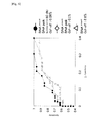
- Fig. 6 shows the results of receiver operating characteristic (ROC) analysis of the judgments based on (1) the fluorescence peak area values ( ⁇ ), (2) the fluorescence peak area values and the fluorescence peak values (0) and (3) the fluorescence peak area values, the fluorescence peak values and the fluorescence pulse widths (•).
- ROC receiver operating characteristic
- Fig. 6 show that the accuracy of the detection of abnormal cells is improved in the order of the judgments based on (1) the fluorescence peak area values, (2) the fluorescence peak area values and the fluorescence peak values and (3) the fluorescence peak area values, the fluorescence peak values and the fluorescence pulse widths. Accordingly, it is suggested that, when abnormal cells are detected in biological samples obtained from the uterine cervix, the use of multiple parameters rather than one parameter among fluorescence peak area value, fluorescence peak value and fluorescence pulse width allows more sensitive detection of abnormal cells. Particularly, the use of all parameters of fluorescence peak area value, fluorescence peak value and fluorescence pulse width allows further more sensitive detection of abnormal cells.
Landscapes
- Health & Medical Sciences (AREA)
- Life Sciences & Earth Sciences (AREA)
- Chemical & Material Sciences (AREA)
- Engineering & Computer Science (AREA)
- Immunology (AREA)
- Hematology (AREA)
- Molecular Biology (AREA)
- Urology & Nephrology (AREA)
- Biomedical Technology (AREA)
- Organic Chemistry (AREA)
- Physics & Mathematics (AREA)
- Biochemistry (AREA)
- Biotechnology (AREA)
- Microbiology (AREA)
- General Health & Medical Sciences (AREA)
- Analytical Chemistry (AREA)
- Cell Biology (AREA)
- General Physics & Mathematics (AREA)
- Pathology (AREA)
- Medicinal Chemistry (AREA)
- Food Science & Technology (AREA)
- Wood Science & Technology (AREA)
- Zoology (AREA)
- Proteomics, Peptides & Aminoacids (AREA)
- Biophysics (AREA)
- Toxicology (AREA)
- Bioinformatics & Cheminformatics (AREA)
- General Engineering & Computer Science (AREA)
- Genetics & Genomics (AREA)
- Investigating Or Analysing Biological Materials (AREA)
- Measuring Or Testing Involving Enzymes Or Micro-Organisms (AREA)
Applications Claiming Priority (2)
| Application Number | Priority Date | Filing Date | Title |
|---|---|---|---|
| JP2008196372 | 2008-07-30 | ||
| PCT/JP2009/063353 WO2010013678A1 (fr) | 2008-07-30 | 2009-07-27 | Réactif permettant de détecter une cellule anormale dans le col de l'utérus, et procédé permettant de détecter une cellule anormale dans le col de l'utérus à l’aide de ce réactif |
Publications (1)
| Publication Number | Publication Date |
|---|---|
| EP2320230A1 true EP2320230A1 (fr) | 2011-05-11 |
Family
ID=41610378
Family Applications (1)
| Application Number | Title | Priority Date | Filing Date |
|---|---|---|---|
| EP09802921A Withdrawn EP2320230A1 (fr) | 2008-07-30 | 2009-07-27 | Réactif permettant de détecter une cellule anormale dans le col de l'utérus, et procédé permettant de détecter une cellule anormale dans le col de l'utérus à l aide de ce réactif |
Country Status (7)
| Country | Link |
|---|---|
| US (1) | US20110124005A1 (fr) |
| EP (1) | EP2320230A1 (fr) |
| JP (1) | JPWO2010013678A1 (fr) |
| KR (1) | KR20110049830A (fr) |
| CN (1) | CN102112875A (fr) |
| AU (1) | AU2009277638A1 (fr) |
| WO (1) | WO2010013678A1 (fr) |
Cited By (6)
| Publication number | Priority date | Publication date | Assignee | Title |
|---|---|---|---|---|
| EP2645083A3 (fr) * | 2012-03-30 | 2013-12-11 | Sysmex Corporation | Procédé de fourniture d'informations de la cancérisation et dispositif de fourniture d'informations de cancérisation |
| WO2014102803A1 (fr) * | 2012-12-31 | 2014-07-03 | Yeda Research And Development Co. Ltd. | Capteur moléculaire et ses procédés d'utilisation |
| CN107576634A (zh) * | 2013-07-16 | 2018-01-12 | 成都深迈瑞医疗电子技术研究院有限公司 | 血液细胞分析仪及其细胞的识别方法与系统 |
| US10067116B2 (en) | 2013-03-29 | 2018-09-04 | Sysmex Corporation | Cell analyzer, cell collecting apparatus, and quality control method including processing and analyzing quality control particles |
| US10557853B2 (en) | 2012-12-31 | 2020-02-11 | Yeda Research And Development Co. Ltd. | Molecular sensor and methods of use thereof |
| CN114878846A (zh) * | 2022-07-08 | 2022-08-09 | 深圳市帝迈生物技术有限公司 | 一种血液分析仪及其清洗方法 |
Families Citing this family (5)
| Publication number | Priority date | Publication date | Assignee | Title |
|---|---|---|---|---|
| WO2012065004A2 (fr) * | 2010-11-12 | 2012-05-18 | Incelldx, Inc. | Procédés et systèmes pour prédire si un sujet a une lésion de néoplasie intraépithéliale cervicale (cin) à partir d'un échantillon de suspension de cellules cervicales |
| JP6318184B2 (ja) * | 2016-01-20 | 2018-04-25 | シスメックス株式会社 | 細胞分析方法および細胞分析装置 |
| CN111307696A (zh) * | 2020-03-19 | 2020-06-19 | 浙江星博生物科技股份有限公司 | 用于检测精子dna碎片率的方法和试剂盒 |
| WO2022099659A1 (fr) * | 2020-11-13 | 2022-05-19 | 大连理工大学 | Réactif de classification de leucocytes, réactif d'analyse d'érythrocytes, kit de réactifs et procédé d'analyse |
| WO2022099658A1 (fr) * | 2020-11-13 | 2022-05-19 | 大连理工大学 | Composé de cyanine, colorant contenant un composé de cyanine et application d'un composé de cyanine |
Family Cites Families (9)
| Publication number | Priority date | Publication date | Assignee | Title |
|---|---|---|---|---|
| US4883867A (en) * | 1985-11-01 | 1989-11-28 | Becton, Dickinson And Company | Detection of reticulocytes, RNA or DNA |
| US5534416A (en) * | 1993-04-13 | 1996-07-09 | Molecular Probes, Inc. | Fluorescent viability assay using cyclic-substituted unsymmetrical cyanine dyes |
| US5436134A (en) * | 1993-04-13 | 1995-07-25 | Molecular Probes, Inc. | Cyclic-substituted unsymmetrical cyanine dyes |
| US6955872B2 (en) * | 2003-03-20 | 2005-10-18 | Coulter International Corp. | Dye compositions which provide enhanced differential fluorescence and light scatter characteristics |
| JP4484442B2 (ja) * | 2003-04-10 | 2010-06-16 | シスメックス株式会社 | 細菌測定方法と装置とプログラム |
| JP2007006709A (ja) * | 2005-06-28 | 2007-01-18 | Matsushita Electric Ind Co Ltd | 発光物の判別方法 |
| JP2007327928A (ja) * | 2006-06-09 | 2007-12-20 | Olympus Corp | 細胞周期弁別方法、その画像処理システム、及びそのプログラム |
| JP5241100B2 (ja) * | 2006-12-28 | 2013-07-17 | シスメックス株式会社 | 抗原賦活化液、抗原賦活化方法及び細胞の検出方法 |
| JP4699400B2 (ja) | 2007-02-13 | 2011-06-08 | ボッシュ株式会社 | 燃料噴射弁のシール構造及び燃料噴射弁 |
-
2009
- 2009-07-27 AU AU2009277638A patent/AU2009277638A1/en not_active Abandoned
- 2009-07-27 WO PCT/JP2009/063353 patent/WO2010013678A1/fr not_active Ceased
- 2009-07-27 EP EP09802921A patent/EP2320230A1/fr not_active Withdrawn
- 2009-07-27 US US13/054,755 patent/US20110124005A1/en not_active Abandoned
- 2009-07-27 CN CN2009801302186A patent/CN102112875A/zh active Pending
- 2009-07-27 JP JP2010522712A patent/JPWO2010013678A1/ja active Pending
- 2009-07-27 KR KR1020117004474A patent/KR20110049830A/ko not_active Withdrawn
Non-Patent Citations (1)
| Title |
|---|
| See references of WO2010013678A1 * |
Cited By (10)
| Publication number | Priority date | Publication date | Assignee | Title |
|---|---|---|---|---|
| EP2645083A3 (fr) * | 2012-03-30 | 2013-12-11 | Sysmex Corporation | Procédé de fourniture d'informations de la cancérisation et dispositif de fourniture d'informations de cancérisation |
| US9784729B2 (en) | 2012-03-30 | 2017-10-10 | Sysmex Corporation | Canceration information providing method and canceration information providing device |
| WO2014102803A1 (fr) * | 2012-12-31 | 2014-07-03 | Yeda Research And Development Co. Ltd. | Capteur moléculaire et ses procédés d'utilisation |
| US9696310B2 (en) | 2012-12-31 | 2017-07-04 | Yeda Research And Development Co. Ltd. | Molecular sensor and methods of use thereof |
| US10281459B2 (en) | 2012-12-31 | 2019-05-07 | Yeda Research And Development Co. Ltd. | Protein biosensors, cross reactive sensor arrays and methods of use thereof |
| US10557853B2 (en) | 2012-12-31 | 2020-02-11 | Yeda Research And Development Co. Ltd. | Molecular sensor and methods of use thereof |
| US10067116B2 (en) | 2013-03-29 | 2018-09-04 | Sysmex Corporation | Cell analyzer, cell collecting apparatus, and quality control method including processing and analyzing quality control particles |
| CN107576634A (zh) * | 2013-07-16 | 2018-01-12 | 成都深迈瑞医疗电子技术研究院有限公司 | 血液细胞分析仪及其细胞的识别方法与系统 |
| CN107576634B (zh) * | 2013-07-16 | 2021-07-23 | 成都深迈瑞医疗电子技术研究院有限公司 | 血液细胞分析仪及其细胞的识别方法与系统 |
| CN114878846A (zh) * | 2022-07-08 | 2022-08-09 | 深圳市帝迈生物技术有限公司 | 一种血液分析仪及其清洗方法 |
Also Published As
| Publication number | Publication date |
|---|---|
| US20110124005A1 (en) | 2011-05-26 |
| AU2009277638A1 (en) | 2010-02-04 |
| KR20110049830A (ko) | 2011-05-12 |
| CN102112875A (zh) | 2011-06-29 |
| JPWO2010013678A1 (ja) | 2012-01-12 |
| WO2010013678A1 (fr) | 2010-02-04 |
Similar Documents
| Publication | Publication Date | Title |
|---|---|---|
| EP2320230A1 (fr) | Réactif permettant de détecter une cellule anormale dans le col de l'utérus, et procédé permettant de détecter une cellule anormale dans le col de l'utérus à l aide de ce réactif | |
| US20240344965A1 (en) | Reagent, method for analyzing platelets and blood cell analyzer | |
| JP4212827B2 (ja) | 骨髄液有核細胞自動分析方法 | |
| EP0882983B1 (fr) | Reactif et méthode de classification et de comptage de leucocytes | |
| JP3886271B2 (ja) | 赤芽球の分類計数用試薬及び分類計数方法 | |
| CN101726579B (zh) | 血液检测试剂和方法 | |
| US12072330B2 (en) | Method for classifying/counting leukocytes, reagent kit for classifying leukocytes, and reagent for classifying leukocytes | |
| KR100463129B1 (ko) | 신규화합물과그용도 | |
| EP1890143A1 (fr) | Réactif pour analyse d'échantillon, kit de réactif d'analyse d'échantillon et procédé d'analyse d'échantillon | |
| AU2008304761B2 (en) | Reagent kit for sample analysis and sample analysis method | |
| EP2166353B1 (fr) | Réactif et trousse de réactifs pour analyse de leucocyte primitif | |
| CN101042397B (zh) | 尿液分析用试剂及尿液分析方法 | |
| CN112485246A (zh) | 一种单项上皮组织肿瘤细胞渗液游离血红素显色液及制备方法 | |
| JP3929283B2 (ja) | 骨髄有核細胞の分類計数方法 | |
| EP3112862B1 (fr) | Procédé d'analyse d'échantillon d'urine, utilisation d'un réactif pour analyse d'échantillon d'urine, et utilisation d'un kit de réactif pour analyse d'échantillon d'urine | |
| EP3112863B1 (fr) | Méthode de détection des plâtres et des érythrocytes dans des échantillons d'urine | |
| JP2778745B2 (ja) | 血液中の白血球弁別測定用試薬 | |
| JP2000187033A (ja) | 細胞染色液 | |
| US20080139827A1 (en) | Method for improving the shelf-life of hematoxylin staining solutions | |
| JP4338206B2 (ja) | 赤芽球の分類計数方法 | |
| US3476514A (en) | Cancer cytoscreening | |
| KR20230080367A (ko) | 백혈구 핵 선택적 염색을 위한 시약 및 이를 이용한 염색 방법 |
Legal Events
| Date | Code | Title | Description |
|---|---|---|---|
| PUAI | Public reference made under article 153(3) epc to a published international application that has entered the european phase |
Free format text: ORIGINAL CODE: 0009012 |
|
| 17P | Request for examination filed |
Effective date: 20110224 |
|
| AK | Designated contracting states |
Kind code of ref document: A1 Designated state(s): AT BE BG CH CY CZ DE DK EE ES FI FR GB GR HR HU IE IS IT LI LT LU LV MC MK MT NL NO PL PT RO SE SI SK SM TR |
|
| AX | Request for extension of the european patent |
Extension state: AL BA RS |
|
| STAA | Information on the status of an ep patent application or granted ep patent |
Free format text: STATUS: THE APPLICATION HAS BEEN WITHDRAWN |
|
| 18W | Application withdrawn |
Effective date: 20110804 |