EP2325327A1 - Verfahren zur beurteilung von spezifischer einlagerung von d-glucose in zellen - Google Patents
Verfahren zur beurteilung von spezifischer einlagerung von d-glucose in zellen Download PDFInfo
- Publication number
- EP2325327A1 EP2325327A1 EP09805069A EP09805069A EP2325327A1 EP 2325327 A1 EP2325327 A1 EP 2325327A1 EP 09805069 A EP09805069 A EP 09805069A EP 09805069 A EP09805069 A EP 09805069A EP 2325327 A1 EP2325327 A1 EP 2325327A1
- Authority
- EP
- European Patent Office
- Prior art keywords
- molecule
- glucose
- glucose derivative
- cells
- fluorescent chromophore
- Prior art date
- Legal status (The legal status is an assumption and is not a legal conclusion. Google has not performed a legal analysis and makes no representation as to the accuracy of the status listed.)
- Granted
Links
Images


Classifications
-
- G—PHYSICS
- G01—MEASURING; TESTING
- G01N—INVESTIGATING OR ANALYSING MATERIALS BY DETERMINING THEIR CHEMICAL OR PHYSICAL PROPERTIES
- G01N33/00—Investigating or analysing materials by specific methods not covered by groups G01N1/00 - G01N31/00
- G01N33/48—Biological material, e.g. blood, urine; Haemocytometers
- G01N33/50—Chemical analysis of biological material, e.g. blood, urine; Testing involving biospecific ligand binding methods; Immunological testing
- G01N33/58—Chemical analysis of biological material, e.g. blood, urine; Testing involving biospecific ligand binding methods; Immunological testing involving labelled substances
- G01N33/582—Chemical analysis of biological material, e.g. blood, urine; Testing involving biospecific ligand binding methods; Immunological testing involving labelled substances with fluorescent label
-
- C—CHEMISTRY; METALLURGY
- C09—DYES; PAINTS; POLISHES; NATURAL RESINS; ADHESIVES; COMPOSITIONS NOT OTHERWISE PROVIDED FOR; APPLICATIONS OF MATERIALS NOT OTHERWISE PROVIDED FOR
- C09B—ORGANIC DYES OR CLOSELY-RELATED COMPOUNDS FOR PRODUCING DYES, e.g. PIGMENTS; MORDANTS; LAKES
- C09B51/00—Nitro or nitroso dyes
-
- C—CHEMISTRY; METALLURGY
- C09—DYES; PAINTS; POLISHES; NATURAL RESINS; ADHESIVES; COMPOSITIONS NOT OTHERWISE PROVIDED FOR; APPLICATIONS OF MATERIALS NOT OTHERWISE PROVIDED FOR
- C09B—ORGANIC DYES OR CLOSELY-RELATED COMPOUNDS FOR PRODUCING DYES, e.g. PIGMENTS; MORDANTS; LAKES
- C09B57/00—Other synthetic dyes of known constitution
-
- C—CHEMISTRY; METALLURGY
- C12—BIOCHEMISTRY; BEER; SPIRITS; WINE; VINEGAR; MICROBIOLOGY; ENZYMOLOGY; MUTATION OR GENETIC ENGINEERING
- C12Q—MEASURING OR TESTING PROCESSES INVOLVING ENZYMES, NUCLEIC ACIDS OR MICROORGANISMS; COMPOSITIONS OR TEST PAPERS THEREFOR; PROCESSES OF PREPARING SUCH COMPOSITIONS; CONDITION-RESPONSIVE CONTROL IN MICROBIOLOGICAL OR ENZYMOLOGICAL PROCESSES
- C12Q1/00—Measuring or testing processes involving enzymes, nucleic acids or microorganisms; Compositions therefor; Processes of preparing such compositions
- C12Q1/02—Measuring or testing processes involving enzymes, nucleic acids or microorganisms; Compositions therefor; Processes of preparing such compositions involving viable microorganisms
-
- C—CHEMISTRY; METALLURGY
- C12—BIOCHEMISTRY; BEER; SPIRITS; WINE; VINEGAR; MICROBIOLOGY; ENZYMOLOGY; MUTATION OR GENETIC ENGINEERING
- C12Q—MEASURING OR TESTING PROCESSES INVOLVING ENZYMES, NUCLEIC ACIDS OR MICROORGANISMS; COMPOSITIONS OR TEST PAPERS THEREFOR; PROCESSES OF PREPARING SUCH COMPOSITIONS; CONDITION-RESPONSIVE CONTROL IN MICROBIOLOGICAL OR ENZYMOLOGICAL PROCESSES
- C12Q1/00—Measuring or testing processes involving enzymes, nucleic acids or microorganisms; Compositions therefor; Processes of preparing such compositions
- C12Q1/54—Measuring or testing processes involving enzymes, nucleic acids or microorganisms; Compositions therefor; Processes of preparing such compositions involving glucose or galactose
-
- G—PHYSICS
- G01—MEASURING; TESTING
- G01N—INVESTIGATING OR ANALYSING MATERIALS BY DETERMINING THEIR CHEMICAL OR PHYSICAL PROPERTIES
- G01N33/00—Investigating or analysing materials by specific methods not covered by groups G01N1/00 - G01N31/00
- G01N33/48—Biological material, e.g. blood, urine; Haemocytometers
- G01N33/50—Chemical analysis of biological material, e.g. blood, urine; Testing involving biospecific ligand binding methods; Immunological testing
- G01N33/5005—Chemical analysis of biological material, e.g. blood, urine; Testing involving biospecific ligand binding methods; Immunological testing involving human or animal cells
-
- G—PHYSICS
- G01—MEASURING; TESTING
- G01N—INVESTIGATING OR ANALYSING MATERIALS BY DETERMINING THEIR CHEMICAL OR PHYSICAL PROPERTIES
- G01N33/00—Investigating or analysing materials by specific methods not covered by groups G01N1/00 - G01N31/00
- G01N33/48—Biological material, e.g. blood, urine; Haemocytometers
- G01N33/50—Chemical analysis of biological material, e.g. blood, urine; Testing involving biospecific ligand binding methods; Immunological testing
- G01N33/53—Immunoassay; Biospecific binding assay; Materials therefor
- G01N33/531—Production of immunochemical test materials
- G01N33/532—Production of labelled immunochemicals
- G01N33/533—Production of labelled immunochemicals with fluorescent label
Definitions
- the present invention relates to a method for accurately evaluating the specific incorporation of D-glucose into cells.
- Glucose is known as one of the most important energy sources for maintaining the survival of cells, from mammals to Escherichia coli and yeast, and in particular, the brain uses glucose as the sole energy source.
- Glucose has enantiomers D-glucose and L-glucose, and among these enantiomers, only D-glucose can be utilized as an energy source by organisms, and living cells have a mechanism of selectively incorporating D-glucose mediated by a transport protein in the cell membrane such as a glucose transporter and utilizing it.
- the group of the present inventors proposed a method using 2-[N-(7-nitrobenz-2-oxa-1,3-diazol-4-yl)amino]-2-deoxy-D-glucose (2-NBDG) that is represented by the following structural formula and emits green fluorescence and is obtained by attaching an N-(7-nitrobenz-2-oxa-1,3-diazol-4-yl)amino group as a fluorescent chromophore to the 2-position of D-deoxyglucose as the method which can be used in a study of the process of dynamic incorporation of D-glucose into living cells, and the usefulness of the method was demonstrated using various mammalian cells (Non-patent document 1).
- This method utilizes the property of 2-NBDG that it is selectively incorporated into living cells, and by tracking changes in the fluorescence intensity due to the incorporation, the dynamic activity of cells for the incorporation of D-glucose can be quantitatively determined. Therefore, this method has been evaluated by researchers around the world as an innovative method in studies as to how organisms incorporate D-glucose into cells and utilize it, and now is positioned as a standard protocol essential in this research field (Non-patent document 2).
- Non-patent document 1 the group of the present inventors administered 2-NBDG to insulin-secreting cells which are representative cells expressing GLUT2 that is a low-affinity (high-Km) glucose transporter, and revealed, from changes in the fluorescence intensity, that 2-NBDG is incorporated into cells depending on the concentration, time, and temperature, and the Km value thereof is equivalent to the value reported for D-glucose. Therefore, it can be said that according to this method, the incorporation of D-glucose mediated by GLUT2 that is a high-Km glucose transporter can be quantitatively evaluated.
- the fluorescence intensity obtained by the administration of 2-NBDG includes the fluorescence emitted by such 2-NBDG. Therefore, in order to accurately evaluate the specific incorporation of D-glucose into cells, it is necessary to consider the existence of such 2-NBDG, and this is considered to be very important when the kinetics of the incorporation of 2-NBDG mediated by GLUT1 that is a high-affinity (low-Km) glucose transporter expressed in many cells, GLUT3 expressed in neurons, or the like is studied. However, there has been no report as to how this point is reflected in the experimental method.
- an object of the present invention is to provide a method for accurately evaluate the specific incorporation of D-glucose into cells.
- the present inventors conceived an idea that when a difference in the fluorescence intensity between before and after administration of 2-NBDG to cells to be evaluated is measured, a fluorescent L-glucose derivative which is not specifically incorporated into cells is used as a control.
- a method for evaluating the specific incorporation of D-glucose into cells relative to L-glucose according to the present invention based on the above idea is characterized by comprising contacting a D-glucose derivative that has a fluorescent chromophore in the molecule and is specifically incorporated into cells and an L-glucose derivative that has a fluorescent chromophore in the molecule with different cells in the same cell strain to be evaluated, respectively, comparing the fluorescence emitted by the D-glucose derivative that has a fluorescent chromophore in the molecule and is specifically incorporated into cells with the fluorescence emitted by the L-glucose derivative that has a fluorescent chromophore in the molecule, and evaluating the specific incorporation of D-glucose into cells relative to L-glucose by taking the difference between the two kinds of fluorescence intensities as described in claim 1.
- the method described in claim 2 is characterized in that in the method described in claim 1, as the D-glucose derivative that has a fluorescent chromophore in the molecule and is specifically incorporated into cells, a D-glucose derivative that has an N-(7-nitrobenz-2-oxa-1,3-diazol-4-yl)amino group in the molecule is used.
- the method described in claim 3 is characterized in that in the method described in claim 2, as the D-glucose derivative that has an N-(7-nitrobenz-2-oxa-1,3-diazol-4-yl)amino group in the molecule, D-deoxyglucose having an N-(7-nitrobenz-2-oxa-1,3-diazol-4-yl)amino group attached to the 2-position thereof is used.
- the method described in claim 4 is characterized in that in the method described in claim 2, as the L-glucose derivative that has a fluorescent chromophore in the molecule, an L-glucose derivative that has an N-(7-nitrobenz-2-oxa-1,3-diazol-4-yl)amino group in the molecule is used.
- the method described in claim 5 is characterized in that in the method described in claim 4, as the L-glucose derivative that has an N-(7-nitrobenz-2-oxa-1,3-diazol-4-yl)amino group in the molecule, L-deoxyglucose having an N-(7-nitrobenz-2-oxa-1,3-diazol-4-yl)amino group attached to the 2-position thereof is used.
- the method described in claim 6 is characterized in that in the method described in claim 1, as the D-glucose derivative that has a fluorescent chromophore in the molecule and is specifically incorporated into cells, a D-glucose derivative that has a 2-(N-methylamino)benzoylamino group in the molecule is used.
- the method described in claim 7 is characterized in that in the method described in claim 6, as the D-glucose derivative that has a 2-(N-methylamino)benzoylamino group in the molecule, D-deoxyglucose having a 2-(N-methylamino)benzoylamino group attached to the 2-position thereof is used.
- the method described in claim 8 is characterized in that in the method described in claim 6, as the L-glucose derivative that has a fluorescent chromophore in the molecule, an L-glucose derivative that has a 2-(N-methylamino)benzoylamino group in the molecule is used.
- the method described in claim 9 is characterized in that in the method described in claim 8, as the L-glucose derivative that has a 2-(N-methylamino)benzoylamino group in the molecule, L-deoxyglucose having a 2-(N-methylamino)benzoylamino group attached to the 2-position thereof is used.
- the method for evaluating the specific incorporation of D-glucose into cells relative to L-glucose is characterized by comprising contacting a mixture of a D-glucose derivative that has a fluorescent chromophore in the molecule and is specifically incorporated into cells and an L-glucose derivative that has, in the molecule, a fluorescent chromophore that emits fluorescence at a wavelength different from that of the fluorescence emitted by the D-glucose derivative that has a fluorescent chromophore in the molecule and is specifically incorporated into cells with cells to be evaluated, and detecting the fluorescence emitted by the D-glucose derivative that has a fluorescent chromophore in the molecule and is specifically incorporated into cells as described in claim 10.
- the method described in claim 11 is characterized in that in the method described in claim 10, a difference in the maximum fluorescence wavelength is set to at least 20 nm.
- the method described in claim 12 is characterized in that in the method described in claim 10, as the D-glucose derivative that has a fluorescent chromophore in the molecule and is specifically incorporated into cells, a D-glucose derivative that has an N-(7-nitrobenz-2-oxa-1,3-diazol-4-yl)amino group in the molecule is used.
- the method described in claim 13 is characterized in that in the method described in claim 12, as the D-glucose derivative that has an N-(7-nitrobenz-2-oxa-1,3-diazol-4-yl)amino group in the molecule, D-deoxyglucose having an N-(7-nitrobenz-2-oxa-1,3-diazol-4-yl)amino group attached to the 2-position thereof is used.
- the method described in claim 14 is characterized in that in the method described in claim 10, as the D-glucose derivative that has a fluorescent chromophore in the molecule and is specifically incorporated into cells, a D-glucose derivative that has a 2-(N-methylamino)benzoylamino group in the molecule is used.
- the method described in claim 15 is characterized in that in the method described in claim 14, as the D-glucose derivative that has a 2-(N-methylamino)benzoylamino group in the molecule, D-deoxyglucose having a 2-(N-methylamino)benzoylamino group attached to the 2-position thereof is used.
- the method described in claim 16 is characterized in that in the method described in claim 12 or 14, as the L-glucose derivative that has, in the molecule, a fluorescent chromophore that emits fluorescence at a wavelength different from that of the fluorescence emitted by the D-glucose derivative that has a fluorescent chromophore in the molecule and is specifically incorporated into cells, an L-glucose derivative having sulforhodamine attached thereto is used.
- the method described in claim 17 is characterized in that in the method described in claim 12, as the L-glucose derivative that has, in the molecule, a fluorescent chromophore that emits fluorescence at a wavelength different from that of the fluorescence emitted by the D-glucose derivative that has a fluorescent chromophore in the molecule and is specifically incorporated into cells, an L-glucose derivative that has a 2-(N-methylamino)benzoylamino group in the molecule is used.
- the method described in claim 18 is characterized in that in the method described in claim 17, as the L-glucose derivative that has a 2-(N-methylamino)benzoylamino group in the molecule, L-deoxyglucose having a 2-(N-methylamino)benzoylamino group attached to the 2-position thereof is used.
- the method described in claim 19 is characterized in that in the method described in claim 14, as the L-glucose derivative that has, in the molecule, a fluorescent chromophore that emits fluorescence at a wavelength different from that of the fluorescence emitted by the D-glucose derivative that has a fluorescent chromophore in the molecule and is specifically incorporated into cells, an L-glucose derivative that has an N-(7-nitrobenz-2-oxa-1,3-diazol-4-yl)amino group in the molecule is used.
- the method described in claim 20 is characterized in that in the method described in claim 19, as the L-glucose derivative that has an N-(7-nitrobenz-2-oxa-1,3-diazol-4-yl)amino group in the molecule, L-deoxyglucose having an N-(7-nitrobenz-2-oxa-1,3-diazol-4-yl)amino group attached to the 2-position thereof is used.
- the method described in claim 21 is characterized in that in the method described in claim 10, as the L-glucose derivative that has, in the molecule, a fluorescent chromophore that emits fluorescence at a wavelength different from that of the fluorescence emitted by the D-glucose derivative that has a fluorescent chromophore in the molecule and is specifically incorporated into cells, two or more kinds of L-glucose derivatives, each of which has a different fluorescent chromophore in the molecule, are mixed and the resulting mixture is used.
- the L-glucose derivative according to the present invention is characterized by having a fluorescent chromophore in the molecule as described in claim 22.
- the method for producing an L-glucose derivative that has a fluorescent chromophore in the molecule according to the present invention is characterized by comprising introducing the fluorescent chromophore at the 2-position of L-glucosamine or a salt thereof obtained by acetylating the five hydroxy groups of L-mannose, substituting the acetoxy group at the 1-position by bromine, introducing an ortho ester group at the 1- and 2-positions, converting the acetyl group at each of the 3-, 4-, and 5-positions into a benzyl group, removing the ortho ester group at the 1- and 2-positions and converting into a methoxy group at the 1-position and a hydroxy group at the 2-position, respectively, trifluoromethanesulfonylating or methanesulfonylating the hydroxy group at the 2-position, followed by azidation, subjecting the azide group to hydrogen reduction, and at the same time, removing the benzyl groups, and finally converting
- the method for producing L-glucosamine or a salt thereof according to the present invention is characterized by comprising acetylating the five hydroxy groups of L-mannose, substituting the acetoxy group at the 1-position by bromine, introducing an ortho ester group at the 1- and 2-positions, converting the acetyl group at each of the 3-, 4-, and 5-positions into a benzyl group, removing the ortho ester group at the 1- and 2-positions and converting into a methoxy group at the 1-position and a hydroxy group at the 2-position, respectively, trifluoromethanesulfonylating or methanesulfonylating the hydroxy group at the 2-position, followed by azidation, subjecting the azide group to hydrogen reduction, and at the same time, removing the benzyl groups, and finally converting the methoxy group at the 1-position into a hydroxy group as described in claim 24.
- the invention is directed to the use of L-glucosamine or a
- a method for accurately evaluating the specific incorporation of D-glucose into cells can be provided.
- the method for evaluating the specific incorporation of D-glucose into cells according to the present invention can be divided into the following two methods.
- the method comprising contacting a D-glucose derivative that has a fluorescent chromophore in the molecule and is specifically incorporated into cells and an L-glucose derivative that has a fluorescent chromophore in the molecule with different cells in the same cell strain to be evaluated, respectively, comparing the fluorescence emitted by the D-glucose derivative that has a fluorescent chromophore in the molecule and is specifically incorporated into cells with the fluorescence emitted by the L-glucose derivative that has a fluorescent chromophore in the molecule, and evaluating the specific incorporation of D-glucose into cells relative to L-glucose by taking the difference between the two kinds of fluorescence intensities.
- the D-glucose derivative (which may be in the form of a salt such as a hydrochloride salt) that has a fluorescent chromophore in the molecule and is specifically incorporated into cells means a compound that is a D-glucose derivative having a fluorescent chromophore in the molecule and is incorporated into cells in a specific manner when compared to L-glucose or a derivative thereof (such as L-glucosamine or L-deoxyglucose), and specific examples thereof include D-glucose derivatives having an N-(7-nitrobenz-2-oxa-1,3-diazol-4-yl)amino group in the molecule such as 2-NBDG described in Non-patent documents 1 and 2 and D-deoxyglucose having an N-(7-nitrobenz-2-oxa-1,3-diazol-4-yl)amino group attached to the 6-position thereof (6-[N-(7-nitrobenz-2-oxa-1,
- the L-glucose derivative (which may be in the form of a salt such as a hydrochloride salt) that has a fluorescent chromophore in the molecule is a compound that is basically not specifically incorporated into cells because it is an L-form and can be used as a control for the D-glucose derivative that has a fluorescent chromophore in the molecule and is specifically incorporated into cells such as 2-NBDG in the present invention.
- L-glucose derivatives having an N-(7-nitrobenz-2-oxa-1,3-diazol-4-yl)amino group in the molecule such as L-deoxyglucose having an N-(7-nitrobenz-2-oxa-1,3-diazol-4-yl)amino group attached to the 2-position thereof (2-[N-(7-nitrobenz-2-oxa-1,3-diazol-4-yl)amino]-2-deoxy-L-glucose: 2-NBDLG) and L-deoxyglucose having an N-(7-nitrobenz-2-oxa-1,3-diazol-4-yl)amino group attached to the 6-position thereof (6-[N-(7-nitrobenz-2-oxa-1,3-diazol-4-yl)amino]-6-deoxy-L-glucose: 6-NBDLG).
- 2-NBDLG is in an enantiomeric relationship with 2-NBDG, and both have the same physicochemical properties except for the sign of optical rotation.
- the nonspecific adsorption property of 2-NBDLG onto the surface of the cell membrane or in the membrane, nonspecific incorporation thereof into cells without being mediated by a glucose transporter, and the like are considered to be the same as or at least equivalent to those of 2-NBDG. Therefore, in the case where the fluorescence intensity obtained by the administration of 2-NBDG includes the fluorescence emitted by 2-NBDG nonspecifically adsorbed onto the surface of the cell membrane or in the membrane, 2-NBDG nonspecifically incorporated into cells without being mediated by a glucose transporter, and the like, a fluorescence intensity that is at least equivalent to the fluorescence intensity derived from such fluorescence can be obtained also by the administration of 2-NBDLG serving as the control.
- the difference can be evaluated as the specific incorporation of 2-NBDG into cells relative to 2-NBDLG, namely, as the specific incorporation of D-glucose into cells relative to L-glucose.
- the chemical structures of the fluorescent chromophores of the two kinds of glucose derivatives are preferably completely the same or are the same except for a substituent which may be contained as a side chain.
- this method is performed by contacting the D-glucose derivative that has a fluorescent chromophore in the molecule and is specifically incorporated into cells and the L-glucose derivative that has a fluorescent chromophore in the molecule with different cells in the same cell strain to be evaluated, respectively (a difference in the fluorescence intensity cannot be obtained, if both glucose derivatives are simultaneously administered to one cell), it is preferred that the activities of the cells to which the glucose derivatives are administered respectively are the same or at least equivalent to each other. In order to ensure this condition, it is preferred to confirm the activities of the cells during experiment.
- Examples of a method for confirming the activities of the cells include a method for confirming that the cell membrane potential is normal using a patch clamp technique or the like, and a method for measuring an action potential (these methods are preferred in the case of neurons) and a method for measuring the metabolic activity.
- the number of cells with which the D-glucose derivative that has a fluorescent chromophore in the molecule and is specifically incorporated into cells and the L-glucose derivative that has a fluorescent chromophore in the molecule are contacted, respectively can be set to more than 1 (n ⁇ 2), and a statistical analysis may be performed.
- the L-glucose derivative that has a fluorescent chromophore in the molecule can be produced by introducing the fluorescent chromophore into L-glucose or a derivative thereof (such as L-glucosamine or L-deoxyglucose).
- L-glucosamine or a salt thereof (such as a hydrochloride salt) that is useful for the production of the L-glucose derivative that has a fluorescent chromophore in the molecule can be obtained by, for example, acetylating the five hydroxy groups of L-mannose, substituting the acetoxy group at the 1-position by bromine, introducing an ortho ester group at the 1- and 2-positions, converting the acetyl group at each of the 3-, 4-, and 5-positions into a benzyl group, removing the ortho ester group at the 1- and 2-positions and converting into a methoxy group at the 1-position and a hydroxy group at the 2-position, respectively, trifluoromethanesulfonylating or methanesulfonylating the hydroxy group at the 2-position, followed by azidation, subjecting the azide group to hydrogen reduction, and at the same time, removing the benzyl groups, and finally converting the methoxy group at the
- a combination of the D-glucose derivative that has a fluorescent chromophore in the molecule and is specifically incorporated into cells with the L-glucose derivative that has a fluorescent chromophore in the molecule which can be used in this method is not limited to a combination of the above-mentioned D-glucose derivative that has an N-(7-nitrobenz-2-oxa-1,3-diazol-4-yl)amino group in the molecule and emits green fluorescence (such as 2-NBDG) with the L-glucose derivative that has an N-(7-nitrobenz-2-oxa-1,3-diazol-4-yl)amino group in the molecule and emits green fluorescence (such as 2-NBDLG), and it may be a combination of a D-glucose derivative that has a 2-(N-methylamino)benzoylamino group in the molecule and emits blue fluorescence with an L-gluco
- Examples of the D-glucose derivative that has a 2-(N-methylamino)benzoylamino group in the molecule include D-deoxyglucose having a 2-(N-methylamino)benzoylamino group attached to the 2-position thereof (2-[N-2-(N'-methylamino)benzoylamino]-2-deoxy-D-glucose: 2-NMAG), and examples of the L-glucose derivative that has a 2-(N-methylamino)benzoylamino group in the molecule include L-deoxyglucose having a 2-(N-methylamino)benzoylamino group attached to the 2-position thereof (2-[N-2-(N'methylamino)benzoylamino]-2-deoxy-L-glucose: 2-NMALG).
- the method comprising contacting a mixture of a D-glucose derivative that has a fluorescent chromophore in the molecule and is specifically incorporated into cells and an L-glucose derivative that has, in the molecule, a fluorescent chromophore that emits fluorescence at a wavelength different from that of the fluorescence emitted by the D-glucose derivative that has a fluorescent chromophore in the molecule and is specifically incorporated into cells with cells to be evaluated, and detecting the fluorescence emitted by the D-glucose derivative that has a fluorescent chromophore in the molecule and is specifically incorporated into cells.
- the D-glucose derivative (which may be in the form of a salt such as a hydrochloride salt) that has a fluorescent chromophore in the molecule and is specifically incorporated into cells means a compound that is a D-glucose derivative having a fluorescent chromophore in the molecule and is incorporated into cells in a specific manner when compared to L-glucose or a derivative thereof (such as L-glucosamine or L-deoxyglucose), and specific examples thereof include D-glucose derivatives having an N-(7-nitrobenz-2-oxa-1,3-diazol-4-yl)amino group in the molecule such as 2-NBDG described in Non-patent documents 1 and 2 and D-deoxyglucose having an N-(7-nitrobenz-2-oxa-1,3-diazol-4-yl)amino group attached to the 6-position thereof (6-[N-(7-nitrobenz-2-oxa-1,
- L-glucose derivative (which may be in the form of a salt such as a hydrochloride salt) that has, in the molecule, a fluorescent chromophore that emits fluorescence at a wavelength different from that of the fluorescence emitted by the D-glucose derivative that has a fluorescent chromophore in the molecule and is specifically incorporated into cells
- L-glucose derivatives having sulforhodamine such as sulforhodamine B, sulforhodamine G, or sulforhodamine 101
- specific examples thereof include L-deoxyglucose having sulforhodamine 101 attached to the 2-position thereof via a sulfonamide bond (referred to as 2-TRLG).
- the maximum fluorescence wavelength of the former is from 540 nm to 550 nm
- the maximum fluorescence wavelength of the latter is from 600 nm to 610 nm.
- both 2-NBDG and 2-TRLG are incorporated into the cells, and hence yellow fluorescence is detected from the inside of the cells. Therefore, by detecting the green fluorescence emitted by 2-NBDG after the mixed liquid of 2-NBDG and 2-TRLG is administered to cells, the specific incorporation of D-glucose into cells relative to L-glucose can be accurately evaluated in real time.
- this method utilizes a difference in the wavelength between the fluorescence emitted by the D-glucose derivative that has a fluorescent chromophore in the molecule and is specifically incorporated into cells and the fluorescence emitted by the L-glucose derivative that has a fluorescent chromophore in the molecule, and therefore, the larger the difference in the maximum fluorescence wavelength between the two kinds of glucose derivatives, the easier and more accurately the difference can be discriminated. Accordingly, it is preferred that the difference in the maximum fluorescence wavelength between both glucose derivatives is at least 20 nm.
- 2-NBDG is used as the D-glucose derivative that has a fluorescent chromophore in the molecule and is specifically incorporated into cells
- L-glucose derivative that has a fluorescent chromophore in the molecule an L-glucose derivative that has a 7-(N,N-dimethylaminosulfonyl)benz-2-oxa-1,3-diazol-4-yl) amino group in the molecule, specifically, L-deoxyglucose having a 7-(N,N-dimethylaminosulfonyl)benz-2-oxa-1,3-diazol-4-yl) amino group attached to the 2-position thereof (2-[N-7-(N',N'-dimethylaminosulfonyl)benz-2-oxa-1,3-diazol-4-yl)amino]-2-deoxy-L-glucose: 2-DBDLG) can also be used (the fluorescence thereof is
- an L-glucose derivative that has a fluorescent chromophore in the molecule an L-glucose derivative that has a 2-(N-methylamino)benzoylamino group in the molecule, specifically, L-deoxyglucose having a 2-(N-methylamino)benzoylamino group attached to the 2-position thereof (2-[N-2-(N'-methylamino)benzoylamino]-2-deoxy-L-glucose: 2-NMALG) can also be used (the fluorescence thereof is blue, and the maximum fluorescence wavelength thereof is from 450 nm to 460 nm).
- a D-glucose derivative that has a fluorescent chromophore in the molecule and is specifically incorporated into cells a D-glucose derivative that has a 2-(N-methylamino)benzoylamino group in the molecule, specifically, D-deoxyglucose having a 2-(N-methylamino)benzoylamino group attached to the 2-position thereof (2-[N-2-(N'-methylamino)benzoylamino]-2-deoxy-D-glucose: 2-NMAG) can also be used (the fluorescence thereof is blue, and the maximum fluorescence wavelength thereof is from 450 nm to 460 nm).
- 2-NBDLG or 2-TRLG can be used as the L-glucose derivative that has a fluorescent chromophore in the molecule.
- the mixing ratio of both glucose derivatives is adjusted within a range of, for example from 1:100 to 100:1 (molar ratio) such that the intensities of the fluorescence spectra emitted by the both glucose derivatives are equivalent to each other.
- the L-glucose derivative that has, in the molecule, a fluorescent chromophore that emits fluorescence at a wavelength different from that of the fluorescence emitted by the D-glucose derivative that has a fluorescent chromophore in the molecule and is specifically incorporated into cells two or more kinds of L-glucose derivatives, each of which has a different fluorescent chromophore in the molecule, may be mixed and used (for example, a combination of 2-TRLG and 2-NMALG, or the like).
- L-glucose derivatives each of which has a different fluorescent chromophore in the molecule
- a difference in the nonspecifically incorporated L-glucose derivative based on a difference in the degree of the deterioration state of the cell membrane can be detected, and therefore, the specific incorporation of D-glucose into cells relative to L-glucose can be evaluated and also the degree of the deterioration state of the cell membrane can be evaluated by the method.
- the experimental condition for evaluating the specific incorporation of D-glucose into cells of the present invention may be according to known condition (if necessary, see Non-patent document 2).
- the method for evaluating the specific incorporation of D-glucose into cells of the present invention other than cells (such as neurons) in any region in mammals including humans, cells of microbial such as Escherichia coli and yeast, plant cells, fertilized eggs, and the like can be used as evaluation targets, and the method contributes to the advancement of the research as to how organisms incorporate D-glucose into cells and utilize it, and also the usefulness of the method is expected in the medical field, hygiene field, and other field for distinguishing normal cells from tumor cells based on a difference in the degree of the incorporation of D-glucose or for preventing or controlling microbial contamination (counting of the number of microorganisms, evaluation for the state thereof, etc.).
- any of the above two methods may be selected, however, by performing the both methods in combination, more accurate evaluation can be performed.
- Example 1 Example of applying 2-NBDG or 2-NBDLG to neurons
- L-(-)-mannose (9.0 g, 49.96 mmol) was dissolved in pyridine (120 ml), and the resulting solution was cooled in ice. Acetic anhydride (60 ml) was added dropwise thereto over about 15 minutes. The resulting solution was stirred overnight while gradually returning to room temperature. Thereafter, the reaction mixture was concentrated under reduced pressure. Toluene was added to the resulting residue, and an azeotropic distillation procedure was performed twice. Ethyl acetate was added to the residue to dissolve the residue, and the resulting solution was washed sequentially with saturated NaHCO 3 solution, water, and saturated saline.
- 1,2,3,4,6-penta-O-acetyl-L-mannopyranose (1) (21.04 g) was dissolved in dehydrated dichloromethane (150 ml), and the resulting solution was cooled in ice. 30% HBr/AcOH (27.5 ml) was added dropwise thereto over about 10 minutes, and after completion of the dropwise addition, the ice bath was removed, and the mixture was stirred at room temperature for 3 hours. The reaction mixture was concentrated under reduced pressure. Toluene was added to the resulting residue, and an azeotropic distillation procedure was performed twice.
- Sodium hydride (NaH) (a 60% oil suspension, 7.38 g) was added thereto in small portions. After foaming due to the generation of hydrogen was stopped, benzyl bromide (12.1 ml) was added dropwise thereto. The resulting solution was stirred overnight while gradually returning to room temperature. The reaction mixture was cooled in ice, and in order to decompose the excess NaH, methanol was added thereto in small portions until foaming was no longer observed. The resulting solution was diluted by adding chloroform thereto, and the obtained solution was washed sequentially with water and saturated saline. The organic layer was dried over anhydrous magnesium sulfate and filtered, and the filtrate was concentrated under reduced pressure.
- NaH sodium hydride
- the obtained residue was dried well under reduced pressure, and the dried residue was stored overnight in a freezer. On the next morning, the stored residue was separated into precipitates and oil. The oil (unnecessary substance) was removed using a Pasteur pipette. The precipitates (wet weight: 17.97 g) was purified by silica gel column chromatography, whereby a target product was obtained (yield: 11.98 g, yield ratio: 74%).
- Methyl 3,4,6-tri-O-benzyl- ⁇ -L-mannopyranoside (5) (9.74 g) was dissolved in dehydrated dichloromethane (220 ml), pyridine (4.24 ml) was added thereto, and the resulting mixture was cooled to -15°C under an argon atmosphere. Trifluoromethanesulfonic acid anhydride (5.29 ml) was added thereto, and the resulting mixture was stirred at room temperature. After 1 hour, saturated NaHCO 3 solution was added to the reaction mixture, and extraction was performed with dichloromethane. The organic layers were combined, and the combined organic layer was washed sequentially with saturated NaHCO 3 solution, water, and saturated saline.
- Methyl 3,4,6-tri-O-benzyl-2-O-trifluoromethanesulfonyl- ⁇ -L-mannopyranoside (6) (12.23 g) was dissolved in dehydrated benzene (400 ml), tetrabutylammonium azide [(n-Bu) 4 NN 3 ] (29.16 g) was added thereto under an argon atmosphere. After 3 days, (n-Bu) 9 NN 3 (3.61 g) was further added thereto. The mixture was stirred at room temperature for an additional 11 days (14 days in total).
- Methyl 2-azido-3,4,6-tri-O-benzyl-2-deoxy- ⁇ -L-glucopyranoside (7) (1.98 g, 4.04 mmol) was dissolved in methanol (70 ml), 1 M HCl (4.85 ml) and palladium black (500 mg) were added thereto, and the resulting mixture was subjected to catalytic reduction by pressurization to 8 atm. After 22 hours, the pressure was returned to normal, and after it was confirmed that only one spot was detected by TLC, the catalyst was removed by filtration. The catalyst was washed with methanol, and the filtrate and the washing solution were combined, and the resulting mixture was concentrated under reduced pressure.
- Methyl 2-amino-2-deoxy- ⁇ -L-glucopyranoside hydrochloride (8) (960 mg, calculated to be equivalent to 4.03 mmol) was dissolved in 6 N HCl (40 ml), and the resulting solution was stirred while heating at 100°C. After the solution was allowed to react for a total of 30 hours, the solution was returned to room temperature and concentrated under reduced pressure. Water was added to the resulting residue, and a concentration procedure under reduced pressure was performed five times. The obtained residue was dissolved in water and the resulting solution was lyophilized. The obtained residue was passed through CHP-20, and the flow-through fraction was collected and lyophilized (crude yield: 805 mg (3.73 mmol, equivalent crude yield ratio: 93%)).
- C57BL/6J mice at the age of 13 to 15 days after birth (juvenile period) or at the age of 33 to 45 days after birth (maturation period) were deeply anesthetized with urethane (i.p. 1.6 g/kg) and decapitated according to a common procedure. Thereafter, the brain was taken out from each mouse and cooled by immersion in a cold Ringer's solution note 1) (at 0°C) for 1 to 2 minutes, and then, coronal brain slices with a thickness of 400 ⁇ m were prepared using a microslicer (ZERO 1, Dosaka EM Co., Ltd.).
- coronal brain slices were regained at room temperature for 1 hour in a chamber (acrylic incubation chamber manufactured by Harvard Corporation) in which a Ringer's solution aerated with 95% O 2 and 5% CO 2 was circulated.
- the brain slices were immersed in an enzyme solution (31°C, maintained at pH 7.4 by aeration with 95% O 2 and 5% CO 2 ) prepared by dissolving a proteolytic enzyme, Pronase (10 mg/60 mL) in the above-mentioned Ringer's solution, thereby performing an enzymatic treatment (the immersion time was 18 to 20 minutes in the case of the juvenile mouse brain, and 50 to 70 minutes in the case of the maturation mouse brain).
- the brain slices were immersed in 100 mL of a HEPES solution note 2) containing 10 mM glucose to stop the enzymatic reaction.
- a 60 mm plastic dish with a silicone-coated bottom was filled with a HEPES solution note 2) containing 10 mM glucose (at room temperature), and the above prepared brain slices were immersed therein.
- Each of the brain slices was immobilized with two 27-G injection needles, and the right and left midbrain substantia nigra pars reticulata were punched out using a 18-G injection needle having an elliptically shaped tip end under microscope, which was stored at room temperature in the above-mentioned HEPES solution note 2) (in a 35-mm culture dish).
- Neurons were isolated by trituration using a glass pipette having a tip end diameter adjusted to various diameters, and were allowed to adhere to a 35-mm glass bottom culture dish note 4) coated with poly-L-lysine note 3) .
- This stock solution was diluted to 1/1000 with 0.15 M boric acid, and the diluted solution was dropped onto the center of a coverslip (13 mm x 22 mm No. 0 glass, Matsunami Glass Ind., Ltd.). The coverslip was left as such at room temperature for 20 minutes, and then washed with distilled water, followed by natural drying.
- the above-mentioned glass coverslip having an upper surface coated with poly-L-lysine note 3) was attached to an elliptically-shaped hole (5 mm x 14 mm) of a 35-mm culture dish (Matsunami code D109501) having the elliptically-shaped hole on the bottom from the back surface using a silicone grease (Dow Corning 111 Sealant) and fixed thereto so that water does not leak out.
- a silicone grease Dow Corning 111 Sealant
- 2-NBDG (or 2-NBDLG), which was purified by HPLC, and the purity of which was confirmed by NMR and HPLC, and the content of which was confirmed by an elemental analysis was weighed and dissolved in water. The solution was dispensed into vials such that each vial contained a net weight of 0.5 mg of 2-NBDG (or 2-NBDLG) and lyophilized, which was stored in dark at -20°C or lower.
- Non-patent document 2 mounted on a heating-type xy stage (Zeiss Tempcontrol 37-2) of an inverted fluorescence microscope from Zeiss Axiovert 135 (100 W, a xenon light source), a 35-mm glass bottom culture dish note 4) was placed, and a glass coverslip (neurons were placed thereon according to the method described in (A)) placed in the dish was perfused with a HEPES solution for recording at a rate of 1.3 mL/min.
- the introduction of the perfusion solution was performed as follows. A tube extending from a medium bottle (a stainless steel pipe and a Pharmed tube were connected to each other so that gaseous partial pressure did not change) was introduced into the 35-mm glass bottom culture dish note 4) using a peristaltic pump (MCP pump 12 rollers, Ismatec, Inc.). A three-way stopcock attached just upstream of the peristaltic pump was used for changing over the solution. Incidentally, the heating of the above-mentioned tube was controlled previously by the DC power supply in accordance with the perfusion rate such that the temperature of the perfusion solution in the chamber was 36 to 37°C using a nichrome wire for adjusting the temperature in the stainless steel pipe portion upstream of the chamber.
- the perfusion solution introduced into the 35-mm glass bottom culture dish note 4) was gently sucked using a vacuum pump subjected to pressure adjustment, and by the adjustment of the suction port, the liquid level of the solution on the cells was regulated. Further, before each experiment, the uniformity and reproducibility of the flow and the temperature of the perfusion solution were confirmed previously using a dye solution and a microthermistor probe (IT-23, Physitemp Instruments, Inc.)
- Non-patent document 2 (Non-patent document 2).
- a 450-490 nm standard bandpass excitation filter manufactured by Zeiss, Inc., a 510 nm dichroic mirror, and a 515-565 nm emission filter were used without using an ND filter, and as the objective lens, a Zeiss Plan Neofluar x20 lens (NA 0.50) was used.
- NA 0.50 a Zeiss Plan Neofluar x20 lens
- the condenser lens a long working distance type was used, and the intensity of the transmitted light was adjusted using an ND filter.
- a SIT camera C2400-08 and a camera controller C2400, and an image capture software Argus 50 manufactured by Hamamatsu Photonics K.K. were used.
- An image (512 x 483, 16 bit) was captured with the shutter of the SIT camera closed, and the background noise pattern specific to the SIT camera was obtained (the drawing is omitted).
- the three-way stopcock upstream of the peristaltic pump was tilted down toward the side of the 2-NBDG solution so as to administer the 200 ⁇ M 2-NBDG solution for 1 minute to incorporate 2-NBDG, and soon thereafter, the solution was returned to the HEPES solution which did not contain 2-NBDG, and fluorescence images were captured at 10 minutes and 30 minutes after initiation of washing. Incidentally, it was confirmed that the cell morphology did not change during this 30 minutes by capturing a phase contrast image after 30 minutes.
- a fluorescence image obtained by applying the 200 ⁇ M 2-NBDG solution to a cell-free dish and capturing it under the same image capturing condition as in the experiment was used as a standard image for shading correction.
- This image shows a manner of two-dimensional distribution of obtained fluorescence intensity derived from the light source unevenness and the property of an optical system including a lens and a camera.
- the shading correction algorithm of Argus 50 note 5 was applied to the respective fluorescence images and the above-mentioned standard images for shading correction, and the fluorescence images after correction were stored as fluorescence images corrected for shading.
- the size of the coverslip portion (5 mm x 14 mm) of the dish is about 1000 times larger than that of a cell, and depending on the location in the dish where the cell to be observed is present, a slight difference occurs in the time period for perfusion with the 2-NBDG solution and washing. Therefore, in order to reduce the effect of the regional difference in perfusion on the evaluation for the degree of incorporation of 2-NBDG into cells, the following correction was performed. That is, substantially in the center of the viewed screen of the SIT camera (x,y) - (256, 242), a 30 x 30 pixel area was set, and the average fluorescence intensity B 0 of this area was detected for each captured fluorescence image.
- the position of the xy stage was adjusted in advance at the time of capturing an image so that the cell was not present in the center of the screen. Subsequently, a value (B(x,y)/B 0 ) obtained by dividing the fluorescence intensity B(x,y) at each point by this B 0 was defined as a fluorescence intensity at each point (x,y).
- Images were obtained by 32-times integration (duration: 1 sec). Further, the fluorescence images obtained by application of 2-NBDG were obtained by reducing the excitation intensity of the laser for minimizing the color fading, and therefore, in order to easily clarify the difference in intensity depending on the location, the intensity was adjusted (gamma 0.3), and for the purpose of image processing, the 16 bit images were reduced to 8 bit and displayed.
- the cells to which 2-NBDG was administered and the cells to which 2-NBDLG was administered were subjected to patch clamp analysis to examine the action potentials of the respective cells, and it was confirmed that the action potentials of the respective cells were equivalent to each other, whereby a possibility that the difference in the activities of the cells may affect the fluorescence intensities was eliminated, and the validity of the evaluation could be verified. From the above result, it was found that by administering 2-NBDLG as the control for 2-NBDG, the specific incorporation of D-glucose into cells can be accurately evaluated.
- Example 2 Example of applying 2-NBDG and 2-TRLG to neurons
- sulforhodamine 101 chloride which is a fluorescent reagent is a mixture of two isomers (ortho and para isomers)
- the product was also a mixture of two isomers (ortho and para isomers), and therefore, a compound whose retention time in HPLC is short (high-polar 2-TRLG) and a compound whose retention time in HPLC is long (low-polar 2-TRLG) were separated and purified.
- the glucose-free HEPES buffer solution containing 100 ⁇ M 2-NBDG was prepared by adding 14.6 mL of a glucose-free HEPES solution note 2) to 0.5 mg of a 2-NBDG vial prepared according to the method described in Example 1, 1-3, (B), (1).
- a commercially available 1-mm glass capillary tube (B100-75-15, Sutter Instrument) was pulled by a puller (P-97, Sutter Instrument), whereby two glass pipettes having one end closed were prepared. Then, by using a microforge (MF-90, manufactured by Narishige Co., Ltd.), a small glass ball attached to the tip end of the platinum heater of the microforge was heated to such an extent that the color of the glass ball does not turn red and instantaneously brought into contact with the tip end of the glass pipette, and simultaneously with the contact, the heater was switched off, whereby a micropipette having an open tip end with an outer diameter of from 3 to 4 ⁇ m (an inner diameter of from 1.5 to 3 ⁇ m) was obtained.
- MF-90 manufactured by Narishige Co., Ltd.
- the micropipette filled with the mixed solution was connected to a universal holder of a commercially available microinjector (Transjector 5246, manufactured by Eppendorf Co., Ltd.). Then, the universal holder was connected to a micromanipulator (a three-dimensional joystick hydraulic manipulator, Narishige Co., Ltd.) on a confocal microscope stage. A cell-free dish was prepared, and the micropipette was immersed in a HEPES solution note 2) , and then, the ejection of a dye solution from the tip end of the micropipette was confirmed by fluorescence while applying a positive pressure (1500 hPa) in an injection mode.
- a positive pressure (1500 hPa
- this glass bottom culture dish was fixed to the universal frame (Leica 11600234) on the confocal microscope stage, and the micropipette was brought closer to the vicinity of the cell (such that the distance between the tip end of the pipette and the center of the cell became 80 ⁇ m) so as to allow the micropipette to come within the visual field of a x63 lens (Zoom 1.7) by operating the micromanipulator while making an observation in a live mode, and fixed to an xyz position where the fluorescent solution ejected from the pipette uniformly covered the entire cell.
- a 488 nm argon laser was used at 15% power.
- a x63 oil lens (HCX PL APO Lambda Blue, NA 1.40) was used, and a 500 nm dichroic mirror (RSP 500) was used.
- the detection sensitivity was set as follows: PMT1: 868 V, PMT2: 868 V, and PMTtrans: 245 V.
- the optimal values of the detection wavelength ranges of green fluorescence (500-580 nm) and red fluorescence (580-740 nm) were selected not on the basis of an emission filter system, but on the basis of a system using a prism spectroscope and a slit in combination (standard of TCS SP5, Leica, Inc.).
- the optical zoom was set to a 1.7-fold optical zoom which is the lowest value in the resonant mode, and the pinhole size was set to 6.28 which is the highest value in the case of using a x63 lens. It was confirmed in the captured images that even in the case of this pinhole size, the nucleus and the cytoplasm in the cell could be practically distinguished from each other in the z-axis direction. By performing three scanning, an average image was captured.
- the administration was performed in a room maintained at a constant room temperature (24°C) for 24 hours.
- the mixed solution of 20 ⁇ M 2-TRLG and 100 ⁇ M 2-NBDG was administered for 1 minute (administration amount: 0.6 ⁇ L) using an ejection pressure of 1500 hPa such that the solution completely covered the entire neurons. Whether the administered solution covered the entire cells was confirmed every time without fail by capturing images of fluorescence in the respective green and red wavelength ranges, and transmitted light images during the administration. When the ejection of the mixed solution was stopped, the mixed solution was immediately diffused in the entire HEPES solution (3 mL) and diluted to 5000-fold, and therefore, the background fluorescence reached a negligible level after 1 minute. Further, it was considered that the amount incorporated into cells was a sufficiently lower level than the detection limit of the device within an experimental time (at most 1 hour).
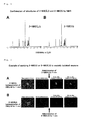
- Fig. 3 shows a fluorescence image and a transmitted light image captured at 1 minute after stopping the administration.
- yellow to red fluorescence based on the incorporation of 2-TRLG into the cell was not observed, and only green fluorescence derived from 2-NBDG was observed, and therefore, it could be evaluated that in this cell, a part of the cell membrane was not disrupted and the membrane was not in a state where both 2-NBDG and 2-TRLG were nonspecifically incorporated, and 2-NBDG was specifically incorporated into the cell (for example, when a hemichannel of the cell membrane is open, it allows a molecule having a molecular weight of about 1000 or less to pass through the cell membrane, and therefore, 2-TRLG having a molecular weight of 768 is also incorporated, however, the fact that the incorporation of 2-TRLG is not observed means that the incorporation of a molecule through the hemichannel is not caused).
- DBD-F 4-(N,N-dimethylaminosulfonyl)-7-fluoro-2,1,3-benzoxadiazole (DBD-F) (100 mg) was placed and dissolved in a mixed solvent of methanol (2.00 ml) and THF (2.00 ml). Under an argon atmosphere, a solution in which L-glucosamine hydrochloride (49.8 mg) was dissolved in 0.3 M NaHCO 3 solution (1.08 ml) was added thereto, and the resulting mixture was stirred at room temperature. After 48 hours, methanol was removed by concentration under reduced pressure, and the resulting precipitates (unnecessary substances derived from DBD-F) were removed by filtration.
- DBD-F 4-(N,N-dimethylaminosulfonyl)-7-fluoro-2,1,3-benzoxadiazole
- Escherichia coli K12 was cultured with shaking overnight at 37°C. A 1 mL portion of the culture was centrifuged to collect cells, and the cells were suspended in 1 mL of phosphate buffered saline (PBS, a mixed solution of 0.01 M phosphate buffer and 0.137 M NaCl at pH 7.4). A 90 ⁇ L portion of the suspension was taken out, and 10 ⁇ L of the 2-NBDG solution or the 2-NBDLG solution was added thereto to give a final concentration of 50 ⁇ M, and then, an incorporation reaction was allowed to proceed at 37°C for 10 minutes.
- PBS phosphate buffered saline
- the cells were collected by centrifugation and resuspended in PBS, and the cells were microscopically observed (objective lens x10) and recorded using a CCD camera for microscopic observation.
- objective lens x10 microscopically observed
- the result is shown in Fig. 5 .
- significant fluorescence due to the incorporation of 2-NBDG into the cells could be detected, however, fluorescence due to the incorporation of 2-NBDLG into the cells could not be detected under the same measurement condition as that for 2-NBDG.
- 2-NMALG was synthesized in the same manner as the method for synthesizing 2-NMAG except that L-glucosamine hydrochloride was used in place of D-glucosamine hydrochloride.
- Example 7 Usefulness in the case of using 2-NBDG, 2-TRLG, and 2-NMALG by mixing them
- FIG. 6 A fluorescence spectrum chart in the case where a solution obtained by mixing 2-NBDG as the D-glucose derivative that has a fluorescent chromophore in the molecule and is specifically incorporated into cells, and 2-TRLG and 2-NMALG as the L-glucose derivative that has a fluorescent chromophore in the molecule at 1:1:25 (molar ratio) was excited by laser light at 405 nm is shown in Fig. 6 . As is apparent from Fig.
- the blue fluorescence emitted by 2-NMALG at around 450 nm which is the maximum fluorescence wavelength of 2-NMALG, the green fluorescence emitted by 2-NBDG at around 540 nm which is the maximum fluorescence wavelength of 2-NBDG, and the red fluorescence emitted by 2-TRLG at around 610 nm which is the maximum fluorescence wavelength of 2-TRLG can be detected, respectively. Since 2-TRLG and 2-NMALG are each an L-glucose derivative, they are incorporated into cells only nonspecifically.
- the sizes (bulkiness) of the fluorescent chromophores of both glucose derivatives are different, and therefore, for example, in the case where the degree of the deterioration state of the cell membrane is small, only 2-NMALG having a small fluorescent chromophore is incorporated into cells, and 2-TRLG having a large fluorescent chromophore is not incorporated into cells until the degree of the deterioration state of the cell membrane becomes large (this was confirmed by another model experiment).
- a difference in the nonspecifically incorporated L-glucose derivative based on a difference in the degree of the deterioration state of the cell membrane can be detected, and therefore, the specific incorporation of D-glucose into cells relative to L-glucose can be evaluated and also the degree of the deterioration state of the cell membrane can be evaluated by the method.
- the present invention has an industrial applicability in the point that it can provide a method for accurately evaluating the specific incorporation of D-glucose into cells.
Landscapes
- Health & Medical Sciences (AREA)
- Life Sciences & Earth Sciences (AREA)
- Chemical & Material Sciences (AREA)
- Engineering & Computer Science (AREA)
- Immunology (AREA)
- Molecular Biology (AREA)
- Organic Chemistry (AREA)
- Hematology (AREA)
- Urology & Nephrology (AREA)
- Biomedical Technology (AREA)
- Biotechnology (AREA)
- Microbiology (AREA)
- Biochemistry (AREA)
- Physics & Mathematics (AREA)
- Analytical Chemistry (AREA)
- General Health & Medical Sciences (AREA)
- Proteomics, Peptides & Aminoacids (AREA)
- Wood Science & Technology (AREA)
- Zoology (AREA)
- Food Science & Technology (AREA)
- Medicinal Chemistry (AREA)
- Cell Biology (AREA)
- Pathology (AREA)
- General Physics & Mathematics (AREA)
- Bioinformatics & Cheminformatics (AREA)
- Biophysics (AREA)
- Genetics & Genomics (AREA)
- General Engineering & Computer Science (AREA)
- Emergency Medicine (AREA)
- Tropical Medicine & Parasitology (AREA)
- Measuring Or Testing Involving Enzymes Or Micro-Organisms (AREA)
- Investigating Or Analysing Biological Materials (AREA)
- Saccharide Compounds (AREA)
Applications Claiming Priority (2)
| Application Number | Priority Date | Filing Date | Title |
|---|---|---|---|
| JP2008205708 | 2008-08-08 | ||
| PCT/JP2009/064053 WO2010016587A1 (ja) | 2008-08-08 | 2009-08-07 | D-グルコースの細胞内への特異的な取り込みを評価するための方法 |
Publications (3)
| Publication Number | Publication Date |
|---|---|
| EP2325327A1 true EP2325327A1 (de) | 2011-05-25 |
| EP2325327A4 EP2325327A4 (de) | 2011-10-19 |
| EP2325327B1 EP2325327B1 (de) | 2013-04-24 |
Family
ID=41663799
Family Applications (1)
| Application Number | Title | Priority Date | Filing Date |
|---|---|---|---|
| EP09805069.3A Active EP2325327B1 (de) | 2008-08-08 | 2009-08-07 | Verfahren zur beurteilung von spezifischer einlagerung von d-glucose in zellen |
Country Status (4)
| Country | Link |
|---|---|
| US (2) | US8986656B2 (de) |
| EP (1) | EP2325327B1 (de) |
| JP (1) | JP5682881B2 (de) |
| WO (1) | WO2010016587A1 (de) |
Cited By (1)
| Publication number | Priority date | Publication date | Assignee | Title |
|---|---|---|---|---|
| US10921256B2 (en) | 2016-03-31 | 2021-02-16 | Hirosaki University | Multi-surface image acquisition system, observation device, observation method, screening method, and stereoscopic reconstruction method of subject |
Families Citing this family (5)
| Publication number | Priority date | Publication date | Assignee | Title |
|---|---|---|---|---|
| US20140154717A1 (en) * | 2011-03-31 | 2014-06-05 | Hirosaki University | Method for detecting cancer cell using fluorescently labeled l-glucose derivative, and cancer cell-imaging agent comprising fluorescently labeled l-glucose derivative |
| CN102199176A (zh) * | 2011-04-12 | 2011-09-28 | 江苏澳新生物工程有限公司 | 2-氨基-2-脱氧-d-葡萄糖及其盐的制备方法 |
| EP2905620B1 (de) | 2012-10-03 | 2017-12-13 | Hirosaki University | Verfahren zur zellbildgebung mit einem fluoreszenzmarkierten zuckerderivat mit daran gebundenem cumarinderivat und kontrastmittel |
| EP3130596B1 (de) | 2014-04-08 | 2021-01-20 | Hirosaki University | Glucosederivat sowie zellabbildungsverfahren und abbildungsmittel mit besagtem derivat |
| CN120484033B (zh) * | 2025-07-14 | 2025-09-26 | 山东焱农生物科技股份有限公司 | 一种银杏寡糖化学修饰的方法及制得的银杏氨基寡糖和应用 |
Family Cites Families (4)
| Publication number | Priority date | Publication date | Assignee | Title |
|---|---|---|---|---|
| JP2958447B2 (ja) * | 1997-03-11 | 1999-10-06 | 東京農工大学長 | 生体組織等の活動状態のイメージング法及びその利用法 |
| AU2002360766A1 (en) * | 2001-12-21 | 2003-07-30 | Threshold Pharmaceuticals, Inc. | Methods for cancer imaging |
| WO2004042360A2 (en) * | 2002-11-04 | 2004-05-21 | The Regents Of The Univeristy Of California | Deuterated glucose or fat tolerance tests for high-throughput measurement of the metabolism of sugars or fatty acids in the body |
| US8211654B2 (en) * | 2008-01-31 | 2012-07-03 | Wisconsin Alumni Research Foundation | High-throughput assay for sugar-mediated drug transport |
-
2009
- 2009-08-07 WO PCT/JP2009/064053 patent/WO2010016587A1/ja not_active Ceased
- 2009-08-07 US US13/058,061 patent/US8986656B2/en active Active
- 2009-08-07 EP EP09805069.3A patent/EP2325327B1/de active Active
- 2009-08-07 JP JP2009185010A patent/JP5682881B2/ja active Active
-
2015
- 2015-02-05 US US14/614,854 patent/US9958450B2/en active Active
Non-Patent Citations (7)
| Title |
|---|
| CARTER J R ET AL: "Glucose transport by trypsin-treated red blood cell ghosts", BIOCHIMICA ET BIOPHYSICA ACTA. BIOMEMBRANES, AMSTERDAM, NL, vol. 291, no. 2, 26 January 1973 (1973-01-26), pages 506-518, XP025416460, ISSN: 0005-2736, DOI: DOI:10.1016/0005-2736(73)90502-6 [retrieved on 1973-01-26] * |
| DATABASE BIOSIS [Online] BIOSCIENCES INFORMATION SERVICE, PHILADELPHIA, PA, US; 7 March 2001 (2001-03-07), KLODA DANIEL G ET AL: "Glucose toxicity induces myocardial insulin resistance", XP002649275, Database accession no. PREV200100258177 * |
| GERMINARIO R J ET AL: "SATURABLE AND NONSATURABLE HEXOSE UPTAKE IN CULTURED HUMAN SKIN FIBROBLASTS", CANADIAN JOURNAL OF BIOCHEMISTRY, vol. 56, no. 2, 1978, pages 80-88, XP009150122, ISSN: 0008-4018 * |
| LUDVIGSEN C ET AL: "A KINETIC ANALYSIS OF D GLUCOSE TRANSPORT BY ADIPOCYTE PLASMA MEMBRANES", JOURNAL OF BIOLOGICAL CHEMISTRY, vol. 254, no. 5, 1979, pages 1444-1446, XP002649276, ISSN: 0021-9258 * |
| See also references of WO2010016587A1 * |
| TOSHIHIRO YAMAMOTO ET AL: "Synthesis of 2-NBDLG, a fluorescent derivative of l-glucosamine; the antipode of d-glucose tracer 2-NBDG", TETRAHEDRON LETTERS, vol. 49, no. 48, 24 November 2008 (2008-11-24), pages 6876-6878, XP55002356, ISSN: 0040-4039, DOI: 10.1016/j.tetlet.2008.09.089 * |
| YAMADA KATSUYA ET AL: "IMAGING STEREOSPECIFIC UPTAKE OF GLUCOSE USING FLUORESCENT D- AND L-GLUCOSE ANALOGUES", JOURNAL OF PHYSIOLOGICAL SCIENCES, vol. 59, no. Suppl. 1, 2009, page 152, XP002649274, & 36TH INTERNATIONAL CONGRESS OF PHYSIOLOGICAL SCIENCES (IUPS2009); KYOTO, JAPAN; JULY 27 -AUGUST 01, 2009 ISSN: 1880-6546 * |
Cited By (1)
| Publication number | Priority date | Publication date | Assignee | Title |
|---|---|---|---|---|
| US10921256B2 (en) | 2016-03-31 | 2021-02-16 | Hirosaki University | Multi-surface image acquisition system, observation device, observation method, screening method, and stereoscopic reconstruction method of subject |
Also Published As
| Publication number | Publication date |
|---|---|
| WO2010016587A1 (ja) | 2010-02-11 |
| US9958450B2 (en) | 2018-05-01 |
| JP5682881B2 (ja) | 2015-03-11 |
| US20110189708A1 (en) | 2011-08-04 |
| US20150212094A1 (en) | 2015-07-30 |
| EP2325327B1 (de) | 2013-04-24 |
| EP2325327A4 (de) | 2011-10-19 |
| US8986656B2 (en) | 2015-03-24 |
| JP2010057484A (ja) | 2010-03-18 |
Similar Documents
| Publication | Publication Date | Title |
|---|---|---|
| US9958450B2 (en) | L-glucose derivatives having fluorescent chromophore | |
| EP1601955B1 (de) | Sauerstoffempfindliche Sonde und Verfahren zum Messen der Sauerstoffaufnahme | |
| CN110563650B (zh) | 一种硫酸酯酶的比率型双光子荧光探针及其合成方法和应用 | |
| EP2905620B1 (de) | Verfahren zur zellbildgebung mit einem fluoreszenzmarkierten zuckerderivat mit daran gebundenem cumarinderivat und kontrastmittel | |
| US5437980A (en) | Phenanthridium dye staining of nucleic acids in living cells | |
| EP2703495A1 (de) | Verfahren zum nachweis von krebszellen unter verwendung eines fluoreszenzmarkierten l-glucose-derivats und krebszellen-bildgebungsmittel mit dem fluoreszenzmarkierten l-glucose-derivat | |
| CN109836394B (zh) | 一种用于识别硫化氢的近红外荧光探针及其制备方法和应用 | |
| EP2880045B1 (de) | Fluorogenes glycosidase-substrat und zugehöriges detektionsverfahren | |
| CN112159396A (zh) | 一种检测γ-谷氨酰转肽酶近红外荧光分子探针及其制备方法与应用 | |
| US20210214559A1 (en) | Fluorescently labeled polysaccharide, preparation method therefor, and use thereof | |
| Okkelman et al. | Multi-parametric imaging of hypoxia and cell cycle in intestinal organoid culture | |
| CN110885326A (zh) | 高水溶性苯基乙酸酯化合物及包含该化合物的羧酸酯酶检测试剂盒 | |
| CN112062755B (zh) | 用于检测天冬氨酰氨肽酶的近红外荧光分子探针、制备方法及用途 | |
| Pan et al. | Accessibility of N-acyl-D-mannosamines to N-acetyl-D-neuraminic acid aldolase | |
| CN119285618B (zh) | 一种持久成像的半胱氨酸荧光探针及其制备方法和应用 | |
| Birmingham et al. | Diffusion and binding measurements within oral biofilms using fluorescence photobleaching recovery methods | |
| Kricka | Chemiluminescence and bioluminescence | |
| CN113999159B (zh) | 一种粘度敏感的荧光探针,其制备方法及应用 | |
| Suzuki et al. | Development of fluorescent probes for “On-Off” switching based detection of lectin–saccharide interactions | |
| Prenna et al. | Cytofluorometric quantification of the activity and reaction kinetics of acid phosphatase | |
| Hickey et al. | SERS imaging analyses of bacteria cells among plant tissues | |
| Wang et al. | Na+-sensing quantum dots for cell-based screening of intracellular Na+ concentrations ([Na+] i) | |
| CN118878524B (zh) | 一种检测羧酸酯酶兼具抑菌活性的近红外荧光探针、其制备方法和应用 | |
| US8513394B2 (en) | Saccharide fluorescent substrates, preparation method and uses thereof | |
| RU2854120C1 (ru) | Способ получения бактериальных клеток, капсулированных биогенными наночастицами металлов |
Legal Events
| Date | Code | Title | Description |
|---|---|---|---|
| PUAI | Public reference made under article 153(3) epc to a published international application that has entered the european phase |
Free format text: ORIGINAL CODE: 0009012 |
|
| 17P | Request for examination filed |
Effective date: 20110307 |
|
| AK | Designated contracting states |
Kind code of ref document: A1 Designated state(s): AT BE BG CH CY CZ DE DK EE ES FI FR GB GR HR HU IE IS IT LI LT LU LV MC MK MT NL NO PL PT RO SE SI SK SM TR |
|
| AX | Request for extension of the european patent |
Extension state: AL BA RS |
|
| A4 | Supplementary search report drawn up and despatched |
Effective date: 20110920 |
|
| RIC1 | Information provided on ipc code assigned before grant |
Ipc: G01N 33/50 20060101ALI20110909BHEP Ipc: C12Q 1/04 20060101ALI20110909BHEP Ipc: C07H 5/06 20060101ALI20110909BHEP Ipc: C07H 1/00 20060101ALI20110909BHEP Ipc: C07H 17/00 20060101ALI20110909BHEP Ipc: C07H 13/00 20060101ALI20110909BHEP Ipc: C12Q 1/02 20060101ALI20110909BHEP Ipc: C12Q 1/54 20060101AFI20110909BHEP |
|
| DAX | Request for extension of the european patent (deleted) | ||
| GRAP | Despatch of communication of intention to grant a patent |
Free format text: ORIGINAL CODE: EPIDOSNIGR1 |
|
| GRAS | Grant fee paid |
Free format text: ORIGINAL CODE: EPIDOSNIGR3 |
|
| GRAP | Despatch of communication of intention to grant a patent |
Free format text: ORIGINAL CODE: EPIDOSNIGR1 |
|
| GRAA | (expected) grant |
Free format text: ORIGINAL CODE: 0009210 |
|
| AK | Designated contracting states |
Kind code of ref document: B1 Designated state(s): AT BE BG CH CY CZ DE DK EE ES FI FR GB GR HR HU IE IS IT LI LT LU LV MC MK MT NL NO PL PT RO SE SI SK SM TR |
|
| REG | Reference to a national code |
Ref country code: GB Ref legal event code: FG4D |
|
| REG | Reference to a national code |
Ref country code: CH Ref legal event code: EP |
|
| REG | Reference to a national code |
Ref country code: AT Ref legal event code: REF Ref document number: 608690 Country of ref document: AT Kind code of ref document: T Effective date: 20130515 |
|
| REG | Reference to a national code |
Ref country code: IE Ref legal event code: FG4D |
|
| REG | Reference to a national code |
Ref country code: DE Ref legal event code: R096 Ref document number: 602009015311 Country of ref document: DE Effective date: 20130627 |
|
| REG | Reference to a national code |
Ref country code: CH Ref legal event code: PUEA Owner name: HIROSAKI UNIVERSITY, JP Free format text: FORMER OWNER: NATIONAL UNIVERSITY CORPORATION TOKYO UNIVERSITY OF AGRICULTURE AND TECHNOLOGY, JP Ref country code: CH Ref legal event code: NV Representative=s name: FIAMMENGHI-FIAMMENGHI, CH |
|
| REG | Reference to a national code |
Ref country code: AT Ref legal event code: MK05 Ref document number: 608690 Country of ref document: AT Kind code of ref document: T Effective date: 20130424 |
|
| REG | Reference to a national code |
Ref country code: LT Ref legal event code: MG4D |
|
| REG | Reference to a national code |
Ref country code: NL Ref legal event code: VDEP Effective date: 20130424 |
|
| PG25 | Lapsed in a contracting state [announced via postgrant information from national office to epo] |
Ref country code: BE Free format text: LAPSE BECAUSE OF FAILURE TO SUBMIT A TRANSLATION OF THE DESCRIPTION OR TO PAY THE FEE WITHIN THE PRESCRIBED TIME-LIMIT Effective date: 20130424 Ref country code: NO Free format text: LAPSE BECAUSE OF FAILURE TO SUBMIT A TRANSLATION OF THE DESCRIPTION OR TO PAY THE FEE WITHIN THE PRESCRIBED TIME-LIMIT Effective date: 20130724 Ref country code: AT Free format text: LAPSE BECAUSE OF FAILURE TO SUBMIT A TRANSLATION OF THE DESCRIPTION OR TO PAY THE FEE WITHIN THE PRESCRIBED TIME-LIMIT Effective date: 20130424 Ref country code: FI Free format text: LAPSE BECAUSE OF FAILURE TO SUBMIT A TRANSLATION OF THE DESCRIPTION OR TO PAY THE FEE WITHIN THE PRESCRIBED TIME-LIMIT Effective date: 20130424 Ref country code: SI Free format text: LAPSE BECAUSE OF FAILURE TO SUBMIT A TRANSLATION OF THE DESCRIPTION OR TO PAY THE FEE WITHIN THE PRESCRIBED TIME-LIMIT Effective date: 20130424 Ref country code: ES Free format text: LAPSE BECAUSE OF FAILURE TO SUBMIT A TRANSLATION OF THE DESCRIPTION OR TO PAY THE FEE WITHIN THE PRESCRIBED TIME-LIMIT Effective date: 20130804 Ref country code: SE Free format text: LAPSE BECAUSE OF FAILURE TO SUBMIT A TRANSLATION OF THE DESCRIPTION OR TO PAY THE FEE WITHIN THE PRESCRIBED TIME-LIMIT Effective date: 20130424 Ref country code: LT Free format text: LAPSE BECAUSE OF FAILURE TO SUBMIT A TRANSLATION OF THE DESCRIPTION OR TO PAY THE FEE WITHIN THE PRESCRIBED TIME-LIMIT Effective date: 20130424 Ref country code: PT Free format text: LAPSE BECAUSE OF FAILURE TO SUBMIT A TRANSLATION OF THE DESCRIPTION OR TO PAY THE FEE WITHIN THE PRESCRIBED TIME-LIMIT Effective date: 20130826 Ref country code: GR Free format text: LAPSE BECAUSE OF FAILURE TO SUBMIT A TRANSLATION OF THE DESCRIPTION OR TO PAY THE FEE WITHIN THE PRESCRIBED TIME-LIMIT Effective date: 20130725 Ref country code: IS Free format text: LAPSE BECAUSE OF FAILURE TO SUBMIT A TRANSLATION OF THE DESCRIPTION OR TO PAY THE FEE WITHIN THE PRESCRIBED TIME-LIMIT Effective date: 20130824 |
|
| PG25 | Lapsed in a contracting state [announced via postgrant information from national office to epo] |
Ref country code: PL Free format text: LAPSE BECAUSE OF FAILURE TO SUBMIT A TRANSLATION OF THE DESCRIPTION OR TO PAY THE FEE WITHIN THE PRESCRIBED TIME-LIMIT Effective date: 20130424 Ref country code: HR Free format text: LAPSE BECAUSE OF FAILURE TO SUBMIT A TRANSLATION OF THE DESCRIPTION OR TO PAY THE FEE WITHIN THE PRESCRIBED TIME-LIMIT Effective date: 20130424 Ref country code: CY Free format text: LAPSE BECAUSE OF FAILURE TO SUBMIT A TRANSLATION OF THE DESCRIPTION OR TO PAY THE FEE WITHIN THE PRESCRIBED TIME-LIMIT Effective date: 20130424 Ref country code: BG Free format text: LAPSE BECAUSE OF FAILURE TO SUBMIT A TRANSLATION OF THE DESCRIPTION OR TO PAY THE FEE WITHIN THE PRESCRIBED TIME-LIMIT Effective date: 20130724 Ref country code: LV Free format text: LAPSE BECAUSE OF FAILURE TO SUBMIT A TRANSLATION OF THE DESCRIPTION OR TO PAY THE FEE WITHIN THE PRESCRIBED TIME-LIMIT Effective date: 20130424 |
|
| PG25 | Lapsed in a contracting state [announced via postgrant information from national office to epo] |
Ref country code: CZ Free format text: LAPSE BECAUSE OF FAILURE TO SUBMIT A TRANSLATION OF THE DESCRIPTION OR TO PAY THE FEE WITHIN THE PRESCRIBED TIME-LIMIT Effective date: 20130424 Ref country code: EE Free format text: LAPSE BECAUSE OF FAILURE TO SUBMIT A TRANSLATION OF THE DESCRIPTION OR TO PAY THE FEE WITHIN THE PRESCRIBED TIME-LIMIT Effective date: 20130424 Ref country code: SK Free format text: LAPSE BECAUSE OF FAILURE TO SUBMIT A TRANSLATION OF THE DESCRIPTION OR TO PAY THE FEE WITHIN THE PRESCRIBED TIME-LIMIT Effective date: 20130424 Ref country code: DK Free format text: LAPSE BECAUSE OF FAILURE TO SUBMIT A TRANSLATION OF THE DESCRIPTION OR TO PAY THE FEE WITHIN THE PRESCRIBED TIME-LIMIT Effective date: 20130424 |
|
| PG25 | Lapsed in a contracting state [announced via postgrant information from national office to epo] |
Ref country code: IT Free format text: LAPSE BECAUSE OF FAILURE TO SUBMIT A TRANSLATION OF THE DESCRIPTION OR TO PAY THE FEE WITHIN THE PRESCRIBED TIME-LIMIT Effective date: 20130424 Ref country code: NL Free format text: LAPSE BECAUSE OF FAILURE TO SUBMIT A TRANSLATION OF THE DESCRIPTION OR TO PAY THE FEE WITHIN THE PRESCRIBED TIME-LIMIT Effective date: 20130424 Ref country code: RO Free format text: LAPSE BECAUSE OF FAILURE TO SUBMIT A TRANSLATION OF THE DESCRIPTION OR TO PAY THE FEE WITHIN THE PRESCRIBED TIME-LIMIT Effective date: 20130424 |
|
| PLBE | No opposition filed within time limit |
Free format text: ORIGINAL CODE: 0009261 |
|
| STAA | Information on the status of an ep patent application or granted ep patent |
Free format text: STATUS: NO OPPOSITION FILED WITHIN TIME LIMIT |
|
| 26N | No opposition filed |
Effective date: 20140127 |
|
| PG25 | Lapsed in a contracting state [announced via postgrant information from national office to epo] |
Ref country code: MC Free format text: LAPSE BECAUSE OF FAILURE TO SUBMIT A TRANSLATION OF THE DESCRIPTION OR TO PAY THE FEE WITHIN THE PRESCRIBED TIME-LIMIT Effective date: 20130424 |
|
| REG | Reference to a national code |
Ref country code: DE Ref legal event code: R097 Ref document number: 602009015311 Country of ref document: DE Effective date: 20140127 |
|
| REG | Reference to a national code |
Ref country code: IE Ref legal event code: MM4A |
|
| PG25 | Lapsed in a contracting state [announced via postgrant information from national office to epo] |
Ref country code: IE Free format text: LAPSE BECAUSE OF NON-PAYMENT OF DUE FEES Effective date: 20130807 |
|
| PG25 | Lapsed in a contracting state [announced via postgrant information from national office to epo] |
Ref country code: SM Free format text: LAPSE BECAUSE OF FAILURE TO SUBMIT A TRANSLATION OF THE DESCRIPTION OR TO PAY THE FEE WITHIN THE PRESCRIBED TIME-LIMIT Effective date: 20130424 |
|
| PG25 | Lapsed in a contracting state [announced via postgrant information from national office to epo] |
Ref country code: TR Free format text: LAPSE BECAUSE OF FAILURE TO SUBMIT A TRANSLATION OF THE DESCRIPTION OR TO PAY THE FEE WITHIN THE PRESCRIBED TIME-LIMIT Effective date: 20130424 Ref country code: MT Free format text: LAPSE BECAUSE OF FAILURE TO SUBMIT A TRANSLATION OF THE DESCRIPTION OR TO PAY THE FEE WITHIN THE PRESCRIBED TIME-LIMIT Effective date: 20130424 |
|
| PG25 | Lapsed in a contracting state [announced via postgrant information from national office to epo] |
Ref country code: MK Free format text: LAPSE BECAUSE OF FAILURE TO SUBMIT A TRANSLATION OF THE DESCRIPTION OR TO PAY THE FEE WITHIN THE PRESCRIBED TIME-LIMIT Effective date: 20130424 Ref country code: HU Free format text: LAPSE BECAUSE OF FAILURE TO SUBMIT A TRANSLATION OF THE DESCRIPTION OR TO PAY THE FEE WITHIN THE PRESCRIBED TIME-LIMIT; INVALID AB INITIO Effective date: 20090807 Ref country code: LU Free format text: LAPSE BECAUSE OF NON-PAYMENT OF DUE FEES Effective date: 20130807 |
|
| REG | Reference to a national code |
Ref country code: FR Ref legal event code: PLFP Year of fee payment: 8 |
|
| REG | Reference to a national code |
Ref country code: FR Ref legal event code: PLFP Year of fee payment: 9 |
|
| REG | Reference to a national code |
Ref country code: FR Ref legal event code: PLFP Year of fee payment: 10 |
|
| REG | Reference to a national code |
Ref country code: DE Ref legal event code: R081 Ref document number: 602009015311 Country of ref document: DE Owner name: ORBIO CORP., JP Free format text: FORMER OWNERS: HIROSAKI UNIVERSITY, HIROSAKI-SHI, JP; NATIONAL UNIVERSITY CORPORATION TOKYO UNIVERSITY OF AGRICULTURE AND TECHNOLOGY, FUCHU, TOKYO, JP Ref country code: DE Ref legal event code: R081 Ref document number: 602009015311 Country of ref document: DE Owner name: NATIONAL UNIVERSITY CORPORATION TOKYO UNIVERSI, JP Free format text: FORMER OWNERS: HIROSAKI UNIVERSITY, HIROSAKI-SHI, JP; NATIONAL UNIVERSITY CORPORATION TOKYO UNIVERSITY OF AGRICULTURE AND TECHNOLOGY, FUCHU, TOKYO, JP |
|
| REG | Reference to a national code |
Ref country code: GB Ref legal event code: 732E Free format text: REGISTERED BETWEEN 20230223 AND 20230301 |
|
| REG | Reference to a national code |
Ref country code: DE Ref legal event code: R081 Ref document number: 602009015311 Country of ref document: DE Owner name: ORBIO CORP., JP Free format text: FORMER OWNERS: NATIONAL UNIVERSITY CORPORATION TOKYO UNIVERSITY OF AGRICULTURE AND TECHNOLOGY, FUCHU, TOKYO, JP; ORBIO CORP., KYOTO, JP |
|
| REG | Reference to a national code |
Ref country code: GB Ref legal event code: 732E Free format text: REGISTERED BETWEEN 20250424 AND 20250430 |
|
| PGFP | Annual fee paid to national office [announced via postgrant information from national office to epo] |
Ref country code: DE Payment date: 20250722 Year of fee payment: 17 |
|
| PGFP | Annual fee paid to national office [announced via postgrant information from national office to epo] |
Ref country code: GB Payment date: 20250822 Year of fee payment: 17 |
|
| PGFP | Annual fee paid to national office [announced via postgrant information from national office to epo] |
Ref country code: FR Payment date: 20250821 Year of fee payment: 17 |
|
| PGFP | Annual fee paid to national office [announced via postgrant information from national office to epo] |
Ref country code: CH Payment date: 20250901 Year of fee payment: 17 |