EP2333059A1 - Ensemble universel de sondes d'acides nucléiques et procédé pour leur utilisation - Google Patents
Ensemble universel de sondes d'acides nucléiques et procédé pour leur utilisation Download PDFInfo
- Publication number
- EP2333059A1 EP2333059A1 EP09803013A EP09803013A EP2333059A1 EP 2333059 A1 EP2333059 A1 EP 2333059A1 EP 09803013 A EP09803013 A EP 09803013A EP 09803013 A EP09803013 A EP 09803013A EP 2333059 A1 EP2333059 A1 EP 2333059A1
- Authority
- EP
- European Patent Office
- Prior art keywords
- nucleic acid
- probe
- fluorescent
- target nucleic
- binding
- Prior art date
- Legal status (The legal status is an assumption and is not a legal conclusion. Google has not performed a legal analysis and makes no representation as to the accuracy of the status listed.)
- Withdrawn
Links
Images






Classifications
-
- C—CHEMISTRY; METALLURGY
- C12—BIOCHEMISTRY; BEER; SPIRITS; WINE; VINEGAR; MICROBIOLOGY; ENZYMOLOGY; MUTATION OR GENETIC ENGINEERING
- C12Q—MEASURING OR TESTING PROCESSES INVOLVING ENZYMES, NUCLEIC ACIDS OR MICROORGANISMS; COMPOSITIONS OR TEST PAPERS THEREFOR; PROCESSES OF PREPARING SUCH COMPOSITIONS; CONDITION-RESPONSIVE CONTROL IN MICROBIOLOGICAL OR ENZYMOLOGICAL PROCESSES
- C12Q1/00—Measuring or testing processes involving enzymes, nucleic acids or microorganisms; Compositions therefor; Processes of preparing such compositions
- C12Q1/68—Measuring or testing processes involving enzymes, nucleic acids or microorganisms; Compositions therefor; Processes of preparing such compositions involving nucleic acids
- C12Q1/6876—Nucleic acid products used in the analysis of nucleic acids, e.g. primers or probes
-
- C—CHEMISTRY; METALLURGY
- C12—BIOCHEMISTRY; BEER; SPIRITS; WINE; VINEGAR; MICROBIOLOGY; ENZYMOLOGY; MUTATION OR GENETIC ENGINEERING
- C12Q—MEASURING OR TESTING PROCESSES INVOLVING ENZYMES, NUCLEIC ACIDS OR MICROORGANISMS; COMPOSITIONS OR TEST PAPERS THEREFOR; PROCESSES OF PREPARING SUCH COMPOSITIONS; CONDITION-RESPONSIVE CONTROL IN MICROBIOLOGICAL OR ENZYMOLOGICAL PROCESSES
- C12Q1/00—Measuring or testing processes involving enzymes, nucleic acids or microorganisms; Compositions therefor; Processes of preparing such compositions
- C12Q1/68—Measuring or testing processes involving enzymes, nucleic acids or microorganisms; Compositions therefor; Processes of preparing such compositions involving nucleic acids
- C12Q1/6813—Hybridisation assays
- C12Q1/6816—Hybridisation assays characterised by the detection means
Definitions
- This relates to the field of genetic engineering, and more specifically, to a nucleic acid probe set useful for analyzing a nucleic acid and a method for using the same.
- single-stranded nucleic acid probes are widely used. These single-stranded nucleic acid probes have base sequences designed to hybridize specifically to the target nucleic acids, and are labeled with fluorescent substances.
- Q-probe Quenching Probe
- Q-probe uses, as a probe-labeling fluorescent substance, a fluorescent substance the fluorescence emission of which is reduced under the action of guanine when the fluorescent substance is located near guanine compared with when it is in a normal state.
- the Q-probe By adding the Q-probe to a solution to be measured and conducting a measurement of fluorescence, SNP genotyping or quantification of a gene can be performed simply and conveniently.
- the Q-probe has excellent advantages in that the structure of the probe is simple, no trial-and-error approach is needed for the designing of the probe, and highly-accurate measurement results can be obtained (see, for example, Patent Document 1 and Patent Document 2).
- a fluorescently-labeled nucleic acid probe having a different base sequence has to be prepared specifically for every target nucleic acid to be detected.
- Such a fluorescently-labeled nucleic acid probe is accompanied by problems that it is relatively costly and its synthesis requires a long time, and therefore, involves problems that an experiment making use of it is costly and requires a lot of time in preparation.
- nucleic acid probe sets (universal Q-probe sets) each of which comprises (A) a fluorescent probe and (B) a binding probe having (b1) a fluorescent probe binding region complementary to the fluorescent probe and (b2) a sequence complementary to a target nucleic acid sequence (C), and have been working toward their practical use.
- the use of a universal Q-probe set in a real-time PCR experiment can drastically reduce the cost required for the preparation of a probe.
- the fluorescence quenching efficiency available from the use of a universal Q-probe set is limited to as low as a half to one-third or so of that available from the use of a conventional, single-stranded Q-probe, and accordingly, there is an outstanding desire for an improvement in fluorescence quenching efficiency for practical use.
- a first object of the present invention is to provide a universal Q-probe set with a fluorescence quenching efficiency improved to a similar level as those of conventional, single-stranded Q-probes and also a method for designing such a universal Q-probe set, and further to provide a method for its use.
- a second object of the present invention is to provide an oligonucleotide that can form a stable complex which does not dissociate in a water system, and further to provide a method for its use.
- a third object of the present invention is to provide a universal Q-probe set with an improved fluorescence quenching efficiency and also a method for designing the universal Q-probe set, and further to provide a method for its use.
- the present invention provides, in a first thereof, a nucleic acid probe set comprising (A) a fluorescent probe, which is formed of an oligonucleotide including (a) a nucleotide unit labeled with (d) a fluorescent substance, and (B) a binding probe formed of an oligonucleotide having (b1) a fluorescent probe binding region, which can hybridize to the fluorescent probe (A), and (b2) a target nucleic acid binding region, which can hybridize to a target nucleic acid sequence (C), wherein the fluorescent substance (d) is a fluorescent substance which changes in fluorescent character upon interaction with guanine, and at least one of nucleotide units which constitute the fluorescent probe (A) is an artificial nucleotide unit having a function to raise a dissociation temperature between the probe (A) and the fluorescent probe binding region (b1).
- a fluorescent probe which is formed of an oligonucleotide including (a) a nucleotide unit labeled with (
- the artificial nucleotide unit having the function to raise the dissociation temperature may preferably be at least one artificial nucleotide unit selected from the group consisting of LNA, PNA, ENA, 2' 4' -BNA NC and 2' , 4' -BNA COC units.
- nucleic acid probe set according to the first aspect of the present invention preferably at least one-third, more preferably at least 80% of the nucleotide units which constitute the fluorescent probe (A) may be artificial nucleotide units.
- the fluorescent substance (d) may preferably be any one selected from the group consisting of fluorescein, fluorescein-4-isothiocyanate, tetrachlorofluorescein, hexachlorofluorescein, tetrabromosulfonefluorescein, EDANS (5-(2-aminoethylamino)-1-naphthalensulfonic acid), 6-carboxy-4',5'-dichloro-2',7'-dimethoxyfluorescein (6-JOE), 3,6-diamino-9-[2,4-bis(lithiooxycarbonyl)phenyl]-4-[lithioxysulfonyl)-5-sulfonatoxanthylium/3,6-diamin o-9-[2,5-bis(lithiooxycarbonyl)phenyl]-4-(lithooxys ulfonyl)-5-
- the target nucleic acid binding region (b2) may be located preferably on a side of a 5' -end of the binding probe (B), and the nucleotide unit (a) labeled with the fluorescent substance (d) is a 3'-terminal nucleotide unit of the fluorescent probe (A).
- the present invention also provides, in the first aspect thereof, a method for detecting a target nucleic acid, which comprises the following steps (1) to (4):
- the present invention further provides, in the first aspect thereof, a method for analyzing a nucleic acid for a base sequence polymorphism, which comprises the following steps (1) to (4):
- the present invention still further provides, in the first aspect thereof, a method, which comprises conducting a melting curve analysis on a complex of the nucleic acid probe set according to the first aspect of the present invention and a target nucleic acid.
- the present invention provides, in a second aspect thereof, an oligonucleotide probe comprising nucleotide units including (a') a nucleotide unit labeled with (h) a labeling substance, a part or all of said nucleotide units being an artificial nucleotide unit or units having a function to raise a dissociation temperature of the oligonucleotide probe from a complementary strand, said dissociation temperature of the oligonucleotide probe from the complementary strand being 100°C or higher under normal pressure conditions.
- the artificial nucleotide unit or units having the function to raise the dissociation temperature from the complementary strand may preferably be one or more artificial nucleotide units each selected from the group consisting of LNA, PNA, ENA, 2', 4' -BNA NC and 2' 4' -BNA COC units; and the labeling substance (h) may preferably be a fluorescent substance, quencher substance, protein or functional group.
- the present invention also provides, in the second aspect thereof, a method, which comprises hybridizing the oligonucleotide probe according to the second aspect of the present invention with (E) an oligonucleotide having a complementary base sequence to label the oligonucleotide (E) with the labeling substance (h); and the nucleic acid probe set according to the first aspect of the present invention, wherein the oligonucleotide probe according to the second aspect of the present invention, in which the labeling substance (h) is a fluorescent substance which changes in fluorescent character upon interaction with guanine, is used as a fluorescent probe (A).
- the present invention provides, in a third aspect thereof, a nucleic acid probe set comprising (A) one fluorescent probe, which is formed of an oligonucleotide including (a) a nucleotide unit labeled with (d) a fluorescent substance, and (B) one binding probe formed of an oligonucleotide having (b1) one fluorescent probe binding region, which can hybridize to the fluorescent probe (A), and (b2) one target nucleic acid binding region, which can hybridize to a target nucleic acid sequence (C), wherein the fluorescent substance (d) is a fluorescent substance which changes in fluorescent character upon interaction with guanine, the nucleotide unit (a) is a 3'-terminal nucleotide unit of the fluorescent probe (A), and the target nucleic acid binding region (b2) is located on a side of a 5' -end of the binding probe (B).
- a universal Q-probe set with a fluorescence quenching efficiency improved to a similar level as those of conventional, single-stranded Q-probes The use of the universal Q-probe set according to the first aspect of the present invention in place of a single-stranded Q-probe makes it possible to significantly reduce the cost required for a real-time PCR experiment.
- an oligonucleotide capable of forming a stable complex that does not dissociate in a water system there is provided.
- a universal Q-probe set with an improved fluorescence quenching efficiency there is provided. The use of the universal Q-probe set according to the third aspect of the present invention in place of a conventional single-stranded Q-probe makes it possible to significantly reduce the cost required for a real-time PCR experiment.
- the hybridized complex of the fluorescent probe (A) and binding probe (B) may be called “the nucleic acid probe complex”.
- nucleotide as used herein is not limited to deoxyribonucleotides as basic units of DNA and ribonucleotides as basic units of RNA, but shall be construed to also include artificially-synthesized monomers such as LNAs (Locked Nucleic Acids) and peptide nucleic acids (PNAs).
- oligonucleotide as used herein means an oligomer formed from a nucleotide monomer. This oligomer may be formed from only deoxyribonucleotide, ribonucleotide, LNA or PNA units, or may be a chimeric molecule thereof.
- target nucleic acid means a nucleic acid to be subjected to quantification, analysis or the like, and shall be construed to also include a portion or portions of one or more of various nucleic acids or genes in some instances.
- Monomers that constitute target nucleic acids can be of any type, and deoxyribonucleotides, ribonucleotides, LNAs, PNAs, artificially-modified nucleic acids and the like can be mentioned.
- target nucleic acid sequence (C) as used herein means a base sequence region, which is located in a target nucleic acid and specifically hybridizes to a target nucleic acid binding region (b2) in a binding probe (B) that constitutes a nucleic acid probe set according to the present invention.
- normal pressure as used herein means one(1) atmospheric pressure.
- FIGS. 1 , and 3 to 6 Examples of the nucleic acid probe set according to the first aspect of the present invention are shown in FIGS. 1 , and 3 to 6 .
- fluorescent probes A
- binding probes B
- target nucleic acid sequences C
- fluorescent substances d
- the nucleic acid probe set according to the first aspect of the present invention comprises the fluorescent probe (A) and the binding probe (B).
- the binding probe (B) has a fluorescent probe binding region (b1), which has a base sequence complementary to the fluorescent probe (A), and a target nucleic acid binding region (b2), which has a base sequence complementary to the target nucleic acid sequence (C).
- the fluorescent probe (A) which constitutes the nucleic acid probe set according to the first aspect of the present invention, is an oligonucleotide including a nucleotide unit (a) labeled with the fluorescent substance (d).
- No particular limitation is imposed on the base sequence of the fluorescent probe (A) insofar as the fluorescent probe (A) can hybridize with the fluorescent probe binding region (b1) in the binding probe (B).
- the base sequence of the fluorescent probe (A) therefore, does not depend on the base sequence of a target nucleic acid to be detected or analyzed.
- the fluorescent probe (A) that constitutes the nucleic acid probe set according to the first aspect of the present invention is not required to have a base sequence corresponding to the specific target nucleic acid, and the fluorescent probe (A) of the same base sequence can be commonly used for different target nucleic acids.
- the nucleic probe set according to the first aspect of the present invention is, therefore, called "a universal nucleic probe set" by the present inventors.
- the use of the nucleic acid probe set according to the first aspect of the present invention for the analysis of a target nucleic acid has an advantage in that it is no longer needed to prepare a fluorescent probe, which has a costly fluorescent substance, specifically for the target nucleic acid to be detected or analyzed and the production cost of the fluorescent probe can be minimized.
- the fluorescent probe (A) includes, as at least one of nucleotide units as the basic units of the probe, an artificial nucleotide unit or units having a function to raise the dissociation temperature between the probe (A) and the fluorescent probe binding region (b1). Owing to the inclusion of the artificial nucleotide unit or units in the fluorescent probe (A), Tm between the fluorescent probe (A) and the fluorescent probe binding region (b1) becomes higher.
- the Tm between the fluorescent probe (A) and the binding probe (B) can be easily made higher than the Tm between the target nucleic acid sequence (C) and the target nucleic acid binding region (b2), thereby making it possible to provide the nucleic acid probe set according to the first aspect of the present invention with higher stability at elevated temperatures, and hence, with improved reliability as a fluorescent probe.
- the Tm between the fluorescent probe (A) and the binding probe (B) can be made higher than the thermal denaturation temperature (for example, 95°C) of PCR, so that the fluorescentprobe (A) and the binding probe (B) canalways remain as a stable nucleic acid probe complex during PCR cycles.
- LNA, PNA, ENA, 2' ,4' -BNA NC and 2' ,4' -BNA COC units can be mentioned.
- An LNA monomer is a nucleotide having two ring structures that the 2'-oxygen and 4'-carbon atoms of ribose are connected together via a methylene unit. Due to the inclusion of these two ring structures, the LNA monomer has low structural freedom, and compared with DNA or RNA monomer, strongly hybridizes with a complementary strand. It is, therefore, known that by substituting one or more mononucleotide units (DNA monomers), which make up an oligonucleotide formed of DNA units, to a like number of LNA units, the Tm between the oligonucleotide and a complementary strand rises.
- DNA monomers mononucleotide units
- PNA is an abbreviation of peptide nucleic acid, and has a structure that a structure composed of N-(2-aminoethyl)glycine units linked together via amide bonds is contained as a backbone and base moieties (purine rings or pyrimidine rings) are connected to nitrogen atoms in the backbone via -COCH 2 -.
- base moieties purine rings or pyrimidine rings
- PNA monomer does not produce strong electrostatic repulsion against a complementary strand as no charge exists on its phosphate moieties. The dissociation temperature from the complementary strand, therefore, rises when one or more DNA units are substituted to a like number of PNA units.
- ENA is an abbreviation of 2'-O,4'-C-ethylene-bridged nucleic acid, and has a structure that the 2-O and 4-C atoms of a furanose ring are bridged together via an ethylene unit. It is known that by substituting one or more of mononucleotide units (DNA units), which make up an oligonucleotide formed of the DNA units, to a like number of ENA units, the Tm between the oligonucleotide and a complementary strand rises.
- DNA units mononucleotide units
- BNA is an abbreviation of bridged nucleic acid.
- 2' ,4' -BNA NC has a structure that in a furanose ring, the 2-O atom is bridged to the 4-C atom via -NRCH 2 - (R: methyl group), while 2' ,4' -BNA COC has a structure that the 2-O and 4-C atoms of a furanose ring are bridged together via -CH 2 OCH 2 -.
- Each of these artificial nucleotide units is also known to raise the Tm between an oligonucleotide formed of DNA units and a complementary strand when one or more of nucleotide units (DNAmonomers) making up the oligonucleotide are substituted to a like number of such BNA units.
- DNAmonomers nucleotide units
- the proportion of the artificial nucleotide unit or units in the fluorescent probe (A), said proportion being required to make the Tm between the fluorescent probe (A) and the binding probe (B) higher than the thermal denaturation temperature of PCR, also depends on the base number and base sequence of the fluorescent probe (A), and cannot be specified.
- the proportion of the artificial nucleotide unit or units may be at least one third of the entire nucleotide units, with at least 80% thereof being more preferred.
- the interaction between the fluorescent probe (A) and the fluorescent probe binding region (b1) is strengthened.
- the base numbers of the fluorescent probe (A) and fluorescent probe binding region (b1) can, therefore, be decreased compared with the case that the fluorescent probe (A) is formed of DNA units alone.
- a binding probe (B) of a large base number an error occurs.
- the use of a fluorescent probe binding region (b1) of a decreased base number can reduce the error, and can increase the synthesis yield of a binding probe (B). This leads to a reduction in the production cost for the probe set according to the first aspect of the present invention.
- fluorescent substance (d) with which the fluorescent probe (A) is labeled in the first aspect of the present invention is a fluorescent substance (d) which changes in fluorescent character upon interaction with guanine.
- fluorescent character means fluorescence intensity
- the expression "guanine and the fluorescent substance interact with each other to change the fluorescent character of the fluorescent substance” means that the fluorescence intensity of the fluorescent substance in a state that guanine and the fluorescent substance are not interacting with each other is different from its fluorescence intensity in a state that they are interacting with each other, and on the extent of this difference, no limitation shall be imposed.
- quenched or quenching of fluorescence means that upon interaction of a fluorescent substance with guanine, the fluorescence intensity decreases compared with the fluorescence intensity when the fluorescent substance is not interacting with guanine, and on the extent of this decrease, no limitation shall be imposed.
- fluorescent substances which can be suitably used in the nucleic acid probe set according to the first aspect of the present invention, include fluorescein and its derivatives [e.g., fluorescein-4-isothiocyanate (FITC), tetrachlorofluorescein, hexachlorofluorescein, tetrabromosulfonefluorescein (TBSF), and derivatives thereof], EDANS (5-(2-aminoethylamino)-1-naphthalenesulfonic acid), 6-carboxy-4' ,5'-dichloro-2' ,7'-dimethoxyfluorescein (6-JOE), 3,6-diamino-9-[2,4-bis(lithiooxycarbonyl)phenyl]-4-(lithioxysulfonyl)-5-sulfonatoxanthylium/3,6-diamin o-9-[2,5-bis(lithiooxycarbonyl)phenyl]
- fluorescein fluorescein-4-isothiocyanate, tetrachlorofluorescein, hexachlorofluorescein, tetrabromosulfonefluorescein, EDANS, 6-JOE, Alexa Fluor 488, Alexa Fluor 532, Pacific Blue, rhodamine 6G, carboxyrhodamine 6G, tetramethylrhodamine, carboxytetramethylrhodamine or BODIPY-FL is more preferred, and the use of Pacific Blue, carboxyrhodamine 6G or BODIPY-FL is most preferred.
- the target nucleic acid binding region (b2) of the binding probe (B) is designed such that, when the nucleic acid probe complex in the present invention has hybridized to the target nucleic acid sequence (C), the fluorescent substance (d) and a guanine base in a target nucleic acid can be brought into contact with each other.
- the fluorescence of the fluorescent substance (d) is quenched by the guanine base, and by detecting this quenching phenomenon, the target nucleic acid can be quantified.
- the guanine which can interact with the fluorescent substance (d) to give the fluorescence quenching effect, may exist either in the base sequence of the target nucleic acid sequence (C) or in a base sequence outside the target nucleic acid sequence (C), insofar as it exists in the target nucleic acid.
- the guanine exists in the target nucleic acid sequence (C) and forms a base pair with cytosine in the hybridized binding probe (B)
- the interaction between the fluorescent substance (d) and the guanine is somewhat weaker although no particular problem arises.
- the nucleotide unit ⁇ which has the guanine base in the target nucleic acid
- the nucleotide unit ⁇ is the nucleotide unit ⁇ in the case of the description to be made hereinafter.
- FIG. 2 shows, on an enlarged scale, an area around the fluorescent substance (d) in FIG. 1 .
- the conditions under which the fluorescent substance (d) and the nucleotide unit ⁇ can interact with each other depend inter alia on the length of a below-described spacer that connects the fluorescent substance (d) and the nucleotide unit (a) labeled with the fluorescent substance (d) each other, and cannot be specified. Generalizing these conditions, however, they can be defined as will be described hereinafter
- the fluorescent probe (A) is now assumed to have hybridized with the binding probe (B).
- Base pairs are formed between the fluorescent probe binding region (b1) and the fluorescent probe (A).
- a nucleotide unit, which exists in the fluorescent probe binding region (b1) and is closest to the target nucleic acid binding region (b2), will hereinafter be called "the nucleotide unit ⁇ ".
- the distance between this nucleotide unit ⁇ and a base, which exists in the binding probe (B) and forms a base pair with the nucleotide unit (a), will be designated as "X" expressed in terms of the number of base (s).
- base pairs are formed between the target nucleic acid sequence (C) and the target nucleic acid binding region (b2).
- a nucleotide unit, which exists in the target nucleic acid binding region (b2) and is closest to the nucleotide unit ⁇ , will be designated as "the nucleotide unit ⁇ ".
- a nucleotide unit, which exists in the target nucleic acid sequence (C) and forms a base pair with the nucleotide unit ⁇ , will be designated as "the nucleotide unit ⁇ ".
- the distance between the nucleotide unit ⁇ and the nucleotide unit ⁇ will be designated as "Y" expressed in terms of the number of base (s).
- the counting method of Y is the same as X.
- the sum of X and Y may preferably be 5 or smaller.
- the sum of X and Y may be more preferably 3 or smaller, with 0 being most preferred, although it also depends on the length of the spacer connecting the fluorescent substance (d) and the nucleotide unit (a) labeled with the fluorescent substance (d).
- the fluorescent probe (A) for use in the nucleic acid probe set according to the first aspect of the present invention its production can rely upon a custom oligonucleotide synthesis service company (for example, Tsukuba Oligo Service Co., Ltd., Ibaraki, Japan) or the like. No particular limitation is imposed on the method for labeling the fluorescent substance on the oligonucleotide, and a conventionally-known labeling method can be used ( Nature Biotechnology, 14, 303-308, 1996 ; Applied and Environmental Microbiology, 63, 1143-1147, 1997 ; Nucleic Acids Research, 24, 4532-4535, 1996 ).
- a fluorescent substance for example, to the 5'-terminal nucleotide unit
- a commercial spacer can be used (for example, Midland Certified Reagent Company, U.S.A).
- n may stand for 3 to 8, with 6 being preferred.
- a fluorescent substance can also be coupled to the 3'-terminal nucleotide unit of the oligonucleotide.
- a commercial spacer can also be used (for example, Midland Certified Reagent Company, U.S.A).
- n may stand for 3 to 8, with 4 to 7 being preferred.
- an oligonucleotide labeled with the fluorescent substance can be synthesized.
- the oligonucleotide can be purified by reverse phase chromatography or the like to provide the fluorescent probe (A) for use in the first aspect of the present invention.
- kit reagent for example, Uni-link aminomodifier, Clonetech Laboratories, Inc.
- the fluorescent substance can then be coupled to the oligonucleotide in a manner known per se in the art.
- the nucleotide unit (a) in the fluorescent probe (A), said nucleotide unit (a) being labeled with the fluorescent substance, is not limited to one of both terminal nucleotide units in the oligonucleotide, and a nucleotide unit other than both the terminal nucleotide units can be labeled with the fluorescent substance ( ANALYTICAL BIOCHEMISTRY, 225, 32-38, 1998 ).
- nucleic acid probe set from one fluorescent probe (A) and one binding probe (B)
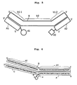
- two ways can be considered, one being to design a target nucleic acid binding region (b2) on the side of the 5' -end of the binding probe (B) as illustrated in FIG. 3 , and the other to design it on the side of the 3' -end of the binding probe (B) as illustrated in FIG. 4 .
- a nucleotide unit (a) labeled with the fluorescent substance (d) is located on the side of the 3'-end of the fluorescent probe (A) when the target nucleic acid binding region (b2) is located on the side of the 5' -end of the binding probe (B).
- the target nucleic acid binding region (b2) is located on the side of the 3' -end of the binding probe (B), on the other hand, it is necessary to conduct the labeling such that the nucleotide unit (a) labeled with the fluorescent substance (d) is located on the side of the 5' -end of the fluorescent probe (A).
- the present inventors have found that the nucleic acid probe set, which is designed such that the target nucleic acid binding region (b2) is located on the side of the 5' -end of the binding probe (B) and the nucleotide unit (a) labeled with the fluorescent substance is located on the side of the 3'-end of the fluorescent probe (A), exhibits a higher quenching efficiency than the nucleic acid probe set, which is designed such that the target nucleic acid binding region (b2) is located on the side of the 3' -end of the binding probe (B) and the nucleotide unit (a) labeled with the fluorescent substance is located on the side of the 5' -end of the fluorescent probe (A).
- the nucleic acid probe set according to the first aspect of the present invention such that the target nucleic acid binding region (b2) is located on the side of the 5' -end of the binding probe (B) and the nucleotide unit (a) labeled with the fluorescent substance is located on the side of the 3' -end of the fluorescent probe (A) and by using the nucleic acid probe set in a real-time PCR measurement or the like, a more accurate measurement can hence be performed than the use of the nucleic acid probe set designed such that the target nucleic acid binding region (b2) is located on the side of the 3' -end of the binding probe (B) and the nucleotide unit (a) labeled with the fluorescent substance is located on the side of the 5' -end of the fluorescent probe (A).
- the former design is preferred accordingly.
- the present inventors presume that, when the target nucleic acid binding region (b2) is located on the side of the 3' -end of the binding probe (B), the fluorescent substance (d) labeled on the side of the 5' -end of the fluorescent probe (A) interacts, in an extension reaction of PCR, with DNA polymerase moved from the side of the 3' -end of the target nucleic acid and the quenching of the fluorescent substance (d) is interfered by the interaction.
- the fluorescent probe (A) that constitutes the nucleic acid probe set according to the first aspect of the present invention is only needed to have a base sequence which can hybridize with the fluorescent probe binding region (b1) in the binding probe (B), and no particular limitation is imposed on its base length.
- a length of 4 bases or less may not be preferred in that it may lead to a lower hybridization efficiency, and a length of 51 bases or more may not be preferred either in that it tends to form non-specific hybrids when used in a real-time PCR measurement or the like. Therefore, the fluorescent probe (A) may be preferably 5 to 50 bases long, more preferably 10 to 35 bases long, especially preferably 10 to 20 bases long.
- the base sequence of the fluorescent probe (A) may include one or more nucleotide units which are not complementary to the corresponding one or ones in the fluorescent probe binging region (b1), insofar as the fluorescent probe (A) can hybridize with the fluorescent probe binging region (b1) in the binding probe (B).
- the base sequence of the fluorescent probe binding region (b1) in the binding probe (B) is not particularly limited insofar as the fluorescent probe binding region (b1) can hybridize with the fluorescent probe (A), and its base length depends on the base length of the fluorescent probe (A).
- the target nucleic acid binding region (b2) in the binding probe (B) is needed to have a base sequence which can hybridize with the target nucleic acid (C).
- the base length of the target nucleic acid binding region (b2) depends on the base length of the target nucleic acid sequence (C). However, a length of 4 bases or less may not be preferred in that it may lead to a lower efficiency of hybridization with the target nucleic acid sequence (C), and a length of 61 bases or more may not be preferred either in that it leads to a reduction in yield upon synthesis of the binding probe (B) and also tends to form non-specific hybrids when used in a real-time PCR measurement or the like.
- the target nucleic acid binding region (b2) may be preferably 5 to 60 bases long, more preferably 15 to 30 bases long.
- the target nucleic acid binding region (b2) may include a base sequence that forms no base pair with the target nucleic acid sequence (C), insofar as it can hybridize with the target nucleic acid sequence (C).
- the nucleic acid probe set according to the first aspect of the present invention can be used in various analysis methods of nucleic acids.
- a description will hereinafter be made of an illustrative detection method of a target nucleic acid, which uses the nucleic acid probe set according to the present invention to determine whether or not the target nucleic acid exists in a solution.
- a solution, which is to be detected for the target nucleic acid and will hereinafter be called “the detection sample” is first serially diluted to prepare several kinds of solutions.
- the nucleic acid probe set according to the first aspect of the present invention in other words, the fluorescent probe (A) and binding probe (B) are added in constant amounts, respectively, to these serially-diluted detection samples.
- the solutions are adjusted in temperature such that the thus-added nucleic acid probe complex in the first aspect of the present invention and the target nucleic acid can hybridize with each other, the solutions are measured for fluorescence intensity.
- the hybridization temperature may be preferably in a temperature range where sequence-specific hybridization takes place between the nucleic acid probe complex and the target nucleic acid but non-specific hybridization does not occur between them, more preferably Tm1 to (Tm1 - 40)°C, still more preferably Tm1 to (Tm1 - 20) °C, even still more preferably Tm1 to (Tm1 - 10)°C.
- Tm1 melting temperature
- the melting temperature (hereinafter called "Tm2") of the complex of the fluorescent probe (A) and binding probe (B), which constitute the nucleic acid probe set according to the first aspect of the present invention may be preferably higher than Tm1, with (Tm2 - Tm1) of 5°C or greater being more preferred, to assure the measurement of the fluorescence intensity.
- Tm2 melting temperature
- the nucleotide units constituting the fluorescent probe (A) are all DNA units
- the substitution of at least one nucleotide unit to a like number of LNA unit or units can raise the Tm2 by 2 to 6°C although this temperature rise also depends on the base length and base sequence.
- the adjustment of the proportion of LNA unit (s) in the oligonucleotide, which constitutes the fluorescent probe (A), such that the Tm2 becomes 95°C or higher can always bring the nucleic acid probe set into the form of a complex, and can use the nucleic acid probe set by considering it practically as a single-stranded nucleic acid probe. It is, therefore, possible to design the fluorescent probe (A) and target nucleic acid binding region (b2) without giving consideration to the above-described (Tm2 - Tm1).
- the target nucleic acid does not exist in the detection sample, a similar fluorescence intensity is observed from each of the serially-diluted detection samples.
- fluorescence from the fluorescent substance in the nucleic acid probe set according to the first aspect of the present invention is quenched by guanine in the nucleic acid which includes the target nucleic acid. The degree of this quenching is varied by changing the ratio of the nucleic acid probe set to the target nucleic acid in the solution.
- nucleic acid probe set according to the first aspect of the present invention By adding the nucleic acid probe set according to the first aspect of the present invention to detection samples, which have been serially diluted as mentioned above, and measuring their fluorescence intensities, it is, therefore, possible to determine the existence/non-existence of the target nucleic acid from the occurrence/non-occurrence of a fluorescence quenching and also to quantify the existing amounts of the target nucleic acid from the magnitudes of the fluorescence quenching.
- the nucleic acid probe set according to the first aspect of the present invention can also be used in a real-time PCR method.
- a base sequence to be amplified by PCR or a portion thereof is chosen as a target nucleic acid, and the base sequence of the target nucleic acid bindingregion (b2) in the binding probe (B) isdetermined such that the target nucleic acid binding region (b2) can hybridize with the target nucleic acid.
- the nucleic acid probe set according to the first aspect of the present invention which has been prepared as described above, is added to a PCR reaction solution, a PCR reaction is conducted, and the fluorescence intensity is measured in each cycle of PCR.
- the fluorescence from the fluorescent substance in the nucleic acid probe set according to the present invention is quenched by guanine in the target nucleic acid.
- the amplification product by PCR can, therefore, be quantified from the fluorescence intensity and the degree of the fluorescence quenching.
- the nucleic acid probe set according to the first aspect of the present invention can also be used in an analysis of a nucleic acid for a base sequence polymorphism.
- base sequence polymorphisms include a single nucleotide polymorphism, base substitution, base deletion, base insertion and the like with respect to a base sequence as a reference.
- an analysis method will be described hereinafter.
- the target nucleic acid sequence (C) is used as a reference base sequence.
- a solution containing the target nucleic acid and another solution containing a nucleic acid to be analyzed are first prepared.
- the nucleic acid probe set according to the first aspect of the present invention that is, the binding probe (B), which has the target nucleic acid binding region (b2) designed to hybridize with the target nucleic acid sequence (C), and the fluorescent probe (A) are added to the respective solutions
- the added nucleic acid probe complex in the first aspect of the present invention is subjected to hybridization with the target nucleic acid and the nucleic acid to be analyzed in the respective solutions, and the temperature dependences of fluorescence intensities are then measured. Described specifically, while changing the temperature of each solution from a low temperature to a high temperature, the fluorescence intensity is measured at each temperature.
- a plot of the measurement results against temperature is called a "melting curve".
- the Tm1 of the hybridized complex of the nucleic acid probe complex in the first aspect of the present invention and the target nucleic acid can be easily determined as a temperature that indicates an extreme value.
- Such a melting curve analysis can be performed by using a commercial program known well to those skilled in the art.
- the fluorescence intensity of the solution which contains the target nucleic acid, is reduced at a low temperature by the fluorescence quenching effect of guanine in the target nucleic acid.
- the solution temperature is raised to around Tm1
- the target nucleic acid dissociates from the nucleic acid probe complex in the first aspect of the present invention, the degree of fluorescence quenching decreases, and therefore, the fluorescence intensity suddenly increases.
- the Tm1 of the hybridized complex of the nucleic acid to be analyzed and the nucleic acid probe complex in the present invention indicates a value lower than the Tm1 of the hybridized complex of the target nucleic acid sequence and the nucleic acid probe complex in the present invention.
- the nucleic acid as the analysis target can, therefore, be analyzed for a base sequence polymorphism with respect to the target nucleic acid sequence (C).
- C target nucleic acid sequence
- their melting curves may be compared with each other.
- the existence or non-existence of a mutation can be readily determined by differentiating the respective melting curves with respect to temperature, determining the Tm1s as temperatures that give extreme values, and then comparing the Tm1s.
- nucleotide unit having a guanine base that applies the quenching effect to the fluorescent substance in the fluorescent probe has mutated in the base sequence of the nucleic acid as an analysis target, no decrease occurs in fluorescence intensity by the fluorescence quenching effect at any temperature so that the mutation can be specified from the melting curve.
- nucleic acid probe set according to the first aspect of the present invention in a melting curve analysis can obviate the preparation of fluorescently-labeled, costly nucleic acid probes specifically for individual target nucleic acids, and therefore, can reduce the preparation time for the melting curve analysis and can more economically perform the melting curve analysis.
- the above-described nucleic acid probe set according to the first aspect of the present invention is consisted of the binding probe (B), which has the one fluorescent probe binding region (b1), and the one fluorescent probe (A).
- the nucleic acid probe set according to the first aspect of the present invention may have two fluorescent probe binding regions (b1) as shown in FIG. 5 .
- These fluorescent probe binding regions (b1-1,b1-2) may have the same base sequence or different base sequences, although the different base sequences are preferred.
- the fluorescent probe may preferably have two fluorescent probes (A1,A2) of different base sequences.
- the nucleic acid probe set according to the first aspect of the present invention in which the binding probe (B) has the two fluorescent probe binding regions of different base sequences, can be suitably used as a replacement for a conventionally-used AB probe in an ABC-PCR ( Alternately Binding Probe Competitive PCR; see Tani et al., Analytical Chemistry, Preprint ) method.
- ABC-PCR Alternately Binding Probe Competitive PCR; see Tani et al., Analytical Chemistry, Preprint ) method.
- nucleic acid probe set according to the first aspect of the present invention in place of the above-described AB probes can obviate the need to prepare different types of fluorescently-labeled, costly fluorescent probes specifically for individual base sequences to be amplified, and therefore, can perform the ABC-PCR method more economically.
- the fluorescent substances that label the two fluorescent probes (A1,A2) may preferably be different kinds of fluorescent substances (d1,d2) which are different in both excitation wavelength and fluorescence wavelength.
- the combination of BODIPY-FL and TAMRA can be mentioned as a preferred example of the combination of the fluorescent substances (d1,d2).
- the second aspect of the present invention relates to an oligonucleotide probe comprising nucleotide units including (a') a nucleotide unit labeled with (h) a labeling substance, a part or all of said nucleotide units being an artificial nucleotide unit or units having a function to raise a dissociation temperature of the oligonucleotide probe from a complementary strand, said dissociation temperature of the oligonucleotide probe from the complementary strand being 100°C or higher under normal pressure conditions.
- one or more artificial nucleotide units each selected from the group consisting of LNA, PNA, ENA, 2' ,4' -BNA NC and 2' ,4' -BNA COC units can be mentioned.
- a fluorescent substance, quencher substance, protein, functional group or the like can be mentioned, and depending on the analysis method, a desired labeling substance can be chosen by one skilled in the art.
- quencher substance means a substance which has a function to weaken the fluorescence to be emitted by the fluorescence substance when the quencher substance is located near the fluorescence substance.
- the oligonucleotide probe according to the present invention can always form a stable complex with a nucleotide (E), which has a sequence complementary to the probe, under normal pressure conditions in a water system, and therefore, can specifically hybridize with the nucleotide (E) to practically label the nucleotide with the labeling substance (h).
- E nucleotide
- the complex can also be used in various analyses such as gene analyses.
- the probe according to the second aspect of the present invention can be used as the fluorescent probe (A) in the first aspect of the present invention.
- FIGS. 12 to 14 and 16 Examples of the nucleic acid probe set according to the third aspect of the present invention are shown in FIGS. 12 to 14 and 16 .
- fluorescent probes A
- binding probes B
- target nucleic acid sequences C
- d a fluorescent substance
- the nucleic acid probe set according to the third aspect of the present invention comprises the one fluorescent probe (A) and the one binding probe (B).
- the binding probe (B) has one fluorescent probe binding region (b1), which has a base sequence complementary to the fluorescent probe (A), and one target nucleic acid binding region (b2), which has a base sequence complementary to the target nucleic acid sequence (C).
- the fluorescent probe (A) which constitutes the nucleic acid probe set according to the third aspect of the present invention, is an oligonucleotide including a nucleotide unit (a) labeled with the fluorescent substance (d).
- No particular limitation is imposed on the base sequence of the fluorescent probe (A) insofar as it can hybridize with the fluorescent probe binding region (b1) in the binding probe (B).
- the base sequence of the fluorescent probe (A) therefore, does not depend on the base sequence of a target nucleic acid to be detected or analyzed.
- the fluorescent probe (A) that constitutes the nucleic acid probe set according to the third aspect of the present invention is not required to have a base sequence corresponding to the specific target nucleic acid, and the fluorescent probe (A) of the same base sequence can be commonly used for different target nucleic acids.
- the nucleic probe set according to the third aspect of the present invention is, therefore, called "a universal nucleic probe set" by the present inventors.
- the use of the nucleic acid probe set according to the third aspect of the present invention for the analysis of a target nucleic acid has an advantage in that it is no longer needed to prepare a fluorescent probe, which has a costly fluorescent substance, specifically for the target nucleic acid to be detected or analyzed and the production cost of the fluorescent probe can be minimized.
- the nucleotide units as basic units of the fluorescent probe (A) are not limited to deoxyribonucleotides as basic units of DNA or ribonucleotides as basic units of RNA, and artificial nucleotide units, which have the above-described function to raise the dissociation temperature between the fluorescent probe (A) and the binding probe (B), can be also used.
- artificial nucleotide units LNA, PNA, ENA, 2' ,4'-BNA NC and 2' ,4' -BNA COC units can be mentioned.
- An LNA monomer is a nucleotide having two ring structures that the 2'-oxygen and 4'-carbon atoms of ribose are connected together via a methylene unit. Due to the inclusion of these two ring structures, the LNA monomer has low structural freedom, and compared with DNA or RNA monomer, strongly hybridizes with a complementary strand.
- the Tm between the fluorescent probe (A) and the binding probe (B) can be easily made higher than the Tm between the target nucleic acid sequence (C) and the target nucleic acid binding region (b2), thereby making it possible to provide the nucleic acid probe set according to the third aspect of the present invention with increased stability at elevated temperatures and hence to provide it with improved reliability as a fluorescent probe.
- the Tm between the fluorescent probe (A) and the binding probe (B) can be made higher than the thermal denaturation temperature (for example, 95°C) of PCR, so that the fluorescent probe (A) and the binding probe (B) can always remain as a stable nucleic acid probe complex during PCR cycles.
- the proportion of the artificial nucleotide unit or units in the fluorescent probe (A), said proportion being required to make the Tm between the fluorescent probe (A) and the binding probe (B) higher than the thermal denaturation temperature of PCR also depends on the base number and base sequence of the fluorescent probe (A) and cannot be specified.
- the proportion of the artificial nucleotide unit or units may be at least one third of the entire nucleotide units, with at least 80% thereof being more preferred.
- the interaction between the fluorescent probe (A) and the fluorescent probe binding region (b1) is strengthened.
- the base numbers of the fluorescent probe (A) and fluorescent probe binding region (b1) can, therefore, be decreased compared with the case that the fluorescent probe (A) is formed of DNAunits alone.
- a binding probe (B) of a large base number an error occurs.
- the use of a fluorescent probe binding region (b1) of a smaller base number can reduce the error, and can increase the synthesis yield of a binding probe (B). This leads to a reduction in the production cost for the probe set according to the third aspect of the present invention.
- the fluorescent substance (d) with which the fluorescent probe (A) is labeled in the third aspect of the present invention is a fluorescent substance (d) which changes in fluorescent character upon interaction with guanine.
- fluorescent character means fluorescence intensity
- the expression "guanine and the fluorescent substance interact with each other to change the fluorescent character of the fluorescent substance” means that the fluorescence intensity of the fluorescent substance in a state that guanine and the fluorescent substance are not interacting with each other is different from its fluorescence intensity in a state that they are interacting with each other, and on the extent of this difference, no limitation shall be imposed.
- quenched or quenching of fluorescence means that upon interaction of a fluorescent substance with guanine, the fluorescence intensity decreases compared with the fluorescence intensity when the fluorescent substance is not interacting with guanine, and on the extent of this decrease, no limitation shall be imposed.
- fluorescent substances which can be suitably used in the nucleic acid probe set according to the third aspect of the present invention, include fluorescein and its derivatives [e.g., fluorescein-4-isothiocyanate (FITC), tetrachlorofluorescein, hexachlorofluorescein, tetrabromosulfonefluorescein (TBSF), and derivatives thereof], EDANS (5-(2-aminoethylamino)-1-naphthalenesulfonic acid), 6-JOE, Alexa Fluor 488 (Invitrogen Corp.), Alexa Fluor 532 (Invitrogen Corp.), Cy3 (GE Healthcare Bioscience), Cy5 (GE Healthcare Bioscience), Pacific Blue (Invitrogen Corp.), rhodamine 6G (R6G) and its derivatives (for example, carboxyrhodamine 6G (CR6G), tetramethylrhodamine (TMR), tetramethylrhodamine isothi
- fluorescein fluorescein-4-isothiocyanate
- tetrachlorofluorescein hexachlorofluorescein
- tetrabromosulfonefluorescein EDANS, 6-JOE
- Alexa Fluor 488 Alexa Fluor 532
- Pacific Blue rhodamine 6G, carboxyrhodamine 6G, tetramethylrhodamine, carboxytetramethylrhodamine or BODIPY-FL
- BODIPY-FL is more preferred, and the use of BODIPY-FL is most preferred.
- the target nucleic acid binding region (b2) of the binding probe (B) is designed such that, when the nucleic acid probe complex in the third aspect of the present invention has hybridized to the target nucleic acid sequence (C), the fluorescent substance (d) and a guanine base in a target nucleic acid can be brought into contact with each other.
- the fluorescence of the fluorescent substance (d) is quenched by the guanine base, and by detecting this quenching phenomenon, the target nucleic acid can be quantified.
- the guanine which can interact with the fluorescent substance (d) to give fluorescence quenching effect, may exist either in the base sequence of the target nucleic acid sequence (C) or in a base sequence outside the target nucleic acid sequence (C), insofar as it exists in the target nucleic acid.
- the guanine exists in the target nucleic acid sequence (C) and forms a base pair with cytosine in the hybridized binding probe (B)
- the interaction between the fluorescent substance (d) and the guanine is somewhat weaker although no particular problem arises.
- the nucleotide unit ⁇ which has the guanine base in the target nucleic acid
- the nucleotide unit ⁇ is the nucleotide unit ⁇ in the case of the description to be made hereinafter.
- FIG. 13 shows, on an enlarged scale, an area around the fluorescent substance (d) in FIG. 12 .
- the conditions under which the fluorescent substance (d) and the nucleotide unit ⁇ can interact with each other depend inter alia on the length of a below-described spacer that connects the fluorescent substance (d) and the nucleotide unit (a) labeled with the fluorescent substance (d) each other, and cannot be specified. Generalizing these conditions, however, they can be defined as will be described hereinafter.
- the fluorescent probe (A) is now assumed to have hybridized with the binding probe (B).
- Base pairs are formed between the fluorescent probe binding region (b1) and the fluorescent probe (A).
- a nucleotide unit, which exists in the fluorescent probe binding region (b1) and is closest to the target nucleic acid binding region (b2), will hereinafter be called "the nucleotide unit ⁇ ".
- the distance between this nucleotide unit ⁇ and a base, which exists in the binding probe (B) and forms a base pair with the nucleotide unit (a), will be designated as "X" expressed in terms of the number of base (s) .
- base pairs are formed between the target nucleic acid sequence (C) and the target nucleic acid binding region (b2).
- a nucleotide unit, which exists in the target nucleic acid binding region (b2) and is closest to the nucleotide unit ⁇ , will be designated as "the nucleotide unit ⁇ ".
- a nucleotide unit, which exists in the target nucleic acid sequence (C) and forms a base pair with the nucleotide unit ⁇ , will be designated as "the nucleotide unit ⁇ ".
- the distance between the nucleotide unit ⁇ and the nucleotide unit ⁇ will be designated as "Y" expressed in terms of the number of base (s).
- the counting method of Y is the same as X.
- the nucleotide unit ⁇ and the nucleotide unit ⁇ are commonly a nucleotide unit having a guanine base located at the 5' -end in the target nucleic acid sequence (C). Therefore, Y is 0.
- the sum of X and Y may preferably be 5 or smaller.
- the sum of X and Y may be more preferably 3 or smaller, with 0 being most preferred, although it also depends on the length of the spacer connecting the fluorescent substance (d) and the nucleotide unit (a) labeled with the fluorescent substance (d).
- the fluorescent probe (A) for use in the nucleic acid probe set according to the third aspect of the present invention its production can rely upon a custom oligonucleotide synthesis service company (for example, Tsukuba Oligo Service Co., Ltd., Ibaraki, Japan) or the like. No particular limitation is imposed on the method for labeling the fluorescent substance on the oligonucleotide, and a conventionally-known labeling method can be used ( Nature Biotechnology, 14, 303-308, 1996 ; Applied and Environmental Microbiology, 63, 1143-1147, 1997 ; Nucleic Acids Research, 24, 4532-4535, 1996 ).
- a fluorescent substance for example, to the 5'-terminal nucleotide unit
- a commercial spacer can be used (for example, Midland Certified Reagent Company, U.S.A).
- n may stand for 3 to 8, with 6 being preferred.
- a fluorescent substance can also be coupled to the 3'-terminal nucleotide unit of the oligonucleotide.
- a commercial spacer can also be used (for example, Midland Certified Reagent Company, U.S.A).
- n may stand for 3 to 8, with 4 to 7 being preferred.
- an oligonucleotide labeled with the fluorescent substance can be synthesized.
- the oligonucleotide can be purified by reverse phase chromatography or the like to provide the fluorescent probe (A) for use in the third aspect of the present invention.
- kit reagent for example, Uni-link amino modifier,Clonetech Laboratories, Inc.
- the fluorescent substance can then be coupled to the oligonucleotide in a manner known per se in the art.
- the nucleotide unit (a) in the fluorescent probe (A), said nucleotide unit (a) being labeled with the fluorescent substance, is not limited to one of both terminal nucleotide units in the oligonucleotide, and a nucleotide unit other than both the terminal nucleotide units can be labeled with the fluorescent substance ( ANALYTICAL BIOCHEMISTRY, 225, 32-38, 1998 ).
- the target nucleic acid binding region (b2) of the binding probe (B) is designed such that it is located on the side of the 5'-end of the binding probe (B).
- nucleic acid probe set from one fluorescent probe (A) and one binding probe (B)
- two ways can be considered, one being to design a target nucleic acid binding region (b2) on the side of the 5' -end of the binding probe (B) as illustrated in FIG. 14 , and the other to design it on the side of the 3' -end of the binding probe (B) as illustrated in FIG. 15 .
- a nucleotide unit (a) labeled with the fluorescent substance (d) is located on the side of the 3'-end of the fluorescent probe (A) when the target nucleic acid binding region (b2) is located on the side of the 5' -end of the binding probe (B) .
- the target nucleic acid binding region (b2) is located on the side of the 3'-end of the binding probe (B)
- it is necessary to conduct the labeling such that the nucleotide unit (a) labeled with the fluorescent substance (d) is located on the side of the 5'-end of the fluorescent probe (A).
- the present inventors have found that a higher quenching efficiency is exhibited when designed such that the target nucleic acid binding region (b2) is located on the side of the 5' -end of the binding probe (B) and the nucleotide unit (a) labeled with the fluorescent substance is located on the side of the 3' -end of the fluorescent probe (A) than when designed such that the target nucleic acid binding region (b2) is located on the side of the 3' -end of the binding probe (B) and the nucleotide unit (a) labeled with the fluorescent substance is located on the side of the 5' -end of the fluorescent probe (A).
- nucleic acid probe set according to the third aspect of the present invention is used in real-time PCR, a more accurate measurement can be performed than the use of the nucleic acid probe set designed such that the target nucleic acid binding region (b2) is located on the side of the 3' -end of the binding probe (B) and the nucleotide unit (a) labeled with the fluorescent substance is located on the side of the 5' -end of the fluorescent probe (A) .
- the present inventors presume that, when the target nucleic acid binding region (b2) is located on the side of the 3'-end of the binding probe (B), the fluorescent substance (d) labeled on the side of the 5'-end of the fluorescent probe (A) interacts, in an extension reaction of PCR, with DNA polymerase moved from the side of the 3' -end of the target nucleic acid and the quenching of the fluorescent substance (d) is interfered by the interaction.
- the fluorescent probe (A) that constitutes the nucleic acid probe set according to the third aspect of the present invention is only needed to have a base sequence which can hybridize with the fluorescent probe binding region (b1) in the binding probe (B), and no particular limitation is imposed on its base length.
- a length of 4 bases or less may not be preferred in that it may lead to a lower hybridization efficiency, and a length of 51 bases or more may not be preferred either in that it tends to form non-specific hybrids when used in a real-time PCR measurement or the like. Therefore, the fluorescent probe (A) may be preferably 5 to 50 bases long, more preferably 10 to 35 bases long, especially preferably 10 to 20 bases long.
- the base sequence of the fluorescent probe (A) may include one or more nucleotide units which are not complementary to the corresponding one or ones in the fluorescent probe binging region (b1), insofar as the fluorescent probe (A) can hybridize with the fluorescent probe binging region (b1) in the binding probe (B).
- the base sequence of the fluorescent probe binding region (b1) in the binding probe (B) is not particularly limited insofar as the fluorescent probe binding region (b1) can hybridize with the fluorescent probe (A), and its base length depends on the base length of the fluorescent probe (A).
- the target nucleic acid binding region (b2) in the binding probe (B) is needed to have a base sequence which can hybridize with the target nucleic acid (C).
- the base length of the target nucleic acid binding region (b2) depends on the base length of the target nucleic acid sequence (C) .
- a length of 4 bases or less may not be preferred in that it may lead to a lower efficiency of hybridization with the target nucleic acid sequence (C)
- a length of 61 bases or more may not be preferred either in that it leads to a reduction in yield upon synthesis of the binding probe (B) and also tends to form non-specific hybrids when used in a real-time PCR measurement or the like.
- the target nucleic acid binding region (b2) may be preferably 5 to 60 bases long, more preferably 15 to 30 bases long.
- the target nucleic acid binding region (b2) may include a base sequence that forms no base pair with the target nucleic acid sequence (C), insofar as it can hybridize with the target nucleic acid sequence (C).
- the nucleic acid probe set according to the third aspect of the present invention can be used in various analysis methods of nucleic acids.
- a description will hereinafter be made of an illustrative detection method of a target nucleic acid, which uses the nucleic acid probe set according to the third aspect of the present invention to determine whether or not the target nucleic acid exists in a solution.
- a solution, which is to be detected for the target nucleic acid and will hereinafter be called “the detection sample” is first serially diluted to prepare several kinds of solutions.
- the nucleic acid probe set according to the third aspect of the present invention in other words, the fluorescent probe (A) and binding probe (B) are added in constant amounts, respectively, to these serially-diluted detection samples. After the solutions are adjusted in temperature such that the thus-added nucleic acid probe complex in the present invention and the target nucleic acid can hybridize with each other, the solutions are measured for fluorescence intensity.
- the hybridization temperature may be preferably in a temperature range where sequence-specific hybridization takes place between the nucleic acid probe complex and the target nucleic acid but non-specific hybridization does not occur between them, more preferably Tm1 to (Tm1- 40)°C, still more preferably Tm1 to (Tm1-20) °C, even still more preferably Tm1 to (Tm1 - 10 ) °C.
- Tm1 melting temperature
- the melting temperature (hereinafter called "Tm2") of the complex of the fluorescent probe (A) and binding probe (B), which constitute the nucleic acid probe set according to the present invention may be preferably higher than Tm1, with (Tm2 - Tm1) of 5°C or greater being more preferred, to assure the measurement of the fluorescence intensity.
- Tm2 melting temperature
- the nucleotide units constituting the fluorescent probe (A) are all DNA units
- the substitution of at least one nucleotide unit to a like number of LNA unit or units can raise the Tm2 by 2 to 6°C although this temperature rise also depends on the base length and base sequence.
- the adjustment of the proportion of LNA unit(s) in the oligonucleotide, which constitutes the fluorescent probe (A), such that the Tm2 becomes 95°C or higher can always bring the nucleic acid probe set into the form of a complex, and can use the nucleic acid probe set by considering it practically as a single-stranded nucleic acid probe. It is, therefore, possible to design the fluorescent probe (A) and target nucleic acid binding region (b2) without giving consideration to the above-described (Tm2 - Tm1).
- the target nucleic acid does not exist in the detection sample, a similar fluorescence intensity is observed from each of the serially-diluted detection samples.
- fluorescence from the fluorescent substance in the nucleic acid probe set according to the present invention is quenched by guanine in the nucleic acid which includes the target nucleic acid. The degree of this quenching is varied by changing the ratio of the nucleic acid probe set to the target nucleic acid in the solution.
- nucleic acid probe set according to the present invention By adding the nucleic acid probe set according to the present invention to detection samples, which have been serially diluted as mentioned above, and measuring their fluorescence intensities, it is, therefore, possible to determine the existence/non-existence of the target nucleic acid from the occurrence/non-occurrence of a fluorescence quenching and also to quantify the existing amounts of the target nucleic acid from the magnitudes of the fluorescence quenching.
- the nucleic acid probe set according to the third aspect of the present invention can also be used in a real-time PCR method.
- a base sequence to be amplified by PCR or a portion thereof is chosen as a target nucleic acid, and the base sequence of the target nucleic acid binding region (b2) in the binding probe (B) is determined such that the target nucleic acid binding region (b2) can hybridize with the target nucleic acid.
- the nucleic acid probe set according to the third aspect of the present invention which has been prepared as described above, is added to a PCR reaction solution, a PCR reaction is conducted, and the fluorescence intensity is measured in each cycle of PCR.
- the fluorescence from the fluorescent substance in the nucleic acid probe set according to the present invention is quenched by guanine in the target nucleic acid.
- the amplification product by PCR can, therefore, be quantified from the fluorescence intensity and the degree of the fluorescence quenching.
- the nucleic acid probe set according to the third aspect of the present invention can also be used in an analysis of a nucleic acid for a base sequence polymorphism.
- Examples of analyzable base sequence polymorphisms include a single nucleotide polymorphism, base substitution, base deletion, base insertion and the like with respect to a base sequence as a reference.
- One example of such an analysis method will be described hereinafter.
- the target nucleic acid sequence (C) is used as a reference base sequence.
- a solution containing the target nucleic acid and another solution containing a nucleic acid to be analyzed are first prepared.
- the nucleic acid probe set according to the third aspect of the present invention that is, the binding probe (B), which has the target nucleic acid binding region (b2) designed to hybridize with the target nucleic acid sequence (C), and the fluorescent probe (A) are added to the respective solutions
- the added nucleic acid probe complex in the present invention is subjected to hybridization with the target nucleic acid and the nucleic acid to be analyzed in the respective solutions, and the temperature dependences of fluorescence intensities are then measured. Described specifically, while changing the temperature of each solution from a low temperature to a high temperature, the fluorescence intensity is measured at each temperature.
- a plot of the measurement results against temperature is called a "melting curve".
- the Tm1 of the hybridized complex of the nucleic acid probe complex in the present invention and the target nucleic acid can be easily determined as a temperature that indicates an extreme value.
- Such a melting curve analysis can be performed by using a commercial program known well to those skilled in the art.
- the fluorescence intensity of the solution which contains the target nucleic acid, is reduced at a low temperature by the fluorescence quenching effect of guanine in the target nucleic acid.
- the solution temperature is raised to around Tm1
- the target nucleic acid dissociates from the nucleic acid probe complex in the third aspect of the present invention, the degree of fluorescence quenching decreases, and therefore, the fluorescence intensity suddenly increases.
- the Tm1 of the hybridized complex of the nucleic acid to be analyzed and the nucleic acid probe complex in the third aspect of the present invention indicates a value lower than the Tm1 of the hybridized complex of the target nucleic acid sequence and the nucleic acid probe complex in the third aspect of the present invention.
- the nucleic acid as the analysis target can, therefore, be analyzed for a base sequence polymorphism with respect to the target nucleic acid sequence (C) .
- their melting curves may be compared with each other.
- the existence or non-existence of a mutation can be readily determined by differentiating the respective melting curves with respect to temperature, determining the Tm1s as temperatures that give extreme values, and then comparing the Tm1s.
- nucleotide unit having a guanine base that applies the quenching effect to the fluorescent substance in the fluorescent probe has mutated in the base sequence of the nucleic acid as an analysis target, no decrease occurs in fluorescence intensity by the fluorescence quenching effect at any temperature so that the mutation can be specified from the melting curve.
- nucleic acid probe set according to the third aspect of the present invention in a melting curve analysis can obviate the preparation of fluorescently-labeled, costly nucleic acid probes specifically for individual target nucleic acids, and therefore, can reduce the preparation time for the melting curve analysis and can more economically perform the melting curve analysis.
- nucleic acid probe set according to the present invention for a portion of the human ⁇ -globin gene as a target nucleic acid sequence, a real-time PCR experiment was performed, and the effectiveness of the nucleic acid probe set according to the present invention was evaluated.
- PCR reaction solutions for real-time PCR a PCR reaction solution, which was free of a human genomic DNA sample (Human Genomic DNA; Novagen Inc.), and PCR reaction solutions, which contained 10 2 , 10 3 , 10 4 , 10 5 , 10 6 and 10 7 copies of the human genomic DNA sample, respectively, were prepared.
- Each reaction solution contained TITANIUM Taq DNA polymerase (Clonetech Laboratories, Inc.) as DNA polymerase, four types of dNTPs (final concentration: 0.2 mM, each), a forward primer (SEQ ID NO: 1, final concentration: 1 ⁇ M), a reverse primer (SEQ ID NO: 2, final concentration: 0.3 ⁇ M), a predetermined amount of TITANIUM Taq PCR buffer (Clonetech Laboratories, Inc.), and a nucleic acid probe set according to the present invention.
- a nucleic acid containing the above-described target nucleic acid sequence was to be amplified.
- SEQ ID NO:1 ggttggccaatctactccagg SEQ ID NO:2 tggtctccttaaacctgtcttg
- the nucleic acid probe set according to the present invention was for the portion of the human ⁇ -glonbin gene as a target nucleic acid sequence (SEQ ID NO:3), and was consisted of a binding probe (SEQ ID NO:4; final concentration: 100 nM) and a fluorescent probe (final concentration: 50 nM).
- the binding probe had, on the side of a 3'-end thereof, a fluorescent probe binding region of a base sequence complementary to the fluorescent probe, and on the side of a 5'-end thereof, a target nucleic acid binding region of a base sequence complementary to the target nucleic acid sequence.
- the fluorescent probe had a base sequence (SEQ ID NO:5), and was labeled at a 3' -terminal nucleotide unit thereof with BODIPY-FL (Invitrogen Corp.). It is to be noted that as all the nucleotide units making up the fluorescent probe, units of LNA (ThermoElectron Measurement Systems, Inc.) were used. SEQ ID NO:3 gttcactagcaacctcaaacagacacc SEQ ID NO:4 ggtgtctgttgaggttgctagtgaactatgaggtggtaggatgggtagtg gt SEQ ID NO:5 accactacccatcctaccacctcata-BODIPY-FL
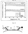
- FIG. 6 shows a schematic diagram of the above-described nucleic acid probe complex in the present invention upon hybridization with the target nucleic acid sequence (SEQ ID NO: 3).
- the fluorescent substance coupled to the fluorescent probe is considered to receive the fluorescence quenching effect from the guanine at the 5' -end of the target nucleic acid sequence.
- the binding probe had a phosphate group at the 3'-end thereof. It is to be noted that, when an LNA-containing fluorescent probe is used as in this example, the above-described phosphorylation at the 3' -end is not essential because the fluorescent probe does not dissociate from its associated binding probe under normal reaction conditions and the binding probe does not function as a primer.
- Fluorescence quenching efficiency G U , 55 / G U , 95 - G 55 / G 95 / G U , 55 / G U , 95 where,
- FIG. 7 shows a graph obtained by plotting the thus-determined fluorescence quenching efficiencies on a graph sheet on which PCR cycles were plotted along the abscissa. It is understood from the graph that a target nucleic acid can be accurately quantified by conducting real-time PCR while using the nucleic acid probe set according to the present invention. It is to be noted that the average of maximum values of fluorescence quenching efficiency with respect to the above-described six kinds of PCR samples was about 37%.
- Example 2 An experiment was performed in a similar manner as in Example 1 except that the five nucleotide units on the side of the 5'-end of the fluorescent probe were changed to a like number of DNA units.
- the average of maximum values of fluorescence quenching efficiency was about 35%. It is to be noted that the proportion of the LNA units in the fluorescent probe used in this example was about 81% of the entire units.
- FIG. 8 shows a graph obtained by plotting the thus-determined fluorescence quenching efficiencies on a graph sheet on which PCR cycles were plotted along the abscissa.
- the average of maximum values of fluorescence quenching efficiency with respect to the six kinds of PCR samples was about 25%, and, compared with Example 1 in which all the nucleotide units making up the fluorescent probe were LNA units, the fluorescence quenching efficiency was about two thirds. In other words, the fluorescence quenching efficiency was improved by about 1.5 times by changing the nucleotide units, which constituted the fluorescent probe, from the DNA units to the LNA units.
- nucleic acid probe set according to the present invention for a portion of the human ⁇ -actin gene as a target nucleic acid sequence (SEQ ID NO:8), a real-time PCR experiment was performed, and the effectiveness of the nucleic acid probe set according to the present invention was evaluated.
- reaction solutions for real-time PCR As reaction solutions for real-time PCR, reaction solutions, which contained the cDNA of the human ⁇ -actin gene as obtained from the seven kinds of samples, respectively, and a PCR reaction solution, which was free of the cDNA, were prepared. Each reaction solution was prepared following a manual provided in a kit (TITANIUM Taq PCR Kit, product of Takara Bio Inc.) except for the inclusion of a forward primer (SEQ ID NO: 6, final concentration: 0.3 ⁇ M) and a reverse primer (SEQ ID NO: 7, final concentration: 1.0 ⁇ M).
- a 262-bp nucleic acid containing the above-described target nucleic acid sequence was to be amplified.
- SEQ ID NO:6 catgtacgttgctatccaggc
- SEQ ID NO:7 ctccttaatgtcacgcacgat
- the nucleic acid probe set according to the present invention was for a portion of the human ⁇ -actin gene as a target nucleic acid (SEQ ID NO: 8), and was consisted of a binding probe (SEQ ID NO: 9; final concentration: 100 nM) and a fluorescent probe (final concentration: 50 nM).
- the binding probe had, on the side of a 3' -end thereof, a fluorescent probe binding region of a base sequence complementary to the fluorescent probe, and on the side of a 5' -end thereof, a target nucleic acid binding region of a base sequence complementary to the target nucleic acid.
- the fluorescent probe had a base sequence (SEQ ID NO: 10), and was labeled at a 3' -terminal nucleotide unit thereof with BODIPY-FL (Invitrogen Corp.). It is to be noted that as all the nucleotide units making up the fluorescent probe, units of LNA (ThermoElectron Measurement Systems, Inc.) were used. SEQ ID NO:8 gtgaggatcttcatgaggtagtcagtcag SEQ ID NO:9 ctgactgactacctcatgaagatcctcactatgaggtggtaggatgggtag tggt SEQ ID NO:10 accactacccatcctaccacctcata-BODIPY-FL
- the binding probe had a phosphate group at the 3' -end thereof. Synthesis of the nucleic acid probe was relied upon Tsukuba Oligo Service Co., Ltd. (Tsukuba, Japan), and syntheses of the forward primer and reverse primer were relied upon Nihon Gene Research Laboratories, Inc. (Sendai, Japan).
- FIG. 9 shows a graph obtained by plotting the thus-determined fluorescence quenching efficiencies on a graph sheet on which PCR cycles were plotted along the abscissa. It is understood from the graph that a target nucleic acid can be accurately quantified by conducting real-time PCR while using the nucleic acid probe set according to the present invention. It is to be noted that the average of maximum values of fluorescence quenching efficiency with respect to the above-described seven kinds of PCR samples was about 32%.
- FIG. 10 shows a graph obtained by plotting the thus-determined fluorescence quenching efficiencies on a graph sheet on which PCR cycles were plotted along the abscissa.
- the average of maximum values of fluorescence quenching efficiency with respect to the seven kinds of PCR samples was about 25%, and, compared with Example 2 in which all the nucleotide units making up the fluorescent probe were LNA units, the fluorescence quenching efficiency was about three quarters. In other words, the fluorescence quenching efficiency was improved by about 1.3 times by changing the nucleotide units, which constituted the fluorescent probe, from the DNA units to the LNA units.
- a solution was prepared by adding an oligonucleotide (SEQ ID NO: 11, final concentration: 50 nM), another oligonucleotide (SEQ ID NO:12, final concentration: 400 nM), KCl (final concentration: 50 mM), Tris-HCl (final concentration: 10 mM), and MgCl 2 (final concentration: 1.5 mM).
- the former oligonucleotide was formed of LNA units only and was labeled at a 3'-terminal nucleotide with BODIPY-FL (Invitrogen Corp.), while the latter was formed of only DNA units only.
- the solution was brought to a volume of 20 ⁇ L, and its pH was adjusted to 8.7 at room temperature.
- SEQ ID NO:11 ccccctcccccaa-BODIPY-FL SEQ ID NO:12 gggttgggggaggggggg
- reaction solution was subjected to a real-time PCR system (LightCycler®, Roche Diagnostics K.K.), and a melting curve analysis was performed. The results are shown in FIG. 11 .
- nucleic acid probe set according to the present invention for a portion of the human ⁇ -globin gene as a target nucleic acid sequence, a real-time PCR experiment was performed, and the effectiveness of the nucleic acid probe set according to the present invention was evaluated.
- PCR reaction solutions for real-time PCR a PCR reaction solution, which was free of a human genomic DNA sample (Human Genomic DNA; Novagen Inc.), and PCR reaction solutions, which contained 10 2 , 10 3 , 10 4 , 10 5 , 10 6 and 10 7 copies of the human genomic DNA sample, respectively, were prepared.
- Each reaction solution contained TITANIUM Taq DNA polymerase (Clonetech Laboratories, Inc.) as DNA polymerase, four types of dNTPs (final concentration: 0.2 mM, each), a forward primer (SEQ ID NO: 13, final concentration: 1 ⁇ M), a reverse primer (SEQ ID NO: 14, final concentration: 0.3 ⁇ M), a predetermined amount of TITANIUM Taq PCR buffer (Clonetech Laboratories, Inc.), and a nucleic acid probe set according to the present invention.
- a nucleic acid containing the above-described target nucleic acid sequence was to be amplified.
- SEQ ID NO:13 ggttggccaatctactccagg SEQ ID NO:14 tggtctccttaaacctgtcttg
- the nucleic acid probe set according to the present invention was for the portion of the human ⁇ -glonbin gene as a target nucleic acid sequence (SEQ ID NO:15), and was consisted of a binding probe (SEQ ID NO:16; final concentration: 100 nM) and a fluorescent probe (final concentration: 50 nM) .
- the binding probe had, on the side of a 3' -end thereof, a fluorescent probe binding region of a base sequence complementary to the fluorescent probe, and on the side of a 5' -end thereof, a target nucleic acid binding region of a base sequence complementary to the target nucleic acid sequence.
- the fluorescent probe had a base sequence (SEQ ID NO:17), and was labeled at a 3' -terminal nucleotide unit thereof with BODIPY-FL (Invitrogen Corp.). It is to be noted that the nucleotide units which made up the fluorescent probe were all DNA units. SEQ ID NO:15 gttcactagcaacctcaaacagacacc SEQ ID NO:16 ggtgtctgttgaggttgctagtgaactatgaggtggtaggatgggtagtg gt SEQ ID NO:17 accactacccatcctaccacctcata-BODIPY-FL
- FIG. 16 shows a schematic diagram of the above-described nucleic acid probe complex in the present invention upon hybridization with the target nucleic acid sequence (SEQ ID NO:15).
- the fluorescent substance coupled on the fluorescent probe is considered to receive the fluorescence quenching effect from the guanine at the 5' -end of the target nucleic acid sequence.
- the binding probe had a phosphate group at the 3' -end thereof. Synthesis of the nucleic acid probe was relied upon Tsukuba Oligo Service Co., Ltd. (Tsukuba, Japan), and syntheses of the forward primer and reverse primer were relied upon Nihon Gene Research Laboratories, Inc. (Sendai, Japan).
- Fluorescence quenching efficiency G U , 55 / G U , 95 - G 55 / G 95 / G U , 55 / G U , 95 where,
- FIG. 17 shows a graph obtained by plotting the thus-determined fluorescence quenching efficiencies on a graph sheet on which PCR cycles were plotted along the abscissa. It is understood from the graph that a target nucleic acid can be accurately quantified by conducting real-time PCR while using the nucleic acid probe set according to the present invention. It is to be noted that the average of maximum values of fluorescence quenching efficiency with respect to the above-described six kinds of PCR samples was about 25%.
- a real-time PCR experiment was performed in a similar manner as in Example 5 except that as a binding probe, an oligonucleotide (SEQ ID NO:18) having, on the side of a 3' -end thereof, a target nucleic acid binding region and, on the side of a 5' -end thereof, a fluorescent probe binding region was used, a fluorescent probe (SEQ ID NO:19) labeled at a 5'-terminal nucleotide unit thereof with BODIPY-FL (Invitrogen Corp.) was used, and as PCR reaction solutions, seven kinds of samples containing 10, 10 2 , 10 3 , 10 4 , 10 5 , 10 6 , 10 7 and 10 8 copies, respectively, of a human genomic DNA sample were used.
- binding probe had a phosphate group at the 3'-end thereof.
- SEQ ID NO:18 cgatgcagcgagtggcggcgatgggtgtctgtttgaggttgctagtgaac
- SEQ ID NO:19 BODIPY-FL-catcgccgccactcgctgcatcg
- FIG. 18 shows a graph obtained by plotting the thus-determined fluorescence quenching efficiencies on a graph sheet on which PCR cycles were plotted along the abscissa.
- the average of maximum values of fluorescence quenching efficiency with respect to the six kinds of PCR samples was about 15%, and, compared with Example 5 which used the fluorescent probe according to the present invention having, on the side of the 5' -end thereof, the binding probe binding region and, on the side of the 3' -end thereof, the fluorescent probe binding region, the fluorescence quenching efficiency was about 60% or so. In other words, the fluorescence quenching efficiency was improved by about 1.7 times by changing the binding probe binding region from the side of the 5' -end to the side of the 3' -end.
- oligonucleotide probe and an oligonucleotide were synthesized.
- the oligonucleotide probe had a base sequence (SEQ ID NO : 20), was labeled at a 3' -end thereof with BODIPY-FL and was formed of LNA units only, while the oligonucleotide (complementary stand) had a base sequence (SEQ ID NO:21) and was formed of DNA units only.
- the complementary strand was designed such that upon hybridization with the probe, guanine is located near the fluorescent dye. Synthesis of the probe was relied upon Gene Design Inc.
- a reaction solution of the composition shown in Table 1 was prepared, and was subjected to a melting curve analysis by a real-time PCR system (LightCycler®, Roche Diagnostics K.K.). As a blank, a similar measurement was also conducted with respect to a sample similar to the reaction solution of Table 1 except that the complementary strand was not added. The results are shown in FIG. 19 . As a result, in the example in which the complementary strand was added, a lower fluorescence intensity was exhibited compared with the blank in which the complementary strand was not added, because by the formation of a double stand of the probe and complementary strand, the fluorescent dye with which the probe was labeled interacted with the guanine in the complementary strand and its fluorescence was thus quenched. From the foregoing, it has become evident that the oligonucleotide probe according to the second aspect of the present invention is suitably usable as a fluorescence quenching probe.
- the fluorescence intensity (-) in FIG. 19 can be of any unit.
- a melting curve analysis was performed in a similar manner as in Example 6 except that the LNA units in the oligonucleotide probe (SEQ ID NO: 20) were changed to PNA units. The results are shown in FIG. 19 . Synthesis of the probe was relied upon Fasmac Co., Ltd. (Atsugi, Kanagawa, Japan).
- a melting curve analysis was performed in a similar manner as in Example 6 except that the LNA units in the oligonucleotide probe (SEQ ID NO: 20) were changed to ENA units. The results are shown in FIG. 19 . Synthesis of the probe was relied upon Sigma-Genosys Japan K.K. (Ishikari, Hokkaido, Japan).
- a melting curve analysis was performed in a similar manner as in Example 6 except that the LNA units in the oligonucleotide probe (SEQ ID NO: 20) were changed to 2', 4'-BNA NC units. The results are shown in FIG. 19 . Synthesis of the probe was relied upon BNA Inc. (Ibaraki, Osaka, Japan).
- a melting curve analysis was performed in a similar manner as in Example 6 except that the LNA units in the oligonucleotide probe (SEQ ID NO: 20) were changed to 2', 4' -BNA coc units. The results are shown in FIG. 19 . Synthesis of the fluorescent probe was relied upon BNA Inc. (Ibaraki, Osaka, Japan).
- oligonucleotide probe which was employed in Example 6, as the fluorescent probe (A) in the nucleic acid probe set according to the first aspect of the present invention
- a real-time PCR experiment was performed, and with respect to the oligonucleotide probe according to the second aspect of the present invention, its effectiveness as the fluorescent probe (A) was evaluated.
- a PCR reaction was conducted under similar conditions as in Example 1 except that a nucleic acid probe set consisting of the oligonucleotide probe, which was employed in Example 6 and was formed of the LNA units only, and a oligonucleotide (SEQ ID NO:22), which was formed of DNA units only, was used and the target nucleic acid (the human genomic DNA sample) was contained as many as 100 copies.
- the 13 bases on the side of the 3' -end were a fluorescent probe binding region (b1) while the side of the 5'-end was a target nucleic acid binding region (b2) that contained a portion of the human ⁇ -globin gene as a target nucleic acid sequence.
- SEQ ID NO:22 ggtgtctgtttgaggttgctagtgaactatgaggaagggggaggggg
- Example 11 In a similar manner as in Example 11 except for the separate use of the fluorescent probes employed in Examples 7 to 10, the fluorescence quenching efficiencies were calculated. The results are shown in Table 2.
- a fluorescent substance that can be used in the nucleic acid probe set according to the present invention. Described specifically, a PCR reaction was conducted under similar conditions as in Example 11 except that the oligonucleotide (SEQ ID NO:20) formed of the LNA units only was labeled at the 3' -end thereof with Pacific Blue (Invitrogen Corp.). Synthesis of the oligonucleotide was relied upon Gene Design Inc. (Ibaraki, Osaka, Japan), and the labeling of the oligonucleotide with the fluorescent substance was relied upon Tsukuba Oligo Service Co., Ltd. (Tsukuba, Ibaraki, Japan).
- SEQ ID NO:20 oligonucleotide formed of the LNA units only was labeled at the 3' -end thereof with Pacific Blue (Invitrogen Corp.). Synthesis of the oligonucleotide was relied upon Gene Design Inc. (Ibaraki, Osaka, Japan), and the labeling of the oligonu
- the resulting reaction solution was diluted ten-fold with a PCR buffer (1X).
- the fluorescence intensity at 95°C and the fluorescence intensity at 55°C were measured by a fluorophotometer equipped with a constant-temperature system (LS50B, manufactured by PerkinElmer Co., Ltd.).
- the fluorescence intensity at 95°C was a fluorescence intensity upon dissociation, while the fluorescence intensity at 55°C was a fluorescence intensity upon hybridization.
- the measured fluorescence intensities were introduced into the below-described equation (3) to determine the fluorescence quenching efficiencies.
- the results are shown in FIG. 4 .
- the excitation wavelength and fluorescence measurement wavelength for each dye were set at the corresponding values shown in Table 3. Further, the slit width was set at 5 nm for both excitation and fluorescence measurement.
- Fluorescence quenching efficiency G B , 55 / G B , 95 - G 100 , 55 / G 100 , 95 / G B , 55 / G B , 95 where,
- Fluorescence quenching efficiencies were measured in a similar manner as in Example 16 except for the use of Alexa Fluor 488 (Invitrogen Corp.), BODIPY-FL (Invitrogen Corp.), fluorescein, 6-JOE, carboxyrhodamine 6G (CR6G), tetramethylrhodamine (TMR) and Cy5 (GE Healthcare Bioscience) as fluorescent substances.
- Alexa Fluor 488 Invitrogen Corp.
- BODIPY-FL Invitrogen Corp.
- fluorescein 6-JOE
- C6G carboxyrhodamine 6G
- TMR tetramethylrhodamine
- Cy5 GE Healthcare Bioscience
- the measured fluorescence quenching efficiencies are shown below in Table 4. Except for Alexa Fluor 488 and Cy5, fluorescence quenching efficiencies of 15% and higher were confirmed. Especially with Pacific Blue, BODIPY-FL and carboxyrhodamine 6G (CR6G), significant fluorescence quenching as high as about 40% was confirmed. From the foregoing results, it has been indicated that Pacific Blue, BODIPY-FL, carboxyrhodamine 6G (CR6G), fluorescein, 6-JOE and tetramethylrhodamine (TMR) are particularly useful as fluorescent substances for use in the present invention.
- the present invention can be applied to various gene analysis methods. A description will hereinafter be made based on examples.
- the nucleic acid probe set is consisted of a binding probe, a fluorescent probe (A) and an oligonucleotide (F) .
- the binding probe has a target nucleic acid binding region (b2), which is formed of a chimeric oligonucleotide of DNA/RNA units, and fluorescent probe binding regions 1, 2 (b1-1,b1-2), which have different sequences and are arranged at opposite ends of the target nucleic acid binding region (b2), respectively.
- the fluorescent probe (A) is labeled at an end thereof with a fluorescent substance, and can hybridize to the fluorescent probe binding region 1.
- the oligonucleotide (F) is labeled at an end thereof with a quencher substance, and can hybridize to the fluorescent probe binding region 2.
- each of the fluorescent probe (A) and oligonucleotide (F) By introducing, into each of the fluorescent probe (A) and oligonucleotide (F), one or more artificial nucleotide units having the function to raise the dissociation temperature from the binding probe, the fluorescent probe (A) and oligonucleotide (F) are firmly bound to the binding probe so that these three molecules always move as a unitary complex. As the fluorescent substance and quencher substance are located very close to each other in this state, FRET (or direct energy transfer) occurs, and therefore, the fluorescence to be emitted from the fluorescent substance is reduced.
- FRET direct energy transfer
- RNaseH which can recognize an RNA-DNA duplex and can cleave a strand at its RNA part is caused to act, whereby the RNaseH cleaves the binding probe at an RNA region (f) in the target nucleic acid binding region.
- FRET direct energy transfer
- the nucleic acid probe set is consisted of a fluorescent probe (A) and a binding probe (B).
- the fluorescent probe (A) includes at least one artificial nucleotide, has a stem region (s1) of several bases at an end thereof, cytosine is contained as at least the outermost base of the stem region (s1), and the cytosine is labeled with a fluorescent substance.
- the binding probe (B) has, at an end thereof, a stem region (s2) complementary to the stem region (s1) of the fluorescent probe and, at an opposite end thereof, a fluorescent probe binding region (b1), and also has a target nucleic acid binding region (b2) between the stem region (s2) and the fluorescent probe binding region (b1).
- stem region (s1) of the fluorescent probe and the stem region (s2) of the binding probe have complementary base sequences as mentioned above, hybridization of the fluorescent probe and binding probe results in a secondary structure as shown in FIG. 22 and a stem structure is formed .
- a guanine base is located near the fluorescent substance so that the guanine and the fluorescent substance interact with each other, and the fluorescence is quenched accordingly.
- the nucleic acid probe set and the target nucleic acid hybridize to each other, and the secondary structure of the nucleic acid probe set changes as shown in FIG. 23 .
- the distance between the guanine and the fluorescent substance is widened, so that the fluorescence quenching is prevented to result in an increased fluorescence intensity.
- the fluorescent probe (A) has one or more artificial nucleotide units.
- the oligonucleotide (F) has one or more artificial nucleotide units, and is labeled at an end thereof with a quencher substance.
- the binding probe (B) has, at ends thereof, base sequence regions (b1-1,b1-2) complementary to the fluorescent probe and oligonucleotide, respectively; base sequence regions located adjacent to the base sequence regions (b1-1,b1-2) to form a stem-loop structure; and also a target nucleic acid binding region (b2) between the latter base sequence regions.
- the target nucleic acid binding region (b2) of the binding probe and a target nucleic acid hybridize with each other as shown in FIG. 26 .
- the stem-loop structure of the nucleic acid probe complex is eliminated, and therefore, the distance between the fluorescent substance and the quencher substance is widened.
- the fluorescent substance By monitoring this change, the existence of the target nucleic acid can be detected.
- the fluorescent probe (A) has one or more artificial nucleotide units.
- the oligonucleotide (F) has one or more artificial nucleotide units, and is labeled at an end thereof with a quencher substance.
- the first binding probe (B1) has a fluorescent probe binding region (b1-1) and a target nucleic acid binding region (b2-1).
- the second binding probe (B2) has a binding region (b2-1), to which the oligonucleotide (F) can be bound, and a target nucleic acid binding region (b2-2).
- the target nucleic acid binding regions (b2-1,b2-2) are designed such that they can hybridize to adjacent parts of a target nucleic acid, respectively.
- the two complexes hybridize with the target nucleic acid as shown in FIG. 29 .
- the fluorescent substance and quencher substance are located adjacent to each other, and therefore, fluorescence quenching occurs via FRET (or direct energy transfer). By monitoring this change, the existence of the target nucleic acid can be detected.
- the fluorescent probe (A) is formed of one or more artificial nucleotide units and one or more DNA units, and is labeled at an end thereof with a fluorescent substance (d) and at an opposite end thereof with a quencher substance (q).
- the binding probe (B) has a fluorescent probe binding region (b1) and a target nucleic acid binding region (b2).
- the fluorescent probe (A) has, at an end thereof, a target nucleic acid binding region (e), which can be brought adjacent to the target nucleic acid binding region (b2) of the binding probe to hybridize to the target nucleic acid.
- nucleic acid probe complex as shown in FIG. 31 is formed.
- the complex of the fluorescent probe which has the one or more artificial nucleotide units, and the fluorescent probe binding region is very high in thermal stability, the complex always exists as a complex in a water system.
- the fluorescent substance and quencher substance both of which are labeled on the fluorescent probe, come close to each other so that fluorescence quenching occurs via FRET (or direct energy transfer).
- the complex hybridizes to the target nucleic acid.
- the distance between the fluorescent substance and the quencher substance does not change much so that the fluorescence intensity does not change.
- DNApolymerase (P) having 5' ⁇ 3' exonuclease activity is added and a DNA synthesis reaction is then conducted, however, the fluorescent probe is hydrolyzed by the DNA polymerase, and as a result, the interaction between the fluorescent substance and the quencher substance is eliminated and the fluorescent substance emits fluorescence. By monitoring this change, the existence of the target nucleic acid can be detected.
- an SNP genotyping experiment was performed on the ⁇ 2-adrenergic receptor (ADRB2) gene (A/G) as a target.
- Genomic DNA was extracted from buccal cells of a volunteer, and corresponding to the three genetic variants (homozygous A allele, homozygous G allele and heterozygous AG allele) of the ADRB2 gene, genomic DNAs were prepared, respectively.
- PCR was conducted using a forward primer (SEQ ID NO:23) and a reverse primer (SEQ ID NO:24).
- the three genetic variants (homozygous A allele, homozygous G allele and heterozygous AG allele) of the ADRB2 gene were prepared.
- SEQ ID NO:23 CATGTACGTTGCTATCCAGGC
- SEQ ID NO:24 CTCCTTAATGTCACGCACGAT
- the expected value of Tm of SEQ ID NO: 23 was 62.5°C
- the expected value of Tm of SEQ ID NO: 24 was 61.9°C.
- samples containing the ADRB2 gene as much as 10 4 copies were prepared, respectively. After those samples were separately subjected to 50-cycle PCR to fully amplify the three genetic variants, the samples were used in melting curve analyses. In the PCR, a forward primer (SEQ ID NO:25, Tm: 59.5°C) and a reverse primer (SEQ ID NO:26, Tm: 59.2°C) were used.
- the wild-type homozygous (homozygous A allele), mutant-type homozygous (homozygous G allele) and heterozygous (heterozygous AG allele) SNP genotypes can be clearly discriminated from one another. Described specifically, the homozygote of the wild-type gene was 53.0°C in Tm, and therefore, was clearly distinguished from the homozygote of the mutant-type gene (Tm: 61.6°C). With respect to the heterozygote, on the other hand, two Tms were observed. From these results, it has become evident that the universal nucleic acid probe set according to the present invention can be also applied to SNP typing.
- the binding probe (SEQ ID NO:27) employed in Example 24 was an oligonucleotide, which was formed of DNA units only and had, on the side of a 5' -end thereof, a target nucleic acid binding region having a sequence complementary to a portion of the ADRB2 gene and, on the side of a 3' -end thereof, a fluorescent probe binding region.
- SEQ ID NO:27 CTTCCATTGGGTGCCAGCttgggggaggggg
- the target nucleic acid binding region is shown in upper-case letters, while the fluorescent probe binding region is shown in lower-case letters.
- Cytosine the 5 th base as counted from the 5' -end, can form a complementary pair with an SNP site (the guanine in the homozygous G allele or heterozygous AG allele).
- the expected value of Tm of the target nucleic acid binding region was 63.0°C.
- the fluorescent probe (SEQ ID NO: 28) was an oligonucleotide, which was formed of LNA units only and was labeled at the 3' -end thereof with BODIPY-FL.
- the expected value of Tm was 102°C.
- SEQ ID NO:28 CCCCCTCCCCCAA-BODIPY-FL
- the reaction solution for the melting curve analysis was 20 ⁇ L in total, and contained 10 4 copies of the sample DNA, LC480 Genotyping Master (Roche Diagnostics K.K.), 0.25 mg/mL of BSA, 0.15 ⁇ M of the forward primer, 0.5 ⁇ M of the reverse primer, 0.15 ⁇ M of the fluorescent probe, 0.5 ⁇ M of the binding probe, and 0.1 unit of uracil DNA glycosylase (Roche Diagnostics K.K.).
- a LightCycler 480 (Roche Diagnostics K.K.) was used as a real-time PCR system.
- an SNP genotyping experiment was performed on the ⁇ 3-adrenergic receptor (ADRB3) gene (C/T) as a target.
- Genomic DNA was extracted from buccal cells of a volunteer, and corresponding to the three genetic variants (homozygous C allele, homozygous T allele and heterozygous CT allele) of the ADRB3 gene, genomic DNAs were prepared, respectively.
- PCR was conducted using a forward primer (SEQ ID NO:29) and a reverse primer (SEQ ID NO:30).
- the three genetic variants (homozygous C allele, homozygous T allele and heterozygous CT allele) of the ADRB3 gene were prepared.
- SEQ ID NO:29 AGCTCTCTTGCCCCATG
- SEQ ID NO:30 GCCAGCGAAGTCACGAA
- the expected value of Tm of SEQ ID NO: 29 was 60.9°C
- the expected value of Tm of SEQ ID NO: 30 was 61 . 7°C.
- the ADRB3 gene was amplified by a similar procedure as in Example 24 except for the use of a forward primer (SEQ ID NO:31, Tm: 60.5°C) and a reverse primer (SEQ ID NO:32, Tm: 60.7°C).
- SEQ ID NO:31 TGGCCTCACGAGAACAG
- SEQ ID NO:32 GAGTCCCATCACCAGGTC Melting curve analyses were performed in a similar manner as in Example 24 except for the use of a binding probe (SEQ ID NO:33). The results are shown in FIG. 34 .
- the homozygous (homozygous T allele) sample of the wild-type gene is indicated by “w”
- the homozygous (homozygous C allele) sample of the mutant-type gene is indicated by “m”
- the heterozygous (heterozygous CT allele) sample of the wild-type and mutant-type gene is indicated by "w+m”.
- the homozygote (homozygous T allele) of the wild-type gene was 63.9°C in Tm, and therefore, was clearly distinguished from the homozygote (homozygous C allele) of the mutant-type gene (Tm: 70.2°C).
- the binding probe (SEQ ID NO:33) employed in Example 25 was an oligonucleotide, which was formed of DNA units only and had, on the side of a 5' -end thereof, a target nucleic acid binding region having a sequence complementary to a portion of the ADRB3 gene and, on the side of a 3' -end thereof, a fluorescent probe binding region.
- SEQ ID NO : 33 CCATCGCCCGGACTCCGAGACTCttgggggaggggg
- the target nucleic acid binding region is shown in upper-case letters, while the fluorescent probe binding region is shown in lower-case letters.
- Cytosine the 9 th base as counted from the 5' -end, can form a complementary pair with an SNP site (the cytosine in the homozygous C allele or heterozygous CT allele) in an antisense strand.
- the expected value of Tm of the target nucleic acid binding region was 71.8°C .
- an SNP genotyping experiment was performed on the uncoupling protein (UCP1) gene (A/G) as a target.
- Genomic DNA was extracted from buccal cells of a volunteer, and corresponding to the three genetic variants (homozygous A allele, homozygous G allele and heterozygous AG allele) of the UCP1 gene, genomic DNAs were prepared, respectively.
- PCR was conducted using a forward primer (SEQ ID NO:34) and a reverse primer (SEQ ID NO:35).
- the three genetic variants (homozygous A allele, homozygous G allele and heterozygous AG allele) of the UCP1 gene were prepared.
- SEQ ID NO:34 AGTGGTGGCTAATGAGAGAA
- SEQ ID NO:35 AAGGAGTGGCAGCAAGT
- the expected value of Tm of SEQ ID NO: 34 was 60.0°C
- the expected value of Tm of SEQ ID NO: 35 was 60. 7°C.
- the UCP1 gene was amplified by a similar procedure as in Example 24 except for the use of a forward primer (SEQ ID NO:36, Tm: 60.8°C) and a reverse primer (SEQ ID NO:37, Tm: 58.6°C).
- SEQ ID NO:36 TTCTTCTGTCATTTGCACATTTATCT SEQ ID NO:37
- AACTGACCCTTTATGACGTAG Melting curve analyses were performed in a similar manner as in Example 24 except for the use of a binding probe (SEQ ID NO:38). The results are shown in FIG. 35 .
- the homozygous (homozygous A allele) sample of the wild-type gene is indicated by “w”
- the homozygous (homozygous G allele) sample of the mutant-type gene is indicated by “m”
- the heterozygous (heterozygous AG allele) sample of the wild-type and mutant-type gene is indicated by "w+m”.
- the homozygote (homozygous A allele) of the wild-type gene was 52.5°C in Tm, and therefore, was clearly distinguished from the homozygote (homozygous G allele) of the mutant-type gene (Tm: 60.2°C).
- the binding probe (SEQ ID NO:38) employed in Example 26 was an oligonucleotide, which was formed of DNA units only and had, on the side of a 5' -end thereof, a target nucleic acid binding region having a sequence complementary to a portion of the UCP1 gene and, on the side of a 3' -end thereof, a fluorescent probe binding region.
- SEQ ID NO:38 CACTCGATCAAACTGTGGTCttgggggaggggg
- the target nucleic acid binding region is shown in upper-case letters, while the fluorescent probe binding region is shown in lower-case letters.
- Cytosine the 5 th base as counted from the 5' -end, can form a complementary pair with an SNP site (the guanine in the homozygous G allele or heterozygous AG allele).
- the expected value of Tm of the target nucleic acid binding region was 59.9°C. From the results of Examples 24 to 26, it has become evident that the universal nucleic acid probe set according to the present invention can be applied to SNP genotyping and can perform analyses at low cost and high speed.
- a nucleic acid probe set comprising a fluorescent probe and a binding probe and having an improved fluorescence quenching efficiency.
- the nucleic acid probe set according to the first aspect of the present invention exhibits a fluorescence quenching efficiency of a similar level as those of conventional, single-stranded nucleic acid probes.
- the nucleic acid probe set according to the first aspect of the present invention does not require preparing a fluorescently-labeled, costly nucleic acid probe specifically for every target nucleic acid to be analyzed, and therefore, has an advantage that a nucleic acid probe for a target nucleic acid can be prepared at low cost and in a short time compared with the conventional nucleic acid probes.
- the nucleic acid probe set according to the first aspect of the present invention can be applied to the detection, quantification and polymorphism analyses of nucleic acids, the detection of mutations, and the like in fields such as medical science, molecular biology and agricultural science.
- an oligonucleotide capable of forming a stable complex that does not dissociate in a water system under normal pressure, and also a method for using the nucleic acid probe set.
- a nucleic acid probe set having an improved fluorescent quenching efficiency.
- the nucleic acid probe set according to the third aspect of the present invention does not require preparing a fluorescently-labeled, costly nucleic acid probe specifically for every target nucleic acid to be analyzed, and therefore, has an advantage that a nucleic acid probe for a target nucleic acid can be prepared at low cost and in a short time compared with the conventional nucleic acid probes.
- the nucleic acid probe set according to the third aspect of the present invention can be applied to the detection, quantification and polymorphism analyses of nucleic acids, the detection of mutations, and the like in fields such as medical science, molecular biology and agricultural science.
Landscapes
- Chemical & Material Sciences (AREA)
- Life Sciences & Earth Sciences (AREA)
- Organic Chemistry (AREA)
- Proteomics, Peptides & Aminoacids (AREA)
- Zoology (AREA)
- Health & Medical Sciences (AREA)
- Engineering & Computer Science (AREA)
- Wood Science & Technology (AREA)
- Analytical Chemistry (AREA)
- Microbiology (AREA)
- Physics & Mathematics (AREA)
- Molecular Biology (AREA)
- Immunology (AREA)
- Biotechnology (AREA)
- Biophysics (AREA)
- Biochemistry (AREA)
- Bioinformatics & Cheminformatics (AREA)
- General Engineering & Computer Science (AREA)
- General Health & Medical Sciences (AREA)
- Genetics & Genomics (AREA)
- Measuring Or Testing Involving Enzymes Or Micro-Organisms (AREA)
Applications Claiming Priority (3)
| Application Number | Priority Date | Filing Date | Title |
|---|---|---|---|
| JP2008196513 | 2008-07-30 | ||
| JP2009142608 | 2009-06-15 | ||
| PCT/JP2009/063562 WO2010013771A1 (fr) | 2008-07-30 | 2009-07-30 | Ensemble universel de sondes d'acides nucléiques et procédé pour leur utilisation |
Publications (2)
| Publication Number | Publication Date |
|---|---|
| EP2333059A1 true EP2333059A1 (fr) | 2011-06-15 |
| EP2333059A4 EP2333059A4 (fr) | 2013-01-09 |
Family
ID=41610470
Family Applications (1)
| Application Number | Title | Priority Date | Filing Date |
|---|---|---|---|
| EP09803013A Withdrawn EP2333059A4 (fr) | 2008-07-30 | 2009-07-30 | Ensemble universel de sondes d'acides nucléiques et procédé pour leur utilisation |
Country Status (5)
| Country | Link |
|---|---|
| US (3) | US20110212442A1 (fr) |
| EP (1) | EP2333059A4 (fr) |
| JP (2) | JP5272230B2 (fr) |
| CA (1) | CA2735250A1 (fr) |
| WO (1) | WO2010013771A1 (fr) |
Families Citing this family (6)
| Publication number | Priority date | Publication date | Assignee | Title |
|---|---|---|---|---|
| GB9725197D0 (en) * | 1997-11-29 | 1998-01-28 | Secr Defence | Detection system |
| EP1698897B1 (fr) | 2003-12-19 | 2010-03-17 | Nippon Steel Kankyo Engineering Co., Ltd. | Nouveaux melanges servant a analyser un acide nucleique nouvelle methode d'analyse d'acide nucleique au moyen de ces melanges et sonde d'acide nucleique |
| EP2722388B1 (fr) | 2011-06-16 | 2017-08-09 | Nippon Steel & Sumikin Eco-Tech Corporation | Sonde acide nucléique pour le dosage d'acides nucléiques |
| WO2016072516A1 (fr) * | 2014-11-06 | 2016-05-12 | 公立大学法人大阪市立大学 | Sonde de fixation |
| JP2016096763A (ja) * | 2014-11-20 | 2016-05-30 | セイコーエプソン株式会社 | 核酸増幅方法 |
| EP4056701A1 (fr) * | 2016-08-12 | 2022-09-14 | Nexuspiral Inc. | Procédé d'édition du génome |
Family Cites Families (28)
| Publication number | Priority date | Publication date | Assignee | Title |
|---|---|---|---|---|
| EP2369007B1 (fr) * | 1996-05-29 | 2015-07-29 | Cornell Research Foundation, Inc. | Détection de différences entre des séquences d'acides nucléiques faisant appel à la réaction de détection par ligation en chaîne couplée à la réaction de polymérisation en chaîne |
| WO1998035012A2 (fr) * | 1997-02-12 | 1998-08-13 | Chan Eugene Y | Procedes et produits permettant d'analyser des polymeres |
| JP4236812B2 (ja) * | 1997-09-12 | 2009-03-11 | エクシコン エ/エス | オリゴヌクレオチド類似体 |
| EP1046717B1 (fr) * | 1999-04-20 | 2010-10-06 | National Institute of Advanced Industrial Science and Technology | Procédé et sondes pour la détermination de la concentration de molécules d'acide nucléique et procédé d'analyse des données obtenues |
| JP3437816B2 (ja) * | 1999-04-20 | 2003-08-18 | 環境エンジニアリング株式会社 | 核酸の測定方法 |
| JP3985959B2 (ja) * | 1999-04-20 | 2007-10-03 | 独立行政法人産業技術総合研究所 | 核酸の測定方法に用いる核酸プローブおよびデータを解析する方法 |
| EP2278030B1 (fr) * | 2000-04-10 | 2017-05-24 | Taxon Biosciences, Inc. | Procédés pour l'étude et l'analyse génétique de populations |
| JP3963422B2 (ja) | 2000-08-03 | 2007-08-22 | 日鉄環境エンジニアリング株式会社 | 核酸の測定方法 |
| US20020081589A1 (en) * | 2000-10-12 | 2002-06-27 | Jing-Shan Hu | Gene expression monitoring using universal arrays |
| EP1390530A2 (fr) * | 2001-01-05 | 2004-02-25 | Genomicfx, Inc. | Methode de quantification relative d'acides nucleiques lies |
| US20040009487A1 (en) * | 2001-08-31 | 2004-01-15 | Kadushin James M. | Methods for blocking nonspecific hybridizations of nucleic acid sequences |
| US6558907B2 (en) * | 2001-05-16 | 2003-05-06 | Corning Incorporated | Methods and compositions for arraying nucleic acids onto a solid support |
| DE60235491D1 (de) * | 2001-11-28 | 2010-04-08 | Bio Rad Laboratories | Paralleles scoring von polymorphismen mittels amplifikation und fehlerkorrektur |
| DE60229890D1 (de) * | 2002-09-30 | 2008-12-24 | Hoffmann La Roche | Oligonukleotide zur genotypisierung des thymidylat-synthase gens |
| US7354706B2 (en) * | 2003-09-09 | 2008-04-08 | The Regents Of The University Of Colorado, A Body Corporate | Use of photopolymerization for amplification and detection of a molecular recognition event |
| WO2005047521A2 (fr) * | 2003-11-10 | 2005-05-26 | Investigen, Inc. | Procedes de preparation d'acides nucleiques en vue de leur detection |
| US8407013B2 (en) * | 2005-06-07 | 2013-03-26 | Peter K. Rogan | AB initio generation of single copy genomic probes |
| EP2059614A4 (fr) * | 2006-08-15 | 2010-06-30 | Zygene Biotech Llc | Compositions de sondes-antisondes et méthodes de détection d'adn ou d'arn |
| US8918193B2 (en) * | 2006-08-16 | 2014-12-23 | Vahe S. Yacoubian | Heart wire |
| JP5096007B2 (ja) * | 2007-01-31 | 2012-12-12 | 日鉄環境エンジニアリング株式会社 | 核酸プローブセットを用いるリアルタイムpcr方法 |
| CA2735246C (fr) * | 2008-07-30 | 2015-02-24 | Nippon Steel Kankyo Engineering Co., Ltd. | Ensemble de sondes d'acides nucleiques et leur procede d'utilisation |
| EP2230312A1 (fr) * | 2009-03-19 | 2010-09-22 | Helmholtz-Zentrum für Infektionsforschung GmbH | Composé de sonde pour détecter et isoler les enzymes et supports et procédés d'utilisation associés |
| FR2945545B1 (fr) * | 2009-05-14 | 2011-08-05 | Univ Aix Marseille Ii | Methode de detection d'adn procaryote extrait d'un echantillon de selles |
| WO2011027268A2 (fr) * | 2009-09-01 | 2011-03-10 | Koninklijke Philips Electronics N.V. | Dispositifs et procédés de sélection par micropuce |
| JP5901046B2 (ja) * | 2010-02-19 | 2016-04-06 | 国立大学法人 千葉大学 | OATP1B3mRNAの新規な選択的スプライシングバリアント |
| US20130225623A1 (en) * | 2010-10-27 | 2013-08-29 | Mount Sinai School Of Medicine | Methods of Treating Psychiatric or Neurological Disorders with MGLUR Antagonists |
| EP2722388B1 (fr) * | 2011-06-16 | 2017-08-09 | Nippon Steel & Sumikin Eco-Tech Corporation | Sonde acide nucléique pour le dosage d'acides nucléiques |
| US9273349B2 (en) * | 2013-03-14 | 2016-03-01 | Affymetrix, Inc. | Detection of nucleic acids |
-
2009
- 2009-07-30 US US12/737,567 patent/US20110212442A1/en not_active Abandoned
- 2009-07-30 EP EP09803013A patent/EP2333059A4/fr not_active Withdrawn
- 2009-07-30 CA CA2735250A patent/CA2735250A1/fr not_active Abandoned
- 2009-07-30 WO PCT/JP2009/063562 patent/WO2010013771A1/fr not_active Ceased
- 2009-07-30 JP JP2010522750A patent/JP5272230B2/ja active Active
-
2013
- 2013-04-17 JP JP2013086379A patent/JP2013150631A/ja active Pending
- 2013-12-02 US US14/094,262 patent/US20140178876A1/en not_active Abandoned
-
2015
- 2015-09-08 US US14/847,687 patent/US20150368712A1/en not_active Abandoned
Also Published As
| Publication number | Publication date |
|---|---|
| US20150368712A1 (en) | 2015-12-24 |
| JP5272230B2 (ja) | 2013-08-28 |
| EP2333059A4 (fr) | 2013-01-09 |
| US20140178876A1 (en) | 2014-06-26 |
| US20110212442A1 (en) | 2011-09-01 |
| WO2010013771A1 (fr) | 2010-02-04 |
| CA2735250A1 (fr) | 2010-02-04 |
| JPWO2010013771A1 (ja) | 2012-01-12 |
| JP2013150631A (ja) | 2013-08-08 |
Similar Documents
| Publication | Publication Date | Title |
|---|---|---|
| EP2029774B1 (fr) | Détection d'acides nucléiques | |
| US8206904B2 (en) | Detection of nucleic acids | |
| EP1278889B1 (fr) | Balise d'hybridation et methode de detection et discrimination rapides de sequences | |
| EP2013561B1 (fr) | Sondes binaires pour analyse par fluorescence d'acides nucléiques | |
| EP2079754B1 (fr) | Amorces snap-back et structures en epingle a cheveux detectables | |
| AU2001242634A1 (en) | Hybridisation beacon and method of rapid sequence detection and discrimination | |
| US20150368712A1 (en) | Universal nucleic acid probe set and method for utilization thereof | |
| JP6144623B2 (ja) | 核酸測定用の核酸プローブ | |
| US20250257389A1 (en) | Methods and probes for performing pcr with melt analysis for increased multiplexing | |
| EP1586662B1 (fr) | Système de détection d' acides nucléiques au moyen de multiples paires de fluorophores doneurs et molécules quencher dans une même sonde | |
| JP5871448B2 (ja) | オリゴヌクレオチド | |
| EP2319922B1 (fr) | Ensemble de sondes d'acides nucléiques et leur procédé d'utilisation | |
| US7482125B2 (en) | Renaturation, reassociation, association and hybridization of nucleic acid molecules | |
| KR20080073321A (ko) | 핵산 혼성화에서 Cot-1 DNA 왜곡의 완화 | |
| HK1178575A (en) | Detection of nucleic acids by an improved invader assay | |
| HK1178575B (en) | Detection of nucleic acids by an improved invader assay |
Legal Events
| Date | Code | Title | Description |
|---|---|---|---|
| PUAI | Public reference made under article 153(3) epc to a published international application that has entered the european phase |
Free format text: ORIGINAL CODE: 0009012 |
|
| 17P | Request for examination filed |
Effective date: 20110225 |
|
| AK | Designated contracting states |
Kind code of ref document: A1 Designated state(s): AT BE BG CH CY CZ DE DK EE ES FI FR GB GR HR HU IE IS IT LI LT LU LV MC MK MT NL NO PL PT RO SE SI SK SM TR |
|
| AX | Request for extension of the european patent |
Extension state: AL BA RS |
|
| RIN1 | Information on inventor provided before grant (corrected) |
Inventor name: TANI HIDENORI Inventor name: TSUNEDA SATOSHI Inventor name: KURATA SHINYA Inventor name: NAKAMURA KAZUNORI Inventor name: MIYATA RYO Inventor name: NODA NAOHIRO Inventor name: SEKIGUCHI YUJI |
|
| DAX | Request for extension of the european patent (deleted) | ||
| A4 | Supplementary search report drawn up and despatched |
Effective date: 20121206 |
|
| RIC1 | Information provided on ipc code assigned before grant |
Ipc: C12N 15/09 20060101ALI20121130BHEP Ipc: C12Q 1/68 20060101AFI20121130BHEP |
|
| 17Q | First examination report despatched |
Effective date: 20130820 |
|
| GRAP | Despatch of communication of intention to grant a patent |
Free format text: ORIGINAL CODE: EPIDOSNIGR1 |
|
| INTG | Intention to grant announced |
Effective date: 20150708 |
|
| STAA | Information on the status of an ep patent application or granted ep patent |
Free format text: STATUS: THE APPLICATION IS DEEMED TO BE WITHDRAWN |
|
| 18D | Application deemed to be withdrawn |
Effective date: 20151119 |