EP2338520A1 - Konjugat mit Zielfindungsligand und dessen Verwendung - Google Patents
Konjugat mit Zielfindungsligand und dessen Verwendung Download PDFInfo
- Publication number
- EP2338520A1 EP2338520A1 EP09015812A EP09015812A EP2338520A1 EP 2338520 A1 EP2338520 A1 EP 2338520A1 EP 09015812 A EP09015812 A EP 09015812A EP 09015812 A EP09015812 A EP 09015812A EP 2338520 A1 EP2338520 A1 EP 2338520A1
- Authority
- EP
- European Patent Office
- Prior art keywords
- ilo
- pei
- agent
- conjugate according
- gene
- Prior art date
- Legal status (The legal status is an assumption and is not a legal conclusion. Google has not performed a legal analysis and makes no representation as to the accuracy of the status listed.)
- Withdrawn
Links
Images





Classifications
-
- A—HUMAN NECESSITIES
- A61—MEDICAL OR VETERINARY SCIENCE; HYGIENE
- A61K—PREPARATIONS FOR MEDICAL, DENTAL OR TOILETRY PURPOSES
- A61K47/00—Medicinal preparations characterised by the non-active ingredients used, e.g. carriers or inert additives; Targeting or modifying agents chemically bound to the active ingredient
- A61K47/50—Medicinal preparations characterised by the non-active ingredients used, e.g. carriers or inert additives; Targeting or modifying agents chemically bound to the active ingredient the non-active ingredient being chemically bound to the active ingredient, e.g. polymer-drug conjugates
- A61K47/51—Medicinal preparations characterised by the non-active ingredients used, e.g. carriers or inert additives; Targeting or modifying agents chemically bound to the active ingredient the non-active ingredient being chemically bound to the active ingredient, e.g. polymer-drug conjugates the non-active ingredient being a modifying agent
- A61K47/54—Medicinal preparations characterised by the non-active ingredients used, e.g. carriers or inert additives; Targeting or modifying agents chemically bound to the active ingredient the non-active ingredient being chemically bound to the active ingredient, e.g. polymer-drug conjugates the non-active ingredient being a modifying agent the modifying agent being an organic compound
- A61K47/542—Carboxylic acids, e.g. a fatty acid or an amino acid
-
- A—HUMAN NECESSITIES
- A61—MEDICAL OR VETERINARY SCIENCE; HYGIENE
- A61K—PREPARATIONS FOR MEDICAL, DENTAL OR TOILETRY PURPOSES
- A61K47/00—Medicinal preparations characterised by the non-active ingredients used, e.g. carriers or inert additives; Targeting or modifying agents chemically bound to the active ingredient
- A61K47/50—Medicinal preparations characterised by the non-active ingredients used, e.g. carriers or inert additives; Targeting or modifying agents chemically bound to the active ingredient the non-active ingredient being chemically bound to the active ingredient, e.g. polymer-drug conjugates
- A61K47/51—Medicinal preparations characterised by the non-active ingredients used, e.g. carriers or inert additives; Targeting or modifying agents chemically bound to the active ingredient the non-active ingredient being chemically bound to the active ingredient, e.g. polymer-drug conjugates the non-active ingredient being a modifying agent
- A61K47/54—Medicinal preparations characterised by the non-active ingredients used, e.g. carriers or inert additives; Targeting or modifying agents chemically bound to the active ingredient the non-active ingredient being chemically bound to the active ingredient, e.g. polymer-drug conjugates the non-active ingredient being a modifying agent the modifying agent being an organic compound
- A61K47/549—Sugars, nucleosides, nucleotides or nucleic acids
-
- A—HUMAN NECESSITIES
- A61—MEDICAL OR VETERINARY SCIENCE; HYGIENE
- A61K—PREPARATIONS FOR MEDICAL, DENTAL OR TOILETRY PURPOSES
- A61K47/00—Medicinal preparations characterised by the non-active ingredients used, e.g. carriers or inert additives; Targeting or modifying agents chemically bound to the active ingredient
- A61K47/50—Medicinal preparations characterised by the non-active ingredients used, e.g. carriers or inert additives; Targeting or modifying agents chemically bound to the active ingredient the non-active ingredient being chemically bound to the active ingredient, e.g. polymer-drug conjugates
- A61K47/51—Medicinal preparations characterised by the non-active ingredients used, e.g. carriers or inert additives; Targeting or modifying agents chemically bound to the active ingredient the non-active ingredient being chemically bound to the active ingredient, e.g. polymer-drug conjugates the non-active ingredient being a modifying agent
- A61K47/56—Medicinal preparations characterised by the non-active ingredients used, e.g. carriers or inert additives; Targeting or modifying agents chemically bound to the active ingredient the non-active ingredient being chemically bound to the active ingredient, e.g. polymer-drug conjugates the non-active ingredient being a modifying agent the modifying agent being an organic macromolecular compound, e.g. an oligomeric, polymeric or dendrimeric molecule
- A61K47/59—Medicinal preparations characterised by the non-active ingredients used, e.g. carriers or inert additives; Targeting or modifying agents chemically bound to the active ingredient the non-active ingredient being chemically bound to the active ingredient, e.g. polymer-drug conjugates the non-active ingredient being a modifying agent the modifying agent being an organic macromolecular compound, e.g. an oligomeric, polymeric or dendrimeric molecule obtained otherwise than by reactions only involving carbon-to-carbon unsaturated bonds, e.g. polyureas or polyurethanes
-
- A—HUMAN NECESSITIES
- A61—MEDICAL OR VETERINARY SCIENCE; HYGIENE
- A61K—PREPARATIONS FOR MEDICAL, DENTAL OR TOILETRY PURPOSES
- A61K47/00—Medicinal preparations characterised by the non-active ingredients used, e.g. carriers or inert additives; Targeting or modifying agents chemically bound to the active ingredient
- A61K47/50—Medicinal preparations characterised by the non-active ingredients used, e.g. carriers or inert additives; Targeting or modifying agents chemically bound to the active ingredient the non-active ingredient being chemically bound to the active ingredient, e.g. polymer-drug conjugates
- A61K47/51—Medicinal preparations characterised by the non-active ingredients used, e.g. carriers or inert additives; Targeting or modifying agents chemically bound to the active ingredient the non-active ingredient being chemically bound to the active ingredient, e.g. polymer-drug conjugates the non-active ingredient being a modifying agent
- A61K47/62—Medicinal preparations characterised by the non-active ingredients used, e.g. carriers or inert additives; Targeting or modifying agents chemically bound to the active ingredient the non-active ingredient being chemically bound to the active ingredient, e.g. polymer-drug conjugates the non-active ingredient being a modifying agent the modifying agent being a protein, peptide or polyamino acid
- A61K47/64—Drug-peptide, drug-protein or drug-polyamino acid conjugates, i.e. the modifying agent being a peptide, protein or polyamino acid which is covalently bonded or complexed to a therapeutically active agent
- A61K47/643—Albumins, e.g. HSA, BSA, ovalbumin or a Keyhole Limpet Hemocyanin [KHL]
-
- A—HUMAN NECESSITIES
- A61—MEDICAL OR VETERINARY SCIENCE; HYGIENE
- A61K—PREPARATIONS FOR MEDICAL, DENTAL OR TOILETRY PURPOSES
- A61K48/00—Medicinal preparations containing genetic material which is inserted into cells of the living body to treat genetic diseases; Gene therapy
- A61K48/0008—Medicinal preparations containing genetic material which is inserted into cells of the living body to treat genetic diseases; Gene therapy characterised by an aspect of the 'non-active' part of the composition delivered, e.g. wherein such 'non-active' part is not delivered simultaneously with the 'active' part of the composition
- A61K48/0025—Medicinal preparations containing genetic material which is inserted into cells of the living body to treat genetic diseases; Gene therapy characterised by an aspect of the 'non-active' part of the composition delivered, e.g. wherein such 'non-active' part is not delivered simultaneously with the 'active' part of the composition wherein the non-active part clearly interacts with the delivered nucleic acid
- A61K48/0041—Medicinal preparations containing genetic material which is inserted into cells of the living body to treat genetic diseases; Gene therapy characterised by an aspect of the 'non-active' part of the composition delivered, e.g. wherein such 'non-active' part is not delivered simultaneously with the 'active' part of the composition wherein the non-active part clearly interacts with the delivered nucleic acid the non-active part being polymeric
-
- A—HUMAN NECESSITIES
- A61—MEDICAL OR VETERINARY SCIENCE; HYGIENE
- A61K—PREPARATIONS FOR MEDICAL, DENTAL OR TOILETRY PURPOSES
- A61K9/00—Medicinal preparations characterised by special physical form
- A61K9/0012—Galenical forms characterised by the site of application
- A61K9/007—Pulmonary tract; Aromatherapy
-
- A—HUMAN NECESSITIES
- A61—MEDICAL OR VETERINARY SCIENCE; HYGIENE
- A61K—PREPARATIONS FOR MEDICAL, DENTAL OR TOILETRY PURPOSES
- A61K9/00—Medicinal preparations characterised by special physical form
- A61K9/48—Preparations in capsules, e.g. of gelatin, of chocolate
- A61K9/50—Microcapsules having a gas, liquid or semi-solid filling; Solid microparticles or pellets surrounded by a distinct coating layer, e.g. coated microspheres, coated drug crystals
- A61K9/5005—Wall or coating material
- A61K9/5015—Organic compounds, e.g. fats, sugars
-
- A—HUMAN NECESSITIES
- A61—MEDICAL OR VETERINARY SCIENCE; HYGIENE
- A61P—SPECIFIC THERAPEUTIC ACTIVITY OF CHEMICAL COMPOUNDS OR MEDICINAL PREPARATIONS
- A61P11/00—Drugs for disorders of the respiratory system
-
- C—CHEMISTRY; METALLURGY
- C12—BIOCHEMISTRY; BEER; SPIRITS; WINE; VINEGAR; MICROBIOLOGY; ENZYMOLOGY; MUTATION OR GENETIC ENGINEERING
- C12N—MICROORGANISMS OR ENZYMES; COMPOSITIONS THEREOF; PROPAGATING, PRESERVING, OR MAINTAINING MICROORGANISMS; MUTATION OR GENETIC ENGINEERING; CULTURE MEDIA
- C12N15/00—Mutation or genetic engineering; DNA or RNA concerning genetic engineering, vectors, e.g. plasmids, or their isolation, preparation or purification; Use of hosts therefor
- C12N15/09—Recombinant DNA-technology
- C12N15/87—Introduction of foreign genetic material using processes not otherwise provided for, e.g. co-transformation
- C12N15/88—Introduction of foreign genetic material using processes not otherwise provided for, e.g. co-transformation using microencapsulation, e.g. using amphiphile liposome vesicle
-
- A—HUMAN NECESSITIES
- A61—MEDICAL OR VETERINARY SCIENCE; HYGIENE
- A61K—PREPARATIONS FOR MEDICAL, DENTAL OR TOILETRY PURPOSES
- A61K48/00—Medicinal preparations containing genetic material which is inserted into cells of the living body to treat genetic diseases; Gene therapy
- A61K48/0008—Medicinal preparations containing genetic material which is inserted into cells of the living body to treat genetic diseases; Gene therapy characterised by an aspect of the 'non-active' part of the composition delivered, e.g. wherein such 'non-active' part is not delivered simultaneously with the 'active' part of the composition
- A61K48/0025—Medicinal preparations containing genetic material which is inserted into cells of the living body to treat genetic diseases; Gene therapy characterised by an aspect of the 'non-active' part of the composition delivered, e.g. wherein such 'non-active' part is not delivered simultaneously with the 'active' part of the composition wherein the non-active part clearly interacts with the delivered nucleic acid
- A61K48/0033—Medicinal preparations containing genetic material which is inserted into cells of the living body to treat genetic diseases; Gene therapy characterised by an aspect of the 'non-active' part of the composition delivered, e.g. wherein such 'non-active' part is not delivered simultaneously with the 'active' part of the composition wherein the non-active part clearly interacts with the delivered nucleic acid the non-active part being non-polymeric
Definitions
- the invention relates to an agent-containing conjugates having as a targeting structure a prostacyclin analogue and the use of such conjugates for gene therapy or gene transfer in bronchial and alveolar epithelial cells.
- the lungs are an organ whose function is vital and, on the other hand, the lungs are an organ which, because of its large surface area and its accessibility, is attractive for infusing active substances or active agents into the body.
- PEI polyethyleneimine polymers
- the object of the invention was therefore to provide conjugates which make it possible to provide active substances or active agents which are suitable for the treatment or alleviation of lung diseases in a form which can be specifically taken up by cells in the lung, in particular bronchial and alveolar epithelial cells.
- epithelial cells in the lung ie bronchial epithelial cells and alveolar epithelial cells carry IP 1 receptors and that these receptors can be addressed for an efficient transfer of active ingredient-containing particles.
- conjugates according to the invention it is possible to target epithelial cells in the bronchi and alveoli via these IP 1 receptors by using at least one prostacyclin analog as targeting structure.
- conjugates which carry prostacyclin analogues as a targeting structure are suitable for the addressing of epithelial cells in the lung, in particular of bronchial and alveolar cells, and can introduce agents into the cells with high efficiency.
- a means is provided for infiltrating active ingredients into lung epithelial cells. This offers new possibilities of therapeutic treatment of various lung diseases.
- Prostacyclin belongs to the class of prostaglandins and is known as prostaglandin l 2 or PGl 2 ; it targets and binds the prostacyclin (IP 1 ) receptor.
- the IP 1 receptor is a 7-transmembrane G protein-coupled receptor that has been found mainly on endothelial cells, especially on muscle cells, for example on muscle cells of blood vessels [15-17].
- the binding of prostacyclin to an IP 1 receptor agonist leads to endosomal internalization of receptor / ligand complexes via clathrin-mediated processes [18, 19].
- the inventors of the present invention have now found that this effect can be used to improve the targeted transfer of active agents in alveolar and bronchial epithelial cells and to enable the uptake of lung-beneficial or lung-aiding active agents.
- a conjugate which has at least one prostacyclin analogue as targeting structure for bronchial and alveolar epithelial cells.
- Prostacyclin itself is too unstable and degrades too quickly to be used for its intended purpose.
- stable prostacyclin analogs are known which also bind to the IP 1 receptor and act as agonists.
- the known prostacyclin analogs are suitable for the conjugates according to the invention.
- the ligands used are louprost and / or treprostinil, two prostacyclin analogs approved as drugs. Other prostacyclin analogs can also be used.
- a prostacyclin analogue useful in the invention is one that is more stable than the naturally occurring prostacyclin and has a prostacyclin-like binding ability to the IP 1 receptor.
- One method of determining the binding ability of a prostacyclin analog is a competitive procedure in which lloprost and / or treprostinil and a prostacyclin analogue candidate are conjugated to fluorescein and bovine serum albumin (BSA) and then added to various lung cell lines, then binding and cellular uptake Flow cytometry and confocal laser scanning microscopy are investigated.
- a candidate who binds as strongly or more strongly as louprost or treprostinil is also preferred as part of the conjugate according to the invention.
- Equivalent binding capability means when the fluorescein-labeled candidate binds at least to the same extent as fluorescein-labeled louprost or fluorescein-labeled treprostinil. If the proportion of fluorescein-labeled candidates is lower, it means that lloprost Treprostinil or the candidate from the binding crowd, so that the binding ability is not so high.
- the known prostacyclin analogues are used for the treatment of pulmonary arterial hypertension (PAH) and are usually administered intravenously or as an aerosol [20], they often have to be dosed throughout the day to fulfill this purpose.
- PAH pulmonary arterial hypertension
- the prostacyclin analogue is used as a targeting structure and is not for the treatment of pulmonary arterial hypertension. It has been found that in the combination according to the invention with cationic coating substance and agent prostacyclin analogues have an anti-inflammatory action and thereby improve the effect of the conjugates according to the invention even more.
- the second part of the conjugate according to the invention is an agent enveloped by a shell material.
- the shell material serves to protect the agent and at the same time not to interfere with the uptake into the cell or possibly even to improve.
- the active principle of the conjugate according to the invention can be any agent which exerts an advantageous, beneficial, soothing, or modulatory effect in the cells targeted by the targeting ligand according to the invention.
- agents used according to the invention are in particular nucleic acids, peptides or polypeptides, active substances or tracers and / or derivatives of the substances mentioned and / or mixtures thereof.
- the agent used according to the invention should preferably be an agent that can alleviate or cure a lung condition.
- the agent is a nucleic acid comprising a gene or gene fragment whose defect or deficiency causes a lung disease or which encodes an immunomodulating protein, in particular an antigen.
- the nucleic acid may be a DNA or RNA and may have one or more genes or fragments.
- the nucleic acid may be an autonomously replicating or integrating sequence, it may be as a plasmid, vector or other form well known to those skilled in the art. It may be linear or circular and single or double stranded. Any nucleic acid active in a cell is suitable here.
- the nucleic acid in a conventional manner further elements necessary for the expression of the gene or are useful, for example, promoters, enhancers, signal sequences, etc. have.
- the active ingredient of the conjugate according to the invention is a peptide, polypeptide, protein or protein fragment which is suitable for remedying a protein deficiency or protein defect which leads to a lung disease or which acts immunomodulatory.
- the active ingredient of the conjugate of the present invention may be an active ingredient which, when present in a bronchial and / or alveolar epithelial cell, results in the healing or alleviation of a diseased condition in the lung.
- active ingredient e.g. anti-inflammatory agents such as steroids used to treat asthma. Since the ligand also has an anti-inflammatory effect, this combination leads to a very effective agent.
- the agent may be a reporter molecule whose uptake into the cell can provide diagnostically important information.
- Reporter molecules which are suitable for diagnostics are known to the person skilled in the art and examples of suitable reporter molecules are radioactive or fluorescent tracer molecules which are known to the person skilled in the art.
- the reporter molecules can e.g. used to check the course of treatment or the condition of the lungs.
- the shell material is suitably a cationic or neutral material, eg a polymer or other layer-forming material. It is important that the shell material is biologically and physiologically compatible, protects the agent during transport and is degraded in the cell to physiologically acceptable molecules and is inert to the agent, ie, not reacted with the agent.
- Suitable shell materials are known and are offered in a variety of forms. For the coating of nucleic acids, cationic shell materials are preferred, while cationic or neutral shell materials can be used for the coating of other agents, such as proteins, drugs or tracers.
- a cationic polymer is used as the shell material. It has been found that cationically charged particles can be more easily taken up by the cell than neutral or anionically charged particles, but they also tend to promote nonspecific deposits. For enveloping nucleic acids as active constituents, cationic shell materials are preferred, since nucleic acids can be enveloped and protected very easily with cationic substances. Corresponding methods are well known to the person skilled in the art.
- the shell material may be a naturally occurring, synthetic or cationically derivatized natural substance, e.g. a lipid or a polymer or oligomer.
- a natural oligomer is spermine.
- synthetic polymers are nitrogen-containing biodegradable polymers, in particular those with protonatable nitrogen atoms.
- Particularly suitable are polyethyleneimines, in particular branched polyethyleneimines, which are commercially available.
- a branched polyethyleneimine having an average molecular weight of 25 kDa which is commercially available, is suitable. It has been found that this polymer is very compatible in combination with the targeting ligand.
- lipids in particular cationic or neutral lipids, as natural, optionally derivatized, layer-forming shell material.
- Lipids are available in many variants and may be e.g. be used to form liposomes. Particularly suitable is a cationically derivatized lipid available under the name Genzyme Lipid 67.
- Suitable polymers are those which are biocompatible and which, at least in combination with the prostacyclin analog of the invention, are not inflammatory or otherwise harmful to the cell and release the agent as soon as it has arrived at the target site in the cell.
- the conjugate complex consisting of shell material and agent can consist, for example, of per se known nanoparticles or nanocapsules, liposomes, etc., the preparation of which is known.
- encapsulation in biodegradable adjustable release polymers such as polylactide and / or is suitable Polyglycolide.
- the shell material can be selected so that the release of the agent takes place in a predetermined manner.
- Such shell materials are widely described in the literature, and those skilled in the art can choose the most suitable one from a variety of materials.
- agent complex This complex of agent and enveloping material is also referred to below as "agent complex.”
- envelopeing is understood to mean that the agent is so far shielded from the physiological environment by the polymer is not changed or dismantled until it has arrived at the destination.
- the sheath may be only one layer surrounding the agent, it may also be a liposome or nano or microparticle in which the agent is embedded or entrapped. Also, it may be included by complexation.
- the active agent e.g. a nucleic acid
- the envelope material e.g. a cationic polymer such as polyethyleneimine
- At least one prostacyclin analogue used according to the invention as targeting structure is bound to the coating substance either before the coating or thereafter.
- the binding of the ligand to the shell material does not affect the activity of the agent.
- the binding or immobilization of the ligand (s) must not interfere with the ability to bind to the receptor.
- Methods for immobilizing ligands are known to those skilled in the art and the known methods can be used here.
- the ligands can either be bound directly to the shell material before it is used to envelop the agent. For example, this embodiment is preferred when the shell material is a cationic polymer and the agent is a nucleic acid. It is also possible first to form the agent complex and then to bind the ligands.
- the binding of the ligand to the shell material can also be effected via a spacer in such a way that the binding-active site of the ligand is accessible for binding.
- a conjugate is created in which the active agent is unaffected by the binding and the at least one prostacyclin analog is freely available on the surface for binding to the IP 1 receptor.
- the coupling of the prostacyclin analogue to the shell material can take place via covalent or ionic bonding directly or via a spacer.
- a spacer known to a person skilled in the art is, for example, polyethylene glycol (PEG).
- the degree of coupling i. the extent of loading of the conjugate or coated particles with ligands expressed as ligand per conjugate particle has an influence on the release of the agent and thus on the activity of the agent in the cell.
- the proportion of ligand to be bound to a coated particle should preferably not be too high, since otherwise the addressing of the receptor could be disturbed due to steric hindrance. The person skilled in the art can find out the appropriate loading with routine tests.
- the proportion of ligands depends on the type of shell material and the size of the particles.
- the degree of coupling should be 15 ligands per polymer or less.
- at least one prostacyclin analog must be bound per enveloped particle to effect the addressing.
- prostacyclin analog can be bound per conjugate or particle. It is also possible to bind a mixture of two or more prostacyclin analogs to enhance, if desired, the binding ability and / or uptake into the cell.
- the ratio of shell material to active agent can have an impact on efficacy. If too little shell material is present, the active agent will not be sufficiently protected. If the proportion of the shell material is too high, on the one hand problems with the compatibility can occur and on the other hand an excessive amount of shell material can lead to the fact that the active agent can no longer be released. In both cases, efficiency suffers of the transfer.
- the expert can find the most suitable ratio in a few routine tests. Particularly suitable is a ratio of shell material to active agent in the range of 10: 1 to 1: 4, by weight, proved. Particularly preferred is a ratio of shell material to agent of 4: 1 to 1: 4.
- the proportion of the polymer can also be stated via the molar ratio of polymer nitrogen to the DNA phosphate fraction; Preferably, this ratio is in a range from 2 to 10, more preferably from 4 to 8. It has been found that the hydrodynamic diameter of the conjugate particles at a molar ratio of polymer nitrogenate to DNA phosphate content of 4 to 8 in a range of 50 to 100 nm, which is optimal for the recording properties.
- the ligand loading density is adjusted to the degree of coating. If the proportion of shell material is relatively high, the loading density should not be too large, otherwise the agent will be overshielded too much. If the proportion of shell material is in the lower range, the loading density can be correspondingly in the upper range.
- the conjugate according to the invention is an ideal means for introducing active substances into bronchial and / or alveolar epithelial cells. While with the art-described particles without target ligands only 5% or less reach their target, the cell, and with the ligand-containing conjugates described in the prior art, only less than 50% or less can reach the target and perform the function, could be shown in experiments for the conjugates according to the invention that more than 50%, 60% and more often and even 80% of the conjugate according to the invention are taken up by cells and release their agent.
- the conjugate according to the invention can be further improved by mixing with the shell material, e.g. a cationic polymer coated particles is additionally provided with polyethylene glycol chains to increase the survival time in the lung on.
- Active molecules such as protecting nucleic acid by PEG-ylation, are known per se and the usual methods can be used here.
- the conjugate of the invention can be used to treat various lung diseases.
- the conjugate according to the invention is suitable for curing or relieving lung diseases based on gene or protein defects.
- An example of this is cystic fibrosis.
- the active ingredient of the conjugate is either an immunomodulatory or immunologically active peptide or protein or a gene encoding an immunomodulatory or immunologically active protein or peptide [1, 2].
- An advantage of this embodiment of the invention is that non-invasive delivery of a vaccine via the lung, e.g. with a nebulizer or via an aerosol. This technology is uncomplicated, allowing use even where injection administration is problematic due to hygienic conditions or lack of properly trained personnel, and allows uncomplicated multiple dosing to boost the immune response.
- the lung is well suited for vaccination.
- the conjugate of the invention is provided for administration to the lung.
- it can be formulated in a manner known per se as a pharmaceutical composition which is brought into the lung by inhalation or by nebulisation.
- a pharmaceutical composition which is brought into the lung by inhalation or by nebulisation.
- Corresponding formulations are known to the person skilled in the art.
- the conjugate as a suspension or emulsion via a nebulizer or as Aerosol, be prepared with an inert gas as a carrier. It can also be used as a powder.
- A549 cells human alveolar epithelial cells: DSMZ (German Collection for Microorganisms and Cell Cultures, Braunschweig, Germany)
- BEAS-2B human bronchial epithelial cells
- H441 human bronchial epithelial cells
- ATCC American Type Culture Collection
- 16HBE14o cells human bronchial epithelial cells
- MEM minimal essential medium
- FCS fetal calf serum
- FCS Gibco-BRL, Düsseldorf, Germany
- the H441 cell line was grown in Roswell Park Memorial Institute Medium 1640 (RPM 11640, Gibco-BRL, Düsseldorf, Germany) supplemented with 10% FCS at 37 ° C in a humidified 5% CO 2 enriched air atmosphere.
- mice 14 week old female BALB / c mice (Charles River Laboratories Sulzfeld, Germany) were maintained under specific pathogen-free conditions. The mice were acclimated for at least 7 days before the experiments. All animal procedures were authorized and controlled by the local ethics committee and conducted in accordance with the guidelines of German law for the protection of animal life.
- A549, BEAS-2B and 16HBE14o cells were washed with PBS and washed on ice in lysis buffer containing 20mM Tris-HCl (pH 7.5), 100mM NaCl, 1mM EDTA, 1% Triton X-100 and 0.05% sodium deoxycholate, lysed. 1 mM DTT and protease inhibitor cocktail (Roche Diagnostics GmbH, Mannheim, Germany) were added fresh just prior to use. Protein concentrations were determined using a Biorad protein assay (Biorad, Kunststoff, Germany).
- SDS sample loading buffer (62.5 mM Tris-HCl (pH 6.8), 2% SDS, 10% glycerol, 2% DTT, 0.001% bromophenol blue), which was boiled for 5 minutes, separated on a 7.5% Tris-HCl gel (Biorad, Kunststoff, Germany) and transferred to a PVDF membrane (Millipore, Schwalbach, Germany).
- the membranes were incubated with TBS-T (20 mM Tris-HCl (pH 7.6), 137 mM NaCl, 0.1% Tween-20) containing 5% skimmed milk powder (Sigma Aldrich, Deisenhofen, Germany) for 1 h at room temperature blocked.
- the primary polyclonal antibody (diluted 1: 500) for the IP 1 receptor (Cayman Chemical, Michigan, USA) was incubated overnight in 0.5% skimmed milk.
- the membranes were washed with TBS-T and incubated with a secondary HRP-conjugated anti-rabbit antibody (diluted 1: 15,000, Biorad, Kunststoff, Germany) for 1.5 h at room temperature in 0.5% skimmed milk.
- chemiluminescence detection was performed using an ECL detection kit (Pierce, Rockfort, USA) according to the manufacturer's instructions.
- the coupling efficiency of the final and intermediate products was determined by TNBS assay [21] and the absorbance at 495 nm was measured.
- the degree of coupling of BSA-ILO and BSA-TRP was found to be 10 moles of ILO or TRP per mole of BSA.
- the concentration of PEI was measured by a CuSO 4 test according to Ungaro et al. [22] determined.
- 1 H-1D NMR spectra of PEI-g-ILO were recorded in D 2 O on a Brooker AV 250 MHz spectrometer.
- FLUO-BSA-ILO Receptor binding / uptake of FLUO-BSA-ILO was studied in A549, H441, 16HBE14o and BEAS-2B cells.
- 100,000 cells / well were seeded in 24-well plates 24 hours before addition of the conjugates (TPP, Trasadingen, Switzerland).
- FLUO-BSA-ILO, FLUO-BSA-TRP and FLUO-BSA conjugates were diluted in MEM to a concentration of 0.5 ⁇ M and the cells incubated for 4 h at 37 ° C. After washing the cells with PBS, the cells were removed from the wells with trypsin treatment and the FACS measurements were performed using a Beckton-Dickinson FACS scan (San Jose, USA).
- FLUO-BSA-ILO Inhibition of receptor binding / uptake of FLUO-BSA-ILO was studied on 16HBE14o cells. 24-well plates were prepared as previously described. CAY10449 was diluted in MEM to concentrations of 15 ⁇ M, 30 ⁇ M and 150 ⁇ M and incubated at 37 ° C for 15 min. Immediately thereafter, FLUO-BSA-ILO and FLUO-BSA were added to a final concentration of 25 nM and incubated with the cells for 4 h at 37 ° C. The binding / uptake was measured using FACS.
- Luciferase reporter gene-containing plasmid pCMV-luc
- PEI or PEI-g-ILO were separately diluted in 25 ⁇ l of double-distilled water.
- N / P ratios molar ratio of PEI nitrogen to DNA phosphate
- the pCMV-luc solution was added to an equal volume of the polymer solution and mixed gently by pipetting up and down eight times resulting in particles at a concentration of 20 ⁇ g pCMV-luc / ml. Gene transfer particles were incubated for 20 minutes at room temperature.
- Particle size (as determined by dynamic light scattering) was measured using a Zeta PALS / Zeta Potential Analyzer (Brookhaven Instruments Corporation, Vienna, Austria). Gene vector particles were generated as described above. The following settings were used: 5 runs with 1 min measurement per sample; Viscosity for water 0.89 cP; Ref. Index 1,330; Temperature 25 ° C.
- loading buffer 0.25% bromophenol blue, 0.25% xylene cyanol FF, 30% glycerol in water
- A549, 16HBE14o and BEAS-2B cells were seeded 24 hours before transfection at a density of 100,000 cells / well in 24-well plates (TPP, Trasadingen, Switzerland) and in MEM containing 10% FCS and 0, 1% (v / v) penicillin / streptomycin supplemented, bred. Prior to transfection, the cells were washed with PBS and 450 ⁇ l of fresh serum-free medium was added per well. Subsequently, 50 ⁇ l of the gene vector particles corresponding to 1 ⁇ g of pCMV-luc were pipetted onto the cells. For the inhibition experiments CAY10449 was added to fresh medium at a concentration of 150 ⁇ M 15 min before addition of the gene vector particles.
- the transfection medium was replaced with MEM containing 10% FCS supplemented with 0.1% (v / v) penicillin / streptomycin.
- luciferase activity was measured using a Wallac Victor 2 1420 Multilabel Counter (Perkin Elmer, Boston, USA) according to Huth et al. [23]. The results were up normalized whole cell protein content using a Biorad protein assay and BSA as protein standard.
- pCpG-luc and PEI or PEI-g-ILO F ILO 5 were each diluted with 4.0 ml of water for injection (B. Braun Melsungen AG, Melsungen, Germany) resulting in concentrations of 250 ⁇ g / ml pCpG-luc and 130.4 ⁇ g / ml PEI, respectively (corresponding to an N / P ratio of 4).
- the pCpG-luc solution was pipetted to the PEI solution, mixed by pipetting up and down 8 times, yielding a final DNA concentration of 125 ⁇ g / ml.
- the particles were incubated for 20 minutes at room temperature before use.
- the particles were prepared using a PARI Turboboy ® N inhalation device with a PARI LC + nebulizer (PARI GmbH, Starnberg, Germany), with a vertical whole body aerosol device according to Rudolph et al. [24] was connected, obscured. After 24 hours, mice were anesthetized and pulmonary administration of D-luciferin substrate (1.5 mg / 50 ⁇ l PBS per mouse) was achieved by sniffing [25]. After 10 minutes, bioluminescence was measured (IVIS 100 imaging system, Xenogen, Alameda, USA) using the following camera settings: field of view 10, F1 f-stop, high resolution binning and exposure time 10 min.
- mice were sacrificed after in vivo bioluminescence uptake by cervical dislocation. After opening the peritoneum through incision along the midline, the lungs of the animals were dissected and perfused with PBS. The lungs were flash frozen in liquid nitrogen and homogenized in the frozen state. After addition of 400 ⁇ l lysis buffer (250 mM Tris, pH 7.8, 0.1% Triton X-100, Roche Complete Protease Inhibitor cocktail tablets) and incubation on ice for 20 minutes, the luciferase activity in the supernatant was measured using a Lumat LB9507 tube luminometer (EG & G Berthold, Kunststoff, Germany). Recombinant luciferase (Roche Diagnostics GmbH, Mannheim, Germany) was used as a standard to calculate the amount of luciferase expressed in lung tissue.
- lysis buffer 250 mM Tris, pH 7.8, 0.1% Triton X-100, Roche Complete Protease Inhibitor cocktail tablets
- the toxicity of PEI / pCMV-luc or PEI-g-ILO F ILO 5 / pCMV-luc particles was evaluated on 16 HBE14o cells with an N / P ratio of 4.
- the cells were used 24 h before the experiment a density of 80,000 cells / well sown in a 24-well plate.
- Transfection was as previously described. After 4 hours, the transfection mixture was replaced with 400 ⁇ l of medium and an MTT-based assay was performed using the Cell Proliferation Kit 1 (Roche Diagnostics GmbH, Mannheim, Germany) according to the manufacturer's instructions. Untreated cells were used as a reference by adjusting the corresponding absorbance to 100% viable cells.
- mice Twenty four hours after delivery of the aerosol, blood samples were taken from the mice and stored at 4 ° C overnight. The blood was centrifuged and the serum collected. The quantitative determination of interleukin-12 (IL-12) and interferon- ⁇ (IFN- ⁇ ) was carried out using the mouse IL-12 (P40 / P70) and mouse INF- ⁇ ELISA kit (Ray Biotech, Norcross, USA) according to the manufacturer's instructions. Untreated mice were used as reference by setting the appropriate concentration to 1.
- IL-12 interleukin-12
- IFN- ⁇ interferon- ⁇
- results are expressed as mean ⁇ standard deviation. Statistically significant differences were evaluated with the unpaired Student's T-test. p ⁇ 0.01 was considered significant.
- IP 1 receptor in human alveolar (A549) and bronchial (BEAS-2B, 16HBE14o) epithelial cells was confirmed by Western blot analysis.
- a protein band at 47 kDa could be detected ( Fig. 1 ), which corresponds to the glycosylated form of the IP 1 receptor protein expressed on the cell membrane [26]. It was therefore investigated whether targeted activation of the IP 1 receptor for the delivery of proteins or genes is possible.
- FLUO-BSA fluorescein-labeled bovine serum albumin
- MFI mean fluorescence intensity
- ILO was further investigated as a targeting ligand on additional lung cell lines due to better cell binding / uptake compared to TRP.
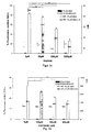
- incubation of H441 and BEAS-2B cells with FLUO-BSA-ILO produced a significantly higher number (p ⁇ 0.01) of positive cells and MFI than the control FLUO-BSA (38 , ⁇ 1.8%) and 82.7 ⁇ 1.6% versus 9.1 ⁇ 1.9% and 13.7 ⁇ 1.2%, respectively ( Fig. 3A ).
- This effect was more pronounced on human bronchial epithelial cells (16HBE14o, BEAS-2B) than on clara (H441) or alveolar (A549) epithelial cells.
- 16HBE14o cells were incubated with FLUO-BSA-ILO in the presence of increasing amounts of CAY10449.
- This compound has previously been reported to be a highly specific potent antagonist of human IP 1 receptor [27, 28].
- 16HBE14o cells were incubated with 25 nM FLUO-BSA-ILO along with increasing concentrations of CAY10449.
- the addition of CAY10449 resulted in a significant dose-dependent decrease (p ⁇ 0.01) in both the number of fluorescence-positive cells and the MFI ( Fig. 3B ).
- FACS measurements along with inhibition experiments indicate cell-type-dependent cell surface expression of IP 1 receptor on lung epithelial cells.
- ILO mediates intracellular uptake of FLUO-BSA-ILO
- additional experiments were performed using confocal laser scanning microscopy. 16HBE14o cells were incubated with either 0.5 ⁇ M FLUO-BSA or FLUO-BSA-ILO. Visualization of the cells by confocal microscopy revealed clear cell surface binding and subsequent intracellular uptake of FLUO-BSA-ILO conjugates ( Fig. 3C ), whereas no uptake of FLUO-BSA could be observed.
- PEI-g-ILO F ILO 5 and PEI gene vector particles were delivered to the lung of BALB / C mice via aerosol and gene expression was analyzed 24 h after gene delivery.
- To evaluate lung tissue quantitatively, the mice were sacrificed and the lungs isolated. Luciferase expression measured in homogenized lung tissue was significantly 14-fold higher for PEI-g-ILO F ILO 5 gene vectors than for PEI gene vectors ( Fig. 6B ).
- the prostaglandin-1 2 analogue ILO an IP 1 receptor agonist
- conjugates according to the invention which contain a prostaglandin-1 2 -analogon as targeting ligand and a cationic polymer as an envelope for an active substance, gene expression can be significantly improved.
- reporter gene expression was significantly increased in human alveolar (A549) and bronchial epithelial cells (16HBE14o-, BEAS-2B) up to 46-fold.
- luciferase activity in the lungs of mice after aerosol treatment was significant, 14-fold higher than in PEI.
- ILO and TRP are agonists of the human IP 1 receptor [29]. Both are approved for the treatment of pulmonary arterial hypertension via aerosol inhalation or iv administration [20, 30, 31]. IP 1 receptors are expressed in the lungs of humans and mice [15, 32-34] and IP 1 receptor / ligand complexes are internalized into the cell [35, 36]. According to the invention, these properties are exploited to provide an improved system for introducing agents into lung cells.
- ILO can be used as a targeting ligand according to the invention that mediates the binding and intracellular uptake of conjugated substances such as FLUO-BSA, which is a prerequisite for receptor-mediated uptake of gene vector nanoparticles.
- ILO was conjugated to branched PEI 25 kDa via an amide linkage.
- the release of pDNA from PEI / p-DNA particles has already been identified as a critical parameter for successful gene transfer [37].
- CpG-free luciferase expression plasmid (pCpG-luc) was used. It has been shown that CpG-free plasmids are less inflammatory than CpG-containing. They have also been shown to increase and prolong gene expression in the lungs [39].
- PEI-g-ILO F ILO 5 / pCpG-luc and PEI / pCpG-luc gene vectors were aerosolized and various fractions collected (fogged, not aerosolized) to test the stability of the particles. Both the gel retardation test and the particle size measurements showed no change in the particles after nebulization as compared to non-nebulized particles.
- a new targeting structure for the delivery of substances into the lung is provided.
- the potential of prostacyclin analogs, and in particular of ILO as a targeting ligand, has been recognized by the inventors as a "ferry" of administration used by substances in lung cells.
- prostacyclin analogs ILO are useful as targeting ligands for non-viral vectors in aerosol form.
- fluorescein-based molecular conjugates it has been shown that the IP 1 receptor is a suitable candidate for receptor-mediated gene transfer in lung cells. The receptor-specific binding and uptake of molecule conjugates has been demonstrated for both alveolar and bronchial epithelial cells and claracels.
- the conjugates according to the invention lead to a specific significant increase in gene expression in vitro and in vivo. By more than 10-fold increase in gene expression, it is possible to reduce the amount of pDNA and gene carriers, which lowers DNA or carrier-mediated toxicity and inflammation.
Landscapes
- Health & Medical Sciences (AREA)
- Life Sciences & Earth Sciences (AREA)
- Chemical & Material Sciences (AREA)
- Engineering & Computer Science (AREA)
- Bioinformatics & Cheminformatics (AREA)
- General Health & Medical Sciences (AREA)
- Veterinary Medicine (AREA)
- Medicinal Chemistry (AREA)
- Public Health (AREA)
- Pharmacology & Pharmacy (AREA)
- Animal Behavior & Ethology (AREA)
- Epidemiology (AREA)
- Genetics & Genomics (AREA)
- Molecular Biology (AREA)
- Biotechnology (AREA)
- Biochemistry (AREA)
- Organic Chemistry (AREA)
- Zoology (AREA)
- Wood Science & Technology (AREA)
- Biomedical Technology (AREA)
- General Engineering & Computer Science (AREA)
- Proteomics, Peptides & Aminoacids (AREA)
- Microbiology (AREA)
- Physics & Mathematics (AREA)
- Biophysics (AREA)
- Pulmonology (AREA)
- Plant Pathology (AREA)
- Chemical Kinetics & Catalysis (AREA)
- General Chemical & Material Sciences (AREA)
- Nuclear Medicine, Radiotherapy & Molecular Imaging (AREA)
- Pharmaceuticals Containing Other Organic And Inorganic Compounds (AREA)
- Medicines That Contain Protein Lipid Enzymes And Other Medicines (AREA)
- Medicinal Preparation (AREA)
- Medicines Containing Antibodies Or Antigens For Use As Internal Diagnostic Agents (AREA)
- Acyclic And Carbocyclic Compounds In Medicinal Compositions (AREA)
Abstract
Es wird ein Konjugat aus Agens-Komplex und mindestens einem Zielfindungsliganden, wobei der Agens-Komplex ein Agens umhüllt von einem Hüllmaterial umfasst und wobei der Zielfindungsligand ein Prostacyclinanalogon ist, sowie dessen Verwendung beschrieben
Description
- Die Erfindung betrifft ein Agens enthaltende Konjugate, die als Zielfindungsstruktur ein Prostacyclinanalogon aufweisen und die Verwendung solcher Konjugate für die Gentherapie bzw. den Gentransfer in Bronchial- und Alveolarepithelzellen.
- Die Lunge ist einerseits ein Organ, dessen Funktion lebensnotwendig ist und andererseits ist die Lunge ein Organ, das aufgrund seiner großen Oberfläche und seiner Zugänglichkeit attraktiv ist, um Wirkstoffe oder aktive Mittel in den Körper einzuschleusen.
- Es ist schon lange bekannt, über Aerosole, Vernebler, Inhalatoren oder Pumpsprays aktive Mittel in die Lunge zu bringen sowohl für lokale als auch systemische Wirkung. Bekannt ist es auch, zur Gentherapie virale oder nicht virale Gentransfermittel über die Lunge zu verabreichen. Sowohl virale als auch nicht virale Trägermittel sind vielfach mit Nebenwirkungen bei ihrer Anwendung verbunden. Dies ist insbesondere dadurch bedingt, dass die Dosis relativ hoch sein muss, da der Gentransfer, d.h. das Einschleusen der gewünschten Gene in Zellen häufig nicht effizient genug ist. Es wurde daher schon lange nach Mitteln gesucht, um die Effizienz des Gentransfers zu verbessern. Hierzu wurde bereits vorgeschlagen, Gene mit einem kationischen Lipid zu umhüllen, da kationische Teilchen leichter phagozytiert werden können. Ein hierzu vorgeschlagenes Mittel, das bereits in klinischen Versuchen getestet wird [7], -ist Genzyme Lipid 67.Bekannt ist auch die Verwendung von Polyethyleniminpolymeren (PEI), um Nukleinsäuren zu umhüllen [8]. Obwohl PEI DNA schützen kann, ist es mit dem Nachteil verbunden, dass die Gentransfereffizienz gering ist, außerdem wurde gefunden, dass es in hoher Dosis, die aufgrund der geringen Transfektionseffizienz erforderlich ist, Entzündungen hervorruft.
- Es wurde daher auch schon versucht, mit kationischen Polymeren umhüllte Teilchen mit Liganden zu versehen, die die Teilchen in Zellen einschleusen sollen. Versuche gab es bereits mit Transferin [10], Folsäure [11], Lactoferin [12], Clenbuterol [13] und Wachstumsfaktoren, wie EGF [14]. Obwohl mit diesen Liganden der PEI-vermittelte Gentransfer verbessert werden konnte, besteht immer noch Bedarf dafür, aktive Mittel der Lunge gezielt und in hoher Ausbeute zuzuführen.
- Weiterhin wurde und wird versucht, neue Wege für die therapeutische Behandlung von chronischen Lungenkrankheiten zu finden, für die der Gentransfer ein viel versprechender Ansatz ist. Auf angeborenen oder erworbenen Protein- und/oder Gendefekten beruhende Lungenkrankheiten könnten durch Bereitstellung der fehlenden oder geschädigten Proteine oder Genprodukte verbessert, gelindert oder sogar geheilt werden. Dazu muss jedoch die Verabreichung regelmäßig erfolgen. Ein Ausgleich zwischen unerwünschten Nebenwirkungen und erwünschter therapeutischer Wirkung muss daher gefunden werden. Außerdem ist auch die erforderliche Dosierungshäufigkeit für eine länger andauernde Therapie ein wichtiger Aspekt.
- Aufgabe der Erfindung war es daher, Konjugate bereitzustellen, die es ermöglichen, Wirkstoffe oder aktive Mittel, die zur Behandlung oder Linderung von Lungenkrankheiten geeignet sind, in einer Form bereitzustellen, die von Zellen in der Lunge gezielt aufgenommen werden können, insbesondere von Bronchial- und Alveolarepithelzellen.
- Diese Aufgabe wird gelöst mit einem Konjugat, wie es in Anpruch 1 definiert ist.
- Überraschenderweise wurde gefunden, dass Epithelzellen in der Lunge, d.h. Bronchialepithelzellen und Alveolarepithelzellen IP1-Rezeptoren tragen und dass diese Rezeptoren für einen effizienten Transfer von wirkstoffhaltigen Teilchen adressiert werden können. Mit den erfindungsgemäßen Konjugaten gelingt es, Epithelzellen in den Bronchien und Alveolen gezielt über diese IP1-Rezeptoren anzusteuern durch Verwendung mindestens eines Prostacyclinanalogons als Zielfindungsstruktur .
- Im folgenden wird der Gegenstand der Erfindung detailliert beschrieben und die Eigenschaften und Vorteile werden näher ausgeführt. Die Erfindung wird auch in den beigefügten Figuren näher erläutert, wobei
-
Fig. 1 die Ergebnisse einer Western-Blot-Analyse der Expression von IP1-Rezeptor in menschlichen Alveolar- und Bronchialepithelzellen zeigt. -
Fig. 2 die Fluoreszenzintensität von A549- und 16HBE14o-Zellen nach Inkubation mit FLUO-BSA-ILO bzw. FLUO-BSA-TRP zeigt. -
Fig. 3a die Fluoreszenzintensität von FLUO-BSA im Vergleich zu FLUO-BSA-ILO in verschiedenen Zelllinien zeigt;Fig. 3b undFig. 3c die mittleren Fluoreszenzintensitäten zeigen bei steigenden Konzentrationen von CAY10449 bzw. ILO;Fig. 3d die mittlere Fluoreszenzintensität von FLUO-BSA-ILO nach Zugabe von CAY10449 zeigt;Fig. 3e Aufnahmen einer konfokalen Laserrastermikroskopie zur Oberflächenbindung zeigt. -
Fig. 4 die DNA-Freisetzung für PEI-g-ILO-Konstrukte mit unterschiedlichen Verhältnissen von N/P zeigt. -
Fig. 5a den Expressionsgrad für mit PEI-g-ILO-Genvektoren transfizierte Zellen im Vergleich zu unmodifiziertem PEI zeigt;Fig. 5b die Genexpression für PEI-g-ILO im Vergleich zu PEI zeigt;Fig. 5c die Expressionsgrade für PEI-g-ILO in A549-und BEAS-2B-Zellen im Vergleich zu PEI zeigt. -
Fig. 6a in vivo Studien zur Genexpression von Luciferase zeigt;Fig. 6b die Luciferaseexpression in homogenisiertem Lungengewebe, das von Mäusen erhalten wurde, die PEI-g-ILO FILO = 5 Genvektoren erhalten hatten, im Vergleich zu PEI-Genvektoren zeigt. -
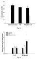
Fig. 7a die metabolische Aktivität von unbehandelten Zellen im Vergleich zu mit PEI oder erfindungsgemäßem Konstrukt behandelten Zellen zeigt;Fig. 7b die Veränderung des Cytokinspiegels nach Verabreichung von PEI oder erfindungsgemäßem Konstrukt im Vergleich zu unbehandelten Zellen zeigt. -
Fig. 8 die dosisabhängige Genvektorzuführung in Lungenzellen zeigt. - Es wurde überraschenderweise gefunden, dass Konjugate, die Prostacyclinanaloga als Zielfindungsstruktur tragen für die Adressierung von Epithelzellen in der Lunge, insbesondere von Bronchial- und Alveolarzellen geeignet sind und mit hoher Effizienz Agentien in die Zellen schleusen können. Damit wird nun erfindungsgemäß ein Mittel bereitgestellt, um aktive Inhaltsstoffe in Lungenepithelzellen einzuschleusen. Dies bietet neue Möglichkeiten der therapeutischen Behandlung von unterschiedlichsten Lungenkrankheiten.
- Prostacyclin gehört zur Klasse der Prostaglandine und ist als Prostaglandin l2 bzw. PGl2 bekannt; es adressiert und bindet den Prostacyclin-(IP1)-Rezeptor. Der IP1-Rezeptor ist ein 7-Transmembran-G-proteingekoppelter Rezeptor, der vor allem auf Endothelzellen gefunden wurde, insbesondere auf Muskelzellen z.B. auf Muskelzellen von Blutgefäßen [15-17]. Die Bindung von Prostacyclin an einen IP1-Rezeptoragonisten führt zu einer endosomalen Internalisierung von Rezeptor/Ligandenkomplexen über Clathrin-vermittelte Prozesse [18, 19]. Die Erfinder der vorliegenden Erfindung haben nun gefunden, dass diese Wirkung dazu genutzt werden kann, den gezielten Transfer von aktiven Mitteln in Alveolar- und Bronchialepithelzellen zu verbessern und die Aufnahme von für die Lunge nützlichen oder Lungenleiden therapierenden aktiven Mittel zu ermöglichen.
- Erfindungsgemäß wird daher ein Konjugat bereitgestellt, das als Zielführungsstruktur für Bronchien- und Alveolarepithelzellen mindestens ein Prostacyclinanalogon aufweist. Prostacyclin selbst ist zu instabil und wird zu schnell abgebaut, um für den vorgesehenen Zweck verwendet werden zu können. Es sind jedoch stabile Prostacyclinanaloga bekannt, die ebenfalls an den IP1-Rezeptor binden und als Agonisten wirken. Für die erfindungsgemäßen Konjugate sind die bekannten Prostacyclinanaloga geeignet.
- Bevorzugt werden als Liganden lloprost und/oder Treprostinil, zwei als Arzneimittel zugelassene Prostacyclinanaloga verwendet. Andere Prostacyclinanaloga können ebenfalls verwendet werden. Ein für die-Erfindung geeignetes Prostacyclinanalogon ist ein solches, das stabiler ist als das natürlich vorkommende Prostacyclin und eine mit Prostacyclin vergleichbare Bindungsfähigkeit an den IP1-Rezeptor hat. Ein Verfahren, um die Bindungsfähigkeit eines Prostacyclinanalogons festzustellen, ist ein kompetitives Verfahren, bei dem lloprost und/oder Treprostinil sowie ein Prostacyclinanalogonkandidat mit Fluorescein und Rinderserumalbumin (BSA) konjugiert werden und dann zu verschiedenen Lungenzelllinien zugegeben werden, wobei dann die Bindung und zelluläre Aufnahme durch Durchflusscytometrie und konfokale Laserrastermikroskopie untersucht werden. Ein Kandidat, der genauso stark oder stärker wie lloprost bzw. Treprostinil bindet, ist als Teil des erfindungsgemäßen Konjugats ebenfalls bevorzugt. Gleiche Bindefähigkeit bedeutet, wenn der fluoresceinmarkierte Kandidat mindestens im selben Ausmaß wie fluoresceinmarkiertes lloprost bzw. fluoresceinmarkiertes Treprostinil bindet. Ist der Anteil an fluoresceinmarkierten Kandidaten niedriger, bedeutet dies, dass lloprost bzw. Treprostinil den Kandidaten aus der Bindung drängen, so dass die Bindefähigkeit nicht so hoch ist.
- Die bekannten Prostacyclinanaloga werden verwendet zur Behandlung von pulmonalem arteriellem Hochdruck (PAH) und werden üblicherweise intravenös oder als Aerosol verabreicht [20], sie müssen häufig über den Tag verteilt dosiert werden, um diesen Zweck zu erfüllen. Für die vorliegende Erfindung wird das Prostacyclinanalogon jedoch als Zielfindungsstruktur eingesetzt und dient nicht zur Behandlung des pulmonalen arteriellen Hochdrucks. Es wurde gefunden, dass in der erfindungsgemäßen Kombination mit kationischer Hüllsubstanz und Agens Prostacyclinanaloga eine entzündungshemmende Wirkung aufweisen und hierdurch die Wirkung der erfindungsgemäßen Konjugate noch verbessern.
- Der zweite Teil des erfindungsgemäßen Konjugats ist ein von einem Hüllmaterial umhülltes Agens. Das Hüllmaterial dient dazu, das Agens zu schützen und gleichzeitig die Aufnahme in die Zelle nicht zu stören oder ggf. sogar zu verbessern.
- Der als wirkendes Prinzip aktive Bestandteil des erfindungsgemäßen Konjugats kann jedes Agens sein, das in den von dem erfindungsgemäßen Zielfindugnsliganden adressierten Zellen eine vorteilhafte, heilsame, lindernde oder modulatorische Wirkung ausübt. Als Beispiele für erfindungsgemäß verwendete Agentien sind insbesondere Nukleinsäuren, Peptide oder Polypeptide, Wirkstoffe oder Tracer und/oder Derivate der genannten Substanzen und/oder Mischungen davon. Das erfindungsgemäß verwendete Agens soll bevorzugt ein Mittel sein, dass ein Lungenleiden lindern oder heilen kann.
- Gemäß einem Aspekt der vorliegenden Erfindung ist das Agens eine Nukleinsäure, die ein Gen oder Genfragment umfasst, dessen Defekt oder Mangel eine Lungenkrankheit hervorruft oder die ein immunmodulierend wirksames Protein, insbesondere ein Antigen kodiert. Die Nukleinsäure kann eine DNA oder RNA sein und sie kann ein oder mehrere Gene oder Fragmente aufweisen. Die Nukleinsäure kann eine autonom replizierende oder integrierende Sequenz sein, sie kann als Plasmid, Vektor oder in anderer dem Fachmann wohlbekannten Form sein. Sie kann linear oder ringförmig und einzel- oder doppelsträngig sein. Jede in einer Zelle aktive Nucleinsäure ist hier geeignet. Außerdem kann die Nukleinsäure in an sich bekannter Weise weitere Elemente, die für die Expression des Gens notwendig oder nützlich sind, z.B. Promotoren, Enhancer, Signalsequenzen etc. aufweisen.
- In einer weiteren Ausführungsform ist der aktive Bestandteil des erfindungsgemäßen Konjugats ein Peptid, Polypeptid, Protein oder Proteinfragment, das geeignet ist, einen Proteinmangel oder Proteindefekt, der zu einer Lungenkrankheit führt, zu beheben oder das immunmodulatorisch wirkt.
- Weiterhin kann der aktive Bestandteil des erfindungsgemäßen Konjugats ein Wirkstoff sein, der, wenn er in einer Bronchial- und/oder Alveolarepithelzelle vorhanden ist, zur Heilung oder Linderung eines krankhaften Zustands in der Lunge führt. Beispiele hierfür sind z.B. entzündungshemmende Mittel wie Steroide, die zur Behandlung von Asthma eingesetzt werden. Da auch der Ligand entzündugshemmend wirkt, führt diese Kombination zu einem sehr wirkungsvollen Mittel.
- In einer weiteren Ausführungsform kann das Agens ein Reportermolekül sein, dessen Aufnahme in die Zelle diagnostisch wichtige Informationen liefern kann. Reportermoleküle, die für die Diagnostik geeignet sind, sind dem Fachmann bekannt und Beispiele für geeignete Reportermoleüle sind radioaktive oder fluoreszierende Tracermoleküle, die dem Fachmann bekannt sind. Die Reportermoleküle können z.B. eingesetzt werden, um den Verlauf einer Behandlung oder den Zustand der Lunge zu überprüfen.
- Ein weiterer wesentlicher Bestandteil des erfindungsgemäßen Konjugats ist ein Hüllmaterial, das das Agens umhüllt, um es vor Abbau oder Veränderung zu schützen und das die Einschleusung in die Zelle nicht stört oder sogar fördert. Das Hüllmaterial ist geeigneterweise ein kationisches oder neutrales Material, z.B. ein Polymer oder ein sonstiges schichtbildendes Material. Wichtig ist, das das Hüllmaterial biologisch und physiologisch verträglich ist, das Agens während des Transports schützt und in der Zelle zu physiologisch verträglichen Molekülen abgebaut wird und inert gegenüber dem Agens ist, d.h. mit dem Agens nicht reagiert. Geeignete Hüllmaterialien sind bekannt und werden in vielfältiger Form angeboten. Für die Umhüllung von Nukleinsäuren sind kationische Hüllmaterialien bevorzugt, während für die Umhüllung anderer Agentien, wie Proteine, Wirkstoffe oder Tracer kationische oder neutrale Hüllmaterialien verwendet werden können.
- In einer Ausführungsform der vorliegenden Erfindung, insbesondere wenn das Agens eine Nukleinsäure ist, wird als Hüllmaterial ein kationisches Polymer verwendet. Es wurde gefunden, dass kationisch geladene Teilchen von der Zelle leichter aufgenommen werden können als neutrale oder anionisch geladene Teilchen, allerdings können sie auch eher unspezifische Anlagerungen fördern. Zur Umhüllung von Nukleinsäuren als aktiven Bestandteilen sind kationische Hüllmaterialien bevorzugt, da sich Nukleinsäuren sehr einfach mit kationischen Substanzen umhüllen und schützen lassen. Entsprechende Verfahren sind dem Fachmann wohlbekannt.
- Das Hüllmaterial kann eine natürlich vorkommende, synthetische oder kationisch derivatisierte natürliche Substanz sein, z.B. ein Lipid oder ein Polymer oder Oligomer. Ein Beispiel für ein natürliches Oligomer ist Spermin. Beispiele für synthetische Polymere sind stickstoffhaltige biologisch abbaubare Polymere, insbesondere solche mit protonierbaren Stickstoffatomen. Besonders geeignet sind Polyethylenimine, insbesondere verzweigte Polyethylenimine, die im Handel erhältlich sind. Geeignet ist beispielsweise ein verzweigtes Polyethylenimin mit einem mittleren Molekulargewicht von 25 kDa, das kommerziell verfügbar ist. Es wurde gefunden, dass dieses Polymer in Kombination mit dem Zielfindungsliganden sehr verträglich ist. Als natürliches ggf. derivatisiertes schichtbildendes Hüllmaterial können auch Lipide, insbesondere kationische oder neutrale Lipide, eingesetzt werden. Lipide sind in vielen Varianten verfügbar und können z.B. zur Bildung von Liposomen eingesetzt werden. Besonders geeignet ist ein kationisch derivatisiertes Lipid, das unter der Bezeichnung Genzyme Lipid 67 erhältlich ist.
- Für andere Agentien, wie Proteine, Wirkstoffe oder Tracer, gibt es eine Anzahl geeigneter Polymere, die dem Fachmann bekannt sind. Geeignet sind solche, die biologisch verträglich sind, und die zumindest in Kombination mit dem erfindungsgemäßen Prostacyclinanalogon nicht entzündlich oder in sonstiger Weise schädlich für die Zelle sind und das Agens, sobald es am Zielort, in der Zelle, angekommen ist, freigeben.
- Der aus Hüllmaterial und Agens bestehende Konjugatkomplex kann z.B. aus an sich bekannten Nanopartikeln oder Nanokapseln, Liposomen etc. bestehen, deren Herstellung wohbekannt ist. Geeignet ist beispielsweise die Verkapselung in biologisch abbaubare Polymere mit einstellbarer Freisetzung wie Polylactid und/oder Polyglycolid. Das Hüllmaterial kann dabei so ausgewählt werden, dass die Freisetzung des Agens in vorbestimmter Art und Weise erfolgt. Derartige Hüllmaterialien sind in der Literatur vielfach beschrieben und der Fachmann kann aus einer Vielzahl von Materialien das jeweils am besten geeignete auswählen.
- Das aktive Mittel wird in an sich bekannter Weise mit dem Hüllmaterial umhüllt oder darin verkapselt. Dieser Komplex aus Agens und umhüllendem Material wird im Folgenden auch als ,,Agens-Komplex" bezeichnet. Unter ,,umhüllen" wird im Zusammenhang mit der vorliegenden Erfindung verstanden, dass das Agens soweit durch das Polymer von der physiologischen Umgebung abgeschirmt wird, dass es nicht verändert oder abgebaut wird, solange bis es am Zielort angekommen ist. Die Umhüllung kann nur eine Schicht sein, die das Agens umgibt, es kann auch ein Liposom oder Nano- oder Mikroteilchen sein, in dem das Agens eingebettet oder eingeschlossen ist. Auch kann es durch Komplexierung eingeschlossen sein. Verschiedene Formen der Verkapselung oder Umhüllung von Agentien sind dem Fachmann bekannt und können für das erfindungsgemäße Konjugat eingesetzt werden, solange sie die Bindung des Zielfindungsliganden an den Rezeptor und die Einschleusung des Konjugats in die Zelle nicht stören und in der Zelle das Agens freisetzen. Die Umhüllung des Agens mit dem Hüllmaterial bzw. die Herstellung entsprechender Teilchen kann mit üblichen Verfahren erfolgen. In der einfachsten Ausführungsform wird das aktive Mittel, z.B. eine Nukleinsäure, mit dem Hüllmaterial, z.B. einem kationischen Polymer wie Polyethylenimin, ggf. in gelöster Form vermischt.
- An die Hüllsubstanz wird entweder vor der Umhüllung oder danach mindestens ein erfindungsgemäß als Zielführungsstruktur verwendetes Prostacyclinanalogon gebunden. Durch die Bindung des Liganden an das Hüllmaterial wird die Aktivität des Agens nicht beeinträchtigt. Die Bindung oder Immobilisierung des bzw. der Liganden darf die Bindefähigkeit an den Rezeptor nicht stören. Verfahren zur Immobilisierung von Liganden sind dem Fachmann bekannt und die bekannten Verfahren können hier angewendet werden. Die Liganden können entweder direkt an das Hüllmaterial gebunden werden, bevor dieses zur Umhüllung des Agens eingesetzt wird. Diese Ausführungsform ist z.B. bevorzugt, wenn das Hüllmaterial ein kationisches Polymer ist und das Agens eine Nukleinsäure ist. Es ist auch möglich zuerst den Agens-Komplex zu bilden und anschließend die Liganden zu binden. Diese Ausführungsform ist bevorzugt, wenn der Agens-Komplex in Form von Nanoteilchen, Nanokugeln oder Liposomen gebildet wird. Ggf. kann die Bindung des Liganden an das Hüllmaterial auch über einen Spacer so erfolgen, dass die bindungsaktive Stelle des Liganden für die Bindung zugänglich ist. Es wird somit ein Konjugat geschaffen, bei dem das aktive Mittel durch die Bindung nicht beeinflusst wird und das mindestens eine Prostacyclinanalogon auf der Oberfläche zur Bindung an den IP1-Rezeptor frei zur Verfügung steht. Die Kupplung des Prostacyclinanalogons an das Hüllmaterial kann über kovalente oder ionische Bindung direkt oder über einen Spacer erfolgen. Ein dem Fachmann bekannter Spacer ist z.B. Polyethylenglycol (PEG).
- Der Kupplungsgrad, d.h. das Ausmaß der Beladung des Konjugats bzw. der umhüllten Teilchen mit Liganden ausgedrückt als Ligand pro Konjugatteilchen, hat Einfluss auf die Freisetzung des Agens und damit auf die Aktivität des Agens in der Zelle. Der Anteil der an ein umhülltes Teilchen zu bindenden Liganden sollte bevorzugt nicht zu hoch sein, da ansonsten aufgrund sterischer Hinderung die Adressierung des Rezeptors gestört sein könnte. Der Fachmann kann die geeignete Beladung mit Routineversuchen herausfinden. Der Anteil an Liganden hängt ab von der Art des Hüllmaterials und der Größe der Teilchen.
- Es wurde auch gefunden, dass ein hoher Kupplungsgrad dazu führen kann, dass das Agens nur noch unvollständig freigesetzt wird. Wird zur Umhüllung ein kationisches Polymer verwendet, sollte daher der Kupplungsgrad 15 Liganden pro Polymer oder weniger sein. Andererseits muss pro umhülltem Teilchen mindestens ein Prostacyclinanalogon gebunden sein, um die Adressierung zu bewirken.
- Pro Konjugat bzw. Teilchen kann jeweils eine Art von Prostacyclinanalogon gebunden werden. Es ist auch möglich, eine Mischung von zwei oder mehr Prostacyclinanaloga zu binden, um gegebenenfalls die Bindefähigkeit und/oder Aufnahme in die Zelle zu verstärken.
- Es wurde gefunden, dass das Verhältnis von Hüllmaterial zu aktivem Mittel einen Einfluss auf die Wirksamkeit haben kann. Wenn zu wenig Hüllmaterial vorhanden ist, wird das aktive Mittel nicht ausreichend geschützt. Ist der Anteil des Hüllmaterials zu hoch, kann es einerseits Probleme mit der Verträglichkeit geben und andererseits kann ein zu hoher Anteil an Hüllmaterial dazu führen, dass das aktive Mittel nicht mehr freigesetzt werden kann. In beiden Fällen leidet die Effizienz des Transfers. Der Fachmann kann in wenigen Routineversuchen das jeweils am besten geeignete Verhältnis herausfinden. Als besonders geeignet hat sich ein Verhältnis von Hüllmaterial zu aktivem Mittel im Bereich von 10:1 bis 1:4, bezogen auf Gewicht, erwiesen. Besonders bevorzugt ist ein Verhältnis von Hüllmaterial zu Agens von 4:1 bis 1:4. Wenn das Konjugat eine Nukleinsäure als Agens und Polyethylenimin als Polymer enthält, kann der Anteil des Polymers auch über das molare Verhältnis von Polymer-Stickstoffanateil zu DNA-Phosphatanteil angegeben werden; bevorzugt liegt dieses Verhältnis in einem Bereich von 2 bis 10, besonders bevorzugt von 4 bis 8. Es wurde gefunden, dass der hydrodynamische Durchmesser der Konjugatteilchen bei einem molaren Verhältnis von Polymer-Stickstoffanateil zu DNA-Phosphatanteil von 4 bis 8 in einem Bereich von 50 bis 100 nm liegt, was für die Aufnahmeeigenschaften optimal ist.
- Weiterhin wurde festgestellt, dass optimale Konjugate dann erhalten werden, wenn die Ligandenbeladungsdichte an den Umhüllungsgrad angepasst wird. Wenn der Anteil an Hüllmaterial relativ hoch ist, sollte die Beladungsdichte nicht zu groß sein, da sonst das Agens zu stark abgeschirmt wird. Wenn der Anteil an Hüllmaterial im unteren Bereich liegt, kann die Beladungsdichte entsprechend im oberen Bereich liegen.
- Es wurde gefunden, dass das erfindungsgemäße Konjugat ein ideales Mittel ist, um aktive Wirkstoffe in Bronchial- und/oder Alveolarepithelzellen einzuschleusen. Während mit den im Stand der Technik beschriebenen Teilchen ohne Targetliganden nur 5% oder weniger ihr Ziel, die Zelle, erreichen und mit den im Stand der Technik beschriebenen ligandenhaltigen Konjugaten auch nur Anteil unter 50 % oder weniger das Ziel erreichen und die Funktion entfalten kann, konnte in Versuchen für die erfindungsgemäßen Konjugate gezeigt werden, dass mehr als 50 %, häüfig 60% und mehr und sogar 80% des erfindungsgemäßen Konjugats von Zellen aufgenommen werden und ihr Agens freisetzen.
- Es wird somit ein Mittel zur Verfügung gestellt, mit dem ein aktiver Bestandteil mit hoher Effizienz in Zielzellen eingeschleust wird, wobei einerseits das aktive Mittel während des Transports in die Zelle gut geschützt ist, so dass der Anteil an aktivem Mittel, der in der Zelle ankommt, sehr hoch ist und andererseits die Aufnahmeeffizienz aufgrund der Struktur des erfindungsgemäßen Konjugats sehr hoch ist.
- In einer weiteren Ausführungsform kann das erfindungsgemäße Konjugat noch weiter verbessert werden, indem das mit Hüllmaterial, z.B. einem kationischen Polymer umhüllte Teilchen noch zusätzlich mit Polyethylenglycolketten versehen wird, um die Überlebensdauer in der Lunge weiter zu erhöhen. Aktive Moleküle, wie Nukleinsäure durch PEG-ylierung zu schützen, ist an sich bekannt und die üblichen Verfahren können hier verwendet werden.
- Das erfindungsgemäße Konjugat kann zur Behandlung von unterschiedlichen Lungenkrankheiten verwendet werden. Insbesondere ist das erfindungsgemäße Konjugat geeignet, um auf Gen- oder Proteindefekten beruhende Lungenkrankheiten zu heilen oder zu lindern. Ein Beispiel hierfür ist zystische Fibrose. Wie oben ausgeführt, ist es sowohl möglich, das mangelnde oder defiziente Gen in die Zelle zu schleusen, als auch ein von dem mangelnden oder defizienten Gen codiertes Protein in die Zelle zu schleusen.
- Ein weiteres Anwendungsgebiet für die erfindungsgemäßen Konjugate ist die Verwendung als Impfstoff. In dieser Ausführungsform ist der aktive Bestandteil des Konjugats entweder ein immunmodulatorisch oder immunologisch aktives Peptid oder Protein oder ein Gen, das ein immunmodulatorisch oder immunologisch aktives Protein oder Peptid codiert [1, 2]. Ein Vorteil dieser Ausführungsform der Erfindung ist es, dass über die Lunge nichtinvasiv ein Impfstoff zugeführt werden kann, z.B. mit einem Vernebler oder über ein Aerosol. Diese Technologie ist un kompliziert, ermöglicht den Einsatz auch dort, wo die Verabreichung von Injektionen aufgrund der hygienischen Verhältnisse oder wegen des Fehlens von entsprechend ausgebildetem Personal problematisch ist und ermöglicht eine unkomplizierte mehrfache Dosierung, um die Immunantwort zu stärken. Darüber hinaus ist die Lunge aufgrund ihrer großen Oberfläche und des Vorhandenseins immunologisch aktiver Zellen gut geeignet für eine Impfung.
- Das erfindungsgemäße Konjugat wird bereitgestellt zur Verabreichung in die Lunge. Dazu kann es in an sich bekannter Weise formuliert werden als pharmazeutische Zusammensetzung, die durch Inhalation oder über Vernebelung in die Lunge gebracht wird. Entsprechende Formulierungen sind dem Fachmann bekannt. So kann das Konjugat als Suspension oder Emulsion über einen Vernebler oder als Aerosol, mit einem inerten Gas als Trägermittel hergestellt werden. Es kann auch als Pulver eingesetzt werden.
- Die vorliegende Erfindung wird durch die folgenden Beispiele weiter erläutert, ohne auf dieses Beispiel beschränkt zu werden.
- Es wurden Konjugate von mit einem Hüllmaterial umhüllten Teilchen und lloprost bzw. Treprostinil als Zielfindungsstruktur hergestellt und untersucht.
- Die Bezugsquellen für Chemikalien und Plasmide und die verwendeten Konzentrationen sind wie folgt:
- lloprost, Treprostinil und CRY10449: Cayman Chemicals (Michigan, USA) Verzweigtes Polyethylenimin (durchschnittliches Molekulargewicht 25 kDa), N-Hydroxysulfosuccinimid (sulfo-NHS), Rinderserumalbumin (BSA), Natriumphosphat, Picrylsulfonsäurelösung, 4-(2-Hydroxyethyl)piperazin-1-ethansulfonsäure (HEPES) und Heparansulfat: Sigma Aldrich (Schnelldorf, Deutschland)
- PEI wurde in doppelt destilliertem Wasser (Wasser zur Injektion, B. Braun Melsungen AG, Melsungen, Deutschland) verdünnt und der pH mit Salzsäure auf 7 eingestellt.
- Natriumphosphat wurde in doppelt destilliertem Wasser auf eine Konzentration von 0,5 mM gelöst und der pH mit Natriumhydroxid auf 7,5 eingestellt.
- HEPES wurde in destilliertem Wasser auf eine Konzentration von 0,1 M gelöst und der pH mit Natriumhydroxid auf 7,4 eingestellt.
- Heparansulfat wurde in doppelt destilliertem Wasser auf eine Konzentration von 5 mg/ml gelöst.
- Ethanol p.a. und 3-(N-Morpholino)propansulfonsäure (MOPS): Merck (Darmstadt, Deutschland)
- MOPS wurde in doppelt destilliertem Wasser auf eine Konzentration von 0,1 M gelöst und der pH mit Salzsäure auf 6 eingestellt.
- 1-Ethyl-3-(3-dimethylaminopropyl)carbodiimidhydrochlorid (EDC) und 5-(und 6-) Carboxyfluoresceinsuccinimidylester (Fluorescein-NHS): Pierce (Rockfort, USA) Dithiothreitol (DTT): Amersham Biosciences (South San Francisco, USA)
- D-Luciferin: Synchem OHG (Flensberg/Altenburg, Deutschland)
- Die Plasmide pCMV-luc, das das Fotinus pyralis Luciferasegen unter der Kontrolle des frühen Promotors von Cytomegalovirus (CMV) enthielt und pCpG-luc wurden in E. coli propagiert und in hoch gereinigter Form (LPS-Gehalt ≤ 0,1 E.U./µg DNA) von Plasmid Factory GmbH (Bielefeld, Deutschland) bereitgestellt. Die Menge an Supercoil pDNA war ≥ 90% ccc (covalently closed circular) für pCMV-luc und ≥ 98% ccc für pCpG-luc.
- A549-Zellen (Humanalveolarepithelzellen): DSMZ (deutsche Sammlung für Mikroorganismen und Zellkulturen, Braunschweig, Deutschland)
BEAS-2B (menschliche Bronchialepithelzellen), H441 (menschliche Broncholarepithelzellen): ATCC (American Type Culture Collection)
16HBE14o-Zellen: Humanbronchialepithelzellen
A549, BEAS-2B und 16HBE14o-Zelllinien wurden in Minimalessenzialmedium (MEM, Gibco-BRL, Karlsruhe, Deutschland), das mit 10% fötalem Kälberserum (FCS, Gibco-BRL, Karlsruhe, Deutschland) ergänzt war, bei 37°C in einer mit 5% CO2 ergänzten angefeuchteten Luftatmosphäre gezüchtet. Die H441-Zellline wurde in Roswell Park Memorial Institute Medium 1640 (RPM11640, Gibco-BRL, Karlsruhe, Deutschland), das mit 10% FCS ergänzt war, bei 37°C in einer mit 5% CO2 angereicherten angefeuchteten Luftatmosphäre gezüchtet. - 14 Wochen alte weibliche BALB/c-Mäuse (Charles River Laboratories Sulzfeld, Deutschland) wurden unter spezifischen pathogenfreien Bedingungen gehalten. Die Mäuse wurden mindestens 7 Tage lang vor den Versuchen akklimatisiert. Alle Tierverfahren waren zugelassen und wurden von der lokalen Ethikkommission kontrolliert und nach den Richtlinien des deutschen Rechts zum Schutz von Tierleben durchgeführt.
- A549-, BEAS-2B- und 16HBE14o-Zellen wurden mit PBS gewaschen und auf Eis in Lysepuffer, der 20 mM Tris-HCL (pH 7,5), 100 mM NaCl, 1 mM EDTA, 1 % Triton X-100 und 0,05% Natriumdesoxycholat enthielt, lysiert. 1 mM DTT und Proteaseinhibitor-Cocktail (Roche Diagnostics GmbH, Mannheim, Deutschland) wurden direkt vor der Verwendung frisch zugefügt. Die Proteinkonzentrationen wurden unter Verwendung eines Biorad-Proteinassays (Biorad, München, Deutschland) bestimmt. Für jede Zelllinie wurden 50 µg Protein in SDS-Probenbeladungspuffer verdünnt (62,5 mM Tris-HCl (pH 6,8), 2% SDS, 10% Glycerin, 2% DTT, 0,001 % Bromphenolblau), der 5 min gesiedet war, auf einem 7,5% Tris-HCl-Gel (Biorad, München, Deutschland) getrennt und auf eine PVDF-Membran (Millipore, Schwalbach, Deutschland) überführt. Die Membranen wurden mit TBS-T (20 mM Tris-HCl (pH 7,6), 137 mM NaCl, 0,1% Tween-20), das 5% Magermilchpulver enthielt (Sigma Aldrich, Deisenhofen, Deutschland) 1 h bei Raumtemperatur blockiert. Der primäre polyklonale Antikörper (1:500 verdünnt) für den IP1-Rezeptor (Cayman Chemical, Michigan, USA) wurde über Nacht in 0,5% Magermilch inkubiert. Die Membranen wurden mit TBS-T gewaschen und mit einem sekundären HRPkonjugierten Antikaninchen-Antikörper (1:15.000 verdünnt; Biorad, München, Deutschland) 1,5 h bei Raumtemperatur in 0,5% Magermilch inkubiert. Nach mehreren Waschstufen mit TBS-T erfolgte die Chemilumineszenzdetektion unter Verwendung eines ECL Detection Kit (Pierce, Rockfort, USA) nach der Anleitung des Herstellers.
- 20 mg (0,3 µmol) BSA wurden in 2,5 ml Natriumphosphatpuffer, pH 7,5, verdünnt und mit einem zehnfach molaren Überschuss an Fluorescein-NHS vermischt. Nach einstündigem Rühren bei Raumtemperatur wurde die Mischung auf einer mit PBS equilibrierten Sephadex G25 MPD-10-Säule (GE Health Care, Uppsala, Schweden) gereinigt. Entweder 0,7 mg (1,8 µmol) ILO oder 0,8 mg (1,8 µmol) TRP wurden in 130 µl Ethanol p.a. gelöst und mit 370 µl MOPS-Puffer, 0,1 M, pH 6, vermischt. 0,5 mg (5 mM) sulfo-NHS (in MOPS-Puffer) und 0,2 mg (2 mM) EDC (in MOPS-Puffer) wurden zugegeben und 15 min bei Raumtemperatur gerührt. Danach wurden 5 µl (20 mM) DTT (in destilliertem Wasser) zugefügt und sofort 3 mg (45,2 nmol) FLUO-BSA in 190 µl und 210 µl Phosphatpuffer 0,5 M in die Reaktionsmischung pipettiert. Nach zweistündigem Rühren bei Raumtemperatur wurde die Mischung auf einer mit PBS equilibrierten Sephadex G25 MPD-10-Säule (GE Health Care, Uppsala, Schweden) gereinigt. Die BSA-Mengen wurden quantitativ ausgewertet mit einem Biorad Proteinassay unter Verwendung einer BSA-Standardkurve. Die Kupplungseffizienz der End- und Zwischenprodukte wurde mit TNBS-Assay [21] bestimmt und die Extinktion bei 495 nm gemessen. Der Kupplungsgrad von BSA-ILO und BSA-TRP ergab sich als 10 mol ILO oder TRP pro mol BSA.
- Verschiedene Kupplungsgrade von PEI-g-ILO wurden synthetisiert durch Variation der EDC-Mengen, die der Reaktionsmischung zugegeben wurden. 1 mg (2,8 µmol) ILO wurde in 100 µl Ethanol p.a. verdünnt, mit 68 nmol PEI in 900 ml HEPES-Puffer, 0,1 M, pH 7,4, und 1 mg (5 mM) sulfo-NHS gemischt. Verschiedene Mengen an EDC wurden auf eine Endkonzentration von 25 mM, 50 mM, 60 mM bzw. 100 mM zugegeben und 4 h lang unter Rühren bei Raumtemperatur inkubiert. Die Reaktionsmischung wurde an einer mit doppelt destilliertem Wasser equilibrierten Sephadex G25 MPD-10-Säule (GE Health Care, Uppsala, Schweden) gereinigt. Die Konzentration von PEI wurde mit einem CuSO4-Test gemäß Ungaro et al. [22] bestimmt. 1H-1D-NMR-Spektren von PEI-g-ILO wurden in D2O auf einem Brooker AV 250 MHz Spektrometer aufgezeichnet. Die Kupplungsgrade von PEI-g-ILO wurden durch Integration des breiten Multipletts von PEI (CH2-CH2-NH-) bei δ (1H) = 2,5 bis 3,1 ppm und des Singuletts der endständigen Methylgruppe von ILO (-C≡C-CH3) bei δ (1H) = 1,73 ppm berechnet. Die kovalente Konjugation von ILO an PEI führte zu vier verschiedenen Kupptungsgraden (FILO (mol ILO/mol PEI) = 2, 5, 8, 16). PEI-g-ILO-Konstrukte wurden in kleine Aliquots aufgeteilt, schockgefroren in flüssigem Stickstoff und bis zur weiteren Verwendung auf - 80°C gehalten.
- Die Rezeptorbindung/Aufnahme von FLUO-BSA-ILO wurde untersucht in A549-, H441-, 16HBE14o- und BEAS-2B-Zellen. Für die FACS-Messversuche wurden 24 Stunden vor Zugabe der Konjugate 100.000 Zellen/Napf in 24-Napfplatten ausgesät (TPP, Trasadingen, Schweiz). FLUO-BSA-ILO, FLUO-BSA-TRP und FLUO-BSA-Konjugate wurden in MEM auf eine Konzentration von 0,5 µM verdünnt und die Zellen 4 h bei 37°C inkubiert. Nach dem Waschen der Zellen mit PBS wurden die Zellen aus den Näpfen mit Trypsinbehandlung entnommen und die FACS-Messungen wurden durchgeführt unter Verwendung eines Beckton-Dickinson-FACS-Scans (San Jose, USA). Für die konfokale Laserrastermikroskopie wurden die Versuche durchgeführt auf Trägern mit 4 Kammern von BD Falcon Culture (BD Biosciences San Jose, USA) mit 25.000 Zellen pro Kammer. Die Inkubation von FLUO-BSA-ILO und FLUO-BSA wurde durchgeführt wie vorher beschrieben. Die Zellen wurden gespült und in 4% Paraformaldehyd fixiert und anschließend wurden die Zellkerne mit 0,33 µM DAPI (4',6-Diamidino-2-phenylindol) und F-Actin mit Alexafluor® 568 Falloidin (Invitrogen GmbH, Karlsruhe, Deutschland) unter Verwendung von Standardprotokollen angefärbt. Die Träger wurden mit Medium (Vectashield, Vector Laboratories Inc., Berlingame, USA) bedeckt und Bilder wurden aufgenommen mit einem konfokalen Laserrastermikroskop (Leica, Solms, Deutschland).
- Die Hemmung der Rezeptorbindung/Aufnahme von FLUO-BSA-ILO wurde untersucht an 16HBE14o-Zellen. 24-Napfplatten wurden vorbereitet, wie vorher beschrieben. CAY10449 wurde in MEM verdünnt auf Konzentrationen von 15 µM, 30 µM und 150 µM und 15 min bei 37°C inkubiert. Sofort danach wurde FLUO-BSA-ILO und FLUO-BSA auf eine Endkonzentration von 25 nM zugegeben und mit den Zellen 4 h bei 37°C inkubiert. Die Bindung/Aufnahme wurde unter Verwendung von FACS gemessen.
- Luciferasereportergen enthaltendes Plasmid (pCMV-luc), PEI bzw. PEI-g-ILO wurden getrennt in 25 µl doppelt destilliertem Wasser verdünnt. Verschiedene N/P-Verhältnisse (molares Verhältnis von PEI-Stickstoff zu DNA-Phosphat) wurden getestet. Die pCMV-luc-Lösung wurde einem gleichen Volumen der Polymerlösung zugefügt und vorsichtig vermischt, indem achtmal auf- und abpipettiert wurde, was zu Teilchen mit einer Konzentration von 20 µg pCMV-luc/ml führte. Gentransferteilchen wurden 20 min bei Raumtemperatur inkubiert.
- Die Teilchengröße (bestimmt mit dynamischer Lichtstreuung) wurde gemessen unter Verwendung eines Zeta PALS/Zeta Potential Analyzers (Brookhaven Instruments Corporation, Wien, Österreich). Genvektorteilchen wurden wie oben beschrieben erzeugt. Die folgenden Einstellungen wurden verwendet: 5 Durchläufe mit 1 min Messung pro Probe; Viskosität für Wasser 0,89 cP; Ref. Index 1.330; Temperatur 25°C.
- PEI/pCMV-luc und PEI/g-ILO/pCMV-luc-Genvektorteilchen mit verschiedenen Kupplungsgraden mit N/P = 4 wurden in doppelt destilliertem Wasser wie oben beschrieben hergestellt. 5 µl jeder Teilchenlösung wurden entweder mit 2 µl doppelt destilliertem Wasser oder 2 µl Heparansulfatlösung (5 mg/ml) vermischt. Nach 45-minütiger Inkubation wurden Proben mit 1 µl Beladungspuffer (0,25% Bromphenolblau, 0,25% Xylolcyanol FF, 30% Glycerin in Wasser) vermischt, in einzelne Näpfe eines 0,8% Agarosegels geladen und mit Agarosegelelektrophorese mit 125 V 1 h lang getrennt. Das Gel wurde mit Ethidiumbromid angefärbt und die DNA-Banden wurden unter UV-Licht sichtbar gemacht.
- A549-, 16HBE14o- und BEAS-2B-Zellen wurden 24 h vor der Transfektion mit einer Dichte von 100.000 Zellen/Napf in 24-Napfplatten (TPP, Trasadingen, Schweiz) gesät und in MEM, das 10% FCS enthielt und mit 0,1% (V/V) Penicillin/Streptomycin ergänzt, gezüchtet. Vor der Transfektion wurden die Zellen mit PBS gewaschen und 450 µl frisches serumfreies Medium wurde pro Napf zugefügt. Anschließend wurden 50 µl der Genvektorteilchen, entsprechend 1 µg pCMV-luc, auf die Zellen pipettiert. Für die Hemmungsversuche wurde CAY10449 zu frischem Medium mit einer Konzentration von 150 µM 15 min vor Zugabe der Genvektorteilchen zugefügt. Nach 4-stündiger Inkubation wurde das Transfektionsmedium durch MEM ersetzt, das 10% FCS enthielt und mit 0,1% (V/V) Penicillin/Streptomycin ergänzt war. 24 h nach der Transfektion wurde die Luciferaseaktivität gemessen unter Verwendung eines Wallac Victor2 1420 Multilabel Counters (Perkin Elmer, Boston, USA) gemäß Huth et al. [23]. Die Ergebnisse wurden auf einen Gesamtzellproteingehalt normalisiert unter Verwendung eines Biorad-Proteinassays und von BSA als Proteinstandard.
- Um Genvektorteilchen für die Aerosolabgabe an Mäuse herzustellen, wurden pCpG-luc und PEI oder PEI-g-ILO FILO = 5 jeweils mit 4,0 ml Wasser zur Injektion (B. Braun Melsungen AG, Melsungen, Deutschland) verdünnt, was zu Konzentrationen von 250 µg/ml pCpG-luc bzw. 130,4 µg/ml PEI führte (entsprechend einem N/P-Verhältnis von 4). Die pCpG-luc-Lösung wurde zu der PEI-Lösung pipettiert, durch 8-maliges Auf- und Abpipettieren vermischt, was eine endgültige DNA-Konzentration von 125 µg/ml lieferte. Die Teilchen wurden 20 min bei Raumtemperatur vor der Verwendung inkubiert. Die Teilchen wurden unter Verwendung einer PARI Turboboy®N Inhalationsvorrichtung mit einem PARI LC+ Vernebler (PARI GmbH, Starnberg, Deutschland), der mit einer vertikalen Gesamtkörperaerosolvorrichtung gemäß Rudolph et al. [24] verbunden war, vernebelt. Nach 24 h wurden Mäuse anästhesiert und eine pulmonale Verabreichung von D-Luciferinsubstrat (1,5 mg/50 µl PBS pro Maus) erfolgte durch Schnüffeln [25]. Nach 10 min wurde die Biolumineszenz gemessen (IVIS 100 Imaging System; Xenogen, Alameda, USA) unter Verwendung der folgenden Kameraeinstellungen: Gesichtsfeld 10, F1 f-stop, High Resolution Binning und Belichtungszeit 10 min. Um die Reportergenexpressionsgrade in den Lungen zu bestätigen, wurden die Mäuse nach-der in-vivo-Biolumineszenzaufnahme durch zervikale Dislokation getötet. Nach Öffnen des Bauchfells durch Einschnitt entlang der Mittellinie wurden die Lungen der Tiere seziert und mit PBS perfundiert. Die Lungen wurden in flüssigem Stickstoff schockgefroren und in gefrorenem Zustand homogenisiert. Nach Zugabe von 400 µl Lysepuffer (250 mM Tris, pH 7,8, 0,1 % Triton X-100, Roche Complete Proteaseinhibitorcocktail-Tabletten) und 20-minütiger Inkubation auf Eis wurde die Luciferaseaktivität im Überstand gemessen unter Verwendung eines Lumat LB9507 Röhrenluminometers (EG & G Berthold, München, Deutschland). Rekombinante Luciferase (Roche Diagnostics GmbH, Mannheim, Deutschland) wurde als Standard verwendet, um die Menge an Luciferase, die im Lungengewebe exprimiert wurde, zu berechnen.
- Die Toxizität von PEI/pCMV-luc oder PEI-g-ILO FILO = 5/pCMV-luc-Teilchen wurde ausgewertet an 16 HBE14o-Zellen mit einem N/P-Verhältnis von 4. Die Zellen wurden 24 h vor dem Versuch mit einer Dichte von 80.000 Zellen/Napf in eine 24-Napfplatte gesät. Die Transfektion erfolgte wie vorher beschrieben. Nach 4 h wurde die Transfektionsmischung durch 400 µl Medium ersetzt und ein auf MTT basierender Test wurde durchgeführt unter Verwendung des Cell Proliferation Kit 1 (Roche Diagnostics GmbH, Mannheim, Deutschland) nach der Anleitung des Herstellers. Unbehandelte Zellen wurden als Referenz verwendet, indem die entsprechende Absorption auf 100% lebensfähige Zellen eingestellt wurde.
- 24 h nach der Abgabe des Aerosols wurden Blutproben von den Mäusen entnommen und bei 4°C über Nacht aufbewahrt. Das Blut wurde zentrifugiert und das Serum gesammelt. Die quantitative Bestimmung von Interleukin-12 (IL-12) und Interferon-y (IFN-γ) erfolgte unter Verwendung des Maus-IL-12 (P40/P70) und Maus-INF-γ-ELISA Kits (Ray Biotech, Norcross, USA) nach der Anleitung des Herstellers. Unbehandelte Mäuse wurden als Referenz verwendet, indem die entsprechende Konzentration auf 1 gesetzt wurde.
- Die Ergebnisse sind angegeben als Mittelwert ± Standardabweichung. Statistisch signifikante Unterschiede wurden ausgewertet mit dem nicht gepaarten Student-T-Test. p < 0,01 wurde als signifikant angesehen.
- Die Expression von IP1-Rezeptor in menschlichen Alveolar-(A549)- und Bronchial-(BEAS-2B, 16HBE14o-)epithelzellen wurde mit Western-Blot-Analyse bestätigt. Eine Proteinbande bei 47 kDa konnte nachgewiesen werden (
Fig. 1 ), was der glycosilierten Form des IP1-Rezeptorproteins, exprimiert auf der Zellmembran entspricht [26]. Es wurde daher untersucht, ob die gezielte Ansteuerung des IP1-Rezeptors für die Zuführung von Proteinen oder Genen möglich ist. - Um die Zielführung zu dem IP1-Rezeptor für rezeptorvermittelten Gentransfer zu untersuchen, wurden TRP und ILO chemisch an Fluorescein-markiertes Rinderserumalbumin (FLUO-BSA) gekuppelt, das als Modellsubstanz diente. Während die Inkubation von A549- und 16HBE14o-Zellen mit FLUO-BSA zu unspezifischer Hintergrundbindung führte, führte die Inkubation mit FLUO-BSA-TRP und FLUO-BSA-ILO zu 5,5 ± 0,5% bzw. 39,3 ± 0,6% positiven A549- und 51 ± 1,8% bzw. 76,1 1,4% positiven 16HBE14o-Zellen (
Fig. 2 ). Die mittlere Fluoreszenzintensität (MFI) von A549- und 16HBE14o-Zellen war signifikant höher nach Inkubation mit FLUO-BSA-ILO als nach Inkubation mit FLUO-BSA-TRP. Diese Ergebnisse zeigen, dass TRP und ILO eine erfolgreiche Bindung der Modellsubstanz FLUO-BSA an Lungenzellen vermitteln können, dass aber ILO der wirksamere Zielführungsligand ist. - ILO wurde weiter untersucht als Zielführungsligand an zusätzlichen Lungenzelllinien aufgrund der besseren Zellbindung/Aufnahme im Vergleich zu TRP. Zusätzlich zu A549- und 16HBE14o-Zellen erzeugte die Inkubation von H441- und BEAS-2B-Zellen mit FLUO-BSA-ILO eine signifikant höhere Anzahl (p < 0,01) von positiven Zellen und MFI als die Kontrolle FLUO-BSA (38,0 ± 1,8%) bzw. 82,7 ± 1,6% gegenüber 9,1 ± 1,9% bzw. 13,7 ±1,2% (
Fig. 3A ). Diese Wirkung war deutiicher an menschlichen Bronchialepithelzellen (16HBE14o-, BEAS-2B) als bei Clara-(H441)- oder Alveolar-(A549)-Epithelzellen. Diese Ergebnisse zeigen die unterschiedliche Zelloberflächenexpression von IP1-Rezeptor in menschlichen Lungenzelltypen. - Um die Rezeptorspezifität der beobachteten Bindung von FLUO-BSA-ILO in Lungenzellen zu bestätigen, wurden 16HBE14o-Zellen mit FLUO-BSA-ILO in Gegenwart von steigenden Mengen CAY10449 inkubiert. Von dieser Verbindung wurde bereits früher berichtet, dass sie ein hochspezifischer potenter Antagonist von Human-IP1-Rezeptor ist [27, 28]. 16HBE14o-Zellen wurden mit 25 nM FLUO-BSA-ILO zusammen mit steigenden Konzentrationen von CAY10449 inkubiert. Die Zugabe von CAY10449 führte zu einer signifikanten dosisabhängigen Verminderung (p < 0,01) sowohl der Anzahl von Fluoreszenz-positiven Zellen als auch des MFI (
Fig. 3B ). Bei der höchsten verwendeten Konzentration von CAY10449 nahm die Anzahl der Fluoreszenz-positiven Zellen von 95,7 ± 0,7% auf 7,4 ± 0,9% ab. Die Zellen, die mit FLUO-BSA-Konjugaten inkubiert worden waren, wurden als Kontrollen verwendet und zeigten keine Wirkung bei Zugabe von CAY10449. Ähnliche Ergebnisse wurden erhalten bei kompetitiven Versuchen mit einem Überschuss von unkonjugiertem ILO. - FACS-Messungen zusammen mit Inhibitionsversuchen deuten auf eine Zelltypabhängige Zelloberflächenexpression von IP1-Rezeptor auf Lungenepithelzellen. Um weiter zu testen, ob ILO eine intrazelluläre Aufnahme von FLUO-BSA-ILO vermittelt, wurden zusätzliche Versuche durchgeführt unter Verwendung von konfokaler Laserrastermikroskopie. 16HBE14o-Zellen wurden entweder mit 0,5 µM FLUO-BSA oder FLUO-BSA-ILO inkubiert. Die Sichtbarmachung der Zellen durch konfokale Mikroskopie zeigte eine klare Zelloberflächenbindung und anschließende intrazelluläre Aufnahme von FLUO-BSA-ILO-Konjugaten (
Fig. 3C ), wohingegen keine Aufnahme von FLUO-BSA beobachtet werden konnte. - ILO wurde an PEI über Carbodümidchemie mit steigendem Kupplungsgrad gekuppelt FILO = 2, 5, 8 bzw. 16 und die Größe der entstehenden Genvektorteilchen wurde mit dynamischer Lichtstreuung gemessen (Tabelle 1). Teilchen mit einem Kupplungsgrad FILO = 2 und 5 mit einem N/P-Verhältnis von 4 bis 8 zeigten hydrodynamische Durchmesser von 50 bis 100 nm, die mit PEI-Genvektoren vergleichbar waren. Teilchen, die mit PEI N/P 2, PEI-g-ILO FILO = 2 N/P 2 bis 3 und PEI-g-ILO FILO = 16 N/P 4 hergestellt wurden, waren nicht stabil und fielen aus. Teilchen, die kleiner als 150 nm waren, zeigten eine Polydispersität von < 0,2.
- Als Nächstes wurde die DNA-Bindungsaffinität der PEI-g-ILO-Konstrukte bestimmt. Teilchen mit N/P 4 wurden hergestellt und ein DNA-Freisetzungstest wurde durchgeführt (
Fig. 4 ). Für PEI, PEI-g-ILO FILO = 2 und PEI-g-ILO FILO = 5 gab es eine vollständige Freisetzung der DNA nach Zugabe von Heparansulfat. Bei einem hohen Kupplungsgrad von 16 konnte nur eine teilweise Freisetzung der DNA beobachtet werden, was auf eine stärkere Bindung der Polymere an das Plasmid hindeutet. Ein Kupplungsgrad von 16 oder mehr ist daher weniger bevorzugt. - Eine erhöhte Bindung und Aufnahme von FLUO-BSA-ILO in verschiedenen Lungenzellen und die Möglichkeit, PEI-g-ILO/pCMV-luc-Teilchen zu bilden, war Anlass, ILO als Liganden weiter zu untersuchen, um den Gentransfer in vitro zu verbessern. 16HBE14o-Zellen wurden mit PEI-g-ILO-Genvektoren transfiziert und mit unmodifiziertem PEI als Kontrolle verglichen. Die Gentransfereffizienz nahm mit dem N/P-Verhältnis zu. Der höchste Grad an Genexpression fand sich bei N/P 4 und FILO = 5. Bei diesen optimierten Bedingungen war die Genexpression signifikant 46-fach höher als für PEI (
Fig. 5A ). Die Teilchenbildung bei höheren N/P-Verhältnissen (> 4) führte zu keinem weiteren Anstieg der Genexpression. PEI-g-ILO-Konjugate mit anderen Kupplungsgraden führten entweder zu geringeren oder gleichen Transfektionsgraden im Vergleich zu PEI. - Die Versuche zur kompetitiven Hemmung mit CAY10449 wurden durchgeführt, um den Rezeptor-vermittelten Gentransfer von PEI-g-ILO-Genvektoren zu bestätigen. 16HBE14o-Zellen wurden entweder mit PEI oder PEI-g-ILO FILO = 5 Genvektoren mit N/P 4 in Gegenwart oder Abwesenheit von 150 µM CAY10449 transfiziert. Die mit PEI-g-ILO FILO = 5 beobachtete Genexpression war signifikant (p < 0,01) 33-fach reduziert gegenüber PEI (
Fig. 5B ). Bei Zellen, die mit PEI transfiziert waren, wurde keine Wirkung durch CAY10449 beobachtet. - Außerdem wurde PEI-g-ILO FILO = 5 auch an A549- und BEAS-2B-Zellen getestet. Bei optimierten Bedingungen war die durch PEI-g-ILO FILO = 5 vermittelte Expression gegenüber PEI bei A549- bzw. BEAS-2B-Zellen 45-fach bzw. 14-fach höher (
Fig. 5C ). - PEI-g-ILO FILO = 5 und PEI-Genvektorteilchen wurden der Lunge von BALB/C-Mäusen über Aerosol zugeführt und die Genexpression wurde 24 h nach der Genabgabe analysiert. Die Messung der Luciferasegenexpression gegenüber der in-vivo-Biolumineszenzabbildung zeigt ein starkes Signal in den Lungen von Mäusen, die mit PEI-g-ILO FILO = 5 Genvektoren behandelt worden waren, war aber an der Nachweisgrenze für PEI-Genvektoren (
Fig. 6A ). Um die Luciferase pro mg Lungengewebe quantitativ auszuwerten, wurden die Mäuse getötet und die Lungen isoliert. Die Luciferaseexpression, die im homogenisierten Lungengewebe gemessen wurde, war signifikant 14-fach höher für PEI-g-ILO FILO = 5 Genvektoren als für PEI-Genvektoren (Fig. 6B ). - Die in-vitro-Lebensfähigkeit nach Anwendung der Genvektorteilchen (PEI-g-ILO FILO = 5/pCMV-luc oder PEI/pCMV-luc) wurde gemessen unter Verwendung eines MTT-Assays. Im Vergleich zu PEI konnte kein Anstieg an Cytotoxizität beobachtet werden (86,0 ± 10,1% Zelllebensfähigkeit für PEl-g-ILO FILO = 5 gegenüber 89,2 ± 3,2% für PEI). Für die Bestimmung der in-vivo-Toxizität und Entzündung wurde Serum von behandelten Mäusen erhalten und es wurden die Entzündungscytokine, einschließlich Interleukin-12 (IL-12) und Interferon-γ (INF-γ) gemessen. Ähnlich wie bei den in-vitro-MTT-Ergebnissen konnte kein signifikanter Anstieg bei den Cytokinen 24 h nach Genabgabe mit ELISA nachgewiesen werden.
- Mit den obigen Versuchen konnte gezeigt werden, dass das Prostaglandin-12-Analog ILO, ein IP1-Rezeptoragonist als Zielführungsligand verwendet werden kann, um den Gentransfer kationischer Polymere, wie PEI in Lungenzellen in vitro und in vivo zu verbessern. Es wurde gefunden, dass mit den erfindungsgemäßen Konjugaten, die ein Prostaglandin-12-Analogon als Zielführungsligand und ein kationisches Polymer als Umhüllung für eine aktive Substanz enthalten, die Genexpression signifikant verbessert werden kann. So wurde in der Studie gezeigt, dass die Reportergenexpression signifikant erhöht war in menschlichen Alveolar-(A549)- und Bronchialepithelzellen (16HBE14o-, BEAS-2B) und zwar um das bis zu 46-Fache. Außerdem war die Luciferaseaktivität in der Lunge von Mäusen nach Aerosolbehandlung signifikant, nämlich 14-fach höher als bei PEI.
- ILO und TRP sind Agonisten des menschlichen IP1-Rezeptors [29]. Beide sind zugelassen zur Behandlung von pulmonalem arteriellem Hochdruck über Aerosolinhalation bzw. i.v.-Applikation [20, 30, 31]. IP1-Rezeptoren werden in der Lunge von Menschen und Mäusen exprimiert [15, 32-34] und IP1-Rezeptor/Ligandkomplexe werden in die Zelle internalisiert [35, 36]. Erfindungsgemäß werden diese Eigenschaften ausgenutzt, um ein verbessertes System zur Einschleusung von Agentien in Lungenzellen zur Verfügung zu stellen.
- Mit Western Blot konnte die IP1-Rezeptorexpression in verschiedenen Lungenzellarten bestätigt werden. Um die IP1-Rezeptorexpression auf der Zelloberfläche von Lungenzellen detaillierter zu charakterisieren, wurden Fluorescein-markierte BSA-Konjugate, die entweder an ILO oder TRP gekuppelt waren, synthetisiert. Beide Konstrukte wurden dann mit Alveolar-(A549)- und Bronchial-(16HBE14o)-Epithellzelllinien inkubiert und die Zellbindung mit Durchflusscytometrie analysiert. Die Ergebnisse zeigen, dass IP1-Rezeptoren auf jeder der getesteten Zelllinien vorhanden sind. ILO zeigt jedoch eine höhere Zelloberflächenbindung als TRP und ILO wurde daher als Zielführungsligand in allen nachfolgenden Versuchen verwendet. Die Spezifität der Bindung des IP1-Rezeptors wurde gezeigt durch Inhibitionsversuche mit dem spezifischen IP1-Rezeptorantagonisten CAY10449 [27, 28, 32] und durch Überschuss von freiem ILO,
- Um diese Beobachtungen zu bestätigen, wurde eine konfokale Laserrastermikroskopie durchgeführt, die die Bindung von FLUO-BSA-ILO an die Zelloberfläche und die intrazelluläre Aufnahme in 16HBE14o-Zellen zeigte. Diese Ergebnisse belegen daher, dass ILO als Zielführungsligand erfindungsgemäß verwendet werden kann, der die Bindung und intrazelluläre Aufnahme von konjugierten Substanzen, wie FLUO-BSA vermittelt, was eine Vorbedingung für eine rezeptorvermittelte Aufnahme von Genvektornanoteilchen ist.
- Für Transfektionsstudien wurde ILO an verzweigtes PEI 25 kDa-über eine Amidbindung konjugiert. Die Synthese führte zu Konjugaten mit einem Kupplungsgrad von FILO = 2, 5, 8 und 16. PEI-g-ILO/pCMV-luc-Teilchen wurden an 16HBE14o-Zellen gescreent und die höchste Transfektionseffizienz wurde bei N/P 4 FILO = 5 beobachtet, wohingegen ein höherer Kupplungsgrad von 8 bis 16 zu einer geringeren Transfektionsrate führte. Dies könnte auf einer unvollständigen Freisetzung von pCMV-luc bei höheren Kupplungsgraden beruhen, was mit einem DNA-Freisetzungstest beobachtet wurde. Die Freisetzung von pDNA aus PEI/p-DNA-Teilchen wurde bereits als kritischer Parameter für einen erfolgreichen Gentransfer festgestellt [37]. Es kann spekuliert werden, dass eine zusätzliche hydrophobe Wechselwirkung von ILO mit pDNA die pDNA-Bindung erhöhen könnte. Die Messung der Größe verschiedener Teilchen zeigte, dass eine steigende Menge an Ligand zu größeren hydrodynamischen Durchmessern für PEI-g-ILO/pCMV-luc-Teilchen führt bis zu 1 µm. Ähnliche Ergebnisse wurden bei Elfinger et al. erhalten, wenn Clenbuterol an PEI gekuppelt wurde [13]. Teilchen mit PEI-g-ILO FILO = 5 zeigten hydrodynamische Durchmesser von weniger als 100 nm. Teilchen ähnlicher Größe konnten effizienter internalisiert werden als größere Teilchen, was bereits gezeigt wurde [38]. Die Transfektion von Alveolar-(A549)- und Bronchial-(16HBE14o-, BEAS-2B)-Epithelzellen mit PEI-g-ILO FILO = 5/pCMV-luc-Teilchen mit N/P 4 führte zu einer 46-fachen Zunahme der Reportergenexpression im Vergleich zu PEI/pCMV-luc-Teilchen mit einem gleichen N/P-Verhältnis in allen getesteten Zelllinien. Die beobachtete verbesserte Genexpression führte nicht zu einem erhöhten Anstieg der metabolischen Toxizität, was mit einem MTT-Test gemessen wurde. Weiterhin wurde die Hypothese des rezeptorvermittelten Gentransfers weiter gestützt durch die Versuche mit einer durch spezifischen Antagonisten vermittelten Hemmung in 16HBE14o-Zellen. Die Zugabe von CAY10449 verringerte die Genexpression auf ein Ausmaß, das mit PEI vergleichbar ist.
- Für Tierversuche wurde ein CpG-freies Luciferaseexpressionsplasmid (pCpG-luc) verwendet. Es hat sich erwiesen, dass CpG-freie Plasmide weniger entzündlich wirken als CpG-haltige. Es wurde auch gezeigt, dass sie zu einer höheren und längeren Genexpression in der Lunge führen [39]. Vor den Tierversuchen wurden PEI-g-ILO FILO = 5/pCpG-luc und PEI/pCpG-luc-Genvektoren vernebelt und verschiedene Fraktionen gesammelt (vernebelt, nicht vernebelt), um die Stabilität der Teilchen zu testen. Sowohl der Gelretardationstest als auch die Teilchengrößenmessungen zeigten keine Veränderung der Teilchen nach Vernebelung im Vergleich zu nicht vernebelten Teilchen. Diese Beobachtungen bestätigten, dass Teilchen nicht negativ beeinflusst wurden durch die Aerosolbildung. Gleiche Ergebnisse wurden bereits berichtet [40]. Nach der Aerosolgenabgabe an die Lungen von Mäusen war die Genexpression signifikant, 14-fach, höher für PEI-g-ILO FILO = 5/pCpG-luc als für PEI/pCpG-luc-Genvektoren. Die Messung von Interleukin-12 (IL-12) und Interferon-γ (INF-γ) im Mausserum zeigte keinen signifikanten Anstieg dieser Cytokine. Diese Beobachtung stimmt überein mit Gautham et al. [41], wo gezeigt wurde, dass die Aerosolabgabe von PEI-DNA-Teilchen keine höhere Cytokinantwort induziert.
- Zusammenfassend ist festzustellen, dass erfindungsgemäß eine neue Zielfindungsstruktur für die Zuführung von Substanzen in die Lunge bereitgestellt wird. Das Potenzial von Prostacyclinanaloga und insbesondere von ILO als Ligand für die Zielführung wurde von den Erfindern erkannt und als "Fähre" für die Verabreichung von Substanzen in Lungenzellen genutzt. Insbesondere sind Prostacyclinanaloga ILO nützlich als Zielführungsliganden für nicht virale Vektoren in Aerosolform. Unter Verwendung von auf Fluorescein basierenden molekularen Konjugaten konnte gezeigt werden, dass der IP1-Rezeptor ein geeigneter Kandidat für einen rezeptorvermittelten Gentransfer in Lungenzellen ist. Die rezeptorspezifische Bindung und Aufnahme von Molekülkonjugaten konnte sowohl für Alveolar- als auch Bronchialepithelzellen und Clarazellen gezeigt werden. Die erfindungsgemäßen Konjugate führen zu einem spezifischen signifikanten Anstieg der Genexpression in vitro und in vivo. Durch die mehr als 10-fache Erhöhung der Genexpression ist es möglich, die Menge an pDNA und an Genträger zu reduzieren, was die durch DNA oder Träger vermittelte Toxizität und Entzündung senkt.
- Die Ergebnisse dieses Beispiels sind in den
Figuren 1 bis 6 und Tabelle 1 gezeigt: - Tabelle 1: Physikalische Charakterisierung von PEI/pCMV-luc und PEI-g-ILO/pCMV-luc-Genvektoren unter Verwendung von PEI-g-ILO mit verschiedenen Kupplungsgraden (FILO = 2, 5, 8, 16) bei verschiedenen N/P-Verhältnissen:
- Messungen der Teilchengröße und der Polydispersität (in Klammern). Die Ergebnisse sind angegeben als Mittelwert ± Standardabweichung (n = 3).
-
Fig. 1
Der Western-Blot zeigt die Expression von IP1-Rezeptorprotein mit 67 kDa in menschlichen Alveolar-(A549)- und Bronchial-(BEAS-2B, 16HBE14o)-Epithelzellen. Jede Bahn wurde mit 40 µg Proteinextrakt beladen. -
Fig. 2
Zielfindung des IP1-Rezeptors mit TRP und ILO an Alveolar-(A549)- und Bronchial-(16HBE14o)-Epithelzelllinien. Die Inkubation von FLUO-BSA, FLUO-BSA-TRP und FLUO-BSA-ILO wurde mit einer Konzentration von 0,5 µM durchgeführt (n = 4): FACS-Messungen. Die Ergebnisse sind angegeben als Mittelwert ± Standardabweichung. ** bedeutet statische Signifikanz mit p < 0,01. -
Fig. 3
Verteilung von IP1-Rezeptor und die Rezeptorbindung in Alveolar-(A549)-, Bronchoalveolar-(H441)- und Bronchial-(16HBE14o-, BEAS-2B)-Epithelzellen. Die Inkubation mit FLUO-BSA-ILO und FLUO-BSA wurde durchgeführt mit einer Konzentration von 0,5 µM (n = 4): FACS-Messungen (a). 16HBE14o- (b,c) und A549-(d)-Zellen wurden mit 25 nM FLUO-BSA-ILO und FLUO-BSA zusammen mit steigenden Konzentrationen von CAY10449 (n = 4) inkubiert; FACS-Messungen (b,c,d). Für die konfokale Laserrastermikroskopie wurden 16HBE14o-Zellen mit 0,5 µM FLUO-BSA-ILO und FLUO-BSA inkubiert (e). Die Ergebnisse sind angegeben als Mittelwert ± Standardabweichung. ** bedeutet statistische Signifikanz mit p < 0,01. -
Fig. 4
DNA-Verzögerungstest für PEI und verschiedene PEI-g-ILO-Konstrukte mit N/P = 4. Polymere, die mit pCMV-luc komplexiert waren, wurden mit (+) und ohne (-) Heparansulfat inkubiert, auf Agarosegel aufgetrennt und unter UV-Licht nach Ethidiumbromidanfärbung sichtbar gemacht. -
Fig. 5
Transfektionseffizienz in vitro. Die Transfektion von 16HBE14o-Zellen-mit pCMV-luc komplexiert mit verschiedenen PEI-g-ILO-Konstrukten bei verschiedenen N/P-Verhältnissen (n = 4): Messung der Luciferasegenexpression (a), Hemmexperiment von PEI/pCMV-luc- und PEI-g-ILO FILO = 5/pCMV-luc-Teilchen mit einem N/P-Verhältnis von 4 mit CAY10449 (n = 3): Messung der Luciferasegenexpression (b). Transfektion von A549, 16HBE14o- und BEAS-2B mit PEI/pCMV-luc und PEI-g-ILO FILO = 5/pCMV-luc mit einem N/P-Verhältnis von 4 (n = 6): Messung der Luciferasegenexpression (c). Die Luciferasegenexpression wurde gemessen als Lumineszenz in relativen Lichteinheiten (RLU) während 10 s/mg zellulärem Protein. Die Ergebnisse sind angegeben als Mittelwert ± Standardabweichung. **bedeutet statistische Signifikanz mit p < 0,01. -
Fig. 6
Die in-vivo-Luciferasegenexpression, die mit PEI/pCpG-luc und PEI-g-ILO F1LO=5/pCpG-luc-Teilchen mit einem Verhältnis von N/P = 4 in Lungen von BALB/c-Mäusen nach Aerosolabgabe (n = 5) erhalten wurde. Die Biolumineszenzbilder mit einer Belichtungszeit von 10 min nach 24 h (a). Luciferaseexpressionsmessung in Lungenhomogenisaten von Mäusen mit einer Belichtungszeit von 30 s wurde 24 h nach der Transfektion durchgeführt (b). Die Ergebnisse sind angegeben als vertikaler Punkt mit Median. ** bedeutet statistische Signifikanz mit p < 0,01. -
Fig. 7
Die Transfektion von Alveolar-(A549)- und Bronchial-(16HBE14o-), BEAS-2B)-Epithelzellen mit PEI-g-ILO FILO=5/pCMV-luc-Teilchen mit N/P 4 führte im Vergleich zu PEI/pCMV-luc-Teilchen bzw. im Vergleich zu unbehandelten Zellen nicht zu einem Anstieg der metabolischen Toxizität, was mit einem MTT-Test gemessen wurde (a). Weiterhin wurden nach der Abgabe von PEI-g-ILO FILO=10 an die Lungen von Mäusen im Vergleich zur Abgabe von PEI und unbehandelten Mäusen Interleukin-12 (IL-12) und Interferon-γ (IFN-γ) im Mausserum gemessen. Es zeigte sich kein signifikanter Anstieg dieser Cytokine.
- 16HBE14o-Zellen wurden mit PEI und PEI-g-ILO-Genvektorteilchen transfiziert, indem die Menge an pCMV-luc von 1 µg auf 0,25 µg verringert wurde (
Fig. 8 ). 24 h nach der Transfektion nahm die Gentransfereffizienz in dosisabhängiger Art und Weise ab. Der höchste Grad an Genexpression wurde gefunden mit 1 µg pCMV-luc. 0,5 µg pCMV-luc komplexiert mit PEI-g-ILO F1LO = 5 führten jedoch zu einer Expression, die gleich war 1 µg pCMV-luc komplexiert mit unmodifiziertem PEI (3,3 105 gegenüber 3,2*105 RLU/10 s/mg Protein). - Um zu zeigen, dass die Gentransfereffizienz in dosisabhängiger Art und Weise abnimmt, wurde ein Transfektionsversuch durchgeführt, bei dem die Menge an Genvektorteilchen reduziert wurde. Hier wurde gezeigt, dass die Reduktion von PEI-g-ILO FILO = 5 Genvektorteilchen bis auf 50% zu einer gleichen Expression führt im Vergleich zu 100% PEI-Genvektorteilchen. Diese Daten zeigen eindeutig, dass die Menge an pDNA und Genträger reduziert werden kann und der gleiche Expressionsgrad erhalten bleibt. Weiterhin kann sowohl pDNA als auch die trägervermittelte Toxizität und Entzündung reduziert werden.
- Die Ergebnisse dieses Beispiels sind auch in
Figur 8 dargestellt. - Im Folgenden sind die Literaturstellen angegeben, auf die in der Beschreibung Bezug genommen wird.
- 1. Gill DR, Davies LA, Pringle IA, Hyde SC. The development of gene therapy for diseases of the lung. Cell Mol Life Sci. 2004 Feb;61 (3):355-68.
- 2. Gurunathan S, Klinman DM, Seder RA. DNA vaccines: immunology, application, and optimization*. Annu Rev Immunol. 2000;18:927-74.
- 3. Davies L, Hyde, SC and Gill, DR Plasmid inhalation: delivery to the airways; 2005.
- 4. Rudolph C, Schillinger U, Ortiz A, Plank C, Golas MM, Sander B, et al. Aerosolized nanogram quantities of plasmid DNA mediate highly efficient gene delivery to mouse airway epithelium. Mol Ther. 2005 Sep;12(3):493-501.
- 5. Canonico AE, Conary JT, Meyrick BO, Brigham KL. Aerosol and intravenous transfection of human alpha 1-antitrypsin gene to lungs of rabbits. Am J Respir Cell Mol Biol. 1994 Jan;10(1):24-9.
- 6. McLachlan G, Baker A, Tennant P, Gordon C, Vrettou C, Renwick L, et al. Optimizing aerosol gene delivery and expression in the ovine lung. Mol Ther. 2007 Feb;15(2):348-54.
- 7. Alton EW, Stern M, Farley R, Jaffe A, Chadwick SL, Phillips J, et al. Cationic lipid-mediated CFTR gene transfer to the lungs and nose of patients with cystic fibrosis: a double-blind placebo-controlled trial. Lancet. 1999 Mar 20;353(9157):947-54.
- 8. Boussif O, Lezoualc'h F, Zanta MA, Mergny MD, Scherman D, Demeneix B, et al. A versatile vector for gene and oligonucleotide transfer into cells in culture and in vivo: polyethylenimine. Proc Natl Acad Sci U S A. 1995 Aug 1;92(16):7297-301.
- 9. Dunlap DD, Maggi A, Soria MR, Monaco L. Nanoscopic structure of DNA condensed for gene delivery. Nucleic Acids Res. 1997 Aug 1;25(15):3095-101.
- 10. Kircheis R, Kichler A, Wallner G, Kursa M, Ogris M, Felzmann T, et al. Coupling of cell-binding ligands to polyethylenimine for targeted gene delivery. Gene Ther. 1997 May;4(5):409-18.
- 11. Chul Cho K, Hoon Jeong J, Jung Chung H, Joe CO, Wan Kim S, Gwan Park T. Folate receptor-mediated intracellular delivery of recombinant caspase-3 for inducing apoptosis. J Control Release. 2005 Nov 2;108(1):121-31.
- 12. Elfinger M, Maucksch C, Rudolph C. Characterization of lactoferrin as a targeting ligand for nonviral gene delivery to airway epithelial cells. Biomaterials. 2007 Aug;28(23):3448-55.
- 13. Elfinger M, Geiger J, Hasenpusch G, Uzgun S, Sieverling N, Aneja MK, et al. Targeting of the beta(2)-adrenoceptor increases nonviral gene delivery to pulmonary epithelial cells in vitro and lungs in vivo. J Control Release. 2009 May 5;135(3):234-41.
- 14. Blessing T, Kursa M, Holzhauser R, Kircheis R, Wagner E. Different strategies for formation of pegylated EGF-conjugated PEI/DNA complexes for targeted gene delivery. Bioconjug Chem. 2001 Jul-Aug;12(4):529-37.
- 15. Coleman RA, Smith WL, Narumiya S. International Union of Pharmacology classification of prostanoid receptors: properties, distribution, and structure of the receptors and their subtypes. Pharmacol Rev. 1994 Jun;46(2):205-29.
- 16. Narumiya S, Sugimoto Y, Ushikubi F. Prostanoid receptors: structures, properties, and functions. Physiol Rev. 1999 Oct;79(4):1193-226.
- 17. Stitham J, Arehart EJ, Gleim SR, Douville KL, Hwa J. Human prostacyclin receptor structure and function from naturally-occurring and synthetic mutations. Prostaglandins Other Lipid Mediat. 2007 Jan;82(1-4):95-108.
- 18. Clark RB, Knoll BJ, Barber R. Partial agonists and G protein-coupled receptor desensitization. Trends Pharmacol Sci. 1999 Jul;20(7):279-86.
- 19. Ferguson SS. Evolving concepts in G protein-coupled receptor endocytosis: the role in receptor desensitization and signaling. Pharmacol Rev. 2001 Mar;53(1):1-24.
- 20. Strauss WL, Edelman JD. Prostanoid therapy for pulmonary arterial hypertension. Clin Chest Med. 2007 Mar;28(1):127-42; ix.
- 21. Snyder SL, Sobocinski PZ. An improved 2,4,6-trinitrobenzenesulfonic acid method for the determination of amines. Anal Biochem. 1975 Mar;64(1):284-8.
- 22. Ungaro F, De Rosa G, Miro A, Quaglia F. Spectrophotometric determination of polyethylenimine in the presence of an oligonucleotide for the characterization of controlled release formulations. J Pharm Biomed Anal. 2003 Feb 5;31(1):143-9.
- 23. Huth S, Lausier J, Gersting SW, Rudolph C, Plank C, Welsch U, et al. Insights into the mechanism of magnetofection using PEI-based magnetofectins for gene transfer. J Gene Med. 2004 Aug;6(8):923-36.
- 24. Rudolph C, Ortiz A, Schillinger U, Jauernig J, Plank C, Rosenecker J. Methodological optimization of polyethylenimine (PEI)-based gene delivery to the lungs of mice via aerosol application. J Gene Med. 2005 Jan;7(1):59-66.
- 25. Buckley SM, Howe SJ, Rahim AA, Buning H, Mclntosh J, Wong SP, et al. Luciferin detection after intranasal vector delivery is improved by intranasal rather than intraperitoneal luciferin administration. Hum Gene Ther. 2008 Oct;19(10):1050-6.
- 26. Zhang Z, Austin SC, Smyth EM. Glycosylation of the human prostacyclin receptor: role in ligand binding and signal transduction. Mol Pharmacol. 2001 Sep;60(3):480-7.
- 27. Bley KR, Bhattacharya A, Daniels DV, Gever J, Jahangir A, O'Yang C, et al. RO1138452 and RO3244794: characterization of structurally distinct, potent and selective IP (prostacyclin) receptor antagonists. Br J Pharmacol. 2006 Feb;147(3):335-45.
- 28. Clark RD, Jahangir A, Severance D, Salazar R, Chang T, Chang D, et al. Discovery and SAR development of 2-(phenylamino) imidazolines as prostacyclin receptor antagonists [corrected]. Bioorg Med Chem Lett. 2004 Feb 23;14(4):1053-6.
- 29. Olschewski H, Rose F, Schermuly R, Ghofrani HA, Enke B, Olschewski A, et al. Prostacyclin and its analogues in the treatment of pulmonary hypertension. Pharmacol Ther. 2004 May; 102(2): 139-53.
- 30. Skoro-Sajer N, Lang 1. Treprostinil for the treatment of pulmonary hypertension. Expert Opin Pharmacother. 2008 Jun;9(8):1415-20.
- 31. Krug S, Sablotzki A, Hammerschmidt S, Wirtz H, Seyfarth HJ. Inhaled iloprost for the control of pulmonary hypertension. Vasc Health Risk Manag. 2009;5(1):465-74.
- 32. Ayer LM, Wilson SM, Traves SL, Proud D, Giembycz MA. 4,5-Dihydro-1H-imidazol-2-yl)-[4-(4-isopropoxy-benzyl)-phenyl]-amine (RO1138452) is a selective, pseudo-irreversible orthosteric antagonist at the prostacyclin (IP)-receptor expressed by human airway epithelial cells: IP-receptor-mediated inhibition of CXCL9 and CXCL10 release. J Pharmacol Exp Ther. 2008 Feb;324(2):815-26.
- 33. Boie Y, Rushmore TH, Darmon-Goodwin A, Grygorczyk R, Slipetz DM, Metters KM, et al. Cloning and expression of a cDNA for the human prostanoid IP receptor. J Biol Chem. 1994 Apr 22;269(16):12173-8.
- 34. Namba T, Oida H, Sugimoto Y, Kakizuka A, Negishi M, lchikawa A, et al. cDNA cloning of a mouse prostacyclin receptor. Multiple signaling pathways and expression in thymic medulla. J Biol Chem. 1994 Apr 1;269(13):9986-92.
- 35. Giovanazzi S, Accomazzo MR, Letari O, Oliva D, Nicosia S. Internalization and down-regulation of the prostacyclin receptor in human platelets. Biochem J. 1997 Jul 1;325 (Pt 1):71-7.
- 36. Smyth EM, Austin SC, Reilly MP, FitzGerald GA. lnternalization and sequestration of the human prostacyclin receptor. J Biol Chem. 2000 Oct 13;275(41):32037-45.
- 37. Huth S, Hoffmann F, von Gersdorff K, Laner A, Reinhardt D, Rosenecker J, et al. Interaction of polyamine gene vectors with RNA leads to the dissociation of plasmid DNA-carrier complexes. J Gene Med. 2006 Dec;8(12):1416-24.
- 38. Rejman J, Oberle V, Zuhorn IS, Hoekstra D. Size-dependent internalization of particles via the pathways of clathrin- and caveolae-mediated endocytosis. Biochem J. 2004 Jan 1;377(Pt 1):159-69.
- 39. Hyde SC, Pringle 1A, Abdullah S, Lawton AE, Davies LA, Varathalingam A, et al. CpG-free plasmids confer reduced inflammation and sustained pulmonary gene expression. Nat Biotechnol. 2008 May;26(5):549-51.
- 40. Rudolph C, Muller RH, Rosenecker J. Jet nebulization of PEI/DNA polyplexes: physical stability and in vitro gene delivery efficiency. J Gene Med. 2002 Jan-Feb;4(1):66-74.
- 41. Gautam A, Densmore CL, Waldrep JC. Pulmonary cytokine responses associated with PEI-DNA aerosol gene therapy. Gene Ther. 2001 Feb;8(3):254-7.
Claims (16)
- Konjugat aus Agens-Komplex und mindestens einem Zielfindungsliganden, wobei der Agens-Komplex ein Agens umhüllt von einem Hüllmaterial umfasst und wobei der Zielfindungsligand ein Prostacyclinanalogon ist.
- Konjugat nach Anspruch 1, dadurch gekennzeichnet, dass das Prostacyclinanalogon Iloprost oder Treprostinil ist.
- Konjugat nach Anspruch 1 oder Anspruch 2, dadurch gekennzeichnet, dass das Agens eine Nukleinsäure oder ein Derivat davon, ein Peptid, Polypeptid oder Derivat davon, ein Wirkstoff oder ein Tracer ist.
- Konjugat nach Anspruch 3, dadurch gekennzeichnet, dass die Nukleinsäure eine DNA oder RNA ist, deren Mangel oder Defizienz eine Krankheit verursacht oder eine DNA oder RNA ist, die ein Polypeptid kodiert, dessen Mangel oder Defizienz eine Krankheit verursacht oder das immunmodulierend wirkt.
- Konjugat nach Anspruch 3, dadurch gekennzeichnet, dass das Agens ein Peptid oder Polypeptid ist, dessen Mangel oder Defizienz eine Krankheit verursacht oder das immunmodulierend wirkt.
- Konjugat nach Anspruch einem der vorhergehenden Ansprüche, dadurch gekennzeichnet, dass das Agens ein einen Proteindefekt oder Mangel ausgleichendes Mittel ausgewählt aus Nukleinsäure, Protein, Proteinderivat oder Proteinfragment oder einem in der Lunge wirksamen Arzneimittel oder eine Mischung davon ist.
- Konjugat nach Anspruch 3, dadurch gekennzeichnet, dass der Wirkstoff ein entzündungshemmender Wirkstoff oder ein Steroid ist
- Konjugat nach einem der vorhergehenden Ansprüche, dadurch gekennzeichnet, dass das Agens ein Reportermolekül, insbesondere ein radioaktiver oder fluoreszierender Tracer ist.
- Konjugat nach einem der vorhergehenden Ansprüche, dadurch gekennzeichnet, dass das Hüllmaterial eine natürlich vorkommende, synthetische oder derivatisierte natürliche Substanz, wie ein Lipid oder ein Polymer oder Oligomer ist.
- Konjugat nach einem der vorhergehenden Ansprüche, dadurch gekennzeichnet, dass das Hüllmaterial ein kationisches Polymer wie ein biologisch abbaubares stickstoffhaltiges, bevorzugt protonierbares Polymer, insbesondere Polyethylenimin oder bioabbaubares Polyethylenimin, ein kationisches Oligomer, wie Spermin, oder ein kationisches Lipid ist.
- Konjugat nach einem der vorhergehenden Ansprüche, dadurch gekennzeichnet, dass der Agens-Komplex aus Hüllmaterial und Nukleinsäure zusätzlich pegyliert ist.
- Konjugat nach einem der vorhergehenden Ansprüche zur Verwendung zur Gentherapie oder zur Verwendung zur Behandlung einer auf einem Proteindefekt oder genetischen Defekt beruhenden pulmonalen Krankheit, insbesondere von zystischer Fibrose.
- Konjugat nach einem der vorhergehenden Ansprüche, dadurch gekennzeichnet, dass das Verhältnis von kationischem Polymer zu Nukleinsäure, gemessen als molares Verhältnis von Polymer-Stickstoffanteil zu DNA-Phosphatanteil, in einem Bereich von 10:1 bis 1:20 liegt.
- Konjugat nach einem der vorhergehenden Ansprüche, dadurch gekennzeichnet, dass das von einem Hüllmaterial umhüllte Agens in Form von Nanokapseln, Nanoteilchen oder Liposomen vorliegt.
- Prostacyclinanalogon zur Verwendung zur Behandlung von Lungenkrankheiten als Zielfindungsstruktur für ein in die Bronchien- und/oder Alveolarepithelzellen zu transportierendes Agens.
- Therapeutische Zusammensetzung zur Behandlung von Lungenleiden enthaltend ein Konjugat nach einem der Ansprüche 1 bis 15 und übliche pharmazeutische Hilfsstoffe in einer zur Inhalation geeigneten Form.
Priority Applications (18)
| Application Number | Priority Date | Filing Date | Title |
|---|---|---|---|
| EP09015812A EP2338520A1 (de) | 2009-12-21 | 2009-12-21 | Konjugat mit Zielfindungsligand und dessen Verwendung |
| ES10805775.3T ES2687442T3 (es) | 2009-12-21 | 2010-12-21 | Conjugado con iloprost o treprostinil como ligando de hallazgo de diana y su uso |
| EP18180512.8A EP3456356A1 (de) | 2009-12-21 | 2010-12-21 | Konjugat mit zielfindungsligand und dessen verwendung |
| EA201200919A EA023893B1 (ru) | 2009-12-21 | 2010-12-21 | Конъюгат с обнаруживающим мишень лигандом и его применение |
| EP10805775.3A EP2515945B1 (de) | 2009-12-21 | 2010-12-21 | Konjugat mit iloprost oder treprostinil als zielfindungsligand und dessen verwendung |
| US13/518,318 US8871230B2 (en) | 2009-12-21 | 2010-12-21 | Conjugate with target-finding ligand and use thereof |
| KR1020127019188A KR101769638B1 (ko) | 2009-12-21 | 2010-12-21 | 표적 탐색 리간드를 포함하는 접합체 및 그의 용도 |
| JP2012545154A JP5748768B2 (ja) | 2009-12-21 | 2010-12-21 | 標的探索リガンドを有する結合体及びその使用 |
| BR112012015147-9A BR112012015147B1 (pt) | 2009-12-21 | 2010-12-21 | Conjugado de complexo agente e um ligante localizador de alvo, e, composição terapêutica para o tratamento de distúrbios pulmonares |
| CA2784705A CA2784705C (en) | 2009-12-21 | 2010-12-21 | Conjugates comprising prostacyclin analog target-finding ligands and encapsulated agents |
| DK10805775.3T DK2515945T3 (en) | 2009-12-21 | 2010-12-21 | CONJUGATE WITH ILOPROST OR TREPROSTINIL AS TARGET-LINKING LIGAND AND USE THEREOF |
| AU2010335528A AU2010335528B2 (en) | 2009-12-21 | 2010-12-21 | Conjugate with target-finding ligand and use thereof |
| CN201080063723.6A CN102770161B (zh) | 2009-12-21 | 2010-12-21 | 具有寻靶配体的偶联物及其应用 |
| PCT/EP2010/007846 WO2011076391A1 (de) | 2009-12-21 | 2010-12-21 | Konjugat mit zielfindungsligand und dessen verwendung |
| ZA2012/04480A ZA201204480B (en) | 2009-12-21 | 2012-06-18 | Conjugate with target-finding ligand and use thereof |
| US14/223,944 US9555122B2 (en) | 2009-12-21 | 2014-03-24 | Conjugate with target-finding ligand and use thereof |
| US15/380,734 US10080804B2 (en) | 2009-12-21 | 2016-12-15 | Conjugate with target-finding ligand and use thereof |
| US16/140,071 US10583195B2 (en) | 2009-12-21 | 2018-09-24 | Conjugate with target-finding ligand and use thereof |
Applications Claiming Priority (1)
| Application Number | Priority Date | Filing Date | Title |
|---|---|---|---|
| EP09015812A EP2338520A1 (de) | 2009-12-21 | 2009-12-21 | Konjugat mit Zielfindungsligand und dessen Verwendung |
Publications (1)
| Publication Number | Publication Date |
|---|---|
| EP2338520A1 true EP2338520A1 (de) | 2011-06-29 |
Family
ID=41727502
Family Applications (3)
| Application Number | Title | Priority Date | Filing Date |
|---|---|---|---|
| EP09015812A Withdrawn EP2338520A1 (de) | 2009-12-21 | 2009-12-21 | Konjugat mit Zielfindungsligand und dessen Verwendung |
| EP10805775.3A Active EP2515945B1 (de) | 2009-12-21 | 2010-12-21 | Konjugat mit iloprost oder treprostinil als zielfindungsligand und dessen verwendung |
| EP18180512.8A Pending EP3456356A1 (de) | 2009-12-21 | 2010-12-21 | Konjugat mit zielfindungsligand und dessen verwendung |
Family Applications After (2)
| Application Number | Title | Priority Date | Filing Date |
|---|---|---|---|
| EP10805775.3A Active EP2515945B1 (de) | 2009-12-21 | 2010-12-21 | Konjugat mit iloprost oder treprostinil als zielfindungsligand und dessen verwendung |
| EP18180512.8A Pending EP3456356A1 (de) | 2009-12-21 | 2010-12-21 | Konjugat mit zielfindungsligand und dessen verwendung |
Country Status (13)
| Country | Link |
|---|---|
| US (4) | US8871230B2 (de) |
| EP (3) | EP2338520A1 (de) |
| JP (1) | JP5748768B2 (de) |
| KR (1) | KR101769638B1 (de) |
| CN (1) | CN102770161B (de) |
| AU (1) | AU2010335528B2 (de) |
| BR (1) | BR112012015147B1 (de) |
| CA (1) | CA2784705C (de) |
| DK (1) | DK2515945T3 (de) |
| EA (1) | EA023893B1 (de) |
| ES (1) | ES2687442T3 (de) |
| WO (1) | WO2011076391A1 (de) |
| ZA (1) | ZA201204480B (de) |
Cited By (12)
| Publication number | Priority date | Publication date | Assignee | Title |
|---|---|---|---|---|
| WO2013182683A1 (en) * | 2012-06-08 | 2013-12-12 | Ethris Gmbh | Pulmonary delivery of messenger rna |
| US8871230B2 (en) | 2009-12-21 | 2014-10-28 | Ethris Gmbh | Conjugate with target-finding ligand and use thereof |
| US9597413B2 (en) | 2011-06-08 | 2017-03-21 | Shire Human Genetic Therapies, Inc. | Pulmonary delivery of mRNA |
| US9629804B2 (en) | 2013-10-22 | 2017-04-25 | Shire Human Genetic Therapies, Inc. | Lipid formulations for delivery of messenger RNA |
| US9713626B2 (en) | 2013-03-14 | 2017-07-25 | Rana Therapeutics, Inc. | CFTR mRNA compositions and related methods and uses |
| US10022455B2 (en) | 2014-05-30 | 2018-07-17 | Translate Bio, Inc. | Biodegradable lipids for delivery of nucleic acids |
| US10138213B2 (en) | 2014-06-24 | 2018-11-27 | Translate Bio, Inc. | Stereochemically enriched compositions for delivery of nucleic acids |
| US10576166B2 (en) | 2009-12-01 | 2020-03-03 | Translate Bio, Inc. | Liver specific delivery of messenger RNA |
| US11173190B2 (en) | 2017-05-16 | 2021-11-16 | Translate Bio, Inc. | Treatment of cystic fibrosis by delivery of codon-optimized mRNA encoding CFTR |
| US11253605B2 (en) | 2017-02-27 | 2022-02-22 | Translate Bio, Inc. | Codon-optimized CFTR MRNA |
| US12195505B2 (en) | 2018-11-21 | 2025-01-14 | Translate Bio, Inc. | Treatment of cystic fibrosis by delivery of nebulized mRNA encoding CFTR |
| US12611442B2 (en) | 2019-09-20 | 2026-04-28 | Translate Bio, Inc. | mRNA encoding engineered CFTR |
Families Citing this family (27)
| Publication number | Priority date | Publication date | Assignee | Title |
|---|---|---|---|---|
| US8168584B2 (en) | 2005-10-08 | 2012-05-01 | Potentia Pharmaceuticals, Inc. | Methods of treating age-related macular degeneration by compstatin and analogs thereof |
| CA2807552A1 (en) | 2010-08-06 | 2012-02-09 | Moderna Therapeutics, Inc. | Engineered nucleic acids and methods of use thereof |
| ES3005233T3 (en) | 2010-10-01 | 2025-03-14 | Modernatx Inc | Modified nucleosides, nucleotides, and nucleic acids, and uses thereof |
| SI2672957T1 (sl) | 2011-02-07 | 2017-03-31 | Scipharm Sarl | Nova sestava za zdravljenje cistične fibroze |
| JP2014511687A (ja) | 2011-03-31 | 2014-05-19 | モデルナ セラピューティクス インコーポレイテッド | 工学操作された核酸の送達および製剤 |
| CA2835627C (en) | 2011-05-11 | 2023-01-10 | Apellis Pharmaceuticals, Inc. | Cell-reactive, long-acting, or targeted compstatin analogs and uses thereof |
| JP2015501844A (ja) | 2011-12-16 | 2015-01-19 | モデルナ セラピューティクス インコーポレイテッドModerna Therapeutics,Inc. | 修飾ヌクレオシド、ヌクレオチドおよび核酸組成物 |
| US9254311B2 (en) | 2012-04-02 | 2016-02-09 | Moderna Therapeutics, Inc. | Modified polynucleotides for the production of proteins |
| WO2013151663A1 (en) | 2012-04-02 | 2013-10-10 | modeRNA Therapeutics | Modified polynucleotides for the production of membrane proteins |
| US9283287B2 (en) | 2012-04-02 | 2016-03-15 | Moderna Therapeutics, Inc. | Modified polynucleotides for the production of nuclear proteins |
| US9572897B2 (en) | 2012-04-02 | 2017-02-21 | Modernatx, Inc. | Modified polynucleotides for the production of cytoplasmic and cytoskeletal proteins |
| WO2014028429A2 (en) | 2012-08-14 | 2014-02-20 | Moderna Therapeutics, Inc. | Enzymes and polymerases for the synthesis of rna |
| EP3929206A1 (de) * | 2012-11-15 | 2021-12-29 | Apellis Pharmaceuticals, Inc. | Langwirkende compstatin-analoga sowie verwandte zusammensetzungen und verfahren |
| ES2921623T3 (es) | 2012-11-26 | 2022-08-30 | Modernatx Inc | ARN modificado terminalmente |
| US20160024181A1 (en) | 2013-03-13 | 2016-01-28 | Moderna Therapeutics, Inc. | Long-lived polynucleotide molecules |
| US10258698B2 (en) | 2013-03-14 | 2019-04-16 | Modernatx, Inc. | Formulation and delivery of modified nucleoside, nucleotide, and nucleic acid compositions |
| US10308687B2 (en) | 2013-03-15 | 2019-06-04 | Apellis Pharmaceuticals, Inc. | Cell-penetrating compstatin analogs and uses thereof |
| WO2015048744A2 (en) | 2013-09-30 | 2015-04-02 | Moderna Therapeutics, Inc. | Polynucleotides encoding immune modulating polypeptides |
| EP3359670B2 (de) | 2015-10-05 | 2024-02-14 | ModernaTX, Inc. | Verfahren zur therapeutischen verabreichung messenger-ribonukleinsäure-arzneimitteln |
| CA3287719A1 (en) | 2015-10-07 | 2026-03-02 | Apellis Pharmaceuticals, Inc. | Use of long-acting compstatin analog for treating a complement-mediated disorder |
| US10898494B2 (en) | 2016-05-05 | 2021-01-26 | Liquidia Technologies, Inc. | Dry powder treprostinil for the treatment of pulmonary hypertension |
| WO2018127382A1 (en) | 2017-01-03 | 2018-07-12 | Ethris Gmbh | Ornithine transcarbamylase coding polyribonucleotides and formulations thereof |
| WO2018160538A1 (en) | 2017-02-28 | 2018-09-07 | Mersana Therapeutics, Inc. | Combination therapies of her2-targeted antibody-drug conjugates |
| CN116059313A (zh) | 2017-04-07 | 2023-05-05 | 阿佩利斯制药有限公司 | 给药方案以及相关组合物和方法 |
| KR20240167712A (ko) | 2017-12-15 | 2024-11-27 | 아펠리스 파마슈티컬스 인코포레이티드 | 투여 요법 및 관련 조성물 및 방법 |
| BR112020021054A2 (pt) | 2018-04-25 | 2021-02-17 | Ethris Gmbh | composição, processo para a preparação de uma composição, e, composições sólida e para uso |
| ES2983019T3 (es) | 2018-08-14 | 2024-10-21 | Ethris Gmbh | Formulaciones a base de lípidos que contienen sales para el suministro de ARN |
Citations (1)
| Publication number | Priority date | Publication date | Assignee | Title |
|---|---|---|---|---|
| WO2003074088A2 (de) * | 2002-03-06 | 2003-09-12 | Biotechnologie - Gesellschaft Mittelhessen Mbh | Kopplung niedermolekularer substanzen an hydroxyalkylstärke |
Family Cites Families (22)
| Publication number | Priority date | Publication date | Assignee | Title |
|---|---|---|---|---|
| US5756353A (en) | 1991-12-17 | 1998-05-26 | The Regents Of The University Of California | Expression of cloned genes in the lung by aerosol-and liposome-based delivery |
| AU4680699A (en) | 1998-06-12 | 1999-12-30 | Aradigm Corporation | Methods of delivering aerosolized polynucleotides to the respiratory tract |
| EP1173224B1 (de) | 1999-04-02 | 2006-09-06 | Research Development Foundation | Polyethyleneimine: dns-formulierungen für die aerosol-verabreichung |
| RU2270683C2 (ru) | 1999-12-04 | 2006-02-27 | Рисерч Дивелопмент Фаундейшн | Увеличение эффективности ингаляционной терапии с помощью диоксида углерода |
| WO2002000870A2 (en) | 2000-06-26 | 2002-01-03 | Christian Plank | Method for transfecting cells using a magnetic field |
| JP4701169B2 (ja) | 2003-06-30 | 2011-06-15 | カンジ,インコーポレイテッド | アデノウイルスのポリマーカプセル化 |
| EP1756289B1 (de) | 2004-05-24 | 2015-01-14 | Midatech Ltd. | Rna-liganden umfassende nanopartikel |
| EP1796653A2 (de) * | 2004-07-26 | 2007-06-20 | Actelion Pharmaceuticals Ltd. | Behandlung von pulmonaler hypertonie durch inhaliertes iloprost mit einer mikroteilchen-formulierung |
| SI1871901T1 (sl) * | 2005-04-11 | 2011-10-28 | Bayer Bioscience Nv | Elitni dogodek A5547-127 ter postopki in kompleti za identificiranje takega dogodka v bioloških vzorcih |
| GB0508110D0 (en) | 2005-04-22 | 2005-06-01 | Univ Keele | Gene delivery |
| HUE043492T2 (hu) | 2005-08-23 | 2019-08-28 | Univ Pennsylvania | Módosított nukleozidokat tartalmazó RNS és eljárások az alkalmazására |
| DE102005041329A1 (de) | 2005-08-29 | 2007-03-01 | Kehrle, Rainer, Dipl.-Ing. | Verfahren und Vorrichtung zur Herstellung eines wellpappeartigen Verbundmaterials |
| DE102005042768A1 (de) | 2005-09-08 | 2007-03-15 | Ludwig-Maximilian-Universität | Magnetfeld-gesteuerter Wirkstofftransfer für die Aerosoltherapie |
| WO2009127230A1 (en) | 2008-04-16 | 2009-10-22 | Curevac Gmbh | MODIFIED (m)RNA FOR SUPPRESSING OR AVOIDING AN IMMUNOSTIMULATORY RESPONSE AND IMMUNOSUPPRESSIVE COMPOSITION |
| US20120177693A1 (en) * | 2008-09-25 | 2012-07-12 | Aradigm Corporation | Deep lung pulmonary delivery of treprostinil |
| WO2010037408A1 (en) | 2008-09-30 | 2010-04-08 | Curevac Gmbh | Composition comprising a complexed (m)rna and a naked mrna for providing or enhancing an immunostimulatory response in a mammal and uses thereof |
| EP2370068A4 (de) * | 2008-12-10 | 2015-09-23 | Paka Pulmonary Pharmaceuticals Inc | Verfahren und zusammensetzungen zur abgabe von medikamenten an die lunge |
| JP5697123B2 (ja) | 2009-07-27 | 2015-04-08 | 国立大学法人 千葉大学 | 酸性化ポリエチレンイミンを用いる細胞への核酸導入方法 |
| EP2338520A1 (de) | 2009-12-21 | 2011-06-29 | Ludwig Maximilians Universität | Konjugat mit Zielfindungsligand und dessen Verwendung |
| US20110229528A1 (en) | 2010-03-12 | 2011-09-22 | Intezyne Technologies, Incorporated | Pegylated polyplexes for polynucleotide delivery |
| US20150126589A1 (en) | 2012-06-08 | 2015-05-07 | Ethris Gmbh | Pulmonary Delivery of Messenger RNA |
| EP2968586B1 (de) | 2013-03-14 | 2018-07-25 | Translate Bio, Inc. | Cftr-mrna-zusammensetzungen und zugehörige verfahren sowie verwendungen |
-
2009
- 2009-12-21 EP EP09015812A patent/EP2338520A1/de not_active Withdrawn
-
2010
- 2010-12-21 CN CN201080063723.6A patent/CN102770161B/zh active Active
- 2010-12-21 CA CA2784705A patent/CA2784705C/en active Active
- 2010-12-21 EA EA201200919A patent/EA023893B1/ru not_active IP Right Cessation
- 2010-12-21 KR KR1020127019188A patent/KR101769638B1/ko active Active
- 2010-12-21 EP EP10805775.3A patent/EP2515945B1/de active Active
- 2010-12-21 AU AU2010335528A patent/AU2010335528B2/en active Active
- 2010-12-21 DK DK10805775.3T patent/DK2515945T3/en active
- 2010-12-21 JP JP2012545154A patent/JP5748768B2/ja not_active Expired - Fee Related
- 2010-12-21 US US13/518,318 patent/US8871230B2/en active Active
- 2010-12-21 EP EP18180512.8A patent/EP3456356A1/de active Pending
- 2010-12-21 BR BR112012015147-9A patent/BR112012015147B1/pt not_active IP Right Cessation
- 2010-12-21 ES ES10805775.3T patent/ES2687442T3/es active Active
- 2010-12-21 WO PCT/EP2010/007846 patent/WO2011076391A1/de not_active Ceased
-
2012
- 2012-06-18 ZA ZA2012/04480A patent/ZA201204480B/en unknown
-
2014
- 2014-03-24 US US14/223,944 patent/US9555122B2/en active Active
-
2016
- 2016-12-15 US US15/380,734 patent/US10080804B2/en active Active
-
2018
- 2018-09-24 US US16/140,071 patent/US10583195B2/en active Active
Patent Citations (1)
| Publication number | Priority date | Publication date | Assignee | Title |
|---|---|---|---|---|
| WO2003074088A2 (de) * | 2002-03-06 | 2003-09-12 | Biotechnologie - Gesellschaft Mittelhessen Mbh | Kopplung niedermolekularer substanzen an hydroxyalkylstärke |
Non-Patent Citations (45)
| Title |
|---|
| ALTON- EW; STERN M; FARLEY R; JAFFE A; CHADWICK SL; PHILLIPS J: "Cationic lipid-mediated CFTR gene transfer to the lungs and nose of patients with cystic fibrosis: a double-blind placebo-controlled trial", LANCET., vol. 353, no. 9157, 20 March 1999 (1999-03-20), pages 947 - 54 |
| AYER LM; WILSON SM; TRAVES SL; PROUD D; GIEMBYCZ MA: "4,5-Dihydro-1 H-imidazol-2-yl)-[4-(4-isopropoxy-benzyl)-phenyl]-amine (R01138452) is a selective, pseudo-irreversible orthosteric antagonist at the prostacyclin (IP)-receptor expressed by human airway epithelial cells: IP-receptor-mediated inhibition of CXCL9 and CXCL10 release", J PHARMACOL EXP THER., vol. 324, no. 2, February 2008 (2008-02-01), pages 815 - 26 |
| BLESSING T; KURSA M; HOLZHAUSER R; KIRCHEIS R; WAGNER E: "Different strategies for formation of pegylated EGF-conjugated PEI/DNA complexes for targeted gene delivery", BIOCONJUG CHEM., vol. 12, no. 4, July 2001 (2001-07-01), pages 529 - 37 |
| BLEY KR; BHATTACHARYA A; DANIELS DV; GEVER J; JAHANGIR A; O'YANG C ET AL.: "R01138452 and R03244794: characterization of structurally distinct, potent and selective IP (prostacyclin) receptor antagonists", BR J PHARMACOL., vol. 147, no. 3, February 2006 (2006-02-01), pages 335 - 45 |
| BOIE Y; RUSHMORE TH; DARMON-GOODWIN A; GRYGORCZYK R; SLIPETZ DM; METTERS KM ET AL.: "Cloning and expression of a cDNA for the human prostanoid IP receptor", J BIOL CHEM., vol. 269, no. 16, 22 April 1994 (1994-04-22), pages 12173 - 8 |
| BOUSSIF O; LEZOUALC'H F; ZANTA MA; MERGNY MD; SCHERMAN D; DEMENEIX B ET AL.: "A versatile vector for gene and oligonucleotide transfer into cells in culture and in vivo: polyethylenimine", PROC NATL ACAD SCI USA., vol. 92, no. 16, 1 August 1995 (1995-08-01), pages 7297 - 301 |
| BUCKLEY SM; HOWE SJ; RAHIM AA; BUNING H; MCLNTOSH J; WONG SP ET AL.: "Luciferin detection after intranasal vector delivery is improved by intranasal rather than intraperitoneal luciferin administration", HUM GENE THER., vol. 19, no. 10, October 2008 (2008-10-01), pages 1050 - 6 |
| CANONICO AE; CONARY JT; MEYRICK BO; BRIGHAM KL: "Aerosol and intravenous transfection of human alpha 1-antitrypsin gene to lungs of rabbits", AM J RESPIR CELL MOL BIOL., vol. 10, no. 1, January 1994 (1994-01-01), pages 24 - 9 |
| CHUL CHO K; HOON JEONG J; JUNG CHUNG H; JOE CO; WAN KIM S; GWAN PARK T: "Folate receptor-mediated intracellular delivery of recombinant caspase-3 for inducing apoptosis", J CONTROL RELEASE, vol. 108, no. 1, 2 November 2005 (2005-11-02), pages 121 - 31 |
| CLARK RB; KNOLL BJ; BARBER R: "Partial agonists and G protein-coupled receptor desensitization", TRENDS PHARMACOL SCI., vol. 20, no. 7, July 1999 (1999-07-01), pages 279 - 86 |
| CLARK RD; JAHANGIR A; SEVERANCE D; SALAZAR R; CHANG T; CHANG D ET AL.: "Discovery and SAR development of 2-(phenylamino) imidazolines as prostacyclin receptor antagonists [corrected]", BIOORG MED CHEM LETT., vol. 14, no. 4, 23 February 2004 (2004-02-23), pages 1053 - 6 |
| COLEMAN RA; SMITH WL; NARUMIYA S: "International Union of Pharmacology classification of prostanoid receptors: properties, distribution, and structure of the receptors and their subtypes", PHARMACOL REV., vol. 46, no. 2, June 1994 (1994-06-01), pages 205 - 29 |
| DAVIES L; HYDE, SC; GILL, DR, PLASMID INHALATION: DELIVERY TO THE AIRWAYS, 2005 |
| DUNLAP DD; MAGGI A; SORIA MR; MONACO L: "Nanoscopic structure of DNA condensed for gene delivery", NUCLEIC ACIDS RES., vol. 25, no. 15, 1 August 1997 (1997-08-01), pages 3095 - 101 |
| ELFINGER M ET AL: "Targeting of the beta2-adrenoceptor increases nonviral gene delivery to pulmonary epithelial cells in vitro and lungs in vivo", JOURNAL OF CONTROLLED RELEASE, ELSEVIER, AMSTERDAM, NL LNKD- DOI:10.1016/J.JCONREL.2009.01.012, vol. 135, no. 3, 5 May 2009 (2009-05-05), pages 234 - 241, XP026035078, ISSN: 0168-3659, [retrieved on 20090124] * |
| ELFINGER M; GEIGER J; HASENPUSCH G; UZGUN S; SIEVERLING N; ANEJA MK ET AL.: "Targeting of the beta(2)-adrenoceptor increases nonviral gene delivery to pulmonary epithelial cells in vitro and lungs in vivo", J CONTROL RELEASE, vol. 135, no. 3, 5 May 2009 (2009-05-05), pages 234 - 41 |
| ELFINGER M; MAUCKSCH C; RUDOLPH C: "Characterization of lactoferrin as a targeting ligand for nonviral gene delivery to airway epithelial cells", BIOMATERIALS, vol. 28, no. 23, August 2007 (2007-08-01), pages 3448 - 55 |
| FERGUSON SS: "Evolving concepts in G protein-coupled receptor endocytosis: the role in receptor desensitization and signaling", PHARMACOL REV., vol. 53, no. 1, March 2001 (2001-03-01), pages 1 - 24 |
| GAUTAM A; DENSMORE CL; WALDREP JC: "Pulmonary cytokine responses associated with PEI-DNA aerosol gene therapy", GENE THER., vol. 8, no. 3, February 2001 (2001-02-01), pages 254 - 7 |
| GEIGER J ET AL: "Targeting of the prostacyclin specific IP1 receptor in lungs with molecular conjugates comprising prostaglandin I2 analogues", BIOMATERIALS, ELSEVIER SCIENCE PUBLISHERS BV., BARKING, GB, vol. 31, no. 10, 1 April 2010 (2010-04-01), pages 2903 - 2911, XP026882498, ISSN: 0142-9612, [retrieved on 20091231] * |
| GEIGER J ET AL: "Vectors for pulmonary gene therapy", INTERNATIONAL JOURNAL OF PHARMACEUTICS, 13 October 2009 (2009-10-13) - 13 October 2009 (2009-10-13), XP002576152, DOI: 10.1016/j.ijpharm.2009.10.010 * |
| GILL DR; DAVIES LA; PRINGLE IA; HYDE SC: "The development of gene therapy for diseases of the lung", CELL MOL LIFE SCI., vol. 61, no. 3, February 2004 (2004-02-01), pages 355 - 68 |
| GIOVANAZZI S; ACCOMAZZO MR; LETARI O; OLIVA D; NICOSIA S: "Internalization and down-regulation of the prostacyclin receptor in human platelets", BIOCHEM J., vol. 325, 1 July 1997 (1997-07-01), pages 71 - 7 |
| GURUNATHAN S; KLINMAN DM; SEDER RA; DNA VACCINES: "immunology, application, and optimization", ANNU REV IMMUNOL., vol. 18, 2000, pages 927 - 74 |
| HUTH S; HOFFMANN F; VON GERSDORFF K; LANER A; REINHARDT D; ROSENECKER J ET AL.: "Interaction of polyamine gene vectors with RNA leads to the dissociation of plasmid DNA-carrier complexes", J GENE MED., vol. 8, no. 12, December 2006 (2006-12-01), pages 1416 - 24 |
| HUTH S; LAUSIER J; GERSTING SW; RUDOLPH C; PLANK C; WELSCH U ET AL., INSIGHTS INTO THE MECHANISM OF MAGNETOFECTION USING PEI-BASED MAGNETOFECTINS FOR GENE |
| HYDE SC; PRINGLE IA; ABDULLAH S; LAWTON AE; DAVIES LA; VARATHALINGAM A ET AL.: "CpG-free plasmids confer reduced inflammation and sustained pulmonary gene expression", NAT BIOTECHNOL., vol. 26, no. 5, May 2008 (2008-05-01), pages 549 - 51 |
| KIRCHEIS R; KICHLER A; WALLNER G; KURSA M; OGRIS M; FELZMANN T ET AL.: "Coupling of cell-binding ligands to polyethylenimine for targeted gene delivery", GENE THER., vol. 4, no. 5, May 1997 (1997-05-01), pages 409 - 18 |
| KRUG S; SABLOTZKI A; HAMMERSCHMIDT S; WIRTZ H; SEYFARTH HJ: "Inhaled iloprost for the control of pulmonary hypertension", VASC HEALTH RISK MANAG., vol. 5, no. 1, 2009, pages 465 - 74 |
| MCLACHLAN G; BAKER A; TENNANT P; GORDON C; VRETTOU C; RENWICK L ET AL.: "Optimizing aerosol gene delivery and expression in the ovine lung", MOL THER., vol. 15, no. 2, February 2007 (2007-02-01), pages 348 - 54 |
| NAMBA T; OIDA H; SUGIMOTO Y; KAKIZUKA A; NEGISHI M; ICHIKAWA A ET AL.: "cDNA cloning of a mouse prostacyclin receptor. Multiple signaling pathways and expression in thymic medulla", J BIOL CHEM., vol. 269, no. 13, 1 April 1994 (1994-04-01), pages 9986 - 92 |
| NARUMIYA S; SUGIMOTO Y; USHIKUBI F: "Prostanoid receptors: structures, properties, and functions", PHYSIOL REV., vol. 79, no. 4, October 1999 (1999-10-01), pages 1193 - 226 |
| OLSCHEWSKI H ET AL: "Prostacyclin and its analogues in the treatment of pulmonary hypertension", May 2004, PHARMACOLOGY AND THERAPEUTICS 200405 US, VOL. 102, NR. 2, PAGE(S) 139 - 153, ISSN: 0163-7258, XP002576617 * |
| OLSCHEWSKI H; ROSE F; SCHERMULY R; GHOFRANI HA; ENKE B; OLSCHEWSKI A ET AL.: "Prostacyclin and its analogues in the treatment of pulmonary hypertension", PHARMACOL THER., vol. 102, no. 2, May 2004 (2004-05-01), pages 139 - 53 |
| REJMAN J; OBERLE V; ZUHORN IS; HOEKSTRA D: "Size-dependent internalization of particles via the-pathways of clathrin-and -caveolae-mediated endocytosis", BIOCHEM J., vol. 377, 1 January 2004 (2004-01-01), pages 159 - 69 |
| RUDOLPH C; MULLER RH; ROSENECKER J: "Jet nebulization of PEI/DNA polyplexes: physical stability and in vitro gene delivery efficiency", J GENE MED., vol. 4, no. 1, January 2002 (2002-01-01), pages 66 - 74 |
| RUDOLPH C; ORTIZ A; SCHILLINGER U; JAUERNIG J; PLANK C; ROSENECKER J: "Methodological optimization of polyethylenimine (PEI)-based gene delivery to the lungs of mice via aerosol application", J GENE MED., vol. 7, no. 1, January 2005 (2005-01-01), pages 59 - 66 |
| RUDOLPH C; SCHILLINGER U; ORTIZ A; PLANK C; GOLAS MM; SANDER B ET AL.: "Aerosolized nanogram quantities of plasmid DNA mediate highly efficient gene delivery to mouse airway epithelium", MOL THER., vol. 12, no. 3, September 2005 (2005-09-01), pages 493 - 501 |
| SKORO-SAJER N; LANG 1: "Treprostinil for the treatment of pulmonary hypertension", EXPERT OPIN PHARMACOTHER, vol. 9, no. 8, June 2008 (2008-06-01), pages 1415 - 20 |
| SMYTH EM; AUSTIN SC; REILLY MP; FITZGERALD GA: "Internalization and sequestration of the human prostacyclin receptor", J BIOL CHEM., vol. 275, no. 41, 13 October 2000 (2000-10-13), pages 32037 - 45 |
| SNYDER SL; SOBOCINSKI PZ: "An improved 2,4,6-trinitrobenzenesulfonic acid method for the determination of amines", ANAL BIOCHEM., vol. 64, no. 1, March 1975 (1975-03-01), pages 284 - 8 |
| STITHAM J; AREHART EJ; GLEIM SR; DOUVILLE KL; HWA J: "Human prostacyclin receptor structure and function from naturally-occurring and synthetic mutations", PROSTAGLANDINS OTHER LIPID MEDIAT., vol. 82, no. 1-4, January 2007 (2007-01-01), pages 95 - 108 |
| STRAUSS WL; EDELMAN JD: "Prostanoid therapy for pulmonary arterial hypertension", CLIN CHEST MED., vol. 28, no. 1, March 2007 (2007-03-01), pages 127 - 42 |
| UNGARO F; DE ROSA G; MIRO A; QUAGLIA F: "Spectrophotometric determination of polyethylenimine in the presence of an oligonucleotide for the characterization of controlled release formulations", J PHARM BIOMED ANAL., vol. 31, no. 1, 5 February 2003 (2003-02-05), pages 143 - 9 |
| ZHANG Z; AUSTIN SC; SMYTH EM: "Glycosylation of the human prostacyclin receptor: role in ligand binding and signal transduction", MOL PHARMACOL., vol. 60, no. 3, September 2001 (2001-09-01), pages 480 - 7 |
Cited By (48)
| Publication number | Priority date | Publication date | Assignee | Title |
|---|---|---|---|---|
| US10576166B2 (en) | 2009-12-01 | 2020-03-03 | Translate Bio, Inc. | Liver specific delivery of messenger RNA |
| US10080804B2 (en) | 2009-12-21 | 2018-09-25 | Ethris Gmbh | Conjugate with target-finding ligand and use thereof |
| US8871230B2 (en) | 2009-12-21 | 2014-10-28 | Ethris Gmbh | Conjugate with target-finding ligand and use thereof |
| US9555122B2 (en) | 2009-12-21 | 2017-01-31 | Ethris Gmbh | Conjugate with target-finding ligand and use thereof |
| US10583195B2 (en) | 2009-12-21 | 2020-03-10 | Ethris Gmbh | Conjugate with target-finding ligand and use thereof |
| US11951179B2 (en) | 2011-06-08 | 2024-04-09 | Translate Bio, Inc. | Lipid nanoparticle compositions and methods for MRNA delivery |
| US11291734B2 (en) | 2011-06-08 | 2022-04-05 | Translate Bio, Inc. | Lipid nanoparticle compositions and methods for mRNA delivery |
| US11951181B2 (en) | 2011-06-08 | 2024-04-09 | Translate Bio, Inc. | Lipid nanoparticle compositions and methods for mRNA delivery |
| US11730825B2 (en) | 2011-06-08 | 2023-08-22 | Translate Bio, Inc. | Lipid nanoparticle compositions and methods for mRNA delivery |
| US11547764B2 (en) | 2011-06-08 | 2023-01-10 | Translate Bio, Inc. | Lipid nanoparticle compositions and methods for MRNA delivery |
| US10507249B2 (en) | 2011-06-08 | 2019-12-17 | Translate Bio, Inc. | Lipid nanoparticle compositions and methods for mRNA delivery |
| US11338044B2 (en) | 2011-06-08 | 2022-05-24 | Translate Bio, Inc. | Lipid nanoparticle compositions and methods for mRNA delivery |
| US11951180B2 (en) | 2011-06-08 | 2024-04-09 | Translate Bio, Inc. | Lipid nanoparticle compositions and methods for MRNA delivery |
| US10238754B2 (en) | 2011-06-08 | 2019-03-26 | Translate Bio, Inc. | Lipid nanoparticle compositions and methods for MRNA delivery |
| US11185595B2 (en) | 2011-06-08 | 2021-11-30 | Translate Bio, Inc. | Lipid nanoparticle compositions and methods for mRNA delivery |
| US11052159B2 (en) | 2011-06-08 | 2021-07-06 | Translate Bio, Inc. | Lipid nanoparticle compositions and methods for mRNA delivery |
| US10888626B2 (en) | 2011-06-08 | 2021-01-12 | Translate Bio, Inc. | Lipid nanoparticle compositions and methods for mRNA delivery |
| US10350303B1 (en) | 2011-06-08 | 2019-07-16 | Translate Bio, Inc. | Lipid nanoparticle compositions and methods for mRNA delivery |
| US10413618B2 (en) | 2011-06-08 | 2019-09-17 | Translate Bio, Inc. | Lipid nanoparticle compositions and methods for mRNA delivery |
| US9597413B2 (en) | 2011-06-08 | 2017-03-21 | Shire Human Genetic Therapies, Inc. | Pulmonary delivery of mRNA |
| US12121592B2 (en) | 2011-06-08 | 2024-10-22 | Translate Bio, Inc. | Lipid nanoparticle compositions and methods for mRNA delivery |
| CN104349794B (zh) * | 2012-06-08 | 2019-01-04 | 埃泽瑞斯公司 | 信使rna的肺递送 |
| WO2013182683A1 (en) * | 2012-06-08 | 2013-12-12 | Ethris Gmbh | Pulmonary delivery of messenger rna |
| CN104349794A (zh) * | 2012-06-08 | 2015-02-11 | 埃泽瑞斯公司 | 信使rna的肺递送 |
| AU2013273504B2 (en) * | 2012-06-08 | 2017-12-07 | Ethris Gmbh | Pulmonary delivery of messenger RNA |
| EA037448B1 (ru) * | 2012-06-08 | 2021-03-30 | Этрис Гмбх | Способ доставки в легкие терапевтической мрнк с pei и фармацевтическая композиция |
| US11510937B2 (en) | 2013-03-14 | 2022-11-29 | Translate Bio, Inc. | CFTR MRNA compositions and related methods and uses |
| US12594295B2 (en) | 2013-03-14 | 2026-04-07 | Translate Bio, Inc. | CFTR mRNA compositions and related methods and uses |
| US9713626B2 (en) | 2013-03-14 | 2017-07-25 | Rana Therapeutics, Inc. | CFTR mRNA compositions and related methods and uses |
| US10420791B2 (en) | 2013-03-14 | 2019-09-24 | Translate Bio, Inc. | CFTR MRNA compositions and related methods and uses |
| US10959953B2 (en) | 2013-10-22 | 2021-03-30 | Translate Bio, Inc. | Lipid formulations for delivery of messenger RNA |
| US10493031B2 (en) | 2013-10-22 | 2019-12-03 | Translate Bio, Inc. | Lipid formulations for delivery of messenger RNA |
| US9629804B2 (en) | 2013-10-22 | 2017-04-25 | Shire Human Genetic Therapies, Inc. | Lipid formulations for delivery of messenger RNA |
| US11890377B2 (en) | 2013-10-22 | 2024-02-06 | Translate Bio, Inc. | Lipid formulations for delivery of messenger RNA |
| US10052284B2 (en) | 2013-10-22 | 2018-08-21 | Translate Bio, Inc. | Lipid formulations for delivery of messenger RNA |
| US11433144B2 (en) | 2014-05-30 | 2022-09-06 | Translate Bio, Inc. | Biodegradable lipids for delivery of nucleic acids |
| US10912844B2 (en) | 2014-05-30 | 2021-02-09 | Translate Bio, Inc. | Biodegradable lipids for delivery of nucleic acids |
| US10493166B2 (en) | 2014-05-30 | 2019-12-03 | Translate Bio, Inc. | Biodegradable lipids for delivery of nucleic acids |
| US10022455B2 (en) | 2014-05-30 | 2018-07-17 | Translate Bio, Inc. | Biodegradable lipids for delivery of nucleic acids |
| US10286083B2 (en) | 2014-05-30 | 2019-05-14 | Translate Bio, Inc. | Biodegradable lipids for delivery of nucleic acids |
| US10286082B2 (en) | 2014-05-30 | 2019-05-14 | Translate Bio, Inc. | Biodegradable lipids for delivery of nucleic acids |
| US10293057B2 (en) | 2014-05-30 | 2019-05-21 | Translate Bio, Inc. | Biodegradable lipids for delivery of nucleic acids |
| US10138213B2 (en) | 2014-06-24 | 2018-11-27 | Translate Bio, Inc. | Stereochemically enriched compositions for delivery of nucleic acids |
| US11104652B2 (en) | 2014-06-24 | 2021-08-31 | Translate Bio, Inc. | Stereochemically enriched compositions for delivery of nucleic acids |
| US11253605B2 (en) | 2017-02-27 | 2022-02-22 | Translate Bio, Inc. | Codon-optimized CFTR MRNA |
| US11173190B2 (en) | 2017-05-16 | 2021-11-16 | Translate Bio, Inc. | Treatment of cystic fibrosis by delivery of codon-optimized mRNA encoding CFTR |
| US12195505B2 (en) | 2018-11-21 | 2025-01-14 | Translate Bio, Inc. | Treatment of cystic fibrosis by delivery of nebulized mRNA encoding CFTR |
| US12611442B2 (en) | 2019-09-20 | 2026-04-28 | Translate Bio, Inc. | mRNA encoding engineered CFTR |
Also Published As
| Publication number | Publication date |
|---|---|
| AU2010335528A1 (en) | 2012-07-12 |
| ES2687442T3 (es) | 2018-10-25 |
| EA201200919A1 (ru) | 2013-02-28 |
| JP2013515023A (ja) | 2013-05-02 |
| US9555122B2 (en) | 2017-01-31 |
| US20140341995A1 (en) | 2014-11-20 |
| BR112012015147B1 (pt) | 2022-05-10 |
| CA2784705A1 (en) | 2011-06-30 |
| EA023893B1 (ru) | 2016-07-29 |
| EP3456356A1 (de) | 2019-03-20 |
| EP2515945A1 (de) | 2012-10-31 |
| KR20120103725A (ko) | 2012-09-19 |
| CN102770161A (zh) | 2012-11-07 |
| KR101769638B1 (ko) | 2017-08-18 |
| US8871230B2 (en) | 2014-10-28 |
| CA2784705C (en) | 2019-03-05 |
| ZA201204480B (en) | 2013-09-25 |
| AU2010335528B2 (en) | 2014-12-04 |
| JP5748768B2 (ja) | 2015-07-15 |
| US20190091342A1 (en) | 2019-03-28 |
| US10080804B2 (en) | 2018-09-25 |
| WO2011076391A1 (de) | 2011-06-30 |
| CN102770161B (zh) | 2015-09-02 |
| BR112012015147A2 (pt) | 2016-07-12 |
| DK2515945T3 (en) | 2018-10-01 |
| US20170252455A1 (en) | 2017-09-07 |
| EP2515945B1 (de) | 2018-07-04 |
| US10583195B2 (en) | 2020-03-10 |
| US20120328520A1 (en) | 2012-12-27 |
Similar Documents
| Publication | Publication Date | Title |
|---|---|---|
| EP2515945B1 (de) | Konjugat mit iloprost oder treprostinil als zielfindungsligand und dessen verwendung | |
| Elfinger et al. | Characterization of lactoferrin as a targeting ligand for nonviral gene delivery to airway epithelial cells | |
| DE60222790T2 (de) | Dendrimere zur verwendung in gezielter verabreichung | |
| DE60032739T2 (de) | Immunoliposome mit einem targeting-antikörperfragment zur systemischen genverabreichung | |
| Davis et al. | Vectors for airway gene delivery | |
| WO1997042975A2 (en) | Chitosan related compositions for delivery of nucleic acids into a cell | |
| Lewis et al. | Optimization of ionizable lipids for aerosolizable mRNA lipid nanoparticles | |
| WO2011012316A2 (de) | Rna mit einer kombination aus unmodifizierten und modifizierten nucleotiden zur proteinexpression | |
| Yu et al. | Systemic delivery of siRNA by T7 peptide modified core-shell nanoparticles for targeted therapy of breast cancer | |
| Loo et al. | Pulmonary delivery of curcumin and quercetin nanoparticles for lung cancer–Part 1: aerosol performance characterization | |
| KR101916941B1 (ko) | 플라스미드 디엔에이 전달용 고분자 나노입자 조성물 및 그의 제조방법 | |
| Sahel et al. | Improved localized mRNA delivery using lipid nanoparticles with a novel synthetic cholesterol derivative | |
| Geiger et al. | Targeting of the prostacyclin specific IP1 receptor in lungs with molecular conjugates comprising prostaglandin I2 analogues | |
| DE69709891T2 (de) | Chitosan enthaltende zusammensetzung | |
| US12364773B2 (en) | Lipid nanoparticle compositions and uses thereof | |
| US20250352664A1 (en) | Meth0ds of administering lipid nanoparticles including poly(ethyloxazoline)-lipid conjugates without raising immune response to polymer | |
| Anderson | Supramolecular Constructs as Inhalable Therapeutics for Lung Disease Treatments | |
| CN121226499A (zh) | 一种多肽及其作为基因递送载体的应用 |
Legal Events
| Date | Code | Title | Description |
|---|---|---|---|
| PUAI | Public reference made under article 153(3) epc to a published international application that has entered the european phase |
Free format text: ORIGINAL CODE: 0009012 |
|
| AK | Designated contracting states |
Kind code of ref document: A1 Designated state(s): AT BE BG CH CY CZ DE DK EE ES FI FR GB GR HR HU IE IS IT LI LT LU LV MC MK MT NL NO PL PT RO SE SI SK SM TR |
|
| AX | Request for extension of the european patent |
Extension state: AL BA RS |
|
| STAA | Information on the status of an ep patent application or granted ep patent |
Free format text: STATUS: THE APPLICATION IS DEEMED TO BE WITHDRAWN |
|
| 18D | Application deemed to be withdrawn |
Effective date: 20111230 |