EP2352432B1 - Konusstrahl-z-achsen-abdeckung - Google Patents
Konusstrahl-z-achsen-abdeckung Download PDFInfo
- Publication number
- EP2352432B1 EP2352432B1 EP09760600.8A EP09760600A EP2352432B1 EP 2352432 B1 EP2352432 B1 EP 2352432B1 EP 09760600 A EP09760600 A EP 09760600A EP 2352432 B1 EP2352432 B1 EP 2352432B1
- Authority
- EP
- European Patent Office
- Prior art keywords
- cone angle
- anode
- angle
- focal spot
- radiation
- Prior art date
- Legal status (The legal status is an assumption and is not a legal conclusion. Google has not performed a legal analysis and makes no representation as to the accuracy of the status listed.)
- Active
Links
Images

Classifications
-
- A—HUMAN NECESSITIES
- A61—MEDICAL OR VETERINARY SCIENCE; HYGIENE
- A61B—DIAGNOSIS; SURGERY; IDENTIFICATION
- A61B6/00—Apparatus or devices for radiation diagnosis; Apparatus or devices for radiation diagnosis combined with radiation therapy equipment
- A61B6/02—Arrangements for diagnosis sequentially in different planes; Stereoscopic radiation diagnosis
- A61B6/03—Computed tomography [CT]
- A61B6/032—Transmission computed tomography [CT]
-
- A—HUMAN NECESSITIES
- A61—MEDICAL OR VETERINARY SCIENCE; HYGIENE
- A61B—DIAGNOSIS; SURGERY; IDENTIFICATION
- A61B6/00—Apparatus or devices for radiation diagnosis; Apparatus or devices for radiation diagnosis combined with radiation therapy equipment
- A61B6/40—Arrangements for generating radiation specially adapted for radiation diagnosis
- A61B6/4064—Arrangements for generating radiation specially adapted for radiation diagnosis specially adapted for producing a particular type of beam
- A61B6/4085—Cone-beams
-
- A—HUMAN NECESSITIES
- A61—MEDICAL OR VETERINARY SCIENCE; HYGIENE
- A61B—DIAGNOSIS; SURGERY; IDENTIFICATION
- A61B6/00—Apparatus or devices for radiation diagnosis; Apparatus or devices for radiation diagnosis combined with radiation therapy equipment
- A61B6/52—Devices using data or image processing specially adapted for radiation diagnosis
- A61B6/5258—Devices using data or image processing specially adapted for radiation diagnosis involving detection or reduction of artifacts or noise
-
- A—HUMAN NECESSITIES
- A61—MEDICAL OR VETERINARY SCIENCE; HYGIENE
- A61B—DIAGNOSIS; SURGERY; IDENTIFICATION
- A61B6/00—Apparatus or devices for radiation diagnosis; Apparatus or devices for radiation diagnosis combined with radiation therapy equipment
- A61B6/40—Arrangements for generating radiation specially adapted for radiation diagnosis
- A61B6/4021—Arrangements for generating radiation specially adapted for radiation diagnosis involving movement of the focal spot
-
- A—HUMAN NECESSITIES
- A61—MEDICAL OR VETERINARY SCIENCE; HYGIENE
- A61B—DIAGNOSIS; SURGERY; IDENTIFICATION
- A61B6/00—Apparatus or devices for radiation diagnosis; Apparatus or devices for radiation diagnosis combined with radiation therapy equipment
- A61B6/52—Devices using data or image processing specially adapted for radiation diagnosis
- A61B6/5258—Devices using data or image processing specially adapted for radiation diagnosis involving detection or reduction of artifacts or noise
- A61B6/5282—Devices using data or image processing specially adapted for radiation diagnosis involving detection or reduction of artifacts or noise due to scatter
Definitions
- CT cone beam computed tomography
- a cone-beam computed tomography (CT) scanner generally includes an x-ray tube and a two-dimensional detector array, which are affixed on a rotor on opposite sides of an examination region.
- the rotor is rotatably supported by a stationary frame and rotates about a longitudinal or z-axis around the examination region, thereby rotating the x-ray tube and the detector array about the longitudinal axis and around the examination region.
- the x-ray tube emits radiation, from a focal spot on an anode of the tube, that traverses the examination region and illuminates the detector array.
- a source collimator collimates the emitted radiation so that a generally cone-shaped radiation beam traverses the examination region.
- a patient support supports an object or subject in the examination region
- the angle of the cone beam along the longitudinal axis defines a z-axis width or scan coverage and has been referred to as the cone angle.
- the extent of the cone angle is limited by the heel effect, which is a function of the anode angle (e.g., typically eight degrees (8°)).
- Rays emanating from the side of the beam closer to the anode, with respect to a center ray of the beam, are attenuated and hardened relative to rays emanating from the side of beam closer to the cathode.
- the intensities of the rays on the anode side are less than the intensities of the rays on the cathode side, with the hardening and drop in intensity and an increase in the mean energy increasing as the angle increase in the direction of the anode.
- the anode angle generally defines an effective maximum cone angle in that the intensity and energy of rays for larger angles generally are not suitable for CT applications.
- the anode angle defines an effective maximum volume or z-axis coverage that can be scanned at any moment in time.
- this volume may not be enough for certain scans, such as scans covering an entire organ such as the entire heart without having to translate the radiation beam or the patient in the z-axis direction.
- scan time may be increased and the resulting data for a whole organ scan may include motion artifact.
- an x-ray tube with a larger anode angle e.g., twelve degrees (12°) has been used to increase the effective maximum volume or z-axis coverage.
- increasing the anode angle also may reduce the focal spot size on the anode, the power rating of the x-ray tube and imaging resolution and increases cone beam artifact.
- the z-axis scan coverage may still not be enough to cover an entire organ without having to translate the radiation beam or the patient in the z-axis direction.
- WO 2004/075118 A1 discloses a multislice computed tomography scanner for imaging a patient.
- the multislice computed tomography scanner comprises an x-ray source that generates a cone beam of x-rays radiated from a focal spot of the x-ray source, wherein the x-ray source is movable in a rotation plane so as to rotate the focal spot about an axial direction, along which the patient is moved to position the patient in a field of view of the scanner.
- the multislice computed tomography scanner further comprises a detector array with a plurality of rows of x-ray detectors that generate signals responsive to x-rays in the cone beam, which signals are used to generate an image of the patient.
- the cone beam is asymmetric with respect to the rotation plane.
- US 2008/0123816 A1 discloses a heel effect compensation filter configured to have a thickness distribution that uniforms an x-ray intensity angular distribution that is nonuniform in a body axis direction of a subject in an x-ray flux irradiated space.
- US 6,330,299 B1 discloses semi-transparent beam filters like shutter leafs with tapered thickness and irregular width shapes that vary in position and angle with user selection and x-ray tube heel effect. The article " An improved analytical model for CT dose simulation with a new look at the theory of CT dose" by R.
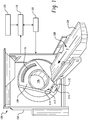
- the rotating gantry 104 supports a radiation source 110, such as an x-ray tube.
- the radiation source 110 includes an eight degree (8°) anode (not visible) and emits radiation from a focal spot (not visible) thereon.
- the emitted radiation traverses the examination region 106 and any object or subject dispose therein.
- the focal spot emits radiation while the rotating gantry 104, and hence the radiation source 110 and the focal spot, rotate around the z-axis 108 during a helical or axial scan or are at a static position for a pilot or scout scan.
- the focal spot can translate along the z-axis physically, for example, by translating the radiation source 110 along the z-axis, and/or electronically.
- the focal spot translates in coordination with motion of an object such as an organ like the heart disposed within the examination region 106 or a flow of a contrast agent or the like through an object disposed within the examination region 106.
- Such coordination can be in connection with fly-by scanning in which the radiation source 110 physically translates along the z-axis 108. Examples of such scanning are discussed at least in patent application serial no. PCT/US07/78130, filed September 11, 2007 , and entitled “Fly-By Scanning," and published as WO2008/042564 .
- a source collimator 112 translates in coordination with the radiation source 110 and collimates the emitted radiation in the z-axis direction to produce a generally conical shaped radiation beam that traverses the examination region 106.
- the collimator 112 includes at least one collimator blade 114 configured to translate in the z-axis direction, relative to the radiation source 110.
- the illustrated scanner 100 includes N collimator blades 114, wherein N is two (2). In other embodiments, N can be more or less than two (2).
- the source collimator 112 is employed to produce a cone beam, which has a cone angle along the z-axis that defines the z-axis width or scan coverage.
- the source collimator 112 increases the effective maximum cone-beam angle in both directions along the z-axis. This may include symmetrically or asymmetrically increasing the cone beam angle in both directions along the z-axis about an imaginary axis that extends perpendicularly from the focal spot through a center region of the examination region 106 through the z-axis 108.
- the z-axis coverage can be expanded while maintaining the same radiation source power, relative to a configuration where the cone beam angle is not increased beyond the effective maximum cone-beam angle, which generally is defined by the angle of the anode (not visible) of the radiation source 110.
- a controller 115 controls the source collimator 112 based on a scan protocol, including, but not limited to, a fly by scan or other scan protocol, and/or otherwise.
- the rotating gantry 104 also supports a radiation sensitive detector array 116, which is disposed about the rotating gantry portion 104 and subtends an angular arc opposite the radiation source 110.
- the detector array 116 includes a multi-slice detector having a plurality of detector elements extending in the axial and transverse directions. Each detector element detects radiation emitted by the radiation source 110 that traverses the examination region 106 for at least one hundred and eighty degrees (180°) plus a fan angle of data, and generates a corresponding output signal or projection data indicative of the detected radiation.
- a non-limiting example of a suitable detector includes a tile detector as are described in US patent 6,510,195 B1 to Chappo et al., filed July 18, 2001 , and entitled "Solid State X-Radiation Detector Modules and Mosaics Thereof, and an Imaging Method and Apparatus Employing the Same".
- a one or two dimensional anti-scatter grid may be employed in connection with the detector array 116 to mitigate or reduce detection of scatter radiation.
- the projection data generated by the detector array 116 are conveyed to a reconstructor 118, which reconstructs the projections and generates volumetric image data.
- the image data can be processed by an image processor to generate one or more images of the scanned region of interest or a subset thereof.
- a data corrector 120 can be used to correct the data for reconstruction. In one instance, this includes applying a correction that reduces the heel effect by providing a correction for increased attenuation and/or beam hardening. Such a correction may be used when the employed cone beam angle exceeds the effective maximum cone beam angle, as defined by the anode angle. As the heel effect varies along the z-axis, the correction may vary along the z-axis, with the largest correction generally being applied to the end ray(s) on the heel side of the cone beam.
- An operator console 122 facilitates user interaction with the imaging system 100.
- Software applications executed by the operator console 122 allow the user to configure and/or control operation of the imaging system 100. For instance, the user can interact with the operator console 122 to select a fly-by or other scan protocol, and initiate, pause and/or terminate a scan, etc.
- a couch or patient support 124 supports an object or subject such as a human or animal within the examination region 106.
- the support 124 can be movable, which enables an operator or the system to suitably position the object or subject within the examination region 106 before, during and/or after scanning.
- the above system 100 can be employed for various applications.
- the system 100 is suited for whole organ scanning, such as scanning the entire heart or a substantial portion thereof in a single heart beat, if desired.
- a scan may be a fly-by or other scan performed with a radiation source having an eight degree (8°) or other angle anode, using full radiation source power for coverage between eight (8) to forty (40) centimeters (cm), such as 8 cm, 9 cm, 10 cm, 11 cm, 12 cm, etc. scans with minimal to substantially no motion artifacts and minimal to substantially no cone beam artifacts.
- a fly-by scan sufficient coverage can be obtained without sacrificing power and image quality.
- the illustrated radiation source 110 can include an eight degree (8°) anode.
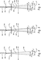
- FIGURE 2 illustrates such an anode 202 in connection with collimator blades 114 1 and 114 2 .
- the collimator blades 114 1 and 114 2 are opened to form a cone angle of eight degrees (8°), with a magnitude of four (4) degrees on each side of an axis 204, which extends perpendicularly from a focal spot 206 through the z-axis 108.
- the detector array 116 includes one hundred and twenty eight (128) rows of detector pixels along the z-axis 108, where each pixel is about 0.625 mm. Of course, more or less rows and/or other size pixels can be used in other embodiments.
- the anode angle defines an effective maximum cone angle of a cone beam 208 that is symmetric about the axis 204, as the heel effect attenuates and hardens beam rays emanating from the focal spot 206 on the anode side of the axis 204 when the cone angle is greater than the effective maximum cone angle.
- the intensity of the rays substantially falls off and the rays are substantially attenuated such that the intensity and energy of the rays generally are not effective for CT applications.
- Such data does not render diagnostic data while still irradiating the patient.
- this angle is about eight degrees (8°), or four degrees (4°) on each side of the axis 204.
- the collimator blades 114 can be positioned to substantially or completely collimate rays emanating from the focal spot 206 at an angle greater than the effective maximum cone angle.
- the radiation source 110 can be operated at full power, with local heating from an electron beam 210 spread about the anode 202 as the anode 202 rotates and spread radially along the anode 202 slope.
- the effective focal spot size, as seen at the detector array 116 is small enough so as to maintain a suitable resolution along the z-axis, while providing suitable z-axis coverage and maximizing radiation source power.
- the collimator blades 114 1 and 114 2 of the system 100 can be symmetrically opened to allow for a larger angle cone beam.
- the collimator blades 114 1 and 114 2 are opened to increase the cone angle from eight degrees (8°), as shown in FIGURE 2 , to about ten degrees (10°), with five degrees (5°) on each side of the axis 204.
- a resulting beam 302 has an expanded width along the z-axis, allowing for more rows of detector pixels, such as, for example, one hundred and sixty (160) rows of detector pixels, where each pixel is about 0.625 mm.
- helical cone beam scans with extended z-axis coverage for example, up to ten centimeters (10 cm) can be performed while maintaining radiation source power, without introducing significantly greater cone beam artifacts and/or decreasing resolution.
- scan time and/or motion artifacts can be reduced.
- more or less rows and/or other size pixels can be used in other embodiments.
- the coverage is smaller, and with a system with a larger anode angle so as to achieve 10 cm of coverage, tube power must reduced, which may reduce image quality.
- the collimator blades 114 1 and 114 2 are asymmetrically opened to allow for a larger angle cone beam.
- the collimator blades 114 1 and 114 2 are asymmetrically opened to increase the cone angle from eight degrees (8°), as shown in FIGURE 2 , to twelve degrees (12°), with five degrees (5°) on the heel side of the axis 204 and seven degrees (7°) on the other side of the axis 204.
- a resulting beam 402 has an expanded width along the z-axis, allowing for more rows of detector pixels, such as, for example, one hundred and ninety-two (192) rows of detector pixels, where each pixel is about 0.625 mm.
- helical cone beam scans with extended z-axis coverage can be performed while maintaining radiation source power, without significantly increasing cone beam artifacts and/or decreasing resolution, and possibly reducing scan time and/or motion artifacts.
- the extended cone beam provides fifty percent (50%) more coverage for the same radiation source power used in connection with the embodiment of FIGURE 2 .
- more or less rows and/or other size pixels can be used in other embodiments.
- the coverage is smaller, and with a system with a larger anode angle so as to achieve 12 cm of coverage, tube power must reduced, which may reduce image quality.
- only the collimator blade 114 1 on the anode 202 side is opened to increase the cone beam angel larger then the effective maximum cone beam angle as determined by the heel effect and anode angle.
- the collimator blade 114 1 may be opened from one (1) to three (3) centimeters. Again, relative to the configuration shown in FIGURE 2 , the resulting beam has an expanded width along the z-axis. Furthermore, a beam hardening or heel correction is applied to compensate for beam hardening artifact.
- the above system 100 can be employed for various applications.
- the system 100 is suited for whole organ scanning, such as scanning the entire heart or a substantial portion thereof in a single heart beat, if desired.
- a scan may be a fly-by or other scan performed with a radiation source having an eight degree (8°) or other angle anode, using full radiation source power for coverage between eight centimeters (8 cm) to forty centimeters (40 cm), such as 8 cm, 9 cm, 10 cm, 11 cm, 12 cm, etc. centimeters scans with minimal to substantially no motion artifacts and minimal to substantially no cone beam artifacts.
- a fly-by scan sufficient coverage can be obtained without sacrificing power and image quality.
- radiation sources with larger or smaller anode angles and/or cone beams with larger or smaller symmetric or asymmetric cone angles are contemplated.
- suitable anode angles range from seven (7) to twelve (12) degree angles correspond to suitable extended cone beam angles up to ten (10) to twenty (20) degree angles, etc.
- the imaging system 100 can be used for various applications, including helical and/or axial scans.
- cone beam artifacts are smaller (e.g., less than twenty percent (20%)) for helical scans relative to axial scans, and for some helical scans, the cone beam artifacts are minimal.
- FIGURE 5 shows a cone beam 502 at a first position 504 and a second position 506, which is one hundred and eighty (180) degrees offset from the first position 504.
- the anode angle is eight degrees (8°) and the collimator blades 114 are positioned so that the cone beam has a twelve degree (12°) angle, as shown in connection with FIGURE 4 above.
- the pitch factor is one and three quarters (1.75).
- a distance 508 between a ray 510 extending perpendicularly from a focal spot through the z-axis 108 at the positions 504, 506 is about ten and a half centimeters (10.5 cm), and a relative angle 512 between the beam 502 at the two positions 504, 506 is less than one and eight tenths degrees (1.8°).
- Slice sensitivity is also generally more uniform for helical scans relative to axial scans. That is, for a given voxel in a helical reconstruction, the z-axis response will vary from about sixty percent (60%) of a nominal size to about one hundred and eighty-eight percent (188%) of the nominal size when the collimator blades 114 are positioned for a cone beam with an extended cone angle of twelve degrees (12°), as illustrated in FIGURE 4 .
- the size of a given voxel at the center of the cone will be about half a millimeter (0.5 mm), whereas the size of the voxel on the heel side of the axis 204 will be about three tenths of a millimeter (0.3 mm) and the size of the voxel on the other side of the axis will be about nine tenths of a millimeter (0.9 mm).
- the nominal size of the voxel will be the composite of the foregoing responses. As such, all voxels may have about the same nominal slice width sensitivity.
- the collimator blades 114 can be positioned to increase z-axis coverage without reducing or sacrificing radiation source power or resolution or significantly increasing cone beam artifacts, noting that a heel effect correction may be performed on the data to correct for both increased attenuation and beam hardening, relative to an embodiment in which the collimator blades 114 are positioned based on the effective maximum cone angle defined by the anode angle, which assumes a maximum heel angle of 4 degrees for an 8 degree anode.
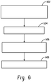
- Example operation of the system 100 is discussed in connection with FIGURE 6 . It is to be appreciated that the order of the following acts is not limiting and may otherwise occur. In addition, more or less, including similar or different acts may be employed in other embodiments.
- a cone beam having an extended cone angle along the z-axis and thus an extended z-axis scan coverage is produced.
- this may include symmetrically or asymmetrically opening the collimator blades 114 about the central ray axis 204 so as to produce a cone beam with a cone angle that is larger than the effective maximum cone angle determined by the anode angle.
- a scan is performed with the cone beam.
- a scan may be a helical scan such as a fly by or other scan, an axial scan, and/or another scan.
- the scan can involve scanning an entire organ, such as a static or moving organ, without having to move or substantially move the radiation source 110 and/or the object or subject being scanned.
- the radiation source power is maintained at a power level such as full power as if the cone beam angle is not extended, but equal to the effective maximum cone beam angle. In one instance, this may be defined by 2( ⁇ -4).
- an optional heel effect correction is applied.
- Such a correction can be applied to the raw data, the reconstructed volumetric image data, and/or one or more images generated as part of the reconstructed volumetric image data.
- the above may be implemented by way of computer readable instructions, which when executed by a computer processor(s), cause the processor(s) to carry out the foregoing.
- the instructions are stored in a computer readable storage medium associated with or otherwise accessible to a relevant computer, such as a dedicated workstation, a home computer, a distributed computing system, the console, and/or other computer.
- a relevant computer such as a dedicated workstation, a home computer, a distributed computing system, the console, and/or other computer.
- the acts need not be performed concurrently with data acquisition.
- the above may be used in various applications including applications where it may be desirable to achieve relatively large coverage in a relatively short period of time without moving the patient support such as for cardiac, trauma, perfusion and/or other applications.
Landscapes
- Health & Medical Sciences (AREA)
- Life Sciences & Earth Sciences (AREA)
- Engineering & Computer Science (AREA)
- Medical Informatics (AREA)
- Pathology (AREA)
- Heart & Thoracic Surgery (AREA)
- High Energy & Nuclear Physics (AREA)
- Physics & Mathematics (AREA)
- Nuclear Medicine, Radiotherapy & Molecular Imaging (AREA)
- Optics & Photonics (AREA)
- Veterinary Medicine (AREA)
- Radiology & Medical Imaging (AREA)
- Biomedical Technology (AREA)
- Biophysics (AREA)
- Molecular Biology (AREA)
- Surgery (AREA)
- Animal Behavior & Ethology (AREA)
- General Health & Medical Sciences (AREA)
- Public Health (AREA)
- Computer Vision & Pattern Recognition (AREA)
- Pulmonology (AREA)
- Theoretical Computer Science (AREA)
- Apparatus For Radiation Diagnosis (AREA)
Claims (11)
- Bildgebungssystem, wobei das Bildgebungssystem ein medizinischer Computertomographie-Scanner ist, umfassend:eine Strahlungsquelle (110), einschließlich einer Anode (202), die dafür ausgelegt sich, sich um eine Untersuchungsregion (106) um eine Längsachse (108) zu drehen und Strahlung von einem Brennfleck (206) aus auf die Anode (202) zu emittieren;wobei die genannte Anode einen Anodenwinkel hat, gekennzeichnet durch einen Quellenkollimator (112), der dafür ausgelegt ist, die emittierte Strahlung zu kollimieren, um ein konisch geformtes Strahlenbündel zu erzeugen, das die Untersuchungsregion durchquert, wobei das konisch geformte Strahlenbündel einen erweiterten Kegelwinkel entlang der Längsachse hat, der größer als ein effektiver maximaler Kegelwinkel ist, der durch eine Begrenzung des Kegelwinkels durch den Heel-Effekt definiert wird, wobei, wenn der Kegelwinkel über den effektiven maximalen Kegelwinkel hinaus zunimmt, die Intensität der Strahlen abnimmt und die Strahlen aufgrund des Heel-Effekts abgeschwächt werden, der die von dem Brennfleck (206) auf der Anodenseite einer Achse (204, 510), die sich senkrecht von dem Brennfleck aus durch die Längsachse erstreckt, ausgehenden Strahlen abschwächt und härtet, wobei der Kollimator positioniert ist, um die von dem Brennfleck ausgehende emittierte Strahlung derartig zu kollimieren, dass der genannte erweiterte Kegelwinkel auf der Heel-Seite größer ist als der Anodenwinkel;ein Detektorarray (116), das dafür ausgelegt ist, Strahlung zu detektieren, die die Untersuchungsregion durchquert, und diese Strahlung angebende Signale zu erzeugen; undeine Rekonstruktionseinheit (118), die dafür ausgelegt ist, die Signale zu rekonstruieren, um volumetrische Bilddaten zu erzeugen, die die Untersuchungsregion angeben; wobeider medizinische Computertomographie-Scanner weiterhin eine Datenkorrektureinheit (120) umfasst, die dafür ausgelegt ist, das Signal hinsichtlich des Heel-Effekts zu korrigieren, wobei die Datenkorrektureinheit dafür ausgelegt ist, die Korrektur sowohl hinsichtlich der erhöhten Abschwächung als auch hinsichtlich der Härtung des Strahlenbündels durchzuführen.
- System nach Anspruch 1, wobei der erweiterte Kegelwinkel symmetrisch in beiden Richtungen entlang der Längsachse erweitert wird.
- System nach Anspruch 1, wobei der erweiterte Kegelwinkel asymmetrisch in beiden Richtungen entlang der Längsachse erweitert wird.
- System nach einem der Ansprüche 1 bis 3, wobei die Strahlungsquelle (110) dafür ausgelegt ist, sich während eines Fly-by-Scans physikalisch entlang der Längsachse (108) zu verschieben.
- System nach einem der Ansprüche 1 bis 4, wobei der effektive maximale Kegelwinkel ungefähr acht Grad beträgt und der erweiterte Kegelwinkel ungefähr zehn bis ungefähr zwölf Grad beträgt.
- System nach Anspruch 5, wobei der erweiterte Kegelwinkel in der Richtung der Anode um ungefähr ein Grad und in der Richtung der Kathode um ungefähr ein bis ungefähr drei Grad erweitert wird.
- System nach einem der Ansprüche 1 bis 6, wobei das System dafür ausgelegt ist, eine spiralförmige Abtastung durchzuführen und mindestens zwei Voxel die gleiche nominale Schichtbreitenempfindlichkeit haben.
- Verfahren, wobei das Verfahren ein Verfahren zum Erfassen von Projektionsdaten zur Verwendung beim Rekonstruieren von volumetrischen Bilddaten der medizinischen Computertomographie umfasst, die eine Untersuchungsregion angeben, wobei das Verfahren dadurch gekennzeichnet ist, dass es Folgendes umfasst:Kollimieren eines von einem Brennfleck (206) aus auf eine Anode (202) emittierten Strahlenbündels, wobei die genannte Anode einen Anodenwinkel hat, eines Strahlungsquelle (110) eines Bildgebungssystems (100), um ein konisch geformtes Strahlenbündel zu erzeugen, das einen erweiterten Kegelwinkel entlang der Längsachse hat, der größer als ein effektiver maximaler Kegelwinkel ist, wobei der effektive Kegelwinkel durch eine Begrenzung des Kegelwinkels durch den Heel-Effekt definiert wird, wobei, wenn der Kegelwinkel über den effektiven maximalen Kegelwinkel hinaus zunimmt, die Intensität der Strahlen abnimmt und die Strahlen aufgrund des Heel-Effekts abgeschwächt werden, der die von dem Brennfleck (206) auf der Anodenseite einer Achse (204), die sich senkrecht von dem Brennfleck aus durch die Längsachse erstreckt, ausgehenden Strahlen abschwächt und härtet, wobei der Kollimator positioniert ist, um die von dem Brennfleck ausgehende emittierte Strahlung derartig zu kollimieren, dass der genannte erweiterte Kegelwinkel auf der Heel-Seite größer ist als der Anodenwinkel; undErfassen von Projektionsdaten, die die Strahlung angeben, welche eine Untersuchungsregion (106) durchquert und ein Detektorarray (116) beleuchtet;wobei eine Datenkorrektureinheit die Projektionsdaten hinsichtlich des Heel-Effekts korrigiert, wobei die Datenkorrektureinheit dafür ausgelegt ist, die Korrektur sowohl hinsichtlich der erhöhten Abschwächung als auch hinsichtlich der Härtung des Strahlenbündels durchzuführen.
- Verfahren nach Anspruch 8, weiterhin umfassend eine symmetrische Erweiterung des Kegelwinkels in beiden Richtungen entlang der Längsachse.
- Verfahren nach Anspruch 8, weiterhin umfassend eine asymmetrische Erweiterung des Kegelwinkels in beiden Richtungen entlang der Längsachse.
- Computerlesbares Medium, dadurch gekennzeichnet, dass das computerlesbare Medium mit computerlesbaren Anweisungen codiert ist, die, wenn sie durch (einen) Computerprozessor(en) ausgeführt werden, den (die) Prozessor(en) veranlassen zum:Bereitstellen eines Steuersignals, um die Kollimatorlamellen (114) eines Kollimators (112) so zu positionieren, dass ein von einem Brennfleck (206) auf eine Anode (202) einer Strahlungsquelle (110) eines medizinischen Computertomographie-Scanners (100) emittiertes Strahlenbündel kollimiert wird, wobei die genannte Anode einen Anodenwinkel hat, um selektiv ein konisch geformtes Strahlenbündel zu erzeugen, das alternativ einen symmetrisch oder asymmetrisch erweiterten Kegelwinkel entlang einer Längsachse hat, der größer als ein effektiver maximaler Kegelwinkel ist, der durch eine Begrenzung des Kegelwinkels durch den Heel-Effekt definiert wird, wobei, wenn der Kegelwinkel über den effektiven maximalen Kegelwinkel hinaus zunimmt, die Intensität der Strahlen abnimmt und die Strahlen aufgrund des Heel-Effekts abgeschwächt werden, der die von dem Brennfleck (206) auf der Anodenseite einer Achse (204), die sich senkrecht von dem Brennfleck aus durch die Längsachse erstreckt, ausgehenden Strahlen abschwächt und härtet, wobei der Kollimator positioniert ist, um die von dem Brennfleck ausgehende emittierte Strahlung derartig zu kollimieren, dass der genannte erweiterte Kegelwinkel auf der Heel-Seite größer ist als der Anodenwinkel, wobei Projektionsdaten, die die Strahlung angeben, welche eine Untersuchungsregion durchquert hat und ein Detektorarray beleuchtet, hinsichtlich des Heel-Effekts korrigiert werden, wobei die Projektionsdaten sowohl hinsichtlich der erhöhten Abschwächung als auch hinsichtlich der Härtung des Strahlenbündels korrigiert werden.
Applications Claiming Priority (2)
| Application Number | Priority Date | Filing Date | Title |
|---|---|---|---|
| US11229108P | 2008-11-07 | 2008-11-07 | |
| PCT/IB2009/054790 WO2010052614A1 (en) | 2008-11-07 | 2009-10-28 | Cone beam z-axis coverage |
Publications (2)
| Publication Number | Publication Date |
|---|---|
| EP2352432A1 EP2352432A1 (de) | 2011-08-10 |
| EP2352432B1 true EP2352432B1 (de) | 2017-02-22 |
Family
ID=41571371
Family Applications (1)
| Application Number | Title | Priority Date | Filing Date |
|---|---|---|---|
| EP09760600.8A Active EP2352432B1 (de) | 2008-11-07 | 2009-10-28 | Konusstrahl-z-achsen-abdeckung |
Country Status (4)
| Country | Link |
|---|---|
| US (1) | US8467494B2 (de) |
| EP (1) | EP2352432B1 (de) |
| CN (1) | CN102202578B (de) |
| WO (1) | WO2010052614A1 (de) |
Families Citing this family (6)
| Publication number | Priority date | Publication date | Assignee | Title |
|---|---|---|---|---|
| WO2009141766A2 (en) * | 2008-05-21 | 2009-11-26 | Koninklijke Philips Electronics, N.V. | Dynamic adjustable source collimation during fly-by scanning |
| US8566619B2 (en) * | 2009-12-30 | 2013-10-22 | International Business Machines Corporation | Cooling appliance rating aware data placement |
| US9295434B2 (en) * | 2011-07-15 | 2016-03-29 | Koninklijke Philips N.V. | Dynamic collimation |
| JP6615439B2 (ja) * | 2014-08-01 | 2019-12-04 | キヤノンメディカルシステムズ株式会社 | X線ct装置 |
| EP3632325A1 (de) * | 2018-10-04 | 2020-04-08 | Koninklijke Philips N.V. | System zur bereitstellung eines spektralbildes |
| CN110368018A (zh) * | 2019-08-22 | 2019-10-25 | 南京安科医疗科技有限公司 | 一种ct系统扫描动态调节方法 |
Citations (2)
| Publication number | Priority date | Publication date | Assignee | Title |
|---|---|---|---|---|
| US20050123100A1 (en) * | 2003-12-05 | 2005-06-09 | Jiang Hsieh | Method and system for target angle heel effect compensation |
| WO2005059592A1 (en) * | 2003-12-16 | 2005-06-30 | Philips Intellectual Property & Standards Gmbh | Correction of artifacts caused by the heel effect |
Family Cites Families (7)
| Publication number | Priority date | Publication date | Assignee | Title |
|---|---|---|---|---|
| US6330299B1 (en) | 2000-06-10 | 2001-12-11 | Ge Medical Systems Global Technology Company, Llc | System and method for determining dose area product in an X-ray imaging system |
| US6510195B1 (en) * | 2001-07-18 | 2003-01-21 | Koninklijke Philips Electronics, N.V. | Solid state x-radiation detector modules and mosaics thereof, and an imaging method and apparatus employing the same |
| DE60234538D1 (de) * | 2002-05-06 | 2010-01-07 | Koninkl Philips Electronics Nv | Hochauflösender ct-scanner |
| WO2004075118A1 (en) | 2003-02-20 | 2004-09-02 | Koninklijke Philips Electronics N.V. | Asymmetric cone beam |
| JP4487032B2 (ja) * | 2004-03-29 | 2010-06-23 | 独立行政法人放射線医学総合研究所 | ヒール効果補正フィルタ、x線照射装置、x線ct装置及びx線ct撮像方法 |
| GB2422759B (en) | 2004-08-05 | 2008-07-16 | Elekta Ab | Rotatable X-ray scan apparatus with cone beam offset |
| WO2008042564A1 (en) | 2006-09-29 | 2008-04-10 | Koninklijke Philips Electronics N. V. | Fly-by scanning |
-
2009
- 2009-10-28 WO PCT/IB2009/054790 patent/WO2010052614A1/en not_active Ceased
- 2009-10-28 CN CN200980144121.0A patent/CN102202578B/zh active Active
- 2009-10-28 US US13/127,241 patent/US8467494B2/en not_active Expired - Fee Related
- 2009-10-28 EP EP09760600.8A patent/EP2352432B1/de active Active
Patent Citations (2)
| Publication number | Priority date | Publication date | Assignee | Title |
|---|---|---|---|---|
| US20050123100A1 (en) * | 2003-12-05 | 2005-06-09 | Jiang Hsieh | Method and system for target angle heel effect compensation |
| WO2005059592A1 (en) * | 2003-12-16 | 2005-06-30 | Philips Intellectual Property & Standards Gmbh | Correction of artifacts caused by the heel effect |
Non-Patent Citations (1)
| Title |
|---|
| MALTZ J S ET AL: "Algorithm for X-ray Scatter, Beam-Hardening, and Beam Profile Correction in Diagnostic (Kilovoltage) and Treatment (Megavoltage) Cone Beam CT", IEEE TRANSACTIONS ON MEDICAL IMAGING, IEEE SERVICE CENTER, PISCATAWAY, NJ, US, vol. 27, no. 12, 1 December 2008 (2008-12-01), pages 1791 - 1810, XP011232055, ISSN: 0278-0062, DOI: 10.1109/TMI.2008.928922 * |
Also Published As
| Publication number | Publication date |
|---|---|
| US8467494B2 (en) | 2013-06-18 |
| WO2010052614A1 (en) | 2010-05-14 |
| CN102202578B (zh) | 2014-08-06 |
| US20110211664A1 (en) | 2011-09-01 |
| CN102202578A (zh) | 2011-09-28 |
| EP2352432A1 (de) | 2011-08-10 |
Similar Documents
| Publication | Publication Date | Title |
|---|---|---|
| US7515678B2 (en) | Method and system for performing CT image reconstruction with motion artifact correction | |
| US6373920B1 (en) | Method and apparatus for acquiring CT perfusion images | |
| JP5905694B2 (ja) | 広いカバー範囲及び低線量での心ct撮像のための動的コリメータを備えた計算機式断層写真法スキャナ | |
| EP2234541B1 (de) | Stereoröhren-dämpfungsfilter | |
| US8031828B1 (en) | Method and apparatus for computed tomography | |
| JP6014323B2 (ja) | X線システム | |
| US6421411B1 (en) | Methods and apparatus for helical image artifact reduction | |
| US6421412B1 (en) | Dual cardiac CT scanner | |
| EP1149558A2 (de) | Mehrschichtcomputertomographiegerät für ein ausgewähltes Gebiet | |
| US9042514B2 (en) | Dose reduction via dynamic collimation adjustment for targeted field of view and/or digital tilt CT | |
| US20060193430A1 (en) | Computerized tomographic imaging system | |
| US20080279328A1 (en) | Systems and Methods Using X-Ray Tube Spectra For Computed Tomography Applications | |
| EP2352432B1 (de) | Konusstrahl-z-achsen-abdeckung | |
| JP2003502130A (ja) | X線被曝を制限した局所的ct画像再構成 | |
| US7532702B2 (en) | Method and system for performing CT image reconstruction with motion artifact correction | |
| EP1959835B1 (de) | Systeme und verfahren zum scannen und zur datenerfassung bei der computer-tomographie (ct) | |
| JP2008012206A (ja) | X線断層撮影装置 | |
| Grasruck et al. | Evaluation of image quality and dose on a flat-panel CT-scanner | |
| WO2008075267A2 (en) | Device and method for imaging an object | |
| US20060243914A1 (en) | Attenuation map generation from pet scans | |
| Obaid | Medical Imaging |
Legal Events
| Date | Code | Title | Description |
|---|---|---|---|
| PUAI | Public reference made under article 153(3) epc to a published international application that has entered the european phase |
Free format text: ORIGINAL CODE: 0009012 |
|
| 17P | Request for examination filed |
Effective date: 20110607 |
|
| AK | Designated contracting states |
Kind code of ref document: A1 Designated state(s): AT BE BG CH CY CZ DE DK EE ES FI FR GB GR HR HU IE IS IT LI LT LU LV MC MK MT NL NO PL PT RO SE SI SK SM TR |
|
| DAX | Request for extension of the european patent (deleted) | ||
| 17Q | First examination report despatched |
Effective date: 20120321 |
|
| RAP1 | Party data changed (applicant data changed or rights of an application transferred) |
Owner name: KONINKLIJKE PHILIPS N.V. |
|
| GRAJ | Information related to disapproval of communication of intention to grant by the applicant or resumption of examination proceedings by the epo deleted |
Free format text: ORIGINAL CODE: EPIDOSDIGR1 |
|
| GRAP | Despatch of communication of intention to grant a patent |
Free format text: ORIGINAL CODE: EPIDOSNIGR1 |
|
| GRAP | Despatch of communication of intention to grant a patent |
Free format text: ORIGINAL CODE: EPIDOSNIGR1 |
|
| INTG | Intention to grant announced |
Effective date: 20160909 |
|
| GRAS | Grant fee paid |
Free format text: ORIGINAL CODE: EPIDOSNIGR3 |
|
| GRAA | (expected) grant |
Free format text: ORIGINAL CODE: 0009210 |
|
| AK | Designated contracting states |
Kind code of ref document: B1 Designated state(s): AT BE BG CH CY CZ DE DK EE ES FI FR GB GR HR HU IE IS IT LI LT LU LV MC MK MT NL NO PL PT RO SE SI SK SM TR |
|
| REG | Reference to a national code |
Ref country code: GB Ref legal event code: FG4D |
|
| REG | Reference to a national code |
Ref country code: CH Ref legal event code: EP |
|
| REG | Reference to a national code |
Ref country code: AT Ref legal event code: REF Ref document number: 868652 Country of ref document: AT Kind code of ref document: T Effective date: 20170315 |
|
| REG | Reference to a national code |
Ref country code: IE Ref legal event code: FG4D |
|
| REG | Reference to a national code |
Ref country code: DE Ref legal event code: R096 Ref document number: 602009044335 Country of ref document: DE |
|
| REG | Reference to a national code |
Ref country code: DE Ref legal event code: R084 Ref document number: 602009044335 Country of ref document: DE |
|
| REG | Reference to a national code |
Ref country code: LT Ref legal event code: MG4D |
|
| REG | Reference to a national code |
Ref country code: NL Ref legal event code: MP Effective date: 20170222 |
|
| REG | Reference to a national code |
Ref country code: AT Ref legal event code: MK05 Ref document number: 868652 Country of ref document: AT Kind code of ref document: T Effective date: 20170222 |
|
| PG25 | Lapsed in a contracting state [announced via postgrant information from national office to epo] |
Ref country code: HR Free format text: LAPSE BECAUSE OF FAILURE TO SUBMIT A TRANSLATION OF THE DESCRIPTION OR TO PAY THE FEE WITHIN THE PRESCRIBED TIME-LIMIT Effective date: 20170222 Ref country code: LT Free format text: LAPSE BECAUSE OF FAILURE TO SUBMIT A TRANSLATION OF THE DESCRIPTION OR TO PAY THE FEE WITHIN THE PRESCRIBED TIME-LIMIT Effective date: 20170222 Ref country code: FI Free format text: LAPSE BECAUSE OF FAILURE TO SUBMIT A TRANSLATION OF THE DESCRIPTION OR TO PAY THE FEE WITHIN THE PRESCRIBED TIME-LIMIT Effective date: 20170222 Ref country code: NO Free format text: LAPSE BECAUSE OF FAILURE TO SUBMIT A TRANSLATION OF THE DESCRIPTION OR TO PAY THE FEE WITHIN THE PRESCRIBED TIME-LIMIT Effective date: 20170522 Ref country code: GR Free format text: LAPSE BECAUSE OF FAILURE TO SUBMIT A TRANSLATION OF THE DESCRIPTION OR TO PAY THE FEE WITHIN THE PRESCRIBED TIME-LIMIT Effective date: 20170523 |
|
| PG25 | Lapsed in a contracting state [announced via postgrant information from national office to epo] |
Ref country code: AT Free format text: LAPSE BECAUSE OF FAILURE TO SUBMIT A TRANSLATION OF THE DESCRIPTION OR TO PAY THE FEE WITHIN THE PRESCRIBED TIME-LIMIT Effective date: 20170222 Ref country code: BG Free format text: LAPSE BECAUSE OF FAILURE TO SUBMIT A TRANSLATION OF THE DESCRIPTION OR TO PAY THE FEE WITHIN THE PRESCRIBED TIME-LIMIT Effective date: 20170522 Ref country code: SE Free format text: LAPSE BECAUSE OF FAILURE TO SUBMIT A TRANSLATION OF THE DESCRIPTION OR TO PAY THE FEE WITHIN THE PRESCRIBED TIME-LIMIT Effective date: 20170222 Ref country code: NL Free format text: LAPSE BECAUSE OF FAILURE TO SUBMIT A TRANSLATION OF THE DESCRIPTION OR TO PAY THE FEE WITHIN THE PRESCRIBED TIME-LIMIT Effective date: 20170222 Ref country code: LV Free format text: LAPSE BECAUSE OF FAILURE TO SUBMIT A TRANSLATION OF THE DESCRIPTION OR TO PAY THE FEE WITHIN THE PRESCRIBED TIME-LIMIT Effective date: 20170222 Ref country code: PT Free format text: LAPSE BECAUSE OF FAILURE TO SUBMIT A TRANSLATION OF THE DESCRIPTION OR TO PAY THE FEE WITHIN THE PRESCRIBED TIME-LIMIT Effective date: 20170622 Ref country code: ES Free format text: LAPSE BECAUSE OF FAILURE TO SUBMIT A TRANSLATION OF THE DESCRIPTION OR TO PAY THE FEE WITHIN THE PRESCRIBED TIME-LIMIT Effective date: 20170222 |
|
| PG25 | Lapsed in a contracting state [announced via postgrant information from national office to epo] |
Ref country code: EE Free format text: LAPSE BECAUSE OF FAILURE TO SUBMIT A TRANSLATION OF THE DESCRIPTION OR TO PAY THE FEE WITHIN THE PRESCRIBED TIME-LIMIT Effective date: 20170222 Ref country code: IT Free format text: LAPSE BECAUSE OF FAILURE TO SUBMIT A TRANSLATION OF THE DESCRIPTION OR TO PAY THE FEE WITHIN THE PRESCRIBED TIME-LIMIT Effective date: 20170222 Ref country code: RO Free format text: LAPSE BECAUSE OF FAILURE TO SUBMIT A TRANSLATION OF THE DESCRIPTION OR TO PAY THE FEE WITHIN THE PRESCRIBED TIME-LIMIT Effective date: 20170222 Ref country code: CZ Free format text: LAPSE BECAUSE OF FAILURE TO SUBMIT A TRANSLATION OF THE DESCRIPTION OR TO PAY THE FEE WITHIN THE PRESCRIBED TIME-LIMIT Effective date: 20170222 Ref country code: SK Free format text: LAPSE BECAUSE OF FAILURE TO SUBMIT A TRANSLATION OF THE DESCRIPTION OR TO PAY THE FEE WITHIN THE PRESCRIBED TIME-LIMIT Effective date: 20170222 |
|
| REG | Reference to a national code |
Ref country code: FR Ref legal event code: PLFP Year of fee payment: 9 |
|
| REG | Reference to a national code |
Ref country code: DE Ref legal event code: R097 Ref document number: 602009044335 Country of ref document: DE |
|
| PG25 | Lapsed in a contracting state [announced via postgrant information from national office to epo] |
Ref country code: PL Free format text: LAPSE BECAUSE OF FAILURE TO SUBMIT A TRANSLATION OF THE DESCRIPTION OR TO PAY THE FEE WITHIN THE PRESCRIBED TIME-LIMIT Effective date: 20170222 Ref country code: SM Free format text: LAPSE BECAUSE OF FAILURE TO SUBMIT A TRANSLATION OF THE DESCRIPTION OR TO PAY THE FEE WITHIN THE PRESCRIBED TIME-LIMIT Effective date: 20170222 Ref country code: DK Free format text: LAPSE BECAUSE OF FAILURE TO SUBMIT A TRANSLATION OF THE DESCRIPTION OR TO PAY THE FEE WITHIN THE PRESCRIBED TIME-LIMIT Effective date: 20170222 |
|
| PLBE | No opposition filed within time limit |
Free format text: ORIGINAL CODE: 0009261 |
|
| STAA | Information on the status of an ep patent application or granted ep patent |
Free format text: STATUS: NO OPPOSITION FILED WITHIN TIME LIMIT |
|
| 26N | No opposition filed |
Effective date: 20171123 |
|
| PG25 | Lapsed in a contracting state [announced via postgrant information from national office to epo] |
Ref country code: SI Free format text: LAPSE BECAUSE OF FAILURE TO SUBMIT A TRANSLATION OF THE DESCRIPTION OR TO PAY THE FEE WITHIN THE PRESCRIBED TIME-LIMIT Effective date: 20170222 |
|
| PG25 | Lapsed in a contracting state [announced via postgrant information from national office to epo] |
Ref country code: MC Free format text: LAPSE BECAUSE OF FAILURE TO SUBMIT A TRANSLATION OF THE DESCRIPTION OR TO PAY THE FEE WITHIN THE PRESCRIBED TIME-LIMIT Effective date: 20170222 |
|
| REG | Reference to a national code |
Ref country code: CH Ref legal event code: PL |
|
| GBPC | Gb: european patent ceased through non-payment of renewal fee |
Effective date: 20171028 |
|
| REG | Reference to a national code |
Ref country code: IE Ref legal event code: MM4A |
|
| PG25 | Lapsed in a contracting state [announced via postgrant information from national office to epo] |
Ref country code: CH Free format text: LAPSE BECAUSE OF NON-PAYMENT OF DUE FEES Effective date: 20171031 Ref country code: LI Free format text: LAPSE BECAUSE OF NON-PAYMENT OF DUE FEES Effective date: 20171031 Ref country code: LU Free format text: LAPSE BECAUSE OF NON-PAYMENT OF DUE FEES Effective date: 20171028 Ref country code: GB Free format text: LAPSE BECAUSE OF NON-PAYMENT OF DUE FEES Effective date: 20171028 |
|
| REG | Reference to a national code |
Ref country code: BE Ref legal event code: MM Effective date: 20171031 |
|
| PG25 | Lapsed in a contracting state [announced via postgrant information from national office to epo] |
Ref country code: BE Free format text: LAPSE BECAUSE OF NON-PAYMENT OF DUE FEES Effective date: 20171031 |
|
| PG25 | Lapsed in a contracting state [announced via postgrant information from national office to epo] |
Ref country code: MT Free format text: LAPSE BECAUSE OF NON-PAYMENT OF DUE FEES Effective date: 20171028 |
|
| REG | Reference to a national code |
Ref country code: FR Ref legal event code: PLFP Year of fee payment: 10 |
|
| PG25 | Lapsed in a contracting state [announced via postgrant information from national office to epo] |
Ref country code: IE Free format text: LAPSE BECAUSE OF NON-PAYMENT OF DUE FEES Effective date: 20171028 |
|
| PG25 | Lapsed in a contracting state [announced via postgrant information from national office to epo] |
Ref country code: HU Free format text: LAPSE BECAUSE OF FAILURE TO SUBMIT A TRANSLATION OF THE DESCRIPTION OR TO PAY THE FEE WITHIN THE PRESCRIBED TIME-LIMIT; INVALID AB INITIO Effective date: 20091028 |
|
| PG25 | Lapsed in a contracting state [announced via postgrant information from national office to epo] |
Ref country code: CY Free format text: LAPSE BECAUSE OF NON-PAYMENT OF DUE FEES Effective date: 20170222 |
|
| PG25 | Lapsed in a contracting state [announced via postgrant information from national office to epo] |
Ref country code: MK Free format text: LAPSE BECAUSE OF FAILURE TO SUBMIT A TRANSLATION OF THE DESCRIPTION OR TO PAY THE FEE WITHIN THE PRESCRIBED TIME-LIMIT Effective date: 20170222 |
|
| PG25 | Lapsed in a contracting state [announced via postgrant information from national office to epo] |
Ref country code: TR Free format text: LAPSE BECAUSE OF FAILURE TO SUBMIT A TRANSLATION OF THE DESCRIPTION OR TO PAY THE FEE WITHIN THE PRESCRIBED TIME-LIMIT Effective date: 20170222 |
|
| PG25 | Lapsed in a contracting state [announced via postgrant information from national office to epo] |
Ref country code: IS Free format text: LAPSE BECAUSE OF FAILURE TO SUBMIT A TRANSLATION OF THE DESCRIPTION OR TO PAY THE FEE WITHIN THE PRESCRIBED TIME-LIMIT Effective date: 20170622 |
|
| PGFP | Annual fee paid to national office [announced via postgrant information from national office to epo] |
Ref country code: DE Payment date: 20241029 Year of fee payment: 16 |
|
| PGFP | Annual fee paid to national office [announced via postgrant information from national office to epo] |
Ref country code: FR Payment date: 20241025 Year of fee payment: 16 |